
Mechanisms of action of green tea catechins, with a focus
Journal of Nutritional Biochemistry 17 (2006) 291 – 306
REVIEWS: CURRENT TOPICS
Mechanisms of action of green tea catechins, with a focus
on ischemia-induced neurodegeneration
Brad A. Sutherland, Rosanna M.A. Rahman, Ian Appleton4
Department of Pharmacology and Toxicology, University of Otago, Dunedin, P.O. Box 913, Dunedin, New Zealand
Received 24 September 2005; accepted 11 October 2005
Abstract
Catechins are dietary polyphenolic compounds associated with a wide variety of beneficial health effects in vitro, in vivo and clinically.
These therapeutic properties have long been attributed to the catechins’ antioxidant and free radical scavenging effects. Emerging evidence
has shown that catechins and their metabolites have many additional mechanisms of action by affecting numerous sites, potentiating
endogenous antioxidants and eliciting dual actions during oxidative stress, ischemia and inflammation. Catechins have proven to modulate
apoptosis at various points in the sequence, including altering expression of anti- and proapoptotic genes. Their anti-inflammatory effects are
activated through a variety of different mechanisms, including modulation of nitric oxide synthase isoforms. Catechins’ actions of attenuating
oxidative stress and the inflammatory response may, in part, account for their confirmed neuroprotective capabilities following cerebral
ischemia. The versatility of the mechanisms of action of catechins increases their therapeutic potential as interventions for numerous clinical
disorders. However, more epidemiological and clinical studies need to be undertaken for their efficacy to be fully elucidated.
D 2006 Elsevier Inc. All rights resereved.
Keywords: Catechins; Cerebral ischemia; Inflammation; Nitric oxide synthase; Antioxidant
1. Introduction
Chinese mythology purports that the emperor Shen Nung
discovered tea in 2737 BC (see Ref. [1]). This is evidenced
Abbreviations: 3-HK, 3-hydroxykynurenine; 6-OHDA, 6-hydroxydopamine; AA, arachidonic acid; AAPH, 2,2V-azobis (2-amidinopropane)
hydrochloride; AD, Alzheimer’s disease; ALT, alanine aminotransferase;
ARE, antioxidant response element; BBB, blood–brain barrier; C, (+)catechin; CCA, common carotid artery; CD, cluster of differentiation; CG,
(+)-catechin gallate; CHD, coronary heart disease; COX, cyclooxygenase;
EC, ( )-epicatechin; ECG, ( )-epicatechin gallate; EGC, ( )-epigallocatechin; EGCG, ( )-epigallocatechin gallate; eNOS, endothelial nitric oxide
synthase; GC, (+)-gallocatechin; GCG, (+)-gallocatechin gallate; HI,
hypoxia–ischemia; ICH, intracerebral hemorrhage; IFN g, interferon g;
IL, interleukin; iNOS, inducible nitric oxide synthase; LDL, low-density
lipoprotein; LPS, lipopolysaccharide; MCAO, middle cerebral artery
occlusion; MIP, macrophage inflammatory protein; MS, multiple sclerosis;
NF-nB, nuclear factor nB; NMDA, N-methyl-d-aspartate; nNOS, neuronal
nitric oxide synthase; NO, nitric oxide; NOS, nitric oxide synthase; PD,
Parkinson’s disease; PI3K, phosphatidyl inositol-3 kinase; ROS, reactive
oxygen species; SOD, superoxide dismutase; TNF a, tumor necrosis factor a;
TXA2, thromboxane A2; VSCC, voltage-sensitive calcium channels; XO,
xanthine oxidase.
4 Corresponding author. Tel.: +64 3 479 5746; fax: +64 3 479 9140.
E-mail address: [email protected] (I. Appleton).
0955-2863/$ – see front matter D 2006 Elsevier Inc. All rights resereved.
doi:10.1016/j.jnutbio.2005.10.005
by the fact that the first reports on the beneficial effects of
green tea also date back to 2700 BC. The first scientific
publication was in 1211 AD, when the Japanese monk Eisai
wrote a book entitled Kissa-yojoki, which loosely translates
into, bHow to keep healthy drinking tea.Q The medicinal
properties of green tea were further recognized in the
16th century by European explorers who used tea extracts to
fight fever, headache, stomachache and articulation pains
(see Ref. [2] for review).
Green tea belongs to the Theacease family and comes
from two main varieties [3]: Camellia sinensis var. sinensis
and C. sinensis var. assamica [4]. Catechins are the main
bioactive constituents of green tea leaves and account for
25% to 35% of their dry weight. The main catechin group
consists of eight polyphenolic flavonoid-type compounds,
namely, (+)-catechin (C), ( )-epicatechin (EC), (+)-gallocatechin (GC), ( )-epigallocatechin (EGC), (+)-catechin
gallate (CG), ( )-epicatechin gallate (ECG), (+)-gallocatechin gallate (GCG) and ( )-epigallocatechin gallate
(EGCG, Fig. 1). ( )-Epigallocatechin gallate is the most
abundant [5] of the tea catechins and thought to be
responsible for the majority of the biological activity of
green tea extracts. Black tea also contains small amounts of
292
B.A. Sutherland et al. / Journal of Nutritional Biochemistry 17 (2006) 291 – 306
Fig. 1. Chemical structures of the green tea catechins. Catechins have a three-ring structure (A) but have differing hydroxyl groups in the B ring and/or a
gallate group at the 3 position of the C ring. The eight different types of catechins are (B) (+)-catechin, (C) EC, (D) GC, (E) EGC, (F) CG, (G) ECG, (H)
GCG and (I) EGCG.
catechins. However, due to the fermentation process of
black tea, its primary antioxidant polyphenols are theaflavins [6]. The polyphenolic tea catechins are also found in
many commonly consumed fruits and beverages (Table 1).
Catechin intake has been associated with a wide variety
of beneficial health effects in vitro, in vivo and clinically
(see Table 2). The wide therapeutic potential of catechins,
factored with their inexpensive production, makes this
B.A. Sutherland et al. / Journal of Nutritional Biochemistry 17 (2006) 291 – 306
Table 1
Concentration of catechins in common dietary products
Food
Catechin contenta
Ref.
Apples (16 varieties)
1000 –7000 mg/kg of
fresh cortex-mainly EC
17 mg/kg C + 129 mg/kg EC
0.1–5.0 mg/L
up to 30 mg/kg
[7]
Apples (Jonagold)
Beer
Black, red and
white currants
Blueberries
Cacao liquor
Chocolate (baking-SRM)b
Chocolate (black)
Chocolate (dark)
Chocolate (milk)
Cocoa
Gooseberries
Grape seeds
(Vitis vinifera)
Kiwi fruit
Strawberry
Tea (black)
Tea (green)
Wine (red)
Wine grape (red)
up to 30 mg/kg
63 mg/L C + 577 mg/L EC
245 mg/kg C + 1220 mg/kg EC
610 mg/kg C + EC
535 mg/kg
159 mg/kg C + EC
78 mg/L C + 132 mg/L EC
up to 30 mg/kg
1892 mg/kg C + 988 mg/kg EC +
353 mg/kg ECG
4.5 mg/kg C + EC
10 –70 mg/kg C + 1 mg/kg EC
20 mg/L C + 37 mg/L EC +
73 mg/L ECG + 42 mg/L EGC +
128 mg/L EGCG
21 mg/L C + 98 mg/L EC +
90 mg/L ECG + 411 mg/L EGC +
444 mg/L EGCG
27–96 mg/L
800 – 4000 mg/kg
[8]
[9]
[10]
[10]
[11]
[12]
[13]
[14]
[14]
[11]
[10]
[15]
[13]
[16]
[17]
[17]
[18]
[19]
a
Concentrations include all catechins unless otherwise specified.
These results are country and variety specific and may be underestimated because current tests only look at monomeric catechins and
neglect oligomers found in chocolate (low concentrations found in tea) [20].
b
group of dietary polyphenols attractive candidates to treat
many terminal and lifestyle-related health concerns such as
cancer, vascular disease and obesity. The ability of each
individual catechin to replicate these findings in clinical
practice, to be administered safely at effective doses that are
nontoxic to humans, has yet to be determined. Epidemiological and clinical studies have unfortunately been inconsistent regarding several of these aforementioned health
benefits (see Ref. [62] for review). Plausible explanations as
to why these irregularities have occurred will be discussed
later on in this review.
Tea is the most widely consumed beverage in the world,
next to water [63,64]. Although green tea constitutes about
20% of the tea manufactured worldwide (black and oolong
make up the rest), its consumption in vast quantities is not
assurance for nontoxic results when administered in purified
form for therapeutic interventions. Experimental research
with Swiss Webster mice has shown that EGCG causes
severe hepatic necrosis and 67% mortality when given daily
at 50 mg/kg ip [65]. In fact, clinical preparations of tea
extracts have also met with cases of hepatic attacks, leading
to a suspension of the marketing authorization of these
products in France and Spain (reviewed in Ref. [64]). In
contrast, there is ample evidence that purified green tea
extracts in vivo are hepatoprotective against ischemiareperfusion injury [66,67]. These results suggest that the
route and method of administration may determine whether
293
catechins induce hepatotoxicity or have hepatoprotective
effects. This review will provide an in depth analysis of the
green tea catechins’ mechanisms of action and their
potential role in inflammatory-based neurodegenerative
diseases (with a focus on cerebral ischemia), followed by
their present and future clinical therapeutic potential.
2. Clinical and epidemiological studies of catechins
To date, the majority of clinical and epidemiological
studies involving catechins have examined the potential
relationship between these dietary compounds and many
types of cancer (which has been reviewed in Refs. [68 –71]
and will not be covered here) or vascular disease prevention.
Between 1986 and 1998, Arts et al. [72] studied 34,492 participants for incidence of coronary heart disease (CHD). They
found an inverse association between the intake of C and EC,
and death due to CHD. Sources of apples and wine were best
correlated with a decreased risk of CHD, but there was no
association between tea catechins and CHD. This finding has
been partly substantiated by Tabak et al. [73] who found that
solid fruit, but not tea intake, was associated with reduced
incidence of chronic obstructive pulmonary disease. In a
related study, Japanese men who drank four or more cups of
green tea a day exhibited an inverse association with
coronary atherosclerosis [74]. The association between
catechins and vascular diseases has been attributed, in part,
to the catechins’ antioxidant abilities that prevent low-density
lipoprotein (LDL) oxidation and may therefore reduce the
occurrence of CHD or related vascular diseases [75].
Prevention of cerebrovascular diseases by catechins has
been evidenced by Sato et al. [76] who conducted a 4 -year
follow-up study and found that the incidence of cerebral
hemorrhage and stroke, and mortality from stroke were
twofold or higher in those who were z 60 years and took
less green tea (less than five cups a day) than in those who
took five cups or more daily. Interestingly, green tea intake
had no association with the incidence of hypertension [76].
Keli et al. [77] were able to virtually replicate this result
Table 2
Studies that demonstrate the catechins’ vast array of beneficial health effects
Beneficial health effects
Ref.
Anticarcinogenic
Antitumorigenic
Antimutagenic
Chemopreventative
Antiproliferative
Anti-inflammatory
Antioxidant
Antidiabetic
Antiallergic
Antihypertensive
Antiplatelet
Antiobesity
Hypocholesterolemic
Protects against ulcerative colitis
Neuroprotective
[21 – 26]
[27 –32]
[33,34]
[35,36]
[37]
[38,39]
[40 – 45]
[46]
[47 – 49]
[50]
[51 – 53]
[54 – 58]
[59]
[60,61]
See Table 3
294
B.A. Sutherland et al. / Journal of Nutritional Biochemistry 17 (2006) 291 – 306
with the Zutphen Study, where 552 men aged between
50 and 69 were followed up for 15 years. They found an
inverse association between flavonoid intake (particularly
with black tea) and the incidence of stroke. Men with a
high intake of flavonoids had a 73% lower risk of stroke.
Despite the previous positive findings, Arts et al. [78] found
no relationship between catechin intake and the incidence
of stroke in a Netherlands-based study. They showed
that catechin intake, mainly due to black tea, in elderly
Dutch men was inversely associated with ischemic heart
disease mortality but had no association with stroke
incidence or mortality.
The conflicting reports on whether catechins are effective
and which is the most effective source of catechins, that is,
tea or fruit, have made conclusions concerning a
bpreventativeQ catechin diet difficult to establish. Despite
the differential results, there exists ample evidence that tea
catechins may be sufficient in themselves to prevent many
debilitating disorders such as chronic inflammatory diseases
[rheumatoid arthritis and multiple sclerosis (MS)] [79] and
lifestyle-related diseases (including cardiovascular disease
and cancer, reviewed in Refs. [1,33,80,81]). The discrepancy between experimental and clinical results and between
similar clinical studies could be due to a number of naturally
occurring confounding factors. These factors are only
applicable to humans and would therefore only be exposed
in clinical practice, influencing whether or not statistical
significance is obtained. For example, many studies rely on
personal reporting of catechin intake (whereas experimental
intake is strictly regimented), and each individual in turn
relies on product descriptions. In the case of green tea
products, Manning and Roberts [82] demonstrated that
actual catechin content from commercially available products ranged from 9% to 48% of label claims, and all values
were significantly lower than those claims. This practice
would increase the likelihood of a nonsignificant result as
individuals ingest lower amounts of catechins than reported.
This also demonstrates the general problem that exists with
quality control in the dietary supplement and herbal
medicine industry.
An additional slew of confounding factors that are
exclusive to clinical studies include the fact that unhealthy
(healthy) behavior tends to cluster. For example, catechin
intake has been shown to be lower in smokers and increased
with socioeconomic status [83]. There also exists a positive
correlation between tea catechin intake and fruits and
vegetables intake. This is significant because many fruits
and vegetables also have catechin and flavonol components
(see Table 1) [62]. These factors may have contributed to the
inconclusive epidemiological studies involving catechin
intake and cerebrovascular disorders.
3. Mechanisms of catechin action
All of the catechins have a wide array of biological
actions pertaining particularly to their chemical structure
(see Fig. 1 and Refs. [84,85] for reviews). At present, many
of the catechins’ actions have been identified, but the
mechanisms behind these actions have not been fully
elucidated. Their most famous and widely renowned
biological action is their antioxidant and free radical
scavenging properties. However, it is the emerging evidence
of diverse actions on alternate cellular pathways that has
attracted so much attention recently (see Ref. [86]). In
addition, the catechins’ glucuronide metabolite that forms
B-ring O-methylated catechins has little antioxidant activity
due to its reduced H-donating properties [87] but can protect
against cytotoxicity as effectively as its parent compound
[88]. This suggests that the catechins protect against
cytotoxicity independently of their free radical scavenging
properties. Therefore, catechins and their active metabolites
may act at different sites and so contain diverse actions,
which will subsequently increase the versatility of catechins
as potential therapeutic interventions. This section investigates many of the multiple actions of the catechins, with a
particular focus on their free radical scavenging capabilities,
inhibition of oxidative stress, influence on apoptosis,
prooxidant properties, anti-inflammatory actions and anticholesterolemia effects. The anticarcinogenic and antiproliferative properties of the catechins have been well
documented (see Refs. [17,89,90] for reviews) and are
beyond the scope of this review.
3.1. Free radical scavenging/antioxidant actions
of catechins
Reactive oxygen species (ROS) such as superoxide and
hydroxyl free radicals, and reactive nitrogen species such as
nitric oxide (NO) and peroxynitrite are formed when a cell
undergoes oxidative stress or inflammation and assists in
the host defense system against pathogens (see Ref. [91] for
review). Free radicals can have damaging effects directly
on the cell, particularly on DNA, proteins and lipids,
causing lipid peroxidation, ultimately leading to apoptotic
cell death [92]. It is well established that the catechins
contain free radical scavenging properties and act as
biological antioxidants. It has been demonstrated that they
can scavenge both superoxide and hydroxyl radicals
[40 – 44], as well as the 1,1-diphenyl-3-picrylhydrazyl
radical [43,44,93], peroxyl radicals [94], NO [95], carbon-center free radicals, singlet oxygen and lipid free
radicals [41,44], and peroxynitrite by preventing the
nitration of tyrosine [96]. Catechins chelate metal ions
such as copper(II) and iron(III) to form inactive complexes
and prevent the generation of potentially damaging free
radicals [41,42,97,98]. Another mechanism by which the
catechins exert their antioxidant effects is through the
ultrarapid electron transfer from catechins to ROS-induced
radical sites on DNA [99]. A third possible mechanism by
which catechins scavenge free radicals is by forming stable
semiquinone free radicals, thus, preventing the deaminating
ability of free radicals [41]. In addition, after the oxidation
of catechins, due to their reaction with free radicals, a
B.A. Sutherland et al. / Journal of Nutritional Biochemistry 17 (2006) 291 – 306
dimerized product is formed, which has been shown to
have increased superoxide scavenging and iron-chelating
potential [100].
The prevention of damage by catechins against free
radicals is effective because catechins can inhibit the ROSinduced damage from a wide array of initiators. These
include 2,2V-azobis (2-amidinopropane) hydrochloride
(AAPH) [101,102], primaquine [97], hydrogen peroxide
[40,97], iron [97,98], paraquat [103], azo-bisisobutyrylnitrile [94] and radiolysis [99]. Furthermore, the catechins
have shown increased antioxidant effects compared to
other antioxidants, such as a-tocopherol [101,104] and
vitamins C and E [105].
There is a wealth of literature that suggests that the
potency of the catechins’ free radical scavenging abilities
relates directly to the chemical structure of each compound,
namely, the gallate moiety esterified at the 3 position of the
C ring, the catechol group (3,4-dihydroxyl groups) on the B
ring and the hydroxyl groups at the 5 and 7 positions on the
A ring (see Fig. 1). In many studies that have compared the
free radical scavenging effects of primarily EGCG, ECG,
EGC and EC, the results have been generally consistent.
They showed that EGCG and ECG were the most potent
free radical scavengers compared to the other catechins
[41,43,44,96,102,104,106]. This was attributed to the
presence of the C ring gallate group. The galloylated
catechins were more active antioxidants due to their higher
phospholipid/water partition coefficients and so affected the
properties of the phospholipid bilayers of membranes and
hence increased solubilization [107]. However, the differences between the antioxidant activity of ECG and EGCG
were slight (their only difference in structure is the
orthohydroxyl group at position 5 of the B ring, see Fig. 1)
and depended on the free radical involved and the model
used. The observation was also made that the more hydroxyl
groups the catechin possesses, such as ECG and EGCG, the
more effective free radical scavenger the catechin becomes
[44]. The A ring of the catechins’ structures are also
important for their antioxidant activity. The A ring becomes
oxidized and decarboxylated after reaction with hydrogen
peroxide, which, for EGC and EGCG, produces different
reaction products [108].
In addition to the catechins’ direct antioxidant effect,
they can also indirectly increase the body’s endogenous
antioxidants to reduce oxidative damage. Rats given green
tea extract orally exhibited increased levels of endogenous
antioxidants such as glutathione peroxidase and reductase,
superoxide dismutase (SOD) and catalase [109]. Furthermore, catechins can directly prevent the levels of endogenous antioxidants, such as a-tocopherol and h-carotene,
from being depleted by lipid oxidation through AAPH
[102]. These animal studies were backed up by clinical
evidence that showed that green tea administration increased
endogenous antioxidants. In a study by Erba et al. [110],
subjects drank two cups of green tea daily for 42 days and
their endogenous plasma total antioxidant activity was
295
increased while their plasma peroxides and oxidative
stress-induced damage was decreased. In a crossover
clinical trial by Young et al. [111], where green tea extract
was administered, plasma antioxidant activity increased,
which subsequently decreased oxidative damage.
In addition to directly scavenging free radicals, tea
catechins may inhibit ROS accumulation by inhibiting
xanthine oxidase (XO). Xanthine oxidase is an enzyme that
catabolizes purines to produce uric acid and ROS [112],
where up-regulation can lead to gout and oxidative stress.
( )-Epigallocatechin gallate inhibited XO with the same
potency as the drug of choice (allopurinol) for gout,
suggesting that inhibition of XO is an effective mechanism
to prevent free radical formation.
3.2. Preventing lipid peroxidation due to oxidative stress
with catechins
Catechins can protect different cells from lipid peroxidation and DNA deamination induced by oxidative stress.
This is evidenced by the fact that green tea extract can
decrease lipid peroxidation markers in the liver, serum and
brain, including lipid hydroperoxides, 4-hydroxynonenal
and malondialdehyde in rats [109]. Catechins have
proven, in isolation, to protect against lipid peroxidation
by initiators of oxidative stress such as t-butylhydroperoxide
[113], 6-hydroxydopamine (6-OHDA) [106,114–117], hydrogen peroxide [118,119], 3-hydroxykynurenine (3-HK)
[120], lead [121,122], ultraviolet B radiation [123] and
iron(II/III) [41,119].
3.3. Catechins and apoptosis
Epigallocatechin gallate has shown evidence of modulating apoptotic pathways to protect against oxidative stress.
Koh et al. [118] showed that after hydrogen peroxide
exposure in PC12 cells, EGCG inhibited many points of
the apoptotic sequence, including caspase 3, cytochrome
c release, poly(ADP-ribose) polymerase cleavage, the
glycogen synthase kinase-3 pathway and modulated cell
signaling by activating the phosphatidyl inositol-3 kinase
(PI3K)/Akt pathway (which promotes cell survival). Further
studies have confirmed this by showing that after 3-HK
exposure in SH-SY5Y human neuroblastoma cells, apoptosis and caspase 3 activity were inhibited by EGCG [120].
Furthermore, after hepatocyte exposure to cytotoxins, such
as rubratoxin B and bromobenzene, EGCG and its methylated metabolite EGCG-3U-OMe protected against necrosis
and apoptosis (by suppressing caspase 3 activity) [124].
Catechins can also modulate apoptosis by altering
the expression of antiapoptotic and proapoptotic genes.
( )-Epigallocatechin gallate prevented the expression of
proapoptotic genes Bax, Bad and Mdm2 while inducing the
antiapoptotic genes Bcl-2, Bcl-w and Bcl-xL to protect
SH-SY5Y cells from 6-OHDA-induced apoptosis [117]. In
this study, EGCG also promoted cell survival by restoring
the protein kinase C and extracellular signal-related kinases
1/2 pathways. Weinreb et al. [125], in part, confirmed this
296
B.A. Sutherland et al. / Journal of Nutritional Biochemistry 17 (2006) 291 – 306
by revealing that low doses of EGCG (1–10 AM) showed
antiapoptotic properties by decreasing Bax, Bad and
caspase-6. However, they also found that high doses of
EGCG (50–500 AM) demonstrated proapoptotic properties
by increasing Bax, Bad, caspase-6, fas and gadd45 and
decreasing Bcl-xL and Bcl-2 (see Ref. [86] for review).
Therefore, the effects of catechins on apoptotic pathways
may in fact be divergent: low concentrations of catechins
may exhibit antiapoptotic effects, whereas high doses
promote apoptosis. Hence, this effect may help explain
the divergent toxicological findings with catechins in vivo
(see Section 6).
3.4. Prooxidant properties
In addition to the plethora of evidence that catechins are
cytoprotective via antioxidant and antiapoptotic effects,
recent observations suggest that the catechins may also
contain prooxidant properties, particularly at high concentrations. Thus, at low concentrations in vitro (1– 50 AM),
they are antioxidant and antiapoptotic, whereas at higher
concentrations (100 – 500 AM), the reverse is true. DNA
isolated from humans were exposed to 200 AM of EGC and
EGCG, which induced oxidative damage due to the
production of hydrogen peroxide [126]. Green tea extract
(10 –200 Ag/ml) and EGCG (20 – 200 AM) exacerbated
oxidant activity, oxidative stress, genotoxicity and cytotoxicity induced by hydrogen peroxide in RAW 264.7 macrophages [127]. Catechins, particularly EGCG (100 AM),
have also been shown to increase the oxidative damage
incurred after exposure of DNA to 8-oxo-7,8-dihydro-2Vdeoxyguanosine [128,129]. This was due to the generation
of the hydroxyl radical and hydrogen peroxide in the
presence of copper(II) and iron(III). Interestingly, copperoxidized catechins were more efficient prooxidants than
unoxidized catechins [130], suggesting that the antioxidant
mechanism of scavenging metals by catechins to stop the
formation of free radicals may lead to prooxidant actions on
DNA. In contrast, the prooxidant effects of high-dose
catechins may induce an up-regulation of endogenous
antioxidants such as SOD, catalase and glutathione, which
may account for some of the cytoprotective actions of
catechins (see Ref. [131] for review).
3.5. Anti-inflammatory effects of catechins through the NO
synthase pathway
Catechins appear to have anti-inflammatory effects as
evidenced by their inhibition of carrageenin-induced edema
[132], but it remains unclear as to the mechanisms of action
of this effect. There is substantial evidence that the
catechins’ anti-inflammatory effects may be due, in part,
to their scavenging of NO and reduction of NO synthase
(NOS) activity [133 –137]. Furthermore, NO and peroxynitrite can be directly scavenged by catechins and green tea
extract with EGCG being the most effective [138].
However, catechins have varying effects on the three
different isoforms of NOS.
The neuronal NOS (nNOS) isoform of NOS produces
toxic effects through NO, and so catechin inhibition of
nNOS may be a mechanism through which catechins
are anti-inflammatory. Stevens et al. [139] showed that
EGCG and oligomeric proanthocyanidins (which are made
up of esterified catechins) inhibited nNOS activity in
BL21(DE3) Escherichia coli cells. In addition, in mouse
peritoneal cells, nNOS activity was inhibited by EGCG after
stimulation with lipopolysaccharide (LPS) and interferon g
(IFN g) [140].
Evidence also exists that the inhibition of inducible NOS
(iNOS) may also be a mechanism behind the antiinflammatory effects of catechins. ( )-Epigallocatechin
gallate and other catechins have inhibited the induction of
iNOS mRNA and activity after treatment with LPS, IFN g
[138,140,141], interleukin (IL)-1 and tumor necrosis factor
a (TNF a) [135] in vitro. Inhibition of iNOS by catechins
appears not to be through a direct mechanism but by
preventing inhibitor nB disappearance, which inhibits
nuclear factor nB (NF-nB) from binding to the promoter
of the iNOS gene thereby inactivating it [141]. However,
Tedeschi et al. [135] showed that green tea extract did not
inhibit iNOS by reducing NF-nB but down-regulated DNA
binding activity of the transcription factor signal transducer
and activator of transcription-1. In contrast, we have shown
in a model of wound healing that ECG improved the quality
of scarring by inducing iNOS and cyclooxygenase (COX)-2,
which were originally thought to be exclusively proinflammatory enzymes [38]. However, this can be explained by the
fact that NO derived from iNOS is vital to the wound
healing process (an angiogenic-dependent process) and can
enhance angiogenesis by inducing vascular endothelial
growth factor [142].
The third isoform of NOS, endothelial NOS (eNOS), is a
vasodilator-inducing enzyme, and its modulation may
directly contribute to the anti-inflammatory effects of
catechins. When EGCG was administered to rat aortic
rings, dose-dependent vasorelaxation occurred simultaneously with eNOS activity induction [143]. The mechanism of action was proposed to be that EGCG induced
eNOS to produce NO, which in turn activated guanylate
cyclase to produce cyclic guanosine monophosphate and
caused vasorelaxation by PI3K, protein kinase A and Aktdependent signaling pathways. However, another mechanism may be that eNOS contains an antioxidant response
element (ARE) on its promoter and green tea polyphenols
can bind to the ARE and activate eNOS [144].
3.6. Other anti-inflammatory effects of catechins
Catechins modulate a vast array of other mediators
involved in inflammation. Topical administration of EGCG
to rats that had been exposed to ultraviolet B prevented
immunosuppression, infiltration of cluster of differentiation
(CD)11b+ leukocytes, including neutrophils and lymphocytes, and depletion of antigen-presenting cells such as
macrophages and dendritic cells [123]. Furthermore, CD11b
B.A. Sutherland et al. / Journal of Nutritional Biochemistry 17 (2006) 291 – 306
expression in isolated CD8+ T lymphocytes was decreased
by both EGCG and ECG [145]. This resulted in a decreased
ability of CD8+ T cells to adhere to the intracellular
adhesion molecule-1, thereby decreasing the migration of
CD8+ T cells to the site of inflammation in response to the
chemokines, macrophage inflammatory protein (MIP)-1a
and MIP-1h. ( )-Epigallocatechin gallate also prevented the
induction of vascular adhesion molecule-1 by TNF a and
IL-1, which subsequently reduced monocyte adhesion [146].
Polymorphonuclear leukocytes exposed to LPS promoted
the proinflammatory cytokine IL-1h production, which was
prevented by many catechins [147]. Furthermore, catechins
attenuated IL-8 production by decreasing the expression of
adhesion molecules CD11b and CD18 on isolated polymorphonuclear leukocytes that had been exposed to IL-1h
[148]. Catechins also enhanced the production of antiinflammatory cytokines such as IL-10 [147]. The inflammation associated with arthritis was inhibited by catechins,
particularly EGCG and ECG, due to prevention of the
breakdown of proteoglycan and Type II collagen in bovine
nasal and metacarpophalangeal cartilage, suggesting that
drinking green tea may be a useful prophylactic against
arthritis-induced cartilage degradation [149].
3.7. Anticholesterolemia properties
High plasma lipid levels and plaque formation can lead
to an increased risk of CHD or ischemic stroke. Catechins
have well-established anticholesterolemic properties that
may in fact prevent the occurrence of cardiovascular disease
(see Ref. [150] for review). Green tea consumption reduced
serum cholesterol and cholesterol absorption in ovariectomized rats that are well established to have a greater risk of
CHD [151]. Interestingly, the epimers CG and GCG of
common catechins ECG and EGCG were more effective at
inhibiting cholesterol absorption [152]. However, another
study showed that in atherosclerosis-susceptible apoproteinE-deficient mice, green tea extract did not lower plasma
lipid levels directly but prevented the development of
atherosclerosis by reducing lipid peroxides, aortic cholesterol and aortic atheromatous areas [153]. The oxidation of
the LDL form of cholesterol is also a major factor that leads
to the pathogenesis of atherosclerosis. ( )-Epicatechin
gallate and EGCG inhibited this oxidative modification of
LDL in porcine serum [154]. Furthermore, catechins
inhibited LDL-induced human vascular smooth muscle
proliferation, which is associated with atherogenesis [155].
4. Chronic neurodegenerative disorders and catechins
The catechins’ proven efficacy as prophylactive and
neuroprotective agents against neurodegenerative/neuroinflammatory diseases such as Parkinson’s disease (PD)
[156 –162], Alzheimer’s disease (AD) [156,157,159 –161,
163,164] and MS [157,165] has been well documented and
therefore will not be discussed here. Briefly, catechins’
efficacy in the treatment of PD, AD and MS have been
297
primarily attributed to their antioxidant and anti-inflammatory capabilities [156,157] as reviewed in Sections 3.1, 3.2,
3.5 and 3.6. Recent evidence suggests that catechins may
also have a role in other neurodegenerative disorders. For
example, catechins have been shown to protect, in vivo,
against white matter oxidative damage in childhood-onset
hydrocephalus in rats [166] and liver damage in chronically
ethanol-intoxicated rats [167], and have been found to be
effective for improving learning and memory in senescenceaccelerated mice (SAMP10) [168].
5. The effects of catechins on cerebral ischemia
Cerebral ischemia or stroke is a highly prevalent disease,
but unfortunately, there are few efficacious treatments
available (see Ref. [169] for review). Many experimental
therapeutics that have shown promise in in vivo models of
stroke have failed to show efficacy in clinical trials (see
Refs. [169–171] for reviews). Recently, the field view on
neurodegeneration poststroke has changed from being one
of a necrotic and apoptotic process to a progressive disorder
with a latent but large and long-term neuroinflammatory
component (see Ref. [172] for review). As catechins elicit
diverse biological effects by acting at multiple sites relating to vascular disorders, oxidative stress and neuroinflammation (see Sections 3.1 Sections 3.2 Sections 3.3
and Sections 3.5 Sections3.6 Sections 3.7), they may prove
to be potent agents in preventing the neuronal damage that
occurs after a stroke.
5.1. Neuroprotective effects of catechins
The neuroprotective properties of the catechins are
rapidly becoming established. In 1995, Uchida et al. [173]
demonstrated that long-term EGCG administration in the
drinking water of stroke-prone spontaneously hypertensive
rats reduced stroke incidence and increased lifespan. Since
then, catechins have repeatedly been shown to reduce
infarct volume in a variety of cerebral ischemia models
(see Table 3). In contrast, Dajas et al. [174] and Rivera et al.
[175] have shown that after permanent middle cerebral
artery occlusion (MCAO), C administration did not reduce
the ischemic lesion. These negative results may be attributed
to the fact that C has less potent biological effects than other
catechins such as EGCG and ECG (see Section 3). It was
also suggested that in these cases, the aqueous preparation
of C that was injected intraperitoneal did not cross the
blood–brain barrier (BBB) and thus did not reach the brain
to elicit an effect. Of all the catechins, ECG has the greatest
capacity to pass through biological membranes [107].
However, due to economical factors, ECG does not tend
to be used for in vivo studies. [3H]-EGCG has been found in
the brain 24 h after an intraperitoneal injection [185],
suggesting that EGCG does in fact cross the BBB, which
may account for its neurological/neuroprotective activities.
The neuroprotective effects of the catechins appear to be
dose dependent and linear. Doses of 10, 25 and 50 mg/kg ip
298
B.A. Sutherland et al. / Journal of Nutritional Biochemistry 17 (2006) 291 – 306
Table 3
Studies that have investigated the neuroprotective effects of individual catechins or green tea extracts using experimental models of cerebral ischemia
Model
Catechin
Dose
Protective?
Ref.
Rat permanent MCAO
Rat permanent MCAO
Rat transient MCAO
C
C
Green tea extract
No
No
Yes
[174]
[175]
[176]
Rat transient MCAO
Rat transient MCAO
EGCG
Green tea extract
EGCG
Yes (only 50 mg/kg)
Yes (0.25% and 0.5%
dose dependently)
Yes
[177]
[136]
Rat transient MCAO
Gerbil transient right CCA ischemia
EGCG
Yes (only 50 mg/kg)
[179]
Gerbil transient bilateral CCA ischemia
Green tea extract
Yes
[180]
Mouse permanent bilateral CCA ischemia
Gerbil transient bilateral CCA ischemia
C and EC
C
Yes
Yes (only with 1 mg/ml)
[132]
[181]
Gerbil transient bilateral CCA ischemia
EGCG
Intraperitoneal 30 min preischemia
Intraperitoneal
0.5% (2% caused toxicity) in drinking
water for 3 weeks prior to MCAO
25 or 50 mg/kg ip immediately postischemia
0.25% or 0.5% in drinking water for
5 days prior to MCAO
50 mg/kg ip daily beginning
immediately pre-MCAO
25 or 50 mg/kg ip 30 min preand immediately postischemia
0.5% or 2% in drinking water for
3 weeks preischemia
100 mg/kg iv immediately preischemia
0.1 and 1 mg/ml in drinking water
2 weeks pre- and 1 week postischemia
10, 25 or 50 mg/kg ip immediately postischemia
[182]
Gerbil transient bilateral CCA ischemia
Rat transient bilateral CCA ischemia
EGCG
EGCG
Rat transient global hypoxia (4 h)
EGCG
50 mg/kg ip 1 or 3 h postischemia
50 mg/kg ip NB not stated when EGCG
was administered
10, 25 or 50 mg/kg ip immediately prehypoxia
Yes (25 and 50 mg/kg
dose dependently)
Yes
Yes
[184]
Rat permanent left CCA ischemia +
transient global hypoxia
EGCG
50 mg/kg ip daily beginning 24 h pre-HI
Yes (25 and 50 mg/kg
dose dependently)
Yes
[178]
[183]
[134]
[137]
CCA — common carotid artery.
have been examined for neuroprotective effects in a model
of global ischemia in the gerbil. It was found that EGCG
had a dose-dependent response with 25 and 50 mg/kg
eliciting significant neuroprotection [182]. These doses had
similar effects at reducing hypoxic damage in rats [184].
Since then, most studies have used the effective dose of
50 mg/kg [134,137,177–179,183] with one study using
100 mg/kg to show efficacy [132].
5.2. Possible mechanisms behind the neuroprotective effects
of catechins
There exists a wealth of knowledge about the role
catechins play in tumor cells and their mechanisms of action
in vitro, but little as to the role catechins play in the
neurodegeneration and inflammatory response initiated after
an ischemic event. Due to the fact that catechin metabolites
are biologically active [87], in fact, more so than their parent
compounds, it would be meaningless to compare the
observed effects of a catechin in vitro with the effects in
vivo. We have therefore highlighted key areas of the
neurodegenerative and neuroinflammatory cascade that the
catechins may be targeting to produce a neuroprotective
effect after cerebral ischemia, with an emphasis on in vivo
mechanisms (see Fig. 2).
The neuronal damage following cerebral ischemia
appears to be largely due to free radical damage induced
by the stimulation of apoptosis and exacerbated by
inflammation. Catechins have been identified as potent free
radical scavengers that can inhibit lipid peroxidation (see
Sections 3.1 and 3.2) and have subsequently been shown to
inhibit neuronal loss after a stroke (see Section 5.1). It was
demonstrated by Inanami et al. [181] that C reduced cell
death postischemia by increasing the brain’s ability to
scavenge superoxides. Prior administration of green tea
extract decreased hydrogen peroxide, lipid peroxidation and
apoptotic cells after ischemia in rats and gerbils [176,180].
Furthermore, malondialdehyde levels (indicative of oxidative stress) were reduced by EGCG administration after
ischemia [177,179]. Ishige et al. [186] have proposed that
catechins prevent oxidative stress by inhibiting the influx of
calcium ions into the cell despite high levels of ROS, which
has been shown in vitro [122]. The increase in intracellular
calcium concentration after ischemia is pivotal to the
neurodegenerative cascade (Fig. 2) as it triggers calciumdependent restriction endonucleases initiating apoptosis.
The inhibition of calcium influx by catechins may therefore
be an important mechanism by which catechins prevent
neuronal damage.
Additional investigation into the properties of catechins
have revealed that they have further mechanisms of actions,
apart from free radical scavenging and inhibition of calcium
influx, to attenuate ischemic damage such as modulation of
NOS. The activity of the differing isoforms of NOS, which
produce NO at varying times after ischemia, has been
shown to be important to the functional outcome following
an ischemic episode (see Ref. [187] for review). The
modulation of the various NOS isoforms can explain the
neuroprotective effects of catechins due to the dual role of
NO in inflammation. It has been shown that EGCG
inhibited nNOS in vitro [140] and in vivo in hypoxic rats,
B.A. Sutherland et al. / Journal of Nutritional Biochemistry 17 (2006) 291 – 306
299
Fig. 2. Mechanisms of action of catechins in the neurodegenerative and neuroinflammatory cascades after cerebral ischemia. Catechins (symbolized by
)
inhibit many aspects of the neurodegenerative cascade leading to the prevention of cell death. In addition, catechins induce eNOS production, which acts
as a neuroprotectant. Over time, however, a neuroinflammatory pathway becomes induced, which can remain activated for months postischemia. Again,
the catechins are able to neuroprotect by attenuating multiple mechanisms in this response. NMDA — N-methyl-d-aspartate; VSCC — voltage-sensitive
calcium channels.
reducing oxidative stress [184]. In one of our previous
studies, nNOS protein expression was induced by EGCG
after hypoxia–ischemia (HI) in juvenile rats, but the local
NO produced from the increased nNOS expression may not
have been sufficient to cause neurotoxicity [137]. There is
conclusive evidence that part of the anti-inflammatory
action of catechins is due to the inhibition of nNOS, but
this mechanism of action alone is not sufficient to explain
such an extensive effect in vivo.
In addition to the catechins’ effects on nNOS, it has been
well established in vitro and in vivo that catechins inhibit
iNOS activity and expression, thereby attenuating the
inflammatory response [135,137,138,140,141]. In a juvenile
rat model of HI, we found that EGCG prevented the
induction of iNOS protein expression and activity associated with the inflammatory response caused by HI [137].
Therefore, the inhibition of a substantial amount of NO
production by iNOS by EGCG may directly contribute to
the neuroprotective properties demonstrated by EGCG.
In contrast, because eNOS-derived NO directly induces
vasodilation (hence, its original term endothelial-derived
relaxing factor) and improves cerebral blood flow, pharmacological inducers of eNOS, such as statins and steroid
hormones, reduced neuronal cell death caused by ischemia
[188,189]. We have shown that 50 mg/kg EGCG treatment
prior to and after HI in rats induced eNOS protein
expression compared to controls, which actions may, in
part, explain the neuroprotective effects of EGCG [137].
Therefore, the combination of eNOS induction to increase
cerebral blood flow and the inhibition of neurotoxic iNOS
and nNOS by EGCG suggested that EGCG may provide
potent neuroprotection following cerebral ischemia through
modulation of NOS isoforms.
The catechins have also been shown to act at other sites
in the inflammatory cascade in vivo. The production of
eicosanoids through catalysis of arachidonic acid (AA)
catabolism by COX is a major pathway leading to the
endpoints of inflammation and is increased postcerebral
ischemia (see Refs. [190,191] for reviews). Cyclooxygenase
is the enzyme that nonsteroidal anti-inflammatory drugs,
such as aspirin and ibuprofen, inhibit [192]. It has been
shown that green tea extract reduced eicosanoid formation
significantly postischemia [176], suggesting that this pathway may represent another site of action where catechins
provide neuroprotection.
Cerebral ischemia induces damage to cell-signaling
pathways throughout the brain and impairs the ATPproducing ability by neuronal mitochondria, which leads
300
B.A. Sutherland et al. / Journal of Nutritional Biochemistry 17 (2006) 291 – 306
to cell apoptosis (see Ref. [193] for review). Mitochondrial
respiratory chain complexes that produce ATP for cellular
functioning have been shown both in vivo and in vitro to be
damaged after ischemic episodes [194 –196]. We have
repeatedly shown that after an HI insult, mitochondrial
complexes I, II–III, IV and V sustained deficits [137,197],
whereas administration of EGCG prevented the damage to
the complexes induced by HI [137]. Furthermore, citrate
synthase activity (a measure of mitochondrial membrane
integrity), which was diminished due to HI, was significantly preserved by EGCG treatment [137]. Therefore,
EGCG protected mitochondrial complex activity as well as
prevented the leakage of important mitochondrial matrix
components into the extracellular space, thus, preserving
normal ATP function and preventing cell death.
Alternate theories exist as to how the catechins confer
their neuroprotective properties. However, the majority of
studies investigating the molecular actions of the catechins
are in vitro studies. Because there are differences in
experimental design between in vivo and in vitro studies,
the results may be completely different in a whole animal
compared to isolated cells. Therefore, before one can
conclude as to the mechanisms of action of the catechins’
neuroprotective effects, they must first be confirmed in vivo.
5.3. Risk of intracerebral hemorrhaging with catechins
Recently, we discovered that although EGCG is neuroprotective postischemia, it may also elicit more significant
neurodegenerative effects. Even though we found that
50 mg/kg ip EGCG for 3 days was significantly neuroprotective, we discovered a substantial increase in the
incidence of intracerebral hemorrhaging (ICH) following
MCAO [178]. The increase in ICH due to EGCG
administration is supported by the evidence illustrating
the antiplatelet and antithrombotic activities of catechins.
Kao et al. [198] showed that 82 mg/kg EGCG increased red
blood cell number and hemoglobin by 20%. Both C and EC
inhibited human plasma platelet aggregation but did not
prolong the clotting time (did not inhibit fibrinogen or
plasma protein activity), suggesting that catechins only
affect primary hemostasis in human blood [51]. Moreover,
green tea extract and EGCG protected mice from pulmonary thrombosis while increasing mice tail bleeding time
and inhibiting platelet aggregation [199]. As neither green
tea extract nor EGCG altered plasma coagulation parameters, it was concluded that catechins might not directly act
on the release of thromboplastin or thrombin formation.
The antiplatelet effects of green tea catechins were
mediated, in part, by the inhibition of cytoplasmic calcium
increase, leading to the prevention of fibrinogen–glycoprotein IIb/IIIa binding and inhibition of inositol triphosphate
formation [52]. The inhibition of prothrombotic thromboxane A2 (TXA2) formation through the reduction of AA
release and TXA2 synthase activity also provide mechanisms of the catechins’ antiplatelet effects [53]. However,
EGCG may have more than one effect on platelet
aggregation as it has been shown to also stimulate tyrosine
phosphorylation of platelet proteins to induce platelet
aggregation [200].
The induction of ICH by catechins may not only be due
to their antiplatelet effects but also due to the concomitant
induction of eNOS, which produces an increase in cerebral
blood flow. Therefore, catechins increase the risk of ICH by
acting as antiplatelets, not as anticoagulants, and may be
toxic if given as a primary intervention against ischemiainduced damage. However, the antiplatelet and hypocholesterolemic effects of the catechins may make them
excellent candidates as prophylactics for the pretreatment
or prevention of ischemic stroke in high-risk individuals.
6. Safety of catechin administration
As described in Section 5.1, the catechins have been
documented to reduce the neuronal damage that occurs
after cerebral ischemia. In spite of this, efficacious doses of
50 mg/kg EGCG in female Swiss Webster mice have
caused hepatic necrosis as shown by significant increases in
plasma alanine aminotransferase (ALT) levels [65]. Therefore, the doses used above may have caused toxicity to the
rat or gerbil, but unfortunately, very few of the studies
listed in Table 3 have investigated the toxicological effects
of catechin administration. Kao et al. [198] showed that
ALT levels were not altered in adult rats administered
82 mg/kg EGCG. Furthermore, Rahman et al. [178]
demonstrated that 50 mg/kg ip EGCG did not cause any
deterioration of the animals’ general health (as evidenced
by the lack of organ wet weight loss) or hepatotoxicity (as
measured by ALT levels), suggesting that in larger animals,
higher doses of catechins may be less toxic. However, it
remains to be seen whether an equivalent dose in humans
will elicit toxic effects.
7. How much green tea do you need to drink to
be effective?
The question is often asked, bHow much green tea will I
need to drink to prevent a stroke?Q To even begin to answer
this question, we must first look at the pharmacokinetics of
the compounds in green tea and, in particular, EGCG, as it is
the most abundant. All of the catechins are rapidly absorbed
and widely distributed after ingesting a cup of green tea (see
Ref. [201] for review), with plasma concentrations reaching
their peak 1.4 to 2.4 h after ingestion [202]. Because the
catechins are metabolized through methylation (EGCG)
[203] or conjugated with glucuronide and/or sulfate groups
(all catechins except EGCG; see Ref. [204] for review), the
free catechins’ ability to produce their biological actions are
reduced. However, the length of time the metabolites remain
active for is unknown. Just as the catechins are rapidly
absorbed following ingestion of green tea, they are also
rapidly eliminated with a half-life of approximately 3 h,
except for EGCG, which has a half-life of approximately
B.A. Sutherland et al. / Journal of Nutritional Biochemistry 17 (2006) 291 – 306
5 h [202]. Even though catechins have relatively low
bioavailability, in vivo studies have shown that 0.33% of
EGCG administration can reach the brain and a subsequent
dose 6 h later can increase EGCG levels in the brain by four
to six times [185]. Average catechin intake is approximately
18 to 50 mg/day [204], and this can be raised with increased
consumption of green tea. Furthermore, greater intake of
green tea, such as five cups or more per day, has been
associated with a reduction in the incidence and mortality of
stroke [76,77]. Each 200-ml cup of green tea contains
approximately 200 mg catechins, including 88 mg EGCG,
which equates to 1.3 mg EGCG/kg body weight [205].
Unfortunately, the products from various brands of green
tea contain inconsistent catechin values [206] and do not
always manufacture the reported purity (as discussed in
Section 2). This makes it difficult to pinpoint the amount of
green tea needed, to be drunk, daily in order to have an
effect. The duration of infusion will also confound the
results and conclusion as to how much tea is required to
be neuroprotective.
At present, we are not equipped with sufficient information to accurately predict how many cups of green tea are
required per day to provide a neuroprotective effect, even
though studies have revealed that five cups or more per day
were effective [76]. Until such time that individual catechins
are tested in humans, the answers to these questions will
remain unknown. Furthermore, we are still unaware as to
which catechins will prove the most efficacious in humans
and whether a combination therapy would be even more
effective. Nevertheless, the evidence from experimental
studies is overwhelming, in that green tea consumption has
wide, positive, beneficial health effects, which, to varying
degrees, translates to clinical efficacy.
8. Conclusion
There is much evidence pertaining to the wide beneficial
health effects of catechins and, in particular, their ability to
protect the brain from ischemia-induced damage. In
addition to their known antioxidant properties, the catechins
utilize many alternate neuroprotective mechanisms of action
that are widespread and elicit effects in both the primary
neurodegenerative pathway postischemia and the subsequent neuroinflammatory cascade (see Fig. 2). Unfortunately, current epidemiological and clinical evidence correlating
catechin intake and the incidence of stroke is inconsistent.
Until more research is carried out, the full effects of the
catechins will not be known. However, the outlook for the
therapeutic use of catechins is promising. They will likely
be used as a prophylactic intervention for the prevention of
stroke rather than as an acute therapy due to their
antiplatelet effects.
Acknowledgment
Thank you to Mr. Toro Hill for technical assistance.
301
References
[1] Mukhtar H, Ahmad N. Mechanism of cancer chemopreventive
activity of green tea. Proc Soc Exp Biol Med 1999;220:234 – 8.
[2] Benelli R, Vene R, Bisacchi D, Garbisa S, Albini A. Anti-invasive
effects of green tea polyphenol epigallocatechin-3-gallate (EGCG), a
natural inhibitor of metallo and serine proteases. Biol Chem 2002;
383:101 – 5.
[3] Graham HN. Green tea composition, consumption, and polyphenol
chemistry. Prev Med 1992;21:334 – 50.
[4] Sang S, Cheng X, Stark RE, Rosen RT, Yang CS, Ho CT. Chemical
studies on antioxidant mechanism of tea catechins: analysis of
radical reaction products of catechin and epicatechin with 2,2diphenyl-1-picrylhydrazyl. Bioorg Med Chem 2002;10:2233 – 7.
[5] Kimura M, Umegaki K, Kasuya Y, Sugisawa A, Higuchi M. The
relation between single/double or repeated tea catechin ingestions
and plasma antioxidant activity in humans. Eur J Clin Nutr 2002;
56:1186 – 93.
[6] Leung LK, Su Y, Chen R, Zhang Z, Huang Y, Chen ZY. Theaflavins
in black tea and catechins in green tea are equally effective
antioxidants. J Nutr 2001;131:2248 – 51.
[7] Sanoner P, Guyot S, Marnet N, Molle D, Drilleau JP. Polyphenol
profiles of French cider apple varieties (Malus domestica sp.). J Agric
Food Chem 1999;47:4847 – 53.
[8] van der Sluis AA, Dekker M, de Jager A, Jongen WM. Activity and
concentration of polyphenolic antioxidants in apple: effect of
cultivar, harvest year, and storage conditions. J Agric Food Chem
2001;49:3606 – 13.
[9] Madigan D, McMurrough I, Smyth MR. Determination of proanthocyanidins and catechins in beer and barley by high-performance
liquid chromatography with dual-electrode electrochemical detection. Analyst 1994;119:863 – 8.
[10] Stohr H, Herrmann K. [ The phenolics of fruits. VI. The phenolics of
currants, gooseberries and blueberries. Changes in phenolic acids
and catechins during development of black currants. (author’s
transl)]. Z Lebensm Unters Forsch 1975;159:31 – 7.
[11] Natsume M, Osakabe N, Yamagishi M, Takizawa T, Nakamura T,
Miyatake H, et al. Analyses of polyphenols in cacao liquor, cocoa,
and chocolate by normal-phase and reversed-phase HPLC. Biosci
Biotechnol Biochem 2000;64:2581 – 7.
[12] Nelson BC, Sharpless KE. Quantification of the predominant
monomeric catechins in baking chocolate standard reference material
by LC/APCI-MS. J Agric Food Chem 2003;51:531 – 7.
[13] Arts IC, van de Putte B, Hollman PC. Catechin contents of foods
commonly consumed in the Netherlands. 1. Fruits, vegetables, staple
foods, and processed foods. J Agric Food Chem 2000;48:1746 – 51.
[14] Arts IC, Hollman PC, Kromhout D. Chocolate as a source of tea
flavonoids. Lancet 1999;354:488.
[15] Guendez R, Kallithraka S, Makris DP, Kefalas P. Determination of
low molecular weight polyphenolic constituents in grape (Vitis
vinifera sp.) seed extracts: correlation with antiradical activity. Food
Chem 2005;89:1 – 9.
[16] Stohr H, Herrmann K. [ V. The phenolics of strawberries and their
changes during development and ripeness of the fruits. (author’s
transl)]. Z Lebensm Unters Forsch 1975;158:341 – 8.
[17] Kuroda Y, Hara Y. Antimutagenic and anticarcinogenic activity of
tea polyphenols. Mutat Res 1999;436:69 – 97.
[18] Arts IC, van De Putte B, Hollman PC. Catechin contents of foods
commonly consumed in the Netherlands. 2. Tea, wine, fruit juices,
and chocolate milk. J Agric Food Chem 2000;48:1752 – 7.
[19] Mattivi F, Zulian C, Nicolini G, Valenti L. Wine, biodiversity,
technology, and antioxidants. Ann N Y Acad Sci 2002;957:37 – 56.
[20] Lazarus SA, Hammerstone JF, Schmitz HH. Chocolate contains
additional flavonoids not found in tea. Lancet 1999;354:1825.
[21] Isozaki T, Tamura H. Epigallocatechin gallate (EGCG) inhibits the
sulfation of 1-naphthol in a human colon carcinoma cell line, Caco-2.
Biol Pharm Bull 2001;24:1076 – 8.
302
B.A. Sutherland et al. / Journal of Nutritional Biochemistry 17 (2006) 291 – 306
[22] Liang YC, Lin-Shiau SY, Chen CF, Lin JK. Inhibition of cyclindependent kinases 2 and 4 activities as well as induction of Cdk
inhibitors p21 and p27 during growth arrest of human breast
carcinoma cells by ( )-epigallocatechin-3-gallate. J Cell Biochem
1999;75:1 – 12.
[23] Sazuka M, Imazawa H, Shoji Y, Mita T, Hara Y, Isemura M.
Inhibition of collagenases from mouse lung carcinoma cells by green
tea catechins and black tea theaflavins. Biosci Biotechnol Biochem
1997;61:1504 – 6.
[24] Sazuka M, Murakami S, Isemura M, Satoh K, Nukiwa T. Inhibitory
effects of green tea infusion on in vitro invasion and in vivo
metastasis of mouse lung carcinoma cells. Cancer Lett 1995;
98:27 – 31.
[25] Uesato S, Kitagawa Y, Kamishimoto M, Kumagai A, Hori H,
Nagasawa H. Inhibition of green tea catechins against the growth
of cancerous human colon and hepatic epithelial cells. Cancer Lett
2001;170:41 – 4.
[26] Hirose M, Hoshiya T, Akagi K, Takahashi S, Hara Y, Ito N. Effects
of green tea catechins in a rat multi-organ carcinogenesis model.
Carcinogenesis 1993;14:1549 – 53.
[27] Jung YD, Kim MS, Shin BA, Chay KO, Ahn BW, Liu W, et al.
EGCG, a major component of green tea, inhibits tumour growth by
inhibiting VEGF induction in human colon carcinoma cells. Br J
Cancer 2001;84:844 – 50.
[28] Naasani I, Oh-Hashi F, Oh-Hara T, Feng WY, Johnston J, Chan K,
et al. Blocking telomerase by dietary polyphenols is a major
mechanism for limiting the growth of human cancer cells in vitro
and in vivo. Cancer Res 2003;63:824 – 30.
[29] Valcic S, Timmermann BN, Alberts DS, Wachter GA, Krutzsch M,
Wymer J, et al. Inhibitory effect of six green tea catechins and
caffeine on the growth of four selected human tumor cell lines.
Anticancer Drugs 1996;7:461 – 8.
[30] Liao S, Umekita Y, Guo J, Kokontis JM, Hiipakka RA. Growth
inhibition and regression of human prostate and breast tumors in
athymic mice by tea epigallocatechin gallate. Cancer Lett 1995;
96:239 – 43.
[31] Sartippour MR, Heber D, Ma J, Lu Q, Go VL, Nguyen M. Green tea
and its catechins inhibit breast cancer xenografts. Nutr Cancer 2001;
40:149 – 56.
[32] Liu JD, Chen SH, Lin CL, Tsai SH, Liang YC. Inhibition of
melanoma growth and metastasis by combination with ( )-epigallocatechin-3-gallate and dacarbazine in mice. J Cell Biochem 2001;
83:631 – 42.
[33] Lee IP, Kim YH, Kang MH, Roberts C, Shim JS, Roh JK.
Chemopreventive effect of green tea (Camellia sinensis) against
cigarette smoke-induced mutations (SCE) in humans. J Cell
Biochem Suppl 1997;27:68 – 75.
[34] Han C. Screening of anticarcinogenic ingredients in tea polyphenols.
Cancer Lett 1997;114:153 – 8.
[35] Arts IC, Jacobs Jr DR, Gross M, Harnack LJ, Folsom AR. Dietary
catechins and cancer incidence among postmenopausal women: the
Iowa Women’s Health Study (United States). Cancer Causes Control
2002;13:373 – 82.
[36] Suganuma M, Okabe S, Sueoka N, Sueoka E, Matsuyama S, Imai K,
et al. Green tea and cancer chemoprevention. Mutat Res 1999;
428:339 – 44.
[37] Chung LY, Cheung TC, Kong SK, Fung KP, Choy YM, Chan ZY,
et al. Induction of apoptosis by green tea catechins in human
prostate cancer DU145 cells. Life Sci 2001;68:1207 – 14.
[38] Kapoor M, Howard R, Hall I, Appleton I. Effects of epicatechin
gallate on wound healing and scar formation in a full thickness incisional wound healing model in rats. Am J Pathol 2004;
165:299 – 307.
[39] Varilek GW, Yang F, Lee EY, deVilliers WJ, Zhong J, Oz HS, et al.
Green tea polyphenol extract attenuates inflammation in interleukin-2-deficient mice, a model of autoimmunity. J Nutr 2001;131:
2034 – 9.
[40] Ruch RJ, Cheng SJ, Klaunig JE. Prevention of cytotoxicity and
inhibition of intercellular communication by antioxidant catechins isolated from Chinese green tea. Carcinogenesis 1989;10:
1003 – 8.
[41] Guo Q, Zhao B, Li M, Shen S, Xin W. Studies on protective
mechanisms of four components of green tea polyphenols against
lipid peroxidation in synaptosomes. Biochim Biophys Acta 1996;
1304:210 – 22.
[42] Kashima M. Effects of catechins on superoxide and hydroxyl radical.
Chem Pharm Bull (Tokyo) 1999;47:279 – 83.
[43] Nanjo F, Mori M, Goto K, Hara Y. Radical scavenging activity of tea
catechins and their related compounds. Biosci Biotechnol Biochem
1999;63:1621 – 3.
[44] Zhao B, Guo Q, Xin W. Free radical scavenging by green tea
polyphenols. Methods Enzymolol 2001;335:217 – 31.
[45] Leenen R, Roodenburg AJ, Tijburg LB, Wiseman SA. A single dose
of tea with or with out milk in creases plasma antioxidant activity in
humans. Eur J Clin Nutr 2000;54:87 – 92.
[46] Rizvi SI, Zaid MA. Intracellular reduced glutathione content in
normal and type 2 diabetic erythrocytes: effect of insulin and
( )epicatechin. J Physiol Pharmacol 2001;52:483 – 8.
[47] Maeda-Yamamoto M, Inagaki N, Kitaura J, Chikumoto T, Kawahara
H, Kawakami Y, et al. O-Methylated catechins from tea leaves
inhibit multiple protein kinases in mast cells. J Immunol 2004;172:
4486 – 92.
[48] Fujimura Y, Tachibana H, Kumai R, Yamada K. A difference
between epigallocatechin-3-gallate and epicatechin-3-gallate on
anti-allergic effect is dependent on their distinct distribution to lipid
rafts. Biofactors 2004;21:133 – 5.
[49] Kawai K, Tsuno NH, Kitayama J, Okaji Y, Yazawa K, Asakage M,
et al. Epigallocatechin gallate induces apoptosis of monocytes.
J Allergy Clin Immunol 2005;115:186 – 91.
[50] Negishi H, Xu JW, Ikeda K, Njelekela M, Nara Y, Yamori Y. Black
and green tea polyphenols attenuate blood pressure increases
in stroke-prone spontaneously hypertensive rats. J Nutr 2004;134:
38 – 42.
[51] Neiva TJ, Morais L, Polack M, Simoes CM, D’Amico EA. Effects of
catechins on human blood platelet aggregation and lipid peroxidation. Phytother Res 1999;13:597 – 600.
[52] Kang WS, Chung KH, Chung JH, Lee JY, Park JB, Zhang YH, et al.
Antiplatelet activity of green tea catechins is mediated by inhibition
of cytoplasmic calcium increase. J Cardiovasc Pharmacol 2001;
38:875 – 84.
[53] Son DJ, Cho MR, Jin YR, Kim SY, Park YH, Lee SH, et al.
Antiplatelet effect of green tea catechins: a possible mechanism
through arachidonic acid pathway. Prostaglandins Leukot Essent
Fatty Acids 2004;71:25 – 31.
[54] Ikeda I, Tsuda K, Suzuki Y, Kobayashi M, Unno T, Tomoyori H,
et al. Tea catechins with a galloyl moiety suppress postprandial
hypertriacylglycerolemia by delaying lymphatic transport of
dietary fat in rats. J Nutr 2005;135:155 – 9.
[55] Klaus S, Pultz S, Thone-Reineke C, Wolfram S. Epigallocatechin
gallate attenuates diet-induced obesity in mice by decreasing energy
absorption and increasing fat oxidation. Int J Obes Relat Metab
Disord 2005;29:615 – 23.
[56] Nagao T, Komine Y, Soga S, Meguro S, Hase T, Tanaka Y, et al.
Ingestion of a tea rich in catechins leads to a reduction in body fat
and malondialdehyde-modified LDL in men. Am J Clin Nutr
2005;81:122 – 9.
[57] Ashida H, Furuyashiki T, Nagayasu H, Bessho H, Sakakibara H,
Hashimoto T, et al. Anti-obesity actions of green tea: possible
involvements in modulation of the glucose uptake system and
suppression of the adipogenesis-related transcription factors. Biofactors 2004;22:135 – 40.
[58] Zheng G, Sayama K, Okubo T, Juneja LR, Oguni I. Anti-obesity
effects of three major components of green tea, catechins, caffeine
and theanine, in mice. In Vivo 2004;18:55 – 62.
B.A. Sutherland et al. / Journal of Nutritional Biochemistry 17 (2006) 291 – 306
[59] Muramatsu K, Fukuyo M, Hara Y. Effect of green tea catechins on
plasma cholesterol level in cholesterol-fed rats. J Nutr Sci Vitaminol
(Tokyo) 1986;32:613 – 22.
[60] Sato K, Kanazawa A, Ota N, Nakamura T, Fujimoto K. Dietary
supplementation of catechins and alpha-tocopherol accelerates the
healing of trinitrobenzene sulfonic acid-induced ulcerative colitis in
rats. J Nutr Sci Vitaminol (Tokyo) 1998;44:769 – 78.
[61] Mazzon E, Muia C, Paola RD, Genovese T, Menegazzi M, De
Sarro A, et al. Green tea polyphenol extract attenuates colon
injury induced by experimental colitis. Free Radic Res 2005;39:
1017 – 25.
[62] Arts IC, Hollman PC. Polyphenols and disease risk in epidemiologic
studies. Am J Clin Nutr 2005;81:317S – 25S.
[63] Krul C, Luiten-Schuite A, Tenfelde A, van Ommen B, Verhagen H,
Havenaar R. Antimutagenic activity of green tea and black tea
extracts studied in a dynamic in vitro gastrointestinal model. Mutat
Res 2001;474:71 – 85.
[64] Schmidt M, Schmitz HJ, Baumgart A, Guedon D, Netsch MI,
Kreuter MH, et al. Toxicity of green tea extracts and their
constituents in rat hepatocytes in primary culture. Food Chem
Toxicol 2005;43:307 – 14.
[65] Goodin MG, Rosengren RJ. Epigallocatechin gallate modulates
CYP450 isoforms in the female Swiss–Webster mouse. Toxicol Sci
2003;76:262 – 70.
[66] Fiorini RN, Donovan JL, Rodwell D, Evans Z, Cheng G, May HD,
et al. Short-term administration of ( )-epigallocatechin gallate
reduces hepatic steatosis and protects against warm hepatic
ischemia/reperfusion injury in steatotic mice. Liver Transpl 2005;
11:298 – 308.
[67] Zhong Z, Froh M, Connor HD, Li X, Conzelmann LO, Mason
RP, et al. Prevention of hepatic ischemia-reperfusion injury by
green tea extract. Am J Physiol Gastrointest Liver Physiol 2002;
283:G957 – 64.
[68] Borrelli F, Capasso R, Russo A, Ernst E. Systematic review: green
tea and gastrointestinal cancer risk. Aliment Pharmacol Ther 2004;
19:497 – 510.
[69] Arab L, Il’yasova D. The epidemiology of tea consumption and
colorectal cancer incidence. J Nutr 2003;133:3310S – 8S.
[70] Chow WH, Blot WJ, McLaughlin JK. Tea drinking and cancer risk:
epidemiologic evidence. Proc Soc Exp Biol Med 1999;220:197.
[71] Kohlmeier L, Weterings KG, Steck S, Kok FJ. Tea and cancer
prevention: an evaluation of the epidemiologic literature. Nutr
Cancer 1997;27:1 – 13.
[72] Arts IC, Jacobs Jr DR, Harnack LJ, Gross M, Folsom AR. Dietary
catechins in relation to coronary heart disease death among
postmenopausal women. Epidemiology 2001;12:668 – 75.
[73] Tabak C, Arts IC, Smit HA, Heederik D, Kromhout D. Chronic
obstructive pulmonary disease and intake of catechins, flavonols,
and flavones: the MORGEN Study. Am J Respir Crit Care Med
2001;164:61 – 4.
[74] Sasazuki S, Kodama H, Yoshimasu K, Liu Y, Washio M, Tanaka K,
et al. Relation between green tea consumption and the severity of
coronary atherosclerosis among Japanese men and women. Ann
Epidemiol 2000;10:401 – 8.
[75] Hertog MG, Feskens EJ, Hollman PC, Katan MB, Kromhout D.
Dietary antioxidant flavonoids and risk of coronary heart disease: the
Zutphen Elderly Study. Lancet 1993;342:1007 – 11.
[76] Sato Y, Nakatsuka H, Watanabe T, Hisamichi S, Shimizu H,
Fujisaku S, et al. Possible contribution of green tea drinking habits
to the prevention of stroke. Tohoku J Exp Med 1989;157:337 – 43.
[77] Keli SO, Hertog MG, Feskens EJ, Kromhout D. Dietary flavonoids,
antioxidant vitamins, and incidence of stroke: the Zutphen study.
Arch Intern Med 1996;156:637 – 42.
[78] Arts IC, Hollman PC, Feskens EJ, Bueno de Mesquita HB,
Kromhout D. Catechin intake might explain the inverse relation
between tea consumption and ischemic heart disease: the Zutphen
Elderly Study. Am J Clin Nutr 2001;74:227 – 32.
303
[79] Sueoka N, Suganuma M, Sueoka E, Okabe S, Matsuyama S, Imai K,
et al. A new function of green tea: prevention of lifestyle-related
diseases. Ann N Y Acad Sci 2001;928:274 – 80.
[80] Katiyar SK, Mukhtar H. Tea antioxidants in cancer chemoprevention. J Cell Biochem Suppl 1997;27:59 – 67.
[81] Hara Y. Influence of tea catechins on the digestive tract. J Cell
Biochem Suppl 1997;27:52 – 8.
[82] Manning J, Roberts JC. Analysis of catechin content of commercial
green tea products. J Herb Pharmacother 2003;3:19 – 32.
[83] Arts IC, Hollman PC, Feskens EJ, Bueno de Mesquita HB,
Kromhout D. Catechin intake and associated dietary and lifestyle
factors in a representative sample of Dutch men and women. Eur J
Clin Nutr 2001;55:76 – 81.
[84] Frei B, Higdon JV. Antioxidant activity of tea polyphenols in vivo:
evidence from animal studies. J Nutr 2003;133:3275S – 84S.
[85] Higdon JV, Frei B. Tea catechins and polyphenols: health effects,
metabolism, and antioxidant functions. Crit Rev Food Sci Nutr
2003;43:89 – 143.
[86] Mandel S, Weinreb O, Amit T, Youdim MB. Cell signaling pathways
in the neuroprotective actions of the green tea polyphenol ( )epigallocatechin-3-gallate: implications for neurodegenerative diseases. J Neurochem 2004;88:1555 – 69.
[87] Harada M, Kan Y, Naoki H, Fukui Y, Kageyama N, Nakai M, et al.
Identification of the major antioxidative metabolites in biological
fluids of the rat with ingested (+)-catechin and ( )-epicatechin.
Biosci Biotechnol Biochem 1999;63:973 – 7.
[88] Schroeter H, Spencer JP, Rice-Evans C, Williams RJ. Flavonoids
protect neurons from oxidized low-density-lipoprotein-induced
apoptosis involving c-Jun N-terminal kinase (JNK), c-Jun and
caspase-3. J Biochem 2001;358:547 – 57.
[89] Rosengren RJ. Catechins and the treatment of breast cancer: possible
utility and mechanistic targets. IDrugs 2003;6:1073 – 8.
[90] Crespy V, Williamson G. A review of the health effects of
green tea catechins in in vivo animal models. J Nutr 2004;134:
3431S – 40S.
[91] Halliwell B, Cross CE. Oxygen-derived species: their relation to
human disease and environmental stress. Environ Health Perspect
1994;102(Suppl 10):5 – 12.
[92] Watanabe H, Kobayashi A, Yamamoto T, Suzuki S, Hayashi H,
Yamazaki N. Alterations of human erythrocyte membrane fluidity by
oxygen-derived free radicals and calcium. Free Radic Biol Med
1990;8:507 – 14.
[93] Sawai Y, Sakata K. NMR analytical approach to clarify the
antioxidative molecular mechanism of catechins using 1,1diphenyl-2-picrylhydrazyl. J Agric Food Chem 1998;46:111 – 4.
[94] Sang S, Tian S, Wang H, Stark RE, Rosen RT, Yang CS, et al.
Chemical studies of the antioxidant mechanism of tea catechins:
radical reaction products of epicatechin with peroxyl radicals. Bioorg
Med Chem 2003;11:3371 – 8.
[95] Kelly MR, Geigerman CM, Loo G. Epigallocatechin gallate protects
U937 cells against nitric oxide-induced cell cycle arrest and
apoptosis. J Cell Biochem 2001;81:647 – 58.
[96] Pannala AS, Rice-Evans CA, Halliwell B, Singh S. Inhibition of
peroxynitrite-mediated tyrosine nitration by catechin polyphenols.
Biochem Biophys Res Commun 1997;232:164 – 8.
[97] Grinberg LN, Newmark H, Kitrossky N, Rahamim E, Chevion M,
Rachmilewitz EA. Protective effects of tea polyphenols against
oxidative damage to red blood cells. Biochem Pharmacol 1997;
54:973 – 8.
[98] Seeram NP, Nair MG. Inhibition of lipid peroxidation and
structure–activity-related studies of the dietary constituents anthocyanins, anthocyanidins, and catechins. J Agric Food Chem 2002;
50:5308 – 12.
[99] Anderson RF, Fisher LJ, Hara Y, Harris T, Mak WB, Melton LD,
et al. Green tea catechins partially protect DNA from ( )OH radicalinduced strand breaks and base damage through fast chemical repair
of DNA radicals. Carcinogenesis 2001;22:1189 – 93.
304
B.A. Sutherland et al. / Journal of Nutritional Biochemistry 17 (2006) 291 – 306
[100] Yoshino K, Suzuki M, Sasaki K, Miyase T, Sano M. Formation of
antioxidants from ( )-epigallocatechin gallate in mild alkaline
fluids, such as authentic intestinal juice and mouse plasma. J Nutr
Biochem 1999;10:223 – 9.
[101] Terao J, Piskula M, Yao Q. Protective effect of epicatechin,
epicatechin gallate, and quercetin on lipid peroxidation in phospholipid bilayers. Arch Biochem Biophys 1994;308:278 – 84.
[102] Lotito SB, Fraga CG. Catechins delay lipid oxidation and alphatocopherol and beta-carotene depletion following ascorbate
depletion in human plasma. Proc Soc Exp Biol Med 2000;225:
32 – 8.
[103] Tanaka R. Protective effects of ( )-epigallocatechin gallate and
(+)-catechin on paraquat-induced genotoxicity in cultured cells.
J Toxicol Sci 2000;25:199 – 204.
[104] Hashimoto R, Yaita M, Tanaka K, Hara Y, Kojo S. Inhibition of
radical reaction of apolipoprotein B-100 and alpha-tocopherol in
human plasma by green tea catechins. J Agric Food Chem
2000;48:6380 – 3.
[105] Zhao BL, Li XJ, He RG, Cheng SJ, Xin WJ. Scavenging effect of
extracts of green tea and natural antioxidants on active oxygen
radicals. Cell Biophys 1989;14:175 – 85.
[106] Nie G, Jin C, Cao Y, Shen S, Zhao B. Distinct effects of tea catechins
on 6-hydroxydopamine-induced apoptosis in PC12 cells. Arch
Biochem Biophys 2002;397:84 – 90.
[107] Caturla N, Vera-Samper E, Villalain J, Mateo CR, Micol V. The
relationship between the antioxidant and the antibacterial properties
of galloylated catechins and the structure of phospholipid model
membranes. Free Radic Biol Med 2003;34:648 – 62.
[108] Zhu N, Huang TC, Yu Y, LaVoie EJ, Yang CS, Ho CT.
Identification of oxidation products of ( )-epigallocatechin gallate
and ( )-epigallocatechin with H(2)O(2). J Agric Food Chem 2000;
48:979 – 81.
[109] Skrzydlewska E, Ostrowska J, Farbiszewski R, Michalak K.
Protective effect of green tea against lipid peroxidation in the rat
liver, blood serum and the brain. Phytomedicine 2002;9:232 – 8.
[110] Erba D, Riso P, Bordoni A, Foti P, Biagi PL, Testolin G.
Effectiveness of moderate green tea consumption on antioxidative
status and plasma lipid profile in humans. J Nutr Biochem 2005;
16:144 – 9.
[111] Young JF, Dragstedt LO, Haraldsdottir J, Daneshvar B, Kal MA,
Loft S, et al. Green tea extract only affects markers of oxidative
status postprandially: lasting antioxidant effect of flavonoid-free
diet. Br J Nutr 2002;87:343 – 55.
[112] Aucamp J, Gaspar A, Hara Y, Apostolides Z. Inhibition of xanthine
oxidase by catechins from tea (Camellia sinensis). Anticancer Res
1997;17:4381 – 5.
[113] Saffari Y, Sadrzadeh SM. Green tea metabolite EGCG protects
membranes against oxidative damage in vitro. Life Sci 2004;74:
1513 – 8.
[114] Jin CF, Shen Sr. SR, Zhao BL. Different effects of five catechins on
6-hydroxydopamine-induced apoptosis in PC12 cells. J Agric Food
Chem 2001;49:6033 – 8.
[115] Levites Y, Youdim MB, Maor G, Mandel S. Attenuation of
6-hydroxydopamine (6-OHDA)-induced nuclear factor-kappaB
(NF-kappaB) activation and cell death by tea extracts in neuronal
cultures. Biochem Pharmacol 2002;63:21 – 9.
[116] Nobre Junior HV, Cunha GM, Maia FD, Oliveira RA, Moraes MO,
et al. Catechin attenuates 6-hydroxydopamine (6-OHDA)-induced
cell death in primary cultures of mesencephalic cells. Comp Biochem
Physiol C Toxicol Pharmacol 2003;136:175 – 80.
[117] Levites Y, Amit T, Youdim MB, Mandel S. Involvement of protein
kinase C activation and cell survival/cell cycle genes in green tea
polyphenol ( )-epigallocatechin 3-gallate neuroprotective action.
J Biol Chem 2002;277:30574 – 80.
[118] Koh SH, Kim SH, Kwon H, Park Y, Kim KS, Song CW, et al.
Epigallocatechin gallate protects nerve growth factor differentiated
PC12 cells from oxidative-radical-stress-induced apoptosis through
[119]
[120]
[121]
[122]
[123]
[124]
[125]
[126]
[127]
[128]
[129]
[130]
[131]
[132]
[133]
[134]
[135]
[136]
[137]
its effect on phosphoinositide 3-kinase/Akt and glycogen synthase
kinase-3. Brain Res Mol Brain Res 2003;118:72 – 81.
Lee SR, Im KJ, Suh SI, Jung JG. Protective effect of green tea
polyphenol ( )-epigallocatechin gallate and other antioxidants on
lipid peroxidation in gerbil brain homogenates. Phytother Res 2003;
17:206 – 9.
Jeong JH, Kim HJ, Lee TJ, Kim MK, Park ES, Choi BS.
Epigallocatechin 3-gallate attenuates neuronal damage induced by
3-hydroxykynurenine. Toxicology 2004;195:53 – 60.
Chen L, Yang X, Jiao H, Zhao B. Tea catechins protect against leadinduced cytotoxicity, lipid peroxidation, and membrane fluidity in
HepG2 cells. Toxicol Sci 2002;69:149 – 56.
Chen L, Yang X, Jiao H, Zhao B. Tea catechins protect against leadinduced ROS formation, mitochondrial dysfunction, and calcium
dysregulation in PC12 cells. Chem Res Toxicol 2003;16:1155 – 61.
Katiyar SK, Mukhtar H. Green tea polyphenol ( )-epigallocatechin3-gallate treatment to mouse skin prevents UVB-induced infiltration
of leukocytes, depletion of antigen-presenting cells, and oxidative
stress. J Leukoc Biol 2001;69:719 – 26.
Kagaya N, Tagawa Y, Nagashima H, Saijo R, Kawase M, Yagi K.
Suppression of cytotoxin-induced cell death in isolated hepatocytes
by tea catechins. Eur J Pharmacol 2002;450:231 – 6.
Weinreb O, Mandel S, Youdim MB. Gene and protein expression
profiles of anti- and pro-apoptotic actions of dopamine, Rapomorphine, green tea polyphenol ( )-epigallocatechine-3gallate, and melatonin. Ann N Y Acad Sci 2003;993:351 – 61
[discussion 87–93].
Szeto YT, Benzie IF. Effects of dietary antioxidants on human DNA
ex vivo. Free Radic Res 2002;36:113 – 8.
Elbling L, Weiss RM, Teufelhofer O, Uhl M, Knasmueller S,
Schulte-Hermann R, et al. Green tea extract and ( )-epigallocatechin-3-gallate, the major tea catechin, exert oxidant but lack
antioxidant activities. Faseb J 2005;19:807 – 9.
Furukawa A, Oikawa S, Murata M, Hiraku Y, Kawanishi S. ( )Epigallocatechin gallate causes oxidative damage to isolated and
cellular DNA. Biochem Pharmacol 2003;66:1769 – 78.
Oikawa S, Furukawaa A, Asada H, Hirakawa K, Kawanishi S.
Catechins induce oxidative damage to cellular and isolated DNA
through the generation of reactive oxygen species. Free Radic Res
2003;37:881 – 90.
Azam S, Hadi N, Khan NU, Hadi SM. Prooxidant property of green
tea polyphenols epicatechin and epigallocatechin-3-gallate: implications for anticancer properties. Toxicol In Vitro 2004;18:555 – 61.
Schroeter H, Boyd C, Spencer JP, Williams RJ, Cadenas E, RiceEvans C. MAPK signaling in neurodegeneration: influences of
flavonoids and of nitric oxide. Neurobiol Aging 2002;23:861 – 80.
Matsuoka Y, Hasegawa H, Okuda S, Muraki T, Uruno T, Kubota K.
Ameliorative effects of tea catechins on active oxygen-related nerve
cell injuries. J Pharmacol Exp Ther 1995;274:602 – 8.
Chan MM, Ho CT, Huang HI. Effects of three dietary phytochemicals from tea, rosemary and turmeric on inflammation-induced
nitrite production. Cancer Lett 1995;96:23 – 9.
Nagai K, Jiang MH, Hada J, Nagata T, Yajima Y, Yamamoto S, et al.
( )-Epigallocatechin gallate protects against NO stress-induced
neuronal damage after ischemia by acting as an anti-oxidant. Brain
Res 2002;956:319 – 22.
Tedeschi E, Menegazzi M, Yao Y, Suzuki H, Forstermann U,
Kleinert H. Green tea inhibits human inducible nitric-oxide synthase
expression by down-regulating signal transducer and activator of
transcription-1alpha activation. Mol Pharmacol 2004;65:111 – 20.
Suzuki M, Tabuchi M, Ikeda M, Umegaki K, Tomita T. Protective
effects of green tea catechins on cerebral ischemic damage. Med Sci
Monit 2004;10:BR166 – 74.
Sutherland BA, Shaw OM, Clarkson AN, Jackson DN, Sammut IA,
Appleton I. Neuroprotective effects of ( )-epigallocatechin gallate
following hypoxia–ischemia-induced brain damage: novel mechanisms of action. Faseb J 2005;19:258 – 60.
B.A. Sutherland et al. / Journal of Nutritional Biochemistry 17 (2006) 291 – 306
[138] Paquay JB, Haenen GR, Stender G, Wiseman SA, Tijburg LB,
Bast A. Protection against nitric oxide toxicity by tea. J Agric
Food Chem 2000;48:5768 – 72.
[139] Stevens JF, Miranda CL, Wolthers KR, Schimerlik M, Deinzer ML,
Buhler DR. Identification and in vitro biological activities of hop
proanthocyanidins: inhibition of nNOS activity and scavenging of
reactive nitrogen species. J Agric Food Chem 2002;50:3435 – 43.
[140] Chan MM, Fong D, Ho CT, Huang HI. Inhibition of inducible nitric
oxide synthase gene expression and enzyme activity by epigallocatechin gallate, a natural product from green tea. Biochem Pharmacol
1997;54:1281 – 6.
[141] Lin YL, Lin JK. ( )-Epigallocatechin-3-gallate blocks the induction
of nitric oxide synthase by down-regulating lipopolysaccharideinduced activity of transcription factor nuclear factor-kappaB. Mol
Pharmacol 1997;52:465 – 72.
[142] Frank S, Stallmeyer B, Kampfer H, Kolb N, Pfeilschifter J. Nitric
oxide triggers enhanced induction of vascular endothelial growth
factor expression in cultured keratinocytes (HaCaT) and during
cutaneous wound repair. Faseb J 1999;13:2002 – 14.
[143] Lorenz M, Wessler S, Follmann E, Michaelis W, Dusterhoft T,
Baumann G, et al. A constituent of green tea, epigallocatechin-3gallate, activates endothelial nitric oxide synthase by a phosphatidylinositol-3-OH-kinase-, cAMP-dependent protein kinase-, and
Akt-dependent pathway and leads to endothelial-dependent vasorelaxation. J Biol Chem 2004;279:6190 – 5.
[144] Yu R, Jiao JJ, Duh JL, Gudehithlu K, Tan TH, Kong AN. Activation
of mitogen-activated protein kinases by green tea polyphenols:
potential signaling pathways in the regulation of antioxidantresponsive element-mediated phase II enzyme gene expression.
Carcinogenesis 1997;18:451 – 6.
[145] Kawai K, Tsuno NH, Kitayama J, Okaji Y, Yazawa K, Asakage M,
et al. Epigallocatechin gallate attenuates adhesion and migration
of CD8+ T cells by binding to CD11b. J Allergy Clin Immunol
2004;113:1211 – 7.
[146] Ludwig A, Lorenz M, Grimbo N, Steinle F, Meiners S, Bartsch C,
et al. The tea flavonoid epigallocatechin-3-gallate reduces cytokineinduced VCAM-1 expression and monocyte adhesion to endothelial
cells. Biochem Biophys Res Commun 2004;316:659 – 65.
[147] Crouvezier S, Powell B, Keir D, Yaqoob P. The effects of
phenolic components of tea on the production of pro- and antiinflammatory cytokines by human leukocytes in vitro. Cytokine
2001;13:280 – 6.
[148] Handa O, Naito Y, Takagi T, Ishikawa T, Ueda M, Matsumoto N,
et al. Inhibitory effects of catechins on neutrophil-dependent gastric
inflammation. Redox Rep 2002;7:324 – 8.
[149] Adcocks C, Collin P, Buttle DJ. Catechins from green tea (Camellia
sinensis) inhibit bovine and human cartilage proteoglycan and type II
collagen degradation in vitro. J Nutr 2002;132:341 – 6.
[150] Kakuda T. Neuroprotective effects of the green tea components
theanine and catechins. Biol Pharm Bull 2002;25:1513 – 8.
[151] Loest HB, Noh SK, Koo SI. Green tea extract inhibits the lymphatic
absorption of cholesterol and alpha-tocopherol in ovariectomized
rats. J Nutr 2002;132:1282 – 8.
[152] Ikeda I, Kobayashi M, Hamada T, Tsuda K, Goto H, Imaizumi K,
et al. Heat-epimerized tea catechins rich in gallocatechin gallate and
catechin gallate are more effective to inhibit cholesterol absorption
than tea catechins rich in epigallocatechin gallate and epicatechin
gallate. J Agric Food Chem 2003;51:7303 – 7.
[153] Miura Y, Chiba T, Tomita I, Koizumi H, Miura S, Umegaki K, et al.
Tea catechin s prevent the development of atherosclerosis in
apoprotein E-deficient mice. J Nutr 2001;131:27 – 32.
[154] Miura S, Watanabe J, Tomita T, Sano M, Tomita I. The inhibitory
effects of tea polyphenols (flavan-3-ol derivatives) on Cu2+ mediated
oxidative modification of low density lipoprotein. Biol Pharm Bull
1994;17:1567 – 72.
[155] Locher R, Emmanuele L, Suter PM, Vetter W, Barton M. Green tea
polyphenols inhibit human vascular smooth muscle cell proliferation
[156]
[157]
[158]
[159]
[160]
[161]
[162]
[163]
[164]
[165]
[166]
[167]
[168]
[169]
[170]
[171]
[172]
[173]
[174]
[175]
305
stimulated by native low-density lipoprotein. Eur J Pharmacol
2002;434:1 – 7.
Weinreb O, Mandel S, Amit T, Youdim MB. Neurological
mechanisms of green tea polyphenols in Alzheimer’s and Parkinson’s diseases. J Nutr Biochem 2004;15:506 – 16.
Kulkarni AP, Kellaway LA, Lahiri DK, Kotwal GJ. Neuroprotection
from complement-mediated inflammatory damage. Ann N Y Acad
Sci 2004;1035:147 – 64.
Pan T, Jankovic J, Le W. Potential therapeutic properties of green tea
polyphenols in Parkinson’s disease. Drugs Aging 2003;20:711 – 21.
Mandel S, Youdim MB. Catechin polyphenols: neurodegeneration
and neuroprotection in neurodegenerative diseases. Free Radic Biol
Med 2004;37:304 – 17.
Li R, Huang YG, Fang D, Le WD. ( )-Epigallocatechin gallate
inhibits lipopolysaccharide-induced microglial activation and protects against inflammation-mediated dopaminergic neuronal injury.
J Neurosci Res 2004;78:723 – 31.
Choi JY, Park CS, Kim DJ, Cho MH, Jin BK, Pie JE, et al.
Prevention of nitric oxide-mediated 1-methyl-4-phenyl-1,2,3,
6-tetrahydropyridine-induced Parkinson’s disease in mice by tea
phenolic epigallocatechin 3-gallate. Neurotoxicology 2002;23:
367 – 74.
Nie G, Cao Y, Zhao B. Protective effects of green tea polyphenols
and their major component, ( )-epigallocatechin-3-gallate (EGCG),
on 6-hydroxydopamine-induced apoptosis in PC12 cells. Redox Rep
2002;7:171 – 7.
Bastianetto S, Quirion R. Natural extracts as possible protective
agents of brain aging. Neurobiol Aging 2002;23:891 – 7.
Levites Y, Amit T, Mandel S, Youdim MB. Neuroprotection and
neurorescue against A beta toxicity and PKC-dependent release of
nonamyloidogenic soluble precursor protein by green tea polyphenol
( )-epigallocatechin-3-gallate. Faseb J 2003;17:952 – 4.
Aktas O, Prozorovski T, Smorodchenko A, Savaskan NE, Lauster R,
Kloetzel PM, et al. Green tea epigallocatechin-3-gallate mediates T
cellular NF-kappa B inhibition and exerts neuroprotection in
autoimmune encephalomyelitis. J Immunol 2004;173:5794 – 800.
Etus V, Altug T, Belce A, Ceylan S. Green tea polyphenol ( )epigallocatechin gallate prevents oxidative damage on periventricular
white matter of infantile rats with hydrocephalus. Tohoku J Exp Med
2003;200:203 – 9.
Skrzydlewska E, Ostrowska J, Stankiewicz A, Farbiszewski R.
Green tea as a potent antioxidant in alcohol intoxication. Addict Biol
2002;7:307 – 14.
Unno K, Takabayashi F, Oku N. Improvement in brain function and
oxidative damage of aged senescence-accelerated mice by green tea
catechins. Intl Congress Series 2004;1260:409 – 12.
Rahman RMA, Nair SM, Appleton I. Current and future pharmacological interventions for the acute treatment of ischaemic stroke.
Curr Anaesth Crit Care 2005;16:99 – 109.
Gladstone DJ, Black SE, Hakim AM. Toward wisdom from failure:
lessons from neuroprotective stroke trials and new therapeutic
directions. Stroke 2002;33:2123 – 36.
Green AR. Why do neuroprotective drugs that are so promising in
animals fail in the clinic? An industry perspective. Clin Exp
Pharmacol Physiol 2002;29:1030 – 4.
Clarkson AN, Rahman R, Appleton I. Inflammation and autoimmunity as a central theme in neurodegenerative disorders: fact or
fiction? Curr Opin Investig Drugs 2004;5:706 – 13.
Uchida S, Ozaki M, Akashi T, Yamashita K, Niwa M, Taniyama K.
Effects of ( )-epigallocatechin-3-O-gallate (green tea tannin) on the
life span of stroke-prone spontaneously hypertensive rats. Clin Exp
Pharmacol Physiol Suppl 1995;22:S302 – 3.
Dajas F, Rivera F, Blasina F, Arredondo F, Echeverry C, Lafon L,
et al. Cell culture protection and in vivo neuroprotective capacity of
flavonoids. Neurotox Res 2003;5:425 – 32.
Rivera F, Urbanavicius J, Gervaz E, Morquio A, Dajas F. Some
aspects of the in vivo neuroprotective capacity of flavonoids:
306
[176]
[177]
[178]
[179]
[180]
[181]
[182]
[183]
[184]
[185]
[186]
[187]
[188]
[189]
[190]
B.A. Sutherland et al. / Journal of Nutritional Biochemistry 17 (2006) 291 – 306
bioavailability and structure–activity relationship. Neurotox Res
2004;6:543 – 53.
Hong JT, Ryu SR, Kim HJ, Lee JK, Lee SH, Kim DB, et al.
Neuroprotective effect of green tea extract in experimental ischemiareperfusion brain injury. Brain Res Bull 2000;53:743 – 9.
Choi YB, Kim YI, Lee KS, Kim BS, Kim DJ. Protective effect of
epigallocatechin gallate on brain damage after transient middle
cerebral artery occlusion in rats. Brain Res 2004;1019:47 – 54.
Rahman RM, Nair SM, Helps SC, Shaw OM, Sims NR,
Rosengren RJ, et al. ( )-Epigallocatechin gallate as an intervention for the acute treatment of cerebral ischemia. Neurosci Lett
2005;382:227 – 30.
Lee H, Bae JH, Lee SR. Protective effect of green tea polyphenol
EGCG against neuronal damage and brain edema after unilateral
cerebral ischemia in gerbils. J Neurosci Res 2004;77:892 – 900.
Hong JT, Ryu SR, Kim HJ, Lee JK, Lee SH, Yun YP, et al. Protective
effect of green tea extract on ischemia/reperfusion-induced brain
injury in Mongolian gerbils. Brain Res 2001;888:11 – 8.
Inanami O, Watanabe Y, Syuto B, Nakano M, Tsuji M, Kuwabara M.
Oral administration of ( )catechin protects against ischemiareperfusion-induced neuronal death in the gerbil. Free Radic Res
1998;29:359 – 65.
Lee S, Suh S, Kim S. Protective effects of the green tea polyphenol
( )-epigallocatechin gallate against hippocampal neuronal damage
after transient global ischemia in gerbils. Neurosci Lett 2000;
287:191 – 4.
Lee SY, Kim CY, Lee JJ, Jung JG, Lee SR. Effects of delayed
administration of ( )-epigallocatechin gallate, a green tea polyphenol on the changes in polyamine levels and neuronal damage
after transient forebrain ischemia in gerbils. Brain Res Bull 2003;
61:399 – 406.
Wei IH, Wu YC, Wen CY, Shieh JY. Green tea polyphenol ( )epigallocatechin gallate attenuates the neuronal NADPH-d/nNOS
expression in the nodose ganglion of acute hypoxic rats. Brain Res
2004;999:73 – 80.
Suganuma M, Okabe S, Oniyama M, Tada Y, Ito H, Fujiki H. Wide
distribution of [3H]( )-epigallocatechin gallate, a cancer preventive
tea polyphenol, in mouse tissue. Carcinogenesis 1998;19:1771 – 6.
Ishige K, Schubert D, Sagara Y. Flavonoids protect neuronal cells
from oxidative stress by three distinct mechanisms. Free Radic Biol
Med 2001;30:433 – 46.
Iadecola C. Bright and dark sides of nitric oxide in ischemic brain
injury. Trends Neurosci 1997;20:132 – 9.
McCarty MF. Up-regulation of endothelial nitric oxide activity as a
central strategy for prevention of ischemic stroke — just say NO to
stroke! Med Hypotheses 2000;55:386 – 403.
Endres M, Laufs U, Liao JK, Moskowitz MA. Targeting eNOS for
stroke protection. Trends Neurosci 2004;27:283 – 9.
Williams KI, Higgs GA. Eicosanoids and inflammation. J Pathol
1988;156:101 – 10.
[191] Iadecola C, Ross ME. Molecular pathology of cerebral ischemia:
delayed gene expression and strategies for neuroprotection. Ann N Y
Acad Sci 1997;835:203 – 17.
[192] Vane JR. Inhibition of prostaglandin synthesis as a mechanism of
action for aspirin-like drugs. Nat New Biol 1971;231:232 – 5.
[193] Sims NR, Anderson MF. Mitochondrial contributions to tissue
damage in stroke. Neurochem Int 2002;40:511 – 26.
[194] Perez-Pinzon MA, Mumford PL, Sick TJ. Prolonged anoxic
depolarization exacerbates NADH hyperoxidation and promotes
poor electrical recovery after anoxia in hippocampal slices. Brain
Res 1998;786:165 – 70.
[195] Rosenthal M, Feng ZC, Raffin CN, Harrison M, Sick TJ.
Mitochondrial hyperoxidation signals residual intracellular dysfunction after global ischemia in rat neocortex. J Cereb Blood Flow
Metab 1995;15:655 – 65.
[196] Welsh FA, Marcy VR, Sims RE. NADH fluorescence and regional
energy metabolites during focal ischemia and reperfusion of rat
brain. J Cereb Blood Flow Metab 1991;11:459 – 65.
[197] Clarkson AN, Liu H, Pearson L, Kapoor M, Harrison JC, Sammut
IA, et al. Neuroprotective effects of spermine following hypoxic–
ischemic-induced brain damage: a mechanistic study. Faseb J 2004;
18:1114 – 6.
[198] Kao YH, Hiipakka RA, Liao S. Modulation of endocrine systems
and food intake by green tea epigallocatechin gallate. Endocrinology
2000;141:980 – 7.
[199] Kang WS, Lim IH, Yuk DY, Chung KH, Park JB, Yoo HS, et al.
Antithrombotic activities of green tea catechins and ( )-epigallocatechin gallate. Thromb Res 1999;96:229 – 37.
[200] Lill G, Voit S, Schror K, Weber AA. Complex effects of different green tea catechins on human platelets. FEBS Lett 2003;
546:265 – 70.
[201] Hollman PC, Tijburg LB, Yang CS. Bioavailability of flavonoids
from tea. Crit Rev Food Sci Nutr 1997;37:719 – 38.
[202] Yang CS, Chen L, Lee MJ, Balentine D, Kuo MC, Schantz SP. Blood
and urine levels of tea catechins after ingestion of different amounts
of green tea by human volunteers. Cancer Epidemiol Biomarkers
Prev 1998;7:351 – 4.
[203] Meng X, Sang S, Zhu N, Lu H, Sheng S, Lee MJ, et al. Identification
and characterization of methylated and ring–fission metabolites of
tea catechins formed in humans, mice, and rats. Chem Res Toxicol
2002;15:1042 – 50.
[204] Manach C, Williamson G, Morand C, Scalbert A, Remesy C.
Bioavailability and bioefficacy of polyphenols in humans. I. Review
of 97 bioavailability studies. Am J Clin Nutr 2005;81:230S – 42S.
[205] Lee MJ, Wang ZY, Li H, Chen L, Sun Y, Gobbo S, et al. Analysis of
plasma and urinary tea polyphenols in human subjects. Cancer
Epidemiol Biomarkers Prev 1995;4:393 – 9.
[206] Lakenbrink C, Lapczynski S, Maiwald B, Engelhardt UH. Flavonoids and other polyphenols in consumer brews of tea and other
caffeinated beverages. J Agric Food Chem 2000;48:2848 – 52.


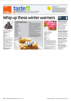
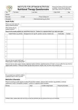


© Copyright 2026