
The first case of Brucella canis in Sweden:
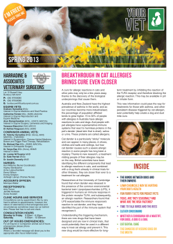
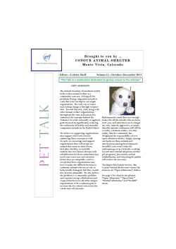
BRIEF COMMUNICATION
The first case of Brucella canis in Sweden:
background, case report and recommendations
from a northern European perspective
Bodil Ström Holst1,2*, Karin Löfqvist3, Linda Ernholm6, Karin Eld4, Maria Cedersmyg5 and Gunilla Hallgren6
Abstract
Infection with Brucella canis has been diagnosed in Sweden for the first time. It was diagnosed in a three-year-old
breeding bitch with reproductive disturbances. Fifteen in-contact dogs were tested repeatedly and all of them
were negative for B. canis. The source of infection could not be defined. The present article describes the case and
the measures undertaken and gives a short review over B. canis. Recommendations on how to avoid the infection
in non-endemic countries are given.
Keywords: Brucella canis, Canine abortion, Reproduction, Canine brucellosis
Background
Until 1994, Sweden had a quarantine requirement for all
dogs entering from abroad, except for dogs from Norway.
Norwegian dogs were allowed to enter without any
requirements at all since Norway has had the same epidemiological status as Sweden for several decades. From
1994 to 2004, however, dogs from EU and EFTA countries
were allowed entry without quarantine if in possession of
an identity mark, rabies vaccination, rabies antibody titre
control and certain other prophylactic treatments. An
import license, amongst other documentation, was
required. In 2004, the new European pet regulation 998/
2003 came into effect allowing entry in combination with
certain national requirements. For dogs from EU and
EFTA countries only identity marking, rabies vaccination,
rabies antibody testing, deworming and a pet passport was
needed. These changes in regulations facilitated travelling
of dogs between Sweden and other European countries,
especially those within the European Union (EU). In addition, it led to an increased responsibility for dog owners to
protect their travelling dogs against unfamiliar infections
that did not exist in Sweden.
Dogs travel with their owners for varying reasons,
such as holiday, dog shows or breeding. The number of
* Correspondence: [email protected]
1
Department of Animal Health and Antimicrobial Strategies, National
Veterinary Institute, SE-751 89 Uppsala, Sweden
Full list of author information is available at the end of the article
litters that are registered within the Swedish Kennel
Club from a stud dog that is not registered in Sweden
or Norway has increased since 1994, and is today
around 8% of the total number of litters born, or
approximately 1000 litters per year (Figure 1). Breeding
leads to a special risk for transmission of infectious diseases due to the close contact between the pair during
mating. Brucella canis is of special concern for breeding
dogs, since it can be transmitted venereally and cause
reproductive problems. There are reports that dogs can
also be infected with other Brucella species: Brucella
melitensis [1], Brucella suis [2] and Brucella abortus [3],
but it is only B. canis that is of epidemiological importance to the canine population.
Prevalence of B. canis
Infection with B. canis is common in Central and South
America and in southern USA [2,4] and has also been
reported from Canada [5,6]. In the USA, the dog trade
has led to a spread of the infection, and a growing need
for regulatory tools has been addressed [7]. In Georgia,
infection of dogs with B. canis was notable in 2003, and
a control strategy for the infection developed [8]. In
Asia the infection has been described and studied in
Japan [9,10], and it has also been reported from India
[11], the Philippines [12], Korea [13], China [14], Malaysia [15] and Taiwan [16], and in Africa from Nigeria
[17]. The number of described cases from central
137
10
% litters
9
8
7
6
5
4
3
2
1
0
1990
1991
1992
1993
1994
1995
1996
1997
1998
1999
2000
2001
2002
2003
2004
2005
2006
2007
2008
2009
2010
Figure 1 The proportion of litters registered from stud dogs not registered in Sweden or Norway (per cent of total number of litters
registered in Sweden).
Europe is low, but cases may be unrecorded since B.
canis is neither notifiable to the World Organisation for
Animal Health (OIE) nor to the EU. Stray dogs in the
Mediterranean area are suggested to serve as a reservoir
[18,19]. In recent years, the number of published reports
on B. canis infection has increased. In Finland the infection has been diagnosed from dogs that were imported
from Russia [20]. The first outbreak in Hungary of B.
canis in a kennel was recently described [21], and cases
in male dogs are reported from Italy and Germany
[22,23]. Infection with B. canis was recently described in
three poodle bitches in a kennel in Austria [24]. Earlier
cases in Austria were a mongrel dog from Greece and a
purebred male dog [24]. Antibodies to both B. abortus
and B. canis have been described in a male dog in
Poland [3]. There has been serological evidence that B.
canis is present in Great Britain [25], Germany [26-28],
Italy [29] and Spain [19]. One case from Great Britain is
reported in a dog that was imported from Spain [30]. In
a dog shelter in Turkey, 4 out of 48 dogs (8%) that had
died of unknown reason were found to be infected with
B. canis [18]. In Europe, infection with B. canis has also
been reported in laboratory dogs in former Czechoslovakia [31] and from a kennel in France [32]. A schematic
drawing of prevalence of B. canis is shown in Figure 2.
Pathogenesis
B. canis enters the body via the mucosal membranes of
the oropharynx, genital tract or conjunctiva [33,34]. The
bacteria are then phagocytised by macrophages and
other phagocytic cells, and transported via the blood to
lymph nodes, spleen and genital organs, where they
multiply [33]. Hyperplasia of the lymphoid tissue is seen
throughout the body [35]. Bacteremia occurs 1 to 4
weeks after the infection and persists for at least 6
months, and then reoccurs intermittently for up to 5
years [36].
Clinical signs
The most typical clinical sign is late-pregnancy abortion,
week 7-9, [37], and this was the first sign described to
be related to infection with Brucella canis when the bacterium was first recognized, in 1966 [38]. Aborted pups
are partially autolysed and show signs of a generalised
bacterial infection, such as subcutaneous oedema,
abdominal hemorrhages, and degenerative lesions in
liver, kidneys, spleen and intestines. The bitch continues
to excrete a brownish or green-gray discharge for 1-6
weeks after the abortion [37]. Sometimes early embryonic death and resorption occurs. Weak pups may die
within a few hours, but other times may survive up to a
month. Seemingly normal pups can also be born, and
develop the disease later [33]. Before puberty, a generalised lymphadenopathy is the most common clinical sign.
Brucellosis does not affect the oestrous cycle, and
bitches that have aborted can give birth to normal litters
in a subsequent pregnancy, or have intermittent reproductive disturbances [33].
138
B
B
BC
BC
A
B
A
A
A
BC
B
A
A
A
B
BC
A
B
A
A
A
A
A
A
A
Figure 2 Geographical distribution of B. canis, clinical cases and seropositive dogs. A: Reports (including serosurveys) indicate that the
infection is endemic. B: Few clinical cases or outbreaks in kennels have been described C: Serosurveys indicate that the seroprevalence is low.
In male dogs, epididymitis and prostatitis are the most
common clinical signs. In the acute stage, the epididymis increases in size and is painful. Frequent licking
may lead to scrotal oedema and dermatitis. In the
chronic phase the epididymides may become small and
hard, and the testicle atrophic. Orchitis and ulcerative
scrotum dermatitis can be seen [39]. Chronically
infected dogs are often oligo- or azoospermic, and infertile. Sperm defects that can be seen include tail defects,
loose heads and distal droplets [40]. In the testicles,
tubuli are fibrosed [41]. The testicular damage leads to
production of auto-antibodies towards the sperm cells.
These antibodies can be detected in serum and seminal
plasma from three months after the infection [42,43].
From 4 months after infection the sperm cells autoagglutinate [42].
Most often there are no general clinical signs, and
infected dogs do not usually have a fever. Sometimes
the dogs have a dull coat, or show decreased exercise
tolerance, but this is not common [44]. Lymphadenopathy of the retropharyngeal or inguinal lymph nodes, or
generalised lymphadenopathy sometimes occurs [33].
Discospondylitis has been reported [45,46]. Dogs that
were experimentally infected with B. canis often showed
recurrent uveitis for several months after the infection
[40] and there are several case reports describing ocular
signs, such as endophthalmitis and anterior uveitis, in
dogs infected with B. canis [47-49]. Osteomyelitis
related to hip prostheses has been described in two dogs
[50]. Chronic meningitis and non-suppurative encephalitis has been associated with bacteria belonging to the
genus Brucella [33].
Diagnosis
The diagnosis is made by culture, polymerase chain
reaction (PCR) or serology, often in combination. To
make a definitive diagnosis by bacterial culture, the dog
should not be treated with antimicrobials. Blood culture
can be used, but often genital samples are better, especially if the dog shows clinical signs of the genital tract.
B. canis may be cultured from semen and vaginal discharges during oestrus or after abortion and a high concentration of the infecting agent can be found in the
placenta. Urine cultures can be positive even if a blood
culture is negative. The highest urine concentration is
seen 8-12 weeks after infection, with the concentration
in urine being higher from male dogs than from bitches
[51]. The organs most suitable for culture, from biopsies
or at post mortem examination, are lymph nodes, prostate and spleen [26]. PCR-analysis of whole blood has a
high sensitivity, whereas serum is not suitable for PCR
analysis [52]. Semen can be analysed with PCR [53,54].
PCR can be a more sensitive method than bacterial culture because it detects not only viable but also dead
bacteria, and because the method is not affected by contamination with other bacteria [55].
For screening, serologic tests are used. A rapid slide
agglutination test, RSAT, is commonly used, although a
drawback to this method is the risk of false positive
samples. By treating the sample with 2-mercapto-ethanol (2-ME) the number of false positive samples
decreases [19,56] because IgM is dissociated and IgM
cross-reacts with other bacteria more commonly than
IgG [57]. The agar gel immunodiffusion (AGID) test has
been described as suitable if a chronic infection is
139
suspected, because more chronic infections are positive
using an AGID than using other tests [36]. However,
the AGID is not used by many laboratories today
because of its low sensitivity and because it requires
trained personnel and special media [8]. Enzyme-linked
immunosorbent assays (ELISA) have been developed
[58] and a lateral flow immune-chromatographic assay
(LFIA) has recently been described [59]. A combination
of different serological tests is often recommended.
Treatment
Different antibiotics have been effective against B. canis
in in vitro studies [1,60], but multiresistant strains have
also been described [61]. Treatments that have been
evaluated include 1-3 weeks of combinations of different
antibiotics such as streptomycin, tetracycline, rifampicin
and sulphonamides [62,63], two weeks’ treatment each
with tetracycline, followed by dihydrostreptomycin and
trimetoprim-sulfadiazine [64], or 30 days of treatment
with enrofloxacin [65]. Clinically, no antibiotic treatment
has been shown to eliminate the infection in all treated
animals. If B. canis is diagnosed in a kennel, the affected
animal should be isolated and not used for breeding.
The environment should be thoroughly cleaned and disinfected. Since an infected dog, even if it is clinically
healthy, can infect other dogs and humans, euthanasia is
often recommended.
Transmission
Dogs are most commonly infected by contact with vaginal discharges at oestrous or after abortions, or by
ingesting infected placentas or foetuses. At abortion, the
placenta and the discharges can contain up to 1010 colony forming units (cfu) per mL, and the oral infection
dose is 2 × 10⁶ cfu. Experimentally, 104 cfu has been
sufficient for conjunctival infection [34,40,66]. Thus, 1
mL placental tissue or vaginal discharge is equal to
approximately 100 000 infectious doses, and the bitches
can have a vaginal discharge for up to 6 weeks after an
abortion. Dogs can also be infected at mating. It should
be observed that chronically infected dogs can be serologically negative and negative on blood culture, although
B. canis can be detected from the prostate, epididymis
and semen, or in female dogs in vaginal secretion, and
thus they can still infect other dogs at mating or via
artificial insemination [36,54,67].
Environmental infection is possible, especially from
areas where dogs often urinate [34], or where vaginal discharges are deposited. Dogs living together are at risk of
infecting each other. Within half a year, infected male
dogs had infected other male dogs, and bitches had
infected other bitches [51]. It was suspected that contaminated urine was an important source of infection in
these cases, especially from male dogs. The concentration
of bacteria in urine and semen is highest from 1 to 4-6
months after infection [51].
Recommendations to avoid introduction of B. canis into
non-endemic areas
To avoid introducing brucellosis into a kennel, the
introduction of untested dogs from endemic areas
should be avoided. Dogs from kennels in which B. canis
has been diagnosed should not be used for breeding
(Table 1). Dogs from endemic areas should be kept isolated until tested free of B. canis to avoid further spread
of the disease. This is recommended for natural mating,
artificial insemination with fresh, chilled or frozen
semen, and when introducing new dogs into the kennels. For kennels in endemic areas it is often recommended that breeding animals are tested annually, and
that all new dogs are tested before being introduced
into the kennel. Serologic tests can be negative up to 4
weeks after infection, and at least 12 weeks must pass to
be sure of detecting antibodies in an infected animal
[36]. Therefore, two negative tests 4-6 weeks apart are
needed in case the dog is incubating the disease [8]. At
least one of the sampling occasions should be no earlier
than 12 weeks after suspected contact with an infected
animal. Bitches that might be chronically infected
should be tested during oestrus, pregnancy or at abortion, as they may have low antibody titres at other times
[44]. Chronically infected male dogs can also be difficult
to detect, as they too can be serologically negative. In
addition to serologic methods, bacterial culture or PCR
analysis can be performed to detect the bacterium. Only
dogs tested free from B. canis should be used for breeding, and recommended tests depend on the level of risk
(Table 1).
If B. canis is detected in a group of dogs, regular testing and removal of infected dogs and strict hygiene is
necessary to eradicate the infection [28]. Common disinfectants are effective against B. canis.
Infection with B. canis in humans
Humans can become infected with B. canis, although
reported cases are scarce. The clinical presentation is
unspecific, including fever (as opposed to the disease in
dogs), headache and fatigue, and a history of exposure
to dogs is usually needed to raise suspicion of infection
with B. canis [68]. The infection may be underdiagnosed
due to its rather unspecific symptoms. Positive blood
cultures confirm the diagnosis, but as the patients have
been treated with antibiotics in many cases, the risk of a
false negative culture is increased [69]. Furthermore, B.
canis is slow-growing and mildly fastidious, producing
low-grade bacteremias, decreasing cultural sensitivity
[68]. Serodiagnosis is problematic due to a lack of antigenic cross-reactivity with the antigens routinely used,
140
Table 1 Recommendations for testing dogs from areas endemic for B. canis when mated or inseminated with dogs
from non-endemic areas, or when imported to non-endemic areas
Risk
level
Situation
Recommended test
1
Mating or insemination, no suspicion of B. canis and no reproductive
disturbances
Blood sample for antibody analysis
2
Mating or insemination with dog with previous reproductive disturbances,
import of dogs without reproductive disturbances.
Serology: 2 samples 4-6 weeks apart
3
Import of or mating/insemination with dogs with previous reproductive
disturbances, infection with B. canis suspected.
Serology, 2 samples 4-6 weeks apart, blood culture and
culture or PCR from semen or vagina
4
Import of or mating/insemination with dog from kennel with endemic infection
of B. canis.
Dissuaded from mating or import.
and a case that was serologically negative for B. canis
despite positive blood culture has also been described
[70]. Treatment with antibiotics is usually effective in
humans.
There are several reports on disease in humans without immune disorders. A 17-month old boy had refusal
to walk as the chief symptom [71]. An outbreak with
fever, diarrhoea and vomiting in children and headache,
fatigue, myalgia and nausea in adults has been described
[72]. Fever, fatigue, and nausea are common clinical
signs [73,74]. A man with endocarditis was serologically
positive for B. canis although the source of infection
could not be established [75]. A fifteen year old boy suffered from weakness, persistent fever and lymphadenopathy [76]. Sometimes relapses and more prolonged
courses of illness are described [69].
Clinical infection with B. canis has also been reported
in persons with immune disorders. Signs such as fatigue,
fever, malaise, headache, cough and arthralgia have been
described [77,78].
Case presentation
In December 2010, a 3-year-old American Staffordshire
terrier imported from Poland, expecting her first litter,
was brought to the Regional Animal Hospital of Helsingborg, Sweden. She was 46 days pregnant but
although her general condition was good, she had a
green vaginal discharge. An ultrasonography was performed which revealed that all pups were dead. The
bitch was aborted with aglepristone and prostaglandins,
and also received amoxicillin. Hematology showed mild
leukocytosis and moderate eosinophilia. Bacterial culture
from the vagina showed no bacterial growth. The bitch
was monitored with ultrasonography and clinical examinations for the following weeks. Her general condition
was good and the discharge ceased rapidly. No foetal
parts could be identified in the discharge.
In the follow-up of the abortion the owner reported
that the bitch had been mated in Poland, and that she
had been mated once previously with a dog from Serbia,
although she did not conceive at the time. This led to
the suspicion of brucellosis, and the bitch was tested
serologically for B. canis in January 2011, with a negative result. An analysis for Giardia and a Baermanns flotation test for parasites were also negative. The owner
was recommended to vaccinate the bitch against canine
herpesvirus at the next mating, and to follow progesterone levels during the next pregnancy.
In the end of May 2011 the bitch was again presented
at the animal hospital. She had been taken to Poland
for mating to the same dog again, and now had a
bloody discharge at day 59 of pregnancy. Ultrasonography was performed and all but one pup were dead. A
caesarean section was performed, but no pups were
alive. Ten dead, partially autolysed pups were delivered.
The uterus was filled with a brown-greenish discharge
and all pups had cutaneous ulcerations. The bitch was
treated with amoxicillin and cabergolin, and recovered
uneventfully. This second abortion in the last trimester
in a clinically healthy bitch mated abroad again raised
the suspicion of brucellosis, and the placentae from
aborted foetuses were sent to the National Veterinary
Institute (SVA) for bacteriological analysis, specifically
asking for B. canis.
The culture result from the placentae was positive. As
this was the first case of B. canis in Sweden, the strain
was sent to EU-RL (European Union Reference Labaratory) in Alfort, France, for confirmation. The bitch was
also tested serologically, and was now positive for B.
canis. The Swedish Board of Agriculture (JV) decided
that the bitch had to be isolated until a definitive diagnosis was made (confirmation by EU-RL); they initiated
contact tracing and contacted the county veterinarian
and the county medical officer. The bitch was tested
again by blood culture, vaginal culture and serology
(RSAT). B. canis grew in the blood culture and in the
vaginal culture, and the bitch still had antibodies to B.
canis. The first bacteriological culture was confirmed by
the EU-RL. The Swedish Board of Agriculture decided
that the bitch should be euthanized, as recommended
by SVA. The bitch was euthanized at the Regional Animal Hospital of Helsingborg.
141
Contact tracing
SVA recommended how and which dogs to test, and JV
had the main responsibility for the contact tracing. Dogs
within the country that had been in contact with the
infected bitch through mating, common housing or
other close contact, were tested. All in-contact dogs
were tested twice, with cultures from blood and the genital tract, and by serology. At least one of the occasions
should be minimum 12 weeks after the latest risk contact. At least one sample from the genital tract should
be during oestrus in bitches, and semen was cultured
from the male dogs.
Possible sources of infection included the two dogs
that had mated the bitch. The first dog that had
mated the bitch, when she did not conceive, had been
sold to Sweden by this time and was a stud dog in
northern Sweden. There was a history of bitches not
conceiving after mating with him. A semen sample in
October 2010 revealed azoospermia, macrophages and
epithelial cells in the small ejaculate of 1 mL. In July
2011 the total sperm count was 115 million, with 60%
sperm having various unspecified defects and an abundance of epithelial cells. Two months later, in September, the total sperm count was 1265 million, 75%
progressive motility and 10% unspecified defects. In
November 2011 a new semen sample was taken and
evaluated. The total number of sperm was good (1700
million), their motility was good and defects were
within normal limits. Epithelial cells and leukocytes
were also within normal limits. He was tested for B.
canis in July, September and November 2011 by blood
and semen culture and by serology (RSAT), and in
November semen was also analysed by PCR. In-contact dogs to this male dog were tested in the same
way as in-contact dogs to the bitch. In total, 15 incontact dogs of different breeds were tested with no
test results being positive. The contact tracing is schematically described in Figure 3.
In Poland, the male that mated the bitch the second
and third time was serologically tested (RSAT) twice
during the autumn 2011, with negative results, and he is
still being used for breeding in Poland. He resides in a
kennel. The Brucella status of the other dogs is not
known.
Discussion
The bitch was imported as a young dog to Sweden from
Poland. The first male dog that she was mated to, from
Serbia, has a history of bitches not conceiving after mating, including the present bitch. There are several causes
for bitches not to conceive, including poor semen quality due to infection with B. canis. This male dog has
been included in the contact tracing as he now resides
in Sweden.
142
At the second and third mating, a male dog from
Poland was used. The bitch conceived after both these
matings, but aborted late in pregnancy, which is typical
for infection with B. canis [44]. Autolysed pups with
subcutaneous hemorrhages are typical and could be
seen in the present case. The vaginal discharges had a
short duration, possibly due to the fact that the bitch
was treated with amoxicillin after both abortions.
The bitch was serologically negative for B. canis in
January 2011, after the first abortion. More than 12
weeks had passed since the bitch mated the first male
dog, and 11 weeks since she mated the second male,
from Poland. In addition she was sampled close to an
abortion, which stimulates antibody production in
chronically infected bitches. No bacterial cultures were
carried out at this occasion. The suspicions were directed against the male dog from Serbia, because bitches
had failed to conceive after mating with him, whereas
the other male dog had sired healthy litters. Extensive
testing of the imported dog from Serbia has been carried out, and all results were negative for B. canis. His
semen quality, although previously was poor, has normalised. The second male dog has been tested twice by
serology, and also found negative. The source of infection for the bitch could thus not be established.
Brucellosis in food-producing animals in Sweden is
regulated by law and the costs for measures against the
disease, including compensation to the animal owner,
are paid by the government. The reasons for the government to act to eradicate the infection in a dog were a
combination of the fact that B. canis is a zoonosis and
that a quick and reliable diagnosis is missing, thereby
making it difficult for owners to protect themselves and
their animals, should the infection be established within
the country. International experiences show that powerful measures, such as euthanasia of infected animals, is
often needed to eradicate the infection in kennels and
to stop further spread of the infection [8].
What are the consequences for the pet owners?
The diagnosis of B. canis in the bitch was a personal
tragedy to the owner. She lost her dog, money and
future pups. She was a small-scale breeder; in larger
kennels the economic consequences will be even bigger
as all breeding stock may be lost. The consequences
were considerable also for the owners of in-contact
dogs. They had to keep their dogs separate and bring
them to animal clinics or hospitals for sampling, and
could not use them for breeding until they were
declared free from B. canis.
Conclusions
A typical case of B. canis in a dog with reproductive disturbances has been diagnosed in Sweden. Handling of
&ĞŵĂůĞ
&ĞŵĂůĞ
DĂůĞ
DĂůĞĨƌŽŵ^ĞƌďŝĂ
DĂůĞ
DĂůĞ
DĂůĞ
&ĞŵĂůĞ
&ĞŵĂůĞ
ŵĂƚŝŶŐ
&ĞŵĂůĞ
DĂůĞ
ĂƐĞĨĞŵĂůĞ
DĂƚŝŶŐƚǁŝĐĞǁŝƚŚ
ĚŽŐŝŶWŽůĂŶĚ
&ĞŵĂůĞ
&ĞŵĂůĞ
&ĞŵĂůĞ
&ĞŵĂůĞ
Figure 3 Schematic figure describing contact tracing. The bitch within the red circle is the one diagnosed with B. canis. Male dog within
blue circle: the first dog (from Serbia) that mated the bitch and that now lives in Sweden. Thick arrows: contact through mating. Thin arrows:
Dogs that had other close contact. Dogs that lived together in the same household are placed together.
the case by the practicing veterinarian and by the authorities is described. Results from the contact tracing indicated that the infection most probably has not spread
within the country. The source of infection could not be
identified. Recommendations on how to avoid introduction of B. canis into non-endemic areas are given.
Author details
Department of Animal Health and Antimicrobial Strategies, National
Veterinary Institute, SE-751 89 Uppsala, Sweden. 2Department of Clinical
Sciences, Swedish University of Agricultural Sciences, Box 7054, SE-750 07
Uppsala, Sweden. 3Helsingborg Animal Hospital, Box 220 97, SE-250 23
Helsingborg, Sweden. 4Department of Bacteriology, National Veterinary
Institute, SE- 751 89 Uppsala, Sweden. 5Swedish Board of Agriculture, 551 82
Jönköping, Sweden. 6Department of Disease Control and Epidemiology,
National Veterinary Institute, SE-751 89 Uppsala, Sweden.
Abbreviations
AGID: Agar gel immunodiffusion; CFU: Colony forming units; ELISA: Enzymelinked immunosorbent assay; LFIA: Lateral flow immune-chromatographic
assay; 2-ME: 2-mercapto-ethanol; PCR: Polymerase chain reaction; RSAT:
Rapid slide agglutination test.
Authors’ contributions
BSH compiled the information, initiated and drafted the manuscript, KL
handled the case including communication with authorities and drafted the
manuscript, LE and GH handled the contact tracing at the National
Veterinary Institute and MC was responsible at the Swedish Board of
Agriculture. KE was responsible for the bacteriological parts. All authors read
and approved the final manuscript.
Acknowledgements
The authors thank Helena Rosenberg on the Swedish Kennel Club for
information on registered litters. Lennart Melin at the National Veterinary
Institute is acknowledged for help with bacteriological matters and Pia
Ehrenpris at the Swedish Board of Agriculture for information on the contact
tracing.
1
Competing interests
The authors declare that they have no competing interests.
Received: 8 March 2012 Accepted: 27 March 2012
Published: 27 March 2012
143
References
1. Mateu-de-Antonio EM, Martin M: In vitro efficacy of several antimicrobial
combinations against Brucella canis and Brucella melitensis strains
isolated from dogs. Vet Microbiol 1995, 45:1-10.
2. Lucero NE, Ayala SM, Escobar GI, Jacob NR: Brucella isolated in humans
and animals in Latin America from 1968 to 2006. Epidemiol Infect 2008,
136:496-503.
3. Kopczewski A, Krolak M, Arent Z, Rudnicki K: A case of brucellosis in a
male dog Przypadek brucelozy u psa. Zycie Weterynaryjne 1995,
70:230-231.
4. Lovejoy GS, Carver HD, Moseley IK, Hicks M: Serosurvey of dogs for
Brucella canis infection in Memphis, Tennessee. Am J Public Health 1976,
66:175-176.
5. Bosu WT, Prescott JF: A serological survey of dogs for Brucella canis in
southwestern Ontario. Can Vet J 1980, 21:198-200.
6. Brennan SJ, Ngeleka M, Philibert HM, Forbes LB, Allen AL: Canine
brucellosis in a Saskatchewan kennel. Can Vet J 2008, 49:703-708.
7. Brower A, Okwumabua O, Massengill C, Muenks Q, Vanderloo P, Duster M,
Homb K, Kurth K: Investigation of the spread of Brucella canis via the U.
S. interstate dog trade. Int J Infect Dis 2007, 11:454-458.
8. Hollett RB: Canine brucellosis: outbreaks and compliance. Theriogenology
2006, 66:575-587.
9. Saegusa J, Ueda K, Goto Y, Fujiwara K: A survey of Brucella canis infection
in dogs from Tokyo area. Nihon Juigaku Zasshi 1978, 40:75-80.
10. Hayashi TT, Isayama Y: Detection of Brucella canis infection in dogs in
Hokkaido. Microbiol Immunol 1977, 21:295-298.
11. Srinivasan VK, Nedunchelliyan S, Venkataraman KS: Seroepidemiology of
canine brucellosis in Madras city. Indian Vet J 1992, 69:978-980.
12. Baluyut CS, Duguies MV: Serological survey for Brucella canis agglutinins
in dogs using macroscopic tube agglutination test. Philippine J Vet Med A
1977, 16:93-101.
13. Park C, Oh J, Park CK, Oh JY: Bacteriological and serological investigation
of Brucella canis infection of dogs in Taegu city, Korea. Korean J Vet Res
2001, 41:67-71.
14. Jiang FX: A survey on canine brucellosis in Wusu County. Chinese J Vet Sci
Technol 1989, , 1: 18-19.
15. Joseph PG, Mahmud ZBH, Sirimanne ES: Canine brucellosis in Malaysia: a
first report. Kajian Veterinar 1983, 15:17-22.
16. Tsai IS, Lu YS, Isayama Y, Sasahara J: Serological survey for Brucella canis
infection in dogs in Taiwan and the isolation and identification of Br.
canis. Taiwan J Vet Med Animal Husbandry 1983, 42:91-98.
17. Adesiyun AA, Abdullahi SU, Adeyanju JB: Prevalence of Brucella abortus
and Brucella canis antibodies in dogs in Nigeria. J Small Anim Pract 1986,
27:31-37.
18. Aras Z, Ucan US: Detection of Brucella canis from inguinal lymph nodes
of naturally infected dogs by PCR. Theriogenology 2010, 74:658-662.
19. Mateu-de-Antonio EM, Martin M, Casal J: Comparison of serologic tests
used in canine brucellosis diagnosis. J Vet Diagn Invest 1994, 6:257-259.
20. [http://www.evira.fi/portal/se/djur/djurhalsa_och_sjukdomar/djursjukdomar/
hundar/brucella_canis].
21. Gyuranecz M, Szeredi L, Ronai Z, Denes B, Dencso L, Dan A, Palmai N,
Hauser Z, Lami E, Makrai L, et al: Detection of Brucella canis-induced
reproductive diseases in a kennel. J Vet Diagn Invest 2011, 23:143-147.
22. Corrente M, Franchini D, Decaro N, Greco G, D’Abramo M, Greco MF,
Latronico F, Crovace A, Martella V: Detection of Brucella canis in a dog in
Italy. New Microbiol 2010, 33:337-341.
23. Nockler K, Kutzer P, Reif S, Rosenberger N, Draeger A, Bahn P, Gollner C,
Erlbeck C: Canine brucellosis-a case report. Berl Munch Tierarztl
Wochenschr 2003, 116:368-372.
24. Schäfer-Somi S, Hofer E: Brucella canis - ein wenig beachteter
Zoonoseereger, aktuelle Fälle. Austrian Vet J 2011, 1:20-24.
25. Taylor DJ: Serological evidence for the presence of Brucella canis
infection in dogs in Britain. Vet Rec 1980, 106:102-104.
26. Weber A, Schliesser T: Serological and cultural determination of Brucella
canis in beagles in laboratory kennels. Zentralbl Veterinarmed B 1975,
22:403-410.
27. Weber A, Schliesser T: The occurrence of antibodies to Brucella canis in
domestic dogs in the Federal Republic of Germany. Berl Munch Tierarztl
Wochenschr 1978, 91:28-30.
28. Kruedener RBv: Outbreak of a Brucella canis infection in a beagle colony
in West Germany. Int Symp on brucellosis (II), Rabat, Morocco, 1975. Develop.
biol. Standard 1976, 31:251-253.
29. Ebani VV, Cerri D, Fratini F, Bey RF, Andreani E: Serological diagnosis of
brucellosis caused by Brucella canis. New Microbiol 2003, 26:65-73.
30. Dunne J, Sehgal K, McMillan A, Perret L: Canine brucellosis in a dog
imported into the UK. Vet Rec 2002, 151:247.
31. Sebek Z, Sykora I, Holda J, Komarek J: Serological demontsration of
Brucella canis in the breeding of laboratory dogs of the beagle breed in
Czechoslovakia. Cesk Epidemiol Mikrobiol Imunol 1976, 25:129-136.
32. Fontbonne A: Incidence of infectious diseases on canine infertility. Book
Incidence of infectious diseases on canine infertility (Editor ed.^eds.). City 2002,
56-57.
33. Carmichael LE, Kenney RM: Canine brucellosis: the clinical disease,
pathogenesis, and immune response. J Am Vet Med Assoc 1970,
156:1726-1734.
34. Serikawa T, Muraguchi T: Significance of urine in transmission of canine
brucellosis. Nihon Juigaku Zasshi 1979, 41:607-616.
35. Spink WW, Morisset R: Epidemic canine brucellosis due to a new species:
Brucella canis. Trans Am Clin Climatol Assoc 1970, 81:43-50.
36. Carmichael LE, Zoha SJ, Flores-Castro R: Problems in the serodiagnosis of
canine brucellosis: dog responses to cell wall and internal antigens of
Brucella canis. Dev Biol Stand 1984, 56:371-383.
37. Carmichael LE, Kenney RM: Canine abortion caused by Brucella canis. J
Am Vet Med Assoc 1968, 152:605-616.
38. Carmichael LE: Canine brucellosis: isolation, diagnosis, transmission. Proc
Annu Meet US Anim Health Assoc 1967, 71:517-527.
39. Schoeb TR, Morton R: Scrotal and testical changes in canine brucellosis: a
case report. J Am Vet Med Assoc 1978, 172:598-600.
40. Carmichael LE: Canine brucellosis: An annotated review with selected
cautionary comments. Theriogenology 1976, 6:105-116.
41. Moore JA, Kakuk TJ: Male dogs naturally infected with Brucella canis. J
Am Vet Med Assoc 1967, 155:1352-1358.
42. Serikawa T, Takada H, Kondo Y, Muraguchi T, Yamada J: Multiplication of
Brucella canis in male reproductive organs and detection of
autoantibody to spermatozoa in canine brucellosis. Dev Biol Stand 1984,
56:295-305.
43. Serikawa T, Muraguchi T, Yamada J, Takada H: Long-term observation of
canine brucellosis: excretion of Brucella canis into urine of infected male
dogs. Jikken Dobutsu 1981, 30:7-14.
44. Wanke MM: Canine brucellosis. Anim Reprod Sci 2004, 82-83:195-207.
45. Kerwin SC, Lewis DD, Hribernik TN, Partington B, Hosgood G, Eilts BE:
Diskospondylitis associated with Brucella canis infection in dogs: 14
cases (1980-1991). J Am Vet Med Assoc 1992, 201:1253-1257.
46. Henderson RA, Hoerlein BF, Kramer TT, Meyer ME: Discospondylitis in three
dogs infected with Brucella canis. J Am Vet Med Assoc 1974, 165:451-455.
47. Ledbetter EC, Landry MP, Stokol T, Kern TJ, Messick JB: Brucella canis
endophthalmitis in 3 dogs: clinical features, diagnosis, and treatment.
Vet Ophthalmol 2009, 12:183-191.
48. Vinayak A, Greene CE, Moore PA, Powell-Johnson G: Clinical resolution of
Brucella canis-induced ocular inflammation in a dog. J Am Vet Med Assoc
2004, 224:1804-1807.
49. Riecke JA, Rhoades HE: Brucella canis isolated from the eye of a dog. J
Am Vet Med Assoc 1975, 166:583-584.
50. Smeak DD, Olmstead ML, Hohn RB: Brucella canis osteomyelitis in two
dogs with total hip replacements. J Am Vet Med Assoc 1987, 191:986-990.
51. Carmichael LE, Joubert JC: Transmission of Brucella canis by contact
exposure. Cornell Vet 1988, 78:63-73.
52. Keid LB, Soares RM, Vasconcellos SA, Salgado VR, Megid J, Richtzenhain LJ:
Comparison of a PCR assay in whole blood and serum specimens for
canine brucellosis diagnosis. Vet Rec 2010, 167:96-99.
53. Kim S, Lee DS, Suzuki H, Watarai M: Detection of Brucella canis and
Leptospira interrogans in canine semen by multiplex nested PCR. J Vet
Med Sci 2006, 68:615-618.
54. Keid LB, Soares RM, Vasconcellos SA, Chiebao DP, Megid J, Salgado VR,
Richtzenhain LJ: A polymerase chain reaction for the detection of
Brucella canis in semen of naturally infected dogs. Theriogenology 2007,
67:1203-1210.
55. Bricker BJ: PCR as a diagnostic tool for brucellosis. Vet Microbiol 2002,
90:435-446.
144
56. Keid LB, Soares RM, Vasconcellos SA, Megid J, Salgado VR, Richtzenhain LJ:
Comparison of agar gel immunodiffusion test, rapid slide agglutination
test, microbiological culture and PCR for the diagnosis of canine
brucellosis. Res Vet Sci 2009, 86:22-26.
57. Deutsch HF, Morton JI: Dissociation of human serum macroglobulins.
Science 1957, 125:600-601.
58. de Oliveira MZ, Vale V, Keid L, Freire SM, Meyer R, Portela RW, BarrouinMelo SM: Validation of an ELISA method for the serological diagnosis of
canine brucellosis due to Brucella canis. Res Vet Sci 2011, 90:425-431.
59. Corde Y, Drapeau A, Albert D, Garin-Bastuji B: Assessment of a lateral flow
immuno-chromatographic assay forr the serological diagnosis of canine
brucellosis due to Brucella canis. losis International Research Conference
Including the 64th Brucellosis Research Conference, September 21-23, Buenos
Aires, Argentina, P139, 123 (abstract) 2011.
60. Hall WH, Manion RE: In vitro susceptibility of Brucella to various
antibiotics. Appl Microbiol 1970, 20:600-604.
61. Terakado N, Ueda H, Sugawara H, Isayama Y, Koyama N: Drug susceptibility
of Brucella canis isolated from dogs. Nihon Juigaku Zasshi 1978,
40:291-295.
62. Flores-Castro R, Carmichael LE: Brucella canis infection in dogs: treatment
trials. Rev Latinoam Microbiol 1981, 23:75-79.
63. Zoha SJ, Walsh R: Effect of a two-stage antibiotic treatment regimen on
dogs naturally infected with Brucella canis. J Am Vet Med Assoc 1982,
180:1474-1475.
64. Johnson CA, Bennett M, Jensen RK, Schirmer R: Effect of combined
antibiotic therapy on fertility in brood bitches infected with Brucella
canis. J Am Vet Med Assoc 1982, 180:1330-1333.
65. Wanke MM, Delpino MV, Baldi PC: Use of enrofloxacin in the treatment of
canine brucellosis in a dog kennel (clinical trial). Theriogenology 2006,
66:1573-1578.
66. Carmichael LE, Zoha SJ, Flores-Castro R: Biological properties and dog
response to a variant (M-) strain of Brucella canis. Dev Biol Stand 1984,
56:649-656.
67. Keid LB, Soares RM, Vasconcellos SA, Chiebao DP, Salgado VR, Megid J,
Richtzenhain LJ: A polymerase chain reaction for detection of Brucella
canis in vaginal swabs of naturally infected bitches. Theriogenology 2007,
68:1260-1270.
68. Rumley RL, Chapman SW: Brucella canis: an infectious cause of
prolonged fever of undetermined origin. South Med J 1986, 79:626-628.
69. Polt SS, Dismukes WE, Flint A, Schaefer J: Human brucellosis caused by
Brucella canis: clinical features and immune response. Ann Intern Med
1982, 97:717-719.
70. Rifkin GD, Supena RB, Axelson JA: Case report. Brucella canis bacteremia:
a case with negative B canis agglutinins. Am J Med Sci 1978, 276:113-115.
71. Tosi MF, Nelson TJ: Brucella canis infection in a 17-month-old child
successfully treated with moxalactam. J Pediatr 1982, 101:725-727.
72. Lucero NE, Corazza R, Almuzara MN, Reynes E, Escobar GI, Boeri E, Ayala SM:
Human Brucella canis outbreak linked to infection in dogs. Epidemiol
Infect 2010, 138:280-285.
73. Nomura A, Imaoka K, Imanishi H, Shimizu H, Nagura F, Maeda K, Tomino T,
Fujita Y, Kimura M, Stein G: Human Brucella canis infections diagnosed by
blood culture. Emerg Infect Dis 2010, 16:1183-1185.
74. Rousseau P: Brucella canis infection in a woman with fever of unknown
origin. Postgrad Med 1985, 78:249-257.
75. Ying W, Nguyen MQ, Jahre JA: Brucella canis endocarditis: case report.
Clin Infect Dis 1999, 29:1593-1594.
76. Lucero NE, Jacob NO, Ayala SM, Escobar GI, Tuccillo P, Jacques I: Unusual
clinical presentation of brucellosis caused by Brucella canis. J Med
Microbiol 2005, 54:505-508.
77. Lawaczeck E, Toporek J, Cwikla J, Mathison BA: Brucella canis in a HIVinfected patient. Zoonoses Public Health 2011, 58:150-152.
78. Lucero NE, Maldonado PI, Kaufman S, Escobar GI, Boeri E, Jacob NR:
Brucella canis causing infection in an HIV-infected patient. Vector Borne
Zoonotic Dis 2010, 10:527-529.
145


© Copyright 2026