
In Vitro for Endocrine Active Substances: What is Miriam N. Jacobs
In Vitro Metabolism and Bioavailability Tests
for Endocrine Active Substances: What is
Needed Next for Regulatory Purposes?
Miriam N. Jacobs 1, Susan C. Laws 2, Kate Willett 3, Pat Schmieder 4,
Jenny Odum 5, and Toine F. Bovee 6
1
2
Scientific Committee and Emerging Risks Unit, European Food Safety Authority, Parma, Italy; Endocrinology
Branch (MD-72), Toxicity Assessment Division, National Health and Environmental Effects Research Laboratory, US EPA,
3
4
Research Triangle Park, NC, USA; The Humane Society of the United States, Washington, DC, USA; Mid-Continent
Ecology Division, National Health and Environmental Effects Research Laboratory, US EPA, Duluth,
5
6
MN, USA; Regulatory Science Associates, Dunoon, Argyll, UK; RIKILT – Institute of Food
Safety, Wageningen University and Research Centre, Wageningen, The Netherlands
Summary
Legislation and prospective legislative proposals internationally (may) require that chemicals be tested
for their ability to disrupt the hormonal systems of mammals. Chemicals found to test positive in vitro are
considered to be endocrine active substances (EAS) and may be putative endocrine disruptors (EDs)
in vivo. While there is a growing body of international in vitro test guidelines addressing EAS mechanisms
and modes of action, to date there are still few or no standardized methods to incorporate metabolic and
toxicokinetic aspects into these in vitro tests for EAS. In vitro assays for EAS should incorporate metabolic
enzyme systems to better address the relevance of EAS tests to in vivo adverse outcome pathways,
and a previous OECD review paper indicated how this could be done. This paper revisits those
recommendations, addressing where research and funding efforts are needed to expedite the development
of suitable in vitro metabolism systems to improve the accuracy of in vitro assays for identifying
EAS and EDs. Recommendations are made for projects to support short, medium, and long-term goals.
The complexity of in vivo metabolism presents major challenges for the development of predictive
models suitable for the extrapolation of data from in silico/in vitro approaches to models that can occur
in vivo. Therefore, the long-term recommendations are intended to foster an international harmonization
of databases, delineation of metabolic pathways, and development of predictive tools that will
provide a fundamental understanding of the processes by which metabolism occurs, increasing the
predictive accuracy of in silico/in vitro methods.
Keywords: regulatory testing, metabolism, endocrine active substances, alternative methods
Abbreviations
A, androgen; AGD, anogenital distance; AhR, aryl hydrocarbon receptor; AOP, adverse outcome pathway; AR, androgen receptor;
BPA, bisphenol A; CAR, constitutive androstane receptor; CYP, cytochrome P450; DAR, Draft Assessment Report (EU);
DER, Data Evaluation Record (US and Canada); DHEA, dehydroepiandrosterone; DRP, Detailed Review Paper; E, estrogen;
EAS, endocrine active substances; EURL ECVAM, EU Reference Laboratory ECVAM; ED, endocrine disruptor; EDSP, US
EPA’s Endocrine Disruptor Screening Program; EDTA-AG, OECD Endocrine Disruptor Testing and Assessment Advisory Group;
ENDOMET, EU 5th framework project “Dysregulation of endogenous steroid metabolism potentially alters neuronal and reproductive
system development: effects of environmental plasticisers”; ER, estrogen receptor; GD, Guidance Document; GR, glucocorticoid
receptor; HPG, hypothalamic-pituitary-gonadal axis; HPTE, 2-bis(p-hydroxyphenyl)-1,1,1-trichloroethane; JaCVAM, Japan Centre
for the Validation of Alternative Methods; ICCVAM, Interagency Coordinating Committee on the Validation of Alternative Methods;
ITS, integrated testing strategy; Log Kow, octanol-water partition coefficient; MIE, molecular initiating event; mRNA, messenger
RNA, NADPH, reduced form of nicotinamide adenine dinucleotide phosphate, OECD, Organisation for Economic Cooperation
and Development; PBTG, performance based test guideline; PHS, prostaglandin H synthase; PPAR, peroxisome proliferator
activated receptor(s); PR, progesterone receptor; PXR, pregnane X receptor; RAR, retinoic acid receptor; RXR, retinoic X receptor;
RCBA, recombinant cell bioassay; S, steroidogenesis; S9, supernatant product of an organ tissue homogenate used in biological
metabolism assays; SOP, standard operating protocols; StAR, steroidogenic acute regulatory protein; STTA, stably transfected
transactivation assay; SULT, sulfotransferase; PAPS, 3’-phosphoadenosine-5’-phosphosulphate; PBTK, physiologically based
toxicokinetic modelling; QSAR, Quantitative Structure Activity Relationship; T, thyroid; T3, triiodothyronine; T4, thyroxine; TIM, TNO
gastro-intestinal tract model; TG, Test Guideline; TR, thyroid receptor; T-Screen, thyroid receptor-screen; TOFMS, time-of-flight
mass spectrometry; UGT, UDP-glucuronosyltransferases; UNEP, United Nations Environment Programme; UPLC, ultra-performance
liquid chromatography; VMG-NA, OECD’s Validation Management Group-Non Animal; VTG, vitellogenin; WHO, World Health
Organisation; YAS, yeast androgen screen; YES, yeast estrogen screen
Received October 5, 2012; accepted in revised form February 20, 2013
Altex 30, 3/13
331
Jacobs et al.
1 Introduction
2 Background
One of the most frequently cited limitations of in vitro assays
concerns the qualitative and quantitative deficiencies in the metabolism of test chemicals in comparison with in vivo assays.
These and related deficiencies are particularly important considerations when testing endocrine active substances (EAS), since
several hormonally active chemicals, including some that occur naturally, are known to require bioactivation. Previous work
has highlighted those problems and concluded that some of the
necessary tools for incorporating an assessment of metabolism
into toxicity testing approaches were already available and sufficiently well characterized and developed to meet practical
regulatory requirements (Jacobs et al., 2008; OECD, 2008).
However, five years later, no such tests are under consideration
for (pre)validation through the European, US, or Japanese validation bodies (EURL ECVAM, ICCVAM, JaCVAM).
The use of in vitro models to better prioritize chemicals for
further testing, to advance the use of in vitro tests for directing
testing that is more targeted for specific endpoints, and to move
towards an eventual replacement of in vivo tests with in vitro are
hampered by insufficient knowledge on chemical metabolism, bioactivation, and bioavailability. At this point in time, approaches
for evaluating in vitro metabolism cannot be used to qualitatively
or quantitatively predict (de)toxification that may occur in vivo,
although these in vitro metabolism methods can provide valuable
insights. A critical initial aspect of the incorporation of metabolizing systems is to ensure that no potentially active metabolites
are missed during in vitro assessment. In addition, it is important
to understand the metabolic capacity of a given in vitro system so
that the results will be better understood.
The purpose of the current paper is to encourage the inclusion
of the assessment of metabolism, particularly human metabolism,
in in vitro assays designed to evaluate chemicals for endocrine activity and bioactivation. Practical recommendations are provided
for short, medium, and long-term projects designed to achieve the
integration of metabolism into EAS in vitro testing. In vitro digestion and bioavailability models also are reviewed briefly.
Any of the metabolizing systems would need to be assessed for
reliability and relevance in accordance with validation principles
with respect to its use in each EAS assay. Test method validation
is a process based on scientifically sound principles by which the
reliability and relevance of a particular test, approach, method,
or process is established for a specific purpose.1
2.1 OECD DRP 97 (OECD, 2008)
In 2008, the Organisation for Economic Cooperation and Development (OECD) published a Detailed Review Paper (DRP) on
the use of metabolizing systems for in vitro testing of endocrine
active substances (EAS) for international regulatory purposes
(OECD, 2008; Jacobs et al., 2008). The focus of this review
paper was to assess how in vitro tests for EAS could mimic reallife metabolism scenarios more accurately and appropriately.
The aim also was to advance regulatory toxicity testing, particularly with regard to improving the quality and predictive capacity of in vitro toxicity testing, reducing animal testing, reducing
costs, and advancing 21st Century Toxicity Testing (NAS, 2007)
and integrated testing strategies (ITS) as part of the chemicals
policy for the European Union (EU). The DRP, authored by an
international group of experts and endorsed by the OECD membership, took several years to develop.
In essence, the review demonstrated that some of the necessary
tools for incorporating an assessment of metabolism into toxicity
testing approaches were already available and characterized well
enough to be developed beyond the typical pharmaceutical and
research applications to meet practical regulatory needs (OECD,
2008). The DRP 97 provided background on testing for EAS/ED 2,
the available test methods, and the role of mammalian metabolism in the activation and inactivation of both endogenous and
exogenous steroids. The available types of systems for evaluating
metabolic activity were compared and the potential problems for
incorporating each system into in vitro tests for EAS, and how
these might be overcome, were discussed. Lastly, recommendations were included for short, medium, and long- term activities
that could lead to the validation and effective use of metabolizing
systems in EAS/ED testing paradigms.
The metabolism DRP 97 (OECD, 2008; Jacobs et al., 2008)
discussed several approaches that could be utilized for the assessment of metabolism in EAS screening, including a tiered
approach that would add metabolism prediction models and in
vitro metabolism tests to tiered batteries of EAS tests, combined
testing where in vitro metabolism tests are paired with individual
in vitro EAS tests, and a screening approach in which chemicals
would be screened for metabolism using prediction models and
in vitro approaches prior to EAS testing. The DRP concluded
that “Although the ideal solution for the simultaneous testing
of [endocrine disrupting] potential and metabolism may not be
1 The “Solna Principles of Validation,” agreed upon at an OECD workshop in Solna, Sweden in 1996, are provided in the OECD
Guidance Document no. 34, on the Validation and International Acceptance of New or Updated Test Methods for Hazard Assessment
(OECD, 2005), and these apply to the validation of new or updated test methods for hazard assessment, whether they are in vivo or in
vitro, or tests for effects on human health or the environment.
2 In the context of the OECD Guidance Document 150 on Standardised Test Guidelines for Evaluating Chemicals for Endocrine
Disruption (OECD, 2012a), an endocrine disrupter (ED) has been defined according to WHO (2002), i.e., “An ED is an exogenous
substance or mixture that alters function(s) of the endocrine system and consequently causes adverse health effects in an intact
organism or its progeny or (sub) populations.” WHO (2002) also defines the term “potential ED” such that “A potential endocrine disruptor
is an exogenous substance or mixture that possesses properties that might be expected to lead to endocrine disruption in an intact
organism or its progeny or (sub)populations.” It is acknowledged that many other definitions exist (e.g., Weybridge Conference, 1996),
but the WHO (2002) definition has been used as a working definition for the GD document because it covers both human health and
wildlife populations. This widely used, but not universally accepted definition is also used in this paper. Furthermore, for the purposes
of the GD, it was agreed by the EDTA-AG to operationally define the term “possible ED” to mean a chemical that is able to alter the
functioning of the endocrine system but for which information about possible adverse consequences of that alteration in an intact
organism is uncertain (OECD, 2012a).
332
Altex 30, 3/13
Jacobs et al.
reached in the near future, this should not prevent the use of the
best possible combination of tests that are already available…”
and made the following recommendations:
– Cell lines used in existing EAS assays should be characterized with respect to endogenous metabolic capacity.
– The utility of combining clearly defined digestion models
with existing EAS assays should be explored.
– The utility of combining S9 fractions with existing EAS assays should be explored.
– The feasibility of expressing Phase I and II metabolic enzymes in engineered cell lines should be explored, as well as
the importance of combining Phase I and Phase II enzymes.
– There should be an investigation into the relevance of using
the prostaglandin H synthase (PHS) system for metabolizing
EASs.
– Approaches should be developed that can predict systemic
exposure, i.e., in vitro to in vivo extrapolation.
– (Q)SARs should continue to be explored and developed for
the analysis of potential endocrine effects through well specified mechanisms (e.g., receptor activation) for the analysis of
potential interference with the metabolism of endogenous hormones and exogenous compounds, as well as for the prediction of metabolism of chemicals by phase I and II enzymes.
As already indicated, there has been relatively little progress addressing these recommendations for regulatory purposes (other
than for pharmaceutical applications) over the past five years.
On the other hand, several in vitro screening tests for EAS have
been validated by the OECD Validation Management Group Non Animal (OECD VMA-NA) and US EPA’s Endocrine Disruptor Screening Program (US EPA EDSP).
2.2 The importance of metabolism in
prediction of response
One of the targets of 21st Century Toxicity Testing (NAS, 2007)
is to move to a greater use of in vitro approaches to predict hazard
to the health of humans and wildlife. One of the most frequently
cited limitations of in vitro assays without metabolic capacity
concerns the qualitative and quantitative deficiencies in the metabolism of test chemicals, in comparison with in vivo metabolism. In an in vitro assay, with or without metabolic activation,
the results are only the same if the parent compound is active
and not metabolized, or if the metabolite formed is as active as
the parent compound. In addition, differences in the metabolic
capacities among in vitro assays may result in different outcomes
that when left unexplained, can lead to confusing interpretations
when making comparisons across assays. This is a particularly
important consideration when testing EAS, since several hormonally active chemicals, including some naturally occurring EAS,
are known to be subject to bioactivation. Thus false negative3
data could arise, if the required metabolic processes for activation are lacking in the in vitro system being used.
It is important to note that comparing assays with and without metabolic capability could result in a variety of responses.
For example, there might be no significant difference in the
observed results between the two assays because the range for
the effective concentrations of the parent chemical remains the
same and activates a similar response in both cases. Alternatively, if a parent chemical is weakly active but is metabolized to a
form that has 10 times more potency, then the addition of metabolic capability to an assay may shift the response curve closer
to that presented by a positive control. Some examples of this
for estrogen receptor modulators are tamoxifen metabolized to
hydroxy-tamoxifen and methoxychlor metabolized to 2-bis(phydroxyphenyl)-1,1,1-trichloroethane (HPTE). Conversely, a
chemical that is apparently inactive in an in vitro assay without
metabolism may be metabolized to an active form. Finally, a
parent chemical that is somewhat active may undergo extensive
metabolism in the in vitro system such that its concentration is
reduced sufficiently to result in a reduction or total elimination
of any activity observed in an assay without metabolism.
Another consideration is that metabolic differences also exist
between species. A chemical that is bioactivated in an in vitro
assay incorporating metabolism from one species may not be
activated in an organism lacking the same metabolic capability
represented in the in vitro assay. In this case, the in vitro effect
cannot be considered to be a false positive but rather can be
explained as a species difference.
It is well established that information on the metabolism of a
substance is important when evaluating its toxic potential. The
liver is usually the primary site of metabolism, but extra-hepatic
tissues may play a significant role. The enzymes of xenobiotic
metabolism have been divided into Phases I and II. The former
involves oxidation of the parent molecule, e.g., by the mixed
function oxidase activities of the many isoforms of the cytochrome P450 (CYP) family of enzymes. Phase II metabolism
involves the conjugation of metabolites generated by oxidation
3 In the context of OECD Guidance Document 34 on the validation and international acceptance of new or updated test methods for
hazard assessment (OECD, 2005), the definitions of controls, false positives, and false negatives are as follows;
“Concurrent controls (negative, solvent/vehicle, and positive), as appropriate, serve as an indication that the test method is operative
under the test conditions and provide a basis for experiment-to-experiment comparisons; they are usually part of the acceptance criteria
for a given experiment. The acceptable ranges for the control responses and historical data used to establish the acceptable range
should be included.” The positive control for EAS in vitro studies is generally the endogenous ligand, e.g., 17β estradiol for estrogen
receptor (ER) assays. The negative control is a compound with another mode of action, e.g., a corticosteroid for ER assays. The solvent
or vehicle control is the solvent or vehicle in which the test chemical is tested, often dimethylsulfoxide (DMSO), water or ethanol.
“False negative: A substance incorrectly identified as negative or non-active by a test method, when in fact it is positive or active. False
negative rate: The proportion of all positive substances falsely identified by a test method as negative. It is one indicator of test method
performance.
False positive: A substance incorrectly identified as positive or active by a test, when in fact it is negative or non-active. False positive
rate: The proportion of all negative (non-active) substances that are falsely identified as positive. It is one indicator of test performance.
Per definition, false positive data could be generated due to the lack of metabolic detoxification of test chemicals, and false negative
data could arise if the required metabolic processes for activation are lacking in the in vitro system used. However, a chemical tested
positive in vitro, but rapidly inactivated in vivo, may still be true positive, as in an organism lacking the metabolism of the test animal,
the effect of the non-metabolized parent chemical is highly relevant” (OECD, 2005).
Altex 30, 3/13
333
Jacobs et al.
reactions in Phase I to water soluble entities, e.g., glucuronidation and sulfation. In extra-hepatic tissues metabolism may differ
from that which occurs during hepatic metabolism. The prostaglandin H synthase (PHS) system is particularly active and oxidizes a wide range of xenobiotics, although extra-hepatic mixed
function oxidases have also been shown to metabolize a number
of chemicals (Bernauer et al., 1999, 2000, 2002, 2003).
The timing and amount of metabolite formed (dependent on
enzymatic rate and capacity), as well as the location of metabolite formation with respect to the subcellular location of the biomolecules that a chemical may interact with, are all factors that
contribute to whether or not the metabolism is an activation or
detoxifying process. Most chemicals are metabolized and conjugated, resulting in more polar forms that are partitioned to aqueous phases within the body and excreted. However, the question
is whether they remain at a “target” location in the body long
enough and in sufficient concentration to initiate an effects pathway prior to their further metabolism and elimination. In vitro
assays can be used to gain the mechanistic understanding of the
chemical form, parent and/or metabolite, likely responsible for
an observed effect.
In addition to xenobiotics, endogenous steroids are extensively metabolized by Phase I and II enzymes in both the liver
and their target tissues. Metabolism may lead to the inactivation
of endogenous steroids and will affect the ability of the cells to
perform their normal functions. Thus, exogenous compounds
that induce de novo synthesis of Phase I and II enzymes can
ultimately alter the availability of natural hormones and thereby
cause endocrine disrupting effects. Specific assays have been
developed to detect compounds that modulate steroidogenesis,
ranging from an inhibition of a specific enzyme (e.g., aromatase;
Cunat et al., 2005), to an induction or inhibition of enzymes at
multiple sites within the steroidogenic pathway (e.g., H295R
steroidogenesis assay, Hecker et al., 2007; OECD TG 456,
OECD 2011a).
Metabolizing systems for use in vitro can be either sub-cellular (e.g., enzyme homogenates, S9, cytosol, or microsomes) or
cellular components (e.g., the residual metabolism of the indicator cells themselves, metabolically competent primary cells
such as hepatocytes, genetically-engineered cell lines, or engineered tissues). S9 mix is the supernatant fraction obtained from
an organ (usually rat liver) homogenate by centrifuging at 9000
g for 20 minutes in a suitable medium; this fraction contains
cytosol and microsomes. The microsomal component of the S9
fraction contains cytochrome P450 isoforms (Phase I metabolism), UDP-glucuronosyltransferases (phase II metabolism) and
other enzyme activities. The cytosolic portion contains the major part of the activities of other transferase enzymes (phase II
metabolism).
Despite the fact that there are several technical problems with
using sub-cellular metabolizing systems in assays for testing
EAS (such as non-specific binding to protein, potential toxicity
to target cells), there is sufficient evidence in the literature to
show that these limitations can be overcome and that the presence of such enzymes can modulate the chemical toxicity of
several types of EAS (e.g., Taxvig et al., 2011). Furthermore,
the data generated can be used to inform the metabolism param334
eters and interspecies extrapolations for physiologically based
toxicokinetics (PBTK) models in chemical risk assessment
(Andersen, 2003; Clewell and Clewell, 2008), that can allow
extrapolation of in vitro concentration response curves to in vivo
dose response curves (Punt et al., 2011).
Recent efforts also have focused on standardizing in vitro S9
systems to measure metabolism in fish species (Johanning et al.,
2012). Related work to measure effects on chemical bioavailability in these systems has been shown to be an important component of improving predictions of in vivo metabolism (Escher
et al., 2011). It had been demonstrated previously that components within in vitro systems can affect chemical bioavailability and should be considered when interpreting the results from
EAS assays (Heringa et al., 2004). Incorporating these new approaches will allow the effects of bioavailability and metabolic
transformation to be determined along with the major effect under study in EAS in vitro assays.
Many cell-based assays for EAS may have some inherent
ability to metabolize xenobiotics. This should be investigated in
order to accurately interpret results from the assays, and when
considering whether or not to add a supplementary metabolizing system to a cell-based assay. Examples of suitable candidates of in vitro assays that could benefit from the incorporation
of metabolism systems include the estrogen (OECD TGs 455
and 457), androgen, and thyroid receptor-mediated transcriptional activation assays, and the H295R assay for steroidogenesis (OECD TG 456) (OECD, 2006, 2011a, 2012a,b,c). These
assays either have been incorporated into guidelines or are in
common use in research laboratories throughout the world.
The metabolic capability of some of these assays has been preliminarily characterized already (e.g., the steroidogenesis assay
based on the H295 adrenocortical carcinoma cell line, and the
ERα-CALUX based on the T47D breast cell line (DesdoitsLethimonier et al., 2012; Hoogenboom et al., 2001), although
the latter had stability and transferability problems until very
recently (van der Burg et al., 2010)).
In some cases, it may be advantageous to use a cell line
without metabolic capability so that any observed effects can
be unequivocally ascribed to the chemical under test rather
than potential metabolites. In this respect, since yeast have
no, or only very limited, metabolic capacities (Bovee et al.,
2006, 2008a,b, 2010), yeast based assays in which hormone
receptors from a variety of species can be inserted along with
reporter genes, may provide a comparative advantage for
screening for EAS by providing clear information on hormone receptor activation due to a parent compound. Moreover, although identified as suffering from limitations such as
problems with materials that have fungicidal activity or inhibit cell proliferation, solubility, permeability, or transport
issues across the cell wall (ICCVAM, 2003), the yeast cell
wall has been demonstrated previously to be easily permeable
for compounds with a molecular weight up to 620, and the
flexibility of the wall of living cells allows an easy passage of
even larger molecules (De Nobel and Barnett, 1991; Scherrer
et al., 1974; Van der Rest et al.,1995). In addition, the OECD
DRP No. 135 on “Environmental endocrine disruptor screening: The use of estrogen and androgen receptor binding and
Altex 30, 3/13
Jacobs et al.
transactivation assays in fish” (OECD, 2010) describes yeast
assays with the following recommendation:
“The YES/YAS-assays are recommended for further detailed
evaluations primarily focusing on assays with fish steroid hormone receptors. It should be further evaluated whether such tests
can provide meaningful information with special emphasis towards fish. Detailed comparisons on advantages/disadvantages
to other in vitro assays, such as regarding yeast cell membrane
permeability to certain compounds or chemical classes or other
potential limitations, need to be clearly demonstrated before
any further test method development or validation is performed.
The sensitivity and specificity of any proposed YES/YAS assay needs to be demonstrated prior any further developments
towards a Test Guideline.”
However, each assay system has its own specific advantages
and disadvantages in differing situations. In this regard yeast
systems are useful to isolate a “pure ER mediated response”
from one that might also include some residual metabolism such
as from vertebrate cell systems (Bovee and Pikkemaat, 2009).
The YES and YAS assays (i.e., yeast estrogen screen and yeast
androgen screen) are commonly used for testing environmental
samples. If the sensitivity and specificity of these assays are adequate, they may eventually be included in performance based
test guidelines (PBTG) as developed by the OECD VMG-NA.
More detailed guidance as to the advantages, disadvantages,
or limitations of each in vitro metabolizing system is provided
in the DRP 97 (OECD, 2008) and Jacobs et al. (2008), and information on the relevant in vitro validated endocrine screening assay OECD TGs, such as TG’s 455, 456, and 457 (OECD,
2011a, 2012b,c). How metabolizing systems for the assays can
be used, and whether interchangeably or not, really depends upon the regulatory purpose. For regulatory pre-screening, there
are instances where the metabolizing systems could be interchangeable, – but for more in depth weight-of-evidence for the
mode of action, more definitive understanding of the adverse
outcome pathway (AOP), research, and other purposes, these
assays are not interchangeable, rather, they individually provide
further biological mechanistic and mode of action information
that, taken together, inform more holistically the biological networks in common with different taxa systems, as well as the
differences.
2.3 The importance of absorption in prediction
of response
Another complication when predicting hazard to human health
using in vitro models is the unknown potential for absorption into
the body. The main route for human exposure to EAS/EDs is oral
(possibly via food). Only the fraction of chemical that is bioavailable can exert toxic effects. Therefore, adequate hazard and risk
assessment requires sufficient knowledge on chemical bioavailability. Thus, besides use of in vitro tools for metabolism, representative in vitro tools that address in vivo biotransformation of
EAS in the whole digestive tract, i.e., including mouth, stomach,
and intestine, also are needed (Oomen et al., 2003).
Digestion models can be better understood in three parts; one
concerned with the upper digestive tract, the second for small
intestinal tract absorption, and the third incorporating metaboAltex 30, 3/13
lism. The large intestinal tract generally is not taken into account, as the small intestine is the primary site for food digestion and absorption.
– The first part of the digestive tract, i.e., mouth, stomach and
duodenum (first part of the small intestine) can be represented by an in vitro digestion model. The model described by
Oomen et al. (2003) includes a standard meal to which to a
chemical of interest is added. Digestion is simulated by adding in sequential order: saliva; gastric juice; duodenal juice;
bile and a bicarbonate solution. Among others, this model has
been used to assess the bioaccessibility of mycotoxins from
food (Versantvoort et al., 2005). The TNO gastro-intestinal
tract model (TIM), a dynamic computer-controlled in vitro
system that mimics the human physiological conditions in the
stomach and small intestine, is more complex. It contains four
compartments and has been used to study the absorption of
heterocyclic aromatic amines and the degradation of gluten
(Krul et al., 2000; Mitea et al., 2008).
– At the point of the small intestine, absorption of substances
and water become the main issue for bioavailability, although occurrence of metabolism in the intestine often has
been underestimated (Van der Kerkhof et al., 2006) and is
bacterial in origin, in addition to endogenous enzymes. For
example, the isoflavone daidzein can be metabolized to the
more selectively potent ligand, S-(-)equol enantiomer, for
ERβ, by gut bacteria in rat models and some adult humans
(Setchell and Clerici, 2010). Absorption is most often modelled in vitro by using human intestinal Caco-2 cells to study
intestinal transport. Trans-well inserts covered with a monolayer of Caco-2 cells exposed at the apical side, and both
transport and metabolism, can be investigated by analyzing
the apical or basolateral medium (Versantvoort et al., 2005;
Verwei et al., 2005).
– Intestinal metabolism and first pass gut extraction may be responsible for about 10-60% of the metabolism of a chemical
and may increase systemic availability through repeated cycles of absorption and efflux. A selected example is reported
in healthy volunteers for an oral dose of cyclosporine from a
Sandimmune® formulation. On average, about 86% was absorbed, and just over half of the oral dose was metabolized
in the gut wall, while only 8% was lost to hepatic first-pass
metabolism, resulting in a bioavailability of 27% (Benet
and Cummins, 2001). The authors suggest that CYP3A and
P-glycoprotein may play complimentary roles in drug absorption, distribution, metabolism, and excretion by biotransformation and counter transport, especially in the villi of the
small intestine, as also indicated in the liver drug metabolism
literature. Other drug examples include terbutaline and isoproterenol: both have been shown to be mainly metabolized
in the gut (Benet and Cummins, 2001; Davies et al., 1974;
Shen et al., 1997; Tegnér et al., 1984).
– The most important isoforms of the cytochrome P450 family in the intestine are CYP2C9, CYP2C19, CYP2D6, and
CYP3A4 and their expression decreases from the duodenum
and jejunum to the ileum and colon (Coecke et al., 2006).
Phase II metabolism in the intestine may even be comparable
to that in liver, although the UDP-glucuronosyltransferases
335
Jacobs et al.
(UGTs) UGT1A7, UGT1A8, and UGT1A10 are intestine
specific and are not expressed in the liver (Gregory et al.,
2004). Use of intestine microsomes may give a strong underestimation of the in vivo intestinal metabolism, while use of
intestine slices has been shown to be a relevant model. For
instance, the metabolism of testosterone in human intestine
slices was shown to be equal to that of liver, yielding androstenedione via conversions by CYP2C9, CYP2C19, and
17β-hydroxysteroid dehydrogenase (Van der Kerkhof et al.,
2006). On the other hand, the metabolic capacity of Caco-2
cells is known to be rather limited (mainly CYP3A4).
– Both the metabolism and the transport in the intestine can be
studied by use of Ussing chambers (Van der Kerkhof et al.,
2006). However, this technique is rather complex and labor
intensive.
Therefore, medium-term goals emanate from these considerations, such as the inclusion of Caco-2, intestine microsomal, or
slice assays in combination with EAS assays separately or in
testing batteries.
3 Proposed projects to foster the integration
of metabolism considerations into
current EAS/ED screening and testing
The following proposed projects are built upon the recommendations in the metabolism DRP (Jacobs et al., 2008; OECD,
2008) and are designed to facilitate the addition of reliable and
ultimately validated methods for evaluating metabolism into
the current framework for the screening and testing of EAS.
Accurate hazard assessment using in vitro models is hampered
not only by insufficient knowledge of metabolism but also by
a lack of information on bioavailability. As described above,
the current paper also briefly addresses in vitro digestion and
bioavailability models. Organized to address short, medium,
and long-term goals, these recommended projects target issues
associated with the evaluation of chemical digestion, metabolism, and bioavailability in assays used for screening and prioritization of chemicals. Potential issues within these projects
and case study examples are given in the following sections.
Although the projects will focus initially on in vitro assays designed for the estrogen, androgen, thyroid, and steroidogenesis
(E, A, T, S) modalities, the methods and approaches described
here could be applied to assays addressing other hormone systems/pathways as they are developed.
3.1 Short term goals
The short-term goals are, first, to validate in vitro assays for
EAS that incorporate metabolizing systems, and, second, to
test the inherent metabolic capability of cell lines used in the
EAS assays. Traditional methods that have been used for many
years to incorporate metabolism into genotoxicity testing can be
used. Uptake in the field of endocrine disruption has been slow,
but some groups are now using these methods successfully in
mammalian and non-mammalian-based assay systems (Li et al.,
2011; Morohoshi et al., 2005; Rijk et al., 2008; Schmieder et al.,
2004; Taxvig et al., 2011).
336
The complexity of the process of integrating metabolic assessment into validated in vitro EAS assays may be simplified
by focusing on targeted questions, which first should address the
characterization of metabolic capability of in vitro systems and
then address how to incorporate (additional) metabolism into in
vitro systems as needed.
While a comprehensive background is provided in the metabolism DRP (OECD 2008) and Jacobs et al. (2008) and does not
need to be duplicated in this paper, applied examples of project
approaches follow below.
3.1.1 Projects
The following list provides the projects that need to be undertaken to address the short term recommendations:
1.A starting point is to establish a reference set of chemicals for
testing in vitro metabolism systems in EAS assays. This will
facilitate the harmonization of the validation of metabolizing
systems to be incorporated into endocrine activity assays.
2.Validation of tests incorporating liver S9 fraction into in vitro
assays designed to detect endocrine activity.
3.The inherent metabolic capability of the cell lines selected for
validation studies in Project 2 should be tested at the same
time.
4.Continue to compile and publish the knowledge on metabolic
capacity of cell lines used in all OECD TGs as well as those
being proposed in new OECD TGs.
Validation studies for Projects 2 and 3 could be initiated immediately.
3.1.2 Key considerations for research approaches
for short term projects
An initial step that is needed to support the accomplishment of
the short term goals (as well as the medium and long-term goals
that follow) is the compilation of a set of reference chemicals
for assessing the metabolic capacity of EAS assays that can
become a part of accepted international validation procedures.
This will allow inter- laboratory comparisons of the EAS assays
with and without metabolizing systems. The list should include
a panel of EAS and steroid hormones known to be modulated by
metabolism, as well as appropriate non-metabolized EAS and
steroid hormones. The potencies of the parent chemical and/or
metabolites for the relevant EAS assays should be documented for each chemical, and both positive and negative controls
(min 25% negative) should be included. Chemicals that might
be included in a reference panel when investigating metabolism of EAS in these assays could be based upon in vitro validation studies included in the OECD TG work program (e.g.,
17β estradiol, methoxychlor, testosterone, and di(2-ethylhexyl)
phthalate (DEHP)). It also would be relevant to include chemicals that address specific enzymes or metabolic systems (e.g.,
specific P450’s and phase II enzymes) that may be unrelated to
EAS/EDs.
To address assay variability versus normal biological metabolic variability, cut-off values for the in vitro assay are set such
that differences are not considered “positive” unless they exceed
the normal range of biological variability. When the variability
among in vitro assays is greater than the normal range of bioAltex 30, 3/13
Jacobs et al.
logical variability, a potential solution is a concordance analysis
with assays for the same endpoints, both in vitro and then in
vivo where data already exists, or for subsequent in vivo screening and test prioritization purposes (e.g., ER binding assays first
with estrogen proliferation assays and ER reporter gene assays,
and then with uterotrophic assay) to discern whether any respond with sufficient precision to allow biologically significant
changes to be identified and thus inform the AOP.
A standard may therefore be set for in vitro metabolic assays
based on an understanding of the normal range of biological
metabolic variability. The utility of an assay – and therefore the
strength of evidence attributed to its results – might be characterized by its ability to detect differences sufficiently small to
identify biologically significant changes.
For example, Kuuranne et al. (2008) successfully used human S9 liver mixes on selective androgen receptor modulators
(SARMs) to predict the in vivo marker metabolites. Similarly,
bovine S9 mixes have been used with SARMs and showed that
the metabolites formed also were found in vivo (De Rijke et al.,
in press). Furthermore, these two examples using human and
bovine (from food industry production) liver sources demonstrate effective replacement of S9 fraction from experimental
animal sources.
With respect to i) relevant concentration levels of test chemicals to be tested, both range finding and subsequent dose response curves are given in the OECD TGs 455, 456, 457; and ii)
the concentrations of S9, and particularly, the required speciesspecific concentrations to add, the reader is referred to the original experimental papers cited in OECD (2008), Jacobs et al.
(2008), and herein. Use of additional models, such as those for
digestion or ADME (bioavailability), may be useful for resolving and refining S9 concentrations (see also Section 2.2). The in
vitro metabolism data can inform metabolic model parameters
of PBTK modeling to assist in the accurate representation of in
vivo metabolic processes, both qualitatively and quantitatively. However, the development of more case study research is
needed for the further development of proofs of principle for
incorporating PBPK modeling (Punt et al., 2011).
Regarding the selection of EAS assays for inclusion of S9
fractions (Project 2), some suggestions are listed in Section 2.2.
In order to reduce variability when testing S9 fractions, it would
be better to test them in combination with EAS assays that have
already undergone successful validation/transferability testing.
Although many assay systems would be suitable, in practice,
assay variability may be greater than metabolic variability. Thus
successful validation is more likely if guideline cell systems are
used. It is also likely to be faster.
If validation with the inclusion of the S9 fraction is successful for the ER transcriptional activation assays with well characterized endogenous metabolic capacities (Project 3), then
the system could be expanded rapidly to other receptor assays, e.g., androgen receptor (AR), thyroid receptor (TR), aryl
hydrocarbon receptor (AhR), progesterone receptor (PR), and
peroxisome proliferator activated receptors (PPARs) (OECD,
2008), and as identified in the OECD Detailed Review Paper:
State of the Science on Novel In vitro and In vivo Screening
and Testing Methods and Endpoints for Evaluating Endocrine
Altex 30, 3/13
Disruptors OECD (2012d). A performance-based test guideline (PBTG) approach, as recently developed for the ER STTA
assays in TG 455 (OECD, 2012b) for the metabolism element,
could possibly be followed.
With respect to the thyroid receptor (TR) and the T-screen
(T-Screen; thyroid induced proliferation of rat GH3 pituitary adenoma cells, Gutleb et al., 2005), the T-screen has a major drawback, as most thyroid EAS/EDs do not interact with the receptor
but interfere with the transport of the hormones triiodothyronine
(T3) and thyroxine (T4) in blood or the T3 and T4 synthesis. Regarding the latter, a cell line or another in vitro cell system that
is able to produce T3 and T4 is urgently needed and regarded as
a top priority. Alternatively, thyroid peroxidase (TPO) enzyme
assays can be set up and combined with S9.
The choice of species and use of enzyme inducers in relation to S9 production, therefore, needs careful consideration. If
a substance is suspected of being a potential ED on the basis
of in vitro tests using enzymes and molecular targets of human
origin, and the substance is going to be finally tested in vivo, it is
advised to first confirm metabolic activity in vitro, with materials obtained from the animal species and strain that will be used
for the in vivo test.
One way forward may be to try both rat and human liver S9
in pre-validation studies. The reason for conducting studies in
these two species would be to assess qualitative and quantitative species differences in metabolism as well as variability
between human S9 sources. Historically, the quality of human
liver subcellular fractions has not always been good, but this
may be improving. Assessment of batch-to-batch variability
also is needed. If human S9 is not found to be a reliable source
of metabolizing enzymes, then the replacement of rat S9 would
be a future medium to long-term goal regarding the reduction of
animal use (see Section 3.3.1: long term recommendations).
The choice of species providing S9
– Rat liver S9 is most commonly used as a source of metabolizing enzymes. It has the advantage of being generally easy to
obtain and of reliable quality. It should be obtained from rats
induced with phenobarbitone and 3-methylcholanthrene, not
Arochlor polychlorinated biphenyl mixes (re: Stockholm Persistent Organic Pollutants Convention). Pre-treatment of the
rats with these enzyme inducers increases the activity of the
xenobiotic metabolizing enzymes. For routine screening purposes this would be a good choice. Positive in vitro endocrine
assays are likely to be followed up by in vivo assays from the
OECD conceptual framework (OECD, 2012a). Since many
of the in vivo assays use the rat as the animal model, it would
make sense to have consistency of metabolism between these
assays by using the same species.
– On the other hand, the choice of species will be influenced by
the overall objectives for the research and/or hazard/risk assessment. For example, from an environmental perspective, if
a substance is suspected to have effects on a fish species, then
the use of a fish liver S9 system may be most appropriate.
Recent sources of fish S9 are described in Han et al. (2009)
and Gomez et al. (2011). Trout S9 fractions have been used
as metabolic “activating” systems in different assays such as
337
Jacobs et al.
Ames tests. De Flora et al. (1982) used both rat and trout S9
fractions. Efforts to standardize trout S9 protocols are described in Johanning et al. (2012).
– When extrapolating results for human health hazard assessment, it may be more appropriate to use human liver S9. In
fact, most of the in vitro assays used for EAS/ED testing use
human receptors and human cell lines.
– Another consideration may be the use of S9 fractions isolated
from porcine or bovine tissues. This could provide an alternative when using animals killed for food purposes. However,
the current use of porcine and bovine tissue for regulatory
purposes is not widespread.
Potential drawbacks for use of S9
– While S9 is very useful, it is important to address a potential drawback for its routine use (see Tab. 1), i.e., that the S9
liver fraction needs cofactors, and it may not be known what
cofactor is needed for the chemical under investigation. Traditionally, NADPH is used as the cofactor for mixed function oxidase activity (Chalbot and Morfin, 2005), but NAD+
facilitates reduction of substances, e.g., by dehydrogenases
and oxidoreductases. As an example, the metabolism of the
prohormone dehydroepiandrosterone (DHEA) using bovine
liver S9 has been shown to be strongly cofactor dependent, allowing conversion to 17β-testosterone via 4-androstenedione
when using NAD+, or via 5-androstenediol when using the
cofactor NADPH (Rijk et al., 2008).
– Binary S9 cofactor systems may include cofactor combinations. Examples of these include: 1. Rat liver microsomes
used with a recombinant cell bioassay (RCBA) expressing
the human ERα linked to a reporter gene for the detection
of EAS in food. Methoxychlor and mestranol are shown as
model compounds (Coldham et al., 2002). 2. A two-step approach whereby the first step (Phase I) used a mixture of human liver microsomal and S9 fractions in a phosphate buffer
with NADPH, and for the second step (Phase II) either UDPGA or PAPS were added for glucuronidation or sulfation,
respectively. In this example, the application was for the
bioactivation and biodegradation of selective AR modulators
(Kuuranne et al., 2008).
Tab. 1: Technical considerations for use of S9 and possible solutions
Potential technical difficulty
Potential solution
The method of S9 incubation needs to be considered
S9 may be co-incubated with the cells and chemical (Li et al., 2011) or a chemical may be
pre-incubated with S9 and then the extract applied to the assay (Taxvig et al., 2011). In general,
co-incubation is a time-saving approach but may lead to cytotoxicity in the assay system.
Cytotoxicity of S9 to cell-based assays is tested
This will vary according to the assay, but a sensitive method of determining cytotoxicity
should be used routinely for all cell-based in vitro assays. A potential solution would be to
perform the metabolism and EAS assays in series (i.e., to separate them physically).
Monitoring of variables
Variables such as incubation time, plastic ware, culture medium, edge effects, etc., may need
monitoring, but close attention to the standard operating protocols (SOPs) and performance
standards, such as those contained in OECD Performance Based Test Guidelines for users and
OECD Performance Standards for test developers (e.g., the Performance Based Test Guideline no. 455 for the Stably Transfected Transcriptional Activation Assay for the Detection of
Estrogen Receptor Agonists and Antagonists (PBTG ER STTA), (OECD, 2012b) together with
guidance from groups routinely running these assays can be obtained. Furthermore, microbial
contamination needs to be avoided.
Protein binding
This should be evaluated, as some chemicals bind extensively to proteins and can potentially
affect chemical availability (e.g., in the case of furazolidone, Hoogenboom et al., 2002; for EAS in
Heringa et al., 2004).
Physico-chemical properties
Properties such as volatility, solubility, absorption to glass or plastic ware should be assessed
to ensure accurate dosimetry of the parent compound and metabolites (e.g., the possible
absorption of dioxins or other chemicals to plastic ware). The protocol needs to include chemical
analysis to determine the proportion of parent compound that has been metabolized as well
as to identify metabolites. Preferably, analytical methods could be recommended to laboratories.
For instance, with respect to androgenic activity, the metabolic quality of liver S9 mixes or liver
slices often is checked with an androgen like testosterone and DHEA, using ultra-performance
liquid chromatography (UPLC) and time-of-flight mass spectrometry (TOFMS) to confirm
the formation and identity of metabolites like 4-androstene-3,17-dione and 5-androstene-3,17-diol
(Rijk et al., 2008).
Batch-to-batch quality control of
This may result in false positives due to protein binding problems, for example, if batches of
serum, medium, and supplements serum are not well controlled. All media should be tightly controlled as far as possible.
used for cell cultivation in cellbased EAS assays
338
Altex 30, 3/13
Jacobs et al.
Are there chemical
STOP
exposure concerns? No
Level 2 a
1. Primary mechanistic
in vitro tests (ER/AR/TR)
TG’s
2. 1+ metabolism assays
3. Steroidogenesis assay
(pathway)
Level 2 b
1. Combine 1+2+3
2. In vitro sperm
cell/oocyte tests
Level 3 In vivo repeat
dose/ED screens giving
oestrus cyclicity, oocyte
maturation/ovarian integrity,
spermatogenesis
Level 4 In vivo
general/specific
Yes
Level 1
Existing data and non
test information, expert
systems
Generate data
(in silico, in vitro)
Are there sufficient data ?
Yes
Is there concern for persistent
adverse outcomes on ED
pathways?
High
Are data sufficient to make
regulatory decisions ?
No
Further testing in vivo
Level 5 In vivo
developmental/reproductive
toxicity pathways
Data generated as
resources, new
Lower
information available;
(possibly no) multivariate data
analyses
Regulatory action
Yes
STOP
e.g., OECD TG
407, 415, 416, 422, 443
In vivo ED endpoints
TG 443 +
metabolomic/toxicogenomic
multivariate data analyses
Fig. 1: Scheme showing the levels for the potential for incorporation of metabolism and “genomic” tools to identify
metabolism in an iterative fashion into the updated OECD EAS Conceptual Framework
– The use of liver slices provides a metabolizing system that
may more closely reflect the in vivo situation with all the
relevant cofactors present. In addition, liver slices combine
Phase I and II metabolism (Rijk et al., 2012). However, a
potential drawback when using slices is their capacity (i.e.,
many slices are needed), and that reproducibility may be reduced, particularly as one cannot use homogenized mixes
from a number of different animals. Efforts have been made
to reduce this by way of cryopreservation of large batches
of slices (e.g., De Graaf et al., 2007). Use of precision cut
liver slices, with their endogenous metabolism and cofactors
has proved to be a reproducible system for measurement of
ER-mediated gene activation in fish (Schmieder et al., 2004).
Other alternatives include the use of liver microsomes (Volotinen et al., 2007), supersomes (Roy et al., 1999), hepatocytes in co-culture with endothelial cells (Harimoto et al.,
2002; Pagliara et al., 2003; Weiss et al., 2002) and liver cell
lines commonly used in drug metabolism studies, such as
HepG2 (Westerink and Schoonen, 2007), although these are
not always very easy to handle, particularly for medium to
high throughput. The advantages and disadvantages of these
different liver models were previously reviewed in relation
to endocrine active substances (Jacobs et al., 2008; OECD,
2008) and drug metabolism (Plant, 2004).
Once a metabolizing system has been validated, it then can be
used in combination with the in vitro tests within Level 2 of
the OECD conceptual framework (Fig. 1). A next step could
Altex 30, 3/13
be to formally document existing knowledge of the metabolic
capability and metabolomic data for validated EAS in vitro
assays as well as for any assays being proposed for validation, including those being indicated in the new endocrine
endpoints DRP No. 178 (OECD, 2012d). This activity could
be undertaken by the VMG-NA, perhaps taking the form of
the development of a guidance document for best practices
for documenting metabolic capability of cell lines, as well as
quality assurance (QA) methods to demonstrate retention of
metabolic capability over time.
3.1.3 Case study
Use of a metabolically competent in vitro assay system
within the context of an adverse outcome pathway
An approach to incorporating metabolism into in vitro assays
is illustrated through the development of an ER Binding Expert System for prioritizing chemicals for the US EPA’s EDSP
Tier I Screening Battery (US EPA, 2009; US EPA FIFRA SAP,
2009a,b). An ER-mediated reproductive impairment AOP pathway in fish is the foundation of the approach. While for fish a
major point of entry of EAS exposure is via the gill, it is well
known that fish also are exposed to EAS via their diet, (see for
example, Jacobs et al., 2002; Carlson and Hites, 2005), and thus
phase II metabolism via the liver is a relevant metabolic pathway for fish.
Two in vitro assays are used in combination to detect weak,
but significant, ER activity of chemicals that are not structural339
Jacobs et al.
ly related to potent ER agonists or antagonists. The molecular
initiating event of the AOP is binding to the ER. Therefore, a
cell-free ER binding assay is used as the primary assay to determine ability of a parent chemical to interact with the receptor. For chemicals demonstrating binding affinity, a second assay, a metabolically competent rainbow trout liver slice assay,
is used to verify that ER binding translates to gene activation
(by measuring vitellogenin (VTG) mRNA) in the presence of
xenobiotic metabolism (Schmieder et al., 2004). If a chemical that was shown to bind ER in the cell-free assay does not
induce VTG mRNA in the liver slice assay, additional slice
assays are run to determine if the chemical can inhibit VTG
formation when administered in combination with estradiol
(e.g., antagonist activity).
The use of the two assays in combination has allowed the
higher throughput ER binding assay to be used to test more
chemicals, and the lower throughput assay to be used to verify
if a chemical has activity at a more complex level of biological organization along the AOP. By testing a smaller subset of
chemicals using in vivo assays, the AOP linkage can be further
confirmed, particularly the ability of a chemical that is a receptor binder (in this case, ER) to produce adverse effects. The two
in vitro assays can be used to prioritize, within a large chemical
inventory, which chemicals are most likely to interact with ER,
to refine targeted in vivo testing. Chemicals within the inventory are organized into chemicals classes, then testing within
the class is done on a few chemicals to (e.g., low, medium, high
Log Kow) to determine the likely limits of ER modulation of all
within the class. In addition, the structural attributes associated
with ER binding, and ultimately with activity or inactivity, can
be determined. Reviews conducted by both the OECD (2009)
and the US EPA (US EPA FIFRA SAP, 2013) concluded that the
model was relevant for both human and environmental chemical screening purposes for EAS activity, particularly for prioritization purposes.
Using analytical chemistry techniques, chemical mass balances and the uptake and elimination dynamics of the chemicals
within the metabolically competent liver slices can be monitored
(Schmieder et al., 2004). Moreover, if a chemical is negative for
ER binding, but with a high potential for metabolic activation
based on its chemical structure, the system also can be used to
detect chemicals that are metabolized to an ER-active metabolite. The ER binding assay then can be used to directly confirm
the binding of the metabolite, where its structure is known and
a chemical standard is available. In addition, the liver slice assay also can be used to verify that chemical ER agonism seen in
other TA assays is sufficient to produce VTG induction in a system where oxidative and conjugative metabolism might convert
and eliminate the parent chemical.
3.2 Medium term goals
The medium term goals are first to address the overall strategy
for the incorporation and integration of in vitro metabolism tools
into testing batteries. The number of in vitro metabolism tools
also needs to be extended in order to cover new EAS assays,
including those discussed in Section 2.3 covering absorption by
the gastrointestinal tract. Second, relevant information should
340
be collected from the in vitro assays and the emerging in vivo
information from the use of the Extended One Generation Reproductive Toxicity Study (OECD TG 443, OECD, 2011b) and
the other higher level tests at Levels 4 and 5 in the OECD revised conceptual framework (OECD, 2012a) (see Fig. 1). These
data should be integrated into new assessment paradigms. The
proposed time for this correlation of in vitro and in vivo results
would be three to five years.
3.2.1. Projects
1. Inclusion of predictive models and in vitro assessments
of metabolism along with assays for EAS activity
This could be implemented in an organized system of test batteries also within the next 3-5 years. These new metabolism
assessment tools could be added to the various levels of the
OECD Conceptual Framework, (see, for example, suggestions for Level 2 in the scheme shown in Figure 1, where for
ease of understanding in vitro test complexity and test combinations are described under 2a and 2b).
In this scheme, the first step would identify any chemical exposure concerns and evaluate existing data and non-test information to determine whether further testing is necessary.
Level 2 tests then would be used for screening purposes and
prioritization of a chemical for further (in vivo) testing. A
tiered approach could be taken where a series of Level 2a(1)
in vitro assays would be conducted to provide mechanistic data that reflect a number of molecular initiating events (MIE),
such as those associated with steroid receptor (e.g., ER, AR,
TR) activation. These assays then could be conducted together with a metabolizing system (Level 2a(2)). Finally, additional pathway tests, such as the steroidogenesis (OECD TG
456, OECD, 2011a) could be conducted (Level 2a(3)). Level
2b, would address the combination and interpretation of the
data generated in Level 2a (1+2+3), together with subsequent
pathway information that could be informed by additional in
vitro tests (e.g., in vitro oocyte and sperm cell tests).
2.Collation and integration of information from
in vitro assays and in vivo assays at higher levels of
the OECD conceptual framework into new
assessment tools
The information generated in this way could enable the subsequent in vivo priority testing of any chemicals that exhibit
a high probability of causing persistent adverse outcomes on
endocrine pathways. Levels 3, 4, and 5 address in vivo testing
with increasing complexity and also animal use/test duration.
Data generated at Levels 3 or 4 may be sufficient to make a
regulatory decision, so it may not be necessary to conduct further in vivo testing in Level 5, e.g., TGs 416 (OECD, 2001),
TG 422 (OECD, 1996), TG 443 (OECD, 2011b). Where additional in vivo information is generated, the metabolism and
genomic tools can assist in the further metabolic development
of the in vitro tests at Level 2.
Criteria for interpretation of the results of combined in silico
and in vitro testing would need to be developed. Uses of principle component, multivariate, and cluster analyses are useful
for study designs that can optimize test batteries addressing
metabolism. For example, meta-analyses of gene expression
Altex 30, 3/13
Jacobs et al.
of micro array data sets, such as that used for 17β estradiol
(E2) in MCF-7 cells (Ochsner et al., 2009), combined with a
characterization of the metabolic capacity of MCF-7 cells, and
the integration of such genome experiments (Lachmann et al.,
2010) for other cell lines and reference chemicals, could aid the
iterative improvement of transcription factor regulation data
and metabolism inferences for the in vitro TGs in Level 2.
A freely available reference collection of gene expression
profiles from cultured human MCF-7 cells, the “Connectivity
Map” (http://www.broadinstitute.org/cmap/) contains data for
over 1500 substances, and includes pattern-matching software
for data mining. It was demonstrated that the map can identify substances sharing a mechanism of action in MCF-7 cells
(Lamb et al., 2006). Profiling new compounds and subsequent
pattern matching can assist in the prioritization of chemicals
for further hazard testing.
3.Extension of the battery of in vitro assays for EAS
incorporated into OECD test guidelines
The battery should be extended by inclusion of other assays
as rapidly as they can be rigorously validated for other modes
of action, or assays with other measurements that reflect MIEs
that complement the receptor assay. Additional relevant assays include the MCF-7 based cell proliferation assay for ER
modulators (e.g., Bitsch et al., 2002; Gutendorf and Westendorf, 2001; Rasmussen and Nielsen, 2002; Rasmussen et al.,
2003; Soto et al., 1995; Swart and Pool, 2009; Villalobos et
al., 1995; Wagner et al., 2008; Wagner and Oehlmann, 2011;
Wang et al., 2012).
4.Inclusion of assays for determination of absorption
and bioavailability
These should also be validated or included as standardized
tests within a battery. The Caco-2 model seems to be the most
practical model to study transport (bioavailability), while intestine slices are a practical model to study intestinal metabolism. Thus, additional efforts are suggested to increase the
metabolic capacity of the Caco-2 model to make it suitable to
study transport and metabolism at the same time.
5.Characterization of predictive value for adverse
endocrine-mediated effects
In vitro methods in general will be viewed as stronger and
will be more readily accepted if their positive and negative
evidence predictive value for adverse endocrine-mediated
effects is characterized. In this respect Akahori et al. (2005)
and Wang et al. (2012), for example, demonstrate good concordance for in vitro estrogen pathway related assays (i.e.,
ER binding, and MCF-7 proliferation assays) with the in vivo
uterotrophic assay for example. Wang et al. (2012), report
that the MCF-7/BOS cell DNA proliferation assay showed
a very good correlation with the in vivo uterotrophic assay
(R2 = 0.87), greater than that for proliferation assays based on
other human cell lines.
3.2.2 Key considerations for research
approaches for medium term projects
To be most effective for international acceptance and use of organized systems of test batteries, key considerations will need
to be addressed to reduce potential bottlenecks in test developAltex 30, 3/13
ment, as seen, for example, in the conflicting recommendations
of different national and supranational authorities. In Europe,
the European Commission Directorate General (DG) Enterprise may encourage patenting of test systems and test batteries.
However, this is not recommended for TG development since
monopolies are not permitted in either Europe or the OECD.
Thus, while patented and/or proprietary assays may be quite applicable for use in a proposed test battery, their incorporation as
part of the OECD Conceptual Framework may be hampered.
Approaches that will further foster international consensus on
how to move forward on these issues would greatly improve
progress.
Analysis of the concordance between specific endpoints from
in vivo and in vitro EAS assays could be used to refine and improve the in vitro tests in an iterative fashion to better predict the
in vivo endpoints and thus reduce animal usage.
Other considerations and recommendations
– Any one battery of assays will not be appropriate for the universe of substances to be tested, but similar optimum designs
are possible with knowledge of chemical categories and specific AOPs of interest.
– The continued iterative development of QSAR and molecular
modeling docking studies for the analysis of potential EAS,
to assess the metabolic capacity of the assay system, and to
compare the results obtained in vitro with those generated by
the in silico pre-screen can continue to document the reliability of in vitro metabolizing systems in batteries of tests. Information generated from the new in vitro test can be utilized to
refine the in silico molecular models and refine and update the
QSARs.
– An agreed approach for using metabolism information along
with EAS assay information could be developed for screening
and prioritization of chemicals. This should address the use of
data from EAS in vitro tests and from metabolism tests.
– Tests for EAS should be identified that are of most scientific
promise or that address endpoints of greatest regulatory need
for prioritized development. Preference should be for those
tests that may be combined most easily with a simultaneous assessment of metabolism of the substance under study
(OECD, 2012a).
– Initiate new studies that would incorporate metabolic capacity
into existing functional tests, particularly in vitro test guidelines or that would incorporate endocrine molecular targets
into existing cell lines with metabolic capacity.
– Mammalian cell lines have long been engineered to express
one or more metabolic enzymes, including various Phase I
and Phase II enzymes (Bull et al., 2001; Coecke et al., 2002;
Jacobs et al., 2008; OECD, 2008; Wiebel et al., 1997) some
of which are now commercially available. Such multiple gene
expressing cell lines could then be utilized to investigate the
metabolic activation and detoxification of potential EAS in
vitro.
– The feasibility of “omics” applications for measuring metabolic capacity for regulatory use should be reviewed.
– To date, most attention has been focused on the estrogen, androgen, thyroid hormone systems and steroidogenesis as mode of
341
Jacobs et al.
actions for EDS. However, other hormonal and ED pathways are
now envisaged that include the hypothalamic-pituitary-gonadal
(HPG) and somatotrophic axes, as well as mechanisms related
to reproduction, development, and lipid homeostasis, such as
the peroxisome proliferator activated receptors (PPARs) and
retinoid signaling (RXR) pathways, the glucocorticoid receptor and vitamin D signaling pathway, and putative epigenetic
mechanisms (OECD, 2012d; Greally and Jacobs, 2013).
– Implementation of 3-dimensional (3D) reconstructed tissues
and microfluidic devices in order to generate cell based effect
assays with improved metabolic capacity, or for metabolite
production as a more relevant replacement for enzyme extracts such as S9. 3D reconstructed liver tissue is being developed by several private enterprises and research facilities.
These tissues and devices not only allow the study of chemical metabolites in models that are closer to the in vivo tissue
structure, they also can be used in combination with other in
vitro assays (e.g., receptor binding, transcriptional activation)
to provide metabolic capacity. The feasibility of combining
these methods should be investigated.
– It is necessary to use all information available on metabolism
from other assays (e.g., in vitro tests to detect the interference
of chemicals with steroid and thyroid hormone synthesis), together with consultation of the relevant related DRPs (OECD,
2006, 2010, 2012a,d) for an adequate weight of evidence
review.
3.2.3 Case Study
Exploration of the feasibility of a battery approach
The EU 5th Framework Programme project entitled “Dysregulation of endogenous steroid metabolism potentially alters neuronal and reproductive system development: effects of environmental plasticisers (QLRT-02637)” (ENDOMET), provides
a case study for the use of a battery approach. The selected
chemicals represented different classes, i.e., phthalates, alkyl
phenols, and adipate classes. Their effects were measured on
parameters from five different tests selected from 23 in vitro
tests using cluster analysis to identify “bottleneck” potential
AOPs for steroid metabolism and function. The analyses identified four different pathways for which the in vitro test panel of
the five selected assays could potentially be used as an indicator
for their classification as a potential human endocrine disruptor. For this example, critical pathways were shown to include
estrogen synthesis and sulfonation, synthesis of sulphate/PAPS
(3’-phosphoadenosine-5’-phosphosulphate), and thyroid hormone regulation so that the activity profiles of some Phase I and
Phase II reactions can be used as biomarkers for detection of
compounds with ED potential.
The five tests identified were: sulfite oxidase (SOX) activity;
sulfotransferase (SULT)1A1 activity; PAPS synthetase (PAPSS)
activity; aromatase; and the T-screen assay. These five tests utilize human or rat cell lines and are considered to be relevant
for humans. The initial 23 tests included the thyroid as a target,
neuronal cell lines and also included tests for steroid synthesis and metabolism, as well as the classical nuclear receptors
(NRs). While this group of tests still needs further optimiza-
342
tion and more substance testing (e.g., so far they have only been
tested for a range of phthalates, alkyl phenols, an adipate, and
miscellaneous EDs: Sung et al., 2012; Waring et al., 2012), this
project demonstrates how cluster and multivariate analyses can
be used for designing specific test batteries for different chemical classes. Critical metabolic pathways and AOPs of toxicological significance can also be identified.
3.3 Long term goals
The long-term goals are intended to build a foundation for the
effective use of in silico and in vitro approaches to metabolism.
Not only for the screening/prioritization of EAS but also for
future use in chemical risk assessment and management. It is
recognized that the complexity of in vivo metabolism presents
tremendous challenges for the development of predictive models suitable for the extrapolation of data from in silico/in vitro
approaches to that which (can) occur in vivo. Thus, the following projects are intended to foster an international harmonization of databases, delineation of metabolic pathways, and development of predictive tools that will provide a fundamental
understanding of the processes by which metabolism occurs and
begin to increase the predictive accuracy of in silico and in vitro
methods.
3.3.1 Projects
Development of systematic (and standardized or validated) approaches for improving and utilizing in vitro metabolizing systems, chemical metabolic databases, and established (or newly
discovered) AOPs to more accurately link chemical exposure
(i.e., parent and/or metabolites) with initial molecular events,
intermediate steps, and adverse outcomes in vivo.
3.3.2 Key considerations for research
approaches for long term projects
Information on metabolites formed from xenobiotic chemicals
in different species and experimental systems will increase the
predictive accuracy of in silico and in vitro methods. For example, in silico systems that report or predict metabolites formed
from a parent chemical can serve as input to in silico models
that predict the ability of any chemical structure, whether as an
administered parent compound, or a metabolite present in sufficient quantity, to interact with a biological target (molecular
initiating event) and start a series of events leading to an adverse
outcome (Fig. 2). Additionally, as described above, activities
measured in vitro using systems that do not include metabolism,
combined with results derived from systems with metabolism,
can be compared to gain an understanding of whether the parent chemical form, or a metabolite, or both, are contributing to
measured assay activities.
Knowing what metabolites are formed from a parent chemical allows each metabolite structure, in turn, to be administered
as the exposure chemical in an in vitro system with no metabolic
capability. If the parent form is inactive in the assay, but any
one of the metabolites is found active, then the potential for an
administered parent chemical to result in endocrine activity in
a metabolically competent system can be assessed. This is ex-
Altex 30, 3/13
Jacobs et al.
Parent
Chemical
Speciation
and
Metabolism
Molecular
Initiating
Events
Measurable
System
Effects
Adverse
Outcomes
Excretion
Fig. 2: Role of metabolism in changing an administered chemical form, resulting in a chemical that can initiate an adverse
outcome pathway, or resulting in inactive metabolites that are excreted
tremely important for in silico systems and QSAR approaches,
i.e., that the in vitro and in vivo observed activities and effects
are addressed to the correct chemical structure.
Toward this goal, the delineation of metabolic pathways is
necessary and, in fact, is fairly well developed in some areas.
For example, the KEGG pathway database 4 contains numerous endogenous metabolism pathways, including several xenobiotic biotransformation pathways for specific compounds
showing metabolites, enzymes likely involved in the biotransformations, and endogenous pathways in which those enzymes
are found. The US EPA’s MetaPath tool is a database of xenobiotic metabolism pathways being developed by the EPA’s
Office of Research and Development in collaboration with the
Office of Pesticide Programs, with growing international support through an OECD MetaPath Users Group (Kolanczyk et
al., 2012). MetaPath is designed to store and analyze summary
information on pesticide metabolites found in several species
(rodents, livestock, plants), residues of concern, and environmental degradates. The information is obtained from expert
reviews of registrant-submitted studies as found in US and Canadian Data Evaluation Records (DERs) 5, European Draft Assessment Reports (DARs) and the published Assessment Reports. A public version including a larger database of industrial
chemicals’ metabolism maps is planned to be released through
the OECD QSAR Toolbox. The pesticide chemical maps in
the database include metabolites collected from in vivo studies, with additional information on the biological matrix (e.g.,
urine bile, plasma, feces) in which the metabolite was measured and the quantity of metabolite (usually as a percentage of
total radioactive residue for the radiolabelled pesticide used
in the exposure). The correspondence of parent chemical and
sequential formation of metabolites is provided, as well as
the experimental condition under which the metabolites were
formed (species, dose level, exposure time, etc.). Information
on enzymes involved in the transformations is not available in
the database at this time, although the database allows addition
of transformation details.
Technical considerations
– Once several such metabolic/adverse outcome pathways
have been articulated, common pathways become evident, as
do the relationships between pathways. Many common pathways and pathway linkages have been established already
by various systems biology networks (e.g., KEGG, GeneGo,
Effectopedia)6. Building pathway databases is a continuous
and iterative process. However, it is recommended that information eventually be collected within a single common,
publically available database (e.g., Effectopedia, MetaPath,
OECD QSAR Application Toolbox) that can be used to feed
one of the several tools for simulating metabolism (by predicting potential metabolites), including a tissue metabolism
simulator (TIMES), (Mekanyan et al., 2004), a knowledgebased expert system for predicting metabolism: METEOR,
(Valerio and Long, 2010), and simulators being developed
in conjunction with MetaPath7. These pathway databases
will allow prediction of metabolism and potential adverse
outcomes of novel substances and their metabolites. The
predictive capacity will continue to increase as information
is added and pathways refined. Finally, in order to populate
these systems with useful information and refine the Level 2
4 http://www.kegg.jp/
5 http://1.usa.gov/15PoDkI
6 http://www.genego.com; http://www.qsari.org/index.php/software/100-effectopedia
7 http://www.epa.gov/pesticides/ppdc/testing/july09/metabolism-tools.pdf
Altex 30, 3/13
343
Jacobs et al.
assays (Fig. 1), it is critical that high quality metabolism data
be collected on the same chemicals being tested in vitro and
in vivo.
– Research is needed to document species and gender differences in metabolism that should be taken into account when
interpreting endocrine assay results. Information regarding
species differences also should be catalogued and integrated
into metabolism pathways to facilitate interspecies and gender extrapolation. In this regard, it is appropriate to have in
vitro cell assays with different genetic backgrounds available
for comparative chemical specific assessment of different tissues and species responses.
clear receptors that regulate CYP genes (e.g., the AhR and
aryl hydrocarbon receptor nuclear translocator (ARNT), the
constitutive androstane receptor (CAR), the pregnane X receptor (PXR), and the PPARs; Fig. 3), and there is an extensive body of literature that documents this (see, for example,
a review by Graham and Lake, 2008). This is in contrast to
the hormone receptors for which the variability in vertebrates
is far narrower, and why, for example, yeast (Li et al., 2011)
and fish ER responses may be considered useful predictors of
rodent and human ER responses, c.f. fish liver slice S9 discussed in Section 3.1.3. in relation to the US EPA’s ER expert
binding system, and analysis conducted by others (Bovee et
al., 2008b; Dang et al., 2011; Wilson et al., 2004).
– There are several documented cases of differences in induction of various CYP proteins in response to xenobiotic exposure (e.g., 2,3,7,8 tetra-chloro-p-dioxin (TCDD) induces
CYP1A2 in human hepatocytes and CYP1A1 in rats). Species
differences should be taken into account when interpreting
endocrine assay results. Information regarding species differences also should be catalogued and integrated into metabolism pathways to facilitate interspecies extrapolation.
Key considerations: Inclusion of physiologically-based kinetics
– A key limitation in the application of in vitro alternatives to
systemic endpoints such as reproductive and developmental
toxicity and carcinogenicity is that in vitro methods do not
provide so-called points of departure, necessary for regulators to set safe exposure limits. PBTK-modelling could contribute to overcoming this limitation by providing a method
that allows extrapolation of in vitro concentration-response
curves to in vivo dose-response curves. However, while commonly used in drug metabolism research, insufficient examples of proof of principle for chemical risk assessment are
available (Punt et al., 2011), although algorithms for predicting partition coefficients into different tissue compartments
have been developed for environmental chemicals (e.g.,
Peyret et al., 2010).
Key considerations: Gender differences
– The development and maintenance of sexual characteristics
is regulated by hormonal control. The biosynthesis and metabolism of steroid hormones are both carried out by P450 enzymes but in different tissues and by different P450 isoforms
(Lewis, 1996). Testosterone metabolism in male rats involves
five male-specific enzymes and three are dominant. Female
rats have only one specific P450 enzyme and three dominant
isoforms (Lewis, 1996). Humans differ from rats and vary
due to lifestyle and pathophysiological states, making interspecies comparisons difficult. Gender differences need to be
accounted for when compiling metabolic pathways and data-
Key considerations: Species differences
– Species variation exists in several basic metabolic processes
(e.g., CYP activity) that can affect the biological outcome of
chemical exposure (Kawai et al., 2003). For example, wide
species differences exist in the ligand binding domains of nu-
Rifampicin
Human
Rabbit
Mouse
PCN
Dexamethasone
Phenobarbital
Troglitazone
1
3
5
7
9
11
13
Fold activation
15
17
19
Fig. 3: Example of species differences in receptor activation: Species differential activation of mouse,
rabbit and human PXR
M. Jacobs, personal communication
344
Altex 30, 3/13
Jacobs et al.
bases and in all of the projects listed here (short, medium, and
long-term).
– A further example of gender differences is the US Centers for
Disease Control and Prevention investigation of in vitro Phase
I metabolism of BPA in rat and human liver microsomes by
using on-line solid-phase extraction–high-performance liquid
chromatography–tandem mass spectrometry to identify metabolites (e.g., BPA oxidation products). BPA catechol was
the main metabolite of BPA with male rat liver microsomes
after incubation of BPA for only 10 min, with a peak after
approximately 30 min, which decreased after incubation for 1
hour. Approximately 90% of the BPA was metabolized after
10 hours and metabolism slowed as the reaction progressed
suggesting that no further metabolism occurred. While BPA
catechol was also the main oxidized metabolite of BPA with
female human liver microsomes, only approximately 10% of
BPA went through this oxidized metabolic pathway (compared with approximately 90% with male rat microsomes).
In female human liver microsomes the peak intensity of BPA
catechol did not drop as was observed for BPA treated with
male rat microsomes. The metabolic profiles and the kinetics of BPA incubated with female rat and male human microsomes were very similar to those from BPA treated with
female human microsomes (Ye et al., 2011).
3.3.3 Case Studies
Linkage of in vitro assays for EAS with metabolic pathways
to determine adverse outcome pathways
1.An example of how an in vitro system lacking metabolism
can be used in concert with methods incorporating metabolism to elucidate the contribution of parent and metabolite(s)
to a measured endocrine activity has been demonstrated in
Schmieder et al. (2004). Cell-free ER binding assays measured the significantly greater affinity of HPTE for the ER
than parent methoxychlor which also had weak, but measurable, affinity for the receptor. Methoxychlor was administered to metabolically competent trout liver slices, and
the parent chemical along with all the metabolites formed
over time were determined using chemical mass balance.
Both Phase I oxidative metabolites and Phase II conjugation
products were formed and included in the mass balance. The
combined systems were used to demonstrate that the more
potent metabolite HPTE was formed in sufficient amounts
from the administered parent methoxychlor to account for
the majority of VTG induction measured. While sulfate conjugates, first, and glucuronide conjugates, later, were eventually formed from both methoxychlor and HPTE, it was determined that there was sufficient unconjugated HPTE prior
to further metabolism to initiate the ER-mediated production
of VTG.
2.Bisphenol A (BPA) provides another example of a comparison of its metabolic pathway with observations from
in vivo studies. In adult rodents, BPA is most prevalently
metabolized via Phase II glucuronidation (reviewed by Van
den Berg et al., 2003). The glucuronidated metabolite has
Altex 30, 3/13
decreased affinity for ERα and β (Matthews et al., 2001)
and is readily excreted in the bile. Glucuronidation also appears to be the main metabolite of BPA in fish. BPA is also
hydroxylated, most likely by P450 enzymes; the resulting
metabolites (including 3-hydroxy BPA and BPA o-quinone)
have decreased ER binding activity. In the presence of both
cytosolic and microsomal S9 fractions, BPA is metabolized
to a more active metabolite, 4-methyl-2,4-bis(p-hydroxyphenyl)pent-1-ene (MBP).
In vivo studies have demonstrated enhanced estrogenic effects of BPA in female rats following subcutaneous versus
oral exposure. Oral exposure in adults results in first pass
metabolism where the majority of BPA is glucuronidated
prior to excretion, while subcutaneous exposure allows general circulation of the parent BPA. There were no observed
effects on pubertal onset in a multi-generation study or
other ER mediated endpoints when BPA was administered
orally (Laws et al., 2000; Tyl et al., 2002). More recently,
Prins et al. (2011) found that while subcutaneous versus oral
exposure affected BPA metabolism, aged male rats given
subcutaneous or oral BPA neonatally and testosterone and
estrogen implants as adults had heightened susceptibility to
estrogen-induced prostate intraepithelial neoplasia. An AOP
combined with a metabolic pathway (showing parent and
metabolites) for BPA would include all linkages between
the initial events associated with the parent chemical and all
metabolic products through to either excretion or adverse
outcomes. A complete pathway also would include quantitative information regarding the relative kinetics for each
step of each critical window of development, and at each life
stage (in utero, postnatally, young adult, old adult).
3.A third example demonstrates the use of metabolizing systems and tools to identify endocrine disruptive effects caused
by an interference with steroid metabolism. Steroid hormone
catalyses are particularly affected by EAS since many xenobiotic-metabolizing enzymes also are involved in steroid
hormone catalysis (see review by Swedenborg et al., 2009).
For example, phthalates can intervene in the steroid biosynthesis pathway at several steps, including down regulating a
rate limiting step, via the steroidogenic acute regulatory protein (StAR), with the delivery of cholesterol into the initial
tiers of the steroidogenic pathway, and impacting upon the
luteinizing hormone (LH) inhibition of testosterone which
is produced in later pathway steps. Phthalates also are ligands for the PPAR and glucocorticoid receptor (GR) ligands
(Fig. 3). They impact downstream events in the testicular
Leydig cells via the steroidogenesis pathway causing permanent adverse outcomes such as changes in anogenital distance
(AGD). On the other hand, although the isoflavones genistein
and daidzein act on some of the same receptors, they do not
appear to impact definitively upon the AGD (Fig. 4).
Isoflavones appear to have effects on Leydig cells in rodent
models but in non-human primates these do not persist at
or after puberty, and there is no impact upon male fertility
(Tan et al., 2006). Phthalates also reduce expression of sev-
345
Jacobs et al.
RXR
RXR
RAR
Isoprenes
Acetyl CoA
Fatty acids & eicosanoids
PPAR RXR
LXR
24(5)25 epoxycholesterol
B-carotene
Squalene
All-trans-retinoic
acid
FXR
Cholesterol
chenodoxycholic acid
PPAR ligands:
phthalates
VDR
CYP11A1
Indirect mechanisms:
upregulation of 1,25-D3
by GEN
GR, Moderate ligands:
PXR, GEN, DAI
CAR
Pregnenolone
3β HSD
Moderate ligands:
GEN, DAI
Moderate ligands: Progesterone
GR, PXR, CAR, PR
GEN, DAI
CYP11B1
CYP17A1
CYP21A1
CAR, AR
Sex steroids:
Androstendione
CYP19A1
Estrone
GR ligands:
phthalates
Corticosteroids, aldosterone
Moderate ligands:
17β
Testosterone
AR GEN, DAI
HSD
Aromatase inhibitors:
CYP19A1
GEN, DAI
Estradiol
ERs Moderate ligands:
GEN, DAI
Fig. 4: Effect of the isoflavones genistein (GEN) and daidzein (DAI) and phthalates on receptor and P450 interactions
in the steroidogenic pathway
(Adapted from Jacobs et al., 2008). The StAR delivery of cholesterol represents the rate limitation step. The LH inhibition of testosterone
is downregulated by phthalates. For the isoflavones, the steroidogenic pathway can be considered to be a biological pathway,
as although pathway effects upon Leydig cells are observed, there is no definitive evidence of a permanent impact upon Leydig cells,
male fertility, or an in vivo endpoint such as anogenital distance (AGD). However, for phthalates, while neonatal Leydig cell effects
also have been observed, the steroidogenic pathway can be perturbed into an AOP, as permanent AGD effects are clearly observed.
eral CYP proteins, the insulin-like peptide hormone-3 gene,
and fetal testosterone levels (Rider et al., 2010). The adverse
results of phthalate exposure include effects on Leydig cell
morphology and function, underdevelopment of the gonadal
primordial in both sexes, and inhibition of testes decent in
males. In a study performed recently by Desdoits-Lethimonier et al. (2012), where human testis explants and H295R
adrenocortical cells were exposed to di-(2-ethylhexyl)phthalate (DEHP) and mono-(2-ethylhexyl)phthalate (MEHP),
DEHP was converted in the human testis into the active
MEHP resulting in an inhibition of the testosterone production in both cell systems. Using time- and tissue-dependent
AOPs for phthalates along with their metabolism maps can
help identify and link parent chemical and metabolites with
AOP initiating events, intermediate steps, and adverse outcomes (e.g., by identifying the differences in these outcomes
of effects upon fetal and adult Leydig cells).
346
4 Conclusions
This paper builds upon recommendations given in OECD DRP
97 (OECD, 2008; Jacobs et al., 2008) for projects to support
short, medium, and long-term goals to advance the integration
of validated methods for metabolizing systems into EAS/ED assays. Acknowledging the complexity of the issue, this endeavor
focuses on building a foundation for the effective use of metabolizing in silico and in vitro approaches not only for the screening and prioritization of EAS, but also for future use in chemical hazard and risk assessment for subsequent risk management.
Most recently, the World Health Organization and the United
Nations Environment Programme have concluded that little is
known about metabolites of EAS/EDs and how such chemicals
are transformed in products in their recently published Global
assessment of the state-of-the-science of endocrine disruptors
conclusions (WHO/UNEP, 2013). WHO/UNEP state that this
Altex 30, 3/13
Jacobs et al.
limits our ability to identify and measure EAS/EDs in wildlife,
humans and the environment.
This paper demonstrates that tools are readily available to be
applied to address this major discrepancy, in the short, medium,
and long term.
An ultimate goal is to enhance the international harmonization and acceptance of these methods for assessing metabolism
of EAS/EDs into the OECD Conceptual Framework for the
screening and testing of EAS. The progress of these projects
will need to be revisited on a regular basis, as the information
base develops.
Disclaimer
The use of trade names is for identification only and does not
constitute endorsement by the authors, or the US EPA, the European Food Safety Authority, the Humane Society of the United
States, or RIKILT-Institute of Food Safety, WUR. This paper
and the views expressed are the sole responsibility of the authors. The views expressed are not intended to represent the
views of the US EPA, and the European Food Safety Authority,
– and may not be considered as an output of the European Food
Safety Authority, the Humane Society of the United States, or
the RIKILT-Institute of Food Safety, WUR.
References
Akahori, Y., Nakai, M., Yakabe, Y., et al. (2005). Two-step
models to predict binding affinity of chemicals to the human
estrogen receptor alpha by three-dimensional quantitative
structure-activity relationships (3D-QSARs) using receptorligand docking simulation. SAR QSAR Environ Res 16, 323337.
Andersen, M. E. (2003). Toxicokinetic modeling and its applications in chemical risk assessment. Toxicol Lett 138, 9-27.
Benet, L. Z. and Cummins, C. L. (2001). The drug efflux-metabolism alliance: biochemical aspects. Adv Drug Deliv 50,
S3-S11.
Bernauer, U., Vieth, B., Ellrich, R., et al. (1999). CYP2E1dependent benzene toxicity: the role of extrahepatic benzene
metabolism. Arch Tox 73, 189-196.
Bernauer, U., Vieth, B., Ellrich, R., et al. (2000). CYP2E1 expression in the bone marrow and its intra- and interspecies
variability. Approaches for a more reliable extrapolation from
one species to another in the risk assessment of chemicals.
Arch Tox 73, 618-624.
Bernauer, U., Ellrich, R., Heinrich-Hirsch, B., et al. (2002). Expression of cytochrome P450 enzymes in human colon. IARC
Sci Publ 156, 487-489.
Bernauer, U., Garritsen, H., Heinrich-Hirsch, B., and GundertRemy, U. (2003). Immunochemical analysis of extrahepatic
Cytochrome P450 variability in human leucapheresed samples and its consequences for the risk assessment process.
Regul Toxicol Pharmacol 37, 318-327.
Bitsch, N., Dudas, C., Körner, W., et al. (2002). Estrogenic activity of musk fragrances detected by the E-screen assay using
Altex 30, 3/13
human mcf-7 cells. Arch Environ Contam Toxicol 43, 257264.
Bovee, T. F. H., Bor, G., Heskamp, H. H., et al. (2006). Validation and application of a robust yeast estrogen bioassay
for the screening of estrogenic activity in animal feed. Food
Addit Contam 23, 556-568.
Bovee, T. F. H., Lommerse, J. P. M., Peijnenburg, A. A. C. M.,
et al. (2008a). A new highly androgen specific yeast biosensor, enabling optimisation of (Q)SAR model approaches. J
Steroid Biochem Mol Biol 108, 121-131.
Bovee, T. F. H., Schoonen, W. G. E. J., Hamers, A. R. M., et al.
(2008b). Screening of synthetic and plant-derived compounds
for (anti)estrogenic and (anti)androgenic activities. Anal
Bioanal Chem 390, 1111-1119.
Bovee, T. F. H. and Pikkemaat, M. G. (2009). Bioactivity-based
screening of antibiotics and hormones. J Chrom A 1216,
8035-8050.
Bovee, T. F. H., Thevis, M., Hamers, A. R. M., et al. (2010).
SERMs and SARMs: detection of their activities with yeast
based bioassays. J Steroid Biochem Mol Biol 118, 85-92.
Bull, S., Langezaal, I., Clothier, R., and Coecke S. (2001). A
genetically engineered cell-based system for detecting metabolism-mediated toxicity. Altern Lab Anim 29, 703-716.
Carlson, D. L. and Hites, R. A. (2005). Polychlorinated biphenyls in salmon and salmon feed: global differences and bioaccumulation. Environ Sci Technol 39, 7389-7395.
Chalbot, S. and Morfin, R. (2005). Human liver S9 fractions:
metabolism of dehydroepiandrosterone, epiandrosterone, and
related 7-hydroxylated derivatives. Drug Metab Dispos 33,
563-569.
Clewell, R. A. and Clewell, H. J. (2008) Development and
specification of physiologically based pharmacokinetic models for use in risk assessment. Regul Toxicol Pharmacol 50,
129-143.
Coecke, S., Eskes, C., Gartlon, J., et al. (2002). Metabolism
and neurotoxicity: The significance of genetically engineered
cell lines and new three-dimensional cell cultures. Altern Lab
Anim 30, Suppl 2, 115-118.
Coecke, S., Ahr, H., Blaauboer, B. J., et al. (2006). Metabolism: A bottleneck in in vitro toxicological test development.
The report and recommendations of ECVAM Workshop 54.
Altern Lab Anim 34, 9-84.
Coldham, N. G., Horton, R., Byford, M. F., et al. (2002). Binary
screening assay for pro-oestrogens in food: metabolic activation using hepatic microsomes and detection with oestrogen
sensitive recombinant yeast cells. Food Addit Contam 19,
1138-1147.
Cunat, S., Rabenoelina, F., Daurès, J. P., et al. (2005). Aromatase
expression in ovarian epithelial cancers. J Steroid Biochem
Mol Biol 93, 15-24.
Dang, Z., Ru, S., Wang, W., et al. (2011). Comparison of chemicalinduced transcriptional activation of fish and human estrogen
receptors: regulatory implications. Toxicol Lett 201, 152-175.
Davies, D. S., George, C. S., Blackwell, E., et al. (1974).
Metabolism of terbutaline in man and dog. Br J Clin Pharmacol 1, 129-136.
347
Jacobs et al.
de Flora, S., Zanacchi, P., Bennicelli, C., and Arillo, A. (1982).
Influence of liver S-9 preparations from rats and rainbow trout
on the activity of four mutagens. Toxicol Lett 10, 345-349.
De Graaf, I. A. M., Draaisma, A. L., Schoeman, O., et al. (2007).
Cryopreservation of rat precision-cut liver and kidney slices
by rapid freezing and vitrification. Cryobiology 54, 1-12.
De Nobel, J. G. and Barnett, J. A. (1991). Passage of molecules
through yeast cell walls: a brief assay-review. Yeast 7, 313323.
De Rijke, E., Essers, M. L., Rijk, J. C. W., et al. (in press). In
vitro and in vivo metabolism of selective androgen receptor
modulators: a validation study. Food Addit Contam.
Desdoits-Lethimonier, C., Albert, O., Le Bizec, B., et al. (2012).
Human testis steroidogenesis is inhibited by phthalates. Human Reprod 27, 1451-1459.
Escher, B., Cowan-Ellsberry, C., Dyer, S., et al. (2011). Protein
and lipid binding parameters to extrapolate from the in vitro
liver S9 metabolic assay to in vivo rainbow trout (Oncorhynchus mykiss) bioaccumulation potential of hydrophobic organic chemicals. Chem Res Toxicol 24, 1134-1143.
Gomez, C. F., Constantine, L., Moen, M., et al. (2011). Ibuprofen metabolism in the liver and gill of rainbow trout, Oncorhynchus mykiss. Bull Environ Contam Toxicol 86, 247-251.
Graham, M. J. and Lake, B. G. (2008). Induction of drug metabolism: Species differences and toxicological relevance.
Toxicology 2254, 184-191.
Greally, J. and Jacobs, M. N. (2013). In vitro and in vivo testing
methods of epigenomic endpoints for evaluating endocrine
disruptors. ALTEX Epub ahead of print.
Gregory, P. A., Lewinsky, R. H., Gardner-Stephen, D. A., and
Mackenzie, P. I. (2004). Regulation of UDP glucuronosyltransferases in the gastrointestinal tract. Toxicol Appl Pharmacol 199, 354-363.
Gutendorf, B. and Westendorf, J. (2001). Comparison of an array of in vitro assays for the assessment of the estrogenic potential of natural and synthetic estrogens, phytoestrogens and
xenoestrogens. Toxicology 166, 79-89. Erratum in: Toxicology (2011) 280, 179.
Gutleb, A., Meerts, I. A. T. M., Bergsma, J. H., et al. (2005).
T-screen as a tool to identify thyroid hormone receptor active
compounds. Environ Toxicol Pharmacol 19, 231-238.
Han, X., Nabb, D. L., Yang, C. H., et al. (2009). Liver microsomes and S9 from rainbow trout (Oncorhynchus mykiss):
comparison of basal-level enzyme activities with rat and determination of xenobiotic intrinsic clearance in support of
bioaccumulation assessment. Environ Toxicol Chem 28, 481488.
Harimoto, M., Yamato, M., Hirose, M., et al. (2002). Novel approach for achieving double-layered cell sheets co-culture:
overlaying endothelial cell sheets onto monolayer hepatocytes utilizing temperature-responsive culture dishes. J Biomed Mater Res 62, 464-470.
Hecker, M., Hollert, H., Cooper, R., et al. (2007). The OECD
validation program of the H295R steroidogenesis assay for
the identification of in vitro inhibitors and inducers of testosterone and estradiol production. Phase 2: Inter-laboratory
348
pre-validation studies. Environ Science Poll Res, Spec Issue
1, 23-30.
Heringa, M. B., Schreurs, R. H. M., Van Der Saag, P. T., et
al. (2004). Toward more useful in vitro toxicity data with
measured free concentrations. Environ Sci Technol 38, 62636270.
Hoogenboom, L. A. P., De Haan, L., Hooijerink, D., et al.
(2001). Estrogenic activity of estradiol and its metabolites in
the ER-CALUX assay with human T47D breast cells. APMIS
109, 101-107.
Hoogenboom, L. A. P., Van Bruchem, G. D., Sonne, K., et al.
(2002). Absorption of a mutagenic metabolite released from
protein-bound residues of furazolidone. Environ Toxicol
Pharmacol 11, 273-287.
ICCVAM (2003). ICCVAM evaluation of in vitro test methods
for detecting potential endocrine disruptors: Estrogen receptor and androgen receptor binding and transcriptional activation assays. NIH Publication No: 03-4503.
Jacobs, M. N., Covaci, A., and Schepens. P. (2002). Investigation of selected persistent organic pollutants in salmon (Salmo
Salar), salmon feed and fish oil components of salmon feeds.
Environ Sci Technol 36, 2797-2805.
Jacobs, M. N., Janssens, W., Combes, R., et al. (2008). The use
of metabolising systems for in vitro testing of endocrine disruptors. Curr Drug Metab 9, 796-826.
Johanning, K., Hancock, G., Escher, B., et al. (2012). Assessment
of metabolic stability using the rainbow trout (Oncorhynchus
mykiss) liver S9 fraction. Curr Prot, 14.10.1-14.10.28.
Kawai, S., Kobayashi, M., and Kaneko, H. (2003). Topic 4.11:
Effects of endocrine active substances in wildlife species:
Genetic, biochemical, and physiological factors in variable
susceptibility to endocrine disruptors. Pure Appl Chem 75,
2335-2341.
Kolanczyk, R. C., Schmieder, P., Jones, W. J., et al. (2012).
MetaPath: An electronic knowledgebase for collating, exchanging and analyzing case studies of xenobiotic metabolism. Regul Tox Pharm 63, 84-96.
Krul, C., Luiten-Schuite, A., Baandagger, R., et al. (2000). Application of a dynamic in vitro gastrointestinal tract model
to study the availability of food mutagens, using hereocyclic
aromatic amines as model compounds. Food Chem Toxicol
38, 783-792.
Kuuranne, T., Leinonen, A., Schänzer, W., et al. (2008). Arylpropionamide-derived selective androgen receptor modulators: liquid chromatography-tandem mass spectrometry characterization of the in vitro synthesized metabolites for doping
control purposes. Drug Metab Dispos 36, 571-581.
Lachmann, A., Xu, H., Krishnan, J., et al. (2010). ChEA: transcription factor regulation inferred from integrating genomewide ChIP-X experiments. Bioinformatics 26, 2438-2444.
Lamb, J., Crawford, E. D., Peck, D., et al. (2006). The Connectivity Map: using gene-expression signatures to connect small
molecules, genes, and disease. Science 313, 1929-1935.
Laws, S. C., Carey, S. A., Ferrell, J. M., et al. (2000). Estrogenic
activity of octylphenol, nonylphenol, bisphenol A and methoxychlor in rats. Toxicol Sci 54, 154-167.
Altex 30, 3/13
Jacobs et al.
Lewis, D. F. V. (1996). Cytochromes P450: Structure, Function
and Mechanism. London, UK: Taylor and Francis.
Li, J., Chen, M., Wang, Z., et al. (2011). Analysis of environmental endocrine disrupting activities in wastewater treatment plant effluents using recombinant yeast assays incorporated with exogenous metabolic activation system. Biomed
Environ Sci 24, 132-139.
Matthews, J. B., Twomey, K., and Zacharewski, T. R. (2001). In
vitro and in vivo interactions of bisphenol A and its metabolite, bisphenol A glucuronide, with estrogen receptors alpha
and beta. Chem Res Toxicol 14, 149-157.
Mekenyan, O. G., Dimitrov, S. D., Pavlov, T. S., and Veith, G.
D. (2004). A systematic approach to simulating metabolism in
computational toxicology. I. The TIMES heuristic modelling
framework. Curr Pharm Des 10, 1273-1293.
Mitea, C., Havenaar, R., Drijfhout, J. W., et al. (2008). Efficient
degradation of gluten by a prolyl endoprotease in a gastrointestinal model: implications for celiac disease. Gut 57, 2532.
Morohoshi, K., Yamamoto, H., Kamata, R., et al. (2005). Estrogenic activity of 37 components of commercial sunscreen
lotions evaluated by in vitro assays. Toxicol In Vitro 19, 457469.
NAS – National Academy of Science (2007). Toxicity Testing in
the 21st Century: A Vision and a Strategy. Washington, DC,
USA: National Academies Press.
Ochsner, S. A., Steffen, D. L., Hilsenbeck, S. G., et al. (2009).
GEMS (Gene Expression MetaSignatures), a web resource
for querying meta-analysis of expression microarray datasets:
17beta-estradiol in MCF-7 cells. Cancer Res 69, 23-26. OECD (1996). OECD Guideline for Testing of Chemicals. Test
No. 422: Combined repeated dose toxicity study with the
reproduction/developmental toxicity screening test. http://
www.oecd-ilibrary.org/environment/oecd-guidelines-for-thetesting-of-chemicals-section-4-health-effects_20745788
OECD (2001). OECD Guideline for Testing of Chemicals.
Test No 416: Two-generation reproduction toxicity. http://
www.oecd-ilibrary.org/environment/test-no-416-twogeneration-reproduction-toxicity_9789264070868en;jsessionid=2b9562w68u34r.x-oecd-live-02
OECD (2005). Guidance Document: The validation and
international acceptance of new or updated test methods for hazard assessment. Series on Testing and Assessment, No. 34. OECD, Paris. http://search.oecd.org/
officialdocuments/displaydocumentpdf/?cote=env/jm/
mono(2005)14&doclanguage=en
OECD (2006). Detailed Review Paper: Thyroid Hormone Disruption Assays. OECD Series on Testing and Assessment,
No. 57. Organisation for Economic Cooperation and Development, Paris.
OECD (2008). Detailed Review Paper: The Use of Metabolising
systems for In vitro Testing of Endocrine Disruptors. OECD
Series on Testing and Assessment, No. 97. Organisation for
Economic Cooperation and Development, Paris.
OECD (2009). Report of the Expert Consultation to Evaluate
an Estrogen Receptor Binding Affinity Model for Hazard
Altex 30, 3/13
Identification. Series on Testing and Assessment, No. 111.
August 21, 2009. ENV/JM/MONO(2009)33. http://search.
oecd.org/officialdocuments/displaydocumentpdf/?cote=env/
jm/mono(2009)33&doclanguage=en
OECD (2010). Detailed Review Paper: Environmental endocrine disruptor screening: the use of estrogen and androgen
receptor binding and transactivation assays in fish. OECD
Series on Testing and Assessment, No. 135. Organisation for
Economic Cooperation and Development, Paris. http://search.
oecd.org/officialdocuments/displaydocumentpdf/?cote=env/
jm/mono(2010)34&doclanguage=en
OECD (2011a). OECD Guideline for Testing of Chemicals. Test
No. 456: H295R Steroidogenesis Assay. http://www.oecdilibrary.org/environment/test-no-456-h295r-steroidogenesisassay_9789264122642-en;jsessionid=2b9562w68u34r.xoecd-live-02
OECD (2011b). OECD Guideline for Testing of Chemicals.
Test No. 443: Extended One-Generation Reproductive
Toxicity Study. http://www.oecd-ilibrary.org/environment/
test-no-443-extended-one-generation-reproductive-toxicitystudy_9789264122550-en;jsessionid=2b9562w68u34r.xoecd-live-02
OECD (2012a). Guidance Document on Standardised Test Guidelines for Evaluating Chemicals for Endocrine Disruption. Series
on Testing andAssessment, No. 150, OECD, Paris. http://search.
oecd.org/officialdocuments/displaydocumentpdf/?cote=env/
jm/mono(2012)22&doclanguage=en
OECD (2012b). OECD Guideline for Testing of Chemicals
Test No. 455: Performance-based test guideline for stably
transfected transactivation in vitro assays to detect estrogen
receptor agonists. OECD, Paris. http://www.oecd-ilibrary.
org/environment/test-no-455-performance-based-testguideline-for-stably-transfected-transactivation-in-vitroassays-to-detect-estrogen-receptor-agonists_97892641
85388-en;jsessionid=2b9562w68u34r.x-oecd-live-02
OECD (2012c). OECD Guideline for Testing of Chemicals
Test No. 457: BG1Luc estrogen receptor transactivation
test method for identifying estrogen receptor agonists and
antagonists.
http://www.oecd-ilibrary.org/environment/
test-no-457-bg1luc-estrogen-receptor-transactivation-testmethod-for-identifying-estrogen-receptor-agonists-and-an
tagonists_9789264185395-en;jsessionid=2b9562w68u34r.xoecd-live-02
OECD (2012d). Detailed Review Paper: The State of the Science
on Novel In vitro and In vivo Screening and Testing Methods
and Endpoints for Evaluating Endocrine Disruptors, Series
on Testing and Assessment, No. 178, OECD, Paris.
Oomen, A. G., Rompelberg, C. J. M., Bruil, M. A., et al. (2003).
Development of an in vitro digestion model for estimation of
bioaccessibility of soil contaminants. Arch Environ Contam
Toxicol 44, 281-287.
Pagliara, P., Carlà, E. C., Caforio, S., et al. (2003). Kupffer cells
promote lead nitrate-induced hepatocyte apoptosis via oxidative stress. Comp Hepatol 2, 8.
Peyret, T., Poulin, P., and Krishnan, K. (2010). A unified algorithm for predicting partition coefficients for PBPK modeling
349
Jacobs et al.
of drugs and environmental chemicals. Toxicol Appl Pharmacol 249, 197-207.
Plant, N. (2004). Strategies for using in vitro screens in drug
metabolism. Drug Discov Today 9, 328-336.
Prins, G. S., Ye, S. H., Birch, L., et al. (2011). Serum bisphenol
A pharmacokinetics and prostate neoplastic responses following oral and subcutaneous exposures in neonatal SpragueDawley rats. Reprod Toxicol 31, 1-9.
Punt, A., Schiffelers, M. J., Horbach, J. G., et al. (2011). Evaluation of research activities and research needs to increase the
impact and applicability of alternative testing strategies in risk
assessment practice. Regul Toxicol Pharmacol 61, 105-114.
Rasmussen, T. H. and Nielsen, J. B. (2002). Critical parameters
in the MCF-7 cell proliferation bioassay (E-Screen). Biomarkers 7, 322-336.
Rasmussen, T. H., Nielsen, F., Andersen, H. R., et al. (2003).
Assessment of xenoestrogenic exposure by a biomarker approach: application of the E-Screen bioassay to determine estrogenic response of serum extracts. Environ Health 15, 12.
Rider, C. V., Furr, J. R., Wilson, V. S., and Gray, L. E. Jr. (2010).
Cumulative effects of in utero administration of mixtures of
reproductive toxicants that disrupt common target tissues via
diverse mechanisms of toxicity. Int J Androl 33, 443-462.
Rijk, J. C. W., Bovee, T. F. H., Groot, M. J., et al. (2008). Evidence of the indirect hormonal activity of prohormones using
liver S9 metabolic bioactivation and an androgen bioassay.
Anal Bioanal Chem 392, 417-425.
Rijk, J. C. W., Bovee, T. F. H., Peijnenburg, A. A. C. M., et al.
(2012). Bovine liver slices: a multifunctional in vitro model
to study the prohormone dehydroepiandrosterone (DHEA).
Toxicol In Vitro 26, 1014-1021.
Roy, P., Yu, L. J., Crespi, C. L., and Waxman, D. J. (1999). Development of a substrate-activity based approach to identify
the major human liver P-450 catalysts of cyclophosphamide
and ifosfamide activation based on cDNA-expressed activities and liver microsomal P-450 profiles. Drug Metab Dispos
27, 655-666.
Scherrer, R., Louden, L., and Gerhardt, P. (1974). Porosity of
the yeast cell wall and membrane. J Bacteriol 118, 534-540.
Schmieder, P., Tapper, M., Denny, J., et al. (2004). Use of trout
liver slices to enhance mechanistic interpretation of ER binding for cost-effective prioritization of chemicals within large
inventories. Environ Sci Technol 38, 6333-6342.
Setchell, K. D. and Clerici, C. (2010). Equol: pharmacokinetics
and biological actions. J Nutr 140, 1363S-1368S.
Shen, D. D., Kunze, K. L., and Thummel, K. E. (1997). Enzyme-catalyzed processes of first-pass hepatic and intestinal
drug extraction. Adv Drug Deliv Rev 27, 99-127.
Soto, A. M., Sonnenschein, C., Chung, K. L., et al. (1995). The
E-SCREEN assay as a tool to identify estrogens: an update
on estrogenic environmental pollutants. Environ Health Perspect 103, Suppl 7, 113-122.
Sung, E., Turan, N., Ho, P. W., et al. (2012). Detection of endocrine disruptors – from simple assays to whole genome scanning. Int J Androl 35, 407-414.
350
Swedenborg, E., Rüegg, J., Mäkelä, S., and Pongratz, I. (2009).
Endocrine disruptive chemicals: mechanisms of action and
involvement in metabolic disorders. J Mol Endocrinol 43,
1-10.
Swart, J. C. and Pool, E. J. (2009). Development of a bio-assay
for estrogens using estrogen receptor alpha gene expression
by MCF7 cells as biomarker. J Immunoassay Immunochem
30, 150-165.
Tan, K. A., Walker, M., Morris, K., et al. (2006). Infant feeding with soy formula milk: effects on puberty progression, reproductive function and testicular cell numbers in marmoset
monkeys in adulthood. Hum Reprod 21, 896-904.
Taxvig, C., Olesen, P. T., and Nellemann, C. (2011). Use of external metabolizing systems when testing for endocrine disruption in the T-screen assay. Toxicol Appl Pharmacol 250,
263-269.
Tegnér, K., Nilsson, H. T., Persson, C. G., et al. (1984). Elimination pathways of terbutaline. Eur J Respir Dis, Suppl 134,
93-100.
Tyl, R. W., Myers, C. B., Marr, M. C., et al. (2002). Three-generation reproductive toxicity study of dietary bisphenol A in
CD Sprague-Dawley rats. Toxicol Sci 68, 121-146.
US EPA – United States Environmental Protection Agency
(2009). Endocrine Disruptor Screening Program (EDSP) Tier
1 battery. http://epa.gov/endo/pubs/assayvalidation/tier1battery.htm
US EPA FIFRA SAP (2009a). An effects-based expert system to predict estrogen receptor binding affinity for food
use inert ingredients and antimicrobial pesticides: Application in a prioritization scheme for endocrine disruptor screening, meeting materials. http://www.regulations.
gov/#!docketDetail;D=EPA-HQ-OPP-2009-0322
US EPA FIFRA SAP (2009b). An effects-based expert system
to predict estrogen receptor binding affinity for food use inert
ingredients and antimicrobial pesticides: Application in a prioritization scheme for endocrine disruptor screening, meeting minutes. http://www.epa.gov/scipoly/sap/meetings/2009/
august/082509minutes.pdf
US EPA FIFRA SAP (2013). Meeting report of the Federal Insecticide, Fungicide, and Rodenticide Act (FIFRA) Scientific
Advisory Panel (SAP). http://www.epa.gov/scipoly/sap
Valerio, L. G. and Long, A. (2010). The in silico prediction of human-specific metabolites from hepatotoxic drugs. Curr Drug
Discov Technol 7, 170-187. http://www.lhasalimited.org
Van den Berg, M., Sanderson, T., Kurihara, N., and Katayama,
A. (2003). Topic 2.7: Role of metabolism in the endocrinedisrupting effects of chemicals in aquatic and terrestrial systems. Pure Appl Chem 75, 1917-1932.
van der Burg, B., Winter, R., Weimer, M., et al. (2010). Optimization and prevalidation of the in vitro ERalpha CALUX
method to test estrogenic and antiestrogenic activity of compounds. Reprod Toxicol 30, 73-80.
Van der Kerkhof, E. G., Ungell, A. B., Sjoberg, A. K., et al.
(2006). Innovative methods to study human intestinal drug
metabolism in vitro: precision-cut slices compared with Uss-
Altex 30, 3/13
Jacobs et al.
ing Chamber preparations. Drug Metab Dispos 34, 18931902.
Van der Rest, M. E., Kamminga, A. H., Nakano, A., et al. (1995).
The plasmamembrane of Saccharomyces cerevisae: structure,
function, and biogenesis. Microbiol Rev 59, 304-322.
Versantvoort, C. H. M., Oomen, A. G., Van de Kamp, E., et al.
(2005). Applicability of an in vitro digestion model in assessing the bioaccessibility of mycotoxins from food. Food Chem
Toxicol 43, 31-40.
Verwei, M., Van den Berg, H., Havenaar R., and Groten, J. P.
(2005). Effect of folate-binding protein on intestinal transport
of folic acid and 5-methyltetrahydrofolate across caco-2 cells.
Eur J Nutr 44, 242-249.
Villalobos, M., Olea, N., Brotons, J. A., et al. (1995) The Escreen assay: a comparison of different MCF7 cell stocks.
Environ Health Perspect 103, 844-850.
Volotinen, M., Turpeinen, M., Tolonen, A., et al. (2007). Timolol
metabolism in human liver microsomes is mediated principally by CYP2D6. Drug Metab Dispos 35, 1135-1141.
Wagner, J., Jiang, L., and Lehmann, L. (2008). Phytoestrogens
modulate the expression of 17alpha-estradiol metabolizing
enzymes in cultured MCF-7 cells. Adv Exp Med Biol 617,
625-632.
Wagner, M. and Oehlmann, J. (2011). Endocrine disruptors in
bottled mineral water: estrogenic activity in the E-Screen. J
Steroid Biochem Mol Biol 127, 128-135.
Wang, S., Aarts, J. M. M. J. G., Evers, N. M., et al. (2012). Proliferation assays for estrogenicity testing with high predictive
value for the in vivo uterotrophic effect. J Steroid Biochem
Mol Biol 128, 98-106.
Waring, R. H., Ramsden, D. B., Jarratt, P. B., and Harris, R. M.
(2012). Biomarkers of endocrine disruption: cluster analysis
of effects of plasticisers on Phase 1 and Phase 2 metabolism
of steroids. Int J Andrology 35, 415-423.
Weiss, T. S., Jahn, B., Cetto, M., et al. (2002). Collagen sandwich culture affects intracellular polyamine levels of human
hepatocytes. Cell Prolif 35, 257-267.
Westerink, W. M. A. and Schoonen, W. G. E. J. (2007). Phase II
enzyme levels in HepG2 cells and cryopreserved primary human hepatocytes and their induction in HepG2 cells. Toxicol
In Vitro 21, 1592-1602.
Weybridge Conference (1996). European workshop on the impact of endocrine disrupters on human health and wildlife.
Altex 30, 3/13
Report of proceedings, EUR 17549, DG XII of the European
Commission.
WHO – World Health Organization (2002). Global assessment
of the state-of-the-science of endocrine disruptors. Eds: Damstra, T., Barlow, S., Bergman, A., Kavlock, R. and Van der
Kraak, G., WHO/PCS/EDC/02.2, World Health Organisation,
Geneva. 180 pp.
WHO/UNEP – World Health Organization/United Nations Environment Programme (2013). State of the science of endocrine disrupting chemicals – 2012. A. Bergman, J. J. Heindel,
S. Jobling, K. A. Kidd, and R. T. Zoeller (eds.). World Health
Organisation, Geneva. 296 pp. http://www.who.int/media
centre/news/releases/2013/hormone_disrupting_20130219/
en/index.html
Wiebel, F. J., Andersson, T. B., Casciano, D. A., et al. (1997).
Genetically engineered cell lines: Characterisation and applications in toxicity testing, the report and recommendations of
ECVAM Workshop 26. Altern Lab Anim 25, 625-639.
Wilson, C. J., Brain, R. A., Sanderson, H., et al. (2004). Cloning
and in vitro expression and characterization of the androgen
receptor and isolation of estrogen receptor r from the fathead
minnow (Pimephales promelas). Environ Sci Technol 38,
6314-6321.
Ye, X., Zhou, X., Needham, L. L., and Calafat, A. M. (2011).
In-vitro oxidation of bisphenol A: Is bisphenol A catechol a
suitable biomarker for human exposure to bisphenol A? Anal
Bioanal Chem 399, 1071-1079.
Acknowledgments
The authors acknowledge the helpful discussion, review, and input on the first drafts of this paper by the members of the OECD
VMG-NA.
Correspondence to
Miriam N. Jacobs, PhD
Scientific Committee and Emerging Risks Unit
European Food Safety Authority
Via Carlo Magno 1/A
Parma 43126
Italy
e-mail: [email protected]
351

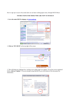


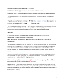
© Copyright 2026