
The development of cervical cancer and its precursors: what is... role of human papillomavirus infection?
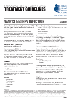
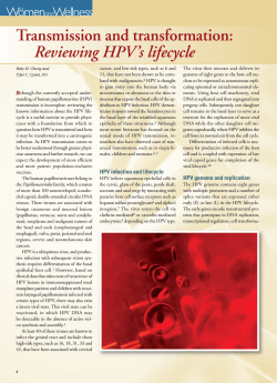
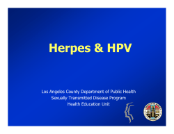
The development of cervical cancer and its precursors: what is the
role of human papillomavirus infection?
J. Thomas Cox
Human papillomavirus (HPV) is a significant health care
burden in the United States. The majority of sexually active
men and women will be infected with HPV at some point in
their lives and are subject to developing human
papillomavirus-associated disease. Current estimates
suggest that 20 million Americans are currently infected,
and more than 5 million new infections occur each year. The
prevalence of human papillomavirus is highest in
populations in their late teens and early twenties, with nearly
half of all new human papillomavirus infections occurring
within 3 years of first intercourse. HPV is the necessary
cause of genital warts, cervical intraepithelial neoplasia, and
invasive cervical cancer. As such, human papillomavirus is
responsible for significant medical morbidity and health
care costs. Screening with cervical cytology has
significantly reduced mortality rates; however,
approximately 3900 women will die in 2005 from cervical
cancer in the United States. Human papillomavirus DNA
testing has shown promise in identifying high-grade
abnormalities as an adjunct to traditional cytology, and
should be used according to guidelines established by the
American Cancer Society and the American College of
Obstetricians and Gynecologists. The epidemiology of HPV
infection and a brief introduction to the natural history of
HPV infection will be presented here.
Keyword
Human papillomavirus (HPV), cervical cancer, genital warts
Curr Opin Obstet Gynecol 18 (suppl 1):S5–S13. ß 2006 Lippincott Williams &
Wilkins.
Gynecology Clinic, Health Services, University of California, Santa Barbara,
California, USA
Correspondence and requests for reprints to J. Thomas Cox, MD, Gynecology
Clinic, Health Services, University of California, Santa Barbara, CA 93106, USA
Tel: +1 805 893 2595; e-mail: [email protected]
Current Opinion in Obstetrics and Gynecology 2006, 18 (suppl 1):S5–S13
Abbreviations
ACOG
ACS
ASCUS
CDC
CIN
HPV
HSIL
LSIL
STD
USPSTF
American College of Obstetricians and Gynecologists
American Cancer Society
atypical squamous cells of undetermined significance
Centers for Disease Control and Prevention
cervical intraepithelial neoplasia
human papillomavirus
high-grade squamous intraepithelial lesion
low-grade squamous intraepithelial lesion
sexually transmitted disease
US Preventive Services Task Force
ß 2006 Lippincott Williams & Wilkins
1040-872X
Epidemiology
The prevalence of human papillomavirus (HPV) infection is underestimated because of the subclinical nature
of most infections [1], the lack of broad population
screening for HPV by molecular testing [2,3], and limited
reporting, as clinicians are not required to report cases of
HPV infection to the Centers for Disease Control and
Prevention (CDC) [1]. Nevertheless, the development of
more sensitive and reliable assays for detecting HPV
DNA within the past two decades has provided a greater
understanding of the magnitude of HPV infection [1,4].
HPV is the most common sexually transmitted infection
in the United States [4]. An estimated 15% of the
population is currently infected with HPV [1], but
because HPV infections are usually transient, this number is far below the lifetime risk of getting one or more
HPV infections, which is likely to be at least 75% (Fig. 1)
[1]. Moreover, the estimated 1-year incidence of HPV
infection is at least 5.5 million [4,5].
HPV infection is most prevalent in young women and
adolescents, most likely as a result of increased transmission during the early years of sexual activity, or
possibly a lack of previous exposure that might generate
a protective immune response [6–10]. Alternatively,
young women may be more susceptible to infection
during adolescence for biological reasons. The transformation zone of the cervical epithelium undergoes a process
of squamous metaplasia during puberty that exposes
normally protected basal cells to infection [11]. In one
study using a polymerase chain reaction (PCR)-based
DNA amplification system to detect HPV DNA, 32%
of women 16–24 years old tested positive for HPV DNA
compared with 4% of women aged 45 years and older [9].
In a more recent study, the prevalence of HPV infection
ranged from 36% in women younger than 25 years of age
to 2.8% in women aged 45 years and older [8]. The
prevalence of HPV infection is extremely high in sexually
active adolescent females, with up to 64% of young
women testing positive for HPV DNA [12].
The rate of acquisition of HPV is extremely high compared with that of other sexually transmitted infections.
For example, among women between the ages of 18 and
35 years, the rate of new HPV infection reported from the
Young Women’s Health Study was 2.9% per month [13],
with a 32% cumulative incidence of new HPV infection
during a 2-year period [14], rising to 43% over 3 years [15].
S5
Copyright © Lippincott Williams & Wilkins. Unauthorized reproduction of this article is prohibited.
S6 Reducing the burden of cervical cancer and HPV-related diseases through vaccination
Figure 1 HPV infection in the United States
collected to determine the actual prevalence rates in
men, but since the virus is sexually transmitted, it is
likely that prevalence is similar in both sexes. Nonetheless, it is well appreciated that both men and women
develop genital warts. At least 1% of sexually active
adults are currently carrying genital warts in the USA
[1]. Therefore, strategies centered on preventing HPV
infection in both men and women will probably produce
the greatest public health benefit.
Transmission of HPV infection
HPV is transmitted by genital contact with an infected
partner. For infection to occur, the virus must have access
to the basal epithelial cells, either in epithelium that is
naturally thin and immature, such as the transformation
zone of the cervix or the anal verge, or through microscopic tears or abrasions in the external genital skin or the
introital or vaginal mucosa [18]. Studies among initially
virginal women strongly confirm the sexually transmitted
nature of HPV infection. In a 2-year study of 205 women,
all virgins were HPV DNA negative for genital HPV
types and seronegative for HPV 16, whereas 35% of
women with two or more partners were HPV DNA
positive and 23% seropositive for HPV 16 [19].
Approximately 1% of people have genital warts and an additional 4% will
probably have evidence of human papillomavirus (HPV) infection that is
detectable by colposcopy. Another 10% are HPV DNA positive, but
upon colposcopy have no evidence of HPV infection. An additional 60%
will have antibodies to HPV, indicative of exposure to the virus; however,
HPV DNA tests may be negative in these individuals and they may not
show clinical manifestations of the disease. The minority of people,
Genital
approximately 25%, will have never been exposed to HPV.
detected by colposcopy;
HPV DNA positive, colposcopy
warts;
negative; presence of antibodies (negative HPV test); not currently
infected.
The rate of new infections with high-risk, oncogenic
HPV types is higher than with low-risk types. High-risk
HPV infections are more common than infections caused
by low-risk, nononcogenic types, as evidenced by the
higher cumulative probability that a woman would
become newly infected with a high-risk HPV type during
a 12-month follow-up period (0.32) compared with that
for nononcogenic types (0.18) [13,16].
Because HPV infections are typically asymptomatic in
men, prevalence rates of HPV in men have been difficult
to assess. Testing is complicated by the fact that sample
collection from the external skin, whether male or
female, has generally been inadequate for molecular
HPV testing [17]. Most published studies have been
conducted outside the USA in men attending sexually
transmitted disease (STD) or university clinics, or among
the male partners of women with HPV infection [17].
The reported prevalence rates of HPV in men range from
16–45% [17]. Additional information will need to be
Intercourse is not absolutely necessary for transmission,
because the transmission of HPV infections may manifest
on external anogenital sites, and subsequently spread by
self-inoculation to other areas [14,20]. However, the age
of onset of intercourse is designated to be the age of onset
of the risk of cervical neoplasia in the American Cancer
Society (ACS) and the American College of Obstetricians
and Gynecologists (ACOG) guidelines. Whereas genital–
oral transmission may be a possible route of infection, the
literature has not reached a consensus regarding whether
or not HPV can be transmitted orally [14]. Although rare,
recurrent respiratory papillomatosis in young children
can occur from the transmission of HPV 6 or 11 from a
mother to a newborn baby, other HPV types have been
detected on neonates but have not been proven to cause
disease [21,22]. Transmission by inanimate objects such
as environmental surfaces and clothing has been hypothesized but not conclusively documented [23–25].
The relationship of HPV infection with
genital neoplasia
Cervical cancer is the third most common gynecological
malignancy and a serious public health issue in the USA
(Fig. 2) [26]. One recent study estimates that the lifetime
risk of cervical cancer would be 3.67% in the absence of
cervical cancer screening, with a lifetime cervical cancer
mortality risk of 1.26%, and a peak incidence of cervical
cancer of 81/100 000 at age 50 [27]. However, because of
cervical cancer screening with the Papanicolaou (Pap)
test, the estimated number of new cases will be down to
10 370 (approximately 1.5% of all new cancer cases in
Copyright © Lippincott Williams & Wilkins. Unauthorized reproduction of this article is prohibited.
The development of cervical cancer and its precursors Cox S7
Figure 2 Estimates for the United States in 2005 are rounded to
the nearest 10
Estimates 45000
for 2005
40000
35000
30000
25000
20000
15000
10000
5000
0
Uterine
cervix
Uterine
corpus
Ovary
Vulva
Vagina
and other
genital
Estimates of incidence are based on incidence rates from 1979–2001,
National Cancer Institute’s Surveillance, Epidemiology, and End Results
program, nine oldest registries. Estimates of deaths are based on data
from US Mortality Public Use Data Tapes, 1969–2002, National Center
for Health Statistics, Centers for Disease Control and Prevention, 2004
[26]. New cases; Deaths.
women) and mortality will be down to 3710 (approximately 1.3% of cancer-related deaths in women) in 2005
[26]. This is a reduction in risk of greater than 75% from
the estimated 40 000–50 000 cervical cancers that would
probably have occurred in 2005 in the absence of screening [28].
A causal relationship exists between HPV infection
and cervical cancer, as well as a significant fraction of
vaginal, vulvar, penile, and anal cancers [29]. Virtually all
cervical cancers contain HPV DNA – an estimated 99.7%
prevalence in cases worldwide – consequently, HPV
holds the highest worldwide attributable fraction ever
identified as the cause of a major human cancer [30].
Oncogenic HPV types 16 and 18 cumulatively account for
approximately 70% of all cervical cancers, and are associated with a more than 200-fold increased risk of developing invasive cancer [31]. Infection with high-risk
HPV types most commonly results in subclinical infections [32]; therefore, most infected women harbor
HPV DNA without showing cytological or histological
changes, or have changes that are so transient that they
are not detected by routine cytological screening [33].
One study reported that 36% of women with normal
cervical cytology tested positive for HPV DNA [34],
but most evaluations of women over the age of 30 years
report high-risk HPV detection to occur in 3–10% of
women with normal concurrent cervical cytology [35–37].
Persistent HPV infections can cause changes in the
cervical cytology of squamous epithelia that may progress
to noninvasive cervical intraepithelial neoplasia (CIN)
2/3, and less frequently and many years later, to invasive
cervical cancer.
Clinical manifestations: low- and high-risk
HPV infection
A common manifestation of low-risk HPV infection is
condylomata acuminata, or genital warts. Genital warts
are polypoid, often cauliflower-like growths that generate
infectious virus and have a low-to-negligible risk of
malignant progression. Over 90% of condyloma acuminata are caused by infection with low-risk HPV types 6 or
11. One study detected HPV 6 in more than 90%, HPV 11
in 32%, and HPV 6 or 11 in 97% of genital warts tested for
HPV DNA [38]. Exophytic genital warts are composed of
fronds of connective tissue covered by an acanthotic
squamous epithelium. Characteristic cellular changes
in the superficial layers of the epithelium include keratinization, multinucleation, and atypical koilocytes,
characterized by perinuclear cytoplasmic vacuolation
and nuclear enlargement, hyperchromasia, and irregularity. Histologically, genital warts can be distinguished
from CIN by the absence of nuclear atypia in the basal
layers of the epithelium [39]. Although genital warts are
medically benign, they represent a significant economic
burden and source of morbidity. The CDC reports that in
2003 there were an estimated 264 000 visits to physicians’
offices for genital warts (Fig. 3) [40]. Some warts may
spontaneously resolve, but treatments, when necessary,
are typically painful and generally aim to remove the
wart, either through excision, desiccation or immune
modulation, and often must be repeated. Costs to treat
genital warts vary considerably, from as low as US$200
(for simple surgical excision or desiccation) to as high
as US$6000 (for IFN-a2b therapy) [41]. An effective
immune response to HPV is most responsible for clearance and ultimately determines the cost [42].
Cervical infection with low-risk HPV types may manifest
as noninvasive, low-grade squamous intraepithelial
lesions (LSILs), also referred to as CIN grade 1 (CIN 1),
Figure 3 Initial visits to physicians’ offices for genital warts
400,000
350,000
300,000
250,000
200,000
150,000
100,000
50,000
0
1965
1975
1985
1995
2005
CDC STD Surveillance Report 2003: Source of data: National Drug and Therapeutic Index (IMS Health)
Genital warts are an increasing health care burden in the United States.
Since 1965, the incidence of genital warts has steadily increased. In
2003, 264,000 cases of genital warts were diagnosed. Source: National
Drug and Therapeutic Index (IMS Health).
Copyright © Lippincott Williams & Wilkins. Unauthorized reproduction of this article is prohibited.
S8 Reducing the burden of cervical cancer and HPV-related diseases through vaccination
and occasionally as high-grade CIN 2, but rarely as
CIN 3 [39]. However, most CIN of any grade is due
to high-risk types of HPV [43]. High-risk HPV types
are called ‘high-risk’ because of their association with
cervical cancer, yet these HPV types are also the most
common types in women with no detectable HPV manifestation [44].
The risk of developing LSILs decreases over time after
incident HPV infection: at 4 months, the hazard ratios are
6.14 for low-risk HPV types and 13.03 for high-risk HPV
types; by 60 months, the hazard ratios are 0.73 for low-risk
HPV types and 3.37 for high-risk HPV types [45]. Most
untreated cases of LSIL regress within 2 years, as
Figure 4 Two, 5, and 10-year cumulative percentages of (a)
progression or (b) regression for initially mild and moderate
dysplasia
(a)
Cumulative % 35
of progression
30
25
20
15
10
5
0
2
5
10
Years
(b)
Cumulative % 100
of regression
90
80
70
60
50
assessed by one or two normal Pap smears (Fig. 4)
[46]. One evaluation of all relevant studies on the natural
history of cervical neoplasia between 1952 and 1992
estimated that 60% of CIN 1 cases regress, 30% persist,
10% progress to CIN 3, and 1% progress to invasive
cancer [47]. A more recent meta-analysis estimated that
47% of LSILs will regress to normal, 21% will progress to
high-grade squamous intraepithelial lesions (HSILs), and
0.15% will progress to cancer [48].
HSILs include CIN grades 2 and 3 and is associated with
persistent infection in high-risk HPV types [49,50]. Highrisk types, mostly 16 and 18, are found in 50–80% of
high-grade lesions [39], and the detection of HPV type 16
DNA is highly predictive of CIN 3 [51]. Additionally,
women infected with high-risk types 16 and 18 have a
greater chance of progressing to CIN 3 or cancer than do
women infected with other oncogenic strains [52,53].
Compared with low-grade lesions, high-grade CIN has
lower rates of spontaneous clearance (30–40%), are much
higher rates of progression to cancer without treatment
(> 12%) [47,50]. By 2 years, it is expected that 35–40% of
CIN 2 will regress to normal, and 1.44% of CIN 3 will
progress to invasive cervical cancer [48].
Persistent HPV infection with high-risk HPV types is the
most important risk factor for developing cervical cancer
precursor lesions and invasive cervical cancer [54–57].
The risk of developing CIN 3 is 14 times higher for
women who have had at least three positive tests for highrisk HPV compared with women who have had negative
tests [58]. Infections with high-risk HPV types are more
persistent than those with low-risk types. For women
aged 18–35 years, the median time to clearance for
high-risk types is 9.8 months, significantly longer than
for low-risk HPV types (4.3 months) [13]. HPV 16, the
highest risk HPV type [16], is more likely to persist than
any other HPV type [15,59–61]. Persistence is also
associated with older age [15,62], infection with multiple
HPV types [15], and with compromised immunity
[63,64].
40
30
Risk factors for HPV infection
20
The foremost risk factor for acquiring HPV infection
is sexual activity. Among men [65] and women [15,34,
66–68], the risk of acquiring HPV dramatically increases
with the number of lifetime sex partners. Another variable that is just as important in determining a woman’s
risk of HPV infection is the number of current and
previous partners of her partner [14,69]. In men, circumcision reduces the risk of the acquisition and transmission
of HPV infection [70]. A recent report from the National
Institutes of Health (NIH) and a detailed review of the
published literature both conclude that there is no consistent epidemiological evidence that the use of latex
condoms reduces the risk of HPV infection [71,72].
10
0
2
5
10
Years
(a) Progression. Mild to moderate or worse; mild to severe or worse;
moderate to severe or worse. (b) Regression. Mild to 1st normal;
moderate to 1st normal;
mild to 2nd normal;
moderate to 2nd
normal. Taken from the results of actuarial life tables from the cytology
database of the Ontario Cancer Registry, collected 1962–1989.
Dysplasia states are classified as in Riotton and Christopherson [40]
and Holowaty et al. [46]. The terms ‘1st normal’ and ‘2nd normal’ refer to
the regression of cervical dysplasia to one or two normal Pap test results,
respectively.
Copyright © Lippincott Williams & Wilkins. Unauthorized reproduction of this article is prohibited.
The development of cervical cancer and its precursors Cox S9
Although latex condoms provide an impermeable barrier
to particles the size of HPV [73,74], they do not offer
protection from HPV infections on anatomical sites that
are not physically covered [72,75]. However, failure to
use a condom is associated with higher rates of genital
warts and cervical cancer [71,72]. Additionally, several
studies have determined that the use of condoms reduces
the risk of genital herpes and chlamydia, both of which
may indirectly contribute to HPV infection and the
development of cervical cancer [76,77].
Immunocompetency also has a significant impact on the
ability to clear HPV infections. A greater rate and incidence of infection has been observed in immunosuppressed renal transplant patients [78] and patients
infected with HIV [79,80], compared with patients
who are not immunocompromised. Additionally, immunity can be normal but immunocompetency for HPV may
be genetically determined to some extent. Hence, certain
human leukocyte antigen (HLA) markers are associated
with a higher risk of cervical cancer, and some individuals
with otherwise normal immunity have a very difficult
time resolving their genital warts [39].
Other risk factors associated with cervical cancer include
high parity (five or more pregnancies) [81] and smoking
[76,82–85]. A recent study reported an association
between active and passive cigarette smoking and cervical neoplasia, providing evidence that even passive smoking is a risk factor for cervical cancer [85]. Long-term use
of oral contraceptives (OCs) has also been associated with
an increased risk for cervical cancer. One study showed
that more than 5 years of OC use is associated with a
2-fold increased risk of cervical cancer, with risks increasing to more than 4-fold by 10 years of OC use risk among
women with active HPV infections [86,87].
Cytological screening and DNA
testing procedures
Regular cervical screening has had a significant impact on
the incidence and mortality associated with cervical
cancer [26]. The goal of cervical screening in the USA
is to identify precancerous lesions so they can be removed
prior to progression of invasive cancer [49]. The survival
of women with preinvasive lesions (CIN 2/3) is nearly
100% [26]. Approximately 90% of women with cervical
cancer survive 1 year, and 5-year survival rates are nearly
75% [26]. Approximately 92% of women diagnosed with,
and treated for, early-stage invasive cervical cancer survive 5 years [26]. It is estimated that 50% of women
diagnosed with cervical cancer have never received a Pap
test, and an additional 10% have not been screened
within 5 years of diagnosis [88].
Although cervical screening has significantly decreased
the mortality associated with HPV infection, the sensi-
tivity of the Pap smear is generally less than many believe
it to be. The Agency for Health Care Policy (AHCPR)
and Research determined that the conventional Pap
smear is only approximately 50% effective at detecting
cervical lesions of all grades, and that the conventional
Pap smear is more accurate when detecting high-grade
(CIN 2/3) than low-grade lesions. Liquid-based cytology
has been reported to detect between 26 and 103% more
cases of CIN 2/3 than the conventional Pap smear;
however, the degree to which sensitivity is increased is
unknown [33]. Poor sensitivity has driven the expectation
that cytology should be repeated annually. Novel
approaches to cervical screening, such as HPV DNA
testing, are now being developed to increase the sensitivity of cervical cancer screens. In March 2003, the
US Food and Drug Administration approved the Hybrid
Capture1 2 HPV DNA test (HC2) for use in the primary
cervical screening of women aged 30 years and above
when used in combination with cervical cytology. The
high-risk panel of HC2 detects the presence of one or
more of 13 high-risk HPV types in exfoliated cervical
cells and was previously approved in March 2000 for the
management of women with a Pap result indicating
atypical squamous cells of undetermined significance
(ASCUS) [89]. HPV DNA testing is very useful for
detecting clinically relevant lesions – in the majority
of studies HC2 has a sensitivity of 90–98% for detecting
CIN 2/3 [90]. In addition, minor HC2 crossreactivity with
low-risk HPV types has little effect on its clinical performance as a general screening test, but some decrease
in specificity when used as a triage test [91]. Most studies
indicate that women with a concurrent normal cytology
result and a negative HC2 test have a substantially
decreased risk of developing CIN 2/3 or cervical cancer
relative to those for whom the only screening information
is a normal conventional cytology result [33]. However, a
positive HC2 result is not an absolute indicator that a
high-grade lesion exists or will develop.
The combined use of cytology and HPV DNA testing
increases sensitivity but decreases specificity [92]. The
cumulative incidence of CIN 3 or cancer in a 45-month
study of women who tested negative for HPV infection using both cytology and HC2 was approximately
1.6/1000; and remained low at 0.8% at 10 years [93].
Hence, the combination of HPV testing and cervical
cytology provide not only greater reassurance that cervical precancer and cancer have not been missed, but also
predict the level of risk for the future, which is not
obtainable through screening with cytology alone.
‘Reflex’ HPV DNA testing is the procedure of performing an HC2 high-risk HPV test directly from the remaining cells in a liquid-based Pap test vial when the cytology
interpretation is ASCUS. ‘Reflex’ HPV DNA testing has
been recommended as a convenient and cost-effective
Copyright © Lippincott Williams & Wilkins. Unauthorized reproduction of this article is prohibited.
S10 Reducing the burden of cervical cancer and HPV-related diseases through vaccination
approach for the management of women with ASCUS
[94]. This may also be done by co-collecting a separate
HPV DNA test at the same time as cervical cytology and
holding the HPV test vial until the Pap result returns. If
the Pap result returns as ASCUS, the co-collected vial is
sent to the laboratory to be tested for HPV, whereas all
vials from women having normal, atypical squamous cells
but cannot exclude HSIL, atypical glandular cell (AGC),
LSIL, or HSIL Pap tests are discarded [33].
Guidelines for cervical cytology
The ACS and the ACOG endorse either the conventional
Pap test or liquid-based preparations (LBP) for cytological
screening [33,49]. The ACOG recommends the following
practices to optimize cervical cytology [33]: cells should
be collected before bimanual examination, and care should
be taken to avoid contaminating the sample with lubricant.
If cervical samples are to be collected to test for STDs,
cell collection for cervical cytology should be undertaken
first. Ideally, the entire portion of the cervix should be
visible when the sample is obtained. Routine swabbing of
the discharge from the cervix may result in cytological
samples of scant cellularity. In an effort to reduce airdrying artifact, the specimen should be transferred and
fixed as quickly as possible.
A ‘satisfactory’ specimen for cytological analysis has been
defined by the Bethesda 2001 Workshop, a consensus
workshop convened by the National Cancer Institute and
cosponsored by 44 professional societies [50]. There
should be at least 8000–12 000 well-visualized squamous
cells for conventional smears and 5000 squamous cells
for liquid-based preparations. There should be at least
10 well-preserved endocervical or squamous metaplastic
cells. A specimen is considered ‘partly obscured’ when
50–75% of epithelial cells cannot be visualized; specimens with more than 75% of epithelial cells obscured are
‘unsatisfactory’.
The Bethesda 2001 Workshop established the following
terminology for reporting cytology results [50]: samples
may be categorized as ‘negative for intraepithelial lesion
or malignancy’ if considered to be within normal limits or
benign cellular changes are detected. ‘Other’ applies to
cases in which there are no morphological abnormalities
in the cells that would be suspicious for CIN but abnormal findings are present, such as cells suspect for ovarian
or other cancer. ‘Other’ also applies to findings that may
indicate some increased risk, such as benign-appearing
endometrial cells in a woman 40 years of age or older.
Atypical squamous cells (ASC) may be qualified as being
of ‘undetermined significance’ (ASCUS) or ‘cannot
exclude high-grade squamous intraepithelial lesion
(ASC-H)’. All cases of ASC are considered to be suggestive but not definitive for squamous intraepithelial lesion
(SIL). Noninvasive squamous intraepithelial lesions may
be classified as LSIL, which includes CIN 1 (mild
dysplasia) and cytological findings consistent with HPV
infection (koilocytotic atypia). Alternatively, they may be
classified as HSIL, which combines CIN 2 and CIN 3
(moderate dysplasia, severe dysplasia, and carcinoma
in situ).
Glandular cell abnormalities less severe than adenocarcinoma may be classified into the following categories:
AGC, either endocervical, endometrial, or glandular
cells not otherwise specified; atypical glandular cells,
either endocervical or glandular cells favor neoplasia;
and endocervical adenocarcinoma in situ. The term
‘atypical epithelial cells’ may be used for cases where
a squamous vs a glandular origin cannot be determined.
An intermediate category ‘atypical endocervical cells,
favor neoplastic’ and ‘atypical glandular cells of undetermined significance, probably neoplastic’ apply to cases
showing some features suggestive of, but not sufficient
to reach an interpretation of, adenocarcinoma in situ
[50].
Guidelines for programs of cytological
screening and HPV DNA testing
According to the ACS, ACOG and the US Preventive
Services Task Force (USPSTF), cervical cytological
screening should begin within 3 years of first sexual
intercourse or by 21 years of age, whichever comes first
[33,49]. Nonetheless, adolescents who may not need a
cervical cytology test should obtain appropriate preventive health care, including an assessment of health risks,
contraception, prevention counseling, screening, and
treatment of STDs.
It is generally agreed that women with an intact cervix
should receive cervical cytological screening for cancer
every 1–3 years, depending on age, prior test results and
risk factors associated with cancer. For women younger
than 30 years, ACS recommends cervical screening
annually with the conventional Pap smear or every 2 years
using liquid-based cytology [49]. ACOG recommends
annual cytology screening regardless of the type of Pap
test utilized [33]. For women aged 30 years and older, it is
generally recommended that screening intervals be
lengthened if the patient is not currently in an accelerated follow-up due to abnormal cervical cytology or
previous cervical treatment. ACS and ACOG recommend
that women aged 30 years and older who have had three
consecutive negative cytology results may be screened
every 2–3 years, women with a history of in utero
diethylstilboestrol (DES) exposure, HIV infection, or
who are immunocompromised may require more frequent screening, and women with a history of HSIL or
cancer should be screened annually [33,49]. For women
aged 30 years and older, as an alternative to cytological
testing alone, both the ACS and ACOG provide as an
Copyright © Lippincott Williams & Wilkins. Unauthorized reproduction of this article is prohibited.
The development of cervical cancer and its precursors Cox S11
option cervical screening with the Pap test combined
with HPV DNA testing. Both organizations suggest that
women who test negative using both cytology and
molecular testing for HPV DNA should be screened
no more frequently than every 3 years. The ACS, ACOG,
and USPSTF all recommend discontinuing cervical
screening in women who have undergone hysterectomy
for benign reasons. ACS and ACOG recommend that
women with a history of CIN 2/3 at the time of the
hysterectomy continue screening after hysterectomy
until three consecutive negative cytology results are
achieved [33,49].
No consensus has been established concerning the upper
age limit for cervical cancer screening among low-risk
women. The ACS suggests that screening may be ceased
in women who are aged 70 years and older with an intact
cervix, after three consecutive negative cervical cytology
tests, and no positive cytology tests within the past
10 years [49]. Women positive for HPV DNA should
continue screening at the discretion of their health care
provider. The USPSTF recommends ceasing screening
in women older than 65 years if they have had consistent
negative test results and are not otherwise at high risk of
cervical cancer [95]. The ACOG does not define an upper
age limit for discontinuing screening [33].
Consensus guidelines are available for the management
of women with cervical cytological abnormalities and
cervical cancer precursors [96,97]. These evidence-based
guidelines were developed in 2001 by an expert consensus conference sponsored by the American Society for
Colposcopy and Cervical Pathology [96,97].
Prophylactic HPV on the horizon
Vaccines are currently being developed to reduce
susceptibility to HPV infection and persistent infection.
A recent study projected that an effective vaccine targeting high-risk HPV types could prevent 1300 deaths
annually from cervical cancer if all 12-year-old girls
currently living in the USA were vaccinated [98]. HPV
vaccines have shown encouraging success in clinical trials
[99]. A vaccine for HPV 16 given to adolescent girls
demonstrated 91% efficacy in preventing HPV 16 infection and 100% efficacy in preventing persistent HPV 16
infection [99].
Although the majority of low-grade cervical lesions spontaneously regress, and genital warts pose little threat of
malignancy, diagnosis with either of these clinical manifestations can evoke great levels of anxiety in women.
Many women initially equate diagnosis with an abnormal
Pap smear as indicative of cervical cancer. Moreover,
women diagnosed with low-grade lesions may have lower
self-esteem, decreased sex drives, and suffer from extremely high anxiety levels [100]. Therefore, vaccines that
protect against the greatest number of cervicovaginal
disease-causing HPV types will be most efficacious.
Two multivalent vaccines have been developed to prevent HPV infection. A quadrivalent vaccine that protects
against HPV types 6, 11, 16, and 18, and a bivalent
vaccine protecting against HPV 16 and 18 were both
over 90% effective in reducing vaccine-type persistent
infections and CIN [101,102]. Widespread acceptance of
these vaccines should significantly reduce the incidence
of HPV-associated disease, thereby alleviating a significant fraction of morbidity associated with HPV infection.
References
1
Koutsky L. Epidemiology of genital human papillomavirus infection. Am J Med
1997; 102 (5a):3–8.
2
Sirovich BE, Welch HG. The frequency of Pap smear screening in the United
States. J Gen Intern Med 2004; 19:243–250.
3
Insinga RP, Glass AG, Rush BB. Pap screening in a U.S. health plan. Cancer
Epidemiol Biomarkers Prev 2004; 13:355–360.
4
Cates W Jr. Estimates of the incidence and prevalence of sexually transmitted diseases in the United States. American Social Health Association
Panel. Sex Transm Dis 1999; 26 (Suppl 4):S2–S7.
5
Ratcliffe J. Estimation of HPV incidence in the US population. Report for
American Social Health Association (ASHA), 23 May 23 1998.
6
Figueroa JP, Ward E, Luthi TE, et al. Prevalence of human papillomavirus
among STD clinic attenders in Jamaica: association of younger age and
increased sexual activity. Sex Transm Dis 1995; 22:114–118.
7
Meisels A. Cytologic diagnosis of human papillomavirus. Influence of age and
pregnancy stage. Acta Cytol 1992; 36:480–482.
8
Burk RD, Kelly P, Feldman J, et al. Declining prevalence of cervicovaginal
human papillomavirus infection with age is independent of other risk factors.
Sex Transm Dis 1996; 23:333–341.
9
Bauer HM, Hildesheim A, Schiffman MH, et al. Determinants of genital human
papillomavirus infection in low-risk women in Portland, Oregon. Sex Transm
Dis 1993; 20:274–278.
10
Herrero R, Schiffman MH, Bratti C, et al. Design and methods of a populationbased natural history study of cervical neoplasia in a rural province of
Costa Rica: the Guanacaste Project. Rev Panam Salud Publica 1997;
1:362–375.
11
Moscicki AB, Burt VG, Kanowitz S, et al. The significance of squamous
metaplasia in the development of low-grade squamous intraepithelial lesions
in young women. Cancer 1999; 85:1139–1144.
12
Tarkowski TA, Koumans EH, Sawyer M, et al. Epidemiology of human
papillomavirus infection and abnormal cytologic test results in an urban
adolescent population. J Infect Dis 2004; 189:46–50.
13
Giuliano AR, Harris R, Sedjo RL, et al. Incidence, prevalence, and clearance
of type-specific human papillomavirus infections: the Young Women’s Health
Study. J Infect Dis 2002; 186:462–469.
14
Winer RL, Lee SK, Hughes JP, et al. Genital human papillomavirus infection:
incidence and risk factors in a cohort of female university students. Am J
Epidemiol 2003; 157:218–226.
15
Ho GY, Bierman R, Beardsley L, et al. Natural history of cervicovaginal
papillomavirus infection in young women. N Engl J Med 1998; 338:423–
428.
16
Zuna RE, Allen RA, Moore WE, et al. Comparison of human papillomavirus
genotypes in high-grade squamous intraepithelial lesions and invasive cervical carcinoma: evidence for differences in biologic potential of precursor
lesions. Mod Pathol 2004; 17:1314–1322.
17
Gerberding J. Report to Congress: Prevention of genital human papillomavirus infection. Atlanta, GA: Centers for Disease Control and Prevention,
Department of Health and Human Services; 2004.
18
Schiffman M, Kjaer SK. Chapter 2: Natural history of anogenital human
papillomavirus infection and neoplasia. J Natl Cancer Inst Monogr 2003;
31:14–19.
Copyright © Lippincott Williams & Wilkins. Unauthorized reproduction of this article is prohibited.
S12 Reducing the burden of cervical cancer and HPV-related diseases through vaccination
19
Kjaer SK, Chackerian B, van den Brule AJ, et al. High-risk human papillomavirus is sexually transmitted: evidence from a follow-up study of virgins
starting sexual activity (intercourse). Cancer Epidemiol Biomarkers Prev
2001; 10:101–106.
20
Marrazzo JM, Koutsky LA, Kiviat NB, et al. Papanicolaou test screening and
prevalence of genital human papillomavirus among women who have sex with
women. Am J Public Health 2001; 91:947–952.
21
Watts DH, Koutsky LA, Holmes KK, et al. Low risk of perinatal transmission of
human papillomavirus: results from a prospective cohort study. Am J Obstet
Gynecol 1998; 178:365–373.
44
Bosch FX, de Sanjose S. Chapter 1: Human papillomavirus and cervical
cancer-burden and assessment of causality. J Natl Cancer Inst Monogr
2003; 31:3–13.
45
Schlecht NF, Platt RW, Negassa A, et al. Modeling the time dependence of
the association between human papillomavirus infection and cervical cancer
precursor lesions. Am J Epidemiol 2003; 158:878–886.
46
Holowaty P, Miller AB, Rohan T, To T. Natural history of dysplasia of the
uterine cervix. J Natl Cancer Inst 1999; 91:252–258.
47
Ostor AG. Natural history of cervical intraepithelial neoplasia: a critical
review. Int J Gynecol Pathol 1993; 12:186–192.
48
Melnikow J, Nuovo J, Willan AR, et al. Natural history of cervical squamous
intraepithelial lesions: a meta-analysis. Obstet Gynecol 1998; 92:727–735.
49
Saslow D, Runowicz CD, Solomon D, et al. American Cancer Society
guideline for the early detection of cervical neoplasia and cancer. CA Cancer
J Clin 2002; 52:342–362.
50
Solomon D, Davey D, Kurman R, et al. The 2001 Bethesda System:
terminology for reporting results of cervical cytology. JAMA 2002;
287:2114–2119.
51
Cuzick J, Terry G, Ho L, et al. Type-specific human papillomavirus DNA in
abnormal smears as a predictor of high-grade cervical intraepithelial neoplasia. Br J Cancer 1994; 69:167–171.
52
Castle PE, Solomon D, Schiffman M, Wheeler CM. Human papillomavirus
type 16 infections and 2-year absolute risk of cervical precancer in women
with equivocal or mild cytologic abnormalities. J Natl Cancer Inst 2005;
97:1066–1071.
53
Khan MJ, Castle PE, Lorincz AT, et al. The elevated 10-year risk of cervical
precancer and cancer in women with human papillomavirus (HPV) type 16 or
18 and the possible utility of type-specific HPV testing in clinical practice.
J Natl Cancer Inst 2005; 97:1072–1079.
22
Smith EM, Ritchie JM, Yankowitz J, et al. Human papillomavirus prevalence
and types in newborns and parents: concordance and modes of transmission. Sex Transm Dis 2004; 31:57–62.
23
Ferenczy A, Bergeron C, Richart RM. Human papillomavirus DNA in fomites
on objects used for the management of patients with genital human papillomavirus infections. Obstet Gynecol 1989; 74:950–954.
24
Roden RB, Lowy DR, Schiller JT. Papillomavirus is resistant to desiccation.
J Infect Dis 1997; 176:1076–1079.
25
Bergeron C, Ferenczy A, Richart R. Underwear: contamination by human
papillomaviruses. Am J Obstet Gynecol 1990; 162:25–29.
26
Macaskill P, editor. Cancer facts and figures 2005. Atlanta, GA: American
Cancer Society; 2005.
27
Myers ER, McCrory DC, Nanda K, et al. Mathematical model for the natural
history of human papillomavirus infection and cervical carcinogenesis. Am J
Epidemiol 2000; 151:1158–1171.
28
Cox JT. Evaluation of abnormal cervical cytology. Clin Lab Med 2000;
20:303–343.
29
Bosch FX, Lorincz A, Munoz N, et al. The causal relation between human
papillomavirus and cervical cancer. J Clin Pathol 2002; 55:244–265.
30
Walboomers JM, Jacobs MV, Manos MM, et al. Human papillomavirus is a
necessary cause of invasive cervical cancer worldwide. J Pathol 1999;
189:12–19.
54
Frazer IH. Prevention of cervical cancer through papillomavirus vaccination.
Nat Rev Immunol 2004; 4:46–54.
55
31
Munoz N, Bosch FX, de Sanjose S, et al. Epidemiologic classification of
human papillomavirus types associated with cervical cancer. N Engl J Med
2003; 348:518–527.
Ho GY, Burk RD, Klein S, et al. Persistent genital human papillomavirus
infection as a risk factor for persistent cervical dysplasia. J Natl Cancer Inst
1995; 87:1365–1371.
56
32
Mao C, Hughes JP, Kiviat N, et al. Clinical findings among young women with
genital human papillomavirus infection. Am J Obstet Gynecol 2003;
188:677–684.
Schlecht NF, Platt RW, Duarte-Franco E, et al. Human papillomavirus
infection and time to progression and regression of cervical intraepithelial
neoplasia. J Natl Cancer Inst 2003; 95:1336–1343.
57
33
ACOG Practice Bulletin. Clinical management guidelines for obstetriciangynecologists, no. 45, August 2003. Cervical cytology screening (replaces
committee opinion 152, March 1995). Obstet Gynecol 2003; 102:417–
427.
Schlecht NF, Kulaga S, Robitaille J, et al. Persistent human papillomavirus
infection as a predictor of cervical intraepithelial neoplasia. JAMA 2001;
286:3106–3114.
58
Moscicki AB, Shiboski S, Broering J, et al. The natural history of human
papillomavirus infection as measured by repeated DNA testing in adolescent
and young women. J Pediatr 1998; 132:277–284.
59
Liaw KL, Hildesheim A, Burk RD, et al. A prospective study of human
papillomavirus (HPV) type 16 DNA detection by polymerase chain reaction
and its association with acquisition and persistence of other HPV types.
J Infect Dis 2001; 183:8–15.
60
Franco EL, Villa LL, Sobrinho JP, et al. Epidemiology of acquisition and
clearance of cervical human papillomavirus infection in women from a highrisk area for cervical cancer. J Infect Dis 1999; 180:1415–1423.
61
Richardson H, Kelsall G, Tellier P, et al. The natural history of type-specific
human papillomavirus infections in female university students. Cancer Epidemiol Biomarkers Prev 2003; 12:485–490.
62
Hildesheim A, Schiffman MH, Gravitt PE, et al. Persistence of type-specific
human papillomavirus infection among cytologically normal women. J Infect
Dis 1994; 169:235–240.
63
Sillman FH, Sentovich S, Shaffer D. Ano-genital neoplasia in renal transplant
patients. Ann Transplant 1997; 2:59–66.
64
Moscicki AB, Ellenberg JH, Farhat S, Xu J. Persistence of human papillomavirus infection in HIV-infected and -uninfected adolescent girls: risk factors
and differences, by phylogenetic type. J Infect Dis 2004; 190:37–45.
34
35
36
37
38
Peyton CL, Gravitt PE, Hunt WC, et al. Determinants of genital human
papillomavirus detection in a US population. J Infect Dis 2001; 183:1554–
1564.
Clavel C, Cucherousset J, Lorenzato M, et al. Negative human
papillomavirus testing in normal smears selects a population at low risk
for developing high-grade cervical lesions. Br J Cancer 2004; 90:1803–
1808.
Cuzick J, Szarewski A, Cubie H, et al. Management of women who test
positive for high-risk types of human papillomavirus: the HART study. Lancet
2003; 362:1871–1876.
Fetterman B, Shaber R, Pawlick G, Kinney W. Lessions from practice: the
first hundred thousand Pap and HPV co-test for general population screening. Paper presented at: International HPV Conference.Vancouver, Canada
2005.
Brown DR, Schroeder JM, Bryan JT, et al. Detection of multiple human
papillomavirus types in condylomata acuminata lesions from otherwise
healthy and immunosuppressed patients. J Clin Microbiol 1999; 37:
3316–3322.
39
Arends MJ, Buckley CH, Wells M. Aetiology, pathogenesis, and pathology of
cervical neoplasia. J Clin Pathol 1998; 51:96–103.
65
40
Riotton G, Christopherson WM. Cytology of the female genital tract. In:
International histological classification of tumors. Geneva, Switzerland:
World Health Organzation; 1973.
Hippelainen M, Syrjanen S, Koskela H, et al. Prevalence and risk factors of
genital human papillomavirus (HPV) infections in healthy males: a study on
Finnish conscripts. Sex Transm Dis 1993; 20:321–328.
66
41
Alam M, Stiller M. Direct medical costs for surgical and medical treatment of
condylomata acuminata. Arch Dermatol 2001; 137:337–341.
Ley C, Bauer HM, Reingold A, et al. Determinants of genital human papillomavirus infection in young women. J Natl Cancer Inst 1991; 83:997–1003.
67
42
Martinelli C, Farese A, Mistro AD, et al. Resolution of recurrent perianal
condylomata acuminata by topical cidofovir in patients with HIV infection.
J Eur Acad Dermatol Venereol 2001; 15:568–569.
Burk RD, Ho GY, Beardsley L, et al. Sexual behavior and partner characteristics are the predominant risk factors for genital human papillomavirus
infection in young women. J Infect Dis 1996; 174:679–689.
68
43
Koutsky LA, Galloway DA, Holmes KK. Epidemiology of genital human
papillomavirus infection. Epidemiol Rev 1988; 10:122–163.
Sellors JW, Karwalajtys TL, Kaczorowski J, et al. Incidence, clearance and
predictors of human papillomavirus infection in women. Can Med Assoc J
2003; 168:421–425.
Copyright © Lippincott Williams & Wilkins. Unauthorized reproduction of this article is prohibited.
The development of cervical cancer and its precursors Cox S13
69
Moscicki AB, Hills N, Shiboski S, et al. Risks for incident human papillomavirus infection and low-grade squamous intraepithelial lesion development in
young females. JAMA 2001; 285:2995–3002.
86
Smith JS, Green J, Berrington de Gonzalez A, et al. Cervical cancer and use
of hormonal contraceptives: a systematic review. Lancet 2003; 361:1159–
1167.
70
Castellsague X, Bosch FX, Munoz N, et al. Male circumcision, penile human
papillomavirus infection, and cervical cancer in female partners. N Engl J Med
2002; 346:1105–1112.
87
Moreno V, Bosch FX, Munoz N, et al. Effect of oral contraceptives on risk of
cervical cancer in women with human papillomavirus infection: the IARC
multicentric case–control study. Lancet 2002; 359:1085–1092.
71
National Institute of Allergy and Infectious Diseases. Workshop summary:
scientific evidence on condom effectiveness for sexually transmitted disease
(STD) prevention. Bethesda, MD: National Institutes of Health; 2001.
88
National Institutes of Health. Cervical cancer. NIH Consensus statement.
Bethesda, MD: National Institutes of Health 1996; 14:1–38; quiz 34 p.
89
72
Manhart LE, Koutsky LA. Do condoms prevent genital HPV infection, external
genital warts, or cervical neoplasia? A meta-analysis. Sex Transm Dis 2002;
29:725–735.
Hubbard RA. Human papillomavirus testing methods. Arch Pathol Lab Med
2003; 127:940–945.
90
Clavel C, Masure M, Bory JP, et al. Hybrid capture II-based human papillomavirus detection, a sensitive test to detect in routine high-grade cervical
lesions: a preliminary study on 1518 women. Br J Cancer 1999; 80:1306–
1311.
91
Peyton CL, Schiffman M, Lorincz AT, et al. Comparison of PCR- and hybrid
capture-based human papillomavirus detection systems using multiple cervical specimen collection strategies. J Clin Microbiol 1998; 36:3248–3254.
92
Mandelblatt JS, Lawrence WF, Womack SM, et al. Benefits and costs of
using HPV testing to screen for cervical cancer JAMA 2002; 287:2372–
2381.
73
Lytle CD, Duff JE, Fleharty B, et al. A sensitive method for evaluating condoms
as virus barriers. J AOAC Int 1997; 80:319–324.
74
Lytle CD, Routson LB, Seaborn GB, et al. An in vitro evaluation of condoms
as barriers to a small virus. Sex Transm Dis 1997; 24:161–164.
75
Van Doornum GJ, Prins M, Juffermans LH, et al. Regional distribution and
incidence of human papillomavirus infections among heterosexual men and
women with multiple sexual partners: a prospective study. Genitourin Med
1994; 70:240–246.
76
Castle PE, Giuliano AR. Chapter 4: Genital tract infections, cervical inflammation, and antioxidant nutrients – assessing their roles as human papillomavirus cofactors. J Natl Cancer Inst Monogr 2003; 29–34.
93
Sherman ME, Lorincz AT, Scott DR, et al. Baseline cytology, human papillomavirus testing, and risk for cervical neoplasia: a 10-year cohort analysis.
J Natl Cancer Inst 2003; 95:46–52.
77
Smith JS, Munoz N, Herrero R, et al. Evidence for Chlamydia trachomatis as a
human papillomavirus cofactor in the etiology of invasive cervical cancer in
Brazil and the Philippines. J Infect Dis 2002; 185:324–331.
94
Kim JJ, Wright TC, Goldie SJ. Cost-effectiveness of alternative triage
strategies for atypical squamous cells of undetermined significance. JAMA
2002; 287:2382–2390.
78
Halpert R, Fruchter RG, Sedlis A, et al. Human papillomavirus and lower
genital neoplasia in renal transplant patients. Obstet Gynecol 1986;
68:251–258.
95
U.S. Preventative Services Task Force. Guide to clinical preventative services. Washington DC: US Dept of Health and Human Services; 2003.
96
79
Matorras R, Ariceta JM, Rementeria A, et al. Human immunodeficiency virusinduced immunosuppression: a risk factor for human papillomavirus infection.
Am J Obstet Gynecol 1991; 164:42–44.
Wright TC Jr, Cox JT, Massad LS, et al. 2001 Consensus guidelines for the
management of women with cervical cytological abnormalities. JAMA 2002;
287:2120–2129.
97
80
Ho GY, Burk RD, Fleming I, Klein RS. Risk of genital human papillomavirus
infection in women with human immunodeficiency virus-induced immunosuppression. Int J Cancer 1994; 56:788–792.
Wright TC Jr, Cox JT, Massad LS, et al. 2001 Consensus guidelines for the
management of women with cervical intraepithelial neoplasia. Am J Obstet
Gynecol 2003; 189:295–304.
98
81
Munoz N, Franceschi S, Bosetti C, et al. Role of parity and human papillomavirus in cervical cancer: the IARC multicentric case–control study. Lancet
2002; 359:1093–1101.
Sanders GD, Taira AV. Cost-effectiveness of a potential vaccine for human
papillomavirus. Emerg Infect Dis 2003; 9:37–48.
99
Koutsky LA, Ault KA, Wheeler CM, et al. A controlled trial of a human
papillomavirus type 16 vaccine. N Engl J Med 2002; 347:1645–1651.
82
Castellsague X, Bosch FX, Munoz N. Environmental co-factors in HPV
carcinogenesis. Virus Res 2002; 89:191–199.
83
Castellsague X, Munoz N. Chapter 3: Cofactors in human papillomavirus
carcinogenesis–-role of parity, oral contraceptives, and tobacco smoking.
J Natl Cancer Inst Monogr 2003; 20–28.
84
Hildesheim A, Herrero R, Castle PE, et al. HPV co-factors related to the
development of cervical cancer: results from a population-based study in
Costa Rica. Br J Cancer 2001; 84:1219–1226.
85
Trimble CL, Genkinger JM, Burke AE, et al. Active and passive cigarette
smoking and the risk of cervical neoplasia. Obstet Gynecol 2005; 105:174–
181.
100 McDonald TW, Neutens JJ, Fischer LM, Jessee D. Impact of cervical
intraepithelial neoplasia diagnosis and treatment on self-esteem and body
image. Gynecol Oncol 1989; 34:345–349.
101 Harper DM, Franco EL, Wheeler C, et al. Efficacy of a bivalent L1 virus-like
particle vaccine in prevention of infection with human papillomavirus types 16
and 18 in young women: a randomised controlled trial. Lancet 2004;
364:1757–1765.
102 Villa LL, Costa RL, Petta CA, et al. Prophylactic quadrivalent human papillomavirus (types 6, 11, 16, and 18) L1 virus-like particle vaccine in young
women: a randomised double-blind placebo-controlled multicentre phase II
efficacy trial. Lancet Oncol 2005; 6:271–278.
Copyright © Lippincott Williams & Wilkins. Unauthorized reproduction of this article is prohibited.


© Copyright 2026