
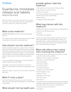
CTCA
l It is important that you advise the radiology facility staff when you make the appointment if you have asthma, diabetes, any kidney problems, irregular heart rhythm or have in the past had an allergy to contrast agents used in a radiology procedure or a strong history of allergy to other things (like foods, pollens or dust). If you have any of these conditions, it might not be possible to have this test. CTCA Consumer Information Contributors: A/Prof John Troupis MBBS FRANZCR Ms Ann Revell, Prof Grant Russell, Prof Graham Buirski The procedure could require several hours of preparation after you arrive at the radiology facility before you have the CTCA. What is a computer tomography coronary angiography (CTCA)? Angiography is the X-ray imaging of blood vessels using contrast agents injected into the bloodstream (see Angiography). CTCA uses computed tomography (CT) scanning to take pictures or images (angiograms) of the coronary arteries of the beating heart. These arteries supply blood to the heart muscle, and disease of these vessels (atherosclerosis) is responsible for most heart attacks. Liquid contrast agents, sometimes called contrast medium (see Iodine-containing contrast medium (ICCM)), are injected into a vein (usually in the arm). Contrast agents increase the density of the blood in the vessels, and allow the inside and outside structure of blood vessels to be clearly visible on the CT angiogram images. Medication to reduce blood flow and slow down the heart rate might also be given to make the images even clearer and easier to interpret. This will either be given in tablet form or into a vein through a cannula (a thin plastic tube) inserted in the arm. Why would my doctor refer me to have this procedure? If you are taking metformin for diabetes, you may or may not need to stop taking it for this test, depending on whether or not your kidney function is normal. You will need to bring the results of a recent kidney function test with you, so that it can be checked. Many patients come to the appointment with a companion who can drive them home. Even though you might have to stay after the procedure until the effects of medication used to lower the heart rate have worn off, you could still feel a little light headed walking or driving. What happens during a CTCA? A CTCA test is usually carried out in three parts: preparation, scanning and recovery. Preparation: Before having the procedure, you will be asked about your medical history (the problems or symptoms that led you to being referred for the test by your general practitioner or specialist). Your heart rate will be checked using an electrocardiogram (or ECG) machine. About four electrode patches will be placed onto your skin on the front of your chest, so the ECG wires can be attached. Your doctor might refer you for this test if they would like to find out if you have narrowing of the coronary arteries, which could be causing your symptoms. The specialist doctor supervising your procedure (a radiologist or cardiologist) will review the ECG. If your heart rhythm is regular, an intravenous (IV) cannula will be inserted into one of your veins, usually on the front of your elbow at the skin crease. How do I prepare for CTCA? Depending on the type of CT scanner used, if your heart rate is above 60 beats per minute, you might be given medication called a beta-blocker either orally (tablets by mouth) or intravenously (through the IV cannula) to reduce your heart rate. Reducing your heart rate makes the images clearer and easier to interpret. Your blood pressure and pulse rate will be monitored, and when the heart rate has reduced to the required level (a regular rhythm), you will be taken from the preparation area to the CT scanner room. CT images are clearer if your heart rate is low, and you might be given medications before the test to slow down your heart rate. It is advisable that you do not have any tea (including herbal teas), coffee, cola, chocolate or other stimulants before the procedure as these contain caffeine, which can raise your heart rate. It is not necessary to go without food or drink before the procedure, but a full stomach is not advisable, as this together with the contrast agent might make you feel nauseated. However, each radiology facility will ask you to follow their own requirements regarding any fasting before the test. ©RANZCR® 2013 Several minutes before the CT scan, nitroglycerin will be sprayed into the back of your mouth from a small spray container similar to that used by asthma sufferers. This is used to dilate (expand) the An initiative of the RANZCR’s Quality and Safety Program Page 1 of 3 www.insideradiology.com.au CTCA coronary arteries to assist in the procedure. This can cause headache or mild lightheadedness if you do not normally take nitroglycerin or use nitroglycerin patches for prevention or treatment of angina (heart muscle-related chest pain). Scanning: You will lie on a bed for the images to be taken by the CT scanner (see Computed Tomography (CT)). The CT scan equipment is a large square machine with a circular hole, sometimes described as looking like a donut. The bed slides in and out of the hole while images of your heart are taken. It is important not to move during the scan, as it will affect the quality of the images. At some radiology facilities, a ‘test dose’ of contrast is given to measure how long it takes for the contrast agent to get from the arm (where it is injected) to the heart, and this determines the exact time to begin the scan. This can also be done automatically by some CT machines just before the proper scan starts. While you are on the bed, you will be given a rapid IV injection of iodine contrast agent through the cannula, using a pressure injector. This is often thought of as an X-ray ‘dye’, but it is a clear and colourless fluid (see Iodine-containing contrast medium (ICCM)). When the iodine contrast reaches the heart through the veins, the scan is started. You will hear the CT machine rotating around you, and the bed will move in and out of the scanner while the images are taken. In some scanners, the bed might not move or might move in a series of short steps. You will be asked to hold your breath for approximately 10–12 seconds each time a scan is taken, because movement will cause blurring of the images. The CT scanner takes a series of picture ‘slices’ of the heart from the top to the bottom. At the same time as these images are being taken, your ECG is recorded. The scanner uses the recording from the ECG of the electric pulses from your heart so that every time it beats, CT scan images are taken. The CT scanning is matched to the ECG, and during a period or periods when the heart moves the least, images of the coronary arteries are taken, free of motion, so that they appear sharp rather than blurred. The images are analysed by the medical imaging technologist (or radiographer) who carries out the scanning using complex computer programs. Narrowing or blockages in the coronary arteries that could be responsible for heart attacks or other symptoms can be confirmed. Other information can also be obtained about heart muscle changes, the inside of the four heart chambers, the valves, the membranes that surround the heart (the pericardium) and the rest of the chest outside the heart if it is included in the scan. Recovery: Once all the scans have been taken (around 20 minutes), you will be taken to a recovery area for observation and the IV cannula will be removed before you are allowed to go home. If you have had medication to lower your heart rate, you might be asked to stay until the effects have worn off. © RANZCR® 2013 Are there any after effects of a CTCA? If medications to slow your heart rate have been given, you will usually be kept under observation until any possible lightheadedness has worn off, which is usually about half an hour, although it might take longer. If you have a headache from the nitroglycerin, this usually eases relatively quickly in about 20 minutes and often even quicker. Allergy to the contrast agent might occur (see Iodine-containing contrast medium (ICCM)). This can range from mild effects, such as sneezing, itchiness, rash and hives, to severe reactions. Severe reactions are rare, but might result in breathing difficulties, a drop in blood pressure and soft tissue swelling in the face and throat. When this occurs in the airways, it can be life threatening. Such reactions are very uncommon, but the reaction must be treated immediately by the medical staff, who are trained for such emergencies. If you have had a previous allergic reaction to contrast agent or you have a strong history of allergy to other things (like foods, pollens or dust), you should inform the medical staff at the radiology facility before having the procedure. The radiation dose for the procedure is approximately 2–21.5 millisieverts (a measure of radiation dose) (see Radiation risk of medical imaging for adults and children). How long does a CTCA take? The whole procedure, including the preparation, scanning and recovery, can take up to 3–4 hours, particularly if you have been given beta-blockers. The actual CT scanning will take approximately 20 minutes. What are the risks of a CTCA? The main risks of a CTCA are: Complications of the IV procedure iodinated contrast agent; for example: and o Rupture of the vein from the cannula, which is rare. o Injection of contrast medium into the surrounding tissues from the rapid injection of contrast agent, which might burst the wall of a small vein. o Air injected into the vein, although most modern power injectors provide safety measures so this does not happen. o Allergic reaction to the contrast agent (see Iodine-containing contrast medium (ICCM)) that can include sneezing, itching, rash and hives, which occur in a small percentage of patients. These usually occur within minutes of the injection. More severe reactions are rare, and include drop in blood pressure and soft tissue swelling, and can be life threatening. These reactions require immediate treatment. Medical staff at the radiology facility where you are having the procedure are trained to treat severe reactions if they occur. An initiative of the RANZCR’s Quality and Safety Program Page 2 of 3 www.insideradiology.com.au o CTCA Patients with renal impairment (kidney problems) might experience worsening of kidney function after the iodinated contrast (see Iodine-containing contrast medium (ICCM)). This usually improves over several days. If impairment of kidney function is severe, the procedure is generally not carried out unless the information provided by the scan is considered to be so valuable that this outweighs the risk of further deterioration in kidney function. Beta blockers can cause bronchospasm (major airway narrowing) in asthmatics, and are not given in patients who depend on high heart rates to maintain normal heart function. Nitroglycerin can cause headache and drop in blood pressure. Patients taking metformin for diabetes may or may not need to stop taking it for this test, depending on whether or not their kidney function is normal. If you are taking metformin, you will need to bring the results of a recent kidney function test with you, so that it can be checked. The procedure would not normally be carried out on pregnant women because of the radiation exposure to the foetus (unborn baby). In breast-feeding patients, the contrast agent can enter the breast milk, but not in sufficient quantities to affect your baby (see Iodine containing contrast medium (ICCM)). What are the benefits of CTCA? CTCA is a relatively new test, and the techniques are still evolving with the rapid development of new equipment. There is still disagreement amongst specialist doctors (cardiologists and radiologists) as to the benefits of the test. Published information would suggest that if this test is carried out and no coronary artery disease is detected, your doctor can use this information to manage your symptoms. When the coronary arteries show abnormalities, then your doctor can change your treatment according to the details of the abnormalities shown. The test has the benefit of being able to show the extent and location of atherosclerosis (a disease that obstructs blood flow in the arteries) within the coronary arteries, even if it is not causing obstruction to the blood flow. Who does the CTCA? A team of medical staff including specialist doctors (radiologists, cardiologists), radiographers and nurses are usually involved. The images are usually interpreted by the radiologist and/or cardiologist, and a written report is provided to your referring doctor. Where is a CTCA done? CTCA is carried out in public and private hospital imaging departments, and in private radiological practices. When can I expect the results of my CTCA? The time that it takes your doctor to receive a written report on the test or procedure you have had will vary, depending on: the urgency with which the result is needed; the complexity of the examination; whether more information is needed from your doctor before the examination can be interpreted by the radiologist; whether you have had previous X-rays or other medical imaging that need to be compared with this new test or procedure (this is commonly the case if you have a disease or condition that is being followed to assess your progress); how the report is conveyed from the practice or hospital to your doctor (in other words, email, fax or mail). Please feel free to ask the private practice, clinic or hospital where you are having your test or procedure when your doctor is likely to have the written report. It is important that you discuss the results with the doctor who referred you, either in person or on the telephone, so that they can explain what the results mean for you. Please note: This information is of a general nature only and is not intended as a substitute for medical advice. It is designed to support, not replace, the relationship that exists between a patient and his/her doctor. It is recommended that any specific questions regarding your procedure be discussed with your family doctor or medical specialist The InsideRadiology project is managed by the RANZCR® and funded by the Australian Commonwealth Department of Health and Ageing. Publication Date: December 31st 2013 Originally Published in 2009 The RANZCR® is not aware that any person intends to act or rely upon the opinions, advices or information contained in this publication or of the manner in which it The RANZCR® intends by this statement to exclude liability for any such opinions, advices or information. The content of this publication is not intended as a substitute for medical advice. It is designed to support, not replace, the relationship that exists between a patient and his/her doctor. Some of the tests and procedures included in this publication may not be available at all radiology providers. The RANZCR® recommends that any specific questions regarding any procedure be discussed with a person's family doctor or medical specialist. Whilst every effort is made to ensure the accuracy of the information contained in this publication, The RANZCR®, its officers, councillors and employees assume no responsibility for its content, use, or interpretation. Each person should rely on their own inquires before making decisions that touch their own interests. © RANZCR® 2013 An initiative of the RANZCR’s Quality and Safety Program Page 3 of 3


© Copyright 2026