
Air Pollution Impact on Children in Delhi: Study
~~
~"ffUT
(11ffif ~
yqfcj~ul
~?fUT
q)f
1:[ci ir-=r
~
WicR)
.
Central Pollution Control Board
\if. -m. ~1\b't1c:t>'(.
(A Govt. of India Organisaton)
Ministry of Environment & Forests
Phone:2230494~22307233
'+TUr.~.
~~
J. M. MAUSKAR,
IAS
Chairman
FOREWORD
Under the provision of Air Act, 1981, Central Pollution Control Board has been
entrusted with the responsibility of preparation of nation-wide plan for control of
air pollution. Information on nature, magnitude and adverse health effects of air
pollution is required for rational planning of pollution control strategies. It has
been observed that over the years, there has been exponential increase in
number of vehicles due to rapid urbanization and economic growth. As a result,
levels of air pollutants such as respirable suspended particulate matter. are found
to exceed the prescribed standards in many cities. Respirable suspended
particulate matter (RSPM) level is closely associated with mortality and morbidity.
Although air pollution affects the population in general, children are particularly
vulnerable to toxic effects of air pollution for several reasons. The most important
of these are more outdoor exposures and greater intake of air in relation to body
weight. Also children are more susceptible due to their underdeveloped
immunity. In order to determine health effects of air pollution on children, CPCB
initiated a Epidemiological study on children in Delhi with the help of Chittaranjan
National Cancer Institute, Kolkata. In the study, several health camps were
organized in various schools in different seasons covering different parts of the
city. The study included questionnaire survey as well as clinical examination. The
study carried out is a one-time study which requires further verification and
detailed investigation. Although the study is a bit dated, it is felt that the facts
contained in it have some relevance still.
I am thankful to Chittaranjan National Cancer Institute, Kolkata for their relentless
effort in carrying out the study. I am also thankful to my colleagues Dr. B.
Sengupta, Member Secretary, Dr. R.C. Trivedi, Additional Director, Sh. Naresh
Badhwar, Environmental Engineer and Dr Sanghita Roychoudhury, SRF for
assisting the project.
I hope the findings of the report would be useful to all concerned.
1)~"""")
CY~.
(J.M. Mauskar)
'Parivesh
Bhawan' C.B.D. - cum-Office Complex, East Arjun Nagar, Delhi-11 0032
Fax: 22304948/22307078
email : [email protected]
Website: http://www.cpcb.nic.in
STUDY ON AMBIENT AIR QUALITY,
RESPIRATORY SYMPTOMS AND LUNG
FUNCTION OF CHILDREN IN DELHI
CENTRAL POLLUTION CONTROL BOARD
MINISTRY OF ENVIRONMENT & FORESTS
Website: http://www.cpcb.nic.in/ e-mail: [email protected]
September 2008
STUDY ON AMBIENT AIR QUALITY,
RESPIRATORY SYMPTOMS AND LUNG
FUNCTION OF CHILDREN IN DELHI
CENTRAL POLLUTION CONTROL BOARD
Ministry of Environment & Forests, Govt. of India
Parivesh Bhavan, East Arjun Nagar
Delhi – 110 032
Website: http://www.cpcb.nic.in/ e-mail: [email protected]
ii
CONTRIBUTIONS
Guidance and Report
Finalisation
:
Dr. B. Sengupta
Coordination, Supervision
& Report Review
:
Dr. R. C. Trivedi
Sh. Naresh Badhwar
CNCI personnel involved
:
CPCB personnel involved
:
Principal Investigator: Dr. Twisha Lahiri
Co-investigator:
Dr. Manas Ranjan Ray
Research team:
Dr. Twisha Lahiri
Dr. Manas Ranjan Ray
Prof. Pulak Lahiri
Dr. G. Mukherjee
Dr. N.B. Dey
Dr. S. S. Mondal
Ms. Saswati Choudhury
Dr. Chandreyi Basu
Dr. Senjuti Roy
Dr. Sanghita Roychoudhury
Ms. Madhuchanda Banerjee
Ms. Shabana Siddique
Dr. Ms. Sayali Mukherjee
Ms. Purba Bhattacharjee
Ms. Sreeparna Chakraborty
Sh. Debanjan Bhattacharjee
Sh. Pulin Behari Paul
Sh. Manoj Kumar Sarkar
Sh. Naresh Badhwar
Dr. Pratima Akolkar
Sh. Tarun Darbari
Dr. Sanghita Roychaudhury
Ms. Charu Sharma
Ms. Abida Khatoon
iii
CONTENTS
Section
no.
Title
Page no.
Executive Summary
xvii
1.0
1.1
1.2
1.3
1.4
1.5
BACKGROUND AND OBJECTIVE OF THE STUDY
Background of the Study
Air Pollution and Adverse Health Effects: Modifying Factors
Air Pollution and its Sources in Delhi
Economic Aspects of Air Pollution inIndia
Scope of the Work and Objectives
1
2
4
7
10
11
2.0
2.1
2.2
2.3
MEASUREMENT OF AMBIENT AIR QUALITY OF DELHI
Ambient Air Quality Monitoring in Delhi
Results
Findings
12
12
13
14
3.0
PREVALENCE OF RESPIRATORY AND ASSOCIATED
SYMPTOMS
Introduction
Material and Methods
Results
Prevalence of Respiratory Symptoms
Prevalence of childhood asthma
Prevalence of associated symptoms
Association between ambient air pollution (PM10 level) and
prevalence of respiratory symptoms
Findings
28
3.1
3.2
3.2
3.2.1
3.2.2
3.2.3
3.2.4
3.3
4.0
4.1
4.2
4.3
4.3.1
4.3.2
4.3.3
4.3.4
4.3.5
4.3.6
4.3.7
4.4
EFFECT OF DELHI’S AIR POLLUTION ON CHILDREN’S LUNG
FUNCTION
Introduction
Material and Methods
Results
Changes in forced vital capacity (FVC) with age in control and
Delhi’s children
Reduction in FEV1 in Delhi’s children
Reduction in FEV1 /FVC ratio in Delhi’s children
Reduction in FEF25-75% in Delhi
Reduction in PEFR
Overall prevalence of lung function deficits in school children:
43.5% in Delhi against 25.7% in control
Body mass index (BMI) and lung function
Findings
iv
29
29
35
38
54
59
62
65
67
68
69
77
78
83
87
89
91
91
99
102
Section
no.
5.0
5.1
5.2
5.3
5.4
6.0
Title
ASSESSMENT OF CELLULAR LUNG REACTION TO DELHI’S
AIR POLLUTION
Introduction
Material and Methods
Results
FINDINGS
Page no.
104
105
105
107
124
6.1
6.2
6.3
6.4
HEMATOLOGICAL AND VASCULAR CHANGES ASSOCIATED
WITH AIR POLLUTION EXPOSURE
Introduction
Methodology
Results
Findings
126
126
128
135
7.0
7.1
7.2
7.3
7.4
BEHAVIOR AND ACTIVITIES OF THE CHILDREN
Introduction
Subjects and Methodologies
Results
Findings
137
138
138
139
144
8.0
DISCUSSION
145
9.0
9.1
9.1.1
9.1.2
9.1.3
9.1.4
9.1.5
165
166
166
166
167
169
170
9.1.6
9.2
SUMMARY AND RECOMMENDATIONS
Summary
Measurement of ambient air quality of Delhi
Prevalence of respiratory and associated symptoms
Effect of Delhi’s air pollution on children’s lung function
Assessment of cellular lung reaction to Delhi’s air pollution
Hematological and vascular changes associated with air pollution
exposure
Behavior and activities of the children
Reccomendations
10.0
REFERENCES
174
11.0
GLOSSARY
198
v
125
170
171
LIST OF TABLES
Table
no.
1.1:
2.1:
2.2:
2.3:
2.4.
2.5.
2.6:
2.7:
2.8.
2.9.
2.10:
2.11:
3.1:
3.2:
3.3:
3.4:
3.5:
3.6:
3.7:
3.8:
3.9:
3.10:
3.11:
3.12:
3.13:
3.14:
3.15:
3.16:
3.17:
3.18:
3.19:
Title
Page no.
Emission comparison of diesel and CNG-powered buses
Air Quality Monitoring Stations in Delhi
Suspended Particulate Matter concentrations (in µg/m3) in Delhi’s air
during 2002-2005
Respirable suspended particulate matter (RSPM) concentrations (µg/m3)
in Delhi’s air during 2002-2005
Four-year (2002-05) average SPM and RSPM concentrations at different
areas of Delhi
Concentration of Sulfur dioxide (µg/m3) in ambient air in different areas of
Delhi during 2002- 2005
Concentrations of nitrogen dioxide (µg/m3) in ambient air of different areas
of Delhi during 2002- 2005
Concentration of SPM-laden total polycyclic aromatic hydrocarbons (ng/m3)
in Delhi’s air
Concentrations of SPM-laden Benzo(a)pyrene (ng/m3) in Delhi’s air
Benzene (μg/m3) level in Delhi during 2002-2004
Volatile organic compound in ambient air in Delhi during winter (Feb 3-10,
2005)
Meteorological data of Delhi during 2004-2005.
Number of children and Names of the Schools of Delhi where the study has
been conducted
Names of the schools of Uttaranchal (UT) and West Bengal (WB) from
where the children of control group were examined.
Demographic Characteristics of the Children.
Distribution (%) of children in different age groups
Prevalence (%) of respiratory symptoms in children in past three months
Prevalence (%) of respiratory symptoms in different seasons
Prevalence (%) of upper respiratory symptoms in past three months
Prevalence of sinusitis among schoolchildren
Prevalence of running or stuffy nose among children in past three months
Prevalence of sneezing among children in past three months
Prevalence of sore throat among children in past three months
Prevalence of common cold and fever among children in past three months
Prevalence (%) of URS in different age groups of Delhi’s children
Prevalence (%) of lower respiratory symptoms in children in past three
months
Prevalence (%) of dry cough
Prevalence (%) of breathless on exertion
Prevalence (%) of disturbed sleep due to breathing problems
Prevalence (%) of disturbed sleep in children from different socio-economic
background
Conditional logistic regression analysis for association between upper
respiratory symptoms and socioeconomic status (SES)
10
13
15
vi
16
19
19
20
21
22
24
26
27
30
31
36
37
38
39
40
41
41
42
43
44
44
46
47
51
52
53
53
Table
no.
3.20:
Title
Page no.
Conditional logistic regression analysis for association between lower
respiratory symptoms and socioeconomic status (SES)
54
54
55
3.23:
Prevalence of bronchial asthma in rural and urban children
Prevalence (%) of current asthma and physician-diagnosed asthma in
relation to age
Prevalence (%) of current asthma in different socio-economic status (SES)
3.24:
3.25:
3.26:
Family size and asthma prevalence (%)
Prevalence (%) of respiratory-associated symptom in children
Prevalence (%) of respiratory-associated symptom in different age group
57
59
61
3.27:
Prevalence (%) of respiratory-associated symptom in different season
61
3.28:
Prevalence (%) of respiratory-associated symptom in different socio
economic status (SES)
62
3.29:
Conditional logistic regression analysis of the association between
particulate air pollution and upper respiratory symptoms
Logistic regression analysis of the association between particulate air
pollution (PM10) and URS after adjustment for potential confounders.
Conditional logistic regression analysis of the association between chronic
exposure to PM10 and lower respiratory symptoms
Logistic regression analysis of association between PM10 and lower
respiratory symptoms in children
Conditional logistic regression analysis for medically-diagnosed asthma,
headache and eye irritation
Logistic regression analysis of association between air pollution PM10 and
asthma and other symptoms
BMI calculation of children
Participants in pulmonary function test (PFT) by spirometry
63
3.21:
3.22:
3.30:
3.31:
3.32:
3.33:
3.34:
4.1:
4.2:
4.3:
4.4:
4.5.
4.6.
4.7:
4.8.
4.9:
4.10:
4.11:
4.12:
4.13:
4.14:
Age distribution of children whose lung function was measured by
spirometry
Comparison of FVC between girls and boys
Gender difference in the prevalence (%) of restrictive type of lung function
deficits
Comparison of the severity of FVC reduction between boys and girls
Prevalence (%) of restrictive type of lung function decrement in different
age groups
Comparison of FEV1 between control and Delhi’s children
Prevalence of FEV1 decrement in different age groups
Percentage of children with reduced FEV1
Prevalence of obstructive type of lung function deficits in children
Severity of obstructive type of lung function deficit in children
Prevalence of obstructive type of lung function deficits in children in relation
to age
Mean FEF25-75% in control and Delhi’s children
vii
56
63
64
64
65
65
74
77
78
80
81
81
82
85
85
86
87
88
89
90
Table
no.
4.15:
Title
Page no.
Percentage of children with reduced FEF25-75
90
91
91
92
92
4.28:
4.29:
4.30:
Comparison of PEFR between control and Delhi’s children
Percentage of children with reduced PEFR
Prevalence of lung function deficits in schoolchildren
Gender difference in the prevalence of lung function deficits in
schoolchildren
Magnitude of lung function reduction in schoolchildren of Delhi compared
with rural control
Prevalence of obstructive type of lung function deficits in school children in
relation to age
Seasonal variation in the prevalence (%) of lung function decrement
Conditional logistic regression analysis for association between lung
function deficits and socio economic status (SES)
Conditional logistic regression analysis of the relationship between PM10
level in ambient air and children’s lung function.
Lung function in children of different areas of Delhi
Conditional logistic regression analysis of the relationship between lung
function of the children and residential area
Comparison of the magnitude of lung function decrement in school children
of different areas of Delhi
Body mass index of boys and girls of Delhi
Body mass index of boys and girls of control
Prevalence of overweight among school children in relation to age
100
100
101
4.31.
5.1:
Lung function deficits in children with abnormal body weight
Sputum cytology of the children
101
107
5.2:
5.3:
116
116
6.4:
6.5:
6.6:
Spearman’s rank correlation between PM10 level and sputum cell count
Spearman’s rank correlation test between sputum cell count and
lung function
Blood pressure of children in relation to height
Diagnosis of hypertension in children according to Task Force on High
Blood Pressure in Children and Adolescent, 1996
Prevalence (%) of pre-hypertension and hypertension in school children
aged 9-17 years
Prevalence (%) of hypertension in school children
Hematological values of the school children
Absolute numbers of leukocytes in peripheral blood
130
132
132
6.7:
7.1:
7.2:
7.3:
Prevalence (%) of abnormal cell types in peripheral blood
Prevalence of ADHD and its subtypes
Percentage of children with different areas of interest
Pastime of the children
133
141
142
143
4.16:
4.17:
4.18:
4.19:
4.20:
4.21:
4.22:
4.23:
4.24:
4.25:
4.26:
4.27:
6.1:
6.2:
6.3:
viii
93
94
95
95
98
98
99
99
128
128
129
LIST OF FIGURES
Figure
no.
1.1:
1.2:
1.3:
2.1:
2.2:
2.3:
2.4:
2.5:
2.6:
2.7:
2.8:
2.9.
2.10:
2.11:
2.12:
2.13:
2.14:
3.1:
3.2:
3.3:
3.4:
3.5.
3.6.
3.7:
3.8:
3.9:
3.10:
3.11:
3.12:
3.13:
3.14:
3.15:
3.16:
Title
Growth in population and number of vehicles in Delhi over a period of 30
years (1970-2001)
Growth of motor vehicles in Delhi
Estimated use of automotive fuel in India
Concentration of SPM (µg/m3) in residential areas of Delhi during 19892005
SPM concentration (µg/m3) in ambient air of residential and other areas of
Delhi
SPM levels ( 4 year average) in different areas of Delhi
RSPM concentrations (µg/m3) at various Monitoring Stations in Delhi
RSPM levels (µg/m3) in ambient air of different areas of Delhi
Pattern of PM10 distribution in Delhi (2002-2005)
Concentration of Sulfur dioxide in residential areas of Delhi
Concentrations of Nitrogen dioxide in residential areas of Delhi
Concentrations of Total PAHs and B(a)P in Delhi during winter (2004-05)
Concentrations of SPM-laden Benzo(a) Pyrene (ng/m3) in Delhi
Pattern of B(a)P distribution in Delhi
Benzene levels in Delhi’s air during 2002-04
Pattern of Benzene distribution in Delhi
Benzene and Toluene in different areas of Delhi during winter
Chilren Health Camp in (a) Bidhan Chandra Vidyalaya, Moti Bagh, New
Delhi and (b) D.A.V. Senior Secondary School, Pusa Road, New Delhi.
Children Health Camp at (a) Government Inter College, Khirsu, Uttaranchal
and at (b) Taki S.L. Girls School, 24 Paraganas (N), West Bengal.
Distribution (%) of the study population
Regional distribution (%) of the study population in Delhi
Prevalence (%) of Respiratory Symptoms (in previous three months) in
Control and Delhi’s children
Prevalence (%) of respiratory symptoms among boys and girls
Prevalence (%) of lower and upper respiratory symptoms in control and
Delhi’s children
Prevalence (%) of respiratory symptoms in Delhi’s Children during the
three season
Prevalence of upper respiratory symptoms among boys and girls
Prevalence (%) of running/stuffy nose among boys and girls
Prevalence of sore throat in boys and girls of control group and of Delhi
Prevalence of common cold and fever in boys and girls of control group
and of Delhi
Prevalence of upper respiratory symptoms in children of different age
groups in Delhi
Prevalence (%) of upper respiratory symptoms in Delhi’s children with
respect to season
Prevalence of URS in children of Delhi from different socio-economic
background
Prevalence (%) of lower respiratory symptoms in children in past three
ix
Page no.
8
8
9
14
14
15
16
17
18
20
21
22
22
23
24
25
26
32
33
36
37
38
39
39
40
40
42
43
44
45
45
46
46
Figure
no.
3.17:
3.18:
3.19:
3.20:
3.21:
3.22:
3.23:
3.24:
3.25:
3.26:
3.27:
3.28:
3.29:
3.30:
3.31:
3.32:
3.33:
3.34
3.35:
3.36:
3.37:
3.38:
4.1:
4.2:
4.3:
4.4:
4.5:
4.6.
4.7:
4.8.
Title
months
Comparison of the prevalence (%) of lower respiratory symptoms between
boys and girls
Comparison of the prevalence of dry cough between boys and girls
Prevalence (%) of dry cough and cough with phlegm in Delhi’s children with
respect to season
Comparison of the prevalence of cough with phlegm between boys and
girls
Comparison of prevalence of cough with phlegm in different socio
economic group
Comparison of prevalence (%) of wheeze between girls and boys
Comparison of prevalence of wheeze in different socio economic group
Comparison of prevalence of wheeze in different season
Prevalence (%) of chest discomfort
Gender difference in the prevalence (%) of breathless on exertion
Prevalence (%) of disturbed sleep due to breathing problems
Prevalence of disturbed sleep in different season
Gender difference in prevalence of current asthma
Gender difference in prevalence of physician-diagnosed asthma
Prevalence of physician diagnosed asthma in different age group of
Children
Prevalence of physician-diagnosed asthma in children from different socioeconomic status
Family size and prevalence of current asthma and physician diagnosed
asthma
Pattern of PM10 distribution and physician-diagnosed asthma in school
children of Delhi
Comparison of prevalence of associated symptoms
Prevalence of associated symptoms in different age group
Comparison of associated symptoms in different socio economic status
Prevalence of associated symptoms in different season
Students lined up with shoes removed for height and weight measurement
at Sukho Khalsa Senior Secondary School, Janak Puri, New Delhi (a) and
Kendriya Vidyalaya, East Arjun Nagar, New Delhi (b)
Pulmonary function test of students at Government Inter College, Khirsu,
Uttaranchal (a) and Sukho Khalsa Senior Secondary School, Janak Puri,
New Delhi (b)
Lung function test of a student of (a) New Delhi’s Guru Harkrishan Public
School at Karol Bagh and Motherland Academy, Kotdwar, Uttaranchal
BMI- for- age growth charts for boys formulated by the Center for Disease
Control (CDC)
BMI- for- age growth charts for girls formulated by the Center for Disease
Control (CDC)
Comparison of FVC (L) between girls and boys of Delhi in relation to age
showing consistently lower value in the girls
Progressive rise in FVC with age in boys. FVC increases steadily with age
in control as well as Delhi boys, but the former group had a higher FVC
value throughout
Progressive rise in FVC with age in girls. FVC reaches a plateau at the age
x
Page no.
47
48
48
49
49
50
50
50
51
52
52
53
55
55
56
56
57
58
59
60
60
60
70
71
72
75
76
78
79
79
Figure
no.
4.9:
4.10:
4.11:
4.12:
4.13:
4.14:
4.15:
4.16:
4.17:
4.18:
4.19:
4.20:
4.21:
4.22:
4.23:
4.24:
4.25:
4.26:
4.27:
4.28:
4.29:
4.30:
4.31:
4.32
4.33:
4.34:
4.35:
4.36:
4.37:
Title
of 15-16 years both in control and Delhi girls. But the controls had a higher
FVC value throughout except for a decline at the age of seventeen
Comparison of FVC measured (litres) between children of control and Delhi
Distribution of children (%) in relation to severity of FVC reduction in control
and Delhi
Comparison of the severity of FVC reduction betwee boys and girls of
Delhi
Prevalence (%) of restrictive type of lung function decrement in different
age groups
Comparison of FEV1 between girls and boys of Delhi in relation to age
showing consistently lower value in the girls
Progressive rise in FEV1 with age in boys. FEV1 increases steadily with
age in control as well as in Delhi boys, but the former group had a higher
FEV1 value throughout
Progressive rise in FEV1 with age in girls of Delhi and control group
Comparison of FEV1 measured (litres) between children of control and
Delhi
Prevalence (%) of FEV1 decrement in different age groups
Prevalence (%) of children with reduced FEV1
Prevalence (%) of obstructive type of lung function deficits in control group
and children of Delhi
Comparison of severity of obstructive type of lung function deficit between
children of control group and Delhi
Comparison of severity of obstructive type of lung function deficit between
girls and boys of Delhi
Changes in FEF25-75% in boys in relation to age
Changes in FEF25-75% in girls in relation to age
Comparison of FEF25-75% measured (litres) between children of control
and Delhi
Comparison of PEFR measured (litres) between children of control and
Delhi
Comparison of lung function tests in control (a) and Delhi’s children (b)
Comparison of prevalence (%) of lung function decrement between girls
and boys of Delhi
Comparison of severity of lung function decrement between children of
control and Delhi
Comparison of severity of lung function deficit between boys and girls of
Delhi
Prevalence (%) of impaired lung function in different age group of children
of control and Delhi
Comparison of lung function decrement in children of Delhi in different
season
Pattern of PM10 distribution and lung function impairment in selected
schoolchildren of Delhi
Lung function decrement among children of different areas of Delhi
The prevalence (%) of obesity in children aged 9-17 years
The prevalence (%) of obesity among the girls and boys of Delhi
Prevalence (%) of overweight in children of different age groups
Comparison of prevalence (%) of lung function decrement in different BMI
xi
Page no.
80
81
82
83
83
84
84
85
86
86
87
88
88
89
89
90
91
92
93
93
94
94
95
97
98
100
100
101
102
Figure
no.
5.1:
5.2:
5.3:
5.4:
5.5:
5.6:
5.7:
5.8:
5.9:
5.10:
5.11:
5.12:
5.13:
5.14:
6.1:
6.2:
Title
group in children of control group and Delhi
Differential distribution of sputum cells in children of control areas and of
Delhi
Sputum of children chronically exposed to Delhi’s air pollution showing
increase in the number of eosinophils (a) and neutrophils (b) indicating
allergic and inflammatory reactions in the airways. Papanicolaou-stained,
x400
Photomicrographs of sputum of school children of Delhi (a, b) showing
increase in the number of lymphocytes suggestive of respiratory viral
infection. Papanicolaou-stained, x 400
Comparison of alveolar macrophage (AM/hpf) in children of control areas
and of Delhi
Photomicrographs showing particle-laden alveolar macrophages in sputum
samples of control (a) and Delhi’s school children (b). Note the remarkable
increase in number and size of cells in sputum of the urban child. Nonspecific esterase-stained, x 400
Sputum cytology of a 14-year old girl of Delhi (b) and age-, sex-matched
control (a) showing abundance of particle-laden alveolar macrophages in
former. Papanicolaou- stained, x 1000
Comparison of sputum cytology between children of Delhi (b) and their
matched rural control (a). Alveolar macrophages in the former are heavily
loaded with particles suggesting greater exposure to particulate air
pollution. Papanicolaou- stained, x 1000
Prevalence (%) of goblet cell hyperplasia in children of control areas and of
Delhi
Photomicrograph of sputum of children of Delhi illustrating aggregates of
columnar epithelial cells (a) suggesting airway injury and cluster of mucus
producing goblet cells (b) indicating hyper production of mucus presumably
to contain inhaled pollutants. Papanicolaou-stained, x 1000
Sputum cytology of school children chronically exposed to Delhi’s pollution
showing abnormal cells such as highly keratinized alveolar macrophages
(a) and ciliocytopthoria (b) which is commonly associated with infection of
influenza virus. Papanicolaou-stained, x 1000
Prevalence (%) of squamous metaplasia in children of control areas and of
Delhi
Sputum cytology of Delhi’s children showing metaplastic changes in
squamous epithelial cells of the airway tract (a) and exfoliation of ciliated
columnar epithelial cells in clusters (b) suggesting damage to the airway
walls. Papanicolaou-stained, x 1000
Number of iron-laden macrophages (siderophages/hpf) in sputum samples
of children of Delhi
Photomicrographs showing iron-laden alveolar macrophages
(siderophages) in sputum. Iron deposition in AM is negligible in control
children (a) but fairly abundant in student of a school in Central Delhi (b, c,
d). Perl’s Prussian blue reaction, x 400 (a,c), x 1000 (b,d)
Measurement of blood pressure of the children at Mahaveer Senior
Secondary School, G.T. Karnal Road, New Delhi (a) and St. Mary’s School,
Safdarjung Enclave, New Delhi, New Delhi (b)
Prevalence (%) hypertension in girls and boys of control areas and of Delhi
xii
Page no.
108
109
111
112
113
114
115
117
118
119
120
121
122
123
127
129
Figure
no.
6.3:
6.4:
6.5:
6.6:
6.7:
6.8:
6.9:
6.10:
7.1:
7.2:
7.3:
7.4:
7.5:
Title
Page no.
Prevalence (%) of prehypertension in girls and boys of control areas and of
Delhi
Prevalence (%) of hypertension in different age group
Prevalence (%) of hypertension in children with different BMI
Prevalence (%) of hypertension in children of different socio economic
group
Comparison of abnormalities in erythrocytes in children of control areas
and of Delhi
Photomicrographs of peripheral blood smear of Delhi’s school children
showing poikilocytosis and abundance of ‘target’ cells (a) along with toxic
granulation in neutrophils (b). Leishman’s stained x 1000
Prevalence (%) of giant platelet in children of Delhi and of control group
Comparison of changes in leukocytes in children of Delhi and of control
group
Prevalence (%) of attention deficit hyperactivity disorder (ADHD) in children
(9-14 years) of control areas and of Delhi
Comparison of the prevalence of ADHD between boys and girls of Delhi
Prevalence (%) of ADHD in children of Delhi belonging to different socio
economic group
Extra curricular activities in children of control group and of Delhi
Hobbies in children of control group and of Delhi
129
xiii
130
131
131
133
134
135
135
140
140
141
142
143
ABBREVIATIONS USED
ADHD
ALL
AM
AML
ARIs
ATS
BAL
B(a)P
BMI
BMRC
CNG
COPD
CO
COx
COPD
CDC
CD
CPCB
95%CI
CNCI
CNS
CRP
CSE
CVD
DSM
DBP
DEP
ESMAP
et al.
ETS
FEV1
FEF25-75%
Fig.
FVC
GEEs
GIS
IARC
IAQ
IL-8
IL-6
IUATLD
LPG
LRS
LRTIs
MMEF
NAAQS
NEERI
Attention-Deficit Hyperactivity Disorder
Acute lymphoblastic leukemia
Alveolar macrophage
Acute myeloblastic leukemia
Acute respiratory infections
American Thoracic Society
Broncho-alveolar lavage
Benzo(a)pyrene
Body mass index
British Medical Research Council
Compressed Natural Gas
Chronic obstructive pulmonary disease
Carbon monoxide
Oxides of carbon
Chronic obstructive pulmonary disease
Centre for Disease Control
Cluster determinant
Central Pollution Control Board
95% confidence interval
Chittaranjan National Cancer Institute
Central nervous system
C-reactive protein
Centre for Science and Environment
Cardiovascular disease
Diagnostic and Statistical Manual of conduct disorders
Diastolic blood pressure
Diesel Exhaust Particles
Energy Sector Management Assistance Program
And others
Environmental tobacco smoke
Forced expiratory volume in one second
Forced expiratory flow between 25 and 75 percent of the vital
capacity
Figure
Forced vital capacity
Generalized estimating equations
Geographical Information System
International Agency for Research on Cancer
Indoor air quality
Interleukin-8
Interleukin-6
International Union against Tuberculosis and Lung Disease
Liquid petroleum gas
Lower respiratory symptoms
Lower respiratory tract infections
Mid maximal expiratory flow
National ambient air quality standards
National Environmental Engineering Institute
xiv
NHANES
NHLI
NK
NOx
NSE
O3
OR
PAH
PCBs
PEFR
PFT
PM
PM10
PM2.5
PM1
Pap
RBC
ROS
RSC
RSPM
RSV
SBP
SD
SES
SM
SOx
SPM
SPSS
TERI
TNF- α
UFP
UNEP
URS
URTIs
US EPA
VOC
WBC
WBPCB
WHO
National Health and Nutrition Examination Survey
National Heart and Lung institute
Natural killer cells
Oxides of nitrogen
Non - specific esterase
Ozone
Odds ratio
Polycyclic aromatic hydrocarbon
Polychlorinated biphenyls
Peak expiratory flow rate
Pulmonary function test
Particulate matter
Particulate matter with less than 10 µm diameter
Particulate matter with less than 2.5 µm diameter
Particulate matter with less than 1 µm diameter
Papanicolaou
Red blood corpuscles
Reactive oxygen species
Respiratory symptom complex
Respiratory suspended particulate matter
Respiratory syncitial virus
Systolic blood pressure
Standard deviation of mean
Socio-economic status
Squamous metaplasia
Oxides of sulfur
Suspended particulate matter
Statistical Package for Social Sciences
Tata Energy Research Institute
Tumor necrosis factor- alpha
Ultra fine particle with diameter of less than 0.1µm
United Nations Environment Program
Upper respiratory symptom
Upper respiratory tract infections
United States environment protection agency
Volatile organic compound
White blood corpuscles
West Bengal Pollution Control Board
World health organization
xv
EXECUTIVE SUMMARY
The study was carried out by Chittranjan National Cancer Institute (CNCI), Kolkata, an
autonomous body under Ministry of Health and Family Welfare. The ambient air quality
data was provided by Central Pollution Control Board. The study carried out is a onetime study which requires further verification and detailed investigation.
1. OBJECTIVES
The objective of the study are as follows:
¾ Assessment of the respiratory health status of school children chronically exposed to
ambient air pollution of Delhi
¾ Assessment of degree of lung function impairment among children of Delhi
2. STUDY DETAILS
The study details are as follows
¾ 11,628 school-going children (7757 boys and 3871 girls) from 36 schools in different
parts of Delhi in different seasons were included in the study
¾ Control: 4536 children, boys 2950 and girls 1586, from 15 schools of rural West
Bengal and 2 schools from Khirsu and Kotdwar in Uttaranchal.
¾ Overall, the age of the children was between 4 to 17 years.
¾ Study was carried out between December 2002 – August 2005
¾ Pulmonary function tests (PFT) was conducted in 5718 participants of Delhi and
2270 control children by electronic, battery-operated spirometer
3. STUDY PROTOCOL
The study protocol is as follows:
Assessment of respiratory health by questionnaire survey and clinical examination
Pulmonary function test (PFT) by spirometry
Assessment of childhood obesity
Examination of cellular lung reaction to inhaled pollutants by sputum cytology and
cytochemistry
¾ Assessment of hematological and vascular changes associated with air pollution
exposure following standard hematological procedure
¾ Assessment of behavioral characteristics
¾
¾
¾
¾
4. FINDINGS
The findings are as follows
4.1 Respiratory and associated symptoms
(a) Compared to control, Delhi’s children had 1.8- times more Upper respiratory
symptoms (sinusitis, running or stuffy nose, sneezing, sore throat and common cold
with fever) and 2-times more Lower respiratory symptoms (frequent dry cough,
sputum-producing cough, wheezing breath, breathlessness on exertion, chest pain
xvi
or tightness and disturbed sleep due to breathing problems) suggesting higher
prevalence of underlying respiratory diseases.
(b) Respiratory and associated symptoms were most prevalent in children from low
socio-economic status, and least in children from families with high socio-economic
background.
(c) The symptoms were more prevalent in children during winter when PM10 level in air
is highest in a year, and lowest during monsoon when particulate air pollution level is
lowest, suggesting a positive association with particulate air pollution.
4.2 Lung function
(a) The results showed reduction of lung function in 43.5% schoolchildren of Delhi
compared with 25.7% in control group. Delhi’s children had increased prevalence of
restrictive (20.3% vs 14.3% in control), obstructive (13.6% vs. 8% in control), as well
as combined (both restrictive and obstructive) type of lung functions deficits (9.6%
vs. 3.5% in control).
(b) Lung function reduction was more prevalent in girls than the boys both in rural and
urban settings.
(c) An inverse relationship exists between socio-economic status (SES) and the
prevalence of lung function deficits: lower the SES, greater the percentage of
children with reduced lung function.
4.3 Cellular lung reaction to air pollution
(a) Although tests were carried out on limited samples, the mean number of alveolar
macrophages (AM) per high power field in Delhi’s children was 5.2 in contrast to 1.7
AM per hpf in control. Hence, school children of Delhi had 3.1 times more AM in their
sputum. Marked increase in AM number signifies greater exposure to particulate
pollution as AM represents the first line of cellular defence against inhaled pollutants.
(b) Although tests were carried out on limited samples, Sputum of Delhi’s children
contained 4-times more iron-laden macrophages (siderophages) than controls
indicating covert pulmonary hemorrhage
(c) Although tests were carried out on limited samples, changes in the sputum cytology
among the school children of Delhi positively correlated with ambient PM10 level.
xvii
CHAPTER-1.0
BACKGROUND AND
OBJECTIVE OF THE STUDY
1
1.1 BACKGROUND OF THE STUDY
Epidemiological studies have established a close relationship between exposure to
ambient air pollution and morbidity and mortality from cardio-pulmonary diseases
(Dockery et al., 1993, Samet et al., 2000, Schwela, 2000). Air pollution is a complex
mixture of various gases, particulates, hydrocarbons, and transition metals. Of all these
pollutants, the association between air pollution and adverse health conditions was the
strongest and most consistent for respirable suspended particulate matters (RSPM) with
an aerodynamic diameter of less than 10 micrometer (PM10). Health risk from particulate
pollution is especially high for some susceptible groups such as the children and the
elderly persons, and those with diseases of the heart and lungs (Ulrich et al., 2002).
(a) Children, the soft target
Children are more susceptible to environmental pollutants than the adults. The special
vulnerability of children to air pollution exposure is related to several differences between
children and the adults (Gilliland et al., 1999):
1. Children generally spend more time and are also more active outdoors than the
adults. They are active outdoors during midday when air pollution levels tend to be
higher. They have significantly higher oxygen demands so their respiration rates
are higher and they inhale more air per unit of body weight than adults.
2. Because of their smaller stature their breathing zone is lower, so they inhale air
loaded with more particles.
3. Diameters of their airways are smaller and therefore more likely to be affected by
inflammation produced by air pollution
4. Their lungs are still developing and hence are more vulnerable to airborne insults.
The efficiency of detoxification system of the body develops in time-dependent
pattern. This in part accounts for increased susceptibility of children at critical points
of time, and finally
5. Their immune defense is immature and hence less active against inhaled
pathogens.
In essence, children represent the largest subgroup of the population susceptible to the
adverse health effects of air pollution (Dockery et al., 2005). Damage to the respiratory
system in children can be devastating and permanent and the adverse effects of air
pollution may be obvious in adult life owing to the long latent periods for several chronic
diseases.
Some children are more susceptible than others. Individuals with underlying chronic lung
disease are at a greater risk than those not having such conditions. Polymorphic
variations in genes involved in protecting against tissue injury or regulating tissue repair
may explain some of the variations in individual susceptibility to adverse effects of air
pollution on health. Furthermore, pattern and magnitude of exposure to indoor air
pollution vary among children; those receiving higher exposures indoor for example from
2
parental smoking and cooking fuel emissions are at greater risk of being affected by
outdoor pollutants.
(b) Health effects of air pollution in children
In the early part of this century, severe air pollution episodes of Meuse Valley, Belgium
in 1930, Donora, Pennsylvania in 1948, New York in 1953 and 1962 and in London
1948, 1952 and 1956 were associated with excess deaths primarily from pneumonia and
cardiovascular diseases (reviewed by Graham, 1990). Since 1960s studies on health
effects of air pollution had shifted focus from mortality to morbidity as more and more
emphasis were given to the effects of chronic, low level exposures of air pollution that
the people in general experience in real life. Low-level air pollution in Japan was found to
be associated with higher respiratory morbidity in all ages, and lung function were poorer
in children residing in highly polluted cities compared with non-polluted rural areas
(Toyama, 1964). A subsequent study in the United Kingdom revealed a relationship
between exposure to high levels of particulates and sulphur dioxide in air and repeated
episodes of acute upper and lower respiratory tract illness in children, after adjusting for
socio-economic status (Lunn et al., 1967, 1970).
Acute exacerbations of chronic bronchitis were related to daily variations in smog and
sulphur dioxide (Lawther et al., 1970). However, these adverse effects are only apparent
above a moderately high threshold level (120 µg/m3 of smoke in children). Successive
studies have examined the health effects of air pollution at much lower levels than earlier
works and they specifically addressed the question as to which component(s) of air
pollution is the most important. It was found that a mean annual difference of 80 µg/m3 of
particulates between the cities doubled the risk of acute cough, bronchitis and other
lower respiratory illness in children (Ware et al., 1986). In another study, 24-hour fine
particulate levels as low as 15µg/m3 were associated with significantly increase rates of
hospitalization both in children and adults for respiratory diseases (Pope, 1989). The
association between particulates and hospitalization was stronger for bronchitis and
asthma than for pneumonia and pleurisy, and the association persisted after adjustment
for meteorological variables. Other studies have confirmed the importance of the
association between particulates and respiratory symptoms in children (Dockery et al.,
1989, Dales et al., 1989). Ozone exposures below the US ambient air quality standards
have been found to be associated with increased risk of cough and lower respiratory
illness (Schwartz et al., 1989), and reduction in lung function (Kinney, 1989). Overall, the
available evidence supports the hypothesis that particulate air pollution significantly
increases the risk of morbidity from acute respiratory illness in children as well as in
adults. However, it is not clear whether this morbidity is caused primarily by bronchial
reactivity and respiratory tract irritation or by the effects of infection.
In both children and adults, upper respiratory illness is mainly caused by viral agents
especially the rhinoviruses, coronaviruses, influenza and parainfluenza virus, adenovirus
and respiratory syncytial virus. Viral infection often leads to bacterial infection such as
pneumonia in the developing countries (Graham, 1990). On the other hand, respiratory
syncytial virus, parainfluenza virus type I, II and III, influenza virus type A and B,
adenoviruses and enteroviruses mainly cause lower respiratory illness in children,
(Graham, 1990). In the developed world most causes of lower respiratory infection in
children are viral, but in developing countries mortality from bacterial pneumonia is the
major problem (Glezen et al., 1973). Respiratory infection by agents other than bacteria
and virus can be associated with respiratory infection in children. For example,
3
Mycoplasma pneumonae and Pneumocystis carinii are important cause of pneumonia
and acute bronchitis in both children and adults (Denny and Clyde, 1986). A large
number of studies have shown that the incidence of viral respiratory illness peaks in
infancy and early childhood and steadily reduces with age (Fox et al., 1972). This trend
is generally attributed to changing pattern of exposure and the acquisition of specific
immunity to the increasingly large array of virus types occurring with age. Respiratory
illness have increased in recent years and the change has been attributed to changing
virulence of respiratory pathogens and increased cumulative exposures to air pollution
(Graham, 1990). Incident rates of acute respiratory infections also vary between sexes,
higher rates have been found in males under the age of nine years, and the reverse
pattern over that age (van Volkenburgh and Froast, 1933). In support of this, Gwaltney
et al. (1966) reported more upper respiratory tract illness in females aged 16 to 24 years
than males of similar age even after controlling confounders.
The accumulated evidence indicates that children’s health is adversely affected by air
pollution levels currently experienced in Europe and the United States. Evidence is
sufficient to infer a causal relationship between exposures to ambient air pollution and
adverse effects on lung function development in children. Chronic exposure to
particulate matter (PM) in ambient air has been shown to cause lung function impairment
(Hoek and Brunekreef, 1994, Schwartz and Neas, 2000), diminished birth weight and
reduced growth of airways in children (Avol et al., 2001; Horak et al., 2002; Gauderman
et al., 2002; Yang et al., 2003).
Studies have also revealed that diminished growth of airways in childhood increases the
probability of subnormal lung function and risk of cardio-pulmonary diseases in
adulthood (Gilliland et al., 1999; Schunemann et al., 2000).
1.2 AIR POLLUTION
MODIFYING FACTORS
AND
ADVERSE
HEALTH
EFFECTS:
(a) Indoor air pollution
Environmental tobacco smoke (ETS) i.e. passive smoking, nitrogen dioxide from gas
cooking / heating and smoke from biomass fuels are the three potential sources of
indoor air pollution that may modify health effects of ambient air pollution. ETS increases
the risk of respiratory symptoms and lung function reduction in children (Ware et al.,
1984). Natural gas cooking and heating stoves increase exposure of family members to
nitrogen dioxide (Melia et al., 1978). Children who are exposed to gas heating in their
homes are more likely to be prone to respiratory illness than those with electric heating,
but the level of significance was only marginal (Graham, 1990). In a study in Nepal,
Pandey et al. (1989), found a relation between hours per day spent near a stove and
acute lower respiratory illness in children.
(b) Housing and family size
Respiratory illnesses caused by respiratory infections are contagious diseases.
Overcrowding favor their propagation. As early as in 1927, Woods reported a highly
significant correlation between overcrowded houses and pneumonia mortality in England
and Wales. Payling-Wright, and Payling-Wright (1945) confirmed this finding by reporting
4
a strong correlation between person per room and number of children per family and
mortality from broncho pneumonia in children. Pneumonia epidemics have also been
observed in crowded living conditions in South African mining camps, and during the
construction of the Panama Canal (Finland, 1982).
(c) Nutrition
Malnutrition is generally regarded as a risk factor for respiratory infection. However,
malnutrition is closely correlated with crowding, poverty, poor education and poor
housing in developing countries. Its independent effect on risk of respiratory infection is
rather difficult to assess. Malnourished children have been shown to experience 2.7
times more bronchitis and 19 times more pneumonia than normal-weight properly
nourished children (James, 1972). A significant relation between malnutrition and
pneumonia but not bronchitis has been reported (Berman et al., 1983). Vitamin A
deficiency in children is associated with increased morbidity from respiratory infection
and increased overall mortality. Breast-feeding reduce mortality in children in the
developing countries (Victora et al., 1987). Whether the protective effect from breast milk
is from its conferred anti-infective properties (Saarinen, 1982) or from nutritional factors
per se is not clear. Conversely, obesity was reported to be associated with increased
incidence of respiratory illness in infants (Tracey, 1971).
(d) Age
Some studies have observed a relationship between acute lower respiratory tract
infection in the first two years of life and chronic respiratory disease in later life. For
example, acute lower respiratory infection in childhood has been related to chronic
cough in young adults (Colley et al., 1973), adult mortality from bronchitis (Barker and
Osmond, 1986), reduced lung function and increased bronchial reactivity (Mok and
Simpson, 1984).
(e) Psychosocial factor
Early cross sectional studies reported relations between anxiety and upper respiratory
illness (Belfer et al., 1968), and between life changes, maladaptive coping, social
isolation, unresolved role crises with respiratory infections (Jacobs et al., 1970). Other
cross sectional studies have found relations between maternal stress and bronchitis in
children (Hart et al., 1984); and poor family functioning with doctor visits for respiratory
infection in children (Foulke et al., 1988). Stressful life events in families are four times
more likely to precede an episode of streptococcal pharyngitis (Meyer and Haggerty,
1962). Stress and anxiety might predispose to respiratory infection by two mechanism:
first, high stress levels may lead to disruption of normal hygiene measures that reduce
transmission of respiratory viruses; second, since psychological stress and other
psychological factors suppress body’s defense against infection (Kieolt-Glaser and
Glaser, 1986), this may lead to increased susceptibility to increased respiratory infection.
(f) Socio-economic status
Socio-economic status (SES) is usually measured in terms of level of income, education
and occupational prestige. Low SES has been associated with increased mortality from
bronchitis and pneumonia in children (Collins et al., 1971). Social class is also related to
respiratory morbidity from predominantly lower respiratory tract infections (Colley and
5
Reid, 1970, Colley et al., 1973). Tupasi et al., (1988) confirmed that SES within
developing countries strongly predicts risk of acute respiratory infection. Question has
been raised about the key component of the low SES that increases the risk of
respiratory infection. Poverty and lower social status are associated with large family
size, crowded living conditions, poorer access to medical care, higher smoking rates,
nutritional deficits and exposure to environmental pollutants including urban air pollution
and stressful living environments. These factors may contribute individually or perhaps
interact between themselves to increase the susceptibility to respiratory diseases.
(g) Meteorological factors
Low temperatures are usually associated with increase in mortality from pneumonia and
bronchitis (Yang, 1924). However, the association could be explained by high PM level
because peak levels of respirable particles occurred in mid winter presumably due to
condensation, cloud cover and precipitation that prevent dispersal (Graham, 1990).
Humidity might play a role in respiratory illness; for example, rhinoviruses survive better
at higher humidity implying greater transmission during high humidity periods (Gwaltney,
1980). In temperate and warm climates, however, high humidity is often associated with
the monsoon when people spent more time indoors. Therefore it remains a matter of
conjecture whether the association was due to humidity or indoor air pollution.
(h) Low birth weight
It has been hypothesized that low birth weight could lead to more respiratory infections
(Pio et al., 1985). Low birth weight (< 2 kg) is associated with chronic cough but not
wheeze (Chan et al., 1989). A study in India by Datta et al., (1987) revealed that low
birth weight infants (<2.5 kg) experienced the same respiratory illness prevalence as
normal weight infants in the first year of life (4.65 vs. 4.56 episodes), but had a much
higher death rate (24.6 vs. 3.2 per 100 episodes of moderate to severe respiratory
illness). Increased mortality from respiratory infection in low birth weight children has
also been reported by Victora et al., (1989) and this relationship persisted after
adjustment for parental income and education. These studies suggest that low birth
weight children do not experience higher rates of respiratory illness, but do experience
more severe infections. Confounding factors for low birth weight such as overcrowding,
poverty and poor nutrition make it difficult to ascertain whether the association is causal
or not.
(i) Particle size, chemical composition and source
It is now well recognized that particulate matter (PM) with aerodynamic diameter of less
than 10 μm (PM10) and less than 2.5 μm (PM2.5) are the primary mediators of toxicity in
the lungs and the airways, while fine (PM2.5) and ultrafine particles (UFP, aerodynamic
diameter less than 0.1 μm) generally mediate toxicity on the heart and blood vessels
(Pope 2004, Brook et al., 2004). It was also observed that exposures to fine particles
from outdoor sources of combustion and from tobacco smoke invoke similar
pathophysiological processes. Indeed, airway inflammation, an important factor in
mediating air pollution effects on the lungs, is a common finding among smokers as well
as in persons who have lived for long in a polluted environment (Gauderman et al.,
2004).
6
There are increasing evidences that mass concentration of PM may not be the most
relevant measure of PM standards to protect respiratory health. In general, relatively
large and therefore heavier particles contribute more than smaller particles to ambient
PM concentration. However, the smaller particles, such as PM2.5 and UFP are
considered more harmful than larger particles (Peters et al., 1997; Schwartz and Neas,
2000; Diociaiuti et al., 2001; Huang et al., 2003; Li et al., 2003). Besides size, the
chemical composition of the PM may determine its harmfulness. Several PM
components like transition metals such as iron, copper, nickel and chromium (Campen
et al., 2001; Aust et al., 2002; Gavett et al., 2003; Okesen et al., 2003), quinoid
structures (Li et al., 2000), and aromatic hydrocarbons (Pacheco et al., 2001; Vassilev et
al., 2001) are known to exert severe toxicity on exposed tissues. Moreover, PM from
specific sources such as diesel exhaust (Hiura et al., 1999; Li et al., 2002) and residual
oil fly ashes (ROFA) (Lambert, 2000) are highly toxic for airway cells probably because
of their high organic aromatic and metal contents respectively (Hogervorst et al., 2006).
(j) Toxic effects of airborne pollutants
Based on findings of laboratory investigations, the consensus opinion is that exposures
to particulate air pollution elicit adverse health effects through generation of reactive
oxygen species (ROS) (Marano et al., 2002; Roberts et al., 2003). ROS can induce
airway cell damage by inducing apoptosis (programmed cell death) and necrosis of
airway cells and thereby increasing airway permeability which allows airborne allergens
and pathogens to penetrate the airways more effectively, and by enhancing transcription
of pro-inflammatory mediators including several cytokines and chemokines including
tumor necrosis factor-alpha (TNF-α) and interleukin-8 (IL-8) (Samet et al., 1998; van
Eeden and Hogg, 2002). These processes can ultimately lead to lung function reduction
in children (Hogervorst et al., 2006). More importantly, it can lead to reduction in lung
growth if the exposure to high level of air pollution is chronic (Hogervorst et al., 2006).
1.3 AIR POLLUTION AND ITS SOURCES IN DELHI
According to 2001 census, 13.8 million people lived in the Delhi within an area of 1483
km2. Due to relatively high employment opportunities and better living conditions, Delhi
has attracted millions of people from rural areas in neighbouring states. Currently Delhi
and its surrounding suburbs is the third largest metropolitan area in the country after
Mumbai and Kolkata. There are 827 women per 1000 men, and the literacy rate is
78.5%. Approximately 90% of the population is urban.
(a) Vehicular source of air pollution: motor vehicles in Delhi
Motor vehicles are responsible for a substantial part of Delhi’s air pollution. The motor
vehicle fleet of Delhi presently stands at 4.2 million, which is more than Mumbai,
Kolkata and Chennai put together (Badami, 2005). Delhi alone with only a little over 1%
of India’s population accounts for 1/8th of national vehicle population (Badami, 2005). In
1975, the number of vehicles in Delhi and Mumbai was almost the same. Today Delhi
has 3 times more vehicles than Mumbai, although Mumbai has 4 million more
inhabitants than Delhi. While Delhi’s population has grown about 5% per annum over
the last three decades motor vehicles grew 20% per annum in the 1970s and 1980s and
10% per annum in the 1990s (Fig.1.1). They are still growing at a current rate of 7% per
7
annum (DDA 1996; Mohon et al., 1997). Vehicular particulate emissions are especially
harmful for human health, because they are small and numerous, and occur near ground
level where people live and work.
40
Figure 1.1: Growth in population and number of vehicles in
Delhi over a period of 30 years (1970-2001)
Vehicle (lakh)
Population (million)
30
20
10
0
1970
1975
1980
1985
1990
1996
2000
2001
(i) Road transportation in Delhi
Delhi’s road transport includes private vehicles such as 2-wheelers, cars, Jeeps etc.;
public transport vehicles, such as bus, taxi, and autorickshaws; and goods transport
vehicles such as trucks and tempos.
(ii) Bus
Number of vehicles in Lakh
Delhi’s buses constitute only small percentage of city’s vehicular population, but they
cater to maximum of the total traffic load. Although personal vehicles such as cars and
two wheelers represent nearly 94% of the total number of vehicles of the city, they cater
to only 30% of the travel demand (Deptt. of Transport, Govt. of Delhi). Growth of motor
vehicles in Delhi is depicted in Fig. 1.2. Delhi Transport Corporation operates large fleet
of compressed natural gas (CNG)-fueled buses. Besides, there are a large number of
private-owned CNG-fueled buses plying in Delhi. Delhi’s buses pollute much less than
diesel-fueled buses of most other cities in India.
18
16
14
12
10
8
6
4
2
0
Figure 1.2: Growth of motor vehicles in Delhi
Car, Jeep
Two wheeler
Three wheeler
Taxi
Bus
Goods vehicle
1985
1990
1995
8
2000
(iii) Taxi and Auto rickshaw
Compared to Mumbai and Kolkata, taxis are fewer in Delhi as people rely more on their
own cars. However, Delhi’s auto rickshaws provide a very important contribution in
mass transport. Because they are CNG-powered, emissions are much less compared to
pre-CNG era.
(iv) Goods transport vehicles
Lorries and trucks plying on Delhi roads including those coming with goods from
adjoining states constitute small percentage of city’s vehicular population. They are a
significant source of air pollution, because the vehicles are run by diesel-powered
engines.
(v) Private vehicles
Private vehicles, such as cars, scooters and motor bikes, constitute 94% of Delhi’s
vehicular population. The numbers of 2-wheelers and cars have increased exponentially
in recent times in Delhi, although the number of public transport vehicles has increased
only marginally. About two-third of the total vehicular population of Delhi is two wheelers.
Currently the city has 2.7 million two wheelers which was 1.4 million in 1993 - 1.5 times
more than the combined number of two wheelers in three other metros of India –
Mumbai 2.4 lakh, Kolkata 2.2 lakh and Chennai 4.6 lakh.
(vi) Automotive fuel
Diesel and petrol (gasoline) were the principal automotive fuels till the introduction of
compressed natural gas (CNG) for public transport vehicles in 2001. There has been a
steady increase in vehicle number and automotive fuel consumption, especially diesel, in
Delhi in the last three decades. Estimated use of automotive use is depicted in Fig.1.3.
Diesel is considered more polluting fuel than petrol, because it emits more particulates,
especially of fine and ultrafine categories, than petrol during combustion. Petrol, on the
other hand, releases more volatile organic compounds, particularly benzene, during
combustion and also due to evaporation.
Figure 1.3: Estimated use of automotive fuel in India
Thousand Tons/year
40
30
Petrol
Diesel
20
10
0
1987-88
1990-93
9
1995-96
2001-2002
Compared with diesel and petrol, CNG is a cleaner fuel as it emits substantially lower
amount of particulates carbon monoxide and oxides of nitrogen as detailed in Table 1.1.
Moreover, these vehicles have low vibrations and fewer odors than diesel engines.
Table 1.1: Emission comparison of diesel and CNG-powered buses
CO
NOx
PM
Diesel
2.4 g/km
21 g/km
380 mg/km
CNG
0.4 g/km
8.9 g/km
12 mg/km
% reduction
83
58
97
Source: Frailey et al., 2000
(b) Industrial source of air pollution in Delhi
There are three thermal power plants at Indraprastha, Badarpur and Rajghat. The
industries emit suspended particulate matter, hydrocarbons, sulphur dioxide, oxides of
nitrogen and carbon monoxide. The emissions from power plants include sulphur
dioxide, oxides of nitrogen and suspended particulate matter.
(c) Domestic source
Pollution from household sources is mainly due to the use of coal, kerosene and
unprocessed solid biomass like firewood, cow dung and agricultural refuse like hay,
husk, dried leaves etc.
(d) Climate and Natural Sources
Climate and natural sources also play an important role in contributing to the pollution
levels of Delhi in addition to man-made sources. The region has a semi-arid climate. A
sporadic pre-monsoon feature is dust storms when winds from the west deposit large
concentrations of suspended particulate matter in the atmosphere of Delhi. Premonsoon calms contribute to increased pollution loads due to lack of mixing between
different atmospheric levels. In winter, ground-based temperature inversions constrain
dispersal of pollutants.
1.4 ECONOMIC ASPECT OF AIR POLLUTION IN INDIA
Improvement of air quality is associated with reduction in the number of premature
deaths, episodes of acute illness such as asthma attacks and the number of chronic
respiratory illness cases. The economists evaluate the value of avoiding an illness
episode as
a.
b.
c.
d.
The value of work time lost due to the illness by the patient or the caregiver, or both
The medical cost of treatment
The amount paid to avoid the pain and suffering associated with the illness, and
The value of leisure time lost due to the illness by the patient or caregiver.
10
1.5 SCOPE OF THE WORK AND OBJECTIVES
Air pollution is considered as the most important contributing factor for respiratory
illnesses. Considering these, it is important to assess the respiratory health of children in
Delhi. Accordingly, the present study was undertaken in 2003 to study the respiratory
health of children in Delhi.
Objective of the Study
The objective of this study was as follows:
¾ Assessment of the respiratory health status of school children chronically exposed to
ambient air pollution of Delhi
¾ Establishment of a database relating to pollution related respiratory problems among
children of the city.
This will serve as the reference data for all future monitoring studies of city’s air quality
with respect to health. Also, it will help to adopt and monitor intervention policies for
betterment of the situation.
11
CHAPTER-2.0
MEASUREMENT OF AMBIENT
AIR QUALITY OF DELHI
12
2.1 AMBIENT AIR QUALITY MONITORING IN DELHI
Data on the concentration of ambient air pollutants with respect to RSPM (respirable
particulate matter with an aerodynamic diameter of less than 10 µm, i.e. PM10),
carcinogenic organic compounds like polycyclic aromatic hydrocarbons (PAHs) and
volatile organic compounds (VOCs), oxides of nitrogen (NOx), sulfur dioxide (SO2),
suspended particulate matter (SPM) and ozone in study areas during and preceding
months of this study were obtained from different air quality monitoring stations operated
under National Air Quality Monitoring Programme (NAMP). Central Pollution Control
Board and National Environmental Engineering Research Institute (NEERI) operate
these stations under NAMP at the following locations as mentioned in Table 2.1.
Table 2.1: Air Quality Monitoring Stations in Delhi
1
2
3
4
5
6
7
8
9
10
Name
Ashok Vihar
ITO
Nizamuddin
Shahadara
Janak Puri
Shahzada Bagh
Siri Fort
Sarojini Nagar
Town Hall Library
Mayapuri Industrial Area
Area
North
Central
South-east
North-east
West
North
South
South
North
West
Operated by
CPCB
CPCB
CPCB
CPCB
CPCB
CPCB
CPCB
NEERI
NEERI
NEERI
(a) Air quality measurements in control areas
Air quality data of rural areas of West Bengal were obtained from monitoring stations of
the West Bengal State Pollution Control Board. Additionally, real-time particulate
pollutant concentration in air by portable, battery-operated laser photometer (DustTrakTM
Aerosol monitor, model 8520, TSI Inc., MN, USA) were measured, particularly for those
areas where monitoring stations were absent. The instrument contains 10-mm nylon
Dor-Oliver cyclone, operates at a flow rate of 1.7 liter/min and measures particle load in
the concentration range of 1µg-100 µg/m3. PM was measured with the aerodynamic
diameter of less than 10 µm (PM10), less than 2.5µm (PM2.5) and les than 1.0 µm (PM1).
The monitor was calibrated to the standard ISO 12103-1 A1 test dust. The monitor was
placed in open space at least 30 feet away from the roads, 3 feet above the ground
level on a wooden stool. The monitoring was carried out 8 hours/day (07:30 –15:30
hours) for three consecutive days in a week and 3 alternate weeks in a season
(summer, monsoon, and winter seasons). Monitor could not be used for longer periods
due to the limitation of battery power.
(b) Efficiency of real-time aerosol monitor and reliability of data
Real-time Dust Trak monitors of TSI, USA was experimentally used in California, USA in
late 90’s simultaneously with traditional monitors using filter-based gravimetric method.
Researchers of Harvard School of Public Health have reported satisfactory performance
(Kim et al., 2004). They measured PM2.5 simultaneously by DustTrak direct-reading
aerosol monitor Model 8520 (which was used in this study) and filter-based gravimetric
method. Spearman correlation proved the two methods as highly correlated. A
subsequent study in Canada by Zhu et al., (2005) confirmed the reliability of data
generated by real-time monitor. They monitored concentrations of fine particles in diesel
exhausts with Dust Trak real-time monitors, and recorded the measured data every 5
seconds. Test variation of real-time monitoring in different test days was found similar to
that measured by filter-based traditional gravimetric method, whereas the repeatability
of the monitored data within the same day was better than that of gravimetric method
(Zhu et al., 2005).
13
(c) Statistical analysis
The results are expressed as mean ± Standard Deviation (SD). The data were
statistically analyzed by student ‘t’ test and p<0.05 was considered significant.
2.2 RESULTS
(a) Suspended Particulate Matter (SPM)
The data shows that the concentration of SPM in Delhi’s air had varied between 300 and
409 µg/m3 from 1989 to 2005. All through these years the SPM level exceeded National
Ambient Air Quality Standards (NAAQS) in residential areas as depicted Figure 2.1 and
2.2. SPM levels at various monitoringstations in Delhi is given in Table 2.2.
SPM (µg/m3 )
Figure 2.1: Concentration of SPM (µg/m3) in residential
areas of Delhi during 1989-2005
500
SPM
NAAQS
400
300
200
100
0
1989
1991
1993
1995
1997
1999
2001
2003
2005
Figure 2.2: SPM concentr ation (µg/m3) in ambient air of
residential and other areas of Delhi
Total SPM conc. (µg/m3 )
600
550
500
2 002
20 03
2 004
20 05
450
400
350
300
250
200
Ashok Vihar
Siri Fort
Sarojini
Nagar
14
Janakpuri Nizamuddin
Town Hall
Table 2.2: Suspended Particulate Matter concentrations (in µg/m3) in Delhi’s air during
2002-2005
Area
2002
Residential areas
Ashok Vihar
Siri Fort
Sarojini Nagar
Janakpuri
Nizamuddin
Town Hall
Mean ± SD
Industrial area
Mayapuri
Shahzada Bagh
Shahdara
Mean ± SD
Traffic intersection
ITO
Overall
2003
2004
4-yr (Mean ±
SD)
2005
425
378
421
442
329
534
422 ± 69
356
281
352
291
315
478
346 ± 72
315
334
356
328
345
508
364 ± 72
318
333
335
320
268
516
348 ± 86
354 ± 51
332 ± 40
366 ± 38
345 ± 66
314 ± 33
509 ± 23
370 ± 35
NA
468
415
442 ± 37
425
354
343
374 ± 45
484
338
357
393 ± 79
523
308
300
377 ± 127
477 ± 49
367 ± 70
354 ± 47
396 ± 31
533
438 ± 67
509
370 ± 77
500
387 ± 78
512
373 ± 101
514 ± 14
392 ± 32
Source, CPCB, Delhi ; NA, data not available; *p<0.05 compared with residential area
During 2002-2005, Delhi had an average concentration of 392 ± 32 µg/m3 (mean ± SD)
of SPM in ambient air. Average SPM concentration in residential areas was 370 ± 35
µg/m3, which was higher than the National Standard for SPM in residential areas. Among
the residential areas monitored during this period, highest SPM level was found in Town
Hall (4-year average 509 µg/m3) in northeast Delhi, followed by Sarojini Nagar (366
µg/m3) and Ashok Vihar (354 µg/m3) in northern part of the city. During this period, the
average concentration of SPM n residential areas declined from 422 to 348 µg/m3,
showing a reduction of 18%. SPM levels (4 year average) at various monitoring statons
in Delhi is deopicted in Figure 2.3.
Figure 2.3: SPM levels ( 4 year average) in different
areas of Delhi
600
NAAQS
SPM (microg/m3)
500
400
300
200
100
IT
O
l
H
al
w
n
am
iz
N
To
kp
na
Ja
in
iN
ro
j
ud
di
n
ur
i
ar
ag
or
t
Si
ri
F
Sa
As
ho
k
Vi
ha
r
0
In Industrial area, mean SPM concentrations (4 year average) was 396 µg/m3. However,
highest SPM level of the city was found in traffic intersection point at ITO (514 ± 14
µg/m3, 4-year average).
15
(b) Respirable Suspended Particulate Matter (RSPM/PM10)
RSPM levels at various monitoring stations are given in Table 2.3. Out of the 10 air
quality monitoring stations operative in Delhi, 6 were located in residential areas. The
mean annual average respirable suspended particulate matter (RSPM) level during
2002-2005 in these areas of Delhi was 142 µg/m3, which exceeded NAAQS. RSPM
levels at various monitoring stations and areas are depicted in Figure 2.4 and 2.5
respectively. Among the residential areas, highest RSPM concentration was reported at
Town Hall (4-year mean 178 µg/m3), followed by Sarojini Nagar (155 µg/m3) and
Janakpuri (141 µg/m3).
Table 2.3: Respirable suspended particulate matter (RSPM) concentrations
Delhi’s air during 2002-2005
Area
Residential and
other areas
Ashok Vihar
Siri Fort
Sarojini Nagar
Janakpuri
Nizamuddin
Town Hall
Mean ± SD
Industrial area
Mayapuri
Shahzada Bagh
Shahdara
Mean ± SD
Traffic
intersection
ITO
Overall
4-yr (Mean ±
SD)
2002
2003
2004
2005
147
138
167
152
133
190
155 ± 21
132
110
166
137
127
189
144 ± 29
137
127
151
141
133
167
143 ± 14
113
111
137
134
100
167
127 ± 24
132 ± 14
122 ± 13
155 ± 14
141 ± 8
123 ± 16
178 ± 13
142 ± 11
NA
186
153
170 ± 23
212
151
136
166 ± 40
213
138
131
161 ± 45
233
130
131
165 ± 59
219 ± 12
151 ± 25
138 ± 10
165 ± 4
270
171 ± 42
244
160 ± 42
228
157 ± 36
259
152 ± 53
250 ± 18
160 ± 8
Note:- NA- Data not available; *p<0.05 compared with residential area
200
(µg/m3) in
Figure 2.4: RSPM concentrations (µg/m3) at various Monitoring
Stations in Delhi
2002
2003
2004
2005
RSPM (µg/m3 )
180
160
140
120
100
80
Ashok Vihar
Siri Fort
Sarojini
Nagar
16
Janakpuri
Nizamuddin
Town Hall
325
Figure 2.5: RSPM levels (µg/m 3) in ambient air of
different areas of Delhi
Residential area
Industrial area
Traffic Intersection
RSPM (µg/m3)
300
275
250
225
200
175
150
125
100
2002
2003
2004
2005
There was a declining trend in RSPM level in residential areas in the preceding four
years. Compared with 2002 average (155 µg/m3), the RSPM level has been decreased
by 18% in 2005 (127µg/m3).Industrial areas had a 4-year mean of 165 µg/m3 of RSPM,
which was higher than the NAAQS. At ITO crossing, the 4-year annual average RSPM
concentration was 250 µg/m3, the highest in Delhi. Taking 2000 data as the baseline, a
sharp decline in RSPM level was recorded in 2001, the year in which CNG was
introduced for public transport vehicles in Delhi. However, in the subsequent years the
benefit was diluted perhaps due to the rise in the number of personal vehicles. The
concentration was highest in traffic intersection point at ITO, followed by industrial areas
and residential areas. RSPM level was lowest in residential areas. PM10 distribution in
Delhi is depicted in Figure 2.6.
17
18
RSPM to SPM ratios are given in Table 2.4. RSPM:SPM ratio was appreciably higher at
traffic intersection areas (0.49) than that of residential (0.39) and industrial areas (0.39),
suggesting that the proportion of respirable particles in SPM was higher at traffic
intersection.
Table 2.4: Four-year (2002-05) average SPM and RSPM concentrations at different areas of
Delhi
Area
SPM (µg/m3)
RSPM (µg/m3)
RSPM/SPM ratio
Residential and other areas
Ashok Vihar
354
132
0.37
Siri Fort
332
122
0.37
Sarojini Nagar
366
155
0.42
Janakpuri
345
141
0.41
Nizamuddin
314
123
0.39
Town Hall
509
178
0.35
Industrial area
Mayapuri Industrial Area
477
219
0.46
Shahzada Bagh
367
151
0.41
Shahdara
354
138
0.39
Traffic intersection
ITO
0.49
514
250
(c) Sulfur dioxide (SO2)
The concentrations of SO2 in Delhi’s air were within the NAAQS during 2002-2005. The
ambient SO2 levels are given in Table 2.5. The 4-year (2002-05) average concentration
of SO2 in residential areas was 10µg/m3. SO2 levels at ITO and industrial areas were 9
and 11 µg/m3 respectively. Trend in ambient SO2 (4 year average) levels in residential
areas is depicted in Figure 2.7. SO2 levels were within the prescribed NAAQS during all
the years.
Table 2.5: Concentration of Sulfur dioxide (µg/m3) in ambient air in different areas of Delhi
during 2002- 2005
Area
Residential and other areas
Ashok Vihar
Siri Fort
Sarojini Nagar
Janakpuri
Nizamuddin
Town Hall
Mean ± SD
Industrial area
Mayapuri
Shahzada Bagh
Shahdara
Mean±SD
Traffic intersection
ITO
Overall
2002
2003
2004
2005
6
12
7
14
13
12
11 ± 3
6
9
7
12
12
12
10 ± 3
10
8
7
10
11
11
10 ± 2
8
9
5
11
10
8
9±2
8±2
10 ± 2
7±1
12 ± 2
12 ± 1
11 ± 2
10 ± 1
NA
10
17
14 ± 5
13
7
11
10 ± 3
12
10
10
11 ± 1
14
8
9
10 ± 3
13 ± 1
9±2
12 ± 4
11 ± 2
10
11 ± 3
9
10 ± 3
8
10 ± 2
9
9±2
9±1
10 ± 1
Note:- NA- data not available
19
4-yr mean±SD
70
Figure 2.7: Concentration of Sulfur dioxide in residential
areas of Delhi
50
3
SO2 (µg/m )
60
NAAQS
40
30
20
10
0
2002
2003
2004
2005
(d) Nitrogen dioxide (NO2)
The ambient NO2 levels are given in Table 2.6. NO2 levels in Delhi’s air in residential
areas varied between 32 and 59 µg/m3 during 2002-2005. The 4-year average in
residential areas was 44 µg/m3. NO2 levels at ITO was higher than other locations. NO2
levels at ITO ranged from 75 µg/m3 in 2002 and 83 µg/m3 in 2005. Trend in ambient
NO2 (4 year average) levels inresidential areas is depicted in Figure 2.8.
Table 2.6: Concentrations of nitrogen dioxide (µg/m3) in ambient air of different areas of
Delhi during 2002- 2005
Area
2002
2003
2004
2005
4-yr
(Mean±SD)
Residential and other
areas
Ashok Vihar
26
32
39
49
37± 10
Siri Fort
27
32
35
35
32 ± 4
Sarojini Nagar
43
46
53
54
49 ± 5
Janakpuri
40
44
41
48
43 ± 4
Nizamuddin
39
43
45
45
43 ± 3
Town Hall
53
59
60
64
59 ± 5
Mean±SD
38 ± 10
43 ± 10
46 ± 9
49 ± 10
44 ± 5
Industrial area
Mayapuri
NA
45
56
49
50 ± 6
Shahzada Bagh
34
39
47
46
42 ± 6
Shahdara
36
33
39
36
36 ± 2
Mean±SD
35 ± 1
39 ± 6
47 ± 9
44 ± 7
41 ± 5
Traffic intersection
ITO
75
94
89
83
85 ± 8
Overall
47 ± 19
50 ± 16
51 ± 14
47 ± 4
41 ± 15
Source, CPCB and NEERI, Delhi; NA, data not available
20
70
Figure 2.8: Concentrations of Nitrogen dioxide in
residential areas of Delhi
NO2 (µg/m3 )
60
50
40
30
NO2
NAAQS
20
10
0
2002
2003
2004
2005
The annual average concentration of NO2 in ambient air of Delhi’s was within the
NAAQS at most of the locations.
(e) Polycyclic aromatic hydrocarbons (PAHs)
(i) Total PAHs
SPM-laden PAH concentrations in air of Delhi in winter (December 2004 and January
2005) are presented in Table 2.7. The mean value of PAHs in residential areas of the
city during this period was 23.8ng/m3. PAH concentration was highest in ITO traffic
intersection area (54.4 ng/m3), followed by Shahdara (44.4 ng/ m3) and Nizamuddin
(42.6 ng/ m3). Lowest PAH level (10.8 ng/ m3) was recorded in Siri Fort in South Delhi.
Ashok vihar and Shahzada Bagh both in North Delhi had 18.0 and 17.0 ng / m3 of PAHs
(2-month average) respectively.
Table 2.7: Concentration of SPM-laden total polycyclic aromatic hydrocarbons (ng/m3) in
Delhi’s air
Location
Residential Areas
Siri Fort
Ashok Vihar
Nizamuddin
Residential areas, mean
Industrial Areas
Shahzada Bagh
Shahdara
Industrial areas , mean
Traffic Intersection
ITO
Overall
December 2004
January 2005
Mean
6.6
27.9
65.2
14.9
8.1
20.0
10.8
18.0
42.6
33.2
14.3
23.8
22.7
72.3
11.3
16.5
17.0
44.4
47.5
13.9
30.7
41.8
66.9
54.4
39.4
23.0
31.2
(ii) Benzo(a)pyrene (B(a)P)
B(a)P is a potential carcinogenic PAH. B(a) P levels in Delhi are presented in Table 2.8.
Its concentration was highest in traffic intersection area of ITO (7.3 ng/m3), and mean
value in residential area was 2.77 ng/ m3 in three residential areas monitored. Among
the residential areas, Nizamuddin had highest level of this carcinogen in its air (3.96
ng/m3), followed by Siri Fort (2.30 ng/m3) in southeast. The lowest B(a)P level (1.98
ng/m3 ) was found in Shahzada Bagh in north Delhi.
21
Concentration of Total PAHs and B(a)P is depicted in Figure 2.9. B(a)P levels in
residential and industrial areas are depicted in Figure 2.10. Pattern of B(a)P distribution
in Delhi is depicted in Figure 2.11.
Table 2.8: Concentrations of SPM-laden Benzo(a)pyrene (ng/m3) in Delhi’s air
December 2004
January 2005
Mean
2.74
3.34
5.86
1.87
0.78
2.07
2.30
2.06
3.96
3.98
1.57
2.77
2.74
7.41
1.22
1.80
1.98
4.60
5.08
1.51
3.29
7.04
7.58
7.31
4.86
2.55
3.70
Figure 2.9. Concentrations of Total PAHs and B(a)P in
Delhi during winter (2004-05)
60
50
SPM- laden PAH
SPM- laden B(a)P
40
30
20
10
0
Siri Fort
Ashok Vihar
Shahzada
Bagh
Shahdara
Nizamuddin
Figure 2.10: Concentrations of SPM-laden Benzo(a) Pyrene
(ng/m3) in Delhi
6
Benz(a)pyrene in ng/m3
Benzo(a)pyrene (ng/m3)
Location
Residential areas
Siri Fort
Ashok Vihar
Nizamuddin
Residential areas, mean
Industrial areas
Shahzada Bagh
Shahdara
Industrial areas, mean
Traffic intersection
ITO (SPM)
Overall
Residential area
Indus trial
Traffic Intersection
5
4
3
2
1
0
Dec-04
Jan-05
22
ITO
23
(f) Volatile organic compounds (VOCs): Benzene and toluene
VOCs contain several organic chemicals like benzene, toluene, xylene (BTX),
formaldehyde etc. Out of these, benzene and toluene levels have been measured in
some selected areas of Delhi by CPCB. Benzene is a highly toxic compound for human
health. Sustained exposure to benzene may cause damage to the bone marrow that may
lead to life-threatening diseases like aplastic anemia and leukemia.
(i) Benzene
Benzene (μg/m3) level in Delhi during 2002-2004 are presented in Table 2.9. Analysis of
benzene data for the past four years (2002-05) shows a slow but steady decline in
airborne benzene level till 2004 in residential localities and traffic intersection area of
ITO. Benzene data of residential areas shows that north Delhi (Ashok Vihar and Moti
Nagar) had higher benzene level in air than that of east (East Arjun Nagar) and JNU,
south Delhi. JNU area had the lowest benzene level in the city. Benzene levels in
residential areas and ITO are presented in Figure 2.12.
Table 2.9: Benzene (μg/m3) level in Delhi during 2002-2004
Area
Traffic intersection area
ITO crossing
Residential and other areas
Siri Fort
Ashok Vihar
Moti Nagar
East Arjun Nagar
JNU
Town Hall
Residential and other area,
mean
Overall
2002
2003
2004
20.7
14.0
12.3
10.9
NA
NA
NA
NA
15.8
11.0
13.0
13.0
11.0
6.0
13.0
8.1
8.1
9.5
7.3
4.7
8.8
13.4
11.2
7.8
15.8
11.6
8.4
Note: NA- Data not available
35
Figure 2.12: Benzene levels in Delhi’s air during 2002-04
mic rog/m3
30
Residential area
ITO
25
20
15
10
5
0
2002
2003
2004
Compared with the residential areas, however, benzene concentration was higher in
traffic intersection area of ITO. Pattern of Bezene distribution in Delhi is depicted in
Figure 2.13.
24
25
(ii) Toluene
Benzene and Toluene levels in Delhi are presented in Table 2.10. Toluene level was
relatively low in residential areas, and higher in traffic intersection area of ITO, and
petrol/CNG refueling stations of the city. The petrol pump and CNG refueling stations
had benzene: toluene ratio of 1.01-1.15. In contrast, roadside measurements at ITO
showed a higher ratio of 1.44. Benzene and Toluene levels in different areas of Delhi
are presented in Figure 2.14.
Table 2.10: Volatile organic compound in ambient air in Delhi during winter (Feb 3-10, 2005)
Location
Residential areas
Siri Fort (NAMP station)
Ashok Vihar (NAMP station)
Petrol Pump
Preet Vihar Petrol Pump
Mansa Petrol Pump
CNG refueling station
Sarai Kale Khan
Traffic intersection area
ITO crossing
45
Benzene
(μg/m3)
Toluene
(μg/m3)
Benzene:
Toluene
3.39
21.44
3.13
14.82
1.08
1.45
34.38
17.40
29.81
17.21
1.15
1.01
17.15
15.15
1.13
34.57
24.02
1.44
Figure 2.14: Benzene and Toluene in different areas of Delhi
during winter
(µg/m3 )
36
Benzene
Toluene
27
18
9
0
ITO crossing
Preet Vihar Mansa Petrol CNG station,
Petrol Pump
Pump
Sarai Kale
Khan
Siri Fort
(NAMP
station)
Ashok Vihar
(NAMP
station)
(g) Meteorological Data
Meteorological data of Delhi such as temperature, relative humidity, wind speed, wind
direction and visibility during the study period was obtained from Indian Meteorological
Department at Mausam Bhawan, Lodhi Road, New Delhi and the website
wunderground.com and the meteorological data are presented in Table 2.11. For the
rural areas of West Bengal data supplied by the regional Meteorological office at Alipore,
Kolkata was used.
26
Table 2.11: Meteorological data of Delhi during 2004-2005.
Temperature (0C)
Minimum
Maximum
Average
Relative humidity (%)
Minimum
Maximum
Average
Wind speed (km/h)
Maximum
Average
Wind direction
Visibility (km)
Summer
(Mar-Jun)
Monsoon
(Jul-Oct)
Winter
(Nov-Feb)
17.5
42.6
31.4
22.2
36.5
28.2
9.7
26.0
18.3
14.2
72.9
38.7
47.6
94.8
63.6
29.0
97.0
64.2
29.3
5.9
WNW / W / WSW
3.1
31.5
4.6
WSW /ENE/ ESE
3.3
38.3
3.4
WSW / ENE
2.1
Source: wunderground.com
(h) Air Quality of Control Areas
The mean (SD) concentrations of SPM and PM10 in air of the control areas of West
Bengal were 179.8±24.7 and 82.5±14.2 µg/m3 respectively. Mean PM2.5 concentration of
these areas was 45.8±5.9 µg/m3. Concentrations of SO2 and NO2 in these areas during
the study period were 5.6± 2.2 and 30.3±5.2 µg/m3 respectively.
Comparison of the air quality of residential areas of Delhi with that of control areas during
2002-2005 revealed significantly higher levels of all the pollutants in residential areas as
compared to control areas. Concentrations of SO2 and NO2 were within standards in
Delhi as well as in control areas, but Delhi had a substantially higher level as compared
with control.
2.3 FINDINGS
1. Mean concentration of total suspended particular matter (SPM) in Delhi’s air during
2002-2005 was 370 µg/m3 in residential areas, 396 µg/m3 in industrial areas, and 514
µg/m3 in traffic intersection point at ITO.
2. Mean concentrations of the respirable suspended particulate matter (RSPM,
particulate matter with less than 10 µm diameter, PM10) during this period were 142,
165, and 250 µg/m3 in residential, industrial, and traffic intersection point respectively.
3. Mean concentrations of sulfur dioxide (SO2) and nitrogen dioxide (NO2) in Delhi’s air
during 2002-2005 were 10 and 47 µg/m3 respectively. In the control areas the
concentrations of SO2 and NO2 were 5.6 and 30.3 µg/m3 respectively. The levels of
these two pollutants were within the Standard in Delhi as well as in control areas.
4. A small decline in the concentrations of SPM and RSPM in ambient air has been
recorded in residential areas of Delhi during 2002-05.
5. The average concentration of benzo(a)pyrene in ambient air of Delhi was 3.70 ng/m3
during December 2004 and January 2005. The concentration was highest at ITO
(7.31 ng/m3).
6. Benzene levels were 7.8 µg/m3 in residential areas of Delhi in 2004. Highest
concentration was found in traffic intersection point at ITO.
27
CHAPTER-3.0
PREVALENCE OF RESPIRATORY AND
ASSOCIATED SYMPTOMS
28
3.1 INTRODUCTION
Air pollution is recognized as a major contributor to several respiratory problems.
Diseases of the lungs and the airways are often manifested by one or more symptoms
that can be easily recognized. Thus, the presence of a particular symptom or a group of
symptoms can confirm the presence of an underlying disease in the upper or lower
airways. This has been utilized by several epidemiological studies in which the
prevalence of respiratory symptoms has been assessed in order to get an insight into
the occurrence of a disease in the airways and the lungs. In view of this, prevalence of
upper and lower respiratory symptoms in children was assessed through questionnaire
survey. Questionnaires are the most commonly used subjective instrument of
measurement in respiratory epidemiology. They represent a convenient tool of
investigating large sample population owing to low cost, easy to use by the investigator,
and good compliance of the subjects (Liard and Neukirch, 2000).
3.2 MATERIALS AND METHODS
(a) Subjects
The study was conducted between December 2002 and August 2005. The sample areas
were divided into different homogeneous strata based on air pollution level. Then
sampling was done from each stratum randomly. Thus stratified random sampling
procedure was followed under the general plan of Simple Random Sampling Without
Replacement method (Rao, 1989). A total number of 16,164 children aged between 4-17
years participated in this study.
(i) Urban children
Among the participants, 11,628 school-going children were enrolled from 36 schools
situated in different parts of Delhi. Out of the 11,628 participants from Delhi, 7757
(66.7%) were boys and 3,871 (33.3%) were girls. Thus, boys: girls ratio was 2.00.
The schools were selected on the basis of following considerations:
•
Located within 3 km radius of the air quality monitoring stations of Central Pollution
Control Board (CPCB) and National Environmental Engineering Research Institute
(NEERI), so that health data of the children could be analyzed vis-à-vis air quality.
•
Represent every section of the people with comparable representations of children
from low, medium and high socio-economic status.
The list of the schools and number of children examined are presented in Table 3.1.
29
Table. 3.1: Number of children and Names of the Schools of Delhi where the study has
been conducted
S.no.
1
2
3
4
5
6
7
8
9
10
11
12
13
14
15
16
17
18
19
20
21
22
23
24
25
26
27
28
29
30
31
32
33
34
35
36
Name of the School
Sarvodaya Vidayalaya, Shakti Nagar
Andhra Education Society, New Ashok Nagar *
Mahaveer Sr. Model School, Sangam Park Ext. *
Montfort S.S. School, Ashok Vihar *
Bengali S.S. School, Civil Lines *
Mata Saheb Kaur Public Shool, Kalyan Vihar *
Siwan Public School, Jawahar Nagar *
Nagar Nigam School, Sec. 7, Rohini *
Maharaja Agrasain Public School, Ashok Vihar *
N.P.Bengali Girls’ S.S. School, Gol Market
D. A. V. Boys S.S. School, Chitragupta Road
S.G.T.B. Khalsa Boys School, Dev Nagar
Govt. Boys S.S. School, Rani Jhansi Road
DAV Sr. Secondary School, Pusa Road
Ramjas School, Pusa Road *
Guru Harkishan S.S. School, Karol Bagh *
Ramjas Girls’ School, Daryaganj
Ramjas Boys’, School, Daryaganj
Bal Bharati Public School, Sir Ganga Ram Road *
M.B.D.AV. S.S. School, Yusuf Sarai *
Navyug School, Sarojini Nagar *
St. Pauls School, Hauz Khas *
Cambridge School, Srinivas Puri *
St. Mary’s School, Safdarjung Enclave *
Delhi Police Public School, Safdarjung Enclave *
Bidhan Chandra Vidyalaya, Moti Bagh-I *
Ramjas School, R.K. Puram *
Gyan Bharati School, Saket *
Kendriya Vidyalaya, East Arjun nagar *
Sarvodaya Vidyalaya, Gandhi Nagar *
Lakshmi Public School, East Arjun Nagar *
Tagore Public School. Naraina Vihar *
J.L.D.A.V. Public School, Paschim Vihar *
Blooming Buds Public School, New Moti Nagar *
Shivmandir S.B Vidyalaya, Jaidev Park *
Sukho Khalsa S.S. School, Janak Puri *
Total
* Co-educational school
Area
North
North
North
North
North
North
North
North
North
Central
Central
Central
Central
Central
Central
Central
Central
Central
Central
South
South
South
South
South
South
South
South
South
East
East
East
West
West
West
West
West
Delhi
No.
64
127
240
602
670
65
21
25
385
345
303
446
246
364
284
335
133
408
449
666
362
103
117
213
256
349
312
435
730
37
971
202
410
245
106
602
11, 628
(ii) Rural children (control)
A total number of 4536 children from 17 schools of rural areas of Uttaranchal and West
Bengal where the level of ambient air pollution was much less for the absence of air
polluting industries and lesser number of motor vehicles were enrolled in this study as
control. There were 2 schools in Khirsu and Kotdwar in Pauri-Garhwal district of
Uttaranchal, and 15 schools in Cooch Behar, South Dinajpur, Maldah, North and South
24-Parganas, and West Medinipur districts of west Bengal. To make the urban (Delhi)
and control children comparable with respect to fuel use at home, children from LPG-
30
using homes were preferred. Boys constituted 65.0% (2950 of 4536) and girls 35.0%
(1586 of 4536) of the control group. The boys: girls’ ratio was 1.86. Name of the schools
are presented in Table 3.2.
Table 3.2: Names of the schools of Uttaranchal (UT) and West Bengal (WB) from
where the children of control group were examined.
S.no.
1
2
3
4
5
6
7
8
9
10
11
12
13
14
15
16
17
Name of the School
Govt. Inter College, Khirsu *
Motherland Academy, Kotdwar *
Manikchak Shiksha Niketan, Manikchak *
Gourio High School, Gour *
Kaliaganj Parbati Sundari High School *
Prachya Bharati Vidyapith, Balurghat *
Khadimpur Girls High School, Balurghat
Sunity Academy, Cooch Behar
Maharaja N.N.High School, Cooch Behar
Toofanganj Ila Devi Girls’ High Schol
Dinhata Girls’ High School, Dinhata
Dinhata High School, Dinhata
Taki Govt High School, Taki *
Taki S.L. Girls’ High School, Taki
Sundarban JKS School, Rudranagar, Sagar
Island*
N.N. High School, Natendrapur, Sagar Is*
Mohar Brahmamoyee H.S., Sabang *
Total
District
Pauri-Garhwal
Pauri-Garhwal
Maldah
Maldah
North Dinajpur
South Dinajpur
South Dinajpur
Cooch Behar
Cooch Behar
Cooch Behar
Cooch Behar
Cooch Behar
N. 24-Parganas
N. 24-Parganas
S. 24-Parganas
State
UT
UT
WB
WB
WB
WB
WB
WB
WB
WB
WB
WB
WB
WB
WB
No.
171
127
450
343
438
145
173
256
179
200
257
167
190
200
315
S. 24-Parganas
West Medinipur
Control
WB
WB
488
437
4536
* Co-educational school
Photographs of schools where health camps were conducted are presented in Figure
3.1 and 3.2.
31
Figure 3.1: Chilren Health Camp in (a) Bidhan Chandra Vidyalaya, Moti Bagh, New Delhi
and (b) D.A.V. Senior Secondary School, Pusa Road, New Delhi
32
Figure 3.2: Children Health Camp at (a) Government Inter College, Khirsu, Uttaranchal and
at (b) Taki S.L. Girls School, 24 Paraganas (N), West Bengal.
33
(b) Questionnaire Survey for Respiratory Symptoms
Respiratory health of the children has been assessed by idetermining the prevalence of
respiratory symptoms; by measuring the lung function and by evaluating the cellular lung
response through sputum cytology and cytochemistry. Respiratory symptoms
prevalence was determined through a structured respiratory symptomlogy questionnaire
based on the questionnaire developed by the British Medical Research Council (BMRC)
Cotes, 1987, American Thoracic Society (ATS) and National Heart and Lung Institute
(NHLI) Division of Lung Diseases (DLD) questionnaire (ATS-DLD-78-C; Ferris, 1978),
and the International Union Againt Tuberculosis and Lung Disease (IUATLD) bronchial
symptoms questionnaire (1984) [Burney et al., 1989]. The questions mainly addressed
respiratory symptoms, such as attacks of shortness of breath, wheezing, dry or wet
cough and chest tightness. The questions were generally formulated to elicit information
covering the last 12-month period of life of the child. It also included extended items on
asthma, asthma-like symptoms, and allergies.
After obtaining consent from the school Principal, respiratory health questionnaires were
distributed among the school children. The children were instructed through class
teachers to take home the questionnaire and ask their parents or guardians to fill these
up. Filled up questionnaire forms signed by the parent or guardian and countersigned by
the class teacher were collected from the respective schools. Information was obtained
regarding:
• Age, sex, height and weight, calculated body mass index in kg/m2
• Prevalence of upper respiratory symptoms (URS) like sinusitis, rhinitis (running or
stuffy nose), common cold and fever and sore throat and in past 3 months and one
year
• Prevalence of lower respiratory symptoms (LRS) like chronic wet or dry cough,
wheeze, heaviness in chest or chest pain, disturbed sleep due to breathing problem
in past three months and one year
• Prevalence of asthma symptoms such as history of dispend attacks associated with
wheezy breathing at any time in the last twelve months (Golshan et al., 2002) and
medically diagnosed asthma established from parents’ answer to the questions “Has
a doctor ever told you that your child had asthma?” (Eisner, 2002).
• Prevalence of symptoms related to carbon monoxide exposure like headache,
dizziness and eye irritation
• Information was also collected for congenital abnormalities, recent illness and history
of medication, if any.
• Schools’ proximity to main road
• Type of cooking fuel use at home, education level of the parents, average family
income, passive smoking as defined by number of smokers at home
• The amount of time the child spends outside, their overall activity and behavior like
responsiveness, physical activity, memory, aptitude for sports, music, and reading;
personality, hobby etc.
(i) Establishment of socio-economic status
Socio-economic status (SES) of the child’s family was ascertained following the
procedure of Srivastava (1978) and Tiwari et al., (2005) by scoring 0 to 10 of seven
indicators: house, material possession (household gadgets, conveyance etc.), education
of the parent or guardian (score 0 for illiterate, 10 for Ph.D., M.D, M.E. etc.), occupation
34
of the parents/guardian (0 for no gainful employment, and 10 for Class I or equivalent
jobs), monthly income of the family (per capita income of the family Rs.500/- and below
got a score of 2, and >15,000 in urban and >10,000 in rural got a score of 10), cost of
the land/house possessed by the family (0 for no land/house, 10 for costing >50 lakh),
social participation and understanding. Scores of seven profiles were added and
classified into 3 categories-low, medium and high.
(ii) Statistical analysis
The collected data were processed and analyzed in EPI info 6.0 and SPSS (Statistical
Package for Social Sciences) software. Chi-square test was done for dichotomous or
multinomial qualitative variables, and the Student’s t-test for quantitative variables of
normal distribution and homogeneous variances. A descendant stepwise logistic
regression adjusted over potential confounding variables was carried out for multivariate
analysis.
(iii) Ethical clearance
Institutional Ethics Committee of Chittaranjan National Cancer Institute (CNCI), Kolkata
approved the study protocol.
3.2 RESULTS
(a) Response to questionnaire survey
12,754 questionnairs were filled by the parents, but 1126 were not accepted due to
incomplete filling, resulting in 11,628 completely filled in questionnaires.
For the control group of children in rural areas of West Bengal and Uttaranchal,
altogether 4875 questionnaires were filled but 339 were excluded due to incomplete
filling, resulting in 4536 completely filled in questionnaires.
(b) Demographic characteristics
The characteristics of both groups of children are presented in Table 3.3. It is evident
that the children of Delhi (case) and rural areas of Uttaranchal and West Bengal (control)
were comparable (p>0.05) with respect to age, gender, BMI, parental smoking, religion
and food habit. However, the control group had lower parental education and family
income than that of Delhi’s (p<0.05). Moreover, a substantially increased number of
households of control children (p<0.05) used biomass fuel such as dung, firewood and
agricultural refuse (dried leaves, hay, jute stick etc.) for domestic cooking
35
Table 3.3: Demographic Characteristics of the Children.
Characteristics
Median age in years
Boys: Girls
Height in cm ± SD
Mean body weight in kg ± SD
Mean BMI in kg/m2 ± SD
Parental smoking (%)
Parents’ education (%)
Up to 5 years of schooling
10 years of schooling
Graduate
Postgraduate
Professional
Religion (%)
Hindu
Muslim, Sikh, Jain and others
Food habit (%)
Vegetarian
Mixed
Cooking fuel use at home (%)
LPG
Kerosene
Biomass
Average family income/month (Rs.)
NS, statistically not significant
Control
(n=4536 )
13.2
1.86
149.5 ± 10.5
38.0 ± 10.3
16.8 ± 3.8
28.2
Delhi
(n=11628 )
13.0
2.0
151.5 ± 11.3
42.5± 12.9
18.2± 3.9
26.8
p-value
11.8
37.1
47.0
3.6
0.5
9.8
30.8
52.7
6.1
0.6
<0.05
<0.05
<0.05
<0.05
NS
88.6
11.4
87.8
12.2
NS
NS
4.7
95.3
6.4
93.6
NS
NS
94.8
0.5
4.7
4700
99.4
0.4
0.2
9800
<0.05
NS
<0.05
<0.05
NS
NS
NS
NS
NS
NS
(c) Gender
Among the 11,628 school-going children participated in this study, 7757 were boys
(66.7%) and 3871 were girls (33.7%). In 4536 control children, 2950 (65%) were boys
and 1586 (35%) were girls as depicted in Figure 3.3.
Figure 3.3: Distribution (%) of the study population
Boys
Girls
% of children
100
80
60
40
20
0
Control
Delhi
36
(d) Age
The age of the children varied between 4 and 17 years. Majority of these children
(around 55%) both in urban and rural groups were 12 to 14 years old as presented in
Table 3.4.
Table 3.4: Distribution (%) of children in different age groups
Age in years
4-5
6-8
9 - 11
12 – 14
15 – 17
Control
0.1
0.9
14.0
55.7
29.2
Delhi
0.1
1.2
15.3
54.2
29.2
(e) Regional distribution in Delhi
About 28% of children were from central Delhi, and 24% was from southern part of the
city. Children from north, east and west Delhi constituted 18.9%, 14.9%, and 13.5%
respectively of the study group as depicted in Figure 3.4.
Figure 3.4: Regional distribution (%) of the study population in
Delhi
50
% of children
40
30
20
10
0
North
Central
East
West
South
(f) Seasonal distribution
The study was conducted in all three seasons i.e. summer, monsoon and winter. Out of
the total 11628 participants in Delhi, 26.1% children were examined in winter, 19.6% in
summer and 54.3% in monsoon. To minimize the impact of seasonal variation on health
outcome, the rural children were examined simultaneously and the proportion of children
examined in each season was made comparable with that of urban group. For instance,
20.8% of rural children were examined in summer, 27% in winter, and 51.1% in
monsoon.
(g) Socio-economic status (SES)
About 32% of urban children participated in this study (3702 out of 11628) belonged to
families of high SES. In contrast, only 14% rural children (647 out of 4536) had high SES
(p<0.05). On the other hand, 35.3% (1604 of 4536) of control children were from families
that had low social and economic status against 21.2% participants in Delhi (2463 out of
37
11628) with similar background. About half of the children in control and 47% in Delhi
belonged to medium SES.
3.2.1 Prevalence of Respiratory Symptoms
The prevalence of respiratory symptoms was recorded through questionnaire survey and
personal interview by the researchers. One or more respiratory symptoms were present
in the past 3 months in nearly one-third (32.1%) of Delhi’s children (3732 out of 11628).
In contrast, 18.2% of rural children (827 out of 4536) had experienced respiratory
symptoms (Table 3.5, Fig. 3.5). The difference in period prevalence (in 3 months) of
respiratory symptoms between urban (Delhi) and rural (Control) children was highly
significant (p<0.05) in Chi- square test.
Table 3.5: Prevalence (%) of respiratory symptoms in children in past three months
Group
Boys
Girls
Overall
Control
Delhi
16.3
22.4
18.2
30.0*
36.3*
32.1*
Many children had more than one symptom; *p<0.05 compared with control in Chi- square test
Figure 3.5. Prevalence (%) of Respiratory Symptoms (in
previous three months) in Control and Delhi’s children
45
% of Total
35
25
15
5
-5
Control
Delhi
The respiratory symptoms were more prevalent in girls than in boys. This is true both in
rural and urban settings. For example, respiratory symptoms were present in 36.3% of
girls in Delhi against 30% of the city’s boys. Similarly, 21.2% of girls from rural areas had
respiratory symptoms compared with 16.3% of rural boys (Fig. 3.6). The girls: boys ratio
in symptom prevalence was 1.21 in Delhi and 1.3 in rural areas included in this study.
Thus, the gender difference in the prevalence of respiratory symptoms was wider in rural
areas. The prevalence of respiratory symptoms in children of Delhi also correlated with
the PM10 level. In summary, respiratory symptoms were significantly more prevalent
among the children of Delhi compared with their rural counterparts, and girls suffered
more than the boys in both rural and urban settings.
38
Figure 3.6. Prevalence (%) of respiratory symptoms
among boys and girls
45
Control
Delhi
40
% of total
35
30
25
20
15
10
5
0
Boys
Girls
Total
% of total
Respiratory symptoms were broadly classified into two: upper respiratory symptoms
(URS) and lower respiratory symptoms (LRS). Delhi’s children exhibited greater
prevalence of both URS (23.1 vs. 14.6%) and LRS (17.0 vs. 8.0%) than the controls
(Fig. 3.7). The prevalence of respiratory symptoms (RSC) varied in different parts of the
city. It was maximum in the residents of Central Delhi where the level of particulate
pollution was also high. The concentration of PM10 was least in South Delhi and this
reflected in the prevalence of RSC in the residents of this part of the city; prevalence
being the lowest. Respiratory symptoms, both URS as well as LRS, was more prevalent
during winter followed by summer and monsoon, corresponding with the concentration of
particulate matter, concentration being highest during winter and lowest during monsoon
(Table 3.6; Fig. 3.8).
Figure 3.7: Prevalence (%) of lower and upper respiratory
symptoms in control and Delhi’s children
45
40
Control
Delhi
35
30
25
20
15
10
5
0
RSC
URS
LRS
Table 3.6: Prevalence (%) of respiratory symptoms in different seasons
Winter
(3035)
39.3
27.5
19.2
Respiratory symptoms (RSC)
Upper respiratory symptoms (URS)
Lower respiratory symptoms (LRS)
39
Summer
(2279)
33.0
25.5
17.5
Monsoon
(6314)
28.3
20.1
15.8
RSC
URS
LRS
PM10
250
200
150
100
50
microgram/m3
% of children
45
40
35
30
25
20
15
10
5
0
Figure 3.8: Prevalence (%) of respiratory symptoms in Delhi’s
Children during the three season
0
Winter
Summer
Monsoon
(a) Prevalence of Upper Respiratory Symptoms (URS)
The prevalence of five URS -sinusitis, rhinitis (running or stuffy nose), sneezing, sore
throat and common cold with fever were the most prevalent examined in this study. In
general, 23.1% children of Delhi had experienced one or more of these URS in the past
3 months. In comparison, 14.6% of control children had URS. The difference in the
prevalence of URS between Delhi and control group was significant (p< 0.05) in Chisquare test (Table 3.7).
Table 3.7: Prevalence (%) of upper respiratory symptoms in past three months
Group
Boy
Girl
Overall
Control
Delhi
P value
12.8
18.0
14.6
21.4
26.5
23.1
<0.001
<0.01
<0.001
Many children had more than one symptom
URS was more prevalent in girls than in boys in Delhi (26.5 vs. 21.4%) as well as in
control group (18.0 vs. 12.8%, p<0.05). The boys: girls ratio of URS prevalence was 1.24
in Delhi and 1.40 in control, implying that URS had female predominance especially in
rural areas (Fig. 3.9).
Figure 3.9: Prevalence of upper respiratory symptoms among
boys and girls
Control
Delhi
% of total
40
30
20
10
0
Boys
Girls
40
Total
(i) Prevalence of Sinusitis
Sinusitis is an inflammation of one or more of the paranasal sinuses. The symptoms are
nasal congestion, facial pain, headache, fever, cough and purulent discharge. Sinusitis
can be subdivided into acute (symptoms present for less than 4 weeks), sub acute
(symptoms for 4-8 week), chronic (symptoms for 8 week or longer of varying severity)
and recurrent (3 or more episodes of acute sinusitis per year). The diagnosis is based
on symptoms, clinical history and physical examination of the child. Sinusitis was
present in 3.4% children of Delhi compared with 3.2% of control children (Table 3.8),
suggesting little difference between Delhi and control groups.
Table 3.8: Prevalence of sinusitis among schoolchildren
Group
Control (n=4536)
Delhi (n=11628)
Boys
3.2
3.5
Prevalence in past 3 months (%)
Girls
3.1
3.2
Total
3.2
3.4
Sinusitis prevalence was slightly higher in boys than in girls both in Delhi and control
groups, but the difference were not significant. Children from west Delhi showed
remarkably higher prevalence of sinusitis (5.8%) compared with children from north
(3.0%), south (2.9%), east (3.0%) and central Delhi (3.6%). The symptom was more
common during monsoon (3.9% against 3.0% in winter and 2.6% during summer).
Children from middle (3.8%) and low SES (3.7%) had greater prevalence of sinusitis
than children from high SES (2.6%). The prevalence of sinusitis increased progressively
with age (Table 3.13, Fig. 3.13).
(ii) Running or stuffy nose
Running or stuffy nose is associated with rhinitis. In nearly 90% cases rhinitis in children
is caused by hypersensitivity i.e. allergic reactions to a host of environmental allergens.
The symptom was present in 9.6% children of Delhi (1116 out of 11628) compared with
5.8% (263 out of 4536) in control, and the difference was highly significant (p<0.05,
Table 3.9).
Table 3.9: Prevalence of running or stuffy nose among children in past three
months
Group
Control
Delhi
Boys
4.8
8.3*
Prevalence (%)
Girls
7.5
12.2*
Total
5.8
9.6*
*p<0.05 compared with respective control in Chi-square test
Girls suffered more from this problem than the boys both in Delhi and in rural areas. The
affected girls: boys ratio was 1.56 in control and 1.47 in Delhi (Fig. 3.10).
41
20
Figure 3.10: Prevalence (%) of running/stuffy nose among boys and
girls
Rural
Urban
% of total
16
12
8
4
0
Boys
Girls
Total
Children from west (12.9%), east (12.5%) and central Delhi (11.2%) suffered more from
running or stuffy nose than children from north (8.5%) and south Delhi (4.8). The
symptom was most prevalent during winter (10.3%) but the prevalence declines in
summer (9.2%) and especially during monsoon (7.2%; OR= 1.85 and 95% CI, 1.28 –
2.46 for winter vs. summer). Children from low SES had highest prevalence of running
nose both in Delhi (14.6%) and in control group (7.0%), while children from high SES
had lowest prevalence (6.5% in Delhi and 3.8% in control). Its prevalence was more in
younger age group especially in 6-8 (12.5%) and 9-11 years (11.3%) of age (Table 3.13,
Fig. 3.13).
(iii) Sneezing
Sneezing, a respiratory allergy was found in 1058 children of Delhi (9.1%) and 236 in
control group (5.2%). Thus, Delhi’s children had 1.7-times more prevalence of sneezing
than control, and the difference was highly significant (p<0.001). Like running or stuffy
nose, sneezing was more prevalent in girls both in rural and urban settings (Table 3.10).
In control group, 6.4% of the girls (102/1586) had this symptom compared with 132
(4.5%) of the boys (p<0.05). In Delhi, 426 girls (11%) and 632 boys (8.1%) had this
symptom (p<0.05).
Table 3.10: Prevalence of sneezing among children in past three months
Group
Control
Delhi
Boys
4.5
8.1*
Prevalence (%)
Girls
6.4
11.0*
Total
5.2
9.1*
*p<0.05 compared with respective control in Chi-square test
Prevalence of sneezing was highest in children of the age group age group of 9 – 11
(11.2%) and 6-8 years (9.3%, Table 3.13, Fig. 3.13), those residing in west Delhi, and
during winter (9.4%) and monsoon (9.0%, Fig. 3.14). Frequency of the symptom was
identical in children belonging to low and medium SES (9.2%) and lightly lowers in high
SES (8.9%, Figure 3.14).
42
(iv) Sore throat
Sore throat was reported by 861 (7.4%) children of Delhi compared with 223 (4.9%) of
controls (Table 3.11). The difference between Delhi and control group in this regard was
significant (p<0.05).
Table 3.11: Prevalence of sore throat among children in past three months
Group
Boys
4.6
7.1*
Control
Delhi
Prevalence (%)
Girls
5.5
8.0*
Total
4.9
7.4*
*p<0.05 compared with respective control in Chi-square test
Sore throat was slightly more prevalent among girls, but the difference was not
significant (Fig. 3.11). The prevalence was highest during winter (9.3%), followed by
summer (7.2%) and monsoon (5.9%). and children from west (6.7%) and east Delhi
(6.3%) had higher prevalence than did children from rest of the city.
20
Figure 3.11: Prevalence of sore throat in boys and girls of control group
and of Delhi
Rural
% of total
16
Urban
12
8
4
0
Boys
Girls
Total
Children belonging to the age group of 12 – 14 years showed highest prevalence (8.6%)
of sore throat (Table 3.13, Fig. 3.13). The symptom was most prevalent in children from
low SES both in Delhi (9.1%) and control group (6.4%), while lowest prevalence was
found in high SES (2.0% in control and 5.8% in Delhi).
(v) Common cold and fever
More than ten per cent children of Delhi (1221/11628) suffered from common cold and
fever in the past 3 months compared with 259/4536 (5.2%) in control (p<0.01, Table
3.12). The symptom was more prevalent among girls than in boys (Fig. 3.12). Children
from west (14.4%) and East Delhi (11.4%) had relatively higher prevalence while the
symptom was least prevalent among the children from Central Delhi (4.5%).
43
Table 3.12: Prevalence of common cold and fever among children in past three months
Group
Prevalence (%)
Girls
6.6
11.8*
Boys
5.2
9.8*
Control
Delhi
Total
5.2
10.5*
*p<0.05 compared with respective control in Chi-square test
Figure 3.12: Prevalence of common cold and fever in boys and girls
of control group and of Delhi
20
Rural
16
% of total
Urban
12
8
4
0
Boys
Girls
Total
The symptom was highest during the winter months (12.2%) when the PM10 level in
ambient air was highest. The prevalence declined to some extent in summer (8.9%) and
more so in monsoon (7.7%, Fig. 3.14). Children of younger age group (6-11 years) had
more symptoms than 12-17 year-old children. For example, 13.2 and 12.5% children had
common cold in previous three months compared with 9.9 and 9.4% children aged 12-14
and 15-17 years respectively (Table 3.13, Fig. 3.13). Prevalence of common cold and
fever was inversely associated with socio-economic conditions as children from low
(10.7%) and medium SES in Delhi (10.0%) suffered more than children belonging to
high SES (8.1%; Fig. 3.15). Similarly, in the control group, the symptom was highest in
low (6.6%) and lowest in high (2.6%) SES.
(vi) URS in different age groups among the children of Delhi
In general, children belonging to younger age groups (6-11 yr) suffered more from
running/stuffy nose and common cold, while children aged more than 11 years had
greater prevalence of sinusitis and sore throat (Fig. 3.13, Table 3.13).
Table 3.13: Prevalence (%) of URS in different age groups of Delhi’s children
Symptoms
Sinusitis
Running nose
Sore throat
Sneezing
Common cold
6-8 years
9-11 years
12 – 14 years
15 – 17 years
2.5
12.5
6.7
9.3
13.2
2.7
11.3
6.9
11.2
12.5
3.6
8.9
8.6
8.3
9.9
3.7
7.5
7.3
5.5
9.4
44
Figure 3.13: Prevalence of upper respiratory symptoms in
children of different age groups in Delhi
% of children
16
Sinusitis
Runny nose
Sore throat
Common cold
12
8
4
0
6-8 years
9-11 years
12-14 years
15-17 years
(vii) Prevalence of URS in Delhi in relation to season
Except for sinusitis which was most prevalent during monsoon, the remaining four
symptoms under URS viz. running/stuffy nose, sore throat, sneezing and common cold
were more prevalent in children during winter (Fig. 3.14).
20
Figure 3.14: Prevalence (%) of upper respiratory symptoms
in Delhi’s children with respect to season
Winter
% of children
16
Summer
12
8
4
0
Sinusitis
Running
nose
Sore throat
Common
cold & fever
Sneezing
(viii) Prevalence of URS in Delhi in relation to socio-economic status
URS was more prevalent in children from families with low SES both in Delhi and
control group (Fig. 3.15).
45
20
Figure 3.15: Prevalence of URS in children of Delhi from
different socio-economic background
Low
Medium
High
% of children
16
12
8
4
0
Sinusitis
Runny nose
Sore throat
Common cold &
fever
(b) Prevalence of lower respiratory symptoms (LRS)
The prevalence of six LRS, viz. dry cough, cough with phlegm, wheeze, breathlessness
on exertion, chest discomfort and sleep disturbance due to breathing problem in past
three months was examined by questionnaire survey.
The overall prevalence of LRS was significantly higher (p<0.001) among children
residing in Delhi when compared with their rural counterparts (control). For example,
17% participants from Delhi (1986 out of 11628) had one or more LRS in contrast to 8%
children in control (363 out of 4536, Table 3.14, Fig. 3.16).
Table 3.14: Prevalence (%) of lower respiratory symptoms in children in past three months
Group
Control
(n=4536)
7.7
8.5
8.0
Boy
Girl
Overall
% of children
20
Delhi
(n=11628)
17.4
16.4
17.0
p
<0.001
<0.001
<0.001
Figure 3.16: Prevalence (%) of lower respiratory
symptoms in children in past three months
16
Control
Delhi
12
8
4
0
46
Delhi’s boys had a slightly greater prevalence of LRS than city’s girls (17.4 vs. 16.4%),
whereas it was just the opposite in control: the girls had greater prevalence of LRS than
the boys (8.5 vs. 7.7%) in control group (Table 3.14, Fig. 3.17).
Figure 3.17: Comparison of the prevalence (%) of lower
respiratory symptoms between boys and girls
Control
Delhi
% of children
20
16
12
8
4
0
Boys
Girls
Total
(i) Prevalence of dry cough
Dry cough was present in 773 (6.6%) children of Delhi and 3.3% children of control
group (p<0.05, Table 3.15). The symptom was more or less similar among boys (6.7%)
and girls (6.6%) of Delhi, but girls in the control group had greater prevalence than the
boys (3.8 vs. 3.1%, Table 3.15, Fig. 3.18).
Table 3.15: Prevalence (%) of dry cough
Group
Control
Delhi
Boys
3.1
6.7*
Girls
3.8
6.6*
*p<0.05 compared with control in Chi-square test
47
Total
3.3
6.6*
% of children
12
Figure 3.18: Comparison of the prevalence of dry cough
between boys and girls
Control
Delhi
9
6
3
0
Boys
Girls
Total
Children from north and central Delhi (7.2 % each) suffered most from this problem while
the prevalence was lowest in south Delhi (4.1%).
The problem of dry cough was more frequent in children of Delhi during winter (7.2%)
and summer (7.0%) than in monsoon (5.5%; Fig. 3.19). In control group also, dry cough
was most prevalent during winter. Children from families with low SES (8.2% in Delhi
and 4.5% in control) and in the 6-8 year age group (7.8% and 4.2% in Delhi and control
group respectively) suffered most both in urban and rural settings. After controlling
potential confounders, dry cough was found to be positively associated with Delhi’s air
pollution (OR=1.48, 95% CI 1.24 – 1.67).
Figure 3.19: Prevalence (%) of dry cough and cough with
phlegm in Delhi’s children with respect to season
Dry cough
Cough with phlegm
PM10
9
250
200
150
6
100
3
microgram/m3
% of children
12
50
0
0
Winter
Summer
Monsoon
(ii) Prevalence of cough with phlegm: 7% in Delhi compared with 3.8% in control
Cough with phlegm (sputum production) or wet cough was present in 7.0% (817 0f
11628) of Delhi’s children. In contrast, only 3.8% of children in control group had this
symptom (p<0.001). Wet cough was more prevalent in boys (7.2%) than in girls (6.7%)
in Delhi, whereas girls experienced more symptoms than the boys in control group (4.4
vs. 3.4%, Fig. 3.20).
48
% of total
12
Figure 3.20: Comparison of the prevalence of cough with
phlegm between boys and girls
Control
Delhi
9
6
3
0
Boys
Girls
Total
Children from north (8.9%), west (8.7%) and central (8.0%) Delhi suffered more from this
symptom did children from east (6.0%) and south (4.0%) Delhi. The problem was more
prevalent during winter (9.8%) than in summer (7.4%) and monsoon (5.4%, Fig. 3.19).
The differences in prevalence between winter vs. summer (OR = 1.98; 95% CI: 1.34 –
2.71) and winter vs. monsoon (OR= 2.23, 95%CI: 1.55-2.95) were significant. The
symptom was less common in children aged 6-8 years (5.8%) compared with those
aged 9 years or more (7.6%). Children with low and middle SES were more susceptible
to cough with phlegm both in Delhi and control group (Table 3.20). For example, the
symptom was present in 9.2% children of Delhi with low SES, 7.9% in medium against
4.3% children from high SES (Fig. 3.21). After controlling potential confounders, cough
with phlegm was found to be positively associated with urban life (OR=1.33, 95% CI 1.12
– 1.56) and PM10.
12
Figure 3.21: Comparison of prevalence of cough with
phlegm in different socio economic group
% of total
9
6
3
0
Low
Medium
High
(iii) Wheezing breath
Wheeze i.e. whistling sound during breathing was present in 4.8% of Delhi’s children
(535 of 11628) in past three months compared with 2.7% children with this symptom in
control (123/4536, Fig. 3.22). Thus, Delhi’s children had 1.8-times more wheeze
prevalence than the controls, and the difference was highly significant (p<0.01). We
found marked difference in the prevalence of wheeze in past three months between
boys and girls both in rural and urban settings. In both cases, boys had greater
prevalence than the girls. For example, 5.6% of the boys experienced wheeze in Delhi
compared with 2.6% of the girls (p<0.05). In controls also, boys suffered more from
49
wheezing breath than the girls (2.9 vs. 2.4 %, Fig. 3.22). It was also seen that children
from the lower socio economic status suffered most from this problem (Fig. 3.23).
Prevalence of wheeze was higher during winter and lowest during monsoon (Fig. 3.24).
After controlling potential confounders, wheeze was found to be positively associated
with particulate air pollution (OR= 1.87, 95% CI, 1.52-2.31).
% of total
12
Figure 3.22: Comparison of prevalence (%) of wheeze
between girls and boys
Control
Delhi
9
6
3
0
Boys
% of children
12
Girls
Total
Figure 3.23: Comparison of prevalence of wheeze in different
socio economic group
9
6
3
0
% of children
12
Medium
High
Figure 3.24: Comparison of prevalence of wheeze in
different season
Wheeze
PM10(microgram/m3)
9
250
200
150
6
100
3
50
0
0
Winter
Summer
50
Monsoon
PM10
Low
(iv) Chest discomfort and pain: 3.2% in Delhi against 1.2% in control
Pain or tightness of the chest was present in 3.3% of the boys and 3% of the girls of
Delhi in previous three months, with an overall prevalence of 3.2%. In contrast, 1.2 % of
control children, 1% of boys and 1.6% of girls, had this symptom (Fig. 3.25). The
difference between Delhi’s and control children in this regard was highly significant
(p<0.001). The symptom was more frequent in Delhi’s children belonging to 9 – 11 year
age group. The prevalence was highest during winter when the PM10 level in air is
highest and lowest during monsoon when the PM10 level is lowest. After controlling
potential confounders, logistic regression analysis showed that chest discomfort and
pain was positively associated with PM10 level in breathing air (OR=1.44, 95% CI 1.18 –
1.76).
% of children
12
Figure 3.25: Prevalence (%) of chest discomfort
Control
Delhi
9
6
3
0
Boys
Girls
Total
(v) Shortness of breath on exertion: 5.9% in Delhi compared with 2.9% in control
Out of breathe or breathless on exertion was reported by 5.9% children of Delhi
compared with 2.9% children in control group (p<0.001; Table 3.16). Its prevalence was
higher in girls than in boys both in urban and control groups (Fig. 3.26). Children from
north and west Delhi suffered most from this problem, and it was more common in 9 –
11 year age group, As in the case of other symptoms, breathlessness was most
prevalent during winter and least common during monsoon (OR = 1.93; 95% C.I.: 1.71 –
2.19, winter vs. monsoon), suggesting a positive association between particulate
pollution and breathlessness. Indeed, logistic regression analysis has shown such an
association between PM10 level in breathing air and breathlessness (OR=1.37, 95% CI
1.15 – 1.63). Children from lower SES suffered most from this symptom (Table 3.21).
Table 3.16: Prevalence (%) of breathless on exertion
Group
Control
Delhi
Boys
2.3
5.8*
*, p<0.05 compared with control in Chi-square test
51
Girls
3.9
6.1*
Total
2.9
5.9*
% of children
12
Figure 3.26: Gender difference in the prevalence (%) of
breathless on exertion
Control
Delhi
9
6
3
0
Boys
Girls
Total
(vi) Disturbed sleep due to breathing problem
Disturbed sleep due to breathing problem was reported by 2.2% children of Delhi
compared with 0.7% of rural children (p<0.001, Table 3.17). The prevalence was slightly
higher in boys than in girls both in Delhi and control group, but the difference was not
statistically significant (p>0.05, Fig. 3.27). The problem was most prevalent during winter
and least in monsoon, implying a positive association with air pollution (Fig. 3.28). After
controlling potential confounders, the symptom was indeed found to be positively
associated with PM10 level (OR=1.68, 95% CI 1.35 – 2.26).
Table 3.17: Prevalence (%) of disturbed sleep due to breathing problems
Group
Control
Delhi
Boys
0.7
2.3*
Girls
0.6
1.9*
Total
0.7
2.2*
% of children
*, p<0.001 compared with control in Chi-square test
12
Figure 3.27: Prevalence (%) of disturbed sleep due to
breathing problems
9
Control
Delhi
6
3
0
Boys
Girls
52
Total
Figure 3.28: Prevalence of disturbed sleep in different season
12
300
Disturbed sleep
PM10(microgram/m3)
250
200
6
150
PM10
% of children
9
100
3
50
0
0
Winter
Summer
Monsoon
Children in the age group of 9 – 11 years and those belonging to low SES suffered most
from disturbed sleep owing to breathing problems both in rural and urban settings (Table
3.18).
Table 3.18: Prevalence (%) of disturbed sleep in children from different socio-economic
background
Group
Low SES
Medium SES
High SES
Control
Delhi
1.0
3.1*
0.6
2.1*
0.3
1.7*
*, p<0.05 compared with corresponding control
(c) Statistical evaluation of the data
(i) Inverse relationship between SES and prevalence of URS and LRS
We found an inverse relationship between SES and the prevalence of all the respiratory
symptoms: lower the SES greater the prevalence. Keeping the prevalence of individual
symptoms in high SES constant (OR=1), conditional logistic regression analysis showed
the prevalence of URS and LRS increased remarkably in children from families
belonging to low SES (Table 3.19, 3.20).
Table 3.19: Conditional logistic regression analysis for association between upper
respiratory symptoms and socioeconomic status (SES)
SES
High
Medium
Low
Sinusitis
1
1.26*
(1.05-1.52)
1.22*
(1.07-1.65)
Running
nose
1
1.82*
(1.56-2.12)
2.34*
(2.01-2.73)
Sneezing
1
1.65*
(1.35-2.18)
2.16*
(1.52-2.80)
Common cold
1
2.06*
(1.84-2.34)
2.07*
(1.82-2.34)
Sore throat
1
1.44*
(1.25-1.66)
1.93*
(1.67-2.23)
Results are expressed as odds ratio with 95% confidence interval in parentheses; *, p<0.05
53
Table 3.20: Conditional logistic regression analysis for association between lower
respiratory symptoms and socioeconomic status (SES)
SES
Dry cough
Wet cough
Wheeze
High
Medium
1
2.31*
(1.96-2.73)
3.34*
(2.83-3.95)
1
1.72*
(1.48-2.01)
2.20*
(1.89-2.57)
1
0.97
(0.79-1.21)
1.24*
(1.04-1.64)
Low
Chest
discomfort
1
1.89*
(1.55-2.30)
2.43*
(1.99-2.96)
Breathlessness
1
1.80*
(1.52-2.13)
2.09*
(1.76-2.49)
Disturbed
sleep
1
1.85*
(1.44-2.24)
2.05*
(1.56-2.81)
Results are expressed as odds ratio with 95% confidence interval in parentheses; *, p<0.05
3.2.2 Prevalence of childhood asthma
(a) Asthma: more prevalent in Delhi
The prevalence of current asthma (dyspnea and wheeze at any time in the last twelve
months), and physician-diagnosed asthma among the children of Delhi were 4.6% and
1.7 % respectively, which were significantly higher than the corresponding values in
control group- 2.5% 1.1% respectively (p<0.001 in Chi-square test, Table 3.21).
Table 3.21: Prevalence of bronchial asthma in rural and urban children
Type of asthma
Control
(n=4536)
Delhi
(n=11628)
p
2.7
2.1
2.5
5.5
2.7
4.6
<0.001
>0.05
<0.001
1.1
0.5
0.9
1.7
1.5
1.7
<0.05
<0.01
<.001
Current asthma
Boys
Girls
Total
Physician-diagnosed asthma
Boys
Girls
Total
Results are expressed as number of affected children with the percentage in parentheses
(b) Boys suffer more than the girls
Physician-diagnosed asthma and current asthma were more prevalent in boys than in
girls both in rural and urban areas. The prevalence of asthma symptoms was 2-times
more in boys than age-matched girls (5.5 vs.2.7%) in Delhi, and the gender difference
was highly significant (p<0.001, Fig. 3.29). Delhi’s boys also had greater prevalence of
physician-diagnosed asthma than the girls (1.8 vs. 1.5%). Like the urban children, boys
in the control group had greater prevalence of current asthma (2.7 vs. 2.1, p<0.05), and
physician-diagnosed asthma (1.1 vs. 0.5%, p<0.001) than the girls (Fig. 3.30).
54
7
Figure 3.29: Gender difference in prevalence of current
asthma
% of children
6
Control
Delhi
5
4
3
2
1
0
Boys
7
Girls
Total
Figure 3.30: Gender difference in prevalence of physiciandiagnosed asthma
% of children
6
Control
Delhi
5
4
3
2
1
0
Boys
Girls
Total
(c) Highest asthma prevalence in 12-14 year old children
Prevalence of current asthma in control children increased progressively up to the age of
14 years, and then a substantial fall in asthma prevalence was observed. A similar
pattern was recorded in case of children of Delhi for current asthma, as well as for
physician-diagnosed asthma. In essence, the prevalence of asthma is high in childhood,
and then it declines substantially when the children enter their late teens and
adolescence (Table 3.22; Fig. 3.31).
Table 3.22: Prevalence (%) of current asthma and physician-diagnosed asthma in
relation to age
Age (yr)
6-8
9-11
12-14
15-17
Current
asthma
2.0
2.5
3.1
1.3
Control
Physiciandiagnosed asthma
2.0
1.1
1.0
0.6
Delhi
Current asthma
2.5
3.7*
5.0*
4.5*
*, p<0.05 compared with respective control group in Chi-square test
55
Physiciandiagnosed asthma
1.3
1.5
1.9*
1.4*
7
Figure 3.31: Prevalence of physician diagnosed asthma in
different age group of Children
% of children
6
5
4
3
2
1
0
6-8 years
9-11 years
12-14 years
15-17 years
(d) Prevalence of asthma in children from different socio-economic background
Prevalence of asthma in different SES is presented in Table 3.23. In control group,
asthma was slightly more prevalent in children from medium and high SES, especially in
case of physician-diagnosed asthma (Fig. 3.31). In Delhi, children from lower and
medium SES had a greater prevalence of asthma symptoms, while children from
medium and high SES had higher prevalence of physician-diagnosed asthma (Fig.
3.32).
Table 3.23: Prevalence (%) of current asthma in different socio-economic status (SES)
Group
Low SES
Current asthma
Control
2.4
Delhi
4.6*
Physician-diagnosed asthma
Control
0.7
Delhi
1.4*
*, p<0.05 compared with corresponding control group
7
Medium SES
High SES
2.6
4.8*
2.5
4.3*
0.9
1.8*
1.4
1.8
Figure 3 .32 : Preva lence of physician- diagnose d asthma in childre n from
diff ere nt socio-ec onomic s tatus
% of children
6
5
Control
4
Delhi
3
2
1
0
Low
Medium
56
High
(e) Higher prevalence of asthma in large-sized families
Numbers of children from small (upto 4 members), medium (5-6 members) and large (>6
members) families were 5481, 4007, 2203 respectively in Delhi and 841, 1843 and 1852
in control. Asthma was found more prevalent in children from medium- and large-sized
families particularly the latter, compared with children from small families. This was valid
both for rural and urban settings (Table 3.24; Fig. 3.33). For example 2.2% from large
families had physician-diagnosed asthma compared with 1.3% children from small
families in Delhi, and the difference was statistically significant (p<0.05). Similarly 1.1%
of control children from large families had physician-diagnosed asthma in contrast to
0.6% children from small sized families (p<0.05). In essence, large family size was more
common among asthmatic children and the association was significant (p<0.05).
Table 3.24: Family size and asthma prevalence (%)
Members in family
Current asthma
Control
Delhi
1.7
4.2*
Up to 4
5-6
>6
2.6
2.8
4.8*
5.1*
Physician-diagnosed asthma
Control
Delhi
0.6
1.3*
0.9
1.1
1.9*
2.2*
*, p<0.05 compared with control in Chi-square test
7
% of children
6
Figure 3.33: Family size and prevalence of current asthma and
physician diagnosed asthma
Current asthma
Physician diagnosed asthma
5
4
3
2
1
0
4 members
5-6 members
>6 members
(f) Risk factors for asthma
There was a strong association between current asthma in children and parental
smoking, and similar illness in father and / or other members of the family. History of
bronchial asthma was present in families of 40.4% of asthmatic children in Delhi, and
46.4% in rural areas. Asthma attacks were most prevalent in children during winter
(2.0%) when the air pollution level was highest. Controlling potential confounders,
logistic regression analysis suggests positive association between particulate pollution
and asthma attacks (OR=1.28; 95% CI 1.07-1.42), but not the prevalence of asthma (OR
= 1.05, 95% CI 0.87-1.15, Fig. 3.34).
57
58
3.2.3 Prevalence of associated symptoms
(a) Recurrent headache
Recurrent headache during past three months was reported by 27.4% children of Delhi
in contrast to 11.8% children of control group. The difference between these two groups
was highly significant (p<0.001, Table 3.25, Fig. 3.35). Headache was more prevalent in
girls than in boys in control (19.0 vs. 7.9% in boys, p<0.001) as well as in Delhi (36.0 vs.
23.1%, p<0.01). Highest prevalence of headache was found in adolescents in the age
group of 15-17 years (33.4%; Table 3.25, Fig. 3.36). Like respiratory symptoms,
headache was most prevalent among children and adolescents with low SES (Fig. 3.37).
The symptom among the children of Delhi was most prevalent during winter months
(40.8%) followed by summer (29.6%) and monsoon (20.2%, Fig. 3.38).
Table 3.25: Prevalence (%) of respiratory-associated symptom in children
Symptom
Recurring headache
Eye irritation
Nausea
Palpitation
Fatigue
Control
11.8
4.2
5.6
3.3
6.7
Delhi
27.4*
14.7*
11.2*
7.2*
12.9*
*, p<0.05 compared with control in Chi-square test
35
Figure 3.35: Comparison of prevalence of associated
symptoms
% of children
30
Control
Delhi
25
20
15
10
5
0
Headache
Eye irritation
Nausea
59
Fatigue
Palpitation
Figure 3.36: Prevalence of associated symptoms in
different age group
35
% of children
30
25
20
15
10
Headache
Eye irritation
5
0
6-8 years
% of children
45
9-11 years
12-14 years
15-17 years
Figure 3.37: Comparison of associated symptoms in
different socio economic status
40
Low
35
Medium
30
High
25
20
15
10
5
0
Eye irritation
Figure 3.38: Prevalence of associated symptoms in different
season
Recurring headache
Eye irritation
45
Palpitation
40
PM10(microgram/m3)
35
30
25
20
15
10
5
0
Winter
Summer
Monsoon
60
250
200
150
100
50
0
microgram/m3
% of children
Headache
(b) Eye irritation
Irritation and watering of the eyes were reported by 14.7% of the children of Delhi,
compared with 4.2% of controls (p<0.001, Table 3.25, Fig. 3.35). Its prevalence in Delhi
was marginally higher in girls (15.8% vs. 14.2% in boys), and the problem was more
frequent during winter as 23.8% of examined children during November-February
complained of eye irritation compared with 9.8% in monsoon (Fig. 3.38). Adolescents in
the age group of 15-17 years (20.6%, Fig. 3.36) and those from North, Central and West
Delhi showed higher prevalence of eye problem. Compared with children from high SES,
the prevalence eye irritation was found doubled in children from low SES (Fig. 3.38).
(c) Nausea
Nausea, i.e. vomiting tendency, was present in nearly 11.2 % children of Delhi against
5.6% of controls (Table 3.25, Fig. 3.35). The prevalence was similar in girls and boys.
Children suffered from this problem most during winter (15.1%), compared with 6.5% in
summer. Its incidence was highest in children of the age group of 9 – 11 years (13.4%;
Table 3.26, Fig. 3.36).
Table 3.26: Prevalence (%) of respiratory-associated symptom in different age group
Symptoms
Recurring headache
Eye irritation
Nausea
Palpitation
Fatigue
6-8
years
13.3
5.1
12.7
1.9
15.8
9-11
years
30.7
18.2
13.4
13.1
18.9
12-14
years
31.4
18.5
8.6
10.2
13.6
15-17
years
33.4
20.6
6.6
7.2
12.7
(d) Palpitation
Palpitation was prevalent among 8.0% of the boys and 5.6% of the girls of Delhi, with an
overall prevalence of 7.2%. In contrast, 3.3% of control children had this problem (Table
3.25, Fig. 3.35). Its prevalence was highest in children of North (8.6%) and Central Delhi
(8.5%) and lowest in children of South Delhi (4.5%). The prevalence of palpitation was
greatest in winter (9.4 %; Table 3.27, Fig. 3.38) and in children of 9 – 11 year age group
(13.1%; Table 3.26, Fig. 3.36).
Table 3.27: Prevalence (%) of respiratory-associated symptom in different season
Symptom
Recurrent headache
Eye irritation
Nausea
Palpitation
Fatigue
Summer
29.6
16.3
6.5
8.0
13.2
Monsoon
20.2
9.8
11.0
5.9
9.0
Winter
40.8
23.8
15.1
9.4
20.7
(e) Fatigue
About 12.9% of the examined children of Delhi reported of getting tired easily, of which
12.5% were girls and 13.2% were boys. Fatigue was found in 6.7% of control children
61
(p<0.001, Table 3.25, Fig. 3.35). Its prevalence was highest during winter (20.7%)
followed by summer (13.2%) and monsoon (9.0%; Table 3.26), and in children of the 9 –
11 year age group (18.9%; Table 3.27, Fig. 3.36). Children from the higher socio
economic status suffered least from this problem as only 10.3% of children complained
of getting tired easily as compared to 17.1% of children coming from the low socio
economic status (Table 3.28; Fig. 3.37).
Table 3.28: Prevalence (%) of respiratory-associated symptom in different socio economic
status (SES)
Symptom
Recurring headache
Eye irritation
Nausea
Palpitation
Fatigue
Low SES
Medium SES
High SES
37.8
22.4
8.2
11.7
17.1
34.9
21.0
11.1
11.2
15.3
22.0
13.2
6.5
5.8
10.3
3.2.4 Association between ambient air pollution (PM10 level) and prevalence
of respiratory symptoms
(a) Weak positive association between PM10 level in breathing air and prevalence
of URS
In univariate analysis the prevalence of upper respiratory symptoms excepting sinusitis
were significantly elevated (p<0.05) in Delhi’s children when the potential confounders
like socio economic conditions, parental smoking, age and gender were controlled in
multivariate logistic regression analysis, only sore throat (OR=1.25, 95% CI 1.03-1.52,
p<0.05, Table 3.29) and common cold with fever (OR=1.35, 95% CI 1.12 – 1.65, p<0.05,
Table 3.30) were found to be positively associated with particulate pollution (PM10). PM10
level was also found to have a significant positive association with URS prevalence in
general (OR=1.24, 95% CI 1.02-1.47). The association between PM10 and URS was
further strengthened by conditional logistic analysis. The association was weakly positive
and non-significant upto a PM10 level of 125 μm/m3. A strong positive association
between PM10 and sore throat and common cold was found thereafter. All symptoms
except sinusitis positively correlated with PM10 when the ambient level reaches above
150 μm/m3 (Table 3.29).
62
Table 3.29: Conditional logistic regression analysis of the association between particulate
air pollution and upper respiratory symptoms
PM10 level
(μg/m3)
50-75
76-100
101-125
126-150
>150
Sinusitis
1
1.03
(0.75-1.43)
1.28
(0.94-1.60)
1.15
(0.91-1.45)
1.09
(0.87-1.37)
Running
nose
1
0.43
(0.31-0.61)
1.06
(0.81-1.20)
1.12
(0.83-1.20)
2.60*
(2.19-3.07)
Sneezing
Common cold
1
1.03
(0.81-1.21)
1.06
(0.78-1.33)
1.33
(0.87-1.53)
2.60
(2.19-3.07)*
Sore throat
1
0.34
(0.26-0.44)
1.01
(0.88-1.16)
1.23
(1.07-1.43)*
2.09
(1.82-2.40)*
1
1.02
(0.57-1.26)
1.22
(0.69-1.43)
1.33
(1.03-1.62)*
1.74
(1.48-2.05)*
Results are presented as odds ratio with 95% confidence interval in parentheses; *, p<0.05
Table 3.30: Logistic regression analysis of the association between particulate air
pollution (PM10) and URS after adjustment for potential confounders.
Symptom
Sinusitis
Running or stuffy nose
Sore throat
Sneezing
Common cold and fever
Total URS
Control
(n=4536)
3.2
5.8
4.9
5.2
5.7
14.6
Delhi
(n=11628)
3.4
9.6
7.4
9.1
10.5
23.1
Odds Ratio (95% CI)
1.06 (0.87 – 1.34)
1.22 (0.95 – 1.49)
1.25 (1.03 – 1.52)*
1.09 (0.86-1.22)
1.35 (1.12 – 1.65)*
1.24 (1.02-1.47)*
The results are expressed as percentage of affected children; *p<0.05
(b) Strong positive association between PM10 level in breathing air and prevalence
of LRS in children
Unlike URS, a strong, statistically significant positive association was observed between
PM10 level in Delhi’s air and the prevalence of six symptoms under LRS in children in
past three months. Taking the prevalence of individual symptoms during low pollution
level (PM10 50-70μg/m3) as constant (OR=1) in conditional regression analysis (Table
3.31), prevalence of LRS symptoms was found doubled or tripled when particulate
pollution level was elevated (PM10 >125 μg/m3).
Logistic regression analysis after controlling potential confounders like parental smoking
and SES particulate pollution was found to be positively associated with all six
symptoms of LRS (Table 3.32). The association was strongest for sleep disturbance due
to breathing trouble and dry cough, followed by chest pain or discomfort, breathlessness
on exertion, cough with sputum production, and wheeze. In essence, the prevalence of
LRS increases with increasing concentration of particulate pollution in breathing air.
63
Table 3.31: Conditional logistic regression analysis of the association between chronic
exposure to PM10 and lower respiratory symptoms
PM10
(μg/m3)
50-75
76-100
101-125
126-150
>150
Dry cough
Wet cough
Wheeze
1
1.22*
(1.04-1.47)
1.86*
(1.54-2.24)
2.20*
(1.81-2.68)
3.12*
(2.36-3.75)
1
1.06
(0.84-1.29)
1.29*
(1.06-1.57)
1.30*
(1.09-1.56)
3.03*
(2.53-3.62)
1
0.97
(0.76-1.14)
1.04
(0.72-1.38)
1.43*
(1.12-1.77)
1.67*
(1.19-2.36)
Breathlessne
ss
1
1.12*
(1.02-1.44)
1.34*
(1.14-1.83)
1.51*
(1.22-1.78)
2.84*
(2.31-3.47)
Chest
discomfort
1
1.13*
(1.02-1.34)
1.66*
(1.27-2.14)
1.94*
(1.53-2.44)
2.65*
(2.09-3.37)
Sleep
disturbance
1
1.26*
(1.10-1.48)
1.64*
(1.25-2.13)
1.78*
(1.33-2.27)
2.73*
(1.89-4.32)
Results are presented as odds ratio with 95% confidence interval in parentheses; *, p<0.05
Table 3.32: Logistic regression analysis of association between PM10 and lower respiratory
symptoms in children
LRS
Dry cough
Cough with phlegm
Wheeze
Breathlessness on exertion
Chest discomfort
Sleep disturbance due to breathing
problem
LRS, total
Control
Delhi
OR (95% CI)
3.3
3.8
2.7
2.9
1.2
0.7
6.6
7.0
4.8
5.9
3.2
2.2
1.48 (1.24 – 1.67)*
1.33 (1.12 – 1.56)*
1.23 (1.04 – 1.45)*
1.37 (1.15 – 1.63)*
1.44 (1.18 – 1.76)*
1.68(1.35– 1.96)*
8.0
17.0
1.42 (1.19 – 1.83)*
The results are expressed as percentage of children with symptom; *p<0.05
(c) Strong positive association between PM10 and eye irritation, but not with
asthma or headache
Conditional and multivariate logistic regression analyses showed a weak association
between PM10 level in Delhi’s air and the prevalence of medically diagnosed asthma and
headache in city’s children. However, a strong, statistically significant (p<0.05) positive
association was found between PM10 level and eye irritation (Table 3.33). Nausea,
palpitation and fatigue were also found to be significantly associated with particulate air
pollution exposure (Table 3.34).
64
Table 3.33: Conditional logistic regression analysis for medically-diagnosed asthma,
headache and eye irritation
PM10 level
(μg/m3)
50-75
76-100
101-125
126-150
>150
Medicallydiagnosed asthma
1
0.31
(0.09-1.04)
1.41
(0.88-2.27)
1.22
(0.72-2.07)
1.55*
(1.05-2.56)
Headache
Eye irritation
1
0.59
(0.47-0.73)
1.19
(1.05-1.35)
1.11
(0.96-1.27)
1.89*
(1.66-2.16)
1
1.18*
(1.05-1.32)
1.27*
(1.12-1.56)
1.30*
(1.11-1.51)
2.35*
(1.54-2.82)
Results are expressed as odds ratio with 95% CI in parenthesis; * p<0.05
Table 3.34: Logistic regression analysis of association between air pollution PM10 and
asthma and other symptoms
Medically-diagnosed asthma
Recurring headache
Eye irritation
Nausea
Palpitation
Fatigue
Control
Delhi
OR (95% CI)
0.9
11.8
4.2
5.6
3.3
6.7
1.7
27.4
14.7
11.2
7.2
12.9
1.08 (0.87-1.42)
1.22 (0.98-1.65)
2.45 (1.56-3.74)*
1.44 (1.12-2.25)*
1.20 (1.07-1.73)*
1.21 (1.07-1.53)*
The results are expressed as percentage of affected children; *p<0.05
3.3 FINDINGS/
(a) The prevalence of respiratory and associated symptoms was investigated in
11,628 children from 36 schools in Delhi and 4536 control children from two
schools in Uttaranchal and 15 from rural West Bengal. The children were aged
between 4 and 17 years, and 55% of them were 12-14 year old. Two-third of the
children was boys, and one-third was girls. Respiratory symptom data were
collected through specially designed structured questionnaire based on three
validated questionnaires of BMRC, ATS-DLD-78-C and IUATALD.
(b) Upper respiratory symptoms (URS) like sinusitis, running or stuffy nose,
sneezing, sore throat and common cold with fever were 1.8-times more (23.1%
vs. 14.6%) prevalent in Delhi than in controls, and the girls suffered more than
the boys.
(c) Children in Delhi had 2-times more (17 % vs. 8%) lower respiratory symptoms
(LRS) such as frequent dry cough, sputum-producing cough, wheezing breath,
breathlessness on exertion, chest pain or tightness and disturbed sleep due to
breathing problems. Thus, compared with control, Delhi’s children had 1.8- times
more URS and 2-times more LRS suggesting higher prevalence of underlying
respiratory diseases.
65
(d) Prevalence of current asthma was present in 4.6% children of Delhi against 2.5%
of controls. Similarly, the instance of physician-diagnosed asthma was 2 times
more in Delhi (1.7 vs. 0.9%). The difference in asthma prevalence between
control and Delhi’s school children was significant (p<0.001).
(e) About 15% of Delhi’s children had frequent eye irritation compared with only 4%
in controls. Similarly, Delhi’s children had significantly higher prevalence of
frequent headache (27.4 vs. 11.8%), nausea (11.2 vs. 5.6%), and palpitation (7.2
vs. 3.3%) and fatigue (12.9 v. 6.7%).
(f) Respiratory and associated symptoms were most prevalent in children from low
socio-economic status, and least in children from families with high socioeconomic background.
(g) The symptoms were more prevalent in children during winter when PM10 level in
air is highest in a year, and lowest during monsoon when particulate air pollution
level is lowest, suggesting a positive association with particulate air pollution.
Conditional and multivariate logistic regression analysis confirmed the
association between exposure to particulate air pollution and the prevalence of
respiratory and associated symptoms in school children, excepting sinusitis,
running nose, sneezing, asthma and headache.
66
CHAPTER-4.0
EFFECT OF DELHI’S AIR POLLUTION
ON CHILDREN’S LUNG FUNCTION
67
4.1 INTRODUCTION
Since lung is the primary target organ for the health effects of air pollution, respiratory
health is commonly assessed by measurements of mechanics of breathing referred to
collectively as lung function. Pulmonary function test is one of the basic tools for
evaluating a person’s respiratory status.
Rapid lung growth begins in utero and continues until the late teens in girls and early 20s
in boys. Lung function reaches a maximum by 18-20 years of age in females and 22-25
years in males (Tager et al., 1988). Some males may show a small increment in lung
function into their mid –20’s. Lung function varies widely among adults. The big
difference in lung function in adults are due to attained lung function at maturity, which
can differ by a factor of two for individuals of the same age, sex, height, weight and race
(Dockery et al., 2005). Thus, factors that can affect growth of lung function in childhood
are important in determining the level of lung function in adulthood.
Several factors can be responsible for disruption of normal lung development and
growth, leading to reduced lung function. These include intrauterine growth retardation,
viral infections, premature birth, inflammatory conditions, genetic changes and
environmental toxicants. A child inhales large volumes of air daily, and in polluted
environments substantial amounts of air toxics are deposited into the airways and air
exchange surfaces. This may impair the lung defense. If lung defense is compromised,
normal developmental and homeostatic process can be disrupted, leading to
disturbances in lung development, organ damage, and consequent chronic reduction in
lung function (Dockery et al., 2005).
Lung function of a child at any age is the result of cumulative lung growth up to that age.
It is generally believed that children with highest lung function had the highest lung
growth (Dockery et al., 2005). Exposure to environmental toxicants, such as ambient air
pollutants, can handicap a child prenatally (before birth), because these pollutants can
impair lung development pre- as well as postnatally. Even maternal smoking during
pregnancy reduces lung development of the fetus in utero (Tager et al., 1995). Reduced
prenatal or postnatal growth rates prevents lung from reaching their optimum
developmental potential. Thus, exposure to air pollution even when the baby is in
mother’s womb may affect the development of the lungs, which are the last organs to
develop and are not fully developed at birth.
A large number of pollutants have the potential to impair growth of the lung, and
impairment of lung function. In this context, time of exposure is most critical, because
susceptibility is highest during the fetal and childhood periods, than in adulthood when
lung activity is declining. Abient air pollution in California increase the risk of low birth
weight and preterm birth (Ritz and Fu, 1999). Premature birth can result in severe lung
damage in neonatal periods.
A summary of the literature on epidemiological studies of air pollution and children’s lung
function (Dockery et al. 2005) reveals that: i. living in areas of high air pollution is
associated with lower lung function, ii. chronic exposure to elevated level of air pollution
is associated with lower rates of lung function growth, iii. improvement in air quality
leads to improvement in lung function level and/or growth rate, and iv. children who
68
spend a significant amount of time outdoors in polluted environments or those with poor
nutrition may be more strongly affected by air pollution.
Lung function is assessed using a broad array of tests that measure lung volume, airflow
and gas diffusion. The most convenient test procedure is spirometry that measures how
well the lung functions in exhaling air. Maximal forced expiratory volume maneuvers and
spirometers are used to assess forced vital capacity (FVC), forced expiratory volume in
one second (FEV1), maximum mid-expiratory flow (MMEF), and peak expiratory flow rate
(PEFR). The information gathered using spirometry is useful in assessing airway
obstruction and functional lung capacity. Poor cooperation limits the use of these tests in
children less than five years of age.
In view of these reports, is necessary to examine the level of lung function and its growth
rate among the school children of Delhi who were chronically exposed to elevated levels
of air pollution relative to control children.
4.2 MATERIALS AND METHODS
(a) Subjects
Out of the 11,628 children enrolled for this study in Delhi, pulmonary function test (PFT)
was carried out in 5718 children, of which 3730 (65.2%) were boys and 1988 ((34.8%)
were girls. In control, 2270 children participated for PFT by spirometry: 1449 (63.8%)
were boys and 821 (36.2%) were girls. The age of the children who participated in PFT
in Delhi and in control group ranged between 9 and 17 years, with a median of 13.5
years. Altogether, PFT was done on a total number of 7988 children (5179 boys and
2809 girls).
(b) Pulmonary function test (PFT) procedure
PFT was carried out following the guideline of American Thoracic Society (ATS, 1995)
using a portable, electronic spirometer (Spirovit SP-1, Schiller, Switzerland) with
disposable filters (SP-150), designed for ambulatory pulmonary function measurements.
The device measures actual respiratory flow at a precision of 2%, in addition to predicted
values according to age, sex, height, weight and race.
Trained research scholars with the permission of school authority did lung function tests
at the school premises usually in the medical room. Before performing the PFT, the
height and weight of the child was measured with shoes removed (Fig. 4.1). Each child
performed forced expiratory maneuvers while sitting with free mobility and nose closed
with a nose clip to prevent passage of air through the nose (Fig. 4.2, 4.3). Each
spirometric test was repeated 3 times to allow the choice of the best values, and 2
values of FEV1 should not differ by more than 5% according to the ATS criteria. Using a
computer assisted quantitative assessment the best maneuver for acceptance was
determined.
69
Figure 4.1: Students lined up with shoes removed for height and weight
measurement at Sukho Khalsa Senior Secondary School, Janak Puri, New Delhi (a)
and Kendriya Vidyalaya, East Arjun Nagar, New Delhi (b)
70
Figure 4.2: Pulmonary function test of students at Government Inter College, Khirsu,
Uttaranchal (a) and Sukho Khalsa Senior Secondary School, Janak Puri, New Delhi (b)
71
Figure 4.3: Lung function test of a student of (a) New Delhi’s Guru Harkrishan Public
School at Karol Bagh and (b) Motherland Academy, Kotdwar, Uttaranchal
72
The data were compared with predictive values based on age, sex, height and ethnic
group. Flow was plotted against volume to display a continuous loop from inspiration to
expiration, as the overall shape of the flow volume loop is important for interpreting
spirometric results. The following spirometric parameters (absolute and relative values
such as ratio of actual and predicted values) were recorded for analysis:
• Forced vital capacity (FVC), i.e. the volume of air in liters that can be maximally
forcefully exhaled
• Forced expiratory volume at 1 second (FEV1), i.e. volume of air (in liter) that is
forcefully exhaled in one second
• Ratio of FEV1 to FVC (FEV1/FVC), expressed as percentage
• Forced expiratory flow at 25-75% (FEF25-75%) or maximal mid-expiratory flow rate
(MMFR), which is the average expiration flow rate during the middle 50% of the FVC.
• Peak expiratory flow rate (PEFR) – the peak flow rate during expiration
(i) Diagnosis of functional impairment of the lung
Decrement of lung function detected by spirometry could be generally of two types:
Obstructive type and Restrictive types of impairment. In some cases combined (both
obstructive and restrictive) type of lung function impairment could be encountered.
Obstructive type of lung function impairment
In obstructive type of lung function deficits such as emphysema or chronic bronchitis the
FEV1 is reduced disproportionately more than the FVC, resulting in an FEV1/FVC ratio
less than 70%. Thus, FEV1/FVC <70% diagnoses airway obstruction. Subjects with
obstructive lung had a rapid peak expiratory flow but the curve descends more quickly
than normal and takes on a concave shape, reflected by a marked decrease in the
FEF25-75%. With more severe obstruction, the peak becomes sharper and the expiratory
flow rate drops precipitously.
Restrictive type of lung function impairment
In restrictive lung type of lung function decrement, the FVC is reduced below 80% of
predicted value. The shape of the flow volume loop is relatively unaffected in restrictive
disease, but the overall size of the curve appears smaller when compared to normal on
the same scale.
Combined type of lung function impairment
In combined type of lung function impairment, both FVC and FEV1/FVC ratio are
appreciably decreased. Subjects having this problem had FVC less than 80% of
predicted value and FEV1/FVC ratio < 70%.
(ii) Age for spirometry
It has been reported that schoolchildren who are 9 years of age can perform forced
expiratory spirometric maneuvers well enough to meet currently established adult-based
maneuver acceptability criteria (Enright et al. 2000). Accordingly, children aged 9 years
and above were enrolled for PFT.
73
(iii) Calibration and quality control
In epidemiological studies in which spirometry results are primary outcome
measurements, the results depend not only on the true lung function of the subject
population, but also on quality of their test performance. Factors related to age, gender,
size, ethnicity, and subject-technician affinity can influence the performance. But their
overall effect is small with well-trained, experienced technicians/researchers (Enright et
al., 2000). The spirometers were calibrated using a 2.0 liter syringe in each morning
before the tests. The instrument was calibrated again after measurements in 50
children.
(c) Calculation of body mass index (BMI)
BMI is calculated by dividing the body weight in kilogram by the square of the standing
height in meter. The value is expressed a kg/m2. It is a reliable indicator of body fatness
for most children. Although BMI does not measure body fat directly, but it correlates to
body fat (Mei et.al, 2002). Moreover, BMI is an inexpensive and easy to perform method
of screening for weight categories that may lead to health problems.
Although the BMI number is calculated the same way for children and adults, the criteria
used to interpret the meaning of the BMI number for children are different from those
used for adults. For children, BMI age- and sex-specific percentiles are used, because
the amount of body fat changes with age, and the amount of body fat differs between
girls and boys of the same age. Separate BMI- for- age growth charts for girls or boys
formulated by the Center for Disease Control (CDC) take into account these differences
and allow translation of a BMI number into a percentile for a child’s sex and age (Fig.
4.4, 4.5).
After BMI calculation, the value of each child was plotted on to obtain a percentile
ranking. The percentile indicates the relative position of the child’s BMI number among
children of the same sex and age. The body weight status of individual child was
ascertained as belonging to either of the four categories ‘underweight’, ‘healthy’, ‘at risk
of overweight’, or ‘overweight’ from the BMI percentile by the following procedure (Table
4.1):
Table 4.1: BMI calculation of children
Weight status category
Underweight
Healthy weight
At risk of overweight
Overweight
BMI Percentile
< 5th percentile
5th to 85th percentile
85th to < 95th percentile
≥95th percentile
74
Figure 4.4: BMI- for- age growth charts for boys formulated by the Center for
Disease Control (CDC)
75
Figure 4.5: BMI- for- age growth charts for girls formulated by the Center
for Disease Control (CDC)
76
(d) Statistical analysis
Bilateral statistical tests were used as follows: the Chi-square test for dichotomous or
multinomial qualitative variables, the Mann Whitney U-test for quantitative variables with
non-homogeneous variances or non-normal distribution and the Student’s t-test for
quantitative variables of normal distribution and homogeneous variances. A descendant
stepwise logistic regression adjusted over potential confounding variables was carried
out for multivariate analysis.
The collected data were processed and analyzed in EPI info 6.0 and SPSS (Statistical
Package for Social Sciences) software. Logistic regression analysis using generalized
estimating equations (GEEs) was used to examine the relationship between respiratory
symptoms and RSPM levels.
4.3 RESULTS
(a) Successful pulmonary function tests (PFT): 5671 in Delhi and 2245 in controls
Forty-five children of Delhi (0.8%), 22 boys and 25 girls, failed to do PFT satisfactorily
after repeated attempts (Table 4.2). In the control group 25 participants (1.1%), 11 boys
and 14 girls failed to do the tests. Hence, these 70 children were excluded from the
study. Therefore, lung function was successfully measured in 7916 children-5671 in
Delhi and 2245 in control group. Out of these 5671 children of Delhi who successfully
performed pulmonary function test, 3708 (65.4%) were boys and 1963 (34.6%) were
girls. In the control group of 2245 successful performers, 1438 (64.1%) were boys and
807 (35.9%) were girls (Table 4.2).
Table 4.2: Participants in pulmonary function test (PFT) by spirometry
PFT
Boys
Girls
Total
Number of participants
Delhi
Control
Unsuccessful performers
Delhi
Control
Successful performers
Delhi
Control
5179
3730
1449
33
22
11
5146
3708
1438
2809
1988
821
39
25
14
2770
1963
807
7988
5718
2270
70
47
25
7916
5671
2245
(b) Age of the examined children
Age distribution of successful performers of spirometry in Delhi showed that 28.6%
children belonged to 9-11 year of age, 38.7% aged 12-14 years and 32.7 % children
were 15-17-year old. In control, 23.5% children were 9-11 years of age, 48.8% belonged
to 12-14 year age group and 27.7% children were 15-17 years of age (Table 4.3).
77
Table 4.3: Age distribution of children whose lung function was measured by spirometry
Age
Control
(n=2245)
527 (23.5)
1095 (48.8)
623 (27.7)
9-11 year
12-14 year
15-17 year
Delhi
(n=5671)
1623 (28.6)
2195 (38.7)
1853 (32.7)
*
The results are expressed as number of children in each age group with percentage in parentheses
4.3.1. Changes in forced vital capacity (FVC) with age in control and Delhi’s
children
The mean FVC value in boys aged 9 years was 1.41 liters in Delhi and 1.68 liters in
control group. Girls of similar age in control and in Delhi had FVC values of 1.32 and
1.22 liters respectively. FVC values increased progressively with age reaching a plateau
at the age of 16-17 years in girls (Fig. 4.6), but the increase continued till 19-20 years in
boys (Fig. 4.6). Comparison of rising trend of FVC in relation to age between control and
Delhi boys revealed a lower value in the latter group throughout (Fig.4.7). On the other
hand, control girls had appreciably higher FVC till 16 years of age, thereafter a modest
fall in FVC was recorded (Fig. 4.8).
Figure 4.6. Comparison of FVC (L) between girls and boys of
Delhi in relation to age showing consistently lower value in the
3.5
girls
3
Boys
Girls
FVC (L)
2.5
2
1.5
1
0.5
0
9
10
11
12
13
14
Age in years
78
15
16
17
4
Figure 4.7: Progressive rise in FVC with age in boys. FVC increases
steadily with age in control as well as Delhi boys, but the former group
had a higher FVC value throughout
3.5
FVC (L)
3
2.5
2
Control
1.5
1
Delhi
0.5
0
9
3
10
11
12
13
14
Age in years
15
16
17
Figure 4.8. Progressive rise in FVC w ith age in girls. FVC reaches a plateau
at the age of 15-16 years both in control and Delhi girls. But the controls had
a higher FVC value throughout except for a decline at the age of seventeen
2.5
FVC (L)
2
1.5
Control
Delhi
1
0.5
0
9
10
11
12
13
Age in years
14
15
16
17
(a) Lower FVC values in girls
FVC differed significantly between the girls and the boys. The measured as well as the
predicted value of FVC was significantly lower (p<0.05) in the girls (Table 4.4). The
male: female ratio of measured FVC was 1.28:1 in control and 1.26:1 in Delhi. A
comparison of FVC between boys and girls of Delhi of similar age revealed a
consistently lower value in girls.
79
Table 4.4: Comparison of FVC between girls and boys
FVC (liter)
Boys
Girls
Measured
Control
(n=1438)
2.90 ± 0.73
Predicted
2.76± 0.49
Delhi
(n=3708)
2.38 ±
0.48*
2.81± 0.69
105.1
84.7**
% predicted
Control
(n=807)
2.24±
0.48
2.19 ±
0.24
102.2
Delhi
(1963)
1.85 ±
0.35*
2.26 ±
0.35
81.8**
Total
Control
(n=2245)
2.67±
0.55
2.55±
0.42
104.7
Delhi
(n=5671)
2.19 ±
0.45*
2.62 ±
0.39
83.6**
*, p<0.05 compared with respective control in Student’s ‘t’-test; **p<0.05 compared with control in
Chi-square test
(b) Reduction of FVC in Delhi
Overall, the school children examined in Delhi had 2.19 ± 0.45 (SD) liters FVC that was
83.6 % of the predicted value based on age, weight, height and ethnicity. In contrast,
school children in control group had 2.67± 0.55 liter FVC, which was 104.7% of the
predicted value. Thus, Delhi’s children showed a significant 480 ml reduction (p<0.001)
in mean FVC (Table 4.4, Fig. 4.9). Compared with controls, boys in Delhi illustrated 520
ml reduction (-18%) in mean FVC, while in case of girls the deficit was 390 ml (-17%).
4
Figure 4.9: Comparison of FVC measured (litres) between
children of control and Delhi
3.5
Control
3
Delhi
litres
2.5
2
1.5
1
0.5
0
(c) Restrictive type of lung function deficits: 17.8% in control and 29.9% in Delhi’s
children
Restrictive type of lung function decrement (FVC<80% of predicted value) was recorded
in 247 boys (17.2%) and 152 girls (18.8%) of control group (Table 4.3). Overall, 399
control children (17.8%) had restrictive type of lung function deficits. In contrast, 34.8%
of the girls of Delhi (683 out of 1963) and 27.3% of the boys (1013 out of 3708) of the
city had FVC less than 80% of predicted value, confirming the diagnosis of restrictive
type of lung function impairment. Overall, 29.9% children of Delhi (1696 of 5671) had
restrictive type of lung function decrement compared with 17.8% of controls, and the
difference was highly significant (p<0.001, Table 4.5).
80
Table 4.5. Gender difference in the prevalence (%) of restrictive type of lung
function deficits
Group
Control
Delhi
Boys
17.2
27.3*
Girls
18.8
34.8*
Total
17.8
29.9*
*, p<0.05 compared with respective control in Chi-square test
(d) Girls suffered more than the boys
Girls had greater prevalence of restrictive type of lung function deficits than the boys
both in control group and in Delhi (Table 4.5). For example, 18.8% and 34.8% of the girls
in control group and Delhi respectively had restrictive type of lung function reductions
compared with 17.2% and 27.3% of the boys in these two groups respectively, and the
gender difference in this regard was significant (p<0.05).
(e) Severe lung restriction in 3.1% children of Delhi compared with 1.3% of control
The magnitude of FVC decrement was classified into 3 categories: mild (FVC 60-79% of
predicted value), moderate (FVC 40-59% of predicted) and severe (FVC<40% of
predicted). A majority of children with restrictive type of lung function deficits had mild
restriction both in control and Delhi. For example, 9.8% control and 17.5% Delhi children
had mild restriction, 6.6% and 9.3% had moderate restriction, and 1.3% and 3.1%
children of control group and Delhi respectively had severe restriction (Table 4.6; Fig.
4.10 & 4.11). The difference in the prevalence of mild, moderate and severe restriction
between control and Delhi children was significant (p<0.001). The prevalence of severe
restriction was more in girls both in control areas and in Delhi.
Table 4.6. Comparison of the severity of FVC reduction between boys and girls
FVC (% predicted)
Boys
Girls
Total
Control
Delhi
Control
Delhi
Control
Delhi
(n= 1438)
(n=3708)
(n=807)
(1963)
(n=2245)
(n=5671)
60-79 (Mild reduction)
8.0
15.9*
13.1
20.4*
9.8
17.5*
40-59 (Moderate)
8.0
8.6*
4.2
10.7*
6.6
9.3*
<40 (Severe)
1.2
2.8*
1.5
3.7*
1.3
3.1*
Results are expressed as percentage of children; *, p<0.05 compared with respective control group in Chisquare test
20
Figure 4.10: Distribution of children (%) in relation to severity of
FVC reduction in control and Delhi
Control
% of total
15
Delhi
10
5
0
Mild
Moderate
81
Severe
25
Figure 4.11: Comparison of the severity of FVC reduction
betwee boys and girls of Delhi
% of total
20
Boys
Girls
15
10
5
0
Mild
Moderate
Severe
(f) Combined type of lung function reduction: 9.6% in Delhi against 3.5% in control
A section of children with restrictive lung also had obstructive type of lung function
reduction (FEV1/FVC <70%). These children are groped under ‘combined’ type (both
restrictive and obstructive) of lung function reduction. The number of such children was
79 (3.5%) in control and 9.6% in Delhi. In the control group, 34 girls (4.2%) and 45 boys
(3.1%) had combined type of lung function deficits. Similar problem was recorded in 200
girls (10.2%) and 344 boys (9.3%) of Delhi who participated in this study. Thus,
combined type of lung function deficits was also more common in girls.
(g) 9-11 year-old children suffered most
The fall in FVC below 80% of predicted value was found in 32.0% of 9-11 year-old
children of Delhi compared with 29.5% and 28.7% children in 15-17 year and 12-14 year
age groups respectively. Similarly, 21.4% of control children aged 9-11 years had
reduced lung function of restrictive type, compared with 15.3% and 18.3% children in 1214 year and 15-17 year age groups respectively. Therefore, restrictive type of lung
function decrement was most prevalent among children aged 9-11 years, and least
prevalent in 12-14 year-old children (Table 4.7; Fig. 4.12).
Table 4.7: Prevalence (%) of restrictive type of lung function decrement in different age
groups
Age (year)
9-11
12-14
15-17
Percentage of children with FVC <80% of predicted value
Control
Delhi
21.4
32.0*
15.3
28.7*
18.3
29.5*
*, p<0.05 compared with respective control group in Chi-square test
82
40
Figure 4.12: Prevalence (%) of restrictive type of lung
function decrement in different age groups
35
% of total
30
25
20
15
Control
Delhi
10
5
0
9-11 years
12-14 years
15-17 years
4.3.2. Reduction in FEV1 in Delhi’s children
The mean FEV1 value in boys aged 9 years was 1.15 liters in Delhi and 1.32 liters in
control group. Girls of similar age in Delhi and control had FVC values of 0.95 and 1.13
liters respectively. FEV1 values increased progressively with age reaching a plateau at
the age of 16-17 years in girls (Fig. 4.13), but the* increase continued till 19-20 years in
boys (Fig. 4.13), as in case of FVC. Comparison of the rising trend in FEV1 in relation to
age between control and Delhi boys and girls revealed a lower value in Delhi in all the
age groups (Fig. 4.14 & 4.15).
Figure 4.13: Comparison of FEV1 between girls and boys of
Delhi in relation to age showing consistently lower value in
3
the girls
Boys
Girls
FEV1 (L)
2.5
2
1.5
1
0.5
0
9
10
11
12
13
14
Age in years
83
15
16
17
3
Fig. 4.14: Progressive rise in FEV1 w ith age in boys. FEV1
increases steadily with age in control as w ell as in Delhi boys,
but the former group had a higher FEV1 value throughout
FEV1 (L)
2.5
2
1.5
Control
Delhi
1
0.5
0
9
10
11
12
13
14
Age in years
15
16
17
Figure 4.15 Progressive rise in FEV1 with age in girls of
Delhi and control group
2.5
FEV1 (L)
2
1.5
1
Control
Delhi
0.5
0
9
10
11
12
13
14
Age in years
15
16
17
(a) Lower FEV1 values in girls
FEV1 differed significantly between the girls and the boys. The measured as well as the
predicted value of FEV1 was significantly lower (p<0.05) in the girls.
(b) Mean FEV1
Mean measured forced expiratory volume in one second (FEV1) was 1.93± 0.25 liter in
Delhi compared with 2.19± 0.36 in control group, thereby showing a reduction of 12% in
mean FEV1 in Delhi’s children (p<0.05, Table 4.8, Fig. 4.16). Compared with respective
controls, the decline in mean FEV1 in Delhi’s children was 11% in boys and 14% in girls.
84
Table 4.8. Comparison of FEV1 between control and Delhi’s children
FEV1 (liter)
Boys
Girls
Total
Control
Delhi
Control
Delhi
Control
(n=1438)
(n=3708)
(n=807)
(1963)
(n=2245)
Measured
2.31 ±
2.05 ±
2.01± 0.41
1.72±
2.19± 0.36
0.58
0.44*
0.37*
Predicted
2.36±
2.38 ±
2.01± 0.22
2.03 ±
2.23± 0.38
0.38
0.45
0.40
% predicted
97.9
86.1**
100.0
84.7**
98.2
*, p<0.05 compared with respective control in Student’s ‘t’-test; **p<0.05 compared with
control in Chi-square test
4
Delhi
(n=5671)
1.93±
0.25*
2.25 ±
0.33
85.8**
Fig. 4.16: Comparison of FEV1 measured (litres)
between children of control and Delhi
3.5
3
Control
litres
2.5
Delhi
2
1.5
1
0.5
0
(c) Reduction of FEV1 in younger age groups
The reduction in FEV1 below 80% of predicted value was most prevalent among children
in 9-11 year age group, and least prevalent in adolescents aged 15-17 years both in
rural and urban settings (Table 4.9; Fig. 4.17). However, Delhi’s children had reduced
FEV1 in all the age groups when compared with that of controls. Altogether, 28.6%
children of Delhi had FEV1<80% of predicted compared with 18.5% of controls
(p<0.001).
Table 4.9: Prevalence of FEV1 decrement in different age groups
Age group (yr)
9-11
12-14
15-17
Total
% of children with FEV1 <80% of predicted
Control (n=2245)
Delhi (n=5671)
23.7
39.1*
18.7
30.4*
13.6
17.4*
18.5
28.6*
*p<0.05 compared with respective control groups in Chi-square test
85
Figure 4.17: Prevalence (%) of FEV1 decrement in different
age groups
50
Control
% of children
40
Delhi
30
20
10
0
9-11 years
12-14 years
15-17 years
Like FVC, the magnitude of FEV1 reduction was classified into 3 categories. Mild
decrement (FEV1 60-79% of predicted value) was present in 9.8% of control and 14.9%
of Delhi’s schoolchildren. Moderate (FEV1 40-59% of predicted value) and severe
(FEV1<40% of predicted value) reductions were present in 6.6% vs. 9.8% and 2.0% vs.
3.9% schoolchildren of control and Delhi respectively (p<0.05). Therefore, the magnitude
of FEV1 reduction was much more in Delhi’s schoolchildren compared with rural controls.
(d) Girls had greater prevalence of reduced FEV1
Girls showed greater prevalence of reduced FEV1 than did the boys. For example, 22%
of control girls had FEV1<80% predicted against 16.5% of boys. Similarly, 33% of girls in
Delhi had lowered FEV1 compared with 26.4% of the city’s boys (Table 4.10; Fig. 4.18).
Table 4.10: Percentage of children with reduced FEV1
FEV1 < 80% predicted
Boys
Girls
Total
Control
Delhi
16.5
26.4*
22.1
32.9*
18.5
28.6*
*, p<0.05 compared with respective control in Chi-square test
% of children
40
Figure 4.18: Prevalence (%) of children with reduced
FEV1
35
Control
30
Delhi
25
20
15
10
5
0
Total
Boys
86
Girls
4.3.3. Reduction in FEV1 /FVC ratio in Delhi’s children
The FEV1/FVC ratio was reduced in Delhi’s schoolchildren compared with that of
controls. The city’s children had a mean FEV1/FVC ratio of 91.4% (92% in boys and
90.4% in girls) in contrast to 94.3% (95.6% n boys and 92.1% in girls) in controls.
(a) Obstructive type of lung function reduction: 23.2% in Delhi and 11.5% in
controls
The hallmark of obstructive type of lung function decrement is reduction in FEV1/FVC
ratio to less than 70%. This type of lung function deficits was found in 1316 (23.2%)
schoolchildren of Delhi and 258 (11.5%) children of control group (Table 4.11), and the
difference between these two groups in this regard was highly significant (p<0.001).
Table 4.11: Prevalence of obstructive type of lung function deficits in children
% of children with FEV1/FVC <70%
Control
Delhi
10.4
21.7*
13.5
26.0*
11.5
23.2*
Group
Boys
Girls
Total
*, p<0.05 compared with control in Chi-square test
(b) Girls are more affected than the boys
Girls in general had greater prevalence of obstructive lung than the boys both in Delhi
and in control group. For example, 26.0% girls of Delhi (510 out of 1963) had obstructive
lung compared with 21.7% of city’s boys (806/3708). Similarly, 13.5% of control girls
(109/807) had lung obstruction compared with 10.4% of control boys (149/1438, Table
4.11; Fig. 4.19).
35
Figure 4.19: Prevalence (%) of obstructive type of lung
function deficits in control group and children of Delhi
% of children
30
Control
Delhi
25
20
15
10
5
0
Total
Boys
Girls
(c) More severe obstruction in lungs of Delhi’ children
After establishing greater prevalence of obstructive type of lung function impairment in
Delhi’s children the severity of the problem was examined. The magnitude of lung
obstruction was classified into three categories on the basis of FEV1/FVC values: mild
(FEV1/FVC 50-69%), moderate (FEV1/FVC 30-49%) and severe (FEV1/FVC <30%). It
87
was found that Delhi’s schoolchildren had 5-times more severe type of lung obstruction
than the controls (5.2% in Delhi vs. 1% in control, p<0.001, Fig. 4.20). The change was
more noticeable in boys (8-times more than control boys) than in girls (3-time more than
control girls, Table 4.12; Fig. 4.21).
20
Figure 4.20: Comparison of severity of obstructive type of
lung function deficit between children of control group and
Delhi
% of children
16
Control
Delhi
12
8
4
0
Mild
Moderate
Severe
Table 4.12: Severity of obstructive type of lung function deficit in children
FEV1/FVC (%)
50-69 (Mild)
30-49 (Moderate)
<30 (Severe)
Boys
Control
6.0
3.8
0.6
Girls
Delhi
11.0*
5.7
5.0*
Control
6.6
5.1
1.8
Total
Delhi
12.6*
7.8*
5.6*
Control
6.2
4.3
1.0
Delhi
11.5*
6.4*
5.2*
Results are expressed as percentage of children*, p<0.001 compared with control in
Chi-square test
16
Figure 4.21: Comparison of severity of obstructive type of
lung function deficit between girls and boys of Delhi
% of children
14
12
Boys
10
Girls
8
6
4
2
0
Mild
Moderate
Severe
The prevalence of obstructive type of lung function deficiency in Delhi was highest
(26.3% ) in the age group of 15-17 years, followed by children aged 9-11 years (23.6%),
while 12-14 year-old children had the lowest (20.8%) prevalence (Table 4.13). In control
group also, children belonging to 12-14 year age group had the lowest prevalence of
88
obstructive lung (10.4%), but greatest prevalence was found in younger age group
(13.3% in 9-11 year-old children).
Table 4.13: Prevalence of obstructive type of lung function deficits in children in relation to
age
Age of the children
% of children with obstructive lung
Control
Delhi
13.3
23.6*
10.4
20.8*
11.9
26.3*
9-11 year
12-14 year
15-17 year
*, p<0.05 compared with control in Chi-square test
4.3.4 Reduction in FEF25-75% in Delhi
The mean FEF25-75% value in boys aged 9 years was 1.44 liters in Delhi and 1.68 liters in
control group. Girls of similar age in Delhi and control had FEF25-75% values of 1.15 and
1.20 liters respectively. FEF25-75% values increased progressively with age reaching a
plateau at the age of 16 years in girls, but the increase continued beyond that age in
boys Fig. 4.22 & 4.23).
4
Figure 4.22: Changes in FEF25-75% in boys in relation to age
FEF 25-75% (L)
3.5
Control
3
Delhi
2.5
2
1.5
1
0.5
0
9
10
11
12
13
14
Age in years
15
16
17
Figure 4.23: Changes in FEF25-75% in girls in relation to age
3
FEF 25-75% (L)
2.5
2
1.5
Control
1
Delhi
0.5
0
9
10
11
12
13
14
Age in years
89
15
16
17
Comparison of FEF25-75% in relation to age between control and Delhi boys and girls
revealed a lower value in Delhi in all the age groups Overall, school children of Delhi
demonstrated 21% reduction in FEF25-75% (p<0.001) than the controls, suggesting
greater prevalence of underlying small airway obstruction. Compared with respective
controls, the reduction of FEF25-75% was 24.4% in boys, and 21.6% in girls of Delhi. It was
observed that the measured value of FEF25-75% was much lower in both boys and girls of
Delhi than the control group (Table 4.14, Fig. 4.24).
Table 4.14: Mean FEF25-75% in control and Delhi’s children
Control
2.99 ± 0.65
2.45± 0.54
2.66± 0.39
FEF25-75%
Boys
Girls
Total
Delhi
2.26 ± 0.79*
1.92± 0.80*
2.10± 0.59*
*, p<0.05 compared with control
4
Figure 4.24: Comparison of FEF25-75% measured (litres)
between children of control and Delhi
3.5
Control
3
Delhi
litres
2.5
2
1.5
1
0.5
0
A total of 70.3% children of Delhi had small airway obstruction as evidenced by
reduction of FEF25-75% below 80% of predicted value. In contrast, 32.6% control children
had FEF25-75% < 80% predicted (p<0.001). In 17.8% of children of Delhi the reduction in
FEF25-75% was <40% of predicted value, implying severe obstruction of the small airways
(Table 4.15). Severe small airway obstruction was found in 9.2% control children,
suggesting a 2-fold rise in the prevalence of this problem among the children of Delhi.
Table 4.15: Percentage of children with reduced FEF25-75%
Boys
FEF25-75%
(% predicted)
60-79
40-59
<40
Control
11.5
9.9
9.2
Girls
Delhi
25.4*
27.8*
17.9*
Control
15.0
12.1
9.2
*, p<0.05 compared with respective control
90
Total
Delhi
25.5*
25.6*
17.7*
Control
12.7
10.7
9.2
Delhi
25.5*
27.0*
17.8*
4.3.5. Reduction in PEFR
PEFR was decreased by 15% in Delhi’s children when compared with that of control
(Table 4.16; Fig. 4.25). The former group had a mean PEFR of 3.46 l/s compared with
4.07 l/s in control (Fig. 4.16). The decline was 17% in boys, and 11% in girls (p<0.05 in
both cases).
Table 4.16: Comparison of PEFR between control and Delhi’s children
PEFR (liter/second)
Boys
Girls
Total
Control
4.39 ± 0.62
3.51± 0.78
4.07± 0.74
Delhi
3.63 ± 0.79*
3.14± 0.81*
3.46± 0.77*
litres
*, p<0.05 compared with control
5
4.5
4
3.5
3
2.5
2
1.5
1
0.5
0
Figure 4.25: Comparison of PEFR measured (litres)
between children of control and Delhi
Control
Delhi
A total of 48.4% children of Delhi and 29.4% control children had PEFR value reduced to
less than 80% of predicted values based on age, gender and BMI. In 14.2% children of
Delhi the reduction in PEFR was <40% of predicted value, against 7.1% in controls
(p<0.001, Table 4.17). PEFR reduction was more prevalent in girls (54%) than in boys
(45.5%) of Delhi.
Table 4.17: Percentage of children with reduced PEFR
PEFR %
predicted
60-79
40-59
<40
Boys
Control
7.9
11.7
6.5
Girls
Delhi
16.0*
16.3*
13.2*
Control
13.0
14.2
8.3
Total
Delhi
17.8*
20.0*
16.2*
Control
9.7
12.6
7.1
Delhi
16.6*
17.6*
14.2*
*, p<0.05 compared with control in Chi-square test
4.3.6 Overall prevalence of lung function deficits in school children: 43.5%
in Delhi against 25.7% in control
Out of 258 control children who had obstructive type of lung function, 79 children (3.5%
of total) had combined type of lung function reduction. Thus, 179 control children (8.0%)
had only obstructive type of lung function deficits. Similarly in Delhi, 772 children
91
(13.6%) had obstructive type of lung function deficits only. Likewise, only restrictive type
of deficit was present in 14.5% control and 20.1% Delhi’s children. Overall, lung function
was reduced in 2468 (43.5%) schoolchildren of Delhi compared with 578 (25.7%)
children of control group (Table 4.18). Therefore, the prevalence of reduced lung
function in school-going children was 1.7-times more in Delhi. Delhi’s schoolchildren had
significantly increased prevalence (p<0.05) of restrictive (20.3% v. 14.3%), obstructive
(13.6% vs. 8%), as well as combined type of lung functions deficits (9.6% vs. 3.5%; Fig.
4.26).
Table 4.18: Prevalence of lung function deficits in schoolchildren
Type of deficit
Number of children with percentages in parentheses
Control (n=2245)
Delhi (n=5671)
320 (14.3)
1152 (20.3)*
179 (8.0)
772 (13.6)*
79 (3.5)
544 (9.6)*
578 (25.7)
2468 (43.5)*
Restrictive
Obstructive
Combined
Overall
*, p<0.05 compared with control in Chi-square test
Figure 4.26: Comparison of lung function tests in control (a) and Delhi’s children (b)
a
8
b
9.6
3.5
Normal
Restrictive
Obstructive
Combined
13.6
4.3
20.3
74.3
56.5
Lung function reduction was more prevalent in girls than the boys both in rural and urban
settings. For example, 993 girls of Delhi (51.0%) had reduced lung function compared
with 1475 boys (39.8%), and the difference was statistically significant (p<0.05).
Similarly, 227 girls in controls group (28.1%) had reduced lung function compared with
351 control boys (24.2%). Moreover, the girls had increased prevalence of all three
types of lung function deficits (Table 4.19; Fig. 4.27).
Table 4.19: Gender difference in the prevalence of lung function deficits in school children
Type of deficit
Restrictive
Obstructive
Combined
Overall
Number of children with percentages in parentheses
Control
Delhi
Boys
Girls
Boys
Girls
202 (14.0)
118 (14.6)
669 (18.0)
483 (24.6)*
104 (7.2)
75(9.3)
462 (12.5)*
310(15.8)*
45 (3.1)
34(4.2)
344(9.3)*
200(10.2)*
351 (24.4)
227(28.1)
1475(39.8)*
993(51.0)*
*, p<0.05 compared with control in Chi-square test
92
60
Figure 4.27: Comparison of prevalence (%) of lung
function decrement between girls and boys of Delhi
% of children
50
Boys
40
Girls
30
20
10
0
Total Impairment
Restrictive
Obstructive
Combined
Besides higher prevalence of lung function deficits, the magnitude of the problem was
greater in Delhi. For example, 7.3% schoolchildren of Delhi had severe lung function
deficits compared with 2.2% children in control group with severe lung function
decrement (p<0.05, Table 4.20; Fig. 4.28). Moderate lung function deficit was present in
13.1% children of Delhi compared with 10.2% of control, while mild deficit in lung
function was present in 23.1% and 13.4% children of Delhi and control group
respectively. Compared with boys, severe reduction in lung function was more prevalent
among the girls both in Delhi and in control group (Table 4.20, Fig. 4.29).
Table 4.20: Magnitude of lung function reduction in schoolchildren of Delhi compared with
rural control
Magnitude of
deficits in PFT
Boys
Control
11.5
11.3
1.7
Mild
Moderate
Severe
Girls
Delhi
20.9*
12.0
6.9*
Control
17.0
8.1
3.1
Total
Delhi
27.7*
15.2*
8.0*
Control
13.4
10.2
2.2
Delhi
23.1*
13.1
7.3*
Results are expressed as percentage of total children; *, p<0.05, compared with respective control in Chisquare test
30
Figure 4.28: Comparison of severity of lung function
decrement between children of control and Delhi
% of children
25
20
Control
15
Delhi
10
5
0
Mild
Moderate
93
Severe
30
Figure 4.29: Comparison of severity of lung function
deficit between boys and girls of Delhi
% of children
25
Boys
20
Girls
15
10
5
0
Mild
Moderate
Severe
(a) Lung function reduction in different age groups
In Delhi, the prevalence of lung function reduction was highest (44.9%) among
schoolchildren in the age group of 15 – 17 years, followed by 44.5% prevalence in 9-11
year-old children and 41.7% in 12-14 year-old children (Table 4.21; Fig. 4.30). On the
other hand, highest prevalence of lung function reduction in control group (29.6%) was
found in 12-14 year age group, followed by 26.8% in 15-17 year-old, and 23.2% in 12-14
year-old children.
Table 4.21: Prevalence of obstructive type of lung function deficits in school children in
relation to age
Age
% Children with lung function deficits
Control
Delhi
29.6
44.5*
23.2
41.7*
26.8
44.9*
9-11 year
12-14 year
15-17 year
*, p<0.001 compared with control in Chi-square test
60
Figure 4.30: Prevalence (%) of impaired lung function in
different age group of children of control and Delhi
% of children
50
40
30
20
Control
Delhi
10
0
9-11 years
12-14 years
15-17 years
(b) Seasonal variation in lung function
Lung function reduction was found to be associated with seasonal variations. In control
group, prevalence of lung function reduction was highest during winter (32.9%) when the
particulate pollution level in ambient air was highest. Conversely, lowest prevalence of
lung function deficits in schoolchildren was recorded in monsoon (19.9%) when the
94
breathing air is cleanest. In Delhi, however, high prevalence of lung function deficits was
observed both in winter and summer (52.7% in both seasons, Table. 4.22, Fig. 4.31),
while a much lower prevalence (39.9%) was observed in monsoon. The difference in the
prevalence of lung function deficits between winter/summer and monsoon in Delhi was
significant in Chi–square test (p<0.05).
Table 4.22: Seasonal variation in the prevalence (%) of lung function decrement
Season
Summer
Monsoon
Winter
Control
25.5
19.9
32.9
Delhi
52.7*
39.9*
52.7*
*, p<0.001 compared with control in Chi-square test
Decreased PFT
PM10
% of children
60
200
150
40
100
20
50
0
PM10 (microgram/m3)
Figure 4.31: Comparison of lung function decrement in children
of Delhi in different season
250
80
0
Winter
Summer
Monsoon
(c) Inverse relationship between SES and lung function deficits
An inverse relationship was found between SES and the prevalence of lung function
deficits: lower the SES greater the percentage of children with reduced lung function.
Keeping the prevalence of lung function deficit in high SES constant (OR=1), conditional
logistic regression analysis showed that the prevalence of reduced lung function
increased significantly in children from families belonging to medium and low SES both
in rural and urban settings (Table 4.23).
Table 4.23: Conditional logistic regression analysis for association between lung function
deficits and socio economic status (SES)
SES
High
Medium
Low
Lung function deficits
Control
1
1.34
(1.12-1.89)*
1.98
(1.25-2.67)*
Delhi
1
1.47
(1.29 – 1.74)*
1.80*
(1.56 – 2.54)*
The results are expressed as odds ratio with 95% CI in parentheses. *, p<0.05 compared with high SES
95
(d) Parental smoking and children’s lung function
About 28% of control and 27% of Delhi’s children were exposed to environmental
tobacco smoke (ETS) at home due to smoking habit of their fathers or some other male
member of the family. These children had lower lung function than those unexposed to
ETS. For example, 50.6% of Delhi’s children exposed to ETS (768/1517) had lung
function deficits, compared with 40.9% non-exposed children (1700/4154). Similarly in
control group, 27% children exposed to ETS and 21.3% non-exposed children had lung
function deficits.
(e) Relationship between PM10 and the prevalence of lung function deficits
After controlling potential confounders like season, socioeconomic conditions and ETS,
PM10 level in ambient air was found to be associated with restrictive (OR= 1.35, 95%CI
1.07-1.58), obstructive (OR=1.45, 95% CI 1.16-1.82), and combined type of lung
function deficits (OR= 1.74, 95%CI 1.37-2.71) in children.
Spearman’s rank correlation test reaffirmed the association. It was found that the
decrease in all the lung function measurements was correlated with PM10 level in
ambient air (Fig. 4.32). The correlation was strongest for FEV1/FVC ratio (rho value –
0.986, p<0.0005), followed by FEF 25-75% (rho value –0.944, p<0.0005), FVC (rho = 0.912, p<0.0005), PEFR (rho = -0.542, p<0.001), and FEV1 (-0.472, p<0.001).
96
97
Similarly, the existence of a direct relationship between PM10 level in ambient air and
lung function deficits was observed in conditional logistic regression analysis. Increasing
levels of PM10 were found to be associated with increased lung function deficits of
obstructive and restrictive type. In nearly all the cases it was found that as PM10
increased lung function deficit also increased (Table 4.24).
Table 4.24: Conditional logistic regression analysis of the relationship between PM10 level
in ambient air and children’s lung function.
Reduced lung function
Restrictive type
Obstructive type
(FVC<80%)
(FEV1/FVC<70%)
1
1
1
1.34*
1.48*
0.96
(1.06 – 1.68)
(1.15 – 1.91)
(0.70 – 1.23)
101-125
1.62*
1.53*
1.42*
(1.40 – 1.87)
(1.30 – 1.81)
(1.12 – 1.80)
>125
3.75*
4.82*
1.59*
(3.50 – 4.60)
(3.90 – 5.97)
(1.28 – 1.20)
The results are expressed as odds ratio with 95% CI in parentheses; *, p<0.05
PM10
(μg/m3)
50-75
76-100
(f) Lung function in schoolchildren of different areas of Delhi
The prevalence of lung function decrement among school-age children varied greatly in
different areas of the city. It was 48.5% in north Delhi, highest in the city, and 34.3% in
south Delhi, the lowest in Delhi (Table 4.25; Fig. 4.33). Children from central (47.2%)
and west Delhi (46.8%) also had higher prevalence of lung function reductions.
Table 4.25: Lung function in children of different areas of Delhi
Area
No. of children
examined
602
511
872
1687
1999
5671
East
West
North
South
Central
Overall
Children with
reduced PFT
259
241
426
589
953
2468
Prevalence
(%)
43.0
47.2
48.9
35.0
47.7
43.5
% Change from city’s
overall prevalence
-1
+9
+12
-20
+10
-
70
PM10
% of children
60
50
40
180
160
140
120
100
80
60
30
20
40
20
0
10
0
North
Central
West
98
East
South
PM10 (microgram/m3)
Figure 4.33: Lung function decrement among children of
different areas of Delhi
80
200
Conditional logistic regression analysis revealed that compare with school children from
the south, school children from west, north and central had significantly greater
prevalence (p<0.05) of reduced lung function (Table 4.26).
Table 4.26: Conditional logistic regression analysis of the relationship between
lung function of the children and residential area
Residential
Area
South
Reduced lung function
Restrictive type
Obstructive type
1
1
1
1.47
(1.20 – 1.80)*
1.72
(1.46 – 2.03)*
1.51
(1.25 – 1.82)*
1.04
(0.92 - 1.20)
1.48
(1.21 – 1.83)*
1.93
(1.63 – 2.29)*
1.22
(0.99 – 1.49)
0.78
(0.68 – 0.91)
1.47
(1.16 – 1.85)*
1.29
(1.05 – 1.57)*
1.62
(1.30 – 2.01)*
1.23
(1.08 – 1.44)*
West
North
Central
East
The results are expressed as odds ratio with 95% CI in parentheses; * p<0.05 compared with South
The magnitude of decrement in lung function also varied in different areas. Severe
reduction in lung function was found among 10% (87/872) and 9.2% (183/1999)
schoolchildren of North and Central Delhi respectively, whereas only 4.3%
schoolchildren of South Delhi (72/1687) had severe reduction in lung function. In
essence, greatest impairment of lung function was found in children of North, Central
and West Delhi while best lung activity was found in children of South Delhi (Table 4.27).
Table 4.27: Comparison of the magnitude of lung function decrement in school children of
different areas of Delhi
Magnitude of lung
function decrement
Mild
Moderate
Severe
East
(n=602)
West
(n=511)
North
(n=872)
South
(n=1687)
Central
(n=1999)
23.4
14.0*
5.6
23.9
15.8*
7.4*
23.0
16.0*
10.0*
21.8
8.8
4.3
24.0
14.6*
9.2*
The results are expressed as percentage of school children with reduced lung function of variable severity; *
p<0.05 in Chi square test compared with South
4.3.7. Body mass index (BMI) and lung function
(a) Prevalence of overweight in school children
It is evident from Table 4.26 and figure 48 that 5.4% school children of Delhi enrolled in
this study were overweight on the basis of their BMI data. In contrast, 2.4% children of
the control group were overweight (Table 4.28), and the difference between these two
groups in this regard was highly significant (p<0.001).
Moreover, 9% of Delhi’s school children were at risk of being overweight compared with
4.4% children of control group (p<0.001). On the other hand, the prevalence of
99
underweight children was greater in control group (26.8 vs. 23.9% in Delhi Tables 4.28 &
4.29, Fig. 4.34).
Table 4.28: Body mass index of boys and girls of Delhi
Total (n=5671)
Boy (n=3708)
Girl (n=1963)
Underweight
(%)
23.8
27.5
17.1
Normal (%)
At risk (%)
Overweight (%)
61.8
58.6
67.9
9.0
8.6
9.6
5.4
5.3
5.5
Table 4.29: Body mass index of boys and girls of controls
% of children
Total (n=2245)
Boy (n=1438)
Girl (n=807)
80
70
60
50
40
30
20
10
0
Underweight
(%)
26.8
30.0
20.9
Normal (%)
At risk (%)
Overweight (%)
66.4
63.5
71.6
4.4
4.0
5.0
2.4
2.4
2.3
Figure 4.34: The prevalence (%) of obesity in children aged
9-17 years
Control
Delhi
Normal
Underweight
At risk
Obese
(b) Difference between boys and girls
% of children
Girls had a slightly higher prevalence of overweight or at risk of being overweight than
the boys both in Delhi and the control group (Tables 4.28 & 4.29, Fig. 4.35).
80
70
60
50
40
30
20
10
0
Figure 4.35: The prevalence (%) of obesity among the girls
and boys of Delhi
Boys
Girls
Normal
Underweight
100
At risk
Obese
(c) Prevalence of overweight in relation to age
The problem of overweight was most prevalent (6.2%) in Delhi’s school children aged
between 9 and 11 years. Thereafter the prevalence progressively declined with age. In
control group, overweight children were most prevalent (2.7%) in 12-14 year age group,
and a modest decline in the prevalence of overweight was recorded thereafter (Table
4.30, Fig. 4.36).
Table 4.30: Prevalence of overweight among school children in relation to age
Age
At risk of overweight (%)
Control
Delhi
3.4
9.1*
4.6
9.1*
4.8
8.8*
9-11 years
12-14 years
15-17 years
Overweight (%)
Control
Delhi
1.9
6.2*
2.7
5.9*
2.2
4.0*
*p<0.05 in Chi square test compared with control
% of children
8
Figure 4.36: Prevalence (%) of overweight in children of
different age groups
7
Control
6
Delhi
5
4
3
2
1
0
9-11 years
12-14 years
15-17 years
(d) BMI and lung function
BMI of the children had profound influence on their lung function. It was observed that
overweight and underweight children had poor lung function than children with normal
weight. For instance, 170 out of 306 overweight children (55.6%) of Delhi had
decreased lung function compared with 39.3% children (1377/3505) with normal body
weight (Table 4.31; Fig. 4.37). Children at risk of being overweight had 48% prevalence
of lung function deficits in Delhi. Underweight children also had high incidence of lung
function deficits in Delhi (50.1%) as well as in control group (30.0%). Thus, children with
excess or subnormal body weight appeared to be more prone to lung function deficits.
Table 4.31. Lung function deficits in children with abnormal body weight
BMI status
Underweight
Normal
At risk
Overweight
% children with reduced lung function
Control
Delhi
30.0
50.1*
19.4
39.3*
28.3
48.0*
33.3
55.6*
*p<0.05 in Chi square test compared with control
101
Figure 4.37: Comparison of prevalence (%) of lung function
decrement in different BMI group in children of control
group and Delhi
60
Control
Delhi
% of children
50
40
30
20
10
0
Obese
Underweight
At risk
Normal
4.4 FINDINGS
1. Lung function test was conducted in schoolchildren aged 9-17 years by portable
spirometer following the protocol of American Thoracic Society. A total number of
2245 control and 5671 Delhi’s children successfully completed the test.
2. The results showed reduction of lung function in 43.5% schoolchildren of Delhi
compared with 25.7% in control group. Delhi’s children had increased prevalence
of restrictive (20.3% vs 14.3%8 in control), obstructive (13.6% vs. 8%), as well as
combined (both restrictive and obstructive) type of lung functions deficits (9.6%
vs. 3.5%).
3. Lung function reduction was more prevalent in girls than the boys both in rural
and urban settings. In Delhi, 51% of the girls had reduced lung function
compared with 39.8% of age-matched boys. In control group, 28.1% of the girls
had lung function deficits compared with 24.4% of the boys.
4. The prevalence of lung function reduction in Delhi was highest (44.9%) among
schoolchildren in the age group of 15 – 17 years. Children belonging to 9-11 year
and 12-14 year age group had 44.5% and 41.7% prevalence of lung function
deficits respectively. On the other hand, highest prevalence of lung function
reduction in control group (29.6%) was found in 12-14 year age group, followed
by 26.8% in 15-17 year-old, and 23.2% in 12-14 year-old children.
5. Besides higher prevalence, the magnitude of lung function impairment was much
more in Delhi. For example, 7.3% schoolchildren of Delhi had severe lung
function deficits compared with 2.2% children in control group.
6. Prevalence of lung function reduction in schoolchildren varied considerably with
season. In control group, prevalence of lung function reduction was highest
during winter (32.9%) when the particulate pollution level in ambient air was
highest. Conversely, lowest prevalence of lung function deficits in schoolchildren
was recorded in monsoon (19.9%) when the breathing air is cleanest. In Delhi,
102
however, high prevalence of lung function deficits was observed both in winter
and summer (52.7% in both seasons), while a much lower prevalence (39.9%)
was observed in monsoon. The difference in the prevalence of lung function
deficits between winter/summer and monsoon in Delhi was significant (p<0.05).
7. An inverse relationship exists between socio-economic status (SES) and the
prevalence of lung function deficits: lower the SES, greater the percentage of
children with reduced lung function. Conditional logistic regression analysis
showed that the correlation was significant.
8. Exposure to environmental tobacco smoke (ETS) at home due to smoking habit
of any member of the family increases the possibility of lung function deficits in
children. For example, 50.6% of Delhi’s children exposed to ETS had lung
function deficits compared with 40.9% non-exposed children. Similarly in control
group, 27% children exposed to ETS and 21.3% non-exposed children had lung
function deficits.
9. After controlling potential confounders like season, socioeconomic conditions and
ETS, PM10 level in ambient air was found to be positively associated with
restrictive (OR= 1.35, 95%CI 1.07-1.58), obstructive (OR=1.45, 95% CI 1.161.82), and combined type of lung function deficits (OR= 1.74, 95%CI 1.37-2.71)
in children. Spearman’s rank correlation test reaffirmed the association. It was
found that the decrease in all the lung function measurements was correlated
with PM10 level in ambient air. The correlation was strongest for FEV1/FVC ratio
(rho value –0.986, p<0.0005), followed by FEF 25-75% (rho value –0.944,
p<0.0005), FVC (rho = -0.912, p<0.0005), PEFR (rho = -0.542, p<0.001), and
FEV1 (-0.472, p<0.001). Similarly, the existence of a direct relationship between
PM10 level in ambient air and lung function deficits was observed in conditional
logistic regression analysis. Increasing levels of PM10 were found to be
associated with increased prevalence of lung function deficits.
10. Based on BMI data, 5.4% children of Delhi enrolled in this study were overweight
against 2.4% children in control (p<0.001). Besides, 9% of Delhi’s children were
at risk of being overweight compared with 4.4% children in controls (p<0.001).
On the other hand, the prevalence of underweight children was greater in the
control group. The problem of overweight was most prevalent in children aged
between 9 and 11 years in Delhi and 12-14 years in control.
11. BMI was shown to have profound influence on lung function. Overweight and
underweight children had poor lung function than children with normal weight.
For instance, 55.6% of overweight children of Delhi had decreased lung function
compared with 39.3% children with normal body weight. Delhi’s children who
were at risk of being overweight demonstrated 48% prevalence of lung function
deficits. Underweight children also had higher rate of reduced lung function in
Delhi as well as in control group. Thus, children with excess or subnormal body
weight appeared to be more prone to lung function deficits.
103
CHAPTER-5.0
ASSESSMENT OF CELLULAR LUNG REACTION TO
DELHI’S AIR POLLUTION
104
5.1 INTRODUCTION
Adverse effect of Delhi’s air pollution on lung function of the children has been shown in
the previous Chapter. It may be emphasized that functional impairment is the ultimate
manifestations of a complex pattern of cellular changes that took place for a long time
inside the lung. Therefore chronic exposures to city’s air pollution are probably impairing
the lung at the cellular level, and it was of interest to explore this possibility.
The lung defense against inhaled particles and gaseous pollutants include innate
mechanism such as aerodynamic filtration, muco-ciliary clearance, particle transport and
detoxification by alveolar macrophages, as well as local and systemic innate and acquired
anti viral immunity. In particular, alveolar macrophages provide an innate defense
mechanism against bacteria and viruses. Virus particles are ingested by phagocytosis and
macrophages, like epithelial and other virus infected cells, produce interferons that
potentially inhibit viral replication. AM also contribute to the neutralization of viral infection
by removing the debris of the destroyed virus-containing cells and by presenting viral
antigen to T lymphocytes. In addition cell mediated immunity such as the development of
cytotoxic T lymphocytes which are capable of destroying cells infected with virus, play an
important role in the control of many viral infections of the respiratory tract. Many of these
functions can be modulated by exposure to PM10, NO2 and other air pollutants (Chauhan
and Johnston, 2003; Chauhan et.al., 2005)
Objective
The objective of this study was to examine whether sustained exposures to ambient air
pollution of the city are causing i) inflammatory and allergic changes in the lung, ii)
impairment of lung defense with special reference to the activity of alveolar macrophages,
the first line of cellular defense in the lung, iii) damage to the alveolar wall, and iv)
microscopic hemorrhages inside the lung that may impair lung activity.
5.2 MATERIALS AND METHODS
(a) Subjects and sputum collection
A total number of 250 school children, 100 (boys 65, girls 35) from control group and 150
from Delhi (boys 98, girls 52) participated in sputum analysis study. The children were
aged between 13-15 years. They were instructed to cough vigorously and the produced
expectorate was collected in a sterile plastic container as sputum. Sputum samples,
collected during 8-30 to 11-00 A.M. at the school premises, were smeared on clean glass
slides. Three smears were made from the non-transparent part of each sample. The slides
were fixed immediately at the site of collection, and brought to the laboratory at Kolkata for
staining and evaluation.
(b) Fixation and staining
The slides were semi-dried in air, and fixed for i) 30 minute in ethyl alcohol for
Papanicolaou staining, ii) 20 min in buffered formalin (40% formaldehyde in 0.1M
105
phosphate buffer, pH 7.4, 3:1, v/v) for non-specific esterase staining and iii) 10 min in 10%
formalin for Perl’s Prussian blue reaction.
(c) Papanicolaou (Pap) staining for cytology
One slide was stained for cytology by Papanicolaou method (Hughes and Dodds, 1966).
The staining mixture contained: i) Harris’ hematoxylin (Sigma Chem, USA); ii) Orange G
(Gurr, Germany); iii) EA 50 containing 0.5% solution of Light green (Gurr, Germany) in
95% alcohol, 0.5% alcoholic solution of Bismarck brown (Gurr, Germany), 0.5% alcoholic
solution of Eosin yellow (Sigma Chem, USA), 200 mg of phosphotungstic acid (Sigma,
USA) and 0.05 ml of saturated aqueous solution of lithium carbonate. Following staining
the slides were dehydrated in graded ethanol, cleared in xylene and mounted in DPX
mountant, and observed under light microscope (Leitz, Germany).
Observation and evaluation
The slides were examined thoroughly under light microscope (Leitz, Germany) at 400 and
1000x magnification for cytology (Grubb, 1994). All the cell types present in the sputum
except squamous epithelial cells were taken into account. The sputum samples were
considered as representative of the lower airways if either cylindrical epithelial cells or AM
or both are observed. Children who had delivered non-representative samples were
excluded from the study for further investigation.
(d) Staining for non-specific esterase (NSE)
NSE, a marker enzyme for macrophages, was localized by Fast Blue B method (Oliver et
al., 1991). The reaction mixture contained 10 mg of alpha naphthyl acetate (Sigma Chem,
USA), 100 mg of Fast Blue B (BDH, UK), 0.25 ml acetone, 20 ml phosphate buffer, pH 7.4.
NSE-positive cells appeared golden brown in color.
(e) Perl’ Prusian blue reaction for ferric iron
Perl’s Prusian blue reaction was done in sputum of 205 control and 411 children of Delhi
by the method of Pearse (1985). Iron (hemosiderin)-laden AM, known as siderophage, is
suggestive of pulmonary hemorrhage.
Staining was done using 2% potassium
ferrocyanide (SD Fine Chem, India) and 2% hydrochloric acid (Merck, India) in iron-free
distilled water. Iron-containing cells appeared blue under microscope
(f) Statistical analysis
All data are expressed as mean± standard deviation. The collected data were processed
and analyzed in EPI info 6.0 and SPSS (Statistical Package for Social Sciences) software.
Logistic regression analysis using generalized estimating equations (GEEs) was used to
examine the relationship between cellular lung reaction and possible confounders such as
RSPM levels. Spearman’s rank test for continuous variables and Chi-square test for
categorical variables were done. P<0.05 was considered as significant.
106
5.3 RESULTS
(a) Rate of acceptance of sputum samples
Altogether, 46 spontaneously expectorated sputum samples of control children (46% of
total) and 35 from Delhi’s children (25% of total) were discarded as they were not
representative of the airways because of the absence of cylindrical epithelial cells and/or
AM. Thus, more children in Delhi (75%) expectorated sputum samples than age- and
gender-matched controls (54%, p<0.05). The findings of the remaining 166 samples (112
from Delhi and 54 from control) are presented in Table 5.1.
Table 5.1: Sputum cytology of the children
Cell type
Total cells/hpf
Neutrophil
%
per hpf
Eosinophil (%)
%
per hpf
Lymphocyte (%)
%
per hpf
AM (%)
%
per hpf
Epithelial cells (%)
%
per hpf
*, p<0.05 compared with control
Control
(n=54)
37.2± 9.9
Delhi
(n=112)
58.8± 17.2*
82.4± 7.6
30.6± 2.8
71.8±8.6*
42.2±4.6*
1.7±0.8
0.6± 0.3
3.2±1.2
1.9±0.7*
6.5±2.5
2.4±0.8
8.9±2.5*
5.2±1.5*
4.6±3.5
1.7±1.0
8.9±3.5*
5.2±1.4*
4.8±1.6
1.8± 1.3
7.2±3.6*
4.2±1.6*
(b) Total and differential counts of sputum cells
The total number of cells present in sputum per high power field (hpf) of microscope (10x
eye piece x 40x objective) was 58.8 in Delhi against 37.2 in controls, establishing a
significant rise of 58% in the former (p<0.05, Table 5.1). Thus, the sputum of Delhi’s
children was more cellular than that of controls.
Neutrophils were the major cell type in sputum of both control and Delhi’s children,
representing 82.4% and 71.8% of the total cells in these two groups respectively. Due to
higher total cell count, however, the number of neutrophil per hpf was more in Delhi than in
controls (42.2 vs. 30.6/hpf, p<0.05, Table 5.1; Fig. 5.1, 5.2).
107
70
70
60
50
cells / hpf
50
cells / hpf
60
*
40
30
*
40
30
20
20
10
10
0
0
Neutrophil
Sputum cells
Control
6
Delhi
6
5
5
4
4
Cells / hpf
Cells/ hpf
*
3
2
3
2
*
1
1
0
0
Eosinophil
Lymphocyte
Figure 5.1: Differential distribution of sputum cells in children of control areas and of
Delhi
108
Figure 5.2: Sputum of children chronically exposed to Delhi’s air pollution
showing increase in the number of eosinophils (a) and neutrophils (b) indicating
allergic and inflammatory reactions in the airways. Papanicolaou-stained, x400
109
Sputum samples of Delhi’s school children also contained significantly greater percentage
and absolute number (p<0.05) of eosinophils, lymphocytes, alveolar macrophages and
airway cells such as basal and parabasal cells and ciliated columnar epithelial cells (Table
5.1, Fig. 5.3). Rise in the inflammatory cells like neutrophils and lymphocytes in sputum of
Delhi’s school children may suggest greater prevalence of pulmonary infection and
inflammation in the city. The rise in eosinophil number in sputum of urban children may
indicate underlying allergy and hypersensitivity response (Fig. 5.2). Marked rise in AM
number with high particle load in these children signifies greater exposure to particulate
pollution, because AMs represent the first line of cellular defense against inhaled particles
and pathogens (Fig. 5.4, 5.5, 5.6, 5.7). Presence of basal and parabasal epithelial cells in
clusters or sheets, that has been found in a number of sputa of children of Delhi, may
indicate cellular damage to the airway walls.
110
*
Figure 5.3: Photomicrographs of sputum of school children of Delhi (a, b) showing
increase in the number of lymphocytes suggestive of respiratory viral infection.
Papanicolaou-stained, x 400
111
Figure 5.4: Comparison of alveolar macrophage
(AM/hpf) in children of control areas and of Delhi
Control
7
Delhi
6
AM / hpf
5
4
3
2
1
0
AM
112
Figure 5.5: Photomicrographs showing particle-laden alveolar macrophages in sputum
samples of control (a) and Delhi’s school children (b). Note the remarkable increase in
number and size of cells in sputum of the urban child. Non-specific esterase-stained, x
400
113
Figure 5.6: Sputum cytology of a 14-year old girl of Delhi (b) and age-, sex-matched
control (a) showing abundance of particle-laden alveolar macrophages in former.
Papanicolaou- stained, x 1000
114
Figure 5.7: Comparison of sputum cytology between children of Delhi (b) and their
matched rural control (a). Alveolar macrophages in the former are heavily loaded with
particles suggesting greater exposure to particulate air pollution. Papanicolaoustained, x 1000
115
(c) Association between particulate air pollution and cellular changes in sputum
The inflammatory changes in sputum of Delhi’s children could be due to exposure to higher
level of air pollution, because the changes were positively correlated with ambient PM10
level in Spearman’ rank correlation test (p<0.001, Table 5.2).
Table 5.2: Spearman’s rank correlation between PM10 level and sputum cell count
PM10
,,
,,
,,
,,
,,
with
with
with
with
with
with
Cells/hpf
Neutrophil/hpf
Eosinophil/hpf
Lymphocyte/hpf
Epithelial cells/hpf
AM/hpf
Rho (r) value
p value
0.643
0.461
0.644
0.462
0.448
0.581
<0.001
<0.001
<0.001
<0.001
<0.001
<0.001
On the other hand, a significant (p<0.05) negative correlation was found between total
cells in sputum and FVC, FEF25-75 and PEFR values (Table 5.3). Likewise, AM number in
sputum was negatively correlated with FVC and PEFR (p<0.05). It implies that the
increase in inflammatory cell population in the airways following exposure to urban air
pollution results predominantly in restrictive type of lung function deficits.
Table 5.3: Spearman’s rank correlation test between sputum cell count and lung function
Total cells in sputum /hpf
‘’
‘’
‘’
AM/hpf
‘’
‘’
‘’
with
with
with
with
with
with
with
with
FVC
FEV1
FEF25-75%
PEFR
FVC
FEV1
FEF25-75%
PEFR
Rho (r) value
p value
-0.317
-0.182
-0.324
-0.241
-0.264
-0.203
-0.209
-0.364
<0.05
NS
<0.05
<0.05
<0.05
NS
NS
<0.01
NS, not significant in statistical test
(d) Changes in the air way epithelial cells
Sputum samples of Delhi’s children often displayed
several qualitative and quantitative
*
changes in airway epithelial cells. For example, goblet cell hyperplasia was found in 14.3%
children against 3.7% of controls, and the difference was highly significant (p<0.001, Fig.
5.8). The change indicates hypersecrertion of mucous that could be a protective measure
to contain inhaled pollutants (Fig. 5.9). Ciliocytophthoria, an indication of influenza virus
infection in the airways was found in 4 (3.6%) samples against none in controls (Fig.5.10).
Multinucleated columnar epithelial cells, suggestive of disregulation of cell division was
found in 6.2% samples of Delhi’s children compared with none in controls (Fig. 5.9).
Metaplasia of airway epithelial cells was present in 16.1% in Delhi’s children compared
with 3.7% of controls (Fig. 5.11). Similarly, dysplasia of airway epithelial cells was present
in 2 samples (1.8%) of Delhi against none in controls. (Fig. 5.12)
116
Figure 5.8: Prevalence (%) of goblet cell hyperplasia in
children of control areas and of Delhi
20
Control
% of children
16
Delhi
12
8
4
0
117
Figure 5.9: Photomicrograph of sputum of children of Delhi illustrating aggregates of
columnar epithelial cells (a) suggesting airway injury and cluster of mucus producing
goblet cells (b) indicating hyper production of mucus presumably to contain inhaled
pollutants. Papanicolaou-stained, x 1000
118
Figure. 5.10: Sputum cytology of school children chronically exposed to Delhi’s
pollution showing abnormal cells such as highly keratinized alveolar macrophages (a)
and ciliocytopthoria (b) which is commonly associated with infection of influenza virus.
Papanicolaou-stained, x 1000
119
Figure 5.11: Prevalence (%) of squamous metaplasia
in children of control areas and of Delhi
20
Control
% of children
16
Delhi
12
8
4
0
120
Figure 5.12: Sputum cytology of Delhi’s children showing metaplastic changes in
squamous epithelial cells of the airway tract (a) and exfoliation of ciliated columnar
epithelial cells in clusters (b) suggesting damage to the airway walls. Papanicolaoustained, x 1000
121
(e) Iron deposition in AM
Iron-laden AM (siderophages) were rare in control children, as less than 5% (average
3.7%) of AM present in sputum contained Prussian blue-positive hemosiderin iron.
Moreover, the intensity of staining, as evaluated by subjective grading, was low (1+). In
contrast, 8% of the AM present in sputum of the children of Delhi contained hemosiderin
iron, and the intensity of Prussian blue staining was moderate (2+). On an average, 0.4 ±
0.1 (SD) siderophages were recorded per high power field of microscope (400 x
magnification) in sputum of Delhi’s children against 0.1 ± 0.05 per high power field in
control children, indicating a 4-fold rise in the number of siderophages in the former group
(p<0.001; Fig. 5.13, 5.14).
Figure 5.13: Number of iron-laden macrophages
(siderophages/hpf) in sputum samples of children of
Delhi
Siderophages / hpf
0.5
Control
Delhi
0.4
0.3
0.2
0.1
0
122
Figure 5.14: Photomicrographs showing iron-laden alveolar macrophages
(siderophages) in sputum. Iron deposition in AM is negligible in control children (a) but
fairly abundant in student of a school in Central Delhi (b, c, d). Perl’s Prussian blue
reaction, x 400 (a,c), x 1000 (b,d)
123
5.4 FINDINGS
1. Sputum samples from 250 school children aged between 13-15 years, 100 (boys
65, girls 35) from control group and 150 from Delhi (boys 98, girls 52) were
analyzed. Altogether, 46 sputum samples of control children (46% of total) and 35
from Delhi’s children (25% of total) were discarded because they were not
representative samples of the airways and alveoli. Finally, 54 samples from control
and 125 from Delhi’s school children were cytologically and cytochemically
analyzed and the results were compared.
2. Microscopical analysis revealed that the sputum samples of Delhi children were
more cellular than that of controls: 58.8 cells/hpf were found in the sputum samples
of Delhi’s children in contrast to 37.2 in the control group. Compared with control
children, therefore, Delhi’s children had 58% more cells in their sputum (p<0.05).
3. The major cell type in sputum of both control and Delhi children were neutrophils,
the number being higher in Delhi. The number of neutrophils per hpf was 42.2 in
Delhi and 30.6 in the control group (p<0.05), which may suggest greater
prevalence of pulmonary infection and inflammation in the city.
4. The percentage of eosinophils was also higher in the sputum samples of Delhi’s
children (3.2% vs. 1.7%) than in control. The rise in eosinophil number is an
indication of underlying allergy and hypersensitivity response. Therefore, a greater
prevalence of airway allergy may be envisioned among the school children of Delhi.
5. The mean number of alveolar macrophages (AM) per high power field in Delhi’s
children was 5.2 in contrast to 1.7 AM per hpf in control. Hence, school children of
Delhi had 3.1 times more AM in their sputum. Marked increase in AM number
signifies greater exposure to particulate pollution as AM represents the first line of
cellular defence against inhaled pollutants.
6. Sputum of Delhi’s children contained 4-times more iron-laden macrophages
(siderophages) than controls (0.4 vs. 0.1 siderophage per high power field,
p<0.001). Abundance of siderophages in lungs may indicate covert pulmonary
hemorrhage among a section of school-going children of Delhi.
7. Changes in the sputum cytology among the school children of Delhi positively
correlated with ambient PM10 level in Spearman’s Rank Correlation, suggesting a
close relationship between chronic exposure to Delhi’s particulate pollution and
cellular changes in the lung (p<0.001).
8. Negative correlation (p<0.05) was found between total cells in sputum and
spirometric lung measurements like FVC, FEF25-75 and PEFR values, thereby
indicating that the increase in inflammatory cell population in the airways plays a
key role in the development of restrictive type of lung function deficits, and small
airway obstruction.
9. In essence children chronically exposed to high level of ambient air pollution in
Delhi are at a higher risk of inflammation and covert hemorrhage in the lungs that
may be lead to lung function deficits.
124
CHAPTER-6.0
HEMATOLOGICAL AND VASCULAR
CHANGES ASSOCIATED WITH AIR POLLUTION
EXPOSURE
125
6.1 INTRODUCTION
Chronic exposures to high particulate air pollution, particularly in the ultrafine range, are
known to provoke alveolar inflammation that release mediators capable of exacerbating
lung disease and increased blood coagulability in susceptible individuals (Seaton, 1995).
The heart can be affected by air pollution exposure, because animal studies by Godleski
and coworkers have shown that inflamed lung, as evidenced by neutrophil accumulation
in bronchoalveolar lavage fluid, release mediator that alter the autonomic nervous
system control of cardiac rhythm. Subsequent studies in human subjects have indicated
a rise in pulse rate in association with exposures to PM10 (Pope et al., 1999a). Air
pollution is associated with lower heart rate variability, implying poor autonomic control
(Liao et al., 1999; Pope et al., 1999b). Impairment of autonomic control of the blood has
also been reported in association with air pollution exposure (Gold et al., 2000), and
PM2.5 has been implicated for these changes (Liao et al., 1999).
Cropper et al., (1997) conducted a pioneering, time-series study on the impact of
particulate air pollution of Delhi on daily mortality in early 1990’s.They found a significant
relationship between particulate air pollution and daily deaths from respiratory and
cardiovascular problems.
Pande and his co-workers (2002) conducted a 2-year (January 1997-December 1998)
time-series analysis in Delhi. Emergency room visits for acute asthma, acute
exacerbation of chronic obstructive airway disease, and acute coronary events at All
India Institute of Medical Sciences (AIIMS) increased by 21.3%, 24.9% and 24.3%
respectively on account of higher than acceptable level of air pollutants (CO, NOx, SO2).
It was concluded that there is a considerable burden of cardiopulmonary diseases in
Delhi due to high level of ambient air pollution (Pande et al., 2002).
Studies on laboratory animals and in human volunteers documented abnormal red cell,
neutrophil and platelet levels (Salvi et al., 1999), increase in blood viscosity (Schwartz,
2001), and changes in the number of T-lymphocytes, B-lymphocytes, and NK cells (Salvi
et al., 1999) in response to air pollution exposure. In addition, neurotoxicity (Anderson et
al., 1983; Kilburn, 2000), change in antioxidant defense of the body (Georgieva et al.,
2002), and genotoxicity at the level of chromosomes (Zhang et al., 2005) and DNA
(Eastman and Barry, 1992; Moller and Wallin, 1998; Don Porto et al., 2001) have been
reported.
6.2 METHODOLOGY
(a) Clinical examination and blood pressure measurement
A physician clinically examined the children for general health problems at the school
premises. Arterial blood pressure (BP) was measured in 1808 school children (1082 in
Delhi and 726 in control) by a sphygmomanometer (Fig. 6.1). An inflatable cuff with a
meter attached was placed around the child’s arm over the artery, while the child was
seated. Systolic (the force that blood exerts on the artery walls as the heart contracts to
126
pump out the blood) and diastolic (force as the heart relaxes to allow the blood to flow
into the heart) blood pressures were expressed in millimeters of mercury (mm Hg). In
general, two BP measurements were done at a minimum of two minutes’ interval. BP
reading tends to decrease with repeated measurement because of an accommodation of
the child to the measurement procedure or relaxation (Sinaiko, 1996).
Figure 6.1: Measurement of blood pressure of the children at Mahaveer Senior
Secondary School, G.T. Karnal Road, New Delhi
(b) Diagnosis of hypertension
A child’s blood pressure is normally lower than an adult’s. Children are at risk for
hypertension if they exceed the 95th percentile of the age-specific height percentile
according to the guideline of National Heart, Lung and Blood Institute’s Task Force on
Blood Pressure Control in Children (Task Force on High Blood Pressure in Children and
Adolescent, updated in 1996, Table 6.1, 6.2).
127
Table 6.1: Blood pressure of children in relation to height
Age
(yr)
Systolic
Height Percentile for Boys*
Height Percentile for Girls
3
6
10
13
16
5 th
104
109
114
121
129
25 th
107
112
117
124
132
75 th
111
115
121
128
136
95 th
113
117
123
130
138
5 th
104
108
116
121
125
25 th
105
110
117
123
127
75 th
108
112
120
126
130
95 th
110
114
122
128
132
3
6
10
13
16
63
72
77
79
83
64
73
79
81
84
66
75
80
83
86
67
76
83
84
87
65
71
77
80
83
65
72
77
81
83
67
73
79
82
85
68
75
80
84
86
Diastolic
* The height percentile was determined with standard growth curves
Ref. Task Force on High Blood Pressure in Children and Adolescent, updated in 1996
Table 6.2: Diagnosis of hypertension in children according to Task Force on High Blood
Pressure in Children and Adolescent, 1996
Diagnosis
Normal
High normal
Hypertension
Systolic /diastolic blood pressure
Below 90th percentile
90-95th percentile
>95th percentile
(c) Blood cell counts
Blood samples (100 µl) were collected from 117 schoolchildren (control 46, Delhi 71)
aged 13-15 years after informed consent of the parent/class teacher by finger prick by
sterile, 21-gauge needle. Blood smears were prepared on glass slides, and routine
hematology (hemoglobin measurement, red blood cell, white blood cells and platelets
counts) was done following the procedures of Dacie and Lewis (1975). Differential
counts of WBC and examination of blood cell morphology were done from Leishmanstained smears under light microscope (Leitz, Germany) following standard procedure
(Dacie and Lewis, 1975).
(d) Statistical analysis
The results, expressed as mean ± standard deviation (SD), were statistically analyzed
by Student’s ‘t’ test and p<0.05 was considered as significant.
6.3 RESULTS
(a) Prevalence of hypertension
Arterial blood pressure was measured in 1082 school children of Delhi (boys 604, girls
478) and 726 of rural control (boys 422, girls 304). Overall, hypertension was found in
128
6.2% participants of Delhi (67/1082), while only 2.1% of control children (15/726) had
hypertension (Table 6.1, Fig. 6.2). The difference in the proportion of hypertensive
children in rural and urban areas was highly significant (p<0.001). Hypertension was
more prevalent among girls than the boys both in Delhi and in control group. For
instance, 7.9% of Delhi’s girls (38/478) had hypertension compared with 4.8% (29/604)
of the boys (Table 6.3). Similarly, 2.3% of control girls (7/304) had hypertension
compared with 1.9% (8/422) of the boys. The difference in hypertension prevalence
between girls and boys was statistically significant (p<0.05) in Delhi, but not in control
group. Quite contrary to hypertension, prehypertension was found to be more in boys
than in girls of Delhi (31.8% vs. 26.4%; Table 6.3, Fig. 6.3).
Figure 6.2: Prevalence (%) hypertension in girls and
boys of control areas and of Delhi
Rural
8
Urban
% of children
10
6
4
2
0
Boys
Girls
Total
Table 6.3: Prevalence (%) of pre-hypertension and hypertension in school children aged 917 years
Girls
Boys
Pre-hypertension
Hypertension
Control
(n=422)
8.6
1.9
Delhi
(n=604)
31.8*
4.8*
Control
(n=304)
9.8
2.3
Total
Delhi
(n=478)
26.4*
7.9*
Control
(n=726)
9.3
2.1
% of children
Figure 6.3: Prevalence (%) of prehypertension in girls
and boys of control areas and of Delhi
35
30
25
20
15
10
5
0
Rural
Urban
Boys
Girls
129
Total
Delhi
(n=1082)
29.4*
6.2*
The prevalence of hypertension increased progressively with age. Highest prevalence
was recorded in children aged 15-17 years both in rural (2.6%; 8/304) and urban settings
(8.9%; Table 6.4, Fig. 6.4).
Table 6.4: Prevalence (%) of hypertension in school children
Gender
Boys
Girls
Age (yr)
9-11
12-14
15-17
BMI
Underweight
Normal
At risk
Obese
Socio-economic status (SES)
Low
Medium
High
Control
(n= 726)
Delhi
(n=1082)
1.9
2.3
4.8*
7.9*
1.1
2.0
2.6
2.2*
6.6*
8.9*
0
2.3
3.6
7.7
1.8*
5.9*
15.1*
16.7*
1.3
1.8
3.7
5.8*
5.1*
7.0*
*, p<0.05 compared with control
Figure 6.4: Prevalence (%) of hypertension in different age
group
12
% of children
Control
9
Delhi
6
3
0
9-11 years
12-14 years
15-17 years
BMI was positively associated with the prevalence of hypertension. The prevalence was
least (1.8%) in underweight children and highest (16.7%) in obese group (Table 6.4; Fig.
6.5).
130
Figure 6.5: Prevalence (%) of hypertension in children
with different BMI
% of children
20
Control
16
Delhi
12
8
4
0
Obese
At risk
Normal
Children from high SES had highest prevalence of hypertension (Table 6.4; Fig. 6.6).
However in Delhi children from medium SES showed lower prevalence of hypertension
(5.1%) than that of children from low SES (5.8%).
Figure 6.6: Prevalence (%) of hypertension in children of
different socio economic group
10
Control
Delhi
% of children
8
6
4
2
0
Low
Medium
High
(b) Hematological changes
Hemoglobin and total count of RBC, WBC, and platelets
Hematological studies were conducted in 46 control and 71 children of Delhi. The results
shown in Table 6.5. Increase in hemoglobin concentration and platelet count correlated
significantly with PM10 level (rho values –0.295 and 0.354 respectively, p<0.001), while
correlation between PM10 level and other hematological changes were not significant in
Spearman’s rank correlation test (p>0.05).
131
Table 6.5: Hematological values of the school children
Parameter
Control
(n=46)
Delhi
(n=71)
Hemoglobin (g/l),
12.2 ± 0.6
13.2±0.7
RBC (x106/ µl)
4.1±0.4
4.5±0.6
WBC (x103/ µl )
5776± 521
6682± 973
Platelet x106/ µl
1.9±0.8
2.2± 0.6
Results are expressed as mean ± S.D ; none of the hematological changes in Delhi’s children
was statistically significant (p>0.05)
Differential count of WBC showed significantly increased number of monocytes and
basophils in peripheral blood of the children of Delhi when compared with that of controls
(p<0.05; Table 6.6).
Table 6.6: Absolute numbers of leukocytes in peripheral blood
Cell type
Neutrophil / µl
Lymphocyte / µl
Monocyte / µl
Eosinophil / µl
Basophil /µl
Control
(n=46)
3228± 322
2254± 208
112± 34
132± 32
24± 12
Delhi
(n=71)
3616±432
2687±218
146± 37*
153± 64
70± 34*
Results are expressed as mean ± SD; *, p<0.05 compared with respective control value in Student’s ‘t’ test
The children of Delhi had greater prevalence of several morphological abnormalities in
blood cells. For example, 9.8% had abundance of ‘target’ cells in their peripheral blood
compared with 4.3% of controls (p<0.001; Table 6.7; Fig. 6.7). Target cells are
erythrocytes with higher surface-to-volume ratio. Their presence in circulation in excess
signifies liver problem. It implies that a number of the Delhi’s children might have liver
problem (Fig. 6.8). Children of Delhi and the control group had similar frequencies of
aniso-poikilocytosis (changes in red cell shape and size), but the urban subjects had
lesser prevalence of hypochromic (RBC with lowered hemoglobin content) cells. The
schoolchildren of Delhi had greater prevalence of giant platelets in circulation (5.9% vs.
1.5%, p<0.05) indicating platelet activation (Fig. 6.9). Higher prevalence of circulating
immature neutrophil (11.3 vs. 6.5%, p<0.05) and toxic granulation in neutrophil (21.0%
vs. 8.7%, p<0.05; Table 6.7; Fig. 6.10) were found in Delhi’s school children, suggesting
greater risk of infection and inflammation.
132
Table 6.7: Prevalence (%) of abnormal cell types in peripheral blood
Cell type
Changes in RBC
‘Target’ cell
Anisocytosis
Poikilocytosis
Hypochromic RBC
Changes in WBC
Toxic granulation in neutrophil
Metamyelocyte/band cell >20%
Change in Platelet
Giant platelets
Control
(n=46)
Delhi
(n=71)
4.3
4.3
6.5
6.5
9.8*
5.6
5.6
4.2*
8.7
6.5
21.0*
11.3*
1.5
5.9*
*, p<0.05 compared with respective control value in Chi-square test
Figure 6.7: Comparison of abnormalities in erythrocytes in
children of control areas and of Delhi
12
Control
10
% of children
Delhi
8
6
4
2
0
Target cell
Anisocytosis
133
Hypochromic red cell
Figure 6.8: Photomicrographs of peripheral blood smear of Delhi’s school children
showing poikilocytosis and abundance of ‘target’ cells (a) along with toxic granulation in
neutrophils (b). Leishman’s stained x 1000
134
Figure 6.9: Prevalence (%) of giant platelet in children
of Delhi and of control group
10
Control
% of children
8
Delhi
6
4
2
0
Giant platelet
Figure 6.10: Comparison of changes in leukocytes in
children of Delhi and of control group
25
Control
% of children
20
Delhi
15
10
5
0
Toxic granulation
Immature neutrophil
6.4 FINDINGS
1. Arterial blood pressure was measured in 1082 school children of Delhi and 726
of controls. The prevalence of hypertension in children was 6.2% in Delhi
compared with 2.1% in control. Thus, Delhi’s school children have 3-times more
incidence of high blood pressure.
2. Hypertension was more prevalent among girls than the boys: 7.9% of Delhi’s girls
had hypertension against 4.8% of boys. The prevalence of hypertension
increased progressively with age, highest being in the age group of 15 – 17
years.
135
3. BMI was positively associated with hypertension: the prevalence of hypertension
was lowest (1.8%) in underweight children and highest (16.7%) in obese group.
4. The absolute numbers of monocytes and basophils were significantly (p<0.05)
increased in peripheral blood of Delhi’s children as compared to that of control.
5. Rise in hemoglobin concentration and platelet number in Delhi’s children were
positively correlated with PM10 levels.
6. Examination of peripheral blood smears revealed abundance of ‘target’ cells in
9.8% of Delhi’s children against 4.3% of controls, implying a greater risk of liver
problem in the former.
7. Higher prevalence of toxic granulation in neutrophils (21.0% vs. 8.7%) and
circulating immature neutrophils (11.3% vs. 6.5%) was found among the children
of Delhi, which suggests greater risk of infection and inflammation.
8. In essence school children of Delhi had three times more prevalence of
hypertension along with several quantitative and qualitative in peripheral blood
that indicates greater possibility of infection and inflammation.
136
CHAPTER-7.0
BEHAVIOR AND ACTIVITIES
OF THE CHILDREN
137
7.1 INTRODUCTION
Studies in laboratory animals have shown that that air pollution may cause brain damage
(Calderon-Garciduenas et al., 2002). Investigations in humans have illustrated brain
lesions and impairment of olfactory function among residents of highly polluted Mexico
City (Hudson et al., 2006).
These reports indicate that chronic exposure to high level of urban air pollution may
cause changes in the brain activity that may manifest themselves in behavioral
alterations. In children a frequently diagnosed psychiatric problem is Attention-Deficit
Hyperactivity Disorder (ADHD), previously known as attention deficit disorder and
characterized by inattentiveness, over activity and impulsiveness. Children with ADHD
are impulsive, forgetful, restless, prone to fail, unable to follow tasks, unpredictable and
moody. Against this background it was decided to find whether the chronic exposure to
Delhi’s high level of particulate pollution can cause behavioral problems in children. To
accomplish this objective the prevalence of ADHD and other behavioral activities was
examined in this study in a cross sectional population of schoolchildren of Delhi and
compared the results with that of controls.
Objective
The goal of this study was to explore whether sustained exposure to high level of urban
air pollution has any impact on the behavior and activities of the in children.
7.2 SUBJECTS AND METHODOLOGIES
(a) Participants
A total number of 4275 children in the age group of 9-14 years were enrolled for ADHD
screening. The control group was represented by 1645 children, boys 1220 and girls
425. In Delhi, a total number of 2630 children were enrolled, of which 1750 were boys
and 880 were girls. The enrolled children did not have history of neurological problems
such as epilepsy, autism, mental retardation, or sensory deficits.
(b) Diagnosis of ADHD
Attention-Deficit Hyperactivity Disorder (ADHD) was screened following the criteria
prescribed in the Diagnostic and Statistical Manual of conduct disorders (DSM-IV) of
American Pediatric Association (APA, 2000). In evaluating ADHD symptomatology, it is
important to obtain independent reports about the child’s behavior at school from the
teacher and about the child’s behavior at home from the parents (de Nijs et al., 2004).
Accordingly, reports from the parent as well as the class teacher of the school had been
obtained
DSM-IV manual emphasizes that presence of 6 or more symptoms of either category # 1
or 6 or more symptoms of category #2 confirms the diagnosis of ADHD.
138
#1. Six or more of the following symptom of Inattention have persisted for at least 6
months to a degree that is maladaptive and inconsistent with developmental level:
Inattention
a. often fails to give close attention to details or makes careless mistakes in
schoolwork, work, or other activities
b. often has difficulty sustaining attention in task or play activities
c. often does not seem to listen when spoken to directly
d. often does not follow through on instruction and fail to finish schoolwork, chores
or duties in the workplace not due to oppositional behavior or failure to
understand instructions
e. often has difficulty organizing tasks or activities
f. often avoids, dislikes, or is reluctant to engage in tasks or activities such as
school work or home work
g. often loses things necessary for tasks or activities (e.g. pencil, pen, books)
h. is often easily distracted by extraneous stimuli
i. is often forgetful in daily activities
# 2. Six or more of the following symptoms of Hyperactivity-Impulsivity have persisted
for at least 6 months to a degree that is maladaptive and inconsistent with
developmental level:
Hyperactivity
a. often fidgets with hands or feet or squirms in seat
b. often leaves seat in classroom or in other situations in which remaining seated is
expected
c. often runs about or climbs excessively in situations in which it is inappropriate
d. often has difficulty in playing or engaging in leisure activities quietly
e. is often ‘on the go’ or often acts as if ‘driven by a motor’
f. often talks excessively
Impulsivity
g. often blurts out answer before question have been completed
h. often has difficulty awaiting turn
i. often interrupts or intrudes on others
7.3 RESULTS
(a) Prevalence of ADHD
Out of 2630 children enrolled in Delhi for this study during 2003-2005, 176 children
(6.7%) had ADHD. In contrast, 44 of 1645 control children (2.7%) examined during this
period had ADHD (Fig. 7.1). Thus, Delhi’s children had 2.5-times more ADHD
prevalence than the controls.
139
10
Figure 7.1: Prevalence (%) of attention deficit
hyperactivity disorder (ADHD) in children (9-14
years) of control areas and of Delhi
% of children
8
Control
Delhi
6
4
2
0
(i) Difference between boys and girls
Of the 176 children with ADHD in Delhi, 158 were boys and 18 were girls. Thus, 9% of
the boys enrolled in Delhi had ADHD against 2% of the girls, giving a male: female ratio
of 4.5:1 in ADHD prevalence in the city (Fig. 7.2). In controls, 41 boys (3.4%) and 3 girls
(0.7%) had this problem, giving a male: female ratio of 5:1. Thus, the boys were about 5times more prone to ADHD than the girls of similar age both in control group and in
Delhi. But, Delhi’s boys and girls had 2.6- and 2.9-times greater prevalence of ADHD
than control boys and girls respectively.
Children with ADHD (%)
10
Figure 7.2: Comparison of the prevalence of ADHD
between boys and girls of Delhi
Boys
8
Girls
6
4
2
0
(ii) Type of ADHD
Inattentive type was predominant in Delhi as well as in control group. Among the ADHD
children of Delhi, 3.6% had inattentive type, 2.4% had hyperactive-impulsive type, and
140
0.7% had combined type of ADHD. In the control group, the relative proportions of these
three categories were 2.1%, 0.4%, and 0.2% respectively (Table 7.1).
Table 7.1: Prevalence of ADHD and its subtypes
ADHD category
Control (n=1645)
Number
% of total
Inattentive
35
2.1
Hyperactive-impulsive
6
0.4
Combined
3
0.2
ADHD, total
44
2.7
*, p<0.05 compared with control in Chi-square test
Delhi (n=2630)
Number
% of total
96
3.6*
62
2.4*
18
0.7*
176
6.7*
(iii) ADHD and socio-economic status
The prevalence of ADHD in Delhi was more in children from medium (8%) and low
(6.3%) SES compared with high SES (5.6%, Fig. 7.3).
Children with ADHD (%)
Figure 7.3: Prevalence (%) of ADHD in children of Delhi
belonging to different socio economic group
10
8
6
4
2
0
Low
Medium
High
(iv) Air pollution and ADHD
After controlling potential confounders like age, gender, socioeconomic status and
parental smoking, PM10 was found to be positively associated with ADHD prevalence in
children (OR=1.32, 95% CI 1.08-1.56).
(b) Comparison of physical and creative activities between urban and rural
children
(i) Participation in sports
Active participation in sports was more prevalent in control group than in Delhi. About 80
% of control children participated in sports compared with 58% children of Delhi
(p<0.05). Boys preferred outdoor games to indoor games, while girls showed an
inclination towards indoor games.
141
(ii) Interest in music, painting and dance
Delhi’ children showed more interest in creative activities such as participation in vocal
and instrumental music, drawing or painting and dance than children of the control
group, and the difference were statistically significant (Table 7.2, Fig. 7.4).
Table 7.2: Percentage of children with different areas of interest
Boys
Control
88.3
11.4
76.9
8.7
4.2
4.5
2.5
0.5
Sports (%)
Indoor (%)
Outdoor (%)
Music (%)
Instrumental (%)
Vocal (%)
Drawing/painting (%)
Dance (%)
Girls
Delhi
61.4*
22.3*
39.1*
25.0*
13.3*
11.7*
7.2*
6.4*
Control
66.4
40.6
25.8
24.5
3.3
21.2
5.6
3.5
Total
Delhi
51.1*
20.0*
31.2*
28.1
13.4*
14.6*
9.9*
10.9*
Control
80.4
21.9
58.5
14.4
3.9
10.5
3.6
1.6
Delhi
58.0*
21.5
36.5*
26.0*
13.3*
12.6
8.2*
7.9*
*, p<0.05 compared with controls in Chi-square test
Figure 7.4: Extra curricular activities in children of control
group and of Delhi
100
% of children
80
Control
Delhi
60
40
20
0
Sports
Music
Drawing
Dance
(iii) Pastimes: watching television
In general, the most preferred pastime of children while at home was watching
television, especially in Delhi. About 54% of Delhi’s schoolchildren watch television 1-3
hours per day regularly including school days compared with 45% of control (Table 7.3).
The boys watched television more than the girls (60.0 vs. 43.1%, p<0.05) in Delhi. In
contrast, girls of the control group were more addicted to television than the boys (48.0%
vs. 43%). Another 12% of Delhi’s children spent 1-2 hours per day with the computers
and video games. The percentage of computer/video games users was negligible (0.7%)
in the control group.
142
Table 7.3: Pastime of the children
Pastime
Boys
Reading books and
periodicals etc.
Watching television
Listening to music
Girls
Total
Control
39.5
Delhi
21.4*
Control
37.8
Delhi
28.4*
Control
38.9
Delhi
23.8*
42.8
17.6
60.0*
8.2*
47.6
14.6
43.1*
12.7
44.5
16.6
54.4*
9.8*
Results are expressed as percentages of children; *, p<0.05 compared with control in
Chi-square test
(iv) Reading books and periodicals
Reading of books other than schoolbooks, and periodicals was the next preferred
pastime for the children. Reading habit was much more prevalent in control group where
40% children had the habit against 24% of city’s schoolchildren (p<0.001). Girls were
more avid readers than the boys in Delhi (28.4 vs. 21.4%, p<0.05), but not in controls
(Table 7.3).
(v) Listening to music
About 10% of Delhi’s children and 17% of control had pastime other than watching
television and reading books, of which listening to music was most important (Fig. 7.5).
60
Figure 7.5: Hobbies in children of control group and of
Delhi
Control
% of children
45
Delhi
30
15
0
Reading
Watching
television
143
Listening to
music
Computer
games
7.4 FINDINGS
1. Delhi’s schoolchildren had 2.5-times more ADHD prevalence than age-and sexmatched controls (6.7% vs.2.7%, p<0.05). Boys had a remarkably higher
prevalence of ADHD than the girls both in Delhi and in control. Boys: girls ratio of
ADHD prevalence was 4.5:1 in Delhi (9% vs. 2%) and 5:1 in control (3.4% vs.
0.7%), and children from medium socio-economic status had the greatest
prevalence (8.0%) of ADHD. Among the ADHD children of Delhi, 3.6% had
inattentive type, 2.4% had hyperactive-impulsive type, and 0.7% had combined
type of ADHD. In the control group, the relative proportions of these three
categories were 2.1%, 0.4%, and 0.2% respectively.
2. Active participation in sports was more prevalent among control children than in
Delhi as 80 % of control children regularly participated in sports compared with
58% schoolchildren of Delhi (p<0.05). Boys preferred outdoor games to indoor
games, while girls showed an inclination towards indoor games.
3. Delhi’ children showed more interest in creative activities such as participation in
music (26% vs. 14.4%), drawing or painting (8.2% vs. 3.6%) and dance (7.9%
vs. 1.6%) than children of the control group, and the differences were statistically
significant (p<0.05).
4. The most preferred pastime of children while at home was watching television.
About 54% of Delhi’s schoolchildren watched television 1-3 hours per day
regularly including school days compared with 45% of control (Table 90). The
boys watched television more than the girls (60.0% vs. 43.1%, p<0.05) in Delhi.
In contrast, girls of the control group were more addicted to television than the
boys (48.0% vs. 43%). Another 12% of Delhi’s children spent 1-2 hours per day
with the computers and video games. The percentage of computer/video games
users was negligible (0.7%) in the control group.
5. Reading of books other than schoolbooks, and periodicals was the next preferred
pastime for the children. Reading habit was much more prevalent in control
group where 40% children had the habit against 24% of city’s schoolchildren
(p<0.001). Girls were more avid readers than the boys in Delhi (28.4% vs. 21.4%,
p<0.05), but the reverse was true in controls. About 10% of Delhi’s children and
17% of control had pastime other than watching television and reading books, of
which listening to music was most important (9.8% in Delhi and 16.6% in control).
144
CHAPTER-8.0
DISCUSSION
145
Compared with age-and sex-matched rural controls, remarkably higher prevalence of
upper and lower respiratory symptoms and lung function deficits were found in Delhi’s
school-age children. The findings could be linked to city’s high level of ambient air
pollution, because a large number of studies have shown positive associations between
air pollution and respiratory symptoms and lung function impairment (Samet, 2002).
Upper and lower respiratory symptoms, which were found in excess in children of Delhi,
could be a fall-out of greater interaction of the airways with allergens and pathogens
and/or impairment in lung defense. A review of the literature on possible association
between air pollution and respiratory symptoms in children show that outdoor and indoor
air pollution are positively associated with upper and lower respiratory tract infections in
children (Chauhan et al., 2005). Available evidence conclusively suggest that outdoor
air pollution, including PM10, PM2.5, NO2, SO2 and ozone, is associated with increased
upper and lower respiratory symptoms in children. Many of these symptoms are related
to infections. Viral and bacterial infections can cause severe pathological abnormalities
in both the upper and lower respiratory tract, the extent of epithelial damage varied with
the pathogen type. The lower airway epithelium between the bronchial and alveolar
regions is particularly susceptible compared with nasal, laryngeal and tracheal regions. It
is possible that the penetration of pathogens into the epithelium could be facilitated both
by epithelial shedding and by reduced ciliary clearance, resulting in easier access of
pathogen to antigen presenting cells leading to increased inflammation. Acute
respiratory infections (ARIs) are divided into two main groups; upper respiratory tract
infections (URTIs) and lower respiratory tract infections (LRTIs). The risk of severe ARIs
is greatest in very young children and in the elderly. WHO has defined URTIs as a
combination of symptoms including one or more of the following; cough with or without
fever, blocked or running nose, sore throat and ear discharge (WHO, 1997). URTIs are
usually viral in origin and include the common cold viruses such as rhinoviruses and
corona viruses. In contrast, LRTIs involve lung infection with symptoms like cough and
dyspnea, bronchitis and pneumonia, which are usually presented in serious forms.
URTIs and LRTIs are not mutually exclusive; they frequently coexist during the same
episode of respiratory infections. Bacteria and viruses cause infection in both the upper
and lower respiratory tracts, and upper respiratory infections is often followed or
accompanied by lower respiratory infections (Chauhan et al., 2005).
There has been a series of studies investigating the link between URTIs and air
pollution. Traffic related air pollution and the risk of respiratory diseases in a cohort of
4000 children were investigated in Netherlands, and a positive association was observed
between air pollution and URTIs. A study in the United Kingdom investigated general
practitioner consultation for upper respiratory tract diseases excluding allergic rhinitis
with outdoor air pollution. In children aged 0-14 years, a 3.5-fold increase in such
consultation was observed for an increase of SO2 levels between 13-31 µg/m3 (Hajat et
al., 2002). Another study in Taiwan, China observed 0.3-1.3% increase in consultations
with physicians for a 10% increase in levels of NO2, SO2 and PM10 (Hwang and Chan,
2002). In a cross sectional study in 10 different areas of Switzerland in 6-15 years old
children, the risk of bronchitis between the most and least polluted areas was found
significant with an odds ratio of 2.17 for PM10 (Braun-Fahrlander et al., 1997). A recent
study from the UK investigated the association between lower respiratory conditions and
primary care consultations in London (Hajat et al., 1991). For a 10-90th percentile
increase in pollutant level in winter increase in consultations for respiratory illness in
children were observed for NO2 (7.2%), CO (6.2%) and SO2 (5.8%), whereas in adults
the only consistent association was with PM10. It is possible that air pollution may modify
symptoms in children who are already infected. Exposure to NO2 at hourly peak levels of
146
the order of less than 160 µg/m3 compared with background level of 40µg/m3 was
associated with a significant increase in sore throat, colds and school absenteeism
although no infection was confirmed (Pilotto et al., 1997).
Allergy and asthma
Allergic rhinitis (running or stuffy nose), wheeze, cough and dyspnea were present in a
substantial number of children of Delhi. Allergic rhinitis is one of the most common
chronic diseases in children (Meltzer, 2006). It can significantly impact a child’s health,
as it impairs quality of life, and can predispose to the development of co-morbidities such
as asthma (Meltzer, 2006). In addition, exposure to fungal spores, such as spores of
Alternaria could elicit these symptoms (Delfino, 2002). Exposure to airborne bioaerosols
including fungus (Aspergillus and Alternaria; Ponikau et al., 2005), bacteria
(Streptococcus pneumoniae; Peltola et al., 2006), and viruses (rhinoviruses; Osur, 2002)
are leading causes of sinusitis. The nose collects bioaerosols that may lead to several
human diseases. For instance, living in a moldy house usually increases the risk of
respiratory symptoms (Kostamo et al., 2005) and diseases like asthma, allergic rhinitis,
sinusitis, and hypersensitivity pneumonitis (Bush et al., 2006). Incidentally, fungal spores
are abundant and the prevalence of fungal sinusitis is high in north India (Chakrabarti
and Sharma, 2000). Besides bioaerosols, other air pollutants can promote airway
sensitization by acting as adjuvant. The prevalence of allergic rhinitis was positively
correlated with traffic-related air pollutants after controlling potential confounding and
climatic factors (Lee et al., 2003). However, a European study has shown that ambient
particles (PM10) from combustion sources act as carrier of allergens (such as pollen) that
could elicit allergic reactions in the airways following inhalation (Namork et al., 2006).
Asthma is a chronic respiratory disease characterized by the inflammation of the airways
and variable airflow obstruction (Holgate, 1999). The underlying mechanism of asthma is
not fully understood. Clinical symptoms include wheeze, shortness of breath, cough,
chest tightness and reduction in FEV1, PEFR and FEF50. It has been proposed that
asthma probably encompasses several disparate groups of disorders that produce
similar clinical effects (Pearce et al., 1999). In the present study, current asthma and
medically diagnosed asthma was in recorded in 4.6% and 1.7% of Delhi’s children
respectively. Although asthma prevalence was significantly higher in Delhi than in rural
areas, prevalence of asthma in Delhi was low compared with urban children of several
other countries. For example, doctor-diagnosed asthma was found in 2.6% children
aged 15-17 years in Lebanon (Salameh et al., 2003). Contrary to the common belief
(Kjellman and Gustafson, 2000), the prevalence of asthma did not decline with
advancing age among the children of Delhi. Similar finding has been rmported from
some Asian cou~tries including Iran (Golshan et al., 2002) and Thailand (Trakultivakorn,
1999).
Information on the prevalence, magnitude of the problem and risk factors of childhood
aóthma in India is scanty. A questionnaire-based survey was carried out in 18,955
children aged 5-17 years from 9 randomly selected schools of Delhi by Chhabra et al.,
(1999). The prevalence of current asthma was 11.9% while past asthma was reported by
3.4% of children. Boys had a significantly higher prevalence of current asthma as
compared to girls (12.8 vs 10.7%). Multiple logistic regression showed that the risk
factors were male sex, family history of atopic disorders, and presence of smokers in
family, but not the economic class, air pollution, and fuel use at home (Chhabra et al.,
1999). In agreement with this study, no causative role of particulate air pollution with
147
asthma was found. However, a lower prevalence of asthma among the schoolchildren of
Delhi compared with earlier report was recorded. Whether this is due to difference in
sample size and procedure between these two studies or a genuine decline in asthma
prevalence in the intervening years after the introduction of CNG, is yet to be
ascertained. In another study in Delhi, univariate analysis showed that the risk factors for
asthma in children are family history of asthma, lack of exclusive breast-feeding, and
associated allergic rhinitis and atopic dermatitis (Ratageri et al., 2000). Early onset of
symptoms, asthma in grandparents and more than10 cigarettes smoked per day by any
member of the family were found to be positively associated with severe asthma in 5-15
year old children. Like the previous study of Chhabra et al., (1999) and the current one,
Ratageri et al., (2000) found no significant effect of air pollution on asthma development
or its severity. On the other hand, the authors reported exclusive breast-feeding for more
than 4 months as the most protective factor for development of asthma. The ISAAC
questionnaire study in Chandigarh, on the other hand, has established a 12.5%
prevalence of wheeze in 13-14 year-old children (Mistry et al., 2004).
There was no association between air pollutants and prevalence of asthma (Ware et al.,
1986). A subsequent follow up study confirmed lack of association between particulate
matter and the prevalence of asthma symptoms and diagnoses, although a positive
association was found between exposures to particulate air pollution and the prevalence
of bronchitis and cough (Dockery et al., 1989). Weiland and Forastiere, (2005) have
recently reviewed the available data on impact of air pollution on asthma and allergies in
children. They found little evidence in favor of a causal association between the
prevalence/incidence of asthma and air pollution in general, although there are some
suggestive evidence for a causal association between the prevalence/incidence of
asthma symptoms and living in close proximity to traffic. Similarly the evidence for an
association between the prevalence/incidence of hay fever and exposure to trafficrelated air pollution is only suggestive. Among children with asthma, air pollutants cause
increase in the frequency of respiratory symptoms, increase in medication use and
transient deficit in lung function. The evidence for an association between air pollution
exposure and the frequency of hospital admissions or emergency department visits for
asthma was sufficient to infer causality. Overall the authors have concluded that air
pollution contributes to asthma aggravation, leading to an increase in symptoms, greater
use of relief medication and transient decline in lung function (Weiland and Forastiere,
2005). After adjusting potential confounders like ETS and atopy exposure to vehicular
pollution before the age of three was found to be significantly associated with asthma
while life long exposure was not (Zmirou et al., 2004).
The risk of developing asthma has a genetic basis, as many patients with asthma are
atopic, i.e. they have a genetically determined immune reactivity that favors IgE
response to multiple environmental antigens (Harris et al., 1997). Still, air pollution could
increase the prevalence of asthma symptoms and their severity. A few reports have
recorded an association between traffic-related air pollution and symptoms of asthma.
For example, vehicular pollution from combustion of diesel has been shown to increase
the prevalence of wheeze (Hoppin et al., 2004). After adjustment for age, history of
atopy, and parental education, asthma prevalence in 13-15 years old children of Taiwan,
China was reported to be associated with traffic-related air pollution, especially CO and
NOx (Guo et al., 1999). Long-term exposure to traffic related air pollutants increase the
risk of asthma in children (Hwang et al., 2005). In general, 3% increase in asthma
symptoms (wheeze and dyspnea) has been observed for every 10 µg/m3 rise in PM10
148
(Donaldson, 2000). Besides PM10, a positive and consistent association between asthma
outcome and ozone has been reported (Delfino, 2002). Ozone (O3), a product of volatile
hydrocarbon degradation to nitrogen oxides, is a non-radical oxidant and a major
component of photochemical smog. Exposure to O3 concentration that exceeds current
US EPA NAAQS (120 ppb) is a daily occurrence for millions of people throughout the
world (e.g. Mexico City ozone >0.12 ppm). School children are especially highly exposed
group because they engage in play and competitive outdoor physical activities in the
afternoon, when ozone levels are at their peak (Calderon-Garciduenas et al., 1997).
Delfino et al., (1998) found that asthma symptoms were associated with both PM10 and
ozone with a greater relative effect from PM10. Ostro and his group (2001) reported
positive association between PM10 and PM2.5 levels, particularly the former, and the
prevalence of cough, shortness of breath, wheeze, and asthma episodes. But the
authors could not find any association between these symptoms and ozone.
Some investigators have examined the relationship between socio-economic conditions
and asthma. Low SES, expressed in terms of low parental education, was found to be
associated with wheeze and nocturnal dry cough, but not with doctor-diagnosed asthma
and bronchitis in children aged 6-12 years over 13 countries in Europe and America
(Gehring et al., 2006). The authors suggested that crowding and mold and moisture in
home could be the connection between low SES and higher prevalence of wheeze in
children. It may be emphasized that living in a moldy house or working in a similar
environment increases the risk of respiratory symptoms and infections (Kostamo et al.,
2005). Increased or subnormal body weight can be important risk factors for asthma and
other respiratory complications. For instance, emphysema is significantly associated with
underweight (BMI <18.5 kg/m2) while asthma and chronic bronchitis are more prevalent
in overweight (BMI ≥28 kg/m2) persons (Guerra et al., 2002).
Vehicular pollution and respiratory symptoms
The combustion of automotive fuels produces a variety of outdoor pollutants such as
NO2, SO2 and PM10. There is increasing evidence to suggest that exposure to these
pollutants is positively associated with the genesis of several respiratory diseases in
children as well as in adults. In many epidemiological studies of outdoor NO2 and PM10
exposure, an association has been found between exposure to these pollutants and
adverse health effects often at levels well below the current WHO guidelines. Many of
these functions can be modulated by exposure to PM10, NO2 and other air pollutants
(Chauhan and Johnston, 2003; Chauhan et al., 2005). Acute exposure to oxidant
pollutants results in ciliostatis in both upper and lower airways, which may prevent the
nasal and bronchial mucosal from filtering inhaled particles such as aeroallergens,
bacteria and viruses (Devalia et al., 1993). NO2 reduces ciliary beat frequency of human
bronchial epithelial cells and causes ciliary dyskinasis (Helleday et al., 1995). Repeated
exposures to NO2 impair bronchial immunity by reducing total lymphocytes in peripheral
blood and CD4+ and CD8+ cell ratio in bronchoalveolar lavage fluid (Sandstrom et al.,
1992). Particulate air pollutants are able to penetrate the lower airways in children (Bunn
et al., 2001). The percentage of particle-containing AM was found increased (median
10% vs. 3%) in children who lived on a main road compared with those living on a quiet
residential road (Bunn et al., 2001).
Several studies have described a close association between traffic-related air pollution
and respiratory symptoms (Wjst et al., 1993) and decreased lung function (Brunekreef et
149
al., 1997) in children. Unlike the concentration of RSPM (PM10), the levels of SOx and
NOx in Delhi’s air were within the standards. Therefore it seems possible that particulate
matter, rather than SOx or NOx, was primarily responsible for the adverse respiratory
changes recorded in city’s children. In agreement with this, epidemiological studies have
shown that particulate matter, but not SO2, is associated with increased prevalence of
URS and LRS in urban children (Aekplakorn et al., 2003), adolescents (Pierce et al.,
2006), and adults (Kamat et al., 1992). However, high level of NO2 in some Korean
cities has been shown to increase URS and LRS in children (Lee et al., 2005). The
American ‘six cities study’ examined the effects of air pollution on the respiratory health
of pre-adolescent children living in communities with different levels of air pollution
between 1974 and 1977. Questionnaire survey revealed that the frequency of cough and
bronchitis was significantly associated with average 24-hr mean concentrations of TSP,
TSO4 and SO2 during the year preceding the health examination. In another study, no
association was found between the prevalence of respiratory symptom in 8-11 year-old
children and last year’ mean PM10 level, suggesting that current air quality is important in
relation to respiratory symptoms (Horak et al., 2000).
Exposure of human AM to NO2 for short durations resulted in a functional impairment of
the AM (Kienast et al., 1996). Exposure of AM to PM10 significantly reduced respiratory
syncitial virus (RSV)-induced production of IL-6 and IL-8. This suggests that AM-related
inflammatory responses to viral infection are altered by exposure to PM (Baker and
Soukup, 1999). Prior exposure of laboratory animals to diesel particulates has been
shown to increase viral replication, lung inflammation and impairment in host defense
mechanism (Harrod et al., 2003). Diesel exhaust particles synergistically enhance
neutrophilic lung injury related to endotoxin from gram-negative bacteria. In the presence
of endotoxin DEP further activated the p65 subunit of NF-kB in the lung and the
expression of ICAM-1, MCP-1 and MIP-1 (Takano et al., 2002). Therefore it is likely that
alterations in AM function by airborne pollutants are important risk factors for potentially
fatal infections in the airways and the alveoli.
Influence of SES
More respiratory symptoms were found in children from low socio-economic status. In
agreement with this, poor sanitation, low birth weight, vitamin A and zinc deficiency, and
poverty which are usually the hallmark of low socio-economic conditions, have been
shown to be significantly associated with 4 million deaths globally due to respiratory
infections between 1997 and 1999 (WHO, 2000). Acute respiratory infections were
responsible for 25% of all deaths in children under five in Europe in 2001. Besides social
and economic conditions, indoor and outdoor air pollution, which itself is intimately
related to social and economic fabric of the society, is emerging as a major contributor to
this problem. Low birth weight, under nutrition, vitamin A and zinc deficiency, lack of
breastfeeding, overcrowding and air pollution increase the risk of pneumonia in children
that cause 2 million death each year, 70% of which in Africa ad South-East Asia (Singh,
2005). A study conducted in Delhi has demonstrated lack of breast-feeding, URS in
mother or sibling and severe malnutrition as important risk factors for acute lower
respiratory symptoms in children below five years of age (Broor et al., 2001). Low socioeconomic conditions in childhood may be a risk factor for respiratory illness in adulthood.
A study in human volunteers in USA demonstrated a close association between low
socio-economic status in childhood and decreased resistance to upper respiratory
infections from rhinoviruses in adulthood (Cohen et al., 2004).
150
Air pollution and lung function deficits in children
Compared with rural controls, reduced lung function growth rate as well as lung function
deficits was found at a given age both in boys and girls of Delhi. Lung development and
lung function are influenced by several factors. Most important among these are birth
weight, infections, nutrition and environmental factors such as air pollution. It is important
to mention in this context that air pollution hampers teenagers’ lung development (Khan,
2004). Lung is affected by chronic exposures to high level of NO2, SO2 and PM, of which
the effect is strongest for PM (Ackerman-Liebrich et al., 1997). Recent study has
identified PM2.5 as the most dangerous particulate fraction in this regard (Bernstein and
Abelson, 2005). Fine particulates (PM 2.5) are ubiquitous because they are largely
derived from common combustion processes such as engines of motor vehicles, power
generation, burning of biomass, and manufacturing, and they are transported over long
distances and readily penetrate indoors (Pope, 2004b). Exposure to fine particulate
matter may be an important public health concern (Pope, 2004b). Such matters that can
be breathed deeply into the lungs include sulfates, nitrates, acids, metals, and carbon
particles with various chemicals adsorbed onto their surfaces (Pope, 2004b). Chemical
composition and radical-generating capacity of the PM depends on the source of
emission. Therefore, chemico-toxicological characteristics of PM vary at locations with
high traffic emissions from that of PM at low traffic locations, regardless of the mass
concentration of PM (Hogervorst et al., 2006). It should be emphasized in this context,
that NAAQS is not the safe level. For example, NO2 below the current European limit
value significantly impairs lung function. It has been shown that for every increase of 10
μg/m3 of PM10 and NO2 lung function parameters are reduced in general, by 1% in
school children aged 7-10 years (Moshammer et al., 2006).
The association between air pollution and children’s lung function deficits was unveiled
by the epidemiologic studies conducted in Europe and the United States. The Second
National Health and Nutrition Examination Survey (NHANES II) in the United States
demonstrated significant negative correlations between annual concentrations of TSP,
NO2 and ozone and FVC and FEV1 in children, adolescents and young adults aged 6-24
years (Schwartz, 1989). Studies in young adults have shown that lung function
decrement is associated with long- term elevated levels of particulates, and lung function
decrement can be further worsened by concomitant exposure of PM10 with ozone
(Abbey et al., 1998). Indeed, the 24-cities study in North America has shown a strong
association of annual mean PM10, ozone and acid aerosols with lung function of
elementary school children (Raizenne et al., 1996). The authors reported 2.4%
decrement in FVC and 2.1% decrement in FEV1 for a rise of 17.3 µg/m3 in annual mean
PM10 level. Similarly, a 3.4% decrease in FVC has been recorded for every rise of 10
μg/m3 in PM10, (Ackerman-Liebrich et al., 1997). In general agreement with these
studies, significant reduction in FVC (7.6% in medium and 9.9% in high pollution zones),
but not in FEV1, has been reported in school children of Moscow in response to varying
ambient air pollution levels (Eroshina et al., 2004). Even in countries with cleaner air
such as in lower Austria where annual PM10 level varies between 15.8-26.9 µg/m3 in
1996, a 10 µg increase in last two week’s mean PM10 was associated with appreciable
decrease in FVC, FEV1, and especially FEF 25-75% (MMEF) in children aged between 8
and 11 years (Horak et al., 2000). Most sensitive indicator for acute effects of
combustion-related pollutant was a change in MMEF in small airways (Moshammer et
al., 2006).
151
Measurement of children’s lung function over several weeks or months (called Panel
studies) before, during and after air pollution episodes in Ohio, USA (Dockery et al.,
1982) and Netherlands (Dassen et al., 1986) have shown that lung function decreased
after episodes of very high particulates and SO2, but returned to pre-episode level within
a few weeks Similarly, ozone (Kinney et al., 1996; Lippmann and Spektor, 1998) and
PM10 concentrations (Pope et al., 1991) have been linked with fall in PEFR. Repeated
measurements of lung function over many years (called Prospective Cohort Study) in
Poland in preadolescent children demonstrated significantly lower mean lung function
growth rate adjusted to height in children living in more polluted areas of Krakow
(Jedrychowski et al., 1999). Similar study in Austria for 3 years has found adverse effect
of ozone on lung function growth in children (Fischer et al. 1999). In a large prospective
cohort study conducted in Los Angeles, California with 3000 children showed a strong
association between exposures to PM10, PM2.5, NO2 and inorganic acid vapor with
deficits in growth of lung function, as measured by changes in FVC, FEV1 and MMEF
(Gauderman et al., 2000). Compared with children living in the least polluted community,
those living in most polluted community of the above study had a cumulative reduction of
3.4% in FEV1 and 5% in MMEF over the four-year study period, and the deficits were
more in children spending longer time outdoors (Gauderman et al., 2000). The latest
report from the Children’s Health Study in the US with 1759 children aged 10 to 18 years
for a long 8-year follow up period has illustrated deficits in FEV1, FVC, and MMEF in
association with exposure to a variety of air pollutants including NO2, acid vapor, PM2.5
and elemental carbon (Gauderman et al., 2004). Impairment of lung function was found
in apparently healthy student at levels below current European standards for NO2- FEV1
by 1%, MMEF 25% by 1.96% (Moshammer et al., 2006). Peripheral resistance
increased by 1.03% per 10 µg/m3 of PM2.5 (Moshammer et al., 2006). Hence NO2
emissions should be strictly controlled. Collectively, these studies suggest that long-term
exposure to elevated levels of air pollution during childhood can produce deficits in lung
function growth. Therefore, poor air quality of the city can account for deficits in lung
function growth in Delhi’s schoolchildren. The level of lung function reflects cumulative
effects of air pollution over a lifetime. Children with the highest level of lung function at
any given age must have been growing faster. This is the ‘horse racing effect’ described
by Fletcher and Peto (1977). This assumes that all children start at the same point in
their lung function development. Yet, factors including air pollution may have affected
lung function prenatally, and thus influence lung function development from birth
onwards. Therefore, the decrement of FVC, FEV1, MMEF and PEFR in Delhi’s children
in comparison with that of rural controls may suggest inhibition of expansion of the
bronchioles and alveoli in the face of sustained exposure to high particulate pollution of
the city.
Greater prevalence of restrictive type of lung function deficit in Delhi
Significant reductions of FVC in Delhi’s children relative to age- and sex-matched
children from rural areas provide clear signs of pulmonary function deficits in the city.
Fall in FVC is associated with fall in total lung capacity and development of restrictive
type of lung function decrement that has been seen more often than not in Delhi’s
children. Lung function and its growth during fetal and neonatal development, infancy,
childhood and adolescence are influenced by many factors. These include genes,
nutrition and a large number of environmental factors including air quality (Dockery et al.,
2005). A recent study has shown that from childhood to adulthood a constant number
(300-480 million) of respiratory units or alveoli is maintained in the lungs while both the
smallest bronchioles and alveoli expand in size (mean linear diameter of alveoli
152
increases from 184 to 231 µm) to produce the increased lung volume with increasing
age and height. Males generally have more alveoli than females at all ages over 1 year,
independent of weight (Thurlbeck, 1975). Lung development is essentially complete in
girls by the age of 18 years, whereas in boys it continues till early twenties (Schwartz et
al., 1988). It is therefore unlikely that the deficits in lung function at the age of 17 years
that has been found in a large number of schoolchildren of Delhi will be reversed as they
complete the transition into adulthood.
In addition to restrictive lung, Delhi’s children had greater prevalence of obstructive type
of lung function deficits. They had significantly reduced levels of FEV1, FEV1/FVC and
FEF25-75%, suggesting airway obstruction. As high as 70% of Delhi’s children had FEF2575% value less than 80% of predicted, and in about 18% children it was reduced below
40% of predicted value. Reduction in FEF25-75% is associated with peripheral (small)
airway obstruction. In fact, it is a more sensitive index of airway obstruction than the
FEV1, especially for the small airways that are dependent on the elastic and resistant
properties of the distal airways (Seaton and Crompton, 2000). Thus, a 2-fold rise in the
prevalence of reduced FEF25-75% in Delhi relative to control suggests a remarkable
increase of small airway obstruction in Delhi that could be attributed to sustained
exposure of these children to city’s air pollution. COPD is not a well-defined entity in
children (Kabra et al., 2001). A child presenting with chronic cough and wheeze should
be investigated for asthma, recurrent aspiration airway compressions, chronic infection,
cystic fibrosis and immune deficiency. In absence of these causes, environmental
factors such as ETS and air pollution could be implicated (Kabra et al., 2001).
Road traffic and breathing problems
Delhi possesses 4.2 million motor vehicles, which is more than the combined number of
automobiles possessed by country’s other three metros-Mumbai, Kolkata and Chennai.
A large number of investigations carried out in Europe and elsewhere support this
hypothesis. For instance, a positive correlation between exposures to car traffic and the
prevalence of cough, recurrent wheeze, recurrent dyspnea, and reduced lung function
particularly PEFR and FEF 25-75% has been reported in Germany (Wjst, 1993). Road
traffic also increases the risk of recurrent bronchitis, broncholitis and pneumonia among
children in Italy (Ciccone et al., 1998). In the metropolitan areas of Turin, Milan and
Rome in Italy the authors reported a positive association between exposures to road
traffic and current respiratory symptoms, such as speech-limiting wheeze, persistent
cough and persistent phlegm (Ciccone et al., 1998). The prevalence of respiratory
symptoms in children aged 0-15 years living in streets with a high traffic density in the
Dutch city of Harlem was much more than children living in quiet streets (Oosterlee et
al., 1996). A group of Dutch investigators examined the prevalence of respiratory and
allergic disorders among children aged 7-12 years who went to schools located less than
1 km from a major highway. Respiratory symptoms including cough, wheeze, and stuffy
or runny nose were more prevalent among children attending schools located close
(within 100 m) to a highway with high density of truck traffic, and the symptoms were
more prevalent in girls (van Vliet, 1997). Lung function measurements of children
attending schools located close to the highways were inversely related to truck traffic
and concentrations of black smoke (Brunekreef et al., 1997).
Long-term periodic exposure to air pollution (SO2, NO2.CO, RSPM) of Jaipur was
associated with increased PEF variability even in healthy subjects (Singh et al., 2003).
Even brief exposures to particulate air pollutants have been associated with acute
153
decrease in lung function. In an early meta-analysis, Dockery and Pope (1994) reported
a 1.5% decrease in FEV1 and a 0.8% decrease in PEFR for each 100µg/m3 increase in
PM10. Similarly, Zmirou et al., (1997) reported 2.2% decline in FEV1 and 0.7% decline in
PEFR for each 100µg/m3 rise in PM10. In a meta-analysis, Ward and Ayres (1994)
reported a 3.3 liter/min decrease in PEFR in association with 100µg/m3 increase in PM10.
PM10 and PM2.5 levels have been associated with decreased lung function in children in
Utah (Pope, 1996) and southern California (Gauderman et al., 2000, 2002). The effect of
particulates may be partially due to particle acidity. Particle strong acidity, categorized by
SO2- derived acidic sulphate particles has been associated with lung function decrement
(Raizenne et al., 1996). In a separate study in college students, it was observed that
living for four or more years in regions of the country with high levels of ozone and
related co-pollutants was associated with lower lung function, but the effects were more
strongly associated with PM10 levels than with ozone (Galizia & Kinney, 1999; Kinney &
Chae, 2005). Cross sectional analysis of the Children Health Studies also found a
negative impact of ozone on PEFR (r = - 0.75, p< 0.005), and that of PM2.5 on MMEF (r =
- 0.80, p< 0.005) [Gauderman et al., 2000]. In the First Children Health Studies Cohort,
ozone was not significantly associated with growth of FVC, FEV1 or MMEF amongst
school-aged children (Gauderman, 2000). In the second cohort of fourth grade student,
however, ozone was associated with reduced growth of PEFR in children who spent
more time outdoors (Gauderman et al., 2002). Overall, the result suggests agedependent effects of ozone on the growth of small airway function that is most acute
during pre-school ages.
In a prospective study of Dutch children followed over a two-year period with serial lung
function measurement NO2 was found negatively associated with MMEF, but the
association was not consistent (Dijkstra et al., 1990). Other studies of the effect of NO2
,mainly from indoor sources, on lung function in children have been inconsistent
(CEOHA, ATS, 1996). Taken together, the available report suggests that NO2 at ambient
levels may not have an independent effect on lung function level or growth. It is possible,
however, that ambient NO2 level may be associated with lung function growth in
combination with other pollutants present in air.
Deficits in lung function have been observed in 1991 Dutch children living within 300
meter of busy roads, and the deficits were larger for traffic counts of diesel powered
trucks than for petrol powered cars (Brunekreef et al., 1997). It was found in this study
that the girls are affected more severely than the boys. In another study on 4320 fourth
grade children in Munich traffic density was associated with diminished lung function
(Wjst et al., 1993). Similar study in Leipzig in pre school children exposure to heavy
traffic was associated with lower FVC and FEV1 (Fritz and Herbarth, 2001). Although the
effects of living near high-traffic roads may be related to NO2 exposure, a number of
other pollutants that are emitted in exhaust are of interest including diesel exhaust
particles and ultra fine particles. Diesel exhaust contains high levels of NO2 fine and ultra
fine particles and organic compounds. Diesel exhausts appear to have acute and
chronic effects on lung function (Sydbom et al., 2001). Elemental carbon, a marker of
diesel exhaust, has been shown to be associated with reduced lung function in children
(Gauderman et al., 2002).
Urban particles exist in three sizes: ultra fine particles < 0.1µm in diameter, accumulation
mode particles between 0.1 µm and 2.5 µm in diameter, and coarse particles between
2.5 µm and 10µm in diameter. The fine particle mass PM2.5 includes both the ultra fine
and accumulation mode particles. UFP contribute very little to the overall mass of fine
154
particles but are very high in number, especially within 100 m of road. These particles
are of special interest because they have high deposition in the distal lungs, have larger
surface areas coated with organic compound and transitional metals and have the ability
to induce oxidative stress and inflammation (Oberdorster, 2001). However, no
investigation of the chronic effect of UFP on lung function growth have been reported till
date.
The impact of urban air pollution on the respiratory health of the children was studied
cross-sectionally in Dresden, Germany (Hirsch, 1999). After adjustment for potential
confounders, positive and statistically significant association between air pollutants and
respiratory symptoms and reduced lung function was observed. Benzene was found to
be associated with asthma and reduce FEF25-75%. A consistent association between
particulate matter and respiratory symptoms has been reported by several panel studies.
A majority of the 21 studies, 10 in Europe and 11 elsewhere, on PM10 have found an
association between particulate pollution and respiratory symptoms. With a notable
exception of the large PEACE study which did not find a clear effect of particulates. Six
studies have been conducted with PM2.5 and all showed a statistically significant effect.
There are only a few studies available for PM10-2.5 and for UFP so that no conclusion can
be drawn for these particle ranges.
Fuel adulteration and children’s health
In most cases adulteration involves i. blending of diesel or kerosene with petrol, ii. mixing
of up to 30% of industrial solvents such as toluene, xylene and hexane with petrol, iii.
mixing of used lubricants with diesel and petrol, and iv. blending as much as 20-30% of
kerosene with diesel. Because kerosene is more difficult to burn than petrol, its addition
results in higher levels of tailpipe emissions of hydrocarbons (HC), carbon monoxide
(CO), oxides of nitrogen (NOx), particulate matter (PM) and known carcinogen like
benzene and polyaromatic hydrocarbons (ESMAP, 2002). Moreover, kerosene has high
sulfur level that can deactivate the catalyst of the vehicle leading to lower conversion of
engine-out pollutants. If too much kerosene is added, octane quality will fall below the
minimum requirement of the engines, causing engine knocking, mechanical damage of
the engine besides abnormally higher emissions of PM, HC and NOx.
Impact of specific air pollutants
There are many studies on the effect of mixtures of ambient air pollutants on lung
function development, but the specific pollutant responsible for reduced lung function is
not clearly understood. Among the large number of chemical species present in ambient
air ozone, NO2, acid vapours, respirable particulates (PM10 and PM2.5), SO2 and acid
aerosols have been identified as candidate pollutants for adverse effects on lung
function (CEOHA, ATS, 1996). Living in regions with high levels of ambient ozone is
associated with chronic deficits in lung function caused by reduced growth and faster
decline in lung function (Lippmann, 1989). Short-term exposures to ozone are
associated with reduced lung function, particularly fall in FEV1 and PEFR (Kinney et al.,
1996). In a meta-analysis of 29 panel studies on children, each 100µg/ m3 increase in
ozone was found to be associated with a 2.2% decrease in FEV1 and a 3.4% decrease
in PEFR (Zmirou et al., 1997). Community ozone level was associated with decrease in
both FVC and FEV1 (Schwartz, 1989). In contrast, no association of ozone with FVC or
FEV1 was found in college students although MMEF was reduced, suggesting that
chronic exposure to ozone adversely affect the small airways (Kuenzli et al., 1997). In
155
agreement with this, high lifetime ozone exposure was found to be associated with
deficits in small airway function, and the effect was more acute if the exposure occurred
earlier in life (Dockery et al., 2005).
Susceptibility
A number of host and genetic factors may contribute to variable response of children’s
lung to air pollutants. Asthma and other respiratory conditions, time-activity patterns and
sex are important determinants of lung function growth. Dietary factors may also affect
both lung growth and responses to air pollutants (Schunemann et al., 2002; Romieu et
al., 2002; Romieu and Trenga, 2001). A growing number of susceptibility genes have
been identified as participants in the pathogenesis of persistent lung damage (Sanford et
al., 2002; He et al., 2002). Genotypes that result in a higher-intensity oxidative stress,
inflammatory responses or altered tissue response to damage appear to be associated
with increase susceptibility to respiratory effects from acute and chronic exposure to air
pollutants (Dockery et al., 2005). Overall lung function studies in children have
suggested that air pollution adversely affects lung function growth. These effects of air
pollution are generally modest. Nevertheless the effects can be cumulative over a 20year growing period, and there is uncertainty over whether the chronic effects are
reversible. Furthermore, even a small shift in average lung function can yield a
substantial increase in the fraction of children with ‘abnormally’ low lung function. That is
small changes in the population mean can reflect large changes in the susceptible
subgroup of the population.
Other risk factors: malnutrition and low socioeconomic status
Underweight children from low SES of this study had greater prevalence of lung function
deficits than that of children with normal weight, suggesting a role of nutrition on lung
function. A recent study has documented that social class and poor air quality are
independently associated with decreased lung function after controlling potential
confounding factors (Wheeler and Ben-Shlomo, 2005). In agreement with the present
finding, significant deficits in FVC, FEV1 and PEFR were found in 5 to18-year-old seminomadic Fulani children and adolescents of Nigeria who were underweight due to poor
nutrition (Glew et al., 2004). Diminished lung function in those underweight,
malnourished children was thought to be due to respiratory muscle weakness and
overall energy deficiency (Glew et al., 2004). In an earlier study with 50 boys aged 1215 years, decreased lung function was found in boys from slums of East Delhi,
compared with control boys studying in public school in east Delhi, indicating that
environmental pollution and poor nutritional status contribute in unison to lung function
deficits in children (Gupta, 1997).
Abnormal BMI
A significantly increased prevalence of overweight was observed among the
schoolchildren of Delhi as compared with their control counterparts.This could be an
important contributor to poor lung function, because lung function is adversely affected
in young people by obesity (Perez-Padilla et al., 2006). A recent study has documented
a strong negative correlation between BMI and FVC, FEV1 and PEFR values (Ulger et
al., 2006).
156
Fuel use at home, location of residence and population density
A study in north India on 200 schoolchildren examined the effect of fuel use at home for
cooking on lung function of the children. It was found that lung function was mostly
affected in children from biomass fuel and kerosene-using homes compared with LPG
using households (Behera et al., 1998). A study in Ecuador supported these findings
(Rinne et al., 2005). Duration of cooking hours also influences respiratory symptoms. For
example, respiratory illness is more in children from households cooking with LPG two
meals a day than one meal a day (Wong et al., 2004). A recent study has shown that
children living near busy roads are 35% more exposed to ‘soot’ than children living in low
traffic areas, although they attended the same school that was located far away from
busy road (Van Roosbroeck et al., 2006). A major difference between the rural and
urban areas covered in this study is the difference in population density, which was
several times higher in Delhi. This could have influenced the prevalence of respiratory
symptoms and lung function deficits in city’s children, as population density is associated
with elevated prevalence of respiratory diseases (Holland and Reid, 1965).
Parental smoking and respiratory illness
Presence of smoker in family increases the risk of respiratory symptom and lung function
deficits in children. In agreement with this, a study in Ecuador has shown that children
living in homes where the parent or a family member is smoker had lower FVC and FEV1
(Rinne et al., 2005). On the other hand, parental history of respiratory disease is a risk
factor for the development of impaired lung function and airborne diseases in children,
especially the boys (Lebowitz, 1989). It has been shown that increase of PM10 level over
100 μg/m3 for 54 days per year is associated with a 7.2% decrease in FEV1 as percent
predicted in males whose parents had asthma, COPD or hay fever (Abbey et al., 1998).
Diet and respiratory health
Apart from difference in air pollution levels, urban and rural children of the current study
have difference in food habit in relation to consumption of fish. While fish intake is high in
children of West Bengal, which constitute bulk of the control group, the intake is low in
Delhi. This could have influenced the prevalence of respiratory symptoms, because low
fish intake is an important predictor of poor respiratory health (Antova et al., 2003).
Alteration of lung defense: changes in sputum cytology
The elevated number of alveolar macrophages in sputum of children in Delhi compared
to their control counterparts is consistent with the relatively high level of air pollution in
the city. The findings corroborate the observation of Mylius and Gullvag (1986) that the
numbers of AM correlate with the level of particulate air pollution. Another significant
finding of the study is the prevalence of iron-laden macrophages in the sputum of
children of Delhi. Since Prussian blue positive ferric ion in AM generally comes from the
disintegration of circulating erythrocytes, greater deposition of iron in AM may suggest
chronic pulmonary irritation leading to microscopic hemorrhage (Grubb, 1994). Since the
pulmonary tract of growing children is more susceptible to insults by air pollution (WHO,
1986), hemorrhage in lung capillaries of the children chronically inhaling polluted air in
Delhi is not unlikely. Indeed, similar changes have been observed in schoolchildren of
Kolkata who were chronically exposed to high level of particulate air pollution (Lahiri et
157
al., 2000). In fact, abundance of iron-laden macrophages has been recognized as an
adverse lung reaction to air pollution (Roy et al., 2001).
Greater presence of neutrophils, eosinophils and lymphocytes in sputum is indicative of
underlying inflammatory and allergic reactions (Nobutomo, 1978). Therefore, the
increased number of these inflammatory cells in sputum of Delhi’s children underscores
inflammatory response of the lungs to air pollution. Sputum neutrophilia in a large
number of children in Delhi is suggestive of allergic reactions to air borne pollutants. A
negative correlation was also found between sputum neutrophil number and spirometric
lung function measurements of the children.
Goblet cell hyperplasia in sputum samples of Delhi’s children underscores
hypersecretion of mucous in response to chronic air pollution exposures. This could be a
defense mechanism to restrict the airborne particle and pathogens and their speedy
disposal through sputum expectoration. But it seems the defense is not adequate to
contain the high load of particulates because abnormal cellular characteristic such as
multinucleation, metaplasia and dysplasia have already set in. Moreover, the chances of
viral infection have apparently increased as recorded by the presence of
ciliocytophthoria. The observation suggest that although the lungs have tried its best to
resist the incoming pathogens and pollutants by secreting more mucous it has only
fulfilled the objective partially and the cells are facing the burnt of toxic effects of air
pollutants. In agreement with the present observation, squamous metaplasia was found
in nasal epithelial cells of more than 30% children aged 9-12 years from highly polluted
urban areas of Mexico City (Calderon-Garciguenas et al., 1997). They all had URS and
damaged DNA in nasal cells. In essence, sputum cytology of the children of Delhi has
illustrated adverse lung reaction at the cellular level. The inflammatory changes in the
lung could be harmful because they exert oxidative stress, which is injurious for the
airway lining cells leading to opportunistic infections.
Consequence of respiratory illness and lung function impairment
Childhood history of respiratory symptoms increases the risk of reduced lung function in
adulthood, particularly in case of females (Lebowitz, 1989; Abbey et al., 1998).
Respiratory symptoms in childhood are considered as significant predictors of all-cause
mortality above the age of 30 years in urban subjects (Frostad et al., 2006). Deficits in
lung function in childhood, on the other hand, is a strong risk factor for lung function
impairment in adulthood (Dockery et al., 1985, Redline and Weiss, 1989), and all-cause
mortality in adult life (Schroeder et al., 2003; Schnemann et al., 2000; Knuiman et al.,
1999; Ryan et al., 1999; Neas and Schwartz, 1998; Hole et al., 1996; Rodriguez et al.,
1994; Bang et al., 1993; Dockery et al. 1993; Krzyzanowski and Wysocki, 1986; Beaty et
al., 1985; Kannell et al., 1983; Friedman et al., 1976; Asley et al., 1975). Of all the lung
function measurements, depletion of FEV1 percent predicted is a significant risk factor
for mortality in both sexes, while reduced FEV1/FVC ratio is associated with all-cause
mortality only in males (Bang et al., 1993).
Is lung function deficit irreversible?
An important question is whether deficits in lung function growth related to air pollution
are permanent or reversible. Cross-sectional prospective studies have shown that lung
function may recover if an individual breathes cleaner air, either because improvement
of air quality or because the person moves to an area with cleaner air (Dockery et al.,
158
2005). Repeated measurement of children’s lung function before, during and after air
pollution episodes in Ohio, USA (Dockery et al., 1982) and Netherlands (Dassen et al.,
1986) have shown that lung function decreased after episodes of very high particulates
and SO2, but returned to pre-episode level within a few weeks. In an earlier study
Arossa and co-workers (1987) have shown decreased FEV1 and MMEF values in Italian
schoolchildren who lived in urban areas compared to their suburban controls. Following
improvement in air quality of the city, the lung function of the urban children was so
improved that it was hardly different from that of controls. Beneficial effects of reducing
air pollution have been described from Dublin, Hong Kong, Atlanta, Schefaild (Chauhan
et al., 2005). Lung function of children from more polluted areas who had moved to less
polluted areas showed improvement in lung function growth rates (FEV1, MMEF PEFR)
[Avol et al., 2001].
Overall, the available reports suggest that reduction in air pollution in a short period
improves lung function of the children. Thus, data from current available studies on
responses to improving air quality or migration from areas of high concentrations of air
pollutants to ones with lower concentrations suggest that recovery of lung function
growth is possible.
Protective effects of antioxidants
Several lung diseases are associated with oxidative stress evoked by environmental
pollutants. Dietary factors and nutrients including fruits and vegetables, antioxidant
vitamins such as vitamin C, vitamin E, beta carotene and other carotenoids, vitamin A,
fatty acids and some minerals such as sodium, magnesium and selenium confer
protection against oxidative process and inflammatory response responsible for the
genesis of these diseases (Romeiu, 2005). Vitamin E was found to be positively
associated with FEV1 while carotenoids increase FVC (Schunemann et al., 2002).
Supplementation of Vitamin E (15 mg /day) and Vitamin C (250 mg/day) in asthmatic
children improved MMEF and PEFR (Romieu et al., 2002).
Greater prevalence of hypertension in Delhi
Hypertension is emerging as a major health problem in India, and the magnitude of the
problem is much more in urban than in rural subjects (Gupta, 1997). Our study has
shown that even the children are not spared from this problem. About 6% children of the
city and 1.4% of controls were suffering from hypertension, and the girls were
particularly vulnerable to this problem as 7.9% of city’s girls had hypertension compared
with 4.8% of the boys. Arterial hypertension is influenced by some non-modifiable and
modifiable factors. Non-modifiable factors such as age, female gender, and family
history of hypertension increase the risk of increased systolic and diastolic blood
pressure. Among the modifiable factors, lower physical activity, body weight gain,
excess sodium chloride intake (> 5-7 g/day), and alcohol consumption increase BP.
Higher prevalence of hypertension has been noted in upper socio-economic groups in
countries at a transitional stage of economics.
In the past few years there has been a perceptible change in the lifestyle of the urban
children in India. They are depending more and more on fast food and aerated
beverages, and their participation in sports and physical activities is declining. It is
conceivable that all these factors are leading to greater prevalence of obesity and to
hypertension in urban children especially in those coming from affluent section of the
159
society. Besides lifestyle changes, high level of air pollution in Indian cities could be
partially responsible for increased prevalence of hypertension in urban children. Study in
Brazil has shown that increase in air pollution from vehicular traffic, especially of CO and
SO2, increases systolic, diastolic and mean ambulatory blood pressure and reduces
heart rate variability (de Paula Santos et al., 2005). Similarly, exposure to fine particulate
pollution (PM2.5) has been shown to increase cardiovascular morbidity and mortality
(Samet et al., 2000; Pope et al., 2002). The mechanism of air pollution-mediated
hypertension is not clearly understood, but exposure to airborne pollutants has been
shown to cause arterial vasoconstriction and changes in autonomic balance (Gold et al.,
2000; Devlin et al., 2003). In essence, greater prevalence of high blood pressure among
the children of Delhi could be attributed to changes in lifestyle along with high level of
ambient air pollution.
One of the important fall- out of exposure during pregnancy to high air pollution level is
reduced birth weight of the baby. Interestingly, SBP correlates inversely with birth
weight, beginning in the first decade of life, and the relation becomes stronger with
increasing age in adulthood (Law et al., 1993).
Prevalence of childhood obesity
Obesity during childhood is a matter of growing concern. The global prevalence of
overweight in school children is 3.3%. Some countries and regions had considerably
higher rates. Highest prevalence of overweight in the developing world is found mainly in
the Middle East, North Africa and Latin America. However, Africa and Asia had 2.5 – 3.5
times more underweight (wasting) children than overweight (deOnis and Blossner,
2000). The problem of overweight amongst preschool children in Asia was 2.9% in 1995.
In the cities, however, the prevalence varied from 5% to 9% (Tee, 2000).
Obesity in school-going children is a problem of the more affluent, Western countries.
For example, 20.7 % of the boys and 17.2% of the girls in third grade at Baltimore City,
USA were obese (Jehn et al., 2006). In contrast, only 2.2% of adolescents aged 10-15
years of Sri Lanka were obese (Jayatissa and Ranbanda, 2006). In fact, underweight
children are a major problem of the developing countries, particularly in the villages.
Every second child under 3 years of age is underweight in West Bengal, India (Mustaphi
and Dobe, 2005). The main cause is malnutrition and poor hygiene. Similarly, the
prevalence of underweight among adolescents of Sri Lanka aged 10-15 years was
47.2% (Jayatissa and Ranbanda, 2006).
Perez-Padilla and co-workers (2006) have reported 9.7% of children and adolescents
aged 8-20 years of Mexico City as overweight, as they had BMI >95th percentile of CDC
growth charts. Obesity was related to male gender, asthma and passive smoking. Lacar
et al., (2000) reported that 22.1% of Mexican-Americans, aged between 12-17 years
were overweight (>95th percentile). These reports provide example of the influence of
better life conditions on body weight: compared with only 9.7% of overweight children in
Mexico city, 22.1% of Mexican children in the United States were overweight.
Health consequences of obesity in children
Children who are overweight may begin to experience health consequences during their
youth as well as put themselves at risk for weight-related health problems later in life.
Lung function is affected adversely by obesity especially in young people (Perez-Padilla
160
et al., 2006). Overweight children have been found to have risk factors for cardiovascular
disease, including high cholesterol, elevated insulin levels, and elevated blood pressure
during childhood. One study showed that approximately 60% of overweight children had
at least one cardiovascular risk factor, such as high cholesterol or high blood pressure;
in comparison, only 10% of children with healthy weight had at least one risk factor.
Additionally, 25% of overweight children had two or more risk factors (Freedman, 1999).
Other health consequences include Type 2 diabetes, sleep apnea (not breathing for at
least 10 seconds during sleep), social consequences including poor self-esteem and
social discrimination (Must and Anderson, 2003).
In addition to the health problems that they may experience during their youth,
overweight children are at increased risk for various chronic diseases as adults
(including hypertension, type 2 diabetes, and coronary heart disease). Overweight
adolescents are at greater risk of becoming overweight or obese as adults (Whitaker,
1997). It has been seen that about one third of all severely obese adults were
overweight children (Ferraro, 2003).
Maintaining a healthy weight during childhood and adolescence may reduce the risk of
becoming overweight or obese as an adult. Therefore, children should be encouraged to
keep up healthy eating habits, participate in physical activity on most (preferably all)
days of the week.
TV watching and obesity
Hours of TV watching were found to be positively correlated with BMI, and the risk of
overweight (Eisenmann et al., 2002). Logistic regression analysis has shown lower
precedence of overweight among adolescents who watched TV for lesser period per
week combined with frequent moderate to vigorous physical activity compared to those
more addicted to television and less inclined to physical activity (Gordon- Larsen et al.,
2002). The association between TV viewing and overweight was stronger in girls (OR =
2.45, 95% C.I. 1.51 – 3.97) than in boys (OR = 1.52 95% C.I. 1.08 – 2.14) (GordonLarsen et al., 2002). It is important to mention in this context that about 27.5% of
adolescents aged 10-15 years of Sri Lanka watched television for more than 2 hours per
day (Jayatissa and Ranbanda, 2006). One fourth of US teenagers (14 – 18 years old)
watch television ≥ 4-hrs/school day.
Air pollution and child development
Exposure of pregnant women to elevated levels of PAHs including benz(a)pyrene is
thought to be associated with reduced fetal growth (Perera et al., 2005). A comparative
study of vitamin D status in 9-24 month old children of more polluted Mori Gate area of
Delhi and less polluted Gurgaon was carried out by Agarwal et al., (2002). They found
54% reduction in vitamin D level in blood of the children residing at more-polluted Mori
Gate, as compared with less-polluted Gurgaon. Absorption of ultraviolet –B (UVB)
photons of sunlight by skin is required for vitamin D synthesis by converting 7dehydrocholesterol to cholecalciferol (vitamin D3).
Air pollution and the nervous system
About half of the world’s children reside in the South and Southeast Asia. These regions
are rapidly undergoing industrialization. It is generally believed that threats to children’s
161
health, both physical and mental, are increasing in the process (Suk et al., 2003).
Human and animal studies have confirmed that brain is a target organ for several
environmental pollutants. Developing brain is particularly vulnerable to toxic chemical
insults, and such insults may have long-lasting or even irreversible developmental
consequences (Winneke, 2005). Prominent among these neurotoxic chemicals are
some heavy metals like lead and mercury, and polyhalogenated aromatic hydrocarbons
(PHAHs) such as polychlorinated biphenyls (PCBs) and dioxins (Kilburn, 2000).
Air pollution is also known to cause brain damage (Calderon-Garciduenas et al., 2004).
Central nervous system (CNS) develops in orderly, synchronized fashion in four steps
viz. proliferation and migration of neurons and glial cells, neuronal differentiation, and
myelinization. By the end of 12th week of gestation, brain development of the child goes
in full swing. Brain damage during these early stages of CNS development gives rise to
gross structural anomalies. The regulation of brain development is partly under hormonal
control, and hypothalamic-pituitary-thyroid axis plays an important role in this regard
(Porterfield, 1994). Hypothyroid conditions during pregnancy or in the neonatal stage, if
left untreated, often results in cretinism associated with mental retardation. This is one of
the mechanism by which chemicals interacting with the endocrine system may interfere
with brain development and associated neurobehavioral dysfunction in the child.
Lead is an important neurotoxic substance found in abundance in urban air. In Europe,
air lead level varies between 0.15 to 0.5 µg/m3. It has been estimated that for each 1
µg/m3 of lead in air blood lead level becomes 19 µg/liter (WHO, 2000). Lead is present in
urban house dust (5.9-127.9 µg/g in Germany), and surface soil and road/pavement dust
(100-600 µg/g) [Brunekreef et al., 1981]. In addition to inhalation, diet is the single most
important route exposure in children. Lead in drinking water from lead pipes in old
houses may add substantially to the dietary lead exposure.
Children are more sensitive to lead because, on a body weight basis, their dietary intake,
absorption, and total retention are markedly higher (Winneke, 2005). In addition, the
pollutants can reach the brain easily from circulation, because the blood-brain barrier is
not yet fully developed in children. Cross-sectional studies in 6-16-year-old children from
six countries-China (Wang et al., 2002), Croatia (Prip-Majic et al., 2000), Mexico
(Calderon et al., 2001), Pakistan (Rahaman et al., 2002), Saudi Arabia (Al Saleh et al.,
2001), and the United States (Lanphear et al., 2000), however, have elicited conflicting
results: four found significant and borderline negative associations between blood lead
level and IQ and two found no effect. In contrast, significant negative association
between blood lead and children’s IQ has been obtained in more recent prospective
studies (Wasserman et al., 2000; Schnass et al., 2006). A study in Rochester, New York
is important in this context because it has demonstrated lead-related IQ deficits below
the presumed critical level of 100µg per liter of blood (Canfield et al., 2003). The study
found an average of 0.46-point deficit in IQ evaluated by Stanford-Binet intelligence test
for each 10µg/liter of blood lead.
Besides IQ, an association between bone lead level and antisocial behavior and
delinquency has been reported (Needleman et al., 1979). Like lead, mercury has been
linked to neurobehavioral disorders in children, but the strength of evidence in favor the
association is weak than that for lead (Winneke, 2005). Lead emissions from gasoline
combustion have been a real threat for children in Southeast Asian countries. However,
decline in blood lead level in children has been achieved in Thailand, Philippines, India
162
and Pakistan after the introduction of unleaded gasoline in a few years back (Suk et al.,
2003).
Prevalence of attention-deficit hyperactivity disorder
In the present study ADHD was diagnosed in 6.7% of schoolchildren of Delhi against
2.7% in controls, suggesting 2.5-times higher prevalence in Delhi. However, factors like
stress which I smore prevalent inurban areas also needs to be considered. To our
knowledge this is the first cross-sectional as well as comparative study on the instance
of ADHD in school-going children of rural and urban India. In two previous reports in
Delhi in the 1990’s, 5% (Sidana et al., 1998) and 11.2% (Bhatia et al., 1991) children
attending pediatric outpatients’ clinics had been diagnosed with ADHD. Similar study in
Kolkata found 15.5% ADHD cases in referred children attending pediatric hospital
(Mukhopadhyay et al., 2003). The authors found 4-times more ADHD prevalence in boys
than the girls, and the ‘inattentive’ subtype was predominant. These observations are
consistent with the present findings. Compared to these studies on referred cases,
however, our study was cross-sectional, and still we got ADHD prevalence as high as
6.7%. It implies that the problem has probably increased in the intervening years.
Present findings are also compatible with the reports from USA, where ADHD
prevalence in school-aged varies between 2% to 18% in a community (CDC, 2005). A
recent study in Texas has reported 5.44% ADHD in elementary schoolchildren
(Schneider and Eisenberg, 2006). CDC (2005) has estimated that 4.4 million U.S.
children aged 4-17 years had a diagnosis of ADHD in 2003; of these 2.5 million were
under medication for the disorder. In Taiwan, 9.9% primary schoolchildren had this
problem and 3.3-times higher prevalence was found in boys than in girls (Wang et al.,
1993).
Health impact of ADHD
ADHD has a significant impact on multiple domains of health-related quality of life
(HRQL) in children and adolescents. They have impaired learning ability, decreased selfesteem, social problems, family difficulty and potential long-term effects. In addition, the
problems of children with ADHD have a significant impact on parents’ emotional health
and parents’ time to meet their own needs, and they interfered with family activities and
family cohesion (Klassen et al., 2004). Recent study has shown that adolescent with
ADHD are more prone to all kind of accidents (Brook and Boaz, 2006).
Etiology and possible association with air pollution
It has been hypothesized that ADHD is due to noncortical dysfunction that manifests
early in ontogeny (Halperin and Schulz, 2006), but the precise etiology of ADHD is
currently unknown. Stress could be a risk factor, because increased pressure for school
performance is an important contributing factor for ADHD (Schneider and Eisenberg,
2006). Maternal smoking is another independent risk factor for ADHD in offspring (Linnet
et al., 2003; Laucht and Schmidt, 2004; Button et al., 2005). Children of these mothers
also exhibit antisocial behavior (Button et al., 2005). By analogy, chronic inhalation of
highly polluted air of Delhi by the would-be mothers appears to be a risk factor for ADHD
in their offspring.
163
7Importance and significance of the study
Since most of the airway diseases take a long latent period to develop, prevalence of
respiratory symptoms, lung function tests, and identification of cellular changes in the
airways through sputum cytology are immensely helpful to identify the children at risk so
that the disease can be diagnosed at an early stage and medical intervention can be
initiated for better control of the disease. The tests that have been used in this study are
simple, non-invasive, cost-effective and highly sensitive. Therefore, they are ideally
suited for biomonitoring of health effects of air pollution in the developing countries like
India.
164
CHAPTER-9.0
SUMMARY AND RECOMMENDATIONS
165
9.1 SUMMARY
Health hazards associated with chronic exposures to urban air pollution are now well
recognized. Although air pollution affects the population in general, children are
particularly vulnerable to toxic effects of air pollution because of more outdoor
exposures, greater intake of air in relation to body weight, lower breathing zone where
particulate concentration is higher and, most importantly, a child’s underdeveloped
immunity. Air pollution-related health effects ranged from low birth weight, reduced lung
development and lung function to behavioral alterations. Compared with the developed
nations, however, very little is known about the health effects of urban air pollution on
children’s health in developing countries including India. One of the main objectives of
Air Act is to protect human health from air pollution. Keeping this in mind Central
Pollution Control Board initiated a detailed study on health impact of air pollution on
children with Chittaranjan National Cancer Institute. The study was carried out through
questionnaire survey and study of detailed physiological parameters. The major findings
of the study are summarized as below:
9.1.1 Measurement of ambient air quality of Delhi
(a) Mean concentration of total suspended particular matter (SPM) in Delhi’s air
during 2002-2005 was 370 µg/m3 in residential areas, 396 µg/m3 in industrial
areas, and 514 µg/m3 in traffic intersection point at ITO.
(b) Mean concentrations of the respirable suspended particulate matter (RSPM,
particulate matter with less than 10 µm diameter, PM10) during this period were
142, 165, and 250 µg/m3 in residential, industrial, and traffic intersection point
respectively.
(c) Mean concentrations of sulfur dioxide (SO2) and nitrogen dioxide (NO2) in
Delhi’s air during 2002-2005 were 10 and 47 µg/m3 respectively. In the control
areas the concentrations of SO2 and NO2 were 5.6 and 30.3 µg/m3 respectively.
The levels of these two pollutants were within the Standard in Delhi as well as in
control areas.
(d) A small decline in the concentrations of SPM and RSPM in ambient air has been
recorded in residential areas of Delhi during 2002-05.
(e) The average concentration of benzo(a)pyrene in ambient air of Delhi was 3.70
ng/m3 during December 2004 and January 2005. The concentration was highest
at ITO (7.31 ng/m3).
(f) Benzene levels were 7.8 µg/m3 in residential areas of Delhi in 2004. Highest
concentration was found in traffic intersection point at ITO.
9.1.2 Prevalence of respiratory and associated symptoms
(a) The prevalence of respiratory and associated symptoms was investigated in
11,628 children from 36 schools in Delhi and 4536 control children from two
schools in Uttaranchal and 15 from rural West Bengal. The children were aged
between 4 and 17 years, and 55% of them were 12-14 year old. Two-third of the
166
children was boys, and one-third was girls. Respiratory symptom data were
collected through specially designed structured questionnaire based on three
validated questionnaires of BMRC, ATS-DLD-78-C and IUATALD.
(b) Upper respiratory symptoms (URS) like sinusitis, running or stuffy nose,
sneezing, sore throat and common cold with fever were 1.8-times more (23.1%
vs. 14.6%) prevalent in Delhi than in controls, and the girls suffered more than
the boys.
(c) Children in Delhi had 2-times more (17 % vs. 8%) lower respiratory symptoms
(LRS) such as frequent dry cough, sputum-producing cough, wheezing breath,
breathlessness on exertion, chest pain or tightness and disturbed sleep due to
breathing problems. Thus, compared with control, Delhi’s children had 1.8- times
more URS and 2-times more LRS suggesting higher prevalence of underlying
respiratory diseases.
(d) Prevalence of current asthma was present in 4.6% children of Delhi against 2.5%
of controls. Similarly, the instance of physician-diagnosed asthma was 2 times
more in Delhi (1.7 vs. 0.9%).
(e) About 15% of Delhi’s children had frequent eye irritation compared with only 4%
in controls. Similarly, Delhi’s children had significantly higher prevalence of
frequent headache (27.4 vs. 11.8%), nausea (11.2 vs. 5.6%), and palpitation (7.2
vs. 3.3%) and fatigue (12.9 v. 6.7%).
(f) Respiratory and associated symptoms were most prevalent in children from low
socio-economic status, and least in children from families with high socioeconomic background.
(g) The symptoms were more prevalent in children during winter when PM10 level in
air is highest in a year, and lowest during monsoon when particulate air pollution
level is lowest, suggesting a positive association with particulate air pollution.
9.1.3 Effect of Delhi’s air pollution on children’s lung function
(a) Lung function test was conducted in schoolchildren aged 9-17 years by portable
spirometer following the protocol of American Thoracic Society. A total number of
2245 control and 5671 Delhi’s children successfully completed the test.
(b) The results showed reduction of lung function in 43.5% schoolchildren of Delhi
compared with 25.7% in control group. Delhi’s children had increased prevalence
of restrictive (20.3% vs 14.3% in control), obstructive (13.6% vs. 8%), as well as
combined (both restrictive and obstructive) type of lung functions deficits (9.6%
vs. 3.5%).
(c) Lung function reduction was more prevalent in girls than the boys both in rural
and urban settings. In Delhi, 51% of the girls had reduced lung function
compared with 39.8% of age-matched boys. In control group, 28.1% of the girls
had lung function deficits compared with 24.4% of the boys.
167
(d) The prevalence of lung function reduction in Delhi was highest (44.9%) among
schoolchildren in the age group of 15 – 17 years. Children belonging to 9-11 year
and 12-14 year age group had 44.5% and 41.7% prevalence of lung function
deficits respectively. On the other hand, highest prevalence of lung function
reduction in control group (29.6%) was found in 12-14 year age group, followed
by 26.8% in 15-17 year-old, and 23.2% in 12-14 year-old children.
(e) Besides higher prevalence, the magnitude of lung function impairment was much
more in Delhi. For example, 7.3% schoolchildren of Delhi had severe lung
function deficits compared with 2.2% children in control group.
(f) Prevalence of lung function reduction in schoolchildren varied considerably with
season. In control group, prevalence of lung function reduction was highest
during winter (32.9%) when the particulate pollution level in ambient air was
highest. Conversely, lowest prevalence of lung function deficits in schoolchildren
was recorded in monsoon (19.9%) when the breathing air is cleanest. In Delhi,
however, high prevalence of lung function deficits was observed both in winter
and summer (52.7% in both seasons), while a much lower prevalence (39.9%)
was observed in monsoon. The difference in the prevalence of lung function
deficits between winter/summer and monsoon in Delhi was significant (p<0.05).
(g) Lower SES had greater percentage of children with reduced lung function.
(h) Exposure to environmental tobacco smoke (ETS) at home due to smoking habit
of any member of the family increases the possibility of lung function deficits in
children. For example, 50.6% of Delhi’s children exposed to ETS had lung
function deficits compared with 40.9% non-exposed children. Similarly in control
group, 27% children exposed to ETS and 21.3% non-exposed children had lung
function deficits.
(i) After controlling potential confounders like season, socioeconomic conditions and
ETS, PM10 level in ambient air was found to be positively associated with
restrictive (OR= 1.35, 95%CI 1.07-1.58), obstructive (OR=1.45, 95% CI 1.161.82), and combined type of lung function deficits (OR= 1.74, 95%CI 1.37-2.71)
in children.
(j) Based on BMI data, 5.4% children of Delhi enrolled in this study were overweight
against 2.4% children in control (p<0.001). Besides, 9% of Delhi’s children were
at risk of being overweight compared with 4.4% children in controls (p<0.001).
On the other hand, the prevalence of underweight children was greater in the
control group. The problem of overweight was most prevalent in children aged
between 9 and 11 years in Delhi and 12-14 years in control.
(k) BMI was shown to have profound influence on lung function. Overweight and
underweight children had poor lung function than children with normal weight.
For instance, 55.6% of overweight children of Delhi had decreased lung function
compared with 39.3% children with normal body weight. Delhi’s children who
were at risk of being overweight demonstrated 48% prevalence of lung function
deficits. Underweight children also had higher rate of reduced lung function in
Delhi as well as in control group. Thus, children with excess or subnormal body
weight appeared to be more prone to lung function deficits.
168
9.1.4 Assessment of cellular lung reaction to Delhi’s air pollution
(a) Sputum samples from 250 school children aged between 13-15 years, 100 (boys
65, girls 35) from control group and 150 from Delhi (boys 98, girls 52) were
analyzed. Altogether, 46 sputum sasmples of control children (46% of total) and
35 from Delhi’s children (25% of total) were discarded because they were not
representative samples of the airways and alveoli. Finally, 54 samples from
control and 125 from Delhi’s school children were cytologically and
cytochemically analyzed and the results were compared.
(b) Microscopical analysis revealed that the sputum samples of Delhi children were
more cellular than that of controls: 58.8 cells/hpf were found in the sputum
samples of Delhi’s children in contrast to 37.2 in the control group. Compared
with control children, therefore, Delhi’s children had 58% more cells in their
sputum (p<0.05).
(c) The major cell type in sputum of both control and Delhi children were neutrophils,
the number being higher in Delhi. The number of neutrophils per hpf was 42.2 in
Delhi and 30.6 in the control group (p<0.05), which may suggest greater
prevalence of pulmonary infection and inflammation in the city.
(d) The percentage of eosinophils was also higher in the sputum samples of Delhi’s
children (3.2% vs. 1.7%) than in control. The rise in eosinophil number is an
indication of underlying allergy and hypersensitivity response. Therefore, a
greater prevalence of airway allergy may be envisioned among the school
children of Delhi.
(e) The mean number of alveolar macrophages (AM) per high power field in Delhi’s
children was 5.2 in contrast to 1.7 AM per hpf in control. Hence, school children
of Delhi had 3.1 times more AM in their sputum. Marked increase in AM number
signifies greater exposure to particulate pollution as AM represents the first line
of cellular defence against inhaled pollutants.
(f) Sputum of Delhi’s children contained 4-times more iron-laden macrophages
(siderophages) than controls (0.4 vs. 0.1 siderophage per high power field,
p<0.001). Abundance of siderophages in lungs may indicate covert pulmonary
hemorrhage among a section of school-going children of Delhi.
(g) Changes in the sputum cytology among the school children of Delhi positively
correlated with ambient PM10 level in Spearman’s Rank Correlation, suggesting a
close relationship between chronic exposure to Delhi’s particulate pollution and
cellular changes in the lung (p<0.001).
(h) Negative correlation (p<0.05) was found between total cells in sputum and
spirometric lung measurements like FVC, FEF25-75 and PEFR values, thereby
indicating that the increase in inflammatory cell population in the airways plays a
key role in the development of restrictive type of lung function deficits, and small
airway obstruction.
169
(i) In essence children chronically exposed to high level of ambient air pollution in
Delhi are at a higher risk of inflammation and covert hemorrhage in the lungs that
may be lead to lung function deficits.
9.1.5 Hematological and vascular changes associated with air pollution
exposure
(a) Arterial blood pressure was measured in 1082 school children of Delhi and 726
of controls. The prevalence of hypertension in children was 6.2% in Delhi
compared with 2.1% in control. Thus, Delhi’s school children have 3-times more
incidence of high blood pressure.
(b) Hypertension was more prevalent among girls than the boys: 7.9% of Delhi’s girls
had hypertension against 4.8% of boys. The prevalence of hypertension
increased progressively with age, highest being in the age group of 15 – 17
years.
(c) BMI was positively associated with hypertension: the prevalence of hypertension
was lowest (1.8%) in underweight children and highest (16.7%) in obese group.
(d) The absolute numbers of monocytes and basophils were significantly (p<0.05)
increased in peripheral blood of Delhi’s children as compared to that of control.
(e) Rise in hemoglobin concentration and platelet number in Delhi’s children were
positively correlated with PM10 levels.
(f) Examination of peripheral blood smears revealed abundance of ‘target’ cells in
9.8% of Delhi’s children against 4.3% of controls, implying a greater risk of liver
problem in the former.
(g) Higher prevalence of toxic granulation in neutrophils (21.0% vs. 8.7%) and
circulating immature neutrophils (11.3% vs. 6.5%) was found among the children
of Delhi, which suggests greater risk of infection and inflammation.
(h) In essence school children of Delhi had three times more prevalence of
hypertension along with several quantitative and qualitative in peripheral blood
that indicates greater possibility of infection and inflammation.
9.1.6 Behavior and activities of the children
(a) Delhi’s schoolchildren had 2.5-times more Attention-Deficit Hyperactivity Disorder
(ADHD) prevalence than age-and sex-matched controls (6.7% vs.2.7%, p<0.05).
Boys had a remarkably higher prevalence of ADHD than the girls both in Delhi
and in control. Boys: girls ratio of ADHD prevalence was 4.5:1 in Delhi (9% vs.
2%) and 5:1 in control (3.4% vs. 0.7%), and children from medium socioeconomic status had the greatest prevalence (8.0%) of ADHD. Among the ADHD
children of Delhi, 3.6% had inattentive type, 2.4% had hyperactive-impulsive
type, and 0.7% had combined type of ADHD. In the control group, the relative
proportions of these three categories were 2.1%, 0.4%, and 0.2% respectively.
170
(b) Active participation in sports was more prevalent among control children than in
Delhi as 80 % of control children regularly participated in sports compared with
58% schoolchildren of Delhi (p<0.05). Boys preferred outdoor games to indoor
games, while girls showed an inclination towards indoor games.
(c) Delhi’ children showed more interest in creative activities such as participation in
music (26% vs. 14.4%), drawing or painting (8.2% vs. 3.6%) and dance (7.9%
vs. 1.6%) than children of the control group, and the differences were statistically
significant (p<0.05).
(d) The most preferred pastime of children while at home was watching television.
About 54% of Delhi’s schoolchildren watched television 1-3 hours per day
regularly including school days compared with 45% of control. The boys watched
television more than the girls (60.0% vs. 43.1%, p<0.05) in Delhi. In contrast,
girls of the control group were more addicted to television than the boys (48.0%
vs. 43%). Another 12% of Delhi’s children spent 1-2 hours per day with the
computers and video games. The percentage of computer/video games users
was negligible (0.7%) in the control group.
(e) Reading of books other than schoolbooks, and periodicals was the next preferred
pastime for the children. Reading habit was much more prevalent in control
group where 40% children had the habit against 24% of city’s schoolchildren
(p<0.001). Girls were more avid readers than the boys in Delhi (28.4% vs. 21.4%,
p<0.05), but the reverse was true in controls. About 10% of Delhi’s children and
17% of control had pastime other than watching television and reading books, of
which listening to music was most important (9.8% in Delhi and 16.6% in control).
9.2 RECOMMENDATIONS
1. Children’s health is most acutely affected by air pollution exposures. Therefore,
potential adverse effects of air pollution on fetus, infant and child should be the
main consideration while setting up standard for an air pollutant as well as during
revision of existing standards, and there should be a sufficient margin of safety.
2. Epidemiological studies have identified particulates, especially PM2.5, as the most
harmful air pollutant in South Asia. It is recommended that regular monitoring of
this pollutant may be carried out in the country because of its harmful effect on
human health.
3. Children generally spend two-third of their time indoor either at home or at
school. Therefore, indoor air quality seems to have significant influence on
children’s health.
4. Ozone is one of the most harmful air pollutants. Children are particularly
susceptible to adverse health effects of ozone. Considering its toxicity, regular
monitoring of ozone in Delhi and elsewhere in the country is recommended.
5.
Ozone is a secondary pollutant. It is not directly emitted from the sources, but
formed via complex chemical reactions between oxides of nitrogen,
171
hydrocarbons and sunlight. As a result, ozone levels tend to be highest in the
afternoon. Teachers and parents/guardians should be enlightened in the matter.
6. Considering the high prevalence of lung function deficits among schoolchildren of
the city, all children aged 9 years and above in Delhi should undergo lung
function test at least once in a year, and those having lung function deficits
should be monitored at regular intervals. The medical facilities at the schools
should include a separate unit for respiratory health, and every unit should be
equipped with a spirometer and trained technicians. Where the schools’ own
medical facilities are absent, the appropriate authority should make alternative
arrangements.
7. In view of the high percentages of children with hypertension in Delhi, blood
pressure of the children should be checked regularly at the schools’ medical
facilities or elsewhere at least once in a year. More frequent follow up should be
undertaken for those children with hypertension and/or obesity. Since
hypertension affects some organ functions, medical conditions such as enlarged
heart and abnormalities in kidney and eye functions should also be checked.
8. Since parental smoking has been found to be associated with respiratory
problems in their children, the school authority should impress upon the
parents/guardians to quit smoking for the sake of the child during parent-teacher
meetings.
9. Diesel exhausts contain fine and ultrafine particles that are very harmful
particularly for the children. Therefore a child’s exposure to diesel fumes should
be avoided as far as practicable. In view of this, school buses and other vehicles
used for transportation of the school children everywhere in the country should
be changed from diesel to cleaner fuel like CNG or LPG.
10. The Government should recognize childhood obesity as a significant problem in
Indian cities. It requires intervention by family-based and school-based multi
component programs that include promotion of physical activity, parents’
awareness, behavioral counseling, and nutrition education in the line of
recommendations of different dietic associations.
11. Fast food and aerated beverages add excess calorie to the body. Delhi already
has a large number of overweight and obese children. Obesity is a risk factor for
several diseases including problems of the heart and lung.
12. High intake of fresh fruit and vegetables has beneficial effect on overall health as
well as functioning of the lungs.
13. In general, city and land use pattern should encourage designing and
redevelopment of communities to promote mass transit, carpooling, pedestrian
walkways, and bicycle use.
14. While allotting sites for new schools and child care facilities, due considerations
should be given on their distance from roads with heavy traffic and other sources
of air pollution. In essence, they should be away from hot spots of localized
pollution.
172
15. Information about air pollution in the country- its potential sources, health
consequences and possible ways of abatement should be disseminated among
the children. They are quick learners and their youthful exuberance can be
utilized for spreading the message of environmental safety among the masses to
garner public support that holds the key to success for every pollution mitigation
projects.
173
CHAPTER-10.0
REFERENCES
174
Abbey DE, Burchette RJ, Knutsen SF, MacDonnel WF, Lebowitz MD, Enright TL. Long-term
particulate and other air pollutants and lung function in non-smokers. Am J Respir Crit Care Med
158, 289-298, 1998.
Ackermann-Liebrich U, Leuenberger P, Schwartz J, Schindler C, Monn C, Bolgnini J, Bongard
JP, Brandli O, Domenigatti G, Elsasser S, Grize L, Carrer W, Keller R, Keller-Wossidol H, Kunzli
N, Martin BW, Medici TC, Perruchoud AP, Schoni MH, Taschopp JM, Villiger B, Wuthrich B,
Zellweger JP, Zemp E. Lung function and long term exposure to air pollutants in Switzerland.
Study on air pollution and lung diseases in adults (SATILDIA) Team. Am J Respir Crit Care Med
155, 122-129, 1997.
ADAM Inc. Blood pressure in children.
Aekplakorn W, Loomis D, Vichit-Vadakan N, Shy C, Plungchuchon S. Acute effects of SO2 and
particles from a power plant on respiratory symptoms of children, Thailand. South East Asian J
Trop Med Public Health 34, 906-914, 2003.
Aekplakorn W, Loomis D, Vichit-Vadakan N, Shy C, Wongtim S, Vitayamon P. Acute effect of
SO2 from a power plant on pulmonary function of children, Thailand. Int J Epidemiol 32, 854861, 2003.
Agarwal KS, Mughal MZ, Upadhyay P, Bury JL, Mawer EB, Puliyel JM. The impact of
atmospheric pollution on vitamin D status of infant and toddlers in Delhi, India. Arch Dis
Childhood 87, 111-113, 2002.
Al Saleh I. Nester M, DeVol E, Shinwari N, Muchari L, al-Shahria S. Relationships between blood
lead concentrations, intelligence, and academic achievement of Saudi Arabian schoolgirls. Int J
Hyg Environ Health 204, 165-174, 2001.
American Dietic Association. Position of the American Dietic Association: Individual-, family-,
school-, and community- based interventions for pediatric overweight. J Am Diet Assoc 106, 925945, 2006.
Aneja VP, Agarwal A, Roelle PA, Phillip SB, Tong Q, Watkins N, Yablonsky R. Measurements
and analysis of criteria pollutants in New Delhi, India. Environ Int 27, 35-42, 2001.
Antova T, Pattenden S, Nikiforov B, Leonardi GS, Boeva B, Fletcher T, Rudnai P, Slachtova H,
Tabak C, Zlotkowska R, Houthuijs D, Brunekreef B, Holikova J. Nutrition and respiratory health in
children in six Central and Eastern European countries. Thorax 58, 231-236, 2003.
APA. American Pediatric Association. Diagnostic and Statistical Manual of Mental Disorders, 4th
Edition (DSM-IV), Text Revision, Washington DC, American Pediatric Association, 2000.
Arossa W, Spinaci S, Bugiani M, Natale P, Bucca C, de Candussio G. Changes in lung function
of children after an air pollution decrease. Arch Environ Health 42, 170-174, 1987.
Ashley F, Kannell WB, Sorlie PD, Masson R. Pulmonary function: relation to aging, cigarette
habit, and mortality. Ann Intern Med 82, 739-745, 1975.
ATS, Standardization of spirometry-1987 update. Statement of the American Thoracic Society.
American review of respiratory diseases, 136, 1285-1298, 1987.
175
Aust AE, Ball JC, Hu AA, Lighty JS, Smith KR, Straccia AM, Veranth JM, Young WC. Particle
characteristics responsible for effects on human lung epithelial cells. Res Rep Health Eff Inst 110,
1-65, 2002.
Avol EL, Gauderman WJ, Tan SM, Loudon SJ, Peters JM. Respiratory effects of relocating to
areas of different air pollution levels. Am J Respir Crit Care Med 164, 2067-2072, 2001.
Badami MG. Transport and urban air pollution in India. Environ Menage 36, 195-204, 2005.
Balachandran S, Meena BR, Khillare PS. Particle size distribution and its elemental composition
in the ambient air of Delhi. Environ Int 26, 49-54, 2000.
Bang KM, Gergen PJ, Kramer R, Cohen B. The effect of pulmonary impairment on all-cause
mortality in a national cohort. Chest 103, 536-540, 1993.
Barker DJP, Osmond C. Childhood respiratory infection and adult chronic bronchitis in England
and Wales. BMJ 293, 1271-1275, 1986.
Beaty TH, Newill CA, Cohen BH, Tockman MS, Bryant SH, Spurgeon HA. Effects of pulmonary
function on mortality. J Chronic Dis 38, 703-710, 1985.
Becker S, Soukup J. Coarse (PM(2.5-10)), fine (PM(2.5)), and ultrafine air pollution particles
induce/increase immune costimulatory receptors on human blood-derived monocytes but not on
alveolar macrophages. J Toxicol Environ Health Part A 66, 847-859, 2003.
Becker S, Soukup JM. Exposure to urban air particulates alters the macrophage – mediated
inflammatory response to respiratory viral infection. J Toxicol Environ Health 57, 445-457, 1999.
Behera D, Sood P, Singhi S. Respiratory symptoms in Indian children exposed to different
cooking fuels. J Asso Phy Ind 46, 182-184, 1998.
Behera D, Balamugesh T. Lung cancer in India. Ind J Chest Dis Allied Sci 46, 269-281, 2004.
Belfer ML, Shader RI, Di Mascio A, Harmatz JS, Nahum JP. Stress and bronchitis. BMJ 3, 805806, 1968.
Berman S, Duenas A, Bedoya A, Constain V, Leon S, Borrero I, Murphy J. Acute lower
respiratory tract illness in Cali, Columbia: a two year ambulatory study. Pediatrics 71, 210-218,
1983.
Bernstein AS, Abelson HT. PM2.5- A killer in our midst. Arch Pediatr Adolesc Med 159: 786-786,
2005.
Bhatia MS, Nigam VR, Bohra N, Malik SC. Attention deficit disorder with hyperactivity among
pediatric outpatients. J Child Psychol Psychiatry 32, 297-306, 1991.
Braun-Fahrlander C, Vuille JC, Sennhauser FJ, Keu U, Kunzle T, Grize L, Gasser M, Minder C,
Schindler C, Varonier HS, Wuthrich B. Respiratory health and long-term exposure to air pollutants
in Swiss school children. SCARPOL team. Swiss study on childhood allergy and respiratory
symptoms with respect to air pollution, climate and pollen. Am J Respir Crit Care Med 155, 10421049, 1997.
Brauer M, Hoek G, Van Vliet P, Meliefste K, Fischer PH, Wijg A, Koopman LP, Neijens HJ,
Kerkhif M, Heinrich J, Bellander T, Brunekreef B. Air pollution from traffic and the development of
respiratory infections and asthmatic and allergic symptoms in children. Am J Respir Crit Care
Med 166, 1092-1098, 2002.
176
Brunekreef B, Veenstra SJ, Biersteker K, Boleij JS. The Arnhem Lead Study I. Lead uptake by 1to 3- year old children living in the vicinity of a secondary lead smelter in Arnhem, The
Netherlands. Environ Res 25, 441-448, 1981.
Brunekreef B, Janssen NA, de Hartog J, Harssema H, Knape M, van Vliet P. Air pollution from
truck traffic and lung function in children living near motorways. Epidemiol 8, 298-303, 1997.
Brook V, Boaz M. Adolescents with attention-deficit hyperactivity disorder/learning disability and
their proneness to accidents. Ind J Pediatr 73, 299-303, 2006.
Brook RD, Franklin B, Cascio W, Hong Y, Howard G, Lipsett M, Luepker R, Mittleman M, Samet
J, Smith SC Jr, Tager I. Expert Panel on Population and Prevention Science of the American
Heart Association. Air pollution and cardiovascular disease: a statement for healthcare
professionals from the Expert Panel on Population and Prevention Science of the American Heart
Association. Circulation 109, 2655-2671, 2004.
Broor S, Pandey RM, Ghoh M, Maitreyi RS, Lodha R, Singhal T, Kabra SK. Risk factors for
severe acute lower respiratory tract infection in under-five children. Indian Pediatr 38, 1361-1369,
2001.
Bunn HJ, Dinsdale D, Smith T, Grigg J. Ultrafine particles in alveolar macrophages from normal
children. Thorax 56, 932-934, 2001.
Burney PG, Laities LA, Perdrizet S, Huckauf H, Tattersfield AE, Chinn S, Poison N, Heeren A,
Britton JR, Jones T. validity and repeatability of the IUATLD (1984) Bronchial Symptom
Questionnaire: an international comparison. Eur Respir J 2, 940-945, 1989.
Bush RK, Portnoy JM, Saxon A, Terr AI, Wood RA. The medical effects of mold exposure. J
Allergy Clin Immunol 117, 326-333, 2006.
Button TM, Thapar A, McGuffin P. Relationship between antisocial behavior, attention-deficit
hyperactivity disorder and maternal prenatal smoking. Br J Psychiatry 187, 155-160, 2005.
Calderon J, Navarro ME, Jimenez-Capdeville ME, Santos-Diaz MA, Golden A, Rodriguez-Leyva
I, Borja-Aburto V, Diaz-Barriga F. Exposure to arsenic and lead and neuropsychological
development in Mexican children. Environ Res 85, 69-76, 2001.
Calderon-Garciduenas L, Osnaya N, Rodriguez-Alcaraz A, Villarreal-Calderon A. DNA damage in
nasal respiratory epithelium from children exposed to urban pollution. Environ Mol Mutagenesis
30, 11-20, 1997.
Calderon-Garciduenas L, Reed W, Maronpot RR, Henriquez-Roldan C, Delgado-Chavez R,
Calderon-Garciduenas A, Dragustinovis I, Franco-Lira M, Aragon-Flores M, Solt AC, Altenburg M,
Torres-Jardon R, Swenberg JA. Brain inflammation and Alzheimer’s-like pathology in individuals
exposed to severe air pollution. Toxicol Pathol 32, 650-658, 2004.
Campen MJ, Nolan JP, Schladweiler MC, Kodavanti UP, Evansky PA, Costa DL, Watkinson WP.
Cardiovascular and thermoregulatory effects of inhaled PM-associated transition metals: a
potential interaction between nickel and vanadium sulfate. Toxicol Sci 64, 243-252, 2001.
Canfield RL, Henderson CR Jr., Cory-Slechta DA, Cox C, Jusko TA, Lanphear BP. Intellectual
impairment in children with blood lead concentration below 10 µ per deciliter. N Eng J Med 348,
1517-1526, 2003.
177
Cassel EJ, Lebowitz M, McCarroll JR. The relationship between air pollution, weather and
symptoms in an urban population. Am Rev Respir Dis 106, 677-683, 1972.
CDC. Centers for disease control and prevention. Mental health in the United States. Prevalence
of diagnosis and medication treatment for attention-deficit/hyperactivity disorder-United States,
2003. MMWR Morb Mortal Wkly Rep 54, 842-847, 2005.
Chakrabarty A, Sharma SC. Paranasal sinus mycoses. Ind J Chest Dis Allied Sci 42, 293-304,
2000.
Chakrabarty A, Sharma SC. Paranasal sinus mycoses. Ind J Chest Dis Allied Sci 42, 293-304,
2000.
Chan KN, Elliman A, Bryan E, Silverman M. Respiratory symptoms in children of low birth weight.
Arch Dis Child 64, 1294-1304, 1989.
Chauhan A, Chatterjee A, Johnston SL. Acute Respiratory Infections. In; Effects of Air Pollution
on Children’s Health and Development, A review of the evidence. World Health Organization;
Special Programme on Health and Environment, European Center for Environment and Health,
Bonn pp 44-69, 2005.
Chauhan A, Johnston SL. Air pollution and infection in respiratory illness. Brit Med Bull 68, 95112, 2003.
Chhabra SK, Gupta CK, Chhabra P, Rajpal S. Risk factors for development of bronchial asthma
in children in Delhi. Ann Allergy Asthma Immunol 83, 385-390, 1999.
Chung A, Chang DP, Kleeman MJ, Perry KD, Cahill TA, Dutcher D, McDougall EM, Stroud K.
Comparison of real-time instruments use to monitor airborne particulate matter. J Air Waste
Manag Assoc 51, 109-120, 2001.
Ciccone G, Forastiere F, Agabiti N, Biggeri A, Bisanti L, Chellini E, Corbo G, Dell’Orco V,
Dalmasso P, Volante TF, Galassi C, Piffer S, Renzoni E, Rusconi F, Sestini P, Viei G. Road
traffic and adverse respiratory effects in children. SIDRIA Collaborative Group. Occup Environ
Med 55, 771-778, 1998.
Clancy L, Goodman P, Sinclair H, Dockery DW. Effect of air pollution control on death rates in
Dublin, Ireland: an intervention study. Lancet 360, 1210-1214, 2002.
Cohen S, Doyle WJ, Turner RB, Alper CM, Skoner DP. Childhood socioeconomic status and host
resistance to infectious illness in adulthood. Psychosom Med 66, 553-558, 2004.
Colley JRT, Reid DD. Urban and social origins of childhood bronchitis in England and Wales.
BMJ 2, 213-217, 1970.
Colley JRT, Douglas JWB, Reid DD. Respiratory disease in young adults: influence of early
childhood lower respiratory tract illness, social class, air pollution and smoking. BMJ 3, 195-198,
1973.
Collins JJ, Kasap HS, Holland WW. Environmental factors in child mortality in England and
Wales. Am J Epidemiol 93, 10-22, 1971.
Committee of the Environmental and Occupational Health Assembly (CEOHA) of the American
Thoracic Society (ATS). Health effects of outdoor air pollution. Am J Respir Crit Care Med 153, 350, 1996.
Cotes JE. Medical Research Council Questionnaire on Respiratory Symptoms (1986). Lancet 2
(8566), 1028, 1987.
178
Cropper ML, Simon NB, Alberini A, Sharma PK. The health effects of air pollution in Delhi, India.
Policy Research Working Paper. The World Bank Development Research Group, 1860, 1-42,
1997.
CSE. Center for Science and Environment. A report on the independent inspection of fuel quality
at fuel dispensing stations, oil tanks and tank lorries. March 2002. Available at
<http://www.cseindia.org/html/cmp/air/Fnladul.pdf.
Dales RE, Spitzer WO, Suissa S, Schechter MT, Tousignant P, Steinmetz N. Respiratory health
of a population living downwind from natural gas refineries. Am Rev Respir Dis 139, 595-600,
1989.
Damstra T. Potential effects of certain persistent organic pollutants and endocrine disrupting
chemicals on the health of children. J Toxicol Clin Toxiol 40, 457-465, 2002.
Daniels MJ, Dominici F, Samet JM, Zeger SL. Estimating particulate matter-mortality doseresponse curves and the threshold levels: an analysis of daily time-series for the 20 largest US
cities. Am J Epidemiol 152, 397-406, 2000.
Dassen W, Brunekreef B, Hoek G, Hofschreuder P, Staatsen B, de Groot H, Schouten E,
Biersteker K. Decline in children’s pulmonary function during an air pollution episode. J Air Poll
Con Asso 36, 1223-1227, 1986.
Datta N, Kumar V, Kumar L, Singhi S. Application of a case management approach to the control
of acute respiratory infections in low birth weight infants: a feasibility study. Bull World Health
Organ 65, 77-82, 1987.
De Nijs PF, Ferdinand RF, de Bruin EI, Dekker MC, van Duijn CM, Verhulst DC. Attention-deficit
/hyperactivity disorder (ADHD): parents’ judgment about school, teachers’ judgment about home.
Eur Child Adolesc Psychiatry 13, 315-320, 2004.
Devalia JL, Campbell AM, Sapsford RJ, Rusznak C, Quint D, Godard P, Bousquet J, Davies RJ.
Effect of nitrogen dioxide on synthesis of inflammatory cytokines expressed by human bronchial
epithelial cells in vitro. Am J Respir Cell Mol Biol 9, 271-278, 1993.
Dockery DW, Ware JH, Ferris BG Jr, Speizer FE, Cook NR, Herman SM. Changes in pulmonary
function in children associated with air pollution episodes. J Air Poll Control Asso 32, 937-942,
1982.
Dockery DW, Ware JH, Ferris BG Jr., Glicksberg DS, fay ME, Spiro A 3rd, Speizer FE.
Distribution of forced expiratory volume in one second and forced vital capacity in healthy, white,
adult never-smokers in six U.S. cities. Am Rev Respir Dis 131, 511-520, 1985.
Dockery DW, Speizer FE, Stram DO, Ware JH, Spengler JD, Farris BG Jr. Effects of inhalable
particles on respiratory health of children. Am Rev Res Dis 139, 587-594, 1989.
Dockery DW, Pope CA III, XU X . An association between air pollution and mortality in six U.S.
cities. N Eng J Med 329, 1753-1759, 1993.
Dockery DW, Skerrett PJ, Walters D, Gilliland F. Development of lung function. In: Effects of air
pollution on children’s health and development : A review of the evidence. World Health
Organization Special Programme on Health and Environment . European Centre for Environment
and Health. Bonn, pp. 108-133, 2005.
179
De Paula Santos U, Braga AL, Giorgi DM, Pereira LA, Grupi VJ, Lin CA, Bussacos MA, Zanetta
DM, do Nascimento Saldiva PH, Filho MT. Effect of air pollution on blood pressure and heart rate
variability: a panel study of vehicular traffic controllers in the city of Sao Paulo, Brazil. Eur Heart J
26, 193-200, 2005.
Delfino RJ, Zeiger RS, Seltzer JM, Street DH. Symptoms in pediatric asthmatics and air pollution:
differences in effects by symptom severity, anti-inflammatory medication use and particulate
averaging time. Environ Health Perspect 106, 751-761, 1998.
Delfino RJ, Zeiger RS, Seltzer JM, Street DH, McLaren CE. Association of asthma symptoms
with peak particulate air pollution and effect modification by anti inflammatory medication use.
Environ Health Perspect 110A, 607-617, 2002.
Denny FW, Clyde WA. Acute lower respiratory tract infections in non-hospitalized children. J
Pediatr 108, 635-646, 1986.
deOnis M, Blossner M. Prevalence and trends of overweight among preschool children in
developing countries. Am J Clin Nutr 72, 1032-1039, 2000
Devlin RB, Ghio AJ, Kehrl H, Sanders G, Cascio W. Elderly humans exposed to concentrated air
pollution particles have decreased heart rate variability. Eur Respir J Suppl 40, 76S-80S, 2003.
Dijkstra L, Houthuijs D, Brunekreef B, Akkerman I, Boleij JS. Respiratory health effects of the
indoor environment in a population of Dutch children. Am Rev Res Dis 142, 1172-1178, 1990.
Diociaiuti M, Balduzzi M, deBerardio B, Cattani G, Stacchini G, Ziemacki G, Marconi A, Paoletti L.
The two PM(2.5) (fine) and PM (2.5 – 10) (Coarse) fractions: evidence of different biological
activity. Environ Res 86, 254-262, 2001.
Donaldson K, Gilmour MI, MacNee W. Asthma and PM10. Respir Res 1, 12-15, 2000.
Don Porto Carero A, Hoet PH, Verschaeve L, Schoeters G, Nemery B. Genotoxic effects of
carbon black particles, diesel exhaust particles and urban air particulates and their extracts on a
human alveolar epithelial cell line (A549) and a human monocytic cell line (THP-1). Environ Mol
Mutagen 37, 155-163, 2001.
Eastman A, Barry MA. The origin of DNA breaks: a consequence of DNA damage, DNA repair or
apoptosis? Cancer Invest 10, 229-240, 1992.
Eisner MD. Environmental tobacco smoke exposure and pulmonary function among adults in
NHANES III: Impact of the general population and adults with current asthma. Environ Health
Perspect 110, 765-770, 2002.
Eisenmann JC, Bartee RT, Wang MQ. Physical activity, TV viewing and weight in US youth: 1999
Youth risk behavior survey. Obes Res 10, 379-385, 2002.
Enright PL, Linn WS, Avol EL, Margolis HG, Gong H Jr. Peters JM. Quality of spirometry test
performance in children and adolescents. Chest 118, 665-671, 2000.
EPCA. Environmental Pollution (Prevention and Control) Authority for the National Capital
Region. The imperative of restricting vehicle numbers in Delhi: designing a parking policy to
manage travel demand in the city. July, 2004.
180
Eroshina K, Danishevski K, Wilkinson P, McKee M. Environmental and social factors as
determinants of respiratory dysfunction in junior schoolchildren in Moscow. J Public Health (Oxf)
26, 197-204, 2004.
ESMAP. Health impacts of outdoor air pollution. Urban Air Pollution. South Asia Urban Air Quality
Management
Briefing
Note
No.11,
February
2003.
Available
at
<http://www.worldbank.org/sarurbanair>.
ESMAP. Energy Sector Management Assistance Programme. UNDP/World Bank. Urban Air
Pollution. South Asia Urban Air Quality Management Briefing Note No.7, July 2002. Available at
http://www.worldbank.org/sarurbanair.
Ferraro KF, Thorpe RJ Jr., Wilkinson JA. The life course of severe obesity: does childhood
overweight matter? Journal of Gerontology: Soc Sciences 58, S110–S119, 2003.
Ferris BG. Epidemiology standardization project. Am Rev Respir Dis 118, 1-88, 1978.
Finland M. Pneumococcal infections. In: Evans AS, Feldman HA, eds. Bacterial infections in
humans. Epidemiology and control. New York: Plenum, 1982.
Fletcher C, Peto R. The natural history of chronic airflow obstruction. Br Med J 1, 1645-1648,
1977.
Forastiere F, Galassi C, Biggeri A, Richiardi L, Baussano I, Simoni M, Viegi G; Gruppo
Collaborativo SIDRIA-2. The proportion of respiratory disorders in childhood attributable to
preventable and not preventable risk factors. Epidemiol Prev 29 (Suppl 2), 67-69, 2005.
Foulke FG, Reeb KG, Graham AV, Zyzanski SJ. Family function, respiratory illness and otitis
media in urban black infants. Fam Med 20, 128-132, 1988.
Fox JP, Hall CE, Cooney MK, Luce RE, Kronmal RA. The Seattle virus watch II. Objectives, study
population and its observation, data processing and summary of illnesses. Am J Epidemiol 96,
270-285, 1972.
Freedman DS, Dietz WH, Srinivasan SR, Berenson GS. The relation of overweight to
cardiovascular risk factors among children and adolescents: The Bogalusa Heart Study.
Pediatrics 103, 1175–1182, 1999.
Friedman GD, Klatsky AL, Siegelaub AB. Lung function and risk of myocardial infarction and
sudden cardiac death. N Eng J Med 294, 1071-1075, 1976.
Frischer T, Studnicka M, gartner C, Tauber E, Horak F, Veiter A, Spengler J, Kuhr J, Urbanek R.
Lung function growth and ambient ozone: a three-year population study in school children. Am J
Respir Crit Care Med 160, 390-396, 1999.
Fritz GJ, Herbarth O. Pulmonary function and urban air pollution in preschool children. Int J
Hygiene Environ Health 203, 235-244, 2001.
Frostad A, Soyseth V, Andersen A, Gulsvik A. respiratory symptoms as predictors of all-cause
mortality in an urban community: a 30-year follow up. J Intern Med 259, 520-529, 2006.
Frye C, Hoelscher B, Cyrys J, Wjst M, Wichmann HE, Heinrich J. Association of lung function
with declining ambient air pollution. Environ Health Perspect 111, 383-387, 2003.
Frye C, Heinrich J. Trends and predictors of overweight and obesity in East German children. Int
J Obes Relat Metab 27, 963-969, 2003.
181
Galizia A, Kinney PL. Long term residence in areas of high Ozone: associations with respiratory
health in a nation wide samples of non smoking young adults. Environ Health Perspect 107, 675679, 1999.
Gauderman WJ, McConnell R, Gilliland F, London S, Thomas D, Avol E, Vora H, Berhane K,
Rappaport EB, Lurmann F, Margolis HG, Peters J. Association between air pollution and lung
function growth in southern California children. Am J Respir Crit Care Med 162, 1383-1390, 2000.
Gauderman WJ, McConnell R, Gilliland F. Association between air pollution and lung function
growth in southern California children. Am J Respir Crit Care Med 162, 1383-1390, 2002 a.
Gauderman WJ, Gilliland F, Vora H, Avol E, Stram D, McConnell R, Thomas D, Lurmann F,
Margolis HG, Rappaport EB, Berhane K, Peters JM. Association between air pollution and lung
function growth in southern California children: results from a second cohort. Am J Respir Crit
Care Med 166, 76-84, 2002 b.
Gauderman WJ, Avol E, Gilliland F, Vora H, Thomas D, Berhane K, McConnell R, Kuenzli N,
Lurmann F, Rappaport E, Margolis H, Bates D, Peters J. The effect of air pollution on lung
development from 10 to 18 years of age. N Eng J Med 351, 1057-1067, 2004.
Gavett SH, Haykal-Coates N, Copeland LB, Heinrich J, Gilmour MI. Metal composition of ambient
PM 2.5 influences severity of allergic disease in mice. Environ Health Perspect 111, 1471-1477,
2003.
Gehring U, Pattenden S, Slachtova H, Antova T, Braun-Fahrlander C, Fabianova E, Fletcher T,
Galassi C, Hoek G, Kuzmin SV, Luttmann-Gibson H, Moshammer H, Rudnai P, Zlotkowska R,
Heinrich J. Parental education and children’s respiratory and allergic symptoms in the Pollution
and the Young (PATY) study. Eur Respir J 27, 95-107, 2006.
Georgieva T, Michailova A, Panev T, Popov T. Possibilities to control the health risk of
petrochemical workers. Int Arch Occup Environ Health 75, 1-6, 2002.
Gilliland FD, Berhane K, McConnell R, Gauderman WJ, Vera H, Rappaport EB, Avol E, Peters
JM. Maternal smoking during pregnancy, environmental tobacco smoke exposure and childhood
lung function. Thorax 55, 271-276, 2000.
Gilliland FD, McConnell R, Peters J, Gong H Jr. A theoretical basis for investigating ambient air
pollution and children’s respiratory health. Environ Health Perspect 107, 403-407, 1999.
Gilliland HE, Armstrong MA, McMurray TJ. The inflammatory response to pediatric cardiac
surgery: correlation of granulocyte adhesion molecule expression with postoperative oxygenation.
Anesth Analg 89, 1188-1191, 1999.
Glew RH, Brock HS, Vander Voort J, Agaba P, Harkins MS, Vander Jagt DJ. Lung function and
nutritional status of semi-nomadic Fulani children and adolescents in Northern Nigeria. J Trop
Pediatr 50, 20-25, 2004.
Glezen W, Denny FW. Epidemiology of acute lower respiratory disease in children. N Engl J Med
288, 498-505, 1973.
Gold DR, Litonjua A, Schwartz J, Lovett E, Larson A, Nearing B, Allen G, Verrier M, Cherry R,
Verrier R. Ambient pollution and heart rate variability. Circulation 101, 1267-1273, 2000.
Golde DW, Drew WL, Klein HZ, Finley TN, Cline MJ. Occult pulmonary hemorrhage in leukaemia.
Br Med J 2, 166-168, 1975.
182
Golshan M, Mohammad –Zadeh Z, Khanlar- Pour A, Iran- Pour R. Prevalence of asthma and
related symptoms in junior school children in Isfahan, Iran. Monaldi Arch Chest Dis 57, 17 – 22,
2002.
Gordon- Larsen P, Adair LS, Popkin BM. Ethnic difference in physical activity and inactivity
patterns and overweight status. Obes Res 10, 141-149, 2002
Graham MH. Psycological factors in the epidemiology of acute respiratory infection. MD thesis.
Adelaide, South Australia: University of Adelaide, 1987.
Graham MH. The epidemiology of acute respiratory infections in children and adults: A global
perspective. Epidemiol Rev 12, 149-178, 1990.
Grubb C. Diagnostic Cytophatology: A Text and Colour Atlas. Churchill Livingstone, London, pp.
65-112, 1994.
Guerra S, Sherrill DL, Bobadilla A, Martinez FD, Barbee RA. The relation of body mass index to
asthma, chronic bronchitis and emphysema. Chest 122, 1256-1263, 2002.
Guo YL, Lin YC, Sung FC, Huang SL, Ko YC, Lai JS, Su HJ, Shaw CK, Lin RS, Dockery DW.
Climate, traffic-related air pollutants and asthma prevalence in middle-school children in Taiwan.
Environ Health Perspect 107, 1001-1006, 1999.
Gupta R. Meta-analysis of prevalence of hypertension in India. Indian Heart J 49, 43-48, 1997.
Gwaltney JM Jr, Hendley JO, Simon G, Jordon WS Jr. Rhinovirus infections in an industrial
population. I. The occurrence of illness. N Engl J Med 275, 1261-1268, 1966.
Gwaltney JM. Epidemiology of the common cold. Ann N Y Acad Sci 353, 54-60, 1980.
Gupta P. Nutritional and lung function profile of boy belonging to east Delhi. J Indian Med Assoc
95, 176-178, 1997.
Hajat S, Haines A, Goubet SA, Atkinson RW, Anderson HR. Association of air pollution with daily
GP consultations for asthma and other lower respiratory condition in London. Thorax 54, 597605, 1999.
Hajat S, Haines A, Atkinson RW, Bremmer SA, Anderson HR, Emberlin J. Association between
air pollution and daily consultations with general practitioners for allergic rhinitis in London, United
Kingdom. Am J Epidemiol 153, 704-714, 2001.
Hajat S, Anderson HR, Atkinson RW, Haines A. Effects of air pollution on general practitioners
consultations for upper respiratory diseases in London. Occu Environ Med 59, 294-299, 2002.
Halperin JM, Schulz KP. Revisiting the role of prefrontal cortex in the pathophysiology of
attention-deficit hyperactivity disorder. Psychol Bull 132, 560-581, 2006.
Harris JR, Magnus P, Samuelsen SO, Tamdes K. No evidence for effects of family environment
on asthma; a retrospective study of Norwegian twins. Am J Respir Crit Care Med 156, 43-49,
1997.
Harrod KS, Jaramilo RJ, Rosenberger CL, Wang SZ, Berger JA, McDonald JD, Reed MD.
Increased susceptibility RSV infection by exposure to inhaled diesel engine emissions. Am J
Respir Cell Mol Biol 28, 451-463, 2003.
Hart H, Bax M, Jenkins S. Health and behaviour in preschool children. Child Care Health Dev 10,
1-16, 1984.
183
He JQ, Ruan J, Connett JE, Anthonisen NR, Pare PD, Sanford AJ. Antioxidant gene
polymorphisms and susceptibility to a rapid decline in lung function in smokers. Am J Respir Crit
Care Med 166, 323-328, 2002
Helleday R, Huberman D, Blomberg A, Stjernberg N, Sanstrom T. Nitrogen dioxide exposure
impairs the frequency of the mucocilliary activity in healthy subjects. Eur Respir J 8, 1664-1668,
1995.
Henderson RF. Use of bronchoalveolar lavage to detect lung damage. In: Target organ
toxicology-lung. New York: Raven Press, p. 239, 1988.
Hirsch T, Weiland SK, von Mutius E, Safeca AF, Grafe H, Csaplovics E, Duhme H, Keil U,
Leupold W. Inner city air pollution and respiratory health and atopy in children. Eur Respir J 14,
669-677, 1999
Hiura TS, Kaszubowski MP, Li N, Nel AE. Chemicals in diesel exhaust particles generate reactive
oxygen radicals and induce apoptosis in macrophages. J Immunol 163, 5582-5591, 1999.
Hoek G, Brunekreef B. Effects of low level winter air pollution concentrations on respiratory health
of Dutch children. Environ Res 64, 136-150, 1994.
Hoek G, Brunekreef B, Goldbohm S, Fischer P, van den Brandt PA. Association between
mortality and indicators of traffic-related air pollution in the Netherlands: a cohort study. Lancet
360, 1203-1209, 2002.
Hogervorst JGF, de Kok TMCM, Briede JJ, Wesseling G, Kleinjans JCS, van Schayck CP.
Relationship between radical generation by urban ambient particulate matter and pulmonary
function of school children. J Toxicol Environ Health Part A 69, 245-262, 2006.
Hole DJ, Watt GC, Davey-Smith G, Hart CL, Gillis CR, Hawthorne VM. Impaired lung function
and mortality risk in men and women: findings from the Renfrew and Paisley prospective
population study. BMJ 313, 711-715, 1996.
Holgate ST. Genetic and environmental interaction in allergy and asthma. J Allergy Clin Immunol
104, 1139-1146, 1999.
Holgate ST. The epidemic of allergy and asthma. Nature 402, (Suppl. 6760), B2-B4, 1999.
Horak F Jr, Studnicka M, Gartner C, Spengler JD, Tauber E, Urbanek R, Veiter A, Frischer T.
Particulate matter and lung function growth in children: a 3-year follow-up study in Austrian school
children. Eur Respir J 19, 838-845, 2002.
Hwang BF, Jaakkola JJ, Lee YL, Lin YC, Guo YL. Relation between air pollution and allergic
rhinitis in Taiwanese school children. Respir Res 7, 23, 2006.
Hwang BF, Lee YL, Lin YC, Jaakkola JJ Guo YL. Traffic related air pollution as a determinant of
asthma among Taiwanese school children. Thorax 60, 467-473, 2006.
Hwang JS, Chan CC. Effects of air pollution on daily clinic visits for lower respiratory tract illness.
Am J Epidemiol 155, 1-10, 2002.
Holland WW, Reid DD. The urban factor in chronic bronchitis. Lancet 1, 445-448, 1965.
184
Hoppin JA, Umbach DM, London SJ, Alvanja MC, Sandler DP. Diesel exhaust, solvents, and
other occupational exposures as risk factors for wheeze among farmers. Am J Resir Crit Care
Med 169, 1308-1313, 2004.
Horak F Jr, Studnicka M, Gartner C, Neumann M, Tauber E, Urbanek R, Veiter A, Frischer T. The
effect of inhalable dust particles (PM10) on lung function and respiratory symptoms of school
children in lower Austria. [in German]. Wein Klin Wochenschr 112, 126-132, 2000.
Hudson R, Arriola A, Martinez-Gomez M, Distel H. Effect of air pollution on olfactory function in
resident of Mexico City. Chem Senses 31, 79-85, 2006.
Hwang JS, Chan CC. Effects of air pollution on daily clinic visits for lower respiratory tract illness.
Am J Epidemiol 155, 1-10, 2002.
IANGV. International Natural gas
http://www.iangv.org/html/ngv/stats.html
Vehicle
Statistics.
August
2001.
Available
at
IARC. International Agency for Research on Cancer. Diesel and gasoline engine exhausts and
some nitroarenes. Lyon, 1989.
Jacobs MA, Spilken AZ, Norman MM, Anderson LS. Life stress and respiratory illness.
Psychosom Med 32, 233-242, 1970.
Jakobsson R, Ahlbom A, Bellander T, Lundberg I. Acute myeloid leukemia among petrol station
attendants. Arch Environ Health 51, 469-471, 1996.
James JW. Longitudinal study of the morbidity of diarrheal and respiratory infections in
malnourished children. Am J Clin Nutr 25, 690-694, 1972.
Jayatissa R, Ranbanda RM. Prevalence of challenging nutritional problems among adolescents in
Sri Lanka. Food Nutr Bull 27, 153-160, 2006.
Jedrychowski W, Flak E, Mroz E. The adverse effects of low levels of ambient air pollutants on
lung function growth in preadolescent children. Environ Health Perspect 107, 669-674, 1999.
Jehn ML, Gittelsohn J, Treuth MS, Caballero B. Prevalence of overweight among Baltimore City
schoolchildren and its associations with nutrition and physical activity. Obesity (Silver Spring) 14,
989-993, 2006.
Kabra SK, Lodha R, Singhal T. Chronic obstructive pulmonary disease in children. Indian J
Pediatr 68 (Suppl. 2), S50-S54, 2001.
Kamat SR, Patil JD, Gregart J, Dalal N, Deshpande JM, Hardikar P. Air pollution related
respiratory
morbidity
in
central
and
north-eastern
Bombay.
J Assoc Physicians India 40, 588-593, 1992.
Khan A. Air pollution hampers teenagers’ lung development. Thorax 59, 1045-1045, 2004.
Kiecolt-Glaser JK, Glaser R. Psychological influences on immunity. Psychosomatics 27, 621-624,
1986.
Kienast K, Knorst M, Muller-Quernheim J, Ferlinz R. Modulation of IL-1 beta, IL-6, IL-8, TNFalpha, and TGF- beta secretions by alveolar macrophages under NO2 exposure. Lung 174, 5767, 1996.
185
Kilburn KH. Visual and neurobehavioral impairment associated with polychlorinated biphenyls.
Neurotoxicity 21, 489-499, 2000.
Kilburn KH. Effects of diesel exhaust on neurobehavioral and pulmonary function. Arch Environ
Health 55, 11-17, 2000.
Kim JY, Magari SR, Herrick RF, Smith TJ, Christiani DC. Comparison of fine particle
measurements from a direct-reading instrument and a gravimetric sampling method. J Occup
Environ Hyg 1, 707-715, 2004.
Kinney PL, Ware JH, Spengler JD, Dockery DW, Speizer FE, Ferris BG Jr. Short-term pulmonary
function change in association with ozone levels. Am Rev Respir Dis 139, 56-61, 1989.
Kinney PL, Nilsen DM, Lippmann M, Brescia M, Gordon T, McGovern T, El-Fawal H, Devlin RB,
Rom WN. Biomarkers of lung inflammation in recreational joggers exposed to Ozone. Am J
Respir Crit Care Med 154, 1430-1435, 1996.
Kinney PL, Chae E. Diminished lung function in young adults is associated with long-term PM10
exposures. In: Proceedings of the 14th Conference of the International Society for Environmental
Epidemiology, Vancouver, 11-15 August 2002 (http://www.webstracts.com/ISEA 2002/ catsort/
10891.pdf, accessed 13 April 2005).
Kjellman B, Gustafson PM. Asthma from childhood to adulthood. Respir Med 94, 454 – 465,
2000.
Klassen AF, Miller A, Fine S. Health-related quality of life in children and adolescents who have a
diagnosis of attention-deficit/hyperactivity disorder. Pediatrics 114, 541-547, 2004.
Klig JE, Shah NB. Office pediatrics: current issues in lower respiratory infections in children. Curr
Opin Pediatr 17, 111-118, 2005.
Knuiman MW, James AL, Divitini ML, Ryan G, Bartholomew HC, Musk AW. Lung function,
respiratory symptoms and mortality: results from the Busselton Health Study. Ann Epidemiol 9,
297-306, 1999.
Korte JE, Hertz-Picciotto I, Schulz MR, Ball LM, Duell EJ. The contribution of benzene to
smoking-induced leukemia. Environ Health Perspect 108, 333-339, 2000.
Kostamo K, Richardson M, Malmberg H, Ylikoski J, Ranta H, Toskala E. Does the triad of fungi,
bacteria and exposure to moisture have an impact on chronic hyperplastic sinusitis? Indoor Air
15, 112-119, 2005.
Kramer U, Behrendt H, Dolger R, Ranft U, Ring J, Willer H, Schlipkoter HW. Airway diseases and
allergies in East and West German children during the first five years after reunification. Time
trends and the impact of sulfur dioxide and total suspended particles. Int J Epidemiol 28, 865873, 1999.
Krzyzanowski M, Wysocki M. The relation of thirteen-year mortality to ventilatory impairment and
other respiratory symptoms: the Cracow Study. Int J Epidemiol 15, 56-64, 1986.
Kunzli N, Lurmann F, Segal M, Ngo L, Balmus J, Tager IB. Association between lifetime ambient
ozone exposure and pulmonary function in college freshmen – results of a pilot study. Environl
Res 72, 8-23, 1997.
Lacar ES, Soto X, Riley WJ. Adolescent obesity in a low-income Mexican American district in
South Texas. Arch Pediatr Adolesc Med 154, 837-840, 2000.
186
Lahiri T, Roy S, Basu C, Ganguly S, Ray MR, Lahiri P. Air pollution in Calcutta elicits adverse
pulmonary reaction in children. Ind J Med Res 112, 21-26, 2000.
Lahiri T, Ray MR, Mukherjee S, Basu C, Lahiri P. Marked increase in sputum alveolar
macrophages in residents of Calcutta: Possible exposure effect of severe air pollution. Curr Sci
78, 399-404, 2000.
Lambert AL, Dong W, Selgrade MK, Gilmour MI.Enhanced allergic sensitization by residual oil fly
ash particles is mediated by soluble metal constituents. Toxicol Appl Pharmacol 165, 84-93,
2000.
Lanphear BP, Dietrich K, Auinger P, Cox C. Cognitive deficits associated with blood lead
concentrations <10 µg/dL in US children and adolescents. Pub Health Rep 115, 521-529, 2000.
Laucht M, Schmidt MH. Maternal smoking during pregnancy: risk factor for ADHD in the
offspring? Z Kinder Jugendpsychiatr Psychother 32, 177-185, 2004.
Lave LB, Seskin EP. Air pollution and human health. Science 169, 723-733, 1970.
Law CM, de Swiet M, Osmond C, Fayers PM, Barker DJ, Cruddas AM, Fall CH. Initiation of
hypertension in utero and it amplification throughout life. BMJ 306, 24-27, 1993.
Lawther PJ, Waller RE, Henderson M. Air pollution and exacerbations of bronchitis. Thorax 25,
525-539, 1970.
Lebowitz MD. The trends in AOD morbidity in the Tucson epidemiological study. Am Rev Respir
Dis 140 S35-S41, 1981.
Lee BE, Ha EH, Park HS, Kim H, Lee HJ, Lee YK, Lee SJ, Hong YC. Air pollution and respiratory
symptoms of school children in a panel study in Seoul. J Prev Med Pub Health 38, 465-472,
2005.
Lee CR, Yoo CI, Lee JH, Kim SR, Kim Y. Hematological changes of children exposed to volatile
organic compounds containing low levels of benzene. Sci Tot Environ 299, 237-245, 2002.
Lee YL, Shaw CK, Su HJ, Lai JS, Ko YC, Huang SL, Sung FC, Guo YL. Climate, traffic-related air
pollutants and allergic rhinitis prevalence in middle-school children in Taiwan. Eur Respir J 21,
964-970, 2003.
Levy D, Gent M, Newhouse MT. Relationship between acute respiratory illness and air pollution
levels in an industrial city. Am Rev Respir Dis 116, 167-173, 1977.
Lewne M, Nise G, Lind ML, Gustavsson P. Exposure to particles and nitrogen dioxide among taxi,
bus and lorry drivers. Int Arch Occup Environ Health 79, 220-226, 2006.
Li N, Sioutas C, Cho A, Schmitz D, Misra C, Sempf J, Wang M, Oberley T, Froines J, Nel A.
Ultrafine particulate pollutants induce oxidative stress and mitochondrial damage. Environ Health
Perspect 111, 455-460, 2003.
Li XY, Gilmour TS, Donaldson K, MacNee W. In vivo and in vitro proinflammatory effects of
particulate air pollution (PM10). Environ Health Perspect 105 (Suppl 5), 1279-1283, 1997.
Liard R, Neukirch F. Questionnaires: a major instrument for respiratory epidemiology. Eur Respir
Mon 15, 154-166, 2000.
187
Linnet KM, Dalsgaard Obel C, Wisborg K, Henriken TB, Rodriguez A, Kotimaa A, Moilanen I,
Thomsen PH, Olsen J, Jarvelin MR. Maternal lifestyle factors in pregnancy risk of attention-deficit
hyperactivity disorder and associated behavior: review of the current evidence. Am J Psychiatry
160, 1028-1040, 2003.
Lippmann M. Health effects of ozone: A critical review. J Air Pollut Control Assoc 39, 672–695,
1989.
Lippmann M, Spektor DM. Peak flow rate changes in Ozone exposed children: spirometry vs.
mini Wright flow meters. J Expo Anal Environ Epidemiol 8, 101-107, 1998.
Lunn JE, Knowelden J, Handyside AJ. Patterns of respiratory illness in Scheffield instant school
children. Br J Prev Soc Med 21, 7-16, 1967.
Lunn JE, Knowelden J, Roe JW. Patterns of respiratory illness in Sheffield infant school children.
Br J Prev Soc Med 24, 223-228, 1970.
Mei Z, Grummer-Strawn LM, Pietrobelli A, Goulding A, Goran MI, Dietz WH. Validity of body
mass index compared with other body-composition screening indexes for the assessment of body
fatness in children and adolescents. Am J Clin Nutr 7, 597–985, 2002.
Marano F, Boland S, Bonvallot V, Baulig A, Baeza-Squiban A. Human airway epithelial cells in
culture for studying the molecular mechanisms of the inflammatory response triggered by diesel
exhaust particles. Cell Biol Toxicol 18, 315-320, 2002.
Melia RJW, Florey C duV, Darby SC. Differences in NO2 levels in kitchens with gas or electric
cookers. Atmos Environ 12, 1379-1381, 1978.
Meltzer EO. Allergic rhinitis: managing the pediatric spectrum. Allergy Asthma Proc 27, 2-8, 2006.
Meyer RJ, Haggerty RJ. Streptococcal infections in families: factors altering susceptibility.
Pediatrics 29, 539-549, 1962.
Mistry R, Wickramaingha N, Ogston S, Singh M, Devasiri V, Mukhopadhyay S. Wheeze and
urban variation in South Asia. Eur J Pediatr 163,145-147, 2004.
MoEF. Ministry of Environment and Forest, Government of India. White Paper On Pollution In
Delhi With An Action Plan. New Delhi, 1997.
Mok JYQ, Simpson H. Outcome for acute bronchitis, bronchiolitis and pneumonia in infancy. Arch
Dis Child 59, 306-309, 1984.
Moller P, Wallin A. Adduct formation, mutagenesis and nucleotide excision repair of DNA damage
produced by reactive oxygen species and lipid peroxidation products. Mutat Res 410, 271-290,
1998.
Moshammer H, Hutter HP, Hauck H, Neuberger M. Low levels of air pollution induce changes of
lung function in a panel of school children. Eur Respir J Feb 2, 2006 [Epub ahead of print].
Mukhopadhyay M, Misra S, Mitra T, Niyogi P. Attention deficit hyperactivity disorder. Indian J
Pediatr 70, 789-792, 2003.
Must A, Anderson SE. Effects of obesity on morbidity in children and adolescents. Nutr Clin Care
6, 4–12, 2003.
188
Mustaphi P, Dobe M. Positive deviance-the West Bengal experience. Ind J Public Health 49,
207-213, 2005.
Mylius EA, Gullvag B. Alveolar macrophage count as an indicator of lung reaction to industrial air
pollution. Acta Cytol 30, 157-162, 1986.
Namork E, Johansen BV, Lovik M. Detection of allergens adsorbed to ambient air particles
collected in four European cities. Toxicol Lett 165, 71-78, 2006.
NCHS. National Center for Health Statistics. Body mass index-for-age percentiles. Developed in
collaboration with the National Center for Chronic Disease Prevention and Health Promotion. May
30, 2000; modified October 16, 2000. Available at http://www.cdc.gov/growthcharts
Neas LM, Schwartz J. Pulmonary function levels as predictors of mortality in a national sample of
US adults. Am J Epidemiol 147, 1011-1018, 1998.
Needleman HL, Gunnoe C, Leviton A, Reed R, Peresie H, maher C, Barrett P. Deficits in
psychological and classroom performance in children with elevated dentine lead levels. N Eng J
Med 30, 689-695, 1979.
Nelson HS. Advances in upper respiratory airway diseases and allergen immunotherapy. J
Allergy Clin Immunol 115, 676-684, 2005.
Nobutomo K. Air pollution and cytological changes in sputum. Lancet i: 523-523, 1978.
Oberdorster G. Pulmonary effects of inhaled ultrafine particles. Int Arch Occu Environ Health 74,
1-8, 2001.
Okeson CD, Riley MR, Fernandez A, Wendt JO. Impact of the composition of combustion
generated fine particles on epithelial cell toxicity: influences of metals on metabolism.
Chemosphere 51, 1121-1128, 2003.
Oliver C, Lewis PR, Stoward PJ. Histochemical methods for esterases. In Stoward P.J. and
Pearse, A.G.E. (eds). Histochemistry, Theoretical and Applied Vol III, Churchill, Livingstone,
London, pp. 607-618, 1991.
Oosterlee A, Drijver M, Lebret E, Brunekreef B. Chronic respiratory symptoms in children and
adults living along streets with high traffic density. Occup Environ Med 53, 241-247, 1996.
Ostro B, Lipsett M, Mann J, Braxton-Owens H, White M. Air pollution and exacerbation of asthma
in African-American children in Los Angeles. Epidemiol 12, 200-208, 2001.
Osur SL. Viral respiratory infections in association with asthma and sinusitis: a review. Ann
Allergy Asthma Immunol 89, 553-560, 2002.
Pacheco KA, Tarkowski M, Sterritt C, Negri J, Rosenwasser LJ, Borish L. The influence of diesel
exhaust particles on mononuclear phagocytic cell derived cytokines: IL-10, TGF-beta and IL-1
beta. Clin Exp Immunol 126, 374-383, 2001.
Pandey MR, Boleji JSM, Smith KR, Wafula EM. Indoor air pollution in developing countries and
acute respiratory infection in children. Lancet 1, 427-429, 1989.
Pande JN, Bhatta N, Biswas D, Pandey RM, Ahluwalia G, Siddaramaiah NH, Khilnani GC.
Outdoor air pollution and emergency room visits at a hospital in Delhi.
Indian J Chest Dis Allied Sci 44,13-9, 2002.
189
Payling-Wright G, Payling-Wright H. Etiological factors in broncho-pneumonia amongst infants in
London. J Hyg (Camb) 44, 15-30, 1945.
Pearce N, Pekkanen J, Beasley R. How much asthma is really attributable to atopy? Thorax 54,
268-272, 1999.
Peltola VT, Boyd KL, McAuley JL, Rehg JE, McCullers JA. Bacterial sinusitis and otitis media
following influenza virus infection in ferrets. Infec Immun 74, 2562-2567, 2006.
Perera FP, Tang D, Rauh V, Lester K, Tsai WY, Tu YH, Weiss L, Hoepner L, King J, Del Priore
G, Lederman SA. Relationships among polycyclic aromatic hydrocarbon-DNA adducts, proximity
to the World Trade Center, and effects on fetal growth. Environ Health Perspect 113, 1062-1067,
2005.
Perera FP, Rauh V, Tsai WY, Kinney P, Camann D, Barr D, Bernert T, Garfinkel R, Tu YH, Diaz
D, Dietrich J, Whyatt RM. Effects of transplacental exposure to environmental pollutants on birth
outcomes in a multiethnic population. Environ Health Perspect 111, 201-205, 2003.
Perez-Padilla R, Rojas R, Torres V, Borja-Aburto V, Olaiz G. Obesity among children residing in
Mexico City and its impact on lung function: a comparison with Mexican-Americans. Arch Med
Res 37, 165-171, 2006.
Peters A, Wichmann HE, Tuch T, Heinrich J, Heyder J. Respiratory effects are associated with
the number of ultrafine particles. Am J Respir Crit Care Med 155, 1376-1383, 1997.
Peters A, Dockery DW, Heinrich J, Wichmann HE. Short term effects of particulate air pollution on
respiratory morbidity in asthmatic children. Eur Respir J 10, 872 – 879, 1997.
Pierse N, Rushton L, Harris RS, Kuchni CE, Silverman M, Grigg J. Locally generated particulate
pollution and respiratory symptoms in young children. Thorax 61, 216-220, 2006.
Pilotto LS, Douglas RM, Attewall R, Wilson SR. Respiratory effects associated with indoor
nitrogen dioxide exposure in children. Int J Epidemiol 26, 788-796, 1997.
Pio A, Leowski J, Ten Dam HG. The magnitude of the problem of acute respiratory infections. In:
Douglas RM, Kerby-Eaton E, eds. Acute respiratory infections: proceedings of an international
workshop. Adelaide, South Australia: University of Adelaide, 3-16, 1985.
Ponikau JU, Sherris DA, Kephart GM, Adolphson C, Kita H. The role of ubiquitous airborne fungi
in chronic rhinosinusitis. Curr Allergy Asthma Rep 5, 472-476, 2005.
Pope CA III. Respiratory disease associated with community air pollution and a steel mill, Utah
valley. Am J Public Health 79, 623-628, 1989.
Pope CA III, Dockery DW, Spengler JD, Raizenne ME. Respiratory health and PM10 pollution. A
daily time series analysis. Am Rev Respir Dis 144, 668-674, 1991.
Pope CA III, Verrier RL, Lovett EG, Larson AC, Raizenne ME, Kanner RE, Schwartz J, Villegas
GM, Gold DR, Dockery DW. Heart rate variability associated with particulate air pollution. Am
Heart J 138, 890-899, 1999.
Pope CA III, Hill RW, Villegas GM. Particulate air pollution and daily mortality on Utah's Wasatch
Front. Environ Health Perspect 107, 567-573, 1999.
Pope CA III. Epidemiology of fine particulate air pollution and human health: biological
mechanisms and who’s at risk? Environ Health Perspect 108, 713-72, 2000.
190
Pope CA III. Air pollution and health- good news and bad news. (Editorial). N Eng J Med 351,
1132-1134, 2004 b.
Pope CA III, Burnett RT, Thurston GD. Cardiovascular mortality and long-term exposure to
particulate air pollution: epidemiological evidence of general pathophysiological pathways of
disease. Circulation 109, 71-77, 2004a.
Pope CA III, Burnett RT, Thurston GD, Thun MJ, Calle EE, Krewski D, Godleski JJ.
Cardiovascular mortality and long-term exposure to particulate air pollution: epidemiological
evidence
of
general
pathophysiological
pathways
of
disease.
Circulation 109, 71-7, 2004.
Pope CA III, Hansen ML, Long RW, Nielson KR, Eatough NL, Wilson WE, Eatough DJ. Ambient
particulate air pollution, heart rate variability, and blood markers of inflammation in a panel of
elderly
subjects.
Environ
Health
Perspect
112,
339-345,
2004.
Pope CA III, Barnett RT, Thun MJ, Calle EE, Krewski D, Ito K, Thurston GD. Lung cancer,
cardiopulmonary mortality, and long term exposure to fine particulate air pollution. JAMA 287,
1132-1141, 2002.
Porterfield SP. Vulnerability of the developing brain to thyroid abnormalities: environmental insults
to the thyroid system. Environ Health Perspect 102, 125-130, 1994.
Prpic-Majic D, Bobicc J, Simic D, House DE, Otto DA, Jurasovicc J, Pizent A. lead absorption and
psychological function in Zagreb (Croatia) school children. Neurotoxicol Teratol 22, 347-356,
2000.
Raaschou-Nielsen O. Air pollution and childhood cancer. In: Effects of air pollution on children’s
health and development : A review of the evidence. World Health Organization Special
Programme on Health and Environment . European Centre for Environment and Health. Bonn,
pp. 138-161, 2005.
Rahman A, Maqbool E, Zuberi HS. Lead-associated deficits in stature, mental ability and
behaviour in Karachi. Ann Tropical Med 22, 301-311, 2002.
Raizenne M, Neas LM, Damokosh AI, Dockery DW, spengler JD, Koutrakis P, Ware JH, Speizer
FE. Health effects of acid aerosols on North American children: pulmonary function. Environ
Health Perspect 104, 506-514, 1996.
Rao NSN. Elements of health statistics. Tara Book Agency: Varanasi, p.87-101, 1989.
Ratageri VH, Kabra SK, Dwivedi SN, Seth V. Factors associated with severe asthma. Indian
Pediatr 37, 1072-1082, 2000.
Redline S, Weiss ST. Genetic and perinatal risk factors for the development of chronic obstructive
pulmonary disease. In: Hensley MJ, Saunders NA. Eds. Clinical epidemiology of chronic
obstructive pulmonary disease. New York, NY, Marcel Dekker, pp. 139-168, 1989.
Rinne ST, Rodas EJ, Bender BS, Rinnie ML, Simpson JM, Galer-unti R, Glickman LT.
Relationship of pulmonary function among women and children to indoor air pollution from
biomass use in rural Ecuador. Respir Med Nov 27, 2005.
Ritz B, Yu F. The effect of ambient carbon monoxide on low birth weight among children born in
southern California between 1989 and 1993. Environ Health Perspect 107, 17-25, 1999.
Roberts ES, Richards JH, Jaskot R, Dreher KL. Oxidative stress mediates air pollution particleinduced acute lung injury and molecular pathology. Inhal Toxicol 15, 1327-1346, 2003.
191
Rodriguez BL, Masaki K, Burchfiel C, Curb JD, Fong KO, Chyou PH, Marcus EB.
Pulmonary
function decline and 17-year total mortality: the Honolulu Heart Program. Am J Epidemiol 140,
398-408, 1994.
Romieu I. Nutrition and lung disease. Int J Tuberc Lung Dis 362, 362-374, 2005.
Romieu I, Sienra-Monge JJ, Ramirez-Aguilar M, Tellez-Rojo MM, Moreno-Macias H, Reyes-Ruiz
NI, del Rio-Navarro BE, Ruiz-Navarro MX, Hatch G, Slade R, Hernandez-Avila M. Antioxidant
supplementation and lung functions among children with asthma exposed to high levels of air
pollutants. Am. J. Respir Crit care Med 155, 463-471, 2002.
Romieu I, Trenga C. Diet and obstructive lung diseases. Epidemiologic Reviews 23, 268-287,
2001.
Roy S, Ray MR, Basu C, Lahiri P, Lahiri T. Abundance of siderophages in sputum: indication of
an adverse lung reaction to air pollution. Acta Cytol 45, 958-964, 2001.
Ryan G, Knuiman MW, Divitini ML, James A, Musk AW, Bartholomew HC. Decline in lung
function and mortality: the Busselton Health Study. J Epidemiol Com Health 53, 230-234, 1999.
Saarinen UM. Prolonged breast-feeding as a prophylaxis for recurrent otitis media. Acta Paediatr
Scand 71, 567-571, 1982.
Salam MT, Millstein J, Li YF, Lurmann FW, Margolis HG, Gilliland FD. Birth outcomes and
prenatal exposure to ozone, carbon monoxide and particulate matter: results from the Children’s
Health Study. Environ Health Perspect 113, 1638-1644, 2005.
Salvi S, Blomberg A, Rudell B, Kelly F, Sandstrom, Holgate ST, Frew A. Acute inflammatory
responses in the airways and peripheral blood after short-term exposure to diesel exhaust in
healthy human volunteers. Am J Respir Crit Care Med 159, 702-709, 1999.
Samet JM, Marbury MC, Spangler JD. Health effects and sources of indoor air pollution. Part I.
Am Rev Respir Dis 136, 1486-1508, 1987.
Sandstrom T, Andersson MC, Kolomodin-Hedman B, Stjernberg N, Angstorm T. Bronchoalveolar
mastocytosis and lymphocytosis after nitrogen dioxide exposure in man: a time-kinetic study. Eur
Respir J 3, 138-143, 1990.
Sandstrom T, Helleday R, Bjermer L, Stjernberg N. Effects of repeated exposure to 4 ppm
nitrogen dioxide on bronchoalveolar lymphocyte subsets and macrophages in healthy men. Eur
Respir J 5, 1092-1096, 1992.
Sanford AJ, Joos L, Pare PD. Genetic risk factors for chronic obstructive pulmonary disease. Curr
Opin Pulmon Med 8, 87-94, 2002.
Schenker MB, Samet JM, Speizer FE. Risk factors for childhood respiratory disease. The effect of
host factors and home environmental exposures. Am Rev Respir Dis 128, 1038-1043, 1983.
Schneider H, Eisenberg D. Who receives a diagnosis of attention-deficit/hyperactivity disorder in
the United States elementary school population? Pediatrics 117, 601-609, 2006.
Schroeder EB, Welch VL, Couper D, Nieto FJ, Liao D, Rosamond WD, Heiss G. Lung function
and incident coronary heart disease: the atherosclerosis risk in communities study. Am J
Epidemiol 158, 1171-1181, 2003.
192
Schunemann HJ, Dorn J, Grant BJ, Winkelstein W Jr, Trevisan M. Pulmonary function is a long
term predictor of mortality in the general population: a 29-year follow up of the Buffalo Health
Study. Chest 118, 656-664, 2000.
Schunemann HJ, McCann S, Grant BJ, Trevisan M, Muti P, Freudenheim JL. Lung function in
relation to intake of carotenoids and other antioxidant vitamins in a population-based study. Am J
Epidemiol 155, 463-471, 2002.
Schwartz J, Katz S, Fegley R, Tockman M. Sex and race differences in the development of lung
function. Am Rev Respir Dis 138, 1415-1421, 1988.
Schwartz J, Dockery DW, Wypii D. Acute effects of air pollution on respiratory symptom reporting
in children. (Abstract). Am Rev Respir Dis 139, A27, 1989.
Schwartz J. Lung function and chronic exposure to air pollution: a cross-sectional analysis of
NHANES II. Environ Res 50, 309-321, 1989.
Schwartz J, Spix C, Wichmann HE, Malin E. Air pollution and acute respiratory illness in five
German communities. Environ Res 56, 1-14, 1991.
Schwartz J, Neas LM. Fine particles are more strongly associated than coarse particles with
acute respiratory health effects in school children. Epidemiol 11, 6-10, 2000.
Schwartz J, Ballester F, Saez M, Perez-Hoyos S, Bellido J, Cambra K, Arribas F, Canada A,
Perez-Boillos MJ, Sunyer J. The concentration-response relation between air pollution and daily
deaths. Environ Health Perspect 109, 1001-1006, 2001.
Schwela D. Air pollution and health in urban areas. Rev Environ Health 15, 13-42, 2000.
Seaton A, MacNee W, Donaldson K, Godden D. Particulate air pollution and acute health effects.
Lancet 354, 176-178, 1995.
Seaton A, Crompton G. Asthma: clinical features. In: Seaton D, Leitch AG, eds. Crofton and
Douglas’s Respiratory Diseases, 5th ed, Blackwell, Oxford, pp. 77-151, 2000.
Salameh PR, Baldi I, Brochard P, Raherison C, Abi Saleh B, Salamon R. Respiratory symptoms
in children and exposure to pesticides. Eur Respir J 22, 507-512, 2003.
Samet J. Air pollution and epidemiology: “Déjà vu all over again?” Epidemiol 13, 118-119, 2002.
Samet JM, Graves LM, Quay J, Dailey LA, Devlin RB, Ghio AJ, Wu W, Bromberg PA, Reed W.
Activation of MAPKs in human bronchial epithelial cells exposed to metals. Am J Physiol 275 (3
Pt 1), L551-L558, 1998.
Samet JM, Zeger SL, Dominici F, Curriero F, Coursac I, Dockery DW, Schwartz J, Zanobetti A.
The National Morbidity, Mortality and Air Pollution study. Part II: Morbidity and mortality from air
pollution in the United States. Res Rep Health Eff Inst 94, 5-70, 2000.
Savitz DA, Feingold L. Association of childhood cancer with residential traffic density. Scand J
Work Environ Health 15, 360-363, 1989.
Schnass L, Rothenberg IJ, Flores MF, Martinez S, Hernandez C, Osorio E, Velaseo SR, Perroni
E. Reduced intellectual development in children with prenatal lead exposure. Environ Health
Perspect 114, 791-797, 2006.
193
Schunemann HJ, Mc Cann S, Grant BJ, Trevisan M, Muti P, Freudenheim JL. Lung function in
relation to intake of carotenoids and other antioxidant vitamins in a population-based study. Am J
Epidemiol 155, 463-471, 2002.
Schwartz J. Lung function and chronic exposure to air pollution: a cross sectional analysis of
NHANES II. Environ Res 50, 309-321, 1989.
Schwartz J, Dockery DW, Wypii D. Acute effects of air pollution on respiratory symptom reporting
in children. Am Rev Respir Dis 139, A27, 1989.
Schwartz JD
Analysis of spirometric data from a national sample of healthy 6-to 24-year-olds
(NHANES II). Am Rev Respir Dis 138, 1405-1414, 1988.
Schwartz J, Dockery DW. Increased mortality in Philadelphia associated with daily air pollution
concentrations. Am Rev Respir Dis 145, 600-604, 1992.
Sidana A, Bhatia MS, Chaudhary S. Prevalence of and pattern of psychiatric morbidity in children.
Indian J Med Sci 52, 556-558, 1998.
Sinaiko AR. Hypertension in children. (Review). N Eng J Med 335, 1968-1973, 1996.
Singh V, Khandelwal R, Gupta AB. Effect of air pollution on peak expiratory flow rate variability. J
Asthma 40, 81-86, 2003.
Singh V. The burden of pneumonia in children: an Asian perspective. Pediatr Respir Rev 6, 8893, 2005.
Srivastava GP. Socio-economic status scale (Urban) Agra: National Psychological Corporation,
1978.
Srivastava A, Joseph AE, More A, Patil S. Emissions of VOCs at urban petrol retail distribution
centers in India (Delhi and Mumbai). Environ Monit Assess 109, 227-242, 2005.
Suk WA, Ruchirawat KM, Balakrishnan K, Berger M, Carpenter D, Damstra T, de Garbino JP,
Koh D, Landrigan PJ, Makalinano I, Sly PD, Xu Y, Zheng BS. Environmental threats to children’s
health in Southeast Asia and the Western Pacific. Environ Health Perspect 111, 13401347,2003.
Sydbom A, Blomberg A, Parnia S, Stenfors N, Sandstorm T, Dahlen SE. Health effects of diesel
exhaust emissions. Eur Respir J 17, 733-746, 2001.
Tager IB, Ngo L, Hanrahan JP. Maternal smoking during pregnancy: Effects on lung function
during the first 18 months of life. Am J Respir Crit Care Med 152, 977-983, 1995.
Tager IB, Segal MR, Speizer FE, Weiss ST. The natural history of forced expiratory volumes.
Effect of cigarette smoking and respiratory symptoms. Am Rev Respir Dis 138, 837-849, 1988.
Takano H, Yanagisawa R, Ichinose T, Sadakane K, Yoshino S, Yoshikawa T, Morita M. Diesel
exhaust particles enhance lung injury related to bacterial endotoxin through expression of
proinflammatory cytokines, chemokines, and intercellular adhesion molecule-1. Am J Respir Crit
Care Med 165, 1329-1335, 2002.
Tee ES. Obesity in Asia: prevalence and issues in assessment methodologies. Asia Pac J Clin
Nutr 11 (Suppl. 8), S694-S701, 2002.
Thurlbeck WM. Postnatal growth and development of the lung. Am Rev Respir Dis 111, 803-844,
1975.
194
Tiwari SC, Kumar A, Kumar A. Development and standardization of a scale to measure socioeconomic status in urban and rural communities in India. Ind J Med Res 122, 309-314, 2005.
Toyama T. Air pollution and its effects in Japan. Arch Environ Health 8, 153-173, 1964.
Tracey VV, De NC, Harper JR. Obesity and respiratory infection in infants and young children.
BMJ 1, 16-18, 1971.
Trakultivakorn M. Prevalence of asthma, rhinitis and eczema in Northern Thai children from
Chiang Mai. Asian Pac J Allergy Immunol 17, 243 –248, 1999.
Tupasi TE, Velmonte MA, Sanvictores ME, Abraham L, De Leon LE, Tan SA, Miguel CA, Saniel
MC. Determinants of morbidity and mortality due to acute respiratory infections: implications for
intervention. J Infect Dis 157, 615-623, 1988.
Ulger Z, Demir E, Tanac R, Goksen D, Gulen F, Darcan S, Can D, Coker M. The effect of
childhood obesity on respiratory function tests and airway hyperresponsiveness. Turk J Pediatr
48, 43-50, 2006.
Ulrich MM, Alink GM, Kumarathasan P, Vincent R, Boere AJ, Cassee FR. Health effects and time
course of particulate matter on the cardiopulmonary system in rats with lung inflammation. J
Toxicol Environ Health A 65, 1571-1595, 2002.
Update on the Task Force (1987) on High Blood Pressure in Children and Adolescents: a working
group from the National High Blood Pressure Education Program. Pediatrics 98, 649-658, 1996.
van Eden SF, Hogg JC. Systemic inflammatory response induced by particulate matter air
pollution: the importance of bone marrow stimulation. J Toxicol Environ Health A 65, 1597-1613,
2002.
van Roosbroeck S, Wichmann J, Janssen NA, Hoek G, van Wijnen JH, Lebret E, Brunekreef B.
Long-term personal exposure to traffic-related air pollution among school children, a validation
study. Sci Total Environ April 28, 2006 (Epub).
van Vliet P, Knape M, de Hartog J, Janssen N, Harssema H, Brunekreef B. Motor vehicles
exhaust and chronic respiratory symptoms in children living near freeways. Environ Res 74, 122132, 1997.
van Volkenburgh VA, Frost WH. Acute minor respiratory diseases prevailing in a group of families
residing in Baltimore, Maryland, 1928-1930. Prevalence, distribution and clinical description of
observed cases. Am J Hyg 17, 122-153, 1933.
Vassilev ZP, Robson MG, Klotz JB. Associations of polycyclic organic matter in outdoor air with
decreased birth weight: a pilot cross-sectional analysis. J Toxicol Environ Health A 64, 595-605,
2001.
Vassilev ZP, Robson MG, Klotz JB. Outdoor exposure to airborne polycyclic organic matter and
adverse reproductive outcome: a pilot study. Am J Int Med 40, 255-262, 2001.
Victora CG, Smith PG, Barros FC, Vaughan JP, Fuchs SC. Risk factors for deaths due to
respiratory infections among Brazilian infants. Int J Epidemiol 18, 918-925, 1989.
Wang YC, Chong MY, Chou WJ, Yang JL. Prevalence of attention deficit hyperactivity disorder in
primary school children in Taiwan. J Formos Med Assoc 92, 133-138, 1993.
195
Wang CL, Chang HY, Ho CK, Yang CY, Tsai JL, Wu TS, Wu TN. Relationship between blood
lead concentrations and learning achievement among primary school children in Taiwan. Environ
Res 89, 12-18, 2002.
Wang YC, Chong MY, Chou WJ, Yang JL. Prevalence of attention deficit hyperactivity disorder in
primary school children in Taiwan. J Formos Med Assoc, 92, 133-138, 1993.
Ward DJ, Ayres JG. Particulate air pollution and panel studies in children: a systematic review.
Occu Environ Med 61(4): e13, Review, 2004.
Ware JH, Dockery DW, Spiro A III, Speizer FE, Ferris BG Jr. Passive smoking, gas cooking and
respiratory health of children living in six cities. Am Rev Respir Dis 129, 366-374, 1984.
Ware JH, Ferris BG Jr, Dockery DW, Spengler JD, Stram DO, Speizer FE. Effects of ambient
sulfur oxides and suspended particles on respiratory health of pre adolescent children. Am Rev
Respir Dis 133, 834-842, 1986.
Wasserman GA, Liu X, Popovac D, Factor-Litvak P, Kline J, Waternaux C, LoIacono N, Graziano
JH. The Yugoslavia Prospective Lead Study: contributions of prenatal and postnatal lead
exposure to early intelligence. Neurotoxicol Teratol 22, 811-818, 2000.
Weiland SK, Bjorksten B, Brunekreef B, Cookson WO, von Mutius E, Strachan DP. Phase II of
the International Study of Asthma and Allergies in Childhood (ISAAC II): rationale and methods.
Eur Respir J 24, 406-412, 2004.
Wheeler BW, Ben-Sholmo Y. Environmental equity, air quality, socioeconomic status, and
respiratory health: a linkage analysis of routine data from Health Survey of England. Epidemiol
Comm Health 59, 948-954, 2005.
Whitaker RC, Wright JA, Pepe MS, Seidel KD, Dietz WH. Predicting obesity in young adulthood
from childhood and parental obesity. N Eng J Med 37, 869–873, 1997.
Winneke G, Walkowiak J, Lilienthal H. PCB-induced neuro developmental toxicity in human
infants and its potential mediation by endocrine dysfunction. Toxicol Review, 181, 161-165, 2002.
Wjst M, Reitmer P, Dold S, Wulff A, Nicolai T, Loeffelholz-Colberg EF, Von Mutius E. Road traffic
and adverse effects on respiratory health in children. Br Med J 307, 596-600, 1993.
Wolf C. Urban air pollution and health: an ecological study of chronic rhinosinusitis in Cologne,
Germany. Health Place 8, 129-139, 2002.
Wong EY, Gohlke J, Griffith WC, Farrow S, Faustman EM. Assessing the health benefits of air
pollution reduction for children. Environ Health Perspect 112, 226-232, 2004.
Wong TW, Yu TS, Liu HJ, Wong AH. Household gas cooking: a risk factor for respiratory illness
in preschool children. Arch Dis Chil 89, 631-636, 2004.
Woods HM. The influence of external factors on the mortality from pneumonia in childhood and
later adult life. J Hyg (Camb) 26, 36-43, 1927.
World Health Organisation. Principles for evaluating health risks from chemicals during infancy
and early childhood: the need for a special approach, WHO Environmental Health Criteria 59.
Geneva: World Health Organisation, 1986.
World Health Organization, Geneva. Integrated management of childhood illness, (documents
WHO/CHD 97.3 A-G), 1997.
196
World Health Organization, The world health report 2000 -health systems: improving
performance, 2000.
Young M. The influence of weather conditions on the mortality from bronchitis and pneumonia in
children. J Hyg 23, 151-175, 1924.
Zeman KL, Bennett WD. Growth of the small airways and alveoli from childhood to the adult lung
measured by aerosol-derived airway morphometry. J Appl Physiol 100, 965-971, 2006.
Zhang L, Yang W, Hubbard AE, Smith MT. Non random aneuploidy of chromosome
1,5,6,7,8,9,11,12 and 21 induced by the benzene metabolites hydroquinone and benzenetriol.
Environ Mol Mutagen 45, 388-396, 2005.
Zhu J, Aikawa B, Pigeon R. Measurements of fine particles in diesel emissions using a real-time
aerosol monitor. J Air Waste Manag Assoc 55, 978-983, 2005.
Zmirou D, Balducci F, Dihenaux J, Piras A, Filippi F, Benoit-Guyod JL. Meta-analysis and doseresponse functions of air pollution respiratory effects. Revue d’Epidemiologie et de Sante
Publique 45, 293-304, 1997
Zmirou D, Gauvin S, Pin I, Momas I, Sahraoui F, just j, Le Moullec Y, Bremont F, Cassadou S,
Reungoat P, Albertini M, Lauvergne n, Chiron M, Labbe A, Vesta Investigators. Traffic related air
pollution and incidence of childhood asthma: results of the Vesta case-control study. J Epidemiol
Comm Health 58, 18-23, 2004.
197
CHAPTER-11.0
GLOSSARY
198
Abatement
A process to reduce, remove, or discontinue a nuisance disease
vector, or pollutant
Acute
Short and severe
Acute illness
A brief but serious episode of illness
ADHD
It is a neurological disorder, usually diagnosed in childhood which
manifests itself with symptoms such as hyperactivity, impulsivity or
inattention
Adverse reaction
Undesirable or unwanted side effect as a consequence of a
preventive, diagnostic, or therapeutic procedure
Aeroallergens
Airborne antigens like pollen, house dust, mite etc. that cause allergic
response in sensitive individuals
Aerosol
Atomized particles suspended in the air that are small having
diameters ranging from 1/100 μm to 1μm
Air
The gaseous mixture which makes up the atmosphere surrounding
the earth. It consists of approximately 78% nitrogen, 20% oxygen,
0.04% carbon dioxide and traces of ozone, neon, helium etc., and a
variable amount of water vapour
Air pollution
Detoriation of the quality of air for the presence of impurities
Air quality standards
Maximum allowable concentration of air pollutants. Concentrations
that exceed these limits are considered harmful
Primary standards are those needed to protect public health.
Secondary standards protect against other effects such as damage to
materials.
Allergen
Substance or agent that causes allergy
Alveolus
The terminal sac like structures of the lung, which provide vast
surface area for gaseous exchange. A large number of blood vessels
surround them
Allergy
An altered or exaggerated susceptibility to various foreign substances
which are harmless to great majority of individuals. Asthma is an
allergic condition
Alveolar
macrophages
A type of defense cell present inside the lungs. The cells readily
engulf inhaled particles and microorganisms
Ambient
Surrounding conditions of the environment, usually referring to air
quality and pollutant levels
Ambient level
The level of pollutant in the general ground level atmosphere, i.e. to
which people may be exposed
199
Antibody
Specific proteins produced in the blood as a reaction to foreign
substance. Antibodies bind to a specific antigen that elicits its
production causing its destruction. Thus antibodies provide protection
against infectious disease
Antigen
It is a substance that stimulates an immune response, especially the
production of antibody.
Aplastic anemia
A type of anemia caused by the failure of the bone marrow to
generate new blood cells of all types.
Asthma
Intermittent narrowing of the airways causing shortness of breath and
wheezing. Usually the muscle of the bronchi walls contract and
excess mucus collects restricting flow of air
Bacteria
A group of micro-organisms some of which are pathogenic to man
Benign
Innocent. A term used to denote opposite of malignant
B- lymphocyte
A subpopulation of lymphocytes, which produces antibodies
Bilirubin
A pigment derived from the breakdown of hemoglobin from red blood
cells
Biomarker
Observable endpoints indicating the process leading to the genesis of
a disease.
Biomarker of
exposure
Measure of a parent compound or the unique response attributable
to that compound in biological samples following exposure
Biomarker of effect
Quantifiable response of an organism that can directly be linked to
the exposure
Biomarker of
susceptibility
The measure of the capability of an individual to respond to exposure
of an environmental toxicant
Bronchiotasis
Abnormal widening of the larger airways in the lung ( bronchi) causing
persistent cough with large amount of sputum
Bronchi
The two tubes into which trachea divides at its lower end
Bronchitis
Inflammation of the bronchus; may be acute or chronic
Cilia
Fine hair like strands. Cilia line the airways and trap foreign particles
as the air pass through them
Carcinogen
A cancer causing substance
Carcinogenesis
The production of cancer or malignant cells
200
Cancer
Abnormal, unorganized and unregulated growth of cells in an
organism
Cardiovascular
disease
One or all of the diseases of the heart and blood vessels
Cardiac
Pertaining to the heart
Case-Control Study
A study of persons with a particular disease in comparison to a
reference or control group, which is otherwise similar to the study
group
Cell
The building blocks of all living organism.
Cellular immune
response
Type of immune response characterized by generation of cytotoxic Tcells.
Cholesterol
A fatty material found in tissues and blood
Chromosome
Thread-like bodies found in cell nucleus which carry hereditary
factors,the genes. The number being constant in each species e.g. in
man 46 in each cell except in mature ovum and sperm where the
number is 23
Chronic
Lingering, lasting, opposed to acute
Chronic Disease
An adverse health condition lasting for 3 months or more
Confidence Interval
(CI)
The calculated range of numbers in a data set which, with a specified
degree of probability (e.g., 95%), includes the true values of variables
such as the mean, proportion, or rate of that set of numbers. The
upper and the lower boundaries of the confidence interval are called
confidence limits.
Confounding
Variable
(Confounder)
A factor or variable that can potentially cause or prevent the outcome
or disease being studied (the dependent variable), and must be taken
into account or it will prevent reaching a conclusion regarding the
impact of independent variable, or hypothesized cause of the
outcome disease being investigated.
Chronic obstructive
pulmonary disease
Progressive damage of the lungs usually leading to shortness of
breath and wheezing. (often mean chronic bronchitis and
emphysema)
Cost-Benefit
Analysis
A system of analysis that attempts to weigh the cost of some policy
such as pollution control, directly against the economic gain.
201
Cost-Effectiveness
Analysis
A form of economic evaluation where all costs are expressed in
monetary terms , but the consequences are expressed in nonmonetary terms, such as life years gained, cases detected, or cases
prevented.
Cough
A reflex in response to irritation or infection in the respiratory tract,
which helps to clear irritant or blockages from the airways.
Cross-sectional
study
A study of the relationship between diseases with other variables of
interest in a defined population at a particular point of time. This
establishes the disease prevalence and the presence of the
characteristic being studied (e.g., smoking or toxic exposure) in the
diseased compared to the non diseased person in the group.
Cytology
Microscopic study of the cells
Dependent Variable
An outcome or manifestation we seek to account for by the influence
of an independent variable(s) or intermediate factor(s) in a
hypothesized relationship being studied
Developed and
Developing
Countries
Countries are defined as to their level of development by per capita
GNP by the World Bank. Those with per capita GNP of $765 or less
in the year 1998 are defined as low income economies or least
developed countries. Those between $765 – 3,035 and $ 3,035 –
9,386 are considered lower and upper income developing countries
respectively. Countries with per capita GNP of $ 9,386 or more are
defined as developed ( high income economies)
Disease
Disease is physiological and psychological dysfunction
DNA
Deoxyribonucleic acid. It is present in cell nucleus. Mitochondria also
contains some DNA. It carries genetic information from parents to
their progeny and therefore accounts for the continuity of species
Dyspnea
Difficult or labored breathing
Elastase
A connective tissue degrading enzyme
Emission standards
Legal limits of the quantities of air pollutants permitted to be emitted
from the exhaust of a source
Emphysema
Alveolar distension often accompanying chronic bronchitis. A
common problem encountered in smokers
Environment
All factors external to the individual that may affect its health behavior
or well-being. Includes physical, biological, social economic and other
factors
Enzyme
Protein molecule which acts as a catalyst in the chemical reactions
within the body
202
Eosinophil
A type of white blood cell containing granules staining in acid dyes
such as eosin. They form 2-5% of white blood cells in man, but
increased manifold in allergy and parasitic infections
Epidemiology
The study of the distribution and determination of health and its
disorders
Epithelium
The surface layer of cells below which lies the basement membrane
Erythrocyte
Hemoglobin-containing red cells present in blood
Etiology
The origin or causes of a disease, health condition or risk factors
Exfoliation
The scaling off of tissues in layers
Expiration
The act of breathing out air from the lungs
Exposure
Quantity and duration of contact between a person and a harmful
agent. Exposure may be continuous, periodic or episodic
Extrapolation
The prediction of points on a graph outside the range of observation
FEV1
Forced expiratory volume in one second. The volume of air that can
be expired in one second
Fibrosis
The formation of excessive fibrous tissue in a structure
Fungus
Plants including microscopic organisms capable of producing disease
in man.
FVC
Forced vital capacity. The volume of air expired forcefully expiration
following maximum inspiration
Gene
The basic unit of heredity composed of molecules of deoxyribonucleic
acid (DNA) and located on the chromosomes of each cell. Genes
govern every single structural and functional characteristic of an
individual
Gland
An organ or structure capable of making internal or external
secretions
Goblet cells
Secretory cells, shaped like a goblet, found in the mucous membrane
Granulocyte
Any cell containing granules, e.g. neutrophils, eosinophils and
basophils
Green house effect
The effect produced by certain gases such as carbon dioxide or water
vapor that causes warming of the earth’s atmosphere by absorption
of infrared radiation.
203
Health
WHO states “Health is a state of complete physical, mental and social
well-being and not merely the absence of disease or infirmity”
Hemoglobin
The respiratory pigment in red blood cells. It is composed of iron
containing substance “heme” combined with protein globin.
Hemorrhage
Escape of blood from a vessel.
Hemosiderin
A form of storage iron in tissue.
Hygiene
The science dealing with the maintenance of health.
Hyperactivity
Excessive activity
Hyperplasia
Excessive formation of cells
Hypersensitivity
Type of immune response, which has an adverse impact. Also known
as allergy
Immune response
Reaction of the body’s defense system to insults by foreign
substances Lymphocytes, macrophages, neutrophils, eosinophils and
basophils play important role in mediating immune response
Immune system
A network of organs and cells responsible for generating immune
response
Immunity
Individual’s resistance to infection. Resistance to a disease by the
host may be natural, passive or acquired
Infection
Invasion of the body by disease-causing organism, with or without
manifestation of the disease.
Inhalation
The breathing in of air or other vapor
Inhalable particles
Particles which may be breathe in. “Inhalability” is defined technically
as the orientation – averaged aspiration efficiency for the human
head (also termed inspirable)
Inspiration
Drawing of air into the lungs
Inflammation
The reaction of living tissue to injury, infection or irritation,
characterized by pain, swelling, redness and heat
Intracellular
Within the cell, opposite to extracellular
Leucocytes
White blood cells engaged in body’s defense against infection
Leukemia
Cancer in bone marrow cells
Lipid
Fat
204
Lungs
The two main organs of respiration which occupy the greater portion
of the thoracic cavity
Lung function test
A test used to detect airflow problems in the lungs
Lymphocytes
A kind of white blood cells that protect the body against diseases by
direct killing or by producing antibodies
Macrophages
Phagocytic cells widely distributed in the body. Plays important role in
body’s defense, disposal of debris, and repair of the injured tissues
Metaplasia
A reversible cellular change in which one cell type is replaced by
another adult cell type
Mucus
A clear, sticky lubricant secreted by glands in the mucus membranes
that line the body cavity
Mutagen
An agent that produces mutation, ie. genetic change
Mutation
A change or alteration in the genes of a living cell. As a result the
characters of the cell change
Mortality
The death rate; the ratio of the total number of deaths to the total
population
Mucosa
A mucous membrane
Natural killer cells
(NK)
Type of leukocytes that preferentially inactivate virus-infected or
cancerous cells
Neutrophils
Granular white blood cells with the properties of chemotaxis,
adherence to immune complexes and phagocytosis: also produce
antimicrobial substances including oxidants and proteases
Particulate matter
Particles of solid or liquid matter in the air, including nontoxic
materials (soot, dust, dirt), heavy metals (e.g., lead), and toxic
materials (as asbestos, suspended sulfates, nitrates)
Pathogen
An organism, toxin, or other agent capable of causing human, animal,
or plant disease
Photochemical
smog
Smog caused by the formation of particles due to a chemical reaction
driven by sunlight
PM10
Particulate matter less than 10 μ m aerodynamic diameter
Pollutant
Any substance that renders the atmosphere or water foul or noxious
or a health hazard.
205
Pollution
The impairment of the quality of some portion of the environment by
the addition of harmful impurities
ppb
Parts per billion, 1 part by volume in 109
ppm
Parts per million, 1 part by volume in 106
Prevalence (Rate)
The total number of all individuals who have a disease including new
and previous cases in a given population at a designated time (point
prevalence) or time period (period prevalence), expressed usually as
a rate per 1000 persons during a year
Relative Risk
The relation of risk of disease or death among the exposed, as
compared to that of an otherwise comparable nonexposed population
group
Respirable particles
Particles which can penetrate to the unciliated regions of the lung
Risk factors
Those characteristics or behavioral pattern known to increase the risk
of disease.
Sampling
A sample is a subset of population selected to be as representative
as possible of the total population. A sample may be random or nonrandom, representative or non-representative. The major categories
include: cluster sample, a group of persons not individually selected,
i.e., all person in city block; grab sample, A simple survey among
people who happen by or show up at a service offered, such as a
street fair, from which no general conclusions can be drawn;
probability (random) sample, where all individuals have an equal or
known chance of being selected, or if stratified, subgroups may be
assigned greater weight in the design; simple random sample, All
person in the group are assigned a number, and the selection of the
sample is according to a random numbers table, until the needed
sample size is achieved; stratified random sample, where the
population is divide into subgroups, and each of these is sampled
randomly; and systematic sample, where the sample is selected on
the basis of a predetermined method, such as alphabetic order or
birth dates
Secondary particle
or aerosol
Particles may be formed when two volatile and non-condensable
vapor species react to give rise to a product with a very low vapor
pressure. Such a product is described as a secondary particle to
distinguish it from those arising from the reaction of liquids or solids
Sinusitis
Inflammation of any of the sinuses leading to headache and
tenderness in the face
Spirometer
An instrument used to measure the volume ( in litres) that one can
inhale or exhale over a period of time .The results can indicate
whether the airways are narrowed due to lung disease
206
Smog
A term often used to describe a mixture of smoke and fog. Also used
to describe photochemical air pollution,or smoky fog the word is
used loosely to describe visible air pollution
Smog precursor
An air pollutant that can undergo chemical reaction in the presence of
sunlight
Smoke
Particulate matter, <15μm diameter, derived from the incomplete
combustion fuels. Or an aerosol that is usually produced by
combustion or decomposition process
Stress
Response to challenge (“ fright- fight- flight”) of changes in the status
quo of a individual causing overt or hidden psychological pressure
that may manifest themselves in overt psychological symptomatology
or in physical illness
Suppressor T- cells
A subpopulation of T- lymphocytes that suppresses immune response
Susceptible
A person with insufficient resistance or with associated risk factors to
a particular pathogenic agent or process, so that there is real danger
to this person contracting the specific disease if or when exposed to
the agent
Symptom
An organic or physiologic manifestation of a disease of which the
patient is usually aware and complains of it
Synergism
A condition in which a whole effect is greater than the sum of its parts
Target cells
Abnormally flat red cells with a central mass of hemoglobin
surrounded by a ring of pallor and an outer ring of hemoglobin. They
are commonly associated with liver disease, impaired or absent
splenic function (hyposplenism) and hemoglobinopathies
T- Lymphocytes
A subpopulation of lymphocytes that matures under the influence of
thymus
Threshold level
The minimal dose of a toxic substance that causes harmful effects.
Total suspended
particulate matter
A term describing the gravimetrically determined mass loading of air
borne particles, most commonly associated with use of the US high
volume air sampler in which particles are collected to filter for
weighing
Toxic substance
Any substance whose physiological action is harmful to health
Tuberculosis
A bacterial infection (Mycobacterium tuberculosis ) that most often
affects the lungs. Usually transmitted by airborne droplets
207
Urbanization
A demographic process characterized by movement of people from
rural to urban settlements
Ultra fine particles
Particles of less than 100nm diameter
Virus
Essentially a capsule of protein that contains either DNA or RNA as
genetic material. There is debate as to if it is a living organism or a
chemical entity. However, it has powers of reproducing when it
invades a living cell
208


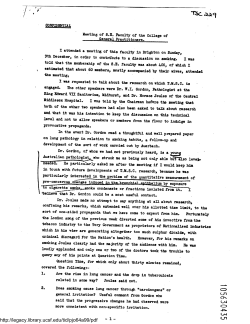


© Copyright 2026