
Small-Scale Sample Handling for Studies of Liquid Crystals and Lipid-Based Soft Matter
THESIS FOR THE DEGREE OF DOCTOR OF TECHNOLOGY Small-Scale Sample Handling for Studies of Liquid Crystals and Lipid-Based Soft Matter Gabriel Ohlsson Department of Applied Physics CHALMERS UNIVERSITY OF TECHNOLOGY Göteborg, Sweden 2012 THESIS FOR THE DEGREE OF DOCTOR OF TECHNOLOGY Small-Scale Sample Handling for Studies of Liquid Crystals and Lipid-Based Soft Matter GABRIEL OHLSSON Göteborg 2012 ISBN 978-91-7385-673-7 © GABRIEL OHLSSON Doktorsavhandlingar vid Chalmers tekniska högskola Ny serie Nr 3354 ISSN 0346-718X Department of Applied Physics Chalmers University of Technology SE-412 96 Göteborg Sweden Telephone + 46 (0)31-772 1000 Chalmers Reproservice Göteborg, Sweden 2012 II Small-Scale Sample Handling for Studies of Liquid Crystals and Lipid-Based Soft Matter GABRIEL OHLSSON Department of Applied Physics Chalmers University of Technology ABSTRACT In the field of sensing in general and biosensing in particular, there is a growing need for integrating miniaturised senor elements in microfluidic systems. The advantages are several, some of which being that the consumption of, often very expensive, sample material can be reduced; sample exchange can be optimized; sensor performance in terms of detection limits can be improved and phenomena occurring on a very small, and even single-molecule, scale can be studied. In this work different microfluidic solutions have been designed and integrated with a number of different sensing techniques chosen based on their capacity to unravel a number of different physically, chemically or biologically relevant phenomena in connection with lipid-based soft matter and liquid crystals. The first part of the thesis is focused on the advantages of miniaturizing one out of three dimensions of a reaction chamber designed for quartz crystal microbalance with dissipation (QCM-D) monitoring, while keeping the size of the radial extension macroscopic. With a high radius-to-height ratio, surface induced effects could be identified and taken advantage of in studies of liquid-crystal phase transitions in confined geometries. A similar approach was implemented to improve the performance of aqueous-phase QCM-D studies of the self assembly of supported cell-membrane mimics, revealing improved reaction rates and reduced sample consumption. The ability of QCM-D to unravel the viscoelastic properties of thin soft matter films was also used to study changes in the viscoelastic properties of surface-immobilized lipid vesicles undergoing a transition from gel to liquid phase during a temperature sweep. By varying buffer salt and pH, it was shown that the vesicle-surface interaction plays an important role in determining the characteristics of the shape fluctuations, phase transition temperature and hysteresis around the phase transition. In the last part of the thesis work, two different microfluidic systems were designed and used to study rapid transport of water and glycerol across the lipid membrane of surface-immobilized lipid vesicles. In this case, fluorescence imaging was employed to resolve transport reactions on the level of individual liposomes. The membrane-transport reactions were induced by rapidly exchange the solution outside the liposomes, for which purpose two conceptually different approaches were developed and compared. The first approach took advantage of the fact that small sample volumes can be moved rapidly over a predetermined probe area containing surface-immobilized lipid vesicles. In the second approach the vesicles were suspended and moved into a mixing region, while the liquid flows were kept constant in order to reduce the limitation imposed by external equipment such as electric valves on the system. Both methods generated lipid-membrane permeability data close to what has been reported in the literature, and statistics from single vesicle data revealed a new means to characterize the efficiency of membrane-protein incorporation. Keywords: Microfluidics, nanocell, QCM-D, fluorescence microscopy, soft matter, liquid crystals, lipid bilayer, phase transitions, membrane transport, aquaporins. III LIST OF APPENDED PAPERS Paper I Phase transitions in adsorbed lipid vesicles measured by quartz crystal microbalance with dissipation monitoring Gabriel Ohlsson, Anna Tigerström, Fredrik Höök and Bengt Kasemo Soft Matter 7, 10749-10755 (2011) Paper II A nanocell for quartz crystal microbalance and quartz crystal microbalance with dissipation-monitoring sensing Gabriel Ohlsson, Christoph Langhammer, Igor Zoric and Bengt Kasemo Review of Scientific Instruments 80, 083905 (2009) Paper III A miniaturized flow reaction chamber for use in combination with QCM-D sensing Gabriel Ohlsson, Pauline Axelsson, Joshua Henry, Sarunas Petronis, Sofia Svedhem and Bengt Kasemo Microfluidics and Nanofluidics 9, 705-716 (2010) Paper IV Solute transport on the sub 100 ms scale across the lipid bilayer membrane of individual proteoliposomes Gabriel Ohlsson, Seyed R. Tabaei (shared first authorship), Jason Beech, Urban Johanson, Per Kjellbom, Jan Kvassman, Jonas O. Tegenfeldt and Fredrik Höök In manuscript. Paper V Lipid-membrane water-permeability measurements on single vesicle level using hydrodynamic focusing Gabriel Ohlsson, Seyed R. Tabaei, Patric Wallin, Göran Petersson, Johan Andersson, Fredrik Höök and Jonas O. Tegenfeldt In manuscript. IV RELATED PAPERS NOT INCLUDED IN THE THESIS Paper VI A combined reflectometry and quartz crystal microbalance with dissipation setup for surface interaction studies G. Wang, M. Rodahl, M. Edvardsson, S. Svedhem, G. Ohlsson, F. Höök, and B. Kasemo Review of Scientific Instruments 79, 075107 (2008) Paper VII Plasma Oxidized Polyhydroxymethylsiloxane-A New Smooth Surface for Supported Lipid Bilayer Formation Cristina Satriano, Malin Edvardsson, Gabriel Ohlsson, Guoliang Wang, Sofia Svedhem and Bengt Kasemo Langmuir 26, 5715-5725 (2010) Paper VIII Simultaneous Characterization of Supported Lipid Bilayers by Quartz Crystal Microbalance with Dissipation monitoring and Electrochemical Impedance Spectroscopy Elisabeth Briand, Yves Van Ingelgem, Isabel Van De Keere, Gabriel Ohlsson, Bengt Kasemo, Annick Hubin and Sofia Svedhem In manuscript. PATENT APPLICATIONS Patent I CELL FOR CONFINEMENT OF VERY SMALL VOLUMES OF SOFT MATTER AND FLUIDS Application number: 12/442,962 Publication number: US 2010-0139420 A1 Gabriel Ohlsson, Christoph Langhammer, Igor Zoric and Bengt Kasemo LIST OF ABBREVIATIONS 5CB 4-n-pentyl-4’cyanobiphenyl AFM Atomic Force Microscopy AQP5 Aquaporin 5 CCD Charge-Coupled Device CVD Chemical Vapour Deposition DLS Dynamic Light Scattering DNA Deoxyribonucleic acid DSPE-PEG[2000]biotin 1,2-distearoyl-sn-glycero-3-phosphoethanolamine-N- [biotinyl(polyethylene glycol)-2000] DTPC 1,2-Ditridecanoyl-sn-Glycero-3-Phosphocholine EDTA Ethylenediaminetetraacetic acid EM Electron Microscopy FRET Fluorescent Resonance Energy Transfer GUV Giant Unilamellar Vesicle HMDS Hexamethyldisilazane ICPInductively Coupled Plasma LC Liquid Crystal LCD Liquid Crystal Display LED Light Emitting Diode LSPR Localized Surface Plasmon Resonance MEMS Microelectromechanical system NMP N-methyl-2-pyrrolidone NTA Nanoparticle Tracking Analysis PE Phosphatidyl Ethanolamine Pe Peclet number PDMS Polydimethylsiloxane PMMA Poly(methylmetacrylat) POPC 1-Palmitoyl,2-oleoyl-sn-Glycero-3-phosphocholine POPG 1-palmitoyl-2-oleoyl-sn-glycero-3-phosphoglycerol PLL-g-PEG Poly(L-lysine)-graft-poly(ethylene glycol) PTFE Poly(tetrafluoroethylene) QCM Quartz Crystal Microbalance VI QCM-D Quartz Crystal Microbalance with Dissipation monitoring Re Reynolds number SDS Sodium Dodecyl Sulfate SEM Scanning Electron Microscopy SLB Supported Lipid Bilayer SPM Scanning Probe Microscopy SPR Surface Plasmon Resonance St Strouhal number SUV Small Unilamellar Vesicle REM Reflection Electron Microscopy TEM Transmission Electron Microscopy TIR Total Internal Reflection TIRF Total Internal Reflection Fluorescence TRIS Tris(hydroxymethyl)-aminomethane hydrochloride UV Ultra Violet VII VIII TABLE OF CONTENTS 1. Introduction 1 2. Soft matter 2.1. What is a soft matter material? 2.2. Thin films of soft matter 2.3. Viscoelasticity 2.4. How to probe soft matter 5 5 6 8 9 3. Liquid crystals 3.1. Theoretical background 3.2. Controlling the molecular orientation at surfaces 13 13 16 4. Lipid membranes –the shells of the cells 4.1. The cell membrane 4.2. Mimics of the cell membrane 4.3. Phase transitions in lipid membranes 4.4. Transport through lipid membranes 19 19 21 22 23 5. Methods 5.1. QCM-D 5.1.1. Fundamental principles of QCM 5.1.2. Adding the D-factor 5.1.3. Quantitative modelling of QCM-D data 5.1.4. The sensor surface –a versatile device 5.1.5. Common application areas 5.2. Fluorescence microscopy 5.2.1. Fundamental principles 5.2.2. Fluorescence self-quenching 5.2.3. The fluorescence microscopy setup 5.3. Microfluidics 5.3.1. Miniaturization of sample volumes – needs and opportunities 5.3.2. Fabrication of microfluidic devices in PDMS 5.3.3. Fabrication of microfluidic devices in silicon 27 27 27 29 32 34 35 37 37 38 39 42 6. Results and experimental procedures 6.1. Brief summary of the papers 6.2. Phase transitions in supported lipid vesicles 6.3. Confining thin films – controlling two interfaces 6.3.1. Basic idea – the nanocell 6.3.2. Fabrication procedure 51 51 53 56 56 57 IX 42 45 47 6.4. The nanocell applied to isotropic liquids 6.4.1. Shear wave-lid coupling 6.4.2. Sample preparation and characterization 6.4.3. Summary of the results 6.5. The nanocell applied to anisotropic liquid crystal systems 6.6. Miniaturized fluidics combined with QCM-D sensing 6.6.1. Integrating the “nanocell concept” in a fluidic system 6.6.2. Fabrication procedure 6.6.3. Summary of the results 6.7. Microfluidics for rapid liquid exchange 6.7.1. Design and characterization of the switching device 6.7.2. Design and characterization of the hydrodynamic mixer 6.8. Measuring solute and water transport across lipid membranes 6.8.1. Permeability measurements using the switching device 6.8.2. Permeability measurements using hydrodynamic focusing 60 60 61 61 63 65 65 66 68 70 70 73 77 77 82 7. Conclusions and Outlook 85 8. Acknowledgements 87 Appendix A: Fabrication details for microfluidics made out of PDMS 89 Appendix B: Fabrication details for microfluidics made out of silicon 91 Appendix C: Calculation of osmotic permeability 93 Bibliography 99 1 INTRODUCTION The human body is an enormously complex machinery that is kept alive thanks to a multitude of precisely synchronized functions designed to corporate with each other. With the eyes it is possible to collect light from the surrounding, which is detected by the retina at the back of the globe where it is transformed to electric signals and transferred to the visual cortex of the brain via the optic nerve, eventually yielding a visual impression. Everywhere in the skin all the way out to the fingertips there are different types of sensory nerves that detect features such as texture, hardness, softness, heat, coolness, and transport the sensation signals all the way to the spine and the brain where they are put together forming an impression of the properties of the objects in touch. The functional units at the nerve ends are on the order of a few nanometres (10-9 m) and below, while the signals travel on the order of a meter. Hence, the distance encompassed by fine tuned molecular self assembly of biological soft matter ranges up to 10 orders of magnitude! Our senses: sight, hearing, sensory, smell and taste bombard us with impressions from our surrounding, and have a huge impact on our well-being and psychological mode. There are also the digestive and metabolic systems, providing the body with nutrition, the airways and lungs taking in vital oxygen and bringing out carbon dioxide from the body. Both oxygen and nutrients are taken up by the blood, which is transported to all parts of the body to provide every single cell with the necessities of life. Cells, with a dimension on the order of 1 to 10 mm, are the functional building blocks of living organisms and are present in a vast number of different types [1]. Different types of brain cells build up the brain, erythrocytes, or red blood cells, are responsible for the oxygenation and leukocytes, and white blood cells, play an important role in the immune system just to mention a few examples. If anything in this complex system is malfunctioning one can often track it down to a single cell or even the building blocks of the cell, such as DNA, proteins, lipids etc. In fact, a dominating fraction of all drugs target the cells, and in order to reach into the active parts of the cell, the active substance often needs to be transported across the protective shell of the cell; the cell membrane [1]. The cell membrane is a soft-matter structure that is built up of amphiphilic molecules. Amphiphilic means that the molecules have one hydrophilic (attracted to water) and one hydrophobic (rejected by water) part. The hydrophobic parts of the molecules tend to avoid water, which results in a dual layered sheet-like structure (a membrane) with the hydrophobic parts directed inwards. A large number of functional units are embedded in the cell membrane, many of which are responsible for transport of different molecules and substances into and out of the cell. Water transporters, which are one subject of this thesis work, belong to a very important family of membrane channels, since water needs to be loaded, unloaded and transported through various types of membranes everywhere in an aqueous environment such as the body. (The human body consists to around 70% of water.) Some examples where water needs to be transported in the body include the kidneys, tear channels and saliva production. There are also many other molecules present in the cell membrane exhibiting functions such as sensing the environment around the cell, linkage to adjacent cells and catalyzing membrane-associated reactions [1]. Furthermore, when alien entities such as viruses infect a cell the initial interaction occurs at the cell membrane. This brief overview is enough to explain the huge efforts currently undertaken to study and understand the behaviour of soft matter such as for example cells and biomembranes. Especially in the field of drug development and drug discovery one is highly interested in sensitive measurement methods that enable detection of the interaction between biological entities such as membranes, membrane proteins and drug candidates. A large number of different approaches have been developed to detect this kind of events, which typically occur on a molecular level. This, in turn, puts high demands on the sensitivity of the measurement system. Very roughly, these measurement methods can be divided into two main categories: The first and historically most common way is to have the monitored sample material in a bulk suspension. However, such methods generally require relatively large amounts of material in order to yield a detectable contrast in the measurement, which is to be considered a disadvantage in many cases. The other main category is the wide range of surface-based bioanalytical instruments. In a simplified way one can say that the dimension is here reduced from three to two dimensions, thus offering a possibility to reduce the material consumption dramatically. The basic idea is that one (or several layers) of the sample material of interest are bound to (or interact with) a sensor surface, the physical properties of which being sensitive to the properties of the sample material. A large number of high precision surface based measurement methods have been developed, such as for example quartz crystal microbalance (QCM), surface plasmon resonance (SPR), atomic force microscopy (AFM), fluorescence microscopy and electrical impedance spectroscopy etc. Some of these even allow single molecule sensitivity, such as for example total internal reflection fluorescence microscopy (TIRF) and AFM. The possibility to probe reactions and material properties using utterly low material consumption is also an important driving force behind the pursuit for miniaturizing the sensor elements. However, to fully utilize these potential benefits it is equally important to integrate (miniaturized) surface-based methods with proper liquid handling. A general approach towards controlled handling of liquids and soft matter on small scales is microfluidics [2]. Microfluidic devices generally allow manipulation of sample volumes on the order of some nanoliters and below, utilizing channels with heights and widths on the order of a few up to some hundred micrometers. On such small dimensions, liquid flows appear laminar, which means that no turbulence is present, allowing very precise control of small scale sample handling. These properties allow for particle sorting, sample mixing by diffusion and highly controlled switching of sample suspensions on short time scales [3], which has been another theme of this thesis work. Microfluidic devices have therefore become an increasingly important accessory to high precision surface-based sensing methods and can favourably be combined with micro and nanoscaled sensing elements. The overall aim of this work has been to develop micron- and sub-micron sized fluidic systems for advanced studies of soft matter materials. In the first part of the work, focus was put on developing microfluidics and miniaturized measurement systems to integrate with quartz crystal microbalance with dissipation (QCM-D) monitoring. The QCM-D technique is widely used in the field of soft matter [4], primarily due to fact that it is capable of simultaneously probe mass uptake with a sensitivity down to < 0.5 ng/cm2 and energy losses in the system caused by internal and interfacial frictional forces. In this way, structural information about thin films can be provided. This was demonstrated in paper I, in which the viscoelastic behaviour of surface-adsorbed lipid vesicles was probed across the liquid-gel phase transition. However, with respect to handling of small sample volumes, the large dimension (diameter around 1 cm) of the sensor element and its sensitivity to external perturbations present certain challenges. These challenges were addressed in order to enable detection of interfacial phenomena in thin (typically a few hundred nanometres thick) confined soft matter films (paper II) as well as to increase sensor performance and decrease sample volumes for general biophysical applications using the QCM-D technique (paper III). In the second part of this thesis work, the initial work (paper I) on structural changes of lipid vesicles (also called liposomes) was extended to the use of microfluidics to explore transport reactions across the lipid bilayer membrane of surface-immobilized lipid vesicles. Besides sufficient sensitivity of the sensor system, a prime challenge relates to the fact that these reactions typically occur on a millisecond time scale, thus requiring systems for very rapid liquid exchange. With particular focus on being able to probe transport of water facilitated by a class of membrane proteins called aquaporins [5], two different setups, presented in paper IV and V, were developed. The transport reactions were probed using fluorescence microscopy, capable of resolving individual fluorescently ladled lipid vesicles, and the motivation for the use of microfluidics was to resolve transport events taking place on down to the 10 ms time scale. Soft matter materials constitute a broad range of materials, including polymers, gels, liquid crystals, biological tissue, cells etc, i.e. materials that have both the properties of solids (elasticity) and liquids (viscosity). In the following sections, I will present and briefly discuss different types soft matter materials, with particular focus on liquid crystals and lipid bilayer membranes, their properties and how they may be affected by changing external physical parameters such as confinement and temperature. Thereafter I will proceed into a section were the sensing methods and microfluidics used in this work are treated in some detail. Finally the outcome and results will be presented and discussed. So let us now go deeper into the details. 2 SOFT MATTER 2.1 What is a soft matter material? When looking around we distinguish different types of materials present in our environment. Some materials are solids, like metals and stones, other are liquids, like water, and some materials are something in between; they are soft, like paint and glue before they have dried, plastic bags, soap, ketchup, liquid crystals in a flat screen television, and not the least the cells and tissue being the building blocks in our bodies, to mention a few examples. What is the main difference between these groups of materials? Our knowledge tells us that solid materials deform slightly when exposed to an external force, a stress, and as the stress is released, providing it is not too large, solid materials regain their original shape. The material is said to be elastic, which is a characteristic property of a solid. In contrast, when a liquid is being exposed to an external stress it is deformed irreversibly – a flow is produced. Some liquids require a larger stress than others to induce the same flow rate. The proportionality factor between external force and induced flow is called viscosity. This is an inherent property of the liquid material. Thus, one can say that the magnitude of the viscosity reflects a distinction between a solid and a liquid (the former has essentially infinitely large viscosity). Soft matter materials exhibit, to various degrees, both elastic and viscous behavior, making them very attractive for a large number of applications. These materials are said to be viscoelastic. For example, gelatin that is used when baking cakes can to a certain extent regain its original shape when deformed, but when a large enough stress is applied it will not brake but instead flow like a liquid. Toothpaste is another everyday example that to some extent behaves like a solid material but still is able to flow like a liquid when it is squeezed out from its tube. In many industrial applications it is very important for the used soft matter materials to have the right viscoelastic properties. One example is the behavior of paint. When painting a wall using a brush it is important that the paint flows easily and can be spread forming a homogenous layer over the painted surface. On the other hand, when it is left to dry one does not want it to run down off the wall because of gravity. In other words, flow must only be induced when a high enough shear stress is applied to the paint, otherwise it is desirable that it acts more “solid-like”. Another example includes the large number of polymers used when fabricating the materials known as plastics. The viscous and elastic properties need to be very different when the polymer is going to be used in a plastic bag compared to in the dashboard of a car, for example. Thus it is desirable that one can characterize viscoelastic properties of polymer chains already at a molecular level, which can be done with a numerous number of methods. Some of them are described later in this chapter. 2.2 Thin films of soft matter In a number of applications soft matter materials are present as thin films like in for example flat computer screens as well as other displays and optoelectronic devices. There, a thin layer of liquid crystals is sandwiched between two plates, the inner interfaces of which may have certain physical and chemical properties (Fig. 1). Liquid crystals are a family of anisotropic soft matter, which exhibit different physical properties depending on the orientation of the molecules with respect to the contacting interface(s). Typical building blocks for many liquid crystals include rod-like molecules. The latter can orient in several different ways at an interface which gives rise to interesting and useful optical properties [6, 7]. Thus the molecular orientation of this particular soft matter material, i.e. liquid crystal, at interface(s) is very important for controlling the physical properties of the soft matter thin film systems. In general a liquid crystal display (LCD) screen comprises a matrix of small “sandwiches”, each one corresponding to one pixel, and one common trend in industry is to manage to make thinner screens with larger area. Figure 1. A Liquid Crystal Display (LCD) screen is an everyday example where soft matter at interfaces plays an important role. The screen is showing a conoscopic image of a liquid crystal sample. Another example of a thin film made of soft matter can be found in biology. In the field of biophysics there is an extensive interest in using cell membranes on surfaces as model systems to mimic processes that occur at the interface of cell [1, 7], for example when developing new drugs, functional biosensor coatings or surfaces for cell cultures. Cell membranes are in principle a thin layer (or more exactly a molecular double layer) of soft matter, playing an important role in transport and signaling processes, including for example transport of proteins, ions, water etc, in and out of the cell. In addition, interactions between cells and surfaces play an important role in various biotechnology applications, including interactions between human cells and surfaces of implant materials. For example integration or rejection of dental implants or hip prosthesis is determined by the nature of such interactions [8]. Also, surface structure and chemistry are very important factors when growing different types of cells, like stem cells, to be used in medical applications [9]. Connected to the biomaterial and surface interactions are several “daily life” examples, such as the relatively strong binding between coagulated proteins and the iron surface of a frying pan. In order to minimize these, in this case unwanted, interactions, frying pans are covered with a thin layer of inert poly(tetrafluoroethylene) (PTFE) (also known as Teflon). It is also commonly known that dry, burnt food residues (including proteins, fibers etc) in metal pans and pots are removed more easily when introducing water to the pan or pot. Since metals exhibit hydrophilic properties, meaning that there are attractive forces between water molecules and the metal, the surface will couple to the water molecules rather than the dry food residues, facilitating removal of the latter. This connects to another interesting surface related issue: wetting and de-wetting effects, which refer to the ability of a liquid to spread over a surface [7]. Polymers in the form of thin films are also used in for example solar cells as well as in polymer-based light emitting diodes (LED:s), where films being only few tenths of a nanometer thick are used [10-14]. Other examples involving very thin polymer films (down to a few nanometres) include fabrication processes of for example micro-/nano devices such as microelectromechanical systems (MEMS), sensors and microfluidic systems, to mention just a few [15, 16]. A final example worth noting in which thin films of soft matter are of interest is the field of paper and pulp industry, where one is working with thin films of starch and cellulose. In the development process of for example paper materials, issues such as swelling, water uptake and water resistance are important to address (see for example [17]). Since paper is used in a large number of applications with various requirements on quality and properties, design on the molecular level is sometimes required. Thus, in the research and development process model systems are often supported on surfaces, in order to study the characteristics of single polymeric layers [18-20]. Generally one can distinguish two conditions for thin films of soft matter materials. The first case is when the thin film rests on a substrate but the other interface is left free (soft matter-air interface), like for the thin polymer films in solar panels or the cellulose films in paper industry. In the second case the thin film is confined between two solid, or soft matter interfaces, alternatively one solid and one soft (or liquid) interface. A typical example is LCD screens, where confined liquid-crystal films of about a few tenths of micrometers are used. Another example is supported lipid vesicles and planar lipid bilayers as used in a bioanalytical sensing context. On top of this a layer of molecules (proteins, antibodies, antigens, etc) might bind, thus forming a sandwich like structure. In one part of this thesis work, I was interested in probing the viscoelastic properties of immobilized lipid vesicles when the temperature is varied across the phase transition temperature of the lipid membrane (paper I). As the films become thin, the contribution to their viscoelastic behaviour from the interfacial regions starts to be significant. This stems for the fact that since the number of molecules in the interfacial region then becomes a significant fraction of the total number of molecules in the system. Near surfaces the molecules are affected by surface potentials and should therefore exhibit different characteristics compared to those in the bulk of the soft film. In this work, we were also interested in developing an experimental tool that can be used for studying viscoelastic properties of thin films of soft matter materials with two well-defined interfaces. Particularly in the limit when one of the thin film dimensions can be reduced down to the nanometre range. In this case, the interfacial properties of the soft material may dominate. The performance of the newly designed experimental system is demonstrated with a number of examples including isotropic liquids as well as more complicated, anisotropic soft matter systems (paper II). 2.3 Viscoelasticity As mentioned above a typical solid material is characterized by being elastic, a liquid is viscous and a soft matter material exhibits both properties; it is viscoelastic. For a fictive ideal elastic material there is a linear relation between the applied shear stress, σ (in Pa), and the resulting shear strain, e (tangents of the shear angle, dimensionless), according to the following relation: E =S /' (2.1) Here, G is a material characteristic constant known as the elastic shear modulus (given in Pa as well). The higher the shear modulus, the more rigid is the material. For example diamond typically has G = 478 GPa, while for aluminium G = 25.5 GPa, meaning that a much larger force is required to deform a diamond than a piece of aluminium having the same shape and size. Viscoelastic materials generally exhibit a relatively low shear modulus compared to solids since they are much more easily deformed. Rubber-like materials can for example have values down to less than 1 MPa. Similarly, for an ideal viscous material the generated flow is proportional to the applied shear stress, σ, according to: S =H ⋅ dv dz (2.2) In this relation dv/dz is the flow velocity gradient in the viscous material (in s-1) and η is the viscosity (given in Pa∙s). Typical values for viscosity are 1 mPa∙s for water, 16.1 mPa∙s for ethylene glycol, 20-30 mPa∙s for some liquid crystals and 2-10 Pa∙s for honey. In principle, two external factors determine whether a viscoelastic material will behave elastically or as a viscous one (for a given elastic modulus and viscosity); the magnitude of the applied shear stress (like in the paint example given earlier) and the time the material is being exposed to stress. Generally, the material first acts like elastic, but after a certain time, the relaxation time of that material, τ, it will start to behave viscously. Try for example to push a piece of gel with a low constant pressure; for a while it will deform more and more and eventually it collapses and starts to flow. However, as we all know no “ideal” material exists and the above-mentioned relations are therefore valid in real life with some modifications, but they give a good rough understanding of the behaviour of viscoelastic materials. 2.4 How to probe soft matter There is a wide range of methods used to study and characterize soft matter in bulk as well as in thin films on surfaces, be they free or confined. For measurements of viscoelastic properties in bulk materials methods such as rotational rheometry, plastometers, capillary viscometry and the method of the falling sphere are key methods [21]. Other methods to probe viscoelastic materials include ultrasonic attenuation, where ultrasonic pulses are sent through a soft matter sample. The scattered and reflected acoustic waves are detected and from their behavior one can draw conclusions regarding material properties such as viscoelasticity. This and other similar technique have been used extensively in for example studies of soft matter systems such as liquid crystals [22, 23]. However, these methods are typically not well suited in studies of thin films. Methods used to study thin films are fewer, though, and include label-based and label-free analysis techniques. Label-based methods include for example fluorescence/ luminescence detection, where the object being studied is labeled with a fluorescent molecule for optical detection using for example a microscope [24, 25]. By probing the fluorescent/luminescent label, the position, movement, size, binding and unbinding of the detected entities can be followed visually. However, a large fraction of the detection methods used to study thin films of soft matter are label free, including (electro)-acoustical tools, such as quartz crystal microbalance (QCM) [26, 27], optical methods such as surface plasmon resonance (SPR) [25, 28], ellipsometry [29], reflectometry [30, 31], optical waveguide techniques [32] and other techniques including for example sensing methods based on impedance measurements [33, 34]. In most cases these are single interface methods, where chemical and physical processes of thin films at only one substrate interface are probed, while the other interface is “free”, i.e. exposed either to vacuum, air or a purely viscous liquid. In biological application, one is often operating in a so-called buffer liquid, which is meant to mimic the environment in which the probed sample material is active in its “natural” state. For example, studies involving proteins, cells and cell membranes are often performed in a buffer suspension having the same pH and salt concentration as human blood. Since the behavior of a soft matter in some cases depends strongly on the properties of the surrounding buffer, means to change the experimental environment is a very important part of the experimental design. One example of this is analyzed in paper I, in which the viscoelastic behavior of surface immobilized lipid vesicles across the liquid-gel phase transition is studied as a function of salt content and pH of the surrounding buffer suspension. Besides changing the buffer, customization of the active sensor surface is another important parameter to consider in order provide proper conditions. However, as also addressed in this thesis work, some applications and experimental conditions require or benefit from alternative approaches in order to obtain full control of the analyzed sample. For example, the molecular orientation and physical properties of anisotropic soft matter (such as liquid crystals) strongly depend on the interfaces. In the case of a thin film it might therefore be beneficial to be able to control not only the solid support but also the upper interface by confinement. One of the most common sensing methods in the large application field of soft matter, after microscopy, is surface plasmon resonance (SPR). This technique has been used widely for detection of adsorption/desorption processes including proteins and other biomolecules [35, 36]. Physically, it is based on the principle that surface plasmons (collective oscillations of the conduction electrons on the sensor surface) are excited by an incoming light beam. The characteristics of the plasmon waves depend on the boundary conditions of the surface of the substrate. As a consequence, soft matter with a refractive index different from the surrounding that is being attached to the surface will change the requirements for surface plasmon excitation. This is a very accurate method for detection of refractive index (or mass) changes on a surface. However, from SPR measurements conclusions regarding viscoelastic properties can primarily be obtained indirectly by studying structural changes and / or molecular orientations of the sample material. With the quartz crystal microbalance (QCM) one can detect, by probing changes in its resonant frequency at around 5 to 100 MHz, a mass attached onto an oscillating piezoelectric quartz crystal sensor [37]. QCM with dissipation monitoring (QCM-D) is a more sophisticated version of the conventional QCM technique, which, in addition to the pure mass uptake is able to detect viscoelastic properties of the adsorbed films. This makes it very well suited to study soft matter and enables detection of structural changes of materials attached to the sensor surface [26]. Generally QCM-D detects the wet mass, which means that the sample mass includes water hydrodynamically coupled to the sample molecules (a more detailed description of the function is found in Chapter 5). Optical techniques, such as SPR, on the other hand, primarily detect the molecular mass, which means that the coupled water is not detected [4, 38]. A combination of SPR and QCM-D can therefore be used to characterize the amount of bound water, which is highly relevant for studies of hybridization and cross-linking of DNA, polymers and proteins [4, 39]. Among surface characterization techniques scanning probe microscopy (SPM) techniques, e.g. atomic force microscopy (AFM), deserves to be mentioned. In principle AFM is based on the interaction forces between a sample surface and a very sharp tip attached on a cantilever. The tip is scanned over the sample surface to be probed using piezoelectric motors that enable the device to move a few Ångström in every step. By letting the probe follow the surface topography in this way, an image with very high resolution can be obtained by laser based detection of the cantilever deflection. This technique has to some extent also been used to detect viscoelastic properties of soft matter materials. In principle, by pressing the tip gently to the sample followed by retraction and analyzing the yielded force curve, viscosity and elasticity of any surface adsorbed material can be extracted. An example is given in Radmacher et al [40] where proteins adsorbed on mica where studied. Another approach is to use the cantilever as an oscillator and probe the softness of cantilever-attached material as shown by for example Lubarsky et al. [41] in their work on hydration and dehydration of protein films. It is also instructive to be able image the surface of a (thin) soft matter sample using high-resolution methods such as for example electron microscopy (EM). Using this technique, the sample surface is bombarded with electrons and an image of the surface can be created by monitoring scattered, transmitted or secondary electrons [42]. The most common modes are called transmission electron microscopy (TEM), where the electrons transmitted through the sample are collected and monitored, reflection electron microscopy (REM) that records electrons reflected from the sample and scanning electron mi10 croscopy (SEM) in which the secondary scattered electrons, originating from the sample atoms, are detected [42]. The latter has proven very well suited for studies of surfaces of bulk samples, while for example TEM requires very thin sample materials. In the first part of this work we were mainly interested in QCM-D as the sensing platform, but the concept presented can of course be used in combination with other sensing methods, as well as in contexts were the sensing techniques include labeling of sample material. In the second part, fluorescence microscopy was the main method used to study soft matter, but then focused on transport across lipid membranes, rather than their actual viscoelastic properties. A deeper description of QCM-D, fluorescence microscopy and their application areas is presented in chapter 5. 11 12 3 LIQUID CRYSTALS An isotropic material is physically uniform in all directions; in other words its looks and responds in the same way regardless of direction/orientation. This is not the case for anisotropic materials. Examples of isotropic materials include water, alcohols, gases, liquid metals, etc. A common feature for isotropic liquids is that they consist of relatively small molecules (or even atoms), while anisotropic liquids contain large molecules with a nonspherical shape such as rod like or disc like. Typical examples of anisotropic liquids are liquid crystals, which will be dealt with in the following sections. 3.1 Theoretical background Liquid crystals are highly anisotropic materials with extremely interesting optical and viscoelastic properties. Probably the most well-known type of liquid crystals are so called thermotropic ones which exhibit a first order phase transition, upon temperature change, from an ordered phase into a disordered one. These materials are used in liquid-crystal displays. They often consist of rod shaped molecules, (calimatic liquid crystals), or disc like molecules, (discotic liquid crystals). However, the most abundant type of liquid crystals are the lyotropic ones, which means that they form liquid crystalline structure upon addition of a solvent such as water. Amphiphiles are typically among this type of molecules and cellular lipid membranes are hence very good examples of lyotropic liquid crystalline structures [6]. These will be described in more detail in a later section. In many applications liquid crystals are used in thin confined spaces [6, 7] and since they are highly anisotropic materials they require certain boundary conditions, at both interfaces, in order to gain full control of the molecular orientation. To optimize the applications of these types of materials there is a need to characterize them in terms of molecular order throughout the system including the interfacial region. In this work, the focus has been put on the calimatic liquid crystal 4-n-pentyl-4’cyanobiphenyl (5CB). 5CB exhibits a first order phase transition from nematic to isotropic phase at around 34°C [43] (Fig. 2), which is within the range of operation of our QCM-D setup used for the liquid crystal studies. The viscosity of 5CB is in the range 2∙10-2 (for isotropic phase) to 3∙10-2 Pa∙s (for nematic phase) and above, depending on external factors like temperature and applied electrical fields etc. The density around room temperature is around 1025 kg/m3. 13 ¿ .EMATICPHASE )SOTROPICPHASE 4EMPINCREASED Figure 2. Nematic liquid crystals, such as 5CB, undergo a first order transition to isotropic phase at a specific temperature. This results in changes in the viscoelastic properties. The molecular length of 5CB is 20 Å. Calimatic liquid crystals consist of rod-shaped molecules containing several functional groups as building blocks. For 5CB the building blocks have a typical length of ca 2.0 nm. In LCD applications one takes advantage of the ability of the liquid crystal molecules to align in an electromagnetic field. This, in turn, enables control of the transmission of light through a liquid crystal pixel by switching the orientation of the confined molecules. The alignment of the liquid crystal in external electric field is called the Fredericks transition [7]. By choosing different combinations of building blocks (i.e. molecules), one can influence properties such as phase transition temperature, critical field strength for the Fredericks transition and the nature of the molecular interaction, to suit specific applications. Calimatic liquid crystals are able to form three different phases: smectic, nematic and isotropic, which differ from each other in the ways the molecules are ordered. The transitions between the different phases are typically of first order for most liquid crystal systems. A first order phase transition is in this context characterized by a latent heat during the transition, which means that the temperature remains constant until the entire sample has entered the new phase. Melting of ice and boiling of water are typical everyday examples of first order phase transitions. In the smectic phase, which is the most ordered phase, all molecules form a well-defined layer structure and are pointing in the same direction. The smectic phase is generally divided into two sub-phases: Smectic A, where the director of the system (which is the average molecular orientation) is pointing in a direction perpendicular to the planes in which the molecules are ordered. In the other one, smectic C, the angle between the ordering planes and the director is anything else but 90°. In contrast, in the nematic phase the well-defined layer structure disappears but the molecules are still oriented in the same direction at an average. Normally this phase occurs at higher temperatures than the smectic one, since it is less ordered. However, some types of liquid crystals (like 5CB studied here) are unable to form the smectic phase, which means that the nematic phase becomes the most ordered one for such systems. Finally in the isotropic phase, which is the least ordered, the molecules are randomly oriented. The microscopic order parameter, S, is a very useful way to describe the molecular ordering in a liquid crystal system [6] and is defined as: S= 1 N ∑ N i =1 1 3 2 cos Q i − 2 2 14 (3.1) where θi is the angle between each single molecule (N molecules) and the director (average direction) of the system. For a smectic system S is typically 1 and for a totally isotropic system S is 0. Thus the order parameter decreases as the thermal motions in the system increases and in case of a first order phase transition a stepwise change of S takes place at the critical temperature. In the specific example addressed in this work, we were interested in studying the changes of the viscoelastic properties of confined films of 5CB in the vicinity of the nematic-isotropic phase transition, using the shear oscillation induced by a QCM-D sensor. Since liquid crystals are anisotropic systems, the viscoelastic behavior depends on the orientation of the molecules in relation to the induced shear wave (in contrast to an isotropic liquid). Ericksen, Leslie and Parodi [23, 44, 45] have worked out a general model based on a classical macroscopic continuum approach that has proven valid for systems operated in the ultrasonic range. This range is of interest in our case, since the QCM-D sensor is operating with frequencies of several MHz. In their description the response of an incompressible nematic liquid crystal system when exposed to an external stress can be described by its dissipative stress tensor. The stress tensor contains three curvature elasticity parameters and the six so called Leslie coefficients αi, i =1, 2, 3, 4, 5, 6. These coefficients characterize the viscous behavior under deformation, i.e. how the system is deformed when applied to a stress in a given direction, and they are generally temperature dependent. Basically, three different effective viscosities for a nematic liquid crystal can be distinguished. Each of them corresponding to different molecular orientation relative to the shear wave induced to the liquid crystal system. In case of a shear wave that propagates parallel to the director that in turn is oriented normally to the surface supporting the shear (homeotropic orientation) the effective viscosity is: Ha = A4 + A5 − A2 A2 + 2 2 G2 1 − G1 (3.2) The second case describes the effective viscosity when the director is oriented parallel to the propagation direction of the shear wave but in a planar way relative to the supporting surface (planar parallel orientation): Hb = A3 + A4 + A6 A3 − 2 2 G2 1 + G1 (3.3) Finally, the effective viscosity for a system where the director is oriented perpendicular to the shear wave propagation direction but planar to the supporting surface (planar perpendicular orientation) is described by: Hc = A4 2 (3.4) Five of these are independent as follows from the Parodi relations [44, 46]: G 1 = A3 −A2 (3.5) G 2 = A2 +A3 = A6 −A5 (3.6) 15 According to this theory all three effective viscosity coefficients of a liquid crystal system decrease with increased temperature (see [43, 47] for further details). This behaviour is a consequence of the general properties of the Leslie coefficients α4 and α5 that are always positive and decrease with increased temperature, while α2, α3 and α6 are negative and increase until the phase transition temperature is reached. In the nematic phase the viscosity values will be clearly different for different molecular orientation according to equations (3.2) to (3.4), while in the isotropic phase we expect that ηa = ηb = ηc [43]. This means that the magnitude and sign of changes in f and D obtained from QCM-D measurements might vary for different liquid crystal samples, for example depending on whether they are confined between two solid interfaces or if oriented parallel / perpendicular to an induced shear wave, during the nematic-isotropic phase transition. 3.2 Controlling the molecular orientation at surfaces When working with a free film of liquid crystals the molecules typically exhibit a strong homeotropic orientation at the interface towards air [48, 49]. On the other hand, at the interface towards a solid sample it is possible to introduce a number of well defined molecular orientations, ranging from pure homeotropic, via tilted, to pure planar orientation [50, 51]. Hence by adding a lid on top of the sample, the orientation may be totally controlled at both interfaces (Fig. 3). In principle one could introduce different molecular orientations at the 5CB-lid interface and the sensor/substrate-5CB interface respectively, making it possible to induce a twist through the sample. This is in turn expected to give rise to very different viscoelastic characteristics compared to the cases where the molecules are oriented uniformly through the entire sample. For example, consider a thin film with the molecules at the sensor oriented parallel to the shear and the molecules at the lid oriented perpendicular to the shear. Such a system may exhibit a viscoelastic behaviour characterized by a phase transition that causes changes in viscoelastic properties that lies in between the pure parallel and the pure perpendicular cases. In this work, this configuration was obtained by thermal evaporation of SiO2 at an angle of 60 degrees relative to the normal of the sample surface (Fig. 3 a), which is a well established method to induce planar orientation of calimatic liquid crystals [50, 51]. The result of this procedure is a grooved topography on the treated surface (Fig. 3 a) that will force the liquid crystal molecules to orient themselves in a well defined way, following the grooves on the surface (Fig. 3 b). In this way the molecules minimize the elastic energy of the system [51]. Due to intermolecular interactions, the molecular orientation propagates through the liquid crystal film both from the lid and from the substrate respectively, giving rise to a well ordered planar structure (Fig. 3 c). Homeotropic orientation, on the other hand, is often induced by using totally different approaches. For example the polyimide Nissan 1211, often used in liquid crystal applications [52] diluted in an N-Methyl-2-pyrrolidone (NMP) suspension could be spincoated on the substrate and the lid. This is followed by baking at elevated temperature, and finally resulting in a thin uniform layer being formed on the surfaces. 5CB molecules are in this case known to exhibit anchoring in homeotropic direction, thus providing a well-ordered structure. 16 A 3I/ C B ,#MOLECULES 3I/ INTERFACES ,ID "OTTOM Figure 3. Surface modification with SiO2 and control of the molecular orientation. (a) SiO2 is deposited through evaporation at an angle of 60° from the normal direction of the sample giving rise to a wave like surface structure. (b) Usage of the modified surface in a calimatic liquid crystal system results in a well defined direction of the surface anchoring. (c) Using the same kind of surface as substrate and lid induce, through intermolecular interactions, the entire confined film of liquid crystals to orient in the same direction. The characterization of the ordering in the samples of liquid crystal prepared as described above can be carried out by using an optical microscope with crossed linear polarisation filters in a reflection mode [53]. The principle is the following. First, the incoming light beam is linearly polarised through the first filter, the polariser, and reflected from the liquid crystal sample. The orientation of the optical axis of the liquid crystals determines the direction of polarization for the reflected light. Finally the reflected light has to pass the last polarization filter, the analyser, which is rotated 90° relative to the polariser. A sample in which all molecules are oriented uniformly planar will at certain angles direct the light beam all the way through the analyser. A homeotropic sample on the other hand has the main optical axis perpendicular to both filters, which prevents light transmission through the analyser. In this manner, it is possible to verify that the sample surface treatment worked as expected. Secondly, as a complementary technique to determine the orientation of sample molecules, light conoscopy is often used [54]. In this method crossed polarizers are used for directing and analyzing the incoming respective outgoing light beams. But instead of using parallel light a conic light beam is sent through the setup. As a result a cross-like interference pattern is observed on the other side of the analyzer, whose position depends on the direction of the main optical axis of the analyzed liquid crystal sample. A pure homeotropic sample will exhibit a centrally positioned cross (Fig. 4 left), while in the case of tilted molecules with respect to the substrate the cross will move towards the edges of the conoscopic picture. Finally, for a totally uniform sample having the planar 17 molecular orientation, the cross is “lying down” resulting in a black line pointing in the direction of the optical axis (Fig. 4 right) [54]. Both methods were used to characterize the liquid crystal used in this work (paper II). However, since conoscopy preferably is performed in transmission mode and only works for samples thicker than the wave length of the light used (typically visible light), conceptual samples of an appropriate thickness were made from two glass lids to confirm the function of the surface treatments used for the QCM-D samples. Figure 4. Conoscopy and polarized microscopy were used to confirm that the concept worked. A cross indicates homeotropic orientation and a line indicates planar orientation in the direction of the line. 18 4 LIPID MEMBRANES THE SHELLS OF CELLS 4.1 The cell membrane All living organisms consist of soft matter. Of these, the cell membrane is definitely one of the most fascinating ones. It separates the inner part of the cell, the cytosol, from the external, extracellular environment. Besides acting as a shell for the inner parts of the cell, the membrane has many other important functions when it comes to cellular interactions with the external environment. For example, proteins integrated in the membrane control transport processes of nutrients, water, ions, waste, etc, into and out of the cell. In addition, through receptors and other mechanisms, nerve signals and other kinds of information are transmitted back and forth through cell membranes (especially in nerve cells). Natural membranes contain a great number, around a hundred, of different lipids [55, 56], the composition of which governs the physical properties such as charge and phase transition temperature of the membrane. The different lipids have their specific place and function and, for example, the functions of certain membrane proteins require a corresponding local environment of specific lipids [1, 55, 56]. In a membrane roughly 50 % of the molecules are lipids and the other 50 % are different types of proteins, such as receptors [1]. Lipids molecules are said to be amphiphilic, which means that they have one hydrophilic and one hydrophobic part: a hydrophilic, polar or negatively charged head group and a hydrophobic, non-polar hydrocarbon (usually fatty acid) tail as illustrated in Fig. 5 (a) [57]. As mentioned in the introduction, cell membranes consist in principle of a dual layer sheet of lipid molecules with the hydrophilic head groups directed outwards, due to the existence of water in and around cells, while the hydrophobic tails point inwards towards each other, forming a hydrophobic core. In fact, the lipid membrane can be considered a lyotropic liquid crystal, since it spontaneously forms an anisotropic ordered structure when in an aqueous environment, as mentioned earlier in this chapter. The hydrocarbon tails can have different lengths and be saturated (only single bonds between the carbon atoms in the chain) or unsaturated (one or several double bonds in the chain). This together with the properties of the head group influence the structure adopted by the membrane as well as the packing ability and hence the fluidity in the cell membrane [1]. For example lipid molecules with only one hydrocarbon tail tend to form spheres, so called micelles, where all the tails point inwards towards each other. These are generally not present in the membrane but rather in other parts of the cell. Lipids with 19 two hydrocarbon tails, on the other hand, are packed such as they form a lipid bilayer sheet, which is the way cell membranes are arranged. In order to minimize the surface energy, bilayer membranes made of lipids with two tails tend to spontaneously form a hollow spherical structure in which only hydrophilic head groups are in contact with the surrounding water – a so called lipid vesicle, or liposome [57] (Fig. 5 b). Importantly, thanks to the fact that the lipid membrane is a two dimensional fluid at temperature above its specific first order liquid-gel phase transition temperature, defects or openings in a lipid bilayer structure are energetically unfavourable and tend therefore to be repaired by replenishment of diffusing molecules. Phospholipids are the most abundant type of lipids in native cell membranes and have in common a negatively charged phosphate group in the hydrophilic head part. In combination with other functional groups phospholipids could either be negatively charged or zwitterionic, which means that they have both positive and negative charges that cancel each other. The second most common lipid is cholesterol. Cholesterol has a stiffening effect on the membrane, reduces both its molecular mobility and the permeability to water and other solutes and also influence the function of membrane incorporated proteins [55, 58]. Embedded in or associated with the cell membrane are also proteins, generally referred to as membrane proteins. These can either be totally integrated, traversing through the bilayer (integral proteins) or be attached at the head groups at the periphery only (peripheral proteins). Because of the importance of cell membranes and their constituted proteins in biological processes around 70% of all pharmaceutical compounds used today are targeted towards them [59]. Both passive and membrane-protein facilitated transport across lipid membranes, which is specifically addressed in papers IV and V, are discussed in further detail below. A E N OLI #H E AT SPH O 0H OL CER Y L ' I AC TY &AT LS AI DT #( #( 0 / #( #( / / # / #( #( #( #( #( #( #( #( #( #( #( #( #( #( #( #( #( #( #( #( #( / / # #( B / / #( .#( #( #( #( #( #( #( #( #( #( #( #( #( Figure 5. (a) POPC lipids consist of a polar headgroup with choline, phosphate and glycerol. Two hydrophobic fatty acid tails form the other part of the lipid. (b) A cell membrane (here illustrated as a vesicle in cross section) is built up as a dual layer structure of lipid molecules. The lipids comprise a hydrophilic head group pointing outwards and a hydrophobic tail pointing inwards the membrane from two directions. 20 4.2 Mimics of the cell membrane There are many different approaches to study lipid membranes; they can be studied in live cells where they adopt their complex native state, with a great mix of dynamically changing lipid molecules and membrane proteins. To gain fundamental insights regarding how different individual processes contribute to the complex functions of the cell membrane, it is convenient to study simplified model systems. Artificial cell membranes are commonly studied in the form of lipid vesicles, or liposomes, either suspended in a bulk suspension or attached in some way to a solid surface. The latter typically connected to a surface sensitive technique, such as a QCM-D crystal, an SPR chip or a glass substrate compatible with fluorescence microscopy. If the sensor surface is covered with a hydrophilic material, like for example silicon oxide, vesicles can rupture to form a uniform planar supported lipid bilayer when critical vesicle coverage is reached [60-62]. This is a very nice approach to use when studying cell membrane mimics with surface-based sensing methods like QCM-D (or SPR), which has become a widely used tool to study applications related to cell membranes [7]. Alternatively, lipid vesicles can be directly attached or anchored on the surface via functional groups such as biotin-NeutrAvidin [63, 64] or DNA strands [65]. Tethered lipid vesicles have emerged as a very attractive alternative to planar supported lipid bilayer, offering a means to study, for example, the influence of membrane curvature on protein binding [66, 67], transport of both ions [68] and uncharged solutes [69]. For additional examples, see the review by Christensen et al in reference [64]. The motivation behind the use of microfluidics in combination with supported cell-membrane mimics is manifold. First, the use of a miniaturized reaction chamber will reduce the sample consumption. This is important since many biological materials such as proteins are very expensive to purify (or synthesize) and therefore it is desirable to keep sample waste at a minimum. Second, microfluidics offer more rapid liquid exchange, due to the lower inertia in a small sample volume compared to a large. Laminar flows also allow more precise modulation and steering of the sample stream and a very small change in position gives a relatively large effect on a small-scale system. Due to the small dimensions and laminar characteristics the only natural mixing that occurs is due to diffusion. This can be used for sample mixing and controlled by modulating flow rates and stream widths as will be described in a later section. Third, microfluidics also offer faster biomolecular immobilization or bilayer formation compared to using a reaction chamber of larger format. For example, under certain conditions, the vesicle adsorption rate is limited by the diffusion through the buffer liquid down to the sensor surface [70]. By minimizing the chamber volume this rate could potentially be increased resulting in that the critical coverage is being reached faster at SLB formation in small chamber than for a large using the same sample flow rate. By in this way using (increasing) flow and using smaller volumes diffusion limitations can be overcome thus allowing resolution of the actual reaction kinetics. The reason is that sample material reaches the active (sensor) surface faster than when it diffuses a longer distance in a stagnant suspension. More of these aspects are discussed in paper III. 21 4.3 Phase transitions in lipid membranes In similarity to thermotropic liquid crystal structures lipid bilayers also undergo phase transitions as a function of temperature. In the high temperature phase the lipid molecules are freely diffusing around in the membrane together with the membrane proteins (which however diffuse more slowly [6]). This is considered the natural state of a lipid membrane and all living cells have their membrane in this phase, although small-scale patches adopting other phases also exist [71]. The diffusivity of lipids and proteins in the liquid state (which can be compared to the isotropic phase of a calimatic liquid crystal) is essential for the function of the membrane and the entire cell, its membrane proteins, enzymes, self reparation, etcetera. As the temperature decreases the membrane eventually undergo a transition to the so called gel phase (to be compared to the smectic or nematic phase in the calimatic liquid crystal case). Then the mobility of the lipids is drastically decreased and they tend to order in a hexagonal pattern with the hydrophobic tail groups straightened out [6] (Fig. 6). In a living organism the gel-liquid phase transition temperature is usually below the ambient temperature of the cells. For example membranes in warm blooded animals like a mammal have a phase transition temperature higher than those in cold blooded animals such as fish or snakes, since the former keep a constant (high) body temperature. If an organism is cooled down below the transition temperature it generally dies, because important functions such as transport processes and enzyme activities cease [1]. ,IQUIDPHASE 4EMPERATURE CHANGE 'ELPHASE Figure 6. When going from the natural liquid phase to the low temperature gel phase, the lipid molecules organize in a hexagonal pattern and the hydrophobic tails stiffen. The phase transition temperature depends on the composition of the lipid molecules. In general molecules with short hydrophobic hydrocarbon tail groups exhibit a lower phase transition temperature. The reason is that the intermolecular interaction then is weaker compared to lipids with longer tail groups. In addition the shape of the tail groups affects the phase transition temperature. For example the presence of kinks, due to unsaturated double bonds, makes the molecules harder to pack and form the hexagonal structure. The result is a lower phase transition temperature [1]. The lipid bilayers made of such lipids have a higher tendency to be in liquid state than lipids with only saturated bonds in 22 the hydrocarbon tail (making them straighter, hence allowing closer packing) at a given temperature. The phase and the viscosity of a lipid membrane also govern other physical properties such as for example the permeability of the membrane to solutes, which is addressed in the next section. 4.4 Transport through lipid membranes There are a vast number of substances, molecules and ions that need to be transported in and out of a cell. Nutrition needs to enter the cell, waste needs to leave and signal substances need to pass both ways, to mention a few examples. As mentioned earlier there are in general two different types of transport mechanisms through cell membranes; relatively slow, passive diffusion through the membrane and faster transport events facilitated by membrane proteins. The hydrophobic nature of the inner part of a lipid membrane prevents hydrophilic entities such as charged molecules (ions) to pass through and slows down the diffusion of polar molecules such as water. Basically, the more charged, the harder to pass through a lipid membrane. Nonpolar molecules can, on the other hand, easily pass through. Examples include oxygen, nitrogen and carbon dioxide. The other factor governing the rate of the passive diffusion is the size of the molecules; the larger the slower diffusion [1]. The physical property describing the rate by which a molecule can pass through or permeate a membrane is called permeability and is generally measured in cm/s. Typical values spans from ca 10-2 (water) and down to ca 10-12 cm/s (sodium ions) [1]. In order to facilitate the rate of transport of polar entities across the lipid bilayer there exist different types of transporter proteins which are integrated into the native cell membrane. In this way, ions and polar molecules can be translocated without coming in contact with and being blocked by the hydrophobic interior of the membrane. There are many different types of such proteins categorized according to their mode of action and specificity to different types of molecules. Broadly speaking, they function according to two main principles; they are either carrier proteins or channel proteins (Fig. 7) [1]. The former first catches the molecule of interest which is subsequently “carried” through the protein while it undergoes a conformational change that leads to release of the molecule on the other side of the membrane. Channel proteins on the other hand function more like a tunnel through which the specific molecules or ions can swiftly pass. This type of transport protein mechanism is generally faster than that controlled by carrier proteins. Further on, membrane proteins can work either passively or actively. Passive transport is driven by diffusion of the solute from one side of the membrane at which the solute concentration is higher to the other side where the concentration is lower. In cases when the solute is charged (ion) passive transport can also be driven by an electrochemical gradient across the membrane. Osmosis is an important cellular process in which water is transferred across biological membranes by passive diffusion in response to a concentration gradient of solutes in the opposite direction to reduce the difference in concentration of solutes on the two sides of the membrane. In the case of active transport, on the other hand, the membrane protein requires input of energy in order to transport solutes or ions against a concentration or electrochemical gradient. Some carrier proteins are active and some are passive, while channel proteins exclusively work passively [1]. 23 A B #ARRIERPROTEIN #HANNELPROTEIN Figure 7. (a) A principal sketch of a transmembrane transport protein, in this case an aquaporin transporting water (size of water molecules exaggerated). (b) Cross-sections of channel protein and carrier protein. The former functions as a tunnel, while the latter carries the solute through. In this work, we have been particularly interested in a class of channel proteins, so called aquaporins that facilitate water transport and increase the rate by which water can cross cellular membranes compared to that of passive diffusion driven by osmosis (see above). Everywhere in the body, water needs to be transported across membranes and for this purpose there are a number of different aquaporins that exhibit slightly different properties such as mode of action and permeability [5, 72]. Some are responsible for tear transportation, other for saliva excretion, other are present in the kidneys, in the lungs or in the vicinity of the brain, to mention a few examples. Aquaporins are generally assembled four by four; thus forming so called tetramers, which can transport up to over a billion water molecules per second [72]. It is believed that dysfunction of aquaporins give rise to a number of diseases, such as brain edema, and that there are possibilities to treat cancer, obesity, epilepsy and other widespread diseases by regulation of aquaporins [5, 73]. Therefore it is highly interesting from a medical point of view to be able to study this type of proteins. In the case of cancer treatment, for example, one is interested in inhibiting the function of the aquaporins in the cancer cell membranes, which could potentially help 24 neutralizing their growth. In order to do this, so called inhibitors need to be discovered. Other examples include the possibility to kill bacterial parasites, responsible for different diseases such as malaria, by eliminate their aquaporins channels using inhibitors [74]. To facilitate the ongoing attempts to discover aquaporin inhibitors or compounds with certain regulatory effects, there is a need for efficient screening methods. Potentially, one would like to test different types of compounds on a statistically large enough set of well defined aquaporins proteins. Ideally, it would be desirable to allow screening of several different compounds on the very same set of aquaporins [74]. In papers IV and V, two new means to probe water transport across the membrane of lipid vesicles with a mean diameter of around 100-200 nm was explored. The small volume of such vesicles, being on the order of an attolitre, contains around 108 water molecules. With a water transport efficiency on the order of 1010 molecules per second per aquaporin tetramer, the entire water volume is exchanged (induced by e.g. an osmotic shock) on the millisecond time scale. This requires fast detection schemes and rapid liquid handling, which was in this work approached using fluorescence imaging. It was designed to resolve single lipid vesicles, and a microfluidic system, designed for rapid liquid exchange around the liposomes (see chapter 6 for details). 25 26 5 METHODS 5.1 QCM-D 5.1.1 Fundamental principles of QCM The fundamental physical principle on which the Quartz Crystal Microbalance technique is based is the piezoelectric properties of quartz. Piezoelectric materials deform when exposed to an external electric field and generate an electric field when deformed. The reason for this behaviour originates from the fact that piezoelectric materials, like quartz, possess a crystalline structure in which subdomains having opposite electrostatic net charges are formed. When an electromagnetic field is applied to the material they will orient themselves in a reversible way, causing a strain in the crystal structure. By applying an alternating voltage to the material the strain amplitude will vary periodically with the same frequency as the applied voltage. At a certain frequency, called the resonance frequency, the oscillation exhibits a totally harmonic behaviour, with a maximum possible amplitude and very low energy loss. This frequency is typically of the order of a few MHz for a quartz plate having a thickness of some hundred micrometers. In general, the amplitude of the oscillation depends on the magnitude of the applied voltage and the so called quality factor (Q factor) which is an inverse measure of the energy loss per oscillation period, where low energy loss implies high Q factor. The oscillation amplitude of the quartz crystal is typically in the range one to a few Å (but can be larger under certain conditions) [75], while the resonance frequency is determined by the thickness of the material according to the following relation [37]: f = n ⋅ vq 2 ⋅ tq (5.1) where f is the resonant frequency, n is the overtone number (odd), vq is the propagation speed of an acoustic wave in quartz (3340 m/s) and tq is the thickness of the quartz plate. Since the applied alternating potentials at the two interfaces of the quartz plate are always 180° out of phase (antiphase) relative each other, the shear at the interfaces will be out of phase as well [76]. This means that at the fundamental resonant frequency, the acoustic shear in the material forms a standing wave where the thickness of the crystal is equal to a half of the wavelength of the standing wave (Fig. 8). For the next harmonic the thickness is equal to three half wavelengths, for the next after that five halves, etcetera. In other words, the standing wave forms in such a way that an odd multiple of a half wavelength for all harmonic overtones matches the thickness of the crystal. This reasoning explains the factor 2 in the denominator of equation (5.1) as well as why n can only be an odd number. 27 N N TQ Figure 8. The mode displacement for an oscillating QCM crystal for n=1 and n=3, respectively. Since the oscillation period at the resonant frequency is very well defined one of the most common application areas for quartz crystals is as reference time keeper in for example watches, computers and a number of other electronics devices. Beside this area, sensing applications (QCM) are important fields of use for quartz crystals, based on the following important characteristics: When adding mass to a QCM, the resonant frequency decreases linearly (assuming that the added mass is homogenously distributed over the sensor area and that the sensor is much thicker than the added film thickness) (Fig. 9), as described by Sauerbrey in 1959 [37]: ∆f = − f ∆m = −X ⋅ ∆m tq R q (5.2) where tq and ρq are the thickness and density of the quartz plate, respectively, f is its resonant frequency and Δm is the mass change. 6 6 Figure 9. The principle of QCM sensing. Applying an alternating voltage induce an oscillation of the quartz disc at its resonant frequency (top figure). Loading the sensor yields a decrease in the resonant frequency (bottom figure). This useful property makes the QCM a very sensitive mass detector allowing weighing of very small amounts of sample material added to the surface. As a consequence it has been widely used in for example vacuum systems as a tool for measuring thicknesses during thin film deposition processes. The latter is easily extracted from (5.2) providing Δf is being monitored and the density of the deposited material is known. In 1985 an important step towards a new application of the QCM took place when Gordon and Kanazawa published a paper on a QCM operating in a liquid environment [77, 78] . The novelty and importance of this work lies in the fact that for the first time one managed to operate the system and measure the frequency changes upon loading the QCM device in a highly viscous environment i.e. in the presence of large energy losses. In this case a strong coupling of the oscillating system to the viscous medium makes the simple Sauerbrey relation (partly) inoperative. This work stimulated the development of the QCM devices in a new direction where in addition to the usual mass uptake one should also follow the energy losses from the oscillation systems when coupling to the 28 viscous medium is significant. Ten years later, in 1995, the first paper on Quartz Crystal Microbalance with Dissipation monitoring (QCM-D) in liquid and gaseous environments was published by Rodahl et al [79]. This introduced simultaneous monitoring of resonant frequency and the dissipation factor of the system (the latter is related to the inverse Q factor introduced earlier). Measurements of the dissipation factor can account for energy losses related to the viscoelastic properties of the adsorbed layer. Furthermore, the dissipation factor measurements may also account for the energy losses from the QCM oscillator to the viscous environment, in the absence of an adsorbed layer. These pieces of work are considered important milestones for the expansion of QCM into new and wider application areas including the possibility to probe viscoelastic properties of materials. 5.1.2 Adding the D-factor The dissipation (D) monitoring adds an extra dimension to regular QCM sensing. It enables, in combination with monitoring of the resonant frequency (f), extraction of further physical properties of a soft matter or a liquid in contact with the active surface of the sensor. Quantities such as viscosity, shear modulus, density and thickness are possible to extract from combined Δf and ΔD data at several overtones, as will be described further in next subsection [80]. The dissipation factor is typically on the order of 10-6 and is basically defined as the energy loss per oscillation as a fraction of the total energy stored in the system as described by: D= Elost 2P ⋅ Estored (5.3) As explained earlier, D defined as above is the inverse of the Q-factor of the quartz crystal [79]. In the QCM-D set up D is extracted from the decay of the so called ringing curve, describing the oscillation amplitude of the quartz crystal, after switching off the driving voltage (Figs. 10 a and b). If a thin rigid film, such as a metal film, is adsorbed onto the crystal, only a very small fraction of energy is dissipated as a direct consequence of the adlayer, during each oscillation period. This results in a slow decay of the oscillation amplitude, which implies a low D factor (Fig. 10 c). If, on the other hand, a thick viscous film, such as a hydrated polymer coupled to surrounding water, is attached to the sensor surface, a much larger fraction of the total energy store in the system is dissipated during each oscillation. The resulting faster amplitude decay means that the system has a relatively high D factor (Fig. 10 d). The QCM-D sensor is realized in an AT-cut circular quartz plate with a thickness of about 300 µm, with a fundamental (n =1) resonant frequency of 4.95 MHz (according to Eq. 5.1), and a diameter of 14 mm. There are several ways to cut quartz relative to its crystalline structure, depending on the desired physical properties, such as temperature stability and shear direction in the resonant oscillation. The AT-cut crystals are proven particularly good for resonator and sensor applications since the induced shear, when applying an external voltage, lies totally parallel to the radial extension and is perpendicular to the thickness of the crystal (provided that the electric field is applied along the thickness direction) [81]. In addition, this cut optimizes the frequency stability around room temperature, which means that the frequency changes upon temperature variations are at a minimum in that regime. This is important not only for sensing applications, but also when the crystal is used as a reference resonator in electronic devices. 29 A 6 B 6 C 6 D 6 Figure 10. An illustration of the principle of dissipation monitoring. When the driving voltage of the QCM-D sensor, driven at its resonant frequency (a), is switched off (b) the oscillation amplitude decreases due to energy dissipation. This dissipation is lower in a rigid system (c) compared to a viscous system (d). There are two electrodes on the QCM-D sensor, one being attached on the backside and one on the “active” side, and they are connected to the driving voltage and ground, respectively (Fig. 11). The electrodes are deposited by evaporation, having a typical thickness of about 50 nm and being adhered onto the quartz via a 5 nm thick layer of chromium. The electrode on the active side has a diameter of 12 mm, whereof which 10 mm covers an optically polished area of the quartz plate, which constitutes the active sensor area. 5PSIDE %LECTRODES $OWNSIDE #ONTACTS Figure 11. The QCM-D sensor is, in principle, an AT-cut quartz crystal sandwiched between two gold electrodes. Contact electrodes connect to driving equipment. During operation, the sensor is probed at its fundamental frequency (n =1) and every odd overtone, as explained above (n = 3, 5, 7 etc. giving resonant overtones of 14.85, 25.75, 36.65 MHz etc., respectively), sequentially. At each overtone the driving voltage is switched off while both f and D are monitored during the decay of the ringing curve. The oscillation amplitude of the quartz crystal is proportional to the applied voltage. In addition it is overtone dependent (as 1/n2) and follows a Gaussian distribution over the sensor surface [37, 82] as illustrated in Fig. 12. The amplitude maximum is in the middle of the sensor and is zero at the electrode edge. (Outside the electrode the applied voltage is zero and no oscillation is therefore induced). 30 /SCILLATION AMPLITUDE N N N N 1UARTZ CRYSTAL Figure 12. The amplitude of the oscillation of the quartz crystal is distributed like a Gaussian. Higher overtones have lower amplitude and smaller width than lower. For clarity the proportions are exaggerated. Due to the smaller oscillation amplitude for the high overtones, the shear wave that is induced in the probed soft matter sample has smaller initial amplitude as well, compared to amplitudes of low overtones. The extinction depth for the shear waves originating from higher overtones when propagating into the probed soft sample material is shorter than the corresponding extinction depth for the shear waves generated by the lower overtones. This is described by: D = 2H WR (5.4) where η is the viscosity of the material, ω is the oscillation frequency and ρ is the density. This means that a material with high viscosity has a larger extinction depth than a material with low viscosity and that increased density results in shorter extinction depth for the shear wave. Practically, by using different overtones it is possible to probe different regions of the sample, and by combining the different input parameters one can get a unified picture of the viscoelastic properties of the examined system. Qualitatively, differences in the behaviour of f and D between different overtones indicate that the viscoelastic properties of the sensed system closer to the surface are different from those in the sample volume as a whole. In a typical QCM-D experiment a series of f and D data points at several overtones are being collected. The most straightforward data analysis performed is to qualitatively study the changes of f and D and the respective behaviour at each overtone, individually and compared to each other. Large frequency shifts indicate a large increase in mass load and vice versa. A large dissipation change, on the other hand, normally corresponds to a significant change in viscoelastic properties due to for example water uptake resulting in swelling or the opposite case; a collapse and water release, or similar. For some cases f and D analysis is not sufficient to obtain a correct qualitative picture of the experimental scenario. One example is when comparing different sets of data showing very large, or very small, changes in both the frequency and the dissipation shift. For such cases it might be difficult to interpret the data in terms of stiffness, since for example an extremely thin film of a viscous sample material might exhibit a quite small dissipation shift, and vice versa. An additional qualitative data analysis could then be done by plotting the dissipation shift as a function of the frequency shift in a so called f-D plot. The advantage of the f-D plot compared to the analysis described above is that the trends in the f and D characteristics relative to each other may be extracted. An example of extracted information is whether the dissipation shift is small or large compared to 31 the frequency shift, indicating a relatively rigid or floppy sample adsorbed to the sensor surface [83]. Finally, by using the advantage of having several input parameters it is possible to extract quantitative numbers describing the viscoelastic properties by relying on theoretical modelling, as described in the following section. 5.1.3 Quantitative modelling of QCM-D data In order to calculate viscosity, shear modulus, density and thickness, f and D data obtained for at least two overtones need to be included since this gives four independent input parameters and thus a determined equation system. Voinova et al [80] have made a thorough model analysis that gives a relationship between the input parameters f and D, at different overtones in a viscoelastic environment. Quantified physical properties can there from be extracted, using a continuum layer approach (Fig. 13) for up to three different layers. In this model the layers are assumed to have infinite extension in the radial direction and a well defined thickness that is much larger than the atomic size (continuum approach is valid). In addition the upper layer is considered infinitely thick –a so called buffer layer. The propagation of the shear wave originating from the quartz crystal surface is then calculated as a function of the physical parameters defining each layer. "UFFER ,AYER ,AYER 1UARTZ CRYSTAL Figure 13. When quantifying QCM-D data a layer model is used according to Voinova et al. The added material may be divided into one or two layers, plus an infinite buffer atop, depending on the properties of the system of interest. Basically, the model combines viscosity, which is mathematically described by a dashpot, and elasticity, described in the model by a spring. The dashpot and the spring are combined in pairs either in parallel forming the so called Voigt element or in series forming the Maxwell element (Fig. 14). The former is the most commonly used building block in the modelling process when extracting physical properties from systems including one or several layers. Examples include the case of molecular interactions on top of a surface, given that the layer is stationary [80]. Mathematically, a shear stress, σxy, applied to the Voigt element can be expressed with the following differential equation [80]: S xy = G ∂u x ( y, t ) ∂v ( y, t ) +H x ∂y ∂y (5.5) where G is the elastic shear modulus, η is the viscosity of the element, as described in the introduction part of this work and the ux and vx are the displacement respective velocity in the Voigt element under deformation. Eq. (5.5) is in fact the sum of the equations (2.1) and (2.2), describing elasticity and viscosity, respectively. The x-direction is defined as the direction of deformation. Maxwell’s element, on the other hand, is rarely used for most QCM-D applica32 tions but may be an option when performing theoretical calculations on a non-Newtonian soft matter solution, using the buffer layer in Fig. 13 only. Theoretically, other combinations of viscosity and elasticity can be used as well, but these are not subjects for this work. A B Figure 14. In the Voigt element the dashpot, representing viscosity, and the spring, representing elasticity, are connected in parallel (a). For the Maxwell element the same components are connected in a series (b). For the general case having three viscoelastic layers as illustrated in Fig. 13, the model system description can be solved using the following relation, describing an oscillating shear wave in a viscoelastic medium: G* ∂ 2u x ( y , t ) = − R W2u x ( y, t ) 2 ∂y (5.6) where G*= G + iωη is the complex shear modulus, η is the viscosity, ρ is the density of the medium and ω is the oscillation frequency. Usually a non-slip condition is assumed to hold at the interfaces between the layers and towards the quartz crystal. The following relations for the frequency and dissipation change can be extracted based on the Voigt element coupling (see Ref. [80] for the details): 1 H3 ∆f ≈ − + 2PR q tq D 3 2 H jW 2 H3 R W − t t 2 ∑ j j j 2 2 2 j =1, 2 D 3 G j + W H j 1 H3 ∆D ≈ + 2PfR q tq D 3 H 2 H jW 3 t 2 ∑ j G 2 + W 2H 2 D j =1, 2 j 3 j (5.7) (5.8) Here tq and ρq the thickness and the density of the quartz crystal, f is the resonant frequency of the unloaded crystal, index j=1 denotes layer 1, index j=2 is layer 2 and index j=3 is the buffer layer. Further ηj and Gj are the viscosity and shear modulus of the layers respectively, tj their thicknesses and δ3 is the shear wave extinction depth in the buffer medium as defined in equation (5.4). By considering a single layer case, and comparing (5.7) and (5.8) with the Sauerbrey relation (5.2), one notices that when ΔD for the layer added on top of the quartz sensor (adlayer) becomes significantly larger than zero, the Sauerbrey relation will underestimate the mass compared to Voinova et al. model predictions. On the other hand, if ΔD is close to zero it is a good approximation and viscoelastic modelling might not be necessary. This is the case given that only the contribution of the adlayer is considered in the calculation, and not the bulk liquid, and that the layer is thin compared to the sensor 33 thickness, as stated before. The viscoelastic modelling approach can be implemented in a software like QTools (Q-Sense AB, Västra Frölunda, Sweden), where an iterative process is used to find viscosity, density, shear modulus and thickness values corresponding to a series of f and D data points. Also, by using assumed or known values for viscosity, elasticity, thickness and density of a system as input parameters in the calculation, it is possible to predict the experimental frequency and dissipation output parameters under certain conditions. This is very useful when designing an experimental system, or in cases where one wishes to improve the theoretical understanding of a complicated system prior to realization. In paper I the viscoelastic properties around the liquid-gel phase transition of adsorbed DTPC liposomes was modelled using a one-layer Voigt model as an approximation of the close-packed liposome layer. To induce a phase transition the temperature is changed across the transition temperature. As a resulting issue the generic resonance frequency of the QCM-D sensor itself changes with temperature, complicating the distinction of the contribution from the change in physical properties of the liposomes on f and D. To solve this, a reference measurement of a blank senor before adsorption of liposomes was necessary. This background value could then be subtracted from the raw data, hence distinguishing the contribution of the liposome phase transition from the quartz crystal temperature dependence. Thickness and viscosity variations around the phase transition could be quantified by initiating the iteration with physically reasonable values of thickness, viscosity and shear modulus and by confining the iteration process into reasonable boundaries. In the modelling process, a homogenous adlayer is assumed to be present on the sensor. In the case of adsorbed, largely spherical liposomes, this assumption is not totally true. Instead they form a close-packed structure with coverage of less than 80%. One should be particularly aware of the potential deviations or errors that might occur in such cases [84]. To be totally correct, the final result could for example be scaled according to the coverage. However, liposomes consist mostly by water, which means similar physical properties. So, the modelling can be considered as estimation. The approximation was further validated by comparing the one-layer approximation with a two layer model and a frequency dependent Voigt model. These two approaches are more complicated, however more correct. The similarity between these and the simple one layer approximation, however, tells us that the one-layer approximation is sufficient for quantification in this case. 5.1.4 The sensor surface –a versatile device It is possible to deposit various materials on top of the electrode of the QCM-D sensor. Examples include: metals, oxides, ceramics, polymers, etc. Possible deposition methods include: sputtering, evaporation, spin coating, chemical vapor deposition (CVD), etc. In addition, the relatively large flat electrode surfaces enable immobilization of functional molecular groups like supported lipid bilayers [65], silanes [85], thiols [86], hydrogels [87] etcetera. Lipid bilayer and hydrogel surfaces enable studies of for example proteins, enzymes, antibodies, DNA and other active substances present in human cells. Furthermore, factors like surface charge, film thickness, roughness and density, hydrophilic/hydrophobic characteristic, to mention a few, may all have an impact on the behaviour of surface sensitive experimental systems on a molecular interaction level. For example, it 34 has been shown that stem cells attach differently on a tantalum surface compared to a chromium surface [88] and that lipids will attach in one way onto a silicon oxide surface and differently to a gold surface or a thiol surface [89]. 5.1.5 Common application areas There are a large (increasing) number of application fields in which the QCM-D technology is used as a sensing method, alone or in combination with other techniques. The most common applications could basically be divided into three main categories (Fig. 15), describing the main characteristics of the physical and chemical processes involved in the performed studies. Of course there are no fixed boundaries between these categories since several applications may fall into more than just one of these groups. -Interaction between sample molecules and a surface (Fig. 15 a) Studies including for example adsorption kinetics and binding events benefit a lot from the ability to adapt the physical and chemical characteristics of the sensor surface through deposition of different kinds of materials and functional groups. Biomaterial-interfaces, drug discovery, drug development and delivery, and biosensor applications constitute a considerable fraction of the applications within this category. These include for example studies of lipid bilayers and bio-membrane related phenomena, adsorption of proteins, enzymes, antibodies, antigens and polymers [27, 62, 65, 90-97] to mention a few examples. A common focus for many of these studies is to get a good fundamental understanding of functions and processes related to the human cells. -Reactions and structural changes on a surface (Fig. 15 b) The next main category is characterization of sample materials that are already attached to a sensor surface. Examples may include structural changes and phase transitions in polymers [91, 92, 98] and the wide field of surface chemistry where studies of surfactants [99], cellulose and starch [18] and polyelectrolyte systems [100], among others, are of interest. Since detection of the wet mass is feasible with the sensing method of interest, studies of water uptake and release inducing swelling and de-swelling and similar phenomena may be addressed. -Detection of soft matter materials and liquids in bulk (Fig. 15 c) The last main concept is about using the QCM-D sensor for detecting changes in viscoelastic properties of a bulk liquid only, for example in the field of rheology [101]. By processing the data according to the Voinova layer model, using either a Voigt or a Maxwell approach depending on the properties of the liquid, physical quantities like viscosity may be extracted. In this way the QCM-D equipment could in principle function as a pure viscometer. 35 A $ F B F $ C $ F Figure 15. The most common QCM-D applications may be divided into three different (single interfacial) concepts: (a) Interactions with the sensor surface (attachment of molecules, which induce a dissipation increase is illustrated), (b) structural changes of sample material attached on the surface (water release from a polymer film is illustrated) and (c) changes of viscoelastic properties in a bulk solution (increase in viscosity is illustrated). The inserted graphs illustrate corresponding typical f and D behaviour. In several application areas, large benefits may be gained, in terms of obtained information about the studied processes, by combining QCM-D with an optical sensing technique such as ellipsometry, reflectometry or similar, see for example [4, 38, 102-104]. As mentioned before the QCM-D method detects the so called wet mass of the sample material, while pure optical techniques detect the dry mass, which means that coupled water is not detected. As a consequence, it is possible to extract for example the actual water content by using two or several methods, as in the mentioned references. In addition, micro-/nano imaging techniques such as AFM have been used in parallel with the QCM-D method in order to extract further information about surface structural changes during reaction processes [20, 41, 98]. For most of the present QCM-D applications the single interface sensor approach is, in many cases, sufficient. However situations may appear in which the sample preparation needs to be adapted and somewhat extended in order to meet conditions of new experimental systems, including the control of two interfaces rather than one. 36 5.2 Fluorescence microscopy 5.2.1 Fundamental principles Label free detection methods such as QCM-D have the advantage that the sample material does not need additions or modifications in order to enable detection. The major drawback is that it is very difficult to detect events on a single probe molecule or particle level. Instead the label free methods in most cases provide information of the average behaviour of an experimental system. To reveal the behaviour from individual parts or single events of a system and to be able to extract heterogeneities from an ensemble one need an imaging approach based on one of all available microscopy techniques. General optical microscopy is probably the most common way to observe samples on a micrometer level, such as entire cells, bacteria, viruses and other biological entities. However, due to the laws of physics it is not possible to detect objects smaller than approximately half the wavelength of light, according to the Abbe criterion, which means a resolution limit of about a couple of hundred nanometres. To reach below that limit there are a number of label free options such as various types of Scanning Probe Microscopy (SPM) [105], Scanning Electron Microscopy (SEM) [106] and Transmission Electron Microscopy (TEM) [107]. In SPM a sample is monitored by scanning it with a very sharp tip attached to a cantilever that moves due to the surface-tip interaction. By monitoring the cantilever motion an image of the surface structure can be created. In SEM and TEM the sample is bombarded with electrons and an image or composition of the sample can be created based on the energies and trajectories of the scattered (SEM) and transmitted (TEM) electrons. The electron beam can be focused far below the resolution limit of a conventional optical microscope, giving a much higher spatial resolution, according to Abbe. With these techniques it is, however, complicated and expensive to monitor biological events in a water-based environment and with the requirements on time resolution of the work presented here. Based on optical microscopy there are a number of approaches to detect objects smaller than the optical resolution limit, single molecules, proteins, liposomes, etc, and most of them are based on fluorescent labelling of the target of interest. The basic idea is that a fluorescently active molecule is attached to the sample material acting as the only optically visible marker, enabling tracking and monitoring of the sample. A fluorescent molecule can be excited by incoming light, which in principle means that a photon with certain energy is being absorbed by the molecule. The photon energy is transmitted to an electron that is excited to a higher energy level, the excited state. The difference between the electron ground state and excited state corresponds to the energy of the absorbed photon. Due to internal conversion and vibrational relaxation, stemming from for example the interactions between the atoms and molecules in the sample, some of the absorbed energy will be lost (inelastic scattering). When the electron finally relaxes back to its original energy level a photon with lower energy than the incoming is emitted (Fig. 16). Roughly spoken this process can occur on a short timescale (down to a few nanoseconds) and is then called fluorescence, and on a longer timescale (up to several minutes), called phosphorescence. Both fluorescence and phosphorescence are said to be red-shifted, which means that the emitted light has longer wavelength (lower energy) than the incoming light. There is also a third scenario, in which the incoming light is elastically scattered and the emitted and the incoming light have the same wavelength. 37 %NERGYSTATE 3 )NTERNALCONVERSIONAND VIBRATIONALRELAXATION 3 %XCITATIONLIGHT %MITTEDLIGHT &LUORESCENCE REDSHIFTED %XCITATION 3 Figure 16. Jablonski diagram showing the principle of fluorescence. There are a huge number of fluorescent (and phosphorescent) molecules available for labelling applications, each with characteristic excitation and emission spectra. One general problem with these is that the fluorescent activity decreases with increased exposure time, resulting in a decrease in sample intensity. This is called photo bleaching and much effort is being put down on designing molecules that bleaches as slowly as possible. Generally low intensity excitation light and short exposure time decreases the photo bleaching effect. In many cases, however, it is desirable to be able to measure several times on the same set of fluorescently labelled sample, for example in the extension of the work presented in this thesis. Three different fluorescent dyes have been used in this work; calcein, carboxyfluorescein and rhodamine B. Calcein and carboxyfluorescein are related and are commonly loaded in the bulk of for example liposomes, while rhodamine conjugated to lipids generally is used in membranes. The excitation/emission spectra peaks at 495/515 nm for calcein, at 492/517 nm for carboxyfluorescein and 510/543 nm for rhodamine B. 5.2.2 Fluorescence self-quenching When two fluorescent molecules approach each other their wave functions will eventually start to overlap and there will be an intermolecular correlation that changes the fluorescent activity. In this way the fluorescent molecules form dimers, sometimes in excited state; excimers. These exhibit other excited states than the single molecules and generally have different pathways back to their ground state. At high concentration excitation energy transfer occurs between dimers and monomers, so that energy diffuses through the system like in a sink. During relaxation there are often more vibrational transitions and internal convection in the dimer state than in the monomer state, meaning that energy is lost as heat rather than as emitted light. The result is that the overall fluorescent activity decreases, or even ceases, when going from monomer to dimer state [108, 109]. Anoth38 er component is supposedly collision interactions between single molecules. The exact mechanism, however, varies depending on the nature of the fluorescent dye. This phenomenon is called fluorescence self-quenching and has become a very useful technique for studying different biophysical reactions, such as fusion and volume changes of cells or liposomes and pore formation and release processes [110-114]. Due to fluorescence self-quenching the intensity will vary as a function of dye concentration in a manner that is characteristic for each specific dye. It is important to be careful when designing an experiment taking advantage of this effect. For example it might be desirable to choose a dye concentration region where the intensity dependence on concentration is as large as possible and linear. For the two dyes used for self-quenching purpose in this work these linear regions are around 5-10 mM and 12-30 mM for calcein [110] and 5-25 mM for carboxyfluorescein [112]. The encapsulated calcein concentration used was then slightly below 30 mM and the carboxyfluorescein slightly below 10 mM, since then we are in regions where doubled intensity roughly corresponds to a doubled volume for both dyes. 5.2.3 The fluorescence microscopy setup The main difference between a regular bright field microscope and a setup for fluorescent detection is that the illumination light path includes a couple of filters as illustrated in Fig. 17. First the light passes through an excitation filter that permeates a thin spectrum around the excitation wavelength of interest. The light is thereafter reflected by a dichroic mirror and passes through the focusing objective and illuminates the sample. Emitted light from the sample is collected by the objective and passes through the dichroic mirror. Reflected excitation light, on the other hand, is reflected by the dichroic mirror and therefore separated from emitted light from the sample. An emission filter, matching the emitted light, permeates a narrow spectrum around the emission peak wavelength, which is eventually detected by for example a CCD camera. In this work filter cubes with integrated excitation and emission filters and dichroic mirrors have been used. Calcein and carboxyfluorescein were used together with a so called FITC filter, while rhodamine B benefited from a TRITC filter. There are several different operation modes and modifications of this basic so called epifluorescence microscopy setup, including confocal fluorescence microscopy, fluorescence resonance energy transfer (FRET) and total internal reflection fluorescence (TIRF) microscopy [115-119]. The latter of these has been used in papers IV and V and takes advantage of a shallow detection depth, which gives a lower background noise compared to regular epifluorescence microscopy. This facilitates improved detection of single molecules, specific parts of cells or lipid vesicles. Technically, in TIRF the light illuminating the sample hits the glass-sample liquid interface (when going from the former to the latter) at an angle such that total internal reflection (TIR) occurs. In optics, when going to a medium with lower refractive index the angle between the normal to the interface and the light beam increases according to Snell’s law: n1 sin (Q1 ) = n2 sin (Q 2 ) (5.9) Here, n1 and n2 are the refractive indices of the two media (incoming and refracting, n1 > n2, (see Fig. 18 a) and θ1 and θ2 the angles relative the normal of the incident and refracted beams, respectively. If θ1 is increased such that θ2 becomes p/2 total internal reflection 39 &LUOROPHORICSAMPLE /BJECTIVE %XCITATIONFILTER $ICHROICMIRROR %MISSIONFILTER )NCOMINGLIGHT $ETECTION Figure 17. The basic principle of fluorescence microscopy. occurs. This is the same phenomenon that can be observed when looking up towards the surface when swimming under the sea and the surface appears mirror-like. When inserting θ2 = p/2 in Eq. (5.9) the critical angle, θ1 = θc, for TIR can be described as: n Q c = arcsin 2 n1 (5.10) Due to the total reflection one might imagine there being no light interacting with the sample on the opposite side of the objective glass. However, there will be an evanescent field reaching out perpendicular from the surface of reflection, which intensity, I, decays exponentially as a function of distance, z, from the point of reflection as [115, 119]: I (z ) = I 0 e (5.11) −z / dp Here I0 is the initial intensity and dp is the penetration depth as given from [115, 119]: dp = L 4P n1 sin 2 (Q1 )− n2 2 2 (5.12) The wavelength of the incoming light is denoted λ. Typically, the penetration depth is on the order of 100 nm, which means that the reflected light will interact only with a very narrow sample region close to the surface. A great advantage with a short penetration depth in the context of fluorescence microscopy is the possibility to focus the detection to the region of interest and that the signal contribution from the background is kept at a minimum. In addition the sample photo bleaching is much lower compared to running in epifluorescence mode. This is particularly useful in applications where the potential of 40 sample reusability is important, which is the case in the extension of this work. There are several types of setups to create total internal reflection in a microscope, where the two most common are the so called prism-type and objective-type (Fig 18 b and c). In the former setup the incoming excitation light is sent through a prism having a refractive index matching the objective glass and being larger than that of the liquid sample. The prism enables coupling of light into the sample chamber at incoming angles which are larger than the critical angle, as illustrated in Fig. 18 (b). In the objective-type on the other hand the excitation light is sent towards the sample through a high numerical aperture (>1.38) objective in order to achieve incident angles larger than θc. A beam stopper assures that light impinging with lower angles is cut out (Fig. 18 c). The emitted light is thereafter collected by the objective and unwanted reflected excitation light is minimized by the use of an asymmetric beam stopper. A 0RINCIPLEOF4OTAL )NTERNAL2EFLECTION B 0RISMTYPE4)2& %VANESCENTFIELD ATREFLECTIONPOINT Z N N N C /BJECTIVETYPE4)2& )MMERSIONMEDIUM 3AMPLE 3AMPLE /BJECTIVEGLASS )Z /BJECTIVE /BJECTIVEGLASS QQC %XCITATIONLIGHT )MMERSIONMEDIUM 3AMPLE 0RISM "EAMSTOPPER %XCITATIONLIGHT %XCITATIONLIGHT Figure 18. (a) The principle of total internal reflection. From the point of reflection there is an evanescent field reaching into the sample with a decay length on the order of 100 nm. (b) In prism-type TIRF the excitation light passes through an external prism before total internal reflection. The emitted fluorescent signal from the excited fluorophores is collected through the objective. (c) In objective-type TIRF both the excitation light and the emitted fluorescent signal passes through the objective. Proportions exaggerated for clarity. In this work the fluorescent signal collected through the objective was detected by a CCD camera. This was connected to a software that allowed fast time lapse measurements in order to detect changes in intensity as a function of time for individual objects. In order to perform image analysis the background intensity value was subtracted and the intensity profile for each individual object was plotted. 41 5.3 Microfluidics 5.3.1 Miniaturization of sample volumes – needs and opportunities For many applications, especially within biotechnology and biophysics, there is a general interest in miniaturizing sensor chambers, feeding channels and as an eventual aim being able to fit an entire “lab on a chip” [2]. There are several reasons for this objective, where the reduction of the consumed sample material is just one of many. For example, in the field of biotechnology, which, as was mentioned earlier, is a major application area for QCM-D, SPR and other analytical sensing approaches, very expensive sample materials are often used. Many biomolecules such as membrane proteins and antibodies, which are important within for example drug discovery and drug delivery studies, are produced in small fractions at very high costs. Thus, it is desirable to be able to analyze sample materials in a more efficient way, i.e. minimize the volume without forsaking the quality of the out coming results or obstructing the handling during the measurement process. Besides the pure cost savings, decreasing the volume of the sensor chamber might as well result in an increased reaction speed. In most of the applications mentioned above, the diffusion of reactants throughout the buffer system is a rate limiting step and reduction of the sensing volume will definitely reduce measurement time. In addition to an improved reaction rate we also expect additional beneficial effects upon miniaturization of the liquid handling, like improved heating and cooling rates, faster sample exchange rate, etc. Another interesting advantage, when miniaturizing, is the possibility to connect multiple reaction chambers in an array configuration. This may be organized in a parallel or a serial manner, connected or separated from each other in a microfluidic system. Thus an integrated lab on a chip is formed, where a large number of experiments could be performed in parallel, leading to increased efficiency. Furthermore, the tiny dimensions in the field of microfluidics allow the study and use of phenomena that are not observable or possible to achieve on a macroscopic level. Examples include highly precise particle separation based on deterministic lateral displacement (a.k.a. bumper-arrays), which is one way to separate particles in human blood in the wide field of medical diagnosis [120] [121]. In this particular case the aim is to facilitate simple and cheap diagnosis in inaccessible areas far away from hospitals. Another group of applications where the small volumes of microfluidics is central is sample mixing and switching between samples. In order to design microfluidic devices suitable for specific applications there is a need to understand their flow characteristics. Generally, the well known Navier-Stokes equation is used to describe the motion of fluids. However, it does not have a simple solution. Instead the properties of microfluidic devices are often described in terms of characteristic dimensionless units such as Reynolds number (Re), Peclet number (Pe) and Strouhal number (St) [122], which are defined as follows: Re = U ⋅L N 42 (5.13) Pe = U ⋅L D (5.14) St = f ⋅L U (5.15) L is the characteristic length or width dimension of the channel, n is the kinematic viscosity, D is the diffusion coefficient and f the vortex shedding frequency. Generally it can be said that Re is a measure of the ratio between the momentum and the viscous forces in a fluidic system, which can be derived from equation (5.13). A low Re implies a laminar flow, while a high Re (in the order of 2000 and above [122]) generally means that turbulence is present in the system. In microfluidic systems L is by definition very small, which implies that a very high flow velocity, U, is required to induce turbulence. On the other hand, a laminar flow is easily maintained, which is a unique property that is taken advantage of in a vast number of applications. For example, one consequence is that there is no other natural mixing process other than the molecular diffusion between two or several parallel flows in a microfluidic channel (Fig. 19). This is a property that can be used in a wide variety of fields, from controlled fast mixing [122], transport of particles through microstructured surfaces [123] to patterning of surfaces with different chemistries [124]. Figure 19. Illustrative example of mixing through sample diffusion between two parallel laminar flows. A fluorescent dye is mixed with water. Channel width: 150 mm, distance between frame (1) and (3): 10 mm, flow rate: 1 ml/min. Pe is a measure of the transport phenomena in a fluidic system. It gives the ratio between mass transport due to advection relative that due to diffusion. Advection can be described as conserved transport in a relatively large scale and in a defined direction, induced by a flow or temperature gradients, for example. The motion of weather systems is a typical example of advective motion. As can be seen in equation (5.14) diffusion is dominant at small Pe and advection at large Pe. Generally, microfluidic mixing benefits from a high diffusion coefficient and small dimensions, which implies a low Pe. By inducing chaotic behaviour in the flow by for example the addition of obstacles or grooves of different shape and size, the diffusion time can potentially be decreased [122, 125]. Finally, St (equation 5.15) describes oscillation mechanisms in a flow, for example induced by external perturbations in the system. It is a measure of the oscillation and rotation contributions to the fluid movement and is relevant in mixing applications. For example when a fluid crosses a cylinder there will be vortices occurring downstream, which is what gives rise to the singing sound that sometimes is heard near poles or power lines at heavy wind. An important property of microfluidics is that small volumes can be dealt with on a much shorter timescale than macroscopic bulk liquids. Several approaches have been 43 developed for performing rapid mixing, which is a very important component in many microfluidic devices [122]. Generally, microfluidic mixing can be divided into two main categories, as is thoroughly reviewed by Nguyen et al [122]: passive mixers and active mixers. In the former case passive advection and diffusion are the governing parameters of the mixing and no external perturbations are applied. One basically very simple approach is based on lamination, in which two or several parallel flows are merged or divided in potentially a vast number of combinations, and the mixing is totally governed by passive diffusion between them [122, 126]. By changing the Peclet number by variation of dimensions and flow rate it is possible to control the diffusion process in such devices in a very precise way. This usually forms the basis for more advanced approaches, where the flows and/or their sample content are moved in order to switch between or separate different targets. Examples include focusing of the flow by steering channels [127, 128], exchange between two or more solutes or sample materials in microfluidic systems by optical switching [129, 130] and pressure driven switching between solutes over a predefined area [130]. The last of the mentioned approaches was used in paper IV to study different passive and facilitated solute transport processes though lipid bilayers with and without transport proteins.. One specific version of lamination that has proven very useful in the field of ultrafast mixing is called hydrodynamic focusing (Fig. 20). There a central sample inlet flow is compressed by two adjacent steering or sheath flows. The width of the central flow can be tuned with high precision by changing the relative flow rates of the central flow and the steering flows, thus allowing stream widths of 100 nm and below [127, 131]. Across such short distances sample molecules introduced from the steering flows diffuse very rapidly, which allows mixing on very short time scales; down to a few microseconds [127, 128, 131]. This approach was used in paper V for studying transport processes through lipid bilayer with very high (sub-millisecond) time resolution. Previously, the technique was successfully used for studies of ultra-fast events such as protein folding [127]. The main advantages of hydrodynamic focusing compared to other mixing techniques is the very small, and well controlled, sample consumption. Changes in the studied events can be tracked as a function of spatial position, which can be readily mapped to time provided the flow velocity in the channel is known. The time resolution is hence determined by the dimensions of the channel and the flow rate of the sample liquid. As a final example of passive mixing droplet mixing can be mentioned. In this approach a sequence of droplets containing the reactants is formed and caught by the microfluidic flow. Also in this case the reaction is followed as a function of spatial position of the droplets flowing in the channels. The time resolution is determined by the mixing rate in the droplets and their sizes in relation to the flow rate. This approach has been used in for example studies of short time scale enzyme activities [132, 133]. In active mixers a perturbation is induced by an external driving force to obtain a chaotic or stirring behavior in the device. Examples include pressure disturbance through steering channels [122, 134-136], integration of tiny magnetic stirrers [137], acoustically driven mixing [138, 139] and thermally induced mixing [140, 141]. Active mixers often demand complicated fabrication processes and are generally more difficult to operate than passive mixers. To summarize, miniaturization of reaction chambers used in combination with sensing methods could bring substantial cost savings to both academy and industry acting in the field. In addition the decreased volumes open up new possibilities when it comes 44 Figure 20. Example of hydrodynamic mixing. The middle flow stream is compressed by the two adjacent flows. to, not only analysis, but also to sample separation, mixing and switching purposes. A number of sensing methods such as SPR [16, 36], fluorescence microscopy [24, 128, 129, 139, 142, 143], electrochemical impedimetric spectroscopy [144] and other impedance based techniques [33, 34] have already been integrated in various microfluidic systems. Several different applications have been targeted, whereof some are related to biotechnology and the pharmaceutical industry. However, integrating a QCM-D sensor, which is an acoustic tool, in such a system, requires special design that does not interfere with the acoustic signal that is induced into the probed sample material (compare to Fig. 12, which shows the amplitude distribution for different overtones). A simple and suitable design is presented in paper II and III. 5.3.2 Fabrication of microfluidic devices in PDMS There are several different approaches to fabricate microfluidic devices. One method that is increasingly used is polydimethylsiloxane (PDMS) replica molding, which has gained in popularity due to its simplicity, rapidness, low cost, biocompatability and the high chemical resistance of PDMS [2, 145, 146]. Due to their pliability polymers, such as PDMS and SU8, also facilitates fabrication of internal membranes and valves for more advanced mixing and pump devices [147]. In addition there is a number of other, more advanced and expensive, fabrication methods, such as etching of channels in silicon or glass, which was used in paper V in this work. These methods generally take longer time and require far more advanced equipment [122] but are generally more robust and sustainable. Microfluidics fabrication according to the PDMS replica molding could be undertaken by following the steps described in Fig. 21 [145]: A dark field photomask is fabricated using based on the channel design. Dark field means that the structure to be fabricated corresponds to the transparent part of the 45 A 3IWAFER B 3PIN COATING OF PHOTORESIST C 56EXPOSURE E 0$-3CASTING F #URINGANDPEELINGOFF G )NLETHOLES 0HOTOMASK 7AFERWITHPHOTORESIST D $EVELOPMENTPATTERN H "ONDINGONGLASSSUBSTRATE Figure 21. The main steps in the procedure of fabricating a microfluidic chip based on soft lithography. Starting with a silicon wafer (a) on top of which a photoresist is spin coated, in this case the negative resist SU-8, followed by baking (b). The films thickness can be controlled down to a micrometer precision. By exposing a predetermined area of the substrate with UV light through a photomask (c), followed by development and baking, a lithographic pattern is formed (d). PDMS is poured and cured on top of the pattern (e) and thereafter gently peeled off (f). Inlet holes are made in order to access the cavities formed (g). Finally the PDMS is bonded to a glass substrate, sealing the microfluidic structure. Inlet tubing is glued to the inlet holes in order to facilitate connection of pump tubing (h). mask. In order to make the casting mold, a clean silicon wafer is used as substrate (a) and a negative photoresist is spin-coated on top of this, according to a predetermined recipe (b). This results in a homogenous resist film with a sub-micrometer precision thickness. The wafer is thereafter soft baked. Using the photomask and a UV-light mask aligner the resist is exposed according to the pattern corresponding to the desired structure (c). After yet another baking step the sample is put in a developer solvent removing all unexposed photoresist polymer and leaving a lithographic mold ready for casting (d). PDMS is mixed at a ratio of 10:1 with its corresponding curing agent, degassed and poured on the mold (e). After baking the PDMS slab has been cross-linked and can gently be peeled off the silicon wafer (f). To access the cavities in the PDMS, inlet and 46 outlet holes are made by a needle or similar tool (g). Finally, the PDMS-structure is bonded anodically to a thin microscope cover glass by treatment of the surfaces with oxygen plasma, which makes them strongly reactive [148]. As a last step elastic rubber tubing is glued to the inlet holes of the channels and the entire device is filled with milliQ water in order to preserve the hydrophobicity of the inner surfaces and avoid contamination from the surrounding air (h). Before use, the devices could be stored in water at 4°C to minimize the risk of bubble formation during the experiments. More detailed process parameters can be found in Appendix A. 5.3.3 Fabrication of microfluidic devices in silicon Fabrication of microfluidics using PDMS is thought of as one possibility when it comes to mass production of devices for commercial use [2], because of the advantages mentioned before. However, as was discovered in the final project in this work, PDMS structures are not optimal for all kinds of applications. For high precision mixing and very narrowly directed flows the slightest flexing in the channel structure might result in relatively large pressure drops or rises. This would potentially disturb the flow or in the worst case dysfunction the entire device. Since PDMS is a rather flexible polymer [149] it was found that there was a need for a solid material solution for fabrication of the device presented in paper V. Microfluidic devices can be fabricated by etching silicon according to the following schematic description as illustrated in Fig. 22: First a photomask is fabricated as described above, which also in this case defined the channel structure. In the previous case, the silicon wafer is used as a support for the channel fabrication and actually did not end up as a part of the final chip. However in this approach the channel cavities are fabricated in the wafer itself. As a first step the wafer is oxidized in high temperature (a). The oxide layer serves as protection when performing the dry etch. After oxidation a thin adhesion promoting prepolymer by a positive photoresist is spin coated subsequently, followed by a soft bake (b). The sample is thereafter exposed to UV light through the photomask in a UV mask aligner. This step is followed by rinse in photoresist developer and hard bake. The result after this process is a structure with a protective photoresist layer everywhere except on the imagined channel structure area (c and d). The photoresist layer functions as a protection, preventing etching on undesirable regions of the wafer. In order to enable dry etch on exposed parts of the wafer, the protective oxide layer there has to be removed, which can be done with a wet etch. During the following dry etching process cavities are dug into the silicon wafer, while the protective photoresist layer is slowly consumed (e). Feeding holes to enable connection of the microfluidic structure can be done either by drilling, sand blasting or etching of the silicon. Etching is the cleaner option and offer higher precision than the two former and therefore it was chosen in this work (f and g). As all etching steps have been performed, a thin cover glass can be used to seal the device, by for example anodic bonding (h). Anodic bonding is performed by applying a voltage over the silicon-glass (typically containing sodium) wafer sandwich during heating. As a result sodium ions in the glass migrate away from the surfaces in contact with each other, giving rise to electric charges on both sides of the interface. In this way a very strong and solid anodic bond is settled due to the static charge gradient appearing 47 [150-152]. Eventually the chips are cut out from the wafer (i and j). More detailed process parameters are presented in Appendix B. A F 3IWAFER B 0ROCESSING AND DEEP ETCHINGONTHEBACKSIDE G 3PIN COATING OF PHOTORESIST &RONTSIDE C 56EXPOSURE H "ONDINGOFGLASSCOVER 0HOTOMASK 7AFERWITHPHOTORESIST D $EVELOPMENTPATTERN I #UTTINGOUTCHIP J E "ACKSIDEOFTHECHIP %TCHING Figure 22. The fabrication process for making microfluidic channels in silicon. Starting with an oxidized silicon oxide wafer (a) that is spin-coated with a prepolymer and a positive photoresist (b). After UV-exposure through a photomask (c) the wafer is developed (d) and etched to create the cavities (e). The backside is processed similarly to (f) create access holes to the cavities (g). A glass slide is bonded anodically on top of the cavities (h), forming an enclosed channel. As a last step the chips are cut out (i, j). 48 To facilitate smooth connection of inlet and outlet tubing, microfluidic chips are preferably mounted in a chuck. Figure 23 shows the chuck that was used to mount the chips in paper V. This could easily be mounted in an inverted fluorescent microscope. A &EEDINGOUTLET HOLES -ICROFLUIDIC CHIP B &EEDINGOUTLET CHANNELS &EEDINGOUTLET CHANNELS C /BSERVATIONDIRECTION Figure 23. The chuck used to mount the microfluidic device. (a) The microfluidic chip is mounted on top of o-ring sealed feeding holes. (b) The device is gently fixed with a Teflon/ stainless steel frame. (c) Microscope access is enabled through the glass lid of the chip. 49 50 6 RESULTS AND EXPERIMENTAL PROCEDURES 6.1 Brief summary of the papers In this thesis different aspects of miniaturization in the field of sensing and biosensing have been explored together with the advantages and new possibilities it brings to the field. Lipid bilayers are the main constituent of most biological membranes and therefore highly relevant to understand within the fields of medicine and biotechnology. Paper I treats the behaviour of immobilized lipid vesicle (also called liposomes) when the temperature is varied across the liquid-gel phase transition temperature of the lipid membrane. QCM-D was chosen as the sensing method in order to explore if it could be used to probe changes in viscoelastic properties without the use of external labels. It was shown that buffer salt content and pH has an important impact on the phase transitional characteristics, such as phase-transition temperature, hysteresis and pre-transitional behaviour. The interaction between the liposomes and the surface, which varies with the mentioned parameters, appeared to influence these features. Paper II presents the idea of a reaction chamber that is macroscopic in one direction and microscopic, and even down to about a few hundred nanometres, in the other direction. We call this device the nanocell, and explored some of the possibilities such a cell offer. The extreme aspect ratio allows the interfacial regions of the sample to be a relatively dominant part of the enclosed system, in favour for detection of surface induced properties of various soft matter materials and liquids. The nanocell was explored in combination with QCM-D sensing on various isotropic liquids and on anisotropic liquid crystals, where the extreme surface to volume ratio was successfully used to induce a very controlled and specific molecular orientation. The viscoelastic properties as a function of molecular orientation, temperature and phase could be explored and successfully interpreted. Due to coupling between the lid and the acoustic wave in the confined liquid or soft matter, the small chamber height also appeared to give raise to some interesting acoustic phenomena. While the nanocell was primarily designed for stationary samples, the concept was also further developed as a flow reaction chamber to be compatible with biophysical applications. Paper III compares the performance of the miniaturized flow chamber with a larger, commercially available QCM-D E4 system, with focus on a well-studies studied and biophysically relevant process: the formation of a planar supported lipid bilayer on a 51 silica substrate. The purpose of the work was to highlight the advantages of miniaturization of the reaction chambers. Due to the considerably smaller chamber volume and inlets and outlets (dead volume) the sample consumption decreases remarkably, while the reaction rate increased. Smaller (lower) reaction chamber also results in larger diffusion regions (relative to the chamber height) allowing the sample material to be more efficiently used, potentially allowing the usage of lower sample concentrations. Another interesting feature with small volumes is that it requires less effort to move them in comparison with larger volumes. In addition, small volumes under flow in a microfluidic system exhibit slightly different physical properties compared to a macroscopic liquid flow; they are laminar instead of largely turbulent. These characteristics can be taken advantage of when dealing with systems that require switching between or mixing of different liquids or solutions over a predefined sample region. In Paper IV a concept for switching liquids by moving their interface over a certain area was introduced. By applying suction and changing its direction via an electronic valve, sample-exchange times down below 10 milliseconds could be achieved in a very well-controlled fashion. This device was then used for the detection of different transport processes through lipid membranes in the form of surface-immobilized liposomes. By switching between solutions with different osmolarity an osmotic net driving force could be applied to the immobilized liposomes. By recording the fluorescent self-quenching profile of encapsulated dyes by TIRF, transport rates and permeability values on a single proteoliposome level could be extracted. Furthermore, the single liposome analysis enabled heterogeneities with respect to membrane-protein incorporation to be analysed in a new way. The major limitations on the time resolution with such a device are however the performance of the valve used for switching and the frame rate of the camera used for monitoring, the slowest of which determine the time resolution. In this work the 8 ms valve set the limit. In order to overcome this and enable monitoring of even faster transport events a microfluidic design whose performance is independent of external equipment is required. Paper V presents such a concept. Instead of moving the liquid interface over immobilized liposomes, the liquid interfaces are kept stationary and the liposomes are feed into the channel. This enables the utilization of the principle of hydrodynamic focusing, in which the sample material is fed through one channel into a narrow mixing region. In the mixing regions, the sample material is compressed by two adjacent sheath flows containing another solution, the content of which diffuses across the narrow, compressed sample flow. This mixing depends solely on the time required for diffusion across the compressed sample flow. This concept was in this work used to study water transport through a lipid membrane by applying an osmotic gradient. Instead of taking a sequence of several picture frames at a high acquisition rate, one single exposure was required in order to get one trajectory line of the fluorescently labelled liposome. Furthermore, by knowing the flow rate and the trajectory of the liposome, the time progress could be derived. Also this method proved to yield water transport rates and permeability values that are the same as in the switching approach and close to what can be found in literature. Two different methods have hence been developed to characterize transport of uncharged solutes across lipid membranes, with or without transport proteins. Each of them exhibits their unique advantages and disadvantages. The switching method is (so far) the most simple and robust approach, but is limited with respect to time resolution. Mixing by hydrodynamic focusing requires a more extensive and complicated analysis, but does not suffer from the limitations in time resolution and can thus potentially be used to study 52 sub-millisecond reactions. However, it requires more fine-tuning and improvements in terms of signal to noise before reaching the robustness of the switching approach. Both methods provide the possibility to rapidly study a large number of individual liposomes in order to get thorough statistics, which is desirable in, for example, the field of drug screening. One unique feature with the switching approach is that the very same set of liposomes can potentially be probed multiple times in order to directly compare different active compounds. In the following sections the results presented in the appended papers are described in further detail. The intention with these sections is to emphasis certain aspects, such as differences between, and applications suitable for, the experimental systems used, with hope to facilitate an understanding of the content of the paper for someone working outside this particular field of research. 6.2. Phase transitions in supported lipid vesicles As described in earlier sections of this thesis, the phase of biological membranes is crucial for their function. This is because physical properties such as its permeability and the diffusivity of membrane proteins and enzymes are heavily affected thereby. Previous lipid phase transition studies have mainly been performed on supported lipid bilayers [153-157] and on lipid vesicles in suspension [158-162], while only a handful of studies have been performed on supported lipid vesicles [163, 164]. The latter has become a very useful and simple system for use in various kinds of biophysical studies. In paper I, immobilized liposomes were investigated with QCM-D in order to study their phase transitional behaviour by ruling out disturbing influences on the QCM-D response originating from temperature variations. As a model system DTPC liposomes with an average diameter of ca 100 nm (measured with DLS) were used. They exhibit a liquid-gel phase transition at around 14˚C, which is well within the temperature range of a QCM-D setup. As has been shown and discussed previously, in chapter 5.4, the resonant frequency and dissipation factor will both respond on changes in shape, viscosity and shear modulus that stems from a phase transition of soft matter. It has also been shown in previous studies that various external factors affect the phase transition behaviour and temperature, such as pH [165-167] and the presence of divalent cations [159, 168, 169]. Furthermore, salt and pH has an influence on the substrate-lipid interaction forces [170-173], which should potentially also have an impact on the phase transition. In the study presented in paper I the salt content and pH of the sample buffer were therefore varied. To monitor the phase transition, the temperature was programmed to follow a predetermined ramp starting with 17˚C, linearly decreasing to 11˚C at a rate of 0.2˚C/min, staying constant at 11˚C for 30 min, followed by a linear 0.2˚C/min increase to 17˚C where it stayed for 30 min before the next decrease. During one sweep it was then possible to follow the transition from liquid to gel phase and then back from gel to liquid phase as illustrated with the QTools modelled data in Fig. 24. In agreement with expectations, the trend is that the viscosity decreases when going from gel to liquid phase (Fig. 24 a). It is also clear that the effective thickness of the liposome layer increases during the phase transition. This can be explained by possible lattice mismatch when domains with different phases are mixed and pores open up, resulting in buffer flux in and out from 53 the liposomes [163, 174, 175] (Fig. 24 b). From this figure we define the fluctuation in effective thickness as Ddeff. DLS measurements in gel and liquid phase and around the phase-transition temperature confirm this picture as can be seen in Fig. 25 (a). A reference sample of the same DTPC lipids (although not from the same extrusion, which explains the slight difference in vesicle size) was used in the DLS. Figure 25 (b) shows the size distribution at around the phase transition temperature as a function of time when going from liquid to gel phase. A deff (nm) H eff (10-3 Pa s) B 4GDOWN $DEFF 4GDOWN 4GUP Temp (oC) 4GUP Temp (oC) Figure 24. (a) Modelled thickness and (b) viscosity as a function of temperature for DTPC vesicles, swept over the phase transition. The arrows indicate the direction of the hysteresis. Ddeff defines the maximum variation in thickness during the shape change in connection to the phase transition. The hysteresis is defined as Tg up minus Tg down. An important observation was that the phase transition tended to be slightly offset with regard to temperature when comparing heating and cooling over the phase transition. This is called hysteresis, and to further investigate this, the scan rate of the temperature ramp was varied followed by a linear extrapolation to infinitely slow scan rate, 0˚C/min. The hysteresis that is extracted from such an extrapolation is proportional to the internal energy dissipation in the system [176, 177], which means that a more confined system will have lower static energy dissipation and hence more narrow hysteresis. This is explained further in paper I. In Fig 26, the hysteresis is plotted as a function of salt and pH. It is clear that with increasing pH the hysteresis increases, which implies that the surface interaction decreases. Similarly, the hysteresis decreases when going from a pure TRIS NaCl buffer to MgCl2 and even further when adding CaCl2, which then would imply increased surface interactions. In conclusion, the work in paper I demonstrates that QCM-D is capable of resolving both phase transitions and lipid-surface interactions with good reproducibility, thus offering a label-free alternative to study such phenomena It has to be noted, though, that the Voigt model in this case yields effective viscoelastic parameters since the vesicles do not form a homogenous film (see chapter 5.1.3). 54 A Size Distribution by Intensity at 17, 14 and 11◦C (first respective measurement) Intensity (%) 10 8 6 Record 1: 17oC (rav=70 nm) 4 2 0 10 Record 7: 11oC (rav=69 nm) Record 4: 14oC (rav=87 nm) 100 Radius (nm) Size Distribution by Intensity at 14◦C for different times after temperature shift B Record 5: 14oC (after 10 min, rav=70 nm) 4 2 0 10 Record 6: 14oC (after 15 min, rav=69 nm) Record 4: 14oC (after 5 min, rav=87 nm) 100 Radius (nm) Figure 25. DLS intensity curves for suspended DTPC vesicles undergoing a liquid to gel phase transition. The top figure (a) illustrates the size distribution at 17°C (liquid phase), 14°C (around the phase transition) and 11°C (gel phase). The size distribution is essentially equal in liquid phase compared to gel phase, which supports the modelled QCM-D data (see main text). Around Tg, the distribution broadens significantly with a larger average radius, probably due to size fluctuation and domain formation (see main text). The bottom figure (b) shows the size distribution of DTPC vesicles at 14°C at different times after the temperature shift from 17°C. The distribution tends to stabilize and approach that of the gel phase as time proceeds. A similar behaviour was observed when crossing Tg from low towards high temperature. These data serves as a reference for Fig. 24 (b). .A#L 3ALT -G#L #A#L Hysteresis (◦C) Intensity (%) 10 8 6 3ALT P( P( Figure 26. The hysteresis as a function of salt content and pH, respectively for vesicles with an average diameter of 100 nm as measured with DLS. 55 6.3 Confining thin films – controlling two interfaces 6.3.1 Basic idea – the nanocell Issues that potentially might be relevant when working with thin soft matter and liquid films on a single (sensor) interface include lack of control of film thickness, interfacial anchoring, mass loss due to evaporation, film geometry, wetting/dewetting properties, etcetera. In order to address these issues in a sensing context and to facilitate handling of very small volumes we introduce a second solid interface on top of the substrate. This is in principle a “lid” that is separated from the substrate with a wall or spacer structure. A chamber with fully controllable inner height, typically in the range of a few nanometers up to several micrometers is then formed (Fig. 27 a and b). The radial extension, on the other hand might be in the order of several millimeters. This enables use of for example a QCM-D sensor as a substrate, since its active area having a diameter of about 1 cm could then be totally integrated inside the spacer walls. A confined volume in the size range several microliters down to a few picoliters, with an extreme radial extension to height ratio (up to 106) is then achieved. We call this conceptual device a nanocell and it is presented in paper II. A !IR #ONFINEDFILM &REEFILM ,ID 3UBSTRATE 3UBSTRATE 3PACERS B .ANOCELL ,ID 3UBSTRATE .ANOMETERSCALE -ILLIMETERSCALE Figure 27. The basic principle of the nanocell. In order to control the properties of a thin film, such as thickness, interfacial chemistry, etc, a lid separated from the substrate in a controlled way by spacers, is put on top of the sample (a). This results in a totally confined thin film. The inner height of the chamber may be sized down to just a few nanometers, while the radial extension could be kept in the macroscopic regime (b). In a nanocell having a thickness in the nanometre range the region in which interfacial interactions dominate will be twice as large compared to a free film with the same thickness. But what is even more important is that both interfaces are subject to the same (or different, but controlled) boundary conditions. This is an advantage where for example control of molecular orientation [50, 51], liquid-solid interfacial effects such as wetting 56 transitions [178], phase separations [179-182] and tribology [183-189] are phenomena of interest. As mentioned before, the chemistry and physics of a sensor surface such as a QCM-D crystal can be modified in a vast number of ways by material deposition or molecular immobilization. This is particularly useful when working with anisotropic soft matter materials including liquid crystal systems or when wetting or phase separation effects are the subjects of investigation. In all the mentioned cases, factors such as surface charge and topography have impact on the interactions at the interfaces. Furthermore, the proposed design allows the spacer structure to have in principle any geometry and material composition depending on the application area and the sensing method being used. In order to facilitate loading and unloading of sample material inlet and outlet channels in the lid or the spacer walls could be integrated into the device. In addition the lid could be a membrane with well defined transparency and the spacers could be made of a piezoelectric material to enable adjustable inner height of the nanocell. In theory such a nanocell can be designed and fabricated in a vast number of ways. In the present design of the nanocell, the inner surfaces of the substrate and the lid are modified prior to assembling and the spacer structure is put in place after the desired modifications have been carried out. Thus, the method does not demand any etching of the surfaces in order to make the spacer walls, which often is the case in the existing fabrication methods of micro and nanofluidic devices [33, 34, 190]. During etching, a hole is, via a chemical process, dug into the sample surface destroying the surface of the latter. It is therefore not possible to retain a controlled surface roughness or chemistry, which might be crucial for many sensing applications. Examples include optical detection where light scattering and extinction at the substrate surface are not beneficial. Thus the big advantage with the method presented here is that full control of the interfaces may be maintained and that these may comprise basically any sensor that has a flat surface. In principle both the lid and the substrate could be sensors, of the same or different kinds, such as a QCM-D sensor, an SPR chip or other. 6.3.2 Fabrication procedure To realize the concept presented above, there are basically three main paths to follow when fabricating the spacer structure that separates the substrate and the lid of the nanocell. As a first option the spacer could be fabricated directly on the surface of the lid after customization or alternatively on top of the sensor surface. Finally, it could be fabricated as a totally separate removable piece that is assembled with the substrate and the lid in a sandwich like approach. The last option looks difficult to realize when one is interested in confined films with thicknesses in the nanometer regime requiring spacers of the same dimension. The second option reduces the reusability of the QCM-D (or other) sensors used as substrates, which is sometimes an important issue because of the costs involved. Since, in most cases, the lid is the cheapest and replaceable part of the nanocell (Fig. 28 a), the first approach seems to be the natural starting point in manufacturing the nanocell. One very simple approach for making the spacer wall includes putting a mask on top of the lid. The mask is placed over the lid such that it is shadowing the same, without being in physical contact with it, and its geometry will define the spacer shape. Finally, the material that will comprise the spacer wall is deposited perpendicularly to the covered lid as illustrated in Fig. 28 (b). After evaporation of the desired material the mask is removed leaving a spacer structure with well defined geometry on the lid surface (Fig. 28 c). 57 Prior to deposition, the lid is cleaned in order to remove macroscopic and microscopic contamination that is of the same size or larger than the spacer height. This is very important in order to avoid dust being stuck in the spacer structure in order to assure that the aimed nanocell thickness is achieved. Sample fabrication should therefore preferably be performed in a cleanroom environment, which is the case in this work where a class 3-6 cleanroom according to the ISO 14644-1 standard was used. A ,ID B -ASK C 3PACERWALL &EEDING CHANNEL Figure 28. An optically flat piece of glass is cleaned (a) and masked with a non-contact mask (which means it is not physically attached to the clean surface, but shadowing it) before the desired spacer wall material is deposited by evaporation, sputtering or similar (b). The result is a spacer wall, whose geometry might vary depending on the intended application (c). Two different thin film deposition equipments were used for surface preparation and spacer fabrication allowing e-beam (4 kW) and thermal based evaporation, respectively. Chromium, titanium and gold were used as spacer materials with thicknesses spanning from typically 30 up to ca 600 nm. Optically polished, cylindrical pieces of borosilicate glass (>80% SiO2) with a diameter of ca 9 mm and thickness of 0.17-2.5 mm, shown in Fig. 28, were used as lids. Onto these chromium and titanium adheres very well. When gold was used as a spacer material, on the other hand, a thin layer of Cr or Ti was required as an intermediate adhesive layer between the glass and the gold spacer (with the same geometry as the latter). The spacer walls were cylindrically shaped having two feeding channels as shown in Fig. 28 (c). Once the substrate (QCM-D sensor), the lid and the spacer structure have been prepared the parts were assembled as illustrated in Fig. 29, forming a nanocell. One option was to physically attach the spacers to the sensor surface either by fusion bonding, electrostatic or magnetic bonding of the parts, or by “gluing” them together. The latter option could for example be done by first depositing a thin layer of a suitable polymer on either the spacers or on the sensor surface. By bringing the parts together and crosslinking the polymer by heating or UV-light or other methods, the nanocell could eventually be sealed (Fig. 29 a). As a simple proof of concept a polymer cross linked by UV light (EpoTek OG198) was spin coated on top of the sensor surface forming a thin layer 58 (typically 10-100 nm) and used as an adhesive in a bonded nanocell with spacer height of about 280 nm. Another simple method to assemble the components of the nanocell is by using capillary forces or just by letting them be physically held together by an external pressure. These two methods facilitate re-usability of the parts, since the nanocell then could be disassembled without braking. This is particularly useful when probing a static confined volume of a soft matter material or liquid. The first option requires filling of the nanocell at the moment of assembling in order to create a stable structure (Fig. 29 b). Either one puts the lid loosely on top of the sensor surface keeping it in place with a weight, followed by feeding the sample material through the feeding channels. The excess sample material is removed by evaporation through for example heating. Alternatively, in case of having a totally confining spacer wall without any feeding channels (when probing material with high vapor pressure this might be preferable or even necessary), deposition of the sample material onto the sensor surface before placing the lid on top would be another solution. Evaporation of excess material until the lid with spacers has “landed” on the sensor surface, closing the volume, is then necessary. Evaporation is however not necessary in case the deposited material already has the same volume as the chamber of the nanocell. A ,ID ,ID 1#-$SENSOR 1#-$SENSOR 0OLYMERLAYER TOBECROSSLINKED B ,ID ,ID 1#-$SENSOR 1#-$SENSOR ,IDHELDONPLACEBY EXTERNALFORCEEGASMALL WEIGHTWHILEFILLING #APILLARYFORCEHOLDING PIECESTOGETHER Figure 29. Different conceptual ways to assemble the nanocell. The spacer walls of the lid could be physically attached on top of the substrate (QCM-D sensor) e.g. by spin coating a thin polymer layer which is cross linked (gluing) like in (a). Other possible methods include anodic bonding, fusion bonding and similar techniques. Another way is simply letting the capillary force holding the pieces together, like shown in (b). First, the lid is kept in place by external forces, like a small weight, while the cell is being filled. The excess liquid is evaporated by heating, resulting in a confined volume. Alternatively, the droplet could be deposited first and the lid put on top as a next step. 59 6.4. The nanocell applied to isotropic liquids 6.4.1 Shear wave-lid coupling Since the nanocell was a novel unexplored system the first step in the experimental part of paper II was to characterize its fundamental behaviour by loading it with some isotropic liquid systems. When a sample of isotropic liquid or soft matter film is confined inside a nanocell the boundary conditions at both interfaces are the same. This stands in contrast to free films where the boundary condition at the free interface is different from the one at the QCM-D electrode interface. This in turn may influence the behaviour of the viscoelastic shear wave primarily through reflections from the confining lid of the nanocell. Furthermore the issues about filling and emptying of the nanocell with samples in a reproducible way and reproducibility of f and D data had to be explored. In principle two different situations might occur when a nanocell contains a confined isotropic liquid or a soft matter (Fig. 30): (a) the distance between the sensor surface and the lid is much larger than the extinction depth of the acoustic shear wave in the medium, meaning that there will be no interaction of the shear wave with the lid. In the other case (Fig. 30 b and c) the cell height is shorter than the extinction depth causing the shear wave to interact with the lid through transmission and reflection at the interface. The transmitted wave could then either decay to zero amplitude inside the lid (b), or propagate all the way to the lid-air interface (Fig. 30 c), depending on the thickness and damping properties of the lid material. A B ,ID 1#-$SENSOR C ,ID ,ID 1#-$SENSOR 1#-$SENSOR Figure 30. In a nanocell confining a thin film of soft matter or liquid material, the shear wave induced by the QCM-D sensor could either decay before reaching the lid (a), or decay totally inside the lid (b), or reach all the way through (c). As model systems for these tests pure deionised water (milliQ from Millipore) and ethylene glycol (C2H4(OH)2) were chosen. Both of these liquids are very good representatives of relatively simple and totally isotropic systems while at the same time they have different physical properties (such as viscosity, density and vapour pressure). Water, having a relatively high vapour pressure (≈2 kPa at room temperature) is an excellent material to use when performing re-chargeability tests of a bonded nanocell, since it is easy to 60 deposit and easy to remove. Ethylene glycol on the other hand has a much lower vapour pressure (8 Pa) and therefore forms a much more stable system since it does not evaporate as easily as a material with high vapour pressure. These inherent properties make the two mentioned isotropic liquid systems suitable for testing the functionality of the nanocell and in particular for the studies of possible influence of multiple shear wave reflections from the lid, on the frequency and dissipation reading. In particular, since the viscosity of ethylene glycol is considerably higher than the corresponding one for water (1.61∙10-2 Pa∙s and 10-3 Pa∙s, respectively) the induced viscoelastic shear wave will exhibit a larger extinction depth in the former case (see Eq. 5.4). This in turn may lead to a stronger interaction with the lid in case of a nanocell filled with ethylene glycol compared to the water filled nanocell. Finally the densities of the two liquids are about the same: 1113 kg/m3 for ethylene glycol and ca 1000 kg/m3 for water. 6.4.2 Sample preparation and characterization Basically two different sample preparation approaches were used in the characterization studies involving isotropic liquids, a loose lid or a bonded lid, as described in section 6.3.2 and illustrated in Fig. 29. The inner surfaces basically consisted of silicon oxide deposited by e-beam evaporation. Since transparent glass lids were used to lock the thin liquid films it was possible to optically see the distribution of liquid inside the nanocell. In addition, by observing any occurrence of Newton’s rings in the sample conclusions regarding film thickness (in)homogeneity could be drawn. 6.4.3 Summary of the results To start with, the interaction between the sensor, the confined liquid, the lid and the spacer walls was explored in a series of experiments. An important conclusion, based on theoretical considerations, was that letting the spacer walls rest outside the active area of the sensor crystal leads to no perturbation of the shear wave signal propagating in the nanocell. In the present case this condition could be fulfilled by running the sensor at higher overtones (n>1) only. This was experimentally confirmed in the observation that for an empty nanocell (containing air only) f and D signals did not differ from the corresponding ones for a blank crystal. Furthermore, reflection of the shear wave at the liquid-lid interface was investigated both theoretically in QTools and experimentally. The latter was done by putting water droplets on top of the lids of nanocells filled with an isotropic liquid. Parameters such as lid thickness and volume of the water droplet were varied. It was found out that the shear wave transmits through the liquid-lid interface and that it propagates all the way through the lid providing: (i) the shear wave amplitude at the first interface is finite and (ii) that the energy dissipation in the lid is much smaller than the corresponding value in the liquid sample. Providing that (ii) holds, there will be no noticeable differences in f and D signals as one varies the lid thickness unless an extra viscous mass is added on top of the lid. Furthermore the variations of f and D signals were investigated as a function of the confined isotropic liquid film thickness both theoretically and experimentally. The following options were analyzed: a free film and a film locked with a thick (2.5 mm) lid. By using the Voigt-Voinova approach in QTools software the changes in f and D relative to an unloaded sensor were calculated for a sequence of cell thickness (30, 50, 80, 120, 200, 61 280, 500 nm and 600 μm=bulk) for the third, fifth and seventh overtones, respectively for ethylene glycol. A clear difference in behavior for the two cases was observed by plotting f and D with respect to thickness as shown in Fig. 31 (light bars correspond to the free film case and dark bars to the confined film case). For a free film both the f and D shifts are steadily decreasing as the liquid film becomes thinner, as expected. A thick lid, on the other hand, causes a very strong increase in D and a clear decrease in f for decreasing film thickness. Positive frequency shifts were observed for films thinner than 100 nm. This behaviour is due to the increasing sensor-lid coupling when the film thickness gets much smaller than the shear wave extinction depth in the confined medium. Finally, experimentally obtained values for changes in f and D for a series of nanocells (50, 120, 280 nm thick and bulk) all having a thick lid are plotted together with the calculated values (see striped bars in Fig. 31). It was found that the experimental values tend to follow the trend of the calculated ones for a film confined with a thick lid. A q B q $$N $FNN(Z q 4HICKNESSNM q 4HICKNESSNM Figure 31. (a) The changes in the resonant frequency (a) and dissipation factor (b) for ethylene glycol at the third, fifth and seventh overtones (going from the left to the right for series of nanocell thickness). The following conditions were modelled using the QTools: A free film (light bars) and a film confined between a substrate and a thick lid (2.5 mm thick) (dark bars). The striped bars represent the experimentally obtained values for 50, 120, 280 nm and 600 μm using a thick lid for confining the film. The experimental values tend to follow the theoretical thick lid case. 62 6.5 The nanocell applied to anisotropic liquid crystal systems In principle the nanocell has two main advantages when probing an anisotropic liquid crystal system: the control of the film thickness and film volume and the orientation of the molecules at the two confining interfaces. To illustrate the former, different samples of 5CB having the molecules oriented perpendicular to the induced acoustic shear wave, as described by Eq. (3.4), were probed in the vicinity of the nematic-isotropic phase transition. Three different thicknesses (defined by the height of the spacer structure used) were considered: bulk (600 μm), 280 nm and 30 nm (Fig. 32, unpublished data). For these three samples the changes in resonant frequency and dissipation factor, relative to the corresponding values of a blank QCM-D sensor factor, at the seventh overtone, were monitored as a function of temperature. A linear temperature ramp of 0.3 K/min was used to assure a near equilibration of the system. Distinct hysteresis behaviour, as described in paper II, was observed. However to give a clear picture and facilitate a comparison of the temperature dependence of f and D for different samples, the curves presented in Fig. 32 (a) and (b) show the changes in f and D upon decreasing the temperature i.e. when the system is transforming from isotropic to the more ordered nematic phase. A $$ $F(Z B 4EMP²# 4EMP²# Figure 32. (a) The normalized frequency shift at the seventh overtone, relative to the values of a blank sensor crystal for 5CB in bulk (squares) and films confined in nanocells with spacer heights of 280 nm (diamonds) and 30 nm (circles). The molecules are oriented perpendicular to the shear direction. (b) The corresponding dissipation values. Thinner films give smaller frequency shift and higher dissipation, as predicted theoretically. As described in paper II one expects theoretically that the shift in resonant frequency decreases and the shift in dissipation factor increases as the inner height of a nanocell filled with a soft matter material or a liquid decreases. The first conclusion that can be drawn from the present experiments is that this trend holds, reproducibly, for 5CB and that it is especially pronounced in the nematic phase i.e. in the lower temperature regime. It appears as if both curves (f and D) converge when the systems transform into the isotropic phase. The next interesting observation was that the phase transition temperature was ca 63 2 degrees higher for the bulk film compared to the two confined films (34.5°C and 32.5°C respectively, Fig. 32) and that the size and direction of the step in f at the phase transition seem to gradually change as the film thickness is changed as clearly seen in (a). For thinner films we attribute this behaviour to the influence of interfacial interactions which cannot be neglected in very thin films. Furthermore, the extinction depth of an acoustic shear wave in 5CB is of the order of 400 nm at the seventh overtone which makes the contribution of the liquid-lid interface to the QCM-D signal much more significant for the 30 nm film compared to the 280 nm film. This is certainly an additional reason why the differences in f and D between 30 and 280 nm are more pronounced than between 280 nm films and a bulk-like one. In the second example we fixed the inner height of the nanocell to 280 nm and compared the characteristics of a confined film with a molecular orientation in the nematic phase that is planar perpendicular to the shear wave (circles in Fig. 33, unpublished data), with the corresponding values of a film having the molecules oriented planar parallel to the shear (squares in Fig. 33, unpublished data). From equations (3.3) and (3.4) we expect the viscoelastic response for the two cases to differ from each other in the nematic phase in such way that the viscosity for the planar parallel case should be lower than the viscosity for the planar perpendicular case, which has been reported before [23, 46]. Viscoelastic modelling of the data presented in Fig. 33 using Q-Tools with a Voigt-Voinova approach confirms this picture; the planar parallel case yields values between 0.015 (near the phase transition) and 0.016 Pa•s (at 27°C), and the planar perpendicular case the corresponding values 0.017 and 0.022 Pa•s, respectively. In the isotropic phase f and D for the two different cases coincide, as would be expected, with a calculated effective viscosity value of ca 0.016 Pa•s. A B $$ $F(Z 4EMP²# 4EMP²# Figure 33. (a) The normalized frequency shift at the seventh overtone, relative to the values of a blank sensor crystal for 5CB films confined in nanocells with a spacer height of 280 nm. Two different molecular orientations are presented: perpendicular to the shear (circles) and parallel to the shear (squares). (b) The corresponding dissipation values. A clear difference between the two cases can be observed in the nematic phase, while both f and D coincide in the isotropic phase, as expected. 64 Physically these results tell us that molecules oriented perpendicular to the direction of the induced acoustic wave show a larger resistance to shear than if they were oriented parallel. This is expected and can be easily related to an analogy from the macroscopic world: It is harder to pull a canoe, lying in a lake, in a sideward direction compared to pulling it forwards. To conclude, we have shown that the nanocell concept can be used to explore viscoelastic properties of anisotropic liquid crystal systems as a function of film thickness and molecular orientation under well defined conditions. 6.6 Miniaturized fluidics combined with QCM-D sensing 6.6.1 Integrating the “nanocell concept” in a fluidic system So far, miniaturization of sample material in one dimension (thickness) has been the main focus of the work. However, for many of the classical QCM-D applications as described in chapter 5.1.5, easy and rapid exchange of liquids is highly desirable to promote, for example, fast binding/unbinding events when changing sample materials. Thus, extending the nanocell concept by adding flow channels (Fig. 34) would add further application possibilities to the device since the advantages of small cell height/small volume are combined with controlled flow. At a first glance, an easy solution would simply be to add an inlet and an outlet channel directly on the lid of the nanocell as illustrated in Fig. 34. &LOWCHANNELS ,ID ,ID 1#-$SENSOR 1#-$SENSOR Figure 34. In principle it is interesting to connect the cell to more sophisticated feeding channels in order to facilitate a rapid and continuous liquid exchange, thus making it possible to apply the advantages of a small volume with “traditional” QCM-D applications. One great challenge in the design of a QCM-D micro reactor system is the miniaturization of the dead volumes, i.e. the volume of feeding channels. Besides the miniaturization of the reaction chamber itself, this is important in order to reduce the total sample consumption and increase the efficiency of the liquid exchange. Another important concern in the design of the flow chamber system, which is particularly connected with the liquid exchange issue, is to have a flow chamber geometry benefiting an efficient flow profile. Then the occurrence of pockets where liquid is more or less stationary would be reduced. A relatively constant flow rate throughout the entire chamber is a desirable design feature of such a device. 65 6.6.2 Fabrication procedure Controlling the flow of liquids in a nanocell puts a number of requirements on design parameters. When introducing a flowing liquid into the chamber, it will result in an increased internal liquid pressure inside the chamber. This, in the worst case, may lead to sample leakage if the parts are not well assembled, or are made from appropriately selected materials. Therefore it is crucial to have a spacer structure that is totally tight and additionally there may appear a need for external forces to keep the sensor and the lid together in one piece. A material that is widely used for fabrication purposes in microfluidic applications is polydimethylsiloxane (PDMS) [15, 16, 142, 143]. This material has favourable elasticity and chemical properties and is known to form very thin and durable structures. Instead of using a metal spacer ring in the nanocell, a PDMS structure would thus function not only as a spacer wall, but also as a sealing o-ring. In this case the spacer structure will actually be fabricated as a separate, “loose” part in contrast to the previous case. The main steps in the fabrication process used in this work are described in Fig. 35. A negative photoresist (SU-8 in this particular case) was spin-coated onto a clean silicon wafer (a), followed by soft-baking. In the next step the wafer is covered by a photomask defining the geometry of the lithographic mould that will describe the structure of the reaction chamber in the QCM-D cell, and exposed to UV light (b), followed by yet another baking step. The wafer was then soaked in the developer liquid matching the used photoresist, followed by rinsing with isopropanol in order to remove the unexposed part of the resist (c). This process results in a lithographic mould that is used for casting the PDMS spacer structure (d). In order to avoid sticking and simplify removal of the casted structure from the mould, it was covered with a self assembled monolayer (SAM) before the casting procedure. A frame and a Teflon cylinder defined, together with the lithographic mould, the volume where PDMS was to be poured according to (e). After hardening of the polymer by baking at 60°C for at least two hours, the Teflon cylinder may be carefully removed and the spacer structure pealed off the SU-8 mould (f). The structure, according to the present design, comprised a thin spacer wall supported by a thicker structure in order to assure its stability and durability and facilitating easy handling which otherwise could be a problem for such thin structures. A lid, which may be made of in principle any material and whose surface chemistry could be modified like in the case of the nanocell described in last section, was then attached to the spacer structure. The parts could be bonded together or just resting loosely on top of each other. If the lid was made of glass the PDMS could be irreversibly bonded to the same after treatment in oxygen plasma according to well known recipes [148]. Finally the QCM-D sensor was put on top of the other side of the spacer structure, forming a confined volume (g). All pieces were preferably held together by external forces, forming a totally tight structure. The feeding channels may be fabricated directly in the PDMS structure, or, as in the present case, made as two tiny holes in the lid (h). These in turn could be connected to other fluidic structures and pumps in order to supply the chamber with sample material in a controlled way. The design enabled disassembling of the entire chamber structure in order to reuse or change one or several parts of the structure. For example, one could in principle change between different spacer structures, defining various chamber heights or radial geometries and different sensors and lids having different surface chemistry. Since like before the lid and the QCM-D sensor could have the same or different surface chemistry, structure or 66 A 0HOTORESIST E #ASTINGFRAME 4EFLONPLUG 3IWAFER 3PINCOAT B 56EXPOSURE 0$-3 F 1#-$SENSOR 0HOTOMASK C "AKED0$-3 G 0$-3SPACER WALLORING $EVELOPMENT D H ,ITHOGRAPHICMOLD ,IDWITHFEEDINGHOLES Figure 35. The fabrication process of the micro flow cell. A photoresist, such as SU-8 which is a negative resist, is spin coated onto a Si-wafer (a) and exposed with UV light through a photomask followed by baking on a hotplate (b). In the next step the sample is put in a suitable developer in order to remove the unexposed resist (in the case of a negative photoresist) (c) giving a lithographic mould (d). The mould together with a casting frame and a Teflon plug defines the volume of the spacer structure that is casted using PDMS (e) and baked (f). The PDMS structure is put on top of a lid with inlet and outlet holes (g). The QCM-D sensor is then put on top of the thin part of the PDMS, defining the chamber volume (h). Using such a material the function of the wall is not only a spacer but also an o-ring, preventing leakage. comprise various material compositions depending on the application of interest. In the proof of concept in this work a piece of poly(methylmetacrylat) PMMA was used as a lid, in which very fine holes for inlet and outlet were drilled. The reason why this material was chosen is that it is easier to handle and to fabricate compared to glass, although being less chemical resistance towards solvents. However, as will be shown later, the chemical resistance did not need to be of high priority. The PDMS structure having a spacer height of 45-50 µm was loosely attached to the PMMA surface and the QCM-D sensor was held in place on the spacer with spring loaded contact electrodes (two operating, and one dummy that acted as a support only). They in turn pressed the PDMS towards the surface of the lid, making the chamber liquid tight. Everything was held together by using external structures assembled with nuts and bolts (as described in paper III). In the feeding holes, having a diameter of about 1 mm, very fine syringe needles were 67 attached to facilitate connection to a peristaltic pump. As an alternative instead of letting needles acting as feeding pipes, a PDMS structure with micro fabricated channels could be attached to the backside of the lid enabling communication with a large number of similar micro reactor chambers integrated with QCM-D or other sensing techniques. By choosing a proper material for the lid (optically flat quartz glass, for example), the micro reactor could, like the nanocell, be integrated with optical detection methods, in combination with the QCM-D sensor. 6.6.3 Summary of the results In the present work the spacer height was typically in the range 40-50 µm and the chamber volume ca 3 µl. The lid was integrated to two feeding channels, allowing connection to a peristaltic pump, enabling a continuous flow of sample material through the reaction chamber. As has been mentioned before, the main purpose with this work was to reduce the total liquid sample consumption, but also to gain some advantages with respect to kinetics and mass transport compared to present commercial instruments. When operated in batch mode the typical sample consumption was 13 µl. In flow mode the cell has been operated at flows ranging from 6 to 50 µl/min i.e. cell volume turnovers of 2-17 per min. As a model system, to characterize the miniaturized flow reaction chamber, the well known formation of supported phospholipid bilayers (SPB) on a silicon oxide surface was studied. The characteristics of SPB formation in the miniaturized chamber were compared to the corresponding values for the same process studied in commercially available QCM-D equipment with much larger cell volume (Q-Sense QCM-D E4 with ca 40 µl chamber volume + ca 300 µl tubing volume). In both cases the active sensor areas were of the same size and the main physical difference was the height of the flow chamber. Both the sample consumption and the kinetics of the bilayer formation were compared as a function of sample flow rate. The decrease in sample consumption needed to produce a bilayer on the sensor surface in the miniaturized chamber was approximately proportional to the decrease in chamber volume, which for the present case was a factor 13. In Fig. 36 this is illustrated first by comparing the normalized frequency shifts at the fifth overtone during a bilayer formation using (a) the same flow rate for both cases (the solid curves represent the commercial instrument and the dashed curves the miniaturized chamber) and (b) by approximately scaling the sample flow rate to the chamber volume, giving the same chamber volume turnover for both cases. In the latter case the curves overlap. Finally, theoretical estimations were performed revealing that the volume of the diffusion regions in relation to the total chamber volume in the small chamber was significantly larger than for the commercial chamber, allowing a larger fraction of sample molecules to be consumed, and decreasing the waste (i.e. the molecules never attached to the sensor surface), in the former compared to the latter case. Further details are found in paper III. 68 A $F(Z 4IMEMIN 4IMEMIN D $$ $$ $F(Z C 4IMEMIN $$ B $$ $F(Z $F(Z 4IMEMIN Figure 36. QCM-D data comparing the formation on supported lipid bilayers in the commercial E4 cell and the miniaturized chamber with for different flows). In the first graph (a) the flow rate of 10 μl/min is the same for both the E4 flow chamber (○) and the miniaturized flow chamber (□) and in the second graph (b) the flow rate differs a factor 10 between the chambers, 100 μl/min in the E4 chamber and 10 μl/min in the miniaturized chamber. The graphs show data collected from the fifth overtone and normalized with respect to the overtone number. 69 6.7 Microfluidics for rapid liquid exchange Another advantage with miniaturization of sample volumes is, as mentioned earlier, the possibility to more rapidly move liquid from one point to another or to mix liquids inside the channel or sensing volume. The laminar flows in a microfluidic device also enable better defined sample regions compared to a macroscopic volume with turbulent flows. Also, less work is required to move less mass. In this work, as described in paper IV, we were interested in studying transport events through lipid membranes, with and without transport proteins. As described in earlier sections, diffusion through membranes and many of the transport reactions that are controlled by proteins need to follow a gradient of pressure, charge, osmolarity, etc and take place, typically on time scales down to around one millisecond. In order to induce the required gradient one need to rapidly change the surrounding environment of the lipid membrane that is going to be monitored. This is where the advantage of small sample volumes in microfluidics plays an important role. In principle, one could handle the problem in two ways; either by moving the liquid or by moving the lipid membrane from one solution to another. One of the most commonly established methods for changing or mixing of liquids or induction of rapid chemical reactions is called stopped-flow [191]. This is usually performed in suspensions, which means bulk samples. An alternative solution would be to use a device that moves the liquid, while the sample is stationary, which would be possibly if the lipid membrane is immobilized on a surface. There are also further advantages when using a surface-based approach for this kind of studies: In principle it allows several measurements on the very same set of sample material. Furthermore, much less sample material is usually required, and by working in two dimensions instead of three (bulk) facilitates detection of single probe entities (molecules, liposomes, etc.), thus allowing determination of heterogeneities in the population. 6.7.1 Design and characterization of the switching device A simple way to create a microfluidic channel that allows for rapid liquid exchange over a predetermined sample area is to facilitate from a dual laminar flow entering from two separate inlet channels. Each of the inlets is connected to a reservoir providing liquid to the device. These two inlet channels merge for a short distance to finally split up into two separate outlet channels. By applying a negative pressure to one of the outlet channels one can steer both laminar flows in that specific direction. Applying the negative pressure to the other outlet will instead make the laminar flows exiting in that direction. The negative pressure can easily be applied using a simple vacuum pump. By connecting a three-way valve between the two microfluidic outlets on one side and the pump on the other side one can easily switch the suction from one outlet to the other. The border between the two flows is thereby moved from one outlet to another. Practically this means that either of the two outlets can be used as measurement areas where the rapid liquid exchange takes place. This so called H-filter design is illustrated in Fig. 37 (a) and (b), where (a) also shows the valve and the reservoirs mounted to the device. The microfluidic channel in paper IV was designed to be ca 60 mm high and the outlet/inlet channels were 200 mm wide and 5000 mm long. The connection in the middle was 100x70 mm (width x length). The structure itself was fabricated using soft lithography and PDMS as is described in Appendix A. An electronically controlled solenoid valve 70 with a specified switching time of less than 10 ms connected the channel and the vacuum pump. In principle, as will be shown in the next section, the main limitation or obstacle for the device to operate faster was the valve. In theory an H-filter design like this would enable liquid exchange times in the sub-millisecond regime given a sufficiently fast valve. A B /UTLET MM 3AMPLEFEEDING MM MM MM MM Figure 37. (a) The microfluidic setup used in this work, including two reservoirs and a solenoid rocker valve (right top corner). (b) A sketch of the microfluidic channel and a zoom-in of the H-junction. The square and the circle in (a) correspond to the same in (b). The flow profile and switching performance of the device was characterized by feeding a fluorescent dye (carboxyfluorescein) dissolved in water from one of the inlets (#2 in Fig. 37 b) and pure water from the other inlet (#1 in Fig. 37 b) and monitor the fluorescence profile by TIRF. Figure 38 (a) shows a fluorescent image of the junction to the outlets 71 when applying the suction through the left outlet (#4). By activation of the solenoid valve the liquid was then sucked through the right channel (#3) as illustrated in Fig. 38 (b). The intensity profile was monitored in TIRF mode (penetration depth of about 100 nm) as a function of time and position in outlet #3 during switching as illustrated in Fig. 38 (a) and (b). The CCD camera used had a spatial resolution of 512x101 pixels (75 mm x 15 mm), which corresponds to the red frame in the lower right of Fig. 38 (a) and (b). The choice of spatial resolution was a result of time resolution optimization giving an 8 milliseconds time resolution. For each intensity value the average over a 2x2 pixels square was measured. The result is illustrated in Fig. 38 (c), showing an exchange time of about 10 milliseconds. This was the same value as stated for the delay time of the solenoid rocker valve used in the setup. A simple estimation reveals that the switching of sample material in the direction perpendicular to the flow is the limiting factor, since the flow speed was in the order of 3 mm/ms, compared to 20 mm/ms for the switch direction. This is also explains why the monitoring position along the channel is less crucial for the result than the position perpendicular to the same. Based on this the conclusion can be drawn that this design, given the same flow rate, can perform an about 100 times faster switching (100 ms time resolution), given that a sufficiently fast valve was available. In principle this design allows for measurement on several subsequent areas by simply moving the monitoring down the outlet channel (red frame moved down). This would be a way to increase statistics or reduce the effect of photo bleaching of the sample material. A B 3UCTION TWO OUTLETS 3UCTION XMM XMM -EASUREDAREA C )NTENSITYAU $ISTANCEXMM 4IMEMS Figure 38. (a) TIRF image of parallel flows of carboxyfluorescein and water with suction applied to the left outlet (#4). (b) Ditto, but with suction through the right outlet (#3). Switching is defined as changing from case (a) to case (b). (c) The fluorescence intensity measured in TIRF mode on a single vesicle sized area (2x2 pixels) within the red frame during switching as illustrated in (a) and (b). Time resolution is limited by the frame rate to 8 ms. 72 6.7.2. Design and characterization of the hydrodynamic mixer In principle, the previous microfluidic switching concept has two major performance limitations that prevent higher time resolution in the readout: The frame rate of the camera and the switching time of the electric solenoid valve used. Together they allow a switching and recording time of about 5-10 ms given that the resolution and the light sensitivity are kept at an adequate level. The concept can hence be improved by upgrading the surrounding equipment and accessories. But in order to become independent of these two limiting factors and becoming able to beat state of the art cameras and valves in terms of time resolution, another technical solution is required. Instead of having liposomes immobilized on a surface and move the liquid environment back and forth over the surface, one could instead keep the liquid flows steady and move the liposomes across the boundaries. One approach is to establish three parallel flows according to the principle of hydrodynamic focusing as described in chapter 5.3. Then the middle stream could be tuned to be thin enough for molecules from the two steering/sheath flows to diffuse across within short enough time scales. The design of the device used in paper V is illustrated in Fig. 39 (a) and comprise three inlets and one much wider outlet channel connecting the mixing region. The steering channels are 10 mm long and 100 mm wide, the sample feeding channel 10-100 mm long and 100 mm wide. The outlet channel is 4000 mm wide, exponentially increasing over 50 mm width from the 4 mm wide mixing region. When focusing on the mixing region (Fig 39 b) and performing COMSOL multiphysics simulations given feeding flow rates of 1 nl/s, mixing times down to 250 ms can potentially be achieved. The calculations are based on the diffusion coefficient of the fluorescent dye calcein in water at room temperature (5•10-10 m2/s). In order to compress the sample flow even more, hence decreasing the mixing time, the sheath flow rates can be increased. The reason to why the sample feed channel is a factor of ten longer than the steering channel allows operation of the device with only one simple vacuum pump connected to all inlets. This will result in a ten times higher pressure in the sheath flows than in the sample flow. In this work, however, we eventually decided to use a more sophisticated pump system and never took advantage of the 10:1 ratio. The critical part in a device with such large differences in the dimensions from one part to another is the narrow mixing region. This is where largest stress is applied to the material and where one potentially would expect operation failure during an experiment. To get a better understanding of this part, COMSOL simulations were performed under the planned dimensions and flow conditions (Fig. 40). From this Reynolds number (a) and pressure (b) were extracted, revealing that the physical conditions were within reasonable limits to maintain a stable laminar flow without breaking the device. It should be noted that the “real life” device sustained pressures up to ca 10000 Pa before breaking. The fabrication was performed in a four inch silicon wafer (500 mm) and a four inch borofloat glass (200 mm) as described in chapter Appendix B. After etching in silicon the devices were analyzed with optical microscopy as illustrated in the photos in Fig 41. Figure (a) shows the inlet junction, where the red square denotes the sample feed and the blue squares the steering channels. In (b) a magnification of the mixing region, as illustrated with the square in (a), is shown. The gap was in the 73 A B /UTLET -IXINGREGION CAMM 3TEERINGCHANNEL NLS 3TEERINGCHANNEL NLS #ONCENTRATIONAU C TzMS 3AMPLEFEEDNLS 4IMEMS Figure 39. (a) The design of the hydrodynamic mixer comprising one sample feed, two steering/sheath channels and one outlet that intersect in the mixing region. (b) A COMSOL multiphysics simulation of the mixing region according to the original dimensions. (c) Given the dimensions and flow rates 1 nl/s for the steering channels and for the sample feed, mixing times down to 250 ms could potentially be achieved. The diffusivity of calcein in water at room temperature was used in the calculations. A B 2EYNOLDSNUMBER 0RESSURE0A Figure 40. COMSOL simulations of the (a) Reynolds number and (b) pressure, given the physical properties from Fig. 39. order of 4 mm. Fig. 41 (c) shows the supporting pillars in the wide outlet channel and (d) the inlets near the connection. The pillars are there to hinder dust particles to enter into the mixing region. A couple of broken devices were also analyzed with SEM as shown in Fig. 41 (e-h) for various cross sections (indicated in the inset optical microscopy images). The channels and supporting pillars exhibited sharp and distinct profiles. In Fig. 41 (e) and (f) the glass lid is lighter than the silicon wafer, in which the channel has been etched. The cracks in the former are results of the breakage of the device. Fig. 41 (e) shows the cross section of one of the steering channels and in (f) one of the supporting pillars in the wide outlet region can be spotted in the centre of the image. In Figs (g) and (h) the small pillars 74 at the feeding holes acting as dust filters are shown with different magnification. In total nine square 1x1 inch chips were fabricated based on one circular four inch wafer. A B #HANNELWIDTH^MM 'APSIZE^MM MM MM D C MM MM E F G H Figure 41. Optical microscopy and SEM images of different parts of the microfluidic device. (a) The junction at the mixing region with the sample feed (red square) and steering channels (blue squares). (b) A magnification of the mixing region. (c) A part of the wide outlet channel with squared pillars for support of the glass lid. (d) The dust filter at the feeding hole in the beginning of one of the inlet channels. (e) Cross section of one of the steering channels. (f) One of the support pillars is visible in the large outlet piece. (g) One of the inlet holes and the corresponding particle filter. (h) A close up of the particle filter as indicated by the yellow square in (g). The approximate location is indicated by the line in the inset optical microscopy images. 75 As a proof of concept to characterize the mixing capabilities of the device, a bulk calcein solution was fed into the feeding channel and water in the sheath flows as illustrated in Fig. 42 (a) and as a close-up in (b). Mixing was induced by diffusion of the sample in sheath flows (in this case milliQ water) through the sample flow as illustrated, and vice versa. Basically the entities with the lowest diffusion coefficient (in this case the calcein) determined the mixing rate. In the example here all three channels were fed at a flow rate of 1 nl/s, giving raise to maximum velocity of ca 0.05 m/s in the narrowest part of the mixing region. Due to the short diffusion distance here (<1 mm), the mixing occurs on a very short time scale. In the present system mixing times of ca 250 ms were observed (Fig. 42 c), in good agreement with the calculated predictions in Fig. 39. The concentration was chosen to ca 40 mM since the self quenching effect should then be at a minimum [111]. By regulating the flow and the width of the mixing region the mixing time can be decreased or increased depending on the time scales in the system to be investigated. A 3HEATHFLOW 3AMPLEFLOW B &LOWRATENLS INALLTHREEINLETS MM -IXINGREGION MM )NTENSITYAU 3HEATHFLOW 3HEATHFLOW C D /NELINE ONELIPOSOME MM TzMS &LOWRATENLS INALLTHREEINLETS 4IMEMS Figure 42. Two examples of proof of concept for the hydrodynamic mixer. (a) A fluorescent dye, 40 mM calcein, diluted in milliQ water is fed through the sample feed channel and compressed by pure milliQ streams from the sheath flows. (b) A close-up on the mixing region revealing the narrow sample stream. (c) The intensity profile when following from (1) to (2) in (b) as a function of time. The decrease reflects the dilution of calcein with water. Mixing time ~250 ms. (d) 150 nm liposomes in TRIS buffer fed through the middle channel. The sheath flows contain a certain concentration NaCl that causes volume shrinkage starting at the mixing region. The line intensity depends on the degree of self-quenching and on the flow velocity. 76 If the bulk calcein is replaced with liposomes that are fluorescently labelled with calcein in their bulk, they will appear like bright lines as shown in Fig. 42 (d). The flow rate in this case was 1 nl/s in all three inlets also in this case. The length of each line depends on the exposure time of the camera. Short black spaces indicate the transition from one frame to another (the time of the frame transfer in the CCD decides the length of the black space). In Fig. 42 (d) a superposition of images of liposomes flowing through the device is shown. The exposure time in this case was in the order of 100 milliseconds. The steering/sheath flows compress the liposome trajectory in the mixing region and as the channel width increases the trajectory spread increases. In this work the flows were regulated with a milliGAT pump, with six syringes operating in parallel in order to maintain an as smooth flow as possible even at low rates. The lowest possible stable flow achieved was 1 nl/s in this specific case. 6.8. Measuring solute and water transport across lipid membranes 6.8.1 Permeability measurements using the switching device As mentioned above, transport events through lipid membranes, in many cases, occur on a very short time scale (down to a few milliseconds or even less). With this in mind the microfluidic liquid switching device developed in this work makes an excellent tool for such studies. As systems for proof of concept, presented in paper IV, a number of different transport events were chosen and lipid membranes in the form of surface immobilized liposomes were used. First, passive diffusion transport through the membrane of a weak acetic acid (small polar molecules) with different concentration was investigated. Secondly, passive diffusion of water and glycerol molecules through lipid membranes was studied. As a third system facilitated transport of water molecules through aquaporin channel proteins, was investigated as a medically very relevant and interesting system (see Chapter 3.3.4 for more details). To detect transport events through biological membranes several different sensing methods, such as light scattering [72, 193-195], radio labeled solutes [196, 197], SPR [74], optical microscopy [198, 199] and the response from a fluorescent entrapped either in the membrane or the bulk of the liposomes [113, 114, 200] have been used. The transport events in this work were characterized using the last of these methods; observation of the intensity response of a fluorescent dye loaded in the bulk of the liposomes. Water and solute transport through the lipid membranes could be followed by observation of fluorescence self quenching (described in Chapter 5.2.2) of the entrapped dye. This is an established method in this context and is a result of liposome volume changes due to induction of an osmotic pressure [200, 201]. To summarize, it works as the following: The dye loaded liposomes are initially kept under osmotic balance. The surrounding buffer is then rapidly exchanged (with the help of for example the microfluidic device developed here) with a hypertonic buffer (higher osmolarity). This results in an external osmotic pressure onto the lipid membrane. As to compensate and even out the osmolarity gradient a water efflux from the liposomes follows, resulting in volume shrinkage. This can be 77 detected by following the fluorescent quenching (intensity decrease) from the resulting dye concentration increase. The resulting intensity curve is a trace of the time needed for the water efflux, which depends on the water permeability of the specific membrane. In case the hypertonic buffer contains a permeable solute, this will, accompanied by water molecules, diffuse into the liposomes to even out the concentration gradient. The result is a volume increase, which can be traced as an intensity increase. Since the solute transport process is considerably slower than the corresponding for water, the first step can be assumed to be totally dominated by the water efflux and the second step by the solute/water influx. See Appendix C and paper IV for further details. In paper IV the liposomes were immobilized on the glass substrate of the channel using NeutrAvidin strands coupled to biotin mixed into the lipid membranes. The concentration and incubation time of the injected liposome solution then determined the degree of packing of the vesicles on the surface. Figure 43 (a) illustrates in principle (however not to scale) the spread of immobilized liposomes in the channel as seen from the two outlets towards the inlet parts. The fluorescence signal was detected by a fluorescence microscope operating in TIRF mode. In Fig 43 (b) a typical field of view of liposomes immobilized on the substrate of the channel and filled with 30 mM of the fluorescent dye calcein is shown. Calcein was chosen as the fluorescent dye in this context because of its relative photo stability and self-quenching ability (see chapter 5.2.2). A B MM Figure 43. Immobilized liposomes in the microfluidic channel. (a) A schematic sketch of the device used. The scale of the liposomes is exaggerated. (b) A typical field of view in TIRF mode. 78 In order to study the facilitated transport of water through aquaporins, proteoliposomes (200 nm diameter) with membrane reconstituted aquaporin 5 (AQP5) was used as a model system. These were immobilized on the channel floor as described above. The efflux of water through AQP5 could be induced by switching from the isotonic buffer to a hypertonic 100 mM sorbitol buffer solution. As lipid membranes, POPC:POPG:cholesterol in a 3:1:1 fraction was used in this work. The membranes are impermeable to sorbitol on the timescale used in one measurement (<10 seconds). Thus the fluorescence profile exhibited only an intensity decrease due to the induced osmotic pressure, water efflux and volume decrease (Fig. 44). As a reference, liposomes of the same size and lipid composition but without AQP5 were studied under the same conditions. A clear difference with regard to water efflux rate constant (at average more than a factor of three) could be distinguished between the two populations as shown for two representative liposomes in Fig. 44. .ORMALIZEDINTENSITYAU B TNO!10zMS Int (t ) = (b − a )⋅ e KT A − ( t − c )⋅ k +a T!10zMS C 4IMEMS Figure 44. Fluorescent image of AQP5 reconstituted proteoliposomes immobilized on glass surface (red curve). As a reference, identical liposomes but without AQP5 were studied under the same conditions (blue curve). A clear difference in average characteristic time constants could be distinguished. To allow calculation of the characteristic transport rates and time constants, the intensity curve for each individual (proteo-) liposome was fitted to a single exponential function of the form: Int (t ) = (b − a )⋅ e − t ⋅k + a (6.1) Here k is the rate and a and b and are the minimum and maximum intensity values, respectively. Given the rate constant, k, and the change in volume during the water efflux for each individual proteoliposome, the permeability could be calculated according to (see Appendix C for detailed derivations): Pf = k ⋅ (Vmax − Vmin ) [At =0 ⋅ Vw ⋅ ∆osm] (6.2) where Vmax and Vmin are the maximal and the minimal liposome volumes, respectively, At=0 is the initial (isoosmotic) surface area, Vw is the partial molar volume of water (18 cm3/mol) and Δosm is the difference between external and internal osmolarity of solution after switching. 79 In paper IV the average permeability for the aquaporin reconstituted POPC:POPG:cholesterol (3:1:1) was determined to be 5.2•10-3 cm/s, while the reference population without aquaporins exhibited a permeability of ca 1.6•10-3 cm/s (Fig. 45 a). The permeability exhibits a slightly skewed Gaussian distribution both for the proteoliposomes and the reference population. However, the proteoliposomes exhibit a (relatively) substantially wider distribution, suggesting that the number of AQP5 per liposomes varies a lot. In addition there is an overlap in which ca 15-20% of all proteoliposomes cannot be distinguished from the reference population. There is thus reason to believe that this fraction of proteoliposomes have a too low number (or none) AQP5 channel to be measured with the present method. This subject is a matter of further discussion in paper IV. Further on, it is possible to extract the number of aquaporin channels for each individual liposome by using the following formula [202, 203]: p AQP 5 = Pproteoliposome − Pliposome SuD (6.3) In this case pAQP5 is the permeability of a single aquaporin channel, which is 5.0 × 10⁻¹⁴ cm3/s [204], Pproteoliposome is the permeability of the entire proteoliposome, Pliposome is the permeability for the corresponding liposome without any aquaporins and SuD is the single channel density per surface area unit (#/cm2). Using the individual Pproteoliposome and the average Pliposome an average SuD number could be calculated to 8•10-10 cm-2. This corresponds to 120 single AQP5 channels or 30 tetramers per proteoliposome at an average. This should be compared to the number obtained when calculating the number of tetramers per proteoliposome based on the amount of aquaporins added to the lipids during the reconstitution (0.167 mg aquaporins added with a lipid to protein mass ratio of 100:1). Assuming that each lipid molecule in the liposomes occupy a surface area of 0.7 nm2 [205] and using the average liposome diameter and the average molecular weights of the lipids (ca 689 D for POPC: POPG:cholesterol at a ratio of 3:1:1) and AQP5 (ca 28 kD for one single, 112 kD for a tetramer) one gets an average of 22 tetramers per proteoliposome. The discrepancy between theoretical and calculated SuD based on measurements could potentially stem from the distribution of Pliposome and from possible dependence on pAQP5 and SuD as a function of preparation and experimental conditions [202-204]. In addition, there is reason to believe that the incorporation efficiency might be size dependent, based on previous work stating that membrane strain and curvature have such effects [206, 207]. A scatter plot of SuD as a function of proteoliposome surface area is shown in Fig. 45 (b). There seems to be a trend that larger vesicles, with lower curvature and membrane strain, can incorporate a higher density of AQP5 channels. This would be in good agreement with the mentioned studies. Based on this one can conclude that small proteoliposomes have an unexpectedly low number of AQP5 channels. Thus is seems reasonable to assume that the 15-20% of the entire population that can not be distinguished from the reference liposomes are relatively small proteoliposomes with very few AQP5 channels, as discussed in paper IV. To conclude this part of the work the developed microfluidic setup has proven to be a well working platform to extract permeability statistics on a single proteoliposome level. However, to resolve even faster AQP channels there is a desire to go down even further in time resolution, which brings us to the final part of this work. 80 A 7ITH!10 7ITHOUT!10 ,IPOSOMES B /SMOTICWATERPERMEABILITYCMSX 0ROTEOLIPOSOMES ! " 3U$CMX 3U$CMX !" 3URFACEAREACM X Figure 45. (a) Distribution of the osmotic water permeability of AQP5 reconstituted proteoliposomes (orange) and reference liposomes with the same composition but without AQP5 (green). An average permeability of 5.2×10⁻³ cm/s was obtained for the former and 1.6×10⁻³ cm/s for the latter. Also shown are Gaussian fits to the histograms.. (b) A scatter plot illustrating the SuD as a function of proteoliposome surface area. The plot was generated using Eq. 6.3 with Pproteliposome obtained from Fig. 45 (a) and with pAQP5 and Pliposome fixed to 5.0×10⁻¹⁴ cm3/s and 1.6×10⁻³ cm/s, respectively. The uncertainty for each P and surface area value was determined from the accuracy of the fit used to obtain k (Eq. 6.1) and the signal-tonoise in the emission intensity. The inset displays histograms for the permeability for vesicles with a surface area above (purple) and below (blue) 1.35×10⁻⁹ cm2. 81 6.8.2. Permeability measurements using hydrodynamic focusing As described in chapter 6.7.2, the time resolution of the hydrodynamic mixer in paper V is independent of external valves and the frame rate of the used camera. Instead the flow rate and the channel dimensions and design alone decide the time resolution and mixing performance of the injected sample material. Typically the exposure time used is matched to the time it takes for one single liposome to cross the field of view, which is in the order of 100 milliseconds in this work. Like in the switching approach described earlier, the principle of fluorescent self-quenching is used for detection of liposome volume decrease. This in turn stems from the efflux of water due to an induced osmotic gradient after the mixing. Instead of recording the intensity of one single, immobilized liposome frame by frame, the intensity profile of the trajectory corresponding to a liposome travelling through the channel is recorded (Fig 42 d). In the example described here passive water transport through a POPC membrane (in the form of 150 nm liposomes) was used as a model system. Typical characteristic time constants are around 50 ms for such processes. Therefore a time window of ca 100 ms was desired. With initial flow rates of 1 nl/s in all of the three channels this could be achieved. The flow rate had also to be chosen so that the sample stream was sufficiently compressed for diffusion to occur on a reasonable time scale. The POPC liposomes were extruded and introduced in a solution containing 30 mM calcein, 10 mM TRIS and 100 mM NaCl. The sheath flows contained a hypertonic 10 mM TRIS and 500 mM NaCl suspension. When these were mixed with the sample in the mixing region an external osmotic pressure was applied onto the lipid membranes causing water efflux, resulting in volume shrinkage and fluorescent quenching. The fluorescence intensity of the trajectories (cf. Fig. 42 d), corresponding to each single liposome, was normalized with regard to their respective velocity profile, since the recorded intensity depends on quenching and velocity (higher velocity gives lower recorded intensity and vice versa). The velocity profile for each trajectory was calculated using COMSOL multiphysics (see paper V for further details) and multiplied with the recorded intensity. The results were analyzed like in Fig. 46 (a). During the first 30 ms, the normalized intensity was typically fairly constant, since this corresponds to the section before the mixing region where there is osmotic balance. In the mixing region the typical feature was a peak that originates from the fact that the intensity is close to zero, giving a very unfavourable signal to noise ratio, and that the flow velocity peaks. As a result, the noise was magnified considerably, giving raise to the mentioned peak region. This is discussed further in paper V. After the mixing region, the normalized intensity decreases in an exponential manner due to the fluorescent self-quenching stemming from the volume decrease due to the water efflux. The small dip in the beginning of the decrease originates from the transfer time between two sequential frames processed by the CCD camera. To confirm that the decrease stems from the self-quenching alone, reference experiments were performed with calcein/rhodamine labelled liposomes. The corresponding rhodamine trace followed approximately the same trajectory and was plotted as a function of time (green line in Fig. 46 a). In the beginning of the mixing region the rhodamine trace exhibits the same peak, which once again confirms that it stems from measurement conditions. After the mixing region, the rhodamine signal is fairly constant, implying that the calcein decline stems from volume shrinkage and quenching. 82 The calcein decline was thereafter fitted to an exponential function as illustrated in Fig. 46 (a) and (b) and described in the previous section. Following the same procedures as in chapter 6.8.1 the characteristic time constant for the water efflux could be determined for each individual liposome. The 94 liposomes in the data set used in this work (Fig. 46 c) yielded an average value of 49 ms. A )NTENSITYXVELOCITY -IXINGSTARTS 2HODAMINE #ALCEIN TIMEMS )NTENSITYXVELOCITY B #ALCEIN Int (t ) = (b − a )⋅ e − (t −c )⋅k + a B KT A C C LIPOSOMES AVERAGEMS ,IPOSOMES TIMEMS TMS Figure 46. (a) The intensity multiplied with the flow velocity. Before the mixing zone this normalized intensity is fairly constant and after the mixing an exponential decay can be observed. As a reference the corresponding rhodamine trace is plotted. (b) A magnification of (a) to clarify the decay. The decay constant k is inversely proportional to the decay time constant t. (c) A histogram illustrating t for a set of 94 liposomes. An average value of 49 ms is obtained. 83 The osmotic water permeability was calculated similarly as described in the previous section. There is however a major difference in this approach compared to the switching approach. Depending on the trajectory a liposome takes through the sample stream, it will due to the lateral diffusion experience different final external solute/salt concentrations cout as described in paper V. Therefore each trajectory needed to be treated individually with regard to concentration profile in an analogous way as for the velocity profile, using simulated COMSOL data. The permeability for each one of the 94 liposomes was extracted and put together in a histogram (Fig. 47). An average value of ca 7.5•10-3 cm/s could be determined based on these calculations, which is the same value that was yielded with the switching method for the same system (see paper IV and Appendix C). This is also to be compared to around the permeability of around 10-2 cm/s that is expected from literature for water transport through POPC [114, 195, 208]. !VERAGE0F^qCMS LITERATURE^CMS ,IPOSOMES /SMOTICWATERPERMEABILITYCMSX Figure 47. The water permeability distribution of all 94 POPC liposomes used in this study. A skewed distribution with an average value close to what is expected is obtained (ca 7.5•10-3 cm/s compared to 10-2 cm/s). The permeability distribution in using this method shows a slightly wider and more skewed characteristic than the corresponding system investigated with the switching approach. There seems to be an overrepresentation of liposomes with a lower permeability than expected. This could partly be explained by the liposomes taking a trajectory experiencing a lower osmotic gradient and shrink less. The decay in normalized fluorescence intensity is then less pronounced. As a consequence of less reliable exponential fits and a slight underestimations of the decay rate k could follow. In addition the unfavourable signal to noise ratio in the mixing region adds an uncertainty to the curve fit and determination of the decay rate. To conclude the results in paper V, mixing by hydrodynamic focusing shows good potential in the field of studying rapid transport processes. However, higher intensity, lower noise and narrower mixing region would be required to go down even further in time resolution. 84 7 CONCLUSIONS AND OUTLOOK In this work a number of microfluidic devices have been presented and applied on various anisotropic (lipid-based) soft matter systems. The natural question that appears after years of development and concept proofs is of course: in what direction do we proceed from here? The current trend in the field of microfluidics is to go smaller and smaller until, where the transition from microfluidics to nanofluidics may open up entirely new opportunities. Within biosensing the steady aim is to reach higher sensitivity and reduce the volumes of the often very rare and expensive reactants. Sorting and detection of single molecules, proteins or DNA are hot topics that require small and innovative microfluidic and nanofluidic devices [209-212]. If we start with the nanocell it would be desirable to be able to in a reproducible manner decrease its height even further to achieve even stronger surface-sample interactions. Since acoustic sensors like QCM-D have problems to oscillate due to the strong capillary forces present at very thin confined films, other techniques would be useful to combine with such a confined nanocell. Pure optical techniques like, SPR, LSPR, optical waveguides or bright field or fluorescent microscopy could be interesting options, in particular in combination with QCM-D for studies of soft matter phase transitions. To avoid unwanted sensor-lid couplings one would, in the case of decreasing the nanocell height, need to decrease the penetration depth by increasing the resonating frequency. Going from 1 Megahertz to 1 Gigahertz would for example decrease the penetration depth with a factor ca 30. Ultra smooth surfaces are also required when decreasing the nanocell height further. For example epitaxially grown gold [213215] or other materials like mica could provide a surface with single atomic smoothness. Single crystalline flakes grown in liquid solutions might be another option [216]. The extreme surface to volume ratio also opens up for new, interesting applications, such as the detection of specific molecules in a very dilute sample. The relatively large surfaces and small volumes increase the probability for a molecule to reach the sensor. To promote detection of very specific molecules the sensor surface can be modified in different way such as with molecular imprinting [217], antibodies [87, 218-220], receptor-containing cell-membrane mimics [221, 222] or live cells [88, 223]. When it comes to the reaction chamber a chip including several, smaller QCMD sensors would be an interesting approach. This could be connected to an integrated microfluidic system, fabricated in for example PDMS, with easy loading and unloading functions. Such a device could include valves of different kinds and facilitate rapid liquid handling in terms of sample exchange and mixing. Potentially such a device would take advantage of all aspects presented and developed in this thesis. If this is combined with 85 fluorescent microscopy or SPR/LSPR we have reached the next level of method development, since in this case, also the dimension of the sensor element matches that of the microfluidic channels. Regarding liquid switching and rapid data acquisition, the development of cameras and valves continuously precedes forward opening up for new possibilities to detect rapid transport events. Examples include water transport through even faster aquaporins than the ones explored in this work and even ion transport through dedicated ion channel proteins. As fluorescent dyes with increasing photo stability are reaching the market the reusability of the robustness of the liposome approach will increase as well, making large scale drug-screening applications feasible. But already within a near future the hydrodynamic mixing principle might open up for very interesting applications when pushing the time limits beyond what is today possible to detect with the fastest cameras. Generally the field of microfluidics is quickly growing into new application areas and has proven to have many advantageous features that can improve performance and simplicity in many fields. In the community the aim is to develop the concept of “a lab on a chip” on which simple medical diagnostic tests can be performed in principle anywhere, far from hospitals. However, there is a still lot of remaining work to be done in order to combine simple readout systems with mass produced microfluidic devices that can be interfaced in a sufficiently convenient manner with the macroscopic world where the samples are collected. I am looking forward to in one way or another contribute to this line of research and development in the future. 86 8 ACKNOWLEDGEMENTS Now as I finally have reached the point where I’m about to finish my PhD, I recall that my years in school actually have overlapped four decades. From my first day in first grade in mid-august 1989, which I remember almost like it was yesterday, until now, the spring of 2012, it has been a long and very exciting journey. During all these years my parents and brothers have always encouraged me to do my very best and been a great support. I’m very grateful to you! I remember one day in the autumn of 2008. It knocked on my door and Fredrik Höök came in to my office. He told me that we could do some really fun and exciting stuff together! I grabbed the opportunity and joined Fredrik’s group in 2009. And I’m very glad I did! It has been some really exciting and fun years working with you! Thanks for all support and inspiration you have provided! During these years Jonas Tegenfeldt has been my co-supervisor. Big thanks to you for all nice and inspiring discussions during this project! Actually, what brought me into academic research in the first place was my master thesis work in Bengt Kasemo’s group. It ended up with some exciting results, which opened up for the opportunity to continue doing research after the master degree. Until my licentiate degree, which I finished in the spring of 2009, I worked under the supervision of Bengt Kasemo and Igor Zoric. Many thanks to you both for the great years I had in the Chemical Physics group! Sofia Svedhem was my co-supervisor and a great support during parts of my work. I would like to direct a special thank to you! Shortly after I joined Fredrik’s group, Seyed Tabaei became like my brother in arms, working in the same project. It has been amazingly fun working with you! We have had some really nice time in the lab and at conferences! Christoph Langhammer was my supervisor during the master thesis that initiated all this, and has been involved in the early parts of this work as well. Thanks a lot for all nice discussions and good input! Segla lugnt! I would also like to specially thank Peter Jönsson and Magnus Brändén for their valuable help during this work! 87 Thanks also to my co-authors as well as co-fighters Patric Wallin, Pauline Tryvall (Axelsson), Joshua Henry, Sarunas Petronis and Anna Tigerström. Johan Andersson and Göran Petersson at MC2 are specially acknowledged for their valuable help with microfabrication in the last part of this work. I would also like to thank some people for valuable discussions regarding the results presented here: Michael Rodal, Jason Beech, Fredrik Persson, Joachim Fritzsche and Vladimir Zhdanov. Several other people have of course helped with practical issues in the labs and with providing good input! There are many names to be mentioned! And my time here wouldn’t have been this good without all you people (present and previous members) in the Biological Physics group and Chemical Physics group. Many of you deserve special thanks for being great colleagues and friends and for all the fun things we have done at work and outside work! 88 A APPENDIX Fabrication details for microfluidics made out of PDMS SU8-master The SU8-master fabrication was performed in a class 3-6 cleanroom according to the ISO 14644-1 standard. The channel layout was designed using AutoCAD (Autodesk) software. Based on this, a dark field photomask was fabricated using an e-beam printer. Dark field means that the structure to be fabricated corresponds to the transparent part of the mask. In order to make the casting mold, the negative photoresist SU8-2035 (MicroChem, USA) was spincoated on top of a a 2-inch silicon wafer to a predetermined thickness of 60 μm. This resulted in a homogenous resist film with a sub-micrometer precision thickness. The wafer was thereafter soft baked (65°C during 2-3 min followed by 95°C during 6-9 min) on a hotplate. Using the photomask and a UV-light mask aligner the resist was exposed according to the pattern corresponding to the desired structure (ca 200 mJ/cm2). This was followed by post-baking (65°C during 1-2 min followed by 95°C during 6-7 min). As a final step the sample was put in the corresponding developer (ca 5 min) removing all unexposed photoresist polymer and leaving a lithographic mold ready for casting. This was followed by rinsing in isopropanol and drying under N2 gas. The height of the obtained structure was measured using a surface profilometer (Tencor AS500). A monolayer of silane (1H,1H,2H,2Hperfluorooctyltrichloro-silane (from ABCR Gmbh, Germany) was deposited on the SU8-master using chemical vapor deposition to prevent sticking during the PDMS casting. PDMS structure PDMS was mixed at a ratio of 10:1 with its corresponding curing agent, degassed and poured on the SU-8 mold. After baking for 1 hour in a 95°C oven the PDMS slab has been cross-linked and could gently be peeled off the silicon wafer. To access the cavities in the PDMS, inlet and outlet holes were made by a custom made 1.4 mm needle. Finally, the PDMS-structure was bonded anodically to a thin (0.13-0.16 mm) microscope cover glass with a diameter of 25 mm (Menzel-Gläser, Germany) after 30 s oxygen plasma treatment (Plasma Cleaner PDC-32G, 18W, Harrick Plasma USA) of the two surfaces, which makes them strongly reactive [148]. 89 To harden the anodic bonds the structure was put in the 95°C oven for a few minutes. As a last step elastic rubber tubing with an inner diameter of 1.5 mm (from VWR International AB, Sweden, art. nr. 228-0702), attached on top of the access holes using a silicone glue (Elastosil A07 from Elfa, Sweden) and the entire device was filled with milliQ water in order to preserve the hydrophobicity of the inner surfaces and avoid contamination from the surrounding air. Before use, the devices were stored in water at 4°C to minimize the risk of bubble formation during the experiments. 90 B APPENDIX Fabrication details for microfluidics made out of silicon A photomask was fabricated defining the channel structure. In the previous case, the silicon wafer was used as a support for the channel fabrication and actually did not end up as a part of the final chip. However in this approach the channel cavities were fabricated in the wafer itself. As a first step the wafer was oxidized in high temperature, forming a 1 mm thick oxide layer on both sides of the surface. This functioned as protection when performing the dry etch. After the oxidation a thin adhesion promoting prepolymer (HMDS-primer) followed by a 1.2 mm positive photoresist AZ1512HS (Clariant GmbH, Germany) was spin coated subsequently (3000 r/m during 30 s for both cases), followed by a soft bake (100°C for 1 min). The sample was thereafter exposed to UV light through the photomask in a UV mask aligner (4.3 seconds at a power of 6 mW/cm2). This step was followed by rinse in photoresist developer MF-322 (Shipley, USA) and hard bake in an oven (120°C for 30 min). In order to enable dry etch on exposed parts of the wafer, the protective oxide layer there was removed. This was done with a wet etch in hydrofluoric acid (HF) during ca 15 min, followed by careful rinsing in milliQ water. During this process the opposite side of the wafer was kept protected by a tape in order to keep the oxide layer on that side. During the dry etching process cavities were dug into the silicon wafer, while the protective photoresist layer was slowly consumed. Therefore it was crucial that the photoresist thickness was tuned with regard to the desired channel depth. The dry etching was performed using a so called ICP etch using SF6 gas followed by C4F8 passivation (6 s + 8 s cycles during 3 min 10 s). After etching the channel profile and depth were carefully determined by a profilometer and optical microscopy focusing. Feeding holes were fabricated through etching of the silicon from the opposite side of the wafer relative to the channel structure, hence the backside. This was performed with a similar procedure as for the other side of the wafer: Spin coating of a positive photoresist AZ4562 (Clariant GmbH, Germany) spin coated at 3000 r/m for 30 s, followed by soft bake (100°C for 3 min) and UV exposure in a UV mask aligner (40 s). 91 Since the final dry etch (3 h with 19 s SF6-C4F8-cycles) needed to go through the entire wafer (ca 500 mm, to be compared to ca 10 mm cavities on the first side), the need for a thicker protective layer was obvious. Photoresist residues were eventually removed by oxide strip etching at 100 W during 2x10 min. The cavities in the Si wafer was measured with a Tencor AS500 (Tencor, USA) profilometer and optical microscopy by focusing. A thin borofloat cover glass (200 mm thick and 100 mm in diameter) was used to seal the device. This was attached by anodic bonding as a lid over the channel structure. Prior to bonding both the substrate and the glass lid were cleaned in a water-ammonium hydroxide-hydrogen peroxide solution (5:1:1) at 80°C during 10 min, followed by rinse in milliQ water (Millipore, France). The anodic bonding was performed at 350°C and 600 V during 30 min. By applying a voltage over the silicon-glass (typically containing sodium) wafer sodium ions in the glass migrate away from the contacting surfaces, giving rise to electric charges on both sides of the interface. In this way a very strong and solid anodic bond is settled due to the static charge gradient appearing [150-152]. Several microfluidic chips could potentially be fabricated on each wafer, depending on the wafer size. In this work nine 1x1 inch chips were fabricated from a 4 inch wafer. Eventually the chips were cut out from the wafer using a dicing saw. 92 C APPENDIX Calculation of osmotic permeability The liposomal volume change due to water efflux as a function of time can be derived from the well known Fick’s first law of diffusion: J = −D ⋅ ∂F ∂x (C.1) Here J is the flux of diffusing particles (number per area and time unit), D is the diffusion coefficient (area per time unit), f is the particle concentration (number per volume) and x distance (length). For transport through a permeable membrane this can be written as: J = − P ⋅ ∆c ⇔ J ⋅ A = − P ⋅ A ⋅ ∆c (C.2) Where P is the membrane permeability (length per time unit), A is the membrane area and Dc the concentration difference over the membrane. The expression for volume change of a liposome due to water efflux can be expressed as [114]: dV = Pf ⋅ A ⋅ Vw ⋅ (cin (t ) − cout ) (C.3) dt Where Pf is the osmotic water permeability, A is the iso-osmotic surface area of the liposome, Vw is the molar volume of water (18 cm3/mol) and cin and cout the osmolarity inside respective outside the liposome after switching/mixing. Upon switching to a solution with higher osmolarity, the volume will hence decrease, and vice versa. It is implicit that cin depends on the volume of the liposome, V(t), such that: cin (t ) = cin ,t =0 (V (t ) / Vt =0 ) (C.4) There is also a slight time dependence for A=A(t) in an analogous fashion. However, since the shrinkage occurs in a wrinkling-like fashion, A can be assumed to be largely constant. In experiments presented in this work dV/dt can be extracted from the intensity curve of each individual liposome upon buffer exchange. The surface area, A, and the concentrations cin and cout are also known, which leaves Pf to be extracted from the relation above: dV (C.5) [A ⋅ Vw ⋅ (cin (t ) − cout )] dt The osmotic water permeability can be measured by detecting the change in the fluorescence intensity of an entrapped dye. By increasing the dye concentration, by for example decreasing the liposome volume, self quenching causes the intensity to decrease correPf = 93 spondingly. This can be done by changing between buffers with different osmolarity over the sample, inducing an osmotic gradient over the liposome membrane. The change in bulk fluorescent intensity will be directly proportional to the change in liposome volume if the fluorophore concentration is chosen properly [112, 201]. V (t ) Int (t ) = ⋅F dt dt (C.6) The F is some proportionality constant correlating the volume change to the change in fluorescent intensity as described in Mathai et al [201]: F= Vmin Vmax Int max − Int min (C.7) The volume and the intensity will then correlate as: (C.8) V (t ) = Int (t ) ⋅ F + K Where K is an integration constant. Upon water efflux (as well as influx) as a result of the osmotic gradient, the change in volume and hence intensity will exhibit (single) exponential characteristics: Int (t ) = (b − a )⋅ e − t ⋅k + a (C.9) Here, b is the initial and a is the final intensity and k is the decay rate (s-1) (Fig. C1). )NTENSITYAU B TWATERzMS KT − (t −c ) k A +a Int (t ) = (b − a )⋅ e C 4IMEMS 2 Figure C1. A close-up on the initial water efflux step in a characteristic biphasic fluorescent intensity profile for a POPC liposome filled with 30 mM calcein when an inward gradient of glycerol was created by switching the external solution from pure buffer to buffer containing glycerol. The insets are screenshots of the vesicle under the corresponding conditions. When taking the time derivative of Int(t) one obtains: dInt (t ) = − k ⋅ (b − a )⋅ e −t ⋅k dt (C.10) The permeability of the membrane is determined by the initial slope of the exponential curve [72, 194], with other words when t is close to zero. This gives the following approximation, by putting (C.10) into (C.6) and (C.9) into (C.8): 94 dV (t ) ≈ −k ⋅ (b − a )⋅ F = −k ⋅ (Vt =0 − Vt →∞ ) dt t →0 (C.11) This inserted into equation (C.5) gives the following expression for the osmotic water permeability: Pf = k ⋅ (Vt =0 − Vt →∞ ) [At =0 ⋅ Vw ⋅ (cout − cin ,t =0 )] (C.12) .UMBEROFLIPOSOMESNORMALIZED Note that cout and cin have changed places due to the fact that the former is larger than the latter under the experimental conditions in this work. There from stems the change of sign as well. Since t is close to zero, cin is close to its initial value and the implicit time dependence can be neglected. To determine the relation between V(t) and Int(t) a size determination of the liposome batch by dynamic light scattering (DLS), Nanoparticles Tracking Analysis (NTA) or similar is required. The mean initial intensity is then matched with the measured mean liposome radius and the size of each liposome can be extracted based on this [68, 224, 225]. In the example shown in Fig. C2 NTA was used to find an average (isoosmotic) diameter for 165 liposomes of 199 nm and a peak value of 180 nm. 0ROTEOLIPOSOMEDIAMETERNM Figure C2. The size distribution of the proteoliposomes as gained by superimposition of a normalized size profile obtained by NTA (red curve) on top of a histogram of third root of the bulk fluorescence intensity. The statistics is based on 165 liposomes extruded through a 200 nm filter. For a two phase system, such as in Fig. C3 Vt=0 should instead be called Vmax (at 1) and Vt→∞ be called Vmin (at 2). Vmin could in principle be determined in two different ways: Either correlated to the minimum intensity (at 2) [113]. This however requires that intensity and volume are correlated and that the dye concentration inside each liposome is known. However, since the dye encapsulation is size dependent [224] there is an uncertainty added to the estimation. Some liposomes might therefore contain dye with lower or higher concentration than what is expected. Thus there is a risk that we are outside the linear intensity versus volume-regime. The second approach is to use the assumption that liposomes act as perfect osmometers. This means that the change in volume is inversely proportional to the change in osmolarity. If for example the osmolarity is increased to 150% of the initial value the volume will shrink to 1/1.5≈67% of the initial value. This is 95 a well established approach that is widely used in literature [114, 201] and is the method used in paper IV and V. In an analogous fashion as for water permeability, the expression for the glycerol (or similar) permeability in a biphasic (water out followed by glycerol in) transport process can be extracted based on (C.1) and (C.2). The situation is however the opposite compared to water in this work; there is a glycerol influx into the liposomes after switching to a glycerol containing solution, causing a liposome volume increase. Equation (C.3) then becomes [113]: dV (C.13) = Pg ⋅ A ⋅ V g + s ⋅ (c g ,out − c g ,in (t ) ) dt Where Pg is the osmotic permeability of glycerol, Vg+s the molar volume of glycerol, buffer molecules and salt/buffer molecules outside the liposome at the given concentrations, cg,out the glycerol concentration outside the liposomes (constant) and cg,in(t) the (time dependent) glycerol concentration inside the liposomes. Under swelling (C.13) is positive, while (C.3) is negative under shrinkage. The single exponential equation used for fitting the intensity increase is: Int (t ) = (a − b )⋅ e − t ⋅k + b (C.14) Here a and b are the same values as in (C.9), but the minimum, a, intensity is the start value and the maximum, b, the final value (Fig. C3). Making the same assumptions as above and letting t→0 yields the following expression for the glycerol permeability: [ Pg = k g ⋅ (Vmax − Vmin ) At =0 ⋅ V g + s ⋅ c g ,out ] (C.15) Where the transport rate for glycerol is denoted kg. )NTENSITYAU B A C B Int (t ) = (a − b )⋅ e − (t −c 1) k + b KT TGLYCEROLzS 4IMES Figure C3. A characteristic biphasic fluorescent intensity profile for a POPC liposome filled with 30 mM calcein when an inward gradient of glycerol was created by switching the external solution from pure buffer to buffer containing glycerol. The initial intensity decrease corresponds to water efflux and the slower recovery to the glycerol influx. The insets are screenshots of the vesicle under the corresponding conditions. In paper IV the measurement of pure POPC liposomes yielded an average water permeability of ca 7.5•10-3 cm/s (Fig. C4 a). With the method used in paper V pure POPC liposomes yielded an average permeability of ca 7.5•10-3 cm/s as well. From literature, 96 values of around ca 10-2 cm/s have been reported for water transport through POPC [114, 195, 208]. In the case of glycerol transport through POPC, the average permeability was determined to ca 2.9•10-6 cm/s (Fig C4 b). This once again agrees very well with what has been determined in previous studies (around 2•10-6 cm/s) [195, 198, 199]. Both for water and glycerol a slightly skewed Gaussian distribution around the average permeability value is obtained. This is further discussed in paper IV and is in agreement with previous observations [68]. A B ,IPOSOMES ,IPOSOMES 7ATERPERMEABILITYCMSX 'LYCEROLPERMEABILITYCMSX Figure C4. Water (a) and glycerol (b) permeability for POPC as measured with the 8 ms switch and calculated according to above. Both cases yielded a slightly Poisson-like distribution with average values in good agreement with literature. 97 98 8 BIBLIOGRAPHY 1. 2. 3. 4. 5. 6. 7. 8. 9. 10. 11. 12. 13. 14. 15. 16. 17. 18. 19. 20. Alberts, B., et al., Molecular Biology of the Cell. 2002, New York: Taylor & Francis Group. Whitesides, G.M., The origins and the future of microfluidics. Nature, 2006. 442(7101): p. 368- 373. Sia, S.K. and G.M. Whitesides, Microfluidic devices fabricated in Poly(dimethylsiloxane) for biological studies. ELECTROPHORESIS, 2003. 24(21): p. 3563-3576. Höök, F., et al., Variations in coupled water, viscoelastic properties, and film thickness of a Mefp-1 protein film during adsorption and cross-linking: A quartz crystal microbalance with dissipation monitoring, ellipsometry, and surface plasmon resonance study. Analytical Chemitry, 2001. 73(24): p. 5796-5804. Verkman, A.S., Aquaporins: translating bench research to human disease. Journal of Experimen tal Biology, 2009. 212(11): p. 1707-1715. Collings, P.J., and Hird, M., Introduction to Liquid Crystals Chemistry and Physics. 1998, Lon- don: Taylor & Francis Ltd. Soft Matter Physics, ed. C.E. Williams and M. Daoud. 1999, Berlin Heidelberg: Springer-Verlag. Buser, D., et al., INFLUENCE OF SURFACE CHARACTERISTICS ON BONE INTEGRA- TION OF TITANIUM IMPLANTS - A HISTOMORPHOMETRIC STUDY IN MINIATURE PIGS. Journal of Biomedical Materials Research, 1991. 25(7): p. 889-902. Hutmacher, D.W., Scaffolds in tissue engineering bone and cartilage. Biomaterials, 2000. 21(24): p. 2529-2543. Hamley, I.W., Introduction to Soft Matter -Polymers, Colloids, Amphiphiles and Liquid Crystals. 2002, West Sussex: John Wiley & Sons Ltd. Hayashi, Y., et al. Bulk heterojunction solar cells based on two kinds of organic polymers and fullerene derivative. in Asian Conference on Nanoscience and Nanotechnology (AsiaNANO 2006). 2006. Busan, SOUTH KOREA: Elsevier Science Bv. Wong, W.Y., et al., Metallated conjugated polymers as a new avenue towards high-efficiency polymer solar cells. Nature Materials, 2007. 6(7): p. 521-527. Yi, H.N., et al., A new class of blue-emitting materials for LED applications: Triarylamine N- functionalised 2,7-linked carbazole polymers. Macromolecular Rapid Communications, 2007. 28(10): p. 1155-1160. Shim, H.K., T. Ahn, and S.Y. Song. Synthesis and LED device properties of carbazole and naph- thalene contained conjugated polymers. in International Conference on Materials for Advanced Technologies (ICMAT2001). 2001. Singapore, Singapore: Elsevier Science Sa. Huang, C.J., et al., Integrated microfluidic systems for automatic glucose sensing and insulin injection. Sensors and Actuators B-Chemical, 2007. 122(2): p. 461-468. Wheeler, A.R., et al., Poly(dimethylsiloxane) microfluidic flow cells for surface plasmon reso- nance spectroscopy. Sensors and Actuators B-Chemical, 2004. 98(2-3): p. 208-214. Sala, R.M. and I.A. Tomka, SORPTION BEHAVIOR OF NATIVE AND THERMOPLASTIC STARCH. Angewandte Makromolekulare Chemie, 1992. 199: p. 45-63. Kontturi, K.S., et al., Adsorption of cationic starch on cellulose studied by QCM-D. Langmuir, 2008. 24(9): p. 4743-4749. Josefsson, P., G. Henriksson, and L. WÃ¥gberg, The Physical Action of Cellulases Revealed by a Quartz Crystal Microbalance Study Using Ultrathin Cellulose Films and Pure Cellulases. Bio- macromolecules, 2008. 9(1): p. 249-254. Ahola, S., M. Osterberg, and J. Laine, Cellulose nanofibrils-adsorption with poly(amideamine) epichlorohydrin studied by QCM-D and application as a paper strength additive. Cellulose, 99 21. 22. 23. 24. 25. 26. 27. 28. 29. 30. 31. 32. 33. 34. 35. 36. 37. 38. 39. 40. 41. 42. 43. 44. 2008. 15(2): p. 303-314. Malkin, A.Y. and A.I. Isayev, Rheology -Concepts, Methods, & Application. 2006, ChemTech Publishing: Toronto. Kiry, F. and P. Martinoty, ULTRASONIC INVESTIGATION OF ANISOTROPIC VISCOSI- TIES IN A NEMATIC LIQUID-CRYSTAL. Journal De Physique, 1977. 38(2): p. 153-157. Martinoty, P. and S. Candau, DETERMINATION OF VISCOSITY COEFFICIENTS OF A NEMATIC LIQUID CRYSTAL USING A SHEAR WAVES REFLECTANCE TECHNIQUE. Molecular Crystals and Liquid Crystals, 1971. 14(3-4): p. 243-&. Robelek, R., et al., Incorporation of in vitro synthesized GPCR into a tethered artificial lipid membrane system. Angewandte Chemie-International Edition, 2007. 46(4): p. 605-608. Ince, R. and R. Narayanaswamy, Analysis of the performance of interferometry, surface plasmon resonance and luminescence as biosensors and chemosensors. Analytica Chimica Acta, 2006. 569(1-2): p. 1-20. Rodahl, M. and B. Kasemo, A simple setup to simultaneously measure the resonant frequency and the absolute dissipation factor of a quartz crystal microbalance. Review of Scientific Instru- ments, 1996. 67(9): p. 3238-3241. Höök, F., et al., Energy dissipation kinetics for protein and antibody-antigen adsorption under shear oscillation on a quartz crystal microbalance. Langmuir, 1998. 14(4): p. 729-734. Yeatman, E.M. Resolution and sensitivity in surface plasmon microscopy and sensing. in Artifi- cal Biosensing Interfaces (ABI) Workshop No 2 - Surface Characterization and Optical Sensing Methods in Biosensors. 1995. Tampere, Finland. Elwing, H., Protein absorption and ellipsometry in biomaterial research. Biomaterials, 1998. 19(4-5): p. 397-406. Dijt, J.C., M.A.C. Stuart, and G.J. Fleer, Reflectometry as a tool for adsorption studies. Advances in Colloid and Interface Science, 1994. 50: p. 79-101. Roques-Carmes, T., et al., Reflectometric study of the adsorption of poly(vinyl imidazole) on a gold electrode, effects of pH, and applied potential. Journal of Colloid and Interface Science, 2006. 299(2): p. 504-512. Vörös, J., et al., Optical grating coupler biosensors. Biomaterials, 2002. 23(17): p. 3699-3710. Ateya, D.A., et al., Volume cytometry: Microfluidic sensor for high-throughput screening in real time. Analytical Chemistry, 2005. 77(5): p. 1290-1294. Boehm, D.A., P.A. Gottlieb, and S.Z. Hua, On-chip microfluidic biosensor for bacterial detection and identification. Sensors and Actuators B-Chemical, 2007. 126(2): p. 508-514. Liedberg, B., C. Nylander, and I. Lundstrom, SURFACE-PLASMON RESONANCE FOR GAS- DETECTION AND BIOSENSING. Sensors and Actuators, 1983. 4(2): p. 299-304. Sjölander, S. and C. Urbaniczky, Integrated fluid handling-system for biomolecular interaction analysis. Analytical Chemistry, 1991. 63(20): p. 2338-2345. Sauerbrey, G., VERWENDUNG VON SCHWINGQUARZEN ZUR WAGUNG DUNNER SCHICHTEN UND ZUR MIKROWAGUNG. Zeitschrift Fur Physik, 1959. 155(2): p. 206-222. Höök, F., et al., A comparative study of protein adsorption on titanium oxide surfaces using in situ ellipsometry, optical waveguide lightmode spectroscopy, and quartz crystal microbalance/ dissipation. Colloids and Surfaces B-Biointerfaces, 2002. 24(2): p. 155-170. Su, X.D., Y.J. Wu, and W. Knoll, Comparison of surface plasmon resonance spectroscopy and quartz crystal microbalance techniques for studying DNA assembly and hybridization. Biosen- sors & Bioelectronics, 2005. 21(5): p. 719-726. Radmacher, M., et al., IMAGING ADHESION FORCES AND ELASTICITY OF LYSOZYME ADSORBED ON MICA WITH THE ATOMIC-FORCE MICROSCOPE. Langmuir, 1994. 10(10): p. 3809-3814. Lubarsky, G.V., M.R. Davidson, and R.H. Bradley, Hydration-dehydration of adsorbed protein films studied by AFM and QCM-D. Biosensors and Bioelectronics, 2007. 22(7): p. 1275-1281. Egerton, R.F., Physical Principles of Electron Microscopy -An Itroduction to TEM, SEM and AEM. 2005, New York: Springer Science+Business Media Inc. Chmielewski, A.G., VISCOSITY COEFFICIENTS OF SOME NEMATIC LIQUID-CRYS- TALS. Molecular Crystals and Liquid Crystals, 1986. 132(3-4): p. 339-352. Parodi, O., STRESS TENSOR FOR A NEMATIC LIQUID CRYSTAL. Journal De Physique, 1970. 31(7): p. 581-&. 100 45. 46. 47. 48. 49. 50. 51. 52. 53. 54. 55. 56. 57. 58. 59. 60. 61. 62. 63. 64. 65. 66. 67. 68. 69. Lee, S.D., THE LESLIE COEFFICIENTS FOR A POLYMER NEMATIC LIQUID-CRYSTAL. Journal of Chemical Physics, 1988. 88(8): p. 5196-5201. Gennes, P.G.d. and J. Prost, The Physics of Liquid Crystals. 1995, Oxford: Oxford University Press. Skarp, K., S.T. Lagerwall, and B. Stebler, MEASUREMENTS OF HYDRODYNAMIC PA- RAMETERS FOR NEMATIC 5CB. Molecular Crystals and Liquid Crystals, 1980. 60(3): p. 215-236. Pan, R.P., H. Hsiung, and Y.R. Shen, LASER-INDUCED FREEDERICKSZ TRANSITION IN NEMATIC-LIQUID-CRYSTAL FILMS WITH AN AIR INTERFACE - STUDY OF ORIENTA- TIONAL ANCHORING AND DAMPIING. Physical Review A, 1987. 36(11): p. 5505-5508. Chen, S.M., T.C. Hsieh, and R.P. Pan, MAGNETIC-FIELD-INDUCED FREEDERICKSZ TRANSITION AND THE DYNAMIC-RESPONSE OF NEMATIC LIQUID-CRYSTAL FILMS WITH A FREE-SURFACE. Physical Review A, 1991. 43(6): p. 2848-2857. Jerome, B., SURFACE EFFECTS AND ANCHORING IN LIQUID-CRYSTALS. Reports on Progress in Physics, 1991. 54(3): p. 391-451. Cognard, J., ALIGNMENT OF NEMATIC LIQUID-CRYSTALS AND THEIR MIXTURES. Molecular Crystals and Liquid Crystals, 1982: p. 1-77. Chen Cai, R.N., Alan Lien, Eileen Galligan, Kalle Levon,, The process to make a ridge- and fringe-field multidomain structure using a photoimageable polymer for a wide viewing angle liquid crystal display application. Journal of Applied Polymer Science, 1999. 73(7): p. 1197- 1203. Gabriel, J.C.P., C. Sanchez, and P. Davidson, Observation of nematic liquid-crystal textures in aqueous gels of smectite clays. Journal of Physical Chemistry, 1996. 100(26): p. 11139-11143. McCrone, W.C., L.B. McCrone, and J.G. Delly, Polarized light microscopy. 1978, Ann Arbor: Ann Arbor Science. Boesze-Battaglia, K. and R.J. Schimmel, Cell membrane lipid composition and distribution: Im- plications for cell function and lessons learned from photoreceptors and platelets. Journal of Experimental Biology, 1997. 200(23): p. 2927-2936. de Kruijff, B., Lipid polymorphism and biomembrane function. Current Opinion in Chemical Biology, 1997. 1(4): p. 564-569. Lodish, H., et al., Molecular Cell Biology. 2001, New York: W. H. Freeman and Company. Lee, A.G., How lipids affect the activities of integral membrane proteins. Biochimica Et Bio physica Acta-Biomembranes, 2004. 1666(1-2): p. 62-87. Lundstrom, K., Structural genomics for membrane proteins. Cellular and Molecular Life Sci- ences, 2006. 63(22): p. 2597-2607. Reimhult, E., F. Höök, and B. Kasemo, Vesicle adsorption on SiO2 and TiO2: Dependence on vesicle size. Journal of Chemical Physics, 2002. 117(16): p. 7401-7404. Dimitrievski, K. and B. Kasemo, Simulations of lipid vesicle adsorption for different lipid mix- tures. Langmuir, 2008. 24(8): p. 4077-4091. Richter, R.P., R. Berat, and A.R. Brisson, Formation of solid-supported lipid bilayers: An inte- grated view. Langmuir, 2006. 22(8): p. 3497-3505. Vermette, P., et al., Immobilized liposome layers for drug delivery applications: inhibition of angiogenesis. Journal of Controlled Release, 2002. 80(1-3): p. 179-195. Christensen, S.M. and D. Stamou, Surface-based lipid vesicle reactor systems: fabrication and applications. Soft Matter, 2007. 3(7): p. 828-836. Pfeiffer, I. and F. Höök, Bivalent cholesterol-based coupling of oligonucletides to lipid mem- brane assemblies. Journal of the American Chemical Society, 2004. 126(33): p. 10224-10225. Hatzakis, N.S., et al., How curved membranes recruit amphipathic helices and protein anchoring motifs. Nature Chemical Biology, 2009. 5(11): p. 835-841. Reynwar, B.J., et al., Aggregation and vesiculation of membrane proteins by curvature-mediated interactions. Nature, 2007. 447(7143): p. 461-464. Kuyper, C.L., et al., Proton permeation into single vesicles occurs via a sequential two-step mechanism and is heterogeneous. Journal of the American Chemical Society, 2006. 128(10): p. 3233-3240. Lagerquist, C., et al., Effects of cholesterol and model transmembrane proteins on drug parti- tioning into lipid bilayers as analysed by immobilized-liposome chromatography. Journal of 101 70. 71. 72. 73. 74. 75. 76. 77. 78. 79. 80. 81. 82. 83. 84. 85. 86. 87. 88. 89. 90. 91. 92. 93. 94. Pharmacy and Pharmacology, 2001. 53(11): p. 1477-1487. Zhdanov, V.P., et al., Simulation of adsorption kinetics of lipid vesicles. Journal of Chemical Physics, 2000. 112(2): p. 900-909. Brown, D.A. and E. London, Structure and origin of ordered lipid domains in biological mem- branes. Journal of Membrane Biology, 1998. 164(2): p. 103-114. Borgnia, M., et al., Cellular and molecular biology of the aquaporin water channels. Annual Review of Biochemistry, 1999. 68: p. 425-458. Papadopoulos, M.C. and A.S. Verkman, Aquaporin-4 and brain edema. Pediatric Nephrology, 2007. 22(6): p. 778-784. Branden, M., et al., Refractive-Index-Based Screening of Membrane-Protein-Mediated Transfer across Biological Membranes. Biophysical Journal. 99(1): p. 124-133. Borovsky, B., B.L. Mason, and J. Krim, Scanning tunneling microscope measurements of the amplitude of vibration of a quartz crystal oscillator. Journal of Applied Physics, 2000. 88(7): p. 4017-4021. Edvardsson, M., QCM-D -with focus on variations in oscillation amplitude, in Department of Applied Physics. 2006, Chalmers University of Technology: Göteborg. Kanazawa, K.K. and J.G. Gordon, FREQUENCY OF A QUARTZ MICROBALANCE IN CON- TACT WITH LIQUID. Analytical Chemistry, 1985. 57(8): p. 1770-1771. Kanazawa, K.K. and J.G. Gordon, THE OSCILLATION FREQUENCY OF A QUARTZ RESO- NATOR IN CONTACT WITH A LIQUID. Analytica Chimica Acta, 1985. 175(SEP): p. 99-105. Rodahl, M., et al., QUARTZ-CRYSTAL MICROBALANCE SETUP FOR FREQUENCY AND Q-FACTOR MEASUREMENTS IN GASEOUS AND LIQUID ENVIRONMENTS. Review of Scientific Instruments, 1995. 66(7): p. 3924-3930. Voinova, M.V., et al., Viscoelastic acoustic response of layered polymer films at fluid-solid inter- faces: Continuum mechanics approach. Physica Scripta, 1999. 59(5): p. 391-396. Lack, F.R., G.W. Willard, and I.E. Fair, Some improvements in quartz crystal circuit elements. Bell System Technical Journal, 1934. 13: p. 453-463. Martin, B.A. and H.E. Hager, Velocity profile on quartz crystals oscillating in liquids. Journal of Applied Physics, 1989. 65(7): p. 2630-2635. Graneli, A., M. Edvardsson, and F. Hook, DNA-based formation of a supported, three-dimen- sional lipid vesicle matrix probed by QCM-D and SPR. Chemphyschem, 2004. 5(5): p. 729-733. Johannsmann, D., I. Reviakine, and R.P. Richter, Dissipation in Films of Adsorbed Nanospheres Studied by Quartz Crystal Microbalance (QCM). Analytical Chemistry, 2009. 81(19): p. 8167- 8176. Tebbe, D., R. Thull, and U. Gbureck, Influence of spacer length on heparin coupling efficiency and fibrinogen adsorption of modified titanium surfaces. Biomedical Engineering Online, 2007. 6: p. 8. Moya, S.E., et al., Following polymer brush growth using the quartz crystal microbalance tech- nique. Macromolecular Rapid Communications, 2005. 26(14): p. 1117-1121. Carrigan, S.D., G. Scott, and M. Tabrizian, Real-time QCM-D immunoassay through oriented antibody immobilization using cross-linked hydrogel biointerfaces. Langmuir, 2005. 21(13): p. 5966-5973. Modin, C., et al., QCM-D studies of attachment and differential spreading of pre-osteoblastic cells on Ta and Cr surfaces. Biomaterials, 2006. 27(8): p. 1346-1354. Keller, C.A. and B. Kasemo, Surface specific kinetics of lipid vesicle adsorption measured with a quartz crystal microbalance. Biophysical Journal, 1998. 75(3): p. 1397-1402. Reimhult, E., F. Hook, and B. Kasemo, Temperature dependence of formation of a supported phospholipid bilayer from vesicles on SiO2. Physical Review E, 2002. 66(5). Moya, S., et al., Locking and unlocking of polyelectrolyte brushes: toward the fabrication of chemically controlled nanoactuators. Angewandte Chemie-International Edition, 2005. 44(29): p. 4578-4581. Liu, G.M., et al., Study of the kinetics of the pancake-to-brush transition of poly(N-isopropy- lacrylamide) chains. Journal of Physical Chemistry B, 2005. 109(47): p. 22603-22607. Vogt, B.D., et al., Effect of film thickness on the validity of the Sauerbrey equation for hydrated polyelectrolyte films. Journal of Physical Chemistry B, 2004. 108(34): p. 12685-12690. Carrigan, S.D., G. Scott, and M. Tabrizian, Rapid three-dimensional biointerfaces for real-time 102 95. 96. 97. 98. 99. 100. 101. 102. 103. 104. 105. 106. 107. 108. 109. 110. 111. 112. 113. 114. 115. 116. immunoassay using hIL-18BPa as a model antigen. Biomaterials, 2005. 26(35): p. 7514-7523. Agheli, H., et al., Large area protein nanopatterning for biological applications. Nano Letters, 2006. 6(6): p. 1165-1171. Stengel, G., F. Hook, and W. Knoll, Viscoelastic modeling of template-directed DNA synthesis. Analytical Chemistry, 2005. 77(11): p. 3709-3714. Weber, N., H.P. Wendel, and J. Kohn, Formation of viscoelastic protein layers on polymeric surfaces relevant to platelet adhesion. Journal of Biomedical Materials Research Part A, 2005. 72A(4): p. 420-427. Ishida, N. and S. Biggs, Direct observation of the phase transition for a Poly(N-isopropylacrya- mide) layer grafted onto a solid surface by AFM and QCM-D. Langmuir, 2007. 23(22): p. 11083-11088. Knag, M., et al., A quartz crystal microbalance study of the adsorption of quaternary ammonium derivates on iron and cementite. Colloids and Surfaces a-Physicochemical and Engineering As pects, 2004. 250(1-3): p. 269-278. Hubsch, E., et al., Controlling the growth regime of polyelectrolyte multilayer films: Changing from exponential to linear growth by adjusting the composition of polyelectrolyte mixtures. Langmuir, 2004. 20(5): p. 1980-1985. Ankit R. Patel, B.A.K., Sekhar R. Kanapuram,, Viscoelastic characterization of high concentra- tion antibody formulations using quartz crystal microbalance with dissipation monitoring. Jour nal of Pharmaceutical Sciences, 2008. 9999(9999): p. n/a. Wang, G., et al., A combined reflectometry and quartz crystal microbalance with dissipation setup for surface interaction studies. Review of Scientific Instruments, 2008. 79(7). Peh, W.Y.X., et al., Understanding ligand binding effects on the conformation of estrogen recep- tor alpha-DNA complexes: A combinational quartz crystal microbalance with dissipation and surface plasmon resonance study. Biophysical Journal, 2007. 92(12): p. 4415-4423. Malmstrom, J., et al., Viscoelastic Modeling of highly hydrated laminin layers at homogeneous and nanostructured surfaces: Quantification of protein layer properties using QCM-D and SPR. Langmuir, 2007. 23(19): p. 9760-9768. Binnig, G., et al., 7X7 RECONSTRUCTION ON SI(111) RESOLVED IN REAL SPACE. Physi- cal Review Letters, 1983. 50(2): p. 120-123. Postek, M.T., CRITICAL ISSUES IN SCANNING ELECTRON-MICROSCOPE METROL- OGY. Journal of Research of the National Institute of Standards and Technology, 1994. 99(5): p. 641-671. Williams, D.B. and C.B. Carter, Transmission Electron Microscopy -A Textbook for Materials Science. 2nd ed. ed. 2009, New York: Springer Science+Business Media. Chen, R.F. and J.R. Knutson, MECHANISM OF FLUORESCENCE CONCENTRATION QUENCHING OF CARBOXYFLUORESCEIN IN LIPOSOMES - ENERGY-TRANSFER TO NONFLUORESCENT DIMERS. Analytical Biochemistry, 1988. 172(1): p. 61-77. Munkholm, C., D.R. Parkinson, and D.R. Walt, INTRAMOLECULAR FLUORESCENCE SELF-QUENCHING OF FLUORESCEINAMINE. Journal of the American Chemical Society, 1990. 112(7): p. 2608-2612. Hamann, S., et al., Measurement of cell volume changes by fluorescence self-quenching. Journal of Fluorescence, 2002. 12(2): p. 139-145. Andersson, A., et al., Kinetic models for peptide-induced leakage from vesicles and cells. Euro- pean Biophysics Journal with Biophysics Letters, 2007. 36(6): p. 621-635. Schwarz, G. and A. Arbuzova, Pore kinetics reflected in the dequenching of a lipid vesicle en- trapped fluorescent dye. Biochimica et Biophysica Acta (BBA) - Biomembranes, 1995. 1239(1): p. 51-57. Kim, Y.K., N.P. Illsley, and A.S. Verkman, RAPID FLUORESCENCE ASSAY OF GLUCOSE AND NEUTRAL SOLUTE TRANSPORT USING AN ENTRAPPED VOLUME INDICATOR. Analytical Biochemistry, 1988. 172(2): p. 403-409. Mathai, J.C., et al., Structural Determinants of Water Permeability through the Lipid Membrane. The Journal of General Physiology, 2008. 131(1): p. 69-76. Axelrod, D., D.L. Taylor, and W. Yu-Li, Chapter 9 Total Internal Reflection Fluorescence Micro- scopy, in Methods in Cell Biology. 1989, Academic Press. p. 245-270. Oheim, M., Imaging transmitter release. I. Peeking at the steps preceding membrane fusion. 103 117. 118. 119. 120. 121. 122. 123. 124. 125. 126. 127. 128. 129. 130. 131. 132. 133. 134. 135. 136. 137. 138. 139. 140. 141. 142. Lasers in Medical Science, 2001. 16(3): p. 149-158. Oheim, M., Imaging transmitter release. II. A practical guide to evanescent-wave imaging. La- sers in Medical Science, 2001. 16(3): p. 159-170. Sako, Y. and T. Uyemura, Total internal reflection fluorescence microscopy for single-molecule imaging in living cells. Cell Structure and Function, 2002. 27(5): p. 357-365. Toomre, D. and D.J. Manstein, Lighting up the cell surface with evanescent wave microscopy. Trends in Cell Biology, 2001. 11(7): p. 298-303. Beech, J.P. and J.O. Tegenfeldt, Tuneable separation in elastomeric microfluidics devices. Lab on a Chip, 2008. 8(5): p. 657-659. Holm, S.H., et al., Separation of parasites from human blood using deterministic lateral displace- ment. Lab on a Chip. 11(7): p. 1326-1332. Nguyen, N.T. and Z.G. Wu, Micromixers - a review. Journal of Micromechanics and Microengi- neering, 2005. 15(2): p. R1-R16. Morton, K.J., et al., Hydrodynamic metamaterials: Microfabricated arrays to steer, refract, and focus streams of biomaterials. Proceedings of the National Academy of Sciences of the United States of America, 2008. 105(21): p. 7434-7438. Kane, R.S., et al., Patterning proteins and cells using soft lithography. Biomaterials, 1999. 20(23- 24): p. 2363-2376. Stroock, A.D., et al., Chaotic mixer for microchannels. Science, 2002. 295(5555): p. 647-651. Ismagilov, R.F., et al., Experimental and theoretical scaling laws for transverse diffusive broad- ening in two-phase laminar flows in microchannels. Applied Physics Letters, 2000. 76(17): p. 2376-2378. Hertzog, D.E., et al., Femtomole mixer for microsecond kinetic studies of protein folding. Ana- lytical Chemistry, 2004. 76(24): p. 7169-7178. Gambin, Y., et al., Ultrafast microfluidic mixer with three-dimensional flow focusing for studies of biochemical kinetics. Lab on a Chip. 10(5): p. 598-609. Wang, M.M., et al., Microfluidic sorting of mammalian cells by optical force switching. Nature Biotechnology, 2005. 23(1): p. 83-87. Allen, P.B., et al., Pressure-driven laminar flow switching for rapid exchange of solution envi- ronment around surface adhered biological particles. Lab on a Chip. 10(6): p. 727-733. Knight, J.B., et al., Hydrodynamic focusing on a silicon chip: Mixing nanoliters in microsec- onds. Physical Review Letters, 1998. 80(17): p. 3863-3866. Song, H. and R.F. Ismagilov, Millisecond kinetics on a microfluidic chip using nanoliters of reagents. Journal of the American Chemical Society, 2003. 125(47): p. 14613-14619. Bringer, M.R., et al., Microfluidic systems for chemical kinetics that rely on chaotic mixing in droplets. Philosophical Transactions of the Royal Society of London Series a-Mathematical Physical and Engineering Sciences, 2004. 362(1818): p. 1087-1104. Glasgow, I. and N. Aubry, Enhancement of microfluidic mixing using time pulsing. Lab on a Chip, 2003. 3(2): p. 114-120. Yang, R.J., et al., A new focusing model and switching approach for electrokinetic flow inside microchannels. Journal of Micromechanics and Microengineering, 2005. 15(11): p. 2141-2148. Niu, X.Z. and Y.K. Lee, Efficient spatial-temporal chaotic mixing in microchannels. Journal of Micromechanics and Microengineering, 2003. 13(3): p. 454-462. Lu, L.H., K.S. Ryu, and C. Liu, A magnetic microstirrer and array for microfluidic mixing. Jour- nal of Microelectromechanical Systems, 2002. 11(5): p. 462-469. Moroney, R.M., R.M. White, and R.T. Howe, MICROTRANSPORT INDUCED BY ULTRA- SONIC LAMB WAVES. Applied Physics Letters, 1991. 59(7): p. 774-776. Yeo, L.Y. and J.R. Friend, Ultrafast microfluidics using surface acoustic waves. Biomicrofluid- ics, 2009. 3(1): p. 23. Tsai, J.H. and L.W. Lin, Active microfluidic mixer and gas bubble filter driven by thermal bubble micropump. Sensors and Actuators A-Physical, 2002. 97-8: p. 665-671. Vreeland, W.N. and L.E. Locascio, Using bioinspired thermally triggered liposomes for high- efficiency mixing and reagent delivery in microfluidic devices. Analytical Chemistry, 2003. 75(24): p. 6906-6911. Marie, R., et al., Use of PLL-g-PEG in micro-fluidic devices for localizing selective and specific protein binding. Langmuir, 2006. 22(24): p. 10103-10108. 104 143. 144. 145. 146. 147. 148. 149. 150. 151. 152. 153. 154. 155. 156. 157. 158. 159. 160. 161. 162. 163. 164. 165. Satoh, W., et al., On-chip microfluidic transport and bio/chemical sensing based on electrochemi- cal bubble formation. Sensors and Actuators B-Chemical, 2007. 123(2): p. 1153-1160. Zou, Z.W., et al., Functionalized nano interdigitated electrodes arrays on polymer with integrated microfluidics for direct bio-affinity sensing using impedimetric measurement. Sensors and Ac- tuators A-Physical, 2007. 136(2): p. 518-526. Duffy, D.C., et al., Rapid prototyping of microfluidic systems in poly(dimethylsiloxane). Ana- lytical Chemistry, 1998. 70(23): p. 4974-4984. Jo, B.H., et al., Three-dimensional micro-channel fabrication in polydimethylsiloxane (PDMS) elastomer. Journal of Microelectromechanical Systems, 2000. 9(1): p. 76-81. Nguyen, N.T., et al., Micro check valves for integration into polymeric microfluidic devices. Journal of Micromechanics and Microengineering, 2004. 14(1): p. 69-75. Bhattacharya, S., et al., Studies on surface wettability of poly(dimethyl) siloxane (PDMS) and glass under oxygen-plasma treatment and correlation with bond strength. Journal of Microelec- tromechanical Systems, 2005. 14(3): p. 590-597. Hardy, B.S., et al., The deformation of flexible PDMS microchannels under a pressure driven flow. Lab on a Chip, 2009. 9(7): p. 935-938. Wallis, G. and Pomerant.Di, FIELD ASSISTED GLASS-METAL SEALING. Journal of Applied Physics, 1969. 40(10): p. 3946-&. Schmidt, M.A., Wafer-to-wafer bonding for microstructure formation. Proceedings of the Ieee, 1998. 86(8): p. 1575-1585. Hanneborg, A. and Ieee, SILICON-WAFER BONDING TECHNIQUES FOR ASSEMBLY OF MICROMECHANICAL ELEMENTS. Ieee Micro Electro Mechanical Systems: An Investi- gation of Micro Structures, Sensors, Actuators, Machines and Robots. 1991. 92-98. Szmodis, A.W., et al., Thermally induced phase separation in supported bilayers of glycosphin- golipid and phospholipid mixtures. Biointerphases. 5(4): p. 120-130. Yarrow, F. and B.W.M. Kuipers, AFM study of the thermotropic behaviour of supported DPPC bilayers with and without the model peptide WALP23. Chemistry and Physics of Lipids. 164(1): p. 9-15. Yang, J. and J. Appleyard, The main phase transition of mica-supported phosphatidylcholine membranes. Journal of Physical Chemistry B, 2000. 104(34): p. 8097-8100. Ahmed, S. and S.L. Wunder, Effect of High Surface Curvature on the Main Phase Transition of Supported Phospholipid Bilayers on SiO(2) Nanoparticles. Langmuir, 2009. 25(6): p. 3682-3691. Charrier, A. and F. Thibaudau, Main phase transitions in supported lipid single-bilayer. Biophysi- cal Journal, 2005. 89(2): p. 1094-1101. Mantsch, H.H. and R.N. McElhaney, PHOSPHOLIPID PHASE-TRANSITIONS IN MODEL AND BIOLOGICAL-MEMBRANES AS STUDIED BY INFRARED-SPECTROSCOPY. Chemistry and Physics of Lipids, 1991. 57(2-3): p. 213-226. Bevan, D.R., W.J. Worrell, and K.D. Barfield, The interaction of Ca2+, Mg2+, Zn2+, Cd2+, and Hg2+ with phospholipid bilayer vesicles. Colloids and Surfaces, 1983. 6(4): p. 365-376. Sun, W.J., et al., ORDER AND DISORDER IN FULLY HYDRATED UNORIENTED BILAY ERS OF GEL PHASE DIPALMITOYLPHOSPHATIDYLCHOLINE. Physical Review E, 1994. 49(5): p. 4665-4676. Lewis, R. and R.N. McElhaney, Calorimetric and spectroscopic studies of the thermotropic phase behavior of lipid bilayer model membranes composed of a homologous series of linear saturated phosphatidylserines. Biophysical Journal, 2000. 79(4): p. 2043-2055. Feitosa, E., P.C.A. Barreleiro, and G. Olofsson, Phase transition in dioctadecyldimethylammoni- um bromide and chloride vesicles prepared by different methods. Chemistry and Physics of Lip- ids, 2000. 105(2): p. 201-213. Bagatolli, L.A. and E. Gratton, Two-photon fluorescence microscopy observation of shape changes at the phase transition in phospholipid giant unilamellar vesicles. Biophysical Journal, 1999. 77(4): p. 2090-2101. Fox, C.B., G.A. Myers, and J.M. Harris, Temperature-controlled confocal Raman microscopy to detect phase transitions in phospholipid vesicles. Applied Spectroscopy, 2007. 61(5): p. 465-469. Pentak, D., W.W.S. Kowski, and A. Sulkowska, Calorimetric and EPR studies of the thermotrop- ic phase behavior of phospholipid membranes. Journal of Thermal Analysis and Calorimetry, 2008. 93(2): p. 471-477. 105 166. 167. 168. 169. 170. 171. 172. 173. 174. 175. 176. 177. 178. 179. 180. 181. 182. 183. 184. 185. 186. 187. 188. Epand, R.M. and W. Lim, MECHANISM OF LIPOSOME DESTABILIZATION BY POLYCA- TIONIC AMINO-ACIDS. Bioscience Reports, 1995. 15(3): p. 151-160. Roux, E., et al., On the characterization of pH-sensitive liposome/polymer complexes. Biomac- romolecules, 2003. 4(2): p. 240-248. Casal, H.L., et al., INFRARED STUDIES OF FULLY HYDRATED UNSATURATED PHOS PHATIDYLSERINE BILAYERS - EFFECT OF LI+ AND CA-2+. Biochemistry, 1987. 26(23): p. 7395-7401. Walter, A. and D.P. Siegel, DIVALENT CATION-INDUCED LIPID MIXING BETWEEN PHOSPHATIDYLSERINE LIPOSOMES STUDIED BY STOPPED-FLOW FLUORESCENCE MEASUREMENTS - EFFECTS OF TEMPERATURE, COMPARISON OF BARIUM AND CALCIUM, AND PERTURBATION BY DPX. Biochemistry, 1993. 32(13): p. 3271-3281. Cho, N.J. and C.W. Frank, Fabrication of a Planar Zwitterionic Lipid Bilayer on Titanium Oxide. Langmuir. 26(20): p. 15706-15710. Cho, N.J., et al., pH-Driven Assembly of Various Supported Lipid Platforms: A Comparative Study on Silicon Oxide and Titanium Oxide. Langmuir. 27(7): p. 3739-3748. Seantier, B., et al., In situ investigations of the formation of mixed supported lipid bilayers close to the phase transition temperature. Nano Letters, 2004. 4(1): p. 5-10. Seantier, B., et al., Dissipation-enhanced quartz crystal microbalance studies on the experimental parameters controlling the formation of supported lipid bilayers. Journal of Physical Chemistry B, 2005. 109(46): p. 21755-21765. Heimburg, T., A model for the lipid pretransition: Coupling of ripple formation with the chain- melting transition. Biophysical Journal, 2000. 78(3): p. 1154-1165. Marrink, S.J., J. Risselada, and A.E. Mark, Simulation of gel phase formation and melting in lipid bilayers using a coarse grained model. Chemistry and Physics of Lipids, 2005. 135(2): p. 223-244. Zhang, J.X., F. Zhong, and G.G. Siu, The scanning-rate dependence of energy dissipation in first- order phase transition of solids. Solid State Communications, 1996. 97(10): p. 847-850. Zheng, G.P. and J.X. Zhang, Thermal hysteresis scaling for first-order phase transitions. Journal of Physics-Condensed Matter, 1998. 10(2): p. 275-284. Bonn, D. and D. Ross, Wetting transitions. Reports on Progress in Physics, 2001. 64(9): p. 1085- 1163. Drew, C., S. Bosselman, and D. Ziegler, Mixed Morphology by the Extrusion of Phase-Separat- ed Blends of a Melt-Processed Polymer and Polymer Solution. Journal of Applied Polymer Sci- ence, 2008. 110(5): p. 2841-2848. Karim, A., Douglas J. F., Sung, L. P., Ermi, B. D., Self-assembly by phase separations in poly- mer thin films, in Encyclopedia of Materials: Science and Technology. 2002, Elsevier Science Ltd. p. 8319-8322. Zope, M., K. Kargupta, and A. Sharma, Self-organized structures in thin liquid films on chemi cally heterogeneous substrates: Effect of antagonistic short and long range interactions. Journal of Chemical Physics, 2001. 114(16): p. 7211-7221. Kanamori, K., et al., Three-dimensional observation of phase-separated siloxane sol-gel struc- tures in confined spaces using laser scanning confocal microscopy (LSCM). Colloids and Sur- faces A: Physicochemical and Engineering Aspects, 2004. 241(1-3): p. 215-224. Demirel, A.L. and S. Granick, Origins of solidification when a simple molecular fluid is confined between two plates. Journal of Chemical Physics, 2001. 115(3): p. 1498-1512. Luengo, G., et al., Thin film rheology and tribology of confined polymer melts: Contrasts with bulk properties. Macromolecules, 1997. 30(8): p. 2482-2494. Ding, J.N., J. Chen, and J.C. Yang, The effects of surface roughness on nanotribology of con fined two-dimensional films. Wear, 2006. 260(1-2): p. 205-208. Gupta, S.A., H.D. Cochran, and P.T. Cummings, Shear behavior of squalane and tetracosane un- der extreme confinement .1. Model, simulation method, and interfacial slip. Journal of Chemical Physics, 1997. 107(23): p. 10316-10326. Gupta, S.A., H.D. Cochran, and P.T. Cummings, Shear behavior of squalane and tetracosane under extreme confinement .2. Confined film structure. Journal of Chemical Physics, 1997. 107(23): p. 10327-10334. Gupta, S.A., H.D. Cochran, and P.T. Cummings, Shear behavior of squalane and tetracosane 106 189. 190. 191. 192. 193. 194. 195. 196. 197. 198. 199. 200. 201. 202. 203. 204. 205. 206. 207. 208. 209. 210. 211. under extreme confinement .3. Effect of confinement on viscosity. Journal of Chemical Physics, 1997. 107(23): p. 10335-10343. Gao, J.P., W.D. Luedtke, and U. Landman, Structure and solvation forces in confined films: Lin- ear and branched alkanes. Journal of Chemical Physics, 1997. 106(10): p. 4309-4318. Yunus, K. and A.C. Fisher, Voltammetry under microfluidic control, a flow cell aproach. Elec- troanalysis, 2003. 15(22): p. 1782-1786. Schechte.An, MEASUREMENT OF FAST BIOCHEMICAL REACTIONS. Science, 1970. 170(3955): p. 273-&. Allen, P.B., et al., Pressure-driven laminar flow switching for rapid exchange of solution envi- ronment around surface adhered biological particles. Lab on a Chip, 2010. 10(6): p. 727-733. Cohen, B.E. and A.D. Bangham, DIFFUSION OF SMALL NONELECTROLYTES ACROSS LIPOSOME MEMBRANES. Nature, 1972. 236(5343): p. 173-&. Kumar, M., et al., Highly permeable polymeric membranes based on the incorporation of the functional water channel protein Aquaporin Z. Proceedings of the National Academy of Sciences of the United States of America, 2007. 104(52): p. 20719-20724. Paula, S., et al., Permeation of protons, potassium ions, and small polar molecules through phos- pholipid bilayers as a function of membrane thickness. Biophysical Journal, 1996. 70(1): p. 339- 348. Garybobo, C.M., Y. Lange, and J.L. Rigaud, WATER DIFFUSION IN LECITHIN-WATER AND LECITHIN-CHOLESTEROL-WATER LAMELLAR PHASES AT 22 DEGREES. Bio chimica Et Biophysica Acta, 1971. 233(1): p. 243-&. Xiang, T.X. and B.D. Anderson, PHOSPHOLIPID SURFACE-DENSITY DETERMINES THE PARTITIONING AND PERMEABILITY OF ACETIC-ACID IN DMPC-CHOLESTEROL BILAYERS. Journal of Membrane Biology, 1995. 148(2): p. 157-167. Peterlin, P. and V. Arrigler, Electroformation in a flow chamber with solution exchange as a means of preparation of flaccid giant vesicles. Colloids and Surfaces B-Biointerfaces, 2008. 64(1): p. 77-87. Peterlin, P., G. Jaklic, and T. Pisanski, Determining membrane permeability of giant phospholi pid vesicles from a series of videomicroscopy images. Measurement Science & Technology, 2009. 20(5). Chen, P.Y., D. Pearce, and A.S. Verkman, Membrane water and solute permeability determined quantitatively by self-quenching of an entrapped fluorophore. Biochemistry, 1988. 27(15): p. 5713-5718. Mathai, J.C. and M.L. Zeidel, Measurements of Water and Solute Permeability by Stopped-Flow Fluorimetry, in Methods in Molecular Biology, A. Dopico, Editor. 2007, Humana Press: Totowa, NJ. p. 323-328. Tanimura, Y., Y. Hiroaki, and Y. Fujiyoshi, Acetazolamide reversibly inhibits water conduction by aquaporin-4. Journal of Structural Biology, 2009. 166(1): p. 16-21. Werten, P.J.L., et al., Large-scale purification of functional recombinant human aquaporin-2. Febs Letters, 2001. 504(3): p. 200-205. Yang, B.X. and A.S. Verkman, Water and glycerol permeabilities of aquaporins 1-5 and MIP determined quantitatively by expression of epitope-tagged constructs in Xenopus oocytes. Jour- nal of Biological Chemistry, 1997. 272(26): p. 16140-16146. Small, D.M., PHASE EQUILIBRIA AND STRUCTURE OF DRY AND HYDRATED EGG LECITHIN. Journal of Lipid Research, 1967. 8(6): p. 551-&. Allen, S.J., et al., Controlling the folding efficiency of an integral membrane protein. Journal of Molecular Biology, 2004. 342(4): p. 1293-1304. Daghastanli, K.R.P., et al., Lipid composition-dependent incorporation of multiple membrane proteins into liposomes. Colloids and Surfaces B-Biointerfaces, 2004. 36(3-4): p. 127-137. Nagle, J.F., et al., Theory of Passive Permeability through Lipid Bilayers. The Journal of General Physiology, 2008. 131(1): p. 77-85. Cao, H., et al., Fabrication of 10 nm enclosed nanofluidic channels. Applied Physics Letters, 2002. 81(1): p. 174-176. Han, J. and H.G. Craighead, Separation of long DNA molecules in a microfabricated entropic- trap array. Science, 2000. 288(5468): p. 1026-1029. Stein, D., M. Kruithof, and C. Dekker, Surface-charge-governed ion transport in nanofluidic 107 212. 213. 214. 215. 216. 217. 218. 219. 220. 221. 222. 223. 224. 225. channels. Physical Review Letters, 2004. 93(3): p. 4. Fu, J.P., et al., A patterned anisotropic nanofluidic sieving structure for continuous-flow separa- tion of DNA and proteins. Nature Nanotechnology, 2007. 2(2): p. 121-128. Prod’homme, P., et al., Electrochemical growth of ultraflat Au (111) epitaxial buffer layers on H-Si (111). Applied Physics Letters, 2008. 93(17): p. 3. Dishner, M.H., et al., Preparation of gold thin films by epitaxial growth on mica and the effect of flame annealing. Journal of Vacuum Science & Technology a-Vacuum Surfaces and Films, 1998. 16(6): p. 3295-3300. Sáfrán, G., et al., The role of chlorine adsorption in the low temperature epitaxial growth of silver, gold and copper on water-treated NaCl. Thin Solid Films, 1993. 229(1): p. 37-43. Gao, Y., et al., Synthesis of single-crystal gold nano- and microprisms using a solvent-reductant- template ionic liquid. European Journal of Inorganic Chemistry, 2008(24): p. 3769-3775. Gupta, R. and A. Kumar, Molecular imprinting in sol-gel matrix. Biotechnology Advances, 2008. 26(6): p. 533-547. Ayela, C., et al., Antibody-antigenic peptide interactions monitored by SPR and QCM-D - A model for SPR detection of IA-2 autoantibodies in human serum. Biosensors & Bioelectronics, 2007. 22(12): p. 3113-3119. El-Sayed, I.H., X.H. Huang, and M.A. El-Sayed, Surface plasmon resonance scattering and ab sorption of anti-EGFR antibody conjugated gold nanoparticles in cancer diagnostics: Applica- tions in oral cancer. Nano Letters, 2005. 5(5): p. 829-834. Vikinge, T.P., et al., Immobilized chicken antibodies improve the detection of serum antigens with surface plasmon resonance (SPR). Biosensors & Bioelectronics, 1998. 13(12): p. 1257- 1262. Wendler, J., et al., Application of an SPR-based receptor assay for the determination of biologi- cally active recombinant bone morphogenetic protein-2. Analytical and Bioanalytical Chemistry, 2005. 381(5): p. 1056-1064. Li, X.L., et al., Study of low molecular weight effectors on the binding between cell membrane receptor IGF-1R and its substrate protein IRS-1 by SPR biosensor. Sensors and Actuators B- Chemical, 2007. 124(1): p. 227-236. Swiston, A.J., et al., Surface Functionalization of Living Cells with Multilayer Patches. Nano Letters, 2008. 8(12): p. 4446-4453. Lohse, B., P.Y. Bolinger, and D. Stamou, Encapsulation Efficiency Measured on Single Small Unilamellar Vesicles. Journal of the American Chemical Society, 2008. 130(44): p. 14372-+. Kunding, A.H., et al., A Fluorescence-Based Technique to Construct Size Distributions from Single-Object Measurements: Application to the Extrusion of Lipid Vesicles. Biophysical Jour- nal, 2008. 95(3): p. 1176-1188. 108 109 110


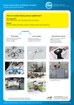


© Copyright 2026