
Practical Residual Dipolar Couplings: Sample Preparation and NMR Macromolecules, 2011
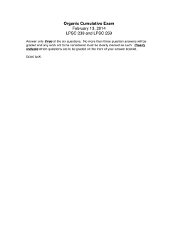
Practical Residual Dipolar Couplings:
Sample Preparation and NMR
Summer School of Magnetic Resonance for Biological
Macromolecules, 2011
School of Life Science
University of Science and Technology of China (USTC),
Hefei, Anhui, P. R. China
June 20th ~ June 25th, 2011
Stephan Grzesiek
Biozentrum der Universität Basel
Klingelbergstr. 50-70
CH-4056 Basel
e-mail: [email protected],
Tel. ++41 61 267 2100 or -2080
6/10/11 6:16 PM
1.
OVERVIEW. . . . . . . . . . . . . . . . . . . . . . . . . . . . . . . . . . . . . . . . . . . . . . . . . . . . . . . . . . . . . . . . . . . . . . . . . . . . . . . . . . . . . . . . . . 2
1.1.
MOTIVATION ................................................................................................................. 2
1.2.
SAMPLE PREPARATION ................................................................................................... 3
Lipid bicelles.......................................................................................................................... 3
Mechanically stressed polyacrylamide and polyelectrolyte gels.......................................................... 4
Ether/alcohol liquid crystalline phases (Otting media) ..................................................................... 6
Filamentous phage Pf1............................................................................................................. 8
Lyotrophic liquid crystalline “Helfrich phases”.............................................................................. 9
Purple membranes ................................................................................................................... 9
DNA nanotubes and crystalline phase G-tetrad DNA....................................................................... 9
Collagen gels ........................................................................................................................11
Paramagnetic ions and tags.......................................................................................................11
1.3.
RESIDUAL DIPOLAR COUPLING MEASUREMENT .................................................................12
NMRPipe Script....................................................................................................................14
1.4.
FITTING OF THE ALIGNMENT TENSOR ................................................................................16
Matlab Script ........................................................................................................................16
1.5.
REFERENCES ................................................................................................................18
2
1. Overview
•
We will show the anisotropic solute alignment by a number of different media such as
bicelles, filamentous phage Pf1, purple membrane and mechanically stressed
polyacrylamide gels.
•
The HDO-quadrupolar splitting, which is a – though not directly scaleable – measure for
the anisotropy of the solutes in a weakly aligning phase, is determined on a prepared
sample of orienting medium via a simple 1D-experiment.
•
With a water-flipback 2D-experiment of HSQC IPAP-type the 1H-15N RDCs of proteinamides are determined. The IPAP (In-Phase Anti-Phase) filter is based on a semi-selective
!-"-! element, with 2! ! 1/2J-1. The selection of one of the 15N spin components is
achieved by the combination of two separately recorded experiments. Two experiments
differ in the position of the two " pulses on the proton spin. RDCs are obtained as
difference in the splitting observed in the IPAP experiments under anisotropic and
isotropic conditions. From the dipolar coupling data and the known structure the
molecular alignment tensor is determined by a simple matlab script.
•
Useful literature to study in advance:
About residual dipolar couplings
1. de Alba, E.; Tjandra, N., Prog. in NMR Spectroscopy 40, (2), 175-197 (2002).
2. Bax, A., Protein Sci. 12, (1), 1-16 (2003).
3. Tolman, J. R., Curr Opin Struct Biol 11(5): 532-9 (2001).
4. Prestegard, J. H., Bougault, C.M., and Kishore, A.I., Chem. Rev. 104, 3519-40 (2004).
15
1
About the N- H dipolar splitting experiments:
1. Ottiger, M., Delaglio, F., and Bax, A., J. Magn. Reson. 131, 373–378 (1998).
2. Cordier, F., Dingley, A.J., and Grzesiek, S., J. Biomol. NMR, 13, 175-180 (1999).
3. Meissner, A., Duus, J. O., Sorensen, O. W., J. Biomol. NMR 10, (1), 89-94 (1997).
1.1.
Motivation
Residual dipolar couplings arise from the partial alignment of biomacromolecules (Tolman
et al., 1995; Tjandra and Bax, 1997). The applications to biomacromolecules are based on
earlier work on small organic molecules (Saupe and Englert, 1963; Bothnerby et al., 1981). For
details on theory, applications and data acquisition consult the lecture scripts and the review
by Prestegard (Prestegard et al., 2004).
Practically useful alignment for biomolecules leaves residual (to a large extent averaged)
dipolar couplings of up to ca. 30 Hz from the several-kHz-couplings observed in solids where
3
no averaging occurs. In an anisotropic medium steric clashing, electrostatic interactions and/or
weak transient binding weakly orient solute and solvent molecules. The spin parts of
heteronuclear J-coupling and dipolar coupling Hamiltonians are identical and simply add up to
the observed splitting. Thus, the value of the RDC is determined by comparison of the
splitting in an aligned state with a reference spectrum in isotropic phase where only the Jsplitting is detected. Dipolar couplings contain very direct information of geometry and
dynamics of the internuclear distance vector relative to a common reference frame.
Aligning media include
-
bicelles consisting of various charged or uncharged lipids
lamellar phases consisting of ether/alcohol mixtures (“Otting media”),
liquid crystalline “Helfrich phases”
mechanically stressed polyacrylamide gels or charged copolymer gels,
collagen gels
filamentous phage Pf1 or other rodlike viruses (fd, TMV)
DNA nanotubes, crystalline phase G-tetrad DNA
purple membranes of Halobacterium salinarum with bacteriorhodopsin in twodimensional crystalline arrangement.
In addition alignment can also be achieved without medium by intrinsic or artificially coupled
paramagnetic groups.
1.2.
Sample Preparation
Lipid bicelles
Bicelles were the first practical medium for the weak alignment of biomacromolecules
(Tjandra and Bax, 1997). This medium achieves large alignment, but may be chemically
instable (hydrolysis) and delicate to handle. Orientation by bicelles can be either steric (in
case of uncharged bicelles) or electrostatic (in case of charged bicelles).
Bicelles are two-dimensional, flat lipid bilayer assemblies produced by the mixing of a
lipid with larger fatty acid chains (e.g. DMPC=dimyristoyl-phosphatidylcholine) and a lipid
with smaller fatty acid chains occupying the edges of the discs (e.g. DHPC=dihexanoylphosphatidylcholine). Bicelles orient themselves in the magnetic field by their intrinsic
magnetic susceptibility (Tjandra and Bax, 1997). The typical thickness of the lipid bilayers is
between 40 and 50 Å. Bicelles are stable only for defined temperature and salt concentration
ranges.
4
Bicelle stock solutions of 15% w/v lipid are prepared using either pure water or a
predefined buffer solution such as 5-10 mM phosphate buffer, pH 6.6, 0.15 mM sodium
azide, 93% H2O, 7% D2O. DHPC (being hygroscopic and instable in presence of water) is
weighed in a dry atmosphere and dissolved in cold buffer or water (typically 4 °C, e.g. in a
cold room). The cold DHPC-solution is added to the solid DMPC to give a predetermined
molar ratio (DHPC:DMPC ! 1:3) and a total lipid concentration of 150 mg/ml. This mixture
is incubated at maximally 18 °C for ca. 10 hours. Incubation in a refrigerator or in a cold room
at 4 °C also yields good results. Occasional vortexing can be used to better dissolve the
DMPC. The lipid-esters are prone to acid and base catalyzed hydrolysis, so that the pH
during sample preparation should be kept in the pH 6-7 range. This can be overcome by using
ether-based bicelles (Cavagnero et al., 1999; Ottiger and Bax, 1999). Bicelles can be stored
frozen. The NMR-samples are prepared by diluting the 15% bicelle stock solutions with the
buffered protein sample to the desired bicelle concentration (typically 5%; below 3 %
alignment becomes unstable). This mixing should also be performed well below the phase
transition temperature of the DMPC (ca. 23 °C) (Ottiger and Bax, 1998).
Above the phase transition temperature, the fluid sample adopts a solid consistency. In
this solid state, the bicelles can be magnetically aligned. To get the best alignments, the cold
sample (~4 °C) is put into the preheated magnet at temperatures above 28 °C such that the
phase transition temperature is passed very quickly within the magnetic field. Above a certain
temperature (depending on salt, pH, lipid, and protein conditions) this magnetically aligned
liquid crystalline state becomes unstable. Usable temperature ranges are typically between 30
and 40 °C. Bicelle systems for acidic and basic pH values (pH 2.3-10.4) have been devised
(Ottiger and Bax, 1999). Doping bicelles with small amounts of charged amphiphiles (e.g. 1:15
CTAB:DMPC) has been shown to improve alignment properties and to modulate the
alignment tensor (Losonczi and Prestegard, 1998; Ramirez and Bax, 1998).
Mechanically stressed polyacrylamide and polyelectrolyte gels
Mechanical stress introduces anisotropy into the pores of a gel. Thus solute molecules
align by steric clashes with the anisotropic pores in uncharged gels (Sass et al., 2000; Tycko
et al., 2000) or by additional electrostatic interactions, if charged monomers are used for gel
preparation. Acrylamide gels are chemically inert and have been used under harsh solvent
conditions like 8 M urea to study protein unfolding (Shortle and Ackerman, 2001). Protein is
readily recovered from gels by mincing the gel and placing it in buffer followed by
centrifugation and concentration of the supernatant.
The pore size and diffusion properties of polyacrylamide gels can be tuned by adjusting
the acrylamide and N,N’-methylenebiscacrylamide concentration from stocks of 29.2% w/v
and 0.78% w/v respectively. A certain mechanical stability of the gels is required for the
orientation experiments. Good results were obtained at concentrations of # 4% (w/v)
5
acrylamide. Polymerization is started in 3.5 mm to 8 mm inner diameter tubes sealed with
parafilm on one side by the addition of 0.1% w/v ammonium persulfate and 0.5 % w/v
TEMED.
The gels are pushed out from these tubes and washed for 5 hours at 37 °C with water and
dried in a drying oven at 37 °C for several hours (over night). After this process, the gels are
dehydrated and completely solid. The gels are then reswollen in a NMR sample tube with the
desired biomacromolecule solution in buffer. Mechanical stress can be applied vertically by
pushing the plunger of a Shigemi tube onto the gel at the end of the reswelling process or
radially if the gel is originally polymerized in a tube of larger diameter than the sample tube.
For these two cases, the alignment tensors of embedded protein are exactly opposite. In
contrast to common intuition this does not yield new information (Sass et al., 2000).
Radial compression can be obtained via a commercially available device (www.neweranmr.com) where a gel, originally polymerized with a 6 mm diameter, is pressed into the NMR
tube of 4.2 mm inner diameter through a Teflon funnel via air pressure from a piston (Chou et
al., 2001). Radially compressed gels yield larger alignment than vertically compressed gels.
Fig.1 Apparatus for stretching the gel and inserting it into the open-ended NMR tube (Chou
et al., 2001). (A) Schematic drawing. (B, C) Photograph of the disassembled and assembled
gel-stretcher. (D) Open-ended NMR tube with the shigemi plunger above the gel. The various
components are: (a) Piston driver, (b) gel cylinder, (c) funnel, (d) piston with o-ring, (e) openended NMR tube, (f) vespel buttom plug of assembled NMR cell with Teflon sleeve, (g)
stretched gel, (h) Shigemi plunger. Detailed dimensions of the gel-stretcher can be downloaded
from http://spin.niddk.nih.gov/bax.
6
The residual alignment in stressed polyacrylamide gels is steric. Alignment due to
electrostatic orientation can be obtained if up to 50 % of the acrylamide monomers are
replaced by acrylic acid in the polymerization reaction. The reaction mix should be
neutralized by the addition of NaOH as the TEMED depends on its unprotonated lone
electron pairs to stabilize the radical reaction. Multivalent cations form strong salt bridges
between the charges of the copolymer so that absence of di- and trivalent cations is
recommended when casting and using these acrylic acid copolymers. The charges in the chain
lead to a strong electro-osmotic swelling (up to ca. 100fold in volume) of the gels. Anisotropy
can be introduced simply by drying the swollen gels on a capillary which results in a net
stretching of the gel. This is then followed by reswelling in the NMR-tube. Anisotropy can
also be obtained by vertical compression with a Shigemi plunger for a conventionally dried
gel. The gels have been used at NaCl concentrations of up to 240 mM to decrease the
alignment. The electrostatic character of the alignment also is evident from the higher
correlation of the obtained alignment tensors in charged gels with those in phages or purple
membrane than with those due to steric alignment in bicelles or polyacrylamide gels (Meier et
al., 2002). Introduction of positive charges is more difficult but can be achieved by replacing
acrylamide by a 10 fold surmount of positively charged resin like diallyldimethylammoniumchloride (Ulmer et al., 2003). Charged and uncharged gels have been applied to the
alignment of membrane proteins (Ulmer et al., 2003; Jones and Opella, 2004).
Various other gel-based systems have been developed for the measurement of RDCs in
non-aqueous solvents like DMSO and DMF (see literature by Luy, Kessler, Griesinger and
coworkers).
Ether/alcohol liquid crystalline phases (Otting media)
Liquid crystalline phases called L phases are formed from n-Alkyl-poly(ethylene glycol)/
glucopone (Fluka) and n-alkyl alcohol (n-hexanol/n-octanol) mixtures (Ruckert and Otting,
2000). The medium is thus made from rather cheap chemicals. Alignment occurs
spontaneously in the magnetic field by the intrinsic diamagnetic susceptibility. Aligned
samples often have excellent narrow linewidths at high degrees of orientation. Protein is
however hard to recover from L phases. The compounds are uncharged and mostly
insensitive to pH and ionic strength. Alignment is steric.
Lamellar phases are prepared by dissolving the ether to around 5% w/w in aqueous buffer
with 10 % D2O, adjusting the pH and adding the alcohol in microliter steps under vigorous
mixing (vortex). The biphasic solution becomes transparent and opalescent upon formation of
the L phases. Higher alcohol amounts decrease the temperature stability of the liquid
crystalline phase. It is advisable to store the samples at room temperature. Samples prepared
in these phases are stable at least over several months.
$
$
$
7
Different alkyl-poly(ethylene glycol) molecules are denoted as CmEn, where m is the number of carbons in the n-alkyl
group and n is the number of glycol units in the poly(ethylene glycol) moiety. The technical product “glucopone”,
contains different n-alkylated carbohydrates.
From: Rückert and Otting, 2000
From: Freyssingeas et al. 1996 (Freyssingeas et al., 1996).
8
Filamentous phage Pf1
is a 7,349-nucleotide DNA-phage where the circular DNA is packaged with coat protein
at a 1:1 nucleotide: coat protein-ratio. The Pf1 phages forms rods of ca 20,000 Å length and
60 Å diameter and spontaneously align by their intrinsic diamagnetic susceptibility in the
magnetic field (Hansen et al., 1998). Pf1-Phages can be grown in Pseudomonas aeruginosa
(might be tricky) or are commercially available (ASLA biotech; might get up to 100 " or so
per sample). Phages have a net negative surface charge and biomolecules are therefore mainly
aligned via electrostatic interactions. Positively charged biomolecules at a pH above their pI
thus might interact too strongly with the phages. Alignment can however be tuned to some
extent by the addition of salt.
The observed deuterium quadrupolar splitting in deuterated water increases with the
phage concentration. Below a certain concentration threshold (~10-20 mg/ml), the dependence
is non-linear (Zweckstetter and Bax, 2001). At NaCl concentrations of up to 600 mM and
above 16 mg/ml phage concentration, pH 7.2 (Zweckstetter and Bax, 2001), the dependence is
linear. pH-values recommended originally are 6.5-8.0 and NaCl-concentrations below 100
mM (Hansen et al., 1998). Phages have a tendency to aggregate at pH values below 6.
Phages are rebuffered by washing with the desired buffer and centrifuging at 95,000 rpm
(320,000 g) in a table ultracentrifuge for one hour. Supernatant is discarded and phage
resuspended preferably with a teflon tube. Washing is repeated twice. The sample volume is
adjusted to the desired phage concentration (30 mg/ml in this case). Alignment can be tuned
by phage concentration and salt concentration. Other rod-shaped viruses like fd and tobacco
mosaic virus have been reported to have the same orienting effect (Clore et al., 1998) but have
a smaller aspect ratio.
From (Zweckstetter and Bax, 2001)
9
Lyotrophic liquid crystalline “Helfrich phases”
These phases (Prosser et al., 1998) have been introduced as liquid crystalline media, which
are stable over a large temperature range. The media consist of a 2-5% (w/w) aqueous solution
of an equal weight mixture of cetylpyridinium chloride and n-hexanol in 200 mM NaCl. The
cetylpyridinium confers positive charges to the phase and alignment is electrostatic. Linewidths
and –shapes can be slightly problematical in our hands.
Purple membranes
Purple membranes (PM) are bacterial membranes containing bacteriorhodopsin as a sole
protein. Typical sizes of PM patches are a few microns in diameter and 45 Å in thickness.
PM is isolated from Halobacterium salinarum. PMs are rather stable with respect to
temperature, pH, and other conditions. PMs align themselves in the magnetic field by the
intrinsic magnetic susceptibility of the seven trans-membrane alpha helices of
bacteriorhodopsin (Koenig et al., 1999; Sass et al., 1999). The alignment is such that the
direction of the membrane normal is parallel to the magnetic field. PMs are highly negatively
charged.
Sample preparation for alignment of biomacromolecules is performed by simply titrating
suitable amounts of purple membranes to the biomolecular solution. Due to the negative
charge of the PM, solute-membrane interactions are usually too strong for positively charged
biomolecules (i.e. below the pI). Good results were obtained for the proteins ubiquitin and
p53 at pH 7.6 and 1-3 mg/ml PM (Sass et al., 1999). The alignment of the PM suspension
can be checked by measuring the deuterium splitting of the H2O/D2O solvent (typically
several Hz). Alignment is temperature independent over a wide range and scaleable by the
addition of more PM.
Above certain salt (70 mM NaCl) and PM concentrations, PM suspensions undergo a
transition from a fluid to a highly viscous state (Sass et al., 1999). In this state, the single PM
patches form aggregates due to van der Waals interactions. When the transition to this “salt
frozen” state is performed in the magnetic field, alignment of embedded proteins can also be
observed.
DNA nanotubes and crystalline phase G-tetrad DNA
Nucleic acid based alignment media can be formed from DNA nanotubes (Douglas et al.,
2007) or from the dinucleotide 2’-deoxyguanylyl-(3’,5’)-2’-deoxyguanosine (GpG), which
forms large stacks of guanosine tetrads (Lorieau et al., 2008). Together with polyacrylamide
gels, they are compatible with detergent for the use with membrane proteins. However, for
larger systems PAGE may not be practical due to strong interactions with the acrylamide
10
mesh, which reduce the molecular tumbling rate. Note that this can be tuned by the gel
concentration. However, acrylamide gels can’t be used at lower concentration than about 4 %,
due to mechanical instability. For the nucleic acid based media, the reduction in molecular
tumbling rates for large systems is much less problematic
Compared to DNA nanotubes, the d(GpG)-based G-tetrad stacks are much easier to
produce and cheaper. From the protocol by Lorieau et al. (Lorieau et al., 2008): “The sodium
salt of 2’-deoxyguanylyl(3’->5’)-2’-deoxyguanosine – d(GpG) was purchased from Sigma
and used without further purification. The powder was dissolved in a buffered solution (25
mM K2HPO4, pH 8) at room temperature overnight, followed by mild vortexing to ensure
homogeneity. KCl is added to a final concentration of 35 mM to ensure that the complexing
potassium concentration is saturating: a 25 mg/ml solution of d(GpG) has a concentration of
40 mM, and a minimum of 20 mM K+ cation is required to displace the sodium in the Gtetrad. The concentration of d(GpG) is monitored with a UV/Vis spectrophotometer at 260
nm, using the estimated absorbance of ~ 24.5 µg/ml for an A260 = 1.0. Dilutions of
concentrated solutions of d(GpG) require 10-15 minutes to form monomeric d(GpG) and
produce accurate absorbance measurements. The d(GpG) threshold concentration for liquid
crystal formation increases with DPC concentration and decreases with the addition of K+.
After addition of the protein, the solution is transferred to a Shigemi NMR tube. A
uniform and bubble-free sample is obtained by slow centrifugation (80-100g) after transferring
the sample to the tube, inserting the plunger to the bottom of the tube and pulling the plunger
to the desired height. Sample alignment is confirmed by measuring the 2H2O residual
quadrupolar splitting with the NMR spectrometer.”
From (Lorieau et al., 2008)
11
The G-tetrad stacks align in the magnetic field with their normal perpendicular to the
magnetic field. Below about 15 mg/ml, their orientation decreases rapidly in a non-linear way
(see Figure 1 Lorieau et al. (Lorieau et al., 2008)). The orientation of the dissolved
macromolecules is caused by electrostatic interaction with the negatively charged G-stacks. It
is thus similar to orientation by phages. Indeed, the alignment tensors are antiparallel (Lorieau
et al., 2008) (phages align parallel, G-stacks perpendicular to the field).
Collagen gels
Collagen, consisting of glycine, proline, and hydroxyproline, is a fibrous protein that can
form a rope-like left-hand triple helix structure. Collagen gels prepared from polymerization
in the magnetic field can provide weak alignment for protein. The alignment induced by
collagen gels is quite small when compared to other alignment media, but the magnitude of the
dipolar couplings can be easily scaled up by increasing the initial concentration of collagen.
The collagen gels show good pH and detergent tolerance. These advantages of collagen gels
make it a promising candidate for the alignment of large biomolecules or membrane proteindetergent complexes in the magnetic field (Ma et al., 2008).
Samples are prepared from collagen monomers (isolated from rat tails!) mixed with
protein at 4 ˚C. The collagen is polymerized in the magnet by changing the temperature
slowly to 37 ˚C. After polymerization, the 2H splitting of the collagen gel becomes stable and
totally temperature independent over the entire range tested (5-40 ˚C). The collagen gels show
good pH and detergent tolerance. The collagen gels (1.4 mg mL-1) were still stable after being
soaked in a low pH acetate buffer solution (pH 4) or in a 100 mM DPC solution for several
days.
Paramagnetic ions and tags
Paramagnetic ions in suitable inherent or engineered sites may have a high magnetic
susceptibility to allow media-free, field-induced orientation of biomolecules. The fieldinduced alignment goes up as field squared, suggesting an increasing future role for these
systems, which still suffer from the small degree of alignment that can be achieved.
Note that already the original work on weak protein alignment made use of field-induced
alignment, measuring weak alignment of cyanmetmyoglobin due to an anisotropic Fe(III)
center (Tolman et al., 1995). Certain lanthanides have been used in high affinity metal-binding
tags to align biomolecules (see literature by Bertini, Otting, Griesinger, Schwalbe, Ubbink,
Opella and coworkers).
12
1.3.
Residual Dipolar Coupling Measurement
•
Exercise 1: Measure the HDO-splitting in an oriented sample. The splitting can be
resolved in a deuterium detected 1-D spectrum of water with sufficiently long acquisition
time. In case the spectrometer setup doesn’t easily allow this, you can lock to the
different lock signals observed for oriented samples. When not locking on deuterium in the
XWINNMR-lock display, you can see two resonances. They arise from the orientation of
deuterated water at the interface to the orienting medium giving rise to a deuterium
quadrupolar splitting. After locking first on the one maximum and then on the other one,
record two simple 1-D experiments in proton dimension and determine the difference in
Hz for the water signal when locking on the different signals. From that, immediately
determine deuterium-splitting (the gyromagnetic ratios of proton and deuteron relate ca.
like 6.5:1). The HDO-splitting is not necessarily proportional to the protein alignment
(see (Ottiger and Bax, 1998)).
•
Exercise 2: Measure the 15N-1H dipolar coupling by recording the 15N-1HN-HSQC IPAP
experiments on one of the samples provided (or simply a 15N HSQC, which is not
proton-decoupled during the 15N evolution).
As a backup, a 15N-1HN-HSQC IPAP experiment has been recorded previously on a sample
of PA4608 - c-diGMP complex + 18 mg/ml of Pf1 phages. The time domain data (ser files)
are located in the directory /w/data/embo2007/8waternh_C/2.
15
N-1HN-HSQC IPAP Pulse Program
#include "bits.sg"
;#define ONE_D
#define CARBON_LABEL
;#define PULSE_CHECK
#define INTERLEAVED
;p1
proton 90 at pl1, 9u
;p2
1ms proton 90 at pl2 ;sklenar
;set phcor14 and phcor18!!!
;p3
~2ms proton 90 with sp1
;"p5=39.8u"
;p7
;p31
;carbon pulse at pl5 on 800 MHz
high power n15 90 pl7 on f2
low power n15 90 (160ms) on f2 at pl31
#ifndef PULSE_CHECK
;"d0=in0*0.5 - p7*0.637 -p1"
"d0=2u"
#else
"d0=5u"
#endif
#ifdef CARBON_LABEL
"d15=2.7m - d0*2 - p5*4 - 8u"
#else
"d15=2.7m - d0*2"
#endif
#ifndef PULSE_CHECK
"p17=p7"
#endif
13
"d11=50m"
"d12=10m"
"d13=25m"
"d16=5u"
;set in16=2.7m-p1*2-10u
"d22=p2"
"d23=p3"
"d26=p7-p1"
"d27=p7-p23"
;gradient pulses
"p10=3m"
;at gp0=+50%
"p11=2m"
;at gp1=-50%
"p12=400u"
;at gp0=+50%
#define ON
#undef OFF
1
2
#ifdef
21
#endif
ze
1m unblank
d13 do:N
d13
INTERLEAVED
d12
d12
d12
d12*3.0
10u do:C1
presaturation
*****
10u pl7:N
#ifdef ON
#ifdef CARBON_LABEL
10u fq4:C1
;jump to 56ppm
#endif
d1
1m blank
100u pl1:H
;***** start 90-degree on h-n *****
(p1 ph0)
d4
(p7*2 ph6):N (d26 p1*2 ph4)
d4
;***** hsqc to nitrogen
*****
(p1 ph6)
2u
10u pl2:f1
p2 ph18:r
2u
p10:gp0
;GRAD( 40, POSITIV, 50)
5m pl1:f1
(p17 ph3):N
d0 pl5:C1
#ifdef CARBON_LABEL
2u
(p5*2 ph10):C1
4u
2u fq4:C1 pl5:C1
;jump to 177ppm
(p5*2 ph10):C1
#endif
d0
d15
(p7*2 ph8):N
(2.7m) (d16 p1*2 ph9):f1
(p7 ph11):N
2u
p11:gp1
;GRAD( 41, NEGATIV, 40)
4m
5u
(p3:sp1 ph13):f1
2u
2u
50u pl1:f1
(p1 ph0)
p12:gp0
;GRAD( 50, POSITIV, 8.0)
d5
150u pl2:f1
(p2 ph14:r)
2u
5u pl1:f1
(p1*2 ph15)
2u
5u pl2:f1
(p7*2 ph10):N (p2 ph14:r)
2u
3
4
;*****
14
p12:gp0
(2u ph0)
d5
d8
5u pl31:N
;GRAD( 60, POSITIV, 8.0)
#endif
#ifndef ONE_D
go=2 ph31 cpds2:N
1m unblank
d13 do:N
d13 wr #0 if #0 zd
#ifdef INTERLEAVED
d12 id16
lo to 21 times 2
d12 rd16
#endif
d12 ip3
;nitrogens
lo to 3 times 2
d12 id0
d12 ip31
d12 ip31
lo to 4 times l3
#else
1m unblank
lo to 2 times 10
d1
10u pl1:f1
p1 ph31
(2u ph0)
go=1 ph31
d11 wr #0
#endif
d12 do:C1
d12 do:N
exit
ph0=0
ph1=0
ph3=0 2
ph4=1
ph6=1
ph8=PHASE_4(0)
PHASE_4(1)
PHASE_4(2)
PHASE_4(3)
ph9=0 0 2 2
ph10=0
ph11=0
ph13=(360)184;
ph14=2 2 2 2 0
ph15=0 0 0 0 2
ph18=0;(360)4;
ph31=0 2 0 2 2
adjusted -x
0 0 0;(360)185 185 5 5; adjusted -x hl2
2 2 2
adjusted x hl2
0 2 0
NMRPipe Script
The NMRPipe scripts used to process the 15N-1HN-HSQC IPAP experiment allow the
separation of the upfield and downfield components in the indirect dimension into different
subspectra. The scripts are given below with a description in italic.
Exercise
1.
Try to understand the parameters for the conversion (use the command bruk2pipe
-help for help).
2.
Run the processing script (p.com) on the reference data (not oriented) in order to
create the in- phase (conv.com 1 0) and then the anti-phase (conv.com 0 1)
components. This will produce two datasets that will be co added or subtracted. Look
at the result in nmrDraw.
3.
Co adding and subtracting the above-generated data sets will produce upfield and
downfield components.
4.
Repeat the whole procedure for the oriented data.
5.
Visualize or quantify changes in the J-couplings.
15
Commands and Functions in conv.com and p.com:
- “bruk2pipe” = conversion of bruker ser file to NMRPipe format
- enter all needed parameters of the experiment for each dimension
1
##### Acquisition dimension { H}:
- COADD = Co-Addition of data to separate the upfield and downfield components
- POLY = polynomial baseline correction (time domain) to subtract low-frequency solvent signal in the FID
- SP = window function (sine-bell)
- ZF = zero-filling
- FT = Fourier transform
- PS = phase correction
1
- EXT = extract a region from the H dimension
nd
15
##### 2 dimension { N}:
nd
- TP = exchange vectors from X- to Y-axis of the data stream to process the 2 dimension as X-vectors
- SP = window function (sine-bell “SP”)
- ZF = zero-filling
- FT = Fourier transform
- PS = phase correction
1
- TP, POLY, TP = add a polynomial baseline correction (frequency domain) in the H dimension
-addNMR =combine two NMR data files
-“-in1 or -in2” = input data for addNMR
- “-out” = write data into a file
p.com
#
csh conv.com 1 1 0
csh conv.com 2 0 1
addNMR -in1 A1.DAT
addNMR -in1 A1.DAT
90
0
-in2 A2.DAT
-in2 A2.DAT
-out C1.DAT -c1 -1 -c2 –1
-out C2.DAT -c1 1 -c2 –1
# upfield component
# downfield component
conv.com
#
bruk2pipe -in ../ser -DMX -decim 16 -dspfvs 12 \
-xN
2048
-yN
400 -zN
0
-xT
1024
-yT
100 -zT
0
-xMODE
Complex -yMODE Complex
-zMODE Complex
-xSW
12019.2307692308 -ySW
2500
-zSW
-xOBS
800.183753 -yOBS 81.09097999871
-zOBS
-xCAR
4.773
-yCAR
116.50 -zCAR
0
-ndim
2
-aq2D
States
-verb -noswap -ov \
| nmrPipe -fn COADD -cList $argv[2-3] -axis Y \
| nmrPipe -fn POLY -time \
| nmrPipe -fn SP -off 0.33 -end 0.95 -pow 2 -c 0.5 -size 512 \
| nmrPipe -fn ZF -size 2048 \
| nmrPipe -fn FT -verb \
| nmrPipe -fn PS -p0 -57 -p1 130 -di \
| nmrPipe -fn EXT -x1 5.9ppm -xn 11.5ppm -sw \
| nmrPipe -fn TP \
| nmrPipe -fn SP -off 0.35 -end 0.99 -pow 1 -c 0.5 \
| nmrPipe -fn ZF -size 1024 \
| nmrPipe -fn FT -verb \
| nmrPipe -fn PS -p0 $4 -p1 0 -di \
| nmrPipe -fn TP \
| nmrPipe -fn POLY -auto -nw 3 -ord 2 \
| nmrPipe -fn TP \
-out A$1.DAT -ov
exit 0
\
0 \
0
\
\
\
\
\
16
1.4.
Fitting of the alignment tensor
Exercise
The following Matlab script (linearfit.m) allows the determination of the alignment
tensor (Az, rhombicity, ...) from measured dipolar coupling data and a known structure
(Moltke and Grzesiek, 1999). The fitting routine is a linear algorithm exploiting the fact that
the couplings depend in a linear way on the alignment tensor
Try to understand the matlab script and run this script on suitable data. J- and RDC data
have been measured and are provided.
Matlab Script
•
linearfit.m
clear all
cosfile = 'NH_1.out';
jfile = 'rdc_fgmy_1.out';
valjerri=1;
clear xdata x1data jnb jmesi
clear xyz_coord
% ____________ end file definitions ___________________________
% ____________ read files ___________________________
global j_res xyzi jmesi jnb nfree_ax nfree_asym ;
file = fopen (cosfile, 'r');
dataxyz = fscanf (file, '%f %f %f %f %f', [5 inf])';
fclose (file);
xyz_res = dataxyz(:,1);
xyz_coord = dataxyz(:,3:5);
xyznb = length(dataxyz);
for ii = 1:xyznb
xyz_index(xyz_res(ii)) = ii;
y = xyz_coord(ii,:);
n = sqrt(y*y');
xyz_coord(ii,1:3)= y/n;
end
file = fopen (jfile, 'r');
dataj = fscanf (file, '%f %f %f', [3 inf])';
fclose (file);
j_res
jmesi
yerrs
jnb =
= dataj(:,1);
= dataj(:,2);
= dataj(:,3);
length(dataj);
for ii = 1:jnb;
xyzi(ii,1:3) = xyz_coord( xyz_index(j_res(ii)),:);
end
for ii = 1:jnb
pol = Cart2pol( xyzi(ii,:));
xdata(ii,1:5) = y2(pol(2), pol(3));
deni(ii,:) = [pol(2) pol(3)];
if (yerrs(ii) < valjerri )
yerrs(ii) = valjerri;
end
end
nfree_ax=jnb-3;
nfree_asym=jnb-5;
% ____________ end read files ___________________________
17
yvec = xdata'*jmesi;
% linear fit
xmatrix = xdata'*xdata;
avec = xmatrix\yvec;
ytheo = real(xdata*avec)';
diff=(ytheo'-jmesi)./yerrs;
chisq = diff'*diff/nfree_asym;
sa = irred2saupe( avec );
% 5*1 vector to 3*3 matrix
[v,d] = eig(sa);
% matrix diagonalisation to get Axx,Ayy,Azz
v = real(v);
d = diag(real(d));
[dd i] = sort(abs(d));
d = d(i);
v = v(:,i);
Ax = d(1); Ay = d(2); Az = d(3)
Rhomb = (Ax-Ay)/Az
% before Rhomb was defined as 2/3*(Ax-Ay)/Az but not in our case
% only for plotting measured vs predicted
inhnames = {'Imatinib(30mg/ml phage)'};
hold off
errorbar( j_res, jmesi, yerrs, '-o');
orient landscape
hold on
plot( j_res, ytheo, '-rd')
grid on
title(sprintf('NH-%s ', char(inhnames)));
xlabel('residue number')
ylabel('D N-H [Hz]')
xlim([225 505])
ylim([-40 40])
set(gca, 'XTickMode', 'manual');
set(gca, 'xtick',[220:40:500]);
return
•
irred2saupe.m
function sa = irred2saupe( avec )
sxx
syy
szz
sxy
syz
sxz
= sqrt(3/8)*(avec(5)+avec(1)) - 0.5*avec(3);
= -sqrt(3/8)*(avec(5)+avec(1)) - 0.5*avec(3);
= avec(3);
= -i*sqrt(3/8)*(avec(5)-avec(1));
=
i*sqrt(3/8)*(avec(4)+avec(2));
=
-sqrt(3/8)*(avec(4)-avec(2));
sa
= [ sxx sxy sxz ; sxy syy syz ; sxz syz szz ]*sqrt(5/(4*pi));
•
y2.m
function c = y2(theta,phi)
c = [3*exp(-2*i*phi)*sqrt(5/(6*pi))*power(sin(theta),2)/4,
3*exp(-i*phi)*sqrt(5/(6*pi))*cos(theta)*sin(theta)/2,
sqrt(5/pi)*(-1 + 3*power(cos(theta),2))/4,
-3*exp(i*phi)*sqrt(5/(6*pi))*cos(theta)*sin(theta)/2,
3*exp(2*i*phi)*sqrt(5/(6*pi))*power(sin(theta),2)/4]';
•
Cartpol.m
function c = cart2pol( x )
r = sqrt(x*x');
x = x /r;
theta = acos(x(3));
phi = atan2(x(2), x(1));
c = [r theta phi];
18
1.5.
References
Bothnerby, A.A., Domaille, P.J. and Gayathri, C. (1981) Ultra-High-Field NMR
Spectroscopy - Observation of Proton-Proton Dipolar Coupling in Paramagnetic
Bis[tolyltris(pyrazolyl)borato]cobalt(II). Journal of the American Chemical Society,
Vol. 103, pp. 5602-5603.
Cavagnero, S., Dyson, H.J. and Wright, P.E. (1999) Improved low pH bicelle system for
orienting macromolecules over a wide temperature range. J Biomol NMR, 13, 387-391.
Chou, J.J., Gaemers, S., Howder, B., Louis, J.M. and Bax, A. (2001) A simple apparatus for
generating stretched polyacrylamide gels, yielding uniform alignment of proteins and
detergent micelles. Journal of Biomolecular NMR, 21, 377-382.
Clore, G.M., Starich, M.R. and Gronenborn, A.M. (1998) Measurement of residual dipolar
couplings of macromolecules aligned in the nematic phase of a colloidal suspension of
rod-shaped viruses. Journal of the American Chemical Society, 120, 10571-10572.
Douglas, S.M., Chou, J.J. and Shih, W.M. (2007) DNA-nanotube-induced alignment of
membrane proteins for NMR structure determination. Proc Natl Acad Sci U S A, 104,
6644-6648.
Freyssingeas, E., Nallet, F. and Roux, D. (1996) Measurement of the membrane flexibility in
lamellar and ''sponge'' phases of the C(12)E(5)/hexanol/water system. Langmuir, 12,
6028-6035.
Hansen, M.R., Mueller, L. and Pardi, A. (1998) Tunable alignment of macromolecules by
filamentous phage yields dipolar coupling interactions. Nat Struct Biol, 5, 1065-1074.
Jones, D.H. and Opella, S.J. (2004) Weak alignment of membrane proteins in stressed
polyacrylamide gels. J Magn Reson, 171, 258-269.
Koenig, B.W., Hu, J.S., Ottiger, M., Bose, S., Hendler, R.W. and Bax, A. (1999) NMR
measurement of dipolar couplings in proteins aligned by transient binding to purple
membrane fragments. Journal of the American Chemical Society, Vol. 121, pp. 13851386.
Lorieau, J., Yao, L. and Bax, A. (2008) Liquid crystalline phase of G-tetrad DNA for NMR
study of detergent-solubilized proteins. J Am Chem Soc, 130, 7536-7537.
Losonczi, J.A. and Prestegard, J.H. (1998) Improved dilute bicelle solutions for highresolution NMR of biological macromolecules. Journal of Biomolecular Nmr, 12, 447451.
Ma, J., Goldberg, G.I. and Tjandra, N. (2008) Weak alignment of biomacromolecules in
collagen gels: an alternative way to yield residual dipolar couplings for NMR
measurements. J Am Chem Soc, 130, 16148-16149.
Meier, S., Haussinger, D. and Grzesiek, S. (2002) Charged acrylamide copolymer gels as
media for weak alignment. J Biomol NMR., 24, 351-356.
Moltke, S. and Grzesiek, S. (1999) Structural constraints from residual tensorial couplings in
high resolution NMR without an explicit term for the alignment tensor. J Biomol
NMR, 15, 77-82.
Ottiger, M. and Bax, A. (1998) Characterization of magnetically oriented phospholipid
micelles for measurement of dipolar couplings in macromolecules. J Biomol NMR, 12,
361-372.
Ottiger, M. and Bax, A. (1999) Bicelle-based liquid crystals for NMR-measurement of
dipolar couplings at acidic and basic pH values. J Biomol NMR, 13, 187-191.
Prestegard, J.H., Bougault, C.M. and Kishore, A.I. (2004) Residual dipolar couplings in
structure determination of biomolecules. Chem Rev, 104, 3519-3540.
19
Prosser, R.S., Losonczi, J.A. and Shiyanovskaya, I.V. (1998) Use of a novel aqueous liquid
crystalline medium for high-resolution NMR of macromolecules in solution. Journal
of the American Chemical Society, Vol. 120, pp. 11010-11011.
Ramirez, B.E. and Bax, A. (1998) Modulation of the alignment tensor of macromolecules
dissolved in a dilute liquid crystalline medium. Journal of the American Chemical
Society, Vol. 120, pp. 9106-9107.
Ruckert, M. and Otting, G. (2000) Alignment of biological macromolecules in novel nonionic
liquid crystalline media for NMR experiments. Journal of the American Chemical
Society, 122, 7793-7797.
Sass, H.J., Musco, G., Stahl, S.J., Wingfield, P.T. and Grzesiek, S. (2000) Solution NMR of
proteins within polyacrylamide gels: Diffusional properties and residual alignment by
mechanical stress or embedding of oriented purple membranes. Journal of
Biomolecular Nmr, 18, 303-309.
Sass, J., Cordier, F., Hoffmann, A., Cousin, A., Omichinski, J.G., Lowen, H. and Grzesiek, S.
(1999) Purple membrane induced alignment of biological macromolecules in the
magnetic field. Journal of the American Chemical Society, 121, 2047-2055.
Saupe, A. and Englert, G. (1963) High-Resolution Nuclear Magnetic Resonance Spectra of
Orientated Molecules. Physical Review Letters, Vol. 11, pp. 462-&.
Shortle, D. and Ackerman, M.S. (2001) Persistence of native-like topology in a denatured
protein in 8 M urea. Science, 293, 487-489.
Tjandra, N. and Bax, A. (1997) Direct measurement of distances and angles in biomolecules
by NMR in a dilute liquid crystalline medium. Science, 278, 1111-1114.
Tolman, J.R., Flanagan, J.M., Kennedy, M.A. and Prestegard, J.H. (1995) Nuclear magnetic
dipole interactions in field-oriented proteins: information for structure determination
in solution. Proceedings of the National Academy of Sciences of the United States of
America, 92, 9279-9283.
Tycko, R., Blanco, F.J. and Ishii, Y. (2000) Alignment of biopolymers in strained gels: A new
way to create detectable dipole-dipole couplings in high-resolution biomolecular
NMR. Journal of the American Chemical Society, 122, 9340-9341.
Ulmer, T.S., Ramirez, B.E., Delaglio, F. and Bax, A. (2003) Evaluation of backbone proton
positions and dynamics in a small protein by liquid crystal NMR spectroscopy.
Journal of the American Chemical Society, 125, 9179-9191.
Zweckstetter, M. and Bax, A. (2001) Characterization of molecular alignment in aqueous
suspensions of Pf1 bacteriophage. Journal of Biomolecular NMR, 20, 365-377.


© Copyright 2026