
An automated sample preparation for detection of 72 doping-related substances
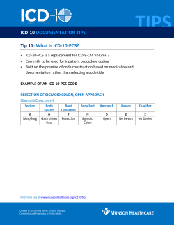
Drug Testing
and Analysis
Research article
Received: 30 April 2013
Revised: 8 August 2013
Accepted: 13 August 2013
Published online in Wiley Online Library
(www.drugtestinganalysis.com) DOI 10.1002/dta.1538
An automated sample preparation for
detection of 72 doping-related substances
Darío Cuervo,* Pablo Díaz-Rodríguez and Jesús Muñoz-Guerra
Automation of sample preparation procedures in a doping control laboratory is of great interest due to the large number of
samples that have to be analyzed, especially in large events where a high throughput protocol is required to process
samples over 24 h. The automation of such protocols requires specific equipment capable of carrying out the diverse
mechanical tasks required for accomplishing these analytical methodologies, which include pipetting, shaking, heating,
or crimping. An automated sample preparation procedure for the determination of doping-related substances by gas
chromatography–mass spectrometry (GC-MS) and gas chromatography-tandem mass spectrometry (GC-MS/MS) analysis,
including enzymatic hydrolysis, liquid-phase extraction and derivatization steps, was developed by using an automated
liquid handling system. This paper presents a description of the equipment, together with the validation data for 72
doping-related compounds including extraction efficiency, evaluation of carry-over, interferences, and robustness.
Validation was approached as a comparison between the results obtained using the manual protocol and the transferred
automated one. The described methodology can be applied for sample preparation in routine anti-doping analysis with
high sample throughput and suitable performance. Copyright © 2013 John Wiley & Sons, Ltd.
Keywords: doping control analysis; urine analysis; automated sample preparation; liquid-liquid extraction
Introduction
Drug Test. Analysis (2013)
Experimental
Chemicals, reagents, and materials
All solvents and reagents used were of analytical grade. Tertbutyl methyl ether (TBME) and methanol were from Merck
(Darmstadt, Germany). Potassium dihydrogen phosphate
(KH2PO4), sodium hydrogen carbonate (NaHCO3) and potassium carbonate (K2CO3) were from Scharlab (Sentmenat,
Spain). Disodium hydrogen phosphate dihydrate (Na2HPO4 · 2
H20) was from Panreac (Castellar del Vallès, Spain). N-MethylN-(trimethylsilyl) trifluoroacetamide (MSTFA) was from
* Correspondence to: Darío Cuervo, PhD, Laboratorio de Control del Dopaje,
Agencia Española de Protección de la Salud en el Deporte, Ministerio de Educación,
Cultura y Deporte, Gobierno de España, c/ Pintor El Greco s/n, 28040, Madrid, Spain.
E-mail: [email protected]
Laboratorio de Control del Dopaje, Agencia Española de Protección de la Salud
en el Deporte, Ministerio de Educación, Cultura y Deporte, Gobierno de España,
c/ Pintor El Greco s/n, 28040, Madrid, Spain
Copyright © 2013 John Wiley & Sons, Ltd.
1
Sample preparation in a doping control laboratory is laborious
work due to both the large number of samples to be treated
and the variety of compounds to be monitored. The diverse
structural and chemical characteristics of the analytes included
on the List of Prohibited Substances published annually by the
World Anti-Doping Agency (WADA),[1] entails different sample
preparation procedures in order to purify, concentrate and
adequate the samples for instrumental analysis by specific
techniques, including gas chromatography–mass spectrometry
(GC-MS), gas chromatography-tandem mass spectrometry (GCMS/MS) or liquid chromatography-tandem mass spectrometry
(LC-MS/MS). The automation of these sample preparation
procedures is of great interest considering the savings in terms
of time, security inside the laboratory and, eventually, the
quality of the results obtained.
Automation of sample preparation protocols needs specific
equipment capable of carrying out the diverse mechanical tasks
required for accomplishing the different analytical methodologies involved.[2,3] Several studies have been undertaken in the
past by this laboratory[4,5] and others[6–11] regarding the
automation of drug analysis in urine or blood using different
platforms. Given that the detection critteria for the different
compounds included on the WADA List of Prohibited Substances
and Methods in Sport are more and more challenging,
particularly in terms of sensitivity, new solutions for automation
need to be developed and investigated. Our laboratory has
recently acquired an automated pipetting system for
liquid-liquid extraction from Zinsser Analytic GmbH (Frankfurt,
Germany),[12] with further capabilities including shaking,
heating, or crimping. In brief, the system was specifically
designed for treating up to 96 samples in a sample preparation
protocol which includes several heating and extraction steps.
As a first step, we focused on the automation of the method
implemented in our laboratory for the determination of anabolic
agents, narcotics, anti-estrogenic substances, cannabinoids, diuretics,
and stimulants in urine by GC-MS and GC-MS/MS analysis, the
sample preparation procedure of which includes enzymatic
hydrolysis, liquid-liquid extraction, and derivatization steps. The aim
was to implement an automated sample preparation protocol able
to carry out the whole scope of the method without loss of
performance. The paper discusses the validation of the automated
sample preparation method, approached as a comparison between
both protocols. Validation data including extraction efficiency and
evaluation of interferences and carry over effects are also presented.
Drug Testing
and Analysis
D. Cuervo, P. Díaz-Rodríguez and J. Muñoz-Guerra
Macheray-Nagel (Düren, Germany). 1,4-Dithioerythritol and
ammonium iodide were from Sigma-Aldrich (St. Louis, MO,
USA). Β-Glucuronidase from E. coli K12 was from Roche
Diagnostics GmbH (Mannheim, Germany).
Internal standard (ISTD) and standards used for process
control: methyltestosterone was from Sigma-Aldrich (St Louis,
MO, USA), androsterone glucuronide-d4 and etiocholanolone-d5
were from NMI (Pymble, Australia), and timolol was from USP
(Rockville, MD, USA).
Commercial solid standards: the systematic names of each
compound are listed in Table 1. Nandrolone M1, nandrolone M2,
stanozolol M1, stanozolol M2, 4OH-testosterone, 5α and 5βmethyltestosterone, metandienone M1, M2, M3 and M4,
bolasterone M1, boldenone M1, calusterone, carphedon, cyclofenil
M2, clostebol M1, danazol M2, drostanolone M1, oxandrolone M1,
trenbolone M1, fluoxymesterone M1 and M2, furazabol M1, letrozole
M1, mesterolone M1, metenolone M1, methyldienolone, methyl-1testosterone, 6-monoacetylmorphine, norboletone M1 and M2,
noretandrolone M1 and M2, dehydrochlormethyltestosterone M1,
tamoxifen M1, zeranol and zeranol M1 were from NMI (Pymble,
Australia). Tibolone M1 and M2, and bromantane M1 were from
Atlanchim Pharma (Nantes, France). Tibolone M3 and zilpaterol
were from TRC (Toronto, Canada). Androstatrienedione, clenbuterol
and danazol were from Sigma-Aldrich (St Louis, MO, USA).
Aminoglutethimide was from Alltech (State College, PA, USA).
Bolasterone, boldenone, danazol M1, estradienedione and
mibolerone were from Steraloids (Newport, RI, USA). Buprenorfine
and parahydroxyamphetamine were from USP (Rockville,
MD, USA). Spironolactone was from European Pharmacopoeia
(Strasbourg, France). Estradienedione M1 was from AK Scientific
(Union City, CA, USA). Furazabol was supplied free of charge by a
pharmaceutical company. Metasterone M1 was obtained from the
World Association of Anti-Doping Scientists (WAADS).
Commercial standards in solution: ampoules of codeine (1.0 mg/ml),
fluoxymesterone (1.0 mg/ml), hydromorphone (1.0 mg/ml),
mesterolone (1.0 mg/ml), metenolone (1.0 mg/ml), morphine
(1.0 mg/ml), buprenorphine M1 (1.0 mg/ml), oxycodone (1.0 mg/ml),
oxymorphone (1.0 mg/ml), pentazocine (1.0 mg/ml) and THC-9-COOH
(0.1 mg/ml) were obtained from Cerilliant (Round Rock, TX, USA). Ampoules of hydrocodone (1.0 mg/ml), oxandrolone (1.0 mg/ml) and
oxymesterone (1.0 mg/ml) were from Alltech (State College, PA, USA).
Preparation of solutions
2
The phosphate buffer was prepared by mixing 10.88 g of KH2PO4
and 14.24 g of Na2HPO4 · 2H20 in 200 ml of purified water. The
carbonate buffer was prepared by mixing 20 g of NaHCO3 and
40 g of K2CO3 in 200 ml of purified water. The silylating reagent
was prepared by mixing 120 mg of 1,4-dithioerythritol and 60
mg of NH4I with 30 ml of MSTFA.
A 100 ml solution of the internal standard and the standards
used for process control (ISTD mixture), containing 5 μg/ml of
methyltestosterone, 5 μg/ml of timolol, 20 μg/ml of androsterone
glucuronide-d4 and 20 μg/ml of etiocholanolone-d5 was prepared
from methanolic solutions of 1 mg/ml for methyltestosterone and
timolol and by dissolving 2 mg of the standards of androsterone
glucuronide-d4 and etiocholanolone-d5.
For fluoxymesterone, hydrocodone, mesterolone, buprenorphine
M1, oxandrolone, oxymesterone and pentazocine 100 μg/ml solutions were prepared by diluting 1 ml of the 1.0 mg/ml standard
solutions in 10 ml of methanol. For metenolone a 50 μg/ml solution
was prepared by diluting 0.5 ml of the 1.0 mg/ml standard solution
wileyonlinelibrary.com/journal/dta
in 10 ml of methanol. For 6-monoacethylmorphine, a 1000 μg/ml
solution was prepared by dissolving 1.0 mg of standard in 1 ml of
methanol. Standards of codeine, hydromorphone, morphine and
oxymorphone were used as acquired (solutions of 1.0 mg/ml). The
standard of THC-9-COOH was also used as acquired (a solution of
100 μg/ml). For the rest of the substances, 100 μg/ml solutions were
prepared by dissolving 1.0 mg of standards in 10 ml of methanol.
Two separate standard stock solutions containing the compounds
listed in Table 1 were prepared from those solutions. Standard stock
solution B contained all the standards in concentrations 20 times
those indicated in Table 1. Standard stock solution A contained all
the standards listed except clenbuterol and norboletone M1, in concentrations 20 times those indicated in Table 1. Clenbuterol and
norboletone M1 were not included in the standard stock solution A
due to the fact that these two substances were not included in the
scope of the method when the sensitivity experiments with nonconductive disposable tips were undertaken. Both solutions were
used during the validation experiments for recovery calculations.
Control samples
Negative and positive urine samples (2 ml) available in our
laboratory, as well as blank samples of purified water (2 ml),
were used for all the optimization and validation experiments.
Negative samples consisted of urine donations from the laboratory staff, checked for adequate pH (between 5.5 and 6.5),
density (between 1.010 and 1.020), free of interferences and
of any trace of substances included in this study. Positive
samples consisted of negative ones spiked with the methanolic
solutions of standards at the concentrations depicted in Table 1.
These concentrations are consistent with the minimum
required performance levels (MRPL) established by WADA in
the technical document TD2013MRPL [13] , and represent the
concentration of a prohibited substance or any of its metabolites or markers that accredited laboratories must be able to
routinely detect and identify.
Manual sample preparation
In routine work, the compounds listed in Table 1 can be extracted
from urine samples following a manual three-step procedure
which includes enzymatic hydrolysis, liquid-liquid extraction,
and derivatization, as depicted in Figure 1.[14] The pH of the
samples is brought to 7 by previous adjustment (if necessary)
and subsequent addition of the phosphate buffer (100 μl). After
the addition of 100 μl of ISTD mixture and 50 μl of β-glucuronidase enzyme, the samples are incubated for 1 h at 55°C, in order
to cleave the glucuronic linkage of most of the analytes with the
glucuronic acid. After cooling, samples are brought to pH 11 by
addition of the carbonate buffer (300 μl) and 5 ml of TBME are
added. After shaking and centrifugation, the upper organic layer
is separated to clean tubes after freezing of the aqueous phase
and evaporated to dryness under a stream of nitrogen. 50 μl of
silylating reagent are added and the samples are transferred into
GC vials after incubation at 65°C for 30 min.
Description of the automated sample preparation system
An automated liquid-liquid extraction system specifically designed
for the needs of our laboratory, with additional shaking, heating,
drying and crimping capabilities, was acquired from Zinsser Analytics (Frankfurt, Germany).[12] The equipment consists of a
Copyright © 2013 John Wiley & Sons, Ltd.
Drug Test. Analysis (2013)
Drug Test. Analysis (2013)
3
S1. Anabolic agents
CATEGORY IN WADA
PROHIBITED LIST
17α-methyl-5β-androstan-3α,17β-diol
7α-17α-dimethylandrost-4-en-17β-ol-3-one
7α,17α-dimethyl-5β-androstan-3α,17β-diol
Androst-1,4-dien-17β-ol-3-one
5β-androst-1-en-17β-ol-3-one
7β,17α-dimetilandrost-4-en-17β-ol-3-one
1-(4-amino-3,5-dichlorophenyl)-2-(tert-butylamino)ethanol
4-chloro-androst-4-en-3α-ol-17-one
17α-ethynyl-androst-4-en-17β-ol-(2,3-d)-isoxazole
17α-ethynyl-androst-4-en-17β-ol-3-one
17α-ethynyl-2α-hydroxymethyl-androst-4-en-17β-ol-3-one
4-chloro-17α-methylandrost-1,4-dien-6β,17β-diol-3-one
2α-methyl-5α-androst-3α-ol-17-one
Estra-4,9-diene-3,17-dione
Estra-4,9-diene-17β-ol-3-one
5β-Methyltestosterone
Bolasterone
Bolasterone M1
Boldenone
Boldenone M1
Calusterone
Clenbuterol
Clostebol M1
Danazol
Danazol M1 (Ethisterone)
Danazol M2
Dehydrochlormethyltestosterone M1
Drostanolone M1
Estradienedione
Estradienedione M1 (9(10)Dehydronandrolone)
Fluoxymesterone
Fluoxymesterone M1
Fluoxymesterone M2
Furazabol
Furazabol M1
Mesterolone
Mesterolone M1
Metandienone M1 (Epimetendiol)
Metandienone M2 (6-OH-Dianabol)
Metandienone M3
Metandienone M4 (17-Epimetandienone)
Metasterone M1 (3-OH-Metasterone)
Metenolone
Metenolone M1
Methyldienolone
Methyl-1-testosterone
Mibolerone
Nandrolone M1 (19-Norandrosterone)
9α-fluoro-17α-methylandrost-4-en-11β,17β-diol-3-one
9α-fluoro-17α-methyl-androst-4-en-3α,6β,11β,17β-tetrol
9α-fluoro-11β-ol-18-nor-17,17-dimethylandrost-4,13-dien-3-one
17α-methyl-5α-androstan-17β-ol-[2,3-c]-furazan
17α-methyl-5α-androsta-16ε,17β-diol-[2,3-c]-furazan
1α-methyl-5α-androstan-17β-ol-3-one
1α-methyl-5α-androstan-3α,17β-diol
17β-methyl-5β-androst-1-en-3α,17α-diol
17α-methylandrosta-1,4-dien-6β,17β-diol-3-one
18-nor-17,17-dimethyl-5β-androst-1,13-dien-3α-ol
17β-metil-androst-1,4-dien-17α-ol-3-ona
2α,17α-dimethyl-5α-androst-3α,17β-diol
1-methyl-5α-androst-1-en-17β-ol-3-one
1-methylen-5α-androstan-3α-ol-17-one
17α-methylestra-4,9-dien-17β-ol-3-one
17α-methyl-5α-androst-1-en-17β-ol-3-one
7α,17α-dimethylestr-4-en-17β-ol-3-one
5α-estran-3α-ol-17-one
Androst-4-en-4,17β-diol-3-one
17α-methyl-5α-androstan-3α,17β-diol
SYSTEMATIC NAME
4-OH-Testosterone
5α-Methyltestosterone
COMPOUND
Copyright © 2013 John Wiley & Sons, Ltd.
Methyldienolone
Methyl-1-testosterone
Mibolerone
Nandrolone
Metasterone
Metenolone
Metandienone
Mesterolone
Furazabol
Fluoxymesterone
Dehydrochlormethyltestosterone
Drostanolone
Estradienedione
Calusterone
Clenbuterol
Clostebol
Danazol
Boldenone
4-OH-Testosterone
Mestanolone, Methyltestosterone,
Methyl-1-testosterone,
Oxymetolone
Metandienone, Methandriol,
Methyltestosterone
Bolasterone
PROHIBITED COMPOUND
5
5
5
5
5
5
5
2
2
10
2
5
5
5
5
5
5
2
(Continues)
5
5
5
5
5
0.2
5
5
5
5
2
5
5
5
2
12
2
CONCENTRATION IN
URINE SAMPLES (PPB)
Table 1. List of substances analysed and concentrations in the positive urine sample. Substances analyzed by GC-MS/MS are shown in italics in the compound column. All other analytes were determined by GC-MS
Automated sample preparation for detection of doping-related substances
Drug Testing
and Analysis
wileyonlinelibrary.com/journal/dta
4
wileyonlinelibrary.com/journal/dta
Copyright © 2013 John Wiley & Sons, Ltd.
S7. Narcotics
S5. Diuretics and other
masking agents
S6. Stimulants
S4. Hormone and
metabolic modulators
CATEGORY IN WADA
PROHIBITED LIST
Table 1. (Continued)
Androsta-1,4,6-triene-3,17-dione
Bis(4-cyanophenyl)methanol
Z-2-[4-(1-phenyl-2-(3-hydroxy-4-methoxyphenyl)-1butenyl)phenoxy]-N,N-dimethylethanamine
7α-acetylthio-3-oxo-17α-pregn-4-ene-21,17-carbolactone
2-(4-bromophenylamine)-adamantan-6-ol
2-(2-oxo-4-phenylpyrrolidin-1-yl)acetamide
4-(2-aminopropyl)phenol
3-hydroxy-6-acetyl-(5α,6α)-7,8-didehydro-4,5-epoxy17-methylmorphinan
9α-cyclopropylmethyl-4,5-epoxy-6,14-ethano-3-hydroxy-6methoxymorphinan-7-yl]-3,3-dimethylbutan-2-ol
4,5-epoxy-6,14-ethano-3-hydroxy-6-methoxymorphinan-7-yl]3,3-dimethylbutan-2-ol
(5α,6α)-7,8-didehydro- 4,5-epoxy-17-methylmorphinan-3,6-diol
4,5α-epoxy-14-hydroxy-3-methoxy-17-methylmorphinan-6-one
4,5α-epoxy-3,14-dihydroxy- 17-methylmorphinan-6-one
Androstatrienedione
Letrozole M1
Tamoxifen M1
Bromantane M1 (6-OH-Bromantane)
Carphedon
Parahydroxyamphetamine
6-Monoacetylmorphine
Morphine
Oxycodone
Oxymorphone
Buprenorfine M1 (Norbuprenorfine)
Buprenorfine
Spironolactone
Aminoglutethimide
Zilpaterol
Zeranol M1 (Taleranol)
5β-estran-3α-ol-17-one
18-methyl-5α-estr-17α-ethyl-3α,17β-diol
18-methyl-5β-estr-17α-ethyl-3α,17β-diol
17α-ethyl-5β-estran-3α-17β-diol
17α-ethyl-5α-estran-3α-17β-diol
17α-methyl-2-oxa-5α-androstan-17β-ol-3-one
17β-methyl-2-oxa-5α-androstan-17α-ol-3-one
17α-methylandrost-4-en-4,17β-diol-3-one
3’-hydroxy-17α-methyl-5α-androst-17β-ol-[3,2-c]pyrazol
17α-methyl-5α-androst-4β,17β-diol-[3,2-c]pirazol
17α-ethynyl-7α-methyl-estr-5(10)-en-3β,17β-diol
17α-ethynyl-7α-methyl-estr-5(10)-en-3α,17β-diol
17α-ethynyl-7α-methyl-estr-4-en-17β-ol-3-one
Estra-4,9,11-trien-17α-ol-3-one
(3S,7R)-7,14,16-trihydroxy-3-methyl-3,4,5,6,7,8,9,10,11,
12-decahydro-1H-2-benzoxacyclotetradecin-1-one
(3S,7S)-7,14,16-trihydroxy-3-methyl-3,4,5,6,7,8,9,10,11,
12-decahydro-1H-2-benzoxacyclotetradecin-1-one
Trans-4,5,6,7-tetrahydro-7-hydroxy-6(isopropylamino)imidazo[4,5,1-jk][1]benzazepin-2(1H)-one
3-(4-aminophenyl)-3-ethyl-piperidine-2,6-dione
SYSTEMATIC NAME
Nandrolone M2 (19-Noretiocholanolone)
Norboletone M1
Norboletone M2
Norethandrolone M1
Norethandrolone M2
Oxandrolone
Oxandrolone M1 (Epioxandrolone)
Oxymesterone
Stanozolol M1
Stanozolol M2
Tibolone M1
Tibolone M2
Tibolone M3
Trenbolone M1 (Epitrenbolone)
Zeranol
COMPOUND
Morphine
Oxycodone
Oxymorphone
Buprenorfine
Bromantane
Carphedon
Parahydroxyamphetamine
Diacetylmorphine (Heroine)
Spironolactone
Androstatrienedione
Letrozole
Tamoxifen
Aminoglutethimide
Zilpaterol
Trenbolone
Zeranol
Tibolone
Oxymesterone
Stanozolol
Oxandrolone
Norethandrolone
Norboletone
PROHIBITED COMPOUND
50
50
50
5
5
100
100
100
50
200
50
20
20
20
5
10
5
5
5
10
5
5
5
5
2
2
5
5
5
5
5
CONCENTRATION IN
URINE SAMPLES (PPB)
Drug Testing
and Analysis
D. Cuervo, P. Díaz-Rodríguez and J. Muñoz-Guerra
Drug Test. Analysis (2013)
50
15
50
50
Pentazocine
Tetrahydrocannabinol
Codeine
Hydrocodone
2-dimethylallyl-5,9-dimethyl-2’-hydroxybenzomorphan
1-hydroxy-6,6-dimethyl-3-pentyl-6a,7,8,10atetrahydrobenzo[c]chromene-9-carboxylic acid
(5α,6α)-7,8-didehydro-4,5-epoxy-3-methoxy-17methylmorphinan-6-ol
4,5α-epoxy-3-methoxy-17-methylmorphinan-6-one
Hydrocodone
Codeine
Other non-prohibited
compounds
CATEGORY IN WADA
PROHIBITED LIST
S8. Cannabinoids
Drug Test. Analysis (2013)
Drug Testing
and Analysis
Figure 1. Flow chart of the manual sample preparation
workbench divided into different zones where the analytical tasks
are carried out (Figure 2). The layout integrates several modules
among which a mobile gripper transports, picks up and drops off
the different racks and tools, and four pipetting probes dispense
and transfer solvents and reagents among the different areas.
These probes self-load and discharge disposable tips thus avoiding
potential cross-contamination. Briefly, the system was designed for
treating up to 96 samples in a sample preparation protocol which
could include several heating and extraction steps, to which end a
module for heating, a module for shaking and/or heating, four
racks for 24 vials of 8 ml, two racks for 48 GC vials (1.8 ml), two
racks for 48 GC vial caps, three racks for a total of 288 disposable
tips, a reagent rack, a drying manifold, two solvent reservoirs, a
crimper and a gripper tool for GC vials and caps were included in
the design. All operations and methods are programmed and
launched via the software Zinsser WinLissy (version 7.0.7). The entire system is located under a large fume hood in order to avoid
dissemination of vapours.
The 8 ml vials, where samples are initially placed, were from Zinsser Analytics (Frankfurt, Germany). The 1.8 ml high recovery GC vials
were from Agilent Technologies (Palo Alto, CA, USA). The carbonate
buffer is placed in a 40-ml brown glass vial on the reagent rack.
The silylating reagent is also located on the reagent rack in an 8-ml
glass vial protected from ambient light and covered with a septum
sealed with a screw-top. The septum allows the disposable tips to
Copyright © 2013 John Wiley & Sons, Ltd.
wileyonlinelibrary.com/journal/dta
5
Table 1. (Continued)
Pentazocine
THC-9-COOH
COMPOUND
SYSTEMATIC NAME
PROHIBITED COMPOUND
CONCENTRATION IN
URINE SAMPLES (PPB)
Automated sample preparation for detection of doping-related substances
Drug Testing
and Analysis
Disposable tip
racks
Reagent
rack
Heating
plate
Heating/Vortexing
plate
Solvent
reservoirs
D. Cuervo, P. Díaz-Rodríguez and J. Muñoz-Guerra
Drying manifold
Racks for 8
mL vials
Parking
area
Cap racks
Racks for
GC vials
Crimper
Gripper tool for
GC vials and caps
Figure 2. The Zinsser Lissy GXL automated sample preparation system
enter and self-closes during each reagent loading. The pipetting
module is able to work with two different types of 1100 μl disposable
tips for the addition and transfer of reagents and solvents: conductive disposable tips (from Zinsser Analytics (Frankfurt, Germany))
and non-conductive ones (from Molecular BioProducts (San Diego,
CA, USA)). Conductive tips allow working with detection of
interphase by conductivity: after shaking of the urine samples with
TBME and waiting for phase separation, the pipetting probes loaded
with conductive tips can detect the interphase position individually
for each sample by closure of an electric circuit, and transfer the organic layer from the 8-ml vials to the GC vials. On the other hand,
when non-conductive disposable tips are used, the volume of water
phase in the samples must be indicated in the software, which
calculates the position of the interphase in order to aspirate and
transfer the organic layer. In this case, the same volume must be
necessarily used in all samples, since there is not an individual detection of interphase. Both kinds of disposable tips were evaluated in
this study in order to assess possible performance differences.
GC-MS analysis
Automated sample preparation
6
The automation of the process described above and depicted in
Figure 1 required specific previous studies in order to adapt the protocol to the characteristics of the equipment, in particular regarding
the liquid-liquid extraction step. The shaking/heating module of the
system consists of a high speed vortexer, in contrast to the lineal
shaking used routinely in the manual sample preparation. Thus the
speed and time of shaking had to be carefully optimized in order
to enhance the recoveries of the analytes. On the other hand, the
extraction is carried out in the equipment by the pipetting probes
which transfer the organic phase from the 8-ml vials to 1.8 ml high
recovery GC vials after shaking. The organic solvent is then
eliminated in the shaking/heating module by means of the drying
manifold, through which a light nitrogen gas stream is distributed.
wileyonlinelibrary.com/journal/dta
As a consequence, just a little more than 1 ml of solvent could be
used in each extraction step (otherwise GC vials would overflow),
and therefore several consecutive extractions had to be envisaged
in order to accomplish adequate recoveries. Finally, in the manual
sample preparation the separation of organic and water phase is
achieved after freezing of the latter and decanting of the TBME phase
to clean tubes. In contrast, phase separation in the equipment is
carried out leaving the samples to stand for the necessary time. In
short, time and speed of shaking, waiting time for phase separation,
number of extractions, quantity of organic solvent and drying time
had to be studied and selected in advance before the validation
experiments were undertaken.
The general protocol followed for the previous studies,
validation experiments and current routine work was as follows.
Adjustment of pH when necessary, together with addition of
the phosphate buffer (100 μl), the ISTD mixture (100 μl) and
the β-glucuronidase enzyme (50 μl), was carried out manually
in the test tubes containing the 2-ml samples. Tubes were then
vortexed, transferred to the 8 ml vials and placed on the racks of
the equipment. The automated procedure was then initiated by
the enzymatic hydrolysis reaction, which was carried out in the
heating module under the same temperature and time conditions as the manual preparation (55°C, 60 min). Following this,
the rack containing the samples was transferred to the initial
position and maintained for 10 min in order to allow the
samples to cool to room temperature. The extraction process,
the distinctive features of which were explained in the previous
paragraphs, was systematically studied and optimized as
described in the Results and Discussion section. The derivatization
reaction was carried out under the same conditions as the manual
preparation (65°C, 30 min). In the automated procedure the
addition of 50 μl of derivatization agent to each sample was
carried out directly by the pipetting probes in the GC vials, which
were subsequently capped by means of the gripper tool and
crimper. The rack was finally transferred to the shaking/heating
module where samples were incubated with slight agitation.
For the investigation of the process parameters and validation
experiments just one rack of 8-ml vials (up to 24 samples) needed
to be used, and all the operations described in the previous
paragraph were accomplished consecutively. In contrast, when
more than one rack is used (meaning the processing of 25 to 96
samples), which is often the case in routine analysis, the different
steps of the sample preparation method are carried out alternately
for each rack (i.e. hydrolysis of samples on rack 1, hydrolysis of
samples on rack 2 during extraction of those on rack 1, extraction
of rack 2 during hydrolysis of rack 3, etc.).
Equipment
All the samples were analysed by mass spectrometry coupled to
GC. The instrumental methods of detection used in this study were
previously established in our laboratory and are not discussed
here. The compounds studied are included in the scope of procedures where analysis is based either on GC-MS (SIM mode) or on
GC-MS/MS (MRM mode). Due to this, some of the compounds included in this work were determined by a triple quadrupole mass
spectrometer and the rest by a single quadrupole spectrometer.
Both systems were from Agilent Technologies (Palo Alto, CA,
USA). The single quadrupole GC-MS system was a 6890N chromatograph with a 7683 Series injector/autosampler, coupled to a
5973 mass analyzer operating in electron ionization (EI) mode.
Copyright © 2013 John Wiley & Sons, Ltd.
Drug Test. Analysis (2013)
Drug Testing
and Analysis
Automated sample preparation for detection of doping-related substances
The triple quadrupole GC-MS/MS system was a 7890 chromatograph with a 7693 injector/autosampler, combined with a 7000 triple quad mass analyzer. Data analysis was carried out with the
Chemstation (D.03.00.611) and MassHunter Workstation (B.05.00)
software from Agilent Technologies.
selected. Analytical results were then compared to those
obtained in the manual sample preparation, in order to check if
there were any interfering peaks in the chromatograms, coming
from the equipment or the materials used, that could avoid or
complicate the determination of any substance.
Chromatographic conditions
Extraction efficiency
Separations were performed using Agilent J&W Scientific HP-Ultra 1
capillary columns. For GC-MS the dimensions were 30 m length, 0.25
mm I.D. and 0.25 mm film thickness. For GC-MS/MS the dimensions
were 17 m length, 0.25 mm I.D. and 0.25 mm film thickness. Split,
straight liners with glass wool, non-deactivated (Part No. 19251–
60540) from Agilent Technologies were used for both systems.
Recoveries were determined by extracting pairs of 2-ml distilled
water samples. One of them was spiked prior to the preparation
procedure at four times the concentrations indicated in Table 1:
200 μl of methanolic standard stock solution A (for validation
with non-conductive tips) or B (for conductive ones) were added
to an 8-mL vial, dried under a light stream of N2, and then 2 ml of
distilled water were added. Another 2-ml distilled water sample
was also prepared together with a GC vial spiked similarly and
dried, indicating full recovery. Samples were then extracted by
using the automated sample preparation method finally selected.
The same protocol was concurrently followed with two 2-ml
blank distilled water samples by using the manual sample preparation method, in order to compare automated/manual extraction efficiency for each substance. This protocol was carried out
five times. Extraction efficiency was calculated as mean percentages of the full-recovery samples.
MS detection
In all mass spectrometric measurements, the injector port, transfer line, quadrupoles and ion source temperature were set at 280,
280, 150, and 230°C, respectively. In the GC-MS system the EI
mode was used at low resolution with ionization energy of 70
eV. All analyses were performed in selected ion monitoring
(SIM) mode for the GC-MS system and in multiple reaction monitoring (MRM) mode for the GC-MS/MS system. Substances
analysed by GC-MS/MS are shown in italics in Table 1. The rest
of the analytes were determined by GC-MS. For each compound
a minimum of two ions/transitions were selected in order to
monitor its presence.
Validation of each experiment
Several analytical criteria are used routinely in our laboratory to
ensure that hydrolysis, extraction and derivatization steps, as
well as instrumental analysis, have been successfully accomplished. These control parameters, based on relations among
the different substances included in the ISTD mixture, were
maintained and monitored during this study in order to validate
that each experiment was performed correctly.
The following ratios were controlled:
• The relationship between areas of the main ion of
androsterone-d4 and ethiocholanolone-d5 as proof of a
correct enzymatic hydrolysis reaction (in the GC-MS analysis).
• The ratio between areas of the main ion of methyltestosterone
(ISTD) and timolol as evidence for the correct extraction
procedure (in the GC-MS analysis).
• The relationship between areas of the selected ion of androsterone bis-TMS/androsterone mono-TMS and etiocholanolone
bis-TMS/etiocholanolone mono-TMS for the control of the
derivatization reaction (in the GC-MS analysis).
• Retention time and area (GC-MS analysis) or height (GC-MS/MS
analysis) of the main ion/transition of methyltestosterone as
proof of a correct injection in the chromatographic system.
Validation of the automated sample preparation procedure
Interferences
Four 2-ml negative urine samples were analysed (five replicate
experiments) according to the automated protocol finally
Drug Test. Analysis (2013)
To check whether the automation allows ensuring the performance to fulfil the requirements of the WADA technical document TD2013MRPL,[13] 2-ml positive urine samples (negative
samples spiked at the concentrations shown in Table 1) were
extracted in triplicate according to the selected procedure. Analytical results were then compared to those obtained in the manual sample preparation. Additionally, signal-to-noise (S/N) ratios
for each ion/transition of each compound and sample were calculated by using the root mean square (RMS) algorithm.
Splash contamination and carry-over
In order to check possible contamination between samples (particularly during the drying step) and carry-over effects, a batch of
48 water samples (2 ml) was extracted following the automated
procedure. Seven of the samples were spiked at twenty times
the concentration indicated in Table 1 and placed in different positions (external and central) on the racks. The absence of traces
of any substance in the non-spiked samples was then checked.
Results and discussion
Optimization of parameters of automated sample
preparation
As depicted above, the automation of the manual sample preparation protocol described in the previous section required the
investigation of several process parameters related to the extraction step, in order to adapt the method to the special features of
the system and enhance the analytical results for all the
substances included in the study.
Shaking speed and time
The type of agitation of the shaking/heating module included in the
system (vortex) is different from that used in the manual method
(linear). So the speed and time of shaking had to be optimized at first.
Obviously, the faster the agitation, the more effective the recoveries
will be. However after the first experiments it was immediately
Copyright © 2013 John Wiley & Sons, Ltd.
wileyonlinelibrary.com/journal/dta
7
The validation was approached as a comparison between the
results obtained with the manual sample preparation method and
the automated one; therefore the instrumental chromatographic
analysis procedure was not changed. The following parameters
were evaluated:
Accomplishment of minimum concentration levels
Drug Testing
and Analysis
D. Cuervo, P. Díaz-Rodríguez and J. Muñoz-Guerra
evident that excessively strong shaking in terms of speed or time led
to wide emulsified interphases, which were inconsistent with
adequate phase separations. Several experiments were undertaken
with negative urine samples of a range of densities at different agitation speeds, covering from 600 to 900 rpm and times from 5 to 30
min. A shaking speed of 750 rpm for 15 min was selected, given that
these values represented the highest velocity during a convenient
time for which no emulsions were observed in the interphases.
Waiting time for phase separation
Several experiments showed that when the interphase is emulsified due to an excessive agitation speed or time, long waiting
times did not help to eliminate the emulsion. So 5 min were selected as the waiting time after agitation.
Volume of sample indicated in the software (for non-conductive tips)
When non-conductive disposable tips are used, the position of
the interphase is calculated by the software according to the
sample volume indicated at the launch of the method. The total
sample volume before the extraction step was 2.85 ml after addition of ISTD mixture, enzyme, and buffers to the initial volume of
the urine. After testing several values, the sample volume which
must be indicated in the software was 3.00 ml, otherwise undesired
aspirations of water phase during the extractions could take place.
Number of extractions
While 5 ml of TBME are used in the manual sample preparation
method, a series of tests showed that no more than 1.3 ml of solvent should be used in an extraction step in the automated system,
in order to prevent solvent overflow from the GC vials. So, to attain
the recoveries of the manual sample preparation and avoid loss of
performance, several extractions must be carried out. The necessary
number of extractions was then studied by monitoring the area of
the main ion of the ISTD (methyltestosterone), and comparing it
with that obtained with the manual method. Several experiments
with different numbers of extractions showed that at least three
of them (a total of 3.9 ml of TBME) were necessary in order to have
similar signal intensities. So for the automated sample preparation
method, three extractions were selected, each one including addition of TBME, agitation, time for phase separation, and transfer
and elimination of organic solvent.
Figure 3. Flow chart of the automated sample preparation. Automated
steps are inside the grey square. Steps that required optimization prior
to the validation study are shown in bold letters
A summary of the values selected after the optimization studies is shown in Table 2. The rest of the conditions were
maintained as in the manual sample preparation, as shown in
Figure 3 which depicts the complete flow chart of the automated method.
Drying time
Validation
The time selected should be the minimum one that guarantees
the full removal of organic solvent for every sample and every extraction. Seven minutes at 45°C were selected.
Study of interferences
Table 2. Parameters optimized in the automated sample preparation prior to the validation studies
8
PARAMETER
VALUE
Volume of sample indicated in the software
(for non-conductive tips)
Shaking speed
Shaking time
Waiting time for phase separation
Volume of organic solvent for each extraction
Number of extractions
Drying time
3.0 ml
wileyonlinelibrary.com/journal/dta
750 rpm
15 min
5 min
1.3 ml
3
7 min
During the initial experiments, which were carried out with conductive tips, from the outset an important interference was noticed
that affected the detection of 19-norandrosterone, a metabolite of
nandrolone. Both species elute at almost the same retention
time and have in common both of the ions monitored for 19norandrosterone (m/z 405.3 and 420.3). As can be seen in Figure 4, when non-conductive tips are used, this interference is
much less intense, and so 19-norandrosterone can be determined correctly. We were able to clarify that the non- conductive tips do not produce this interfering product, so its
presence on the chromatograms comes from other parts of
the equipment. The interference signal is also present as a little signal when samples are treated manually (Figure 4). Several analyses were carried out with the purpose of
discovering the structure of this species, but we have not been
Copyright © 2013 John Wiley & Sons, Ltd.
Drug Test. Analysis (2013)
Drug Testing
and Analysis
Automated sample preparation for detection of doping-related substances
Positive (autom. method)
Positive (manual method)
Non conductive tips
Conductive tips
Negative
Figure 4. Interference in the window of 19-norandrosterone. When
using conductive tips its intensity does not allow correct detection of
this analyte
able to elucidate its exact nature to date. Apparently, the interference comes from a degradation product of a polymer of
common use in analytical material, and which is also part of
the formulation of the conductive tips.
On the other hand, no further interfering peaks were
observed that could affect the performance of the method
for the rest of the analytes included. So regarding selectivity, non-conductive tips must be used to perform the
whole scope of the method. The required experiments of
validation for the inclusion of 19-norandrosterone in the
GC-MS/MS instrumental method are currently being developed in our laboratory. Due to the better selectivity of
the MRM technique (GC-MS/MS) versus the SIM one
(GC-MS), the interference signal should be eliminated and
both types of tips may be used in the near future, in terms
of selectivity.
Extraction efficiency
Drug Test. Analysis (2013)
Accomplishment of minimum concentration levels
Analytical results for each compound were compared
between automated and manual protocols in samples spiked
at the concentrations depicted in Table 1. Additionally, S/N
ratios were calculated (RMS algorithm) for every compound
and every ion/transition, being higher than three in all cases
(data not shown).
Overall, successful determination was achieved for all the
analytes included in the study after the automated sample
preparation, except for nandrolone M1 by using conductive tips.
Based on the similar results obtained for the overall scope of
the method by manual and automated sample preparations,
the limits of detection assigned were maintained as regards
those obtained individually for each compound in their validation studies.
Splash contamination and carry-over
An experiment to check contamination between samples or
carry-over effects was conducted according to the protocol
described above. No trace of any compound was detected in any
of the non-spiked samples, demonstrating the absence of cross
contamination or carry-over issues.
Routine applicability
The automated sample preparation protocol has been used
daily in our laboratory for several weeks in 24 to 96 routine
sample batches. No mechanical or computer failures were observed during the experiments that could affect the performance of the preparations, so all of them were successfully
performed in times ranging from 5 h for 24 samples to 17 h
for 96 samples. Examples of routine results for several
compounds of spiked blank, negative and positive samples,
extracted with the manual and automated method, are shown
in Figure 5.
Copyright © 2013 John Wiley & Sons, Ltd.
wileyonlinelibrary.com/journal/dta
9
The recovery values, standard deviations and relative standard
deviations (in percentage) are shown in Table 3 for the automated sample preparation with both types of disposable tips as
well as the manual sample preparation. Overall, the average
recoveries were slightly inferior in the automated sample
preparation method versus the manual for most of the analytes.
This is probably a consequence of the lesser quantity of
TBME used in the automated method (3.9 ml versus 5 ml).
Further experiments showed that a supplementary step of
extraction would equal the results in terms of recoveries.
Nevertheless, values were good enough to maintain the three
extractions, which implies a saving in terms of quantity of
solvent needed, quantity of disposable tips required and time
taken for performing the whole method. On the other hand,
when recoveries between the validations carried out with
conductive versus non-conductive tips are compared, those
obtained with non-conductive tips were slightly better for most
of the compounds.
Recovery values ranged widely due to the different
nature of the substances analysed. For steroids recoveries
were between 70 and 100% for most of them. Narcotics
ranged between 60 and 100% except for morphine and
hydromorphone, with recovery ranging from 19 to 33% in
manual and automated methods. Anti-estrogenic substances ranged from 60 to 90%. Analytes belonging to the
‘other anabolic agents’, category which gathers substances of
diverse chemical structures, presented recoveries ranging from
30–40% (zilpaterol) to 80–100% (clenbuterol, tibolone M1, M2,
and M3, zeranol and taleranol). The stimulants studied in this
research (bromantane and its metabolite 6-OH-bromantane,
carphedon and parahydroxyamphetamine) have also very
different structures and therefore chemical properties. In this
way, recoveries for bromantane and 6-OH-bromantane were
quite good (74–91%), while those observed for carphedon
and parahydroxyamphetamine were low (8–42%). The
diuretic spironolactone had recoveries of 72–80% and the
metabolite of cannabis THC-9-COOH showed values ranging
from 79 to 87%.
Drug Testing
and Analysis
D. Cuervo, P. Díaz-Rodríguez and J. Muñoz-Guerra
Table 3. Recoveries, standard deviations and relative standard deviations (percentage) for the automated and manual sample preparation
COMPOUND
10
4-OH-Testosterone
5α-Methyltestosterone
5β-Methyltestosterone
6-Monoacetylmorphine
Aminoglutethimide
Androstatrienedione
Bolasterone
Bolasterone M1
Boldenone
Boldenone M1
Bromantane M1 (6-OH-Bromantane)
Buprenorphine
Buprenorphine M1 (Norbuprenorphine)
Calusterone
Carphedon
Clenbuterola
Clostebol M1
Codeine
Cyclofenil M2
Danazol
Danazol M1 (Ethisterone)
Danazol M2
Dehydrochlormethyltestosterone M1
Drostanolone M1
Estradienedione
Estradienedione M1 (9(10)-Dehydronandrolone)
Fluoxymesterone
Fluoxymesterone M1
Fluoxymesterone M2
Furazabol
Furazabol M1
Hydrocodone
Hydromorphone
Letrozole M1
Mesterolone
Mesterolone M1
Metandienone M1 (Epimetendiol)
Metandienone M2 (6-OH-Dianabol)
Metandienone M3
Metandienone M4 (17-Epimetandienone)
Metasterone M1 (3-OH-Metasterone)
Metenolone
Metenolone M1
Methyldienolone
Methyl-1-testosterone
Mibolerone
Morphine
Nandrolone M1 (19-Norandrosterone)b
Nandrolone M2 (19-Noretiocholanolone)
Norboletone M1a
Norboletone M2
Norethandrolone M1
Norethandrolone M2
Oxandrolone
DETECTION OF INTERPHASE
BY CONDUCTIVITY
DETECTION OF INTERPHASE
BY HEIGHT
MANUAL PREPARATION
AV REC
(%)
STD
DEV
RSD
(%)
AV REC
(%)
STD
DEV
RSD
(%)
AV REC
(%)
STD
DEV
RSD
(%)
87.4
78.0
79.3
68.3
59.3
64.6
83.5
78.2
94.8
82.6
80.6
74.7
77.7
83.2
8.7
79.5
82.3
74.8
87.2
86.9
81.0
85.9
74.7
78.2
88.0
89.1
114.0
50.1
81.5
73.1
35.8
88.4
19.4
83.3
123.7
76.7
76.5
87.1
66.4
87.9
81.6
83.3
83.1
87.1
85.1
84.0
19.6
84.4
75.1
75.6
80.1
80.8
35.7
3.9
5.7
5.8
3.8
9.1
16.2
5.5
2.8
4.8
9.9
6.1
5.4
8.9
5.7
1.7
6.3
6.2
7.0
5.7
9.5
6.9
6.6
14.5
5.0
9.6
7.3
39.1
15.0
5.5
7.9
7.3
8.1
3.8
5.2
10.9
7.1
2.9
4.4
6.7
4.7
5.9
6.4
7.0
13.3
6.4
7.7
2.9
6.0
1.4
5.2
4.2
5.3
4.7
4.5
7.3
7.4
5.6
15.3
25.1
6.6
3.6
5.0
12.0
7.5
7.2
11.4
6.8
19.0
8.0
7.5
9.4
6.6
11.0
8.6
7.7
19.4
6.4
10.9
8.2
34.3
29.9
6.7
10.8
20.5
9.2
19.8
6.3
8.8
9.2
3.8
5.0
10.0
5.3
7.2
7.6
8.5
15.3
7.5
9.2
14.6
7.2
1.9
6.8
5.3
6.6
13.3
84.7
79.5
80.3
73.0
72.9
65.0
84.7
78.3
91.6
88.0
80.1
75.6
82.5
83.1
10.1
83.5
80.4
91.1
87.1
84.1
92.2
98.5
79.8
99.0
98.2
87.1
39.8
82.9
76.4
52.5
100.3
24.2
89.6
85.8
78.5
78.6
93.4
69.6
88.3
80.1
86.8
84.9
98.5
87.6
89.0
21.9
85.6
87.8
73.0
80.2
81.2
44.3
6.8
6.9
6.4
3.6
11.7
15.4
5.9
6.6
6.2
7.4
5.4
3.8
3.0
4.0
0.7
5.1
3.5
4.4
11.0
3.5
3.7
6.5
6.0
5.0
5.8
46.3
21.9
5.1
4.7
32.9
6.8
1.3
5.8
5.2
6.3
6.0
3.9
4.8
2.1
5.9
4.0
3.1
6.7
3.8
3.3
0.7
5.6
4.8
4.3
5.3
5.5
4.1
8.0
8.7
8.0
4.9
16.1
23.7
6.9
8.5
6.8
8.4
6.8
5.0
3.7
4.9
7.2
6.1
4.3
4.9
12.6
4.2
4.0
6.6
7.5
5.1
6.0
53.1
55.1
6.1
6.2
62.7
8.6
5.2
6.5
6.0
8.0
7.6
4.2
6.8
2.4
7.4
4.6
3.6
6.8
4.3
3.7
3.2
6.5
5.5
5.9
6.6
6.9
9.2
93.4
90.7
90.8
71.9
75.1
70.8
91.3
90.7
88.6
92.3
91.9
86.6
62.6
94.2
13.5
92.7
74.3
91.9
104.0
95.9
98.8
94.5
90.8
91.2
88.3
85.4
43.4
94.6
89.4
81.2
97.5
33.7
90.2
95.7
91.4
89.5
91.2
82.7
93.6
91.6
93.9
94.8
91.4
94.2
98.4
26.6
94.0
90.6
89.3
90.4
91.1
29.0
3.2
8.9
10.7
2.9
16.9
39.2
9.1
9.4
8.4
4.5
4.0
7.0
9.0
3.6
0.73
4.7
2.6
4.8
26.1
5.0
5.2
40.6
1.7
9.7
11.9
31.3
16.5
3.3
14.5
23.8
25.1
7.5
3.2
2.1
8.5
8.2
2.8
4.0
6.0
3.6
3.9
4.4
13.7
9.3
6.7
3.5
6.7
5.1
6.0
3.6
4.4
10.4
3.5
9.8
11.8
4.1
22.5
55.4
9.9
10.3
9.4
4.9
4.4
8.1
14.4
3.8
5.4
5.1
3.4
5.3
25.1
5.2
5.2
43.0
1.9
13.4
10.6
36.7
38.1
3.5
16.2
29.3
25.7
22.2
3.6
2.2
9.3
9.1
3.0
4.8
6.4
3.9
4.2
4.6
15.0
9.9
6.8
13.0
7.2
5.6
6.7
4.0
4.9
36.2
(Continues)
wileyonlinelibrary.com/journal/dta
Copyright © 2013 John Wiley & Sons, Ltd.
Drug Test. Analysis (2013)
Drug Testing
and Analysis
Automated sample preparation for detection of doping-related substances
Table 3. (Continued)
COMPOUND
Oxandrolone M1 (Epioxandrolone)
Oxycodone
Oxymesterone
Oxymorphone
Parahydroxyamphetamine
Pentazocine
Spironolactone
Stanozolol M1
Stanozolol M2
Tamoxifen M1
THC-9-COOH
Tibolone M1
Tibolone M2
Tibolone M3
Trenbolone M1 (Epitrenbolone)
Zeranol
Zeranol M1 (Taleranol)
Zilpaterol
DETECTION OF INTERPHASE
BY CONDUCTIVITY
DETECTION OF INTERPHASE
BY HEIGHT
MANUAL PREPARATION
AV REC
(%)
STD
DEV
RSD
(%)
AV REC
(%)
STD
DEV
RSD
(%)
AV REC
(%)
STD
DEV
RSD
(%)
36.5
88.5
86.1
59.7
39.8
81.3
72.9
77.8
77.3
70.0
79.5
80.6
78.8
81.1
85.6
87.9
88.9
31.9
6.8
43.9
4.0
31.3
6.1
5.0
10.3
20.2
1.9
8.8
9.3
6.0
5.1
7.9
10.1
6.2
5.7
6.0
18.5
49.6
4.6
52.5
15.4
6.2
14.1
26.0
2.4
12.5
11.7
7.5
6.5
9.8
11.8
7.0
6.4
18.9
43.5
99.9
84.9
65.8
42.6
83.5
80.7
97.7
98.3
77.0
87.4
83.7
80.8
84.6
97.2
92.6
93.4
39.2
3.9
31.0
5.2
20.2
5.6
7.4
2.2
19.5
4.4
2.1
2.4
6.0
4.5
2.9
6.8
4.1
4.7
3.5
8.9
31.1
6.1
30.8
13.1
8.8
2.7
19.9
4.5
2.7
2.7
7.1
5.6
3.4
7.0
4.4
5.0
8.9
28.6
109.7
98.9
82.1
50.1
91.4
79.1
89.6
103.1
87.8
84.8
97.2
92.8
96.3
101.7
93.3
92.0
41.9
9.4
25.6
4.7
25.5
16.7
3.5
9.3
40.2
29.0
8.5
4.8
7.2
4.4
5.2
42.3
5.1
3.7
2.7
33.0
23.3
4.8
31.1
33.4
3.9
11.8
44.9
28.1
9.7
5.7
7.4
4.7
5.4
41.6
5.5
4.1
6.4
a
Recoveries for clenbuterol and norboletone M1 were not calculated in the sample preparation with conductive tips and manual sample preparation,
due to the fact that these substances were included in the scope of the procedure after the finalisation of this part of the validation.
b
19-norandrosterone (nandrolone M1) in the sample preparation with conductive tips could not be determined due to the presence of interference
(see the study of interferences section in Results and Discussion).
MANUAL
PREP.
SUBSTANCES
Positive
AUTOMATED PREPARATION
Blank 1
Blank 2
Positive
Negative 1
Negative 2
Negative 3
Negative 4
ISTD
Stanozolol M1
Metandienone M1
Oxandrolone M1
Furazabol
11
Figure 5. Examples of chromatograms of a positive sample prepared manually and spiked blanks, positive and negative samples prepared using the
automated method
Drug Test. Analysis (2013)
Copyright © 2013 John Wiley & Sons, Ltd.
wileyonlinelibrary.com/journal/dta
Drug Testing
and Analysis
D. Cuervo, P. Díaz-Rodríguez and J. Muñoz-Guerra
Conclusion
A fully automated sample preparation method for the analysis of 72
substances including anabolics, anti-estrogenics, narcotics, diuretics,
cannabinoids, and stimulants was developed by using an automated
liquid handling system specifically designed for the laboratory. The
method allows preparation of up to 96 urine samples in one simple
experiment with minimal manual preparation prior to the launch of
the automated method. The recoveries and concentration levels
detected with the automated sample preparation method were similar
to those obtained with the manual method and fulfilled the requirements of WADA13 for all the substances involved. The competence
of the automated protocol was also tested in terms of interferences,
contamination and carry-over effects. By using one of the disposable
tips checked (non-conductive ones), the full scope of the
compounds including in the method can be successfully determined.
As a result, the described method is at present suitable for routine
analyses and is being applied daily in our laboratory with high sample throughput. After the initial investment, the implementation of
the protocol would allow an anti-doping laboratory to benefit from
a fully automated sample preparation method that can coexist with
the traditional manual one without loss of performance. The automation minimizes human errors while analysts are released from hazardous exposure to reagents and time-consuming tasks, thus being
more available for additional duties carried out in the laboratories.
References
[1] World Anti-Doping Agency. The 2013 List of Prohibited Substances and
Methods. Available at: http://www.wada-ama.org/Documents/World_
Anti-Doping_Program/WADP-Prohibited-list/2013/WADA-ProhibitedList-2013-EN.pdf [29 April 2013].
[2] M.D. Luque de Castro, J.L. Luque García, Automation of Sample Preparation, in Comprehensive Analytical Chemistry, (Eds: Z. Mester, R.
Sturgeon), Elsevier, Amsterdam, 2003, 41, 649–680.
[3] H.L. Lord, E.A. Pfannkoch, Sample Preparation Automation for GC
Injection, in Comprehensive Sampling and Sample Preparation,
(Ed: J. Pawliszyn), Elsevier, Amsterdam, 2012, 2, 597–612.
[4] C. Soriano, J. Muñoz-Guerra, D. Carreras, C. Rodríguez, A.F. Rodríguez,
R. Cortés. Automated analysis of drugs in urine. J. Chromatogr.
B 1996, 687, 183.
[5] E. Haber, J.A. Muñoz-Guerra, C. Soriano, D. Carreras, C. Rodríguez, F.A.
Rodríguez. Automated sample preparation and gas chromatography–
mass spectrometry analysis of urinary androgenic anabolic steroids.
J. Chromatogr. B 2001, 755, 17.
[6] R.W. Taylor, S.D. Le. Robotic method for the analysis of cocaine and
benzoylecgonine in urine. J. Anal. Toxicol. 1991, 15, 276.
[7] B. Houlihan. Robotic extraction of cocaine and benzoylecgonine by
solid phase chemistry. Adv. Lab. Autom. Rob. 1991, 7, 583.
[8] D.M. Steinberg, L.J. Sokoll, K.C. Bowles, J.H. Nichols, R. Roberts,
S.K. Schultheis, C.M. O’Donnell. Clinical evaluation of Toxi · Prep:
A semiautomated solid-phase extraction system for screening
of drugs in urine. Clin. Chem. 1997, 43, 2099.
[9] D. Vuckovic, E. Cudjoe, D. Hein, J. Pawliszyn. Automation of solidphase microextraction in high-throughput format and applications
to drug analysis. Anal. Chem. 2008, 80, 6870.
[10] W. Xie, W.M. Mullett, C.M. Miller-Stein, J. Pawliszyn. Automation of
in-tip solid-phase microextraction in 96-well format for the determination of a model drug compound in human plasma by liquid
chromatography with tandem mass spectrometric detection.
J. Chromatogr. B 2009, 877, 415.
[11] H. Kataoka. Recent developments and applications of
microextraction techniques in drug analysis. Anal. Bioanal. Chem.
2010, 396, 339.
[12] Zinsser Analytic Website. A description of the characteristics of this
type of systems. Available at: http://www.zinsser-analytic.com/Catalogue/exeProduct/?id=67. [09 September 2013]
[13] WADA. Technical document TD2013MRPL: Minimum required
performance levels for detection and identification of non-threshold
substances. Available at: http://www.wada-ama.org/Documents/World_
Anti-Doping_Program/WADP-IS-Laboratories/Technical_Documents/
WADA-TD2013MRPL-Minimum-Required-Performance-Levels-v1-2012EN.pdf [29 April 2013].
[14] R. Massé, C. Ayotte, R. Dugal. Studies on anabolic steroids: I. Integrated methodological approach to the gas chromatographic-mass
spectrometric analysis of anabolic steroid metabolites in urine.
J. Chromatogr. B 1989, 489, 23.
12
wileyonlinelibrary.com/journal/dta
Copyright © 2013 John Wiley & Sons, Ltd.
Drug Test. Analysis (2013)


© Copyright 2026