
Document 278351
CLIN. CHEM.
27/12,
1969-1973
(1981)
Internal Sample Attenuator Counting (IsAc). A New Technique for
Separating and Measuring Bound and Free Activity in Radioimmunoassays
Jan I. Thorell
I describe a new method for the separation and counting
of bound and free activity in radioimmunoassays. Particles
containing a radiation-absorbing (attenuating) material are
added to the assay. They shield the radiation from either
the antibody-bound or the free radioligand. This obviates
such manipulations conventionally involved in the separation and counting steps of radioimmunoassays as centrifugation and decanting. Bismuth oxide is used as the
attenuator. Particles with different properties are described. In one type, bismuth oxide is combined with active
charcoal in an agarose matrix and serves as an adsorbant
for the free radioligand. In another type bismuth oxide is
trapped within a polyacrylamide matrix to which antibodies
are coupled. This particle can be used with a first- or a
second-antibody bound activity. Application of the technique is illustrated with radioimmunoassays for thyroxin,
triiodothyronine, human choriogonadotropin, and Iutropin
(luteinizing hormone).
AdditIonal Keyphrases:
dlolodine
tion
.
homogeneous radioassay
raimmobilized antibodies
potential for automa-
Radioimmunoassay
and other radioligand
assays are
“heterogeneous”
in nature, i.e., they require separation of
antibody-bound
and free ligand before the radioactivity
is
counted. In most assays this separation involves several steps.
Typically, the bound or the free ligand is insolubilized by (e.g.)
precipitation
or by binding or adsorption to a non-soluble
material. Then this material is centrifuged down, and the
soluble phase is removed by decanting, suction, etc. These
procedures are work intensive, and both the centrifugation
and the removal of the supernatant
fluid constitute main
obstacles for the development of completely automated radioimmunoassay
systems. In addition, the separation step is
one of the major causes of imprecision of the assays, and it
involves some risk of radioactive contamination of the outside
of the test tubes.
The separation procedures have been simplified in various
ways, in particular by coupling the antibody to solid phases
such as microparticles (1), discs (2), or to the inside of the test
tubes (3). These methods still require removal of the liquid
phase to permit counting of the bound activity. In assays with
tritiated ligands the extraction of the free ligand into the liquid
scintillator (4,5) makes it possible to count the tubes without
further separation of the phases.
An alternative approach is to selectively shield (attenuate)
the radiation of the bound or the free radioligand within the
test tube. The low-energy y-radiation of ‘I (X-rays of 23-35
keV), the most commonly used tracer in radioimmunoassays,
penetrates most high-Z materials poorly (6). It would therefore be feasible to attain this effect by including an attenuating
Department of Nuclear Medicine, University
General Hospital, S-214 01 Malm#{246},
Sweden.
material as a reagent in the assay. This report describes the
development of such an internal sample attenuator counting
(IsAc) method and exemplifies its application
to some different radioimmunoassays.
Materials and Methods
Materials
Triiodothyronine
(T3) and thyroxin
(T4)1 were from
Henning AG, Berlin; human choriogonadotropin
(hCG) from
Leo AB, Helsingborg, Sweden; human lutropin (hLH) from
KABI, Stockholm, Sweden; and porcine insulin (Actrapid
MC) from Novo AS, Copenhagen. 1251-labeled T3 and 1251
labeled T4 were purchased from New England Nuclear, Boston, MA 02118. Na’251 for the iodination of hCG, hLH, and
insulin with the lactoperoxidase method (7) was obtained from
Amersham International
Ltd., Amersham,
Bucks, U.K.
If not otherwise stated, the diluent for all reagents in the
assays was 75 mmol/L barbital buffer, pH 8.6, containing 2.5
g of bovine serum albumin (BSA) per liter, from Armour
Pharmaceutical,
Phoenix, AZ 85077.
Antisera. Hormone antisera were raised in rabbits. T3 and
T4 were conjugated
to BSA with carbodiimide
(MerckSchuchardt,
Schuchardt,
8011 Hohenbrunn
bei Munchen,
F.R.G.) according to Gharib et al. (8). Other first-antibodies
were produced by immunization with unconjugated hormones
as described in detail previously (9). Precipitating
secondantibodies were raised by injection of purified rabbit IgG into
goats as described elsewhere (10).
Attenuators.
Attenuation
of the low-energy
photons
emitted by 125J is mainly caused by photoelectric interaction.
Attenuation
is greatest with elements having the absorption
edge of the K or L electrons just below 27.2 keV (the lowest
energy of 1251 X-rays). Table 1 shows the attenuation
characteristics
of some elements.
Metallic tungsten
powder
(Wolfram, puriss.; Kebo, Stockholm, Sweden) was used in the
initial studies when the physical feasibility of sample attenuators was being investigated. Thereafter,
crystallized bismuth oxide (E. Merck AG, Darmstadt, F.R.G.) was used for
the production of the attenuator reagents.
Adsorber-attenuator
reagent. Particles of agarose containing bismuth oxide and active charcoal were produced to
obtain a reagent with both attenuating
and adsorbing properties (“charcoal-bismuth
particles”). We mixed 40 g of bismuth oxide with 1.5 g of activated charcoal (Aktivkohle zur
Analyse, E. Merck AG), added 15 mL of hot agarose (Marine
Colloids Div. FMC Corp., Rockland, ME 04841), 20 g/L in the
barbital buffer (no albumin), and mixed carefully to wet the
material thoroughly. The mixture was left in the refrigerator
to gel. The gel was coarsely disintegrated
in a mortar, then
suspended in 100 mL of barbital buffer containing 2.5 g of
bovine serum albumin and 1.5 g of Dextran T 70 (Pharmacia,
Uppsala, Sweden) per liter. The suspension was homogenized
of Lund at Malm#{246}
Presented in part at the 24th annual meeting of the Society of
Nuclear Medicine, Chicago, June 1977.
Received May 22, 1981; accepted Aug. 25, 1981.
1 Nonstandard
abbreviations:
ISAC, internal sample attenuator
counting; T3, triiodothyronine;
T4, thyroxin; hCG, human choriogonadotropin; hLH, human lutropin (luteinizing hormone); BSA, bovine
serum albumin; DEAE, diethylaminoethyl.
CLINICAL CHEMISTRY,
Vol. 27, No. 12, 1981
1989
Table 1. Attenuation Characteristics of Various
Elements for 27.4-keV Photons a
Density,
g’cm3
Mass-attenuation
coefficient,
cm2,g1
Concn attenuating
90% of
radloacty b
Elements
(Z)
Fe
Cu
(26)
(29)
7.9
8.9
10.2
13.7
2.1
Cd
(48)
8.6
47.5
0.45
I
Ba
(53)
(56)
4.9
3.5
10.2
11.3
2.1
W
(74)
19.4
34.4
Hg
(80)
13.5
40.5
Pb
(82)
(83)
(92)
11.3
9.8
35.5
39.8
50.5
Bi
U
19.0
a Based on data from Storm
g
‘
cm3
1.6
1.8
0.62
0.53
0.60
0.54
0.42
and Israel (11). 5The concentration of the element
needed to cause 90% attenuatIon of the radiation emitted from 1251 evenly
distributed
within a 200-ILL sphere of the element. Calculated according to
Francois (12).
for lOs with a Polytron
(Kinematica,
Lucerne, Switzerland).
Stored at 4 #{176}C,
it could be used for several months.
Antibody-attenuator
reagent. Particles of polyacrylamide
containing
bismuth
oxide, to which antibodies
were coupled
with glutaraldehyde,
were produced
to obtain a solid-phase
antibody
with attenuation
properties
(“antibody-bismuth
particles”).
The particles were made with the emulsion technique of Ekman and Sj#{246}holm
(13), with the following modifications.
The water phase consisted
of 10 mL of 0.1 mol/L
Tris buffer, pH 7.0, containing
1.9 g of acrylamide
(BDH
Chemicals
Ltd., Poole, U.K.) and 0.4 g of N,N’-methylenebisacrylamide
(BDH Chemicals
Ltd.) in which 1 g of bismuth
oxide was suspended.
The organic phase was 100 mL of carbon
tetrachloride
(E. Merck AG) containing
20 g of Arlacel (Serva
Feinbiochemica,
Heidelberg,
F.R.G.) as emulsifier.
The activators, 0.1 mL of a 500 g/L solution of ammonium
persulfate
(E. Merck AG) and 0.1 mL of N,N,N’,N’-tetramethylethylenediamine
(BDH Chemicals
Ltd.), were added to the water
phase immediately
before it was mixed with the organic phase.
The phases were mixed by vigorous shaking. The micropartides formed were washed in phosphate
buffer (0.1 mol/L,
coal-bismuth
particle suspension
was added (most easily pipetted with a fixed-volume
manual pipet of the Eppendorf
type with 3-4 mm of the tip cut off, to widen its opening). The
particles
were kept in homogeneous
suspension
with a magnetic stirrer during pipetting.
The contents of the tubes were
vortex-mixed
for 5 s and the tubes left upright for 15 mm, to
permit the charcoal-bismuth
particles
to settle. Then the
radioactivity
in the tubes was measured
in an automated
well-crystal
gamma counter (Ultro Gamma II, LKB-Wallac).
Radioimmunoassay
with antibody-attenuator
separation.
Two alternatives
were tried. One was a hCG assay with the
first (hCG) antibody
coupled
to polyacrylamide-bismuth
particles.
The other was an hLH assay with second antibody
(anti-rabbit
IgG) coupled
to the polyacrylamide-bismuth
particles
for a double-antibody
solid-phase
separation.
The components
of the first-antibody
method were: 0.1 mL
of the sample (or standard),
0.2 mL of ‘251-labeled hCG (0.2
ng), and 1 mL of a suspension
containing
antibody-bismuth
particles with hCG antibodies
and 400 mg of bismuth
oxide
powder. The amount of the antibody-bismuth
particles
was
titrated
for each individual
batch, to give approximately
50%
binding when incubated with ‘251-labeled hCG only. Pipetting
was performed
as mentioned
above. The mixture
was incubated at 4 #{176}C
overnight
under slow rotation;
the tubes were
then left standing
on a bench for 15 mm to permit the bismuth-antibody
reagent to sediment.
Then the radioactivity
in the tubes was counted in the well counter.
In the second-antibody-attenuator
assay, 0.1 mL of the
sample (or standard),
0.2 mL of 1251-labeled LH (0.2 ng), and
0.2 mL of hLH antiserum
(1:4000) were incubated
overnight
Activity
in
Activity
precipitate
in
supernatarit
cpm
10000
6000
9000
5000
Act
in supernatant
4000
8000
3000
7000
pH 7.4).
The microparticles
were activated
by incubation
in 12 mL
of a 60 mL/L aqueous solution of glutaraldehyde
(E. Merck
AG) at 40 #{176}C
for 24 h. Then they were washed 10 times with
10-mL portions of distilled water and suspended
in 10 mL of
phosphate
buffer.
The IgG of the antiserum
was fractionated
by chromatography on DEAE-Sephadex
(14). Three milliliters
of the IgG
solution
(about 10 g/L, in phosphate
buffer) was added to 2
mL of the particle suspension.
This mixture was slowly rotated
overnight
at room temperature.
The particles
were washed
repeatedly
with phosphate
buffer and finally with buffer
containing,
per liter, 1 mol of NaCl and 0.1 mol of lysine . HC1
to saturate any remaining
uncoupled
reactive sites. They were
then suspended
in 10 mL of the barbital-BSA
diluent.
Methods
All assays were performed
in 11 X 55 mm polystyrene
tubes
with round bottoms.
Radioimmunoassay
with adsorber-attenuator
separation
(applied to a T3-assay).
We incubated
0.05 mL of serum (or
standards
diluted
in charcoal-treated
serum),
0.2 mL of
1251-labeled T3 (200 pg), and 0.2 mL of rabbit antiserum
to
T3 (diluted 1:2000) overnight
at 4 #{176}C.
Then 1 mL of char1970
CLINICAL CHEMISTRY, Vol. 27, No. 12, 1981
Act
2000
in precipitate
attenuated
est
w
6000
precipitate
1000
5000
0
0
0 2550
100
INSULIN,
400
200
milli.int.
units/L
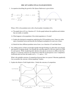
Fig. 1. Radioimmunoassay
of insulin with double-antibody
precipitation and tungsten-powder
attenuator counting
The horizontal axis gives the concentration of standards. Filled circles are the
measured activity. Open circles denote the attenuated antibody-bound activity
(total activity minus measured activity). The triangles give the results from a
conventional assay with the sternate decanted and the actMty of the precipitate
counted. Each tube of the assay contained 0.1 ml of insulin standards, 0.2 mL
of guinea pIg anti-insulin serum (1:150 000) and 0.2 ml of 125l-labeled insulin
(0.1 ng). After incubation overnight at 4 #{176}C
antibodies were precipitated with
0.05 mL of anti-guinea pig igG (1:10) and 0.05 ml of normal guinea pig serum
(1:500). Four hours later, 0.4 g of tungsten powder was added to all tubes. They
were centrIfuged at 1700 X gand then another 0.4 g of tungsten powder was
added. The radioactivity in the tubes was counted In a well counter
Table 2. Attenuation by Various Concentrations of
Bismuth Oxide in Particles a
Concn
of B1203,
gIlO mL gel
a
Amt of B1203
per tube,
Table 4. Attenuated by Variation in Test Tube
Dimensions
Outer dimensions
Leakage, b
9
25
30
0.25
19
0.30
12
35
0.35
40
0.40
Charcoal-bismuth
9.9
7.9
11
1.0
6.0
12
1.0
6.3
14
1.0
7.7
1.0
1.12
1.78
6.3
5.7
5.0
particles were made as described in Materials except
1 mL of a suspension
containing
second-anti-
body-bismuth
particles and 400 mg of bismuth oxide powder
were added. The amount of the antibody-bismuth
particles
was that found, by titration, to bind all of the first antibody.
The tubes were rotated for 1 h, then placed on the bench to
permit sedimentation.
The radioactivity
in them was then
counted in the well counter.
Results
The principal of internal sample attenuator counting was
first tested in some model experiments. Figure 1 shows that
the antibody-bound
radioactivity in a conventional doubleantibody assay can be effectively shielded within the test tube
so that
Leakage,
%
A
for variation in the amount of bismuth oxkle (91203)added. b “Leakage” denotes
the fraction of the radioactivity contained In the sediment of the adsorber-attenuator method that can be detected in a well counter. 100% is the actIvity
detected when the same activity is contained In 0.2 ml of dlluent (the volume
of the sediment).
at 4 #{176}C.
Then,
Vol of particle
suspension,
mi/tube
of tube,
mm
only the activity
of the supernate
is recorded
by the
detector. When the antibody-bound
activity hidden within
the shielded sediment was estimated (total activity added to
the tube minus measured activity), the typical standard curve
for a competitive assay resulted.
The efficiency with which bismuth oxide attenuates
the
radioactivity within test tubes is illustrated in Tables 2-4. The
concentration
of the attenuator material in the pellet influences the attenuation
quite markedly; the volume and dimensions of the sediment have less influence. With regular
test tube sizes, 92-95% of the activity enclosed in the sediment
B
11
12
14
h the A series the same volqjTle of dwcoal-blsmuih
paittcle suspension was
added per tube. In the B series, the volume was Increased in proportion to the
tube size, to give the same height of pellet In all tubes.
a See footnote b of Table 2.
Antibody-attenuator
methods.
Figure
3 illustrates
a
standard curve from the determination
of hCG with the hCG
antiserum coupled to the polyacrylamide-bismuth
particles.
Only 1-5 mg of these particles could be added per tube to
obtain proper binding conditions for the assay, because of
their high antibody-binding
capacity. The additional attenuator material needed was added as crystalline bismuth oxide
powder as described in Methods.
Figure 4 shows the application of the second-antibody
attenuator particles to an LH assay and a comparison with the
same assay performed with a conventional double-antibody
solid-phase separation of bound and free radioactivity,
with
decantation of this supernate.
Discussion
Internal sample attenuator
counting is a new technique
for
CPM
7000
is shielded.
Adsorber-attenuator
method. The combination of bismuth
oxide with charcoal within a matrix of agarose gives a reagent
that both adsorbs and attenuates the unbound activity. The
high density of the bismuth oxide causes the particles to
sediment
within
5-10
mm
without
centrifugation.
Because
this method measures the antibody-bound
activity in the
supernate, it shows the common type of standard curve with
upward concavity. Its application to T4 assay is shown in
Figure 2. The results, both for this assay and a similar method
for T3, agreed closely
says (Table 5).
with those of our routine
a
4000
Volume, a
mi/tube
Amt of B1203,
g/tube
Leakage, b
0.25
0.50
0.1
0.2
0.3
0.4
0.5
16
The charcoal-bismuth
Table 2, footnote
b
5000
T3 and T4 as-
Table 3. AttenuatIon by Variation in the Amount of
Particles Added per Tube
0.75
1.0
1.25
6000
3000
%
11
7.5
6.2
2000
0
5.7
particle suspension as described In Materials.
50
100
150
200
250
nmo(/L
THYROXIN
b
Fig. 2. Standard curve for ISAC radioimmunoassay
charcoal-bismuth particles
of T4 with
CLINICAL CHEMISTRY, Vol. 27, No. 12, 1981
1971
Table 5. Comparison of T3 and T4 Assays with the
Adsorber-Attenuator
(CCB) Method and with
Conventional Charcoal Separation
T3
Measured
activity
activity
.
e
c Pm
T4
0.3 nmol/L
10
nmol/L
variation
7.9%
6.7%
(CV)t)
Interassay variation
10.1%
8.3%
11000
3O
25
(CV)C
Serum samples
(mean ± SEM)d
Charcoal separation
Sensitivity5
Intra-assay
u(ated
bound
35
CCB separation
Sensitivity
Intra-assay
CaIc
variation
3.39 ± 0.26
nmol/L
127.6 ± 4.5
0.3 nmol/L
8.6%
11 nmol/L
(CV)b
Interassay variation
nmol/L
10000
20
9000
15
8.1%
10.2%
11.4%
3.37 ± 0.29
128.6 ± 5.8
nmol/L
0.97
nmol/L
10
8000
(CV)C
Serum samples
(mean ± SEM)d
Corr. coeff. between
CCB and
5
0.95
0
0
0
charcoal methods’
No. samples
66
2 X SEM of the “0-standard.’
5
10
95
15
20
25
LH pg/L
Calculated from duplicate determInation
of seven standards and more than 40 samples in each of five consecutive assays.
CCalculated from determination of one normal and one high-level sample in five
Fig. 4. Radioimmunoassay of LH with second-antibody-attenuator particles
consecutIve assays.
See Fig. 1 for notations
b
Same samples measured In both assays.
determining
antibody-bound
or free radioactivity
in radioimmunoassays.
It markedly
simplifies
the handling
of the
assays, and by obviating
the need for centrifugation
it offers
C PM
10000
the potential
of full automation.
A material
radiation,
suitable as an internal attenuator
in radioimnot only should be an effective absorber of the
it also must not otherwise interfere in the assay.
Elements
with
munoassays
9000
a high capacity
for absorbing
1251 radiation
include cadmium, tungsten, mercury, lead, and bismuth.
Classical X-ray-contrast agents such as iodine and barium are
poor attenuators
for this radiation.
However, the experiments
showed that most compounds
of the high attenuating
ele-
8000
ments are not suitable because their presence
affects the reaction conditions of the assay: either they adsorb the reactants
or they interfere with the antigen-antibody
binding. Metallic
tungsten
bismuth
7000
powder was the first useful attenuator
found, but
oxide is the best one we have found so far. At neutral
pH, it is an insoluble, non-toxic, inexpensive powder with no
obvious side effects on the assay, and shows little adsorption
of soluble
reactants.
cluded in polymers
6000
fied. The
When
the attenuating
material
of various types, its properties
attenuating
capacity
is mainly
is in-
are modi-
a function
of the
concentration
of the attenuator;
the dimensions of the test
tubes and the size of the pellet have less influence. A “leakage”
of about 5 to 10% of the radiation from the activity included
in the pellet is usually found. But, the extent of this leakage
is highly reproducible within each type of assay, so this source
5000
of variation
contributes
little to the overall
imprecision
of the
assay.
4.000
_______________________________________
0
50
500
5000
hCG, tnt. units/I
Fig. 3. Standard curve for ISAC radioimmunoassay
first-antibody-polyacrylamide-bismuth
particles
1972 CLINICAL CHEMISTRY, Vol. 27, No. 12, 1981
of hCG with
The reagent
charcoal
shielding
in which bismuth oxide is combined with active
in agarose matrix gives efficient
adsorption
and
of the free radioligand.
The results are essentially
the same as those obtained
by a conventional
separation
method. It has proven highly reproducible
over many months
of trials. This applies
also to a modified
bismuth
oxide-
charcoal particle with a starch matrix, which has been developed recently (15). In addition to the T3 and T4 assays described here, we have tried this reagent successfully in assays
for digoxin, phenytoin,
and testosterone.
The inclusion of the attenuator
in microparticles
to which
first or second antibodies were conjugated demonstrated
the
feasibility
of solid-phase
antibody-attenuator
assays. The
ISAC assays had the same sensitivity
and other characteristics
as our regular hCG and LH assays. The particles used for this
application sedimented quite rapidly. Further refinement of
this technique is under way such as production of very small
particles with a narrow size distribution
that would sediment
slowly enough to permit the antigen-antibody
reaction to take
place while the particles
are sedimenting.
ISAC gives radioimmunoassays
properties
that approach
those of homogeneous
assays. Hitherto, homogeneous
systems
have been restricted
to assays with light-emitting
or lightmodifying
indicators
such as fluorescent,
fluorescence-polarizing, or light-scattering
compounds
(16-18).
A most promising development
of the homogeneous
methods
is the kinetic assays. Such systems might also be designed with ISAC
radioimmunoassays
based on non-sedimenting
antibodyattenuator
particles. The radioactive
antigen would enter the
shielded microparticles
when it binds to the antibody,
permitting determination
of the binding rate. Such a kinetic assay
will have potential
for improved
sensitivity
and briefer incubation than the current
end-point
type of radioimmunoassays.
rapid, sensitive
and specific
sterone
by radio-immunoassay.
Suppl. 155,94 (1973).
method
for the measurement
of aldoActa Endocrinol.
(Copenhagen),
5. Loriaux,
D. L., Guy, R., and Lipsett,
M. B., A simple,
quick,
solid-phase
method for radioimmunoassay
of plasma estradiol in late
pregnancy
and of plasma cortisol. J. Clin. Endocrinol.
Metab.
36,
788-790 (1973).
6. Myers, W. G., Radioisotopes
of iodine. In Radioactive
ceuticals,
U.S. Atomic
Energy
Commission
Symposium
USAEC, Oak Ridge, TN, 1970, pp 565-681.
PharmaSeries 6,
7. Thorell,
J. I., and Johansson,
B. G., Enzymatic
iodination
of
polypeptides
with 1251 to high specific activity.
Biochim.
Biophys.
Acta 251, 363-369 (1971).
8. Gharib, H., Ryan, F. J., Mayberry,
W. E., and Hockert,
T., Radioimmunoassay
for triiodothyronine
(T2): I. Affinity and specificity
of the antibody for T3. J. Clin. Endocrinol.
Metab.
33, 509-516
(1971).
9. Thorell, J. I., and Holmstr#{246}m,B., Production of antisera against
highly purified
human
follicle-stimulating
hormone,
luteinizing
hormone
and thyroid-stimulating
hormone.
J. Endocrinol.
70,
335-344 (1976).
10. Thorell, J. 1., and Larson, S. M., Double antibody production
testing. In Radioimmunoassay
Louis, MO, 1978, p 276.
and Related
Techniques,
Mosby,
and
St.
11. Storm, E., and Israel, H. I., Photon cross section from 1 keV to
100 MeV for elements Z = I to Z = 100. Nuclear Data Tables A, 7,
Academic Press, New York, NY, 1970, pp 565-681.
12. Francois, J. P., On the calculation of the self-absorption
in
spherical radioactive
sources. Nuci. Inst rum. Methods
117, 153-156
(1974).
13. Ekman,
B., and Sj#{246}holm,
L., Use of macromolecules
particles.
Nature
257, 825-826 (1975).
in micro-
The excellent technical assistance
of Mr. Ingvar Larsson, the cal14. Thorell, J. I., and Larson, S. M., Isolation of immunoglobulin
from
culations of the physical properties of various attenuators
by Soren
serum by DEAE Sephadex. In ref. 10, p 279.
Mattsson, Ph.D., and the secretarial assistance of Mrs. Barbro R#{246}ing 15. Eriksson, H., Mattiasson, B., and Thorell, J. I., Use of internal
are very much appreciated.
sample attenuator in radioimmunoassay.
Assay of triiodothyronine
(T3) using starch particles containing entrapped charcoal and bismuth
oxide in combination
with free antibodies.
J. Immunol.
Methods,
in
References
1. Wide, L., and Porath, J., Radioimmunoassay
use of Sephadex-coupled
antibodies. Biochim.
257-259
of proteins with the
Biophys.
Acta 130,
(1966).
2. Catt, K., Niall, H. D., and Tregear,
munoassay
of human
growth
(1966).
3. Catt, K., and Tregear,
antibody-coated
tubes.
G. W., Solid phase radioimhormone.
Biochem.
J. 100, 31, 33c
G. W., Solid phase radio-immunoassay
Science
158, 1570-1572
(1967).
in
press, 1981.
16. Rubenstein,
K. E., Schneider,
R. S., and UlIman, E. F., “Homogeneous” enzyme immunoassay,
a new immunochemical
technique.
Biochem.
Biophys.
Res. Common.
47, 846-851 (1972).
17. Dandliker, W. B., Schapiro, H. C., Meduski, J. W., et al., Application of fluorescence polarization to the antigen-antibody
reaction.
Theory and experimental
method. Immunochemistry
1, 165-191
(1964).
18. Cohen, R. J., and Benedek, G. B., Immunoassay
by light scattering
spectroscopy. Immunochemistry
12, 349-351 (1975).
CLINICAL CHEMISTRY, Vol. 27, No. 12, 1981
1973


© Copyright 2026