ABC
docz
Explore
Log in
Create new account
Download
Report
No category
UK Biobank Blood Sample Collection, Processing and Transport Version 1.0
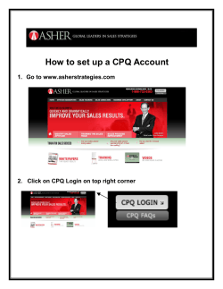
How to set up a CPQ Account
TA RGET A UDIENCE COMPLIANCE REVIEW SERVICES, INC.
How to Prepare a Quality Sample 10 8-10
Research Methods Exam 12 June Thursday afternoon
Biobanking sample and data management: Automation and best practice
How to moderate Safari Montage Live .
How to register for the 29th annual meeting of ESHRE in London? Click on “Registrations” Step 1: Go to our website:
LIABILITY WAIVER FORM EXCLUSION OF CERTAIN RIGHTS TO SUE
INVESTING OFFSHORE –
Central Line Blood Draw
© Copyright 2026
About abcdocz
DMCA / GDPR
Report