
Enteral nutrition manual for adults in
Enteral
nutrition
manual for
adults in
health care
facilities
Nutrition Support Interest Group
October 2011
1
FOREWORD
The DAA Nutrition Support Interest Group has developed this manual for
dietitians and other health care professionals who need a practical resource for
managing enteral nutrition support. It should be used in consultation with an
appropriately qualified dietitian. The Interest Group would like to acknowledge
the valuable contribution of the members responsible for the previous editions
of the manual.
This updated edition was prepared by members of the Interest Group’s NSW
Chapter:
Suzie Ferrie (National IG convenor)
Suzie Daniells (NSW IG coordinator)
Sandra Gagnon
Jenny Hamlyn
Katja Jukkola
Nicola Riley
Karen Storer
Amanda Whiteman
Nazy Zarshenas
A subset of this group met face-to-face six-weekly over 18 months to develop
the content of the manual, with various sections divided between small groups
which were then incorporated into the whole document under the oversight of
the full committee and selected reviewers including new-graduate dietitians. A
modified Delphi approach was used to resolve any areas of controversy. After
the whole manual was completed, it was examined in detail by reviewers
including representatives from all Australian states and territories, and New
Zealand.
Thanks to those who have kindly participated in reviewing this manual,
including:
Lisa Barker
Andrea Bramley
Christine Chapman-Kiddell
Cindy Chau
Alison Close
Deidre Coote
Brooke Donaldson
Felicity George
Gwen Hickey
Erin Hoebee
Kit Lai
Jessica Middleton
Lauren Muir
Emma Osland
Siong Pang
Karen Peters
2
Rubina Raja
Fiona Rezannah
Emma Rolls
Ashley Sanderson
Marie Smith
Emma Spillmann
Charmaine Trejaut
Di Wintle
TABLE OF CONTENTS
Foreword .............................................................................................. 2
Introduction ......................................................................................... 5
What is enteral nutrition support? ...................................................................................................5
Why is nutritional assessment necessary? ......................................................................................5
Flowsheet for nutrition support planning ............................................................................................6
Enteral nutrition formulations .......................................................... 7
Considerations in choosing a feeding formula ....................................................................................8
Osmolality.......................................................................................................................................8
Nutritionally complete formulations ...............................................................................................8
RDI Volume....................................................................................................................................8
Fluid content ...................................................................................................................................8
Specialty formulations ....................................................................................................................8
Other alternatives, home-made formulae .......................................................................................8
Routes of enteral feeding .................................................................... 9
Equipment.......................................................................................... 11
Enteral feeding tubes .........................................................................................................................11
Nasoenteric / oroenteric feeding tubes ..........................................................................................11
Comparison of fine-bore and wide-bore feeding tubes ............................................................11
Confirming correct placement of feeding tubes............................................................................12
Gastrostomy ..................................................................................................................................12
Care of the PEG tube ................................................................................................................13
Jejunostomy ..................................................................................................................................13
Cervical pharyngostomy or oesophagostomy ...............................................................................13
Administration reservoir, giving sets and syringes ...........................................................................13
Enteral feeding pumps.......................................................................................................................14
Gravity drip feeding ......................................................................................................................14
Approximate drip counts to achieve a desired feed rate ...........................................................14
Regimens for enteral feeding ........................................................... 15
Choice of feeding regimen ................................................................................................................15
Continuous feeding .......................................................................................................................15
Cyclic / intermittent feeding .........................................................................................................15
Bolus feeding ................................................................................................................................16
Tube feeding rate...............................................................................................................................16
Water flushes ................................................................................................................................18
Warning about dilution of feeds ...................................................................................................18
Monitoring of nutrition support ...................................................... 19
Anthropometry ..................................................................................................................................19
Biochemistry / haematology .............................................................................................................20
‘EUC’ and ‘CMP’ tests ................................................................................................................20
Liver enzymes ...............................................................................................................................21
Iron studies....................................................................................................................................22
Lipid studies..................................................................................................................................22
Vitamins, minerals and trace elements .........................................................................................23
Serum proteins ..............................................................................................................................24
Clinical assessment and monitoring ..................................................................................................26
Nutritional assessment ..................................................................................................................26
Nutrition support management .....................................................................................................27
Dietary intake ....................................................................................................................................28
Adequacy of nutrition support ......................................................................................................28
3
Issues in long-term enteral nutrition .............................................. 29
Transitional feeding ..........................................................................................................................29
Options for transitional feeding ....................................................................................................29
Ceasing feeds during meal times ..............................................................................................29
Nocturnal feeding .....................................................................................................................29
Bolus feeding ............................................................................................................................29
Monitoring during transitional feeding .........................................................................................29
Oral intake ................................................................................................................................29
Fluid balance ............................................................................................................................29
Swallow function ......................................................................................................................30
Discontinuing enteral feeds ...............................................................................................................30
Stopping the feeds.........................................................................................................................30
Removing the feeding tube ...........................................................................................................30
Discharge/transfer of the tube-fed patient .........................................................................................31
Trouble Shooting Guide ................................................................... 32
Allergies, intolerances and restrictive food practices........................................................................32
Allergies ........................................................................................................................................32
Food chemical sensitivities and intolerances ................................................................................32
Food avoidances ...........................................................................................................................33
Blocked feeding tube.........................................................................................................................33
Unblocking a feeding tube with water......................................................................................34
Unblocking a feeding tube with pancreatic enzyme .................................................................34
Aspiration of the feeds ......................................................................................................................35
Blue food colouring and enteral feeds ..........................................................................................35
Nausea and vomiting .........................................................................................................................36
Gut dysmotility problems ..................................................................................................................37
Delayed gastric emptying .............................................................................................................37
Diarrhoea ......................................................................................................................................38
Constipation ..................................................................................................................................39
Ileus...............................................................................................................................................39
Metabolic abnormalities ....................................................................................................................40
Refeeding syndrome .....................................................................................................................40
Dehydration ..................................................................................................................................41
Fluid overload ...............................................................................................................................42
Abnormal biochemical parameters ...............................................................................................42
Drug-nutrient interactions .............................................................................................................44
Appendix: Glossary of Terms .......................................................... 45
References .......................................................................................... 47
4
INTRODUCTION
What is enteral nutrition support?
Enteral nutrition support refers to the introduction of a nutritionally complete liquid formula
directly into the stomach or small intestine via a narrow, often specifically designed, tube.
When is enteral nutrition used?
Enteral nutrition should be considered when an individual is not safe for oral intake or when
oral intake is not adequate to meet their nutritional requirements. Enteral nutrition has been
shown to be safe, cost effective, and compatible with the body’s normal processes. Adequate
nutrition support is important for the following reasons:
* malnutrition has been found to affect around 40% of patients in major Australian
hospitals1,2
* patients with chronic or debilitating disease processes are often already malnourished on
admission to hospital/health care facilities
* without appropriate nutrition support, malnourished patients continue to deteriorate in their
nutritional status during their hospitalisation, especially if repeatedly placed “nil by mouth”
for investigations and surgical interventions.
* if uncorrected, malnutrition may result in a prolonged and complicated recovery from
illness or surgery with increased risk of infection and compromised wound healing, and
persisting functional deficits. This, in turn, leads to a longer hospital stay with an associated
increase in cost,1,2 and a negative impact on quality of life.
When is enteral nutrition not used?
In some clinical conditions, such as gut failure, intestinal obstruction, or an inability to gain
enteral access, it may be necessary to bypass the gut and deliver nutrients directly into the
bloodstream (parenteral nutrition).
Aggressive nutrition support is not always appropriate in the care of palliative or elderly
patients. Careful consideration should be given to patient and family wishes concerning the
appropriateness of initiating or continuing enteral feeding. Potential benefits, including
quality of life; possible complications and expected outcomes should be considered.
Why is nutritional assessment necessary?
When providing enteral nutrition support to a patient it is important to assess their nutrition
status. A formal assessment based on anthropometry, biochemistry, clinical and diet history
should be carried out by the dietitian. The nutrition assessment is used to determine priorities
of nutritional management, estimate the patient’s nutritional requirements, and provide a
baseline measure for monitoring the effectiveness of intervention. Based on this assessment,
a treatment goal can be set and a nutrition care plan developed.
5
Flowsheet for nutrition support planning
Is the gut functional and accessible?
YES
NO
Can nutritional requirements be met
with oral intake?
YES
Parenteral Nutrition
NO
Enteral/parenteral
nutrition not indicated
Enteral nutrition indicated
Does the patient have impaired digestion
or absorption?
YES
Consider elemental
or semi-elemental
formula
NO
Does the patient require electrolyte
restriction?
YES
Consider renal
formula
YES
High energy
formula
(1.5 – 2 kcal/mL)
NO
Does the patient require fluid restriction?
Or have high energy needs?
NO
Does the patient have high protein
needs?
YES
NO
Standard formula (1 – 1.2 kcal/mL)
6
High protein
standard formula
ENTERAL NUTRITION FORMULATIONS
Many different enteral formulas are available, through several different companies. The dietitian selects an appropriate formula based on an individual
nutrition assessment of the patient. Only a typical macronutrient composition is shown below; further information on individual products is available
from the manufacturer.
Descrption/
indication
ENERGY
kcal/mL
PROTEIN FAT
CARBOHYDRATE OSMOLALITY
%energy %energy %energy
mOsm/kg
No special
requirements
1.0-1.2
15%
30%
55%
300-500
1.0-1.2
20%
30%
50%
300-500
High energy High energy needs, or 1.5
feeds
fluid restrcition
(1.5kcal/mL)
20%
30%
50%
500-650
High energy High energy needs, or 2.0
feeds
fluid restriction
(2kcal/mL)
15%
40%
45%
450-800
Semielemental
70%
320-520
STANDARD POLYMERIC Standard
FEEDS
feeds
May or may not contain fibre: High protein ncreased protein
requirement
fibre-enriched formulae have standard
feeds
fibre content approx. 10-15g/L
7
PRE-DIGESTED
FORMULA
Minimal residue,
protein as peptides
1.0
20%
10%
Nutrients in their simple form Elemental
(protein as peptides or free
amino acids, carbohydrate as
monosaccharides, low in fat,
may contain MCT.)
Minimal residue,
protein as free amino
acids
1.0
15-20%
3-15%
70-85%
(varies
widesly
between
products)
500-730
RENAL FORMULAE
Restricted fluid and
electrolytes
1.8-2.0
15%
45%
40%
650-700
5-10%
45-50%
45-50%
450-650
Energy dense, reduced fluid
and electrolyte content.
Modified protein content.
Moderate
protein
Low protein End-stage renal
2.0
failure, not for dialysis
Considerations in choosing a feeding formula
Osmolality
An osmolality value of 300-500mOsm/kg is considered to be iso-osmolar. This means that
the formula has the same concentration of solutes as the blood. In theory, this optimises
absorption of the formula. In practice, this may be difficult to demonstrate3 especially when
feeding into the stomach which helps to regulate the osmolality in the small intestine anyway.
Nutritionally complete formulations
A formula is described as nutritionally complete if it can be used as an individual’s sole
source of nutrition. This means it must contain all essential nutrients in a defined volume.
Note that some products that are described as nutritionally complete may have a high RDI
volume (see below). For an average patient, the required amount of formula may be
unrealistically high, significantly exceeding the patient’s energy requirements.
RDI Volume
The RDI volume is the amount of formula that provides adequate amounts of all essential
micronutrients (i.e. vitamins, minerals and trace elements).
Fluid content
Enteral feeding formulae are about 70-80% water and the remainder of the volume consists of
solids that are not used as fluid by the body. For accurate calculation of fluid input (when
looking at the patient’s fluid requirements, or fluid restrictions), it is important to use the
formula’s actual water content, not the total volume of formula being given.
Specialty formulations
There are many specialty formulae available in addition to the ones listed above. For
example, formulae specifically designed for people with diabetes or respiratory disorders;
formulae for wound healing; low sodium formulae; low energy formulae etc. In many cases
the evidence for their benefit is limited and should be reviewed critically in order to assess
the formula’s appropriateness for an individual patient.4
Other alternatives
Home-made tube-feeding formulae are not recommended. They are time-consuming to
prepare, and it is very difficult to ensure that such a formula is nutritionally adequate. Recipes
for formulae made from whole foods5,6 with a blender, are more likely to cause tube blockage
and tube deterioration, are usually not nutritionally complete, and pose a markedly increased
risk of bacterial contamination.7 Meticulous attention to food safety is essential and the risks
and disadvantages should be noted carefully. If enteral formula is not available, infant
formula is a reasonable substitute that carries less risk.
8
ROUTES OF ENTERAL FEEDING
Enteral feeding tubes may enter the body at a number of different sites (see diagram). The
choice of enteral feeding route depends on several factors, such as the intended duration of
nutrition support, the patient’s condition, and any limitations to access (such as trauma or
obstructions).
nasal tube
(may be nasogastric,
nasoduodenal or
nasojejunal)
trans-oesophageal
feeding (“TOF”)
or
oesophagostomy tube
oral tube
(usually orogastric)
percutaneous
enteric tubes
(gastrostomy or
jejunostomy)
Figure 1: Routes of enteral feeding
9
SITES OF DELIVERY
Enteral feeds can be delivered, via a variety of different tubes, to the stomach, duodenum or jejunum. There are advantages and disadvantages to
each.
10
SITE
ACCESS
INDICATIONS
ADVANTAGES
Gastric
(stomach)
• Nasogastric tube (NGT)
• Orogastric tube (OGT)
• Trans-Oesophageal feeding
tube (TOF)
• Percutaneous Endoscopic
Gastrostomy (PEG)
• Surgical or radiologically
placed gastrostomy tube
Patients with normal
• Large reservoir
• Increased risk of oesophageal reflux and/or
emptying of gastric
capacity of stomach
pulmonary aspiration
and duodenal contents • Most cost effective
• Easiest to insert
• Can give bolus feeds,
without need for a
pump
Duodenum
(small bowel)
Nasoduodenal tube (NDT)
Patients who have
impaired gastric
emptying or who are
at risk of oesophageal
reflux
Jejunum
(small bowel)
• Nasojejunal tube (NJT)
• Surgical jejunostomy (JJ)
• Percutaneous Endoscopic
Gastrostomy with jejunal
extension (PEJ)
• Can be used for
early enteral
feeding, eg: 4-6
hours after trauma
• May reduce risk of
oesophageal reflux
and/or pulmonary
aspiration
• Patients who have
• Can be used for early
impaired gastric
enteral feeding, eg:
emptying or who are
4-6 hours after
at risk of oesophageal
trauma
reflux
• Reduces risk of
• Patients post upper
oesophageal reflux
GI surgery (jejunal
and/or pulmonary
feeding bypasses the
aspiration
surgical site)
DISADVANTAGES
• Potential gastrointestinal intolerance (bloating,
cramping, diarrhoea) due to lack of reservoir
capacity in duodenum; likely to need pump to
control feed rate
• May require fluoroscopic or fibre-optic endoscopic
placement of tube
•Risk of displacement/migration back into stomach
•Unable to use tube aspirates to indicate feeding
tolerance
• Potential gastrointestinal intolerance (bloating,
cramping, diarrhoea) due to lack of reservoir
capacity in jejunum; likely to need pump to control
feed rate
• May require fluoroscopic or fibre-optic endoscopic
placement of tube
• Risk of displacement/migration back into stomach
• Unable to use tube aspirates to indicate feeding
tolerance
EQUIPMENT
Equipment involved in enteral feeding may include the following:
• Enteral feeding tube
• Administration reservoir (Feed bag, bottle, etc)
• Giving set
• Syringes
• Enteral pump
Enteral feeding tubes
Nasoenteric / oroenteric feeding tubes
A nasoenteric feeding tube refers to any feeding tube placed through the nose into the
stomach or small bowel. Oroenteric feeding tubes are placed through the mouth into the
stomach or small bowel, and are most often used in people with facial or base-of-skull
trauma.
Fine-bore feeding tubes are widely available and are the tubes of preference for enteral
feeding. Wide bore tubes may be used for short term feeding.
Comparison of fine-bore and wide-bore feeding tubes
SIZE (French gauge*)
FINE BORE TUBE
WIDE-BORE TUBE
6-12 F
14-22 F
* The external diameter of the tube is designated by French (F) units: 1F ≈ 0.33mm
MATERIAL
INTENDED PURPOSE
PVC / polyurethane / silicon
Enteral feeding
PATIENT COMFORT
Relatively comfortable. Soft
(polyurethane and silicon tubes)
and more flexible.
APPROPRIATE
DURATION OF USE
4-6 weeks before replacement.
Gastrostomy feeding tube should
be considered if feeding required
long term.
(refer to manufacturer for
information on intended
lifespan of product)
GUIDEWIRE
WEIGHTED TIP
EXAMPLES
Available with or without
guidewire to assist in tube
placement.
Available with or without weighted
tip.
Many different products are
available.
PVC
Gastric drainage, gastric aspiration and
gastric decompression/deflation
Can be uncomfortable.
Remains hard.
Stiffens when exposed to gastric juices.
May cause nasal/oesophageal irritation or
ulceration if used long term.8
10 days.
If feeding required for longer than 10
days, replacement with a softer, fine bore
feeding tube or gastrostomy should be
considered.
No guidewire (tube is stiff).
No weighted tip.
Salem sump tube
Ryles tube
Levin tube
The length of different feeding tubes can vary significantly. Nasogastric tubes are generally
up to 90cm in length, while nasoduodenal or nasojejunal tubes are longer (105cm or longer)
to enable the tip to reach the correct position.
11
Confirming correct placement of feeding tubes
Ensuring the correct position of the feeding tube is essential prior to commencing feeding. If
the tube is incorrectly positioned, aspiration of feed may occur with potentially fatal results.
Xray confirmation is considered the most reliable way to confirm tube position9,10 but still
has limitations.11 Other common methods for checking the position of gastric feeding tubes
include air insufflation with auscultation of the stomach, and aspiration of gastric contents
with pH testing. It is not possible to aspirate gastric contents with some fine bore feeding
tubes. The tube should not be used if there is any doubt about its position.
Placement of tubes into the small bowel for nasoduodenal/nasojejunal feeding can be
difficult, sometimes requiring endoscopic or radiologically-guided placement. Various
modified tubes have been developed to facilitate placement. These generally have alterations
to the distal end of the tube, such as an altered shape or barbed/textured surface, that allows
peristalsis to grip the tube more effectively and move it spontaneously down the gut.
Gastrostomy
A gastrostomy feeding tube is one which has been inserted directly through the abdominal
wall into the stomach and is commonly used for long term enteral support (more than 4 – 6
weeks).
Most gastrostomies are inserted by the percutaneous endoscopic technique (PEG), but they
may also be placed surgically or radiologically.
Gastrostomy tubes vary in size from 9-28F, and normally last for 18 months to 2 years.
Replacement gastrostomy tubes include the following:
* ‘balloon’ gastrostomy, which has an end balloon that is inflated with saline to hold the
internal tip against the gastric wall.
* obturated gastrostomy, which has a mushroom-shaped internal flange that is stretched into a
suitable shape for insertion, using a special introducer.
* low-profile gastrostomy devices or ‘buttons’. These sit flush at skin level and have a
detachable extension tubing for feeding. They are more commonly used for young, more
ambulant patients or those who have a tendency to pull out their tube.
The external fixation plate or flange on a gastrostomy tube keeps the tube from rubbing
around the PEG site, protecting the skin from damage, and also prevents the tube from being
drawn further into the gut by peristalsis. It should not be removed, and if faulty should be
replaced.
It is important that a patient's tube is correctly identified (type of tube, size, and
manufacturer) by staff caring for the patient, and that these details are documented in the
patient’s notes. This allows appropriate care, and replacement parts to be provided when
required. For example, a balloon gastrostomy should be checked fortnightly to ensure that the
balloon is inflated with the correct amount of water, following manufacturer’s instructions.
Urinary catheters (eg Foley’s catheters) are designed for urinary drainage and are not
recommended for use as feeding tubes. As they do not have an external flange, there is a
much greater risk of tube migration and duodenal obstruction; also they are not designed to
withstand gastric acid. In an emergency, they may be useful as a cheap, short term
replacement when a PEG tube falls out, but this is temporary only. A proper replacement
gastrostomy tube should be inserted as soon as practical.
12
Care of the PEG tube
Taking care of the PEG site is important to reduce the risk of skin breakdown and infection.
The PEG site should be washed daily with warm soapy water, rinsed and dried thoroughly.
The external flange will need to be lifted to clean around the tube. Unless the tube has been
stitched into place, it should be gently pulled in and out (1-2 mm only) and rotated through
one full turn daily. The PEG site should not be covered with a dressing, as this can cause
dampness, skin damage and infection. After the initial PEG insertion, endoscopists’
instructions may vary. Generally the PEG tube is rotated for the first time 24 hours after
insertion, and the external flange left in its initial (quite tight) position for the first 5-7 days to
encourage tract formation, after which it can be loosened to allow 1-2 mm movement in and
out.
Jejunostomy
A jejunostomy feeding tube is a specialised tube designed for jejunal feeding. It is inserted
into the proximal jejunum, creating a tract between the jejunum and abdominal surface. The
jejunostomy tube may be placed surgically, radiologically or endoscopically (when it would
be called a PEJ). Needle catheter jejunostomy is the most commonly used type of surgical
jejunostomy. A combined gastrojejunostomy tube, with a double lumen, can be used to
facilitate gastric decompression and jejunal feeding simultaneously. Jejunal Extension Tubes
(JETs) are also available, which can be used to extend an existing PEG into the jejunum.
Cervical pharyngostomy or oesophagostomy
An oesophagostomy or pharyngostomy refers to the surgical procedure whereby a feeding
tube is placed into the oesophagus or pharynx and down into the stomach. This procedure is
occasionally used after head and neck surgery, however it is not widely used for long term
feeding because of the ease and relative safety of inserting a PEG instead.
Administration reservoir, giving sets and syringes
Administration reservoirs are available as either bags or bottles, in a range of sizes.
‘Closed system’ or ‘ready-to-hang’ formulae are available, where the formula is presented in
a container that can be used as a reservoir and connects aseptically to the giving set by a
recessed spike. Closed feeding systems have been found to have a lower risk of bacterial
contamination compared to an open system, where the formula is decanted into the
administration reservoir.12 Closed system formulae can hang safely at room temperature for
24 hours, whereas decanted formula should not be allowed to hang for longer than 8 hours
(shorter times may be necessary if the environmental temperature is warm). While
manufacturers may claim a longer hang time is possible for closed system formula, this is
limited by the fact that the feeds can hang only until the giving set needs to be changed,
which should occur every 24 hours.13
The giving set is plastic tubing, attached from the administration reservoir to the enteral tube,
with a roller clamp to regulate or stop the flow rate. They may have an optional side port for
the administration of drugs or water flushes. In some systems the giving set and reservoir
come as an all-in-one combined system. The reservoir should have volume levels indicated,
and a hook or other facility to allow it to hang, as well as a secure connection to the giving
set. The giving set should be an appropriate length to enhance patient mobility. It should be
incompatible with IV equipment to avoid confusion. Syringes are useful for flushing enteral
tubes with water and for the delivery of bolus feeding. A 60 ml catheter tip syringe is
recommended for this, as smaller syringes produce greater pressure and may split the tube.
13
Giving sets and syringes are ‘single use items’ and should be discarded after use. Re-using
equipment is not recommended – refer to manufacturer’s guidelines. The Australian
Therapeutic Goods Administration (TGA) does not permit the re-use of single-use items.14,15
Enteral feeding pumps
Use of a pump for feeding allows precise delivery of formula, at an even rate, which should
maximise feeding tolerance and reduce the likelihood that the tube will block. High-fibre and
energy-dense formulae may be easier to deliver with a pump, as the viscosity of these feeds
can make gravity feeding problematic. Pumps can be used to deliver bolus and intermittent
feeds, as well as for continuous feeding.
Pump feeding is the preferred method of feeding directly into the small bowel, as the small
bowel may not tolerate large changes in volume infusion. Rapid administration of formula
into the jejunum may result in dumping syndrome, or signs of feeding intolerance such as
abdominal distension, cramps and diarrhoea.
A power source is required for regular use of enteral pumps. Useful features of the pump
include battery back-up, portability, simplicity of use and ease of cleaning, alarm sound, quiet
operation, excess pressure cut-out, a hold facility for administration of medications and a
method of attaching the pump to the bed/IV pole/other location.
Pumps should be kept clean by wiping daily with a cloth moistened in mild detergent and
water. Spillage of formula onto the pump should be wiped clean immediately.
Many different types of enteral pumps are available, including lightweight, compact pumps
for ambulatory patients and pumps with the capacity for programming automated water
flushes. Check that the supplier provides maintenance service. Some companies provide
pumps for hire or loan in addition to selling them.
Gravity drip feeding
If an enteral pump is unavailable, gravity feeding can be used. By adjusting the roller clamp
of the giving set to deliver a specified number of drops of formula per minute, a consistent
feeding rate can be achieved (see table below). This method is not precise and the drip rate
should be checked regularly to ensure the desired rate of formula is being given.
Approximate drip counts to achieve a desired feed rate
mls/hr
No. of drops/minute
25
7
50
13
75
20
100
27
125
33
150
40
175
47
200
53
14
REGIMENS FOR ENTERAL FEEDING
The choice of enteral feeding regimen is based on assessment of the individual needs of the
patient. The goal is to provide safe enteral nutrition and hydration appropriate to the clinical
status of the patient, taking quality of life issues into consideration.
Choice of feeding regimen
Enteral feeds can be administered by continuous, intermittent or bolus methods.
Continuous feeding
Defined as feeding for 24 hours continuously either by gravity drip or feeding pump. (See
Equipment section for information on gravity feeding).
Continuous feeding at low volume is often used as the first step to commencing a patient on
an enteral feeding regimen. It is the preferred method of use if the patient has rapid intestinal
transit and may be more suitable for critically ill patients than other regimens. When
postpyloric feeding is required, continuous feeding is often tolerated better than intermittent.
The formula needs to be replenished every 4-24 hours depending upon whether the system
used is decanted feed or closed system.
Advantages:
• Allows the lowest possible hourly feed rate to meet nutrient requirements
• Better gastrointestinal tolerance due to the lower feed rate
• Better control of blood glucose levels due to continuous carbohydrate input
Disadvantages:
• Physical attachment to the feeding apparatus (may affect quality of life)
• Expense of equipment (pump and giving sets)
Cyclic / intermittent feeding
Enteral nutrition is stopped for a 4-16 hour period either during the day or at night. The
shorter the period of feeding, the higher the rate may need to be in order to meet the patient’s
requirements. Suitable for pump and gravity drip.
Advantages:
• Allows greater patient mobility (may improve quality of life)
• Allows breaks for physical activity, for administration of medications that are
incompatible with feeds, and to encourage oral intake if applicable.
• Useful in the transition from continuous to bolus feeding, or from tube feeding to oral
intake.
• Daytime feeds may reduce aspiration risk if it is difficult to maintain a 30° elevation
overnight
• Feeding during daytime only is more physiological and may therefore have benefits
such as helping to re-establish the diurnal cycle;16 promoting normal gastrointestinal
motility17 and promoting re-acidification of the stomach (which protects against
bacteria).18,19 There is no evidence for a benefit of ‘gut rest’, however.
15
Disadvantages:
• Compared with continuous feeding, a higher infusion rate is required to provide the
same volume of feed. This may be less well-tolerated, with a higher risk of problems
such as reflux, aspiration, abdominal distension, diarrhoea and nausea.
Bolus feeding
Defined as rapid administration of a bolus feed /water by syringe (usually by gravity, without
the plunger). Bolus feeding is usually into the stomach, which has the reservoir capacity to
tolerate a large volume of feed. A prescribed volume of feed is given (such as 100-400ml)
over 15-60 minutes at regular intervals. The patient must have a competent oesophageal
sphincter and be able to protect his/her airway adequately to minimise aspiration risk, as this
risk is higher with larger feed volumes and administration rates.
Advantages:
• Physiologically similar to a typical eating pattern
• Allows greater patient mobility
• Convenient for gastrostomy feeding
• Can be used to supplement oral intake
• Can be flexible to suit the patient’s lifestyle and improve quality of life
• May facilitate transition to oral intake
• Avoids the use of expensive equipment
Disadvantages:
• Large boluses may be poorly tolerated, especially in small bowel feeding
• Requires more nursing time compared with pump-controlled feeding
• Highest risk of aspiration, reflux, abdominal distension, diarrhoea and nausea
Tube feeding rate
All patients require individual assessment for determining the rate of delivery of nutritional
support.
Determination of the starting rate involves consideration of the patient’s medical condition,
formula type, site of delivery, and degree of inadequate nutrition prior to starting feeds. The
decision to advance the feeding rate is based on assessment of feeding tolerance. See
Monitoring section for criteria that may be helpful in identifying and managing intolerance
symptoms such as vomiting, abdominal distension, diarrhoea, large gastric aspirates, and
decreased bowel motility.
Where there is no obvious risk of intolerance or refeeding syndrome, there is little evidence
to support the practice of gradually initiating tube feeding,20,21 however many institutions still
prefer to do this. There is wide variation of recommendations in the literature for feeding rate
commencement and advancement, with suggestions varying from 15-50ml/hr for starting
rates and 10-50ml/h for the amount to increase the rate each 4-24 hours.
Individual needs of the patient should always be considered. A slow initiation regimen may
significantly delay the patient receiving his or her full nutritional requirements, but may be
necessary for patients who are at risk of refeeding syndrome, bowel ischaemia or feeding
intolerance.22 These may include patients who have been nil-by-mouth for a prolonged
period, critically ill patients, patients being fed jejunally or those who have had major
surgery, particularly abdominal surgery. Patients receiving feeds via the stomach rather than
16
postpylorically may have a better tolerance for hypertonic feeds, higher feeding rates and
bolus feeding. Patients with normal gastrointestinal function and who have had a normal
nutritional intake in the week preceding enteral feeding may safely be commenced at higher
initial volumes than recommended below.
The following table provides a guide to common practice in starting and advancing feed rates.
MODE OF
START RATE
INCREASE
COMMENTS
DELIVERY
BY
40-50mL/h
10-50mL/h
Stomach can usually tolerate high
Nasogastric
every 4 hours
rates of feeding (as in bolus or
intermittent feeding). This varies
between individual patients and
regimens. To estimate maximum
tolerable feed rates, it is useful to
relate it to the volume of nutrientdense fluid that the individual patient
could drink: maximum bolus might be
the volume of a small glass of milk
(150mL) for a smaller/older person, or
a milkshake (400-600mL) for a
bigger/younger person. Maximum
continuous rate might be up to one
cup of milk (250mL) every hour.
Nasoduodenal 20-30mL/h
or
nasojejunal
10-30mL/h
every 4-8 hours
Gastrostomy / As for nasogastric As for
nasogastric
PEG
Jejunostomy
As for
nasojejunal
As for
nasojejunal
17
Continuous feeding at a controlled
rate is usually needed due to lack of
reservoir capacity in small bowel.
Feeds may be tolerated at rates as
high as 100-120mL/h. Some patients
may tolerate higher rates; others may
experience abdominal distension or
discomfort.
Sterile water should be used for tube
flushing (see below).
After the insertion of a gastrostomy or
jejunostomy feeding tube, a water trial
is sometimes used to check for leaks
before feed is given. Feeds are usually
commenced 12-24 hours after
insertion of the tube, but there is no
evidence to support delaying feeds for
more than 3-4 hours after PEG
placement.23,24
As for nasojejunal. Surgeon will
usually advise when feeds can
commence (usually within 24-48
hours of insertion); this depends on
the surgical procedure.
Water flushes
Flushing an enteral tube regularly is essential to ensure tube patency and minimise the risk of
tube blockage, as well as to meet the patient’s fluid requirements. Water is considered the
accepted flushing fluid and there is no evidence presented that tap water flushes in reasonable
volumes pose any risk to humans. However sterile water for irrigation, commercially filtered
or cooled boiled tap water have been suggested for use in particular patients who may be at
increased risk. This would include immunocompromised patients and those being fed
postpylorically.25 Post pyloric feeding bypasses the acidic environment of the stomach, which
normally provides protection against bacteria.
Where possible any drugs administered via an enteral feeding tube should be liquid and given
separately from the feed, with flushing of the tube before and after each separate medication.
Recommendations:
• Flush tubes regularly with at least 30mls of water before, between and after
medication administration, before and after bolus feedings and before and after
checking gastric residuals and when feeds are stopped. See Troubleshooting section
on delayed gastric emptying
• Smaller bore tubes such as jejunal tubes and fine bore tubes are more prone to
clogging and may need to be flushed more frequently
• When several drugs are to be given, each should be given separately with a 5-10ml
water flush given between each drug
• Use a 60ml syringe for flushes as smaller syringes may exert a force great enough to
damage the tube
• Additional water flushes or increased volumes may be required, to meet the patient’s
hydration needs – see Monitoring section for details on assessing fluid status.
• Avoid flushing with acidic fluids (fruit juices or carbonated drinks like cola/soda
water/mineral water) which can make the chance of blockages more likely – refer to
the Troubleshooting section for advice on unblocking tubes.
Warning about dilution of feeds
Diluting an enteral feed, or mixing anything into the feed, is not recommended as it increases
the risk of bacterial contamination of the feed.26 If a closed system of feeding is used,
opening the reservoir to add water (or medications, or supplemental protein, fibre, salt etc)
will invalidate the stated hang time and can lead to increased wastage. No evidence exists to
suggest that diluting feeds is helpful in tolerating tube feeding; in fact, absorption of the
formula may be less effective if it is hypo-osmolar. Also, diluting the formula may simply
delay achieving nutritional targets.
18
MONITORING OF NUTRITION SUPPORT
Once an enteral feed regimen has been initiated, ongoing monitoring is essential to ensure
adequate nutrition delivery, and identification and management of potential complications.
Anthropometry
MEASURE
APPLICATION
INTERPRETATION
IDEAL FREQUENCY
OF MONITORING
Weight
Unstable fluid
balance (including
large stoma output)
-whether fluid input is
meeting needs
-whether it should be
restricted or increased
-whether fluid input is
meeting needs
Baseline then daily
-whether energy input is
meeting needs
-interpret in the light of
fluid changes
-whether energy input is
appropriate
Baseline and then weekly
-whether energy and
protein input are
appropriate
-interpret in the light of
changes in patient’s activity
level
-acute changes in fluid
balance may confound this
measure in trauma and
critical illness
Baseline and then monthly.
(Serial measures are more
informative than comparing
single measures to percentile
charts.)
Patients in first 1-2
weeks of tube
feeding
Patients in first 3-6
weeks of tube
feeding
Mid-arm
circumference
(at midpoint on
back of arm,
halfway between
acromial surface
of scapula and
olecranon process
of elbow)
Calf
circumference
Continuation of
long term tube
feeding
Long term tube
feeding
(at widest point)
19
Baseline and then seconddaily
Baseline and then monthly
Biochemistry / haematology
Check with your laboratory for the normal range for each parameter as some of these will
vary between sites. Only a basic survey of each test is given here. For more details of each
test and its intepretation, refer to a clinical chemistry text or your laboratory’s handbook, and
discuss with medical team.27,28
‘EUC’ and ‘CMP’ tests
MEASURE
DESCRIPTION
INTERPRETATION
IDEAL
FREQUENCY OF
MONITORING
Sodium
* major
extracellular
electrolyte
* plays an
important role in
fluid balance
* also involved in
nerve conduction
and membrane
transport
INCREASED in dehydration,
refeeding syndrome.
DECREASED in overhydration,
SIADH, and salt-wasting conditions.
-consider whether all the other
electrolytes are increased or
decreased.
-check total fluid input, and whether
IV fluids are saline or dextrose
-serum osmolality can help determine
fluid status
-urinary sodium and osmolality can
help identify SIADH or salt wasting.
Daily in the acute
setting
Monthly if possible,
for long term feeding
Potassium
* major
intracellular
electrolyte
* influences fluid
balance
* also involved in
nerve conduction
and muscle
contraction (esp.
cardiac)
INCREASED in dehydration, renal
failure (acute, or serious chronic), or
when K+ shifts out of the cell (as in
acidosis or insulin deficiency)
DECREASED by increased losses
(vomiting, diarrhoea) or when K+
shifts into cell (as in refeeding
syndrome, reversal of acidosis,
administration of insulin)
-consider effect of diuretics (may
cause K+ wasting or retention)
-consider possible losses
Daily in the acute
setting
Monthly, if possible,
for long term feeding
INCREASED in renal failure or high
intake (eg magnesium antacids)
DECREASED by some diuretics,
malnutrition and muscle loss,
malabsorption, and when Mg2+ shifts
into cell (as in refeeding syndrome)
Daily in the acute
setting
Monthly, if possible,
in long term feeding
INCREASED in renal failure,
hypoparathyroidism, laxatives
DECREASED in muscle loss and
malnutrition, malabsorption, vitamin
D deficiency, hyperparathyroidism
and refeeding syndrome.
Daily, in the acute
setting.
Monthly, if possible,
in long term feeding.
Magnesium * intracellular
* cofactor for
enzymes in nearly
every stage of
aerobic metabolism
* required in
muscle and nerve
function
Phosphate
* intracellular
* part of ATP, so
involved in all
energy production /
storage processes
* found in bone and
muscle
20
MEASURE
DESCRIPTION
INTERPRETATION
IDEAL
FREQUENCY OF
MONITORING
Urea
* end product of
protein metabolism,
formed by liver to
detoxify ammonia
* excreted in urine,
rate depends on
GFR (glomerular
filtration rate) and
urine output.
* produced by body
muscle, also small
amounts come from
meat in the diet
* excreted in urine,
rate depends on
GFR and tubular
secretion
* proportional to
muscle mass
INCREASED in renal failure,
dehydration, high protein intake (from
diet, or GI bleed), renal obstruction
DECREASED with reduced muscle
turnover (small muscle mass, low
protein intake, low activity) or severe
liver dysfunction
- Urea:Creatinine ratio >1:10 can
indicate dehydration or GI bleed.
INCREASED in renal failure, fever,
large meat meal, large muscle mass
(bodybuilder, acromegaly) some
medications
DECREASED with loss of muscle,
low protein intake
Daily, in the acute
setting.
Monthly, if possible,
in long term feeding.
Creatinine
Daily, in the acute
setting.
Monthly, if possible,
in long term feeding.
Liver enzymes
MEASURE
DESCRIPTION
INTERPRETATION
IDEAL
FREQUENCY OF
MONITORING
AST, ALT
* ‘transaminases’
* released in cell
damage
* ALT in liver cells
* AST in liver,
muscle and red
blood cells
Daily in the acute setting
Monthly if possible, for
long term feeding
ALP, GGT
* ‘cholestatic
enzymes’
* produced by liver
in increased
quantities during
biliary obstruction
* GGT induced by
some medications,
and obesity
* ALP in bone and
placenta
* breakdown
product of haem
molecules
* WBC in spleen
produce bilirubin
* liver makes
bilirubin soluble
and secretes it into
bile
INCREASED in liver damage
ALT only: mild liver damage
AST only: chronic liver disease
with tissue destruction, or damage
to other body muscle
Both AST and ALT raised: major
damage; can indicate overfeeding,
esp. with excessive carbohydrate.
INCREASED in biliary
obstruction
GGT only: drugs, alcohol, obesity
ALP only: bone, growth, fractures
Both GGT and ALP: cholestasis;
will increase in overfeeding, esp.
with excessive carbohydrate, but
not usually to abnormal level.
Bilirubin
INCREASED in internal bleeding
or haemolysis, or liver
dysfunction, or biliary
obstruction, also many drugs
May DECREASE in overfeeding.
21
Daily in the acute setting
Monthly, if possible, for
long term feeding
Daily in the acute setting
Monthly, if possible, in
long term feeding
Iron studies
MEASURE
DESCRIPTION
Haemoglobin
(Hb)
Iron containing
portion of the RBC
which binds oxygen
for transport
throughout the body
Storage form of iron
Ferritin
Transferrin
Serum iron
INTERPRETATION
INCREASED in
haemochromatosis
DECREASED with blood
loss, acute phase and in all
types of anaemia
INCREASED in acute phase
and sepsis (to reduce
availability to bacteria), iron
overload.
DECREASEDiron deficiency
(diagnosis is iron deficiency
anemia if Hb also low).
Transport form of
INCREASED in iron
iron
deficiency
DECREASED in acute phase
response, malnutrition, iron
overload, liver disease
Free iron in the serum DECREASED in iron
deficiency
Haematocrit
(also known
as Packed
Cell Volume
or PCV)
Volume of red cells
in relation to the total
volume of blood
Mean
corpuscular
volume
(MCV)
Reflects RBC size, to
classify type of
anaemia
INCREASED with
dehydration
DECREASED in anaemia
and other blood
abnormalities, blood loss,
nutritional iron deficiency.
INCREASED in macrocytic
anaemia; NORMAL in
normocytic anaemia
DECREASED in microcytic
anaemia
IDEAL
FREQUENCY OF
MONITORING
Daily in the acute setting
Monthly, if possible, in
long term feeding
To assist in interpretation
of abnormal iron studies.
More sensitive and
specific than other iron
tests.
To assist in interpretation
of abnormal iron studies.
Not a sensitive measure
of protein status due to
wide normal range.
Not very sensitive or
specific. Significant
diurnal changes and wide
normal range.
To assist in interpretation
of abnormal iron studies.
Relatively insensitive:
detects only severe iron
deficiency.
To assist in interpretation
of abnormal iron studies.
Lipid studies
MEASURE
DESCRIPTION
INTERPRETATION
IDEAL
FREQUENCY
OF
MONITORING
Triglycerides
Storage form of fat in
the body. Consumed
in diet, or made by
the liver.
Need fasting blood
sample for accurate
assessment
Made in the liver,
needed for production
of hormones, bile
acid and vitamin D.
INCREASED in overfeeding,
glucose intolerance,
hyperlipidaemias, hypothyroidism,
pancreatitis.
DECREASED in malabsorption or
very low fat intake,
hyperthyroidism.
INCREASED in acute phase and
sepsis (to reduce availability to
bacteria) as well as hyperlipidaemia
and increased dietary intake.
DECREASED with malnutrition,
liver disease, hyperthyroidism
As needed
Cholesterol
22
As needed
Vitamins, minerals and trace elements
Note: blood levels may not reflect total body stores, due to varying distribution of
vitamins/minerals in body tissues. Requirements may be increased in illness.29
MEASURE
DESCRIPTION
INTERPRETATION MONITORING
Fat-soluble
vitamins
Vitamin B12
Folate
Vitamin A (retinol and
carotenoids, essential for
vision, growth, iron
metabolism. Deficiency
symptoms include night
blindness, anaemia,
immune impairment,
follicular hyperkeratosis.)
DECREASED in fat
malabsorption.
Vitamin D (active form is
1,25-dihydroxyvitamin D,
controls calcium
utilisation, has immune
function, interacts with
serum calcium and
phosphorus, and
parathyroid hormone.
Deficiency symptoms
include bone
abnormalities, low serum
phosphate, raised ALP.)
Cyanocobalamin; acts as
cofactor in fat
metabolism and DNA
synthesis. Stored in liver.
Deficiency symptoms
include neurologic
symptoms (fatigue,
weakness, depression),
glossitis, stomatitis,
pallor, gut dysfunction,
megaloblastic anaemia.
Folic acid and related
compounds, involved in
coenzymes of many
metabolic processes, esp.
amino acid
interconversions and
DNA synthesis.
Deficiency symptoms
include neurologic
symptoms (fatigue,
weakness, depression),
glossitis, stomatitis,
pallor, gut dysfunction,
megaloblastic anaemia.
Also increased
cardiovascular risk.
DECREASED in lack of
sun exposure,
malabsorption (esp. in
elderly)
Usually test Vitamins A
and D to exclude fat
malabsorption.
Individual tests (serum
retinol,) to exclude
deficiency.
Blood samples for retinol
testing should be wrapped
(in foil/paper) after
collection to avoid light
exposure.
Usually test serum level
of precursor form (25hydroxyvitamin D) as this
is the major circulating
form and a more sensitive
test than 1,25 form.
INCREASED in acute
phase and sepsis
DECREASED with loss
of stomach or ileum
function, malabsorption
(eg in coeliac disease or
pancreatic insufficiency),
vegan diets, old age,
pernicious anaemia,
nitrous oxide anaesthesia.
Serum cobalamin assay
checks B12 level, levels
are low in deficiency.
Serum methylmalonic
acid is elevated in B12
deficiency but not in
folate deficiency.
Unstable, so blood
sample should be frozen
after collection.
DECREASED in folate
deficiency, and in
smokers. May be
INCREASED in cases of
small bowel bacterial
overgrowth.
Erythrocyte (not serum)
folate is best test.
Unstable, so blood
sample should be frozen
after collection.
23
MEASURE
DESCRIPTION
INTERPRETATION
MONITORING
Zinc
Important cofactor for
many enzymes, role in
immune function.
Serum value should be
corrected for low albumin
level.
Trace element free tube
should be used for
collection as zinc is a
common contaminant.
Selenium
Antioxidant, interacts
with thyroid hormones.
Absorbed in duodenum.
Deficiency signs include
muscle weakness,
cardiomyopathy.
DECREASED in
malabsorption or chronic
renal failure, or with poor
intake or loss of skin
tissue (as in burns or
psoriasis). Folate
supplementation may
interfere with zinc
absorption.
DECREASED in people
living in regions with
low-selenium soil
(incl.Australia, New
Zealand). Also in
pregnancy, alcoholism,
malabsorption and in
immunocompromised
patients.
Measured by testing
serum selenium level or
glutathione peroxidase
activity.
Serum proteins
None of these indicators reflects simple nutritional status in a hospital patient.30,31,32,33 Note
the effect of the acute phase process on all of these indicators. Acute phase markers (such as
C-reactive protein, see below) may be useful in quantifying this.
MEASURE
DESCRIPTION INTERPRETATION MONITORING
Albumin
A non-specific
carrier protein,
plays important
role in controlling
fluid distribution
between tissue
compartments.
Made in the liver.
Half-life 18-20
days.
INCREASED in
dehydration or when IV
albumin is given.
DECREASED in acute
phase, liver failure,
protein malnutrition,
overhydration, loss of
body tissue (as in
bleeding, burns, surgery,
fistula), diarrhoea.
Strong prognostic indicator,34
because it is a measure of
disease severity. Cannot be
used to assess nutritional status
in acute phase situation.
Good screening parameter as it
indicates long-term nutritional
status (not in acute phase
situation).
Prealbumin,
also known as
thyroxinebinding
protein or
transthyretin
Transport protein
for thyroxine, also
associated with
retinol-binding
protein transport.
Made in the liver,
mostly cleared by
the kidneys.
Half-life 3-5 days.
INCREASED in renal
failure, steroid therapy,
non-hodgkin’s
lymphoma, head injury
(? via cortisol secretion.)
DECREASED in acute
phase, liver failure,
protein malnutrition,
hyperparathyroidism.
Much earlier indicator of
patient’s clinical improvement,
compared to albumin.
Correlates with previous 5
days’ cumulative nutrition
intake in a stable patient (even
in acute phase) and should be
normal in a stable fed patient
after 1-2 weeks.
Retinolbinding
protein
Transport protein
for retinol.
Made in the liver.
Half-life 12 hours.
INCREASED in renal
failure, vit A deficiency.
DECREASED in acute
phase, liver failure,
protein malnutrition.
Extremely sensitive indicator of
patient’s clinical condition.
May fluctuate too much to be a
useful measure.
24
MEASURE
DESCRIPTION
INTERPRETATION
MONITORING
Total protein
Total of serum
proteins. Albumin
and
immunoglobulins
are the major
components of this
total.
Most direct
measurement of
actual protein
status, by
comparing nitrogen
output and input.
DECREASED in acute
phase, or any other
situation where albumin
is decreased.
Severe acute phase may cause a
decrease in alb:total protein
ratio, due to falling albumin and
increasing contribution by acute
phase proteins (such as creactive protein)
Positive nitrogen balance
(input > output) indicates
anabolism. Negative
nitrogen balance indicates
lean tissue loss. (Will be
negative in acute phase,
and improvement may
not be possible.)
Less accurate if there are
unquantifiable additional
protein losses (such as
from wounds/fistulae)
INCREASED very
rapidly in acute phase,
sensitive correlation with
disease severity.
NORMAL level is around
0.
24-hour urine collection is
tested for urea (or, preferably,
total nitrogen35) content.
Calculation then estimates
nitrogen balance:
Nitrogen input (g) = protein
intake (g) ÷ 6.25
Nitrogen output = 24-hour
urinary urea + “insensible
losses”, usually use 3-5 g but
may be more.
Nitrogen
balance
C-reactive
protein (CRP)
An acute phase
protein – a nonspecific marker of
infection and
inflammation.
Erythrocyte
sedimentation
rate (ESR)
A less specific
indicator of acute
phase response.
INCREASED in acute
phase and inflammation.
DECREASED in CCF,
sickle cell anaemia and
polycythaemia.
C-reactive protein is a better
indicator of the acute phase
response.
White cell
count (WCC)
(= total of
lymphocytes,
neutrophils,
basophils,
eosinophils.) A
good general
indicator of
infection and stress.
Marker of bone
marrow and
immune function.
Good predictor of
healing.36
Immune cell that
engulfs and
destroys bacteria. A
marker of bacterial
infection.
INCREASED in tissue
damage and infection.
DECREASED in
immunocompromised
patients.
Decreases promptly as infection
resolves.
DECREASED in the
acute phase response and
in immunocompromised
patients.
Daily in the acute setting,
monthly if possible in long term
care.
INCREASES in infection
DECREASES in
neutropaenic
immunocompromised
patients.
Decreases promptly as infection
resolves.
Lymphocytes
(Total
lymphocyte
count: TLC)
Neutrophils
25
Helps in interpretation of other
indicators, by quantifying acute
phase situation. Should have
inverse correlation with
prealbumin etc, in an
adequately nourished patient.
Clinical assessment and monitoring
Physical examination of the patient is important during the initial assessment, particularly in
critical illness when other indicators are affected by the patient’s acute condition.
Nutritional assessment
MEASURE
DESCRIPTION
METHOD OF MONITORING
IDEAL
FREQUENCY
OF
MONITORING
Lean tissue
stores
* muscle mass
* main determinant
of resting metabolic
rate
* decreases in
critical illness and
immobility
* lost during rapid
weight loss,
regained only
slowly
Baseline and then
weekly-monthly in
longer term care.
Adipose
tissue
* fat mass
* minimal
metabolic activity
at rest, but the
added weight
increases energy
expenditure during
activity.
* increased esp. in
rapid weight gain
Indicator of general
nutritional status,
also protein, zinc,
vitamin C and
vitamin E nutrition.
Assess:
* temporal muscles (should be flat or
plump, not hollowed)
* clavicle (should not be prominent with
hollowing; should not be visible at all in
men)
* shoulder (should be rounded, not square
looking with prominent bones)
* scapula (should not be prominent;
surrounding ribs should not be prominently
visible)
* quadriceps (should be plump with good
tone, and without bone dent or bony
prominence at knee)
* calf (should be plump with good tone)
* interosseus muscle (between thumb and
forefinger, should be plump or flat, not
depressed)
Assess:
* fat pads under eyes (should be slightly
plump, not hollowed)
* triceps skin fold (should have substantial
fold, not thin or loose)
* biceps skin fold (should have substantial
fold, not thin or loose)
Examine skin appearance with particular
attention to wound sites, check areas not
exposed to sun which should be intact
without significant blemishes, soft, dewy.
Wounds should look clean and pink,
without very red or gummy edges. Sores,
bruising, dryness or flaking can all be
significant. Breakdown at corners of eyes,
nose and mouth (and tongue, gums) are
significant in vitamin deficiencies.
Inspect hair, test pluckability of individual
hair. Should be shiny, not coarse / dry. Loss
of pigmentation, and easy pluckability may
be significant.
Fingernails should have a slight gloss, pink
nail bed and white lunula. Horizontal ridges,
yellowing, pale nail bed, loss of lunula,
raised nail edges can all be significant.
Better for initial
assessment than for
monitoring as
changes are often
slow and difficult
to quantify.
Objective tools are
useful (such as the
PUSH tool for
wound healing).37
Good for initial
assessment but not
for monitoring in
most patients, as
changes are
extremely gradual.
Indicates long term
nutritional status.
Skin
condition
Hair/nails
Indicator of general
nutritional status,
also protein, zinc,
vitamin C and
vitamin E nutrition.
26
MEASURE
DESCRIPTION
Fluid status Patients who can
drink to thirst and
have normal renal
function and no
abnormal losses
should maintain
normal fluid status.
Involuntary
nutrition, altered
renal function, or
increased losses
will all increase
risk of abnormal
fluid status.
METHOD OF MONITORING
IDEAL
FREQUENCY
OF
MONITORING
Assess dehydration by:
* dry skin, reduced skin turgor, dry mucosa
* reduced urine output
* very yellow or dark urine
* low blood pressure, increased heart rate
Abnormal fluid (such as ascites, oedema)
should be taken into consideration.
(Dehydrated patients can still be
oedematous.)
Daily
Note: in the critically ill, fluid balance
would normally be monitored closely by
medical/nursing staff. The dietitian should
still be aware of the patient’s fluid status
and the team’s goals for fluid management.
On the ward level, the dietitian may need to
take an active role in ensuring that the
patient’s fluid requirements are met.
Day-to-day management of an enterally fed patient requires attention to practical aspects of
tube feeding. (See Troubleshooting section for management of feeding problems.)
Nutrition support management
MEASURE
DESCRIPTION
METHOD OF MONITORING
IDEAL
FREQUENCY OF
MONITORING
Feeding
tolerance
* gastric aspirates
* abdominal
distension or
discomfort
* bowel activity
Daily in acute care
situation; 2-3 times
weekly in stable hospital
patients; weekly –
monthly in long term
care.
Feed
delivered
* is the feed rate
correct?
* has the patient
received the
prescribed amount
of formula?
* reasons for feed
interruptions
* regular tube
flushing
* PEG site care
* correct taping of
NGT
Hospital flowsheet or fluid balance
charts, medical record documentation.
Daily abdominal girth measurements
(using a tape measure, at umbilicus) if
increasing abdominal distension.
Bowel charts to record stool amount
and frequency. (Bristol Stool Chart
for objective classification of stool.)
Hospital flowsheet or fluid balance
charts
Pump with “total volume delivered”
function
Medical record documentation
Hospital flowsheet/ care plans;
medical record documentation.
Examination of PEG/NGT site.
Daily in acute care
situation; 2-3 times
weekly in stable hospital
patients; weekly –
monthly in long term
care.
Continuous in acute care
situation; at least every
shift in stable patients.
Care of
feeding
equipment
Patient
positioning
* patient’s head
and shoulders must
be elevated 30-45°
above chest level
Observation
27
Daily in acute care
situation; 2-3 times
weekly in stable hospital
patients; weekly –
monthly in long term
care.
Dietary intake
Adequacy of nutrition support
MEASURE
DESCRIPTION
METHOD OF
MONITORING
IDEAL
FREQUENCY OF
MONITORING
Nutritional
input
* is the patient
receiving/tolerating
the prescribed
amount of formula?
* is the formula
appropriate for the
patient’s needs
(energy and protein
needs, RDI volume)?
* is oral intake (if
applicable) increasing
or decreasing?
* is the patient
receiving adequate
fluid?38
Hospital flowsheet or fluid
balance charts
Pump with “total volume
delivered” function
Medical record documentation
Food charts/observation
Patient report of intake
Daily in acute care
situation; 2-3 times
weekly in stable hospital
patients; weekly –
monthly in long term
care.
Review of
requirements
* have requirements
changed (due to new
infections or surgery,
or improvement in
condition, or change
in activity level)?
* is amount of feed
still appropriate to
meet needs? Has oral
intake (if applicable)
changed?
* is feeding regimen
still appropriate?
Should it be stopped
during physiotherapy
or when an
incompatible drug is
being given?
Free water input should match
fluid output in a stable patient.
Input includes: water from feeds
(only 70-80% of the formula’s
volume is water), tube-flushes, IV
fluids, fluids given with drugs,
oral fluid intake. Output includes:
urine output, stoma/faecal output,
losses from wounds/fistulae,
discarded gastric aspirates/vomit,
insensible losses (perspiration,
breath moisture) increased in
fever.
Patient weight monitoring
Biochem/haem
Medical record documentation
Estimation/measurement of
energy expenditure
Medication chart and DrugNutrient Interaction manual (or
pharmacist advice)
Patient’s preferences
28
Weekly in acute care
situation; monthly in
stable hospital patients. 16 times per year in long
term care.
ISSUES IN LONG-TERM ENTERAL NUTRITION
Transitional feeding
Transitional feeding describes the process by which a tube-fed patient returns to an oral diet
and ceases tube feeding. The ultimate goal of transitional feeding is that the patient’s full
nutritional needs will be met with oral intake alone. Ideally, the transition will be a smooth
process, which may take a few days or several weeks. Abrupt cessation of tube feeding is not
recommended, as nutritional status may be compromised. Reductions in the tube-feeding rate
should be made in proportion to increases in oral intake.
Options for transitional feeding
Satiety and lack of appetite are common barriers to achieving an adequate oral diet.
Transitional feeding strategies aim to promote the patient’s normal appetite and encourage an
increase in oral intake, so that reliance on the tube feeding is gradually reduced.
Ceasing feeds during meal times
This aims to improve the patient’s appetite and oral intake at mealtimes. The tube feeds are
stopped approximately 1-2 hours before each main meal. Feeds can resume when the patient
has finished eating, or 1-2 hours afterwards.
Nocturnal feeding
Feeding during overnight hours (8-16 hours) is less satiating than daytime feeding, and
therefore encourages oral intake during the day. It also has the advantage of allowing more
time for patient activities such as physiotherapy. By tube-feeding overnight it is possible to
satisfy nutritional requirements not met orally. The length of the overnight feeding cycle will
also depend upon the patient’s tolerance of increased enteral feeding rates. A more energydense formula (such as 1.5kcal/mL or 2kcal/mL) can be useful for meeting the patient’s needs
using a lower feed rate.
Bolus feeding
Bolus feeding during the day may be used as an alternative to continuous overnight infusion.
Administer bolus feeds at times separate from oral meals to minimise impact on appetite.
Bolus size can be adjusted according to the intake at the previous meal.
Monitoring during transitional feeding
Refer also to Monitoring section.
Oral intake
Food charts can be used to record the patient’s oral intake. Energy, protein and fluid intakes
are monitored by the dietitian and compared with the patient’s requirements (for example by
calculating the percentage of requirements being met by oral intake). The tube-feeding
regimen should be adjusted accordingly, decreasing tube feeding rate or infusion time as the
oral intake increases.
Fluid balance
When the rate or volume of tube feeding is reduced, it is important to monitor the patient’s
fluid input to ensure that requirements are still being met. The amount of tube-flushes may
need to be increased if the patient’s oral fluid intake remains inadequate. (This is particularly
29
common when the oral diet has a modified texture and/or fluids are thickened). Any changes
to the patient’s IV fluids should also be noted, as these often change during the transitional
period, and additional (or larger) water flushes may be needed.
Swallow function
Where dysphagia has been the indicator for enteral nutrition, a speech pathologist will assess
and monitor the patient’s swallow function. Speech pathologists advise on the appropriate
texture and consistency of the oral diet to minimise aspiration risk. Level of alertness will
affect the patient’s ability to eat and to learn strategies that assist in safe swallowing. The
dietitian and speech pathologist should liaise closely, to ensure successful coordination of
enteral and oral feeding for the patient.
Discontinuing enteral feeds
The goal of transitional feeding is the eventual cessation of tube feeds, but it is important that
this occurs at the appropriate time.
Stopping the feeds
In general, tube feeding may be ceased once the patient is able to maintain an adequate oral
intake. This may require improvement in their swallowing, their gut function, or their general
wellbeing, and the time required for this will differ between individual patients.
If the patient is able to consume 65-75% of their nutritional requirements orally, it may be
appropriate to discontinue the tube feeding and use oral supplements to meet the remainder of
the patient’s requirements. For other patients, it may be prudent to ensure that the full
requirements can be met orally before feeds are stopped, and perhaps continue some form of
nutrition support (such as intermittent tube feeding) to achieve a further improvement in the
patient’s nutritional status, even when the patient has an adequate oral intake.
For some patients, additional fluids, or medications, may be easier to give via the feeding
tube and this may continue for some time after tube feeding has ceased.
Removing the feeding tube
The time frame for removing the tube will vary greatly between patient types and the reason
for feeding tube insertion.
After the initial insertion, it is generally recommended that a gastrostomy or jejunostomy tube
should remain in place for at least 2-3 weeks, to allow time for the tract to heal fully. Where
patients are discharged from hospital with a feeding tube in place, it is important for the
dietitian to liaise closely with the patient’s doctor(s) regarding use and/or removal of the tube.
In particular, it is important to make clear any concerns about the patient’s oral intake or
nutritional status, where the patient would benefit from a longer period of nutrition support.
This is also an opportunity to discuss the patient’s possible future needs for nutrition support
(such as planned chemotherapy or radiotherapy which might compromise oral intake or
nutritional status) as it may be sensible to keep the tube in place, and avoid unnecessary
removal and reinsertion procedures, if it will be needed in the future.
30
Discharge/transfer of the tube-fed patient
When the patient is transferred from one health care facility to another, or discharged home
under the care of their local doctor, it is important to provide adequate information to enable
continuity of nutritional care. For the nutrition support patient, particular information that is
useful to include in a handover/discharge summary would include:
1. Date when the feeding tube was inserted, type of tube (brand, size, type)
2. Date when feeding commenced, if different from tube insertion date
3. Indication for tube feeding
4. Feeding route
5. Name of feed
6. Reason for choice of the particular feeding formula
7. Feeding regimen, including water flushes and feeding times
8. Total volume of feed per day; amount of energy, protein and fluid provided
9. Type of oral intake (if relevant), or reason why patient is kept NBM
10. Other relevant information (such as nutritional assessment data, estimated
requirements and how these were calculated)
11. Other recommendations (such as weight monitoring)
12. Follow-up plan
31
TROUBLE SHOOTING GUIDE
Allergies, intolerances and restrictive food practices
It is easy to forget that tube feed formulae can contain many of the food components that the
patient may be avoiding in their oral diet. While they are receiving tube feeding, some
patients may be willing to relax some of their habitual food restrictions if these are for
lifestyle or religious reasons. Allergies and intolerances, however, may cause difficulties in
the choice of tube feeding formula.6
Allergies
PROBLEM: ALLERGIES
CAUSE
Egg protein
allergy
Soy protein
allergy
Milk protein
allergy
Coeliac disease
ACTION
OTHER INTERVENTION /
REMARKS
Most tube feeds are egg-free.
Before using, check the
ingredients list for the specific
feed to be used.
Many tube feeds contain soy
protein. Before using, check the
ingredients list for the specific
feed to be used.
Most tube feeds contain casein
and/or whey protein. Before
using, check the ingredients list
for the specific feed to be used.
Most tube feeds are gluten-free.
Before using, check the
ingredients list for the specific
feed to be used.
Elemental formula (amino acid based) contains
no intact protein and may be safest in the case
of allergy. Hypoallergenic baby formula can
also be used for adult tube feeding.
Elemental formula (amino acid based) contains
no intact protein and may be safest in the case
of allergy. Hypoallergenic baby formula can
also be used for adult tube feeding.
Elemental formula (amino acid based) contains
no intact protein and may be safest in the case
of allergy. Hypoallergenic baby formula can
also be used for adult tube feeding.
Some fibre-containing feeds have oat bran as a
component of the fibre. This meets the
standards for gluten-free ingredients in other
countries but not in Australia.
Food chemical sensitivities and intolerances
PROBLEM: INTOLERANCES
CAUSE
ACTION
OTHER INTERVENTION /
REMARKS
Lactose
All tube feeds are lactose free.
Amines
All tube feeds are low-amine.
Salicylates
All tube feeds are low in salicylates.
Glutamates
All tube feeds are low-glutamate.
FODMAPs
Different feeds have widely varying
FODMAP content. Added fibre (soluble
or insoluble) is the main source. Check
the label.
32
Closed-system bottles have preservatives
added to prolong hangtime. Patients
sensitive to these should use canned or
powdered formula instead).
Food avoidances
PROBLEM: FOOD AVOIDANCES
CAUSE
ACTION
OTHER INTERVENTION /
REMARKS
Pork products
No tube feeds contain pork.
Beef products
No tube feeds contain beef.
Kosher
Most tube feeds are certified Kosher;
check the label.
Some tube feeds are certified Halal;
check the label.
Halal
Vegan
In general, products from Europe are
certified both Halal and Kosher; products
from the US are usually Kosher but not
Halal. This reflects the target market
rather than any difference in the
ingredients.
All tube feeds contain animal-derived Discuss with the patient to establish what
Vitamin D and/or Vitamin B12. Most is acceptable to him/her.
contain proteins derived originally
from dairy and/or egg.
Blocked feeding tube
PROBLEM: BLOCKED FEEDING TUBE
CAUSE
ACTION
OTHER INTERVENTION /
REMARKS
Irregular tube flushings
(more likely to cause a
problem with fine-bore
tubes, concentrated or
fibre-enhanced feeding
formula, or gravity
feeding.)
Flush with warm water. Repeat
using a push-pull method for at
least 30 minutes (see below)
Avoid flushing with other
substances.39
If unsuccessful, flush with
activated pancreatic enzyme
preparation (see note below).
Incorrect
administration of
medications
Flush with warm water. Repeat
using a push-pull method for at
least 30 minutes (see below)
Avoid flushing with other
substances.39
33
Prevent by always flushing tube with
at least 30mL water before and after
each feed or every 4-6 hours during
continuous feeding. Tube should also
be flushed before and after checking
gastric aspirates.
Blockage risk is reduced by using a
feeding pump for continuous feeding.
Gastrostomy tubes are less prone to
blocking due to their shorter length
and larger bore. Very fine bore tubes
(such as those 8FR or smaller) may
need to be flushed even more
frequently, eg two-hourly.
Prevent by always flushing tube with
at least 30mL water before and after
each individual medication.
Ensure that all prescribed medications
are suitable for giving via feeding
tube – discuss with doctor /
pharmacist. All medications should be
liquid forms, where available, or
crushed well and completely
dissolved, and many require quite
large amounts of water.
PROBLEM: BLOCKED FEEDING TUBE
CAUSE
ACTION
OTHER INTERVENTION /
REMARKS
Feeding tube
deterioration
Remove and replace feeding
tube.
Kinked or twisted
feeding tube
Confirm with xray. Attempt to
reposition. Tube may need to be
removed and replaced.
Flush with warm water. Repeat
using a push-pull method for at
least 30 minutes (see below)
Avoid flushing with other
substances.39
If unsuccessful, flush with
activated pancreatic enzyme
preparation (see note below).
Inappropriate
substances being given
into feeding tube
Note tube life time recommendations.
Inadequate care of the feeding tube
and entry site can shorten tube life.
Tube should be examined prior to
insertion, and discarded if faulty.
Ensure that patient / carer is aware
that lumpy or fibrous substances are
not suitable for administration via
feeding tube. Ideally only enteral
feeding formula should be used. Any
other foods or fluids should be of a
smooth consistency and well-diluted,
and the tube well-flushed with water
beforehand and afterwards.
Warning
Feeding-tube guidewires or introducers should never be reinserted into a feeding tube while
the tube is in the patient. They can perforate the tube and cause serious injury.
Unblocking a feeding tube with water
Use a 5mL luer-lock syringe (or smallest syringe available that will fit onto the feeding tube).
Fill the syringe with warm water and flush into the tube using moderate pressure. Clamp tube,
wait 10 minutes then suck out as much of the tube content as possible. Repeat. If this does not
work, pancreatic enzyme can be tried, however this is expensive and should be used only if
tube replacement is not a realistic option. There is no evidence that other substances (such as
fizzy soft drinks, urinary alkaliniser etc) are more effective than warm water.
Unblocking a feeding tube with pancreatic enzyme
Pancreatic enzyme supplements are enterically coated and designed to be activated by an
alkaline environment. This means you need to crush away the enteric coating and provide an
alkaline activator to make them work. Use a mortar and pestle to crush and mix VERY
thoroughly:
* 10mL warm tap water
* sodium bicarbonate: one capsule of pharmaceutical bicarb, or one 10mL vial of 8.4%
sodium bicarbonate for injection, or about ¼ teaspoon cooking bicarb soda = ~800mg
* 2-3 capsules or granule scoops of pancreatic enzyme supplement (will differ according to
brand and format: need equivalent to at least 10,000 IU lipase + 15,000 BP units amylase +
1000 BP units protease)
Flush well-crushed mixture into tube, clamp tube, wait 10-30 minutes, then attempt to flush
tube.
Very valuable tubes that cannot be replaced may be prophylactically locked with pancreatic
enzyme when not in use, to prevent blockage.40
34
Aspiration of the feeds
Aspiration, meaning the inhalation of oropharyngeal or gastric contents into the larynx and
lower respiratory tract, is not completely prevented by tube feeding. While the risk is less
than with oral intake, there is little difference between PEG tubes, duodenal tubes and
nasogastric tubes in terms of aspiration risk. Feeding into the jejunum may reduce the risk of
aspirating tube feeds, but still does not affect the risk of aspirating oropharyngeal secretions,
which may be laden with bacteria.41
PROBLEM: ASPIRATION
CAUSE
ACTION
Reflux or vomiting of the feeds
Incorrect patient positioning (eg
feeding whilst lying flat)
Physical agitation of the patient (eg
during showering or patient transfers)
Tube dislodgement
(eg accidental displacement, or
confused patient pulling at tube)
Medications (eg those causing reduced
level of consciousness, vomiting, or
delayed gastric emptying (eg:
antipsychotics, anticholinergics,
narcotics)
Other risk factors that affect airway
protection or gut motility (eg advanced
age, decreased level of consciousness,
neurological disorders, abdominal
trauma/ surgery)44
OTHER
INTERVENTION
/ REMARKS
Refer to sections on Reflux,
Delayed Gastric Emptying, and
Vomiting.
Reduced rate of feeding, and
slow advancement of feed rate,
may reduce risk. For patients at
high risk of aspiration, start
feeds at a slow rate (eg 20mL/h)
and gradually progress to goal
rate (eg over 48-72 hours).41
Avoid bolus feeding.
Upright positioning (>30°)
during feeding and >30 mins
after ceasing feeds.
Stop feeds at least 30 minutes
before transfers or vigorous
physiotherapy.
Check position of tube
regularly. Replace tube if
needed.
Review medications and cease
non-essential medications.
Poor oral hygiene (eg
xerostomia, dental
caries, gingivitis) is
associated with
increased aspiration
pneumonia risk.
Routine oral hygiene
for all patients
including those who
are NBM or
edentate41,42,43
Head and shoulders
should be >30° from
horizontal.
Identify risk factors and manage
as above.
Blue food colouring and enteral feeds
The use of blue dye (either methylene blue or blue food colouring) in enteral tube feeds
should be avoided.
The addition of blue food colouring to enteral feeding formula has previously been used as a
method for detecting aspiration of gastric contents into the tracheobronchial tree, by the
appearance of blue colour in patients’ respiratory secretions during suctioning. However, blue
35
food colouring has been shown to be a poor marker for detecting aspiration, and there are
possible serious adverse effects.45,46,47 Critically ill patients can be at risk of systemic
absorption of the blue food dye. (Patients at highest risk are those with conditions in which
gastro-intestinal permeability is increased, such as sepsis, severe burns, and trauma.) This can
cause fatal mitochondrial toxicity. There are no accepted safe standards for the quantity of
blue food colouring to be added to feeds, and this practice is no longer permitted by the
American FDA. It is therefore not recommended.48,49,50,51
Blue food colouring is occasionally used in gastro-surgical patients to test for leaks in the
gastrointestinal tract (leaking anastomoses or fistulae), by the appearance of blue colour in
the patient’s drains or wound. The patient’s doctor should take responsibility for the risks of
this practice, and this should be clearly documented in the patient’s medical record.
Nausea and vomiting
If the patient experiences nausea, assess the cause and treat appropriately. It is usually not
necessary to cease the feeds. If significant vomiting occurs, tube feeding should be
discontinued and reviewed by the medical officer to assess the cause.
PROBLEM: NAUSEA OR VOMITING
CAUSE
Fast delivery rate of
feed/ Overfeeding/
Excessive volume of
feeding (eg: bolus size
too large)
Delayed gastric
emptying or lower
oesophageal
dysfunction
Incorrect patient
positioning (eg feeding
whilst lying flat)
Physical agitation of the
patient (eg during
showering or patient
transfers)
Tube dislodgement (eg
accidental
displacement, or
confused patient pulling
at tube)
ACTION
Reduce feed rate if possible.
Reduce amount of each bolus.
Check requirements to prevent
overfeeding.
Ensure patient’s needs are met by
feeding over a longer period, eg
continuous administration rather than
bolus, or give each bolus more
slowly (eg: allow at least 20 minutes
to give 250mL bolus).
Refer to section on Delayed Gastric
Emptying. Consider postpyloric
feeding. Consider prokinetic
medications.
Upright positioning (>30°) during
feeding and >30 mins after ceasing
feeds.
OTHER INTERVENTION /
REMARKS
Consider using a more
concentrated feeding formula
Smaller, more frequent bolus
feeds may be better tolerated
Consider changing to a more
concentrated feed formula, to
meet patient’s needs in a smaller
volume of feed.
Head and shoulders should be
>30° from horizontal. Lying on
right side may relieve
nausea/fullness.
Stop feeds at least 30 minutes before
transfers or vigorous physiotherapy.
Check position of tube regularly and
replace tube if needed.
36
Migration of feeding tube can
lead to obstruction of the gastric
outlet or small intestine. Ensure
the tube has a flange or retaining
bar (at the tube exit site on the
skin) to prevent migration, and
mark the feeding tube on the
outside to indicate skin level.
PROBLEM: NAUSEA OR VOMITING
CAUSE
Medications
(eg opioids)
Feeding formula
administered too cold
(when served straight
from the refrigerator),
or too concentrated
ACTION
OTHER INTERVENTION /
REMARKS
Review medications and cease nonessential medications. Consider
prokinetic agent (eg:
metoclopramide, erythromycin).
Measure amount of feed required and
let stand at room temperature for 30
minutes before use. (Formula should
be at room temperature when
administered.)
Consider changing to isotonic
formula.
Gut dysmotility problems
Delayed gastric emptying
PROBLEM: DELAYED GASTRIC EMPTYING
CAUSE
May be due to effect of
illness, medications, postsurgical “fight-or-flight”
response, hyperglycaemia.
Gastrointestinal
obstruction
ACTION
OTHER INTERVENTION /
REMARKS
Assess patient tolerance by
considering gastric aspirate
volume, abdominal distension,
abdominal discomfort,
nausea/fullness.52 (Refer to
Monitoring section)
Consider a gastric aspirate
>500mL as “large”; aspirates
greater than 200mL require care
to minimise aspiration risk.48
Replace gastric aspirates (up to
500mL) into the patient. If in
doubt, place patient on right side
to check gastric aspirate. Note
trend of gastric aspirates rather
than reacting to a single large
aspirate.
Refer to medical team. Cease
feeding.
37
Discuss with team. Consider
post-pyloric feeding. Consider
prokinetic medications. Consider
tighter BSL control. Note drugs
given into stomach may not be
effective / may be discarded in
aspirates.
Some patients have better
tolerance of a more concentrated
feed, at a lower feed rate. Others
may respond to a lower feed
osmolality or a reduced level of
fat.
Diarrhoea
PROBLEM: DIARRHOEA
CAUSE
ACTION
OTHER INTERVENTION
/ REMARKS
Antibiotics
Either oral or IV route can affect the
gastrointestinal tract and cause diarrhoea;
discuss with medical team / pharmacist.
Check for Clostridium difficile infection as this
can appear when normal gut flora are disrupted,
and may cause serious complications.
Medications
Review medications. Cease laxatives!
Oral/enteral liquid form of many drugs may
contain a large amount of sorbitol. Prokinetic
drugs, and magnesium delivered enterally, are
other common causes of diarrhoea.
Review enteral feed. Consider a fibre-enriched
feeding formula if current formula does not
contain fibre.53 Mixture of different types of
fibre may be more effective.
Decreased
bulk/fibre
content of
liquid formula
Bolus feeding
or rapid
administration
Feed
administered
too cold (when
served straight
from the
refrigerator)
Hyperosmolar
formula
Malabsorption
Bacterial
contamination
Overflow
diarrhoea
(from faecal
impaction due
to
constipation)
Probiotics (eg supplements or
probiotic-containing food
products) and prebiotics (eg
fibre in feeding formula or fibre
supplements) can help to
normalise gut flora after
antibiotics.
Medications often change from
IV to oral form when feeding
tube is inserted. Resulting
diarrhoea is blamed on feeding
due to the coincident timing.
Consider fibre supplementation.
Try a fibre-free formula if the
diarrhoea is occurring with a
fibre-enriched formula.
Use continuous feeding / slower rate over a
longer time period to reduce intolerance.
Measure amount of feed required and let stand
at room temperature for 30 minutes before use.
(Formula should be at room temperature when
administered.)
It is unclear whether osmolality of the feeding
formula can cause diarrhoea,3 however some
patients may be more sensitive. Consider
changing to iso-osmolar feed.
Consider a semi-elemental / elemental or lowfat formula. Investigate suspected malabsorption
(need for pancreatic enzyme replacement
therapy, etc.)
Refer to Equipment section for
recommendations about hygiene and hang time.
Check fluid status. Note that
many medications have an
extremely high osmolality.54
Note that tube feeding formulae
are generally lactose free and
gluten free.
Avoid adding anything to tube
feeds. Medications, protein or
salt supplements or extra water
is ideally given as a bolus rather
than mixed with feeds.
Check patient’s history for impaction risk
factors (such as previous constipation, time
since last solid bowel movement, immobility,
medications). Confirm impaction with PR
(rectal) exam or abdominal xray. See separate
section on Constipation for further
management.
There is limited evidence that
this can be a cause of diarrhoea.
Low serum
albumin
(<25g/L)
38
Constipation
PROBLEM: CONSTIPATION
CAUSE
ACTION
Inadequate fluid, causing
dehydration and faecal
impaction
Inadequate fibre
Disruption of usual routine
and body diurnal cycle, lack of
normal food and activity, lack
of toileting privacy.
Medications that affect
gastrointestinal motility (such
as anaesthetics and analgesics,
anticonvulsants, diuretics).
Medical conditions affecting
motility (such as spinal cord
trauma, cancer).
GI tract obstruction
OTHER INTERVENTION
/ REMARKS
Ensure adequate fluid.
Review requirements and
monitor intake and output.
Discuss with team. Needs to be
managed promptly, as the stool
becomes harder and drier the
longer it stays in the colon.
Additional water can be given
(ideally as extra tube-flushes,
rather than by adding water to
feeding formula).
Review enteral feed. Consider Consider fibre supplementation.
using a fibre-enriched feeding
formula.
Consider intermittent or bolus Discuss issues with team.
feeding regimen (where
Mobilising where possible, and
appropriate, and tolerated) to attention to privacy, may help.
promote normal body
processes.
Review medications. A bowel
management protocol, with
regular laxatives, may help to
prevent problems.
Refer to medical team.
Cease feeding.
Ileus
PROBLEM: ILEUS (REDUCED GUT MOTILITY)
CAUSE
Postoperative
(“primary”) ileus
– caused by autonomic
nervous system “fight
or flight” response to
trauma/surgery55,56
ACTION
* Discuss with medical team.
* Should be able to resume enteral nutrition
within 24 hours of surgery or trauma, for gut
and immune benefits.57 Early feeding may
help to reduce ileus risk/duration.58
* Postpyloric feeding may be used if gastric
emptying is delayed, as small bowel function
usually recovers promptly.
* A low-residue formula may be preferred
initially, as colon function may be slow to
recover.
39
OTHER INTERVENTION
/ REMARKS
* Bowel sounds are of doubtful
clinical significance
* Neostigmine may be used to
counteract the “fight or flight”
response in the gut.59
PROBLEM: ILEUS (REDUCED GUT MOTILITY)
CAUSE
ACTION
Paralytic
(“secondary”) ileus
– caused by ongoing
inflammation and/or
medications
(anaesthetics, opiates
etc).55,56 Can occur after
a period of normal
feeding tolerance.
* Discuss with medical team. * Cautious
enteral nutrition may help to reduce ileus
duration.58 Postpyloric feeding may be used
if gastric emptying is delayed, but this
should be used carefully in patients with a
poorly-perfused gut due to risk of bowel
necrosis. A low-residue formula may be
preferred.
* The patient should be monitored closely
for abdominal distension or pain, or reflux,
belching, nausea or increased gastric
aspirates, and feeds stopped if any of these
appear to be worsening. Regular
measurement of abdomen girth (if distension
is present) can provide a useful objective
measure.
OTHER INTERVENTION
/ REMARKS
* Abdominal xray is used in
diagnosis of ileus.
* Enteral naloxone may be used
to reverse opiate effect in the
gut.
* Aperients such as lactulose
are gas-forming and should not
be used in ileus as they
exacerbate gut distension.
* Parenteral nutrition may be
needed to supplement enteral
feeds in patients who are
experiencing persistent ileus.
* Low-residue enteral nutrition,
delivered at a very slow rate, is
not usually contraindicated in
such patients unless symptoms
are worsening.
Metabolic abnormalities
Refeeding syndrome
PROBLEM: REFEEDING SYNDROME60,
CAUSE
Resumption of
nutrition in a
patient who is
severely
malnourished or
who has adapted
to a state of
starvation
ACTION
OTHER INTERVENTION
/ REMARKS
Recognise the risk - patients who have:
• Alcoholism
• Malnutrition
• Anorexia nervosa
• Loss of >10% of body weight within 3
months (including obese patients)
• Low serum levels of phosphate, potassium,
magnesium (however, patients with normal
levels may still be at risk)
• 7-10 days of fasting and evidence of
physiological stress/depletion (weakness,
shortness of breath, bradycardia, peripheral
numbness)
Refeeding syndrome may
emerge as late as a week after
nutrition support starts.
When electrolyte levels are
stable within the normal range,
and the patient is tolerating
feeds at the goal rate,
supplementation can be ceased.
Discuss with team. Give thiamine, multivitamin
and supplement electrolytes to upper end of normal
range before commencing nutrition support.
Start feeding at 50% of the patient’s basal
requirement and increase gradually every second
40
Note: oral/enteral
supplementation of vitamins
and electrolytes may not be
well-absorbed, and IV route
may be preferred. In particular,
oral/enteral magnesium can
cause diarrhoea.
PROBLEM: REFEEDING SYNDROME60,
CAUSE
ACTION
OTHER INTERVENTION
/ REMARKS
day, aiming to reach goal in 1-2 weeks. Fluid
balance should be monitored as refeeding can cause
fluid overload due to sodium shift. Fluid and/or
sodium restriction may be required.
Check electrolytes within 6 hours of commencing
nutrition support and then at least daily during first
week. Supplement as needed. Stop feeding and
supplement if any electrolyte level drops to
critically low level.
Check electrolyte levels three days after stopping
supplementation.
Dehydration
Refer to Monitoring section for details on assessment of fluid status.
PROBLEM: DEHYDRATION
CAUSE
ACTION
Inadequate fluid
intake
Review fluid requirements.
Monitor fluid balance: review feed volume
received and water flushes. Give extra/larger
flushes if needed. Fluid intake may need to
be supplemented with IV fluids.
Excessive fluid losses
(from diarrhoea /
vomiting / diuresis /
burns / fever / fistula
etc)
Ensure that the patient is receiving adequate
fluid to make up for losses. Review fluid
requirements and give extra/larger water
flushes if needed. Fluid intake may need to
be supplemented with IV fluids or
rehydration fluids.
41
OTHER INTERVENTION
/ REMARKS
Consider changing to a less
concentrated feeding formula.
Hypertonic dehydration (=
“tube feeding syndrome”) can
occur when a concentrated high
protein feed is given to a patient
who cannot drink to thirst or
whose fluid losses are high, and
who cannot concentrate urine
(especially the elderly).61
Discuss with medical team.
Address individual conditions
as needed.
Fluid overload
Refer to Monitoring section for details on assessment of fluid status.
PROBLEM: FLUID OVERLOAD
CAUSE
ACTION
Excessive fluid intake
Review feed volume received, and water
flushes; reduce flush amount if needed
Consider other possible fluid sources
(medications and the associated tube-flushes,
IV fluids and medications)
Compromised renal
or cardiac function
Review feed volume and water flushes;
reduce flush amount if needed
Refeeding syndrome
Refeed gradually according to a set protocol.
(See separate section on Refeeding Syndrome
above for more details on this).
OTHER INTERVENTION
/ REMARKS
Consider changing to a more
concentrated feeding formula
Check with medical team re:
other sources of fluid that could
be reduced (eg unnecessary IV
fluids).
Consider changing to a more
concentrated feeding formula:
check with medical team
regarding appropriate fluid
restriction
Abnormal biochemical parameters
Refer to Monitoring section for details on assessment of biochemical parameters.
PROBLEM: ABNORMAL BIOCHEMISTRY
CAUSE
ACTION
Sodium – high
(hypernatraemia)
Discuss with medical team. Larger or
more frequent water flushes may be
required. Check medications as some are
very high in sodium eg paracetamol
tablets, many antibiotics
Sodium – low
(hyponatraemia)
Discuss with medical team. Fluid
restriction may be required. Check that
patient is not receiving excessive fluid.
Potassium – high
(hyperkalaemia)
It may be necessary to change to a lowpotassium feeding formula. Discuss with
medical team. Some medications cause
potassium retention.
Potassium – low
(hypokalaemia)
Check that patient is not receiving
excessive fluid. Manage causes (eg
vomiting, diarrhoea, inadequate intake,
Refeeding Syndrome). Discuss with
medical team. May need potassium
supplementation.
42
OTHER INTERVENTION /
REMARKS
Changing feed formula rarely
makes any difference as dietary
sodium is not a common cause of
hypernatraemia, and enteral
feeds are very low in sodium
compared to oral diets.
If urinary sodium losses are high,
it may be necessary to replace
these (by giving salt tablets or
ordinary table salt via NGT, or
using saline to flush tube)
Potassium may be raised in
situations of metabolic acidosis,
when there is anaerobic
metabolism (in hypoxic or
poorly-perfused or damaged
tissue). Reduced enteral feed rate
may be needed while gut is
poorly-perfused.
Discuss with medical team.
Some medications (eg some
diuretics) may increase
potassium losses.
PROBLEM: ABNORMAL BIOCHEMISTRY
CAUSE
ACTION
OTHER INTERVENTION /
REMARKS
Check that the patient is receiving
adequate fluid. Changing feed formula
rarely makes any difference due to large
body store in skeleton. Note need for
“corrected calcium” calculation if
albumin level is abnormal (most labs do
this automatically).
It may be necessary to change to a lowphosphate feeding formula. Discuss with
medical team. Phosphate binders may be
used.
Discuss with medical team. Can
be due to excess vitamin D, A or
calcium supplementation
(particularly with poor renal
function or alkalosis). There are
many other possible causes (eg
malignancy, medications).
Can be caused by excess vitamin
D supplementation.
Phosphate – low
(hypophosphataemia)
Discuss with medical team. May need
phosphate supplementation. Manage
causes (vomiting, malabsorption,
Refeeding Syndrome).
Discuss with medical team.
Some medications bind
phosphate and prevent
absorption (eg antacids, calcium)
or increase losses (eg diuretics).
Glucose – high
(hyperglycaemia)
Discuss with medical team. In general,
nutrition provision should not be
compromised in order to control blood
sugar levels. It may be necessary to
review the patient’s need for insulin
and/or oral hypoglycaemic medications.
Discuss with medical team. In general,
nutrition provision should not be
compromised in order to control blood
lipid levels. Check that the patient is not
being overfed.
Controlling BSLs improves
outcomes, indicated by an
influential trial (which controlled
BSLs by using large amounts of
insulin rather than altering
nutrition input).62,63
Overfeeding, especially with
excessive carbohydrate (more
than about 5mg/minute/kg body
weight) can lead to fatty liver
(abnormal liver function tests,
esp. AST and ALT) and high
triglycerides.61
A urea:creatinine ratio of >1:10
may indicate dehydration or GI
bleeding.
Calcium – high
(hypercalcaemia)
Phosphate – high
(hyperphosphataemia)
Triglycerides – high
(hypertriglyceridaemia)
and/or abnormal liver
function tests
Urea and creatinine –
high
Albumin – low
(hypoalbuminaemia)
Discuss with medical team. In general,
nutrition provision should not be
compromised in order to control urea and
creatinine levels. It may be necessary to
change to a feeding formula with a more
moderate level of protein. In general,
providing about 1.0-1.2g protein per kg
body weight is a reasonable restriction
unless the patient is not for dialysis.64,65
Check that the patient is receiving
adequate feeding. Review energy and
protein requirements to ensure that these
are both being met. (Note: feeding in
excess of patient’s needs will not cause
albumin level to increase more quickly.)
43
Level will not reach normal
range if there is ongoing
inflammation or infection (acute
phase response). Nutritional
indicators with a shorter half-life
(such as prealbumin) may show
faster improvements. Trend
(over time) may be more
informative than any single
result. See Monitoring section
for more details.
Drug-nutrient interactions
This section discusses basic information for a few selected medications only. Drug-nutrient
handbooks, and your pharmacist, can provide more details.
PROBLEM: DRUG-NUTRIENT INTERACTIONS
CAUSE
Phenytoin
an anticonvulsant drug
with poorly-understood
pharmacokinetics and
very individual dosing
requirements. Tube feeds
and oral supplements
reduce phenytoin
effectiveness,
mechanism(s) unclear.
Mono-amine oxidase
inhibitors / MAOI
a group of antidepressant
drugs that interact with
dietary tyramine (a
protein breakdown
product), causing extreme
hypertension. A low
tyramine diet is required.
Warfarin
an anticoagulant drug that
inhibits vitamin-Kdependent blood clotting.
Tube feeds reduce
warfarin effectiveness,
mechanism unclear.
Rarely, a patient may
exhibit an unusually
marked warfarin
resistance that causes
difficulties in establishing
a therapeutic dose. This is
often assumed to be due
to the vitamin K content
of the feed, but instead
appears to be caused by
warfarin binding to the
feeding tube itself.67
ACTION
Discuss with medical team. Feeding regimen
may need to be altered if phenytoin is given
enterally, to allow for a break (1-2 hours
before and after the phenytoin.)66 Ensure that
the phenytoin is well-diluted, and flush the
tube well before and after the dose as for
other medications.
OTHER INTERVENTION
/ REMARKS
Discuss with medical team.
Feeds may not need to be
stopped if it is possible to
monitor serum phenytoin levels
very closely, and adjust the
dose frequently.
Phenytoin may have poor
absorption if delivered jejunally
(i.e. via jejunal feeding tube).
IV phenytoin does not interact
with enteral feeds/tubes and
may be preferred.
Discuss with medical team. It should not be
necessary to make any changes to the feeding
regimen, as all tube feeding formulae are low
in tyramine.
Discuss with medical team. There is no
evidence to support a particular strategy in
managing this problem. It is important to
flush the tube well before and after warfarin
is given and ensure that it is well-diluted. It
may help to stop the feeds for warfarin
administration (eg 1-2 hours before and after
dose), adjusting feed regimen accordingly.
Elemental feeding formula (which does not
contain intact protein) is another option.
44
Discuss with medical team
when feeds are being weaned as
the patient may need more
careful monitoring of warfarin
levels.
Interactions may be a
combination of the feed’s
protein, vitamins (K, C, A, E all
have influences on
coagulation), and the
cytochrome P450 pathway and
this may help to explain why it
is difficult to manage in those
few patients who experience
problems.
APPENDIX: GLOSSARY OF TERMS
Acute phase response
The body’s natural response to injury or infection, involving a process of inflammation and
alterations in metabolic pathways. In particular, the liver prioritises the manufacture of
inflammatory proteins (such as c-reactive protein) and the levels of these are greatly
increased. Normal proteins, such as those involved in carrying vitamins and hormones, are
downregulated and their levels decrease independently of nutritional status or the current
level of feeding. For this reason, increasing nutrition support during the acute phase response
will not cause an increase in normal visceral protein levels such as albumin. Levels will
slowly rise on their own as the acute phase response resolves.
Feeding system:
closed system
Feed is presented in a sealed container, with recessed spike system and air filter, that prevents
the introduction of bacteria when the giving set is attached. This permits a longer hang-time
at room temperature.
open system
Feed is delivered in a system that may be exposed to bacterial contamination, either because
there is no air filter, or because feed is decanted from its original container into a reservoir or
feeding bag. This reduces the hang-time at room temperature to 4-8 hours, less in hot
conditions.
FODMAPs
Fermentable Oligosaccharides, Disaccharides, Monosaccharides And Polyols – the many
different short-chain carbohydrates found in a normal diet that may have limited absorption in
the gut, with wide variability between individuals as to the extent and the effect (if any) of
this malabsorption.
Dumping Syndrome
One of the key functions of the stomach is to release its contents gradually into the
duodenum. Normally, concentrated foods (such as those with a high fat content, or high
osmolality) are released more slowly. If the stomach is removed, or if tube feeding is
delivered directly into the small bowel (by a duodenal or jejunal feeding tube), this controlled
release does not occur. Dumping syndrome describes the effects of concentrated food being
received by the small bowel at too fast a rate.
early dumping syndrome
Sudden distension of the small bowel causes a feeling of fullness, even nausea. Fluid may
shift suddenly into the small bowel to dilute the concentrated food, causing a drop in blood
volume. Heart rate increases; blood pressure may decrease sufficiently to cause faintness. The
now-diluted gut contents may have a fast transit which promotes malabsorption.
late dumping syndrome
The insulin response to a concentrated glucose load can cause a rebound hypoglycaemia. The
drop in blood glucose levels can cause sweating, shakiness, weakness. Bloating, cramping
and diarrhoea can result from malabsorption.
45
Osmolality
The concentration of a solution, expressed in terms of the amount of osmotically active solute
particles per kilogram of solvent. (For example, a mole of sodium chloride, when in solution
will dissociate into a mole of osmotically active sodium ions and a mole of osmotically active
chlorine ions, so the solution contains two osmoles. A mole of glucose in solution is
osmotically active, but does not dissociate into smaller particles, so it will contain one
osmole.) Units are usually milli-osmoles per kilo, or mOsm / kg.
Osmolarity
The concentration of a solution, expressed in terms of the amount of osmotically active solute
per litre of the final formula solution. Units are usually milli-osmoles per litre, or mOsm / L.
This is a less common way of describing the concentration, because of the way that volume
can change with temperature, altering the measurement.
RDI Volume
The minimum volume of a tube feeding formula that will meet the daily requirements for all
macro- and micro-nutrients (including minerals and trace elements) of an ‘average’ patient
(reference values are usually the RDI for adults aged 19-54 years)
Refeeding syndrome
A group of signs and symptoms caused by the metabolic disturbances that occur when
nutrition is re-introduced to someone who has metabolically adapted to starvation. Refeeding
the starved or semi-starved patient causes acute cellular uptake of phosphate, potassium and
magnesium in particular, and the resulting drop in serum levels can cause a variety of
problems that can be fatal if not managed appropriately. See Refeeding Syndrome section of
the Troubleshooting Guide for more details.
SIADH – Syndrome of Inappropriate Anti-Diuretic Hormone secretion
Abnormal retention of fluid caused by dysfunction in one of the body’s normal control
mechanisms. Can be prompted by some medications, traumas, endocrine problems. Usual
management consists of fluid restriction.
46
REFERENCES
Introduction
1
Middleton MH, Nazarenko G, Nivison-Smith I, Smerdely P. Prevalence of malnutrition and 12-month
incidence of mortality in two Sydney teaching hospitals. Int Med J 2001; 31: 455-461.
2
Lazarus C, Hamlyn J. Prevalence and documentation of malnutrition in hospitals: a case study in a large
private hospital setting. Nutr Diet 2005; 62(1): 41-47.
Considerations in choosing a feeding formula
3
Keohane PP, Attrill H, Love M, Frost P, Silk DB. Relation between osmolality of diet and gastrointestinal side
effects in enteral nutrition. British Medical Journal Clinical Research Ed 1984; 288(6418): 678-680.
4
Malone AM. The clinical benefits and efficacy of using specialized enteral feeding formulas. Support Line
2002; 24(1): 3-11.
5
Davidson, S Passmore, R, Brock, J and Truswell,S, Human Nutrition and Dietetics, 7th edition,1979, p. 446.
6
Malone AM. Enteral formula selection: a review of selected product categories. Practical Gastroenterology
2005; 28: 44-74. (see : http://www.medicine.virginia.edu/clinical/departments/medicine/divisions/digestive-health/nutrition-support-team/resources-page)
7
Mokhalalati JK, Druyan ME, Shott SB, Comer GM. Microbial, nutritional and physical quality of commercial
and hospital prepared tube feedings in Saudi Arabia. Saudi Med J 2004; 25(3): 331-341.
Equipment
8
Baeten C, Hoefnagels J. Feeding via nasogastric tube or percutaneous endoscopic gastrostomy: a comparison.
Scand J Gastroenterol Suppl. 1992;194: 95-98.
9
Metheny NA, Meert KL. Monitoring feeding tube placement. Nutr Clin Pract 2004;19(5): 487-495.
10
Taylor S.J, Clemente R. Confirmation of nasogastric tube position by pH testing. J Hum Nutr Diet 2005; 18:
371-373.
11
Lamont T, Beaumont C, Fayaz A, et al. Checking placement of nasogastric feeding tubes in adults
(interpretation of x ray images): summary of a safety report from the National Patient Safety Agency. BMJ
2011;342:d2586.
12
Wagner DR, Elmore MF, Knoll DM. Evaluation of ‘closed’ vs ‘open’ systems for the delivery of peptidebased enteral diets. JPEN 1994; 19(5): 453-457.
13
Kohn CL. The relationship between enteral formula contamination and length of enteral delivery set usage.
JPEN 1991; 15(5): 567-571.
14
Osland E. Promoting the reuse of enteral feeding equipment in ambulatory patients: Where do we stand?
Nutrition & Dietetics 2008; 65(1):23–28.
15
Statement by the TGA on regulations for sterilisation of single use devices. Australian Government
Department of Health and Ageing, Therapeutic Goods Administration, 21st July 2003.
Regimens for enteral feeding
16
Saito M. Nishimura K. Kato H. Modifications of circadian cortisol rhythm by cyclic and continuous total
enteral nutrition. Journal of Nutritional Science & Vitaminology 1989; 35(6): 639-647.
17
Van Berge Henegouwen MI, Akkermans LM, van Gulik TM, Masclee AA, Moojen TM, Obertop H, Gouma
DJ. Prospective, randomized trial on the effect of cyclic versus continuous enteral nutrition on postoperative
gastric function after pylorus-preserving pancreatoduodenectomy. Ann Surg 1997; 226(6): 677-687.
18
Spilker CA, Hinthorn DR, Pingleton SK. Intermittent enteral feeding in mechanically ventilated patients: the
effect on gastric pH and gastric cultures. Chest 1996; 110(1): 243-248.
19
Heyland DK, Cook DJ, Schoenfeld PS, Frietag A, Varon J, Wood G. The effect of acidified enteral feeds on
gastric colonization in critically ill patients: Results of a multicenter randomized trial. Crit Care Med 1999;
27(11): 2399-2406.
20
Rees RG, Keohane PP, Grimble GK, Frost PG, Attrill H, Silk DB. Tolerance of elemental diet administered
without starter regimen. British Medical Journal Clinical Research Ed 1985; 290(6485): 1869-1870.
21
Rees RG, Keohane PP, Grimble GK, Frost PG, Attrill H, Silk DB. Elemental diet administered nasogastrically
without starter regimens to patients with inflammatory bowel disease. JPEN 1986; 10(3):258-262.
22
ESPEN Guidelines on Enteral Nutrition. Clin Nutr 2006; 25.
47
23
Dubaganta S, Still CD, Kumar A, Makhdoom Z, Inverso NA, Bross RJ, Komar MJ, Mulhisen L, Rogers JZ,
Whitmire S, Whilden B. Early initiation of enteral feeding after percutaneous endoscopic gastrostomy tube
placement. Nutr Clin Pract 2002;17(2): 123-125.
24
Srinivasan R, Fisher RS. Early initiation of post-PEG feeding: Do published recommendations affect clinical
practice? Digestive Diseases and Sciences. 2000; 45(10): 2065-2068.
25
CREST (Clinical Resource Efficiency Support Team) Home Enteral Tube Feeding Working Groups.
Guidelines for the management of enteral tube feeding in adults. Northern Ireland: CREST Secretariat, 2002,
p.38.
26
Freedland CP, Roller RD, Wolfe BM, Flynn NM. Microbial contamination of continuous drip feedings. JPEN
1989; 13(1): 18-22.
Monitoring
27
Sauberlich HE. Laboratory tests for the assessment of nutritional status. 2nd Edition. Florida: CRC Press
1999.
28
Walmsley RN, White GH. A guide to diagnostic clinical chemistry. 3rd Edition. Melbourne: Blackwell 1994.
29
Sriram K, Lonchyna VA. Micronutrient supplementation in adult nutrition therapy: practical considerations.
JPEN 2009;33:548-562.
30
Franch-Arcas G. The meaning of hypoalbuminaemia in clinical practice. Clinical Nutrition 2001; 20(3): 265269.
31
Fuhrman, MP. The albumin-nutrition connection: separating myth from fact. Nutrition 2002; 18: 199-200.
32
Fuhrman, MP, Charney P, Mueller CM. Hepatic proteins and nutrition assessment. JADA 2004; 104: 12581264.
33
Omran M, Morley J. Assessment of protein energy malnutrition in older persons, part II: laboratory
evaluation. Nutrition 2000; 16: 131-140.
34
Vanek V. The use of serum albumin as a prognostic or nutritional marker and the pros and cons of IV
albumin therapy. Nutrition in Clinical Practice 1998; 13: 110-122
35
Locer P, Kee A, Horsburgh R et al. Validity of urinary urea nitrogen as a measure of total urinary nitrogen in
adult patients requiring parenteral nutrition . Crit Care Med 1989: 17(4): 309-312
36
Marin LA, Salido JA, Lopez A, Silva A.Preoperative nutritional evaluation as a prognostic tool for wound
healing. Acta Orthop Scand 2002; 73(1): 2-5
37
Thomas DR, Rodeheaver GT, Bartolucci AA, et al. Pressure Ulcer Scale for Healing: derivation and
validation of the PUSH tool. The PUSH task force. Adv Wound Care 1997; 10(5): 96-101.
38
Sawka MN et al. Human water needs. Nutrition Reviews 2005: 63(6): S30-S39.
Troubleshooting:
blocked feeding tubes
39
Frankel E, Enow N, Jackson K, Kloiber L. Methods of restoring patency to occluded feeding tubes. Nutrition
in Clinical Practice 1998; 13: 129-131.
40
Sriram K, Janyanthi V, Lakshmi G, George V. Prophylactic locking of enteral feeding tubes with pancreatic
enzymes. JPEN 1997; 21(6): 353-356.
Troubleshooting:
aspiration
41
Scolapio JS. Methods for decreasing risk of aspiration pneumonia in critically ill patients. JPEN 2002; 26(6):
S58-S61.
42
Simmons-Trau D, Cenek P, Counterman J, Hockenbury D, et al. Reducing VAP with 6 Sigma. Nursing
Management 2004; 35(6): 41-45.
43
Oh E, Weintraub N, Dhanani S. Can we prevent aspiration pneumonia in the nursing home? Journal of the
American Medical Directors Association 2004; 5(3): 174-179.
44
Metheny NA. Risk Factors for Aspiration. JPEN 2002; 26(6): S26-S33.
45
Czop M, Daniel LH. Green skin discolouration associated with multiple organ failure. Crit Care Med 2002;
30(3): 598-601.
46
Lucarelli MR, Shirk MB, Julian MW, Crouser ED. Toxicity of food drug and cosmetic blue no. 1 dye in
critically ill patients. Chest 2004; 125(2): 793-795.
47
Maloney J, Halbower AC, Fouty BF, Fagan KA, Balasubramaniam V, Pike AW, Fennessey PV, Moss M.
Systemic Absorption of food dye in patients with sepsis. NEJM 2000; 343 (14): 1047-1048.
48
McClave SA, DeMeo MT, DeLegge MH, et al. North American Summit on Aspiration in the Critically Ill
Patient: Consensus Statement. JPEN 2002; 26 (6): S80-S85.
49
Maloney JP and Ryan TA. Detection of aspiration in enterally fed patients: A requiem for bedside monitors of
aspiration. JPEN 2002; 26(6): S34-42.
48
50
Potts RG, Zaroukian MH, Guerrero PA, Baker CD. Comparison of blue dye visualization and glucose
oxidation test strip methods for detecting pulmonary aspiration of enteral feedings in intubated adults. Chest
1993; 103(1): 117-121.
51
Sanko J. Aspiration assessment and prevention in critically ill enterally fed patients: Evidence based
recommendations for practice. Gastroenterology Nursing 2004; 27(6): 279-285.
Troubleshooting:
delayed gastric emptying
52
McClave SA, Snider HL, Lowen CC et al. Use of residual volume as a marker for enteral feeding intolerance:
retrospective blinded comparison with physical examination and radiographic findings. JPEN 1992; 16:99-105.
Troubleshooting:
diarrhoea
53
Elia M, Engfer MB, Green CJ, Silk DB. Systematic review and meta-analysis: the clinical and physiological
effects of fibre-containing enteral formulae. Aliment Pharmacol Ther 2008; 27(2):120-145.
54
Dickerson RN, Melnik G. Osmolality of oral drug solutions and suspensions. Am J Hosp Pharm 1988; 45:
832–834.
Troubleshooting:
ileus
55
Bauer AJ. Boeckxstaens GE. Mechanisms of postoperative ileus. Neurogastroenterology & Motility 2004; 16
Suppl 2: 54-60.
56
Luckey A. Livingston E. Tache Y. Mechanisms and treatment of postoperative ileus. Arch Surg 2003; 138(2):
206-214.
57
Moore FA. Moore EE. Haenel JB. Clinical benefits of early post-injury enteral feeding. Clinical Intensive
Care 1995; 6(1): 21-27.
58
Basse L, Madsen J, Kehlet H. Normal gastrointestinal transit after colonic resection using epidural analgesia,
enforced oral nutrition and laxative. Br J Surg 2001; 88: 1498–1500.
59
Trevisani GT, Hyman NH, Church JM. Neostigmine: safe and effective treatment for acute colonic pseudoobstruction. Dis Colon Rectum 2000; 43: 599–603.
Troubleshooting:
Refeeding Syndrome
60
Boateng AA, Sriram K, Meguid MM, Crook M. Refeeding syndrome: treatment considerations based
on collective analysis of literature case reports. Nutrition 2010; 26(2): 156–167.
Troubleshooting:
dehydration, fluid overload, abnormal biochemical parameters
61
Klein CJ, Stanek GS, Wiles CE. Overfeeding macronutrients to critically ill adults: metabolic complications.
JADA 1998; 98:795-806.
62
van den Berghe G, Wouters P, Weekers F et al. Intensive insulin therapy in the critically ill patients. New
England Journal of Medicine 2001; 345(19): 1359-1367.
63
Van den Berghe G, Wilmer A, Hermans G et al. Intensive insulin therapy in the medical ICU. New England
Journal of Medicine 2006; 354(5): 449-461.
64
Ashe S, Campbell K, MacLaughlin H et al. Evidence based practice guidelines for nutritional management of
chronic kidney disease. Nutrition & Dietetics 2006; 63 (Suppl. 2): S35 – S45.
65
National Kidney Foundation (USA). K/DOQI clinical practice guidelines for chronic kidney disease:
evaluation, classification, and stratification. Am J Kidney Dis 2002; 39(2 Suppl 1): S1 - S266.
Troubleshooting:
drug-nutrient interactions
66
www.mims.hcn.net.au
Klang M, Graham D, McLymont V. Warfarin bioavailability with feeding tubes and enteral formula. JPEN
2010; 34(3): 300-304.
67
49
This information has been independently reviewed by the Dietitians Association
of Australia. For expert nutrition and dietary advice contact an Accredited
Practising Dietitian (APD). Visit ‘Find an APD’ at www.daa.asn.au or call
1800 812 942.
This manual is due for review on 28th September 2014.
Questions? Comments? Ideas for improving our manual?
Please email Suzie Ferrie
[email protected]
50
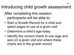


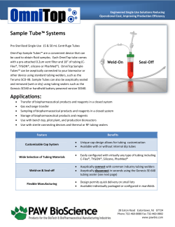


© Copyright 2026