
Resource Letter: LBOT-1: Laser-based optical tweezers Matthew J. Lang
RESOURCE LETTER Roger H. Stuewer, Editor School of Physics and Astronomy, 116 Church Street SE, University of Minnesota, Minneapolis, Minnesota 55455 This is one of a series of Resource Letters on different topics intended to guide college physicists, astronomers, and other scientists to some of the literature and other teaching aids that may help improve course content in specified fields. 关The letter E after an item indicates elementary level or material of general interest to persons becoming informed in the field. The letter I, for intermediate level, indicates material of somewhat more specialized nature; and the letter A indicates rather specialized or advanced material.兴 No Resource Letter is meant to be exhaustive and complete; in time there may be more than one letter on some of the main subjects of interest. Comments on these materials as well as suggestions for future topics will be welcomed. Please send such communications to Professor Roger H. Stuewer, Editor, AAPT Resource Letters, School of Physics and Astronomy, University of Minnesota, 116 Church Street SE, Minneapolis, MN 55455; e-mail: [email protected]. Resource Letter: LBOT-1: Laser-based optical tweezers Matthew J. Langa) and Steven M. Block Department of Biological Sciences and Department of Applied Physics, Stanford University, Stanford, California 94305-5020 共Received 20 August 2002; accepted 30 October 2002兲 This Resource Letter provides a guide to the literature on optical tweezers, also known as laser-based, gradient-force optical traps. Journal articles and books are cited for the following main topics: general papers on optical tweezers, trapping instrument design, optical detection methods, optical trapping theory, mechanical measurements, single molecule studies, and sections on biological motors, cellular measurements and additional applications of optical tweezers. © 2003 American Association of Physics Teachers. 关DOI: 10.1119/1.1532323兴 I. INTRODUCTION The field of optical tweezers has enjoyed a wide range of applications since its inception in the early 1970s. By using light to trap microscopic objects noninvasively, optical tweezers provide a flexible tool for ultrafine positioning, measurement, and control. In practice, forces up to 200 pN or thereabouts may be applied with sub-pN resolution on objects whose characteristic dimensions are similar to the wavelength of light. Particle positioning and detection capabilities are therefore on a spatial scale of micrometers down to angstroms. The emerging applications of laser-based optical traps are quite diverse and extensive, ranging from atomic physics to the medical sciences. As a result, optical tweezers have been a focal point for interdisciplinary science. Trapping apparatus ranges from simple, lens-based traps to complex instrumentation integrating multiple optical technologies. A variety of novel techniques have been developed for rapid position detection, trap stiffness determination, and applying controlled, calibrated forces. Instrument advances, such as the use of multiple laser beams, computerized automation of laser beams and sample positioning, and optical tweezers used in combination with other methodologies, such as fluorescence spectroscopy, micropipettes, and optical microbeams, have all helped to make optical tweezers an extremely versatile tool. a兲 Current address: Biological Engineering Division and Department of Mechanical Engineering, Massachusetts Institute of Technology, Cambridge, MA 02139. Electronic mail: [email protected] 201 Am. J. Phys. 71 共3兲, March 2003 http://ojps.aip.org/ajp/ Owing to their exquisitely controllable force-exerting properties, optical tweezers are useful for a variety of nanomechanical measurements, particularly those with biological applications. Objects such as biopolymers 共e.g., microtubules, DNA molecules兲, lipid membranes, intact or fractionated cells, and single biological macromolecules have all been studied successfully with optical tweezers. There are many broad areas of current research in biophysics, including the mechanical unfolding and refolding of proteins or nucleic acids, the strength of receptor-ligand bonding interactions, and the nanoscale mechanics of biological motors, which are especially well suited to work with optical tweezers. Optical tweezers are also useful purely as manipulators and positioning devices. Tweezers can be used to confine or constrain microscopic objects, as well as to organize, assemble, locate, or modify them. In addition to studies of single proteins, biological applications such as intracellular particle tracking and positioning, selective cell harvesting, and probing the mechanics of cell membranes have all been pursued with vigor. Laser-based tweezers also have been used to study the interactions of many-particle systems, e.g., colloids and quasi-crystals. A full theory of optical tweezers, covering the full range of spatial scales and levels of sophistication, has evolved comparatively slowly over the years, and lags somewhat behind experimental work at the present. Variations in the size, shape, and composition of trapped objects, the nonuniformity of the trapping light distribution, the fact that dimensions of trapped objects are often comparable to the wavelength of © 2003 American Association of Physics Teachers 201 light, combined with the large numerical apertures employed 共which preclude scalar paraxial approximations, necessitating a full vector treatment兲, have all conspired to make general theories difficult to develop. However, there has been much current progress, and many papers combine limited aspects of trapping theory with experiment. Our goal for this Resource Letter is to provide a guide to the literature. Our strategy has been to organize selected papers into a few main categories, rather than to provide a comprehensive review of all literature. Thus, numerous articles were omitted, some of which can be found among the citations papers in the papers we list. We apologize to colleagues whose work may thereby have been underrepresented. Inevitably, some of the literature can be classified under multiple categories. Therefore, we strongly encourage reader to browse related titles and topics. For example, sections of research reports frequently include design details not necessarily covered in specific instrument papers. We present a general section on optical tweezers first, including books and reviews on the subject. However, we caution readers that this is a fast-moving area, and much of the material found in books and early reviews is not particularly up-to-date. A focus on the earliest literature follows, including the seminal papers on optical tweezers. Papers relevant to optical instrument construction, calibration, and detection are listed next, followed by papers that deal mainly with optical trapping theory. The remaining sections are geared towards specific biological applications, including uses with cells, molecular motors, and additional applications of optical tweezers. II. JOURNALS The following are selected journals carrying articles on optical tweezers: Applied Optics Applied Physics Letters Biophysical Journal Cytometry Experimental Cell Research Fertility and Sterility Human Reproduction Journal of Applied Physics Journal of Modern Optics Methods in Cell Biology Nature Optics Letters Physical Review Letters Proceedings of the National Academy of Sciences Science III. BOOKS, REVIEWS, AND GENERAL PAPERS 1. ‘‘Laser Tweezers in Cell Biology,’’ M. P. Sheetz, in Methods in Cell Biology, Vol. 55, edited by L. Wilson and P. Matsudaira 共Academic, San Diego, 1998兲. Includes a number of topics in laser tweezers and applications. 共I,A,E兲 2. ‘‘Optical Tweezers: A New Tool for Biophysics,’’ S. M. Block, in Noninvasive Techniques in Cell Biology, edited by B. H. Satir 共Wiley-Liss, New York, 1990兲, pp. 375– 402. The working principle of optical tweezers is described, including details on instrument construction. Examples of trapped cells and inner structures are presented. A good all-around introduction. 共E兲 3. ‘‘Laser Manipulations of Atoms and Particles,’’ S. Chu, Science 253, 861– 866 共1991兲. Discussion of applications to atoms and particles. 共E兲 202 Am. J. Phys., Vol. 71, No. 3, March 2003 4. ‘‘Making light work with optical tweezers,’’ S. M. Block, Nature 360 共6403兲, 493– 495 共1992兲. A short review with basic principles. 共E兲 5. ‘‘Laser Trapping of Neutral Particles,’’ S. Chu, Sci. Am. 268 共2兲, 71–76 共1992兲. Good introduction to trapping capabilities. 共E兲 6. ‘‘Optical tweezers in cell biology,’’ S. C. Kuo and M. P. Sheetz, Trends Cell Biol. 2, 116 –118 共1992兲. A short review. 共E兲 7. ‘‘Optical Tweezers: Glasperlenspiel—II,’’ R. M. Simmons and J. T. Finer, Curr. Biol. 3 共5兲, 309–311 共1993兲. A general discussion of optical tweezers is provided. 共E兲 8. ‘‘Biological applications of optical forces,’’ K. Svoboda and S. M. Block, Annu. Rev. Biophys. Biomol. Struct. 23, 247–285 共1994兲. This review provides a good foundation for general understanding of optical tweezers for the serious reader. Includes an introduction to instrument construction, trapping theory, calibration and detection methods, and a table of objective transmittances in the near infrared. 共I兲 9. ‘‘Optical trapping and manipulation of microscopic particles and biological cells by laser beams,’’ S. Sato and H. Inaba, Opt. Quantum Electron. 28, 1–16 共1996兲. Review of basic principles and features of single beam optical trapping of cells, latex spheres, crystals, and metal particles. A review. 共E兲 10. ‘‘Optical trapping and manipulation of neutral particles using lasers,’’ A. Ashkin, Proc. Natl. Acad. Sci. USA 94 共10兲, 4853– 4860 共1997兲. Outlines the history and recent developments of optical trapping. 共I兲 11. ‘‘Laser scissors and tweezers,’’ M. W. Berns, Sci. Am. 278 共4兲, 62– 67 共1998兲. 共E兲 12. ‘‘Versatile optical traps with feedback control,’’ K. Visscher and S. M. Block, Methods Enzymol. 298, 460– 489 共1998兲. 共I兲 13. ‘‘Single-molecule biomechanics with optical methods,’’ A. D. Mehta, M. Rief, J. A. Spudich, D. A. Smith, and R. M. Simmons, Science 283 共5408兲, 1689–1695 共1999兲. A review that describes a number of single molecule methods using optical tweezers. 共E兲 14. ‘‘History of optical trapping and manipulation of small-neutral particle, atoms, and molecules,’’ A. Ashkin, IEEE J. Sel. Top. Quantum Electron 6 共6兲, 841– 856 共2000兲. A review. 共I兲 15. ‘‘Single molecule nanomanipulation of biomolecules,’’ Y. Ishii, A. Ishijima, and T. Yanagida, Trends Biotechnol. 19 共6兲, 211–216 共2001兲. A good introduction to combined single molecule imaging and manipulation techniques for the study of molecular motors. 共E兲 16. ‘‘Single molecule nanobioscience,’’ A. Ishijima and T. Yanagida, Trends Biochem. Sci. 26 共7兲, 438 – 444 共2001兲. Exciting advances in single molecule fluorescence and manipulation methods including combined SMF with optical tweezers are reviewed. 共E兲 17. ‘‘Using optics to measure biological forces and mechanics,’’ S. C. Kuo, Traffic 2 共11兲, 757–763 共2001兲. A review including optical stretching. 共I兲 IV. OPTICAL TWEEZERS, CURRENT RESEARCH TOPICS A. Earlier works on radiation pressure 18. ‘‘Optical levitation by radiation pressure,’’ A. Ashkin and J. M. Dziedzic, Appl. Phys. Lett. 19, 283–285 共1971兲. Glass spheres are levitated with radiation pressure in air and vacuum. 共I兲 19. ‘‘Acceleration and trapping of particles by radiation pressure,’’ A. Ashkin, Phys. Rev. Lett. 24 共4兲, 156 –159 共1970兲. The first observation of the acceleration of suspended particles using radiation pressure. 共I兲 20. ‘‘Optical Levitation of Liquid Drops by Radiation Pressure,’’ A. Ashkin and J. M. Dziedzic, Science 187 共4181兲, 1073–1075 共1975兲. Drops in the size range of 1 to 40 micrometers are levitated and manipulated with the trap. 共I兲 21. ‘‘Optical Levitation in High-Vacuum,’’ A. Ashkin and J. M. Dziedzic, Appl. Phys. Lett. 28 共6兲, 333–335 共1976兲. Optical levitation down to a pressure of 10⫺6 Torr was observed under high-vacuum. 共I兲 22. ‘‘Feedback Stabilization of Optically Levitated Particles,’’ A. Ashkin and J. M. Dziedzic, Appl. Phys. Lett. 30 共4兲, 202–204 共1977兲. 共I兲 23. ‘‘Trapping of atoms by resonance radiation pressure,’’ A. Ashkin, Phys. Rev. Lett. 40 共12兲, 729–732 共1978兲. A method for trapping, cooling, and manipulating sodium atoms is described. 共I兲 24. ‘‘Applications of Laser Radiation Pressure,’’ A. Ashkin, Science 210 共5兲, 1081–1088 共1980兲. Radiation pressure is discussed for neutral particles, including applications for microscopic particles and atoms. 共I兲 M. J. Lang and S. M. Block 202 25. ‘‘Observation of light scattering from nonspherical particles using optical levitation,’’ A. Ashkin and J. M. Dziedzic, Appl. Opt. 19 共5兲, 660– 668 共1980兲. Objects including spheroids, spherical doublets, triplets, etc. were studied. 共I兲 26. ‘‘Continuous-wave self-focusing and self-trapping of light in artificial Kerr media,’’ A. Ashkin, J. M. Dziedzic, and P. W. Smith, Opt. Lett. 7 共6兲, 276 –278 共1982兲. Beam trajectory and shapes arising from selftrapping are presented for laser modes exhibiting self-focusing in suspensions of submicroscopic particles. 共I兲 B. Seminal studies on optical tweezers 27. ‘‘Observation of a Single-Beam Gradient Force Optical Trap for Dielectric Particles,’’ A. Ashkin, J. M. Dziedzic, J. E. Bjorkholm, and S. Chu, Opt. Lett. 11 共5兲, 288 –290 共1986兲. This is the original paper describing the invention of optical tweezers. Trapping of particles ranging from 10 m to ⬃25 nm was observed in this single beam trap. 共I兲 28. ‘‘Optical trapping and manipulation of viruses and bacteria,’’ A. Ashkin and J. M. Dziedzic, Science 235 共4795兲, 1517–1520 共1987兲. Tobacco mosaic virus and single Escherichia coli bacteria. One of the first reports of biological applications of optical traps. 共E兲 29. ‘‘Optical trapping and manipulation of single cells using infrared laser beams,’’ A. Ashkin, J. M. Dziedzic, and T. Yamane, Nature 330 共6150兲, 769–771 共1987兲. One of the first reports of biological applications of optical trapping including the manipulation of particles within the cytoplasm of cells. 共E兲 C. Instrument design The most common and straightforward method of building optical tweezers instrumentation is to custom-fit an optical microscope that already incorporates imaging capabilities and a good objective lens used for forming a trap. Attention to stable instrument construction and alignment details will improve the usability of the instrument. When deciding where to place an instrument, minimizing room temperature variations, acoustical noise, and mechanical vibrations should all be considered. The references below describe a range of instruments from simple, single-beam traps to sophisticated multi-component systems. The incorporation of technologies in optical tweezers designs, frequently requiring ingenuity, has led to powerful new experimental methods. A broad range of components including trapping lasers, lenses, detection systems, calibration methods, and beam steering solutions has been incorporated into tweezers designs. Technologies for beam steering and multiple trap generation, including acoustooptic deflectors and galvanometer scanning mirrors, are outlined in some of the following papers. Computer control, automation, and data acquisition are critical components of optical tweezers experiments. The experimental requirements 共speed of a motor, required position sensitivity, force regime desired兲 should provide a guide for optimizing the design of an instrument. Multiple feedback methods for force and position clamping have been implemented. Note that many research papers, found in other sections of this Resource Letter, contain instrument design details outlined in materials and methods sections. 30. ‘‘Constructing optical tweezers,’’ S. M. Block, in Cell Biology: A Laboratory Manual, edited by D. Spector, R. Goldman, and L. Leinward 共Cold Spring Harbor, Cold Spring Harbor, NY, 1998兲. A good place to start. 共E兲 31. ‘‘Single beam optical trapping integrated in a confocal microscope for biological applications,’’ K. Visscher and G. J. Brakenhoff, Cytometry 12 共6兲, 486 – 491 共1991兲. Includes trapping theory, force calculation, and a description of the principle of trap manipulation by objective lens movement. 共I兲 203 Am. J. Phys., Vol. 71, No. 3, March 2003 32. ‘‘Optical tweezers using a diode laser,’’ R. S. Afzal and E. B. Treacy, Rev. Sci. Instrum. 63 共4兲, 2157–2163 共1992兲. Straightforward demonstration of using a diode laser to form an optical trap. 共E兲 33. ‘‘Optical-Trapping Micromanipulation Using 780-Nm Diode-Lasers,’’ T. C. B. Schut, E. F. Schipper, B. G. de Grooth, and J. Greve, Opt. Lett. 18 共6兲, 447– 449 共1993兲. 共E兲 34. ‘‘Micromanipulation by ‘multiple’ optical traps created by a single fast scanning trap integrated with the bilateral confocal scanning laser microscope,’’ K. Visscher, G. J. Brakenhoff, and J. J. Krol, Cytometry 14 共2兲, 105–114 共1993兲. Includes a description of the instrument with fast scanning by acousto-optic modulation and galvanometric scan mirrors. 共A兲 35. ‘‘Beam Magnification and the Efficiency of Optical Trapping with 790-nm AlGaAs Laser Diodes,’’ G. J. Escandon, Y. Liu, G. J. Sonek, and M. W. Berns, IEEE Photonics Technol. Lett. 6 共5兲, 597– 600 共1994兲. Discussion of trap efficiency with respect to input beam shape. Includes correction of diode output elipticity using anamorphic prisms. 共E兲 36. ‘‘Constructions and Applications of a Simple Optical Tweezers,’’ Y. C. Jong, H. M. Chen, J. H. Hsu, and W. S. Fann, Zool. Stu. 34 共S1兲, 209–210 共1995兲. 共E兲 37. ‘‘Construction of multiple-beam optical traps with nanometerresolution position sensing,’’ K. Visscher, S. P. Gross, and S. M. Block, IEEE J. Sel. Top. Quantum Electron. 2 共4兲, 1066 –1076 共1996兲. This paper discusses two types of optical tweezers instruments. Includes calibration methods, time-shared traps, instrument construction details, and a discussion of general desired features. 共I兲 38. ‘‘Quantitative measurements of force and displacement using an optical trap,’’ R. M. Simmons, J. T. Finer, S. Chu, and J. A. Spudich, Biophys. J. 70 共4兲, 1813–1822 共1996兲. Includes a schematic of the optical trap and detection system along with circuits with feedback arrangements. 共I兲 39. ‘‘Interferometric optical tweezers,’’ A. E. Chiou, W. Wang, G. J. Sonek, J. Hong, and M. W. Berns, Opt. Commun. 133 共1-6兲, 7–10 共1997兲. Two beams generate an interference fringe for trapping and micro-manipulation. 共I兲 40. ‘‘Design for fully steerable dual-trap optical tweezers,’’ E. Fallman and O. Axner, Appl. Opt. 36 共10兲, 2107–2113 共1997兲. A detailed recipe for the construction is provided. 共E兲 41. ‘‘Optical tweezers based on near infrared diode laser,’’ S. Grego, E. Arirnondo, and C. Frediani, J. Biomed. Opt. 2 共3兲, 332–339 共1997兲. A single-mode 100 mW diode operating at 840 nm was used. 共E兲 42. ‘‘Self-aligned dual-beam optical laser trap using photorefractive phase conjugation,’’ W. Wang, A. E. Chiou, G. J. Sonek, and M. W. Berns, J. Opt. Soc. Am. B 14 共4兲, 697–704 共1997兲. Phase conjugation in a crystal is used to form a dual trap in a counterpropagating arrangement. Includes a description of the instrument, theoretical analysis, and a performance comparison against a single beam trap. 共A兲 43. ‘‘Optical tweezer arrays and optical substrates created with diffractive optics,’’ E. R. Dufresne and D. G. Grier, Rev. Sci. Instrum. 69 共5兲, 1974 –1977 共1998兲. A diffractive optical element is used to create multiple optical tweezers from a single laser beam. 共I兲 44. ‘‘Inexpensive optical tweezers for undergraduate laboratories,’’ S. P. Smith, S. R. Bhalotra, A. L. Brody, B. L. Brown, E. K. Boyda, and M. Prentiss, Am. J. Phys. 67 共1兲, 26 –35 共1999兲. General introduction to setting up an instrument. 共E兲 45. ‘‘Optical tweezers for confocal microscopy,’’ A. Hoffmann, G. Meyer zu Horste, G. Pilarczyk, S. Monajembashi, V. Uhl, and K. O. Greulich, Appl. Phys. B 71 共5兲, 747–753 共2000兲. A method is presented to keep the trap fixed while doing 3D z-sectioning imaging. 共I兲 46. ‘‘Multi-functional optical tweezers using computer-generated holograms,’’ J. Liesener, M. Reicherter, T. Haist, and H. J. Tiziani, Opt. Commun. 185 共1-3兲, 77– 82 共2000兲. Seven spheres are trapped independently. 共I兲 47. ‘‘Design of a scanning laser optical trap for multiparticle manipulation,’’ C. Mio, T. Gong, A. Terray, and D. W. M. Marr, Rev. Sci. Instrum. 71 共5兲, 2196 –2200 共2000兲. Scanning is achieved using a piezo-actuated mirror. Details of the experimental arrangement and demonstration of trapping of multiple particles simultaneously is provided. 共I兲 48. ‘‘Construction of an optical tweezers: Calculation and experiments,’’ W. Sun, Y. Q. Wang, and C. M. Gao, Chin. Phys. 9 共11兲, 855– 860 共2000兲. 共I兲 49. ‘‘An integrated laser trap/flow control video microscope for the study M. J. Lang and S. M. Block 203 of single biomolecules,’’ G. J. L. Wuite, R. J. Davenport, A. Rappaport, and C. Bustamante, Biophys. J. 79 共2兲, 1155–1167 共2000兲. Detailed description of an instrument that combines optical tweezers and micropipettes to perform experiments deep within a flow chamber. Video microscopy and deflection are used for detection. Forces are applied with optical tweezers and a computer-controlled flow system. Used to study the transcription of RNA polymerase. 共A兲 50. ‘‘Computer-generated holographic optical tweezer arrays,’’ E. R. Dufresne, G. C. Spalding, M. T. Dearing, S. A. Sheets, and D. G. Grier, Rev. Sci. Instrum. 72 共3兲, 1810–1816 共2001兲. An adaptive-additive algorithm method is presented for creating planar arrays of holographic optical tweezers. 共A兲 51. ‘‘Design and construction of a space-borne optical tweezer apparatus,’’ A. Resnick, Rev. Sci. Instrum. 72 共11兲, 4059– 4065 共2001兲. Optical tweezers in space; a rugged design is detailed. 共E兲 52. ‘‘An Automated 2D Force Clamp for Single Molecule Studies,’’ M. J. Lang, C. L. Asbury, J. W. Shaevitz, and S. M. Block, Biophys. J. 83, 491–501 共2002兲. This paper describes a fully-automated trapping instrument used as a two-dimensional force clamp for kinesin motility mesurements. The instrument is capable of simultaneous optical tweezers and single molecule fluorescence. 共I兲 1. Detection method: video, quadrant photodiode, interferometry, and others Position detection may be achieved in many ways including video, quadrant photodiode, and interferometric methods. Time response and position sensitivity should be considered when deciding on a detection method. Video microscopy is straightforward and can be used to track a particle with subpixel resolution. Video detection has limited time response and is not as convenient for systems requiring fast positional feedback. Quadrant photodiodes, placed in either an image or back focal plane, can be used for two- or threedimensional position sensing. Quadrant-photodiode detection, which in some instances utilizes a separate detector beam for convenience, has both a faster time response and greater position sensitivity. Interferometry is another sensitive position-sensing method that is used to detect displacement along one axis. 53. ‘‘Direct Measurement of Nanometric Displacement under an Optical Microscope,’’ S. Kamimura, Appl. Opt. 26 共16兲, 3425–3427 共1987兲. 共I兲 54. ‘‘Optical measurement of picometer displacements of transparent microscopic objects,’’ W. Denk and W. W. Webb, Appl. Opt. 29 共16兲, 2382–2391 共1990兲. Description of the instrument and interferometer including signal detection and amplification circuitry. 共A兲 55. ‘‘High-resolution axial and lateral position sensing using two-photon excitation of fluorophores by a continuous-wave Nd:YAG laser,’’ E.-L. Florin, J. K. H. Horber, and E. H. K. Stelzer, Appl. Phys. Lett. 69 共4兲, 446 – 448 共1996兲. Changes in fluorescence due to displacement are used as a spatial sensor. Includes a fluorescence intensity versus z-position graph. 共I兲 56. ‘‘Determination of the force constant of a single-beam gradient trap by measurement of backscattered light,’’ M. E. J. Friese, H. RubinszteinDunlop, N. R. Heckenberg, and E. W. Dearden, Appl. Opt. 35 共36兲, 7112–7116 共1996兲. Model of the trap as a harmonic oscillator with measurements. 共I兲 57. ‘‘Improved nm displacement detector for microscopic beads at frequencies below 10 Hz,’’ D. Q. Li and B. J. Schnapp, Rev. Sci. Instrum. 68 共5兲, 2195–2199 共1997兲. Laser interferometry detection is outlined in this paper. 共E兲 58. ‘‘Detection of single-molecule interactions using correlated thermal diffusion,’’ A. D. Mehta, J. T. Finer, and J. A. Spudich, Proc. Natl. Acad. Sci. U.S.A. 94 共15兲, 7927–7931 共1997兲. Methods are described to detect and correlate the motion of optically trapped beads attached to both ends of a single actin filament. 共I兲 59. ‘‘Three-dimensional potential analysis of radiation pressure exerted on a single microparticle,’’ K. Sasaki, M. Tsukima, and H. Masuhara, Appl. Phys. Lett. 71 共1兲, 37–39 共1997兲. Total internal reflection microscopy is used in this three-dimensional position sensing method. 共I兲 204 Am. J. Phys., Vol. 71, No. 3, March 2003 60. ‘‘Interference model for back-focal-plane displacement detection in optical tweezers,’’ F. Gittes and C. F. Schmidt, Opt. Lett. 23 共1兲, 7–9 共1998兲. Description including a model comparison with experiment for the signal of back-focal-plane imaging using a quadrant photodiode. 共A兲 61. ‘‘Three dimensional single-particle tracking with nanometer resolution,’’ I. M. Peters, B. G. de Grooth, J. M. Schins, C. G. Figdor, and J. Greve, Rev. Sci. Instrum. 69 共7兲, 2762–2766 共1998兲. Axial position sensitivity is achieved by positioning a photodiode so that it captures on the order of half of the transmitted light intensity. A feedback system controls the position of the collection objective using a piezo tube. 共I兲 62. ‘‘Three-dimensional imaging with optical tweezers,’’ M. E. J. Friese, A. G. Truscott, H. Rubinsztein-Dunlop, and N. R. Heckenberg, Appl. Opt. 38 共31兲, 6597– 6603 共1999兲. This paper reports that features of approximately 200 nm can be resolved with a sensitivity of 5 nm. 共I兲 63. ‘‘Nanometer-displacement detection of optically trapped metallic particles based on critical angle method for small force detection,’’ E. Higurashi, R. Sawada, and T. Ito, Rev. Sci. Instrum. 70 共7兲, 3068 – 3073 共1999兲. Detection is based on critical-angle prisms where angle changes originating from trapped particle motion provide a sensitive measure of position. 共I兲 64. ‘‘3D single-particle tracking and optical trap measurements on adhesion proteins,’’ I. M. Peters, Y. vanKooyk, S. J. van Vliet, B. G. de Grooth, C. G. Figdor, and J. Greve, Cytometry 36 共3兲, 189–194 共1999兲. Cell adhesion studies. 共I兲 65. ‘‘Three-dimensional high-resolution particle tracking for optical tweezers by forward scattered light,’’ A. Pralle, M. Prummer, E. L. Florin, E. H. K. Stelzer, and J. K. H. Horber, Microsc. Res. Tech. 44 共5兲, 378 –386 共1999兲. The ratio of the intensity of scattered light to the total amount of light is used for axial position determination. A model for the position signal is presented. 共A兲 2. Calibration The force exerted on an object by an optical trap depends both on the trap 共shape and intensity兲 and the object 共size and composition兲. Detailed knowledge of the force exerted on a particle is a critical quantity in biochemical, kinetic, and mechanical trapping experiments. Force calibration is achieved by a number of methods, each with different advantages. The drag or escape force method is performed by moving an object or stage while monitoring an ‘‘escape’’ velocity, and is particularly useful to check the linearity of trapping potential in regions far from the trap center. The equipartition method, which is straightforward and fast, measures thermal fluctuations in position of a trapped particle. The power spectral method provides stiffness information in addition to a diagnostic for noise sources at various frequencies. In addition to the methods having different advantages, multiple methods provide a good consistency check of the overall trap stiffness. 66. ‘‘Optical binding,’’ M. M. Burns, J. M. Fournier, and J. A. Golovchenko, Phys. Rev. Lett. 63 共12兲, 1233–1236 共1989兲. Longrange bound states of plastic spheres, detection using fringe spacing, in the presence of an optical field are presented. Fringe spacing is used for detection. 共I兲 67. ‘‘Measurement of small forces using an optical trap,’’ L. P. Ghislain, N. A. Switz, and W. W. Webb, Rev. Sci. Instrum. 65, 2762–2768 共1994兲. Includes calibration using drag force and signal source considerations. 共A兲 68. ‘‘Calibration of Light Forces in Optical Tweezers,’’ H. Felgner, O. Muller, and M. Schliwa, Appl. Opt. 34 共6兲, 977–982 共1995兲. It is shown that trapping in different axial positions is possible. 共I兲 69. ‘‘Optical Trapping and Fluorescence Detection in Laminar-Flow Streams,’’ W. Wang, Y. Liu, G. J. Sonek, M. W. Berns, and R. A. Keller, Appl. Phys. Lett. 67 共8兲, 1057–1059 共1995兲. Escape velocities are measured relative to the position within the flow stream. 共A兲 70. ‘‘Three-dimensional optical trapping and evanescent wave light scattering for direct measurement of long range forces between a colloidal particle and a surface,’’ A. R. Clapp, A. G. Ruta, and R. B. Dickinson, M. J. Lang and S. M. Block 204 Rev. Sci. Instrum. 70 共6兲, 2627–2636 共1999兲. Total internal reflection of a laser beam creates an evanescent wave that is used to determine particle position. 共I兲 71. ‘‘Three-dimensional force calibration of optical tweezers,’’ W. Singer, S. Bernet, N. Hecker, and M. RitschMarte, J. Mod. Opt. 47 共14 –15兲, 2921–2931 共2000兲. 共I兲 72. ‘‘Optical tweezers as sub-pico-newton force transducers,’’ C. C. Huang, C. F. Wang, D. S. Mehta, and A. Chiou, Opt. Commun. 195 共1– 4兲, 41– 48 共2001兲. A drag method was used to determine the trapping force, including a weak probing beam. 共I兲 3. Fiber-based traps Light exiting from a fiber, because of its steep spatial gradient, can be used to trap objects, provided that the repulsive scattering force is more than balanced. The most common fiber-based trap involves two counter-propagating beams, to neutralize the scattering force in the central region. Because there are no local lenses, fiber-based traps have the advantage of being able to penetrate deep into solution. Fiberbased traps have also been used for cell stretching studies. 73. ‘‘Demonstration of a fiberoptic light-force trap,’’ A. Constable, J. Kim, J. Mervis, F. Zarinetchi, and M. Prentiss, Opt. Lett. 18 共21兲, 1867– 1869 共1993兲. Single-mode fibers. Includes sample cell construction information. 共I兲 74. ‘‘Confinement and bistability in a tapered hemispherically lensed optical fiber trap,’’ E. R. Lyons and G. J. Sonek, Appl. Phys. Lett. 66 共13兲, 1584 –1586 共1995兲. Axial and transverse trap efficiencies are predicted and confirmed. 共I兲 75. ‘‘Trapping forces in a multiple-beam fiber-optic trap,’’ E. Sidick, S. D. Collins, and A. Knoesen, Appl. Opt. 36 共25兲, 6423– 6433 共1997兲. Forces are calculated for microspheres located both on and off axis relative to the beam axis. 共A兲 76. ‘‘Microinstrument gradient-force optical trap,’’ S. D. Collins, R. J. Baskin, and D. G. Howitt, Appl. Opt. 38 共28兲, 6068 – 6074 共1999兲. Consists of four single-mode fibers. 共I兲 77. ‘‘Laser guidance and trapping of mesoscale particles in hollow-core optical fibers,’’ M. J. Renn, R. Pastel, and H. J. Lewandowski, Phys. Rev. Lett. 82 共7兲, 1574 –1577 共1999兲. Laser light coupled into a hollow-core fiber can be used to trap and guide mesoscale particles over relatively long distances. 共I兲 78. ‘‘The optical stretcher: a novel laser tool to micromanipulate cells,’’ J. Guck, R. Ananthakrishnan, H. Mahmood, T. J. Moon, C. C. Cunningham, and J. Kas, Biophys. J. 81 共2兲, 767–784 共2001兲. Objects trapped between two fibers are stretched in a nonfocused geometry. 共I兲 D. Theory of optical tweezers A wide range of models and degrees of sophistication have been applied to the theory of optical tweezers. The size, shape, and composition of an object are important quantities when determining an appropriate theory. Laser focusing properties such as the mode, input beam diameter, and numerical aperture of the lens are also critical. Theories have been developed for describing the expected signal detection shapes. Many of the references below include both theory and experiments. 79. ‘‘Internal and near-surface electromagnetic fields for a spherical particle irradiated by a focused light beam,’’ J. P. Barton, J. Appl. Phys. 64 共4兲, 1632–1639 共1988兲. 共I兲 80. ‘‘Fifth-order corrected electromagnetic field components for a fundamental Gaussian beam,’’ J. P. Barton and D. R. Alexander, J. Appl. Phys. 66 共7兲, 2800–2802 共1989兲. 共I兲 81. ‘‘Theoretical determination of net radiation force and torque for a spherical particle illuminated by a focused laser beam,’’ J. P. Barton, D. R. Alexander, and S. A. Schaub, J. Appl. Phys. 66 共10兲, 4594 – 4602 共1989兲. Calculations for a 5 m diameter water droplet levitated in air. 共A兲 205 Am. J. Phys., Vol. 71, No. 3, March 2003 82. ‘‘Forces of a Single-Beam Gradient Laser Trap on a Dielectric Sphere in the Ray Optics Regime,’’ A. Ashkin, Biophys. J. 61 共2兲, 569–582 共1992兲. Includes the effect of index of refraction on the performance of a trap. 共A兲 83. ‘‘Calculation of the Trapping Force in a Strongly Focused LaserBeam,’’ R. Gussgard, T. Lindmo, and I. Brevik, J. Opt. Soc. Am. B 9 共10兲, 1922–1930 共1992兲. 共I兲 84. ‘‘Theoretical study of optically induced forces on spherical particles in a single beam trap I: Rayleigh scatterers,’’ K. Visscher and G. J. Brakenhoff, Optik 89 共2兲, 174 –180 共1992兲. Uses electromagnetic diffraction theory. 共I兲 85. ‘‘Theoretical study of optically induced forces on spherical particles in a single beam trap II: Mie scatterers,’’ K. Visscher and G. J. Brakenhoff, Optik 90 共2兲, 57– 60 共1992兲. Vector diffraction theory of Mie scatterers. 共A兲 86. ‘‘Radiation trapping forces on microspheres with optical tweezers,’’ W. H. Wright, G. J. Sonek, and M. W. Berns, Appl. Phys. Lett. 63 共6兲, 715–717 共1993兲. Forces are predicted and compared, for spheres of various size and composition, with experimental measurements. 共I兲 87. ‘‘Radiation Pressure Forces Exerted on a Particle Arbitrarily Located in a Gaussian-Beam by Using the Generalized Lorenz-Mie Theory, and Associated Resonance Effects,’’ K. F. Ren, G. Greha, and G. Gouesbet, Opt. Commun. 108 共4 – 6兲, 343–354 共1994兲. 共A兲 88. ‘‘Parametric study of the forces on microspheres held by optical tweezers,’’ W. H. Wright, G. J. Sonek, and M. W. Berns, Appl. Opt. 33 共9兲, 1735–1748 共1994兲. Includes theory and tables of trapping parameters through various microscope objectives and trapping efficiencies for different sizes and types of spheres and input polarizations. 共A兲 89. ‘‘Radiation Forces on a Micrometer-Sized Sphere in an Evanescent Field,’’ E. Almaas and I. Brevik, J. Opt. Soc. Am. B 12 共12兲, 2429– 2438 共1995兲. Theoretical investigation used to predict the radiation forces in an evanescent wave of given polarization. 共A兲 90. ‘‘Radiation forces on a dielectric sphere in the Rayleigh scattering regime,’’ Y. Harada and T. Asakura, Opt. Commun. 124 共5– 6兲, 529– 541 共1996兲. 共I兲 91. ‘‘Theoretical determination of the influence of the polarization on forces exerted by optical tweezers,’’ T. Wohland, A. Rosin, and E. H. K. Stelzer, Optik 102 共4兲, 181–190 共1996兲. Significant differences in lateral forces can occur depending on the polarization orientation. 共A兲 92. ‘‘Thermal noise limitations on micromechanical experiments,’’ F. Gittes and C. F. Schmidt, Eur. Biophys. J. with Biophys. Lett 27 共1兲, 75– 81 共1998兲. Strategies for maximizing signal-to-noise ratios are investigated theoretically. Includes experimental examples. 共I兲 93. ‘‘Dynamics and dynamic light scattering properties of Brownian particles under laser radiation pressure,’’ Y. Harada and T. Asakura, Pure Appl. Opt. 7 共5兲, 1001–1012 共1998兲. 共A兲 94. ‘‘Localized dynamic light scattering: A new approach to dynamic measurements in optical microscopy,’’ A. Meller, R. Bar-Ziv, T. Tlusty, E. Moses, J. Stavans, and S. A. Safran, Biophys. J. 74 共3兲, 1541–1548 共1998兲. The force constants for a single bead and pair of beads are calculated. 共A兲 95. ‘‘Heating by absorption in the focus of an objective lens,’’ A. Schonle and S. W. Hell, Opt. Lett. 23 共5兲, 325–327 共1998兲. Numerical results for local heating under typical optical trapping conditions are presented. 共I兲 96. ‘‘Optical gradient forces of strongly localized fields,’’ T. Tlusty, A. Meller, and R. Bar-Ziv, Phys. Rev. Lett. 81 共8兲, 1738 –1741 共1998兲. Predictions for force-dependent curves, maximal trapping forces, and force constants are derived. 共I兲 97. ‘‘Optical forces on microparticles in an evanescent laser field,’’ M. Lester and M. Nieto-Vesperinas, Opt. Lett. 24 共14兲, 936 –938 共1999兲. Theory of forces in an evanescent laser field. 共A兲 98. ‘‘Optical traps as force transducers: The effects of focusing the trapping beam through a dielectric interface,’’ A. C. Dogariu and R. Rajagopalan, Langmuir 16 共6兲, 2770–2778 共2000兲. Trap forces are calculated using wave optics calculations. Includes the identification of secondary traps. 共A兲 99. ‘‘Analysis of the scattered light distribution of a tightly focused laser beam by a particle near a substrate,’’ W. Inami and Y. Kawata, J. Appl. Phys. 89 共11兲, 5876 –5880 共2001兲. Scattering fields near a substrate are calculated. 共I兲 100. ‘‘Calculation and optical measurement of laser trapping forces on non-spherical particles,’’ T. A. Nieminen, H. Rubinsztein-Dunlop, and N. R. Heckenberg, J. Quant. Spectrosc. Radiat. Transf. 70 共4 – 6兲, M. J. Lang and S. M. Block 205 627– 637 共2001兲. Includes calculations of force and torque. 共I兲 101. ‘‘Optical trapping of dielectric particles in arbitrary fields,’’ A. Rohrbach and E. H. K. Stelzer, J. Opt. Soc. Am. A 18 共4兲, 839– 853 共2001兲. In-depth study of calculating trapping potentials. 共A兲 102. ‘‘Optical trapping near-resonance absorption,’’ R. Agayan, F. Gittes, R. Kopelman, and C. F. Schmidt, S. P. I. E. 共Int. Soc. Opt. Eng.兲 Proc. 4431, 341–351 共2001兲. Theory and analysis for enhanced trapping forces of near-resonant frequencies are presented. 共A兲 112. 113. E. Experiments using optical tweezers 114. 1. Mechanical and single molecule measurements Mechanical properties such as elasticity, stiffness, rigidity, and torque can be measured using optical tweezers. Light is easily manipulated and relatively noninvasive, making laserbased mechanical measurements straightforward for studying biological systems. Cells, intracellular structures, filaments, and single molecules have all been probed. Multiple traps can be used to construct additional geometries for mechanical measurements. Combinations of optical tweezers and other methods, such as micropipettes, fluorescence microscopy, and microsurgery, provide very powerful tools for studying biological systems. Single molecule mechanical measurements using optical tweezers, including biological motor motility, protein-protein unbinding, and protein unfolding, have experienced a tremendous growth in recent years. Throughout these papers, assay development remains a critical component, including details of slide/flow cell construction, methods for attaching samples to microspheres, and general assay conditions. 103. ‘‘Buckling of a Single Microtubule by Optical Trapping Forces: Direct Measurement of Microtubule Rigidity,’’ M. Kurachi, M. Hoshi, and H. Tashiro, Cell Motil. Cytoskeleton 30 共3兲, 221–228 共1995兲. The microtubule rigidity was found to be dependent on length. 共I兲 104. ‘‘Biased diffusion; optical trapping; and manipulation of single molecules in solution,’’ D. T. Chiu and R. N. Zare, J. Am. Chem. Soc. 118 共27兲, 6512– 6513 共1996兲. 共I兲 105. ‘‘Flexural rigidity of microtubules measured with the use of optical tweezers,’’ H. Felgner, R. Frank, and M. Schliwa, J. Cell Sci. 109 共2兲, 509–516 共1996兲. Microtubules are actively moved with optical tweezers while the microtubule shape is observed. 共I兲 106. ‘‘Strength and lifetime of the bond between actin and skeletal muscle alpha-actinin studied with an optical trapping technique,’’ H. Miyata, R. Yasuda, and K. Kinosita, Jr., Biochim. Biophys. Acta 1290 共1兲, 83– 88 共1996兲. Suggestion of two classes of actin-actinin bonds, based on unbinding time measurements. 共I兲 107. ‘‘Optical tweezers for single molecule mechanics,’’ R. M. Simmons, J. A. Sleep, A. Trombetta, and P. Marya, in Nanotechnology in Medicine and the Biosciences, Developments in Nanotechnology, edited by R. R. H. Coombs and D. W. Robinson 共Gordon & Breach, The Netherlands, 1996兲. A short book chapter that includes a design for studying actin myosin muscle proteins. 共E兲 108. ‘‘Torsional rigidity of single actin filaments and actin-actin bond breaking force under torsion measured directly by in vitro micromanipulation,’’ Y. Tsuda, H. Yasutake, A. Ishijima, and T. Yanagida, Proc. Natl. Acad. Sci. U.S.A. 93 共23兲, 12937–12942 共1996兲. This study includes the use of optical tweezers and fluorescent beads to measure rotational Brownian motion. 共I兲 109. ‘‘Injection of ultrasmall samples and single molecules into tapered capillaries,’’ D. T. Chiu, A. Hsiao, A. Gaggar, R. A. GarzaLopez, O. Orwar, and R. N. Zare, Anal. Chem. 69 共10兲, 1801–1807 共1997兲. Optical tweezers place objects at or near the inlet of tapered capillaries. 共I兲 110. ‘‘Actin filament mechanics in the laser trap,’’ D. E. Dupuis, W. H. Guilford, J. Wu, and D. M. Warshaw, J. Muscle Res. Cell Motil. 18 共1兲, 17–30 共1997兲. Independent laser traps were used in this study to determine the compliance of actin filaments. 共I兲 111. ‘‘Imaging and nano-manipulation of single biomolecules,’’ T. Funatsu, Y. Harada, H. Higuchi, M. Tokunaga, K. Saito, Y. Ishii, R. D. 206 Am. J. Phys., Vol. 71, No. 3, March 2003 115. 116. 117. 118. 119. 120. 121. 122. 123. 124. 125. 126. Vale, and T. Yanagida, Biophys. Chem. 68 共1–3兲, 63–72 共1997兲. Early experiments with combined single molecule fluorescence and optical tweezers. 共I兲 ‘‘Folding-Unfolding Transitions in Single Titin Molecules Characterized with Laser Tweezers,’’ M. S. Z. Kellermayer, S. B. Smith, H. L. Granzier, and C. Bustamante, Science 276, 1112–1116 共1997兲. Includes unfolding and refolding curves. A combined micropipette and optical trap geometry is employed. 共I兲 ‘‘A method for determination of stiffness of collagen molecules,’’ Z. P. Luo, M. E. Bolander, and K. N. An, Biochem. Biophys. Res. Commun. 232 共1兲, 251–254 共1997兲. 共I兲 ‘‘Elasticity and unfolding of single molecules of the giant muscle protein titin,’’ L. Tskhovrebova, J. Trinick, J. A. Sleep, and R. M. Simmons, Nature 387 共6630兲, 308 –312 共1997兲. Single molecule mechanical experiment of titin with optical tweezers. 共I兲 ‘‘Complete unfolding of the titin molecule under external force,’’ M. S. Z. Kellermayer, S. B. Smith, C. Bustamante, and H. L. Granzier, J. Struct. Biol. 122 共1–2兲, 197–205 共1998兲. Titin molecules are stretched with forces above 400 pN. 共I兲 ‘‘Manipulation of single molecules in biology,’’ M. D. Wang, Curr. Opin. Biotechnol. 10 共1兲, 81– 86 共1999兲. Highlights optical tweezers single molecule motor assays. 共E兲 ‘‘Grabbing the cat by the tail: Manipulating molecules one by one,’’ C. Bustamante, J. C. Macosko, and G. J. L. Wuite, Nat. Rev. Mol. Cell Biol. 1 共2兲, 130–136 共2000兲. A review. 共E兲 ‘‘Measurement of the elasticity of the actin tail of Listeria monocytogenes,’’ F. Gerbal, V. Laurent, A. Ott, M. F. Carlier, P. Chaikin, and J. Prost, Eur. Biophys. J. with Biophys. Lett. 29 共2兲, 134 –140 共2000兲. 共I兲 ‘‘Short-term binding of fibroblasts to fibronectin: optical tweezers experiments and probabilistic analysis,’’ O. Thoumine, P. Kocian, A. Kottelat, and J. J. Meister, Eur. Biophys. J. with Biophys. Lett. 29 共6兲, 398 – 408 共2000兲. Adhesion tests of fibroblasts on fibronectincoated glass. 共I兲 ‘‘Force spectroscopy on single passive biomolecules and single biomolecular bonds,’’ R. Merkel, Phys. Rep. Phys. Lett. 346 共5兲, 344 – 385 共2001兲. A review. 共I兲 ‘‘Optical measurement of microscopic torques,’’ T. A. Nieminen, N. R. Heckenberg, and H. Rubinsztein-Dunlop, J. Mod. Opt. 48 共3兲, 405– 413 共2001兲. 共I兲 ‘‘Mechanical fatigue in repetitively stretched single molecules of titin,’’ M. S. Z. Kellermayer, S. B. Smith, C. Bustamante, and H. L. Granzier, Biophys. J. 80 共2兲, 852– 863 共2001兲. Optical tweezers were used to repetitively stretch and release titin to study mechanical fatigue. 共I兲 ‘‘Detection and characterization of individual intermolecular bonds using optical tweezers,’’ A. L. Stout, Biophys. J. 80 共6兲, 2976 –2986 共2001兲. Details of the instrument, technique and geometry for rupture force measurements are shown. 共I兲 ‘‘Stretching short biopolymers using optical tweezers,’’ Y. L. Sun, Z. P. Luo and K. N. An, Biochem. Biophys. Res. Commun. 286 共4兲, 826 – 830 共2001兲. The stiffness of procollagen molecules was studied. 共I兲 ‘‘Single molecule measurements of titin elasticity,’’ K. Wang, J. G. Forbes, and A. J. Jin, Prog. Biophys. Mol. Biol. 77 共1兲, 1– 44 共2001兲. A review. 共I兲 ‘‘Flexural rigidity of a single microtubule,’’ T. Takasone, S. Juodkazis, Y. Kawagishi, A. Yamaguchi, S. Matsuo, H. Sakakibara, H. Nakayama, and H. Misawa, Jpn. J. Appl. Phys. 41 共5兲, 3015–3019 共2002兲. Shear compression and bending stress on a microtubule was studied using optical tweezers. 共I兲 2. Biological motors Biological motors are excellent model systems for observing protein motions and conformational changes, and are a subject of intense research. Motor properties such as speed, force, processivity, working stroke distance, and substrate should be considered when designing an experiment. Many technological developments, including force clamping, the three-bead assay, and computer automation of trap and sample positioning have been used in biological motor reM. J. Lang and S. M. Block 206 search. We encourage the reader to explore experimental innovations implemented in multiple motor systems. a. General motors 127. ‘‘Molecular Motors: Structure, Mechanics and Energy Transduction,’’ edited by R. Cooke, Biophys. J. 68 共4兲, 1s–382s 共1995兲. This supplemental issue is a Biophysical Discussions conference proceedings dedicated to molecular motors, and contains several papers related to optical trapping studies. 共E兲 128. ‘‘Microscopic approaches to dynamics and structure of biological motors,’’ F. Gittes and C. F. Schmidt, Curr. Opin. Solid State Mater. Sci. 1 共3兲, 412– 424 共1996兲. A review. 共I兲 129. ‘‘Molecular motors: single-molecule mechanics,’’ R. Simmons, Curr. Biol. 6 共4兲, 392–394 共1996兲. 共E兲 130. ‘‘Real engines of creation,’’ S. M. Block, Nature 386 共6622兲, 217– 219 共1997兲. An introduction to the study of molecular motors with new biophysical techniques. 共E兲 131. ‘‘Force effects on biochemical kinetics,’’ S. Khan and M. P. Sheetz, Annu. Rev. Biochem. 66, 785– 805 共1997兲. Applications of force measurements to enzyme activity and motor proteins are presented including a comparison with other force measurement methods. 共I兲 132. ‘‘Two-dimensional tracking of ncd motility by back focal plane interferometry,’’ M. W. Allersma, F. Gittes, M. J. deCastro, R. J. Stewart, and C. F. Schmidt, Biophys. J. 74 共2兲, 1074 –1085 共1998兲. Includes 2D detection with a quadrant photodiode and theory. 共A兲 133. ‘‘Use of optical traps in single-molecule study of nonprocessive biological motors,’’ A. D. Mehta, J. T. Finer, and J. A. Spudich, Methods Enzymol. 298, 436 – 459 共1998兲. A review. 共I兲 134. ‘‘Dynein arms are oscillating force generators,’’ C. Shingyoji, H. Higuchi, M. Yoshimura, E. Katayama, and T. Yanagida, Nature 393 共6686兲, 711–714 共1998兲. 共I兲 135. ‘‘Developmental regulation of vesicle transport in Drosophila embryos: Forces and kinetics,’’ M. A. Welte, S. P. Gross, M. Postner, S. M. Block, and E. F. Wieschaus, Cell 92 共4兲, 547–557 共1998兲. 共I兲 136. ‘‘Biomechanics, one molecule at a time,’’ A. D. Mehta, M. Rief, and J. A. Spudich, J. Biol. Chem. 274 共21兲, 14517–14520 共1999兲. Focus on some single molecule optical tweezers measurements. 共E兲 137. ‘‘Working strokes by single molecules of the kinesin-related microtubule motor ncd,’’ M. J. deCastro, R. M. Fondecave, L. A. Clarke, C. F. Schmidt, and R. J. Stewart, Nat. Cell Biol. 2 共10兲, 724 –729 共2000兲. Uses a three-bead geometry to measure the working stroke of ncd. 共I兲 138. ‘‘Processive movement of single 22S dynein molecules occurs only at low ATP concentrations,’’ E. Hirakawa, H. Higuchi, and Y. Y. Toyoshima, Proc. Natl. Acad. Sci. U.S.A 97 共6兲, 2533–2537 共2000兲. Stepwise movement of dynein is shown. 共I兲 b. Kinesin Kinesin, which hydrolyzes ATP to move along microtubules, is a processive motor that takes about 100 steps before detaching. Kinesin’s processivity makes it ideal for optical tweezers studies. Optical tweezers measurements have identified that kinesin steps in discrete, 8 nm increments and hydrolyzes one ATP per step. Instrumental innovations specifically geared towards measuring kinesin motility have led to a number of advances in optical tweezers. 139. ‘‘Bead movement by single kinesin molecules studied with optical tweezers,’’ S. M. Block, L. S. Goldstein, and B. J. Schnapp, Nature 348 共6299兲, 348 –352 共1990兲. 共E兲 140. ‘‘Nucleotide-dependent single-to double-headed binding of kinesin,’’ K. Kawaguchi and S. Ishiwata, Science 291 共5504兲, 667– 669 共2001兲. Optical tweezers were used to measure the unbinding force of kinesin attached to microtubules under various nuclotide conditions. 共I兲 141. ‘‘Direct observation of kinesin stepping by optical trapping interferometry,’’ K. Svoboda, C. F. Schmidt, B. J. Schnapp, and S. M. Block, Nature 365 共6448兲, 721–727 共1993兲. Kinesin moves with 8 nm steps. 共I兲 142. ‘‘Force and velocity measured for single kinesin molecules,’’ K. Svoboda and S. M. Block, Cell 77 共5兲, 773–784 共1994兲. Optical trapping combined with interferometry. 共I兲 207 Am. J. Phys., Vol. 71, No. 3, March 2003 143. ‘‘Fluctuation Analysis of Motor Protein Movement and Single Enzyme-Kinetics,’’ K. Svoboda, P. P. Mitra, and S. M. Block, Proc. Natl. Acad. Sci. U.S.A. 91 共25兲, 11782–11786 共1994兲. 共I兲 144. ‘‘Nanometers and Piconewtons: The Macromolecular Mechanics of Kinesin,’’ S. M. Block, Trends Cell Biol. 5 共4兲, 169–175 共1995兲. Perspective on single motor assay measurements on kinesin. 共E兲 145. ‘‘Detection of sub-8-nm movements of kinesin by high-resolution optical-trap microscopy,’’ C. M. Coppin, J. T. Finer, J. A. Spudich, and R. D. Vale, Proc. Natl. Acad. Sci. U.S.A. 93 共5兲, 1913–1917 共1996兲. Kinesin steps of both 8 and ⬃5 nm are presented in this sensitive measurement. 共I兲 146. ‘‘The load dependence of kinesin’s mechanical cycle,’’ C. M. Coppin, D. W. Pierce, L. Hsu, and R. D. Vale, Proc. Natl. Acad. Sci. U.S.A. 94 共16兲, 8539– 8544 共1997兲. Forward loads of kinesin motility are presented in this paper. 共I兲 147. ‘‘Kinetics of force generation by single kinesin molecules activated by laser photolysis of caged ATP,’’ H. Higuchi, E. Muto, Y. Inoue, and T. Yanagida, Proc. Natl. Acad. Sci. U.S.A. 94 共9兲, 4395– 4400 共1997兲. Combination of optical tweezers and UV photolysis of caged compounds. 共I兲 148. ‘‘Movements of truncated kinesin fragments with a short or an artificial flexible neck,’’ Y. Inoue, Y. Y. Toyoshima, A. H. Iwane, S. Morimoto, H. Higuchi, and T. Yanagida, Proc. Natl. Acad. Sci. U.S.A. 94 共14兲, 7275–7280 共1997兲. Stepwise movement of kinesin observed with optical tweezers. 共I兲 149. ‘‘Mechanics of single kinesin molecules measured by optical trapping nanometry,’’ H. Kojima, E. Muto, H. Higuchi, and T. Yanagida, Biophys. J. 73 共4兲, 2012–2022 共1997兲. Includes compliance correction to determine the displacement. The measurement was performed on axonemes. 共I兲 150. ‘‘Coupled chemical and mechanical reaction steps in a processive Neurospora kinesin,’’ I. Crevel, N. Carter, M. Schliwa, and R. Cross, EMBO J. 18 共21兲, 5863–5872 共1999兲. Shows kinesin stepping. 共I兲 151. ‘‘Mechanical and chemical properties of cysteine-modified kinesin molecules,’’ S. Iwatani, A. H. Iwane, H. Higuchi, Y. Ishii, and T. Yanagida, Biochemistry 38 共32兲, 10318 –10323 共1999兲. Mechanical properties of kinesin mutants were measured. 共I兲 152. ‘‘Single kinesin molecules studied with a molecular force clamp,’’ K. Visscher, M. J. Schnitzer, and S. M. Block, Nature 400 共6740兲, 184 – 189 共1999兲. Force clamping was implemented to generate forcevelocity relationships for kinesin motility. Details of implementing the force clamp are presented. 共I兲 153. ‘‘A mutant of the motor protein kinesin that moves in both directions on microtubules,’’ S. A. Endow and H. Higuchi, Nature 406 共6798兲, 913–916 共2000兲. Optical tweezers were used to observe the directional bias of ncd and mutant motors. 共I兲 154. ‘‘Temperature dependence of force, velocity, and processivity of single kinesin molecules,’’ K. Kawaguchi and S. Ishiwata, Biochem. Biophys. Res. Commun. 272 共3兲, 895– 899 共2000兲. The temperature dependence for various aspects of kinesin motility is measured. 共I兲 155. ‘‘Force production by single kinesin motors,’’ M. J. Schnitzer, K. Visscher, and S. M. Block, Nat. Cell Biol. 2 共10兲, 718 –723 共2000兲. Kinesin mechanochemistry, obtained from a force clamp, is modeled. 共I兲 156. ‘‘Substeps within the 8-nm step of the ATPase cycle of single kinesin molecules,’’ M. Nishiyama, E. Muto, Y. Inoue, T. Yanagida, and H. Higuchi, Nat. Cell Biol. 3 共4兲, 425– 428 共2001兲. This measurement was optimized to observe fast kinesin transients. 共I兲 c. Myosin Myosin, which moves on an actin substrate, is the subject of intense research. A three-bead assay has been developed to measure the properties of skeletal muscle myosin, a nonprocessive motor. In this geometry, two trapped beads suspend an actin filament above a third motor-coated bead. Motor interaction and power stroke movement of the filament can be detected by monitoring fluctuations and movement of the double bead system. Many innovations have been implemented to both simultaneously generate multiple traps and detect position in this geometry. More recently, processive myosins have been discovered 共myosin V being an example兲 M. J. Lang and S. M. Block 207 with properties somewhat similar to kinesin, and therefore amenable to many of the same techniques. 170. 157. ‘‘Actin cores of hair-cell stereocilia support myosin motility,’’ G. M. Shepherd, D. P. Corey, and S. M. Block, Proc. Natl. Acad. Sci. U.S.A. 87 共21兲, 8627– 8631 共1990兲. Optical tweezers were used to deposit myosin-coated beads on actin cores of hair-cell stereocilia in an in vitro assay. 共I兲 158. ‘‘Force on single actin filaments in a motility assay measured with an optical trap,’’ R. M. Simmons, J. T. Finer, H. M. Warrick, B. Kralik, S. Chu, and J. A. Spudich, Adv. Exp. Med. Biol. 332, 331–336 共1993兲. Use of optical tweezers to measure actin filament movement on HMM 共heavy meromyosin兲. Includes quadrant-photodiode detection, acousto-optic modulation, feedback control, and a question and answer discussion. 共I兲 159. ‘‘In Vitro Methods for Measuring Force and Velocity of the ActinMyosin Interaction Using Purified Proteins,’’ H. M. Warrick, R. M. Simmons, J. T. Finer, T. Q. P. Uyeda, S. Chu, and J. A. Spudich, in Methods in Cell Biology 39, edited by J. M. Scholey 共Academic, New York, 1993兲, pp. 1–21. Includes feedback control using acoustooptic deflectors. 共I兲 160. ‘‘Single myosin molecule mechanics: piconewton forces and nanometre steps,’’ J. T. Finer, R. M. Simmons, and J. A. Spudich, Nature 368 共6467兲, 113–119 共1994兲. Contains a description of their feedback-enhanced laser trap system which includes two beams and uses acousto-optic modulation and quadrant photodiode detection. 共A兲 161. ‘‘Microscopic measurement of sliding and binding force between muscle proteins with optical tweezers,’’ T. Nishizaka, H. Miyata, H. Yoshikawa, S. Ishiwata, and K. J. Kinosita, in Optical Methods in Biomedical and Environmental Sciences, edited by H. Ohzu and S. Komatsu 共Elsevier Science, Amsterdam, 1994兲, pp. 195–198. A short report where the sliding and binding force between an actin filament, attached to the bead, and heavy-meromyosin molecules, on the surface, were measured using a dual 共fluorescence and phase-contrast兲 imaging microscope. 共E兲 162. ‘‘Movement of single myosin filaments and myosin step size on an actin filament suspended in solution by a laser trap,’’ K. Saito, T. Aoki, and T. Yanagida, Biophys. J. 66 共3兲, 769–777 共1994兲. The instrument used in this experiment includes dual laser traps that are positioned using galvanometer scanners. 共I兲 163. ‘‘Movement and force produced by a single myosin head,’’ J. E. Molloy, J. E. Burns, J. Kendrick-Jones, R. T. Tregear, and D. C. White, Nature 378 共6553兲, 209–212 共1995兲. 共I兲 164. ‘‘Unbinding force of a single motor molecule of muscle measured using optical tweezers,’’ T. Nishizaka, H. Miyata, H. Yoshikawa, S. Ishiwata, and K. Kinosita, Jr., Nature 377 共6546兲, 251–254 共1995兲. The unbinding force was measured repeatedly and found to be ⬃9 pN and angle independent. 共I兲 165. ‘‘In vitro motility of immunoadsorbed brain myosin-V using a Limulus acrosomal process and optical tweezer-based assay,’’ J. S. Wolenski, R. E. Cheney, M. S. Mooseker, and P. Forscher, J. Cell Sci. 108 共Pt 4兲, 1489–1496 共1995兲. 共I兲 166. ‘‘Direct measurement of the torsional rigidity of single actin filaments,’’ R. Yasuda, H. Miyata, and K. Kinosita, Jr., J. Mol. Biol. 263 共2兲, 227–236 共1996兲. Video detection can underestimate the range of Brownian motion; this paper discusses a method to relate the experimental variance to true variance, enabling position detection calibration using an ordinary video camera. 共I兲 167. ‘‘Smooth muscle and skeletal muscle myosins produce similar unitary forces and displacements in the laser trap,’’ W. H. Guilford, D. E. Dupuis, G. Kennedy, J. R. Wu, J. B. Patlak, and D. M. Warshaw, Biophys. J. 72 共3兲, 1006 –1021 共1997兲. Mean-variance analysis is used to analyze the data in this study. 共I兲 168. ‘‘Smooth and skeletal muscle single-molecule mechanical experiments,’’ J. E. Molloy and D. C. White, Biophys. J. 72 共3兲, 984 –986 共1997兲. An introduction. 共E兲 169. ‘‘Simultaneous observation of individual ATPase and mechanical events by a single myosin molecule during interaction with actin,’’ A. Ishijima, H. Kojima, T. Funatsu, M. Tokunaga, H. Higuchi, H. Tanaka, and T. Yanagida, Cell 92 共2兲, 161–171 共1998兲. Exciting work that combines optical tweezers in a dual-beam geometry with total internal reflection fluorescence for observing single molecule 208 Am. J. Phys., Vol. 71, No. 3, March 2003 171. 172. 173. 174. 175. 176. 177. 178. events. A diagram and description of the instrument are provided in the experimental procedures section including quadrant photodiode imaging of the bead. 共I兲 ‘‘Orientation dependence of displacements by a single one-headed myosin relative to the actin filament,’’ H. Tanaka, A. Ishijima, M. Honda, K. Saito, and T. Yanagida, Biophys. J. 75 共4兲, 1886 –1894 共1998兲. Use of dual optical traps for angle-resolved measurements; includes epifluorescence. 共I兲 ‘‘The stiffness of rabbit skeletal actomyosin cross-bridges determined with an optical tweezers transducer,’’ C. Veigel, M. L. Bartoo, D. C. White, J. C. Sparrow, and J. E. Molloy, Biophys. J. 75 共3兲, 1424 – 1438 共1998兲. An advanced instrument is described including position detection for two traps, acousto-optic deflectors, a piezoelectric substage, and fluorescence visualization of actin filaments. 共A兲 ‘‘Imaging of thermal activation of actomyosin motors,’’ H. Kato, T. Nishizaka, T. Iga, K. Kinosita, and S. Ishiwata, Proc. Natl. Acad. Sci. U.S.A. 96 共17兲, 9602–9606 共1999兲. For temperature-dependent motility measurements, an IR laser beam is used as a local heat source that sets up a temperature gradient that is monitored using fluorescence. 共I兲 ‘‘Myosin-V is a processive actin-based motor,’’ A. D. Mehta, R. S. Rock, M. Rief, J. A. Spudich, M. S. Mooseker, and R. E. Cheney, Nature 400 共6744兲, 590–593 共1999兲. A dual trap geometry, where the position was determined by oscillating one of the beads. 共I兲 ‘‘The motor protein myosin-I produces its working stroke in two steps,’’ C. Veigel, L. M. Coluccio, J. D. Jontes, J. C. Sparrow, R. A. Milligan, and J. E. Molloy, Nature 398 共6727兲, 530–533 共1999兲. The temporal aspects of myosin steps are explored. 共I兲 ‘‘Characterization of single actomyosin rigor bonds: load dependence of lifetime and mechanical properties,’’ T. Nishizaka, R. Seo, H. Tadakuma, K. Kinosita, Jr., and S. Ishiwata, Biophys. J. 79 共2兲, 962– 974 共2000兲. A description of the dual-view instrument including the use of fluorescence imaging to visualize the actin filament is provided. The load dependence of the lifetime of the actin filament myosin bond was studied by pulling on an actin filament. 共I兲 ‘‘Single molecule analysis of the actomyosin motor,’’ T. Yanagida, K. Kitamura, H. Tanaka, A. Hikikoshi Iwane, and S. Esaki, Curr. Opin. Cell Biol. 12 共1兲, 20–25 共2000兲. Optical tweezers of an actin filament, stretched between two beads in a ‘‘dumbbell’’ geometry, combined with single molecule fluorescence produced using total internal reflection fluorescence excitation. 共A兲 ‘‘Analysis of single-molecule mechanical recordings: application to acto-myosin interactions,’’ A. E. Knight, C. Veigel, C. Chambers, and J. E. Molloy, Prog. Biophys. Mol. Biol. 77 共1兲, 45–72 共2001兲. Page’s test is compared with other variance methods for analysing mechanical events. 共I兲 ‘‘Alternative exon-encoded regions of Drosophila myosin heavy chain modulate ATPase rates and actin sliding velocity,’’ D. M. Swank, M. L. Bartoo, A. F. Knowles, C. Iliffe, S. I. Bernstein, J. E. Molloy, and J. C. Sparrow, J. Biol. Chem. 276 共18兲, 15117–15124 共2001兲. Optical tweezers were used to monitor myosin step sizes in a three-bead suspended filament geometry. 共I兲 d. Nucleic acid-based enzymes RNA- and DNA-based enzymes with motor-like properties also have been studied with optical tweezers. Multiple geometries for motility assays have been implemented. The stretching properties of DNA have been used as a centering tool and as a ruler to monitor the progress of nucleotide motors. These motor studies have benefited enormously from powerful biochemical, as well as biophysical, methods available for manipulating nucleic acids. 179. ‘‘RNA Polymerase gets very pushy,’’ C. O’Brien, Science 70, 1568 共1995兲. An introduction to a polymerase, optical tweezers measurement. 共E兲 180. ‘‘Transcription against an Applied Force,’’ H. Yin, M. D. Wang, K. Svoboda, R. Landick, S. M. Block, and J. Gelles, Science 270 共5242兲, 1653–1657 共1995兲. Velocity and stall forces are measured for Escherichia coli RNA polymerase using interferometry. 共I兲 181. ‘‘Single-molecule imaging of RNA polymerase-DNA interactions in real time,’’ Y. Harada, T. Funatsu, K. Murakami, Y. Nonoyama, A. Ishihama, and T. Yanagida, Biophys. J. 76 共2兲, 709–715 共1999兲. DNA was suspended with two beads above a pedestal. Single dye M. J. Lang and S. M. Block 208 labeled molecules of RNA polymerase were visualized using single molecule fluorescence excited in a total internal reflection geometry. 共A,I兲 182. ‘‘Direct observation of DNA rotation during transcription by Escherichia coli RNA polymerase,’’ Y. Harada, O. Ohara, A. Takatsuki, H. Itoh, N. Shimamoto, and K. Kinosita, Nature 409 共6816兲, 113–115 共2001兲. 共I兲 183. ‘‘The bacteriophage phi 29 portal motor can package DNA against a large internal force,’’ D. E. Smith, S. J. Tans, S. B. Smith, S. Grimes, D. L. Anderson, and C. Bustamante, Nature 413 共6857兲, 748 –752 共2001兲. Optical tweezers are used to pull on DNA as it is packaged by a portal complex. 共I兲 197. 198. e. Flagellar motors 184. ‘‘Compliance of bacterial flagella measured with optical tweezers,’’ S. M. Block, D. F. Blair, and H. C. Berg, Nature 338 共6215兲, 514 – 518 共1989兲. Flagellar torsional compliance measured with a Stokescalibrated trap for tethered Escherichia coli and a motile strain of Streptococcus. 共I兲 185. ‘‘Morphology and dynamics of protruding spirochete periplasmic flagella,’’ N. W. Charon, S. F. Goldstein, S. M. Block, K. Curci, J. D. Ruby, J. A. Kreiling, and R. J. Limberger, J. Bacteriol. 174 共3兲, 832– 840 共1992兲. Cells were held with optical tweezers to observe the motion of protrusions by video-enhanced DIC microscopy. 共I兲 186. ‘‘Absence of a barrier to backwards rotation of the bacterial flagellar motor demonstrated with optical tweezers,’’ R. M. Berry and H. C. Berg, Proc. Natl. Acad. Sci. U.S.A. 94 共26兲, 14433–14437 共1997兲. Optical tweezers were used to stall a tethered cell and measure its torque. A piezoelectric stage was used to rotate the cell about this tethered point. 共I兲 187. ‘‘Powering the Flagellar Motor of Escherichia-Coli with an External Voltage-Source,’’ D. C. Fung and H. C. Berg, Nature 375 共6534兲, 809– 812 共1995兲. 共I兲 188. ‘‘Torque-generating units of the flagellar motor of Escherichia coli have a high duty ratio,’’ W. S. Ryu, R. M. Berry, and H. C. Berg, Nature 403 共6768兲, 444 – 447 共2000兲. 共I兲 199. 200. 201. 202. 203. 204. 3. Measurements involving DNA DNA stretching studies have been the subject of much experimental and theoretical development. Measurements ranging from base pair interactions to chromosome mobility have been studied. 189. ‘‘Relaxation of a single DNA molecule observed by optical microscopy,’’ T. T. Perkins, S. R. Quake, D. E. Smith, and S. Chu, Science 264 共5160兲, 822– 826 共1994兲. 共I兲 190. ‘‘Direct observation of tube-like motion of a single polymer chain,’’ T. T. Perkins, D. E. Smith, and S. Chu, Science 264 共5160兲, 819– 822 共1994兲. 共I兲 191. ‘‘Stretching of a single tethered polymer in a uniform flow,’’ T. T. Perkins, D. E. Smith, R. G. Larson, and S. Chu, Science 268 共5207兲, 83– 87 共1995兲. Flow-extended, tethered DNA molecules attached to a latex sphere were trapped and visualized with fluorescence microscopy. 共I兲 192. ‘‘Overstretching B-DNA: The elastic response of individual doublestranded and single-stranded DNA molecules,’’ S. B. Smith, Y. J. Cui, and C. Bustamante, Science 271 共5250兲, 795–799 共1996兲. 共I兲 193. ‘‘Single DNA molecule grafting and manipulation using a combined atomic force microscope and an optical tweezer,’’ G. V. Shivashankar and A. Libchaber, Appl. Phys. Lett. 71 共25兲, 3727–3729 共1997兲. Combined AFM with optical tweezers. 共I兲 194. ‘‘Stretching DNA with optical tweezers,’’ M. D. Wang, H. Yin, R. Landick, J. Gelles, and S. M. Block, Biophys. J. 72 共3兲, 1335–1346 共1997兲. Force-extension relationships were measured for single DNA molecules using a position clamp. 共I兲 195. ‘‘DNA attachment to optically trapped beads in microstructures monitored by bead displacement,’’ J. Dapprich and N. Nicklaus, Bioimaging 6 共1兲, 25–32 共1998兲. Changes in the viscous drag force are used to detect DNA attachment. 共I兲 196. ‘‘Single-molecule manipulation of double-stranded DNA using optical tweezers: Interaction studies of DNA with RecA and YOYO-1,’’ M. L. Bennink, O. D. Scharer, R. Kanaar, K. Sakata-Sogawa, J. M. 209 Am. J. Phys., Vol. 71, No. 3, March 2003 205. 206. Schins, J. S. Kanger, B. G. de Grooth, and J. Greve, Cytometry 36 共3兲, 200–208 共1999兲. Uses a combined tweezers, micropipette instrument. 共I兲 ‘‘The active digestion of uniparental chloroplast DNA in a single zygote of Chlamydomonas reinhardtii is revealed by using the optical tweezer,’’ Y. Nishimura, O. Misumi, S. Matsunaga, T. Higashiyama, A. Yokota, and T. Kuroiwa, Proc. Natl. Acad. Sci. U.S.A. 96 共22兲, 12577–12582 共1999兲. Optical tweezers were used to trap cells of interest in a harvesting procedure. 共I兲 ‘‘RecA polymerization on double-stranded DNA by using singlemolecule manipulation: The role of ATP hydrolysis,’’ G. V. Shivashankar, M. Feingold, O. Krichevsky, and A. Libchaber, Proc. Natl. Acad. Sci. U.S.A. 96 共14兲, 7916 –7921 共1999兲. Force extension is used to study the polymerization of RecA on DNA. A model for nucleation and growth is presented. 共I兲 ‘‘Stretching of single collapsed DNA molecules,’’ C. G. Baumann, V. A. Bloomfield, S. B. Smith, C. Bustamante, M. D. Wang, and S. M. Block, Biophys. J. 78 共4兲, 1965–1978 共2000兲. Use of optical trap and micropipette to measure the elastic response of DNA. 共I兲 ‘‘Single-molecule studies of DNA mechanics,’’ C. Bustamante, S. B. Smith, J. Liphardt, and D. Smith, Curr. Opin. Struct. Biol. 10 共3兲, 279–285 共2000兲. 共E兲 ‘‘Unfolding individual nucleosomes by stretching single chromatin fibers with optical tweezers,’’ M. L. Bennink, S. H. Leuba, G. H. Leno, J. Zlatanova, B. G. de Grooth, and J. Greve, Nat. Struct. Biol. 8 共7兲, 606 – 610 共2001兲. The assembly and stretching of chromatin fibers was studied with optical tweezers. 共I兲 ‘‘Direct integration of micromachined pipettes in a flow channel for single DNA molecule study by optical tweezers,’’ C. Rusu, R. van’t Oever, M. J. de Boer, H. V. Jansen, J. W. Berenschot, M. L. Bennink, J. S. Kanger, B. G. de Grooth, M. Elwenspoek, J. Greve, J. Brugger, and A. van den Berg, J. Micromech. Sys. 10 共2兲, 238 –246 共2001兲. Various shaped micropipettes are presented. 共I兲 ‘‘Kinetics and mechanism of DNA uptake into the cell nucleus,’’ H. Salman, D. Zbaida, Y. Rabin, D. Chatenay, and M. Elbaum, Proc. Natl. Acad. Sci. U.S.A. 98 共13兲, 7247–7252 共2001兲. The extension of DNA between a bead and the nucleus was measured. 共I兲 ‘‘Mechanism for nucleic acid chaperone activity of HIV-1 nucleocapsid protein revealed by single molecule stretching,’’ M. C. Williams, I. Rouzina, J. R. Wenner, R. J. Gorelick, K. Musier-Forsyth, and V. A. Bloomfield, Proc. Natl. Acad. Sci. U.S.A. 98 共11兲, 6121– 6126 共2001兲. DNA is stretched with a combined dual-beam optical trap, micropipette instrument. 共I兲 ‘‘Effect of pH on the overstretching transition of double-stranded DNA: Evidence of force-induced DNA melting,’’ M. C. Williams, J. R. Wenner, L. Rouzina, and V. A. Bloomfield, Biophys. J. 80 共2兲, 874 – 881 共2001兲. Solution pH from 6.0 to 10.6 was studied. 共I兲 ‘‘Entropy and heat capacity of DNA melting from temperature dependence of single molecule stretching,’’ M. C. Williams, J. R. Wenner, I. Rouzina, and V. A. Bloomfield, Biophys. J. 80 共4兲, 1932–1939 共2001兲. Temperature ranging from 11 °C to 52 °C was used in this study. 共I兲 F. Cells and optical tweezers Optical tweezers have numerous cell biology applications. Intracellular materials including organelles and chromosomes have been probed using optical tweezers. Cell function, in particular mitosis and motility, have been studied by methods such as laser inactivation and tweezers-assisted chromosome movement. Localized studies of membrane rigidity and fluidity have increased our understanding of cell morphology. Many cellular measurements involve combinations of optical tweezers with other methodologies, such as microsurgery and fluorescence characterization, to form powerful tools for cell research. 1. General cells Cell types including mammalian cells, Escherichia coli, red blood cells, nerve cells and gametes have been studied. 207. ‘‘Optical Trapping and Manipulation of Single Living Cells Using M. J. Lang and S. M. Block 209 208. 209. 210. 211. 212. 213. 214. 215. 216. 217. 218. 219. 220. 221. 222. 210 Infrared-Laser Beams,’’ A. Ashkin and J. M. Dziedzic, Ber. Bunsenges. Phys. Chem. 93 共3兲, 254 –260 共1989兲. Plant cell cytoplasmic viscoelasticity is measured. 共I兲 ‘‘Internal cell manipulation using infrared laser traps,’’ A. Ashkin and J. M. Dziedzic, Proc. Natl. Acad. Sci. U.S.A. 86 共20兲, 7914 –7918 共1989兲. The cytoplasmic viscoelasticity in plant cells was measured. 共I兲 ‘‘Use of a laser-induced optical force trap to study chromosome movement on the mitotic spindle,’’ M. W. Berns, W. H. Wright, B. J. Tromberg, G. A. Profeta, J. J. Andrews, and R. J. Walter, Proc. Natl. Acad. Sci. U.S.A. 86 共12兲, 4539– 4543 共1989兲. Chromosome motility against the trapping force was demonstrated. 共I兲 ‘‘Force generation of organelle transport measured in vivo by an infrared laser trap,’’ A. Ashkin, K. Schutze, J. M. Dziedzic, U. Euteneuer, and M. Schliwa, Nature 348 共6299兲, 346 –348 共1990兲. Study in the giant amoeba Reticulomyxa of organelle transport. 共E兲 ‘‘Laser Trapping in Cell Biology,’’ W. H. Wright, G. J. Sonek, Y. Tadir, and M. W. Berns, IEEE J. Quantum Electron. 26 共12兲, 2148 – 2157 共1990兲. The movement of chromosomes and motile sperm cells was altered with the optical trap. Calculations modeling the forces exerted on dielectric spheres are presented. 共I兲 ‘‘The study of cells by optical trapping and manipulation of living cells using infrared laser beams,’’ A. Ashkin, ASGSB Bull. 4 共2兲, 133–146 共1991兲. A review of the use of optical traps in manipulating cells. 共I兲 ‘‘Compliance of bacterial polyhooks measured with optical tweezers,’’ S. M. Block, D. F. Blair, and H. C. Berg, Cytometry 12 共6兲, 492– 496 共1991兲. Cells are tethered to the glass by single flagellum in these measurements. 共I兲 ‘‘Preferential attachment of membrane glycoproteins to the cytoskeleton at the leading edge of lamella,’’ D. F. Kucik, S. C. Kuo, E. L. Elson, and M. P. Sheetz, J. Cell Biol. 114 共5兲, 1029–1036 共1991兲. Particles were tracked for different placement points on cells and tracked to localize attachment and transport properties. 共I兲 ‘‘Directed positioning of nuclei in living Paramecium tetraurelia: use of the laser optical force trap for developmental biology,’’ K. J. Aufderheide, Q. Du, and E. S. Fry, Dev. Genet. 13, 235–240 共1992兲. Used to reposition small structures inside a living cell. Includes a discussion on damage and a straightforward discussion on incorporating the trap into a microscope. 共E兲 ‘‘Chromosome microtechnology: microdissection and microcloning,’’ K. O. Greulich, Trends Biotechnol. 10 共1–2兲, 48 –51 共1992兲. Introduction to the technique. 共E兲 ‘‘The isolated human red blood skeleton: an example of a flexible tethered membrane,’’ C. F. Schmidt, K. Svoboda, N. Lei, C. F. Safinya, S. M. Block, and D. Branton, in The Structure and Conformation of Amphilic Membranes, edited by R. Lipowsky, D. Richter, and K. Kremer 共Springer-Verlag, Berlin, 1992兲, pp. 128 – 132. 共I兲 ‘‘Conformation and elasticity of the isolated red blood cell membrane skeleton,’’ K. Svoboda, C. F. Schmidt, D. Branton, and S. M. Block, Biophys. J. 63 共3兲, 784 –793 共1992兲. 共I兲 ‘‘Isolation of single yeast cells by optical trapping,’’ J. A. Grimbergen, K. Visscher, D. S. Gomes de Mesquita, and G. J. Brakenhoff, Yeast 9 共7兲, 723–732 共1993兲. Selected cells are transferred to a plastic capillary using optical tweezers. 共I兲 ‘‘Micromanipulation of chromosomes in PTK-2 cells using laser microsurgery 共optical scalpel兲 in combination with laser-induced optical force 共optical tweezers兲,’’ H. Liang, W. H. Wright, S. Cheng, W. He, and M. W. Berns, Exp. Cell Res. 204 共1兲, 110–120 共1993兲. Microsurgery was used to laser-dissect chromosomes and optical tweezers were used to inhibit movement. The fate of the fragments is discussed and pictures of the process are presented. 共I兲 ‘‘Directed movement of chromosome arms and fragments in mitotic newt lung cells using optical scissors and optical tweezers,’’ H. Liang, W. H. Wright, C. L. Rieder, E. D. Salmon, G. Profeta, J. Andrews, Y. Liu, G. J. Sonek, and M. W. Berns, Exp. Cell Res. 213 共1兲, 308 –312 共1994兲. Demonstrates a high degree of facility in manipulating chromosomes. 共I兲 ‘‘Optical trapping for chromosome manipulation: a wavelength dependence of induced chromosome bridges,’’ I. A. Vorobjev, H. Liang, W. H. Wright, and M. W. Berns, Biophys. J. 64 共2兲, 533–538 共1993兲. A Ti:sapphire laser is used in a wavelength comparison from 700 to Am. J. Phys., Vol. 71, No. 3, March 2003 223. 224. 225. 226. 227. 228. 229. 230. 231. 232. 233. 234. 235. 236. 237. 238. 239. 840 nm of biological response. Wavelength sensitivities are presented. 共I兲 ‘‘Mechanical properties of neuronal growth cone membranes studied by tether formation with laser optical tweezers,’’ J. Dai and M. P. Sheetz, Biophys. J. 68 共3兲, 988 –996 共1995兲. IgG-coated beads were used to measure membrane mechanical properties through the extension of filopodia-like tethers. Membrane viscosity in the presence of various reagents is presented. 共I兲 ‘‘Isolation of a hyperthermophilic archaeum predicted by in situ RNA analysis,’’ R. Huber, S. Burggraf, T. Mayer, S. M. Barns, P. Rossnagel, and K. O. Stetter, Nature 376 共6535兲, 57–58 共1995兲. 共I兲 ‘‘Micromanipulation of statoliths in gravity-sensing Chara rhizoids by optical tweezers,’’ G. Leitz, E. Schnepf, and K. O. Greulich, Planta 197 共2兲, 278 –288 共1995兲. 共I兲 ‘‘Cell surface organization by the membrane skeleton,’’ A. Kusumi and Y. Sako, Curr. Opin. Cell Biol. 8 共4兲, 566 –574 共1996兲. 共I兲 ‘‘Optically controlled collisions of biological objects to evaluate potent polyvalent inhibitors of virus-cell adhesion,’’ M. Mammen, K. Helmerson, R. Kishore, S. K. Choi, W. D. Phillips, and G. M. Whitesides, Chem. Biol. 3 共9兲, 757–763 共1996兲. Two particles are caused to collide using independently controlled optical tweezers. 共I兲 ‘‘Giant cell formation in cells exposed to 740 nm and 760 nm optical traps,’’ H. Liang, K. T. Vu, T. C. Trang, D. Shin, Y. E. Lee, D. C. Nguyen, B. Tromberg, and M. W. Berns, Lasers Surg. Med. 21 共2兲, 159–165 共1997兲. 共I兲 ‘‘Micromanipulation of retinal neurons by optical tweezers,’’ E. Townes-Anderson, R. S. St Jules, D. M. Sherry, J. Lichtenberger, and M. Hassanain, Mol. Vis. 4, 12 共1998兲. Optical tweezers are used to position and group neuron cells. The outgrowth of manipulated cells is compared to unmanipulated cells. 共I兲 ‘‘Keratocytes pull with similar forces on their dorsal and ventral surfaces,’’ C. G. Galbraith and M. P. Sheetz, J. Cell Biol. 147 共6兲, 1313– 1323 共1999兲. A laser trap was used to place and hold a fibronectincoated bead on the lamella of a keratocyte to monitor cellular force and displacement. 共I兲 ‘‘Elasticity of the red cell membrane and its relation to hemolytic disorders: an optical tweezers study,’’ J. Sleep, D. Wilson, R. Simmons, and W. Gratzer, Biophys. J. 77 共6兲, 3085–3095 共1999兲. Two beads were used to measure the force-extension relation of red cell membranes. 共I兲 ‘‘A diffusion barrier maintains distribution of membrane proteins in polarized neurons,’’ B. Winckler, P. Forscher, and I. Mellman, Nature 397 共6721兲, 698 –701 共1999兲. In this study, optical tweezers are used to measure the lateral mobility of membrane proteins. 共I兲 ‘‘Changes in Hechtian strands in cold-hardened cells measured by optical microsurgery,’’ C. S. Buer, P. J. Weathers, and G. A. Swartzlander, Plant Physiol. 122 共4兲, 1365–1377 共2000兲. In this study concanavalin-coated spheres were inserted through an ablated hole in the cell wall and attached to a hechtian strand. 共I兲 ‘‘Measuring the forces involved in polyvalent adhesion of uropathogenic Escherichia coli to mannose-presenting surfaces,’’ M. N. Liang, S. P. Smith, S. J. Metallo, I. S. Choi, M. Prentiss, and G. M. Whitesides, Proc. Natl. Acad. Sci. U.S.A. 97 共24兲, 13092–13096 共2000兲. Optical tweezers are used to orient the bacteria relative to a surface of functionalized self assembled monolayers. 共I兲 ‘‘Optical deformability of soft biological dielectrics,’’ J. Guck, R. Ananthakrishnan, T. J. Moon, C. C. Cunningham, and J. Kas, Phys. Rev. Lett. 84 共23兲, 5451–5454 共2000兲. Includes some information on damage. 共I兲 ‘‘Cell spreading and lamellipodial extension rate is regulated by membrane tension,’’ D. Raucher and M. P. Sheetz, J. Cell Biol. 148 共1兲, 127–136 共2000兲. Optical tweezers were used to determine membrane tension in a tether-force measurement. 共I兲 ‘‘Chiral self-propulsion of growing bacterial macrofibers on a solid surface,’’ N. H. Mendelson, J. E. Sarlls, C. W. Wolgemuth, and R. E. Goldstein, Phys. Rev. Lett. 84 共7兲, 1627–1630 共2000兲. Optical tweezers were used to measure the Young’s modulus of the bacterial cell wall. 共I兲 ‘‘Micromanipulation of chloroplasts using optical tweezers,’’ S. Bayoudh, M. Mehta, H. Rubinsztein-Dunlop, N. R. Heckenberg, and C. Critchley, J. Microsc. 203 共Pt 2兲, 214 –222 共2001兲. Dual optical tweezers were used to probe chloroplast arrangement. 共I兲 ‘‘Direct measurement of the area expansion and shear moduli of the human red blood cell membrane skeleton,’’ G. Lenormand, S. Henon, M. J. Lang and S. M. Block 210 A. Richert, J. Simeon, and F. Gallet, Biophys. J. 81 共1兲, 43–56 共2001兲. Galvanometric mirrors form the traps in this three-bead measurement. 共I兲 240. ‘‘Cell traction forces on soft biomaterials. I. Microrheology of Type I collagen gels,’’ D. Velegol and F. Lanni, Biophys. J. 81 共3兲, 1786 – 1792 共2001兲. A refraction plate on a galvanometric scanner was used to translate the trapped particle. 共I兲 241. ‘‘Stretching biological cells with light,’’ J. Guck, R. Ananthakrishnan, C. Casey Cunningham, and J. Kas, J. Phys.: Condens. Matter 14, 4843– 4856 共2002兲. A description of the experimental setup is provided. Cell viability is also discussed. 共I兲 2. Gamete cells Optical tweezers can be used to manipulate and determine the force generation and swimming properties of sperm. Implantation and fertilization developments use combinations of zonal drilling with short-wavelength 共blue-to-UV兲 lasers and manipulation with optical tweezers. Laser-assisted hatching has also been investigated to possibly improve implantation efficiency. 242. ‘‘Micromanipulation of sperm by a laser generated optical trap,’’ Y. Tadir, W. H. Wright, O. Vafa, T. Ord, R. H. Asch, and M. W. Berns, Fertil. Steril. 52, 870– 873 共1989兲. 共E兲 243. ‘‘Force generated by human sperm correlated to velocity and determined using a laser generated optical trap,’’ Y. Tadir, W. H. Wright, O. Vafa, T. Ord, R. H. Asch, and M. W. Berns, Fertil. Steril. 53 共5兲, 944 –947 共1990兲. 共I兲 244. ‘‘Micromanipulation of gametes using laser microbeams,’’ Y. Tadir, W. H. Wright, O. Vafa, L. H. Liaw, R. Asch, and M. W. Berns, Hum. Reprod. 6 共7兲, 1011–1016 共1991兲. A review. 共E兲 245. ‘‘Controlled micromanipulation of human sperm in three dimensions with an infrared laser optical trap: effect on sperm velocity,’’ J. M. Colon, P. Sarosi, P. G. McGovern, A. Askin, J. M. Dziedzic, J. Skurnick, G. Weiss, and E. M. Bonder, Fertil. Steril. 57 共3兲, 695– 698 共1992兲. 共I兲 246. ‘‘Lasers for gamete micromanipulation: basic concepts,’’ Y. Tadir, J. Neev, P. Ho, and M. W. Berns, J. Assist. Reprod. Genet. 10 共2兲, 121–125 共1993兲. A review. 共E兲 247. ‘‘Exposure of human spermatozoa to the cumulus oophorus results in increased relative force as measured by a 760 nm laser optical trap,’’ L. M. Westphal, I. el Dansasouri, S. Shimizu, Y. Tadir, and M. W. Berns, Hum. Reprod. 8 共7兲, 1083–1086 共1993兲. 共I兲 248. ‘‘Relative force of human epididymal sperm,’’ E. Araujo, Jr., Y. Tadir, P. Patrizio, T. Ord, S. Silber, M. W. Berns, and R. H. Asch, Fertil. Steril. 62 共3兲, 585–590 共1994兲. 共I兲 249. ‘‘Optical manipulations of human gametes,’’ J. Conia and S. Voelkel, Biotechniques 17 共6兲, 1162–1165 共1994兲. Male gamete selection and laser-assisted fertilization is described using a commercially available system. 共E兲 250. ‘‘Zona drilling and sperm insertion with combined laser microbeam and optical tweezers,’’ K. Schutze, A. Clement-Sengewald, and A. Ashkin, Fertil. Steril. 61 共4兲, 783–786 共1994兲. Demonstration of combined micromachine optical tweezers used to transport a sperm through a UV laser-drilled hole. 共I兲 251. ‘‘Effect of freezing on the relative escape force of sperm as measured by a laser optical trap,’’ Z. N. Dantas, E. Araujo, Jr., Y. Tadir, M. W. Berns, M. J. Schell, and S. C. Stone, Fertil. Steril. 63 共1兲, 185–188 共1995兲. Clinical trial. 共I兲 252. ‘‘Spatiotemporal relationships among early events of fertilization in sea urchin eggs revealed by multiview microscopy,’’ K. Suzuki, Y. Tanaka, Y. Nakajima, K. Hirano, H. Itoh, H. Miyata, T. Hayakawa, and K. Kinosita, Jr., Biophys. J. 68 共3兲, 739–748 共1995兲. A multiview microscopy system for both polarization and fluorescence wavelength imaging was implemented. 共I兲 253. ‘‘Zona thinning with the use of laser: a new approach to assisted hatching in humans,’’ S. Antinori, C. Panci, H. A. Selman, B. Caffa, G. Dani, and C. Versaci, Hum. Reprod. 11 共3兲, 590–594 共1996兲. Clinical trial. 共I兲 211 Am. J. Phys., Vol. 71, No. 3, March 2003 254. ‘‘Animal experimentation. Fertilization of bovine oocytes induced solely with combined laser microbeam and optical tweezers,’’ A. Clement-Segenwald and K. Schutze, J. Assist. Reprod. Gen. 13, 259– 265 共1996兲. 共I兲 255. ‘‘Determination of motility forces of human spermatozoa using an 800 nm optical trap,’’ K. Konig, L. Svaasand, Y. G. Liu, G. Sonek, P. Patrizio, Y. Tadir, M. W. Berns, and B. J. Tromberg, Cell. Mol. Biol. 42 共4兲, 501–509 共1996兲. 共I兲 256. ‘‘Palm Robot-MicroBeam for laser-assisted fertilization, embryo hatching and single-cell prenatal diagnosis,’’ A. Clement-Segenwald, K. Schütze, S. Sandow, C. Nevinny, and H. Pösl, in Photomedicine in Gynecology and Reproduction, edited by P. Wyss, Y. Dadir, B. J. Tromberg, and U. Haller 共Karger, Basel, 2000兲, pp. 340–351. 共I兲 257. ‘‘Effect of pentoxifylline on the intrinsic swimming forces of human sperm assessed by optical tweezers,’’ P. Patrizio, Y. Liu, G. J. Sonek, M. W. Berns, and Y. Tadir, J. Androl. 21 共5兲, 753–756 共2000兲. 共I兲 3. Cell damage In general, optical tweezers are much more ‘‘cell friendly’’ than many alternative methods because of the noninvasive character of light. Cell photodamage remains an issue, however, one that has been investigated for various systems using a range of trapping wavelengths. The papers below discuss a number of relevant issues, and possible solutions to tweezers-induced cell damage. 258. ‘‘Evidence for localized cell heating induced by infrared optical tweezers,’’ Y. Liu, D. K. Cheng, G. J. Sonek, M. W. Berns, C. F. Chapman, and B. J. Tromberg, Biophys. J. 68 共5兲, 2137–2144 共1995兲. Environmental and temperature-sensitive dye was used with spatially-resolved fluorescence in this study. A heat conduction model is also presented. 共I兲 259. ‘‘In-situ microparticle analysis of marine phytoplankton cells using infrared laser-based optical tweezers,’’ G. J. Sonek, Y. Liu, and R. H. Iturriga, Appl. Opt. 34, 7731–7741 共1995兲. Spectroscopic observation of cellular physiology related to chlorophyll in the presence of the optical trap. 共A兲 260. ‘‘Cell damage in near-infrared multimode optical traps as a result of multiphoton absorption,’’ K. Konig, H. Liang, M. W. Berns, and B. J. Tromberg, Opt. Lett. 21 共14兲, 1090–1092 共1996兲. Cell damage is shown to be greater in lasers that have unstable temporal power outputs. 共I兲 261. ‘‘Effects of ultraviolet exposure and near infrared laser tweezers on human spermatozoa,’’ K. Konig, Y. Tadir, P. Patrizio, M. W. Berns, and B. J. Tromberg, Hum. Reprod. 11 共10兲, 2162–2164 共1996兲. 共I兲 262. ‘‘Wavelength dependence of cell cloning efficiency after optical trapping,’’ H. Liang, K. T. Vu, P. Krishnan, T. C. Trang, D. Shin, S. Kimel, and M. W. Berns, Biophys. J. 70 共3兲, 1529–1533 共1996兲. Wavelengths from 700 to 900 nm and 1064 nm were investigated. Includes growth by exposure time and wavelength for various durations. Lasers include a Nd:YAG and Ti:sapphire. 共I兲 263. ‘‘Physiological monitoring of optically trapped cells: assessing the effects of confinement by 1064-nm laser tweezers using microfluorometry,’’ Y. Liu, G. J. Sonek, M. W. Berns, and B. J. Tromberg, Biophys. J. 71 共4兲, 2158 –2167 共1996兲. Two-photon excited fluorescence is collected to monitor the physiology of optically trapped cells. 共A兲 264. ‘‘Characterization of photodamage to Escherichia coli in optical traps,’’ K. C. Neuman, E. H. Chadd, G. F. Liou, K. Bergman, and S. M. Block, Biophys. J. 77 共5兲, 2856 –2863 共1999兲. A study of cell damage through the wavelength range of 共790–1064 nm兲 using a tunable Ti:sapphire laser by measuring the rotation rates of Escherichia coli cells tethered to glass. Includes a table and curves for microscope objective transmission. 共A兲 265. ‘‘Cell viability and DNA denaturation measurements by two-photon fluorescence excitation in CWAl:GaAs diode laser optical traps,’’ Z. X. Zhang, G. J. Sonek, X. B. Wei, C. Sun, M. W. Berns, and B. J. Tromberg, J. Biomed. Opt. 4 共2兲, 256 –259 共1999兲. 共I兲 266. ‘‘Cell viability in optical tweezers: high power red laser diode versus Nd:YAG laser,’’ H. Schneckenburger, A. Hendinger, R. Sailer, M. H. Gschwend, W. S. L. Strauss, M. Bauer, and K. Schutze, J. Biomed. Opt. 5 共1兲, 40– 44 共2000兲. 共I兲 M. J. Lang and S. M. Block 211 4. Tools for cells 267. ‘‘Automated single-cell manipulation and sorting by light trapping,’’ T. N. Buican, M. J. Smyth, H. A. Crissman, G. C. Salzman, C. Stewart, and J. C. Martin, Appl. Opt. 26 共24兲, 5311–5316 共1987兲. Optical tweezers are used to sort and transport cells automatically without mechanical contact or significant fluid flow. 共I兲 268. ‘‘Optical Trapping, Cell Manipulation, and Robotics,’’ T. N. Buican, D. L. Neagley, W. C. Morrison, and B. D. Upham, New Technol. Cytom. 1063, 190–197 共1989兲. A tool for cytometry; image analysis is used to locate particles inside an enclosed manipulation chamber. Automated positioning and biological microrobotic applications are presented. 共I兲 269. ‘‘With lasers, you can operate on cells,’’ R. Lewis, Photon. Spectra July, 74 –78 共1990兲. An introduction to the technology of moving and cutting cells with light. 共E兲 270. ‘‘Laser microbeam as a tool in cell biology,’’ M. W. Berns, W. H. Wright, and R. Wiegand Steubing, Int. Rev. Cytol. 129, 1– 44 共1991兲. Gives a general introduction to optical tweezers, construction tips, and applications to cell biology, including combinations with microbeam methods. 共E兲 271. ‘‘Application of laser optical tweezers in immunology and molecular genetics,’’ S. Seeger, S. Monajembashi, K. J. Hutter, G. Futterman, J. Wolfrum, and K. O. Greulich, Cytometry 12 共6兲, 497–504 共1991兲. Microsorting and trap-induced cell contact is presented. 共I兲 272. ‘‘Laser induced cell fusion in combination with optical tweezers: the laser cell fusion trap,’’ R. Wiegand-Steubing, S. Cheng, W. H. Wright, Y. Numajiri, and M. W. Berns, Cytometry 12 共6兲, 505–510 共1991兲. The optical trap is used to bring the cells together while a UV beam initiates the cell fusion. 共I兲 273. ‘‘The light microscope on its way from an analytical to a preparative tool,’’ K. O. Greulich and G. Weber, J. Microsc. 167 共2兲, 127–151 共1992兲. Description and applications of a combined microbeam and optical trap instrument. 共I兲 274. ‘‘Manipulation of cells, organelles, and genomes by laser microbeam and optical trap,’’ G. Weber and K. O. Greulich, Int. Rev. Cytol. 133, 1– 41 共1992兲. The working principle of the optical trap is presented along with biological applications including cell fusion and cell wall perforation. Microdissection of chromosomes is also presented as a tool. Organelle movement is also presented. Includes many references. 共I兲 275. ‘‘Laser micromanipulators for biotechnology and genome research,’’ N. Ponelies, J. Scheef, A. Harim, G. Leitz, and K. O. Greulich, J. Biotechnol. 35 共2–3兲, 109–120 共1994兲. A review. 共E兲 276. ‘‘Catch and move—cut or fuse,’’ K. Schutze and A. ClementSengewald, Nature 368 共6472兲, 667– 669 共1994兲. A review that provides a general introduction to microbeam, tweezers methods, and applications. 共E兲 277. ‘‘Optical tweezers-based immunosensor detects femtomolar concentrations of antigens,’’ K. Helmerson, R. Kishore, W. D. Phillips, and H. H. Weetall, Clin. Chem. 43 共2兲, 379–383 共1997兲. Optical tweezers are used to measure antigen-antibody bonds forces. 共I兲 278. ‘‘Cut out or poke in—the key to the world of single genes: laser micromanipulation as a valuable tool on the look-out for the origin of disease,’’ K. Schutze, I. Becker, K. F. Becker, S. Thalhammer, R. Stark, W. M. Heckl, M. Bohm, and H. Posl, Genet. Anal. 14 共1兲, 1– 8 共1997兲. Review article discussing microbeam and optical tweezers applications. 共E兲 279. ‘‘Laser tweezers and optical microsurgery in cellular and molecular biology. Working principles and selected applications,’’ K. O. Greulich and G. Pilarczyk, Cell Mol. Biol. 共Noisy-le-grand兲 44 共5兲, 701– 710 共1998兲. 共E兲 280. Micromanipulation by Light in Biology and Medicine: The Laser Microbeam and Optical Tweezers, K. O. Greulich 共Birkhauser, Basel, 1999兲. This book provides a broad introduction to optical tweezers topics and related methods. 共E,I兲 281. ‘‘Micromanipulation by laser microbeam and optical tweezers: from plant cells to single molecules,’’ K. O. Greulich, G. Pilarczyk, A. Hoffmann, G. Meyer Zu Horste, B. Schafer, V. Uhl, and S. Monajembashi, J. Microsc. 198 共Pt 3兲, 182–187 共2000兲. A review. 共E兲 282. ‘‘Laser-guided direct writing of living cells,’’ D. J. Odde and M. J. Renn, Biotechnol. Bioeng. 67 共3兲, 312–318 共2000兲. 共I兲 283. ‘‘A new microsystem for automated electrorotation measurements using laser tweezers,’’ C. Reichle, T. Schnelle, T. Muller, T. Leya, and 212 Am. J. Phys., Vol. 71, No. 3, March 2003 G. Fuhr, Biochim. Biophys. Acta 1459 共1兲, 218 –229 共2000兲. Optical tweezers are used as a bearing system for rotational studies for determining cytoplasmic properties. 共I兲 284. ‘‘Micromanipulation by laser microbeam and optical tweezers,’’ K. O. Greulich, in Plant Cell Biology: A Practical Approach, edited by C. Hawes and B. Satiat-Jeunemaitre 共Oxford U. P., Oxford, 2001兲, pp. 159–169. 共E兲 285. ‘‘Automated single-cell sorting system based on optical trapping,’’ S. C. Grover, A. G. Skirtach, R. C. Gauthier, and C. P. Grover, J. Biomed. Opt. 6 共1兲, 14 –22 共2001兲. 共I兲 G. Trapping various objects 1. Particles, hard spheres, gels, and polymers 286. ‘‘Laser Manipulation and Ablation of a Single Microcapsule in Water,’’ H. Misawa, N. Kitamura, and H. Masuhara, J. Am. Chem. Soc. 113, 7859–7863 共1991兲. Deformation of a trapped particle with a pulse of light from a second laser. 共A兲 287. ‘‘Spatial Pattern-Formation, Size Selection, and Directional Flow of Polymer Latex-Particles by Laser Trapping Technique,’’ H. Misawa, M. Koshioka, K. Sasaki, N. Kitamura, and H. Masuhara, Chem. Lett. 共3兲, 469– 472 共1991兲. Ring and line image patterns are shown. 共I兲 288. ‘‘Three-dimensional optical trapping and laser ablation of a single polymer latex particle in water,’’ H. Misawa, M. Koshioka, K. Sasaki, N. Kitamura, and H. Masuhara, J. Appl. Phys. 70 共7兲, 3829–3836 共1991兲. Includes microscope hole drilling in a PMMA latex particle. 共I兲 289. ‘‘Optical trapping of small particles using a 1.3-m compact InGaAsP diode laser,’’ S. Sato, M. Ohyumi, H. Shibata, H. Inaba, and Y. Ogawa, Opt. Lett. 16 共5兲, 282–284 共1991兲. Calibration of this diode laser trap using Stokes’ law is presented. 共E兲 290. ‘‘Optical trapping of a metal particle and a water droplet by a scanning laser beam,’’ K. Sasaki, M. Koshioka, H. Misawa, N. Kitamura, and H. Masuhara, Appl. Phys. Lett. 60 共7兲, 807– 809 共1992兲. A scanning laser trap is used to construct a potential well ‘‘cage’’ around the metal particle. 共A兲 291. ‘‘Poly共N-isopropylacrylamide兲 Microparticle Formation in Water by Infrared Laser-Induced Photo-Thermal Phase Transition,’’ M. Ishikawa, H. Misawa, N. Kitamura, and H. Masuhara, Chem. Lett. 481– 484 共1993兲. Local heating promotes the phase transition and trapinduced formation of the microparticle. 共I兲 292. ‘‘Laser Manipulation and Assembling of Polymer Latex Particles in Solution,’’ H. Misawa, K. Sasaki, M. Koshioka, N. Kitamura, and H. Masuhara, Macromolecules 26 共2兲, 282–286 共1993兲. Presentation of spatial alignment of particles in arbitrary shapes in addition to molecular structures for radical formation. 共A兲 293. ‘‘Optical trapping of microscopic metal particles,’’ S. Sato, Y. Harada, and Y. Waseda, Opt. Lett. 19 共22兲, 1807–1809 共1994兲. Trapping of bronze, silver, and gold. 共I兲 294. ‘‘Optical trapping of metallic rayleigh particles,’’ K. Svoboda and S. M. Block, Opt. Lett. 19 共13兲, 930–932 共1994兲. 共I兲 295. ‘‘Optical trapping of metallic particles by a fixed Gaussian beam,’’ H. Furukawa and I. Yamaguchi, Opt. Lett. 23 共3兲, 216 –218 共1998兲. Gold particles were used in this study. 共I兲 296. ‘‘Optical trapping of absorbing particles,’’ H. Rubinsztein-Dunlop, T. A. Nieminen, M. E. J. Friese, and N. R. Heckenberg, in Advances in Quantum Chemistry, 30, Modern Trends in Atomic Physics, edited by P. O. Löwdin 共Academic, San Diego, 1998兲, pp. 469– 493. Includes a comparison of trapping force and torque for Gaussian and doughnut beams. 共I兲 297. ‘‘Photophysics and photochemistry of a laser manipulated microparticle,’’ H. Misawa and S. Juodkazis, Prog. Polym. Sci. 24 共5兲, 665– 697 共1999兲. Scanning laser micromanipulation is surveyed. Includes methods for caging particles. A review. 共I兲 298. ‘‘Effects associated with bubble formation in optical trapping,’’ D. W. Berry, N. R. Heckenberg, and H. Rubinsztein-Dunlop, J. Mod. Opt. 47 共9兲, 1575–1585 共2000兲. 共I兲 299. ‘‘Reversible phase transitions in polymer gels induced by radiation forces,’’ S. Juodkazis, N. Mukai, R. Wakaki, A. Yamaguchi, S. Matsuo, and H. Misawa, Nature 408 共6809兲, 178 –181 共2000兲. 共I兲 M. J. Lang and S. M. Block 212 2. Vesicles and membranes 300. ‘‘Lateral movements of membrane glycoproteins restricted by dynamic cytoplasmic barriers,’’ M. Edidin, S. C. Kuo, and M. P. Sheetz, Science 254 共5036兲, 1379–1382 共1991兲. Antibody-coated gold particles were moved across the cell surface with tweezers until a barrier was encountered. 共I兲 301. ‘‘Instability and ‘pearling’ states produced in tubular membranes by competition of curvature and tension,’’ R. Bar-Ziv and E. Moses, Phys. Rev. Lett. 73 共10兲, 1392–1395 共1994兲. Tweezers initiate an instability in tubular membranes. A model is used to interpret the observations. ‘‘Pearls’’ are formed at high amplitudes. 共I兲 302. ‘‘Entropic Expulsion in Vesicles,’’ R. Bar-Ziv, T. Frisch, and E. Moses, Phys. Rev. Lett. 75 共19兲, 3481–3484 共1995兲. After pressurization with optical tweezers, inner vesicles pierce through and exit larger encapsulating vesicles. 共I兲 303. ‘‘Local Unbinding of Pinched Membranes,’’ R. Bar-Ziv, R. Menes, E. Moses, and S. A. Safran, Phys. Rev. Lett. 75 共18兲, 3356 –3359 共1995兲. Tweezing produces local swelling of bound membranes. Theoretical explanations are presented. 共I兲 304. ‘‘Critical dynamics in the pearling instability of membranes,’’ R. BarZiv, T. Tlusty, and E. Moses, Phys. Rev. Lett. 79 共6兲, 1158 –1161 共1997兲. 共I兲 305. ‘‘Spontaneous expulsion of giant lipid vesicles induced by laser tweezers,’’ J. D. Moroz, P. Nelson, R. Bar-Ziv, and E. Moses, Phys. Rev. Lett. 78 共2兲, 386 –389 共1997兲. Includes quantative theoretical models. 共I兲 306. ‘‘Dynamic excitations in membranes induced by optical tweezers,’’ R. Bar-Ziv, E. Moses, and P. Nelson, Biophys. J. 75 共1兲, 294 –320 共1998兲. Includes the demonstration and a quantitative framework for pearling instability, expulsion of vesicles, and other shape transformations excited with optical tweezers. 共A兲 307. ‘‘Hard spheres in vesicles: curvature-induced forces and particleinduced curvature,’’ A. D. Dinsmore, D. T. Wong, P. Nelson, and A. G. Yodh, Phys. Rev. Lett. 80 共2兲, 409– 412 共1998兲. Entropic forces due to the curvature of a vesicle wall are studied. 共I兲 308. ‘‘Giant vesicles: Micromanipulation of membrane bilayers,’’ F. M. Menger and J. S. Keiper, Adv. Mater. 10 共11兲, 888 – 890 共1998兲. 共E兲 309. ‘‘A new determination of the shear modulus of the human erythrocyte membrane using optical tweezers,’’ S. Henon, G. Lenormand, A. Richert, and F. Gallet, Biophys. J. 76 共2兲, 1145–1151 共1999兲. Includes a scheme of the optical tweezers experimental setup that uses galvanometric mirrors to position multiple beads. 共I兲 310. ‘‘Characteristics of a membrane reservoir buffering membrane tension,’’ D. Raucher and M. P. Sheetz, Biophys. J. 77 共4兲, 1992–2002 共1999兲. The elongation rate was studied for membrane-attached beads to identify different phases of formation. 共I兲 311. ‘‘Microscopy and optical manipulation of dendrimer-built vesicles,’’ T. Gensch, K. Tsuda, G. C. Dol, L. Latterini, J. W. Weener, A. Schenning, J. Hofkens, E. W. Meijer, and F. C. De Schryver, Pure Appl. Chem. 73 共3兲, 435– 441 共2001兲. 共I兲 3. Colloids 312. ‘‘Methods of Digital Video Microscopy for Colloidal Studies,’’ J. C. Crocker and D. G. Grier, J. Colloid Interface Sci. 179, 298 –310 共1996兲. Video imaging is used to track many particles simultaneously. 共I兲 313. ‘‘Forces on a colloidal particle in a polymer solution: A study using optical tweezers,’’ M. T. Valentine, L. E. Dewalt, and H. D. OuYang, J. Phys.: Condens. Matter 8 共47兲, 9477–9482 共1996兲. Sinusoidal motion of a particle with optical tweezers is employed in this study. 共I兲 314. ‘‘Entropic control of particle motion using passive surface microstructures,’’ A. D. Dinsmore, A. G. Yodh, and D. J. Pine, Nature 383, 239–242 共1996兲. Entropic forces at step edges are studied using tweezers and measurement of particle motions. 共I兲 315. ‘‘Localized dynamic light scattering: Probing single particle dynamics at the nanoscale,’’ R. Bar-Ziv, A. Meller, T. Tlusty, E. Moses, J. Stavans, and S. A. Safran, Phys. Rev. Lett. 78 共1兲, 154 –157 共1997兲. Dynamic light scattering is collected from a single particle held with optical tweezers. 共A兲 213 Am. J. Phys., Vol. 71, No. 3, March 2003 316. ‘‘Optical tweezers in colloid and interface science,’’ D. G. Grier, Curr. Opin. Colloid Interface Sci. 2 共3兲, 264 –270 共1997兲. A review. 共E兲 317. ‘‘Entropic colloidal interactions in concentrated DNA solutions,’’ R. Verma, J. C. Crocker, T. C. Lubensky, and A. G. Yodh, Phys. Rev. Lett. 81 共18兲, 4004 – 4007 共1998兲. Interparticle potentials of colloidal particles with DNA were studied using line optical tweezers. 共I兲 318. ‘‘Entropic attraction and repulsion in binary colloids probed with a line optical tweezer,’’ J. C. Crocker, J. A. Matteo, A. D. Dinsmore, and A. G. Yodh, Phys. Rev. Lett. 82 共21兲, 4352– 4355 共1999兲. An introduction to line optical tweezers and ‘‘equilibrium’’ measurements. 共I兲 319. ‘‘Tailored surfaces using optically manipulated colloidal particles,’’ C. Mio and D. W. M. Marr, Langmuir 15 共25兲, 8565– 8568 共1999兲. 共I兲 320. ‘‘Optical trapping for the manipulation of colloidal particles,’’ C. Mio and D. W. M. Marr, Adv. Mater. 12 共12兲, 917–920 共2000兲. 共I兲 321. ‘‘Direct measurement of static and dynamic forces between a colloidal particle and a flat surface using a single-beam gradient optical trap and evanescent wave light scattering,’’ A. R. Clapp and R. B. Dickinson, Langmuir 17 共7兲, 2182–2191 共2001兲. 共I兲 322. ‘‘Entropically driven self-assembly and interaction in suspension,’’ A. G. Yodh, L. Keng-Hui, J. C. Crocker, A. D. Dinsmore, R. Verma, and P. D. Kaplan, Philos. Trans. R. Soc. London, Ser. A 359 共1782兲, 921–937 共2001兲. Laser tweezers interaction measurements are reviewed including an extensive description of the technique. 共I兲 323. ‘‘Colloidal interactions in suspensions of rods,’’ K. Lin, J. C. Crocker, A. C. Zeri, and A. G. Yodh, Phys. Rev. Lett. 87 共8兲, 088301 共2001兲. Rod flexibility, rod ahesion, and bridging are studied. 共I兲 324. ‘‘Measurement of long-range steric repulsions between microspheres due to an adsorbed polymer,’’ R. J. Owen, J. C. Crocker, R. Verma, and A. G. Yodh, Phys. Rev. E 64 共1-1兲, 011401 共2001兲. Extensive discussion of the experimental technique of line optical tweezers. 共I兲 H. Nonstandard traps and trapped objects 1. Alternate trap shapes 325. ‘‘Optical Matter: Crystallization and Binding in Intense Optical Fields,’’ M. M. Burns, J.-M. Fournier, and J. A. Golovchenko, Science 249, 749–754 共1990兲. Standing-wave optical fields create arrays of trapped structures. 共I兲 326. ‘‘Direct observation of transfer of angular momentum to absorptive particles from a laser beam with a phase singularity,’’ H. He, M. E. Friese, N. R. Heckenberg, and H. Rubinsztein-Dunlop, Phys. Rev. Lett. 75 共5兲, 826 – 829 共1995兲. Trapping of black or reflective particles with a hologram-generated doughnut laser beam. 共I兲 327. ‘‘Optical particle trapping with higher-order doughnut beams produced using high efficiency computer generated holograms,’’ H. He, N. R. Heckenberg, and H. Rubinsztein-Dunlop, J. Mod. Opt. 42 共1兲, 217–223 共1995兲. On how to produce holographic doughnuts. 共I兲 328. ‘‘Helical Beams Give Particles a Whirl,’’ S. Bains, Science 273, 36 共1996兲. Discussion on making particles spin with light. 共E兲 329. ‘‘Optical angular-momentum transfer to trapped absorbing particles,’’ M. E. J. Friese, J. Enger, H. Rubinsztein-Dunlop, and N. R. Heckenberg, Phys. Rev. A 54 共2兲, 1593–1596 共1996兲. Rotation of particles in a doughnut beam via input polarization. 共I兲 330. ‘‘Optical tweezers and optical spanners with Laguerre-Gaussian modes,’’ N. B. Simpson, J. L. Allen, and M. J. Padgett, J. Mod. Opt. 43 共12兲, 2485–2491 共1996兲. Models of trapping forces from these modes. 共A兲 331. ‘‘Mechanical equivalence of spin and orbital angular momentum of light: an optical spanner,’’ N. B. Simpson, K. Dholakia, L. Allen, and M. J. Padgett, Opt. Lett. 22 共1兲, 52–54 共1997兲. Demonstration of transfer of orbital angular momentum to a trapped particle. 共A兲 332. ‘‘Optical alignment and spinning of laser-trapped microscopic particles,’’ M. E. J. Friese, T. A. Nieminen, N. R. Heckenberg, and H. Rubinsztein-Dunlop, Nature 394 共6691兲, 348 –350 共1998兲. Rotation of a trapped calcite crystal due to an elliptically polarized trapping beam is shown in successive frames. 共I兲 333. ‘‘Optical torque controlled by elliptical polarization,’’ M. E. J. Friese, T. A. Nieminen, N. R. Heckenberg, and H. Rubinsztein-Dunlop, Opt. Lett. 23 共1兲, 1–3 共1998兲. Rotation frequency is measured and calculated. 共I兲 M. J. Lang and S. M. Block 213 334. ‘‘Force measurements of optical tweezers in electro-optical cages,’’ G. Fuhr, T. Schnelle, T. Muller, H. Hitzler, S. Monajembashi, and K. O. Greulich, Appl. Phys. A 67 共4兲, 385–390 共1998兲. 共I兲 335. ‘‘Optical trapping of Rayleigh particles using a Gaussian standing wave,’’ P. Zemanek, A. Jonas, L. Sramek, and M. Liska, Opt. Commun. 151 共4 – 6兲, 273–285 共1998兲. 共I兲 336. ‘‘Mechanical effects of optical vortices,’’ N. R. Heckenberg, M. E. J. Friese, T. A. Nieminen, and H. Rubinsztein-Dunlop, in Optical Vortices, edited by K. Staliunas and M. Vasnetsov 共Nova Science, New York, 1999兲, pp. 75–105. Includes discussions on trapping nontransparent absorbing or reflecting particles. 共I兲 337. ‘‘Optical particle trapping with computer-generated holograms written on a liquid-crystal display,’’ M. Reicherter, T. Haist, E. U. Wagemann, and H. J. Tiziani, Opt. Lett. 24 共9兲, 608 – 610 共1999兲. Use of a liquid-crystal display as an optical element to generate arbitrary traps. 共I兲 338. ‘‘Optical trapping of nanoparticles and microparticles by a Gaussian standing wave,’’ P. Zemanek, A. Jonas, L. Sramek, and M. Liska, Opt. Lett. 24 共21兲, 1448 –1450 共1999兲. 共I兲 339. ‘‘Dynamic array generation and pattern formation for optical tweezers,’’ P. C. Mogensen and J. Gluckstad, Opt. Commun. 175 共1–3兲, 75– 81 共2000兲. 共I兲 340. ‘‘Combined dielectrophoretic field cages and laser tweezers for electrorotation,’’ T. Schnelle, T. Muller, C. Reichle, and G. Fuhr, Appl. Phys. B 70 共2兲, 267–274 共2000兲. 共A兲 341. ‘‘Combined laser tweezers and dielectric field cage for the analysis of receptor-ligand interactions on single cells,’’ C. Reichle, K. Sparbier, T. Muller, T. Schnelle, P. Walden, and G. Fuhr, Electrophoresis 22 共2兲, 272–282 共2001兲. 共I兲 2. Alternate trapped objects 342. ‘‘Optical trapping of low-refractive-index microfabricated objects using radiation pressure exerted on their inner walls,’’ E. Higurashi, O. Ohguchi, and H. Ukita, Opt. Lett. 20 共19兲, 1931–1933 共1995兲. Trapping of a ring-shaped object. 共I兲 343. ‘‘Trapping model for the low-index ring-shaped micro-object in a focused, lowest-order Gaussian laser-beam profile,’’ R. C. Gauthier, J. Opt. Soc. Am. B 14 共4兲, 782–789 共1997兲. 共I兲 344. ‘‘Beth’s experiment using optical tweezers,’’ D. N. Moothoo, J. Arlt, R. S. Conroy, F. Akerboom, A. Voit, and K. Dholakia, Am. J. Phys. 69 共3兲, 271–276 共2001兲. Birefringent calcite particles are made to rotate with circularly polarized light. 共E兲 345. ‘‘Cell manipulation by use of diamond microparticles as handles of optical tweezers,’’ C. K. Sun, Y. C. Huang, P. C. Cheng, H. C. Liu, and B. L. Lin, J. Opt. Soc. Am. B 18 共10兲, 1483–1489 共2001兲. Irregularly shaped diamond microparticles were used. 共I兲 I. Optical tweezers and other technologies 346. ‘‘Laser Trapping, Spectroscopy, and Ablation of a Single Latex Particle in Water,’’ H. Misawa, M. Koshioka, K. Sasaki, N. Kitamura, and H. Masuhara, Chem. Lett. 共11兲, 1479–1482 共1990兲. 共I兲 347. ‘‘Two-color trapped-particle optical microscopy,’’ L. Malmqvist and H. M. Hertz, Opt. Lett. 19 共12兲, 853– 855 共1994兲. A trapped lithium niobate particle is used to generate the second color. 共I兲 348. ‘‘Second-harmonic and sum-frequency generation from optically trapped KTiOPO4 microscopic particles by use of Nd:YAG and Ti:Al2 O3 lasers,’’ S. Sato, Opt. Lett. 19 共13兲, 927–929 共1994兲. Particles of nonlinear, optically-active materials are trapped and shown to generate the second-harmonic and sum-frequency of trapping wavelengths. 共A兲 349. ‘‘Autofluorescence spectroscopy of optically trapped cells,’’ K. Konig, Y. Liu, G. J. Sonek, M. W. Berns, and B. J. Tromberg, Photochem. Photobiol. 62 共5兲, 830– 835 共1995兲. Includes a description of the instrument where a CCD array is used to collect the fluorescence spectrum. 共I兲 350. ‘‘Combined Near-infrared Raman Microprobe and Laser Trapping System: Application to the Analysis of a Single Organic Microdroplet in Water,’’ K. Ajito, Appl. Spectrosc. 52 共3兲, 339–342 共1998兲. Use of a Ti:sapphire laser for simultaneous trapping and Raman spectroscopy. 共I兲 351. ‘‘Transmission and confocal fluorescence microscopy and timeresolved fluorescence spectroscopy combined with a laser trap: Inves214 Am. J. Phys., Vol. 71, No. 3, March 2003 352. 353. 354. 355. 356. tigation of optically trapped block copolymer micelles,’’ T. Gensch, J. Hofkens, J. vanStam, H. Faes, S. Creutz, K. Tsuda, R. Jerome, H. Masuhara, and F. C. DeSchryver, J. Phys. Chem. B 102 共43兲, 8440– 8451 共1998兲. 共I兲 ‘‘Imaging and spectroscopic analysis of single microdroplets containing p-cresol using the near-infrared laser tweezers Raman microprobe system,’’ K. Ajito and M. Morita, Surf. Sci. 428, 141–146 共1999兲. 共I兲 ‘‘Investigation of the molecular extraction process in single subpicoliter droplets using a near-infrared laser Raman trapping system,’’ K. Ajito, M. Morita, and K. Torimitsu, Anal. Chem. 72 共19兲, 4721– 4725 共2000兲. 共I兲 ‘‘Micromechanics of magnetorheological suspensions,’’ E. M. Furst and A. P. Gast, Phys. Rev. E61 共6/pt. B兲, 6732– 6739 共2000兲. 共I兲 ‘‘Near-infrared Raman spectroscopy of single particles,’’ K. Ajito and K. Torimitsu, Trends Anal. Chem. 20 共5兲, 255–262 共2001兲. 共I兲 ‘‘Microelectrophoresis of a bilayer-coated silica bead in an optical trap: Application to enzymology,’’ R. Galneder, V. Kahl, A. Arbuzova, M. Rebecchi, J. O. Radler, and S. McLaughlin, Biophys. J. 80 共5兲, 2298 –2309 共2001兲. A field/trap apparatus is described and discussed. 共I兲 1. Two-photon generation 357. ‘‘2-Photon Fluorescence Excitation in Continuous-Wave Infrared Optical Tweezers,’’ Y. Liu, G. J. Sonek, M. W. Berns, K. Konig, and B. J. Tromberg, Opt. Lett. 20 共21兲, 2246 –2248 共1995兲. A Nd:YAG laser 共1064 nm兲 is used to excite Propidium Iodine and Snarf in human sperm and hamster ovary cells. 共I兲 358. ‘‘Laser tweezers are sources of two-photon excitation,’’ K. Konig, Cell. Mol. Biol. 44 共5兲, 721–733 共1998兲. 共I兲 359. ‘‘Multiphoton fluorescence excitation in continuous-wave infrared optical traps,’’ Z. X. Zhang, G. J. Sonek, H. Liang, M. W. Berns, and B. J. Tromberg, Appl. Opt. 37 共13兲, 2766 –2773 共1998兲. The BODIPY dye is used. 共I兲 360. ‘‘Laser tweezers and multiphoton microscopes in life sciences,’’ K. Konig, Histochem. Cell Biol. 114 共2兲, 79–92 共2000兲. Focuses on optical tweezers and multiphoton, femtosecond microscopes. 共I兲 2. Optical probe microscopy 361. ‘‘Scanning-force microscope based on an optical trap,’’ L. P. Ghislain and W. W. Webb, Opt. Lett. 18 共19兲, 1678 –1680 共1993兲. Imaging with a glass stylus is described using optical tweezers including ⬃20 nm features. 共I兲 362. ‘‘Photonic force microscope based on optical tweezers and twophoton excitation for biological applications,’’ E. L. Florin, A. Pralle, J. K. H. Horber, and E. H. K. Stelzer, J. Struct. Biol. 119 共2兲, 202– 211 共1997兲. Includes two-photon excitation and images using this technique. 共I兲 J. Additional applications of optical tweezers 363. ‘‘Pattern formation and flow control of fine particles by laserscanning micromanipulation,’’ K. Sasaki, M. Koshioka, H. Misawa, N. Kitamura, and H. Masuhara, Opt. Lett. 16 共19兲, 1463–1465 共1991兲. Pattern formation of ⬃50 particles by scanning with galvanometer mirrors. 共I兲 364. ‘‘Multibeam laser manipulation and fixation of microparticles,’’ H. Misawa, K. Sasaki, M. Koshioka, N. Kitamura, and H. Masuhara, Appl. Phys. Lett. 60 共3兲, 310–312 共1992兲. Demonstration of construction by ordering particles and linking them with photopolymerization fixation. 共I兲 365. ‘‘Escape and synchronization of a Brownian particle,’’ A. Simon and A. Libchaber, Phys. Rev. Lett. 68 共23兲, 3375–3378 共1992兲. Measurement of well energy barrier. 共A兲 366. ‘‘Simultaneous Manipulation and Lasing of a Polymer Microparticle Using a CW 1064 nm Laser Beam,’’ H. Misawa, R. Fujisawa, K. Sasaki, N. Kitamura, and H. Masuhara, Jpn. J. Appl. Phys. 32, L788 – L790 共1993兲. Report of two-photon pumped lasing of a rhodamine640 doped polystyrene particle in an optical trap. 共A兲 M. J. Lang and S. M. Block 214 367. ‘‘Optical Thermal Ratchet,’’ L. P. Faucheux, L. S. Bourdieu, P. D. Kaplan, and A. J. Libchaber, Phys. Rev. Lett. 74 共9兲, 1504 –1507 共1995兲. The trap intensity is modulated to present an asymmetric potential that induces directed motion of a Brownian particle. 共I兲 368. ‘‘Micro-objective manipulated with optical tweezers,’’ M. Sasaki, T. Kurosawa, and K. Hane, Appl. Phys. Lett. 70 共6兲, 785–787 共1997兲. A 25-m diameter sphere is trapped and used as a lens in a microobjective application. 共I兲 369. ‘‘Optical tweezers in pharmacology,’’ M. Zahn and S. Seeger, Cell. Mol. Biol. 44 共5兲, 747–761 共1998兲. 共I兲 370. ‘‘Tying a molecular knot with optical tweezers,’’ Y. Arai, R. Yasuda, K. Akashi, Y. Harada, H. Miyata, K. Kinosita, Jr., and H. Itoh, Nature 399 共6735兲, 446 – 448 共1999兲. Knot tied using an actin filament. The pulling force at breakage was determined. 共I兲 371. ‘‘Manipulating crystals with light,’’ P. A. Bancel, V. B. Cajipe, and F. Rodier, J. Cryst. Growth 196 共2-4兲, 685– 690 共1999兲. Individual micro-seeds are selected and transferred into a crystal growth solution. 共I兲 372. ‘‘Chemical transformations in individual ultrasmall biomimetic containers,’’ D. T. Chiu, C. F. Wilson, F. Ryttsen, A. Stromberg, C. Farre, A. Karlsson, S. Nordholm, A. Gaggar, B. P. Modi, A. Moscho, R. A. GarzaLopez, O. Orwar, and R. N. Zare, Science 283 共5409兲, 1892– 1895 共1999兲. Use of optical traps in a vesicle mixing experiment with fluorescence detection. 共I兲 373. ‘‘Fast optical switching by a laser-manipulated microdroplet of liquid 215 Am. J. Phys., Vol. 71, No. 3, March 2003 374. 375. 376. 377. crystal,’’ S. Juodkazis, M. Shikata, T. Takahashi, S. Matsuo, and H. Misawa, Appl. Phys. Lett. 74 共24兲, 3627–3629 共1999兲. Polarized optical fields reorient liquid-crystal droplets. 共I兲 ‘‘Laser-guided direct writing for applications in biotechnology,’’ D. J. Odde and M. J. Renn, Trends Biotechnol. 17 共10兲, 385–389 共1999兲. 共E兲 ‘‘Light-driven microfabrication: Assembly of multicomponent; threedimensional structures by using optical tweezers,’’ R. E. Holmlin, M. Schiavoni, C. Y. Chen, S. P. Smith, M. G. Prentiss, and G. M. Whitesides, Angew. Chem., Int. Ed. Engl. 39 共19兲, 3503–3506 共2000兲. 共I兲 ‘‘Optically driven micromachine elements,’’ M. E. J. Friese, H. Rubinsztein-Dunlop, J. Gold, P. Hagberg, and D. Hanstorp, Appl. Phys. Lett. 78 共4兲, 547–549 共2001兲. 共I兲 ‘‘Morphology-dependent resonant laser emission of dye-doped ellipsoidal microcavity,’’ S. Juodkazis, K. Fujiwara, T. Takahashi, S. Matsuo, and H. Misawa, J. Appl. Phys. 91, 916 –921 共2002兲. Lasing microparticles are manipulated with optical tweezers. These structures can be used for local excitation of small objects. 共A兲 ACKNOWLEDGMENTS We sincerely thank Susan LaCoste and Jolande Murray for their invaluable help in compiling, listing, and formatting the many references assembled here. M. J. Lang and S. M. Block 215
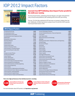


© Copyright 2026