
UDP-Glo™ Glycosyltransferase Assay Instruc ons for Use of Products
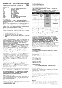
TECHNICAL MANUAL UDP-Glo™ Glycosyltransferase Assay Instruc ons for Use of Products V6961, V6962 and V6963 9/14 TM413 UDP-Glo™ Glycosyltransferase Assay All technical literature is available at: www.promega.com/protocols/ Visit the web site to verify that you are using the most current version of this Technical Manual. E-mail Promega Technical Services if you have questions on use of this system: [email protected] 1. Description......................................................................................................................................... 1 2. Product Components and Storage Conditions ........................................................................................ 6 3. Preparing for the UDP-Glo™ Glycosyltransferase Assay ......................................................................... 9 3.A. Preparing the UDP Detection Reagent .......................................................................................... 9 3.B. Generating a Standard Curve for UDP ........................................................................................ 10 4. UDP-Glo™ Glycosyltransferase Assay Protocols................................................................................... 11 4.A. UDP-Glo™ Glycosyltransferase Assay Protocol ........................................................................... 11 4.B. Optimizing Glycosyltransferase Reaction Conditions ................................................................... 12 4.C. Determining IC50 Values of Glycosyltransferase Inhibitors ........................................................... 15 5. General Considerations ..................................................................................................................... 18 6. References ........................................................................................................................................ 20 7. Composition of Buffers and Solutions ................................................................................................. 20 8. Related Products ............................................................................................................................... 21 1. Description The UDP-Glo™ Glycosyltransferase Assay(a,b) is a bioluminescent assay for detecting the activity of glycosyltransferases that use UDP-sugars as donor substrates and release UDP as a product. Glycosylation reactions catalyzed by glycosyltransferases are central to many biological processes, including cell:cell interactions, cell signaling and bacterial cell wall biosynthesis (1). Glycosyltransferases transfer sugar from a nucleotide-glycosyl donor (e.g., UDP-Galactose, UDP-Glucose, UDP-GlcNAc, UDP-GalNAc, and UDP-Glucuronic acid) to an acceptor molecule. Other sugars are typical glycosyltransferase acceptor substrates but can also be lipids, proteins, nucleic acids, xenobiotics or other small molecules (2). In a glycosyltransferase reaction, the UDP moiety is released as a product; therefore, an assay that detects UDP would be suitable for monitoring the activity of the majority of glycosyltransferases, including the important O-GlcNAc transferase (OGT). OGT transfers N-acetylglucosamine from UDP-GlcNAc to serine and threonine residues on a diverse population of nuclear and cytosolic proteins, and is involved in many cellular processes such as nutrient sensing, stress response, transcription, translation and cell signaling. Defects in OGT activity have been associated with a number of diseases (3), suggesting OGT as a novel therapeutic target for inhibition (4). Promega Corpora on · 2800 Woods Hollow Road · Madison, WI 53711-5399 USA · Toll Free in USA 800-356-9526 · 608-274-4330 · Fax 608-277-2516 www.promega.com TM413 · 9/14 1 1. Description (continued) The UDP-Glo™ Glycosyltransferase Assay is a homogenous, single-reagent-addition method to rapidly detect UDP formation in glycosyltransferase reactions. After the glycosyltransferase reaction, an equal volume of UDP Detection Reagent is added to simultaneously convert the UDP product to ATP and generate light in a luciferase reaction. The light generated is detected using a luminometer (Figure 1). Luminescence can be correlated to UDP concentration by using an UDP standard curve. The light output is proportional to the concentration of UDP from low nM to 25µM (Figure 2, Panel A). The assay is easy to use and highly sensitive (Figure 2, Panel B), two features that are desirable and essential for measuring the activity of different UDP-sugar-utilizing glycosyltransferases covering the majority of glycosyltransferase classes (Figure 3). Therefore, the UDP detection assay uses less enzyme in glycosyltransferase reactions. This assay is fast and simple (Figure 4). The UDP-Glo™ Assay is performed in a single well of a multiwell plate, and can be used to detect glycosyltransferase activity in as little as a 5µl reaction. UDP Detection Assay OH Glycosyltransferase O O HO OH + UDP OH HO Glycosyltransferase substrate O O HO UDP UDP Detection Reagent One-step reaction converts UDP to ATP and generates light. Light 12347MA HO Reaction products OH UDP-sugar substrate UDP-forming reaction Figure 1. UDP-Glo™ Glycosyltransferase Assay principle. The assay is performed in one step. After the glycosyltransferase reaction, UDP Detection Reagent is added to convert UDP to ATP and allow the newly synthesized ATP to be measured using a luciferase/luciferin reaction. The light generated correlates to the amount of UDP produced by the glycosyltransferase, which is indicative of glycosyltransferase activity. The UDP-Glo™ Glycosyltransferase Assay relies on the properties of a proprietary thermostable luciferase (Ultra-Glo™ Recombinant Luciferase) that is formulated to generate a stable glow-type luminescent signal and improve performance across a wide range of assay conditions. The signal produced by the luciferase reaction initiated by adding the UDP Detection Reagent is stable for more than 3 hours (Figure 2, Panel C). This extended stability eliminates the need for a luminometer equipped with injectors and allows batch-mode processing of multiple plates. Furthermore, the combination of Ultra-Glo™ Recombinant Luciferase (5) and proprietary formulation of the UDP Detection Reagent results in luminescence that is much less susceptible to interference from library compounds than other luciferase- or fluorescence-based assays (6). In addition to providing biochemical values (e.g., Km of UDP-sugars or Km of acceptor substrates) comparable to those reported in the literature, the UDP-Glo™ Glycosyltransferase Assay can be used in screening for specific glycosyltransferase inhibitors and the study of their mode of action (see Section 4.C). 2 Promega Corpora on · 2800 Woods Hollow Road · Madison, WI 53711-5399 USA · Toll Free in USA 800-356-9526 · 608-274-4330 · Fax 608-277-2516 TM413 · 9/14 www.promega.com A. 1.4 × 10⁷ 1.0 × 10⁷ r2 = 0.9973 0.8 × 10⁷ 0.6 × 10⁷ Luminescence (RLU) Luminescence (RLU) 1.2 × 10⁷ 7 0.4 × 10 5 × 10⁵ 4 × 10⁵ r2 = 0.9999 3 × 10⁵ 2 × 10⁵ 1 × 10⁵ 0 7 0.2 × 10 0 0.2 0.4 0.6 0.8 UDP (µM) 1.0 0 0 5 10 15 20 25 30 UDP (µM) B. UDP (µM) 25 12.5 6.25 3.13 1.56 0.78 0.39 0.20 0.10 0.05 0.02 0 Signal-to-Background Ratio 6,397 3,448 1,912 at 60 minutes 855 477 235 119 62 30 15 8 1 C. 140 12.5µM UDP 6.25µM UDP 1.6µM UDP 0.4µM UDP 0.2µM UDP 0.1µM UDP 0.05µM UDP Percent Signal Remaining 120 100 80 60 40 0 12346MA 20 1 hour 2 hours 3 hours Time Figure 2. Linearity and sensitivity of the UDP-Glo™ Glycosyltransferase Assay. Panel A. UDP standard curve was prepared over the indicated range of UDP concentrations in 25μl of 1X glycosyltransferase reaction buffer in a solid white 96-well plate. (Standard curve preparation is described in Section 3.B.) UDP-Glo™ Glycosyltransferase Assay was performed using 25μl of UDP Detection Reagent at room temperature as described in Section 4. Luminescence was recorded using a GloMax® 96 Microplate Luminometer (Cat.# E6501). Values represent the mean of four replicates. Panel B. Luminescence was measured 1 hour after adding the UDP Detection Reagent, and signal-to-background ratios calculated for each concentration of the UDP standard curve. Panel C. To determine signal stability, luminescence was recorded again every hour. The signal-to-background ratio did not change over the time measured (data not shown). RLU = relative light units. Promega Corpora on · 2800 Woods Hollow Road · Madison, WI 53711-5399 USA · Toll Free in USA 800-356-9526 · 608-274-4330 · Fax 608-277-2516 www.promega.com TM413 · 9/14 3 1. Description (continued) A. B. 1.5 × 10⁶ 2.0 × 10⁷ UDP-GalNAc + Mucin 10 EA2 (peptide) UDP-Galactose + GlcNAc (sugar) 6 1.3 × 10 1.5 × 10⁷ Luminescence (RLU) Luminescence (RLU) 1.8 × 10⁷ 1.3 × 10⁷ 1.0 × 10⁷ 0.8 × 10⁷ 0.5 × 10⁷ 1.0 × 10⁶ 0.8 × 10⁶ 0.5 × 10⁶ 0.3 × 10⁶ 0.3 × 10⁷ 0 0 0.1 1 10 1 100 β-1,4-Galactosyltransferase 1 (ng) 10 100 Polypeptide GalNAc Transferase 1 (ng) C. 5 × 10⁴ UDP-GA + Estradiol (drug) Luminescence (RLU) 4 × 10⁴ 3 × 10⁴ 2 × 10⁴ 12348MA 1 × 10⁴ 1 10 100 1,000 UDP-Glucuronosyltransferase 1A1 (ng) Figure 3. Detection of the activity of various UDP-sugar-utilizing enzymes. Panel A. β4GalT1 (R&D Systems Cat.# 3609-GT) was titrated in 1X glycosyltransferase reaction buffer the presence of 100µM of Ultra Pure UDP-Galactose (Cat.# V7171) and 10mM N-acetylglucosamine (GlcNAc) as an acceptor substrate. Panel B. GalNT1 (R&D Systems Cat.# 7140-GT) was titrated in 1X GALNT reaction buffer in the presence of 100µM of Ultra Pure UDP-GalNAc (Cat.# V7081) and 0.5mM Mucin-derived peptide Mucin 10 (153–165) EA2 (AnaSpec Inc Cat.# 63841), as an acceptor substrate. Panel C. UGT1A1 (BD Gentest Cat.# 456411) was titrated in 1X UGT reaction buffer in the presence of 100µM of UDP-glucuronic acid (UDP-GA) and 200µM estradiol as an acceptor substrate. All enzyme reactions were performed in 25µl volume in a solid white 96-well plate and incubated at 23°C for 60 minutes. The UDP-Glo™ Glycosyltransferase Assay was performed as described in Section 4.A. Each point is an average of two experiments, and the error bars represent the standard deviations. Curve fitting was performed using GraphPad Prism® version 6, sigmoidal dose-response (variable slope) software. 4 Promega Corpora on · 2800 Woods Hollow Road · Madison, WI 53711-5399 USA · Toll Free in USA 800-356-9526 · 608-274-4330 · Fax 608-277-2516 TM413 · 9/14 www.promega.com ATP Detection Substrate Nucleotide Detection Buffer Dispense into aliquots. UDP-Glo™ Solution Nucleotide Detection Reagent Step 1 UDP Detection Reagent Add UDP Detection Reagent to completed UDP-generating reactions, and mix. 12349MA Incubate for 60 minutes. Record luminescence. GloMax®-Multi+ Detection System Figure 4. Schematic representation of the UDP-Glo™ Glycosyltransferase Assay protocol. Promega Corpora on · 2800 Woods Hollow Road · Madison, WI 53711-5399 USA · Toll Free in USA 800-356-9526 · 608-274-4330 · Fax 608-277-2516 www.promega.com TM413 · 9/14 5 1. Description (continued) Note: This assay detects only the activity of glycosyltransferases that use UDP-sugar as a donor substrate and can only be used with purified glycosyltransferases not whole cells or cell extract. However, glycosyltransferases can be purified from cell extract using immunoprecipitation or affinity tag pull down then used in the UDP-Glo™ Glycosyltransferase Assay. The sensitivity of the UDP-Glo™ Glycosyltransferase Assay means it can detect low UDP concentrations with high dynamic range. Therefore, whether assaying for a low-activity glycosyltransferase whose sugar transfer rate is low or using a low concentration of enzyme that produces a small amount of UDP, a high signal-to-background ratio is obtained with the UDP-Glo™ Glycosyltransferase Assay. See Figure 2, Panel B. Advantages of the UDP-Glo™ Glycosyltransferase Assay • Linear response in the nM to µM range: Uses low concentrations of UDP-sugar, decreasing feedback glycosyltransferase inhibition issues. • High dynamic range: High signal-to-background ratios at lower concentrations of UDP means using less enzyme during the glycosyltransferase reaction. • High sensitivity: Detect 0.1–0.5pmol UDP with a more than twofold difference over background. • Reliable, reproducible data: Routinely obtain Z´ factor values >0.7 even with low UDP production rates. • • • • • Universal assay: Use any UDP-sugar-utilizing glycosyltransferase and glycosyltransferase:substrate combination, including peptide, protein, lipid and sugar substrates. Positive correlation: Assay signal increases linearly with increasing product formation. Luminescence-based UDP detection: Experience less overall assay interference from chemical compounds. Batch plate processing: Highly stable luminescent signal with >80% signal remaining after 3 hours. Homogeneous non-radioactive assay. 2. Product Components and Storage Conditions PRODUCT UDP-Glo™ Glycosyltransferase Assay SIZE C A T. # 200 assays V6961 This system is sufficient for 200 assays performed in 96-well plates using 25µl of glycosyltransferase reaction and 25µl UDP Detection Reagent. This system also can be used in 384-well plates using 5µl:5µl for a total of 1,000 assays. Includes: • • • • 6 100µl 24µl 5ml 1 vial UDP, 10mM UDP-Glo™ Solution Nucleotide Detection Buffer ATP Detection Substrate (lyophilized) Promega Corpora on · 2800 Woods Hollow Road · Madison, WI 53711-5399 USA · Toll Free in USA 800-356-9526 · 608-274-4330 · Fax 608-277-2516 TM413 · 9/14 www.promega.com PRODUCT UDP-Glo™ Glycosyltransferase Assay SIZE C A T. # 400 assays V6962 This system is sufficient for 400 assays performed in 96-well plates using 25µl of glycosyltransferase reaction and 25µl UDP Detection Reagent. This system also can be used in 384-well plates using 5µl:5µl for a total of 2,000 assays. Includes: • • • • 100µl 24µl 10ml 1 vial UDP, 10mM UDP-Glo™ Solution Nucleotide Detection Buffer ATP Detection Substrate (lyophilized) PRODUCT UDP-Glo™ Glycosyltransferase Assay SIZE C A T. # 4,000 assays V6963 This system is sufficient for 4,000 assays performed in 96-well plates using 25µl of glycosyltransferase reaction and 25µl UDP Detection Reagent. This system also can be used in 384-well plates using 5µl:5µl for a total of 20,000 assays. Includes: • • • • 100µl 240µl 100ml 1 vial UDP, 10mM UDP-Glo™ Solution Nucleotide Detection Buffer ATP Detection Substrate (lyophilized) Storage Conditions: Store the UDP-Glo™ Glycosyltransferase Assay at –20°C. Before use, completely thaw all components at room temperature except the UDP-Glo™ Solution, which should be thawed only prior to use and any remaining volume should be returned to –20°C. Once thawed, all components should be thoroughly mixed before use. Any remaining Nucleotide Detection Reagent (Nucleotide Detection Buffer + ATP Detection Substrate) should be dispensed into aliquots and stored at –80°C. For best results, prepare only the amount of UDP Detection Reagent (Nucleotide Detection Reagent + UDP-Glo™ Solution) needed. If smaller amounts of UDP Detection Reagent are needed for each use, the UDP-Glo™ Solution should be dispensed in single-use aliquots and stored at –20°C. UDP-Sugar Substrates PRODUCT SIZE C A T. # Ultra Pure UDP-GlcNAc, 100mM 50μl V7071 5 × 50μl V7072 50μl V7081 5 × 50μl V7082 50μl V7091 5 × 50μl V7092 50μl V7171 5 × 50μl V7172 Ultra Pure UDP-GalNAc, 100mM Ultra Pure UDP-Glucose, 100mM Ultra Pure UDP-Galactose, 100mM Promega Corpora on · 2800 Woods Hollow Road · Madison, WI 53711-5399 USA · Toll Free in USA 800-356-9526 · 608-274-4330 · Fax 608-277-2516 www.promega.com TM413 · 9/14 7 2. Product Components and Storage Conditions (continued) UDP-Glo™ Glycosyltransferase Assay + UDP-Sugar Substrate PRODUCT SIZE C A T. # UDP-Glo™ Glycosyltransferase Assay (V6961) + Ultra Pure UDP-GlcNAc, 100mM (V7071) 200 assays V6971 UDP-Glo™ Glycosyltransferase Assay (V6962) + Ultra Pure UDP-GlcNAc, 100mM (V7071) 400 assays V6972 UDP-Glo™ Glycosyltransferase Assay (V6961) + Ultra Pure UDP-GalNAc, 100mM (V7081) 200 assays V6981 UDP-Glo™ Glycosyltransferase Assay (V6962) + Ultra Pure UDP-GalNAc, 100mM (V7081) 400 assays V6982 UDP-Glo™ Glycosyltransferase Assay (V6961) + Ultra Pure UDP-Glucose, 100mM (V7091) 200 assays V6991 UDP-Glo™ Glycosyltransferase Assay (V6962) + Ultra Pure UDP-Glucose, 100mM (V7091) 400 assays V6992 UDP-Glo™ Glycosyltransferase Assay (V6961) + Ultra Pure UDP-Galactose, 100mM (V7171) 200 assays V7051 UDP-Glo™ Glycosyltransferase Assay (V6962) + Ultra Pure UDP-Galactose, 100mM (V7171) 400 assays V7052 8 Promega Corpora on · 2800 Woods Hollow Road · Madison, WI 53711-5399 USA · Toll Free in USA 800-356-9526 · 608-274-4330 · Fax 608-277-2516 TM413 · 9/14 www.promega.com 3. Preparing for the UDP-Glo™ Glycosyltransferase Assay Materials to Be Supplied by the User • solid white multiwell plate (do not use black plates or clear plates) • • • enzyme reaction buffers; used for enzyme, substrate and compound dilution multichannel pipette or automated pipetting station glycosyltransferase • sugar acceptor substrate • • luminometer capable of reading multiwell plates plate shaker 3.A. Preparing the UDP Detection Reagent Calculate the required volumes of each reagent needed for your experiment, and increase or decrease the volumes appropriately. Nucleotide Detection Reagent Preparation 1. Equilibrate the Nucleotide Detection Buffer and ATP Detection Substrate to room temperature before use. 2. Transfer the entire volume of Nucleotide Detection Buffer into the amber bottle containing ATP Detection Substrate to reconstitute the lyophilized luciferase enzyme/substrate mixture. This forms the Nucleotide Detection Reagent. 3. Mix to homogeneity by gently vortexing, swirling or by inverting the contents. The ATP Detection Substrate should go easily into solution in less than 1 minute. 4. Use Nucleotide Detection Reagent immediately or dispense into aliquots and store at –80°C. UDP Detection Reagent Preparation 1. Equilibrate an aliquot of Nucleotide Detection Reagent to room temperature. 2. Create UDP Detection Reagent by adding 2µl of UDP-Glo™ solution to each 1ml of Nucleotide Detection Reagent immediately before use. Note: Prepare only enough UDP Detection Reagent required for the experiment. Return the remaining UDP-Glo™ Solution to –20°C. 3. Mix contents to homogeneity by gently pipetting or vortexing. Promega Corpora on · 2800 Woods Hollow Road · Madison, WI 53711-5399 USA · Toll Free in USA 800-356-9526 · 608-274-4330 · Fax 608-277-2516 www.promega.com TM413 · 9/14 9 3.B. Generating a Standard Curve for UDP To estimate the amount of UDP produced in the glycosyltransferase reaction, we recommend creating a UDP standard curve of 0–25µM UDP. The UDP standards can be prepared in a separate 96-well or 384-well plate. Once the standards are prepared, transfer the appropriate amount to the same assay plate where the glycosyltransferase reaction is being performed. We recommend assaying each UDP standard concentration in triplicate. Figure 2 shows representative data from a UDP standard curve. 1. Prepare 200µl of 25µM UDP solution in preferred 1X glycosyltransferase reaction buffer. Then add all 200µl of the 25µM UDP solution to well A1 of a preparative 96-well plate. 2. Add 100μl of 1X glycosyltransferase buffer to wells A2 through A12 of the preparative 96-well plate. Note: Depending on the requirements of your system, you can use glycosyltransferase buffer containing UDP and other appropriate substrate or only UDP. 3. Perform a serial twofold dilution as shown in Figure 5 by transferring 100μl from well A1 to well A2, pipetting to mix. Transfer 100μl from well A2 to well A3, pipetting to mix. Repeat for wells A4 through A11. Discard the extra 100μl from well A11. Do not add UDP to the no-UDP control reactions in well A12. no-UDP control 100µl 1 2 3 4 5 6 7 8 9 10 11 12 A 200µl of UDP Solution 12350MA 100µl of 1X glycosyltransferase buffer per well Figure 5. Dilution scheme for creating a UDP standard curve. 4. Transfer the desired volume of each UDP standard from the 96-well plate to the wells reserved for the UDP standard curve on your assay plate. 5. Proceed immediately to the assay protocol (Section 4). We recommend the following volumes for different plate formats: 96-well assay plate: Transfer 25μl of UDP standards. 384-well assay plate: Transfer 10μl of UDP standards. Low-volume 384-well or 1,536-well assay plate: Transfer 5μl or less of UDP standards. The luminescence output of the assay is proportional to the concentration of UDP in the standard curve so that luminescence readout can be directly compared to those UDP concentrations generated in a glycosyltransferase reaction sample as long as the volume of the UDP standards used is the same as the volume of the glycosyltransferase reaction. 10 Promega Corpora on · 2800 Woods Hollow Road · Madison, WI 53711-5399 USA · Toll Free in USA 800-356-9526 · 608-274-4330 · Fax 608-277-2516 TM413 · 9/14 www.promega.com 4. UDP-Glo™ Glycosyltransferase Assay Protocols Prior to performing the UDP-Glo™ Glycosyltransferase Assay, prepare the reagents and UDP standards as described in Section 3. Calculate the volume of Nucleotide Detection Reagent required for your experiments, and equilibrate that volume to room temperature before use. Return the remaining reagents to –80°C. The UDP Detection Reagent is stable for 2 hours at 22°C with minimal loss of signal and up to 5 hours with ~20% loss of signal. However the signal-to-background ratio is stable for at least 5 hours. The UDP-Glo™ Solution should be added to the Nucleotide Detection Reagent just prior to use. Note: Once mixed, the Nucleotide Detection Reagent should be used immediately or dispensed into aliquots and stored at –80°C. The reconstituted reagent remains stable for at least a month with no loss of signal observed after 5 freeze-thaw cycles. 4.A. UDP-Glo™ Glycosyltransferase Assay Protocol The UDP-Glo™ Glycosyltransferase Assay is a single step after the completed glycotransferase reaction as outlined in Figures 1 and 4. For 96-well plates, we recommend a 25μl glycosyltransferase reaction and 25μl UDP Detection Reagent for a total volume of 50μl. For 384-well plates, volumes may be reduced fivefold to a 5μl glycosyltransferase reaction and 5μl UDP Detection Reagent for a total volume of 10μl. Other volumes may be used provided the 1:1 ratio of glycosyltransferase reaction volume to UDP Detection Reagent volume is maintained. The UDP-Glo™ Glycosyltransferase Assay protocol for 96-well plates is described below. 1. Perform a 25µl glycosyltransferase reaction using the 1X glycosyltransferase buffer of your choice. (See Section 7 for buffer examples.) If the glycosyltransferase reaction was not incubated at room temperature, equilibrate the plate to room temperature before adding the UDP Detection Reagent. 2. Prepare the UDP Detection Reagent by adding 2µl of UDP-Glo™ Solution to each 1ml of Nucleotide Detection Reagent. 3. Add 25µl of UDP Detection Reagent to each well of the assay plate. The UDP Detection Reagent terminates the glycosyltransferase reaction; therefore there is no need to add an inhibitor to terminate the glycosyltransferase reaction (e.g., EDTA, acid, etc.). However, if a glycosyltransferasetermination reagent is added to the glycosyltransferase reaction, do not use a magnesium-chelating agent such as EDTA because the UDP-Glo™ Glycosyltransferase Assay requires magnesium. The optimal pH for this assay is pH 6–9. 4. Mix assay plate with a plate shaker for 30 seconds, and incubate at room temperature for 60 minutes. 5. Measure the luminescence with a plate-reading luminometer or charge-coupled device (CCD) camera. Note: Instrument settings depend on the manufacturer. An integration time of 0.25–1 second per well should serve as a guideline. The long half-life of the UDP-Glo™ Glycosyltransferase Assay signal allows plates to be left longer at room temperature before reading, if desired. Promega Corpora on · 2800 Woods Hollow Road · Madison, WI 53711-5399 USA · Toll Free in USA 800-356-9526 · 608-274-4330 · Fax 608-277-2516 www.promega.com TM413 · 9/14 11 4.B. Optimizing Glycosyltransferase Reaction Conditions For optimal performance when using the UDP-Glo™ Glycosyltransferase Assay, optimize the amounts of glycosyltransferase and glycosyltransferase substrates in the reaction. If the amount of glycosyltransferase or its substrates has been determined, proceed to Section 4.A. Notes: 1. Use only the provided Ultra Pure UDP-sugar substrates when performing the UDP-Glo™ Glycosyltransferase Assay. Other sources of UDP-sugar may contain free UDP that could result in high background. Due to the sensitivity of the UDP-detection assay, there is no need to use higher amounts of UDP-sugars in the glycosyltransferase reactions. We typically use 100µM UDP-sugars or lower. 2. We recommend optimizing the glycosyltransferase reaction conditions at room temperature to ensure uniform temperature across the plate during the UDP-Glo™ Glycosyltransferase Assay. Preparation of Glycosyltransferase Titration Components This protocol is written for O-GlcNAc Transferase (OGT) enzyme titration as an example to select an optimal enzyme concentration for use in subsequent experiments such as an inhibitor titration (Figure 6). The OGT reaction is performed in 96-well plate using 1X OGT buffer, 100µM UDP-GlcNAc as a sugar donor, 50µM OGT peptide substrate (Anaspec Cat.#63726) as an acceptor, and a serial dilution of OGT enzyme from 0–40ng/reaction in 25µl volume. A UDP standard curve is performed in the same assay plate to correlate luminescence to the UDP concentrations generated in each OGT reaction. Note: Different OGTs or other glycosyltransferases have varying specific activities. Therefore, the useful enzyme dilution range may vary greatly and should be determined experimentally prior to inhibitor potency determinations. 12 Promega Corpora on · 2800 Woods Hollow Road · Madison, WI 53711-5399 USA · Toll Free in USA 800-356-9526 · 608-274-4330 · Fax 608-277-2516 TM413 · 9/14 www.promega.com 2.0 × 10⁶ UDP-GlcNAc + OGT substrate 6 Luminescence (RLU) 1.8 × 10 1.5 × 10⁶ 1.3 × 10⁶ 1.0 × 10⁶ 2.5ng OGT 200 nM UDP Signal-to-background ratio of 67 0.8 × 10⁶ 0.5 × 10⁶ 0 0.01 12351MA 0.3 × 10⁶ 0.1 1 10 100 O-GlcNAc Transferase (ng) Figure 6. Detection of OGT activity with UDP-Glo™ Glycosyltransferase Assay. Recombinant His-linked sOGT enzyme was titrated in 25μl in 1X OGT reaction buffer in a solid white 96-well plate in the presence of 50µM OGT peptide substrate (Anaspec Cat.# 63726) and 100µM UltraPure UDP-GlcNAc (Cat.# V7071). After incubating 1 hour at 23°C, the UDP-Glo™ Glycosyltransferase Assay was performed using 25μl of UDP Detection Reagent as described in Section 4.A. Luminescence was recorded 1 hour after adding the UDP Detection Reagent using a GloMax® 96 Microplate Luminometer (Cat.# E6501). Values represent the mean of two replicates. UDP produced by different OGT amounts was calculated by linear regression in Microsoft Excel® using a UDP standard curve performed at the same time (data not shown). Curve fitting was performed using GraphPad Prism® version 6, sigmoidal dose-response (variable slope) software. The insert highlights OGT activity at nanogram amounts with a high signal-to-background ratio. RLU = relative light units. 1. Substrate Mix Preparation: Prepare 400μl of 2.5X UDP-GlcNAc/OGT Peptide Substrate Mix (10μl/reaction/ well) in a 1.5ml tube as described below, and keep on ice until ready to dispense in the assay plate. Component 4X OGT reaction buffer Volume 100μl 100mM UDP-GlcNAc 1μl 1mM OGT Substrate 50μl ATP-free water 249µl Promega Corpora on · 2800 Woods Hollow Road · Madison, WI 53711-5399 USA · Toll Free in USA 800-356-9526 · 608-274-4330 · Fax 608-277-2516 www.promega.com TM413 · 9/14 13 4.B. Optimizing Glycosyltransferase Reaction Conditions (continued) 2. OGT Solution Preparation: Prepare 100μl OGT enzyme solution as described below (15μl/reaction/well). This will give 40ng OGT/15μl starting concentration. Component Volume 4X OGT reaction buffer 25μl OGT (4ng/μl) 66.6μl ATP-free water 8.4μl a. Prepare 1ml of 1X OGT reaction buffer by mixing 250µl 4X OGT reaction buffer with 750µl ATP-free water. b. Add OGT enzyme solution to well A1 of a 96-well plate. c. Add 50μl of 1X OGT reaction buffer to wells A2 through A12 of the 96-well plate. d. Perform a serial twofold dilution by transferring 50μl from well A1 to well A2, pipetting to mix as described in Table 1. Transfer 50μl from well A2 to well A3, pipetting to mix. Repeat for wells A4–A11. Discard the extra 50μl from well A11. Do not add OGT to the no-enzyme control reaction in well A12. Note: Do not create bubbles while preparing the dilution series. Table1. Performing Serial 1:1 Dilutions of OGT. OGT (ng) Starting Volume of Each Well Volume to Transfer A1 40 100µl 50µl A2 20 50µl 50µl A3 10 50µl 50µl A4 5 50µl 50µl A5 2.5 50µl 50µl A6 1.25 50µl 50µl A7 0.625 50µl 50µl A8 0.312 50µl 50µl A9 0.156 50µl 50µl A10 0.078 50µl 50µl A11 0.039 50µl 0µl; No transfer A12 0 50µl Buffer only Well Number 14 Promega Corpora on · 2800 Woods Hollow Road · Madison, WI 53711-5399 USA · Toll Free in USA 800-356-9526 · 608-274-4330 · Fax 608-277-2516 TM413 · 9/14 www.promega.com OGT Reaction and UDP Standard Curve Experiment 1. Transfer 25μl of the UDP serial dilution in duplicates into the standard curve-designated wells of the 96-well assay plate. 2. Transfer 15μl of OGT dilution samples in duplicates from the wells of the OGT titration plate to the wells of the assay plate. 3. Transfer 10μl of the 2.5X UDP-GlcNAc/Substrate Mix to the rows of the OGT dilutions. 4. Centrifuge the plate, and mix with a plate shaker for 2 minutes. Incubate the reaction at room temperature for 60 minutes or the desired time. 5. Follow the UDP-Glo™ Glycosyltransferase Assay protocol described in Section 4.A, starting at Step 2. 6. Record luminescence. Note: Instrument settings depend on the manufacturer. An integration time of 0.25–1 second per well should serve as a guideline. Note: The optimal amount of a glycosyltransferase to use in subsequent experiments including chemical compound screens and IC50 determinations is the amount that produces luminescence within the linear range of the OGT titration curve and generates an adequate signal-to-background ratio. Because the UDP-Glo™ Glycosyltransferase Assay is very sensitive, it can detect a very small amount of UDP with a high signal-to-background ratio. As a result, a small amount of enzyme that produces low amount of UDP is sufficient for use with the UDP-Glo™ Glycosyltransferase Assay. Figure 6 shows that the amount of UDP produced with a small amount of enzyme results in high signal-to-background ratios (2.5ng OGT produced 200nM UDP with a signal-tobackground ratio of 67-fold). 4.C. Determining IC50 Values of Glycosyltransferase Inhibitors The following protocol is an example of an inhibitor titration in OGT reaction at a final concentration of 100μM UDP-GlcNAc and 2.5ng OGT (optimal amount in the linear range of the enzyme titration). Representative inhibitor titration data is shown in Figure 7. This protocol is designed for a 96-well plate using a 25µl:25µl ratio of glycotransferase reaction volume to UDP Detection Reagent. To perform the assay in a 384-well plate, reduce volumes fivefold. Other volumes may be used, provided the 1:1 ratio of glycosyltransferase reaction volume to UDP Detection Reagent volume is maintained. Promega Corpora on · 2800 Woods Hollow Road · Madison, WI 53711-5399 USA · Toll Free in USA 800-356-9526 · 608-274-4330 · Fax 608-277-2516 www.promega.com TM413 · 9/14 15 4.C. Determining IC50 Values of Glycosyltransferase Inhibitors (continued) 120 80 IC50 = 55µM ± 7 µM 60 S N 40 C N 20 S OH 12352MA Percent OGT Activity 100 O 0 0.1 1 10 100 1,000 STO78925 (µM) Figure 7. Determination of IC50 for OGT inhibitor. Titration of OGT inhibitor ST078925 (Fisher Cat.# NC0023843) was performed in solid white, half-volume 96-well plate in a total volume of 25μl using 2.5ng/well sOGT, 50μM OGT Peptide substrate, 100µM Ultra pure UDP-GlcNAc (Cat.# V7071) and the indicated concentrations of inhibitor. Glycosyltransferase reactions were incubated for 60 minutes at room temperature (23°C). Values represent the mean of two replicates. IC50 value for ST078925 potency determined using the UDP-Glo™ Glycosyltransferase Assay compare favorably to the IC50 value reported for this compound in the literature (7). Curve fitting was performed using GraphPad Prism® version 6, sigmoidal dose-response (variable slope) software. Preparation of Inhibitor Titration Components 1. OGT inhibitor Solution Preparation: Prepare 100μl of 1.25mM OGT inhibitor (with 5% DMSO) as described below (final 10μl/reaction/well). This gives 500μM OGT inhibitor (with 1% DMSO) starting concentration in the assay. Component 16 Volume 4X OGT reaction buffer 25μl 25mM OGT inhibitor in DMSO 5μl ATP-free water 70μl a. Prepare 1ml of 1X OGT reaction buffer (with 5% DMSO) by mixing 250μl of 4X OGT reaction buffer, 50µl DMSO and 700μl ATP-free water. b. Add OGT inhibitor solution to well A1 of a 96-well plate. c. Add 50μl of 1X OGT reaction buffer (with 5% DMSO) to wells A2–A12 of the 96-well plate. d. Perform a serial threefold dilution by transferring 25μl from well A1 to well A2 and pipetting to mix as described in Table 2. Transfer 25μl from well A2 to well A3 and pipet to mix. Repeat for wells A4–A10. Do not add OGT inhibitor to the no-inhibitor control reaction in well A11 and the no-enzyme control reaction in well A12. Promega Corpora on · 2800 Woods Hollow Road · Madison, WI 53711-5399 USA · Toll Free in USA 800-356-9526 · 608-274-4330 · Fax 608-277-2516 TM413 · 9/14 www.promega.com Table2. Performing Serial 1:3 Dilutions of OGT Inhibitor. Final OGT Inhibitor Concentration (nM) Starting Volume of Each Well Volume to Transfer A1 500 100µl 25µl A2 166.7 50µl 25µl A3 55.6 50µl 25µl A4 18.5 50µl 25µl A5 6.2 50µl 25µl A6 2.1 50µl 25µl A7 0.7 50µl 25µl A8 0.20 50µl 25µl A9 0.08 50µl 25µl A10 0.025 50µl 0µl; No transfer A11 No inhibitor 50µl Buffer only A12 No enzyme 50µl Buffer only Well Number 2. 3. OGT Solution Preparation: Prepare 150μl of OGT solution (excess amount of 30 reactions at 5μl/reaction/ well) in a 1.5ml tube as described below, and keep on ice until ready to dispense in the assay plate. This will give 2.5ng of OGT/reaction. Component Volume 4X OGT reaction buffer 37.5μl OGT (4ng/μl) 18.75μl ATP-free water 93.75μl Substrate Mix Preparation: Prepare 400μl of 2.5X UDP-GlcNAc/OGT peptide Substrate Mix (10μl/reaction/ well) in a 1.5ml tube as described below and keep on ice until ready to dispense in the assay plate. Component Volume 4X OGT reaction buffer 100μl 100mM UDP-GlcNAc 1μl 1mM OGT Substrate 50μl ATP-free water 249µl Inhibitor Titration Experiment 1. Transfer 10μl of OGT inhibitor samples in duplicate from the inhibitor titration plate to the corresponding wells of the assay plate (e.g., well A1 from the 96-well titration plate to well A1 and B1 of the 96-well assay plate, etc.) 2. Transfer 5μl of OGT samples in duplicate to wells A1–A11 and B1–B11 of the 96-well assay plate. Note: Add only 5μl of 1X OGT reaction buffer to wells A12 and B12 for the no-enzyme control. Promega Corpora on · 2800 Woods Hollow Road · Madison, WI 53711-5399 USA · Toll Free in USA 800-356-9526 · 608-274-4330 · Fax 608-277-2516 www.promega.com TM413 · 9/14 17 4.C. Determining IC50 Values of Glycosyltransferase Inhibitors (continued) Inhibitor Titration Experiment (continued) 3. Mix and incubate at room temperature for 10 minutes. 4. Transfer 10μl of the 2.5X UDP-GlcNAc/Substrate Mix to all the assay rows. 5. Centrifuge the plate. Mix with a plate shaker for 2 minutes. Incubate the reaction at room temperature for 60 minutes or the desired time. 6. Follow the UDP-Glo™ Glycosyltransferase Assay protocol described in Section 4.A, starting at Step 2. 7. Record luminescence. Note: Instrument settings depend on the manufacturer. An integration time of 0.25–1 second per well should serve as a guideline. 8. Calculating Percent Enzyme Activity: First, subtract the signal of the negative control (no-enzyme) from all the samples signal. Then use the 0% OGT activity (no-enzyme control) and the 100% OGT activity (no-inhibitor control) to calculate the percent enzyme activity remaining in the presence of the different dilutions of OGT inhibitor. 5. General Considerations Temperature: The intensity and stability of the luminescent signal from the UDP-Glo™ Glycosyltransferase Assay depends on the rate of the luciferase reaction. Environmental factors that affect the rate of the luciferase reaction will result in a change in the intensity of light output and stability of the luminescent signal. Temperature is one factor that affects the rate of this enzymatic assay and thus the light output. For consistent results, equilibrate assay plates and reagent to room temperature before adding the UDP Detection Reagent. Insufficient equilibration may result in a temperature gradient between the wells in the center and at the edge of the plate and therefore, variability across the plate. Solvents and Other Chemicals: The chemical environment in which the UDP-Glo™ Glycosyltransferase Assay is performed will affect the enzymatic rates and thus luminescence intensity. We recommend a pH of 6–9 for the glycosyltransferase buffer. Some vehicles used to resuspend the various test compounds or reagents used in the glycosyltransferase reaction buffer may interfere with the luciferase reaction and thus, affect the light output of the assay. Various chemicals were shown to be compatible with or tolerated by the UDP-Glo™ Glycosyltransferase Assay (Table 3). Interference with the assay reaction can be detected by performing a UDP standard curve in the intended buffer compared with a simple buffer (Section 7). 18 Promega Corpora on · 2800 Woods Hollow Road · Madison, WI 53711-5399 USA · Toll Free in USA 800-356-9526 · 608-274-4330 · Fax 608-277-2516 TM413 · 9/14 www.promega.com Table 3. Solvents and Chemicals Compatible with the UDP-Glo™ Glycosyltransferase Assay. Chemical Maximum Concentration Tolerated1 NaCl ≤200mM CaCl2 ≤10mM DTT ≤10mM ® Tween -20 ≤2% Triton® X-100 ≤2% DMSO ≤5% -mercaptoethanol (BME) ≤20mM MgCl2 ≤20mM MnCl2 ≤5mM NaF <30mM2 1 Higher concentra ons of these chemicals will either decrease or increase the overall luminescence without affec ng assay sensi vity. In some instances, higher concentra ons might decrease the performance of the assay. 2 The component will decrease the overall luminescence of the reac on; however, the signal to background and r2 values over me are not affected. UDP-Sugar Substrates: UDP-sugars are prone to hydrolysis, releasing the UDP moiety and causing background in the assay, which could lead to reduced assay sensitivity when testing low-activity enzyme with high concentration UDP-sugar. The UDP-sugar substrates offered with UDP-Glo™ Glycosyltransferase Assay are highly pure, stabilized sugar donors that have less than 0.005% UDP contamination. To preserve the high sensitivity of the UDP-Glo™ Glycosyltransferase Assay, use only the provided Ultra Pure UDP-sugar substrates. Other sources of UDP-sugar may contain UDP that could result in high background and low assay sensitivity. Also, using the provided Ultra Pure UDPsugar substrates will safeguard against UDP feedback inhibition of certain glycosyltransferases at the lower end of the enzyme activity curve. Using UDP-contaminated sugar donors might create erroneous results due to this inhibition. Plates and Instruments: We recommend using standard solid white, multiwell plates suitable for luminescence measurements (e.g., Corning Cat.# 3912, 3693, 3674). Luminescence can be recorded on a variety of plate readers although the relative light units will depend on the instrument. Assay well geometry and small dispensing volumes may affect the efficiency of mixing, thus, poor assay homogeneity in individual wells may result in increased reaction noise or reduced signals or both. A UDP standard curve is useful for liquid handling and instrument optimization. Testing for Compounds that Interfere with the UDP-Glo™ Glycosyltransferase Assay: Compounds that interfere with the UDP-Glo™ Glycosyltransferase Assay are rare. We screened 1,280 compounds from the LOPAC chemical library using the UDP-Glo™ Assay reagents with 10µM compound; none of the compounds interfered with the UDP-Glo™ Glycosyltransferase Assay (6). When screening for glycosyltransferase inhibitors, compounds that inhibit only the glycosyltransferase will result in lower luminescence compared to vehicle-only controls and are easily distinguishable from compounds that inhibit other components of the assay. Test compounds that inhibit other components of the assay either alone or together with the glycosyltransferase might increase or decrease the luminescent signal, depending on the level of inhibition of the glycosyltransferase, luciferase or other enzyme components of the assay. To test hits from a glycosyltransferase screen for the possibility of chemical interference with enzymatic conversion of UDP or generation of the luminescent signal, set up mock reactions without glycosyltransferase but with Promega Corpora on · 2800 Woods Hollow Road · Madison, WI 53711-5399 USA · Toll Free in USA 800-356-9526 · 608-274-4330 · Fax 608-277-2516 www.promega.com TM413 · 9/14 19 all other assay components present, including a concentration of UDP that mimics the glycosyltransferase reaction results. Add the appropriate concentration of test compound (usually 10µM) or vehicle control (e.g., 1% DMSO) to the mock reactions. A test compound that affects assay performance would alter luminescence by greater than 20% compared to vehicle control reactions without test compounds. Test compounds that inhibit luciferase may result in false hits, albeit rarely. However, the unique combination of Ultra-Glo™ Recombinant Luciferase and proprietary buffer compositions of the UDP-Glo™ Glycosyltransferase Assay will significantly reduce the number of false hits (5). 6. References 1. Haltiwanger, R.S. and Lowe, J.B. (2004) Role of glycosylation in development. Annu. Rev. Biochem. 73, 491–537. 2. Varki, A. et al. (2009) Essentials of Glycobiology, 2nd edition. Cold Spring Harbor (NY): Cold Spring Harbor Laboratory Press. 3. Slawson, C., Copeland R.J. and Hart G.W. (2010) O-GlcNAc signaling: A metabolic link between diabetes and cancer? Trends Biochem. Sci. 35, 547–55. 4. Jiang, J. et al. (2011) A neutral diphosphate mimic crosslinks the active site of human O-GlcNAc transferase. Nat. Chem. Biol. 8, 72–7. 5. Auld, D.S. et al. (2009) A basis for reduced chemical library inhibition of firefly luciferase obtained from directed evolution. J. Med. Chem. 52, 1450–8. 6. Zegzouti, H. et al. (2013) Detection of glycosyltransferase activities with homogenous bioluminescent UDP detection assay. Poster presented in the 2013 Annual Conference of the Society for Glycobiology. November 17–20, 2013 St. Petersburg, Florida, US. Abstract in Glycobiology 23 (11), 1340. 7. Gross, B.J., Kraybill, B.C. and Walker, S. (2005) Discovery of O-GlcNAc transferase inhibitors. J. Am. Chem. Soc. 127, 14588–89. 7. Composition of Buffers and Solutions 1X glycosyltransferase reaction buffer 50mM Tris (pH 7.5) 5mM MnCl2 1X UGT reaction buffer 50mM TES (pH 7.5) 8mM MgCl2 0.125mg/ml Alamethicin 30mM NaF 1X OGT reaction buffer 25mM Tris (pH 7.5) 12.5mM MgCl2 0.06mg/ml BSA 1mM DTT 1X GALNT reaction buffer 25mM Tris (pH 7.5) 5mM MnCl2 2.5mM 20 CaCl2 Promega Corpora on · 2800 Woods Hollow Road · Madison, WI 53711-5399 USA · Toll Free in USA 800-356-9526 · 608-274-4330 · Fax 608-277-2516 TM413 · 9/14 www.promega.com 8. Related Products Product Name Size Cat.# 10,000 units (500u/μl) V4871 50,000 units (500u/μl) V4875 PNGase F 500 units (10u/μl) V4831 Fetuin 500μg (10mg/ml) V4961 20 reactions V4931 Endo H Protein Deglycosylation Mix (a) U.S. Pat. Nos. 6,602,677, 7,241,584 and 8,030,017, European Pat. No. 1131441, Japanese Pat. Nos. 4537573 and 4520084 and other patents pending. (b) The method of recombinant expression of Coleoptera luciferase is covered by U.S. Pat. Nos. 5,583,024, 5,674,713 and 5,700,673. © 2014 Promega Corpora on. All Rights Reserved. UDP-Glo and Ultra-Glo are trademarks of Promega Corpora on. GloMax is a registered trademark of Promega Corpora on. Excel is a registered trademark of Microso Corpora on. GraphPad Prism is a registered trademark of GraphPad So ware, Inc. Tween is a registered trademark of Croda Interna onal PLC. Triton is a registered trademark of The Dow Chemical Company (“Dow”) or an affiliated company of Dow. Products may be covered by pending or issued patents or may have certain limita ons. Please visit our Web site for more informa on. All prices and specifica ons are subject to change without prior no ce. Product claims are subject to change. Please contact Promega Technical Services or access the Promega online catalog for the most up-to-date informa on on Promega products. Promega Corpora on · 2800 Woods Hollow Road · Madison, WI 53711-5399 USA · Toll Free in USA 800-356-9526 · 608-274-4330 · Fax 608-277-2516 www.promega.com TM413 · 9/14 21


© Copyright 2026