
Medicare National Coverage Determinations Manual Coverage Determinations
Medicare National Coverage Determinations Manual Chapter 1, Part 1 (Sections 10 – 80.12) Coverage Determinations Table of Contents (Rev. 172, 08-29-14) Transmittals for Chapter 1, Part 1 Foreword - Purpose for National Coverage Determinations Manual 10 - Anesthesia and Pain Management 10.1 - Use of Visual Tests Prior to and General Anesthesia During Cataract Surgery 10.2 - Transcutaneous Electrical Nerve Stimulation (TENS) for Acute Post-Operative Pain 10.3 - Inpatient Hospital Pain Rehabilitation Programs 10.4 - Outpatient Hospital Pain Rehabilitation Programs 10.5 - Autogenous Epidural Blood Graft 10.6 - Anesthesia in Cardiac Pacemaker Surgery 20 - Cardiovascular System 20.1 - Vertebral Artery Surgery 20.2 - Extracranial - Intracranial (EC-IC) Arterial Bypass Surgery 20.3 - Thoracic Duct Drainage (TDD) in Renal Transplants 20.4 – Implantable Automatic Defibrillators (Various Effective Dates Below) 20.5 - Extracorporeal Immunoadsorption (ECI) Using Protein A Columns 20.6 - Transmyocardial Revascularization (TMR) 20.7 - Percutaneous Transluminal Angioplasty (PTA) (Various Effective Dates Below) 20.8 - Cardiac Pacemakers 20.8.1 - Cardiac Pacemaker Evaluation Services 20.8.1.1 - Transtelephonic Monitoring of Cardiac Pacemakers 20.8.2 - Self-Contained Pacemaker Monitors 20.8.3 – Single Chamber and Dual Chamber Permanent Cardiac Pacemakers 20.9 - Artificial Hearts And Related Devices – (Various Effective Dates Below) 20.9.1 - Ventricular Assist Devices (Various Effective Dates Below) 20.10 - Cardiac Rehabilitation Programs 20.10.1 – Cardiac Rehabilitation Programs for Chronic Heart Failure 20.11 - Intraoperative Ventricular Mapping 20.12 - Diagnostic Endocardial Electrical Stimulation (Pacing) 20.13 - HIS Bundle Study 20.14 - Plethysmography 20.15 - Electrocardiographic Services 20.16 - Cardiac Output Monitoring By Thoracic Electrical Bioimpedance (TEB) – Various Effective Dates Below 20.17 - Noninvasive Tests of Carotid Function 20.18 - Carotid Body Resection/Carotid Body Denervation 20.19 - Ambulatory Blood Pressure Monitoring 20.20 - External Counterpulsation (ECP) Therapy for Severe Angina (Effective March 20, 2006) 20.21 - Chelation Therapy for Treatment of Atherosclerosis 20.22 - Ethylenediamine-Tetra-Acetic (EDTA) Chelation Therapy for Treatment of Atherosclerosis 20.23 - Fabric Wrapping of Abdominal Aneurysms 20.24 - Displacement Cardiography 20.25 - Cardiac Catheterization Performed in Other Than a Hospital Setting 20.26 - Partial Ventriculectomy 20.27 - Cardiointegram (CIG) as an Alternative to Stress Test or Thallium Stress Test 20.28 – Therapeutic Embolization 20.29 – Hyperbaric Oxygen Therapy 20.30 - Microvolt T-Wave Alternans (MTWA) (Effective March 21, 2006) 20.31 - Intensive Cardiac Rehabilitation (ICR) Programs 20.31.1 - Pritikin Program (Effective August 12, 2010) 20.31.2 - Ornish Program for Reversing Heart Disease (Effective August 12, 2010 20.32 – Transcatheter Aortic Valve Replacement (TAVR) 30 - Complementary and Alternative Medicine 30.1 - Biofeedback Therapy 30.1.1 - Biofeedback Therapy for the Treatment of Urinary Incontinence 30.2 - Thermogenic Therapy 30.3 - Acupuncture 30.3.1 – Acupuncture for Fibromyalgia 30.3.2 – Acupuncture for Osteoarthritis 30.4 - Electrosleep Therapy 30.5 - Transcendental Meditation 30.6 - Intravenous Histamine Therapy 30.7 - Laetrile and Related Substances 30.8 - Cellular Therapy 30.9 - Transillumination Light Scanning, or Diaphanography 40 - Endocrine System and Metabolism 40.1 - Diabetes Outpatient Self-Management Training 40.2 - Home Blood Glucose Monitors 40.3 - Closed-Loop Blood Glucose Control Device (CBGCD) 40.4 - Insulin Syringe 40.5 – Treatment of Obesity 40.7 - Outpatient Intravenous Insulin Treatment (Effective December 23, 2009) 50 - Ear, Nose and Throat (ENT) 50.1 - Speech Generating Devices 50.2 - Electronic Speech Aids 50.3 - Cochlear Implantation (Effective April 4, 2005) 50.4 - Tracheostomy Speaking Valve 50.5 - Oxygen Treatment of Inner Ear/Carbon Therapy 50.6 - Tinnitus Masking 50.7 - Cochleostomy With Neurovascular Transplant for Meniere’s Disease 50.8 - Ultrasonic Surgery 60 - Emergency Medicine 70 - Evaluation and Management of Patients - Office/hospital/home 70.1 - Consultations With a Beneficiary’s Family and Associates 70.2 - Consultation Services Rendered by a Podiatrist in a Skilled Nursing Facility 70.3 - Physician’s Office Within an Institution - Coverage of Services and Supplies Incident to a Physician’s Services 70.4 - Pronouncement of Death 70.5 - Hospital and Skilled Nursing Facility Admission Diagnostic Procedures 80 - Eye 80.1 - Hydrophilic Contact Lens for Corneal Bandage 80.2 - Photodynamic Therapy 80.2.1 - Ocular Photodynamic Therapy (OPT) - Effective April 3, 2013 80.3 - Photosensitive Drugs 80.3.1 - Verteporfin - Effective April 3, 2013 80.4 - Hydrophilic Contact Lenses 80.5 - Scleral Shell 80.6 - Intraocular Photography 80.7 - Refractive Keratoplasty 80.7.1 - Keratoplasty 80.8 - Endothelial Cell Photography 80.9 - Computer Enhanced Perimetry 80.10 - Phaco-Emulsification Procedure - Cataract Extraction 80.11 - Vitrectomy 80.12 - Intraocular Lenses (IOLs) Foreword - Purpose for National Coverage Determinations Manual (Rev. 2, 10-17-03) A. Purpose The statutory and policy framework within which National Coverage Decisions are made may be found in title XVIII of the Social Security Act (the Act), and in Medicare regulations and rulings. The National Coverage Determinations Manual describes whether specific medical items, services, treatment procedures, or technologies can be paid for under Medicare. National coverage decisions have been made on the items addressed in this manual. All decisions that items, services, etc. are not covered are based on §1862(a)(1) of the Act (the “not reasonable and necessary” exclusion) unless otherwise specifically noted. Where another statutory authority for denial is indicated, that is the sole authority for denial. Where an item, service, etc. is stated to be covered, but such coverage is explicitly limited to specified indications or specified circumstances, all limitations on coverage of the items or services because they do not meet those specified indications or circumstances are based on §1862(a)(1) of the Act. Where coverage of an item or service is provided for specified indications or circumstances but is not explicitly excluded for others, or where the item or service is not mentioned at all in the CMS Manual System the Medicare contractor is to make the coverage decision, in consultation with its medical staff, and with CMS when appropriate, based on the law, regulations, rulings and general program instructions The coverage decisions in the manual will be kept current, based on the most recent medical and other scientific and technical advice available to CMS. Other manuals in this system in which coverage-related instructions may be found are: Pub 100-02 (Benefit Policy); Pub 100-04 (Claims Processing); Pub 100-05 (Medicare Secondary Payer); and Pub 100-08 (Program Integrity) These manuals usually contain more general coverage descriptions and/or processing instructions. There should be no inconsistencies among the instructions in any of these manuals and the National Coverage Determinations Manual. If any such inconsistencies are found, bring them to the attention of CMS, OSORA. B. Organization The NCD manual is organized by categories, e.g., medical procedures, supplies, diagnostic services. A table of contents is provided at the beginning of the manual designating coverage decision categories. Each subject discussed within the category is listed and identified by a number. The revision transmittal sheet identifies new material and summarizes the principal changes. When a change in policy or procedure is involved, the background and effective date for the change is provided. If, at a later date, the reader wishes to refer to the background explanation given on a transmittal sheet, the reader can identify the transmittal by its number which appears on each manual page. C. CMS Coverage Web site The CMS Coverage Web page http://www.cms.hhs.gov/coverage contains information about pending National Coverage Determinations and also provides access to a database of National Coverage Determinations, National Coverage Analyses, and Local Medical review Policies. 10 - Anesthesia and Pain Management (Rev. 1, 10-03-03) 10.1 - Use of Visual Tests Prior to and General Anesthesia During Cataract Surgery (Rev. 1, 10-03-03) CIM 35-44 A. Presurgery Evaluations Cataract surgery with an intraocular lens (IOL) implant is a high volume Medicare procedure. Along with the surgery, a substantial number of preoperative tests are available to the surgeon. In most cases, a comprehensive eye examination (ocular history and ocular examination) and a single scan to determine the appropriate pseudophakic power of the IOL are sufficient. In most cases involving a simple cataract, a diagnostic ultrasound A-scan is used. For patients with a dense cataract, an ultrasound B-scan may be used. Accordingly, where the only diagnosis is cataract(s), Medicare does not routinely cover testing other than one comprehensive eye examination (or a combination of a brief/intermediate examination not to exceed the charge of a comprehensive examination) and an A-scan or, if medically justified, a B-scan. Claims for additional tests are denied as not reasonable and necessary unless there is an additional diagnosis and the medical need for the additional tests is fully documented. Because cataract surgery is an elective procedure, the patient may decide not to have the surgery until later, or to have the surgery performed by a physician other than the diagnosing physician. In these situations, it may be medically appropriate for the operating physician to conduct another examination. To the extent the additional tests are considered reasonable and necessary by the carrier’s medical staff, they are covered. B. General Anesthesia The use of general anesthesia in cataract surgery may be considered reasonable and necessary if, for particular medical indications, it is the accepted procedure among ophthalmologists in the local community to use general anesthesia. 10.2 - Transcutaneous Electrical Nerve Stimulation (TENS) for Acute Post-Operative Pain (Rev. 149, Issued: 11-30-12, Effective: 06-08-12, Implementation: 01-07-13) The use of transcutaneous electrical nerve stimulation (TENS) for the relief of acute postoperative pain is covered under Medicare. TENS may be covered whether used as an adjunct to the use of drugs, or as an alternative to drugs, in the treatment of acute pain resulting from surgery. The TENS devices, whether durable or disposable, may be used in furnishing this service. When used for the purpose of treating acute post-operative pain, TENS devices are considered supplies. As such they may be hospital supplies furnished inpatients covered under Part A, or supplies incident to a physician’s service when furnished in connection with surgery done on an outpatient basis, and covered under Part B. It is expected that TENS, when used for acute post-operative pain, will be necessary for relatively short periods of time, usually 30 days or less. In cases when TENS is used for longer periods, contractors should attempt to ascertain whether TENS is no longer being used for acute pain but rather for chronic pain, in which case the TENS device may be covered as durable medical equipment as described in §160.27. Cross-references: Medicare Benefit Policy Manual, Chapter 1, “Inpatient Hospital Services,” §40; Medicare Benefit Policy Manual, Chapter 2, “Hospital Services Covered Under Part B,” §§20, 20.4, and 80; Medicare Benefit Policy Manual, Chapter 15, “Covered Medical and other Health Services, §110.” 10.3 - Inpatient Hospital Pain Rehabilitation Programs (Rev. 1, 10-03-03) CIM 35-21 Pain rehabilitation programs are an innovative approach to the treatment of intractable pain. The goal of such programs is to give a patient the tools to manage and control his/her pain and thereby improve his/her ability to function independently. A hospital level pain rehabilitation program is one that employs a coordinated multidisciplinary team to deliver, in a controlled environment, a concentrated program that is designed to modify pain behavior through the treatment of the physiological, psychological, and social aspects of pain. Such programs generally include diagnostic testing, skilled nursing, psychotherapy, structured progressive withdrawal from pain medications, physical therapy, and occupational therapy to restore physical fitness (mobility and endurance) to a maximal level within the constraints of a patient’s physical disability, and the use of mechanical devices, and/or activities to relieve pain or modify a patient’s reaction to it (e.g., nerve stimulator, hydrotherapy, massage, ice, systemic muscle relaxation training, and diversional activities). The nurse’s responsibility in such pain rehabilitation programs is to observe and assess, on a continuing basis, a patient’s condition and response to the program as reflected by his actions while in the nursing unit, and to assure that the atmosphere within the unit is not supportive of pain behavior. The day-to-day activities involved in carrying out the program are under the general supervision and, as needed, direct supervision of a physician. Since pain rehabilitation programs of a lesser scope than that described above would raise a question as to whether the program could be provided in a less intensive setting than on an inpatient hospital basis, carefully evaluate such programs to determine whether the program does, in fact, necessitate a hospital level of care. Some pain rehabilitation programs may utilize services and devices which are excluded from coverage, e.g., acupuncture dorsal column stimulator, and family counseling services. In determining whether the scope of a pain program does necessitate inpatient hospital care, evaluate only those services and devices which are covered. Although diagnostic tests may be an appropriate part of pain rehabilitation programs, such tests would be covered in an individual case only where they can be reasonably related to a patient’s illness, complaint, symptom, or injury and where they do not represent an unnecessary duplication of tests previously performed. An inpatient program of 4 weeks’ duration is generally required to modify pain behavior. After this period, it would be expected that any additional rehabilitation services which might be required could be effectively provided on an outpatient basis under an outpatient pain rehabilitation program (see §10.4) or other outpatient program. The first 7-10 days of such an inpatient program constitute, in effect, an evaluation period. If a patient is unable to adjust to the program within this period, it is generally concluded that it is unlikely that the program will be effective and the patient is discharged from the program. On occasions, a program longer than four weeks may be required in a particular case. In such a case, there should be documentation to substantiate that inpatient care beyond a 4-week period was reasonable and necessary. Similarly, where it appears that a patient participating in a program is being granted frequent outside passes, a question would exist as to whether an inpatient program is reasonable and necessary for the treatment of the patient’s condition. An inpatient hospital stay for the purpose of participating in a pain rehabilitation program would be covered as reasonable and necessary to the treatment of a patient’s condition where the pain is attributable to a physical cause, the usual methods of treatment have not been successful in alleviating it, and a significant loss of ability to function independently has resulted from the pain. Chronic pain patients often have psychological problems which accompany or stem from the physical pain, and it is appropriate to include psychological treatment in the multi-disciplinary approach. However, patients whose pain symptoms result from a mental condition, rather than from any physical cause, generally cannot be successfully treated in a pain rehabilitation program. 10.4 - Outpatient Hospital Pain Rehabilitation Programs (Rev. 1, 10-03-03) CIM 35-21.1 Some hospitals also provide pain rehabilitation programs for outpatients. In such programs, services frequently are provided in group settings even though they are being furnished pursuant to each patient’s individualized plan of treatment. Coverage of services furnished under outpatient hospital pain rehabilitation programs, including services furnished in group settings under individualized plans of treatment, is available if the patient’s pain is attributable to a physical cause, the usual methods of treatment have not been successful in alleviating it, and a significant loss of ability by the patient to function independently has resulted from the pain. If a patient meets these conditions and the program provides services of the types discussed in §10.3, the services provided under the program may be covered. Noncovered services (e.g., vocational counseling, meals for outpatients, or acupuncture) continue to be excluded from coverage, and intermediaries would not be precluded from finding, in the case of particular patients, that the pain rehabilitation program is not reasonable and necessary under §1862(a)(1) of the Act for the treatment of their conditions. 10.5 - Autogenous Epidural Blood Graft (Rev. 1, 10-03-03) CIM 45-11 Autogenous epidural blood grafts are considered a safe and effective remedy for severe headaches that may occur after performance of spinal anesthesia, spinal taps or myelograms, and are covered. In the procedure, blood is removed from the patient’s vein and injected into his epidural space, to seal the spinal fluid leak and stop the pain. 10.6 - Anesthesia in Cardiac Pacemaker Surgery CIM 35-79 The use of general or monitored anesthesia during transvenous cardiac pacemaker surgery may be reasonable and necessary and therefore covered under Medicare only if adequate documentation of medical necessity is provided on a case-by-case basis. The contractor obtains advice from its medical consultants or from appropriate specialty physicians or groups in its locality regarding the adequacy of documentation before deciding whether a particular claim should be covered. A second type of pacemaker surgery that is sometimes performed involves the use of the thoracic method of implantation which requires open surgery. Where the thoracic method is employed, general anesthesia is always used and should not require special medical documentation. 20 - Cardiovascular System (Rev. 1, 10-03-03) 20.1 - Vertebral Artery Surgery (Rev. 1, 10-03-03) CIM 35-32 Obstructions which block the flow of blood through the vertebral artery can cause vertigo, visual or speech defects, ataxia, mental confusion, or stroke. These symptoms in patients result from reduction in blood flow to the brain and range from symptoms of transient basilar ischemia to mental deterioration or completed stroke. Five types of surgical procedures are performed to relieve obstructions to vertebral artery blood flow. They are: • Vertebral artery endarterectomy, a procedure which cleans out arteriosclerotic plaques which are inside the vertebral artery; • Vertebral artery by-pass or resection with anastomosis or graft; • Subclavian artery resection with or without endarterectomy; • Removal of laterally located osteophytes anywhere in the C6 (C7)-C2 course of the vertebral artery; and • Arteriolysis which frees the artery from surrounding tissue, with or without arteriopexy (fixation of the vessel). These procedures can be medically reasonable and necessary, but only if each of the following conditions is met: • Symptoms of vertebral artery obstruction exist; • Other causes have been considered and ruled out; • There is radiographic evidence of a valid vertebral artery obstruction; and • Contraindications to the procedure do not exist, such as coexistent obstructions of multiple cerebral vessels. Angiograms documenting a valid obstruction should show not only the aortic arch with the vessels off the arch, but also show the vessels in the neck and head (providing biplane views of the carotid and vertebral vascular system). In addition, serial views are needed to diagnose “subclavian steal,” the condition in which subclavian artery obstruction causes the symptoms of vertebral artery obstruction. Because the symptoms are not specific for vertebral artery obstruction, other causes must be considered. In addition to vertebral artery obstruction, the differential diagnosis should include various degenerative disorders of the brain, orthostatic hypotension, acoustic neuroma, labyrinthitis, diabetes mellitus and hypoglycemia related disorders. Obstructions which can cause symptoms of blocked vertebral artery blood flow and which can be documented by an angiogram include: • Intravascular obstructions - arteriosclerotic lesions within the vertebral artery or in other arteries. • Extravascular obstructions; • Bony tissue or osteophytes, located laterally in the C6 (C7)-C2 cervical vertebral area course of the vertebral artery, most commonly at C5 -C6; • Anatomical variations - Anomalous location of the origin of the vertebral artery, a congenital aberration, and tortuosity and kinks of the vertebral artery; to • Fibrous tissue - Tissue changed as a result of manipulation of the neck for neck pain or injury associated with hematoma; external bands, tendinous slings, and fibrous bands. The most controversial obstructions include vertebral artery tortuosity and kinks and connective tissue along the course of the vertebral artery, and variously called external bands, tendinous slings and fibrous bands. In the absence of symptoms of vertebral artery obstruction, vascular surgeons feel such abnormalities are insignificant. Vascular surgery experts, however, agree that these abnormalities in very rare cases do cause symptoms of vertebral artery obstruction and do necessitate surgical correction. Vertebral artery construction and vertebral artery surgery are phrases which most physicians interpret to include only surgical cleaning (endarterectomy) and bypass (resection) procedures. However, some physicians who use these terms mean all operative manipulations which remove vertebral artery blood flow obstructions. Also, some physicians use general terms of vascular surgery, such as endarterectomy, when vertebral artery related surgery is performed. Use of the above terminology specifies neither the surgical procedure performed nor its relationship to the vertebral artery. Therefore, in developing claims for this type of procedure, require specific identification of the obstruction in question and the surgical procedure performed. Also, in view of the specific coverage criteria given, develop all claims for vertebral artery surgery on a caseby-case basis. Make payment for a surgical procedure listed above if: (1) it is reasonable and necessary for the individual patient to have the surgery performed to remove or relieve an obstruction to vertebral artery flow, and (2) the four conditions noted are met. In all other cases, these procedures cannot be considered reasonable and necessary within the meaning of §1862(a)(1) of the Act and are not reimbursable under the program. 20.2 - Extracranial - Intracranial (EC-IC) Arterial Bypass Surgery (Rev. 1, 10-03-03) CIM 35-37 Extracranial-Intracranial (EC-IC) arterial bypass surgery is not a covered procedure when it is performed as a treatment for ischemic cerebrovascular disease of the carotid or middle cerebral arteries which includes the treatment or prevention of strokes. The premise that this procedure which bypasses narrowed arterial segments, improves the blood supply to the brain and reduces the risk of having a stroke has not been demonstrated to be any more effective than no surgical intervention. Accordingly, EC-IC arterial bypass surgery is not considered reasonable and necessary within the meaning of §1862(a)(1) of the Act when it is performed as a treatment for ischemic cerebrovascular disease of the carotid or middle cerebral arteries. 20.3 - Thoracic Duct Drainage (TDD) in Renal Transplants (Rev. 1, 10-03-03) CIM 35-58 Thoracic duct drainage (TDD) is an immunosuppressive technique used in renal transplantation. This procedure which removes lymph from kidney transplant recipients as a means of achieving suppression of the immune mechanism, is currently being used both pre- and post-transplant in conjunction with more conventional immunotherapy. TDD is performed on an inpatient basis, and the inpatient stay is covered for patients admitted for treatment in advance of a kidney transplant as well as for those receiving it post-transplant. The TDD is a covered technique when furnished to a kidney transplant recipient or an individual approved to receive kidney transplantation in a hospital approved to perform kidney transplantation. 20.4 - Implantable Automatic Defibrillators (Various Effective Dates Below) (Rev. 29, Issued: 03-04-05, Effective: 01-27-05, Implementation: 01-27-05, Implementation QR Modifier: 04-04-05) A. General The implantable automatic defibrillator is an electronic device designed to detect and treat life-threatening tachyarrhythmias. The device consists of a pulse generator and electrodes for sensing and defibrillating. B. Covered Indications 1. Documented episode of cardiac arrest due to ventricular fibrillation (VF), not due to a transient or reversible cause (effective July 1, 1991). 2. Documented sustained ventricular tachyarrhythmia (VT), either spontaneous or induced by an electrophysiology (EP) study, not associated with an acute myocardial infarction (MI) and not due to a transient or reversible cause (effective July 1, 1999). 3. Documented familial or inherited conditions with a high risk of life-threatening VT, such as long QT syndrome or hypertrophic cardiomyopathy (effective July 1, 1999). Additional indications effective for services performed on or after October 1, 2003: 4. Coronary artery disease with a documented prior MI, a measured left ventricular ejection fraction (LVEF) ≤ 0.35, and inducible, sustained VT or VF at EP study. (The MI must have occurred more than 40 days prior to defibrillator insertion. The EP test must be performed more than 4 weeks after the qualifying MI.) 5. Documented prior MI and a measured LVEF ≤0.30 and a QRS duration of >120 milliseconds (the QRS restriction does not apply to services performed on or after January 27, 2005). Patients must not have: a. b. rhythm; New York Heart Association (NYHC) classification IV; Cardiogenic shock or symptomatic hypotension while in a stable baseline c. Had a coronary artery bypass graft (CABG) or percutaneous transluminal coronary angioplasty (PTCA) within past 3 months; d. Had an enzyme positive MI within past month (Effective for services on or after January 27, 2005, patients must not have an acute MI in the past 40 days); e. Clinical symptoms or findings that would make them a candidate for coronary revascularization; or f. Any disease, other than cardiac disease (e.g., cancer, uremia, liver failure), associated with a likelihood of survival less than 1 year. Additional indications effective for services performed on or after January 27, 2005: 6. Patients with ischemic dilated cardiomyopathy (IDCM), documented prior MI, NYHA Class II and III heart failure, and measured LVEF ≤ 35%; 7. Patients with non-ischemic dilated cardiomyopathy (NIDCM) > 9 months, NYHA Class II and III heart failure, and measured LVEF ≤ 35%; 8. Patients who meet all current Centers for Medicare & Medicaid Services (CMS) coverage requirements for a cardiac resynchronization therapy (CRT) device and have NYHA Class IV heart failure; All indications must meet the following criteria: a. Patients must not have irreversible brain damage from preexisting cerebral disease; b. MIs must be documented and defined according to the consensus document of the Joint European Society of Cardiology/American College of Cardiology Committee for the Redefinition of Myocardial Infarction;1 1 Alpert and Thygesen et al., 2000. Criteria for acute, evolving or recent MI. Either one of the following criteria satisfies the diagnosis for an acute, evolving or recent MI: 1) Typical rise and gradual fall (troponin) or more rapid rise and fall (CK-MB) of biochemical markers of myocardial necrosis with at least one of the following: a) b) c) d) ischemic symptoms; development of pathologic Q waves on the ECG; ECG changes indicative of ischemia (ST segment elevation or depression); or coronary artery intervention (e.g., coronary angioplasty). 2) Pathologic findings of an acute MI. Criteria for established MI. Any one of the following criteria satisfies the diagnosis for established MI: 1) Development of new pathologic Q waves on serial ECGs. The patient may or may not remember previous symptoms. Biochemical markers of myocardial necrosis may have normalized, depending on the length of time that has passed since the infarct developed. 2) Pathologic findings of a healed or healing MI. Indications 3 - 8 (primary prevention of sudden cardiac death) must also meet the following criteria: a. Patients must be able to give informed consent; b. Patients must not have: • Cardiogenic shock or symptomatic hypotension while in a stable baseline rhythm; • • • Had a CABG or PTCA within the past 3 months; Had an acute MI within the past 40 days; Clinical symptoms or findings that would make them a candidate for coronary revascularization; • Any disease, other than cardiac disease (e.g., cancer, uremia, liver failure), associated with a likelihood of survival less than 1 year; c. Ejection fractions must be measured by angiography, radionuclide scanning, or echocardiography; d. The beneficiary receiving the defibrillator implantation for primary prevention is enrolled in either a Food and Drug Administration (FDA)-approved category B investigational device exemption (IDE) clinical trial (42 CFR §405.201), a trial under the CMS Clinical Trial Policy (National Coverage Determination (NCD) Manual §310.1) or a qualifying data collection system including approved clinical trials and registries. Initially, an implantable cardiac defibrillator (ICD) database will be maintained using a data submission mechanism that is already in use by Medicare participating hospitals to submit data to the Iowa Foundation for Medical Care (IFMC)--a Quality Improvement Organization (QIO) contractor--for determination of reasonable and necessary and quality improvement. Initial hypothesis and data elements are specified in this decision (Appendix VI) and are the minimum necessary to ensure that the device is reasonable and necessary. Data collection will be completed using the ICDA (ICD Abstraction Tool) and transmitted via QNet (Quality Network Exchange) to the IFMC who will collect and maintain the database. Additional stakeholder-developed data collection systems to augment or replace the initial QNet system, addressing at a minimum the hypotheses specified in this decision, must meet the following basic criteria: • • • Written protocol on file; Institutional review board review and approval; Scientific review and approval by two or more qualified individuals who are not part of the research team; • Certification that investigators have not been disqualified. For purposes of this coverage decision, CMS will determine whether specific registries or clinical trials meet these criteria. f. Providers must be able to justify the medical necessity of devices other than single lead devices. This justification should be available in the patient’s medical record. 9. Patients with NIDCM >3 months, NYHA Class II or III heart failure, and measured LVEF ≤ 35%, only if the following additional criteria are also met: a. Patients must be able to give informed consent; b. Patients must not have: • Cardiogenic shock or symptomatic hypotension while in a stable baseline rhythm; • • • Had a CABG or PTCA within the past 3 months; Had an acute MI within the past 40 days; Clinical symptoms or findings that would make them a candidate for coronary revascularization; • Irreversible brain damage from preexisting cerebral disease; • Any disease, other than cardiac disease (e.g. cancer, uremia, liver failure), associated with a likelihood of survival less than 1 year; c. Ejection fractions must be measured by angiography, radionuclide scanning, or echocardiography; d. MIs must be documented and defined according to the consensus document of the Joint European Society of Cardiology/American College of Cardiology Committee for the Redefinition of Myocardial Infarction; 2 e. The beneficiary receiving the defibrillator implantation for this indication is enrolled in either an FDA-approved category B IDE clinical trial (42 CFR §405.201), a trial under the CMS Clinical Trial Policy (NCD Manual §310.1), or a prospective data collection system meeting the following basic criteria: • • • Written protocol on file; Institutional Review Board review and approval; Scientific review and approval by two or more qualified individuals who are not part of the research team; • Certification that investigators have not been disqualified. For purposes of this coverage decision, CMS will determine whether specific registries or clinical trials meet these criteria. d. Providers must be able to justify the medical necessity of devices other than single lead devices. This justification should be available in the patient’s medical record. 2 Ibid. C. Other Indications All other indications for implantable automatic defibrillators not currently covered in accordance with this decision will continue to be covered under Category B IDE trials (42 CFR §405.201) and the CMS routine clinical trials policy (NCD §310.1). (This NCD last reviewed February 2005.) 20.5 - Extracorporeal Immunoadsorption (ECI) Using Protein A Columns (Rev. 1, 10-03-03) CIM 35-90 Extracorporeal immunoadsorption (ECI), using Protein A columns, has been developed for the purpose of selectively removing circulating immune complexes (CIC) and immunoglobulins (IgG) from patients in whom these substances are associated with their diseases. The technique involves pumping the patient’s anticoagulated venous blood through a cell separator from which 1-3 liters of plasma are collected and perfused over adsorbent columns, after which the plasma rejoins the separated, unprocessed cells and is retransfused to the patient. For claims with dates of service on or after January 1, 2001, Medicare covers the use of Protein A columns for the treatment of ITP. In addition, Medicare will cover Protein A columns for the treatment of rheumatoid arthritis (RA) under the following conditions: • Patient has severe RA. Patient disease is active, having > 5 swollen joints, > 20 tender joints, and morning stiffness > 60 minutes; or • Patient has failed an adequate course of a minimum of 3 Disease Modifying AntiRheumatic Drugs (DMARDs). Failure does not include intolerance. Other uses of these columns are currently considered to be investigational and, therefore, not reasonable and necessary under the Medicare law. (See §1862(a)(1)(A) of the Act.) 20.6 - Transmyocardial Revascularization (TMR) (Rev. 1, 10-03-03) CIM 35-94 Transmyocardial revascularization (TMR) is a surgical technique which uses a laser to bore holes through the myocardium of the heart in an attempt to restore perfusion to areas of the heart not being reached by diseased or clogged arteries. This technique is used as a late or last resort for relief of symptoms of severe angina in patients with ischemic heart disease not amenable to direct coronary revascularization interventions, such as angioplasty, stenting or open coronary bypass. The precise workings of this technique are not certain. The original theory upon which the technique was based, that the open channels would result in increased perfusion of the myocardium, does not appear to be the major or only action at work. Several theories have been proposed, including partial denervation of the myocardium, or the triggering of the cascade of biological reactions which encourage increased development of blood vessels. However, research at several facilities indicates that, despite this uncertainty, the technique does offer relief of angina symptoms for a period of time in patients for whom no other medical treatment offering relief is available. Studies indicate that both reduction in pain and reduction in hospitalizations are significant for most patients treated. Consequently, CMS has concluded that, for patients with severe angina (Class III or IV, Canadian Cardiovascular Society, or similar classification system) for whom all other medical therapies have been tried or evaluated and found insufficient, such therapy offers sufficient evidence of its medical effectiveness to treat the symptomatology. It is important to note that this technique does not provide for increased life expectancy, nor is it proven to affect the underlying cause of the angina. However, it appears effective in treating the symptoms of angina, and reducing hospitalizations and allowing patients to resume some of their normal activities of daily living. The CMS therefore covers TMR as a late or last resort for patients with severe (Canadian Cardiovascular Society classification Classes III or IV) angina (stable or unstable) which has been found refractory to standard medical therapy, including drug therapy at the maximum tolerated or maximum safe dosages. In addition, the angina symptoms must be caused by areas of the heart not amenable to surgical therapies such as percutaneous transluminal coronary angioplasty, stenting, coronary atherectomy or coronary bypass. Coverage is further limited to those uses of the laser used in performing the procedure which have been approved by the Food and Drug Administration for the purpose for which they are being used. Patients would have to meet all of the following additional selection guidelines: • An ejection fraction of 25 percent or greater; • Have areas of viable ischemic myocardium (as demonstrated by diagnostic study) which are not capable of being revascularized by direct coronary intervention; and • Have been stabilized, or have had maximal efforts to stabilize acute conditions such as severe ventricular arrhythmias, decompensated congestive heart failure or acute myocardial infarction. Coverage is limited to physicians who have been properly trained in the procedure. Providers of this service must also document that all ancillary personnel, including physicians, nurses, operating room personnel and technicians, are trained in the procedure and the proper use of the equipment involved. Coverage is further limited to providers which have dedicated cardiac care units, including the diagnostic and support services necessary for care of patients undergoing this therapy. In addition, these providers must conform to the standards for laser safety set by the American National Standards Institute, ANSIZ1363. 20.7 - Percutaneous Transluminal Angioplasty (PTA) (Various Effective Dates Below) (Rev. 151, Issued: 02-08-13, Effective: 01-01-13, Implementation: 03- 11-13) A. General This procedure involves inserting a balloon catheter into a narrow or occluded blood vessel to recanalize and dilate the vessel by inflating the balloon. The objective of PTA is to improve the blood flow through the diseased segment of a vessel so that vessel patency is increased and embolization is decreased. With the development and use of balloon angioplasty for treatment of atherosclerotic and other vascular stenoses, PTA (with and without the placement of a stent) is a widely used technique for dilating lesions of peripheral, renal, and coronary arteries. Indications and Limitations of Coverage B. Nationally Covered Indications The PTA is covered when used under the following conditions: 1. Treatment of Atherosclerotic Obstructive Lesions -In the lower extremities, i.e., the iliac, femoral, and popliteal arteries, or in the upper extremities, i.e., the innominate, subclavian, axillary, and brachial arteries. The upper extremities do not include head or neck vessels. -Of a single coronary artery for patients for whom the likely alternative treatment is coronary bypass surgery and who exhibit the following characteristics: • • • Angina refractory to optimal medical management; Objective evidence of myocardial ischemia; and Lesions amenable to angioplasty. -Of the renal arteries for patients in whom there is an inadequate response to a thorough medical management of symptoms and for whom surgery is the likely alternative. PTA for this group of patients is an alternative to surgery, not simply an addition to medical management. -Of arteriovenous dialysis fistulas and grafts when performed through either a venous or arterial approach. 2. Concurrent with Carotid Stent Placement in Food and Drug Administration (FDA)-Approved Category B Investigational Device Exemption (IDE) Clinical Trials Effective July 1, 2001, Medicare covers PTA of the carotid artery concurrent with carotid stent placement when furnished in accordance with the FDA-approved protocols governing Category B IDE clinical trials. PTA of the carotid artery, when provided solely for the purpose of carotid artery dilation concurrent with carotid stent placement, is considered to be a reasonable and necessary service when provided in the context of such a clinical trial. 3. Concurrent with Carotid Stent Placement in FDA-Approved Post-Approval Studies Effective October 12, 2004, Medicare covers PTA of the carotid artery concurrent with the placement of an FDA-approved carotid stent and an FDA-approved or –cleared embolic protection device (effective December 9, 2009) for an FDA-approved indication when furnished in accordance with FDA-approved protocols governing post-approval studies. CMS determines that coverage of PTA of the carotid artery is reasonable and necessary in these circumstances. 4. Concurrent with Carotid Stent Placement in Patients at High Risk for Carotid Endarterectomy (CEA) Effective March 17, 2005, Medicare covers PTA of the carotid artery concurrent with the placement of an FDA-approved carotid stent with embolic protection for the following: • Patients who are at high risk for CEA and who also have symptomatic carotid artery stenosis ≥70%. Coverage is limited to procedures performed using FDAapproved carotid artery stenting systems and FDA-approved or -cleared (effective December 9, 2009) embolic protection devices. If deployment of the embolic protection device is not technically possible, and not performed, then the procedure is not covered by Medicare (effective December 9, 2009); • Patients who are at high risk for CEA and have symptomatic carotid artery stenosis between 50% and 70%, in accordance with the Category B IDE clinical trials regulation (42 CFR 405.201), as a routine cost under the clinical trials policy (Medicare NCD Manual 310.1), or in accordance with the NCD on carotid artery stenting (CAS) post-approval studies (Medicare NCD Manual 20.7); • Patients who are at high risk for CEA and have asymptomatic carotid artery stenosis ≥80%, in accordance with the Category B IDE clinical trials regulation (42 CFR 405.201), as a routine cost under the clinical trials policy (Medicare NCD Manual 310.1), or in accordance with the NCD on CAS post-approval studies (Medicare NCD Manual 20.7). Coverage is limited to procedures performed using FDA-approved carotid artery stents and FDA-approved or -cleared embolic protection devices. The use of an FDA-approved or cleared embolic protection device is required. If deployment of the embolic protection device is not technically possible, and not performed, then the procedure is not covered by Medicare. Patients at high risk for CEA are defined as having significant comorbidities and/or anatomic risk factors (i.e., recurrent stenosis and/or previous radical neck dissection), and would be poor candidates for CEA. Significant comorbid conditions include but are not limited to: • • • • • • • • Congestive heart failure (CHF) class III/IV; Left ventricular ejection fraction (LVEF) <30%; Unstable angina; Contralateral carotid occlusion; Recent myocardial infarction (MI); Previous CEA with recurrent stenosis; Prior radiation treatment to the neck; and Other conditions that were used to determine patients at high risk for CEA in the prior carotid artery stenting trials and studies, such as ARCHER, CABERNET, SAPPHIRE, BEACH, and MAVERIC II. Symptoms of carotid artery stenosis include carotid transient ischemic attack (distinct focal neurological dysfunction persisting less than 24 hours), focal cerebral ischemia producing a non-disabling stroke (modified Rankin scale <3 with symptoms for 24 hours or more), and transient monocular blindness (amaurosis fugax). Patients who have had a disabling stroke (modified Rankin scale ≥3) shall be excluded from coverage. The determination that a patient is at high risk for CEA and the patient’s symptoms of carotid artery stenosis shall be available in the patient medical records prior to performing any procedure. The degree of carotid artery stenosis shall be measured by duplex Doppler ultrasound or carotid artery angiography and recorded in the patient’s medical records. If the stenosis is measured by ultrasound prior to the procedure, then the degree of stenosis must be confirmed by angiography at the start of the procedure. If the stenosis is determined to be <70% by angiography, then CAS should not proceed. In addition, CMS has determined that CAS with embolic protection is reasonable and necessary only if performed in facilities that have been determined to be competent in performing the evaluation, procedure and follow-up necessary to ensure optimal patient outcomes. Standards to determine competency include specific physician training standards, facility support requirements and data collection to evaluate outcomes during a required reevaluation. The CMS has created a list of minimum standards modeled in part on professional society statements on competency. All facilities must at least meet CMS’s standards in order to receive coverage for carotid artery stenting for high-risk patients. • Facilities must have necessary imaging equipment, device inventory, staffing, and infrastructure to support a dedicated carotid stent program. Specifically, highquality x-ray imaging equipment is a critical component of any carotid interventional suite, such as high-resolution digital imaging systems with the capability of subtraction, magnification, road mapping, and orthogonal angulation. • Advanced physiologic monitoring must be available in the interventional suite. This includes real time and archived physiologic, hemodynamic, and cardiac rhythm monitoring equipment, as well as support staff who are capable of interpreting the findings and responding appropriately. • Emergency management equipment and systems must be readily available in the interventional suite such as resuscitation equipment, a defibrillator, vasoactive and antiarrhythmic drugs, endotracheal intubation capability, and anesthesia support. • Each institution shall have a clearly delineated program for granting carotid stent privileges and for monitoring the quality of the individual interventionalists and the program as a whole. The oversight committee for this program shall be empowered to identify the minimum case volume for an operator to maintain privileges, as well as the (risk-adjusted) threshold for complications that the institution will allow before suspending privileges or instituting measures for remediation. Committees are encouraged to apply published standards from national specialty societies recognized by the American Board of Medical Specialties to determine appropriate physician qualifications. Examples of standards and clinical competence guidelines include those published in the December 2004 edition of the American Journal of Neuroradiology, and those published in the August 18, 2004, Journal of the American College of Cardiology. • To continue to receive Medicare payment for CAS under this decision, the facility or a contractor to the facility must collect data on all CAS procedures done at that particular facility. This data must be analyzed routinely to ensure patient safety. This data must be made available to CMS upon request. The interval for data analysis will be determined by the facility but shall not be less frequent than every 6 months. Since there currently is no recognized entity that evaluates CAS facilities, CMS has established a mechanism for evaluating facilities. Facilities must provide written documentation to CMS that the facility meets one of the following: 1. The facility was an FDA-approved site that enrolled patients in prior CAS IDE trials, such as SAPPHIRE, and ARCHER; 2. The facility is an FDA-approved site that is participating and enrolling patients in ongoing CAS IDE trials, such as CREST; 3. The facility is an FDA-approved site for one or more FDA post approval studies; or 4. The facility has provided a written affidavit to CMS attesting that the facility has met the minimum facility standards. This should be sent to: Director, Coverage and Analysis Group 7500 Security Boulevard, Mailstop S3-02-01 Baltimore, MD 21244 The letter must include the following information: • • • • • • Facility's name and complete address; Facility's national provider identifier (formerly referred to as the Medicare provider number); Point-of-contact for questions with telephone number; Discussion of how each standard has been met by the hospital; Mechanism of data collection of CAS procedures; and Signature of a senior facility administrative official. A list of certified facilities will be made available and viewable at: http://www.cms.hhs.gov/coverage/carotid-stent-facilities.asp. In addition, CMS will publish a list of approved facilities in the Federal Register. Facilities must recertify every two (2) years in order to maintain Medicare coverage of CAS procedures. Recertification will occur when the facility documents that and describes how it continues to meet the CMS standards. The process for recertification is as follows: 1. At 23 months after initial certification: • Submission of a letter to CMS stating how the facility continues to meet the minimum facility standards as listed above. 2. At 27 months after initial certification: • Submission of required data elements for all CAS procedures performed on patients during the previous two (2) years of certification. Data elements: a. b. c. d. Patients’ Medicare identification number if a Medicare beneficiary; Patients’ date of birth; Date of procedure; Does the patient meet high surgical risk criteria (defined below)? ○ ○ ○ ○ ○ ○ ○ ○ ○ ○ ○ ○ ○ ○ Age ≥80; Recent (<30 days) Myocardial Infarction (MI); Left Ventricle Ejection Fraction (LVEF) <30%; Contralateral carotid occlusion; New York Heart Association (NYHA) Class III or IV congestive heart failure; Unstable angina: Canadian Cardiovascular Society (CCS) Class III/IV; Renal failure: end stage renal disease on dialysis; Common Carotid Artery (CCA) lesion(s) below clavicle; Severe chronic lung disease; Previous neck radiation; High cervical Internal Carotid Artery (ICA) lesion(s); Restenosis of prior carotid endarterectomy (CEA); Tracheostomy; Contralateral laryngeal nerve palsy. e. Is the patient symptomatic (defined below)? ○ Carotid Transient Ischemic Attack (TIA) persisting less than 24 hours; ○ Non-disabling stroke: Modified Rankin Scale ○ Transient monocular blindness:amaurosis fugax. f. g. h. i. Modified Rankin Scale score if the patient experienced a stroke. Percent of stenosis of stented lesion(s) by angiography. Was embolic protection used? Were there any complications during hospitalization (defined below)? o All stroke: an ischemic neurologic deficit that persisted more than 24 hours; o MI; o All death. Recertification is effective for two (2) additional years during which facilities will be required to submit the requested data every April 1 and October 1. The CMS will consider the approval of national CAS registries that provide CMS with a comprehensive overview of the registry and its capabilities, and the manner in which the registry meets CMS data collection and evaluation requirements. Specific standards for CMS approval are listed below. Facilities enrolled in a CMS-approved national CAS registry will automatically meet the data collection standards required for initial and continued facility certification. Hospitals’ contracts with an approved registry may include authority for the registry to submit required data to CMS for the hospital. A list of approved registries will be available on the CMS Coverage Web site. National Registries As noted above, CMS will approve national registries developed by professional societies and other organizations and allow these entities to collect and submit data to CMS on behalf of participating facilities to meet facility certification and recertification requirements. To be eligible to perform these functions and become a CMS-approved registry, the national registry, at a minimum, must be able to: 1. Enroll facilities in every U.S. state and territory; 2. Assure data confidentiality and compliance with HIPPA; 3. Collect the required CMS data elements as listed in the above section; 4. Assure data quality and data completeness; 5. Address deficiencies in the facility data collection, quality, and submission; 6. Validate the data submitted by facilities as needed; 7. Track long term outcomes such as stroke and death; 8. Conduct data analyses and produce facility specific data reports and summaries; 9. Submit data to CMS on behalf of the individual facilities; and 10. Provide quarterly reports to CMS on facilities that do not meet or no longer meet the CMS facility certification and recertification requirements pertaining to data collection and analysis. Registries wishing to receive this designation from CMS must submit evidence that they meet or exceed our standards. Though the registry requirements pertain to CAS, CMS strongly encourages all national registries to establish a similar mechanism to collect comparable data on CEA. Having both CAS and CEA data will help answer questions about carotid revascularization, in general, in the Medicare population. The CAS for patients who are not at high risk for CEA remains covered only in FDAapproved Category B IDE clinical trials under 42 CFR 405.201. The CMS has determined that PTA of the carotid artery concurrent with the placement of an FDA-approved carotid stent and an FDA-approved or –cleared embolic protection device is not reasonable and necessary for all other patients. 5. Concurrent with Intracranial Stent Placement in FDA-Approved Category B IDE Clinical Trials Effective November 6, 2006, Medicare covers PTA and stenting of intracranial arteries for the treatment of cerebral artery stenosis ≥50% in patients with intracranial atherosclerotic disease when furnished in accordance with the FDA-approved protocols governing Category B IDE clinical trials. CMS determines that coverage of intracranial PTA and stenting is reasonable and necessary under these circumstances. C. Nationally Non-Covered Indications All other indications for PTA with or without stenting to treat obstructive lesions of the vertebral and cerebral arteries remain non-covered. The safety and efficacy of these procedures are not established. All other indications for PTA without stenting for which CMS has not specifically indicated coverage remain non-covered. D. Other Coverage of PTA with stenting not specifically addressed or discussed in this NCD is at local Medicare contractor discretion. 20.8 - Cardiac Pacemakers (Rev. 16, 06-25-04) 20.8.1 - Cardiac Pacemaker Evaluation Services (Rev. 1, 10-03-03) CIM 50-1 Medicare covers a variety of services for the post-implant follow-up and evaluation of implanted cardiac pacemakers. The following guidelines are designed to assist contractors in identifying and processing claims for such services. NOTE: These new guidelines are limited to lithium battery-powered pacemakers, because mercury-zinc battery-powered pacemakers are no longer being manufactured and virtually all have been replaced by lithium units. Contractors still receiving claims for monitoring such units should continue to apply the guidelines published in 1980 to those units until they are replaced. There are two general types of pacemakers in current use - single-chamber pacemakers which sense and pace the ventricles of the heart, and dual-chamber pacemakers which sense and pace both the atria and the ventricles. These differences require different monitoring patterns over the expected life of the units involved. One fact of which contractors should be aware is that many dual-chamber units may be programmed to pace only the ventricles; this may be done either at the time the pacemaker is implanted or at some time afterward. In such cases, a dual-chamber unit, when programmed or reprogrammed for ventricular pacing, should be treated as a single-chamber pacemaker in applying screening guidelines. The decision as to how often any patient’s pacemaker should be monitored is the responsibility of the patient’s physician who is best able to take into account the condition and circumstances of the individual patient. These may vary over time, requiring modifications of the frequency with which the patient should be monitored. In cases where monitoring is done by some entity other than the patient’s physician, such as a commercial monitoring service or hospital outpatient department, the physician’s prescription for monitoring is required and should be periodically renewed (at least annually) to assure that the frequency of monitoring is proper for the patient. Where a patient is monitored both during clinic visits and transtelephonically, the contractor should be sure to include frequency data on both types of monitoring in evaluating the reasonableness of the frequency of monitoring services received by the patient. Since there are over 200 pacemaker models in service at any given point, and a variety of patient conditions that give rise to the need for pacemakers, the question of the appropriate frequency of monitoring is a complex one. Nevertheless, it is possible to develop guidelines within which the vast majority of pacemaker monitoring will fall and contractors should do this, using their own data and experience, as well as the frequency guidelines which follow, in order to limit extensive claims development to those cases requiring special attention. 20.8.1.1 - Transtelephonic Monitoring of Cardiac Pacemakers (Rev. 1, 10-03-03) CIM 50-1 A. General Transtelephonic monitoring of pacemakers is furnished by commercial suppliers, hospital outpatient departments and physicians offices. Telephone monitoring of cardiac pacemakers as described below is medically efficacious in identifying early signs of possible pacemaker failure, thus reducing the number of sudden pacemaker failures requiring emergency replacement. All systems that monitor the pacemaker rate (bpm) in both the free-running and/or magnetic mode are effective in detecting subclinical pacemaker failure due to battery depletion. More sophisticated systems are also capable of detecting internal electronic problems within the pulse generator itself and other potential problems. In the case of dual chamber pacemakers in particular, such monitoring may detect failure of synchronization of the atria and ventricles, and the need for adjustment and reprogramming of the device. NOTE: The transmitting device furnished to the patient is simply one component of the diagnostic system, and is not covered as durable medical equipment. Those engaged in transtelephonic pacemaker monitoring should reflect the costs of the transmitters in setting their charges for monitoring. B. Definition of Transtelephonic Monitoring In order for transtelephonic monitoring services to be covered, the services must consist of the following elements: • A minimum 30-second readable strip of the pacemaker in the free-running mode; • Unless contraindicated, a minimum 30-second readable strip of the pacemaker in the magnetic mode; and • A minimum 30 seconds of readable ECG strip. C. Frequency Guidelines for Transtelephonic Monitoring The guidelines below constitute a system which contractors should use, in conjunction with their knowledge of local medical practices, to screen claims for transtelephonic monitoring prior to payment. It is important to note that they are not recommendations with respect to a minimum frequency for such monitorings, but rather a maximum frequency (within which payment may be made without further claims development). As with previous guidelines, more frequent monitorings may be covered in cases where contractors are satisfied that such monitorings are medically necessary; e.g., based on the condition of the patient, or with respect to pacemakers exhibiting unexpected defects or premature failure. Contractors should seek written justification for more frequent monitorings from the patient’s physician and/or any monitoring service involved. These guidelines are divided into two broad categories - Guideline I which will apply to the majority of pacemakers now in use, and Guideline II which will apply only to pacemaker systems (pacemaker and leads) for which sufficient long-term clinical information exists to assure that they meet the standards of the Inter-Society Commission for Heart Disease Resources (ICHD) for longevity and end-of-life decay. (The ICHD standards are: (l) 90 percent cumulative survival at 5 years following implant; and (2) an end-of-life decay of less than a 50 percent drop of output voltage and less than 20 percent deviation of magnet rate, or a drop of 5 beats per minute or less, over a period of 3 months or more.) Contractors should consult with their medical advisers and other appropriate individuals and organizations (such as the North American Society of Pacing and Electrophysiology which publishes product reliability information) should questions arise over whether a pacemaker system meets the ICHD standards. The two groups of guidelines are then further broken down into two general categories single chamber and dual-chamber pacemakers. Contractors should be aware that the frequency with which a patient is monitored may be changed from time to time for a number of reasons, such as a change in the patient’s overall condition, a reprogramming of the patient’s pacemaker, the development of better information on the pacemaker’s longevity or failure mode, etc. Consequently, changes in the proper set of guidelines may be required. Contractors should inform physicians and monitoring services to alert contractors to any changes in the patient’s monitoring prescription that might necessitate changes in the screening guidelines applied to that patient. (Of particular importance is the reprogramming of a dual-chamber pacemaker to a single-chamber mode of operation. Such reprogramming would shift the patient from the appropriate dual-chamber guideline to the appropriate single-chamber guideline.) Guideline I 1 - Single-chamber pacemakers 1st month - every 2 weeks. 2nd through 36th month - every 8 weeks. 37th month to failure - every 4 weeks. 2 - Dual-chamber pacemaker 1st month - every 2 weeks. 2nd through 6th month - every 4 weeks. 7th through 36th month - every 8 weeks. 37th month to failure - every 4 weeks. Guideline II 1 - Single-chamber pacemakers 1st month - every 2 weeks. 2nd through 48th month - every 12 weeks. 49th through 72nd month - every 8 weeks. Thereafter - every 4 weeks. 2 - Dual-chamber pacemaker 1st month - every 2 weeks. 2nd through 30th month - every 12 weeks. 31st through 48th month - every 8 weeks. Thereafter - every 4 weeks. D. Pacemaker Clinic Services 1. General Pacemaker monitoring is also covered when done by pacemaker clinics. Clinic visits may be done in conjunction with transtelephonic monitoring or as a separate service; however, the services rendered by a pacemaker clinic are more extensive than those currently possible by telephone. They include, for example, physical examination of patients and reprogramming of pacemakers. Thus, the use of one of these types of monitoring does not preclude concurrent use of the other. 2. Frequency Guidelines As with transtelephonic pacemaker monitoring, the frequency of clinic visits is the decision of the patient’s physician, taking into account, among other things, the medical condition of the patient. However, contractors can develop monitoring guidelines that will prove useful in screening claims. The following are recommendations for monitoring guidelines on lithium-battery pacemakers: • For single-chamber pacemakers - twice in the first 6 months following implant, then once every 12 months. • For dual-chamber pacemakers - twice in the first 6 months, then once every 6 months. 20.8.2 - Self-Contained Pacemaker Monitors (Rev. 1, 10-03-03) CIM 60-7 Self-contained pacemaker monitors are accepted devices for monitoring cardiac pacemakers. Accordingly, program payment may be made for the rental or purchase of either of the following pacemaker monitors when a physician for a patient prescribes it with a cardiac pacemaker: A. Digital Electronic Pacemaker Monitor This device provides the patient with an instantaneous digital readout of his pacemaker pulse rate. Use of this device does not involve professional services until there has been a change of five pulses (or more) per minute above or below the initial rate of the pacemaker; when such change occurs, the patient contacts his physician. B. Audible/Visible Signal Pacemaker Monitor This device produces an audible and visible signal which indicates the pacemaker rate. Use of this device does not involve professional services until a change occurs in these signals; at such time, the patient contacts his physician. NOTE: The design of the self-contained pacemaker monitor makes it possible for the patient to monitor his pacemaker periodically and minimizes the need for regular visits to the outpatient department of the provider. Therefore, documentation of the medical necessity for pacemaker evaluation in the outpatient department of the provider should be obtained where such evaluation is employed in addition to the self-contained pacemaker monitor used by the patient in his home. Cross-reference: §20.8.1 20.8.3 – Single Chamber and Dual Chamber Permanent Cardiac Pacemakers (Rev. 170, Issued: 07-10-14, Effective: 08-13-13, Implementation: To Be Determined) A. General Permanent cardiac pacemakers refer to a group of self-contained, battery operated, implanted devices that send electrical stimulation to the heart through one or more implanted leads. They are often classified by the number of chambers of the heart that the devices stimulate (pulse or depolarize). Single chamber pacemakers typically target either the right atrium or right ventricle. Dual chamber pacemakers stimulate both the right atrium and the right ventricle. The implantation procedure is typically performed under local anesthesia and requires only a brief hospitalization. A catheter is inserted into the chest and the pacemaker’s leads are threaded through the catheter to the appropriate chamber(s) of the heart. The surgeon then makes a small “pocket” in the pad of the flesh under the skin on the upper portion of the chest wall to hold the power source. The pocket is then closed with stitches. The Centers for Medicare & Medicaid Services (CMS) has determined that the evidence is sufficient to conclude that implanted permanent cardiac pacemakers, single chamber or dual chamber, are reasonable and necessary for the treatment of non-reversible symptomatic bradycardia due to sinus node dysfunction and second and/or third degree atrioventricular block. Symptoms of bradycardia are symptoms that can be directly attributable to a heart rate less than 60 beats per minute (for example: syncope, seizures, congestive heart failure, dizziness, or confusion). B. Nationally Covered Indications The following indications are covered for implanted permanent single chamber or dual chamber cardiac pacemakers: 1. Documented non-reversible symptomatic bradycardia due to sinus node dysfunction, and 2. Documented non-reversible symptomatic bradycardia due to second degree and/or third degree atrioventricular block. C. Nationally Non-Covered Indications The following indications are non-covered for implanted permanent single chamber or dual chamber cardiac pacemakers: 1. Reversible causes of bradycardia such as electrolyte abnormalities, medications or drugs, and hypothermia, 2. Asymptomatic first degree atrioventricular block, 3. Asymptomatic sinus bradycardia, 4. Asymptomatic sino-atrial block or asymptomatic sinus arrest, 5. Ineffective atrial contractions (e.g., chronic atrial fibrillation or flutter, or giant left atrium) without symptomatic bradycardia, 6. Asymptomatic second degree atrioventricular block of Mobitz Type I unless the QRS complexes are prolonged or electrophysiological studies have demonstrated that the block is at or beyond the level of the His Bundle (a component of the electrical conduction system of the heart), 7. Syncope of undetermined cause, 8. Bradycardia during sleep, 9. Right bundle branch block with left axis deviation (and other forms of fascicular or bundle branch block) without syncope or other symptoms of intermittent atrioventricular block, 10. Asymptomatic bradycardia in post-myocardial infarction patients about to initiate long-term beta-blocker drug therapy, 11. Frequent or persistent supraventricular tachycardias, except where the pacemaker is specifically for the control of tachycardia, and 12. A clinical condition in which pacing takes place only intermittently and briefly, and which is not associated with a reasonable likelihood that pacing needs will become prolonged. D. Other Medicare Administrative Contractors will determine coverage under section 1862(a)(1)(A) of the Social Security Act for any other indications for the implantation and use of single chamber or dual chamber cardiac pacemakers that are not specifically addressed in this national coverage determination. 20.9 - Artificial Hearts And Related Devices (Various Effective Dates Below) (Rev. 172, Issued: 08-29-14, Effective:10-30-13, Implementation:09-30-14) A. General An artificial heart is a biventricular replacement device which requires removal of a substantial part of the native heart, including both ventricles. Removal of this device is not compatible with life, unless the patient has a heart transplant. B. Nationally Covered Indications 1. Bridge-to-transplant (BTT) (effective for services performed on or after May 1, 2008) An artificial heart for bridge-to-transplantation (BTT) is covered when performed under coverage with evidence development (CED) when a clinical study meets all of the criteria listed below. The clinical study must address at least one of the following questions: • Were there unique circumstances such as expertise available in a particular facility or an unusual combination of conditions in particular patients that affected their outcomes? • What will be the average time to device failure when the device is made available to larger numbers of patients? • Do results adequately give a reasonable indication of the full range of outcomes (both positive and negative) that might be expected from more widespread use? The clinical study must meet all of the criteria stated in Section D of this policy. The above information should be mailed to: Director, Coverage and Analysis Group, Centers for Medicare & Medicaid Services (CMS), Re: Artificial Heart, Mailstop S3-02-01, 7500 Security Blvd, Baltimore, MD 21244-1850. Clinical studies that are determined by CMS to meet the above requirements will be listed on the CMS Web site at: http://www.cms.gov/Medicare/Coverage/Coverage-withEvidence-Development/Artificial-Hearts.html. 2. Destination therapy (DT) (effective for services performed on or after May 1, 2008) An artificial heart for destination therapy (DT) is covered when performed under CED when a clinical study meets all of the criteria listed below. The clinical study must address at least one of the following questions: • Were there unique circumstances such as expertise available in a particular facility or an unusual combination of conditions in particular patients that affected their outcomes? • What will be the average time to device failure when the device is made available to larger numbers of patients? • Do results adequately give a reasonable indication of the full range of outcomes (both positive and negative) that might be expected from more widespread use? The clinical study must meet all of the criteria stated in Section D of this policy. The above information should be mailed to: Director, Coverage and Analysis Group, Centers for Medicare & Medicaid Services, Re: Artificial Heart, Mailstop S3-02-01, 7500 Security Blvd, Baltimore, MD 21244-1850. Clinical studies that are determined by CMS to meet the above requirements will be listed on the CMS Web site at: http://www.cms.gov/Medicare/Coverage/Coverage-withEvidence-Development/Artificial-Hearts.html. C. Nationally Non-Covered Indications All other indications for the use of artificial hearts not otherwise listed remain noncovered, except in the context of Category B investigational device exemption clinical trials (42 CFR 405) or as a routine cost in clinical trials defined under section 310.1 of the National Coverage Determinations (NCD) Manual. D. Other Clinical study criteria: • The study must be reviewed and approved by the Food and Drug Administration (FDA). • The principal purpose of the research study is to test whether a particular intervention potentially improves the participants’ health outcomes. • The research study is well supported by available scientific and medical information, or it is intended to clarify or establish the health outcomes of interventions already in common clinical use. • The research study does not unjustifiably duplicate existing studies. • The research study design is appropriate to answer the research question being asked in the study. • The research study is sponsored by an organization or individual capable of executing the proposed study successfully. • The research study is in compliance with all applicable Federal regulations concerning the protection of human subjects found at 45 CFR Part 46. If a study is FDA-regulated it also must be in compliance with 21 CFR Parts 50 and 56. • All aspects of the research study are conducted according to appropriate standards of scientific integrity (see http://www.icmje.org). • The research study has a written protocol that clearly addresses, or incorporates by reference, the standards listed here as Medicare requirements for CED. • The clinical research study is not designed to exclusively test toxicity or disease pathophysiology in healthy individuals. Trials of all medical technologies measuring therapeutic outcomes as one of the objectives meet this standard only if the disease or condition being studied is life threatening as defined in 21 CFR §312.81(a) and the patient has no other viable treatment options. • The clinical research study is registered on the www.ClinicalTrials.gov Web site by the principal sponsor/investigator as demonstrated by having a Clinicaltrials.gov Identifier. • The research study protocol specifies the method and timing of public release of all pre-specified outcomes to be measured including release of outcomes if outcomes are negative or study is terminated early. The results must be made public within 24 months of the end of data collection. If a report is planned to be published in a peer-reviewed journal, then that initial release may be an abstract that meets the requirements of the International Committee of Medical Journal Editors (ICMJE) (http://www.icmje.org). However a full report of the outcomes must be made public no later than three (3) years after the end of data collection. • The research study protocol must explicitly discuss subpopulations affected by the treatment under investigation, particularly traditionally under-represented groups in clinical studies, how the inclusion and exclusion criteria effect enrollment of these populations, and a plan for the retention and reporting of said populations in the trial. If the inclusion and exclusion criteria are expected to have a negative effect on the recruitment or retention of under-represented populations, the protocol must discuss why these criteria are necessary. • The research study protocol explicitly discusses how the results are or are not expected to be generalizable to the Medicare population to infer whether Medicare patients may benefit from the intervention. Separate discussions in the protocol may be necessary for populations eligible for Medicare due to age, disability, or Medicaid eligibility. Consistent with section 1142 of the Social Security Act (the Act), the Agency for Healthcare Research and Quality (AHRQ) supports clinical research studies that CMS determines meet the above-listed standards and address the above-listed research questions. The principal investigator of an artificial heart clinical study seeking Medicare payment should submit the following documentation to CMS and should expect to be notified when the CMS review is complete: • Complete study protocol (must be dated or identified with a version number); • Protocol summary; • Statement that the submitted protocol version has been agreed upon by the FDA; • Statement that the above study standards are met; • Statement that the study addresses at least one of the above questions related to artificial hearts; • Complete contact information (phone number, email address, and mailing address); and, • Clinicaltrials.gov Identifier. 20.9.1 - Ventricular Assist Devices (Various Effective Dates Below) (Rev. 172, Issued: 08-29-14, Effective: 10-30-13, Implementation: 09-30-14) A. General A ventricular assist device (VAD) is surgically attached to one or both intact ventricles and is used to assist or augment the ability of a damaged or weakened native heart to pump blood. Improvement in the performance of the native heart may allow the device to be removed. B. Nationally Covered Indications 1. Post-cardiotomy (effective for services performed on or after October 18, 1993) Post-cardiotomy is the period following open-heart surgery. VADs used for support of blood circulation post-cardiotomy are covered only if they have received approval from the Food and Drug Administration (FDA) for that purpose, and the VADs are used according to the FDA-approved labeling instructions. 2. Bridge-to-Transplant (effective for services performed on or after January 22, 1996) The VADs used for bridge to transplant are covered only if they have received approval from the FDA for that purpose, and the VADs are used according to FDA-approved labeling instructions. All of the following criteria must be fulfilled in order for Medicare coverage to be provided for a VAD used as a bridge to transplant: • The patient is approved for heart transplantation by a Medicare-approved heart transplant center and is active on the Organ Procurement and Transplantation Network (OPTN) heart transplant waitlist. • The implanting site, if different than the Medicare-approved transplant center, must receive written permission from the Medicare-approved transplant center under which the patient is listed prior to implantation of the VAD. 3. Destination Therapy (DT) (effective for services performed on or after October 1, 2003) Destination therapy (DT) is for patients that require mechanical cardiac support. The VADs used for DT are covered only if they have received approval from the FDA for that purpose. Patient Selection (effective November 9, 2010): The VADs are covered for patients who have chronic end-stage heart failure (New York Heart Association Class IV end-stage left ventricular failure) who are not candidates for heart transplantation at the time of VAD implant, and meet the following conditions: • Have failed to respond to optimal medical management (including beta-blockers and ACE inhibitors if tolerated) for 45 of the last 60 days, or have been balloon pumpdependent for 7 days, or IV inotrope-dependent for 14 days; and, • Have a left ventricular ejection fraction (LVEF) <25%; and, • Have demonstrated functional limitation with a peak oxygen consumption of ≤14 ml/kg/min unless balloon pump- or inotrope-dependent or physically unable to perform the test. Facility Criteria (effective October 30, 2013): Facilities currently credentialed by the Joint Commission for placement of VADs as DT may continue as Medicare-approved facilities until October 30, 2014. At the conclusion of this transition period, these facilities must be in compliance with the following criteria as determined by a credentialing organization. As of the effective date, new facilities must meet the following criteria as a condition of coverage of this procedure as DT under section 1862(a)(1)(A) of the Social Security Act (the Act): Beneficiaries receiving VADs for DT must be managed by an explicitly identified cohesive, multidisciplinary team of medical professionals with the appropriate qualifications, training, and experience. The team embodies collaboration and dedication across medical specialties to offer optimal patient-centered care. Collectively, the team must ensure that patients and caregivers have the knowledge and support necessary to participate in shared decision making and to provide appropriate informed consent. The team members must be based at the facility and must include individuals with experience working with patients before and after placement of a VAD. The team must include, at a minimum: • At least one physician with cardiothoracic surgery privileges and individual experience implanting at least 10 durable, intracorporeal, left VADs as BTT or DT over the course of the previous 36 months with activity in the last year. • At least one cardiologist trained in advanced heart failure with clinical competence in medical and device-based management including VADs, and clinical competence in the management of patients before and after heart transplant. • A VAD program coordinator. • A social worker. • A palliative care specialist. Facilities must be credentialed by an organization approved by the Centers for Medicare & Medicaid Services. C. Nationally Non-Covered Indications All other indications for the use of VADs not otherwise listed remain non-covered, except in the context of Category B investigational device exemption clinical trials (42 CFR 405) or as a routine cost in clinical trials defined under section 310.1 of the National Coverage Determinations (NCD) Manual. D. Other This policy does not address coverage of VADs for right ventricular support, biventricular support, use in beneficiaries under the age of 18, use in beneficiaries with complex congenital heart disease, or use in beneficiaries with acute heart failure without a history of chronic heart failure. Coverage under section 1862(a)(1)(A) of the Act for VADs in these situations will be made by local Medicare Administrative Contractors within their respective jurisdictions. 20.10 - Cardiac Rehabilitation Programs (Rev. 116; Issued: 03-05-10; Effective Date: 02-22-10; Implementation Date: 0405-10) This section of the NCD Manual was repealed February 22, 2010, as a result of section 144 of the Medicare Improvements for Patients and Providers Act. Instead, refer to Pub. 100-04, chapter 32, section 140. 20.10.1 – Cardiac Rehabilitation Programs for Chronic Heart Failure (Rev. 171, Issued: 07-18-14, Effective: 02-18-14, Implementation: 08-18-14) A. General As per sections 1861(s)(2)(CC) and 1861(eee)(1) of the Social Security Act, items and services furnished under a Cardiac Rehabilitation (CR) program may be covered under Medicare Part B. Among other things, Medicare regulations at 42CFR410.49 define key terms, address the components of a CR program, establish the standards for physician supervision, and limit the maximum number of program sessions that may be furnished. The regulations also describe the cardiac conditions that would enable a beneficiary to obtain CR services. Effective for dates of service on and after January 1, 2010, coverage is permitted for beneficiaries who have experienced one or more of the following: • • • • • • Acute myocardial infarction within the preceding 12 months Coronary artery bypasses surgery Current stable angina pectoris Heart valve repair or replacement Percutaneous transluminal coronary angioplasty (PTCA) or coronary stenting A heart or heart-lung transplant The Centers for Medicare & Medicaid Services (CMS) may add “other cardiac conditions as specified through a national coverage determination” (See 42 CFR §410.49(b)(1)(vii). B. Nationally Covered Indications Effective for dates of service on and after February 18, 2014, CMS has determined that the evidence is sufficient to expand coverage for cardiac rehabilitation services under 42 CFR § 410.49(b)(1)(vii) to beneficiaries with stable, chronic heart failure, defined as patients with left ventricular ejection fraction of 35% or less and New York Heart Association (NYHA) class II to IV symptoms despite being on optimal heart failure therapy for at least six weeks. Stable patients are defined as patients who have not had recent (≤6 weeks) or planned (≤6 months) major cardiovascular hospitalizations or procedures. (See section A above for other indications covered under 42 CFR §410.49(b)(1)(vii). C. Nationally Non-Covered Indications Any cardiac indication not specifically identified in 42 CFR § 410.49(b)(1)(vii) or identified as covered in this NCD or any other NCD in relation to cardiac rehabilitation services is considered non-covered. D. Other NA (This NCD last reviewed February 2014.) 20.11 - Intraoperative Ventricular Mapping (Rev. 1, 10-03-03) CIM 35-75 Intraoperative ventricular mapping is the technique of recording cardiac electrical activity directly from the heart. The recording sites are usually identified from an anatomical grid and may consist of epicardial, intramural, and endocardial sites. A probe with electrodes is used to explore these surfaces and generate a map that displays the sequence of electrical activation. This information is used by the surgeon to locate precisely the site of an operative intervention. The intraoperative ventricular mapping procedure is covered under Medicare only for the uses and medical conditions described below: • Localize accessory pathways associated with the Wolff-Parkinson-White (WPW) and other preexcitation syndromes; • Map the sequence of atrial and ventricular activation for drug-resistant supraventricular tachycardias; • Delineate the anatomical course of His bundle and/or bundle branches during corrective cardiac surgery for congenital heart diseases; and • Direct the surgical treatment of patients with refractory ventricular tachyarrhythmias. 20.12 - Diagnostic Endocardial Electrical Stimulation (Pacing) (Rev. 1, 10-03-03) CIM 35-78 Diagnostic endocardial electrical stimulation (EES), also called programmed electrical stimulation of the heart, is covered under Medicare when used for patients with severe cardiac arrhythmias. Diagnostic endocardial electrical stimulation involves the detection and stimulation of cardiac electrical activity for the purpose of studying arrhythmias and abnormalities of the heart’s conduction system. Intracardiac electrode catheters, intracardiac and extracardiac recordings and a stimulator device are required. From two to six multi-polar electrode catheters are inserted percutaneously, usually through the femoral veins, and advanced to the heart under fluoroscopic control. Other venous or arterial routes may be employed as well. An intracardiac His bundle cardiogram is usually obtained during EES as are conventional electrocardiograms. No separate charge will be recognized for the His Bundle cardiogram. (See §20.16.) The EES is used to investigate the mechanisms, site of origin and pathways of cardiac arrhythmias as well as to select therapeutic approaches for their resolution. EES is also employed to identify patients at risk of sudden arrhythmic death. The principal use for EES is in the diagnosis and treatment of sustained ventricular tachycardia. However, it has also proven to be of value in the diagnosis and management of other complex arrhythmias, conduction defects, and after cardiac arrest. 20.13 - HIS Bundle Study (Rev. 1, 10-03-03) CIM 50-3 The HIS Bundle Study is a specialized type of electrocardiography requiring catheterization of the right side of the heart and is a recognized diagnostic procedure. Medicare coverage of the procedure would be limited to selected patients: those with complex ongoing acute arrhythmias, those with intermittent or permanent heart block in whom pacemaker implantation is being considered, and those patients who have recently developed heart block secondary to a myocardial infarction. When heart catheterization and the His Bundle Study are performed at the same time, the program will cover only one catheterization and a small additional charge for the study. When a His bundle cardiogram is obtained as part of a diagnostic endocardial electrical stimulation, no separate charge will be recognized for the His bundle study. (See §20.12, “Diagnostic Endocardial Electrical Stimulation.”) 20.14 - Plethysmography (Rev. 1, 10-03-03) CIM 50-6 Plethysmography involves the measurement and recording (by one of several methods) of changes in the size of a body part as modified by the circulation of blood in that part. Plethysmography is of value as a noninvasive technique for diagnostic, preoperative and postoperative evaluation of peripheral artery disease in the internal medicine or vascular surgery practice. It is also a useful tool for the preoperative podiatric evaluation of the diabetic patient or one who has intermittent claudication or other signs or symptoms indicative of peripheral vascular disease which have a bearing on the patient’s candidacy for foot surgery. The oldest form of plethysmography is the venous occlusive pneumoplethysmography. This method is cumbersome, time consuming, and requires considerable training to give useful, reproducible results. Nonetheless, in the setting of the hospital vascular laboratory, this technique is considered a reasonable and necessary procedure for the diagnostic evaluation of suspected peripheral arterial disease. It is unsuitable for routine use in the physician’s office. Recently, however, a number of other plethysmographic methods have been developed which make use of phenomena such as changes in electric impedance or changes in segmental blood pressure at constant volume to assess regional perfusion. Several of these methods have reached a level of development which makes them clinically valuable. Medicare coverage is extended to those procedures listed in Category I below when used for the accepted medical indications mentioned above. The procedures in Category II are still considered experimental and are not covered at this time. Denial of claims because a noncovered procedure was used or because there was no medical indication for plethysmographic evaluation of any type should be based on §1862(a)(1) of the Act. Category I - Covered Segmental Plethysmography - Included under this procedure are services performed with a regional plethysmograph, differential plethysmograph, recording oscillometer, and a pulse volume recorder. Electrical Impedance Plethysmography Ultrasonic Measurement of Blood Flow (Doppler) - While not strictly a plethysmographic method, this is also a useful tool in the evaluation of suspected peripheral vascular disease or preoperative screening of podiatric patients with suspected peripheral vascular compromise. (See §220.5 for the applicable coverage policy on this procedure.) Oculoplethysmography - See §20.17, “Noninvasive Tests of Carotid Function.” Strain Gauge Plethysmography - This test is based on recording the non-pulsatile aspects of inflowing blood at various points on an extremity by a mercury-in-silastic strain gauge sensor. The instrument consists of a chart recorder, an automatic cuff inflation and deflation system, and a recording manometer. Category II - Experimental The following methods have not yet reached a level of development such as to allow their routine use in the evaluation of suspected peripheral vascular disease. Inductance Plethysmography - This method is considered experimental and does not provide reproducible results. Capacitance Plethysmography - This method is considered experimental and does not provide reproducible results. Mechanical Oscillometry - This is a nonstandardized method which offers poor sensitivity and is not considered superior to the simple measurement of peripheral blood pressure. Photoelectric Plethysmography - This method is considered useful only in determining whether or not a pulse is present and does not provide reproducible measurements of blood flow. Differential plethysmography, on the other hand, is a system which uses an impedance technique to compare pulse pressures at various points along a limb, with a reference pressure at the mid-brachial or wrist level. It is not clear whether this technique, as usually performed in the physician’s office, meets the definition of plethysmography because quantitative measurements of blood flow are usually not made. It has been concluded, in any event, that the differential plethysmography system is a blood pulse recorder of undetermined value which has the potential for significant overutilization. Therefore, reimbursement for studies done by techniques other than venous occlusive pneumoplethysmography should be denied, at least until additional data on these devices, including controlled clinical studies, become available. 20.15 – Electrocardiographic Services (Rev. 26, Issued: 12-10-04, Effective: 08-26-04, Implementation: 12-10-04) A. General 1. An electrocardiogram (EKG) is a graphic representation of electrical activity within the heart. Electrodes placed on the body in predetermined locations sense this electrical activity, which is then recorded by various means for review and interpretation. EKG recordings are used to diagnose a wide range of heart disease and other conditions that manifest themselves by abnormal cardiac electrical activity. EKG services are covered diagnostic tests when there are documented signs and symptoms or other clinical indications for providing the service. Coverage includes the review and interpretation of EKGs only by a physician. There is no coverage for EKG services when rendered as a screening test or as part of a routine examination unless performed as part of the one-time, “Welcome to Medicare” preventive physical examination under section 611 of the Medicare Prescription Drug, Improvement, and Modernization Act of 2003. 2. Ambulatory electrocardiography (AECG) refers to services rendered in an outpatient setting over a specified period of time, generally while a patient is engaged in daily activities, including sleep. AECG devices are intended to provide the physician with documented episodes of arrhythmia, which may not be detected using a standard 12-lead EKG. AECG is most typically used to evaluate symptoms that may correlate with intermittent cardiac arrhythmias and/or myocardial ischemia. Such symptoms include syncope, dizziness, chest pain, palpitations, or shortness of breath. Additionally, AECG is used to evaluate patient response to initiation, revision, or discontinuation of arrhythmic drug therapy. 3. The Centers for Medicare & Medicaid Services (CMS), through the national coverage determination (NCD) process, may create new ambulatory EKG monitoring device categories if published, peer-reviewed clinical studies demonstrate evidence of improved clinical utility, or equal utility with additional advantage to the patient, as indicated by improved patient management and/or improved health outcomes in the Medicare population (such as superior ability to detect serious or life-threatening arrhythmias) as compared to devices or services in the currently described categories below. Descriptions of Ambulatory EKG Monitoring Technologies 1. Dynamic electrocardiography devices that continuously record a real-time EKG, commonly known as Holter™ monitors, typically record over a 24-hour period. The recording is captured either on a magnetic tape or other digital medium. The data is then computer-analyzed at a later time, and a physician interprets the computer-generated report. A 24-hour recording is generally adequate to detect most transient arrhythmias. Documentation of medical necessity is required for monitoring longer than 24 hours. The recording device itself is not covered as durable medical equipment (DME) separate from the total diagnostic service. 2. An event monitor, or event recorder, is a patient-activated or event-activated EKG device that intermittently records cardiac arrhythmic events as they occur. The EKG is recorded on magnetic tape or other digital medium. Cardiac event monitor technology varies among different devices. For patient-activated event monitors, the patient initiates recording when symptoms appear or when instructed to do so by a physician (e.g., following exercise). For self-sensing, automatically triggered monitors, an EKG is automatically recorded when the device detects an arrhythmia, without patient intervention. Some devices permit a patient to transmit EKG data trans-telephonically (i.e., via telephone) to a receiving center where the data is reviewed. A technician may be available at these centers to review transmitted data 24hours per day. In some instances, when the EKG is determined to be outside certain preset criteria by a technician or other non-physician, a physician is available 24-hours per day to review the transmitted data and to make clinical decisions regarding the patient. These services are known as “24-hour attended monitoring”. In other instances, transmitted EKG data is reviewed at a later time and are, therefore, considered “nonattended.” Cardiac event monitors without trans-telephonic capability must be removed from the patient and taken to a location for review of the stored EKG data. Some devices also permit a "time sampling" mode of operation. The "time sampling" mode is not covered under ambulatory EKG monitoring technology. Some cardiac event monitoring devices with trans-telephonic capabilities require the patient to dial the phone number of a central EKG data reception center and initiate transmission of EKG data. Other devices use Internet-based in-home computers to capture and store EKG data. When such devices detect pre-programmed arrhythmias, data is automatically sent via modem and standard telephone lines to a central receiving center, or independent diagnostic testing facility (IDTF), where the data is reviewed. Internet-based in-home computer systems may also provide the receiving center with a daily computer-generated report that summarizes 24 hours of EKG data. Certain cardiac event monitors capture electrical activity with a single electrode attached to the skin. Other devices may employ multiple electrodes in order to record more complex EKG tracings. Additionally, devices may be individually programmed to detect patient-specific factors, electrode malfunction, or other factors. Cardiac event monitors can be further categorized as either “pre-event” or “post-event” recorders, based on their memory capabilities: a. Pre-symptom Memory Loop Recorder (MLR) Upon detecting symptoms, the wearer presses a button, which activates the recorder to save (i.e., memorize) an interval of pre-symptom EKG data along with data during and subsequent to the symptomatic event. Self-sensing recorders (also known as eventactivated or automatic trigger) do not require patient input to capture these data. Single or multiple events may be recorded. The device is worn at all times, usually for up to 30 days. o Implantable (or Insertable Loop) Recorder (ILR) Another type of pre-symptom MLR, it is implanted subcutaneously in a patient’s upper left chest and may remain implanted for many months. An ILR is used when syncope is thought to be cardiac-related, but is too infrequent to be detected by either a Holter™ monitor or a traditional pre-symptom MLR. b. Post-symptom Recorder The patient temporarily places this device against the chest when symptoms occur and activates it by pressing a button. These recorders represent old technology, as they do not include a memory loop. The device transmits EKG data telephonically in real-time and is usually used for up to 30 days. B. Nationally Covered Indications The following indications are covered nationally unless otherwise indicated: 1. Computer analysis of EKGs when furnished in a setting and under the circumstances required for coverage of other EKG services. 2. EKG services rendered by an independent diagnostic testing facility (IDTF), including physician review and interpretation. Separate physician services are not covered unless he/she is the patient’s attending or consulting physician. 3. Emergency EKGs (i.e., when the patient is or may be experiencing a lifethreatening event) performed as a laboratory or diagnostic service by a portable x-ray supplier only when a physician is in attendance at the time the service is performed or immediately thereafter. 4 Home EKG services with documentation of medical necessity. 5. Trans-telephonic EKG transmissions (effective March 1, 1980) as a diagnostic service for the indications described below, when performed with equipment meeting the standards described below, subject to the limitations and conditions specified below. Coverage is further limited to the amounts payable with respect to the physician’s service in interpreting the results of such transmissions, including charges for rental of the equipment. The device used by the beneficiary is part of a total diagnostic system and is not considered DME separately. Covered uses are to: a. Detect, characterize, and document symptomatic transient arrhythmias; b. Initiate, revise, or discontinue arrhythmic drug therapy; or, c. Carry out early post-hospital monitoring of patients discharged after myocardial infarction (MI); (only if 24-hour coverage is provided, see C.5. below). Certain uses other than those specified above may be covered if, in the judgment of the local contractor, such use is medically necessary. Additionally, the transmitting devices must meet at least the following criteria: a. They must be capable of transmitting EKG Leads, I, II, or III; and, b. The tracing must be sufficiently comparable to a conventional EKG. 24-hour attended coverage used as early post-hospital monitoring of patients discharged after MI is only covered if provision is made for such 24-hour attended coverage in the manner described below: 24-hour attended coverage means there must be, at a monitoring site or central data center, an EKG technician or other non-physician, receiving calls and/or EKG data; tape recording devices do not meet this requirement. Further, such technicians should have immediate, 24-hour access to a physician to review transmitted data and make clinical decisions regarding the patient. The technician should also be instructed as to when and how to contact available facilities to assist the patient in case of emergencies. C. Nationally Non-covered Indications The following indications are non-covered nationally unless otherwise specified below: 1. The time-sampling mode of operation of ambulatory EKG cardiac event monitoring/recording. 2. Separate physician services other than those rendered by an IDTF unless rendered by the patient’s attending or consulting physician. 3. Home EKG services without documentation of medical necessity. 4. Emergency EKG services by a portable x-ray supplier without a physician in attendance at the time of service or immediately thereafter. 5. 24-hour attended coverage used as early post-hospital monitoring of patients discharged after MI unless provision is made for such 24-hour attended coverage in the manner described in section B.5. above. 6. Any marketed Food and Drug Administration (FDA)-approved ambulatory cardiac monitoring device or service that cannot be categorized according to the framework below. D. Other Ambulatory cardiac monitoring performed with a marketed, FDA-approved device, is eligible for coverage if it can be categorized according to the framework below. Unless there is a specific NCD for that device or service, determination as to whether a device or service that fits into the framework is reasonable and necessary is according to local contractor discretion. Electrocardiographic Services Framework Attended Pre-symptom memory loop ● Insertable Non-attended ● Non-insertable Patient/Event-Activated Intermittent Recorders Post-symptom (no memory loop)_____ Non-attended Non-Activated Continuous Recorders ________Dynamic Electrocardiography _______ Non-attended (e.g., Holter™ Monitor) (This NCD last reviewed December 2004.) 20.16 - Cardiac Output Monitoring By Thoracic Electrical Bioimpedance (TEB) – Various Effective Dates Below (Rev. 63, Issued: 12-15-06; Effective: 11-24-06; Implementation: 01-16-07) A. General Thoracic electrical bioimpedance (TEB) devices, a form of plethysmography, monitor cardiac output by non-invasively measuring hemodynamic parameters, including: stroke volume, systemic vascular resistance, and thoracic fluid status. Under a previous coverage determination, effective for services performed on and after July 1, 1999, use of TEB was covered for the “noninvasive diagnosis or monitoring of hemodynamics in patients with suspected or known cardiovascular disease.” In reconsidering this policy, the Centers for Medicare & Medicaid Services (CMS) concluded that this use was neither sufficiently defined nor supported by available clinical literature to offer the guidance necessary for practitioners to determine when TEB would be covered for patient management. Therefore, CMS revised its coverage policy language in response to a request for reconsideration to offer more explicit guidance and clarity for coverage of TEB based on a complete and updated literature review. B. Nationally Covered Indications Effective for services performed on and after January 23, 2004, TEB is covered for the following uses: 1. Differentiation of cardiogenic from pulmonary causes of acute dyspnea when medical history, physical examination, and standard assessment tools provide insufficient information, and the treating physician has determined that TEB hemodynamic data are necessary for appropriate management of the patient. 2. Optimization of atrioventricular (A/V) interval for patients with A/V sequential cardiac pacemakers when medical history, physical examination, and standard assessment tools provide insufficient information, and the treating physician has determined that TEB hemodynamic data are necessary for appropriate management of the patient. 3. Monitoring of continuous inotropic therapy for patients with terminal congestive heart failure, when those patients have chosen to die with comfort at home, or for patients waiting at home for a heart transplant. 4. Evaluation for rejection in patients with a heart transplant as a predetermined alternative to a myocardial biopsy. Medical necessity must be documented should a biopsy be performed after TEB. 5. Optimization of fluid management in patients with congestive heart failure when medical history, physical examination, and standard assessment tools provide insufficient information, and the treating physician has determined that TEB hemodynamic data are necessary for appropriate management of the patient. C. Nationally Non-Covered Indications 1. TEB is non-covered when used for patients: a. With proven or suspected disease involving severe regurgitation of the aorta; b. With minute ventilation (MV) sensor function pacemakers, since the device may adversely affect the functioning of that type of pacemaker; c. During cardiac bypass surgery; or, d. In the management of all forms of hypertension (with the exception of drugresistant hypertension as outlined below). 2. All other uses of TEB not otherwise specified remain non-covered. D. Other Contractors have discretion to determine whether the use of TEB for the management of drug-resistant hypertension is reasonable and necessary. Drug resistant hypertension is defined as failure to achieve goal blood pressure in patients who are adhering to full doses of an appropriate 3-drug regimen that includes a diuretic. Effective November 24, 2006, after reconsideration of Medicare policy, CMS will continue current Medicare policy for TEB. (This NCD last reviewed November 2006.) 20.17 - Noninvasive Tests of Carotid Function (Rev. 1, 10-03-03) CIM 50-37 Noninvasive tests of carotid function aid physicians in studying and diagnosing carotid disease. There are varieties of these tests which measure various anatomical and physiological aspects of carotid function, including pressure (systolic, diastolic, and pulse), flow, collateral circulation, and turbulence. For operational purposes, it is useful to classify noninvasive tests of carotid function into direct and indirect tests. The direct tests examine the anatomy and physiology of the carotid artery, while the indirect tests examine hemodynamic changes in the distal beds of the carotid artery (the orbital and cerebral circulations). It is important to note that the names of these tests are not standardized. Following are some of the acceptable tests, recognizing that this list is not inclusive and that local medical consultants should make determinations: Direct Tests • • • • • • Carotid Phonoangiography Direct Bruit Analysis Spectral Bruit Analysis Doppler Flow Velocity Ultrasound Imaging including Real Time B-Scan and Doppler Devices Indirect Tests • • • • Periorbital Directional Doppler Ultrasonography Oculoplethysmography Ophthalmodynamometry 20.18 - Carotid Body Resection/Carotid Body Denervation (Rev. 1, 10-03-03) CIM 35-7 Carotid body resection is occasionally used to relieve pulmonary symptoms, including asthma, but has been shown to lack general acceptance of the professional medical community. In addition, controlled clinical studies establishing the safety and effectiveness of this procedure are needed. Therefore, all carotid body resections to relieve pulmonary symptoms must be considered investigational and cannot be considered reasonable and necessary within the meaning of §1862(a)(l) of the Act. No program reimbursement may be made in such cases. However, there is one instance where carotid body resection has been accepted by the medical community as effective. That instance is when evidence of a mass in the carotid body, with or without symptoms, indicates the need for surgery to remove the carotid body tumor. Denervation of a carotid sinus to treat hypersensitive carotid sinus reflex is another procedure performed in the area of the carotid body. In the case of hypersensitive carotid sinus, light pressure on the upper part of the neck (such as might be experienced when turning or raising one’s head) results in symptoms such as dizziness or syncope due to hypotension and slowed heart rate. Failure of medical therapy and continued deterioration in the condition of the patient in such cases may indicate need for surgery. Denervation of the carotid sinus is rarely performed, but when elected as the therapy of choice with the above indications, this procedure may be considered reasonable and necessary. 20.19 - Ambulatory Blood Pressure Monitoring (Rev. 1, 10-03-03) CIM 50-42 Ambulatory blood pressure monitoring (ABPM) involves the use of a noninvasive device which is used to measure blood pressure in 24-hour cycles. These 24-hour measurements are stored in the device and are later interpreted by the physician. ABPM must be performed for at least 24 hours to meet coverage criteria. The ABPM is only covered for those patients with suspected white coat hypertension. Suspected white coat hypertension is defined as 1. Office blood pressure > 140/90 mm Hg on at least three separate clinic/office visits with two separate measurements made at each visit; 2. At least two documented blood pressure measurements taken outside the office which are < 140/90 mm Hg; and 3. No evidence of end-organ damage. The information obtained by ABPM is necessary in order to determine the appropriate management of the patient. ABPM is not covered for any other uses. In the rare circumstance that ABPM needs to be performed more than once in a patient, the qualifying criteria described above must be met for each subsequent ABPM test. For those patients that undergo ABPM and have an ambulatory blood pressure of < 135/85 with no evidence of end-organ damage, it is likely that their cardiovascular risk is similar to that of normotensives. They should be followed over time. Patients for which ABPM demonstrates a blood pressure of > 135/85 may be at increased cardiovascular risk, and a physician may wish to consider antihypertensive therapy. 20.20 - External Counterpulsation (ECP) Therapy for Severe Angina (Effective March 20, 2006) (Rev. 50, Issued: 03-31-06; Effective: 03-20-06; Implementation: 04-03-06) A. General External counterpulsation (ECP), commonly referred to as enhanced external counterpulsation, is a noninvasive outpatient treatment for coronary artery disease refractory to medical and/or surgical therapy. Although ECP devices are cleared by the Food and Drug Administration (FDA) for use in treating a variety of cardiac conditions, including stable or unstable angina pectoris, acute myocardial infarction and cardiogenic shock, the use of this device to treat cardiac conditions other than stable angina pectoris is not covered, since only that use has developed sufficient evidence to demonstrate its medical effectiveness. Non-coverage of hydraulic versions of these types of devices remains in force. B. Nationally Covered Indications Effective for services performed on or after July 1, 1999, coverage is provided for the use of ECP for patients who have been diagnosed with disabling angina (Class III or Class IV, Canadian Cardiovascular Society Classification or equivalent classification) who, in the opinion of a cardiologist or cardiothoracic surgeon, are not readily amenable to surgical intervention, such as PTCA or cardiac bypass, because: 1. Their condition is inoperable, or at high risk of operative complications or postoperative failure; 2. Their coronary anatomy is not readily amenable to such procedures; or 3. They have co-morbid states that create excessive risk. A full course of therapy usually consists of 35 one-hour treatments which may be offered once or twice daily, usually 5 days per week. The patient is placed on a treatment table where their lower trunk and lower extremities are wrapped in a series of three compressive air cuffs which inflate and deflate in synchronization with the patient’s cardiac cycle. During diastole, the three sets of air cuffs are inflated sequentially (distal to proximal) compressing the vascular beds within the muscles of the calves, lower thighs and upper thighs. This action results in an increase in diastolic pressure, generation of retrograde arterial blood flow and an increase in venous return. The cuffs are deflated simultaneously just prior to systole which produces a rapid drop in vascular impedance, a decrease in ventricular workload and an increase in cardiac output. The augmented diastolic pressure and retrograde aortic flow appear to improve myocardial perfusion, while systolic unloading appears to reduce cardiac workload and oxygen requirements. The increased venous return coupled with enhanced systolic flow appears to increase cardiac output. As a result of this treatment, most patients experience increased time until onset of ischemia, increased exercise tolerance, and a reduction in the number and severity of anginal episodes. Evidence was presented that this effect lasted well beyond the immediate post-treatment phase, with patients symptom-free for several months to 2 years. This procedure must be done under direct supervision of a physician. C. Nationally Non-Covered Indications All other cardiac conditions not otherwise specified as nationally covered for the use of ECP remain nationally non-covered. (This NCD last reviewed March 2006.) 20.21 - Chelation Therapy for Treatment of Atherosclerosis (Rev. 1, 10-03-03) CIM 35-64 Chelation therapy is the application of chelation techniques for the therapeutic or preventive effects of removing unwanted metal ions from the body. The application of chelation therapy using ethylenediamine-tetra-acetic acid (EDTA) for the treatment and prevention of atherosclerosis is controversial. There is no widely accepted rationale to explain the beneficial effects attributed to this therapy. Its safety is questioned and its clinical effectiveness has never been established by well-designed, controlled clinical trials. It is not widely accepted and practiced by American physicians. EDTA chelation therapy for atherosclerosis is considered experimental. For these reasons, EDTA chelation therapy for the treatment or prevention of atherosclerosis is not covered. Some practitioners refer to this therapy as chemoendarterectomy and may also show a diagnosis other than atherosclerosis, such as arteriosclerosis or calcinosis. Claims employing such variant terms should also be denied under this section. Cross-reference: §20.22. 20.22 - Ethylenediamine-Tetra-Acetic (EDTA) Chelation Therapy for Treatment of Atherosclerosis (Rev. 1, 10-03-03) CIM 45-20 The use of EDTA as a chelating agent to treat atherosclerosis, arteriosclerosis, calcinosis, or similar generalized condition not listed by the FDA as an approved use is not covered. Any such use of EDTA is considered experimental. See §20.21 for an explanation of this conclusion. 20.23 - Fabric Wrapping of Abdominal Aneurysms (Rev. 1, 10-03-03) Not Covered CIM 35-34 Fabric wrapping of abdominal aneurysms is not a covered Medicare procedure. This is a treatment for abdominal aneurysms which involves wrapping aneurysms with cellophane or fascia lata. This procedure has not been shown to prevent eventual rupture. In extremely rare instances, external wall reinforcement may be indicated when the current accepted treatment (excision of the aneurysm and reconstruction with synthetic materials) is not a viable alternative, but external wall reinforcement is not fabric wrapping. Accordingly, fabric wrapping of abdominal aneurysms is not considered reasonable and necessary within the meaning of §1862(a)(1) of the Act. 20.24 - Displacement Cardiography (Rev. 1, 10-03-03) CIM 50-50 Displacement cardiography, including cardiokymography and photokymography, is a noninvasive diagnostic test used in evaluating coronary artery disease. A. Cardiokymography Cardiokymography is covered for services rendered on or after October 12, 1988. Cardiokymography is a covered service only when it is used as an adjunct to electrocardiographic stress testing in evaluating coronary artery disease and only when the following clinical indications are present: • • For male patients, atypical angina pectoris or nonischemic chest pain; or For female patients, angina, either typical or atypical. B. Photokymography - Not Covered Photokymography remains excluded from coverage. 20.25 - Cardiac Catheterization Performed in Other Than a Hospital Setting (Effective January 12, 2006 – Repealed) (Rev. 46, Issued: 01-27-06, Effective: 01-12-06, Implementation: 02-27-06) CIM 35-45 20.26 - Partial Ventriculectomy (Rev. 1, 10-03-03) CIM 35-95 (Also Known as Ventricular Reduction, Ventricular Remodeling, or Heart Volume Reduction Surgery) Not Covered Partial ventriculectomy, also known as ventricular reduction, ventricular remodeling, or heart volume reduction surgery, was developed by a Brazilian surgeon and has been performed only on a limited basis in the United States. This procedure is performed on patients with enlarged hearts due to end-stage congestive heart failure. Partial ventriculectomy involves reducing the size of an enlarged heart by excising a portion of the left ventricular wall followed by repair of the defect. It is asserted that this procedure makes the failing heart pump better by improving the efficiency of the remaining left ventricle. Since the mortality rate is high and there are no published scientific articles or clinical studies regarding partial ventriculectomy, this procedure cannot be considered reasonable and necessary within the meaning of §1862(a)(1) of the Act. Therefore, partial ventriculectomy is not covered by Medicare. 20.27 - Cardiointegram (CIG) as an Alternative to Stress Test or Thallium Stress Test (Rev. 1, 10-03-03) CIM 50-47 Not Covered A cardiointegram device consists of a microcomputer which receives output from a standard electrocardiogram (EKG) and transforms it to produce a graphic representation of heart electrophysiologic signals. This procedure is used primarily as a substitute for Exercise Tolerance Testing with Thallium Imaging in patients for whom a resting EKG may be inadequate to identify changes compatible with coronary artery disease. Because this device is still considered investigational pending additional data on its clinical efficacy/sensitivity and value as a diagnostic tool, program payment may not be made for its use at this time. 20.28 – Therapeutic Embolization (Rev. 1, 10-03-03) CIM 35-35 Therapeutic embolization is covered when done for hemorrhage, and for other conditions amenable to treatment by the procedure, when reasonable and necessary for the individual patient. Renal embolization for the treatment of renal adenocarcinoma continues to be covered, effective December 15, 1978, as one type of therapeutic embolization, to: • • • Reduce tumor vascularity preoperatively; Reduce tumor bulk in inoperable cases; or Palliate specific symptoms. 20.29 – Hyperbaric Oxygen Therapy (Rev. 48, Issued: 03-17-06; Effective/Implementation Dates: 06-19-06) CIM 35-10 For purposes of coverage under Medicare, hyperbaric oxygen (HBO) therapy is a modality in which the entire body is exposed to oxygen under increased atmospheric pressure. A. Covered Conditions Program reimbursement for HBO therapy will be limited to that which is administered in a chamber (including the one man unit) and is limited to the following conditions: 1. Acute carbon monoxide intoxication, 2. Decompression illness, 3. Gas embolism, 4. Gas gangrene, 5. Acute traumatic peripheral ischemia. HBO therapy is a valuable adjunctive treatment to be used in combination with accepted standard therapeutic measures when loss of function, limb, or life is threatened. 6. Crush injuries and suturing of severed limbs. As in the previous conditions, HBO therapy would be an adjunctive treatment when loss of function, limb, or life is threatened. 7. Progressive necrotizing infections (necrotizing fasciitis), 8. Acute peripheral arterial insufficiency, 9. Preparation and preservation of compromised skin grafts (not for primary management of wounds), 10. Chronic refractory osteomyelitis, unresponsive to conventional medical and surgical management, 11. Osteoradionecrosis as an adjunct to conventional treatment, 12. Soft tissue radionecrosis as an adjunct to conventional treatment, 13. Cyanide poisoning, 14. Actinomycosis, only as an adjunct to conventional therapy when the disease process is refractory to antibiotics and surgical treatment, 15. Diabetic wounds of the lower extremities in patients who meet the following three criteria: a. Patient has type I or type II diabetes and has a lower extremity wound that is due to diabetes; b. Patient has a wound classified as Wagner grade III or higher; and c. Patient has failed an adequate course of standard wound therapy. The use of HBO therapy is covered as adjunctive therapy only after there are no measurable signs of healing for at least 30 days of treatment with standard wound therapy and must be used in addition to standard wound care. Standard wound care in patients with diabetic wounds includes: assessment of a patient’s vascular status and correction of any vascular problems in the affected limb if possible, optimization of nutritional status, optimization of glucose control, debridement by any means to remove devitalized tissue, maintenance of a clean, moist bed of granulation tissue with appropriate moist dressings, appropriate off-loading, and necessary treatment to resolve any infection that might be present. Failure to respond to standard wound care occurs when there are no measurable signs of healing for at least 30 consecutive days. Wounds must be evaluated at least every 30 days during administration of HBO therapy. Continued treatment with HBO therapy is not covered if measurable signs of healing have not been demonstrated within any 30-day period of treatment. B. Noncovered Conditions All other indications not specified under §270.4(A) are not covered under the Medicare program. No program payment may be made for any conditions other than those listed in §270.4(A). No program payment may be made for HBO in the treatment of the following conditions: 1. Cutaneous, decubitus, and stasis ulcers. 2. Chronic peripheral vascular insufficiency. 3. Anaerobic septicemia and infection other than clostridial. 4. Skin burns (thermal). 5. Senility. 6. Myocardial infarction. 7. Cardiogenic shock. 8. Sickle cell anemia. 9. Acute thermal and chemical pulmonary damage, i.e., smoke inhalation with pulmonary. 10. Acute or chronic cerebral vascular insufficiency. 11. Hepatic necrosis. 12. Aerobic septicemia. 13. Nonvascular causes of chronic brain syndrome (Pick’s disease, Alzheimer’s disease, Korsakoff’s disease). 14. Tetanus. 15. Systemic aerobic infection. 16. Organ transplantation. 17. Organ storage. 18. Pulmonary emphysema. 19. Exceptional blood loss anemia. 20. Multiple Sclerosis. 21. Arthritic Diseases. 22. Acute cerebral edema. C. Topical Application of Oxygen This method of administering oxygen does not meet the definition of HBO therapy as stated above. Also, its clinical efficacy has not been established. Therefore, no Medicare reimbursement may be made for the topical application of oxygen. Cross reference: §270.5 of this manual. 20.30 - Microvolt T-Wave Alternans (MTWA) (Effective March 21, 2006) (Rev. 88; Issued: 07-25-08; Effective Date: 05-12-08; Implementation Date: 08-2508) A. General Microvolt T-wave Alternans (MTWA) testing is a non-invasive diagnostic test that detects minute electrical activity in a portion of the electrocardiogram (EKG) know as the T-wave. MTWA testing has a role in the risk stratification of patients who may be at risk for sudden cardiac death (SCD) from ventricular arrhythmias. Within patient groups that may be considered candidates for implantable cardioverter defibrillator (ICD) therapy, a negative MTWA test may be useful in identifying low-risk patients who are unlikely to benefit from, and who may experience worse outcomes from, ICD placement. The test is performed by placing highly sensitive electrodes on a patient’s chest prior to a period of controlled exercise. These electrodes detect tiny beat-to-beat changes, on the order of one-millionth of volt, in the EKG T-wave. Spectral analysis is used to calculate these minute voltage changes. Spectral analysis is a sensitive mathematical method of measuring and comparing time and the EKG signals. Software then analyzes these microvolt changes and produces a report to be interpreted by a physician. B. Nationally Covered Indications Microvolt T-wave Alternans diagnostic testing is covered for the evaluation of patients at risk for SCD, only when the spectral analysis method is used. C. Nationally Non-Covered Indications Microvolt T-wave Alternans diagnostic testing is non-covered for the evaluation of patients at risk for SCD if measurement is not performed employing the spectral analysis method. D. Other N/A (This NCD last reviewed May 2008.) 20.31 – Intensive Cardiac Rehabilitation (ICR) Programs (Rev. 125, Issued: 09-24-10, Effective: 08-12-10, Implementation: 10-25-10) Intensive cardiac rehabilitation (ICR) refers to a physician-supervised program that furnishes cardiac rehabilitation services more frequently and often in a more rigorous manner. As required by §1861(eee)(4)(A) of the Social Security Act (the Act), an ICR program must show, in peer-reviewed published research, that it accomplished one or more of the following for its patients: (1) positively affected the progression of coronary heart disease; (2) reduced the need for coronary bypass surgery; and, (3) reduced the need for percutaneous coronary interventions. The ICR program must also demonstrate through peer-reviewed published research that it accomplished a statistically significant reduction in five or more of the following measures for patients from their levels before cardiac rehabilitation services to after cardiac rehabilitation services: (1) low density lipoprotein; (2) triglycerides; (3) body mass index; (4) systolic blood pressure; (5) diastolic blood pressure; and, (6) the need for cholesterol, blood pressure, and diabetes medications. Individual ICR programs must be approved through the national coverage determination process to ensure that they demonstrate these accomplishments. 20.31.1 - The Pritikin Program (Effective August 12, 2010) (Rev. 125, Issued: 09-24-10, Effective: 08-12-10, Implementation: 10-25-10) A. General The Pritikin diet was designed and adopted by Nathan Pritikin in 1955. The diet was modeled after the diet of the Tarahumara Indians in Mexico, which consisted of 10% fat, 13% protein, 75-80% carbohydrates and provided 15-20 grams per day of crude fiber with only 75 mg/day of cholesterol. Over the years, the Pritikin Program (also known as the Pritikin Longevity Program) evolved into a comprehensive program that is provided in a physician’s office and incorporates a specific diet (10%-15% of calories from fat, 15%-20% from protein, 65%-75% from complex carbohydrates), exercise, and counseling lasting 21-26 days. An optional residential component is also available for participants. B. Nationally Covered Indications Effective for claims with dates of service on and after August 12, 2010, the Pritikin Program meets the intensive cardiac rehabilitation (ICR) program requirements set forth by Congress in §1861(eee)(4)(A) of the Social Security Act and in regulations at 42 C.F.R. §410.49(c) and, as such, has been included on the list of approved ICR programs available at http://www.cms.gov/MedicareApprovedFacilitie/. C. Nationally Non-Covered Indications Effective August 12, 2010, if a specific ICR program is not included on the list as a Medicare-approved ICR program, it is non-covered. D. Other N/A (This NCD last reviewed August 2010.) 20.31.2 – Ornish Program for Reversing Heart Disease (Effective August 12, 2010) (Rev. 125, Issued: 09-24-10, Effective: 08-12-10, Implementation: 10-25-10) A. General The Ornish Program for Reversing Heart Disease (also known as the Multisite Cardiac Lifestyle Intervention Program, the Multicenter Cardiac Lifestyle Intervention Program, and the Lifestyle Heart Trial Program) was initially described in the 1970s and incorporates comprehensive lifestyle modifications including exercise, a low-fat diet, smoking cessation, stress management training, and group support sessions. Over the years, the Ornish Program has been refined but continues to focus on these specific risk factors. B. Nationally Covered Indications Effective for claims with dates of service on and after August 12, 2010, the Ornish Program for Reversing Heart Disease meets the Intensive Cardiac Rehabilitation (ICR) program requirements set forth by Congress in §1861(eee)(4)(A) of the Social Security Act, and in regulations at 42 C.F.R. §410.49(c) and, as such, has been included on the list of approved ICR programs available at http://www.cms.gov/MedicareApprovedFacilitie/. C. Nationally Non-Covered Indications Effective August 12, 2010, if a specific ICR program is not included on the list as a Medicare- approved ICR program, it is non-covered. D. Other N/A (This NCD last reviewed August 2010.) 20.32 – Transcatheter Aortic Valve Replacement (TAVR) (Rev. 147, Issued: 09-24-12, Effective: 05-01-12, Implementation: 01-07-13) A. General Transcatheter aortic valve replacement (TAVR - also known as TAVI or transcatheter aortic valve implantation) is used in the treatment of aortic stenosis. A bioprosthetic valve is inserted percutaneously using a catheter and implanted in the orifice of the aortic valve. B. Nationally Covered Indications The Centers for Medicare & Medicaid Services (CMS) covers transcatheter aortic valve replacement (TAVR) under Coverage with Evidence Development (CED) with the following conditions: A. TAVR is covered for the treatment of symptomatic aortic valve stenosis when furnished according to a Food and Drug Administration (FDA)-approved indication and when all of the following conditions are met 1. The procedure is furnished with a complete aortic valve and implantation system that has received FDA premarket approval (PMA) for that system’s FDA approved indication. 2. Two cardiac surgeons have independently examined the patient face-toface and evaluated the patient’s suitability for open aortic valve replacement (AVR) surgery; and both surgeons have documented the rationale for their clinical judgment and the rationale is available to the heart team. 3. The patient (preoperatively and postoperatively) is under the care of a heart team: a cohesive, multi-disciplinary, team of medical professionals. The heart team concept embodies collaboration and dedication across medical specialties to offer optimal patient-centered care. TAVR must be furnished in a hospital with the appropriate infrastructure that includes but is not limited to: a. On-site heart valve surgery program, b. Cardiac catheterization lab or hybrid operating room/catheterization lab equipped with a fixed radiographic imaging system with flat-panel fluoroscopy, offering quality imaging, c. Non-invasive imaging such as echocardiography, vascular ultrasound, computed tomography (CT) and magnetic resonance (MR), d. Sufficient space, in a sterile environment, to accommodate necessary equipment for cases with and without complications, e. Post-procedure intensive care facility with personnel experienced in managing patients who have undergone open-heart valve procedures, f. Appropriate volume requirements per the applicable qualifications below. There are two sets of qualifications; the first set outlined below is for hospital programs and heart teams without previous TAVR experience and the second set is for those with TAVR experience. Qualifications to begin a TAVR program for hospitals without TAVR experience: The hospital program must have the following: a. ≥ 50 total AVRs in the previous year prior to TAVR, including ≥ 10 highrisk patients, and; b. ≥ 2 physicians with cardiac surgery privileges, and; c. ≥ 1000 catheterizations per year, including ≥ 400 percutaneous coronary interventions (PCIs) per year. Qualifications to begin a TAVR program for heart teams without TAVR experience: The heart team must include: a. Cardiovascular surgeon with: i. ≥ 100 career AVRs including 10 high-risk patients; or, ii. ≥ 25 AVRs in one year; or, iii. b. ≥ 50 AVRs in 2 years; and which include at least 20 AVRs in the last year prior to TAVR initiation; and, Interventional cardiologist with: i. Professional experience with 100 structural heart disease procedures lifetime; or, ii. 30 left-sided structural procedures per year of which 60% should be balloon aortic valvuloplasty (BAV). Atrial septal defect and patent foramen ovale closure are not considered left-sided procedures; and, c- Additional members of the heart team such as echocardiographers, imaging specialists, heart failure specialists, cardiac anesthesiologists, intensivists, nurses, and social workers; and, d. Device-specific training as required by the manufacturer. Qualifications for hospital programs with TAVR experience: The hospital program must maintain the following: a. ≥ 20 AVRs per year or ≥ 40 AVRs every 2 years; and, b. ≥ 2 physicians with cardiac surgery privileges; and, c. ≥ 1000 catheterizations per year, including ≥ 400 percutaneous coronary interventions (PCIs) per year. Qualifications for heart teams with TAVR experience: The heart team must include: a. cardiovascular surgeon and an interventional cardiologist whose combined experience maintains the following: i. ≥ 20 TAVR procedures in the prior year, or, ii. ≥ 40 TAVR procedures in the prior 2 years; and, b. Additional members of the heart team such as echocardiographers, imaging specialists, heart failure specialists, cardiac anesthesiologists, intensivists, nurses, and social workers. 4. The heart team’s interventional cardiologist(s) and cardiac surgeon(s) must jointly participate in the intra-operative technical aspects of TAVR. 5. The heart team and hospital are participating in a prospective, national, audited registry that: 1) consecutively enrolls TAVR patients; 2) accepts all manufactured devices; 3) follows the patient for at least one year; and, 4) complies with relevant regulations relating to protecting human research subjects, including 45 CFR Part 46 and 21 CFR Parts 50 & 56. The following outcomes must be tracked by the registry; and the registry must be designed to permit identification and analysis of patient, practitioner and facility level variables that predict each of these outcomes: i. ii. iii. iv. v. vi. vii. Stroke; All cause mortality; Transient Ischemic Attacks (TIAs); Major vascular events; Acute kidney injury; Repeat aortic valve procedures; Quality of Life (QoL). The registry should collect all data necessary and have a written executable analysis plan in place to address the following questions (to appropriately address some questions, Medicare claims or other outside data may be necessary): • When performed outside a controlled clinical study, how do outcomes and adverse events compare to the pivotal clinical studies? • How do outcomes and adverse events in subpopulations compare to patients in the pivotal clinical studies? • What is the long term (≥ 5 year) durability of the device? • What are the long term (≥ 5 year) outcomes and adverse events? • How do the demographics of registry patients compare to the pivotal studies? Consistent with section 1142 of the Act, the Agency for Healthcare Research and Quality (AHRQ) supports clinical research studies that CMS determines meet the above-listed standards and address the above-listed research questions. B. TAVR is covered for uses that are not expressly listed as an FDA-approved indication when performed within a clinical study that fulfills all of the following. 1. The heart team’s interventional cardiologist(s) and cardiac surgeon(s) must jointly participate in the intra-operative technical aspects of TAVR. 2. As a fully-described, written part of its protocol, the clinical research study must critically evaluate not only each patient’s quality of life preand post-TAVR (minimum of 1 year), but must also address at least one of the following questions: What is the incidence of stroke? What is the rate of all cause mortality? What is the incidence of transient ischemic attacks (TIAs)? What is the incidence of major vascular events? What is the incidence of acute kidney injury? What is the incidence of repeat aortic valve procedures? 3. The clinical study must adhere to the following standards of scientific integrity and relevance to the Medicare population: a. The principal purpose of the research study is to test whether a particular intervention potentially improves the participants’ health outcomes. b. The research study is well supported by available scientific and medical information or it is intended to clarify or establish the health outcomes of interventions already in common clinical use. c. The research study does not unjustifiably duplicate existing studies. d. The research study design is appropriate to answer the research question being asked in the study. e. The research study is sponsored by an organization or individual capable of executing the proposed study successfully. f. The research study is in compliance with all applicable Federal regulations concerning the protection of human subjects found in the Code of Federal Regulations (CFR) at 45 CFR Part 46. If a study is regulated by the Food and Drug Administration (FDA), it also must be in compliance with 21 CFR Parts 50 and 56. In particular, the informed consent includes a straightforward explanation of the reported increased risks of stroke and vascular complications that have been published for TAVR. g. All aspects of the research study are conducted according to appropriate standards of scientific integrity (see http://www.icmje.org). h. The research study has a written protocol that clearly addresses, or incorporates by reference, the standards listed as Medicare coverage requirements. i. The clinical research study is not designed to exclusively test toxicity or disease pathophysiology in healthy individuals. Trials of all medical technologies measuring therapeutic outcomes as one of the objectives meet this standard only if the disease or condition being studied is life threatening as defined in 21 CFR §312.81(a) and the patient has no other viable treatment options. j. The clinical research study is registered on the www.ClinicalTrials.gov website by the principal sponsor/investigator prior to the enrollment of the first study subject. k. The research study protocol specifies the method and timing of public release of all pre-specified outcomes to be measured including release of outcomes if outcomes are negative or study is terminated early. The results must be made public within 24 months of the end of data collection. If a report is planned to be published in a peer reviewed journal, then that initial release may be an abstract that meets the requirements of the International Committee of Medical Journal Editors (http://www.icmje.org). However a full report of the outcomes must be made public no later than three (3) years after the end of data collection. l. The research study protocol must explicitly discuss subpopulations affected by the treatment under investigation, particularly traditionally underrepresented groups in clinical studies, how the inclusion and exclusion criteria affect enrollment of these populations, and a plan for the retention and reporting of said populations on the trial. If the inclusion and exclusion criteria are expected to have a negative effect on the recruitment or retention of underrepresented populations, the protocol must discuss why these criteria are necessary. m. The research study protocol explicitly discusses how the results are or are not expected to be generalizable to the Medicare population to infer whether Medicare patients may benefit from the intervention. Separate discussions in the protocol may be necessary for populations eligible for Medicare due to age, disability or Medicaid eligibility. Consistent with section 1142 of the Act, AHRQ supports clinical research studies that CMS determines meet the above-listed standards and address the above-listed research questions. 4. The principal investigator must submit the complete study protocol, identify the relevant CMS research question(s) that will be addressed, and cite the location of the detailed analysis plan for those questions in the protocol, plus provide a statement addressing how the study satisfies each of the standards of scientific integrity (a. through m. listed above), as well as the investigator’s contact information, to the address below. The information will be reviewed, and approved studies will be identified on the CMS Website. Director, Coverage and Analysis Group Re: TAVR CED Centers for Medicare & Medicaid Services (CMS) 7500 Security Blvd., Mail Stop S3-02-01 Baltimore, MD 21244-1850 C. Nationally Non-Covered Indications TAVR is not covered for patients in whom existing co-morbidities would preclude the expected benefit from correction of the aortic stenosis. D. Other NA (This NCD last reviewed May 2012.) 30 - Complementary and Alternative Medicine (Rev. 1, 10-03-03) 30.1 - Biofeedback Therapy (Rev. 1, 10-03-03) CIM 35-27 Biofeedback therapy provides visual, auditory or other evidence of the status of certain body functions so that a person can exert voluntary control over the functions, and thereby alleviate an abnormal bodily condition. Biofeedback therapy often uses electrical devices to transform bodily signals indicative of such functions as heart rate, blood pressure, skin temperature, salivation, peripheral vasomotor activity, and gross muscle tone into a tone or light, the loudness or brightness of which shows the extent of activity in the function being measured. Biofeedback therapy differs from electromyography which is a diagnostic procedure used to record and study the electrical properties of skeletal muscle. An electromyography device may be used to provide feedback with certain types of biofeedback. Biofeedback therapy is covered under Medicare only when it is reasonable and necessary for the individual patient for muscle re-education of specific muscle groups or for treating pathological muscle abnormalities of spasticity, incapacitating muscle spasm, or weakness, and more conventional treatments (heat, cold, massage, exercise, support) have not been successful. This therapy is not covered for treatment of ordinary muscle tension states or for psychosomatic conditions. (See the Medicare Benefit Policy Manual, Chapter 15, for general coverage requirements about physical therapy requirements.) 30.1.1 - Biofeedback Therapy for the Treatment of Urinary Incontinence (Rev. 1, 10-03-03) CIM 35-27.1 Biofeedback Therapy for the Treatment of Urinary Incontinence This policy applies to biofeedback therapy rendered by a practitioner in an office or other facility setting. Biofeedback is covered for the treatment of stress and/or urge incontinence in cognitively intact patients who have failed a documented trial of pelvic muscle exercise (PME) training. Biofeedback is not a treatment, per se, but a tool to help patients learn how to perform PME. Biofeedback-assisted PME incorporates the use of an electronic or mechanical device to relay visual and/or auditory evidence of pelvic floor muscle tone, in order to improve awareness of pelvic floor musculature and to assist patients in the performance of PME. A failed trial of PME training is defined as no clinically significant improvement in urinary incontinence after completing four weeks of an ordered plan of pelvic muscle exercises to increase periurethral muscle strength. Contractors may decide whether or not to cover biofeedback as an initial treatment modality. Home use of biofeedback therapy is not covered. 30.2 - Thermogenic Therapy (Rev. 1, 10-03-03) CIM 35-6 Not Covered Thermogenic therapy which is the production of artificial fever, has been in use since 1919 in the treatment of certain types of resistant infectious diseases, rheumatoid arthritis and Sydenham’s chorea. Regardless of the medium by which the fever is induced, this modality is not scientifically accepted for the treatment of any specific disease. Since the advent of potent antibiotics, the procedure has for all practical purposes been replaced as a mode of treatment. Therefore, thermogenic therapy is not considered reasonable and necessary for the treatment of an illness or injury as required by §1862(a)(1) of the Act. (Of course, where other covered services are needed and it would be reasonable and necessary that they be furnished on an inpatient hospital basis, payment would not be excluded for the inpatient stay, notwithstanding the fact that reimbursement may not be made for thermogenic therapy furnished during the hospital stay.) 30.3 - Acupuncture (Rev. 1, 10-03-03) CIM 35-8 Not Covered Although acupuncture has been used for thousands of years in China and for decades in parts of Europe, it is a new agent of unknown use and efficacy in the United States. Even in those areas of the world where it has been widely used, its mechanism is not known. Three units of the National Institutes of Health, the National Institute of General Medical Sciences, National Institute of Neurological Diseases and Stroke, and Fogarty International Center have been designed to assess and identify specific opportunities and needs for research attending the use of acupuncture for surgical anesthesia and relief of chronic pain. Until the pending scientific assessment of the technique has been completed and its efficacy has been established, Medicare reimbursement for acupuncture, as an anesthetic or as an analgesic or for other therapeutic purposes, may not be made. Accordingly, acupuncture is not considered reasonable and necessary within the meaning of §1862(a)(1) of the Act. 30.3.1 – Acupuncture for Fibromyalgia (Effective April 16, 2004) (Rev. 11, 04-16-04) General Although acupuncture has been used for thousands of years in China and for decades in parts of Europe, it is still a relatively new agent of unknown use and efficacy in the United States. Even in those areas of the world where it has been widely used, its mechanism is not known. Three units of the National Institutes of Health, the National Institute of General Medical Sciences, National Institute of Neurological Diseases and Stroke, and Fogarty International Center were designated to assess and identify specific opportunities and needs for research attending the use of acupuncture for surgical anesthesia and relief of chronic pain. Following thorough review, and pending completion of the scientific assessment and efficacy of the technique, CMS initially issued a national noncoverage determination for acupuncture in May 1980. Nationally Covered Indications Not applicable. Nationally Noncovered Indications After careful reconsideration of its initial noncoverage determination for acupuncture, CMS concludes that there is no convincing evidence for the use of acupuncture for pain relief in patients with fibromyalgia. Study design flaws presently prohibit assessing acupuncture’s utility for improving health outcomes. Accordingly, CMS determines that acupuncture is not considered reasonable and necessary for the treatment of fibromyalgia within the meaning of §1862(a)(1) of the Social Security Act, and the national noncoverage determination for acupuncture continues. (This NCD last reviewed April 2004.) 30.3.2 – Acupuncture for Osteoarthritis (Effective April 16, 2004) (Rev. 11, 04-16-04) General Although acupuncture has been used for thousands of years in China and for decades in parts of Europe, it is still a relatively new agent of unknown use and efficacy in the United States. Even in those areas of the world where it has been widely used, its mechanism is not known. Three units of the National Institutes of Health, the National Institute of General Medical Sciences, National Institute of Neurological Diseases and Stroke, and Fogarty International Center were designated to assess and identify specific opportunities and needs for research attending the use of acupuncture for surgical anesthesia and relief of chronic pain. Following thorough review, and pending completion of the scientific assessment and efficacy of the technique, CMS initially issued a national noncoverage determination for acupuncture in May 1980. Nationally Covered Indications Not applicable. Nationally Noncovered Indications After careful reconsideration of its initial noncoverage determination for acupuncture, CMS concludes that there is no convincing evidence for the use of acupuncture for pain relief in patients with osteoarthritis. Study design flaws presently prohibit assessing acupuncture’s utility for improving health outcomes. Accordingly, CMS determines that acupuncture is not considered reasonable and necessary for the treatment of osteoarthritis within the meaning of §1862(a)(1) of the Social Security Act, and the national noncoverage determination for acupuncture continues. (This NCD last reviewed April 2004.) 30.4 - Electrosleep Therapy (Rev. 1, 10-03-03) CIM 35-18 Not Covered Electrosleep therapy consists of the application of short duration, low-amplitude pulses of direct current to the patient’s brain via externally placed occipital electrodes. It is commonly used in the treatment of chronic insomnia, anxiety, and depression, but has also been used for psychosomatic disorders such as asthma, spastic colitis, or tension headache, and for organic disorders including essential hypertension. Until scientific assessment of this technique has been completed and its efficacy is established, no program payment may be made for electrosleep therapy. 30.5 - Transcendental Meditation (Rev. 1, 10-03-03) CIM 35-92 Not Covered Transcendental meditation (TM) is a skill that is claimed to produce a state of rest and relaxation when practiced effectively. Typically, patients are taught TM techniques over the course of several sessions by persons trained in TM. The patient then uses the TM technique on his or her own to induce the relaxed state. Proponents of TM have urged that Medicare cover the training of patients to practice TM when it is medically prescribed as treatment for mild hypertension, as adjunctive therapy in the treatment of essential hypertension, or as the sole or adjunctive treatment of anxiety and other psychological stress-related disorders. After review of this issue, CMS has concluded that the evidence concerning the medical efficacy of TM is incomplete at best and does not demonstrate effectiveness and that a professional level of skill is not required for the training of patients to engage in TM. Although many articles have been written about application of TM for patients with certain forms of hypertension and anxiety, there are no rigorous scientific studies that demonstrate the effectiveness of TM for use as an adjunct medical therapy for such conditions. Accordingly, neither TM nor the training of patients for its use are covered under the Medicare program. 30.6 - Intravenous Histamine Therapy (Rev. 1, 10-03-03) CIM 35-19 The only accepted and scientifically valid medical use of histamine is diagnostic, including tests to assess: • • • • The ability of the stomach to secrete acid; The integrity of peripheral sensory nerves (e.g., in leprosy); The circulatory competency in limb extremities; and The presence of a pheochromocytoma. However, there is no scientifically valid clinical evidence that histamine therapy is effective for any condition regardless of the method of administration, nor is it accepted or widely used by the medical profession. Therefore, histamine therapy cannot be considered reasonable and necessary, and program payment for such therapy is not made. 30.7 - Laetrile and Related Substances (Rev. 1, 10-03-03) CIM 45-10 Not Covered Laetrile (and the other drugs called by the various terms mentioned below) have been used primarily in the treatment or control of cancer. Although the terms “Laetrile,” “laetrile,” “amygdalin,” “Sarcarcinase,” “vitamin B-17,” and “nitriloside” have been used interchangeably, the chemical identity of the substances to which these terms refer has varied. The FDA has determined that neither Laetrile nor any other drug called by the various terms mentioned above, nor any other product which might be characterized as a “nitriloside” is generally recognized (by experts qualified by scientific training and experience to evaluate the safety and effectiveness of drugs) to be safe and effective for any therapeutic use. Therefore, use of this drug cannot be considered to be reasonable and necessary within the meaning of §1862(a)(1) of the Act and program payment may not be made for its use or any services furnished in connection with its administration. A hospital stay only for the purpose of having laetrile (or any other drug called by the terms mentioned above) administered is not covered. Also, program payment may not be made for laetrile (or other drug noted above) when it is used during the course of an otherwise covered hospital stay. 30.8 - Cellular Therapy (Rev. 1, 10-03-03) CIM 35-5 Not Covered Cellular therapy involves the practice of injecting humans with foreign proteins like the placenta or lungs of unborn lambs. Cellular therapy is without scientific or statistical evidence to document its therapeutic efficacy and, in fact, is considered a potentially dangerous practice. Accordingly, cellular therapy is not considered reasonable and necessary within the meaning of §1862 (a) (1) of the Act. 30.9 - Transillumination Light Scanning, or Diaphanography (Rev. 1, 10-03-03) CIM 50-46 Not Covered While transillumination light scanning, or diaphanography, for use in detection of cancer and other diseases of the breast, appears safe, the usefulness of this instrumentation, when compared to existing modes of cancer and other breast disease detection, has not clearly been established. Further study of this technology is needed to determine its role in breast cancer diagnosis. Program payment may not be made for this procedure at this time. 40 - Endocrine System and Metabolism (Rev. 1, 10-03-03) 40.1 - Diabetes Outpatient Self-Management Training (Rev. 1, 10-03-03) CIM 80-2 Refer to 42 CFR 410.140 - 410.146 for conditions that must be met for Medicare coverage. 40.2 - Home Blood Glucose Monitors (Rev. 48, Issued: 03-17-06; Effective/Implementation Dates: 06-19-06) CIM 60-11 There are several different types of blood glucose monitors that use reflectance meters to determine blood glucose levels. Medicare coverage of these devices varies, with respect to both the type of device and the medical condition of the patient for whom the device is prescribed. Reflectance colorimeter devices used for measuring blood glucose levels in clinical settings are not covered as durable medical equipment for use in the home because their need for frequent professional re-calibration makes them unsuitable for home use. However, some types of blood glucose monitors which use a reflectance meter specifically designed for home use by diabetic patients may be covered as durable medical equipment, subject to the conditions and limitations described below. Blood glucose monitors are meter devices that read color changes produced on specially treated reagent strips by glucose concentrations in the patient’s blood. The patient, using a disposable sterile lancet, draws a drop of blood, places it on a reagent strip and, following instructions which may vary with the device used, inserts it into the device to obtain a reading. Lancets, reagent strips, and other supplies necessary for the proper functioning of the device are also covered for patients for whom the device is indicated. Home blood glucose monitors enable certain patients to better control their blood glucose levels by frequently checking and appropriately contacting their attending physician for advice and treatment. Studies indicate that the patient’s ability to carefully follow proper procedures is critical to obtaining satisfactory results with these devices. In addition, the cost of the devices, with their supplies, limits economical use to patients who must make frequent checks of their blood glucose levels. Accordingly, coverage of home blood glucose monitors is limited to patients meeting the following conditions: 1. The patient has been diagnosed as having diabetes; 2. The patient’s physician states that the patient is capable of being trained to use the particular device prescribed in an appropriate manner. In some cases, the patient may not be able to perform this function, but a responsible individual can be trained to use the equipment and monitor the patient to assure that the intended effect is achieved. This is permissible if the record is properly documented by the patient’s physician; and 3. The device is designed for home rather than clinical use. There is also a blood glucose monitoring system designed especially for use by those with visual impairments. The monitors used in such systems are identical in terms of reliability and sensitivity to the standard blood glucose monitors described above. They differ by having such features as voice synthesizers, automatic timers, and specially designed arrangements of supplies and materials to enable the visually impaired to use the equipment without assistance. These special blood glucose monitoring systems are covered under Medicare if the following conditions are met: • The patient and device meet the three conditions listed above for coverage of standard home blood glucose monitors; and • The patient’s physician certifies that he or she has a visual impairment severe enough to require use of this special monitoring system. The additional features and equipment of these special systems justify a higher reimbursement amount than allowed for standard blood glucose monitors. Separately identify claims for such devices and establish a separate reimbursement amount for them. 40.3 - Closed-Loop Blood Glucose Control Device (CBGCD) (Rev. 1, 10-03-03) CIM 35-70 The closed-loop blood glucose control device (CBGCD) is a hospital bedside device designed for short-term management of patients with insulin dependent diabetes mellitus (Type I). It consists of a rapid on-line glucose analyzer; a computer with a controller for the calculation and control of the infusion of either insulin or dextrose; a multi-channel infusion system; and a printer designed to record continuous glucose values and to provide cumulative totals of the substances infused. Its primary use is for the stabilization of Type I diabetics during periods of stress, such as trauma, labor and delivery, and surgery, when there are wide fluctuations in blood sugar levels. It serves to temporarily correct abnormal blood glucose levels (hyper- or hypo-glycemia) and this correction is made by infusion of either insulin or dextrose. Its use is generally limited to a 24- to 48-hour period because of potential complications; (e.g., sepsis, thromboses, and nonportability, etc.). The CBGCD requires specialized training for use and interpretation of its diagnostic and therapeutic contribution and continuous observation by specially trained medical personnel. Use of the CBGCD is covered for short-term management of insulin dependent diabetics in crisis situations, in a hospital inpatient setting, and only under the direction of specially trained medical personnel. 40.4 - Insulin Syringe (Rev. 1, 10-03-03) CIM 45-3 Medical supplies are covered under §1861(s)(2)(A) of the Act only when they are furnished incident to a physician’s professional services. To be covered under this provision an insulin syringe must have been used by the physician or under his/her direct personal supervision, and the insulin injection must have been given in an emergency situation (e.g., diabetic coma). The use of an insulin syringe by a diabetic would not meet the requirements of §1861(s)(2)(A) of the Act. See the Medicare Benefit Policy Manual, Chapter 15, “Covered Medical and Other Health Services,” §30. 40.5 – Treatment of Obesity (Rev. 158, Issued: 12-23-13, Effective: 09-24-13, Implementation: 12-17-13) Please note section 40.5 has been removed from the NCD Manual and incorporated into NCD 100.1. 40.7 – Outpatient Intravenous Insulin Treatment (Effective December 23, 2009) (Rev. 117, Issued: 03-09-10, Effective Date: 12-23-09; Implementation Date: 04-0510) A. General The term outpatient intravenous (IV) insulin therapy (OIVIT) refers to an outpatient regimen that integrates pulsatile or continuous intravenous infusion of insulin via any means, guided by the results of measurement of: • respiratory quotient; and/or • urine urea nitrogen (UUN); and/or • arterial, venous, or capillary glucose; and/or • potassium concentration; and performed in scheduled recurring periodic intermittent episodes. This regimen is also sometimes termed Cellular Activation Therapy (CAT), Chronic Intermittent Intravenous Insulin Therapy (CIIT), Hepatic Activation Therapy (HAT), Intercellular Activation Therapy (iCAT), Metabolic Activation Therapy (MAT), Pulsatile Intravenous Insulin Treatment (PIVIT), Pulse Insulin Therapy (PIT), and Pulsatile Therapy (PT). In OIVIT, insulin is intravenously administered in the outpatient setting for a variety of indications. Most commonly, it is delivered in pulses, but it may be delivered as a more conventional drip solution. The insulin administration is adjunctive to the patient’s routine diabetic management regimen (oral agent or insulin-based) or other disease management regimen, typically performed on an intermittent basis (often weekly), and frequently performed chronically without duration limits. Glucose or other carbohydrate is available ad libitum (in accordance with patient desire). B. Nationally Covered Indications N/A C. Nationally Non-Covered Indications Effective for claims with dates of service on and after December 23, 2009, the Centers for Medicare and Medicaid Services (CMS) determines that the evidence does not support a conclusion that OIVIT improves health outcomes in Medicare beneficiaries. Therefore, CMS has determined that OIVIT is not reasonable and necessary for any indication under section 1862(a)(1)(A) of the Social Security Act. Services comprising an OIVIT regimen are nationally non-covered under Medicare when furnished pursuant to an OIVIT regimen (see subsection A. above). D. Other Individual components of OIVIT may have medical uses in conventional treatment regimens for diabetes and other conditions. Coverage for such other uses may be determined by other local or national Medicare determinations, and do not pertain to OIVIT. For example, see Pub. 100-03, NCD Manual, Section 40.2, Home Blood Glucose Monitors, Section 40.3, Closed-loop Blood Glucose Control Devices (CBGCD), Section 190.20, Blood Glucose Testing, and Section 280.14, Infusion Pumps, as well as Pub. 10004, Claims Processing Manual, Chapter 18, Section 90, Diabetics Screening. (This NCD last reviewed December 2009.) 50 - Ear, Nose and Throat (ENT) (Rev. 1, 10-03-03) 50.1 - Speech Generating Devices (Rev. 1, 10-03-03) CIM 60-23 Effective January 1, 2001, augmentative and alternative communication devices or communicators which are hereafter referred to as “speech generating devices” are now considered to fall within the DME benefit category established by §1861(n) of the Social Security Act. They may be covered if the contractor’s medical staff determines that the patient suffers from a severe speech impairment and that the medical condition warrants the use of a device based on the following definitions. Definition of Speech Generating Devices Speech generating devices are defined as speech aids that provide an individual who has a severe speech impairment with the ability to meet his functional speaking needs. Speech generating are characterized by: • Being a dedicated speech device, used solely by the individual who has a severe speech impairment; • May have digitized speech output, using prerecorded messages, less than or equal to 8 minutes recording time; • May have digitized speech output, using prerecorded messages, greater than 8 minutes recording time; • May have synthesized speech output which requires message formulation by spelling and device access by physical contact with the device-direct selection techniques; • May have synthesized speech output which permits multiple methods of message formulation and multiple methods of device access; or • May be software that allows a laptop computer, desktop computer or personal digital assistant (PDA) to function as a speech generating device. Devices that would not meet the definition of speech generating devices and therefore, do not fall within the scope of §1861(n) of the Act are characterized by: • Devices that are not dedicated speech devices, but are devices that are capable of running software for purposes other than for speech generation, e.g., devices that can also run a word processing package, an accounting program, or perform other than nonmedical function. • Laptop computers, desktop computers, or PDA’s which may be programmed to perform the same function as a speech generating device, are noncovered since they are not primarily medical in nature and do not meet the definition of DME. For this reason, they cannot be considered speech-generating devices for Medicare coverage purposes. • A device that is useful to someone without severe speech impairment is not considered a speech-generating device for Medicare coverage purposes. 50.2 - Electronic Speech Aids (Rev. 1, 10-03-03) CIM 65-5 Electronic speech aids are covered under Part B as prosthetic devices when the patient has had a laryngectomy or his larynx is permanently inoperative. There are two types of speech aids. One operates by placing a vibrating head against the throat; the other amplifies sound waves through a tube which is inserted into the user’s mouth. A patient who has had radical neck surgery and/or extensive radiation to the anterior part of the neck would generally be able to use only the “oral tube” model or one of the more sensitive and more expensive “throat contact” devices. Cross-reference: The Medicare Benefit Policy Manual, Chapter 15, “Covered Medical and Other Health Services,” §120. 50.3 - Cochlear Implantation (Effective April 4, 2005) (Rev. 42, Issued: 07-01-05; Effective: 04-04-05; Implementation: 07-25-05) A. General A cochlear implant device is an electronic instrument, part of which is implanted surgically to stimulate auditory nerve fibers, and part of which is worn or carried by the individual to capture, analyze, and code sound. Cochlear implant devices are available in single-channel and multi-channel models. The purpose of implanting the device is to provide awareness and identification of sounds and to facilitate communication for persons who are moderately to profoundly hearing impaired. B. Nationally Covered Indications 1. Effective for services performed on or after April 4, 2005, cochlear implantation may be covered for treatment of bilateral pre- or-post-linguistic, sensorineural, moderate-to- profound hearing loss in individuals who demonstrate limited benefit from amplification. Limited benefit from amplification is defined by test scores of less than or equal to 40% correct in the best-aided listening condition on tape-recorded tests of open-set sentence cognition. Medicare coverage is provided only for those patients who meet all of the following selection guidelines. • Diagnosis of bilateral moderate-to-profound sensorineural hearing impairment with limited benefit from appropriate hearing (or vibrotactile) aids; • Cognitive ability to use auditory clues and a willingness to undergo an extended program of rehabilitation; • Freedom from middle ear infection, an accessible cochlear lumen that is structurally suited to implantation, and freedom from lesions in the auditory nerve and acoustic areas of the central nervous system; • No contraindications to surgery; and • The device must be used in accordance with Food and Drug Administration (FDA)-approved labeling. 2. Effective for services performed on or after April 4, 2005, cochlear implantation may be covered for individuals meeting the selection guidelines above and with hearing test scores of greater than 40% and less than or equal to 60% only when the provider is participating in, and patients are enrolled in, either an FDA-approved category B investigational device exemption clinical trial as defined at 42 CFR 405.201, a trial under the Centers for Medicare & Medicaid (CMS) Clinical Trial Policy as defined at section 310.1 of the National Coverage Determinations Manual, or a prospective, controlled comparative trial approved by CMS as consistent with the evidentiary requirements for National Coverage Analyses and meeting specific quality standards. C. Nationally Noncovered Indications Medicare beneficiaries not meeting all of the coverage criteria for cochlear implantation listed are deemed not eligible for Medicare coverage under section 1862(a)(1)(A) of the Social Security Act. D. Other All other indications for cochlear implantation not otherwise indicated as nationally covered or non-covered above remain at local contractor discretion. (This NCD last reviewed May 2005.) 50.4 - Tracheostomy Speaking Valve (Rev. 1, 10-03-03) CIM 65-16 A trachea tube has been determined to satisfy the definition of a prosthetic device, and the tracheostomy speaking valve is an add on to the trachea tube which may be considered a medically necessary accessory that enhances the function of the tube. In other words, it makes the system a better prosthesis. As such, a tracheostomy speaking valve is covered as an element of the trachea tube which makes the tube more effective. 50.5 - Oxygen Treatment of Inner Ear/Carbon Therapy (Rev. 1, 10-03-03) CIM 35-29 Not Covered Oxygen (95 percent) and carbon dioxide (5 percent) inhalation therapy for inner ear disease, such as endolymphatic hydrops and fluctuant hearing loss, is not reasonable and necessary. The therapeutic benefit deriving from this procedure is highly questionable. 50.6 - Tinnitus Masking (Rev. 1, 10-03-03) CIM 35-63 A tinnitus masker is a device designed to be worn like a behind-the-ear hearing aid by persons seeking relief from tinnitus. Tinnitus is the perception of noise in the ear and/or head area. The masker produces external sounds to distract the person from the tinnitus. By producing an external sound a few decibels above the person’s audible threshold, tinnitus masking is thought to provide sufficient distraction from subjective idiopathic tinnitus to alleviate the discomfort and debilitation associated with endogenous sounds within the ear and/or head area. Tinnitus masking is considered an experimental therapy at this time because of the lack of controlled clinical trials demonstrating effectiveness and the unstudied possibility of serious toxicity in the form of noise induced hearing loss. Therefore, it is not covered. 50.7 - Cochleostomy With Neurovascular Transplant for Meniere’s Disease (Rev. 1, 10-03-03) CIM 35-50 Not Covered Meniere’s disease (or syndrome) is a common cause of paroxysmal vertigo. Meniere’s syndrome is usually treated medically. When medical treatment fails, surgical treatment may be required. While there are two recognized surgical procedures used in treating Meniere’s disease (decompression of the endolymphatic hydrops and labyrinthectomy), there is no scientific evidence supporting the safety and effectiveness of cochleostomy with neurovascular transplant in treatment of Meniere’s syndrome. Accordingly, Medicare does not cover cochleostomy with neurovascular transplant for treatment of Meniere’s disease. 50.8 - Ultrasonic Surgery (Rev. 1, 10-03-03) CIM 35-4 Reimbursement may be made for ultrasonic surgery when required in the treatment of patients with severe and recurrent episodes of vertigo due to Meniere’s syndrome. This procedure utilizes a machine that produces ultrasonic waves of high intensity and frequency that selectively irradiate certain portions of the inner ear thereby destroying the tissue. The procedure is usually done under local anesthesia, and requires the services of a surgeon and another individual who is responsible for calibrating the electrical equipment, and who assists in observing certain physical changes (e.g., movement of the eyes, “nystagmus") indicative of inner ear reaction to the ultrasonic destruction. Except in rare instances the desired result is achieved with one treatment. At present, there are two different approaches being used to apply the ultrasound to the inner ear: one through the lateral semicircular canal and, more recently, a simpler approach from a technical viewpoint, through the round window. 60 - Emergency Medicine (Rev. 1, 10-03-03) No coverage determinations. 70 - Evaluation and Management of Patients - Office/hospital/home (Rev. 1, 10-03-03) 70.1 - Consultations With a Beneficiary’s Family and Associates (Rev. 1, 10-03-03) CIM 35-14 In certain types of medical conditions, including when a patient is withdrawn and uncommunicative due to a mental disorder or comatose, the physician may contact relatives and close associates to secure background information to assist in diagnosis and treatment planning. When a physician contacts his patient’s relatives or associates for this purpose, expenses of such interviews are properly chargeable as physician’s services to the patient on whose behalf the information was secured. If the beneficiary is not an inpatient of a hospital, Part B reimbursement for such an interview is subject to the special limitation on payments for physicians’ services in connection with mental, psychoneurotic, and personality disorders. A physician may also have contacts with a patient’s family and associates for purposes other than securing background information. In some cases, the physician will provide counseling to members of the household. Family counseling services are covered only where the primary purpose of such counseling is the treatment of the patient’s condition. For example, two situations where family counseling services would be appropriate are as follows: (1) where there is a need to observe the patient’s interaction with family members; and/or (2) where there is a need to assess the capability of and assist the family members in aiding in the management of the patient. Counseling principally concerned with the effects of the patient’s condition on the individual being interviewed would not be reimbursable as part of the physician’s personal services to the patient. While to a limited degree, the counseling described in the second situation may be used to modify the behavior of the family members, such services nevertheless are covered because they relate primarily to the management of the patient’s problems and not to the treatment of the family member’s problems. Cross-references: The Medicare Benefit Policy Manual, Chapter 6, “Hospital Services Covered Under Part B,”§20. The Medicare Claims Processing Manual, Chapter 12, “Physician/Practitioner Billing,” §10. The Medicare General Information, Eligibility, and Entitlement Manual, Chapter 3, “Deductibles, Coinsurance Amounts, and Payment Limitations,” §30. 70.2 - Consultation Services Rendered by a Podiatrist in a Skilled Nursing Facility (Rev. 1, 10-03-03) CIM 50-8 Consultation services rendered by a podiatrist in a skilled nursing facility are covered if the services are reasonable and necessary and do not come within any of the specific statutory exclusions. Section 1862(a)(13) of the Act excludes payment for the treatment of flat foot conditions, the treatment of subluxations of the foot, and routine foot care. To determine whether the consultation comes within the foot care exclusions, apply the same rule as for initial diagnostic examinations, i.e., where services are performed in connection with specific symptoms or complaints which suggest the need for, covered services, the services are covered regardless of the resulting diagnosis. The exclusion of routine physician examinations is also pertinent and would generally exclude podiatric consultation performed on all patients in a skilled nursing facility on a routine basis for screening purposes, except in those cases where a specific foot ailment is involved. Section 1862(a)(7) of the Act excludes payment for routine physical checkups. (See the Medicare Benefit Policy Manual, Chapter 16, “General Exclusions from Coverage,” §90 and §100.) 70.2.1 - Services Provided for the Diagnosis and Treatment of Diabetic Sensory Neuropathy with Loss of Protective Sensation (aka Diabetic Peripheral Neuropathy) (Rev. 1, 10-03-03) CIM 50-8.1 Presently, peripheral neuropathy, or diabetic sensory neuropathy, is the most common factor leading to amputation in people with diabetes. In diabetes, sensory neuropathy is an anatomically diffuse process primarily affecting sensory and autonomic fibers; however, distal motor findings may be present in advanced cases. Long nerves are affected first, with symptoms typically beginning insidiously in the toes and then advancing proximally. This leads to loss of protective sensation (LOPS), whereby a person is unable to feel minor trauma from mechanical, thermal, or chemical sources. When foot lesions are present, the reduction in autonomic nerve functions may also inhibit wound healing. Diabetic sensory neuropathy with LOPS is a localized illness of the feet and falls within the regulation’s exception to the general exclusionary rule (see 42 CFR 411.15(l)(1)(i)). Foot exams for people with diabetic sensory neuropathy with LOPS are reasonable and necessary to allow for early intervention in serious complications that typically afflict diabetics with the disease. Effective for services furnished on or after July 1, 2002, Medicare covers, as a physician service, an evaluation (examination and treatment) of the feet no more often than every six months for individuals with a documented diagnosis of diabetic sensory neuropathy and LOPS, as long as the beneficiary has not seen a foot care specialist for some other reason in the interim. LOPS shall be diagnosed through sensory testing with the 5.07 monofilament using established guidelines, such as those developed by the National Institute of Diabetes and Digestive and Kidney Diseases guidelines. Five sites should be tested on the plantar surface of each foot, according to the National Institute of Diabetes and Digestive and Kidney Diseases guidelines. The areas must be tested randomly since the loss of protective sensation may be patchy in distribution, and the patient may get clues if the test is done rhythmically. Heavily callused areas should be avoided. As suggested by the American Podiatric Medicine Association, an absence of sensation at two or more sites out of 5 tested on either foot when tested with the 5.07 SemmesWeinstein monofilament must be present and documented to diagnose peripheral neuropathy with loss of protective sensation. The examination includes: 1. A patient history, and 2. A physical examination that must consist of at least the following elements: • Visual inspection of forefoot and hindfoot (including toe web spaces); • • • • Evaluation of protective sensation; Evaluation of foot structure and biomechanics; Evaluation of vascular status and skin integrity; Evaluation of the need for special footwear; and 3 Patient education. A. Treatment includes, but is not limited to: • • • Local care of superficial wounds; Debridement of corns and calluses; and Trimming and debridement of nails. The diagnosis of diabetic sensory neuropathy with LOPS should be established and documented prior to coverage of foot care. Other causes of peripheral neuropathy should be considered and investigated by the primary care physician prior to initiating or referring for foot care for persons with LOPS. 70.3 - Physician’s Office Within an Institution - Coverage of Services and Supplies Incident to a Physician’s Services (Rev. 1, 10-03-03) CIM 45-15 Coverage of Services and Supplies Incident to a Physician’s Services Where a physician establishes an office within a nursing home or other institution, coverage of services and supplies furnished in the office must be determined in accordance with the “incident to a physician’s professional service” provision (see the Medicare Benefit Policy Manual, Chapter 6, “Hospital Services Covered Under Part B,” §20.4.1 or the Medicare Benefit Policy Manual, Chapter 15, “Covered Medical and Other Health Services,” §60.1) as in any physician’s office. A physician’s office within an institution must be confined to a separately identified part of the facility which is used solely as the physician’s office and cannot be construed to extend throughout the entire institution. Thus, services performed outside the “office” area would be subject to the coverage rules applicable to services furnished outside the office setting. In order to accurately apply the criteria in the Medicare Benefit Policy Manual, Chapters 6, §20.4.1, or Chapter 15, “Covered Medical and Other Health Services,” §60.1, the contractor gives consideration to the physical proximity of the institution and physician’s office. When his office is located within a facility, a physician may not be reimbursed for services, supplies, and use of equipment which fall outside the scope of services “commonly furnished” in physician’s offices generally, even though such services may be furnished in his institutional office. Additionally, make a distinction between the physician’s office practice and the institution, especially when the physician is administrator or owner of the facility. Thus, for their services to be covered under the criteria in the Medicare Benefit Policy Manual, Chapter 6, §20.4.1, or the Medicare Benefit Policy Manual, Chapter 15, “Covered Medical and Other Health Services,” §60.1, the auxiliary medical personnel must be members of the office staff rather than of the institution’s staff, and the cost of supplies must represent an expense to the physician’s office practice. Finally, services performed by the employees of the physician outside the “office” area must be directly supervised by the physician; his presence in the facility as a whole would not suffice to meet this requirement. (In any setting, of course, supervision of auxiliary personnel in and of itself is not considered a “physician’s professional service” to which the services of the auxiliary personnel could be an incidental part, i.e., in addition to supervision, the physician must perform or have performed a personal professional service to the patient to which the services of the auxiliary personnel could be considered an incidental part). Denials for failure to meet any of these requirements would be based on §1861(s)(2)(A) of the Act. Establishment of an office within an institution would not modify rules otherwise applicable for determining coverage of the physician’s personal professional services within the institution. However, in view of the opportunity afforded to a physician who maintains such an office for rendering services to a sizable number of patients in a short period of time or for performing frequent services for the same patient, claims for physicians’ services rendered under such circumstances would require careful evaluation by the carrier to assure that payment is made only for services that are reasonable and necessary. Cross-reference: The Medicare Benefit Policy Manual, Chapter 15, “Covered Medical and Other Health Services.” The Medicare Benefit Policy Manual, Chapter 6, “Hospital Services Covered Under Part B,” §20.4.1. 70.4 - Pronouncement of Death (Rev. 1, 10-03-03) CIM 50-19 According to established legal principles, an individual is not considered deceased until there has been official pronouncement of death. An individual is therefore considered to have expired as of the time he/she is pronounced dead by a person who is legally authorized to make such a pronouncement, usually a physician. Reasonable and necessary medical services rendered up to and including pronouncement of death by a physician are covered diagnostic or therapeutic services. 70.5 - Hospital and Skilled Nursing Facility Admission Diagnostic Procedures (Rev. 1, 10-03-03) CIM 50-28 These instructions clarify the application of the reasonable and necessary payment exclusion to diagnostic procedures, such as chest x-rays, urinalysis, etc. provided to patients upon admission to a hospital or skilled nursing facility. The major factors which support a determination that a diagnostic procedure performed as part of the admitting procedure to a hospital or skilled nursing facility is reasonable and necessary, are: A. The test is specifically ordered by the admitting physician (or a hospital or skilled nursing facility staff physician having responsibility for the patient where there is no admitting physician): i.e., it is not furnished under the standing orders of a physician for his patients; B. The test is medically necessary for the diagnosis or treatment of the individual patient’s condition; and C. The test does not unnecessarily duplicate the same test performed on an outpatient basis prior to admission or performed in connection with a recent hospital or skilled nursing facility admission. Where the contractor has not already done so, consult with the Quality Improvement Organizations (QIOs) to obtain information gathered by the QIOs on a sample basis as to whether x-rays and diagnostic tests are being specifically ordered as described under subsection (A). 80 - Eye (Rev. 1, 10-03-03) 80.1 - Hydrophilic Contact Lens for Corneal Bandage (Rev. 1, 10-03-03) CIM 45-7 Some hydrophilic contact lenses are used as moist corneal bandages for the treatment of acute or chronic corneal pathology, such as bulbous keratopathy, dry eyes, corneal ulcers and erosion, keratitis, corneal edema, descemetocele, corneal ectasis, Mooren’s ulcer, anterior corneal dystrophy, neurotrophic keratoconjunctivitis, and for other therapeutic reasons. Payment may be made under §1861(s)(2) of the Act for a hydrophilic contact lens approved by the Food and Drug Administration (FDA) and used as a supply incident to a physician’s service. Payment for the lens is included in the payment for the physician’s service to which the lens is incident. Contractors are authorized to accept an FDA letter of approval or other FDA published material as evidence of FDA approval. (See §80.4 for coverage of a hydrophilic contact lens as a prosthetic device.) See the Medicare Benefit Policy Manual, Chapter 15, “Covered Medical and Other Health Services,” and the Medicare Benefit Policy Manual, Chapter 6, “Hospital Services Covered Under Part B,” §20.4. 80.2 - Photodynamic Therapy (Rev. 155, Issued: 06-14-13, Effective: 04- 03-13, Implementation: 07-16-13) Photodynamic therapy is a medical procedure which involves the infusion of a photosensitive (light-activated) drug with a very specific absorption peak. This drug is chemically designed to have a unique affinity for the diseased tissue intended for treatment. Once introduced to the body, the drug accumulates and is retained in diseased tissue to a greater degree than in normal tissue. Infusion is followed by the targeted irradiation of this tissue with a non-thermal laser, calibrated to emit light at a wavelength that corresponds to the drug’s absorption peak. The drug then becomes active and locally treats the diseased tissue. Ocular Photodynamic Therapy (OPT) Ocular Photodynamic Therapy (OPT) is used in the treatment of ophthalmologic diseases. OPT is only covered when used in conjunction with verteporfin (see section 80.3, “Photosensitive Drugs”). • Classic Subfoveal Choroidal Neovascular (CNV) Lesions - OPT is covered with a diagnosis of neovascular age-related macular degeneration (AMD) with predominately classic subfoveal choroidal neovascular (CNV) lesions (where the area of classic CNV occupies ≥ 50 percent of the area of the entire lesion) at the initial visit as determined by a fluorescein angiogram. Subsequent follow-up visits will require either an optical coherence tomography (OCT) or a fluorescein angiogram (FA) to access treatment response. There are no requirements regarding visual acuity, lesion size, and number of re-treatments. • Occult Subfoveal Choroidal Neovascular (CNV) Lesions - OPT is noncovered for patients with a diagnosis of age-related macular degeneration (AMD) with occult and no classic CNV lesions. • Other Conditions - Use of OPT with verteporfin for other types of AMD (e.g., patients with minimally classic CNV lesions, atrophic, or dry AMD) is noncovered. OPT with verteporfin for other ocular indications such as pathologic myopia or presumed ocular histoplasmosis syndrome, is eligible for coverage through individual contractor discretion. 80.2.1 - Ocular Photodynamic Therapy (OPT) - Effective April 3, 2013 (Rev. 155, Issued: 06-14-13, Effective: 04- 03-13, Implementation: 07-16-13) A. General Ocular Photodynamic Therapy (OPT) is used in the treatment of ophthalmologic diseases; specifically, for age-related macular degeneration (AMD), a common eye disease among the elderly. OPT involves the infusion of an intravenous photosensitizing drug called verteporfin followed by exposure to a laser. OPT is only covered when used in conjunction with verteporfin. Effective July 1, 2001, OPT with verteporfin was approved for a diagnosis of neovascular AMD with predominately classic subfoveal choroidal neovascularization (CNV) lesions (where the area of classic CNV occupies ≥ 50% of the area of the entire lesion) at the initial visit as determined by a fluorescein angiogram (FA). On October 17, 2001, the Centers for Medicare & Medicaid Services (CMS) announced its “intent to cover” OPT with verteporfin for AMD patients with occult and no classic subfoveal CNV as determined by an FA. The October 17, 2001, decision was never implemented. On March 28, 2002, after thorough review and reconsideration of the October 17, 2001, intent to cover policy, CMS determined that the current non-coverage policy for OPT for verteporfin for AMD patients with occult and no classic subfoveal CNV as determined by an FA should remain in effect. Effective August 20, 2002, CMS issued a non-covered instruction for OPT with verteporfin for AMD patients with occult and no classic subfoveal CNV as determined by an FA. B. Nationally Covered Indications Effective April 1, 2004, OPT with verteporfin continues to be approved for a diagnosis of neovascular AMD with predominately classic subfoveal CNV lesions (where the area of classic CNV occupies ≥ 50% of the area of the entire lesion) at the initial visit as determined by an FA. (CNV lesions are comprised of classic and/or occult components.) Subsequent follow-up visits require either an optical coherence tomography (OCT) (effective April 3. 2013) or an FA (effective April 1, 2004) to access treatment response. There are no requirements regarding visual acuity, lesion size, and number of retreatments when treating predominantly classic lesions. In addition, after thorough review and reconsideration of the August 20, 2002, noncoverage policy, CMS determines that the evidence is adequate to conclude that OPT with verteporfin is reasonable and necessary for treating: 1. Subfoveal occult with no classic CNV associated with AMD; and, 2. Subfoveal minimally classic CNV (where the area of classic CNV occupies <50% of the area of the entire lesion) associated with AMD. The above 2 indications are considered reasonable and necessary only when: The lesions are small (4 disk areas or less in size) at the time of initial treatment or within the 3 months prior to initial treatment; and, 2. The lesions have shown evidence of progression within the 3 months prior to initial treatment. Evidence of progression must be documented by deterioration of visual acuity (at least 5 letters on a standard eye examination chart), lesion growth (an increase in at least 1 disk area), or the appearance of blood associated with the lesion. C. Nationally Non-Covered Indications Other uses of OPT with verteporfin to treat AMD not already addressed by CMS will continue to be non-covered. These include, but are not limited to, the following AMD indications: • • • D. Juxtafoveal or extrafoveal CNV lesions (lesions outside the fovea), Inability to obtain a fluorescein angiogram, Atrophic or “dry” AMD. Other The OPT with verteporfin for other ocular indications, such as pathologic myopia or presumed ocular histoplasmosis syndrome, continue to be eligible for local coverage determinations through individual contractor discretion. 80.3 - Photosensitive Drugs (Rev. 155, Issued: 06-14-13, Effective: 04- 03-13, Implementation: 07-16-13) Photosensitive drugs are the light-sensitive agents used in photodynamic therapy. Once introduced into the body, these drugs selectively identify and adhere to diseased tissue. The drugs remain inactive until they are exposed to a specific wavelength of light, by means of a laser, that corresponds to their absorption peak. The activation of a photosensitive drug results in a photochemical reaction which treats the diseased tissue without affecting surrounding normal tissue. Verteporfin Verteporfin, a benzoporphyrin derivative, is an intravenous lipophilic photosensitive drug with an absorption peak of 690 nm. This drug was first approved by the Food and Drug Administration (FDA) on April 12, 2000, and subsequently, approved for inclusion in the United States Pharmacopoeia on July 18, 2000, meeting Medicare’s definition of a drug when used in conjunction with ocular photodynamic therapy (OPT) (see section 80.2, “Photodynamic Therapy”) when furnished intravenously incident to a physician’s service. For patients with age-related macular degeneration (AMD), Verteporfin is only covered with a diagnosis of neovascular age-related macular degeneration with predominately classic subfoveal choroidal neovascular (CNV) lesions (where the area of classic CNV occupies ≥ 50 percent of the area of the entire lesion) at the initial visit as determined by a fluorescein angiogram (FA). Subsequent follow-up visits will require either an optical coherence tomography (OCT) or an FA to access treatment response. OPT with verteporfin is covered for the above indication and will remain non-covered for all other indications related to AMD (see section 80.2). OPT with Verteporfin for use in non-AMD conditions is eligible for coverage through individual contractor discretion. 80.3.1- Verteporfin - Effective April 3, 2013 (Rev. 155, Issued: 06-14-13, Effective: 04- 03-13, Implementation: 07-16-13) A. General Verteporfin, a benzoporphyrin derivative, is an intravenous lipophilic photosensitive drug with an absorption peak of 690 nm. Verteporfin was first approved by the Food and Drug Administration on April 12, 2000, and subsequently approved for inclusion in the United States Pharmacopoeia on July 18, 2000, meeting Medicare’s definition of a drug as defined under §1861(t)(1) of the Social Security Act. Verteporfin is only covered when used in conjunction with ocular photodynamic therapy OPT) when furnished intravenously incident to a physician’s service. B. Nationally Covered Indications Effective April 1, 2004, OPT with verteporfin is covered for patients with a diagnosis of neovascular age-related macular degeneration (AMD) with: • Predominately classic subfoveal choroidal neovascularization (CNV) lesions (where the area of classic CNV occupies ≥ 50% of the area of the entire lesion) at the initial visit as determined by a fluorescein angiogram. (CNV lesions are comprised of classic and/or occult components.) Subsequent follow-up visits require either an optical coherence tomography (OCT) (effective April 3, 2013) or a fluorescein angiogram (FA) (effective April 1, 2004) to access treatment response. There are no requirements regarding visual acuity, lesion size, and number of retreatments when treating predominantly classic lesions. • Subfoveal occult with no classic associated with AMD. • Subfoveal minimally classic CNV CNV (where the area of classic CNV occupies <50% of the area of the entire lesion) associated with AMD. • The above 2 indications are considered reasonable and necessary only when: 1. The lesions are small (4 disk areas or less in size) at the time of initial treatment or within the 3 months prior to initial treatment; and, 2. C. The lesions have shown evidence of progression within the 3 months prior to initial treatment. Evidence of progression must be documented by deterioration of visual acuity (at least 5 letters on a standard eye examination chart), lesion growth (an increase in at least 1 disk area), or the appearance of blood associated with the lesion. Nationally Non-Covered Indications Other uses of OPT with verteporfin to treat AMD not already addressed by the Centers for Medicare & Medicaid Services will continue to be non-covered. These include, but are not limited to, the following AMD indications: juxtafoveal or extrafoveal CNV lesions (lesions outside the fovea), inability to obtain an FA, or atrophic or “dry” AMD. D. Other The OPT with verteporfin for other ocular indications, such as pathologic myopia or presumed ocular histoplasmosis syndrome, continue to be eligible for local coverage determinations through individual contractor discretion. 80.4 - Hydrophilic Contact Lenses (Rev. 1, 10-03-03) CIM 65-1 Hydrophilic contact lenses are eyeglasses within the meaning of the exclusion in §1862(a)(7) of the Act and are not covered when used in the treatment of nondiseased eyes with spherical ametrophia, refractive astigmatism, and/or corneal astigmatism. Payment may be made under the prosthetic device benefit, however, for hydrophilic contact lenses when prescribed for an aphakic patient. Contractors are authorized to accept an FDA letter of approval or other FDA published material as evidence of FDA approval. (See §80.1 for coverage of a hydrophilic lens as a corneal bandage.) Cross-references: The Medicare Benefit Policy Manual, Chapter 15, “Covered Medical and Other Health Services,” §100 and §120. The Medicare Benefit Policy Manual, Chapter 16, “General Exclusions from Coverage,” §20 and §90. 80.5 - Scleral Shell (Rev. 1, 10-03-03) CIM 65.3 Scleral shell (or shield) is a catchall term for different types of hard scleral contact lenses. A scleral shell fits over the entire exposed surface of the eye as opposed to a corneal contact lens which covers only the central non-white area encompassing the pupil and iris. Where an eye has been rendered sightless and shrunken by inflammatory disease, a scleral shell may, among other things, obviate the need for surgical enucleation and prosthetic implant and act to support the surrounding orbital tissue. In such a case, the device serves essentially as an artificial eye. In this situation, payment may be made for a scleral shell under §1861(s)(8) of the Act. Scleral shells are occasionally used in combination with artificial tears in the treatment of “dry eye” of diverse etiology. Tears ordinarily dry at a rapid rate, and are continually replaced by the lacrimal gland. When the lacrimal gland fails, the half-life of artificial tears may be greatly prolonged by the use of the scleral contact lens as a protective barrier against the drying action of the atmosphere. Thus, the difficult and sometimes hazardous process of frequent installation of artificial tears may be avoided. The lens acts in this instance to substitute, in part, for the functioning of the diseased lacrimal gland and would be covered as a prosthetic device in the rare case when it is used in the treatment of “dry eye.” Cross-references: The Medicare Benefit Policy Manual, Chapter 15, “Covered Medical and Other Health Services,” §120 and §130 The Medicare Benefit Policy Manual, Chapter 1, “Inpatient Hospital Services,” §40 and §120.1. 80.6 - Intraocular Photography (Rev. 1, 10-03-03) CIM 35-39 Intraocular photography is covered when used for the diagnosis of such conditions as macular degeneration, retinal neoplasms, choroid disturbances and diabetic retinopathy, or to identify glaucoma, multiple sclerosis and other central nervous system abnormalities. Make Medicare payment for the use of this procedure by an opthalmologist in these situations when it is reasonable and necessary for the individual patient to receive these services. 80.7 - Refractive Keratoplasty (Rev. 1, 10-03-03) CIM 35-54 Not Covered Refractive keratoplasty is surgery to reshape the cornea of the eye to correct vision problems such as myopia (nearsightedness) and hyperopia (farsightedness). Refractive keratoplasty procedures include keratomileusis, in which the front of the cornea is removed, frozen, reshaped, and stitched back on the eye to correct either near or farsightedness; keratophakia, in which a reshaped donor cornea is inserted in the eye to correct farsightedness; and radial keratotomy, in which spoke-like slits are cut in the cornea to weaken and flatten the normally curved central portion to correct nearsightedness. The correction of common refractive errors by eyeglasses, contact lenses or other prosthetic devices is specifically excluded from coverage. The use of radial keratotomy and/or keratoplasty for the purpose of refractive error compensation is considered a substitute or alternative to eye glasses or contact lenses which are specifically excluded by §1862 (a)(7) of the Act (except in certain cases in connection with cataract surgery). In addition, many in the medical community consider such procedures cosmetic surgery which is excluded by §§1862 (a)(10) of the Act. Therefore, radial keratotomy and keratoplasty to treat refractive defects are not covered. 80.7.1 - Keratoplasty (Rev. 1, 10-03-03) Keratoplasty that treats specific lesions of the cornea, such as phototherapeutic keratectomy that removes scar tissue from the visual field, deals with an abnormality of the eye and is not cosmetic surgery. Such cases may be covered under §1862(a)(1)(A) of the Act. The use of lasers to treat ophthalmic disease constitutes opthalmalogic surgery. Coverage is restricted to practitioners who have completed an approved training program in ophthalmologic surgery. 80.8 - Endothelial Cell Photography (Rev. 1, 10-03-03) CIM 50-38 Endothelial cell photography involves the use of a specular microscope to determine the endothelial cell count. It is used by ophthalmologists as a predictor of success of ocular surgery or certain other ocular procedures. Endothelial cell photography is a covered procedure under Medicare when reasonable and necessary for patients who meet one or more of the following criteria: • Have slit lamp evidence of endothelial dystrophy (cornea guttata), • Have slit lamp evidence of corneal edema (unilateral or bilateral), • Are about to undergo a secondary intraocular lens implantation, • Have had previous intraocular surgery and require cataract surgery, • Are about to undergo a surgical procedure associated with a higher risk to corneal endothelium; i.e., phacoemulsification, or refractive surgery (see §80.7 for excluded refractive procedures), • With evidence of posterior polymorphous dystrophy of the cornea or iridocorneal-endothelium syndrome, or • Are about to be fitted with extended wear contact lenses after intraocular surgery. When a presurgical examination for cataract surgery is performed and the conditions of this section are met, if the only visual problem is cataracts, endothelial cell photography is covered as part of the presurgical comprehensive eye examination or combination brief/intermediate examination provided prior to cataract surgery, and not in addition to it. (See §10.1) 80.9 - Computer Enhanced Perimetry (Rev. 1, 10-03-03) CIM 50-49 Computer enhanced perimetry involves the use of a micro-computer to measure visual sensitivity at preselected locations in the visual field. It is a covered service when used in assessing visual fields in patients with glaucoma or other neuropathologic defects. 80.10 - Phaco-Emulsification Procedure - Cataract Extraction (Rev. 1, 10-03-03) CIM 35-9 In view of recommendations of authoritative sources in the field of ophthalmology, the subject technique is viewed as an accepted procedure for removal of cataracts. Accordingly, program reimbursement may be made for necessary services furnished in connection with cataract extraction utilizing the phaco-emulsification procedure. 80.11 - Vitrectomy (Rev. 48, Issued: 03-17-06; Effective/Implementation Dates: 06-19-06) CIM - 35-16 Vitrectomy may be considered reasonable and necessary for the following conditions: vitreous loss incident to cataract surgery, vitreous opacities due to vitreous hemorrhage or other causes, retinal detachments secondary to vitreous strands, proliferative retinopathy, and vitreous retraction. See chapter 23 of the Medicare Claims Processing Manual for how to determine payment for physician vitrectomy services and the Medicare Claims Processing Manual, Chapter 14, “Ambulatory Surgical Centers,” §40, for how to determine payment for ASC facility vitrectomy services. Also, see the Medicare Claims Processing Manual, Chapter 23, “Fee Schedule Administration and Coding Requirements,” §20.9, to identify when, for Medicare payment purposes, certain vitrectomy codes are included in other codes or when codes for other services include vitrectomy codes. 80.12 - Intraocular Lenses (IOLs) (Rev. 1, 10-03-03) CIM 65-7 An intraocular lens, or pseudophakos, is an artificial lens which may be implanted to replace the natural lens after cataract surgery. Intraocular lens implantation services, as well as the lens itself, may be covered if reasonable and necessary for the individual. Implantation services may include hospital, surgical, and other medical services, including preimplantation ultrasound (A-scan) eye measurement of one or both eyes. Cross-reference: The Medicare Benefit Policy Manual, Chapter 6,”Hospital Services Covered Under Part B,” §10. The Medicare Benefit Policy Manual, Chapter 15, “Covered Medical and Other Health Services,” §120. The Medicare Benefit Policy Manual, Chapter 16, “General Exclusions from Coverage,” §20 and §90. Transmittals Issued for this Chapter Rev.# Issue Date R172NCD 08/29/2014 R171NCD 07/18/2014 R170NCD 07/10/2014 R161NCD 02/06/2014 R158NCD 12/23/2013 R157NCD 11/15/2013 R155NCD 06/14/2013 R151NCD 02/08/2013 R149NCD 11/30/2012 R147NCD 09/24/2012 R145NCD 08/03/2012 R144NCD 08/03/2012 R129NCD 12/08/2010 Subject Ventricular Assist Devices for Bridgeto-Transplant and Destination Therapy Cardiac Rehabilitation Programs for Chronic Heart Failure National Coverage Determination (NCD) for Single Chamber and Dual Chamber Permanent Cardiac Pacemakers National Coverage Determination (NCD) for Single Chamber and Dual Chamber Permanent Cardiac Pacemakers – Rescinded and replaced by Transmittal 170 Bariatric Surgery for Treatment of CoMorbid Conditions Related to Morbid Obesity Bariatric Surgery for Treatment of CoMorbid Conditions Related to Morbid Obesity – Rescinded and replaced by Transmittal 158 Ocular Photodynamic Therapy (OPT) with Verteporfin for Macular Degeneration Change of Address for Percutaneous Transluminal Angioplasty (PTA) of the Carotid Artery Concurrent with Stenting Facility Approval and Recertification Letter Submission Transcutaneous Electrical Nerve Stimulation (TENS) for Chronic Low Back Pain (CLBP) National Coverage Determination (NCD) for Transcatheter Aortic Valve Replacement National Coverage Determination (NCD) for Transcatheter Aortic Valve Replacement – Rescinded and replaced by Transmittal 147 Transcutaneous Electrical Nerve Stimulation (TENS) for Chronic Low Back Pain (CLBP) – Rescinded and replaced by Transmittal 149 Ventricular Assist Devices (VAD) as Impl. Date 09/30/2014 CR 8803 08/18/2014 8758 To Be Determined 8525 07/07/2014 8525 12/17/2013 8484 12/17/2013 8484 07/16/2013 8292 03/11/2013 8199 01/07/2013 7836 01/07/2013 7897 01/07/2013 7897 01/07/2013 7836 01/06/2011 7220 Rev.# Issue Date R128NCD 11/19/2010 R125NCD 09/24/2010 R117NCD 03/09/2010 R116NCD 03/05/2010 R115NCD 03/05/2010 R114NCD 02/22/2010 R112NCD 02/05/2010 R98NCD 12/24/2008 R95NCD R93NCD 09/10/2008 08/29/2008 R88NCD R87NCD 07/25/2008 07/11/2008 R81NCD 04/04/2008 R77NCD 09/12/2007 R71NCD 06/29/2007 R68NCD R64NCD 04/13/2007 01/05/2007 R63NCD 12/15/2006 R54NCD 04/28/2006 R53NCD 04/21/2006 Subject Destination Therapy Ventricular Assist Devices (VAD) as Destination Therapy - Rescinded and replaced by Transmittal 129 Intensive Cardiac Rehabilitation (ICR) Programs-Dr. Ornish’s Program for Reversing Heart Disease and the Pritikin Program Outpatient Intravenous Insulin Treatment (Therapy) Repeal of Section 20.10, Cardiac Rehabilitation (CR) Program Percutaneous Transluminal Angioplasty (PTA) of the Carotid Artery Concurrent With Stenting Outpatient Intravenous Insulin Treatment (Therapy) – Rescinded and replaced by Transmittal 117 Outpatient Intravenous Insulin Treatment (Therapy) - Rescinded and replaced by Transmittal 114 Percutaneous Transluminal Angioplasty of the Carotid Artery Concurrent With Stenting Artificial Hearts Artificial Hearts - Replaced by Transmittal 95 Microvolt T-Wave Alterans (MTWA) Percutaneous Transluminal Angioplasty (PTA) With Stenting Percutaneous Transluminal Angioplasty (PTA) Percutaneous Transluminal Angioplasty (PTA) Percutaneous Transluminal Angioplasty (PTA) - Replaced by Transmittal 77 Ventricular Assist Devices (VADs) Intracranial Percutaneous Transluminal Angioplasty (PTA) with Stenting Cardiac Output Monitoring by Thoracic Electrical Bioimpedance (TEB) Bariatric Surgery for Treatment of Morbid Obesity Clarification on Billing Requirements for Percutaneous Transluminal Impl. Date CR 01/06/2011 7220 10/25/2010 7113 04/05/2010 6775 04/05/2010 6855 04/05/2010 6839 03/08/2010 6775 03/08/2010 6775 01/26/2009 6301 12/01/2008 10/06/2008 6185 6185 08/25/2008 08/11/2008 6154 6137 05/05/2008 5984 07/30/2007 5660 07/30/2007 5660 05/14/2007 02/05/2007 5516 5432 01/16/2007 5414 05/30/2006 5013 N/A 5022 Rev.# Issue Date R52NCD R50NCD R49NCD 04/21/2006 03/31/2006 03/24/2006 R48NCD 03/17/2006 R46NCD 01/27/2006 R42NCD R39NCD 07/01/2005 06/24/2005 R33NCD 04/22/2005 R29NCD R26NCD R25NCD 03/04/2005 12/10/2004 10/15/2004 R23NCD R16NCD R12NCD 10/01/2004 06/25/2004 04/30/2004 R11NCD 04/16/2004 R10NCD R09NCD 04/06/2004 04/01/2004 R06NCD 01/23/2004 R02NCD R01NCD 10/17/2003 10/01/2003 Back to top of Chapter Subject Angioplasty (PTA) Concurrent with the Placement of an Investigational or FDA-Approved Carotid Stent Cardiac Rehabilitation Programs External Counterpulsation Therapy Microvolt T-Wave Alternans (MTWA) Diagnostic Testing Technical Corrections to the NCD Manual Cardiac Catheterization Performed in Other Than a Hospital Setting Cochlear Implantation Cochlear Implantation - Replaced by Revision 42 Percutaneous Transluminal Angioplasty (PTA Implantable Automatic Defibrillators Electrocardiographic Services Percutaneous Transluminal Angioplasty (PTA) Treatment of Obesity Cardiac Pacemaker Cardiac Pacemakers - Replaced by Revision 16 Noncoverage of Acupuncture for Fibromyalgia/Osteoarthritis Re-release of NCD Manual NCD - Ocular Photodynamic Therapy (OPT) With Verteporfin for AgeRelated Macular Degeneration (AMD) Cardiac Output Monitoring by Thoracic Electrical Bioimpedance Artificial hearts and Related Devices Initial Release of Manual Impl. Date CR 06/21/2006 04/03/2006 04/03/2006 4401 4350 4351 06/19/2006 4278 02/27/2006 4280 07/25/2005 07/05/2005 3796 3796 07/05/2005 3811 04/04/2005 10/10/2004 10/12/2004 3604 3590 3489 10/01/2004 04/30/2004 04/30/2004 3502 3369 3290 04/06/2004 3250 N/A 04/01/2004 N/A 3191 02/23/2004 2689 10/01/2003 N/A 2958 N/A


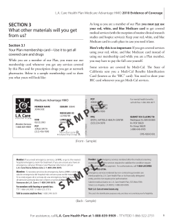

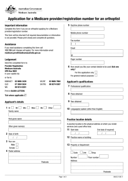


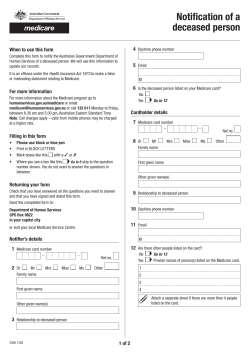

© Copyright 2026