
WASHINGTON STATE PATROL Materials Analysis Primary Foundation Training Manual
WASHINGTON STATE PATROL
Materials Analysis
Primary Foundation Training Manual
CRIME LABORATORY DIVISION
July 2014
Washington State Patrol Crime Laboratory Division
Table of Contents
1
INTRODUCTION ................................................................................................................................. 5
1.1
PURPOSE AND SCOPE ....................................................................................................... 5
1.2
ASSESSMENT OF TRAINEE'S BACKGROUND ................................................................. 5
1.3
ORGANIZATION OF THE TRAINING MANUAL ................................................................... 6
2
LAB SAFETY..................................................................................................................................... 8
2.1
OBJECTIVES ........................................................................................................................ 8
2.2
TOPIC AREAS ....................................................................................................................... 8
2.3
SUGGESTED READING ....................................................................................................... 9
2.4
STUDY QUESTIONS ............................................................................................................ 9
2.5
PRACTICAL EXERCISES ..................................................................................................... 9
3
GENERAL FORENSIC CASE CONSIDERATIONS .................................................................................. 11
3.1
OBJECTIVES ...................................................................................................................... 11
3.2
TOPIC AREAS ..................................................................................................................... 11
3.3
SUGGESTED READING ..................................................................................................... 17
3.4
STUDY QUESTIONS .......................................................................................................... 17
3.5
PRACTICAL EXERCISES ................................................................................................... 18
4
CRIMINALISTICS 101 ....................................................................................................................... 19
4.1
OBJECTIVES ...................................................................................................................... 19
4.2
TOPIC AREAS ..................................................................................................................... 19
5
ETHICS ........................................................................................................................................... 21
5.1
OBJECTIVES ...................................................................................................................... 21
5.2
TOPIC AREAS ..................................................................................................................... 21
5.3
SUGGESTED READINGS .................................................................................................. 22
5.4
PRACTICAL EXERCISES ................................................................................................... 23
6
LAW BASICS AND COURT TESTIMONY.............................................................................................. 24
6.1
OBJECTIVES ...................................................................................................................... 24
6.2
TOPIC AREAS ..................................................................................................................... 24
6.3
SUGGESTED READINGS .................................................................................................. 30
6.4
STUDY QUESTIONS .......................................................................................................... 30
6.5
PRACTICAL EXERCISES ................................................................................................... 31
7
BALANCES AND WEIGHING.............................................................................................................. 32
7.1
OBJECTIVES ...................................................................................................................... 32
7.2
TOPIC AREAS ..................................................................................................................... 32
7.3
SAFETY ............................................................................................................................... 33
7.4
SUGGESTED READING ..................................................................................................... 33
7.5
STUDY QUESTIONS .......................................................................................................... 33
7.6
PRACTICAL EXERCISES ................................................................................................... 34
8
THIN LAYER CHROMATOGRAPHY..................................................................................................... 35
8.1
OBJECTIVES ...................................................................................................................... 35
8.2
TOPIC AREAS ..................................................................................................................... 35
8.3
SAFETY ............................................................................................................................... 38
8.4
SUGGESTED READING ..................................................................................................... 38
8.5
STUDY QUESTIONS .......................................................................................................... 38
8.6
PRACTICAL EXERCISES ................................................................................................... 39
9
GAS CHROMATOGRAPHY ................................................................................................................ 40
9.1
OBJECTIVES ...................................................................................................................... 40
9.2
TOPIC AREAS ..................................................................................................................... 40
9.3
SAFETY ............................................................................................................................... 43
9.4
SUGGESTED READING ..................................................................................................... 43
9.5
STUDY QUESTIONS .......................................................................................................... 44
9.6
PRACTICAL EXERCISES ................................................................................................... 46
10 M ASS SPECTROMETRY & PYROLYSIS .............................................................................................. 47
10.1
OBJECTIVES ...................................................................................................................... 47
10.2
TOPIC AREAS ..................................................................................................................... 47
MAT Primary Foundation Training
Approved by CLD Manager
Page 2 of 117
All Printed Copies are Uncontrolled
Revision Date: July 8, 2014
Revision 2
Washington State Patrol Crime Laboratory Division
10.3
SAFETY ............................................................................................................................... 48
10.4
SUGGESTED READING ..................................................................................................... 48
10.5
STUDY QUESTIONS .......................................................................................................... 49
10.6
PRACTICAL EXERCISES ................................................................................................... 52
11 CAPILLARY ELECTROPHORESIS....................................................................................................... 54
11.1
OBJECTIVES ...................................................................................................................... 54
11.2
TOPIC AREAS ..................................................................................................................... 54
11.3
SAFETY ............................................................................................................................... 61
11.4
SUGGESTED READING ..................................................................................................... 61
11.5
STUDY QUESTIONS .......................................................................................................... 63
11.6
PRACTICAL EXERCISES ................................................................................................... 63
12 FOURIER TRANSFORM INFRARED SPECTROSCOPY ........................................................................... 65
12.1
OBJECTIVES ...................................................................................................................... 65
12.2
TOPIC AREAS ..................................................................................................................... 65
12.3
SAFETY ............................................................................................................................... 67
12.4
SUGGESTED READING ..................................................................................................... 67
12.5
STUDY QUESTIONS .......................................................................................................... 67
12.6
PRACTICAL EXERCISES ................................................................................................... 68
13 FOURIER TRANSFORM RAMAN SPECTROSCOPY ............................................................................... 69
13.1
OBJECTIVES ...................................................................................................................... 69
13.2
TOPIC AREAS ..................................................................................................................... 69
13.3
SAFETY ............................................................................................................................... 70
13.4
SUGGESTED READING ..................................................................................................... 70
13.5
STUDY QUESTIONS .......................................................................................................... 71
13.6
PRACTICAL EXERCISES ................................................................................................... 71
14 HIGH PERFORMANCE LIQUID CHROMATOGRAPHY ............................................................................ 73
14.1
OBJECTIVES ...................................................................................................................... 73
14.2
TOPIC AREAS ..................................................................................................................... 73
14.3
SAFETY ............................................................................................................................... 76
14.4
SUGGESTED READING ..................................................................................................... 76
14.5
STUDY QUESTIONS .......................................................................................................... 76
14.6
PRACTICAL EXERCISES ................................................................................................... 77
15 SCANNING ELECTRON MICROSCOPY/ENERGY DISPERSIVE X-RAY SPECTROSCOPY ..........................79
15.1
OBJECTIVES ...................................................................................................................... 79
15.2
TOPIC AREAS ..................................................................................................................... 79
15.3
SAFETY ............................................................................................................................... 81
15.4
SUGGESTED READING ..................................................................................................... 82
15.5
STUDY QUESTIONS .......................................................................................................... 82
15.6
PRACTICAL EXERCISES ................................................................................................... 83
16 SPECTROSCOPE ............................................................................................................................. 86
16.1
OBJECTIVES ...................................................................................................................... 86
16.2
TOPIC AREAS ..................................................................................................................... 86
16.3
SAFETY ............................................................................................................................... 86
16.4
SUGGESTED READING ..................................................................................................... 86
16.5
STUDY QUESTIONS .......................................................................................................... 86
16.6
PRACTICAL EXERCISES ................................................................................................... 87
17 X-RAY FLUORESCENCE .................................................................................................................. 88
17.1
OBJECTIVES ...................................................................................................................... 88
17.2
TOPIC AREAS ..................................................................................................................... 88
17.3
SAFETY ............................................................................................................................... 89
17.4
SUGGESTED READING ..................................................................................................... 90
17.5
STUDY QUESTIONS .......................................................................................................... 90
17.6
PRACTICAL EXERCISES ................................................................................................... 91
18 BASIC PRACTICAL MICROSCOPY ..................................................................................................... 94
18.1
OBJECTIVES ...................................................................................................................... 94
18.2
TOPIC AREAS ..................................................................................................................... 94
MAT Primary Foundation Training
Approved by CLD Manager
Page 3 of 117
All Printed Copies are Uncontrolled
Revision Date: July 8, 2014
Revision 2
Washington State Patrol Crime Laboratory Division
18.3
SAFETY ............................................................................................................................... 95
18.4
SUGGESTED READING ..................................................................................................... 95
18.5
STUDY QUESTIONS .......................................................................................................... 96
18.6
PRACTICAL EXERCISES ................................................................................................... 97
19 IMAGING AND VISUALIZATION .......................................................................................................... 99
19.1
OBJECTIVES ...................................................................................................................... 99
19.2
TOPIC AREAS ..................................................................................................................... 99
19.3
SAFETY ............................................................................................................................. 101
19.4
SUGGESTED READING ................................................................................................... 101
19.5
STUDY QUESTIONS ........................................................................................................ 102
19.6
PRACTICAL EXERCISES ................................................................................................. 102
20 EVIDENCE RECOVERY ................................................................................................................... 104
20.1
OBJECTIVES .................................................................................................................... 104
20.2
TOPIC AREAS ................................................................................................................... 104
20.3
SAFETY ............................................................................................................................. 104
20.4
SUGGESTED READING ................................................................................................... 104
20.5
STUDY QUESTIONS ........................................................................................................ 105
20.6
PRACTICAL EXERCISES ................................................................................................. 105
21 PHYSICAL M ATCH ......................................................................................................................... 108
21.1
OBJECTIVES .................................................................................................................... 108
21.2
TOPIC AREAS ................................................................................................................... 108
21.3
SAFETY ............................................................................................................................. 110
21.4
SUGGESTED READINGS ................................................................................................ 110
21.5
STUDY QUESTIONS ........................................................................................................ 110
21.6
PRACTICAL EXERCISES ................................................................................................. 111
PRIMARY FOUNDATION MODULE 1 CHECKLIST ....................................................................................... 112
PRIMARY FOUNDATION MODULE 2 CHECKLIST - PAGE 1 ......................................................................... 113
PRIMARY FOUNDATION MODULE 2 CHECKLIST - PAGE 2 ......................................................................... 114
PRIMARY FOUNDATION MODULE 3 CHECKLIST ....................................................................................... 115
PRIMARY FOUNDATION MODULE 4 CHECKLIST ....................................................................................... 116
PRIMARY FOUNDATION MODULE 5 CHECKLIST ....................................................................................... 117
MAT Primary Foundation Training
Approved by CLD Manager
Page 4 of 117
All Printed Copies are Uncontrolled
Revision Date: July 8, 2014
Revision 2
Washington State Patrol Crime Laboratory Division
1 INTRODUCTION
1.1
PURPOSE AND SCOPE
This manual contains an outline for training and/or assessing a forensic scientist in the area of materials
analysis. Each scientist will have a unique training program depending on the individual’s strengths and
weaknesses, previous background, the needs of the laboratory, and available personnel to provide the
training. The sequence in which the various sections are presented should not necessarily be considered
as a mandatory order of training. Exposure to legal aspects, testimony, and many other facets of forensic
science will be continuous throughout the training.
This manual endeavors to promote and maintain consistency and quality among scientists performing
materials analyses across the Crime Laboratory Division. Certain inherent aspects of materials analysis
prohibit the establishment of a rigid set of standard procedures to cover every case. Sufficient latitude
should be given to allow for independent thought and individual freedom in selecting alternative courses
of action. Upon completion of this training program, the trainee will be thoroughly familiar with the options
available to perform an examination of most types of evidence that may be received.
1.2
ASSESSMENT OF TRAINEE'S BACKGROUND
Commencement of this training program does not assume prior experience in forensic materials analysis.
It is assumed, however, that the trainee will have a solid understanding of basic organic and inorganic
chemistry and instrumental analysis.
Trainees who have prior related training and experience may be able to progress through the training
program at an accelerated pace or perform competency testing to demonstrate their ability in a particular
materials analysis discipline or sub-discipline rather than performing redundant training. All trainees will
be required to demonstrate competence in a particular area in which they will be performing analysis.
The instructors must be experienced in the area of materials analysis for which they are to instruct. The
instructor’s casework and courtroom experiences, both prior and present, provide a unique aspect to the
trainee’s learning process that is impossible to duplicate in this training program. It is expected that the
instructor will share such experiences with the trainee. Although the trainee’s primary interaction will be
with the assigned instructor, this program promotes and encourages discussions with other experienced
examiners. The trainee’s instructor and/or supervisor, or other experienced scientist, will preliminarily
assess the trainee’s:
• Knowledge of:
o Analytical chemistry
o Organic chemistry
o Inorganic chemistry
o Physical chemistry
o Microscopy
o Instrumentation
o Physics
• Laboratory experience
• Determination of strengths and deficiencies:
o From this an initial plan developed for remedial readings, lectures, and schooling, if
necessary.
o Training will be customized to meet the needs of the trainee.
Once the course of training has been established and initiated, the trainee will maintain a notebook or
multiple notebooks for the various segments throughout the duration of this training program and will
record notes and observations for each study segment. The trainee notebook should be maintained in a
MAT Primary Foundation Training
Approved by CLD Manager
Page 5 of 117
All Printed Copies are Uncontrolled
Revision Date: July 8, 2014
Revision 2
Washington State Patrol Crime Laboratory Division
neat and current fashion and should be present during conversations with the trainer. Upon completion of
training, the trainee will maintain the training notebook for the duration of their career.
The trainee should be continuously evaluated throughout the training for comprehension and competency
in theoretical knowledge, basic practical skills, and critical thinking skills. Training is progressive and
continuously builds on and reinforces prior learning. Deficiencies on any of the training steps may occur
during the course of the training and should be rectified. It is important that these deficiencies be openly
and promptly discussed among the trainee, trainer, technical lead, and/or supervisor, as appropriate.
Repeating training steps and testing may be necessary to satisfactorily complete this training program.
In order to successfully complete this training program, the trainee must, after completion of each study
segment, demonstrate their capability to apply learned knowledge and skills. Training checklists for this
training are located in Appendix A and must be completed by the trainee and trainer. The trainer is
responsible for writing an interoffice communication (IOC) to the trainee’s supervisor when the trainee has
successfully completed the Primary Foundation Training. The trainee’s supervisor will maintain copies of
training IOCs and authorizations in their files.
1.3
ORGANIZATION OF THE TRAINING MANUAL
The training manual consists of several study segments, each covering different aspects of chemical
analysis.
Each study segment is comprised of six parts:
• The Objectives summarizes the purpose of each training segment.
• The Topic Areas designates topics to be included in the training segment.
• The Safety section indicates specific safety information relating to the training segment.
• The Suggested Readings section lists the reference material that should be read to successfully
complete the study segments. The reading assignments are cumulative; comprehension of prior
readings may be required to successfully complete study/discussion questions and exercises of
subsequent study segments. It may not be necessary or practical to read every reference listed.
The trainee will work with the trainer for specifics.
• The Study Questions have a number of purposes:
o To assist reading comprehension by providing a focus on certain concepts prior to completing
the Reading section;
o To evaluate understanding of relevant concepts after completing the Readings; and
o To promote active discussions between the trainer, trainee and trainee’s co-workers using
the questions as a starting point.
o Written answers to these questions will be maintained in the training notebook as
documentation of training.
• The Practical Exercises are designed to provide the trainee first-hand experience with the main
concepts of each study segment. Data or written explanation for each Practical Exercise must be
maintained in the training notebooks.
Module 1 of the Primary Foundation is the Introduction which includes:
• General Forensic Procedures
• Lab Safety
• Criminalistics 101
• Ethics/Professional Conduct
• Law Basics and Court Testimony
Module 2 of the Primary Foundation covers Instrumentation and Techniques, which includes:
• Balances and Weighing
• Thin Layer Chromatography
• Gas Chromatography/Flame Ionization Detector
• Mass Spectrometry & Pyrolysis
MAT Primary Foundation Training
Approved by CLD Manager
Page 6 of 117
All Printed Copies are Uncontrolled
Revision Date: July 8, 2014
Revision 2
Washington State Patrol Crime Laboratory Division
•
•
•
•
•
•
Capillary Electrophoresis
Infrared Spectroscopy
Raman Spectroscopy
Scanning Electron Spectroscopy/Energy Dispersive X-Ray Spectroscopy
Spectroscope
X-Ray Fluorescence Spectrometry
Module 3 of the Primary Foundation is Basic Practical Microscopy
Module 4 of the Primary Foundation covers:
• Imaging & Visualization
• Evidence Recovery
Module 5 of the Primary Foundation is Physical Match
A comprehension examination will follow Modules 2, 3, 4 and 5. Competency testing will follow Modules
3, 4 and 5. Co-signed casework for Module 5 may begin with successful completion of the written and
competency testing. An IOC will be written by the trainer upon the successful completion of co-signed
casework recommending the trainee for independent casework. Once the IOC has been approved
through the chain of command, the trainee will be allowed to perform independent casework for Physical
Match.
The instructor is responsible for ensuring that the trainee is prepared to testify as an expert witness. This
can be done with mock trials, prearranged as well as impromptu question and answer sessions, and
observation of courtroom testimony given by experienced forensic scientists. One or more mock trials will
be scheduled during the training program.
MAT Primary Foundation Training
Approved by CLD Manager
Page 7 of 117
All Printed Copies are Uncontrolled
Revision Date: July 8, 2014
Revision 2
Washington State Patrol Crime Laboratory Division
2 LAB SAFETY
2.1
OBJECTIVES
•
•
•
•
2.2
To familiarize the trainee with potential safety hazards in the laboratory.
To instruct the trainee on how to properly deal with chemical spills.
To make the trainee aware of safety equipment available in the laboratory.
To familiarize the trainee with proper handling and disposal of chemicals.
TOPIC AREAS
1. Safety tour
2. Material Safety Data Sheets (MSDS)
3. Proper gas cylinder handling
4. Work station set up
5. Specific cautions
6. Fume hood use
7. Glass and sharps disposal
8. Chemical waste plan
9. Injury reports
10. Safety committee
11. Personal Protective Equipment (PPE)
a. Gloves
b. Goggles
c.
Lab coats
d. Other
12. Chemical spills
13. Scalpel use and syringe techniques
14. Biohazards and disposal
15. Specific precautions
a. Some controlled substances, like LSD (Lysergic Acid Diethylamide), can be
absorbed into the skin.
16. Biohazards: Not all requests are labeled if this is the case. Evidence can be from a body
cavity.
17. Sharps hazard: Though not normally accepted as evidence, syringes, broken glass, or
razor blades can be found in evidence. Do not blindly place your hand into an evidence
container. Repackaging may be necessary to reduce the risk of injury to others who may
handle the evidence in the future.
18. Chemical hazards: The majority of chemicals used in this unit have one or more
hazardous properties.
MAT Primary Foundation Training
Approved by CLD Manager
Page 8 of 117
All Printed Copies are Uncontrolled
Revision Date: July 8, 2014
Revision 2
Washington State Patrol Crime Laboratory Division
2.3
SUGGESTED READING
1. Crime Laboratory Division’s “Safety Manual”
2. Crime Laboratory Division’s “Operations Manual”
3. Crime Laboratory Division’s “Quality Manual”
4. Forensic Services Guide
5. WSP Safety and Wellness Manual
6. WSP Bloodborne Pathogens Training for Civil Service Employees PowerPoint
2.4
STUDY QUESTIONS
1. What is the FLSB policy regarding accepting hypodermic needles, razor blades and
other sharps?
2. What is the FLSB policy regarding accepting evidence retrieved from body cavities?
3. Research gloves. Which ones are best for chemical analysis and why?
4. Find the following MSDS and answer the questions: Chloroform, Methanol, Methylene
Chloride, Pentane, Hydrochloric Acid, and Sodium Bicarbonate.
a. What is the target organ?
b. What is the first aid measure for eye exposure? Dermal exposure?
c.
What are Quickits? Where can they be found?
d. What is the recommended PPE?
e. What are the signs and symptoms of exposure?
f.
2.5
What is the HMIS (Hazardous Materials Identification System) flammability
rating?
PRACTICAL EXERCISES
1. Complete a facility safety tour. At a minimum the following should be covered in this
tour:
a. Operation and location of emergency showers.
b. Operation and location of emergency eyewashes.
c.
Operation and location of the spill clean-up kits.
d. Location of fire alarms, emergency exits, and evacuation plan.
e. Location and understanding of Material Safety Data Sheets.
f.
The rules concerning eating and drinking in the laboratory.
g. Location and appropriate use of safety glasses.
h. Appropriate use and disposal of laboratory coats.
i.
Location and appropriate use of hood(s) in your area.
j.
Location and explanation of the fire extinguishers.
k.
Location and contents of first aid stations and kits.
l.
Location and use of fire blankets.
m. Proper disposal of scalpel blades, syringes, broken glass, and biohazards.
MAT Primary Foundation Training
Approved by CLD Manager
Page 9 of 117
All Printed Copies are Uncontrolled
Revision Date: July 8, 2014
Revision 2
Washington State Patrol Crime Laboratory Division
n. How to deal with bomb threats.
o. Explanation of the alarm and security system.
p. Proper handling and storage of compressed gas cylinders.
2. Complete the New Employee/Transfer Safety Orientation form 3000-342-175.
3. Complete the Safety Orientation Checklist form (CLD-SAF-15001).
4. Perform the eyewash inspection with the designated person.
5. Perform the fume hood inspection.
6. Review the mandatory online WSP Bloodborne Pathogens Training for Civil Service
Employees PowerPoint.
7. In the fume hood and wearing the appropriate PPE, pour approximately 10 milliliters of
water onto the counter. Go through the proper clean up procedure, using the spill kit, if
the water was methanol. Repeat as if the water was sulfuric acid. Repeat for sodium
hydroxide.
8. With the assistance of a colleague, blindfolded, make your way to the nearest shower or
eyewash from your lab bench.
9. Determine if the following are compatible and therefore can be stored next to each
other:
a. aluminum hydroxide and aluminum oxide
b. aluminum oxide and aluminum sulfate
c.
benzoyl chloride and benzylamine
d. oxalic acid and nitric acid
e. nitric acid and pine-sol
f.
methanol and heptanes
MAT Primary Foundation Training
Approved by CLD Manager
Page 10 of 117
All Printed Copies are Uncontrolled
Revision Date: July 8, 2014
Revision 2
Washington State Patrol Crime Laboratory Division
3 GENERAL FORENSIC CASE CONSIDERATIONS
3.1
OBJECTIVES
•
•
•
•
•
3.2
To introduce the trainee to evidence handling, accountability and chain of custody policies.
To instruct the trainee on proper preparation of case notes and reports.
To make the trainee aware of forensic ethics and standards of professional conduct.
To introduce the trainee to case approach and interpretation of data.
To familiarize the trainee with special safety considerations involved in materials analysis
casework.
TOPIC AREAS
1. Evidence Handling
a. Preservation of evidence
i.
The integrity and security of the evidence shall be maintained.
ii.
Any significant change in the evidence whether intentional or accidental
must be noted.
iii.
Care will be taken to ensure analytical techniques employed do not
adversely affect future testing of the evidence by other functional areas,
or to anticipate the possibility and plan the analysis scheme accordingly.
b. Chain of custody
c.
i.
Definition
ii.
Performing evidence transfers with user agencies
iii.
Performing evidence transfers within the lab
Security measures
i.
Seals (tape, staples, markings)
ii.
Laboratory security and vault procedures
iii.
Personal evidence lockers
d. Contamination Issues
i.
Potential for DNA and fingerprint contamination
ii.
Maintain a clean, organized work area and use clean utensils.
iii.
Work one case at a time.
iv.
Work one item at a time.
v.
Minimize exposures from large items (such as large quantities of powder
or moldy vegetable material).
vi.
Keep evidence covered or sealed when not present.
e. Safety
i.
Safety checks performed on guns and explosives
ii.
Do not blindly reach into evidence packaging especially potential drug
kits or clothing pockets as they may contain sharps.
iii.
Be cognizant of potentially explosive or ignitable evidence.
iv.
Handle potential biohazard evidence using appropriate techniques.
MAT Primary Foundation Training
Approved by CLD Manager
Page 11 of 117
All Printed Copies are Uncontrolled
Revision Date: July 8, 2014
Revision 2
Washington State Patrol Crime Laboratory Division
f.
Initial Observation of Evidence
i.
Are all seals intact and properly marked?
ii.
Note signs of tampering, multiple seals.
iii.
Are evidence description and numbers consistent with the Request for
Laboratory Examination?
g. Labels and other devices/procedures to avoid confusion and/or enable future
identification (such as in court):
i.
of the original evidence and packaging
ii.
of generated samples (e.g. GC/MS vials)
iii.
of data and notes
iv.
of packaging provided by the Lab in addition to the original packaging
v.
of items that may need fingerprint examinations
h. Proper Seals
i.
Evidence tape must be used whenever practical when sealing evidence
packaging.
ii.
Staples do not constitute proper seals.
iii.
Proper evidence seals must be initialed or otherwise marked to
document the person sealing the evidence. Such markings on all taped
seals must be made so that the initials flow from the tape to the package.
Inclusion of the date of sealing and the Crime Lab number is also
recommended.
iv.
Manufactured seams may be sufficiently secure or may be taped.
v.
Heavy packages may need a reinforcement tape, such as book tape,
below the evidence tape in order to prevent the evidence from falling out
of a resealed package.
vi.
For bottles, jars, cans and film canisters, make sure the lid is secure and
that evidence tape extends across the top of the lid and down both sides
of the lid to the container body.
vii.
Heat sealed packages must have initials or other identification across the
heat seal to be properly sealed.
viii.
Evidence seals should ensure that any efforts of entry or attempted entry
will be obvious.
ix.
i.
Proper seals protect evidence from loss, cross-transfer and/or
contamination.
Introduction to LIMS and how evidence is logged (See also the LIMS Manual)
2. Case Work Documentation
Case files must be complete, organized, legible, and must support all conclusions stated in the written
report. Case notes and analytical documentation include but are not limited to the following:
a. Relevant case information/history/background; communications with
investigators, attorneys, etc.
b. Chain of custody records
c.
Description of evidence packaging, seals, and case identifiers (or lack of)
MAT Primary Foundation Training
Approved by CLD Manager
Page 12 of 117
All Printed Copies are Uncontrolled
Revision Date: July 8, 2014
Revision 2
Washington State Patrol Crime Laboratory Division
i.
Note and address discrepancies in evidence versus the Request for
Laboratory Examination Form. Evidence shortages will be witnessed by
a coworker and documented in the notes. Evidence surplus will also be
documented in the notes. (See the Operations Manual Section 3.3 for
details.)
ii.
Description of items of evidence (packaging, quantity, size, colors,
physical appearance/conditions), both analyzed and not analyzed.
1. Use Submitting Agency exhibit numbers where possible.
2. Outer packaging descriptions are adequate for items not
analyzed.
iii.
Refer to the Operations Manual, Section 3.1.2 for instructions regarding
high value items, especially currency.
iv.
Describe photographs, diagrams, and/or photocopies if necessary.
v.
Weight or volume of examined items: methods used to derive weights or
volumes are specifically outlined.
d. Examination procedures used and the parameters for those procedures
e. Analytical procedures
i.
Special handling of evidence (such as for prints)
ii.
Any trace evidence collection
iii.
Microscopic examinations
iv.
Preparation of controls or comparison samples
v.
Description of solvents used and extractions performed
vi.
Instrument parameters
vii.
Description of mathematical calculations
viii.
Identity and source of any standards or references used
ix.
f.
Results of examinations
Data generated
i.
Spectra, chromatograms and other instrumental printouts
ii.
Photographs, digital images and diagrams
iii.
Observations and descriptions
iv.
Drawings, sketches or other illustrations
g. Records of inter/intralaboratory transfers
h. Notations showing the generation and disposition of generated evidence items
(trace collections, sub-samples, controls, etc…) and any repackaging of evidence
i.
Case notes and analytical documentation must be marked with unique laboratory
case numbers, the handwritten initials of the examiner (and cosigner if
applicable), dates, and page numbers.
j.
Dates should be recorded in the documentation to indicate when work was
performed.
k.
Documentation of technical review on each page of the draft copy of the
laboratory report will remain in the case notes.
MAT Primary Foundation Training
Approved by CLD Manager
Page 13 of 117
All Printed Copies are Uncontrolled
Revision Date: July 8, 2014
Revision 2
Washington State Patrol Crime Laboratory Division
l.
Errors in Note taking
i.
Nothing in the case notes and analytical documentation may be erased
or obliterated. Strikeouts to designate information removed will be
documented by the examiner’s initials. Changes, additions, or any other
form of alteration must be initialed by the person making the alteration.
3. Case Reports
a. Refer to WSP CLD QM & OM sections concerning the protocol for case reports.
b. All reports should be able to stand alone without the necessity of explanation by
supporting verbal testimony.
c.
All conclusions must be accurate, thorough, clearly stated, and justified by data
in notes.
d. All evidence examined must be listed in the report.
e. Items not examined must be documented in the report. The analyzed items are
clearly differentiated from the non-analyzed items.
f.
Conclusions, opinions and interpretations must be identified in the report.
g. Other report contents include:
i.
Complete and accurate case heading;
ii.
The controlled substances conclusively identified;
iii.
Weight/amount of analyzed exhibits;
iv.
Procedures must be clearly and completely outlined except for controlled
substance reports.
v.
Test certifications on controlled substance case reports, and other court
or RCW requirements;
vi.
Signature and date line.
h. Verbal reports
i.
j.
i.
May be issued when preapproved by the section supervisor.
ii.
A draft of the verbal report must be written.
iii.
The draft of the verbal report and the supporting data must be technically
reviewed.
iv.
Documentation by the technical reviewer will be entered in LIMS.
v.
Analyst must document the date time and name of the person to whom
the verbal report was released on the written version of the verbal report.
Amended reports
i.
Issued to modify the content of previously issued laboratory reports.
ii.
The report will be titled “Amended Report” and will contain a brief
explanation describing the need for the amended report.
iii.
All amended reports must be documented in the Case Information field in
LIMS. The reason for the amended reported will be noted in the case file.
iv.
Review of the amended report is required.
Use of LIMS for report generation
i.
MAT Primary Foundation Training
Approved by CLD Manager
Edit findings
Page 14 of 117
All Printed Copies are Uncontrolled
Revision Date: July 8, 2014
Revision 2
Washington State Patrol Crime Laboratory Division
ii.
Milestones
iii.
Activities
iv.
NFLIS
v.
Analytical techniques
Refer to the Crime Laboratory Division Operating Manual and Quality Manual for additional details on
case notes, administrative documentation and case reports.
4. Case Approach and Analysis
a. Prioritization of casework
i.
Immediate Response
1. Immediate investigative need to identify, arrest, hold/release a
suspect in custody;
2. Lab work needed to avert danger to someone’s life;
3. Immediate need to preserve physical evidence for future work.
ii.
Court Deadlines in crimes against persons
1. Information from lab work needed to prosecute crimes against
persons and drug crimes
2. Trial date set
iii.
30 Day Turnaround Goal
1. Consider number of items to be examined including one item per
suspect. (See the Forensic Laboratory Services Guide for
submitting agency guidelines and general analysis policy.)
b. Prior to opening evidence:
c.
i.
Consult user agencies as necessary.
ii.
Consider any inter/intralaboratory case coordination.
iii.
Preparation of work area
iv.
Preparation of various commonly used chemical solutions/reagents
Evaluate evidence to plan analytical strategy.
i.
Documentation of evidence prior to manipulation
1. description, photos, sketches
ii.
Choose appropriate analytical methods
iii.
Examine using least destructive methods first and minimize sample
consumption.
iv.
Obtain appropriate reference material or control samples as necessary.
1. verification and documentation of reference materials
v.
Collection of evidence for analysis by other functional areas
vi.
Packaging/labeling of items to be examined by other functional areas
vii.
Consider travel to other facilities to use instrumentation not available
locally. (See the Quality Manual Section 6.2.3.6 for details on use of
outside instrumentation.)
MAT Primary Foundation Training
Approved by CLD Manager
Page 15 of 117
All Printed Copies are Uncontrolled
Revision Date: July 8, 2014
Revision 2
Washington State Patrol Crime Laboratory Division
d. Interpretation of Data
i.
Make use of reference materials, libraries, consultations, manufacturers
or specialists, and/or other forensic scientists.
ii.
Specificity of applied techniques
iii.
Prevention of false-positives
iv.
Two-prong analysis: if possible, each analysis utilizes independent
theory, reagents, etc.
1. It is desirable that different portions be taken from a sample for
analysis by different techniques; however, evidence items
containing a small amount of material may not be sufficient to
allow multiple samples to be taken. When only one sampling of
an item is practical, particular care should be taken.
v.
Use of blanks/controls
vi.
Formulation of Conclusion
1. Objective vs. subjective (identification and qualified conclusions)
2. Inconclusive/lack of conclusion
3. Insufficient sample
4. Residues
5. All conclusions justified by data
5. Case Review
a. Analyst review
i.
Analysts will conduct a thorough case review of their own work prior to
technical review.
ii.
The analyst review must cover all elements of the technical and
administrative review.
b. Technical review
i.
The technical reviewer must be technically proficient in the functional
area and the specific sub-discipline to participate in technical review.
ii.
An approved Technical Review Checklist will be completed for all cases.
iii.
Covers the elements listed in the CLD QM for technical review.
iv.
Reviewer will initial and date each page of the final draft report upon
completion of the review and will mark the technical review milestone in
LIMS. For cases involving inter-laboratory review, each page of the draft
will be signed and dated by the technical reviewer.
v.
Keys to successful technical review
1. Listen courteously.
2. Always be respectful.
3. Be open and receptive to suggestions.
4. Keep an open mind and behave professionally.
5. Address each of the suggested changes/improvements.
MAT Primary Foundation Training
Approved by CLD Manager
Page 16 of 117
All Printed Copies are Uncontrolled
Revision Date: July 8, 2014
Revision 2
Washington State Patrol Crime Laboratory Division
6. Work with the reviewer to resolve all issues at the peer level, as
much as possible. For situations where the technical review
issues cannot be resolved, see the Quality Manual for guidance.
c.
Inter-Lab Technical Review
i.
One case per quarter from each analyst will be sent to another lab for
technical review.
ii.
Original case file will be retained in the lab and a copy of the file will be
sent out for technical review by the section supervisor or designee.
d. Administrative review
3.3
i.
Conducted on the case file and final report prior to the release of the
written report.
ii.
Covers the elements listed in the CLD QM for administrative review.
iii.
Administrative reviewer does not need to be technically proficient in the
functional area.
iv.
The administrative review is documented in LIMS and may be
documented in the case file as well.
v.
The administrative review may be combined with the technical review.
SUGGESTED READING
1. WSP Forensic Services Guide
2. WSP CLD Quality Manual
3. WSP CLD Operations Manual
4. WSP CLD LIMS Manual
5. WSP CLD Materials Analysis Technical Manual
6. Forensic Sciences Handbook, Volume I, Chapter 1; Volume II, Chapters 2 and 3;
Volume III, pages 1-23, Saferstein, R.
7. DeForest PR, Gaensslen RE, Lee HC. 1983. Forensic Science: An Introduction to
Criminalistics, Chapters 1-3. New York:McGraw Hill. p. 1-94.
3.4
STUDY QUESTIONS
1. Discuss the methods and purpose of evidence security and chain of custody.
2. Discuss the marking of evidence. What are the minimum markings required on evidence
packaging?
3. Discuss the required documentation used for analytical examinations.
4. What types of “new evidence” may be created during the course of analysis? How
would you document this new evidence in your notes, in your report and in LIMS?
5. You are analyzing evidence and you are paged to go to the phone. Describe your
activities.
6. You receive a phone call from an officer with a syringe. What do you tell him/her?
7. Its lunchtime and you have an open case on your desk. Describe your actions. It's
quitting time and the same case is on your desk. Describe your actions.
8. What must be considered when a piece of evidence has multiple exam codes listed on
the RFLE?
MAT Primary Foundation Training
Approved by CLD Manager
Page 17 of 117
All Printed Copies are Uncontrolled
Revision Date: July 8, 2014
Revision 2
Washington State Patrol Crime Laboratory Division
9. How are cases received and logged into the Lab?
10. What are some elements that a report should contain?
3.5
PRACTICAL EXERCISES
1. Observe your instructor or other qualified employee during the analysis of a case.
2. Review a minimum of 10 case folders from several experienced forensic scientists.
3. Observe the entire process that a case follows while in the laboratory.
4. Log a case into the Lab and into the LIMS system.
MAT Primary Foundation Training
Approved by CLD Manager
Page 18 of 117
All Printed Copies are Uncontrolled
Revision Date: July 8, 2014
Revision 2
Washington State Patrol Crime Laboratory Division
4 CRIMINALISTICS 101
4.1
OBJECTIVES
This two day training program will cover the fundamental aspects of criminalistics including the
interdisciplinary chain of evidence examination in the crime laboratory. The trainee will learn what types of
evidence are examined in each functional area. The trainee will learn how to safely handle and evaluate a
broad range of evidence including firearms, smears and stains, blood patterns, cuts and tears and small
particle characterization. Trainees will develop strategies to document, recover evidence, prevent
contamination and preserve evidence (e.g. A DNA examiner will understand the potential significance of
smokeless powder grains, metal turnings or vomit on a shirt).
4.2
TOPIC AREAS
1. INTRODUCTION TO CRIMINALISTICS
a. Goals of the workshop
b. PowerPoint lectures and labs - types of physical evidence encountered.
c.
Team building approach-we all have value
d. Interdisciplinary study of physical evidence
e. Contamination and evidence preservation
f.
i.
Overview of each section of the laboratory
ii.
Specific evidence types encountered
Documentation, collection, and packaging of evidence
i.
References and useful documents such as the ABC study guide
2. LAW and ETHICS
3. CSRT
a. Blood Stain Pattern Analysis
b. Trajectory analysis
c.
Evidence recovery
4. LATENT PRINTS
a. Overview of work performed
b. Evidence preservation, contamination prevention, and packaging
c.
Examples of impressions on materials such as handguns and duct tape
5. MATERIALS ANALYSIS
a. Overview of work performed
b. Trace evidence recovery
c.
Use of microscopes
d. Evidence sub-disciplines
i.
Fabric damage
ii.
Physical match
iii.
Impressions/smears/stains
iv.
Fibers/fabric/cordage
MAT Primary Foundation Training
Approved by CLD Manager
Page 19 of 117
All Printed Copies are Uncontrolled
Revision Date: July 8, 2014
Revision 2
Washington State Patrol Crime Laboratory Division
v.
Hairs
vi.
Paint
vii.
Explosives/residue
viii.
Soil
ix.
Botanicals
x.
Tape
xi.
Lamps
xii.
Glass
xiii.
Controlled substances
xiv.
Fire debris
xv.
Clandestine laboratories
xvi.
Unknowns
6. DNA/SEROLOGY/CODIS
a. Overview of work performed
b. Blood and body fluids
c.
Blood testing methods
d. DNA
e. Contamination prevention methods
7. FIREARMS/TOOLMARKS
a. Overview of work performed
b. Safety concerns
c.
Types of Weapons
d. Ammunition - cartridges/cartridge cases/bullets
e. Distance determinations
f.
Bullet holes in clothing
g. Gunpowder particles
8. DOCUMENTS EXAMINATION
a. Overview of work performed
b. Evidence preservation, contamination prevention, and packaging
9. LABORATORY EXAMINATIONS AND DEMONSTRATIONS
a. Examination/identification of particle types (hairs/fibers, paint chips, glass,
building materials, insect parts, botanicals, cosmetics, mud/soil, vomit, etc.)
b. Clothing damage (cuts and tears)
c.
Impressions, smears and stains
d. Handgun safety, prior packaging and handling
e. Awareness of hazardous evidence submissions
f.
Bloodstain patterns on clothing, tools/weapons, porous and non-porous surfaces
MAT Primary Foundation Training
Approved by CLD Manager
Page 20 of 117
All Printed Copies are Uncontrolled
Revision Date: July 8, 2014
Revision 2
Washington State Patrol Crime Laboratory Division
5 ETHICS
5.1
OBJECTIVES
•
•
5.2
To make the trainee aware of forensic ethics and standards of professional conduct.
To make the trainee familiar with State policies on use of resources and acceptance of gifts.
TOPIC AREAS
The following were compiled verbatim from SWGDRUG (Scientific Working Group for the Analysis of
Seized Drugs) and the American Board of Criminalistics’ Rules of Professional Conduct in Forensic
Science. They are considered to meet general acceptance by peers in this profession.
1. Analysts should:
a. act with honesty, integrity and objectivity;
b. maintain an attitude of independence and impartiality in order to ensure an
unbiased analysis of the evidence;
c.
work only within the bounds of their professional competence;
d. take reasonable steps to maintain their competence;
e. recognize that their overriding duty is to criminal justice;
f.
declare to their employer any prior contact or personal involvement, which may
give rise to conflict of interest, real or perceived;
g. declare to their employer or other appropriate authority any pressure intended to
influence the result of an examination;
h. carry out the duties of the profession in such a manner so as to inspire the
confidence of the public.
2. Regarding casework, analysts should:
a. ensure and be able to demonstrate that the integrity and security of evidential
materials and the information derived from their analysis have been maintained
while in their possession;
b. ensure that they have a clear understanding of what the customer needs and all
the necessary information, relevant evidential materials and facilities available to
reach a meaningful conclusion in an appropriate timeframe;
c.
employ an appropriate analytical approach, using the facilities available;
d. make and retain full, contemporaneous, clear and accurate records of all
examinations and tests conducted, and conclusions drawn, in sufficient detail to
allow meaningful review and assessment of the conclusions by an independent
person competent in the field;
e. accept responsibility for all casework done by themselves and under their
direction;
f.
conduct all professional activities in a way that protects the health and safety of
themselves, co-workers, the public and the environment.
3. Regarding reporting, analysts should:
a. present advice and testimony, whether written or oral, in a clear and objective
manner;
MAT Primary Foundation Training
Approved by CLD Manager
Page 21 of 117
All Printed Copies are Uncontrolled
Revision Date: July 8, 2014
Revision 2
Washington State Patrol Crime Laboratory Division
b. be prepared to reconsider and, if necessary, change their conclusions, advice or
testimony in light of new information or developments, and take the initiative in
informing their employer and customers promptly of any such changes that need
to be made;
c.
take appropriate action if there is potential for, or there has been, a miscarriage
of justice due to new circumstances that have come to light, incompetent practice
or malpractice;
d. preserve customer confidentiality unless officially authorized to do otherwise;
e. render opinions and conclusions strictly in accordance with the evidence in the
case (hypothetical or real) and only to the extent justified by that evidence;
f.
testify in a clear, straightforward manner and refuse to extend oneself beyond
one’s field of competence, phrasing one’s testimony in such a manner so that the
results are not misinterpreted;
g. not exaggerate, embellish or otherwise misrepresent qualifications when
testifying;
h. consent to, if it is requested and allowed, interviews with counsel for both sides
prior to trial;
5.3
i.
make efforts to inform the court of the nature and implications of pertinent
evidence if reasonably assured that this information will not be disclosed to the
court;
j.
find it appropriate to report to their supervisor any violation of these professional
rules of conduct by another scientist.
SUGGESTED READINGS
1. Budowle B. et al. 2009. A Perspective on Errors, Bias and Interpretation in the Forensic
Sciences and Direction for Continuing Advancement. J Forensic Sci. 54(4):798-809.
2. Dror IE. 2008. Biased Brains. Police Review. 116:23-23.
3. Dror IE, Charlton D, Péron AE. 2006. Contextual Information Renders Experts
Vulnerable to Making Erroneous Identifications. J For Sci Int. 156:74-78.
4. Dror IE, Péron AE, Hind SL, Charlton D. 2005. When Emotions Get the Better of Us:
The Effect of Contextual Top-down Processing on Matching Fingerprints. Appl. Cognit.
Psychol. 19:799-809.
5. Dror IE, Charlton D. 2006. Why Experts Make Errors. J For Ident. 56(4):600-616.
6. Lucas DM. 1989. The Ethical Responsibilities of the Forensic Scientist: Exploring the
Limits. J For Sci. 34(4):719-729.
7. Peterson JL, Murdock JE. 1989. Forensic Science Ethics: Developing an Integrated
System of Support and Enforcement. 24(3):749-762.
8. Revised Code of Washington (RCW) 42.52.
9. Saks MJ. 1989. Prevalence and Impact of Ethical Problems in Forensic Science. J For
Sci. 34(3):772-793.
10. Schroeder OC. 1984. Ethical and Moral Dilemmas Confronting Forensic Scientists. J For
Sci. 29(4):966-986.
11. Starrs JE. 1971. The Ethical Obligations of the Forensic Scientist in the Criminal Justice
System. J AOAC. 54(4):906-914.
MAT Primary Foundation Training
Approved by CLD Manager
Page 22 of 117
All Printed Copies are Uncontrolled
Revision Date: July 8, 2014
Revision 2
Washington State Patrol Crime Laboratory Division
12. Thompson WC. 1995. Subjective Interpretation, Laboratory Error and the Value of
Forensic DNS Evidence: Three Case Studies. Genetica. 96:153-168.
The following documents can be located on the Portal FLSB Home Page under the Ethics area:
1. ASCLD/LAB Guiding Principles of Professional Responsibility for Crime Laboratories
and Forensic Scientists.
2. Guidance on Gifts
3. Guidance on Use of State Resources
4. Use of State Resources
5.4
PRACTICAL EXERCISES
Review the Ethics Training for WSP and Ethics in Forensics PowerPoint presentations located on the
Portal FLSB Home Page under the Ethics area. Discuss the presentations and scenarios with the trainer.
Review the Ethics Scenarios and Answers on the Portal FLSB Home Page under the Ethics area.
Discuss these scenarios with the trainer.
Review the ASCLD/LAB: Guiding Principles. Discuss this information with the trainer.
RTI International provides free online forensic training. These courses are optional and may be
recommended by your trainer.
MAT Primary Foundation Training
Approved by CLD Manager
Page 23 of 117
All Printed Copies are Uncontrolled
Revision Date: July 8, 2014
Revision 2
Washington State Patrol Crime Laboratory Division
6 LAW BASICS AND COURT TESTIMONY
6.1
OBJECTIVES
•
•
•
•
•
•
•
•
6.2
Demonstrate a basic understanding of terms, legal decisions and issues relevant to the forensic
scientist.
Demonstrate a basic understanding of the judicial process and how cases are tried in various
courts of law.
Provide an overview of the legal system and its participants.
Present a brief summary of criminal procedure.
Understand the forensic significance of the Bill of Rights and other amendments to the US
Constitution.
Understand the importance of and how to prepare for testimony.
Understand the demeanor and delivery of an expert witness testimony.
Demonstrate how to effectively employ visual displays to aid in testimony.
TOPIC AREAS
1. The Legal System Overview
a. The basis of law
b. Law foundations
c.
i.
The Constitution
ii.
Statutes
iii.
Administrative regulations
iv.
Judicial decisions
Adversarial system
d. Presumption of innocence
i.
Beyond a reasonable doubt
ii.
Preponderance of the evidence
e. Rights of the accused
f.
i.
Due process
ii.
Bill of Rights
Civil versus criminal law
2. Dual Court System
a. Jurisdiction
b. Federal Courts
i.
US District Courts
ii.
US Court of Appeals
iii.
US Supreme Court
iv.
Specialized Courts
1. Military Justice
2. Tribal Courts
MAT Primary Foundation Training
Approved by CLD Manager
Page 24 of 117
All Printed Copies are Uncontrolled
Revision Date: July 8, 2014
Revision 2
Washington State Patrol Crime Laboratory Division
c.
State Courts
i.
Courts of Limited Jurisdiction: Lower Courts
1. District
2. City
3. Municipal
ii.
Courts of General Jurisdiction: Major Trial Courts
1. Superior
2. Criminal and civil cases
iii.
Intermediate Courts of Appeals
iv.
State Supreme Court
3. Participants in the Court System
a. Law Enforcement
i.
Security Staff
ii.
Sheriff’s Deputies
iii.
Bailiff
b. Courts
i.
Legal
1. Prosecutor
2. Public defender
3. Private defense attorney
4. Judge
5. Law clerk
ii.
Court Support Staff
1. Court clerk
2. Court reporter
3. Court administrator
4. Translator
c.
Corrections
i.
Probation officer
ii.
Pretrial services
iii.
Drug rehabilitation program
d. Public
i.
Defendant
ii.
Victim
iii.
Witness
iv.
Jurors
v.
Bail agent
MAT Primary Foundation Training
Approved by CLD Manager
Page 25 of 117
All Printed Copies are Uncontrolled
Revision Date: July 8, 2014
Revision 2
Washington State Patrol Crime Laboratory Division
vi.
Victim witness assistance program
vii.
Rape crisis center
viii.
Child advocate
ix.
Court watchers
x.
Media
4. Steps of Criminal Procedure
a. Crime
b. Arrest
c.
Initial appearance
i.
Accused is told of the charges, advised of their rights, bail is set and a
date for the preliminary hearing is set.
ii.
Suspect may enter a plea.
d. Charging
i.
Information
ii.
Complaint
iii.
Arrest warrant
e. Preliminary hearing
i.
f.
Hearing to determine if there is probable cause to proceed with the case.
Grand jury
i.
Required in twenty-two states and federal courts
ii.
Indictment
g. Arraignment
i.
The defendant is formally charged and must enter a plea.
ii.
Types of pleas
1. Guilty
a.
b.
c.
d.
Regular guilty plea
Nolo contendere (no contest)
Alford plea
Conditional guilty plea
2. Not guilty
3. Insanity plea
h. Bail
i.
Disclosing and suppressing evidence
j.
Negotiated Justice
i.
Plea agreements
1. Charge bargaining
2. Count bargaining
3. Sentence bargaining
MAT Primary Foundation Training
Approved by CLD Manager
Page 26 of 117
All Printed Copies are Uncontrolled
Revision Date: July 8, 2014
Revision 2
Washington State Patrol Crime Laboratory Division
k.
Trials
l.
Sentencing
m. Appeals
5. Constitutional Rights of Forensic Significance
a. Fourth Amendment
b. Fifth Amendment
c.
Sixth Amendment
d. Fourteenth Amendment
6. The Uniform Rules of Evidence
a. Rule 401
b. Rule 402
c.
Rule 702
d. Rule 703
e. Rule 705
f.
Rule 1002
7. Court Decisions of Forensic Significance
a. Jencks v. United States
b. Brady v. Maryland
c.
Giglio v. United States
d. Frye v. United States
e. Daubert v. Merrell Dow Pharmaceuticals
f.
General Electric Co. v. Joiner
g. Kuhmo Tire Co. v. Carmichael
h. Arizona v. Youngblood
i.
Crawford v. Washington
j.
Melendez-Diaz v. Massachusetts
k.
Bullcoming v. New Mexico
l.
State v. Nation
m. United States v. Plaza
n. Williams v. Illinois
o. Washington v. Manion
p. Washington v. Lui
8. Testimony
a. Preparation for testimony
i.
pretrial discussions and/or meetings with attorneys
ii.
review of case notes and report
iii.
Curriculum Vitae (CV)
MAT Primary Foundation Training
Approved by CLD Manager
Page 27 of 117
All Printed Copies are Uncontrolled
Revision Date: July 8, 2014
Revision 2
Washington State Patrol Crime Laboratory Division
iv.
notification of supervisor
b. Courtroom appearance and dress
c.
i.
professional appearance
ii.
appropriate attire
iii.
posture
Courtroom demeanor
i.
exclusion from the courtroom
ii.
outside courtroom considerations
iii.
eye contact
iv.
tone of voice
v.
volume
vi.
courtesy
vii.
impartiality
viii.
ethics
d. Presenting Testimony
i.
taking the oath
ii.
taking the witness stand
iii.
qualifying as an expert
1. voir dire
e. handling evidence on the stand
f.
i.
recognition
ii.
safety
iii.
gloves
iv.
refrain from displaying evidence (“publishing”) to jury without prior
permission
direct examination
i.
communication skills
1. verbal: avoid slang, professional jargon, profanity, unexplained
abbreviations and acronyms
2. nonverbal
3. pace
ii.
use of analogies
iii.
credibility
1. recognizing limits of knowledge or expertise
iv.
admitting mistakes, limitations and problems
v.
inability to remember
vi.
objections
vii.
cross-examination
MAT Primary Foundation Training
Approved by CLD Manager
Page 28 of 117
All Printed Copies are Uncontrolled
Revision Date: July 8, 2014
Revision 2
Washington State Patrol Crime Laboratory Division
1. open-ended questions
2. leading questions
3. unclear questions
4. compound questions
5. hypothetical questions
6. yes or no questions
7. listening carefully
viii.
re-direct
ix.
re-cross-examination
x.
leaving the witness stand
xi.
being finally excused
g. Defense tactics
i.
attacking credibility and qualifications
ii.
attacking chain of custody
iii.
attacking procedures
iv.
attacking conclusions
v.
weight of testimony
vi.
proffering “authoritative” texts
vii.
hostile demeanor
h. Use of visual displays and other presentation aids
i.
j.
i.
prior discussion with attorney
ii.
easily seen and understood
iii.
photographs and glare
iv.
digital images
v.
use of colors on charts, graphs
vi.
retention as court exhibit
vii.
demonstrations
Testimony review
i.
required annually
ii.
performed by supervisor, another analyst or officer of the court
iii.
documented on testimony review form
iv.
evaluation criteria
Discovery
i.
WAC 446-10-050
ii.
WSP policies
MAT Primary Foundation Training
Approved by CLD Manager
Page 29 of 117
All Printed Copies are Uncontrolled
Revision Date: July 8, 2014
Revision 2
Washington State Patrol Crime Laboratory Division
6.3
SUGGESTED READINGS
1. Cleary EW. 1972. McCormick’s Handbook of the Law of Evidence. 2nd ed. West
Publishing Co.
2. Farley M. 1993. Legal Standards for the Admissibility of Novel Scientific Evidence. In:
Saferstein, R. Editor. Forensic Science Handbook. Vol. 3. Englewood Cliffs (NJ):
Prentice-Hall.
3. Kogan, J.D. 1978. On Being a Good Expert Witness in a Criminal Case”, J For Sci.
23:190-200.
4. Matson JV. 2003. Effective Expert Witnessing, 4th ed. CRC Press.
5. Neubauer DW. Fradella HF. 2010. America’s Courts and the Criminal Justice System.
th
10 Ed. Blemont (CA): Wadsworth. or equivalent book.
6. Sapir GJ. 2002. Legal Aspects of Forensic Science. In: Saferstein R. Editor. Forensic
nd
Science Handbook. Vol. 1. 2 ed. Englewood Cliffs (NJ): Prentice-Hall.
7. Training Seminar: Ron Smith & Associates, Inc., “Courtroom Testimony Techniques
“Success Instead of Survival”.
8. The Uniform Rules of Evidence
9. Court opinions for the listed decisions
6.4
STUDY QUESTIONS
1. Briefly describe the difference between the Frye Standard and the Daubert Standard?
Which standard is the law in Washington?
2. What is the Confrontation Clause of the Sixth Amendment? How does this impact
forensic science? Which legal decisions are based on the Confrontation Clause?
3. What is “Relevant Evidence”? Where is relevant evidence legally defined?
4. What is a subpoena duces tecum? What should you do if you receive this type of
subpoena?
5. You have spent months analyzing evidence in a case and on the eve of the trial you
learn the evidence was excluded under the “fruit of the poisonous tree doctrine”. What
does this mean?
6. What is a bench trial? How does this differ from a jury trial?
7. It is estimated that less than ten percent of cases go to trial. What processes could
contribute to so few of cases going to trial?
8. What is an Omnibus hearing? What is a 3.5-3.6 hearing?
9. Discuss appropriate court demeanor and dress.
10. What is the difference between a regular witness and an expert witness?
11. As you are delivering testimony, one of the attorneys exclaims, “Objection”. What do you
do?
12. How can you avoid technical jargon when testifying?
13. In a jury trial, whom should the witness address?
14. Define the following terms.
a. Continuance
b. Deposition
MAT Primary Foundation Training
Approved by CLD Manager
Page 30 of 117
All Printed Copies are Uncontrolled
Revision Date: July 8, 2014
Revision 2
Washington State Patrol Crime Laboratory Division
c.
Disclosure
d. Discovery
e. Omnibus hearing
f.
Spoliation
g. Stipulation
h. Subpoena duces tecum
i.
Voir dire
15. What does WAC 446-10-050 say regarding availability of public records? Understand
and discuss the policy regarding discovery and disclosure.
16. How would you address a question regarding why you didn’t examine an item for
fingerprints? For DNA?
17. Why is testimony reviewed annually? What criteria are included in the annual review?
6.5
PRACTICAL EXERCISES
1. Develop a Curriculum Vitae (CV).
2. Observe court testimony by other members of the laboratory staff.
MAT Primary Foundation Training
Approved by CLD Manager
Page 31 of 117
All Printed Copies are Uncontrolled
Revision Date: July 8, 2014
Revision 2
Washington State Patrol Crime Laboratory Division
7 BALANCES AND WEIGHING
7.1
OBJECTIVES
•
•
•
7.2
To familiarize the trainee with the appropriate use of electronic balances.
To familiarize the trainee with the common terms associated with weighing and electronic
balances.
To familiarize the trainee with balance quality assurance and techniques that will minimize error.
TOPIC AREAS
1. Basic concepts
2. Mass versus weight
a. Scales or balances measure force (weight) caused by mass that is influenced by
gravity.
3. Mechanical balance scales
a. An unknown mass is balanced by a known counter mass. This direct mass to
mass comparison is equally influenced by gravity and therefore calibration is not
required when moving the scale.
4. Electronic scales
a. Load cell transducers measure force that occurs when gravity acts on a mass.
As gravity changes, it is necessary to calibrate the scale to accommodate for the
change in gravity.
b. When an object is placed on the load cell transducer in the electronic scale, the
mechanical force is converted to an electric property. This electric property is
amplified and processed to be displayed as the weight of the object.
5. International System of Units
a. Kilogram
b. Traceability
6. Types of electronic balances /Readability
a. Microbalance
b. Analytical balance
c.
Precision balance
7. Terms used in weighing
a. Accuracy
b. Adjustment
c.
Calibration
d. Corner load
e. Hysteresis
f.
Linearity
g. Precision
h. Readability
MAT Primary Foundation Training
Approved by CLD Manager
Page 32 of 117
All Printed Copies are Uncontrolled
Revision Date: July 8, 2014
Revision 2
Washington State Patrol Crime Laboratory Division
i.
Repeatability
j.
Sensitivity
8. Optimizing the performance of the balance
***The following are conditions that will improve the precision and reproducibility of weighing especially
for analytical and micro-balances. Not all of the conditions can be achieved in each laboratory due to
facilities/ environmental conditions or are necessary for weighing with precision balances.
a. Location
i.
Balance placement
ii.
Temperature
iii.
Humidity
iv.
Light
v.
Air
b. Balance operation
7.3
i.
Leaving the balance on at all times
ii.
Leveling the balance
iii.
Balance adjustment
iv.
Reading
v.
Weighing pan
vi.
Weighing vessel
vii.
Draft shield
viii.
Care of the balance
SAFETY
As with all electronic equipment, caution should be taken to avoid exposing the balance to liquids.
7.4
SUGGESTED READING
1. Correct Use and Handling of Analytical and Microbalances. Sartorius. Available as a
.pdf at www.sartonews.com
2. The Fundamentals of Weighing Technology: Terms, Methods of Measurement, Errors
in Weighing. Sartorius, 1996. Available as a .pdf at www.sartonews.com.
3. Norden KE. 1993. Electronic weighing: Fundamentals and applications. ButterworthHeinemann, Ltd. Oxford.
4. Proper Weighing with Laboratory Balances. Mettler Toledo, 2008. Available as a .pdf at
www.mt.com.
5. Quantitative Chemical Analysis text of your choosing.
7.5
STUDY QUESTIONS
1. Define the following terms
a. Accuracy
MAT Primary Foundation Training
Approved by CLD Manager
Page 33 of 117
All Printed Copies are Uncontrolled
Revision Date: July 8, 2014
Revision 2
Washington State Patrol Crime Laboratory Division
b. Corner load
c.
Hysteresis
d. Linearity
e. Precision
f.
Readability
g. Repeatability
h. Sensitivity
2. How do calibration and adjustment differ in regards to electronic balances?
3. What is the difference between microbalances, analytical balances, and precision
balances?
4. Discuss how the location in which a balance is placed can affect weighing precision and
reproducibility.
5. Why should the balance be left on at all times? How long does it take to warm-up
analytical and precision balances?
6. What factors should be considered in the selection and handling of a weighing vessel?
7. Which of the following physical influences can impact the weight determination of routine
controlled substances cases? What can be done to minimize the physical influences
that impact weighing items of controlled substances?
a. Temperature
b. Moisture gain/evaporation
c.
Electrostatics
d. Magnetism
e. Static buoyancy
f.
Gravitation
8. What is “traceability” in regards to the quality assurance of balances? What standard
are balances traced to?
9. What is the difference between internal and external calibration? Should internal
calibration be performed on our balances? Why or why not?
10. How would you describe to a jury the concept of a gram? What is the mathematical
conversion between grams and ounces?
7.6
PRACTICAL EXERCISES
1. Check the performance of the balances in your laboratory following the quality
assurance plan.
2. Weigh the following:
a. Weighing paper – various sizes
b. Weigh boats – various sizes
c.
Contents of a “Sweet ‘n Low” or “Equal” package.
d. Ten marihuana seeds, if available.
e. A piece of rolling paper, if available.
MAT Primary Foundation Training
Approved by CLD Manager
Page 34 of 117
All Printed Copies are Uncontrolled
Revision Date: July 8, 2014
Revision 2
Washington State Patrol Crime Laboratory Division
8 THIN LAYER CHROMATOGRAPHY
8.1
OBJECTIVES
•
•
8.2
To familiarize the trainee with the theory and application of thin layer chromatography in drug
analysis.
To familiarize the trainee with the advantages and limitations of using thin layer chromatography
in materials analysis.
TOPIC AREAS
1. Theory
a. Thin-layer chromatography (TLC) is a method for separating chemical mixtures.
Compounds are separated from each other based on differences in their
interactions between a stationary phase and a mobile phase.
2. Materials
a. Stationary phase
b. Mobile phase
c.
Developing chamber
d. Visualizing agents
3. TLC Separation Concepts
a. Partitioning/adsorption
b. Mobile phase selection
c.
Stationary phase selection
d. Retention factor (Rf)
i.
Sample concentration effects
ii.
Mobile phase composition
iii.
Mobile phase/stationary phase equilibrium
iv.
Edge effects
v.
Stationary phase uniformity
vi.
Temperature
e. Developing methods
f.
i.
Closed chamber
ii.
Open chamber
Visualization methods
i.
UV
ii.
Fluorescence
iii.
Color reagent sprays
g. Preparative TLC
i.
Sample application
ii.
Development
MAT Primary Foundation Training
Approved by CLD Manager
Page 35 of 117
All Printed Copies are Uncontrolled
Revision Date: July 8, 2014
Revision 2
Washington State Patrol Crime Laboratory Division
iii.
Visualization
iv.
Sample collection
v.
Sample recovery
4. TLC Procedures
a. Sample preparation
b. Sample application
c.
i.
Spot size
ii.
Spot location
QA/QC
i.
Blanks
ii.
Standards
iii.
Documentation
d. Safety
i.
Developing agents
ii.
Visualizing agents
iii.
Stationary phase
5. TLC Information Generated
a. Number of components
i.
Visualized
ii.
Not visualized
b. Rf values
c.
Color of visualized spots
d. Value of information
i.
Category B test
ii.
Comparative
iii.
Indicative
iv.
Semi-quantitative
6. The following TLC systems and visualizing agents are examples that may be used and
are not to be considered all inclusive or the only appropriate method for conducting the
analysis of the example compounds. Ultraviolet light and reagents used for color tests
may also be used as TLC location aids.
Barbiturates
Solvent -- chloroform:acetone (9:1)
Visualized – potassium permanganate; mercuric nitrate, oversprayed with
diphenylcarbazone
Cocaine
Solvent -- chloroform:methanol (4:1)
Visualized -- acidified iodoplatinate
Diazepam
Solvent -- methanol:concentrated ammonia (100:1.5)
Visualized -- acidified iodoplatinate
MAT Primary Foundation Training
Approved by CLD Manager
Page 36 of 117
All Printed Copies are Uncontrolled
Revision Date: July 8, 2014
Revision 2
Washington State Patrol Crime Laboratory Division
Ephedrine
Solvent -- chloroform:methanol (4:1)
Visualized -- acidified iodoplatinate
Heroin
Solvent – diethyl ether:diethylamine (9:1) chloroform:methanol (4:1)
Visualized -- acidified iodoplatinate
LSD
Solvent -- Acetone (or acetone:chloroform 1:4)
Spot sample versus known LAMPA, LSD and mix. Plate can be irradiated with
long wavelength UV and replaced in developing reservoir. Visualize with UV. Can
be over sprayed with Erhlich's. Also can be visualized with acidified iodoplatinate.
Marihuana
Solvent – hexane:diethyl ether (4:1); toluene
Visualized -- Fast Blue B (carcinogenic), or Fast Blue BB
Methamphetamine
Solvent -- chloroform:methanol (4:1)
Visualized -- acidified iodoplatinate
Pseudoephedrine
Solvent -- chloroform:methanol (4:1)
Visualized -- acidified iodoplatinate
Psilocyn/Psilocybin
Solvent -- methanol:concentrated ammonia (20:1)
Visualized -- Fast Blue B, overspray with conc. HCl
-- Erhlichs: Psilocyn, Psilocybin
Steroids
Solvent -- chloroform:acetone (8:2)
Visualized -- sulfuric acid/ethanol reagent
7. Visualizing Agents
Mercuric chlorideDiphenylcarbazone
19mg in 200mL (50% acetone/water). Used for barbiturates.
Ceric sulfate
5g Ce(SO4)2 in 500mL water and 14 mL sulfuric acid. Used as an overspray to
intensify the reaction with iodoplatinate.
Dragendorff
1.3g of bismuth subnitrate in 60Ml water with 15mL acetic acid. Add this to 12g
KI in 30 mL water. Dilute with 100mL of water and 25mL acetic acid. General
spray, good for diazepam, alkaloids, and nitrogenous compounds.
Ehrlich's
2g p-dimethylaminobenzaldehyde in 50mL 95% ethanol and 50mL
concentrated HCl. Visualizes LSD, reacts with indole nucleus of alkaloids. Heat
plate to intensify color.
Fast Blue B
Approx. 0.5% to 1% solution of Fast Blue B in water. Used for marihuana.
Δ9-THC — red, cannabidiol — orange, cannabinol — purple
Fluorescamine
20mcg in 100mL acetone. Visualize amino acids, amines and amino sugars.
Heat after spraying, check under long wavelength UV light.
MAT Primary Foundation Training
Approved by CLD Manager
Page 37 of 117
All Printed Copies are Uncontrolled
Revision Date: July 8, 2014
Revision 2
Washington State Patrol Crime Laboratory Division
Furfuraldehyde
Furfuraldehyde in ethanol, HCl. May heat plate after spraying. For nonaromatic carbamates; black spots.
HCl - 6N
Used to acidify plates (e.g., with Fast Blue B for psilocyn/psilocybin).
Iodoplatinate
1g chloroplatinic acid in 10mL conc. HCl, plus 20g of KI in 400mL water. Used
for nitrogenous compounds.
Ninhydrin
Ninhydrin in various solvents. For amino acids, amines and amine sugars. Heat
after spraying, view under long wave UV.
Potassium
Permanganate
Potassium permanganate in water. Unsaturated hydrocarbons — yellow on
purple.
Sulfuric Acid/Ethanol
Gradually add 10mL conc. sulfuric acid to 90mL of ethanol. Used for steroids.
8.3
SAFETY
1. UV radiation can be harmful to the eyes and care should be exercised to avoid direct
exposure to UV radiation.
2. Visualizing reagent sprays may be hazardous.
3. Good chemical safety practices should be employed when working with reagents. TLC
should be performed in a functional fume hood. When the TLC plate has been reviewed
and observations recorded, the plate should be disposed of properly and should not be
kept as part of the case record.
8.4
SUGGESTED READING
1. Gough TA. 1991. The Analysis of Drugs of Abuse: Separation Science Series.
Chichester, England: Johan Wiley and Sons, p. 3-22.
2. Hamilton R. Hamilton S. 1978. Thin Layer Chromatography: Analytical Chemistry by
Open Learning. London: Johan Wiley and Sons.
3. Moffat AC. Clarke’s Isolation and Identification of Drugs. London: The Pharmaceutical
press.
4. Saferstein R. 2011. Organic Analysis. In: Criminalistics - An Introduction to Forensic
Science, 10th ed. Englewood Cliffs (NJ): Prentice-Hall.
8.5
STUDY QUESTIONS
1. Describe the safety hazards associated with TLC and how these hazards can be
mitigated.
2. What materials are widely used as stationary phases and what applications are each
best used for?
3. What “samples” should be spotted onto a TLC plate when analyzing case exhibits?
4. How are TLC plates documented in the case file?
5. What is retention factor and how can it be affected by the following:
a. Sample concentration
b. Mobile phase
MAT Primary Foundation Training
Approved by CLD Manager
Page 38 of 117
All Printed Copies are Uncontrolled
Revision Date: July 8, 2014
Revision 2
Washington State Patrol Crime Laboratory Division
c.
Mobile phase/stationary phase equilibrium
d. Edge effects
e. Stationary phase uniformity
f.
Temperature
6. Describe the technique of TLC as you would to a jury.
8.6
PRACTICAL EXERCISES
1. Separate a mixture of cocaine, lidocaine, nicotinamide and lactose. Compare to the
individual standards.
2. Separate a mixture of psilocin, psilocybin and bufotenine.
3. Separate a mixture of cannabinoids using an extraction from marihuana. Develop with
Fast Blue B.
4. Choose an analyte (e.g., cocaine) and place spots of increasing concentration onto a
single TLC plate and observe overloading effects.
5. Demonstrate edge effects.
6. Experiment using different visualizing agents with different analytes. Determine the
advantages and disadvantages.
7. Run plates consecutively using the same mobile phase to evaluate how often the mobile
phase can be used without replacement.
MAT Primary Foundation Training
Approved by CLD Manager
Page 39 of 117
All Printed Copies are Uncontrolled
Revision Date: July 8, 2014
Revision 2
Washington State Patrol Crime Laboratory Division
9 GAS CHROMATOGRAPHY
9.1
OBJECTIVES
•
•
•
•
•
9.2
To familiarize the trainee with the theory and application of gas chromatography (GC) in materials
analysis.
To familiarize the trainee with the GC instrumentation and software used in the laboratory.
To have the trainee understand the advantages and disadvantages of gas chromatography.
To have the trainee demonstrate familiarity with gas chromatography terminology.
To have the trainee demonstrate how to properly interpret gas chromatographic data.
TOPIC AREAS
1. Theory
a. GC involves the partitioning of gaseous solutes between an inert gas mobile
phase and a stationary solid or liquid phase. It is an instrumental separation
technique based on the difference in the distribution or partition coefficients of
substances having appreciable volatility at temperatures below approximately
350 - 400ºC.
b. History of chromatography
c.
Gas/liquid phase equilibrium
i.
distribution coefficient
2. Instrument Design
a. General: oven, carrier gas, injection port, analytical column, detector, recorder
b. Injection systems
c.
i.
Syringe types/methods, septa sweep
ii.
Packed and capillary, on-column, split/splitless injections
iii.
Solvent effects
iv.
Background, maintenance/trouble-shooting strategies
Types of columns
i.
Packed columns – filled with granular packing that is kept in place by
gas-permeable plugs at both ends.
ii.
Capillary columns
1. Wall-coated open tubular (WCOT) – have the liquid phase
coated directly on the inside, relatively smooth wall of the column
tubing.
2. Porous-layer open-tubular (PLOT) – have a solid porous layer
present on the tube wall but still maintain the unobstructed
central gas-flow channel. This porous solid layer can either act
as an adsorbent or a support which in turn is coated with a thin
film of the liquid phase, or both. The solid layer can either be
deposited on the inside tube wall or formed by chemical means
from the wall.
3. Support-coated open tubular (SCOT) – those PLOT columns
where the solid layer consists of the particles of a solid support
which were deposited on the inside tube wall.
MAT Primary Foundation Training
Approved by CLD Manager
Page 40 of 117
All Printed Copies are Uncontrolled
Revision Date: July 8, 2014
Revision 2
Washington State Patrol Crime Laboratory Division
iii.
Capillary columns (current)
1. Advantages of modern capillary columns; cross linked and
bonded liquid phases.
2. Capillary column nomenclature
3. Common stationary phases used in our labs
a. HP/DB-1 nonpolar 100% Dimethylpolysiloxane
b. HP/DB-5 nonpolar (5%-Phenyl)-methylpolysiloxane
c. RTX/DB-200 midpolarity 35% Trifluoropropyl)methylpolysiloxane
4. Column construction
5. Liquid phases/selection/temperature limits
a. column bleed
6. Solid supports
7. Adsorbents
8. Column diameters (narrow, wide megabore)
9. Film thickness and column capacity
10. Compatibility, practical operating tips
11. Installation of columns
a.
b.
c.
d.
cutting columns
gas flow adjustments
column conditioning
troubleshooting
12. Linear velocity through capillary column
13. Causes and prevention of column damage
a. physical damage
b. oxygen damage – most common reason for a column to
fail
c. thermal damage – lack of carrier gas to the column while
heating is probably the most common cause of thermal
damage
d. chemical damage
i.
organic solvents and water – generally not
damaging although phase stripping will occur
over time
ii.
bases – inorganic bases are particularly
damaging, while organic bases are not
iii.
acids – in general, mineral acids will damage
stationary phases
e. guard or pre-columns
3. Separation Concepts
a. Retention time and retention volume
b. Dead volume
c.
Adjusted retention time
MAT Primary Foundation Training
Approved by CLD Manager
Page 41 of 117
All Printed Copies are Uncontrolled
Revision Date: July 8, 2014
Revision 2
Washington State Patrol Crime Laboratory Division
d. Flow rate and average linear velocity
e. Partition coefficients
f.
Column efficiency
i.
Theoretical plates and HETP
ii.
van Deemter equation and plots
iii.
Choice of carrier gas and liquid phase
g. Resolution
h. Temperature effects
4. Optimization of Operating Conditions
a. Temperature programming
b. Isothermal programs
c.
Electronic pressure control
5. Kovats indices and McReynolds constants
6. Detection
a. Flame ionization
i.
principle of operation
ii.
gases
iii.
column position
iv.
linear range and sensitivity
v.
common problems
b. Thermal conductivity
c.
Electron capture
d. Others (NPD, HEC, PID, AED, MSD, IR, AA)
e. Strengths and weakness of each detector
7. Quality Assurance/Quality Control
a. System check-out
b. Documentation
c.
Log books
d. Calibrations
e. Blanks and reference materials (instrument libraries)
8. Instrument Maintenance
a. Routine maintenance
i.
Changing septa
ii.
Changing injection port liners and O-rings
iii.
Carrier gas and detector gases
iv.
FID flame jet and collector cleaning
b. Leak detection
MAT Primary Foundation Training
Approved by CLD Manager
Page 42 of 117
All Printed Copies are Uncontrolled
Revision Date: July 8, 2014
Revision 2
Washington State Patrol Crime Laboratory Division
c.
Calibration
d. Oxygen traps and carrier gas considerations
e. Column installation
f.
Cleaning injection ports and detectors
g. Troubleshooting
i.
baseline disturbances
1. spiking
2. noise
3. wander
4. drift
5. offset
ii.
ghost peaks
iii.
irregular peak shape or size
1. reduced peak size
2. tailing peaks
3. fronting peaks
4. rounded or flat-top peaks
5. split peaks
iv.
retention time shifts
v.
loss of separation or resolution
vi.
rapid column deterioration
9. Special Techniques
a. Derivatives (see Chemistry Principles in the Controlled Substances Training
Manual)
b. Pattern recognition in analyzing complex mixtures
c.
9.3
Quantitation (overview)
SAFETY
All systems should be turned off and cooled prior to performing maintenance. Refer to the GC technical
procedures for specific safety information regarding the handling of compressed gases.
9.4
SUGGESTED READING
1. ASTM E 355 (Current version) - Standard Practice for Gas Chromatography Terms and
Relationships
2. Barry EF, Grob RL. 2004. Modern Practice of Gas Chromatography. New York: John
Wiley & Sons.
3. Fowlis IA. 1995. Gas Chromatography, Analytical Chemistry by Open Learning. 2
West Sussex (England): John Wiley & Sons.
nd
ed.
4. Moffat, A.C. Clarke’s Isolation and Identification of Drugs. London: Pharmaceutical
Press.
MAT Primary Foundation Training
Approved by CLD Manager
Page 43 of 117
All Printed Copies are Uncontrolled
Revision Date: July 8, 2014
Revision 2
Washington State Patrol Crime Laboratory Division
5. Poole CF, Schuette SA. 1984. Contemporary Practice of Chromatography. New York:
Elsevier.
6. Regis Chemical Company. 1976. A User’s Guide to Chromatography. Illinois: Innovative
Products and Technical Support.
7. Rood D. 1999. A Practical Guide to the Care, Maintenance, and Troubleshooting of
Capillary Gas Chromatographic Systems, Systems. 3rd ed. New York: Wiley-VCH.
8. Stafford DT. 1988. Forensic Capillary Gas Chromatography. In: Saferstein R, editor.
Forensic Science Handbook. Vol II. Englewood Cliffs (N J): Prentice Hall. p.38-67.
9. Stafford DT. 1992. Forensic Gas Chromatography. In: Tebbett I. Gas Chromatography
in Forensic Science. West Sussex (England): Ellis Horwood.
10. Willard HH, Merritt LL Jr., Dean JA, Settle FA Jr. 1988. Instrumental Methods of
th
Analysis. 7 ed. New York: Van Nostrand.
11. Instrument manufacturer manuals.
NOTE: Most any general text on chromatography may substitute for one or more of the above references.
The material in this outline may be covered by a basic GC course sponsored by a local vendor.
9.5
STUDY QUESTIONS
1. What is gas chromatography?
2. What types of information are obtained using a GC-FID system?
3. Draw a schematic diagram for a GC and describe the purpose of each component.
4. Describe the differences between the solid support used in packed columns and that
used in a capillary column GC system.
5. What general criteria should all stationary phases possess? How do they differ between
packed and capillary systems?
6. What general criteria should all mobile phases possess?
7. Besides the stationary phase, what factors influence column selection for a given GC
application?
8. What determines the appropriate column diameter for a given GC system? The
appropriate length? Why are packed column lengths usually less than 3 meters?
9. Describe how the following concepts affect GC separation between components:
a. Solubility
b. Boiling point
c.
Intermolecular forces
10. How are packed columns or liners deactivated after installation? How does it work?
11. What is column bleed?
12. When and why are columns conditioned? Describe the process.
13. Define:
a. Retention time (TR or tR),
b. Relative retention time (RRT),
c.
Retention volume,
d. Unretained retention time (tm)
MAT Primary Foundation Training
Approved by CLD Manager
Page 44 of 117
All Printed Copies are Uncontrolled
Revision Date: July 8, 2014
Revision 2
Washington State Patrol Crime Laboratory Division
e. Phase ratio (β)
f.
Selectivity (α)
14. Define the following:
a. Theoretical plate (n)?
b. Effective theoretical plate (N)?
c.
Theoretical plate height /height equivalent to a theoretical plate (H or HETP)
d. Height equivalent to an effective theoretical plate (H or HEETP)
e. Average linear gas velocity (μ)
f.
What is a good value for the HETP? And why?
g. How is the # of N related to column efficiency?
15. Define Resolution (R).
a. What is chromatographic resolution a function of?
b. Why is resolution not the best measure of column efficiency and column
performance?
16. Diagram and explain the van Deemter plot. Why does the lab use helium or hydrogen as
a carrier gas for its drug instruments?
17. What is the Kovats retention index (I)? What does it mean if I = 650?
18. What affect do the following have on retention time?
a. Concentration
b. Other compounds in the sample
c.
Free base/acid form vs. salt form
19. Discuss the sample introduction of gases and vapors, volatile liquids and solids into a
GC.
20. What factors govern the amount of sample to be injected? How much
sample/component can the average capillary column hold? What factors influence this?
21. What temperature should the injection port be under normal circumstances and why?
22. What type of septa are recommended for GC work and why?
23. What are the differences and purposes of split, splitless, pulsed split, pulsed splitless,
on-column, and direct on-column injection?
24. What is an injection port liner? What is it made of? Why is it used? Describe the packing
process including the materials used.
25. What is a “split ratio” and how is it calculated?
a. What factors govern the use of a particular split ratio (100:1 vs. 50:1)?
26. Why is it necessary to regulate the carrier gas flow?
a. How is this done?
b. What factors influence the optimum flow rate for a given carrier gas?
c.
If the carrier gas is too fast or too slow how will it affect the peak shapes of your
sample components?
27. What is “make-up” gas?
MAT Primary Foundation Training
Approved by CLD Manager
Page 45 of 117
All Printed Copies are Uncontrolled
Revision Date: July 8, 2014
Revision 2
Washington State Patrol Crime Laboratory Division
a. How and why is it used?
b. What determines which gas will be used as a make-up gas?
28. What are some of the common causes and remedies for the following GC system
problems?
a. No peaks
b. Solvent peak only
c.
Baseline drift or unstable baseline
d. Ghost peaks
e. Tailing peaks
f.
Leading peaks
g. Split peaks
h. Retention time shift
29. Explain how derivatization is performed, including why it is used sometimes for analysis.
30. If two drug compounds were to co-elute on the GC, what could be done to resolve the
peaks?
31. Explain as to a jury how a GC operates.
9.6
PRACTICAL EXERCISES
(Note: Not all laboratories have a GC/FID. Some of these exercises may be done with either a GC/FID,
GC/MSD, or both. These exercises may be combined with exercises in the GC/MSD training module.
Obtain instructor approval before manipulating any parameters.)
1. Familiarize yourself with instrument software.
2. Run a 50/50 mix of ephedrine and pseudoephedrine base on a GC/FID or GC/MS
having a DB-1, DB-5 and/or DB-200 column. How many peaks do you see? Now rerun
the sample with acetic anhydride (derivatizing agent). Under which conditions are the
substances separated?
3. Run a primary, secondary and tertiary amine sample on a GC/FID or GC/MS. How do
the spectral patterns differ? How can you account for the differences that are present in
the spectra?
4. Run a mix of methamphetamine, phentermine and N, N-dimethyl amphetamine.
Determine which spectrum belongs to its respective compound.
5. Choose a known dilution of a substance in solution and run on the GC/FID or GC/MS.
Repeat the injection with successive dilutions. What are the sensitivity/levels of
detection of your instrument? Is the GC more sensitive than the GC/MS when run
splitless and at a given split ratio?
MAT Primary Foundation Training
Approved by CLD Manager
Page 46 of 117
All Printed Copies are Uncontrolled
Revision Date: July 8, 2014
Revision 2
Washington State Patrol Crime Laboratory Division
10 MASS SPECTROMETRY & PYROLYSIS
10.1 OBJECTIVES
•
•
•
•
•
•
•
To familiarize the trainee with the theory and application of mass spectrometry in materials
analysis.
To familiarize the trainee with the MS instrumentation and software used in the laboratory.
To have the trainee demonstrate a basic understanding of how a mass spectrometer operates.
To have the trainee demonstrate how to properly interpret mass spectral data.
To familiarize the trainee with the theory and application of pyrolysis.
To familiarize the trainee with the pyrolysis instrumentation and software.
To have the trainee demonstrate a basic understanding of how the pyrolysis instrument operates.
10.2 TOPIC AREAS
1. Theory
a. Mass spectrometry
b. Pyrolysis
2. Instrumentation
a. Mass spectrometry
i.
Inlet systems
1. Chromatographic column
2. Direct inlet (DIP)
ii.
Ionization
1. Electron impact (EI)
2. Chemical ionization
3. ICP
4. Laser ablation
iii.
Mass analysis
1. Magnetic sector
2. Time of flight
3. Quadrupole
4. Ion trap
iv.
Detection
b. Pyrolysis
i.
Furnace pyrolyzers
ii.
Inductively heated (Curie-point) filament pyrolyzers
iii.
Resistively heated filament pyrolyzers
1. CDS 5150 autosampler
2. Sample introduction
3. Valve oven
MAT Primary Foundation Training
Approved by CLD Manager
Page 47 of 117
All Printed Copies are Uncontrolled
Revision Date: July 8, 2014
Revision 2
Washington State Patrol Crime Laboratory Division
4. Transfer line and interface with GC column
3. Routine instrument maintenance
4. Calibration and tuning
5. Sample preparation
6. Interpretation of mass spectra & pyrograms
a. Molecular formula
i.
Isotopic abundances
ii.
Rings and double bonds
iii.
Nitrogen rule
b. Molecular ion
c.
Fragmentation
i.
Ionization energies
ii.
Ion stability
iii.
Common fragments
iv.
Common fragmentation mechanisms
1. Sigma-bond dissociation
2. alpha cleavage
3. Inductive cleavage
a. Rearrangements
b. Radical site
c. Charge site
10.3 SAFETY
1. Components of the pyrolysis system are very hot. All systems should be turned off and cooled
prior to performing maintenance. Refer to the GC/MS technical procedures for specific safety
information regarding the handling of compressed gases.
2. Scientists should be aware that there are two sources of exhaust on the GC/MSD system: the
foreline pump and the GC split vent. The foreline pump outputs gas removed from the vacuum
manifold by the high vacuum pumps. The foreline pump exhaust will also contain traces of
solvent and sample.
3. Caution should be exhibited when working with vacuum pumps. Waste oil should be treated as
hazardous and should be handled and disposed of appropriately.
10.4 SUGGESTED READING
1. Allen AC et al. 1981. The Cocaine Diastereomers. J For Sci. 26(1):12-26.
2. Challinor JM. 2006. Chapter 8: Examination of Forensic Evidence. In Wampler TP,
editor. Applied Pyrolysis Handbook, 2nd Edition. CRC Press: 175-200.
3. Maddock CJ, Ottley TW. 2006. Chapter 3: Pyrolysis Mass Spectrometry:
Instrumentation, Techniques, and Applications In: Wampler TP, editor. Applied Pyrolysis
Handbook, 2nd Edition. CRC Press. p.47-64.
MAT Primary Foundation Training
Approved by CLD Manager
Page 48 of 117
All Printed Copies are Uncontrolled
Revision Date: July 8, 2014
Revision 2
Washington State Patrol Crime Laboratory Division
4. McLafferty FW, Turecek F. 1993 Interpretation of Mass Spectra. 4th ed. Sausalito (CA):
University Science Books.
5. McLafferty FW, Venkataraghavan R. 1982. Mass Spectral Correlations. 2nd ed.
Washington (DC): American Chemical Society.
6. Message GM. 1984. Practical Aspects of Gas Chromatography/Mass Spectrometry.
New York: John Wiley & Sons, New York.
7. Mills T. Roberson JC. 2005. Instrumental Data for Drug Analysis, 3nd ed. New
York:Elsevier.
8. Moffat, A.C. Clarke’s Isolation and Identification of Drugs. London: Pharmaceutical
Press.
9. Moldoveanu SC. 1998. Chapter 1. Introduction and nomenclature. In: Analytical
Pyrolysis of Natural Organic Polymers. Elsevier. p. 3 - 7.
10. Moldoveanu SC. 1998. Chapter 2. The chemistry of the pyrolytic process. In: Analytical
Pyrolysis of Natural Organic Polymers. Elsevier. p. 9 - 32.
11. Moldoveanu SC. 1998. Chapter 3. Physico-Chemical aspects of the pyrolytic process.
In: Analytical Pyrolysis of Natural Organic Polymers. Elsevier. p. 33 - 69.
12. Moldoveanu SC. 1998. Chapter 5. Analytical techniques used with pyrolysis. In:
Analytical Pyrolysis of Natural Organic Polymers. Elsevier. p. 97 - 199.
13. Saferstein R. 1982. Forensic Applications of Mass Spectrometry. In Saferstein R. editor.
Forensic Science Handbook. Vol I. Englewood Cliffs (N J): Prentice Hall 1982. p. 92138.
14. Saferstein, R. 1982. Forensic Science Handbook, Vol. 1, pp. 28-91.
15. Silverstein RM, Bassler GC. 1991. Spectrometric Identification of Organic Compounds.
5th ed, New York: John Wiley & Sons.
16. Watson J T. 1997. Introduction to Mass Spectrometry. 3rd ed. New York: Lippincott.
17. Wampler TP. Chapter 1: Analytical Pyrolysis: An Overview. In: Wampler TP, editor.
Applied Pyrolysis Handbook, 2nd Edition. CRC Press. p.1-26.
18. Wampler TP. Chapter 2: Instrumentation and Analysis. In: Wampler TP, editor.
Applied Pyrolysis Handbook, 2nd Edition. CRC Press. p.27-46.
19. Yinon J. 1987. Forensic Mass Spectrometry. Boca Raton:CRC Press, Inc.
20. Computer-based NIST library of organic compounds (NIST98.l or higher)
21. GC/MS instrument manuals.
22. Instrument manufacturer computer-based tutorials.
NOTE: Most any general text on chromatography may substitute for one or more of the above references.
The material in this outline may be covered by a basic GCMS course sponsored by a local vendor.
10.5 STUDY QUESTIONS
Discuss the importance and relevance of questions below with your instructor.
1. What is mass spectrometry?
2. Describe the theory behind its use as an identification technique.
3. What types of information are obtained from a GC/MS?
4. Draw a schematic diagram of a GC/MS. What is the purpose of each component?
MAT Primary Foundation Training
Approved by CLD Manager
Page 49 of 117
All Printed Copies are Uncontrolled
Revision Date: July 8, 2014
Revision 2
Washington State Patrol Crime Laboratory Division
5. Define the following terms:
a. Relative abundance
b. Base peak
c.
Molecular ion
d. Quasimolecular ion
e. Parent/Precursor ion
f.
Daughter/Product ion
g. Mass/charge ratio
h. Mass spectrum
i.
Resolution
j.
Unit mass resolution
k.
Normalization
l.
PFTBA Normalization
m. Cleavage
n. AMU
o. Isobaric
p. Radical
q. Doubly charged ion
r.
Calibration compound
s.
Torr
t.
Atmosphere
u. Total Ion Current
6. What is a “metastable peak”? When and where does it occur?
7. What is the sensitivity of a GC/MS?
8. What is the difference between spectrometry and spectroscopy?
9. Describe any method considerations in using a MS detector instead of an FID detector
for the GC.
10. Why can column bleed cause a problem in GC/MS and how is it corrected? Septum
bleed?
11. What types of septa are required?
12. How can non-volatile compounds be introduced into a mass spectrometer?
13. What things must an interface between a GC and a MS accomplish?
14. What is the most common mode of ionization?
15. Diagram the EI source for the MS systems in your laboratory.
a. Are the ions formed positive or negative?
16. Do they have an even or odd number of electrons?
17. What is the ionization efficiency of this technique?
18. What governs the relative abundance of the ions formed?
MAT Primary Foundation Training
Approved by CLD Manager
Page 50 of 117
All Printed Copies are Uncontrolled
Revision Date: July 8, 2014
Revision 2
Washington State Patrol Crime Laboratory Division
19. What governs the number and energy of the electrons emitted by the filaments?
20. From what are the filaments made?
21. What is an “ionization appearance potential” curve?
a. What is the usual electron energy used in an EI source for complete ionization
and why?
b. What effect does variation in this energy have on ion abundance?
c.
If a molecule is ionized with energy just at its appearance potential, what
information may be obtained?
22. What vacuum conditions are necessary in the ionization source and the analyzing
regions of a MS and why?
a. Describe how a rough pump works.
b. Describe how a diffusion pump works.
c.
Describe how a turbomolecular pump works.
d. Is it necessary that the vacuum remain constant?
23. What temperature conditions must be maintained in the ion source?
24. Describe how the ions are accelerated once they are formed.
25. Describe how a quadrupole mass analyzer works.
a. What factors influence the practical limits of the quadrupole as a mass filter?
b. What determines whether an ion will have a stable trajectory through the
quadrupoles?
26. Describe how an electron multiplier works.
27. Why is the electron multiplier the detector of choice? What are the limiting factors as to
how well an electron multiplier can detect incoming ions?
28. What reference spectra collections are available for your use?
a. Do they consist of “normalized” data?
b. Do they consist of DFTPP ion abundance calibrated data?
c.
Do they contain verified data?
d. If not, are they still viable references for spectral comparisons?
29. Can optical isomers and diastereomers be differentiated via MS?
30. What is the nitrogen rule?
31. Describe how fragmentation patterns are influenced by:
a. Branched carbon atoms
b. Double bonds
c.
Rings
d. Hetero-atoms
e. Carbonyl groups
32. What requirements are necessary for an ion to be considered a molecular ion?
a. What does increasing saturation and number of rings result in with respect to the
abundance of a molecular ion?
MAT Primary Foundation Training
Approved by CLD Manager
Page 51 of 117
All Printed Copies are Uncontrolled
Revision Date: July 8, 2014
Revision 2
Washington State Patrol Crime Laboratory Division
b. What effect does chain branching have?
33. In what types of compounds is a molecular ion peak frequently not detectable?
34. In what types of compounds are molecular ion peaks most likely to occur?
35. What do the peaks occurring at higher mass numbers than the molecular ion represent?
36. Describe the isotope pattern for Cl and Br.
37. What ions can be associated with the following m/e ratios?
a. 43
b. 58
c.
77
d. 91
38. Define the following terms and describe how these terms relate mass spectrometry to
chromatography?
a. scan rate
b. scan cycle time
c.
reset time
d. a/d conversion rate
e. spectral tilting
f.
mass peak detect threshold
g. GC peak detect threshold
39. What macros are used on the GC/MS in your laboratory and how do they work?
40. What is SIM and what is it used for?
41. Explain as to a jury how a mass spectrometer operates.
42. Explain the key advantages of resistively heated filament pyrolysis instruments over
furnace and Curie-point instruments.
43. Explain the advantages and disadvantages of Curie-point pyrolysis instruments.
44. Explain how sample size and shape can affect reproducibility of pyrolysis analysis.
45. Discuss the importance of sample homogeneity and how analysts can deal with samples
that are non-homogeneous.
46. Define pyrolysis as an analytical method.
47. Explain relative bond strengths and how bond strengths determine macromolecular
degradation in pyrolysis.
48. Explain random scission, side group scission, and monomer reversion mechanisms.
10.6 PRACTICAL EXERCISES
(Note: Obtain instructor approval before manipulating any instrument parameters. These exercises may
be combined with exercises from the GC/FID training module.)
1. Perform a standard spectra autotune on the GC/MS and describe what each value on
the report represents. What types of parameter values may indicate a problem with the
instrument?
MAT Primary Foundation Training
Approved by CLD Manager
Page 52 of 117
All Printed Copies are Uncontrolled
Revision Date: July 8, 2014
Revision 2
Washington State Patrol Crime Laboratory Division
2. Perform an autotune on the GC/MS and compare the results to the standard spectra
autotune. Run the QA mix of drugs using both types of tunes and discuss the
differences in spectra observed.
3. Obtain an unknown spectrum from your instructor. Using interpretive methods, give as
much information about the unknown compound as possible.
4. Obtain mass spectra for groups of similar compounds (e.g., phenethylamines,
homologous series of alcohols, etc) and compare data from each compound.
5. Derivatize certain compounds, including ephedrine and pseudoephedrine, and observe
the resulting data. Compare to data from the underivatized compound.
6. Obtain pyrograms for several polymers, including polyethylene, polypropylene,
polystyrene, polymethyl methacrylate, and nylon. Determine degradation mechanisms
for each, and identify major degradation products.
7. Test reproducibility of pyrolyisis by running a polymer such as polypropylene numerous
times over a period of several days. How much variation in retention times and peak
height ratios is observed?
MAT Primary Foundation Training
Approved by CLD Manager
Page 53 of 117
All Printed Copies are Uncontrolled
Revision Date: July 8, 2014
Revision 2
Washington State Patrol Crime Laboratory Division
11 CAPILLARY ELECTROPHORESIS
11.1 OBJECTIVES
•
•
•
•
•
•
To familiarize the trainee with the theory and application of capillary electrophoresis (CE) in
materials analysis.
To familiarize the trainee with the CE instrumentation and software used in the laboratory.
To have the trainee understand the advantages and disadvantages of capillary electrophoresis.
To have the trainee demonstrate how to properly interpret capillary electrophoresis data.
To familiarize the trainee with quality control and maintenance associated with capillary
electrophoresis.
To familiarize the trainee with safety regarding the use and maintenance associated with capillary
electrophoresis.
11.2 TOPIC AREAS
1. History
a. 1800’s
i.
Early 1800’s - Faraday – Electrophoresis
ii.
1886 – Lodge – H+ migration
b. 1900’s
i.
1930 – Tiselius –moving boundary
ii.
1967 – Hjerten –electrophoresis in tube
iii.
1979 – Mikkers –CZE with Teflon tube
iv.
1981 – Jorgenson –CZE with silica tube
2. Theory
a. Basic electrophoretic principles
i.
Size and charge
ii.
Electrophoresis
iii.
Electroösmosis
1. Electroösmosis factors
a. Dielectric constant
i.
Solvent (i.e. water, acetonitrile, etc.)
b. Zeta potential
c.
i.
Effected by buffer counter ions
ii.
pH of buffer
iii.
Concentration of buffer
Viscosity
i.
Concentration dependent
ii.
Temperature dependent
d. Other interactions
i.
MAT Primary Foundation Training
Approved by CLD Manager
Analyte Velocity
Page 54 of 117
All Printed Copies are Uncontrolled
Revision Date: July 8, 2014
Revision 2
Washington State Patrol Crime Laboratory Division
ii.
Migration time
iii.
Column length
1. Equations use total column length = L
2. Migration is for analyte to travel L.
iv.
Equations of merit
1. Elecrophoretic mobility
µ ep =
q
6πηr
where q = ion charge
η = solution viscosity
r = ion radius
2. Electroömostic mobility
µ eo =
εζ
4πη
where ε = dielectric constant
ζ = zeta potential
η = solution viscosity
3. Analyte velocity
v = (µep + µoe) V/L
where v = analyte velocity
V = applied voltage
L = length of column
4. Migration time
Tm = L/v
where Tm = migration time
L = column length
v = analyte velocity
b. Flow profile
i.
Efficiency
1. Inversely proportional to peak width
2. Effected by:
a. Eddy diffusion – minimal in CE
b. Longitudinal diffusion – biggest factor
c. Mass transfer – minimal in normal CE
N=
3.
where
MAT Primary Foundation Training
Approved by CLD Manager
i.
Stationary phase
ii.
Mobile phase
µEl
2D
µ = mobility
E = electric field
Page 55 of 117
All Printed Copies are Uncontrolled
Revision Date: July 8, 2014
Revision 2
Washington State Patrol Crime Laboratory Division
l = effective length
D = diffusion constant
4. Efficiency comparison
N / meter
2,500
4,000
60,000
1,000,000
5,000,000
Packed GC
Capillary GC
HPLC
CE – normal
CE – gel filled
N / typical column
15,000 / 6 meter
120,000 / 30 meter
15,000 / 0.25 meter
6-800,000 / 0.8 meter
2,500,000 / 0.5 meter
5. Resolution
a. Improves proportional to efficiency
b. As resolution increases, peak capacity increase
R=
c.
N
∆µ
µ
where Δµ = µ2 - µ1
µ=
c.
1
4
µ 2 + µ1
2
Quantitation
i.
Migration dependent
ii.
Peak areas
iii.
Internal standards
iv.
Calibration curves
d. What does it all mean?
i.
Liquid phase separation method
ii.
Electroösmotic pump
1. no laminar flow
2. flat flow profile
iii.
Size and charge separations
iv.
High efficiency – large peak capacity
3. Instrumentation
a. Power supply
i.
Typically supply 0-30 kV
ii.
Special applications >30 kV
b. Polarity is reversible
c.
Modes of operation
i.
constant voltage
ii.
constant current
iii.
constant power
d. Safety cabinet
MAT Primary Foundation Training
Approved by CLD Manager
Page 56 of 117
All Printed Copies are Uncontrolled
Revision Date: July 8, 2014
Revision 2
Washington State Patrol Crime Laboratory Division
i.
Protects from high voltage
ii.
Protects from detector radiation
iii.
Allows for temperature control
1. capillary
a.
b.
c.
d.
Liquid
Heat block
Peltier
Forced air
2. samples
a. Liquid
b. Forced air
3. buffers
a. Liquid
b. Forced air
iv.
Automation of injection
e. Buffer / Sample reservoir
f.
i.
Same for both sample and buffer
ii.
Different for both sample and buffer
iii.
May be thermostated
iv.
Glass or plastic
v.
Various sizes
vi.
Single or multiple
vii.
Manual or automated
Buffers
i.
Aqueous based
1. Conductive
2. May include small amounts of organic modifier
3. Detection may dictate composition
ii.
Non-aqueous based
1. conductive
2. e.g. Dimethylformamide based
g. Capillary
i.
Types
1. Open tubular
a. Fused silica
i.
Uncoated inside
ii.
Coated inside
iii.
Various outside coatings
b. Teflon
MAT Primary Foundation Training
Approved by CLD Manager
Page 57 of 117
All Printed Copies are Uncontrolled
Revision Date: July 8, 2014
Revision 2
Washington State Patrol Crime Laboratory Division
2. Packed
ii.
Sizes
1. 25 – 150 microns i.d.
2. <10 cm to about 1 meter long
h. Injection
i.
Hydrodynamic
1. pressure
2. vacuum
3. siphoning
4. Advantages/Disadvantages
a. Reproducible
b. No sample bias
c. Introduces laminar flow for sample zone
∆P ⋅ d 4πt
Volume =
128ηL
where ΔP = pressure differences across capillary [Pa]
d = capillary inside diameter [m]
t = time [s]
π = buffer viscosity [Pa∙s]
L = total capillary length [m]
For siphoning ΔP = 2.8x10⁸∙Δh, where Δh is the height
differential
ii.
Electrokinetic
1. Voltage
a. Normal
b. Stacking
2. Advantages/disadvantages
a. Reproducible
b. Injection bias for charged analytes
c. No laminar flow
Quantity =
vC ⋅ r 2πt
L
where v = analyte velocity
C = analyte concentration
r = capillary inside radius
t = time
L = total capillary length
i.
Detection
i.
UV
1. Direct
2. Indirect
3. Diode-array, fast scanning, variable, fixed
MAT Primary Foundation Training
Approved by CLD Manager
Page 58 of 117
All Printed Copies are Uncontrolled
Revision Date: July 8, 2014
Revision 2
Washington State Patrol Crime Laboratory Division
ii.
Fluorescence
1. Direct
2. Indirect
3. Laser-induced, non-laser
iii.
Amperometry
iv.
Conductivity
v.
Mass Spectrometry
vi.
Others
1. Radioactivity
2. Refractive index
3. Raman
4. Thermal Lens
5. NMR
j.
Data Collection
i.
Instrument control
1. onboard
2. microprocessor
ii.
Data collection
1. large complex files
iii.
Data handing
1. calibrations
2. normalizations
iv.
Data output
1. electronic
2. hardcopy
4. Techniques
a. Methods
i.
Anion analysis
1. Uses a dynamic coating method and a commercially available
buffer kit.
2. This method allows for separation of a wide variety of inorganic
anions and organic acids.
3. Detection is achieved by indirect UV detection.
4. See reference 17 listed below.
ii.
Cation analysis
1. Uses a commercially available buffer kit.
2. This method is a free zone electrophoresis method that uses
indirect UV detection.
MAT Primary Foundation Training
Approved by CLD Manager
Page 59 of 117
All Printed Copies are Uncontrolled
Revision Date: July 8, 2014
Revision 2
Washington State Patrol Crime Laboratory Division
3. See reference 33 listed below.
iii.
Organic species analysis
1. Adapted from reference 29 listed below.
2. Micellar electrokinetic separation method that allows for the
separation of neutral species based on partitioning of analytes
with a micellar pseudophase.
3. This method uses direct UV detection.
iv.
General drug screen
1. This method is similar to the organic species method and is
based on the procedures outlined in reference 40 listed below.
2. This is a modified micellar electrokinetic method that includes a
small fraction of an organic solvent in the buffer to broaden the
dynamic elution range for organic species.
v.
Other analysis methods
1. Analysis of other species (such as chiral species) can be
accomplished by following numerous published procedures.
2. Methods for non-aqueous CE may be used for analytes that do
not readily dissolve in polar solvents.
b. Sample preparation
i.
Dissolving sample in appropriate solvent.
ii.
CE grade water is most appropriate
iii.
A polar solvent used for species that are not water soluble
iv.
The best results are obtained when the ionic strength of the sample is
100 fold lower than the ionic strength of the run buffer.
1. Typical analyte concentrations are in the millimolar range.
v.
c.
Method may require the use of an internal standard.
Instrument maintenance
i.
Maintenance should follow the manufacturer’s recommended
procedures.
ii.
Maintenance tasks include:
1. changing the capillary
2. cutting and preparing new capillaries
3. detector lamp replacement
4. detector calibration
5. electrode cleaning and replacement
6. vacuum system maintenance
7. buffer preparation
d. Case approach
i.
MAT Primary Foundation Training
Approved by CLD Manager
Selection of the appropriate analysis method is dependent upon the form
of the sample and the types of analytes that may be suspected.
Page 60 of 117
All Printed Copies are Uncontrolled
Revision Date: July 8, 2014
Revision 2
Washington State Patrol Crime Laboratory Division
ii.
In general the analysis is conducted by running a blank, the sample, and
then the sample plus the appropriate standard(s).
iii.
Alternatively the sample can be run with the appropriate internal
standard(s).
11.3 SAFETY
A high voltage power supply is required to drive the separation in the CE. A safety interlock is in place to
shield the user from the high voltage. This safety mechanism should not be tampered with.
Exposure to high powered UV can cause blindness and thermal burns. The interior top cover in the
detector chamber should not be opened while the UV lamp is turned on.
11.4 SUGGESTED READING
1. Required reading
a. Dittman MM, et al., 1995. Theory and practice of capillary
electrochromatography. LC/GC. 13:800-814.
b. Foret, F., et al. 1989. Indirect photometric detection in capillary zone
electrophoresis. J. Chroma. 370: 299-308.
c.
Heiger D. 1992. High Performance Capillary Electrophoresis – An Introduction.
Hewlett Packard Co.
d. Huang X. et al, 1987. On-column conductivity detector for capillary zone
electrophoresis. Anal. Chem. 59(23):2747-2749
e. Jorgenson JW, Lukacs KD. 1981. High-Resolution Separations Based on
Electrophoresis and Electroosmosis. J. of Chromatography. 218:209-216.
f.
Knops LA, Northrop DM, Person EC. 2006. Capillary Electrophoretic Analysis of
Phosphorus Species in Clandestine Methamphetamine Laboratory Samples. J.
For Sci. 35(1):82-86.
g. Mikkers, et al. 1979. Concentration Distributions in Free Zone Electrophoresis. J.
of Chromatography. 169:1-10.
h. Northrup DM. 2002. Forensic Applications of High Performance Liquid
Chromatography and Capillary Electrophoresis. In: Saferstein R. Forensic
Science Handbook. Vol. 1, 2nd Ed. Upper Saddle River (NJ): Prentice Hall. P.
69-116.
i.
Northrop DM, Martire DE, MacCrehan WA, 1991. Separation and Identification of
Organic Gunshot and Explosive Constituents by Micellary Electrokinetic Capillary
Electrophoresis. Anal Chem. 63(10):1038-1042.
j.
Smith RD, et al. 1988. Capillary zone electrophoresis-mass spectrometry using
an electrospray ionization interface. Anal. Chem. 60(5):436-441.
k.
Terabe S, et al. 1984. Electrokinetic separations with micellar solutions and
open-tubular capillaries. Anal. Chem. 56:111-113.
l.
Weinberger R, Lurie IS. 1991. Micellar Electrokinetic Capillary Chromatography
of Illicit Drug Substances. Anal. Chem. 63(10):823-826.
2. Additional Reading
a. Bruno AE. et al, 1991. On-column laser-based refractive index detector for
capillary electrophoresis. Anal. Chem. 63(23):2689-2697.
b. Burger, F. et al, 2005. Forensic Analysis of Condom and Personal Lubricants by
Capillary Electrophoresis. Talanta, 67(2):368-376.
MAT Primary Foundation Training
Approved by CLD Manager
Page 61 of 117
All Printed Copies are Uncontrolled
Revision Date: July 8, 2014
Revision 2
Washington State Patrol Crime Laboratory Division
c.
Camilleri, P. 1998. Capillary Electrophoresis Theory and Practice. 2nd Ed. CRC
Press.
d. Curcuruto, O., et al, 1995. Capillary Zone Electrophoresis/Electrospray Ionization
Mass Spectrometry for the Characterization of Drugs of Forensic Interest. Rapid
Commun. Mass Spectrom. 9:1487-1491.
e. Fujiwara S, Honda S. 1987. Determination of ingredients of antipyretic analgesic
preparations by micellar electrokinetic capillary chromatography. Anal. Chem.
59(23):2773-2776.
f.
Flurer CL, Wolnik, KA. 1994. Chemical Profiling of Pharmaceuticals by Capillary
Electrophoresis in the Determination of Drug Origin. J. Chromatography A.
674:153-163.
g. Gassman E. et al, 1985. Electrokinetic Separation of Chiral Compounds.
Science. 230:813-814.
h. Guzman N. 1993. Capillary Electrophoresis Technology. CRC Press, Inc.
i.
Hjerten S. 1967. Chromatography Reviews. 9:122-219.
j.
Issaq HJ. et al. 1990. Factors that influence mobility, resolution, and selectivity in
capillary zone electrophoresis. I, Sodium phosphate vs potassium phosphate. J.
of Liquid Chromatography. 13:1247-1259.
k.
Jorgenson J. 1987. ACS Symposium Series. 335:182-188.
l.
Knox JH, Grant IH. 1991. Electrochromatography in Packed Tubes Using 1.5 to
50 µm Silica Gels and ODS Bonded Silica Gels. Chromatographia. 32(7/8):317328.
m. Landers JP. 1994. Handbook of Capillary Electrohopresis. CRC Press, Inc.
n. Lee TT, Yeung ED. 1991. Facilitating data transfer and improving precision in
capillary zone electrophoresis with migration indexes. Anal. Chem. 63(24):28422848.
o. Lurie IS. 1995. Analysis of seized drugs by capillary electrophoresis. In:
Adamovics JA. editor. Analysis of Addictive Misused Drugs. New York: CRC
Press.
p. Lurie IS, Meyers RP, Conver TS. 1998. Capillary Electrochomatography of
Cannabinoids. Anal Chem. 70(15):3255-3260.
q. Lurie IS, Hays PA, Parker K. 2004. Capillary Electrophoresis Analysis of a Wide
Variety of Seized Drugs Using the Same Capillary with Dynamic Coatings.
Electrophoresis. 25(10-11):1580-1591.
r.
Lurie IS, et al. 1994. Chiral Separations of Drugs of Forensic Interest by Capillary
Electrophoresis Using Cyclodextrins Including Mixed Inclusion Complexing
Additives. Anal. Chem. 66:4019-4026.
s.
Lurie IS, et al. 1994. The determination of anabolic steroids by MECC, gradient
HPLC, and capillary GC. J. For. Sci. 39:74-85.
t.
Lurie IS. 1992. Micellar Electrokinetic Capillary Chromatography of the
Enantiomers of Amphetamine, Methamphetamine and Their
Hydroxyphenethylamine Precursors. J. Chromatogr. 605:269-275.
u. Lurie IS, Bethea MJ, McKibben TD, et al. 2001. Use of dynamically coated
capillaries for the routine analysis of methamphetamine amphetamine, MDA,
MDMA, MDEA, and cocaine using capillary electrophoresis. J For Sci 46:1025.
MAT Primary Foundation Training
Approved by CLD Manager
Page 62 of 117
All Printed Copies are Uncontrolled
Revision Date: July 8, 2014
Revision 2
Washington State Patrol Crime Laboratory Division
v.
Pentoney, et al. 1989. On-Line Radioisotope Detection for Capillary
Electrophoresis. Anal. Chem. 61: 1642-1647.
w. Pumera M. 2006. Analysis of Explosives via Microchip Electrophoresis and
Conventional Capillary Electrophoresis: A review. Electrophoresis. 27: 244-256.
x.
Righetti P. 1995. Capillary Electrophoresis in Analytical Biotechnology. CRC
Press, Inc.
y.
Riviello JM, Harrold MP. 1993. Capillary Electrophoresis of Inorganic Cations and
Low-Molecular Mass Amines Using a Copper-Based Electrolyte with Indirect UV
Detection. J. Chromatography, 652:385-392.
z.
Tagliaro F, et al. 1996. Capillary Electrophoresis: Principles and Applications in
Illicit Drug Analysis. Forensic Sci. Int. 77:211-229.
aa. Walker JA, et al. 1996. A Free Zone Capillary Electrophoresis Method for the
Quantitation of Common Illicit Drug Samples. J For Sci. 41:824-829.
bb. Wallingford RA, Ewing AG. 1987. Capillary Zone Electrophoresis with
Electrochemical Detection. Anal. Chem. 59:1762-1766.
cc. Wu N, et al. 19994. Nanoliter Volume Sample cells for 1H NMR: Application to
Online Detection in Capillary Electrophoresis. J. Amer. Chem. Soc.
116(17):7929-7930.
dd. Xu X, et al. 2001. Analysis of Single Textile Fibers by Sample-Induced
Isotachophoresis – Micellar Electrokinetic Capillary Chromatography,” Sci.
Justice. 41(2):93-105.
ee. Yu M, Dovichi NJ. 1989. Attomole amino acid analysis by capillary zone ... with
thermo-optical absorbance detection. Anal. Chem. 61:37–40.
11.5 STUDY QUESTIONS
1. List the main components of a capillary electrophoresis system and briefly describe their
function.
2. What are the two main injection methods for CE? Briefly describe how each method
works.
3. What is Joule heating? What limitations does it place on CE?
4. Why is the efficiency of CE separations so much greater than HPLC?
5. How is it possible to use much higher separation voltages in CE than in traditional
electrophoresis?
6. What is capillary zone electrophoresis?
7. What is micellar electrokinetic capillary chromatography?
8. Which analytical capabilities make CE particularly useful for controlled substances?
11.6 PRACTICAL EXERCISES
1. Spend time with an experienced CE user learning basic operation of the CE unit in your
lab and discussing specific techniques and theories.
2. Install a new capillary and prepare the instrument to run a method.
3. Obtain three cation unknowns and use the cation method to identify the unknowns.
4. Obtain three anion unknowns and use the anion method to identify the unknowns.
5. Obtain three organic unknowns and use the micellar method to identify the unknowns.
MAT Primary Foundation Training
Approved by CLD Manager
Page 63 of 117
All Printed Copies are Uncontrolled
Revision Date: July 8, 2014
Revision 2
Washington State Patrol Crime Laboratory Division
6. Obtain three drug unknowns and use the drug screen method to identify the unknowns.
MAT Primary Foundation Training
Approved by CLD Manager
Page 64 of 117
All Printed Copies are Uncontrolled
Revision Date: July 8, 2014
Revision 2
Washington State Patrol Crime Laboratory Division
12 FOURIER TRANSFORM INFRARED SPECTROSCOPY
12.1 OBJECTIVES
•
•
•
•
To familiarize the trainee with the theory and application of FT-IR spectroscopy in materials
analysis.
To familiarize the trainee with the FT-IR instrumentation and software used in the laboratory.
To have the trainee demonstrate a basic understanding of how a FT-IR operates.
To have the trainee demonstrate how to properly interpret FT-IR spectra.
12.2 TOPIC AREAS
1. Theory
a. Classical model of vibrational motion
i.
Harmonic oscillator (one mass on a spring)
1. Force constant
2. Potential energy function
3. Frequency of vibration
ii.
Two masses joined by a spring
iii.
Three or more masses
b. Quantum mechanical model.
i.
Vibrational energy levels for a harmonic oscillator
ii.
Diatomic molecules
1. Homonuclear
2. Heteronuclear
c.
Selection rules for infrared / Raman absorption
i.
Allowed energy level changes
ii.
Change in dipole moment
iii.
Deviations from selection rules
d. Polyatomic molecules
i.
CO2
ii.
H2O
e. Overtone, combination, and difference bands
2. Derivation of Beers’ Law
3. The infrared region of electromagnetic spectrum
4. Instrumentation
a. Dispersive IR
i.
Instrument design (overview)
ii.
Limitations
b. Fourier Transform IR
MAT Primary Foundation Training
Approved by CLD Manager
Page 65 of 117
All Printed Copies are Uncontrolled
Revision Date: July 8, 2014
Revision 2
Washington State Patrol Crime Laboratory Division
i.
Principles of operation
1. Michelson interferometer
2. Interferogram
3. Fourier transform
ii.
Detectors
1. DTGS
2. MCT
iii.
Signal to noise ratio
1. Resolution
2. Acquisition time
c.
Routine maintenance and calibrations
5. Sampling
a. DRIFTS
b. Diamond anvil cell
c.
Salt plates
d. Salt pellets
e. IR microscope
f.
Vapor cells
g. ATR
6. Operating parameters
a. Spectral resolution
b. Acquisition time
c.
Gain
d. Apodization
e. Zero filling
7. Analysis of drugs using IR
a. Advantages and disadvantages
b. Specificity of IR for identification
c.
Choosing the correct sample holder
d. Interpretation
8. Hydrocarbons
a. Characteristic functional group frequencies
b. Inorganics
c.
Comparing to known standard
d. Handling mixtures
e. Identifying specific frequencies in a spectrum
f.
Detectors
MAT Primary Foundation Training
Approved by CLD Manager
Page 66 of 117
All Printed Copies are Uncontrolled
Revision Date: July 8, 2014
Revision 2
Washington State Patrol Crime Laboratory Division
i.
Stretching Frequencies
ii.
Bending Frequencies
iii.
Scissoring Frequencies
iv.
Prominent Frequencies
12.3 SAFETY
Appropriate safety precautions should be employed when refilling the liquid nitrogen dewar on
instruments equipped with an MCT detector. Personal protective equipment including safety goggles,
face shields, insulating gloves and long sleeves should be used when handling liquid nitrogen.
12.4 SUGGESTED READING
1. Cooper JW. 1980. Spectroscopic Techniques for Organic Chemists. New York: John
Wiley & Sons. p. 22-52.
2. Suzuki EM. 2009. Forensic Applications of Infrared Spectroscopy. In: Saferstein R.
nd
Forensic Science Handbook. Vol 3. 2 ed. p. 71-195.
3. Suzuki EM. Infrared Spectroscopy. Training guide.
4. Willard HH, Merritt LL Jr., Dean JA, Settle FA Jr. 1988. Instrumental Methods of
th
Analysis. 7 ed. New York: Van Nostrand.
5. Any instrumental analysis book, which discusses infrared spectroscopy.
6. Any organic chemistry book, which discusses infrared spectroscopy.
12.5 STUDY QUESTIONS
1. What is Infrared Spectroscopy?
2. Describe the theory behind its use as an identification technique.
a. What wavelengths in the electromagnetic spectrum are used in FT-IR?
b. Describe the vibrations in a molecule from an FT-IR.
3. Draw a schematic diagram of a FT-IR. What is the purpose of each component?
4. How is this design different from a dispersive IR?
a. What items can be used as dispersive elements?
5. How does a Michelson interferometer work?
6. What is a Fourier transform? What does it do?
7. Discuss the following sampling techniques, their drawbacks and benefits:
a. Diffuse reflectance
b. Diamond anvil cell
c.
Infrared microscope
d. Gas chromatography
e. Attenuated total reflectance
f.
Vapor Cell
8. What differences can be observed in spectra from inorganic versus organic samples?
9. Discuss the limitations of IR.
MAT Primary Foundation Training
Approved by CLD Manager
Page 67 of 117
All Printed Copies are Uncontrolled
Revision Date: July 8, 2014
Revision 2
Washington State Patrol Crime Laboratory Division
a. Why must we take a background spectrum?
b. What happens if the sample is not pure?
c.
What sort of samples gives little or no IR data?
d. What sort of different chemicals produce very similar IR’s?
e. When should a H2O/CO2 algorithm and/or purging be used?
12.6 PRACTICAL EXERCISES
(Note: Obtain instructor approval before manipulating any instrument parameters.)
1. Perform a standard calibration of the FT-IR. Why do we do this? What is a failure and
what does it mean?
2. Open up the FT-IR and identify all major components.
3. Analyze methamphetamine, pseudoephedrine and ephedrine on the FT-IR; how are
they different from each other? Are there substances that this technique can
differentiate that GC/MS cannot? Why?
4. Analyze and compare the spectra of cocaine, heroin and methamphetamine, using each
of the available sampling techniques. How do the sampling techniques change the
spectra? Are these differences significant?
5. Analyze a set of inorganic materials given to you by your instructor. Why do some
chemicals have an infrared spectrum and others do not?
6. Analyze ethanol, chloroform, pentane, petroleum ether, methanol and acetone using a
vapor cell. If you have an ATR analyze them with that as well. How do the spectra
differ between the two sample holders?
7. You will be given an unknown spectrum by your instructor, attempt to identify the
substance.
8. Analyze a series of unknowns given to you by your instructor.
MAT Primary Foundation Training
Approved by CLD Manager
Page 68 of 117
All Printed Copies are Uncontrolled
Revision Date: July 8, 2014
Revision 2
Washington State Patrol Crime Laboratory Division
13 FOURIER TRANSFORM RAMAN SPECTROSCOPY
13.1 OBJECTIVES
•
•
•
•
To familiarize the trainee with the theory and application of FT-Raman spectroscopy in materials
analysis.
To familiarize the trainee with the FT-Raman instrumentation and software used in the laboratory.
To have the trainee demonstrate a basic understanding of how a FT-Raman operates.
To have the trainee demonstrate how to properly interpret FT-Raman spectra.
13.2 TOPIC AREAS
1. Theory
a. Inelastic scattering of monochromatic light, usually from a laser source.
i.
The frequency of photons of monochromatic light changes upon
interaction with a sample.
ii.
Photons from the laser light are absorbed by the sample and reemitted.
iii.
The frequency of the reemitted photons is shifted up or down in
comparison with the original monochromatic frequency (Raman effect).
b. Raman effect is based on molecular deformations determined by molecular
polarizability.
c.
Nuclear displacement is the amplitude of vibration. Three different types exist:
i.
Rayleigh scattering
ii.
Stokes frequency
iii.
Anti-Stokes frequency
d. Comparison of Infrared and Raman
Infrared
Absorption
Requires a dipole moment change (O-H, N-H, C=O)
Sample preparation of accessory usually necessary
Short optical pathlength required
Non-aqueous samples
Raman
Emission of scattered laser light
Requires polarizability change (C=C, aromatics)
Little or no sample preparation necessary
Measure through transparent packaging
Aqueous samples
2. Instrumentation
a. Dispersive
i.
Technology Basics
1. Spectral analysis performed by grating
2. Visible lasers
a. Diode laser
b. Ar+ and Kr+ ion lasers
3. Detectors
a. CCD arrays
ii.
High sensitivity
iii.
Resolution/coverage trade-off
MAT Primary Foundation Training
Approved by CLD Manager
Page 69 of 117
All Printed Copies are Uncontrolled
Revision Date: July 8, 2014
Revision 2
Washington State Patrol Crime Laboratory Division
iv.
Variable resolution across spectrum
v.
Potential fluorescence interference
b. Fourier-Transform
i.
Technology Basics
1. Spectral analysis performed by interferometer
2. Near infrared lasers
3. Detectors
ii.
Avoids fluorescence interference
iii.
Advantages
1. Inherent wavelength calibration
2. Higher resolution possible – limited by laser line width and
optical path difference of the interferometer
3. Full spectrum coverage at high resolution
c.
A typical Raman System consists of:
i.
Beam Splitter
ii.
Excitation Laser
iii.
Detector
iv.
Sample compartment
3. Analysis
a. Choosing the right sample holder
b. Adjusting the sample position
c.
Setting laser power
i.
Is a filter needed for the sample?
d. Dealing with problems
i.
Is the sample fluorescing?
ii.
Is the sample heating up too much?
4. Results
a. Compared to known standard
b. Dealing with spectral drift
c.
Handling mixtures
13.3 SAFETY
Analysts need to be aware of two aspects of using Raman spectroscopy that may pose potential personal
safety hazards: laser exposure and possible ignition of samples. Refer to the Raman spectroscopy
technical procedures for specific safety information.
13.4 SUGGESTED READING
1. Bowie BT, Chase B, Griffiths PR. 2000. Factors affecting the performance of bench-top
Raman spectrometers. Part I: instrumental effects. Appl Spectrosc. 54:164A-73A.
MAT Primary Foundation Training
Approved by CLD Manager
Page 70 of 117
All Printed Copies are Uncontrolled
Revision Date: July 8, 2014
Revision 2
Washington State Patrol Crime Laboratory Division
2. Hodges CM, Akhavan J. 1990. The use of Fourier transform Raman spectroscopy in the
forensic identification of illicit drugs and explosives. Spectrochim Acta. 46A:303-7.
3. Hodges CM, Hendra PJ, Willis HA, Farley T. 1989. Fourier transform Raman
spectroscopy of illicit drugs. J. Raman Spectrosc. 20:745-9.
4. Kuptsov AH.1994. Applications of Fourier transform Raman spectroscopy in forensic
science. J. Forensic Sci. 39:305-18.
5. Neville GA, Shurvell HF. 1990. Fourier transform Raman and infrared vibrational study
of diazepam and four closely related 1,4-benzodiazepines. J. Raman Spectrosc. 21:919.
6. Pestaner JP, Mullick FG, Centeno JA. 1996. Characterization of acetaminophen:
molecular microanalysis with Raman microprobe spectroscopy. J. Forensic Sci.41:10603.
7. Ryder AG, O’Connor GM, Glynn TJ. 1999. Identifications and quantitative
measurements of narcotics in solid mixtures using near_IR Raman spectroscopy and
multivariate analysis. J Forensic Sci. 44:1013-9.
8. Sands HS, Hayward IP, Kirkbride TE, Bennett R, Lacey RJ, Batchelder DN. 1998. UVexcited resonance Raman spectroscopy of narcotics and explosives. J. Forensic Sci.
43:509-13.
9. Tsuchihashi H, Katagi M, Nishikawa M, Tatsuno M, Nishioka H, Nara A, Nishio E, Petty
C. 1997. Determination of methamphetamine and its related compounds using Fourier
transform Raman spectroscopy. Appl Spectrosc. 51:1796-9.
13.5 STUDY QUESTIONS
1. What is Raman Spectroscopy?
2. Describe the theory behind its use as an identification technique.
3. How is this technique different than FT-IR?
4. Draw a schematic diagram of a FT-Raman. What is the purpose of each component?
5. Define the following terms:
a. Stokes scattering
b. Anti-stokes scattering
c.
Fluorescence
13.6 PRACTICAL EXERCISES
(Note: Obtain instructor approval before manipulating any parameters.)
1. Perform a standard calibration of the FT-Raman. Read the manufacture’s explanation
of how the calibration works, and what the pass/fail limits are.
2. Analyze methamphetamine, pseudoephedrine and ephedrine on the FT-Raman, how
are they different from each other? How are they different from their FT-IR spectra?
3. Analyze and compare the spectra of cocaine, heroin and methamphetamine, using each
of the available sample holders. How do these spectra differ from their FT-IR spectra?
Which sample holder is best for each sample?
4. Analyze lithium carbonate, sodium carbonate, and potassium carbonate. Compare their
spectra and explain why it is important to calibrate your instrument regularly, and how it
relates to these chemicals specifically.
MAT Primary Foundation Training
Approved by CLD Manager
Page 71 of 117
All Printed Copies are Uncontrolled
Revision Date: July 8, 2014
Revision 2
Washington State Patrol Crime Laboratory Division
5. Analyze red phosphorus and iodine. Compare their spectra. Explain why you might
need to use a filter to analyze red phosphorus.
6. Analyze chloroform, ethanol, methanol, and pentane. Compare their spectra.
7. Combine lithium carbonate, sodium carbonate and potassium carbonate in a ratio of
3:2:1. If needed, re-run the chemicals to have current spectra, then run the combination
and practice subtracting out the extraneous peaks to identify each individual substance.
8. Analyze a series of unknowns given to you by your trainer. The will include pure
chemicals and mixtures. Identify each significant chemical present in each sample.
MAT Primary Foundation Training
Approved by CLD Manager
Page 72 of 117
All Printed Copies are Uncontrolled
Revision Date: July 8, 2014
Revision 2
Washington State Patrol Crime Laboratory Division
14 HIGH PERFORMANCE LIQUID CHROMATOGRAPHY
14.1 OBJECTIVES
• To familiarize the trainee with the theory and application of high performance liquid
chromatography (HPLC) in materials analysis.
• To familiarize the trainee with the HPLC instrumentation and software used in the laboratory.
• To have the trainee understand the advantages and disadvantages of HPLC.
• To have the trainee demonstrate familiarity with HPLC terminology.
• To have the trainee demonstrate how to properly interpret HPLC data.
14.2 TOPIC AREAS
1. Theory
a. HPLC involves the partitioning of solutes between a liquid phase and a stationary
solid phase. It is an instrumental separation technique based on the difference in
the distribution or partition coefficients of substances having solubility in the
mobile phase.
b. History of chromatography
c.
Chromatographic principles
i.
distribution coefficient
ii.
Chromatographic mechanisms
iii.
Retention factor, retention time, retention volume
iv.
Effect of temperature on retention time
v.
Column efficiency
vi.
Resolution
vii.
Peak Shape
viii.
Chemical Bonding and Polarity
ix.
Intermolecular forces
x.
Polarity of compounds and solubility
xi.
Van Deemter equation
1. Maximizing theoretical plates
2. Instrument Design
a. General overview
i.
Mobile phase reservoir
ii.
Pumps
iii.
Injectors: septum, stop flow
iv.
Thermostats
v.
Column switches
vi.
Detectors
MAT Primary Foundation Training
Approved by CLD Manager
Page 73 of 117
All Printed Copies are Uncontrolled
Revision Date: July 8, 2014
Revision 2
Washington State Patrol Crime Laboratory Division
vii.
Data systems
viii.
Pre-columns/columns
ix.
Fraction collection
b. Types of columns
c.
d.
i.
Types: preparative, normal, mini-bore, micro-bore, capillary,
ii.
Packing materials: silica based, zirconia, polymer, monolithic
Mobile phase considerations
i.
Isocratic
ii.
Gradient
iii.
Buffers
iv.
Filtration
v.
Degassing
vi.
Mixing
Separation Types
i.
Normal phase
ii.
Reverse phase
iii.
Ion exchange
iv.
Derivatization
v.
Chiral separations
vi.
High-speed/high-temperature HPLC
e. Injector Systems
f.
i.
Manual system considerations
ii.
Automatic injector considerations
iii.
Sample loop size consideration
iv.
Effects on quantitive analysis
Detection systems
i.
UV/visible
ii.
Diode array
iii.
Electrochemical
iv.
Mass spectrometry/Infrared/NMR
v.
Fluorescence
vi.
Refractive index
vii.
Strengths and weakness of each detector
g. Method development
i.
Normal phase
ii.
Reverse phase
iii.
Method Validation
MAT Primary Foundation Training
Approved by CLD Manager
Page 74 of 117
All Printed Copies are Uncontrolled
Revision Date: July 8, 2014
Revision 2
Washington State Patrol Crime Laboratory Division
iv.
Qualitative/Quantitative method consideration
v.
Considerations when transferring a method between instruments
1.
Software packages
h. Quantitative Analysis
i.
Peak height vs. peak area in calculations
ii.
Dynamic range considerations
iii.
Calibration Tables
iv.
Specialized software packages
3. Quality Assurance/Quality Control
a. System check-out
b. Documentation
c.
Log books
d. Calibrations
e. Blanks and reference materials
4. Instrument Maintenance
a. Routine maintenance
b. Column installation
c.
Troubleshooting
i.
Baseline disturbances
1.
2.
3.
4.
5.
ii.
Peaks
1.
2.
3.
4.
iii.
spiking
noise
wander
drift
offset
Tailing
Fronting
Broad peaks
Splitting peaks
Column Care
1. Storage
2. Deterioration/Damage
3. Regeneration
5. Forensic Applications
a. Drug analysis
b. Toxicology
c.
Color analysis
d. Explosives
e. Food and environmental samples
MAT Primary Foundation Training
Approved by CLD Manager
Page 75 of 117
All Printed Copies are Uncontrolled
Revision Date: July 8, 2014
Revision 2
Washington State Patrol Crime Laboratory Division
f.
Poisons and toxins
g. Dyes
h. Other
14.3 SAFETY
All systems should be turned off prior to performing maintenance. Special consideration needs to be
taken when working with solvent preparation and handling around the instrument due to the hazards of
working with large quantities of hazardous solvents. Disposal of the solvents generated by the HPLC
should follow the local laboratory’s established procedures for solvent disposal. Also, the UV sources
used in the detection system pose potential eye and skin hazards. Refer to the HPLC technical
procedures and the manufacturers guidelines for specific safety and maintenance information. Also
consult the CLD safety manual.
14.4 SUGGESTED READING
1. Moffat, A.C. Clarke’s Isolation and Identification of Drugs. London: Pharmaceutical
Press, Chapter 29.
2. R.N. Smith 1988. Forensic Applications of High-Performance Liquid Chromatography.
In: Saferstein R, editor. Forensic Science Handbook. Vol II. Englewood Cliffs (N J):
Prentice Hall. p.28-91.
3. Bayne, Shirley; Carlin, Michelle. Forensic Applications of High Performance Liquid
Chromatography, CRC Press, 2010.
4. Waters Corporation. How does high-performance liquid chromatography work?
(www.waters.com)
5. Waters Corporation. What is HPLC (high-performance liquid chromatography)?
(www.waters.com)
6. Willard H.H., Merritt L.L. Jr., Dean J.A., Settle F.A. Jr. 1988. Instrumental Methods of
th
Analysis. 7 ed. New York: Van Nostrand.
7. Instrument manufacturer’s manuals.
8. van Deemter JJ, Zuiderweg FJ and Klinkenberg A (1956). "Longitudinal diffusion and
resistance to mass transfer as causes of non ideality in chromatography". Chem. Eng.
Sc. 5: 271–289
NOTE: Most any general text on chromatography may substitute for one or more of the above references.
The material in this outline may be covered by a basic HPLC course sponsored by a local vendor.
14.5
STUDY QUESTIONS
1. What is high performance liquid chromatography? What information is obtained using an
HPLC system?
2. Draw a schematic diagram for an HPLC and describe the purpose of each component.
3. What is the difference between an isocratic and gradient method? Why might each be
used?
4. How do polarity, molecular size and electrical charge impact separation? Discuss
columns and mobile phases as they relate to polarity, molecular size and electrical
charge.
MAT Primary Foundation Training
Approved by CLD Manager
Page 76 of 117
All Printed Copies are Uncontrolled
Revision Date: July 8, 2014
Revision 2
Washington State Patrol Crime Laboratory Division
5. How are samples introduced into an HPLC system? What are the advantages and
disadvantages of the different types of autosampler systems?
6. What is the capacity factor? How is it related to the chemistry of the system?
7. Why is it important to select the appropriate wavelength for the analyte when using a
DAD?
8. How will excess tubing in the system influence chromatography?
9. Define:
a. Retention time (TR or tR),
b. Relative retention time (RRT),
c.
Retention volume,
d. Unretained retention time (tm)
e. Phase ratio (β)
f.
Selectivity (α)
g. Average linear gas velocity (μ)
h. Theoretical plate (n)?
i.
Effective theoretical plate (N)?
j.
Theoretical plate height /height equivalent to a theoretical plate (H or HETP)
k.
Height equivalent to an effective theoretical plate (H or HEETP)
10. What is a good value for the HETP? And why?
11. How is the # of N related to column efficiency?
12. Define Resolution (R).
a. What is chromatographic resolution a function of?
b. Why is resolution not the best measure of column efficiency and column
performance?
13. What are some of the common causes and remedies for the following system problems?
a. No peaks
b. Baseline drift or unstable baseline
c.
Ghost peaks
d. Tailing peaks
e. Leading peaks
f.
Split peaks
g. Retention time shift
14. Explain how derivatization is performed, including why it may be used for analysis.
15. Explain as to a jury how an HPLC operates.
14.6 PRACTICAL EXERCISES
(Note: Not all laboratories have an HPLC. Obtain instructor approval before manipulating any
parameters.)
1. Familiarize yourself with instrument software.
MAT Primary Foundation Training
Approved by CLD Manager
Page 77 of 117
All Printed Copies are Uncontrolled
Revision Date: July 8, 2014
Revision 2
Washington State Patrol Crime Laboratory Division
2. You will be given an isocratic method and a known mixture of standards. Change the
isocratic method into a gradient method with the goal of making the run shorter, without
significantly losing peak separation.
3. Calculate the column volume for the following columns:
a. 150mm, 4.6 mm i.d.
b. 100mm, 2.1 mm i.d.
c.
75 mm, 3.0 mm i.d.
4. At a flow rate of 1.25 ml/min how long does it take to replace the column solvent once in
3a, 3b, and 3c?
5. Measure your dwell volume using the acetone and methanol method. Why is a lower
dwell volume helpful in separating components on the HPLC?. Describe any way to
lower your dwell volume.
6. Create a 90:10 mix of water: methanol. Measure 90 mL of water in a graduated
cylinder. Measure 10 mL of methanol and add to the cylinder of water. Mix, measure
the volume and explain the results.
7. Identify extracts of a variety of unknowns (provided by your trainer). You will be told
what method to use for each sample. Samples may be mixtures.
MAT Primary Foundation Training
Approved by CLD Manager
Page 78 of 117
All Printed Copies are Uncontrolled
Revision Date: July 8, 2014
Revision 2
Washington State Patrol Crime Laboratory Division
15 SCANNING ELECTRON MICROSCOPY/ENERGY DISPERSIVE XRAY SPECTROSCOPY
15.1 OBJECTIVES
•
•
•
•
To familiarize the trainee with the theory and application of scanning electron microscopy and
elemental analysis.
To familiarize the trainee with the SEM/EDX instrumentation and software used in the laboratory.
To have the trainee demonstrate a basic understanding of how a SEM/EDX operates.
To have the trainee demonstrate how to properly interpret data generated by SEM/EDX.
15.2 TOPIC AREAS
1. Theory
a. General
i.
The SEM/EDX can be used like a traditional light microscope to give
visual information, but can also provide data regarding the elemental
composition a specimen.
ii.
“Electron gun” is the source of electrons for illumination.
iii.
Electrons are focused by electromagnetic lenses.
iv.
Beam and specimen interaction produces signals which are collected
and processed by various detectors.
b. Magnification is the result of the ratio between the scanning area of the beam to
the scanning area of the display monitor.
c.
Image Formation
i.
Primary electron beam
1. Primary electrons
ii.
Interaction volume
1. A variety of signals (including secondary electrons,
backscattered electrons, and X-rays) are produced from this
zone.
2. The size and shape of this zone ultimately determines the
maximum resolution of a given SEM with a particular specimen.
3. Imaging typically utilizes either the secondary electrons or the
backscattered electrons.
4. The X-rays produced in the beam interaction with the specimen
will provide the data for elemental analysis (described later under
Elemental Analysis).
iii.
Secondary electrons.
1. Have an energy of less than 50 eV.
2. Most common type of image produced by modern SEMs. It is
most useful for examining surface structure and gives the best
resolution image of any of the scanning signals.
3. Depending on the initial size of the primary beam and various
other conditions (composition of sample, accelerating voltage,
MAT Primary Foundation Training
Approved by CLD Manager
Page 79 of 117
All Printed Copies are Uncontrolled
Revision Date: July 8, 2014
Revision 2
Washington State Patrol Crime Laboratory Division
position of specimen relative to the detector) a secondary
electron signal can resolve surface structures down to the order
of 10 nm or better.
4. The topographical image is dependent on how many of the
secondary electrons actually reach the detector.
5. Secondary electrons that are prevented from reaching the
detector will not contribute to the final image and these areas will
appear as shadows or darker in contrast than those regions that
have a clear electron path to the detector.
iv.
A backscatter electron is defined as one which has undergone a single
or multiple scattering events and escapes with an energy greater than 50
eV.
1. The result of elastic collisions with the atoms of the sample and
usually retain about 80% of their original energy.
2. The number of backscattered electrons produced increases with
increasing atomic number of the specimen. For this reason, a
sample that is composed of two or more different elements which
differ significantly in atomic number will produce an image that
shows differential contrast of the elements despite a uniform
topography. Elements that are of a higher atomic number will
produce more backscattered electrons and will therefore appear
brighter than neighboring elements of lower atomic number.
3. The region of the specimen from which backscattered electrons
are produced is considerably larger than it is for secondary
electrons. For this reason the resolution of a backscattered
electron image is considerably less (1.0 um) than it is for a
secondary electron image (10 nm).
4. Because of their greater energy, backscattered electrons can
escape from much deeper regions of the sample than can
secondary electrons, hence the larger region of excitation. By
colliding with surrounding atoms of the specimen, some
backscattered electrons can also produce X-rays, auger
electrons, cathodoluminescence, and even additional secondary
electrons.
d. Elemental Analysis (X-rays)
i.
X-ray formation
ii.
Each X-ray is characteristic of the atom from which it originated.
iii.
X-ray maps
1. Resolution is usually greater than 1 micron.
iv.
Bremsstrählung X-radiation (braking radiation)
2. Applications in Casework
a. Wide variety of applications
b. Not useful when samples are so small that they do not accurately represent the
bulk material from which it originated.
c.
Samples with large inclusions can be problematic.
d. Generally not used for quantitative analysis in our system.
MAT Primary Foundation Training
Approved by CLD Manager
Page 80 of 117
All Printed Copies are Uncontrolled
Revision Date: July 8, 2014
Revision 2
Washington State Patrol Crime Laboratory Division
3. Advantages and Limitations
a. SEM
i.
Advantages
1. Provides greater resolution, magnification, and depth of field
2. Low energy electrons, or secondary electrons, can be used to
observe fine surface details at either low or high magnifications.
3. The high energy electrons (energy greater than 50 eV, or
backscattered electrons) show a strong correlation with atomic
number and can be used in the contrast mode to examine
relative composition
4. Non-destructive method
ii.
Limitations
1. Image is monochromatic
2. The sample may be subjected to charging, possibly creating
damage to the specimen. This can sometimes be avoided or
minimized through additional sample preparation such as carbon
coating before analysis (if allowable).
b. EDX
i.
Advantages
1. Elements can be quickly determined according to the various
energies of x-rays produced.
2. Data may be obtained from a bulk sample, or individual particles
within a specimen may be analyzed by spot focusing the electron
beam rather than scanning.
ii.
Limitations
1. Inability to detect elements in trace concentrations or below
atomic number 6 (carbon)
2. Need for a conductive coating of some samples
3. Inability to remove a sample from most embedding materials
after analysis
4. Discoloration of materials by irradiation
5. Energy dispersive X-ray spectrometry resolution is generally no
better than approximately 140eV. As a result, there may be an
overlap of peaks in the energy dispersive X-ray spectrometry
spectrum of materials containing several elements.
15.3 SAFETY
1. Appropriate safety precautions should be employed when refilling the liquid nitrogen
dewar. When filling the dewar, eye level should be above the funnel. Personal
protective equipment including safety goggles, face shields, insulating gloves and long
sleeves should be used when handling liquid nitrogen.
2. Caution should be exhibited when working with vacuum pumps. Waste oil should be
treated as hazardous and should be handled and disposed of appropriately.
MAT Primary Foundation Training
Approved by CLD Manager
Page 81 of 117
All Printed Copies are Uncontrolled
Revision Date: July 8, 2014
Revision 2
Washington State Patrol Crime Laboratory Division
3. The SEM/EDX is a high voltage system. After powering off the system, allow any stored
energy to discharge prior to performing any maintenance. All maintenance should be
undertaken with caution.
15.4 SUGGESTED READING
1. ASTM E766 (current version) – Standard Practice for Calibrating the Magnification of a
Scanning Electron microscope.
2. ASTM E1508 (current version) - Standard Guide for Quantitative Analysis by EnergyDispersive Spectroscopy.
3. Goldstein JI, Newbury DE, Echlin P et al. 1992. Scanning Electron Microscopy and Xnd
Ray Microanalysis: A Text for Biologists, Materials Scientists, and Geologists. 2 Ed.
New York: Plenum Press.
4. Haffner B. Scanning Electron Microscopy Primer.
http://www.charfac.umn.edu/education.html.
5. Haffner B. Energy Dispersive Spectroscopy on the SEM: A Primer.
http://www.charfac.umn.edu/education.html.
6. JEOL USA. www.jeolusa.com.
RESOURCES/ELECTRONOPTICS/DocumentsDownloads.
7. Johnson R. Environmental Scanning Electron Microscopy: An Introduction to ESEM.
http://www.cb.uu.se/~ewert/SEM.pdf
8. Microbeam Analysis – Instrumental Specification for Energy Dispersive X-Ray
Spectrometers with Semiconductor Detectors. Geneva, Switzerland: International
Organization for Standardization; 2002. Standard Number IOS 15632:2002.
9. Newbury D. 2005. Misidentification of major constituents by automatic qualitative energy
dispersive x-ray microanalysis: a problem that threatens the credibility of the analytical
community. Microsc. Microanal. 11:545-561.
10. Oxford Instruments NanoAnalysis. 2006. An introduction to energy-dispersive and
wavelength-dispersive x-ray microanalysis. Microscopy and Analysis. 20(4):S5-S8.
11. Posteck MT, Howard KS, Johnson AH, McMichael KL. 1980. Scanning Electron
Microscopy: A Student’s Handbook. Ladd Research Industries.
12. Ward D. Sample preparation for scanning electron microscopy in forensic science.
Quantico, VA: Federal Bureau of Investigation Laboratory; 2006. Notes from course
Forensic Analysis of Pressure Sensitive Tapes held June 5-9, 2006.
13. Watt IM. 1985. The Principles and Practice of Electron microscopy. New York:
Cambridge University Press.
14. www.mse.iastate.edu/microscopy/choice.html
15. User’s manual for the SEM-EDX available in the analyst’s laboratory.
15.5 STUDY QUESTIONS
1. How is the SE image produced from an electron beam?
2. How is elemental data produced from an electron beam?
3. Briefly describe the Böhr model of an atom. How it is used to name characteristic Xrays?
4. What are you adjusting in the SEM to focus an image? How is this different from light
microscopy?
MAT Primary Foundation Training
Approved by CLD Manager
Page 82 of 117
All Printed Copies are Uncontrolled
Revision Date: July 8, 2014
Revision 2
Washington State Patrol Crime Laboratory Division
5. What is astigmatism? How do you correct astigmatism in the SEM?
6. What are the various types of resolution in the SEM and in the EDX? (There are three.)
Describe how to optimize each.
7. How do you increase “dead time” in the EDX? Why would you want to do this? What
happens if dead time becomes excessively high?
8. What are sum peaks, escape peaks and system peaks?
9. Why do peak overlaps occur in EDX spectra? How can you resolve them?
10. Why is the backscatter electron detector positioned in the sample chamber as it is?
11. What is critical excitation energy?
12. When would you choose to use low KeV vs. high KeV accelerating voltage? What are
the effects on the analytical results (imaging and elemental analysis)?
13. What is Bremsstrählung radiation?
14. How does working distance affect imaging results and X ray results?
15. How do you change depth of field in the SEM? What other effects should you consider
when adjusting depth of field?
16. How do you change the size of the final aperture? What advantage or disadvantage
results from this?
17. What is sample charging? Why and how should you avoid it?
18. What information is gained from the backscatter image that is not in the SE image?
19. Can you detect Lithium in the SEM/EDX? Why or why not?
20. How is magnification achieved in the SEM image?
15.6 PRACTICAL EXERCISES
Obtain the approval of the trainer before manipulating any parameters on the instrument. The trainer will
determine which of the following exercises will be completed or if additional exercises are more
appropriate.
1. X-checker Instrument Verification
a. Test SEM resolution using a test sample having high contrast, sharp edges and
closely spaced features such as a gold, tin, or aluminum-tungsten resolution test
specimen or any other comparable specimen. Examine the specimen at
progressively higher magnifications from 100x to 20,000x or higher. Capture and
save highly resolved images at a variety of magnifications. Include the
instrument parameters that were used to capture the image.
b. Obtain a commercially available measurement grid and compare the results from
the software measurement tool against the known dimensions of the grid. It is
recommended to check both the resolution of the image and the measurement
tool accuracy at different magnifications.
c.
Using the X-Checker Standard ™ and the directions supplied, perform the
following tests:
i.
Spectrum calibration using copper
ii.
Resolution of x-ray detector using Mn Kα peak &1/2 height of peak
iii.
Check for detector window contamination by using Mn Kα and Cu Kα
peak ratios.
MAT Primary Foundation Training
Approved by CLD Manager
Page 83 of 117
All Printed Copies are Uncontrolled
Revision Date: July 8, 2014
Revision 2
Washington State Patrol Crime Laboratory Division
iv.
Check artifact peaks using the carbon sample. Also using the adhesive
carbon tabs used in casework mounted on aluminum stubs, check for
what elements are observed in the background.
d. Using commercially available reference standards such as the Micro-Analysis
Consultants Limited reference block, perform semi-quantitative analysis on a
variety of standards and compare to the supplied results.
2. Bulk Analysis
a. Cut a newer penny, dated from the 1980s or later, in half. (Newer pennies are
coated. Older pennies are alloyed. These exercises may be repeated with an
older - 1970s or earlier - penny to compare the results.)
b. Mount one half of the penny on an SEM sample stub using double-sided sticky
tape or carbon tape so that the exposed cross section is facing up.
c.
Mount the other half of the penny lying flat on another SEM sample stub. (This
sample will be used later in this exercise.)
d. Place the sample stubs with the penny halves in the SEM chamber.
e. Evacuate the chamber using the high vacuum mode.
f.
Check the tuning of the instrument.
g. Set the scan parameters to obtain a secondary electron image of the penny
cross section at an accelerating voltage of 20KeV. Include all layers of the cross
section in the field of view.
h. Set the X-ray parameters to obtain approximately 1000 counts per second and
30 to 40 percent dead time.
i.
Collect an X-ray spectrum scanning across the entire cross section for 300 live
seconds.
j.
Identify the peaks in the spectrum.
3. Spot Analysis
a. Using the same beam parameters, choose one layer of the penny cross section
and set the SEM for spot (non-rastered) analysis on an area of that layer (be
sure the magnification is high enough so that the beam area is smaller than the
layer thickness).
b. Collect an X-ray spectrum for 300 live seconds.
c.
Identify the peaks in the spectrum.
d. Repeat the procedure for a different layer of the penny.
4. X-Ray Mapping a Secondary Electron Image
a. Set the scan parameters to again include all layers of the cross section of the
penny. Using the same conditions as for the bulk analysis above, collect a quick
spectrum over several seconds for preliminary identification of the peaks in the
sample.
b. Place element labels on the significant peaks and “select” the peaks that will be
mapped in the final image.
c.
Set the EDX monitor to display multiple windows in live scan mode.
d. Collect maps for 1000 live seconds or until sufficient detail is obtained to
manually stop the collection.
MAT Primary Foundation Training
Approved by CLD Manager
Page 84 of 117
All Printed Copies are Uncontrolled
Revision Date: July 8, 2014
Revision 2
Washington State Patrol Crime Laboratory Division
e. Maps can be overlaid to demonstrate that two or more elements are in the same
layer, such as a metal alloy layer containing both copper and nickel.
5. Effects of Beam Penetration in Layered Samples
a. Move the sample stub with the penny lying flat into position for examination in the
sample chamber.
b. Set the scan parameters to obtain a secondary electron image of the penny at an
accelerating voltage of 30KeV.
c.
Set the X-ray parameters to obtain approximately 1000 counts per second and
30 to 40 percent dead time.
d. Collect an X-ray spectrum for 300 live seconds.
e. Identify the peaks in the spectrum and compare these to the bulk analysis result
in part A of this exercise.
f.
Decrease the accelerating voltage to 25KeV and adjust the beam parameters to
again obtain approximately 1000 counts per second and 30 to 40 percent dead
time.
g. Collect an X-ray spectrum for 300 live seconds.
h. Identify the peaks in the spectrum and compare the peaks and relative peak
ratios to the bulk analysis result in part A of this exercise.
i.
Repeat this procedure stepping down by increments of 5 KeV to a final
accelerating voltage of 5 KeV.
j.
Compare the peaks and relative peak ratios as the accelerating voltage (beam
penetration) is decreased.
6. Imaging With Back Scattered Electrons
a. Place the cross sectioned penny back into position for examination in the sample
chamber.
b. Set the scan parameters to include all layers of the penny cross section. Using
the same conditions as for the bulk analysis above, select the backscatter
detector and adjust the beam parameters to obtain a backscattered electron
image.
c.
Noting areas that appear darker or lighter in the image, collect x-ray spectra of
these various areas in non-rastered mode. Note the elemental composition of
each of these areas.
d. Place the “X-Checker” stub into position for examination in the sample chamber.
e. Set the scan parameters to include several squares of the grid. Using the same
conditions as for the penny analysis above, select the backscatter detector and
adjust the beam parameters to obtain a backscattered electron image.
f.
Noting areas that appear darker or lighter in the image, collect x-ray spectra of
these various areas in non-rastered mode. Note the elemental composition of
each of these areas.
g. Try the various settings for the backscatter detector and observe any changes in
contrast in the image.
MAT Primary Foundation Training
Approved by CLD Manager
Page 85 of 117
All Printed Copies are Uncontrolled
Revision Date: July 8, 2014
Revision 2
Washington State Patrol Crime Laboratory Division
16 SPECTROSCOPE
16.1 OBJECTIVES
•
•
•
•
•
To familiarize the trainee with the theory and application of emission spectroscopy in chemical
analysis.
To familiarize the trainee with the spectroscope used in the laboratory.
To have the trainee understand the advantages and disadvantages of spectroscope use.
To have the trainee demonstrate familiarity with spectroscope terminology.
To have the trainee demonstrate how to properly interpret spectroscope results.
16.2 TOPIC AREAS
1. Theory
a. When a material is heated to incandescence, it emits light that is characteristic of
the atomic makeup of the material. A flame test can be performed by presenting
a sample to a lightly colored or uncolored flame. Samples containing certain
elements will change the color of the flame, the color of which may indicate what
elements are present in the sample. Elements emit specific wavelengths of light
described as their emission, bright line, or line spectra. These individual
emission lines contribute to the observed flame color. A spectroscope separates
different wavelengths of light using a diffraction grating or prism and can be used
to measure emission spectra.
2. Instrument Design
a. Prisms and diffraction gratings
i.
transmission grating
ii.
reflection grating
iii.
replica grating
iv.
master grating
v.
holographic grating
b. Spectroscope internal wavelength scale
i.
units of measure
ii.
measuring emission and absorption spectra
16.3 SAFETY
Care should be taken when working with an open flame. The spectroscope should be used in a
functional and operating fume hood as the examination of some samples may result in the evolution of
gases. Analysts should be familiar with the material attempted to be detected in the sample and should
know any hazards associated with those materials.
16.4 SUGGESTED READING
Most any analytical textbook will offer information on this technique.
16.5 STUDY QUESTIONS
1. Define the term emission and explain the significance to this technique.
2. Explain how a diffraction grating works.
MAT Primary Foundation Training
Approved by CLD Manager
Page 86 of 117
All Printed Copies are Uncontrolled
Revision Date: July 8, 2014
Revision 2
Washington State Patrol Crime Laboratory Division
3. What advantages does a spectroscope offer over visual observation of flame color?
4. What disadvantages are there to using a spectroscope for detection of atoms?
5. How are absorption spectra measured with a spectroscope?
16.6 PRACTICAL EXERCISES
1. Observe the flame colors and line spectra for the following salts:
a. Barium chloride
b. Calcium chloride
c.
Lithium chloride
d. Sodium chloride
e. Strontium chloride
f.
A mixture of lithium and sodium salts
g. Analyze increasingly dilute solutions of lithium chloride and determine a lower
detection limit.
MAT Primary Foundation Training
Approved by CLD Manager
Page 87 of 117
All Printed Copies are Uncontrolled
Revision Date: July 8, 2014
Revision 2
Washington State Patrol Crime Laboratory Division
17 X-RAY FLUORESCENCE
17.1 OBJECTIVES
•
•
•
•
To have trainees become familiar with the theory, instrumentation, sample preparation, spectral
interpretation, advantages and limitations, and applications of X-ray fluorescence (XRF)
spectrometry for the analysis and comparison of a wide variety of materials.
To familiarize trainees with the particular XRF instrument and software used in their laboratories
and how they should properly care for and maintain this instrument.
To have trainees demonstrate a basic understanding of how a XRF instrument operates to
include optimization of analysis parameters to achieve maximum sensitivity for a particular
element or group of elements, and how these operational parameters affect the spectral results
obtained.
To have trainees demonstrate that they have a working knowledge of how to properly interpret
XRF spectral data.
17.2 TOPIC AREAS
1. Caveats
a. Critical areas of concern regarding instrument damage
b. Perils of the “black box” approach
i.
Why is there argon in my powder? - Know your periodic table!
ii.
Fe or Tb?
2. History
3. Overview: Features, Applications and Limitations of XRF Spectrometry
a. Advantages
b. Limitations
c.
Sensitivity curve of XRF for various atomic numbers
d. Fluorescence-what it means
e. A comparison of XRF and X-ray analysis using SEM/EDX
4. Theory of XRF Spectrometry
a. The electromagnetic spectrum
b. Rayleigh and Compton scattering
c.
Bremsstrählung
d. Absorption of matter by X-rays
e. K, L and M series
5. XRF Instrumentation
a. EDXRF versus WDXRF
b. Direct excitation
c.
X-ray tubes
d. Secondary target excitation
e. Detection system
i.
MAT Primary Foundation Training
Approved by CLD Manager
Si(Li) detector
Page 88 of 117
All Printed Copies are Uncontrolled
Revision Date: July 8, 2014
Revision 2
Washington State Patrol Crime Laboratory Division
f.
ii.
Pulse generation/discrimination
iii.
A to D converter
iv.
Multichannel analyzer
v.
Dead time
Detector artifacts
i.
Compton escape
ii.
Silicon escape peaks
iii.
Sum peaks
1. Causes/prevention
2. Predicting relative intensities
g. Other artifacts
i.
Diffraction peaks
h. Analysis configuration
6. XRF Analysis Considerations
a. Direct excitation
b. Secondary target excitation
c.
Use of vacuums or helium purges
d. Practical considerations
7. Spectral Interpretation
a. Penetration depth
b. Comparisons of samples based on relative peak intensities
c.
i.
Elements having similar atomic numbers
ii.
Elements having moderately different atomic numbers
Relative intensities observed using secondary targets
d. Interferences
i.
Chlorine using direct rhodium excitation, sulfur using direct molybdenum
excitation, etc.
ii.
Overlapping lines for lighter elements
iii.
Examples
1. Ti, Ba and V in an automotive paint
2. Molybdate orange (S, Mo, Pb)
iv.
Lead traces from instrument shielding
v.
Diffraction lines
17.3 SAFETY
1. XRF instruments are surrounded with lead shielding and equipped with safety switches
which prevent the X-ray tube from being turned on if one of the access panels is
removed. However, as a precaution, it is recommended that each instrument have a
MAT Primary Foundation Training
Approved by CLD Manager
Page 89 of 117
All Printed Copies are Uncontrolled
Revision Date: July 8, 2014
Revision 2
Washington State Patrol Crime Laboratory Division
radiation monitoring badge kept in the vicinity of the user to insure that analysts are not
getting exposed to X-rays.
2. Appropriate safety precautions should be employed when refilling the liquid nitrogen
dewar. Personal protective equipment including safety goggles, face shields, insulating
gloves and long sleeves should be used when handling liquid nitrogen.
17.4 SUGGESTED READING
1. Bertin EP. 1975. Principles and Practice of X-Ray Spectrometric Analysi. 2
York: Plenum Press.
nd
ed. New
2. Burke VE, Jenkins R, Smith DK. 1998. A Practical Guide For The Preparation of
Specimens for X-Ray Fluorescence and X-Ray Diffraction Analysis. New York: John
Wiley & Sons, Inc.
3. Jenkins R. 1999. X-Ray Fluorescence Spectrometry. New York: John Wiley & Sons, Inc.
4. Vaughan D. 1989. Energy Dispersive X-Ray Microanalysis: An Introduction. San Carlos
(CA): Kevex Corp.
5. www.learnxrf.com
6. en.wikipedia.org/wiki/X-ray_fluorescence
17.5 STUDY QUESTIONS
1. What is fluorescence? Why is this term applied to an XRF analysis and not to the
process that produces X-rays in a SEM instrument?
2. Describe what happens at the molecular/atomic level to produce a XRF spectrum.
3. Describe how an XRF analysis differs from an X-ray analysis using an SEM/EDX
instrument. What are the advantages and disadvantages of each of these instruments?
4. What is a secondary target and what are the advantages of having a secondary target?
5. Describe the differences between conventional XRF instruments (with secondary targets
or filters) and micro-XRF systems. What are the advantages and disadvantages of
each of these instruments?
6. Describe the components of an XRF instrument, what each does, and how a spectrum
is produced.
7. Describe briefly how a lithium-drifted silicon (Si[Li]) works. Why do most Si(Li) detectors
always have to be kept at liquid nitrogen temperatures?
8. What happens to the Si(Li) detector if a high voltage is applied (that is, the detector is
turned on) and the detector is not cold?
9. How do you clean the window of the Si(Li) detector?
10. What is a K series? L series? M series?
11. Where in the XRF spectrum of helium would you expect the L series to occur?
12. For which element or elements are all three series observed between 1 and 40 KeV?
13. Describe what type of pattern each of the three series produces. For a K or L series,
why might this pattern change somewhat as the atomic number of an element changes?
14. What are Kα, Kβ and Kab?
15. Why must a vacuum or a helium purge be used to analyze the lightest detectable
elements?
MAT Primary Foundation Training
Approved by CLD Manager
Page 90 of 117
All Printed Copies are Uncontrolled
Revision Date: July 8, 2014
Revision 2
Washington State Patrol Crime Laboratory Division
16. You are trying to determine if there is magnesium in a powder suspected of being
residue from a magnesium flare. How would you prepare your sample for analysis
(using an instrument with a sample tray)?
17. Argon is often seen in a XRF spectrum. Where is it coming from?
18. What is Bremsstrählung? In what component (if any) of an XRF instrument is
Bremsstrählung produced? What produces Bremsstrählung in an SEM/EDX?
19. Describe the spectrum of X-rays produced by an X-ray tube. What effect does changing
the voltage and current have on the X-rays produced?
20. What are Compton and Rayleigh scattering? Give a general rule (or rules) that predicts
the relative amounts of these two types of scattering for X-rays.
21. What factor (or factors) determines the analysis depth of XRF spectrometry?
22. What are the two things that should never be done when working with an XRF
instrument (and if care is not taken in this regard, will lead to a very costly repair bill)?
23. What is a silicon escape peak? How is it recognized?
24. What is a sum peak? How is it recognized?
25. What is dead time? How do you control dead time? What happens when the dead time
is too high? What is the optimal range for dead time?
26. You are trying to determine the presence of a small amount of a moderately heavy
element using an XRF instrument with an X-ray source of your choosing (you are
working in a facility that has a cyclotron that can generate either a monochromatic beam
of X-rays of any energy and intensity, or any other X-ray distribution that you desire).
Keeping in mind that a source that does not produce optimal excitation will only cause a
large scattering background and raise your dead time, what type of X-ray excitation
would you pick for this element?
27. You are analyzing a laminate that is situated in the instrument with a flat orientation.
You know that the second layer of the laminate is comprised of PVC (poly [vinyl
chloride]) and you suspect that a barium catalyst may have been used in this PVC layer.
There is no barium or chlorine in the first layer (which is situated closest to the detector).
You detect K lines of barium but no lines of chlorine (keeping in mind that there is
probably much more chlorine in the second layer than barium). Explain.
28. You are analyzing an unknown metal by XRF and detect aluminum, iron, copper and
zinc. You have used a tin secondary target and analyzed the sample in air. The
intensities of the iron, copper and zinc Kα lines are 10 to 20 times as intense as the Kα
line of aluminum. How would you report the composition of this metal?
17.6 PRACTICAL EXERCISES
1. Before any samples are run, examine where the various components of the XRF
instrument are located and, in particular, determine the exact location of the detector.
When any material is analyzed without a substrate, insure that no part of the object will
ever come near the detector. Take special precautions if a narrow object, such as a
wire or thin tube, is analyzed to insure that the object is never able to penetrate the
detector aperture and reach the window. An automatic sequence analysis should not be
made that includes such objects, as they may shift position when the sample tray is
rotated or moved.
2. Become familiar with how the level of liquid nitrogen in the Dewar is monitored and be
aware of any safeguards that are built into the instrument to warn users when the
nitrogen drops below a certain level (such an audible alarm or a flashing light).
MAT Primary Foundation Training
Approved by CLD Manager
Page 91 of 117
All Printed Copies are Uncontrolled
Revision Date: July 8, 2014
Revision 2
Washington State Patrol Crime Laboratory Division
3. You are strongly encouraged to run as many samples of everyday items as possible to
become familiar with the instrument response for a very wide variety of materials, and
how to use optimal analysis conditions for each sample.
4. Determine where the escape peaks are for every sample that you analyze. How
accurate is the instrument software in predicting the intensity of a particular escape
peak?
5. Pick a steel object, such as a wrench, and analyze it without a substrate in air. Using
direct excitation, use three different primary excitation conditions: 6, 8 and 10keV with a
currents high enough to give dead times of around 20% (or use the maximum current if
this dead time is not reached). Check your X-ray “slide rule” that came with the
instrument and determine what the Kab is for iron. Based on the distribution of X-ray
energies from your source (how did you arrive at this determination?), estimate roughly
what percent of the source energy for each voltage was actually able to excite the iron
atoms in your sample.
6. Analyze this same steel object with direct excitation using 20KV and a high enough
current so that you can produce a dead time of 80% or higher (if necessary, increase the
voltage). Identify the sum peaks in the spectrum. If this were an unknown material from
a case, would you have been able to identify these peaks as sum peaks? Rerun the
same sample with a dead time of 20% and compare the intensities of the sum peaks for
both spectra.
7. Obtain the spectrum of a potassium chloride powder in a sample cup and compare it to
the spectrum of the empty cup run under similar conditions using the overlay software.
Use a primary excitation condition that you think might be optimal for analyzing these
two elements. If you have an instrument with a secondary target, rerun the sample with
a secondary target: pick a secondary target that will produce optimal excitation for the
heavier of these two elements. If you are using a micro-XRF system and use a
diamond anvil cell (DAC) as a substrate for XRF analyses, run a blank of the DAC. Note
the relative intensities of the two elements that you have run. (Observation of a similar
intensity ratio in a sample of suspected potassium chlorate or potassium perchlorate—
both oxidants used in explosives, pyrotechnic devices and fusees—or in residues where
these oxidants were suspected of being used, is significant. Why?) If using the DAC,
orient the DAC in different positions and note and explain any differences in the
spectrum.
8. Obtain XRF spectra of sodium chloride. Analyze the powder in both a sample cup with
a Mylar window and mounted under a strip of tape, in both air and under a vacuum. If
you are using an instrument with secondary targets, use both primary excitation and an
appropriate secondary target of your choosing. Which analysis method is preferred for
detecting sodium? For a condition where lines of both elements can be observed,
compare the ratios of the sodium and chlorine K lines and compare these to the ratios
observed below.
9. Obtain XRF spectra of potassium chloride and silver chloride, again using various
excitation conditions. Compare the relative intensities of the cation and the chloride
anion for sodium chloride, potassium chloride, and silver chloride. These two samples
can be run in air and you don’t have to worry about obtaining background spectra.
10. Pick two elements of your choosing having atomic numbers of at least 20 (calcium) that
are adjacent members of the periodic table and prepare an equimolar mixture of the two.
If possible, pick a salt where the companion cation or anion is not detected using an
XRF instrument since this exercise is designed to give you an idea of how the
instrument sensitivity is similar for adjacent elements regardless of their chemical
natures. Run the sample using primary excitation and secondary targets. A vacuum
does not need to be used. Compare the intensities of the lines of the two elements.
MAT Primary Foundation Training
Approved by CLD Manager
Page 92 of 117
All Printed Copies are Uncontrolled
Revision Date: July 8, 2014
Revision 2
Washington State Patrol Crime Laboratory Division
11. Prepare an equimolar mixture of salts that contain potassium (atomic number 19) , some
intermediate element having atomic number from 26 iron, to 38 strontium, and silver
(atomic number 47), tin (atomic number 50), or antimony (atomic number 51). Analyze
using different conditions. This range of elements was chosen since K lines are readily
observed for all of these and the relative sensitivity of the instrument for a wide range of
atomic numbers can be observed using both primary excitation and secondary targets.
12. If using a microXRF, select a sample and run the sample using different time constants.
Observe the difference in peak shape and resolution.
MAT Primary Foundation Training
Approved by CLD Manager
Page 93 of 117
All Printed Copies are Uncontrolled
Revision Date: July 8, 2014
Revision 2
Washington State Patrol Crime Laboratory Division
18 BASIC PRACTICAL MICROSCOPY
18.1 OBJECTIVES
•
•
•
To instruct the trainee on how to properly use a variety of microscopes in the laboratory.
To instruct the trainee on polarized light microscopy and to properly set-up a microscope for
Kohler illumination.
Demonstrate the most appropriate techniques to make basic observations of the physical and
optical properties of common evidential materials.
18.2 TOPIC AREAS
1. Particle characterization concepts
2. Historical Perspective
3. Components and function of stereobinocular microscopes
4. Components and function of polarized light microscopes
5. Mechanical alignment of the polarized light microscope for Köhler Illumination
6. Micrometry and calibration of the eyepiece micrometer
7. Small particle manipulation
8. Lens optics
9. Electromagnetic spectrum
10. Physical laws, theories and terminology
a. Snell’s Law
b. Reflection, refraction
c.
Abbe’s theory of resolution
d. Numerical aperture, resolving power, resolution
11. Introductory Crystallography
a. Symmetry
b. Crystal habits
c.
Crystal axes, systems, and classes
d. Crystal forms
12. Polarization of light
13. Particle Identification –Observations using ordinary light
a. Morphology
b. Size
c.
Absorption color
d. Magnetism
e. Brittleness/elastomericity
14. Particle Identification- Observations in Plane Polarized Light
a. Pleochroism
b. Refractive Index
MAT Primary Foundation Training
Approved by CLD Manager
Page 94 of 117
All Printed Copies are Uncontrolled
Revision Date: July 8, 2014
Revision 2
Washington State Patrol Crime Laboratory Division
15. Particle Identification using Crossed Polarized Light
a. Birefringence
b. Retardation
c.
Michel-Levy Chart
d. Sign of elongation
e. Extinction angles
f.
Interference figures
16. Use of Comparison microscopes
17. Maintenance of microscopes
18.3 SAFETY
1. Glass microscope slides and cover slips should be considered a sharp and should be
handled with care.
2. Blades used for cross-sectioning samples are extremely sharp and must be handled
carefully.
3. Never look directly into a light source.
4. Halogen lights should not be touched with bare hands as surface contaminants such as
oils can create hot spots on the quartz envelope. This localized damage to the quartz
envelope can lead to weakening and explosion of the bulb. Areas of contamination on a
bulb should be thoroughly cleaned with alcohol and dried prior to use.
18.4 SUGGESTED READING
1. Abramowitz M. 1985. Microscope Basics and Beyond. Success (NY):Olympus
Corporation Lake, Precision Instrument Division.
2. Bisbing RE, Schneck WM. 2006. Particle Analysis in Forensic Science, Forensic
Science Review. 18(2):119-144.Delly JG. 2011. Essentials of Polarized Light
Microscopy. Westmont (IL): Hooke College of Applied Sciences.
3. Goldberg O. 1980. Kohler Illumination. Microscope. 28:15-21.
4. McCrone WC. 1995. Polarized Light Microscopy. Ann Arbor (MI): Ann Arbor Science
Publishers, Inc.
5. McCrone WC. 1982. Particle Characterization by PLM Part 1: No Polars. Microscope
3(3):185-196.
6. McCrone WC. 1982. Particle Characterization by PLM Part 2: Single Polar. Microscope
30(4):315-331.
7. McCrone WC. 1983. Particle Characterization by PLM Part 3: Crossed Polars.
Microscope 31(2):187-206.
8. McCrone WC. 1985. Unit Operations. Microscope 33:121.
9. Palenik S. 1997. Forensic Microscopy. USA Microscopy and Analysis.
10. Petraco N, Kubic T. 2004. Color Atlas and Manual of Microscopy for Criminalists,
Chemists, and Conservators. Boca Raton (FL): CRC Press.
11. Pluta M. 1990. R.M.S. Microscopy Handbook No. 15: R.M.S. Dictionary of Light
Microscopy. Journal of Microscopy. 157: 255–256.
MAT Primary Foundation Training
Approved by CLD Manager
Page 95 of 117
All Printed Copies are Uncontrolled
Revision Date: July 8, 2014
Revision 2
Washington State Patrol Crime Laboratory Division
12. Teetsov A. 1999. Preparation and Use of Needles and Micropipettes for Handling Very
Small Particles. Microscope. 47:63-70.
13. Teetsov, A. 2002. An Organized Approach to Isolating and Mounting Small Particles for
Polarized Light Microscopy. Microscope. 50:159-68.
14. Weaver R. 2003. The Life Work of Dr. Walter C. McCrone Jr., Bibliography and Selected
Historical Facts. Microscope 51(1):31-44.
15. Microscope manuals supplied with the microscopes in your laboratory.
16. The following web sites offer interactive tutorials on microscopy:
a. www.olympusmicro.com/
b. www.microscopyu.com/
c.
micro.magnet.fsu.edu/primer/
d. www.olympusmicro.com/primer/lightandcolor/lenseshome
e. www.olumpusmicro.com/primer/lightandcolor/mirrorhome
17. The following PowerPoint presentation located on the Portal:
a. Basic Practical Microscopy
b. Basic Microscopy Lecture 1, Principles of Optics
c.
Basic Microscopy Lecture 2, Determination of Optical Properties and Isotropic
Refractive Index
d. Basic Microscopy Lecture 3, Determining the Refractive Index of Anisotropic
Materials
18.5 STUDY QUESTIONS
1. Name and describe the components of a stereomicroscope and explain how they work.
2. Name and describe the components of a polarized light microscope and explain how
they work and what they do.
3. A ray of light strikes the surface of crown glass at an incident angle of 50º. Determine
the direction of the refracted ray. The refractive index of the crown glass is 1.50.
4. The refractive index of diamond is 2.42. What is the critical angle, i of light passing from
C
diamond into air?
5. A beam of light strikes the surface of water at an incident angle of 60º. Determine the
direction of the refracted ray. The index of refraction of water is 1.33.
6. What is chromatic aberration in lenses?
7. What are two ways to correct chromatic aberrations in lenses?
8. What is the highest theoretical Numerical Aperture for a dry objective?
9. How can we increase Numerical Aperture?
10. Describe the ‘Bracketing Method’, and give examples of when it is useful.
11. What is a Becke Line? How is this optical phenomena formed and what are its practical
implications?
12. Perform the following calculations on the effects of temperature on refractive index:
a. A Cargille liquid has a nD of 1.520 ± 0.0002 at 25ºC.
b. Its coefficient for temperature change is (-) dnD/dt = 3.96 X 10⁻⁴/ºC
MAT Primary Foundation Training
Approved by CLD Manager
Page 96 of 117
All Printed Copies are Uncontrolled
Revision Date: July 8, 2014
Revision 2
Washington State Patrol Crime Laboratory Division
c.
Calculate the refractive index for this liquid at 20ºC:_____________
d. Calculate the refractive index for this liquid at 30ºC:_____________
13. What is the Michel-Levy Chart and how is it used?
14. What is extinction as it is applied to optical microscopy? Name and describe at least 5
different types of extinction.
15. What is pleochroism and is it any different than dichroism?
16. What is the sign of elongation?
17. How do you measure extinction angles?
18. What is conoscopy and how do you set-up the PLM to achieve this?
19. What is an interference figure?
20. You have measured a fiber and found that the diameter is 20 micrometers and you have
determined that it exhibits a retardation of approximately 334 nm between crossed
polars. Using the Michel-Levy chart provided, what is the approximate birefringence for
the fiber?
18.6 PRACTICAL EXERCISES
1. Set-up a stereomicroscope for reflected and transmitted light observation.
2. Calibrate the eyepiece reticle of a stereomicroscope.
3. Set up two different models/brands of polarized light microscopes for Köhler illumination.
4. Set-up and color balance a comparison light microscope.
5. Measure the diameter of the supplied particles using the stereomicroscope and the
PLM.
6. Sharpen several tungsten needles.
7. Particle pick and separate small particles supplied by the trainer using forceps and
tungsten needles.
8. Estimate the refractive indices of the supplied fibers in both the parallel and
perpendicular position as compared to the mounting media (Dd~1.525). Use the
following notation: <<(much less than), <(less than), ~(approximately equal to),
>(greater than), >>(much greater than).
9. Determine if the supplied particles are isotropic or anisotropic.
10. Estimate the relative retardation of the supplied fibers as almost zero, low, medium,
high, very high.
11. Calculate the numerical birefringence of the supplied fibers.
12. Name the type of extinction exhibited in the supplied particles.
13. Estimate the extinction angle of the minerals hornblende and tremolite.
14. Examine the supplied particles and determine if any show pleochroism.
15. Record the sign of elongation as positive, negative or none using the supplied fibers.
16. Determine the refractive index of three unknowns supplied by your instructor using the
bracketing method.
17. Determine the relative retardation (almost zero, low, med, high, very high), sign of
elongation, birefringence, and actual retardation of the fibers supplied by your trainer.
18. Examine both uniaxial and biaxial interference figures of the minerals supplied.
MAT Primary Foundation Training
Approved by CLD Manager
Page 97 of 117
All Printed Copies are Uncontrolled
Revision Date: July 8, 2014
Revision 2
Washington State Patrol Crime Laboratory Division
19. Characterize the following 10 unknowns using the supplied table containing,
morphology, color, pleochroism, relative refractive indices, retardation, sign of
elongation, diameter, identity.
20. Additional exercises can be added by the trainer as necessary.
MAT Primary Foundation Training
Approved by CLD Manager
Page 98 of 117
All Printed Copies are Uncontrolled
Revision Date: July 8, 2014
Revision 2
Washington State Patrol Crime Laboratory Division
19 IMAGING AND VISUALIZATION
19.1 OBJECTIVES
•
•
To obtain an understanding of different types of imaging and visualization techniques available in
the Crime Laboratory Division.
To familiarize the trainee with image processing software and the advantages and limitations of
this software in casework.
19.2 TOPIC AREAS
1. Introduction
a. The ability to record observations is a vital part of the forensic examination of
evidence. The scientist must be trained in various imaging and visualization
techniques.
b. It is highly recommended that the trainee attend a photography and/or Adobe
®
Photoshop course taught at a local community college or camera shop. The
trainee is also encouraged to attend classes taught by other law enforcement
agencies such as the FBI.
c.
Since each laboratory in the Division:
i.
Has different cameras and imaging systems, the trainer for this module
in each lab will describe the main features of each camera that is used,
the reason for using one camera over another in a particular situation,
and how the cameras are attached to the microscope.
ii.
Has different imaging processing software the trainer for this module in
each lab will describe the imaging processing software used in that
laboratory and go over the basic points of image duplication, adjusting
size, adjusting brightness, contrast, color, hue and saturation, affixing
scales or magnification descriptors to images.
2. Visualization Techniques
a. Oblique lighting
i.
It works by scattering light off of particles thus causing contrast between
an object and the substrate. This is particularly useful in searching for
reflective material on clothing such as glass or photographing twodimensional objects in which there is only a light deposit or removal of
dust on a substrate
b. Filters
i.
c.
Various filters are available which can enhance a certain color or
contrast, to polarize the light waves before they enter the camera’s lens
thus reducing glare, or neutral density filters to control the amount of light
getting to the lens. There are too many filters and their uses to mention
here but sometimes the best one to use can be found by trial and error.
Hand-held ultra-violet (long and short wave) – often referred to as a ‘black light’
i.
These can be used to search for stains such as vomit, examine the ultraviolet properties of rocks and minerals, and used to observe if optical
brighteners are present on fibers such as seen in duct tape.
d. Alternate light source
MAT Primary Foundation Training
Approved by CLD Manager
Page 99 of 117
All Printed Copies are Uncontrolled
Revision Date: July 8, 2014
Revision 2
Washington State Patrol Crime Laboratory Division
i.
An ALS delivers a high intensity light of adjustable wavelength. Different
types of physical evidence such as semen and fibers may fluoresce
when exposed to this light.
e. Infrared (IR)
i.
Certain substances will either absorb or reflect different wavelengths that
are outside the visible region. For example, blood and gunshot residue
will both absorb infrared and x-rays. This can be useful when searching
for or trying to document these types of evidence when they are on a
dark substrate. However, this is a trial by error method, if the substrate
also absorbs IR, then there is no contrast between the two substances
and thus they cannot be readily photo-documented.
3. Imaging Technology
a. Digital SLR Cameras
i.
These cameras vary from film SLR cameras in many ways. The biggest
difference is the way in which the image is recorded. The D-SLR
captures and image and stores the digital information to some sort of
media device so that it can later be read by a computer of directly by a
printer. A comprehensive discussion on the difference between film
based and digital SLR cameras is well beyond the scope of this
document. It is important to understand the different files and types of
photographs that can be saved using a digital SLR.
b. Compact Digital Cameras - Often referred to as “point and shoot”.
c.
i.
Compact digital cameras represent the newest generation of the point
and shoot camera. They differ from SLR cameras in that they do not
possess the ability to changes lenses and sometimes do not possess
other functions, such as manual control of both the shutter and aperture
at the same time.
ii.
These cameras are extremely versatile and often provide many useful
features that are not found on SLR cameras. These features may
include a rotating viewing screen, the ability to easily mount to a
microscope, and macro capabilities without having to switch lenses.
These cameras often provide high quality photographs without any
setup. For our purposes, they are best utilized for taking general
photographs of evidence items with the lab.
Digital microscope cameras
i.
These devices are connected directly to microscopes and can be used in
photomicrography, live video and video-recording.
d. IR/UV digital cameras
i.
Certain substances will either absorb or reflect different wavelengths that
are outside the visible region. For example, blood and gunshot residue
will both absorb infrared and x-rays. This can be useful when searching
for or trying to document these types of evidence when they are on a
dark substrate. However, this is a trial by error method, if the substrate
also absorbs IR, then there is no contrast between the two substances
and thus they cannot be readily photo-documented.
e. Since each laboratory in the Division:
i.
MAT Primary Foundation Training
Approved by CLD Manager
Has different cameras and imaging systems, the trainer for this module
in each lab will describe the main features of each camera that is used,
Page 100 of 117
All Printed Copies are Uncontrolled
Revision Date: July 8, 2014
Revision 2
Washington State Patrol Crime Laboratory Division
the reason for using one camera over another in a particular situation,
and how the cameras are attached to the microscope.
ii.
Has different imaging processing software the trainer for this module in
each lab will describe the imaging processing software used in that
laboratory and go over the basic points of image duplication, adjusting
size, adjusting brightness, contrast, color, hue and saturation, affixing
scales or magnification descriptors to images.
4. Digital Imaging System
a. Knowledge of a digital imaging system is imperative to producing high quality
photographs that may be used for comparisons or analysis.
b. A digital imaging system may consist of a computer, an imaging capture source,
printer and scanner. Any of the devices may be the limiting factor in the
resolution of the final product.
c.
The computer must be of sufficient quality to allow for digital enhancements of
digital images.
d. The scanner must be of sufficient quality to properly represent the item being
scanned.
e. The final output, the printer must be of sufficient quality to display the image to
the end user.
5. Photography Techniques
a. To be able to use the lighting techniques discussed above with the camera(s)
available and be able to capture the observations of interest.
®
®
6. Adobe Photoshop
®
®
a. To be able to have an understanding of how Adobe Photoshop may be used
as an evidence screening tool for contact transfer examinations.
b. George Reis’ book (See Readings and References) contains a small sampling of
the capability of Photoshop. The trainee should be familiar with a series of these
skills including:
i.
Levels adjustment
ii.
Color balance
iii.
Color isolation
iv.
Measuring objects
19.3 SAFETY
There are no additional safety issues beyond general laboratory safety precautions.
19.4 SUGGESTED READING
1. DeForest PR, Gaensslen RE, Lee HC. 1983. Appendix 3: Fundamentals of
Photography. In: Forensic Science: An Introduction to Criminalistics. New York:
McGraw-Hill. p. 426-350.
2. Delly J G. 1988. Photography through the Microscope. Eastman Kodak Company.
3. McCrone WC, McCrone LB, Delly JG. 1995. Photomicrography. In: Polarized Light
Microscopy. Chicago: McCrone Research Institute, Inc. p. 69-94.
MAT Primary Foundation Training
Approved by CLD Manager
Page 101 of 117
All Printed Copies are Uncontrolled
Revision Date: July 8, 2014
Revision 2
Washington State Patrol Crime Laboratory Division
4. Reis G. 2007. Photoshop CS3 for Forensics Professionals. Indianapolis: Wiley
Publishing.
5. Scientific Working Group Imaging Technology (These documents can be accessed
through the International Association for Identification website, www.theiai.org.)
a. Section 1: Overview of SWGIT and the Use of Imaging Technology in the
Criminal Justice System, Current Version.
b. Section 3: Guidelines for Field Applications of Imaging Technologies in the
Criminal Justice System, Current Version.
c.
Section 5: Recommendations and Guidelines for the Use of Digital Image
Processing in the Criminal Justice System, Current Version.
d. Section 6: Guidelines and Recommendations for Training in Imaging
Technologies in the Criminal Justice System, Current Version.
e. Section 11: Best Practices for Documenting Image Enhancement, Current
Version.
6. www.forensictv.net/Downloads/digital_imaging_and_photography/forensic_scientists_gu
ide_to_photography_by_brian_gestring.pdf
7. User manual from your camera.
8. Other manuals suggested by your trainer.
19.5 STUDY QUESTIONS
1. Why do you take pictures of evidence?
2. When should you use a scale during documentation of case work?
3. Why should a scale be used with photography? How does the plane of the scale versus
the plane of the object in the photograph impact the resultant image?
4. Why is it important to know the workings and controls of your camera(s)?
5. Why is it important to have your camera mounted to a copy stand, tripod, or other type
of stationary device while taking close-up images?
6. When processing your image why is it important to do the editing on the copy of the
image rather than the original?
19.6 PRACTICAL EXERCISES
Record everything you do to obtain the images in the following exercises in your notes. Make sure all
images have an indication of the scale and/or magnification.
1. Your instructor will provide you with an item of clothing that had been “seeded” with a
variety of trace evidence (paint, fibers, other) for you to search using oblique lighting.
Document your observations and discuss with your instructor.
2. Create dusty shoe impressions on a non-carpeted floor and flat substrates such as
cardboard, wood, plastic and glass. Using a flash light and, if available, a high intensity
light source, search using different angles of lighting for the impressions which were
created. Note if there are any impressions that become more apparent or if the angle of
light makes any difference. Document your observations and discuss with your
instructor.
3. Get several pieces of duct tape and look at the fibers with a “black light”. Document
your observations and discuss with your instructor.
MAT Primary Foundation Training
Approved by CLD Manager
Page 102 of 117
All Printed Copies are Uncontrolled
Revision Date: July 8, 2014
Revision 2
Washington State Patrol Crime Laboratory Division
4. Working with your instructor you will become familiar with the ALS in your facility. Your
instructor will demonstrate safety, wavelengths and filters, and common forensic
materials/fluids that fluoresce. The trainee will search known samples on textiles and
document their observations and ALS settings. The known samples must include:
Seminal fluid
Saliva
Vomit
Blood on a dark cloth
Gun shot through a dark cloth
Discuss your results with your instructor.
5. Get comfortable in the operation of your lab’s point and shoot digital camera by
practicing taking images of various items. Be familiar with using different colored
backgrounds (white, gray and black) and various rulers. Take images with and without
flash. Take images with various settings of the cameras white balance. Items to be
photographed for review must include:
~ ½ teaspoon of table salt
image(s) of your shoes including outsoles
a pair of adult jeans
a mirror
Discuss your results with your instructor.
6. Get comfortable in the operation of your lab’s digital SLR camera by practicing taking
images of various items. Take a series of images while adjusting the f/stop (keeping the
aperture fixed) of 2 objects (such as books) at different distances (one at 3 feet and one
at 6 feet) from the camera.
a. Capture an image of the closet book to the camera being the only object in focus.
Record the settings of your camera for review.
b. Capture an image of the farthest book to the camera being the only object in
focus. Record the settings of your camera for review.
c.
Capture an image of both books being in focus. Record the settings of your
camera for review.
7. Using the low and high shutter speed settings of your digital SLR camera, take
photographs of a moving vehicle.
a. Capture an image so that the car in your image appears not to be moving.
Record the settings of your camera for review.
b. Capture an image so that the car in your image appears to be a blur. Record the
settings of your camera for review.
8. If your laboratory has an image capture system your instructor will give the trainee a
basic overview and demonstrate how to utilize.
9. Your instructor will give the trainee a basic overview of the Photoshop software and its
uses in the field of forensics.
10. Additional exercises may be added at the discretion of the trainer.
MAT Primary Foundation Training
Approved by CLD Manager
Page 103 of 117
All Printed Copies are Uncontrolled
Revision Date: July 8, 2014
Revision 2
Washington State Patrol Crime Laboratory Division
20 EVIDENCE RECOVERY
20.1 OBJECTIVES
•
•
•
•
•
The trainee will be able to recognize multiple types of evidence on submitted items.
The trainee will be able to document, collect, and preserve trace evidence from various items of
evidence.
To become familiar with the proper packaging of debris and trace material.
The trainee will become aware of the relationship of the physical characteristics of trace evidence
and substrates and the persistence of transferred evidence.
To familiarize the trainee with substrates which may contain latent prints and/or biological
evidence and the proper preservation of this evidence.
20.2 TOPIC AREAS
1. Locard's Exchange Principle states in principle, whenever objects come into contact, a
transfer of material will occur between the objects. Trace evidence that is transferred
can be used to demonstrate a connection between objects, individuals, vehicles, and
locations. An understanding of the transfer and persistence of various types of materials
on different substrates will aid the examiner in interpreting the significance of the trace
evidence.
2. The importance of trace material(s) as associative evidence relies on the detection of
the material, the documentation of the location of the material on the substrate, a
description of the material, a description of the substrate, and the appropriate collection
of the material to preserve the integrity of the evidence. Patterns or markings found on
trace evidence may require additional written and photographic documentation. Since a
physical match between a questioned item of evidence and its source is possible, care
should be taken to protect the edges and markings that may be on both items.
3. It is important to recognize those materials that are commonly found in a specific type of
evidence, such as reflective beads in road debris, as they may be of little evidential
value. Alternatively, the case scenario may dictate that some more common types of
trace materials are critical evidence.
4. The item may also bear evidence for examination in other areas of expertise, such as
biological stains and fingerprints. Coordination of the order of multiple examinations is
critical to maintain the integrity of all evidence types. Preservation of all types of
evidence is critical during collection and documentation of trace materials.
20.3 SAFETY
1. Hands must not be blindly placed into packaging or into evidence clothing pockets.
2. A particulate mask should be worn when scraping down evidence as this process can
generate airborne dust and blood particles.
20.4 SUGGESTED READING
1. Locard E.1930. The Analysis of Dust Traces. Part I. American Journal of Police
Science. 1:276-298.
2. Pounds CA, Smalldon KW. 1975. The transfer of fibres between clothing materials
during simulated contacts and their persistence during wear, part I. J Forensic Sci
Soc.15:17-27.
3. Pounds C A, Smalldon KW. 1975. The transfer of fibres between clothing materials
during simulated contacts and their persistence during wear, part II. J Forensic Sci
Soc.15:29-7.
MAT Primary Foundation Training
Approved by CLD Manager
Page 104 of 117
All Printed Copies are Uncontrolled
Revision Date: July 8, 2014
Revision 2
Washington State Patrol Crime Laboratory Division
4. Pounds C A, Smalldon KW. 1975. The transfer of fibres between clothing materials
during simulated contacts and their persistence during wear, part III. J Forensic Sci
Soc.15:197-207.
5. Robertson J, Roux C. 1999. Transfer, persistence and recovery of fibres. In: Robertson
J, Grieve MC, editors. Forensic Examination of Fibres, 2nd edition. London: Francis &
Taylor. 89-100.
6. WSP FLSB Forensic Services Guide.
20.5 STUDY QUESTIONS
1. Using your knowledge/experience and the FLSB Forensic Services Guide describe how
each of the following should be packaged and what types of examinations may be
conducted on these types of evidence:
a. tobacco leaves
b. wet cigarette (with water)
c.
short human hairs with roots attached
d. small, thin, yellow yarns
e. putty
f.
paper clip
g. soil (dried)
h. paint chip (~ ½ cm)
i.
smokeless powder
j.
slightly folded over piece of duct tape
k.
metal pieces (from lathe) for a metal examination
l.
glass (1 2 x 2 inch square, 1 ½ cm fragment and several round spheres)
m. shoe
n. mylar film with impression
o. round piece of pumice (piece originated from a pocket of “stone washed jeans”)
20.6 PRACTICAL EXERCISES
1. Debris collection exercise
a. Collect debris from various indoor and outdoor sources such as:
i.
Floors and carpeting from a residence and vehicles
ii.
Airborne debris fallen onto tape left in indoor and outdoor settings
iii.
Road and gutter debris
iv.
Air dried debris from the banks of fresh and saltwater sources
v.
Tape lifts of debris from clothing, furniture, vehicles, etc.
b. Examine the debris visually with overhead and oblique lighting, with UV light, and
with stereomicroscopy at low to high magnification. Examine tape lifts visually
and with stereomicroscopy.
c.
Separate like materials into groups such as hairs, fibers, vegetative materials,
glass, plastic, etc.
MAT Primary Foundation Training
Approved by CLD Manager
Page 105 of 117
All Printed Copies are Uncontrolled
Revision Date: July 8, 2014
Revision 2
Washington State Patrol Crime Laboratory Division
d. Mark items on the tape lifts, excising selected items for further examination (see
your trainer for which items).
e. Describe the materials in your notes and sketch items.
f.
Determine the dimensions of representative samples of each group with a ruler
or a microscale.
g. Transfer representative samples of each group to the appropriate container;
paper packets, Post-its, cushioned boxes, etc.
h. Photograph the debris and selected samples.
2. Prepare “evidence items” so that there will be duplicates to examine in exercise “c” Persistence of Trace Evidence. Consult the trainer for specifics of preparation of these
items.
a. Examine items loaded with known trace evidence such as:
i.
Materials such as glass, wood, wallboard, duct tape, electrical tape,
architectural and automotive paint, forcefully transferred to clothing and
weapons such as wrenches, baseball bats, axe handles, etc.
ii.
Clothing items, other items that may be submitted as items of evidence
loaded with debris, food and drink stains, biological stains, weapon
damage including gunshot holes, building materials, footwear/tire tread
impressions, etc.
b. Examine each item visually with intense and oblique lighting, alternate light
sources, and with stereomicroscopy.
c.
Document and collect obvious trace evidence found on the items.
d. Document the position and physical properties.
e. Brush down the item to collect debris (taking care to preserve stains and
damage), separating the debris into groups.
f.
Separate, examine, document and preserve debris materials as described in
Exercise 1.
3. Persistence of Trace Evidence exercise
a. Prepare (load) a variety of clothing types to include nylon windbreaker, wool
sweater, and denim jeans with known brightly colored fibers. Wear each garment
for the same period of time (4-8 hours), and tape lift each and compare the
amount of fibers transferred to the tape.
b. Examine or re-examine items; documenting and collecting trace evidence as
described in previous lessons.
c.
Compare numbers and types of trace materials collected from “new” vs. “old”
evidence on the different substrates.
4. Contamination Exercise
a. Observe the adhesive of several new sticky notes under a stereomicroscope
using at least 250X magnification. Place these notes in multiple exam areas that
you may use. Observe the changes in materials present the next day.
b. Put on a pair of exam gloves and observe them under a stereomicroscope using
at least 250X magnification. Type on a keyboard, write a few notes, take a photo
with the camera, and/or touch some clothing. What do the gloves look like after
20 minutes? An hour?
MAT Primary Foundation Training
Approved by CLD Manager
Page 106 of 117
All Printed Copies are Uncontrolled
Revision Date: July 8, 2014
Revision 2
Washington State Patrol Crime Laboratory Division
c.
Survey the laboratory for in-use two-inch tape roll dispensers with attached tape.
Using a stereomicroscope, examine the tape edges for contamination,
specifically fibers. Record what you find.
d. Obtain a two-inch tape roller dispenser. Attach a roll of new clear tape used for
tape lifting to the roller. Examine the edges for any contamination using a
stereomicroscope. Record what you find. Find several willing participants and
gently rub the tape roll over their clothing. Drop the tape on the floor several
times. Examine the edges for fibers and trace evidence and record what you
find. Always remember to store the tape lifting tape rolls in a zip lock bag when
not in use to prevent contamination.
MAT Primary Foundation Training
Approved by CLD Manager
Page 107 of 117
All Printed Copies are Uncontrolled
Revision Date: July 8, 2014
Revision 2
Washington State Patrol Crime Laboratory Division
21 PHYSICAL MATCH
21.1 OBJECTIVES
•
•
•
To familiarize the trainee with some of the possible types of rigid objects, pliable objects and
continuous roll objects that may be subject to physical match examinations.
To practice several methods which can be used for documentation of a physical match.
The trainee will be able to understand and articulate why some objects do not lend themselves
well to a physical match.
21.2 TOPIC AREAS
1. Introduction
Physical Match evidence can be of great value in criminal investigations. The ability to
definitively link specific items, to the exclusion of all other like items, is virtually unique in
the field of forensic science. Despite the potential value physical match, also called
fracture match examinations, can easily be overlooked by investigators. The collection
of evidence for this type of examination often requires meticulous searches looking for
extremely small particles. Likewise, an examiner in the laboratory must have a logical
scheme of examination whereby the possibility of a fracture match is one of the first
considerations.
2. Fracture Match of Rigid Materials
a. Normal fracture match examinations and comparisons involve the direct
comparison of broken, torn or cut edges against the comparable edges of a
second item. This examination may be done with the naked eye or may require
the use of magnification such as a stereomicroscope.
b. Care must be taken to ensure that the individual pieces can be identified as to
the item from which they came. This is most easily done by marking the pieces
as they are examined. If the item is too small or the nature of the item precludes
this, then detailed notes, sketches, and/or photos must be taken so that the
source of each individual piece can be identified.
c.
Commonly encountered rigid items in fracture match examinations can come in
all sizes, ranging from a small paint chip or piece of wood to large vehicle parts.
Additionally, the items examined may be fragile, which may present problems
associated with the actual comparison procedure. Care should be taken to avoid
altering the item during the examination.
d. In some cases an actual fracture match may not be possible, but the examination
may still produce significant results. Sanding striations on the bottom of a paint
chip, decals, stains, inherent design elements, and wood grain patterns are
examples of features which may lead the examiner to infer an association
between two items even if intervening pieces are missing. This association may
fall sort of the significance of a fracture match but may still be beneficial to the
whole case.
3. Fracture Match of Pliable Materials
a. Commonly encountered pliable items in fracture match examinations can come
in all sizes and compositions.
b. Pliable items such as plastic films can be stretched or otherwise deformed in the
process of being torn or cut.
c.
Paper matches torn from a matchbook often have a torn edge which is not
sufficiently unique for comparison purposes. In these cases the examiner must
MAT Primary Foundation Training
Approved by CLD Manager
Page 108 of 117
All Printed Copies are Uncontrolled
Revision Date: July 8, 2014
Revision 2
Washington State Patrol Crime Laboratory Division
rely on the comparison of individual fibers or other fillers in adjoining matches.
Small paint chips and bits of paper are often compressed into the matches during
the manufacturing process. If these particles are cut through leaving
corresponding pieces in adjacent matches, a fracture match can be inferred even
though the actual torn edge is unremarkable.
d. A blank piece of paper that is cut can pose problems for the examiner which may
preclude a definitive associate. However, papers such as pieces of newspaper
are generally quite easily compared.
4. Fracture Match of Continuous Roll Materials
a. There are generally two types of comparison which may be encountered in
continuous roll products.
i.
The attempt to associate of cut or torn sections of the product.
1. This examination proceeds like most fracture match exams in
that one looks for a unique correspondence of the cut or torn
edges.
ii.
The attempt to associate two items which may have once been
continuous, but were separated either during the manufacturing process
of during normal use, such as paper towels separated at the
perforations.
b. Continuous roll products come in many forms, some of which are not initially
recognized as a continuous product.
i.
Plastic garbage bags are produced as a continuous roll product but are
often separated into individual components during the manufacturing
process. A box of this type of product may contain sequentially
produced bags which at one time were joined, but likely will also contain
bags which are out of sequence. In some instances, the bags may show
“striations” when viewed with backlighting. These are generally caused
by the slight variations in the thickness of the film. These can be used to
associate two individual bags or to associate two or more sections of the
same bag.
ii.
With items such as torn tapes, especially duct tape, it is generally fairly
easy to establish a correspondence based solely on the torn edges,
although stretching can pose some problems. However, the tapes are
cut, the comparison will likely focus on the correspondence of the class
characteristics and their relationships in the individual items.
1. Duct tapes vary greatly in their construction. Some tapes exhibit
very straight threads across the width of the tape and along the
length of the tape while others exhibit fibers that undulate in an
irregular pattern. A cut tape with irregular thread spacing may
lend itself to a positive association based solely on the
correspondence of these aligned threads.
iii.
The examination and comparison of cut wire generally focuses on the
correspondence of class characteristics such as extrusion marks on the
wire and/or the plastic insulation.
iv.
Items such as paper towels may be associated based on the
correspondence of the printed or “dimple” patterns.
MAT Primary Foundation Training
Approved by CLD Manager
Page 109 of 117
All Printed Copies are Uncontrolled
Revision Date: July 8, 2014
Revision 2
Washington State Patrol Crime Laboratory Division
21.3 SAFETY
Rigid, brittle materials may have sharp edges and should be handled with caution.
21.4 SUGGESTED READINGS
1. Bradley MJ, et al. 2006. A Validation Study for Duct Tape End Matches. J For Sci.
51(3):504-508.
2. Castle DA, Gibbons B, Hamer PS. 1994. Physical Methods for Examining and
Comparing Transparent Plastic Bags and Cling Films. J For Sci Society. 34(1):61-68.
3. Costello PA, Lawton ME. 1990. Do stab-cuts reflect the weapon which made them? J
For Sci. 30(2):89-95.
4. DeForest PR, Gaensslen RE, Lee HC. 1983. Physical Patterns. In: Forensic Science:
An Introduction to Criminalistics. New York: McGraw-Hill. p. 280-329.
5. Dixon KC. 1983. Positive Identification of Torn Matches with Emphasis on Crosscut and
Torn Fiber Comparisons. J For Sci. 28(2):351-359.
6. Funk HJ. 1968. Comparison of Matches. J For Sci. 13(1):137-143.
7. Laux DL. 1984. Identification of a Rope by Means of a Physical Match Between the Cut
Ends. J For Sci. 29(4):1246-1248.
8. Miller ET. 1982. Forensic Glass Comparisons. In: Saferstein, R. Forensic Science
st
Handbook, Volume 1. 1 Ed. Englewood Cliffs (NJ):Prentice Hall. p.139-183.
9. Monahan DL, Harding HWJ. 1990. Damage to Clothing – Cuts and Tears. J For Sci.
35(4):901-912.
10. Ogle RR, Mitosinka BS. 1974. The Identification of Cut Multistranded Wires. J For Sci.
19(4):865-867.
11. Osterburg JW. 1982. Miscellaneous Traces. In: The Crime Laboratory: Case Studies of
nd
Scientific Criminal Investigation. 2 Ed. St. Paul (MN): West Group. p. 96-115.
12. Taupin JM. 1998. Damage to a Wire Security Screen: Adapting Principles of Clothing
Damage Analysis. J For Sci. 43(4):897-900.
13. Taupin JM. 1998. Testing Conflicting Scenarios – A Role for Simulation Experiments in
Damage Analysis of Clothing. J For Sci. 43(4):891-896.
14. Taupin JM. 2000. Clothing Damage Analysis and the Phenomenon of False Sexual
Assault. J For Sci. 45(3):568-572.
15. Van Hoven HA, Fraysier HD. 1983. The Matching of Automotive Paint Chips by Surface
Striation Alignment. J For Sci. 28(2):463-467.
16. Von Bremen UG, Blunt L. 1983. Physical Comparison of Plastic Garbage Bags and
Sandwich Bags. J For Sci. 28(3):644-654.
17. Zugibe F, Costello J. 1986. The Jigsaw Puzzle Identification of a Hit and Run
Automobile. J For Sci. 31(1):329-332.
21.5 STUDY QUESTIONS
1. What is a class characteristic? What is an individual characteristic? Give examples of
each.
2. What things should be considered when collecting and packaging physical evidence
which may be the subject of physical match examinations?
MAT Primary Foundation Training
Approved by CLD Manager
Page 110 of 117
All Printed Copies are Uncontrolled
Revision Date: July 8, 2014
Revision 2
Washington State Patrol Crime Laboratory Division
3. What is a direct physical match? What is an indirect physical match? Give examples of
each.
4. What is a striation? Give some examples of striation marks?
5. You have determined that a physical match exists between two pieces of black electrical
tape. The ‘match’ is a straight diagonal cut across the tape. What would be the
differences between this example and one in which a jagged tear is matched between
two pieces of duct tape? What additional analysis could be performed?
6. How many points of comparison, if any, are needed to call two juxtaposed items a
physical match?
7. How would you document a physical match in your notes?
8. What are tool marks?
21.6 PRACTICAL EXERCISES
The trainer will procure rigid items which are commonly encountered in forensic casework. The items
should be torn, broken and/or cut into pieces suitable for examination. The total number of items
examined will be determined by the trainer. The trainee will use a combination of notes, sketches, and
photography to document their examination and any physical matches which may be identified. The
trainee should be able to articulate:
• The class and individual characteristics encountered in the samples examined and how they may
help or hinder examinations.
• The problems encountered in the examinations of the materials and how they may be overcome.
• The best methods for documenting the examinations.
• The significance of their findings.
• Additional analysis that can be/should be performed.
• The wording of their report.
1. Rigid materials: These should include, but are not limited to: wooden sticks or rods, paint chips, glass,
metal pipe and vehicle parts such as lens elements or grill pieces.
2. Pliable items: These should include, but are not limited to: plastic films such as ziplock type bags,
paper, paper matches, and cloth/clothing.
3. Continuous roll products: These should include, but are not limited to: duct tape, electrical tape, trash
bags, and paper towels.
MAT Primary Foundation Training
Approved by CLD Manager
Page 111 of 117
All Printed Copies are Uncontrolled
Revision Date: July 8, 2014
Revision 2
Washington State Patrol Crime Laboratory Division
PRIMARY FOUNDATION MODULE 1 CHECKLIST
Trainee:
Trainer:
Trainee
Initials/Date
General Forensic Procedures
Intro/orientation/HR paperwork
TAS/TEMS/SharePoint
QM/OM/FSG
LIMS
Evidence handling
Lab Safety
Safety tour/orientation
Reading
Study Questions
Practical Exercises
Criminalistics 101
Ethics
Reading
Lecture/Discussion
Law Basics & Court Testimony
Reading
Lecture/Discussion
Observation of testimony
Scientist Observed
MAT Primary Foundation Training
Approved by CLD Manager
Page 112 of 117
All Printed Copies are Uncontrolled
Trainer
Initials/Date
Date
Time for
Completion
Type of case
Revision Date: July 8, 2014
Revision 2
Washington State Patrol Crime Laboratory Division
PRIMARY FOUNDATION MODULE 2 CHECKLIST - PAGE 1
Trainee:
Trainer:
Trainee
Initials/Date
Trainer
Initials/Date
Time for
Completion
Balances and Weighing
Reading
Study questions
Practical exercises
Thin Layer Chromatography
Reading
Demonstration/observation
Study questions
Practical exercises
Gas Chromatography/Flame Ionization Detector
Reading
Demonstration/observation
Study questions
Practical exercises
Mass Spectrometry & Pyrolysis
Reading
Demonstration/observation
Study questions
Practical exercises
Capillary Electrophoresis
Reading
Demonstration/observation
Study questions
Practical exercises
Infrared Spectroscopy
Reading
Demonstration/observation
Study questions
Practical exercises
Raman Spectroscopy
Reading
Demonstration/observation
Study questions
Practical exercises
MAT Primary Foundation Training
Approved by CLD Manager
Page 113 of 117
All Printed Copies are Uncontrolled
Revision Date: July 8, 2014
Revision 2
Washington State Patrol Crime Laboratory Division
PRIMARY FOUNDATION MODULE 2 CHECKLIST - PAGE 2
Trainee
Initials/Date
Trainer
Initials/Date
Time for
Completion
High Performance Liquid Chromatography
Reading
Demonstration/observation
Study questions
Practical exercises
Scanning Electron Microscopy/Energy Dispersive X-Ray
Reading
Demonstration/observation
Study questions
Practical exercises
Spectroscope
Reading
Demonstration/observation
Study questions
Practical exercises
X-Ray Fluorescence Spectrometry
Reading
Demonstration/observation
Study questions
Practical exercises
Comprehension examination
MAT Primary Foundation Training
Approved by CLD Manager
Page 114 of 117
All Printed Copies are Uncontrolled
Revision Date: July 8, 2014
Revision 2
Washington State Patrol Crime Laboratory Division
PRIMARY FOUNDATION MODULE 3 CHECKLIST
Trainee:
Trainer:
Trainee
Initials/Date
Trainer
Initials/Date
Time for
Completion
Basic Practical Microscopy
Reading
Review PowerPoint presentations
Demonstration/observation
Study questions
Practical exercises
Comprehension examination
Competency testing
MAT Primary Foundation Training
Approved by CLD Manager
Page 115 of 117
All Printed Copies are Uncontrolled
Revision Date: July 8, 2014
Revision 2
Washington State Patrol Crime Laboratory Division
PRIMARY FOUNDATION MODULE 4 CHECKLIST
Trainee:
Trainer:
Trainee
Initials/Date
Trainer
Initials/Date
Time for
Completion
Imaging & Visualization
Reading
Review PowerPoint presentations
Demonstration/observation
Study questions
Practical exercises
Evidence Recovery
Reading
Demonstration/observation
Study questions
Practical exercises
Comprehension Examination
Competency Testing
MAT Primary Foundation Training
Approved by CLD Manager
Page 116 of 117
All Printed Copies are Uncontrolled
Revision Date: July 8, 2014
Revision 2
Washington State Patrol Crime Laboratory Division
PRIMARY FOUNDATION MODULE 5 CHECKLIST
Trainee:
Trainer:
Trainee
Initials/Date
Trainer
Initials/Date
Time for
Completion
Physical Match
Reading
Demonstration/observation
Study questions
Practical exercises
Comprehension examination
Competency testing
Co-Signed Cases
Case Number
Findings
MAT Primary Foundation Training
Approved by CLD Manager
Page 117 of 117
All Printed Copies are Uncontrolled
Co-Signer
Revision Date: July 8, 2014
Revision 2


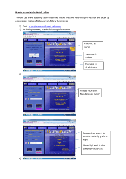


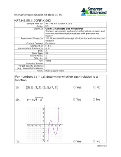

© Copyright 2026