
ABSTRACTS BOOK 6 biennial meeting of the
6th biennial meeting of the Association for European Cardiovascular Pathology 9-11 October 2014 Paris ABSTRACTS BOOK SOMMAIRE: Oral session 1 ……………………………………. p 2 Oral session 2 ……………………………………. p 8 Posters session 1…………………………………. p 14 Posters session 2…………………………………. p 40 What it is? Session………………………………. p 66 Other Abstracts………………………………….. p 72 Authors’ Index…………………………………… p 82 -1- ORAL COMMUNICATION SESSION 1 THURSDAY 9th OCTOBER -2- ORAL SESSION 1: O1: THE CRY UK SUDDEN CARDIAC DEATH DATABASE. JL. Robertus, MN. Sheppard Department of Cardiovascular Pathology, St George's University of London, London, United Kingdom Sudden cardiac death (SCD) from non-atherosclerotic causes is increasingly recognized as a genetic cause of death in the young. We have established a national sudden death referral practice in UK and report the latest results showing that 3225 cases were referred. The majority were ≤35 years (57%) and males dominated 2.8:1. Median age was 34 years, range < 98 years. Less than 10% of deaths (n=227) occurred in the paediatric age group (<16 years). In males the highest number of SCD was 26-30 years followed by 16-20 years. Females were older peaking 36-40 years. Family history of SCD was recorded in 224 cases. SCD with a normal heart predominated (n=1707, 53%) highlighting Sudden Adult Death Syndrome (SADS). Normal hearts occurred mostly in the young (58%) and males were more commonly affected than females (1.7:1). Cardiomyopathy accounted for 22 % of the deaths (n=717) with left ventricular hypertrophy (LVH) (n=210, 10%) without evidence of disarray as the most common structural abnormality. Hypertrophic cardiomyopathy (HCM) (n=161, 5%) and arrythmogenic right ventricular cardiomyopathy (ARVC) (n=131, 4%). Cardiomyopathy was associated with males (2.6:1) and affected the older age groups (37% ≥35 years). Other causes of SCD included cases with coronary atherosclerotic disease (n=190), myocarditis (n=79), congenital heart disease (n=73), anomalous coronary arteries (n=67), other coronary abnormalities (n=61), valve disease (n=68), aortic dissection (n= 26), sarcoidosis (n=23) and hypertensive heart disease (n=17). Thus SADS and cardiomyopathies account for 82% of SCD cases. -3- ORAL SESSION 1: O2: VISUALIZATION OF MYOCARDIAL INFARCTION IN POST-MORTEM MULTIPHASE CT-ANGIOGRAPHY C. Egger3, J. Vanhaebost3-1, K. Michaud3, J. Grimm3-2, P. Mangin3, S. Grabherr3 1 Service de Médecine Légale, Cliniques Universitaires Saint-Luc, Brussels, Belgium 2Department of Diagnostic and Interventional Radiology, University Hospital Lausanne 3University Center of Legal Medicine Lausanne-Geneva, Lausanne, Switzerland Background Multiphase Post-Mortem CT-Angiography (MPMCTA) is a minimally-invasive technique for post-mortem angiography using an oily contrast agent. In some cases of arterial phase MPMCTA, increased contrast enhancement of the myocardium was observed, seeming to correspond to the morphological finding of myocardial infarction. The aim of this study was to investigate the possibility to identify a myocardial infarction by MPMCTA. Materials & Methods We retrospectively selected 25 cases of myocardial infarction diagnosed by macroscopic and/or histological findings (group 1), and 25 cases without such findings representing a control group (group 2). Both groups underwent pre-autopsy MPMCTA. The presence or absence of arterial phase myocardial enhancement and its distribution were investigated by a forensic pathologist together with a board-certified radiologist, mean attenuation in Hounsfield Units was recorded (HU). Results In all group 1 cases, increased myocardial enhancement was observed in regions correlating with the localization of infarction. No pathological enhancement was observed in all cases of group 2. A cut-off value at mean attenuation of 95 HU yielded an excellent sensitivity and specificity. In some cases, the enhancement was especially pronounced (> 200 HU), mostly observed in subendocardial regions, where chronic infarction could be observed. Conclusion This study suggests the feasibility of identifying myocardial infarction on MPMCTA by evaluating myocardial contrast enhancement (≥ 95 HU). -4- ORAL SESSION 1: O3: POSTMORTEM GENETIC TESTING OF THE RYANODINE RECEPTOR 2 (RYR2) GENE IN A COHORT OF SUDDEN UNEXPLAINED DEATH CASES M. Larsen3, K. Berge5, T. Leren5, P. Nissen2, J. Hansen3, I. Kristensen3, J. Banner4, H. Jensen1 1 Department of Cardiology, Aarhus University Hospital 2Department of Clinical Biochemistry, Aarhus University Hospital 3Department of Forensic Medicine, Aarhus 4Department of Forensic Medicine, University of Copenhagen, Copenhagen, Denmark 5Unit for Cardiac and Cardiovascular Genetics, Oslo University Hospital, Oslo, Norway The aim of this investigation was to identify pathogenic variants of the ryanodine receptor 2 (RYR2) gene in a cohort of persons aged 0–40 years who died of sudden unexpected death syndrome (SUD), including a cohort of infants who died of sudden infant death syndrome (SIDS). We genetically screened 29 of the 105 exons of the RYR2 gene associated with catecholaminergic polymorphic ventricular tachycardia (CPVT) in 74 cases of SUD without reported structural abnormalities of the heart. Cases were selected from the case database at the Institute of Forensic Medicine in Aarhus, and subsequent mutational screening by DNA sequencing was performed to detect variants in DNA samples extracted from blood samples of deceased persons. A total of 7 of the examined 74 cases were heterozygous for a rare sequence variant in the RYR2 gene. We identified five novel missense variants, one synonymous variant, and one previously reported missense variant. Follow-up studies were possible in family members of three probands , and clinical examinations were conducted in family members of two of these probands. In conclusion, we identified a higher prevalence of variants in the CPVT-associated gene RYR2 than in a previously reported cohort of SIDS (9.4% vs. 1–2%). Segregation studies show that one variant (p.H4579Y) cosegregates with CPVT and is presumed to be pathogenic. Further studies, we are in the process of examining the cohort of SIDS further by targeted screening of 100 genes, including CALM 1-3, associated with inherited cardiomyopathies and channelopathies, using Next-Generating Sequencing (NGS). -5- ORAL SESSION 1: O4: INCREMENTAL VALUE OF RIGHT VENTRICULAR ENDOMYOCARDIAL BIOPSY TO THE PHENOTYPING OF PHOSPHOLAMBAN P.ARG14DEL MUTATION CARDIOMYOPATHY W.P. Te Rijdt1-2-3, P.A. Van Der Zwaag2, R.A. De Boer1, M.P. Van Den Berg1, J.P. Van Tintelen2, AJH. Suurmeijer3 1 Cardiology 2Genetics 3Pathology, UMC Groningen, Groningen, The Netherlands Background: The pathogenic p.Arg14del phospholamban (PLN) mutation has been identified in 12-15% of Dutch patients with arrhythmogenic right ventricular cardiomyopathy (ARVC) and/or dilated cardiomyopathy (DCM). The purpose of this study was to evaluate the additional value of RV endomyocardial biopsy (RVEMB) to diagnose ARVC in these mutation carriers and detect PLN protein aggregates in cardiomyocytes. Materials & Methods: RVEMB specimens from 23 mutation carriers (12 males, 11 females; mean[±SD] age at RVEMB 42±12 years) were studied. Masson’s trichrome staining was used to determine the amount of fibrofatty replacement (FFR), immunohistochemistry (IHC) for PLN to visualize protein aggregation. All patients were evaluated using clinical ARVC and DCM criteria. Results: Histological examination showed FFR consistent with ARVC in 17/23 (74%) patients (11/23 (48%) major criterium; 6/23 (26%) minor criterium according to task force criteria). IHC analysis revealed PLN aggregates in only 2/15 (13%) cases. By clinical evaluation before RVEMB, 11/23 (48%) patients were diagnosed with DCM, 2/23 (9%) with ARVC, 2/23 (9%) with combined ARVC/DCM while 8/23 (35%) did not meet sufficient criteria for either DCM or ARVC. Interestingly, by adding RVEMB analysis, 5/23 (22%) additional patients were diagnosed with combined ARVC/DCM, and 4/23 (17%) with ARVC. Conclusion: In PLN p.Arg14del mutation cardiomyopathy, FFR of the RV was found in the majority of biopsies. Adding this finding may establish a definitive ARVC diagnosis and reveal the biventricular ARVC/DCM phenotype typical for this cardiomyopathy. PLN IHC analysis in these RVEMB samples had a very low sensitivity for demonstration of PLN protein aggregates. -6- ORAL SESSION 1: O5: MITOCHONDRIAL REMODELING AND OXIDATIVE MYOCARDIAL HYPERTROPHY AND HEART FAILURE STRESS IN C. Giordano2, M. Orlandi2, M. Mancini2, M. Pelullo2, C. Preziuso2, GM. Bates5, H. Langping5, J. Lucena4, P. Lilla Della Monica3, C. Nediani1, RW. Taylor5, G. D'amati2 1 Department of Biochemical Sciences, University of Florence, Florence 2Dept. of Radiology, Oncology and Pathology, Sapienza University, Roma 3Department of Cardiac Surgery and Transplantation, San Camillo Hospital, Rome, Italy 4Forensic Pathology Service, Institute of Legal Medicine, Seville, Spain 5Wellcome Trust Centre for Mitochondrial Research, Newcastle University, Newcastle, United Kingdom Background Mitochondrial biogenesis derangement and increased mitochondrial-derived oxidative stress are implicated in the pathophysiology of cardiac remodelling. Whether these are early events, already present in compensatory cardiac hypertrophy, and their underlying molecular mechanisms are matter of controversy. Materials and Methods We compared morphologic and molecular features of mitochondrial biogenesis and markers of oxidative stress in: a) normal hearts (NH) obtained either from autopsies of subjects dead for non-cardiac causes (n=5, obtained within 4 hours from death) or from transplant procedures (donors, n = 5); b) compensated myocardial hypertrophy (HCM, myocardial samples from myectomy procedures for hypertrophic cardiomyopathy, n=10), and c) failing hearts (FH, n=15) transplanted for end-stage ischemic heart disease. Results Both hypertrophic and failing heart showed a ~ 30% decrease of mtDNA content. The expression level of PGC1-a, the master regulator of mitochondrial biogenesis, was unchanged both in HCM and FH. However in both conditions there was a significant reduction (about 25%) of NRF1 and ERR-a (selected transcriptional regulators that PGC1-a co-activates), TFAM (the key regulator of mtDNA transcription) and mitochondrial DNA polymerase gamma. Ultrastructural morphometric analysis revealed both increased number and reduced size of mitochondria. Markers of myocardial oxidative damage and activities of the antioxidant enzymes catalase, glutathione peroxidase and SOD2 were significantly increased only in FH. Conclusion Partial mtDNA depletion and mitochondrial ultrastructural alterations are early events in cardiac remodeling and are associated with reduced expression of the regulators of mitochondrial biogenesis downstream to PGC1-a. Oxidative stress is a marker only of endstage heart disease. -7- ORAL COMMUNICATION SESSION 2 FRIDAY 10th OCTOBER -8- ORAL SESSION 2: O6: ADVENTITIA REMODELING IN CARDIAC ALLOGRAFT VASCULOPATHY: THE FORGOTTEN LAYER C. Castellani1, M. Fedrigo1, OJ. De Boer2, AC. Frigo1, X. Li2, LB. Meijer-Jorna2, F. Tona1, G. Feltrin1, G. Gerosa1, M. Valente1, G. Thiene1, AC. Van Der Wal2, A. Angelini1 1 Dept of Cardiac,Thoracic and Vascular Science, University of Padua, Padova, Italy 2Dept of pathology, University of Amsterdam, Amsterdam, The Netherlands Background:Plaque hemorrhage (IPH), inflammation and microvessel leakage (ML) are key determinants of plaque vulnerability in native coronary atherosclerosis (ATS) and in cardiac allograft vasculopathy(CAV). IPH is closely associated with neoangiogenesis at site of ML and may represent a potent atherogenic stimulus both biochemically and mechanically. Aim of the present study was to investigate potential role of hemorrhage, inflammation,microvessel density and leakage in the adventia of CAV played in rapid and concentric growth of CAV lesions in post-transplanted patients. Material and Methods:In 12 patients who died for CAV, native heart at time of transplantation and cardiac allograft at autopsy were collected. Seventy coronary plaques were selected and inflammation, ML and hemorrhage were detected and assessed semiquantitatively in the adventitial and plaque lesions. Results:Inflammation(CAV 25/35 88.6%, ATS 14/34 41.2%, p=0.00013) associated with adventitial fibrosis and microvessels(CAV 31/35 88.6%, ATS 25/35 71.4%, p=ns) was found in both CAV and ATS but with a higher degree in the former. The degrees of fibroinflammatory response and microvessel density were directly correlated in plaque and adventitia in CAV (respectively 25/35, 71.4% p=0.04 and 25/35, 71.4% p=0.004) and in ATS (respectively 23/34, 35.7 % p<0.0001and 13/34, 38.2% p=0.03). Microvessel had a circumferential distribution confined mainly in the adventitia-media border in CAV. In the adventitia of CAV, ML was associated with hemorrhage extension. Conclusions:The adventitia of CAV underwent a more severe fibrotic remodeling compared to ATS. The strong association between ML and hemorrhage in adventitia of CAV suggests a role of adventitia in vessel remodeling and plaque growth -9- ORAL SESSION 2: O7: THE DISTRIBUTION PATTERN CARDIOMYOPATHY IS RELATED TO MUTATION OF FIBROSIS IN GENETIC THE TYPE OF PATHOGENIC S. Sepehrkhouy, J. Gho, R. Van Es, M. Harakalova, N. De Jonge, J. Van Der Smagt, R. Goldschmeding, R. De Weger, F. Asselbergs, A. Vink UMC Utrecht, Utrecht, The Netherlands Background: Genetic cardiomyopathies form a heterogeneous group in which myocardial fibrosis is frequently observed. Thus far, studies that compare the exact cardiac fibrosis pattern among cardiomyopathy hearts with different types of pathogenically mutated genes are lacking. Knowledge about the pattern of fibrosis will improve diagnosis and may lead to better understanding of disease mechanisms. We aimed to unravel the exact distribution pattern of cardiac fibrosis in cardiomyopathies caused by different specific pathogenically mutated genes. Materials & methods: A complete transversal slice was obtained from hearts of 22 patients with non-ischemic cardiomyopathy and a known mutation. Microscopic slides were stained with Masson’s trichrome and fibrosis was quantitatively analyzed. The different mutated genes were grouped in functional groups of encoded proteins: sarcomeric (n=8), calcium pump (phosholamban; n=8), desmosomal (n=2), nuclear envelop (n=2) and desmin filament network (n=2). Results: The hearts with mutations in the sarcomeric and nuclear envelop proteins showed circumferential subendocardial fibrosis in the left ventricle, except hearts with mutated myosin binding protein C (MYBPC3), which showed a more diffuse distribution. In hearts with a desmosomal or a calcium pump mutation the fibrosis was predominantly localized in the outer myocardium, mainly in the posterolateral wall of the left ventricle and in the right ventricle. In hearts with a desminopathy the fibrosis was in the outer myocardium of the left ventricle. Conclusion: Distribution of fibrosis in genetic cardiomyopathies is related to the type of pathogenic mutations. These specific patterns may provide a roadmap for cardiac MRI interpretation and add to our understanding of disease mechanisms. - 10 - ORAL SESSION 2: O8: A NOVEL TISSUE microRNA SIGNATURE FOR ARRHYTHMOGENIC CARDIOMYOPATHY E. Zorio2, P. Molina1, E. Plana3, E. Fernandez3, D. Domingo2-3, S. Navarro3, S. Navarro, S. Ferrer1, J. Sanz2, R. Camacho1, P. Medina3 1 Pathology Department, Institute of Legal Medicine 2Cardiology Department 3Sanitary Research Institute, La Fe University Hospital, Valencia, Spain Background: microRNAs (miRNAs) are small non-coding RNA molecules that regulate protein expression, including those relevant for the desmosomes, adipogenesis, and fibrogenesis. The aim of our study was to identify a novel miRNA profile distinctive of arrhythmogenic cardiomyopathy (ACM) patients victims of a sudden cardiac death (SCD). Methods: We collected formalin-fixed paraffin-embedded ventricular myocardium from ACM patients victims of SCD (N=19), and from victims of brain hemorrhages or car crashes without cardiac abnormalities (N=12). We measured miRNA expression with the GeneChip miRNA 3.0 Array (Affymetrix) in 9 ACM patients and in 6 controls. We selected those miRNAs with a significantly different expression level in patients and with targets mechanistically related with ACM. Finally, we confirmed these differences in expression in all our samples by RT-qPCR. Results: Expression arrays identified a large number of dysregulated miRNAs, 5 of them with confirmed differences by RT-qPCR showing a 26-49% reduced level in ACM patients compared to controls (p<0.05 in all of them). Conclusions: For the first time we have identified a miRNA profile characteristic of ACM patients, possibly related with the ventricular fibrofatty substitution. To prove an association with the SCD endpoint we should corroborate our results in samples from ACM patients victims of a noncardiac sudden death. A dysregulated miRNA profile could be useful for the diagnosis, risk stratification, and identification of new therapeutic targets in ACM. ISCIII (CP09/00065, PI11/00019), FEDER and Red RIC (RD12/0042/0029), Generalitat Valenciana (GE-034/11, Prometeo 2011/027), and IIS La Fe. PM is a Miguel Servet Researcher (ISCIII-FIS CP09/00065) - 11 - ORAL SESSION 2: O9: INCREASE OF ABCG2/BCRP+ SIDE POPULATION STEM CELLS IN THE MYOCARDIUM AFTER VENTRICULAR UNLOADING J. Wohlschlaeger4-3, B. Levkau2, J. Stypmann5, C. Schmid6, H. Milting1, KW. Schmid3, HA. Baba3 1 Erich und Anna Klessmann Institut für Kardiovaskuläre Forschung und Entwicklung, Herzund Diabeteszentrum NRW, Klinik für Thorax- und Kardiovaskularchirurgie, Bad Oeynhausen 2Division of Pathophysiology 3Institute of Pathology, University Hospital Essen, Essen 4Zentrum für Pathologie, Diakonissenkrankenhaus Flensburg, Flensburg 5Department of Cardiology and Angiology 6Department of Cardiovascular and Thoracic Surgery, University Hospital Münster, Münster, Germany Background: A significant decrease of the mean cardiomyocyte DNA content and increased numbers of diploid cardiomyocytes after ventricular unloading was demonstrated, suggesting a numerical increase of cardiomyocytes. The heart harbours several stem cells populations including c-kit (CD 117)+ cells and side population cells (SPC), that might proliferate after unloading and generate diploid cardiomyocytes. It was tested, whether there is an increase of ABCG2+ SPC and CD117+ cells after unloading.Methods: In paired myocardial samples (before and after LVAD), the number of cells with immunoexpression of ABCG-2, c-kit/CD 117 and MEF-2 was assessed by immunohistochemistry and morphometrically determined.Results: A significant increase of SPC and cells with coexpression of c-kit and MEF-2 after unloading was observed (p = 0.001). A significant positive correlation between both SPC and cells with coexpression of c-kit and MEF-2 expression was observed (p = 0.007 and 0.01). No correlation was found between the number of SPC and the mean cardiomyocyte DNA content.Conclusion: SPC are significantly increased in the myocardium after ventricular unloading, suggesting a role of stem cell proliferation during “reverse cardiac remodelling”. These cells might proliferate and commit to different cell lineages such as cardiomyocytes or endothelium, and thus ameliorate cardiac function. - 12 - ORAL SESSION 2: O10: THE MOLECULAR PHENOTYPE OF ANTIBODY-MEDIATED REJECTION IN HEART TRANSPLANTS A. Loupy6, T. Beuscart6, J. Reeves1, MC. Bories6, C. Toquet2, S. Varnous4, F. Iserin5, R. Guillemain3, A. Gay7, X. Jouven6, P. Bruneval3, J. Duong Van Huyen5, PF. Halloran1 1 ATAGC, Edmonton, Canada 2CHU Laennec, Nantes 3HEGP 4La Pitié 5Necker Hospital 6PARCC INSERM 970, Paris 7CHU Charles Nicolle, Rouen, France Background ABMR is a major cause of organ transplant failure. Recently, the molecular landscape of ABMR has been defined in kidney transplantation. We hypothesized that there are common molecular signatures and overlapping rejection mechanisms across these two organs. Methods We enrolled heart transplant recipients from four French centers with a diagnosis of biopsy proven ABMR and adequate endomyocardial (EMB) material for microarray processing (n=21). ABMR diagnosis was based on the pAMR classification(ISHLT 2013). We used the Molecular Microscope system of diagnostic classifier equations (ABMR Score) and pathogenesis-based transcript sets (PBTs) derived from literature and validated in kidney transplantation: endothelial DSA-selective transcripts (eDSAST), macrophage transcripts (QCMAT), gamma-interferon response (GRIT), injury-repair response transcripts (IRRAT) and the ABMR Score. We compared the gene expression in EMB in the ABMR group to a matched control group of heart recipients with normal EMB (n=37). Results The gene expression analysis revealed that as compared EMB from heart transplant patients without rejection, EMB with ABMR showed distinct molecular signal characterized by higher expression of eDSAST (p<0.0001), QCMAT (p<0.0001), GRIT (p<0.0001), IRRAT (p=0.0001) and increase of the ABMR Score that reflects interferon-effects, microcirculation stress and NK burden (p<0.0001). Conclusion The molecular signature of ABMR in heart shows common features with relevant gene scores and PBTs identified in kidney transplant biopsies. This suggest common mechanisms involved in ABMR – endothelial response to injury, NK transcripts, IFNG effects - and may provide basis for improving diagnosis and identifying adapted therapeutic strategies in heart transplantation. - 13 - POSTER SESSION 1 THURSDAY 9th OCTOBER - 14 - POSTER 1: ENGINEERING OF POLYMERIC VASCULAR GRAFTS: IS CELL SEEDING STILL NEEDED? M. Pontailler5, E. Illangakoon7, G. Williams7, C. Marijon5, V. Bellamy5, D. Balvay4, G. Autret4, V. Vanneaux3, J. Larghero3, V. Planat-Benard6, MC. Perier4, P. Bruneval1, P. Menasché5-2, D. Kalfa5-8 1 APHP, Hôpital Européen Georges Pompidou Department of Pathology 2AP-HP, Hôpital Européen Georges Pompidou, Department of Cardiovascular Surgery 3APHP, Hôpital SaintLouis, Laboratory of Cell Therapy 4ILRI / PARCC, INSERM U 970 5Laboratoires de Recherches Biochirurgicales - PARCC - INSERM U970, Paris 6STROMALab, INSERM U1031, UMR 5273, Université Paul Sabatier/CNRS, Toulouse, France 7UCL School of Pharmacy, University College London, London, United Kingdom 8Morgan Stanley Children's Hospital of New York, Columbia University Medical Center, Department of Pediatric Cardiac Surgery, New York, United States As part of a program targeted at developing a resorbable valved tube for replacement of the right ventricular outflow tract, we compared three biopolymers (polyurethane [PU], polyhydroxyalkanoate [PHBVV] and polydioxanone [PDO]) and two biofunctionalization techniques (using adipose-derived stem cells [ADSC] or the RGD peptide) in a rat model of partial inferior vena cava (IVC) replacement. Fifty-three Wistar rats first underwent partial replacement of the IVC with an acellular electrospun PDO, PU or PHBVV patch and 31 nude rats subsequently underwent the same procedure using a PDO patch biofunctionalized either by ADSC or RGD. Results were assessed both in vitro (proliferation and survival of ADSC seeded onto the different materials) and in vivo by magnetic resonance imaging (MRI), histology, immunohistochemistry [against markers of vascular cells (von Willebrand factor [vWF], smooth muscle actin [SMA]) and macrophages ([ED1 and ED2] immunostaining)] and ELISA (for the expression of various cytokines and iNOS). PDO showed the best in vitro properties. Six weeks after implantation, MRI did not detect significant luminal changes in any group. All biopolymers were evenly lined by vWF-positive cells but only PDO and PHBVV showed a continuous layer of SMA-positive cells at 3 months. PU patches resulted in a marked granulomatous inflammatory reaction. ADSC and RGD biofunctionalization yielded similar outcomes. These data confirm the good biocompatibility of PDO and support the concept that appropriately peptide-functionalized polymers may be successfully substituted for cell-loaded materials. - 15 - POSTER 2: LONG TERM IN VIVO REACTIONS TO PTFE AND POLYESTER IN THE CARDIOVASCULAR SYSTEM – PREDICTION OF CALCIFICATIONS LOCALLY RELATED TO SEPTAL DEFECT OCCLUSION DEVICES M. Sigler, S. Huell, R. Foth, T. Paul Pediatric Cardiology, Goettingen, Germany Objectives: We compared biological reactions to explants containing textile fibers after surgical removal from patients with congenital heart disease. Methods: We studied and compared tissue reactions to surgical patches (n=16, PTFE or polyester, 8.3 years to 35.2 years), and septal defect occluder (n=13, PTFE or polyester, 3 months to 15 years). After embedding in methylmethacrylate we performed complete histology and immunohistochemistry work-up of all specimen. Results: Inflammatory reactions consisted of lymphocytic infiltrations and macrophages/foreign body giant cells at the implant/tissue interface and were more pronounced in the polyester group. There was no change in quantity of inflammatory cells over time in both groups. All implants with implantation times of 8 years and more showed significant calcifications. The shortest implantation time with calcifications was 5,9 years in a PTFE occluder. All implants with an implantation time of more than 78 days were completely endothelialized. No cell dysplasia or metaplasia was seen in our series locally related to the textile fibers. Conclusions: Chronic inflammatory reactions as well as calcifications were significant in both material groups and were seen more pronounced locally related to polyester fibers. Both reactions were seen in a similar time pattern. Based on the results of our study one can predict that significant calcifications will occur with time in all septal defect occlusion devices which contain textile fibers. This will definitely deteriorate the ability to puncture the septum when left atrial access eventually is needed for interventional treatment for example in arrhythmias. - 16 - POSTER 3: HISTOPATHOLOGICAL PULMONARY CONDUITS EVALUATION OF EXPLANTED VALVED M. Sigler1, M. Vogt2, R. Boekenkamp3, J. Hoerer2, J. Goebbert1, R. Foth1, H. Schneider1 1 Pediatric Cardiology, Goettingen 2Pediatric Cardiology and Heart Surgery, German Heart Center, Muenchen, Germany 3Pediatric Cardiology, LUMC, Leiden, The Netherlands Background: Development of stenosis and/or valve insufficiency may limit the long term success of valved pulmonary conduits. We describe and compare our histological findings in different types of conduits after explantation. Methods: We analyzed 31 conduits 10 homografts, 10 Contegra conduits und 11 Hancock conduits (which had been implanted percutaneously or surgically, n = 7/24) using a standardized work-up protocol with embedding in methylmethacrylate and subsequent sewing and grinding. For immunohistochemical staining, mounting on glass slides was performed. Time interval between implantation and explantation was 6 months to 19 years. Principal reason for explantation of the conduit was stenosis in 25 patients, valve insufficiency in 2, and endocarditis in 4. Results: All explants showed partial or complete endothelialization as confirmed by immunohistochemistry. Inflammatory reactions were more pronounced in xenografts compared to homografts and persisting in the patients with a history of endocarditis. Correspondingly, there was more pseudointima formation in xenografts. Calcification of the conduit wall was a constant finding which appeared to increase with longer time intervals between implantation and explantation. There were few thrombotic deposits locally related to the sinus of the conduit valves. The latter was a constant finding in specimen of endocarditis patients. Conclusions: In a large series of explanted valved pulmonary conduits we were able to demonstrate more accentuated inflammation and pseudointima formation in xenografts as compared to homografts. Even after clinically successful therapy of endocarditis there were significant inflammatory infiltrates within the conduits walls. - 17 - POSTER 4: THE APPROPRIATE TIME-FRAME FOR USE OF VASCULAR HOMOGRAFT TISSUE- AN UNSOLVED PROBLEM. K. Wassilew, J. Schroeter, R. Hetzer Deutsches Herzzentrum Berlin, Berlin, Germany Introduction Although standards regarding the use of homograft tissue have been set up, little is known about the durability of frozen homograft tissue. Hypothesis We aimed to analyze vascular homograft tissue to demonstrate its integrity beyond the legally set time-frame of 5 years for its use. Material Thawed parts of one ascending aorta and two spare veins from different patients (58, 61 and 73 years old), the latter intended for autologous use in bypass operations, were submitted for histological examination after storage at −180°C for 5, 11 and 13 years, respectively. The homograft tissue was evaluated for integrity, freezing artefacts and signs of vitality on formalin-fixed paraffin embedded tissue sections using conventional histology, an elastica van Gieson stain and immunohistochemistry (alpha smooth muscle actin, CD31). The findings were compared to those of the histological examination before freezing. Results There were no significant differences in the pathological findings of the tissue fragments investigated before and after cryopreservation. The mild degenerative changes were within physiological limits for the age of the patients. Neither freezing artefacts nor signs of loss of tissue integrity were noted. There was evenly distributed strong diffuse expression of both smooth muscle actin of the smooth muscle cells and CD31 in the intimal cells and the endothelial cells of the vessels in the adventitia, suggestive of cell viability. Conclusion Our preliminary data suggest that vascular homograft tissue could be used beyond the legally set time-frame, although larger studies are required to confirm our findings. - 18 - POSTER 5: IDENTIFICATION OF A microRNA PROFILE RELATED TO HYPERTROPHIC CARDIOMYOPATHY E. Zorio2, P. Molina1, E. Plana3, E. Fernandez3, D. Domingo2-3, S. Navarro3, MA. Arnau2, MC. Salvador1, M. Fenollosa1, J. Giner1, P. Medina3 1 Pathology Department, Institute of Legal Medicine 2Cardiology Department 3Sanitary Research Institute, La Fe University Hospital, Valencia, Spain Background: microRNAs (miRNAs) are small non-coding RNA molecules that modulate protein expression, including those of cardiac hypertrophy and ion channels. We aimed to identify a novel miRNA profile distinctive of HCM patients victims of a sudden cardiac death (SCD). Methods: We collected fresh (N=5) and paraffin-embedded (N=23) ventricular myocardium from SCD-HCM patients and controls (N=33 and N=12, respectively, victims of brain hemorrhages or car crashes without cardiac abnormalities). We measured miRNA expression with the GeneChip miRNA 3.0 Array (Affymetrix) in fresh samples. We selected those miRNAs with a significantly different expression level between groups, and with targets mechanistically related to HCM. Finally, we confirmed by RT-qPCR these differences in all our samples. Results: Expression arrays identified a large number of dysregulated miRNAs, 6 of them with confirmed differences by RT-qPCR showing a reduced level in HCM patients compared to controls: miR-486-3p 48% reduced (p=0.003), miR-222-3p 31% (p=0.043), miR-103a-2-5p 30% (p=0.029), miR-1 43% (p=0.002), miR-133a 36% (p=0.004), and miR133b 40% (p=0.004). Conclusions: For the first time we have identified a miRNA profile characteristic of HCM patients, possibly related with ventricular hypertrophy. To prove an association with the SCD endpoint we should corroborate our results in samples from HCM patients victims of a non-cardiac sudden death. A dysregulated miRNA profile could be useful for the diagnosis, risk stratification, and identification of new therapeutic targets in HCM. ISCIII (CP09/00065, PI11/00019), FEDER and Red RIC (RD12/0042/0029), Generalitat Valenciana (GE-034/11, Prometeo 2011/027), and IIS La Fe. PM is a Miguel Servet Researcher (ISCIII-FIS CP09/00065). - 19 - POSTER 6: THE HUMAN CARDIAC AND SKELETAL MUSCLE PROTEOMES DEFINED BY TRANSCRIPTOMICS AND ANTIBODY-BASED PROFILING E. Erik Larsson, F. Ponten, C. Bergström, C. Kampf, J. Linne, M. Uhlen, BM. Hallström3, L. Fagerberg3, CJ. Sundberg2, M. Lindholm2, L. Väremo1, L. Nielsen1 1 Chalmers University, Department of Chemical Engineering, Gothenburgh 2Karolinska Institute, Department of Physiology and Pharmacology 3Science for Life Laboratory, Stockholm 4immunology,genetics and pathology, Uppsala, Sweden ABSTRACT To understand cardiac muscle function and disease, it is important to define and explore its molecular constituents and their physiological role. The aims of this study were: To determine the genes and proteins with elevated expression in cardiac muscle in relation to skeletal muscle and other tissue types and to localize these proteins to various subcompartments within cardiac muscle. Material: Tissues used for RNA extraction was based on biopsies from four heart muscle samples and five skeletal muscle samples. Methods and Results: A genome-wide deep RNA sequencing analysis in 28 human tissues was employed to identify genes overrepresented in striated muscles, complemented with antibody-based profiling in 44 tissues to localize the corresponding proteins. Almost one third of the transcripts in cardiac muscle correspond to mitochondrial proteins involved in energy metabolism, while 14% of the mRNA pool was represented by 283 genes with elevated expression in cardiac muscle. Conclusion: Our results provide a comprehensive list of genes and proteins elevated in striated muscles. A number of proteins not previously characterized in cardiac muscle were identified and localized to specific cellular subcompartments. These proteins represent an interesting starting point for further functional analysis of their role in cardiac muscle biology and disease. These data will be presented and discussed. - 20 - POSTER 7: PROTEOMIC APPROACH FOR CARDIAC TISSUE AMYLOIDOSIS CHEMICAL CHARACTERIZATION OF C. Castellani1, M. Fedrigo1, G. Fregonese1, P. Polverino De Laureto2, M. Della Barbera1, G. Thiene1, M. Valente1, A. Angelini1 1 Dept of Cardiac Thoracic and Vascular Science, Padova 2Dept of Pharmacology Science, Dsf, University of Padua, Padua, Italy Background:In this study we tested the feasibility of determining the type of amyloidosis by biochemical analysis on endomyocardial biopsies. Methods:10 frozen and paraffin-embedded right ventricle endomyocardial biopsies and cardiac surgical/autoptic samples were obtained from patients affected by amyloidosis which was characterized by gold standard pathologic criteria(electron immunomicroscopy), and 10 cardiac samples as control group. Samples were homogenized and solubilized in sodium dodecyl sulfate (SDS) buffer and total protein fractions were incubated under different conditions of temperature, buffer composition, pH, and additives like 2βmercaptoethanol, followed by SDS-PAGE and Western Blot analysis. Results:K and λ proteins identification were not modified by temperature, buffer composition, pH, and additives when subjected to SDS-PAGE and Western Blot analysis, while TTR protein(trimer, dimer, and false positive monomer) showed great modifications in protein band fragments under different conditions. Acid and neutral pH lead to identification of false positive 14 kDa band(TTR monomers) in all amyloid samples and also in controls. Heating of the samples to >80oC in acid, neutral or alkaline pH resulted false positive 14 kDa band (TTR monomers) in all amyloid samples and also in controls. Only high alkaline pH preserved correctly TTR monomer formation and identification only in TTR+ amyloid samples but not in TTR- samples. Sensitivity and specificity of this biochemical test on tissue samples were 100% compared to gold standard (electron immunomicroscopy). Conclusions:We have developed and standardized a useful diagnostic technique for chemical tissue amyloidosis characterization, that could be applied in the clinical field with cost and time-effective improvement - 21 - POSTER 8: MORPHOLOGICAL CHARACTERISTIC AND GENETIC ANALYSIS OF 6 LVNC CASES L. Mitrofanova2, P. Konovalov2, E. Zakliazminskaia1, D. Grigorieva2, L. Oun1 1 Petrovsky National Research Centre of Surgery, Moscow 2Almazov Federal Medical Research Centre, Saint-Petersburg, Russia Background: non-compact left ventricular myocardium (LVNC) is a genetic disorder associated with mutations in genes encoding mitochondrial, cytoskeletal, sarcomeric and cell junction proteins. However there is opinion that various cardiomyopathies may appear phenotypically as "non-compact myocardium". Material and methods: 5 patients with LVNC revealed at cardiac transplantation and 1 deceased with LVNC combined with bicuspid aortic valve were studied. Heart morphometry, histological examination of atrial myocardium, ventricles and interventricular septum, immunohistochemistry of the right ventricular myocardium with antibodies against HLA-DR, CD3, CD45, CD68, dystrofin, connexin-43, vinculin were performed. We have screened the coding sequence and adjacent intronic areas of genes referred to LVNC in 4 patients. Results: The ratio of the thicknesses of trabeculae carneae to the one of LV wall was more than 2 (from 2.2 to 6.6) in all the patients. Four patients had abnormal trabeculation of right ventricle. Hypertrophy and myofiber disarray of ventricles and the interventricular septum were revealed in all the patients. Immunohistochemistry of 4 cases showed the evidence of myocarditis. Focal absence of connexin-43 expression was revealed in cardiomyocyte intercalated disc in 4 patients. One of four patients had a mutation in MYH7 gene (c.del57545756). He had a positive family history of LVNC. No mutations causing LVNC were found in other genes. Conclusion: The results testify that phenotype LVNC can be formed under the influence of different modifying factors. Histological examination of LVNC patients can reveal features specific to HCM and to DCM. Causes of phenotypical transformation in cardiomyopathy invite further investigation. - 22 - POSTER 9: VIRAL MYOCARDITIS: FACTORS INFLUENCING DETECTION YIELD ON ENDOMYOCARDIAL BIOPSY VIRAL GENOME E. Carturan, C. Basso, S. Rizzo, M. Fedrigo, A. Angelini, G. Thiene Dept. of Cardiac, Thoracic and Vascular Sciences, Padova, Italy Background. The role of EMB for the diagnosis and its contribution to patient management has been addressed by a Consensus document of AECVP-SCVP. Our aim was to assess the impact of fixation and number of EMB samples on molecular diagnosis findings. Methods. Consecutive EMBs referred at our Institution for histology/immunohistochemistry evaluation in the time interval 1996-2012 were enrolled for molecular investigation by RTPCR and PCR technique. Results. A total of 467 EMBs have been diagnosed as myocarditis: 79 in pediatric and 388 adult patients (pts.). Viral etiology was identified in 28 (36%) pediatric and 101 (26%) adult pts, and the most prevalent type of viruses were enterovirus (8/28, 36%; 26/101, 24%). In a more recent subgroup of 137 EMBs (virus positive 30/137, 22%) seasonality, type of fixation and number of EMB samples were assessed. The number of EMB samples per pt. was ≤3 (either formalin or RNAlater) in 94/137 (69%) and >3 in 43/137 (31%), with a lower prevalence of virus positive in the former (17/94, 18% vs. 13/43, 30%). 81 EMB samples were frozen while 56 were paraffin embedded, with a higher prevalence of viral genome in the former (26/81, 32% vs. 4/56, 7%; p=0.001). Conclusions. The diagnosis of myocarditis on EMB samples requires standardized protocols including molecular techniques. Viral genomes are identifiable in more than one third of pediatric and one fourth of adult cases. Methodological factors like the type of tissue fixation and the number of samples could impact on viral genome detection on EMB. - 23 - POSTER 10: PRESENCE OF INTRAPLAQUE HEMORRHAGE RELATES TO THE USE OF COUMARIN-TYPE ANTICOAGULANTS IN PATIENTS WITH EXTENSIVE CORONARY ATHEROSCLEROSIS X. Li1, A. Vink3, H. Niessen1, O. De Boer1, A. Van der wal1 1 Academic Medical Center Amsterdam 2VUMC, Amsterdam 3UMCU, Utrecht, The Netherlands Background Intraplaque hemorrhage (IPH) is important for rapid growth and vulnerability of atherosclerotic plaques. We investigated the association between exposure to antithromboembolic therapy of patients during life and presence of IPH in coronary arteries at autopsy. Methods Coronary arteries with extensive atherosclerosis were obtained at autopsy from hearts of patients who had received oral anticoagulants (n=10), platelet aggregation inhibitors (n=10) or no anti-thrombotic drugs (n=10) before death. Coronary arteries were cut in 3-mm segments, and sections with plaques were evaluated for presence of IPH and microvessels , with the use of Haematoxylin stains, ) and with monoclonal antibodies GFA (erythrocytes), CD61 (platelets) and von Willebrandfactor (endothelial cells). Pathology data were related to the use and type of anti-thromboembolic therapy of each patient. Results IPH was found in 483 out of 904 (53%) coronary sections containing plaque, and significantly more in patients on oral anticoagulants (174/284, 61%) than those on antiplatelets (198/376, 53%) or without any therapy (111/244, 46%) (P=0.02 and P=0.001). Moreover, IPH appeared to be larger in plaques of patients on anti-coagulant treatment compared with the other groups (P<0.001). Density of intraplaque microvessels was highest in plaques of patients on platelet inhibitors (P<0.05), but this was not associated with increased numbers of plaque bleeding. Conclusion Oral coumarin-type anticoagulant therapy is associated with higher numbers intraplaque hemorrhages in atherosclerotic coronary arteries, which may have implications for rapid atherosclerosis progression in these patients. - 24 - POSTER 11: THE ROLE OF IL-17 IN THE GROWTH OF VASCULAR ANOMALIES: ITS EXPRESSION IN PYOGENIC GRANULOMA, INFANTILE HEMANGIOMA, VENOUS AND ARTERIOVENOUS MALFORMATION AND REGULATION IN VITRO S. Sairras2, L. Meijer-Jorna1, O. De Boer2, H. Niessen3, A. Van der wal2 1 Symbiant, Alkmaar 2Academic Medical Center Amsterdam 3VUMC, Amsterdam, The Netherlands INTRODUCTION: benign vascular lesions differ significantly in patterns of growth of microvessels. Recent study showed microvascular proliferation and inflammation within arteriovenous types of vascular malformations. We studied the expression and regulation of angiogenic IL-17 cytokines in different types of vascular anomalies in vivo and in vitro. MATERIALS AND METHODS: 36 paraffin embedded tissues blocks of pyogenic granuloma (PG,n=11), infantile angioma (IH, n=8), venous malformation (VM, n=5) and arteriovenous malformation (AVM, n=12) were immunohistochemically double stained for IL-17A, IL-17B and IL-17E expression in mast cells, endothelial cells and neutrophils respectively. rt-PCR for IL-17B and E was performed on 18 samples of VM and AVM and on stimulated HUVECs. FACS analysis was used to study the IL-17 expression of the stimulated HUVECs. RESULTS: IL-17A was expressed on neutrophils and mast cells, and IL-17B was expressed on all endothelial cells of all vascular anomalies with no difference between mature and immature vessels. Moreover, endothelial cells of all lesions were IL17E +, except for PG, of which 4 out of 9 were negative. IL-17B mRNA was expressed on both immature as well as mature lesions. IL-17A, IL-17F, TNFα, TGFß, IFNγ and VEGF up regulated IL-17B mRNA but downregulated IL-17E mRNA in HUVEC. CONCLUSION: Distinct subsets of IL-17 cytokines are active in all types of vascular anomalies, with no differences between mature and immature type of vessels. IL-17B is up regulated, but IL-17E is down regulated by pro-inflammatory cytokines. Such differences may underlie the variation in growth behaviour among the different types of vascular anomalies. - 25 - POSTER 12: AN EXCEPTIONAL CASE OF DUAL VALVULAR GRANULOMATOSIS WITH POLYANGIITIS. INVOLVEMENT IN F. Mittler1, O. Espitia4, L. Droy1, S. Pattier2, F. Naudin5, A. Cavailles5, M. Hamidou4, P. Bruneval6, C. Agard4, C. Toquet1 1 CHU service d'anatomie pathologique 2CHU service de cardiologie 3CHU service de chirurgie cardiothoracique 4CHU service de medecine interne 5CHU service de pneumologie, Nantes 6HEGP Service d'anatomie pathologique, Paris, France Introduction Granulomatosis with polyangiitis (GPA) (Wegener’s) is a necrotizing systemic vasculitis of small-sized blood vessels affecting most of the time kidney, lung and upper respiratory tract. Frequency of GPA heart involvement is estimated at 25% in autopsy studies and is mainly represented by pericarditis. Case report We report the case of a 60 year-old woman with arthritis and lung nodules due to GPA without anti-neutrophil cytoplasmic antibodies (ANCA) at diagnosis. Remission was obtained with cyclophosphamide (CyC) and corticosteroid. Azathioprine was then prescribed during two years. Four years later, she developed an aortic and mitral regurgitation. Results Microscopic examination of the valve specimens demonstrated typical GPA histopathological lesions : granulomatous inflammation, polymorphonuclear microabscesses with minute and geographic necrosis. Stains for fungi, bacteria or other organisms were negative. The search for pANCA anti-myeloperoxdase was positive. Conclusion Cardiac valvular involvement is a rare and potentially fatal complication of GPA and may falsely suggest infectious endocarditis. Only few cases of histologically well documented cardiac valvular involvement in GPA are listed in literature. Clinical, biological and pathological data must be taken into account to propose a final diagnosis of GPA heart involvement. - 26 - POSTER 13: AORTIC AND MITRAL VALVE DISEASE IN PATIENTS TREATED WITH THE ANTIDIABETIC AGENT BENFLUOREX L. Gibault3, V. Thomas De Montpreville1, F. Paraf2, P. Bruneval3 1 Pathology, Centre Medico-Chirurgical Marie Lannelongue, Le Plessis Robinson 2Pathology, CHU Limoges, Limoges 3Pathology, Hopital Européen Georges Pompidou, Paris, France Aims: Benfluorex, a fenfluramine derivative exerting serotoninergic effects, has been used for 30 years as an antidiabetic agent. It was withdrawn from the market in France in 2009 due to a high incidence of valvular toxicity, resulting mainly in restrictive aortic and mitral valve insufficiency. The aim of our study was to highlight the benfluorex-induced valve damages in patients necessitating valve replacement. Methods: 27 aortic and 31 mitral valves surgically removed from 44 patients [Gender: 5M/39F; Age: 57 ± 1.5 y. (mean ± SD)] treated with benfluorex for 95 ± 8.6 mo. were retrieved from our pathological files from 2006 to 2014. Results: The major marker of benfluorex-induced valve toxicity was endocardial fibrous thickening: 0.60 ± 0.08 mm for aortic valve; 0.4 ± 0.04 mm for the mitral valve; 0.7 ± 0.06 for the mitral chordae tendinae. This endocardial fibrosis was most frequently pure devoided of features related to other diseases such as architecture loss, inflammation, and neoangiogenesis for rheumatic valve disease, as well as degenerative changes and calcifications for mitral valve prolapse or calcified aortic stenosis. The intensity of the involvement was variable, from a mild endocardial fibrosis to severe retractile scarring, mimicking rheumatic mitral stenosis on gross examination. No correlation between the intensity of lesions and the treatment duration could be evidenced. Conclusions: In this large series, we described the benfluorex-induced endocardial fibrosis in aortic and mitral valves. Histology is mandatory to differentiate them from degenerative or rheumatic diseases, as gross examination alone can be misleading. - 27 - POSTER 14: CAUSE OF AORTIC REGURGITATION IN PATIENTS WITH TAKAYASU ARTERITIS, VALVE INFRAMMATION OR AORTIC ROOT DILATATION? M. Matsumoto, K. Ohta-Ogo, T. Matsuyama, Y. Ikeda, H. Ishibashi-Ueda National Cerebral and Cardiovascular Center, Osaka, Japan Background Takayasu arteritis is an inflammatory large vessel vasculitis of unknown origin affecting mainly the aorta and its branches, but inflammation involvement to aortic valve in Takayasu arteritis is unclear. The objective of this study is to examine whether aortic valve inflammation would occur in Takayasu arteritis. Materials and Methods Among fifty-six patients were diagnosed Takayasu arteritis between 1997 and 2013, 19 patients were underwent aortic valve replacement therapy because of severe aortic regurgitation. We retrospectively examined ascending aortic walls and aortic valves. Results Average patient age was 58±13 years, and over 75% were female (M/F, 4/15). Cardiovascular risk factors such as hypertension, diabetes, dyslipidemia were seen in 12 (67%), 2 (11%) and 4 (22%). 6 patients were diagnosed Takayasu arteritis in younger age (26±12 years), but remaining 13 (67%) have not diagnosed until microscopic examination revealed after surgical therapy. 14 patients had the history of aortic valve regurgitation including 1 who had been underwent aortic valve replacement. Preoperative immunosuppressive therapies performed in 4 (22%) and mean administrated period was 24±19 years. Histologically, scar stage with ongoing aortitis was seen in 14 cases (74 %). Operated all aortic valves showed myxomatous degeneration because of severe aortic regurgitation. Only 2 cases of aortic valve (11%) obtained from valve replacement for regurgitation showed inflammatory or post-inflammatory change. Conclusion Aortic root dilatation is major cause of severe aortic regurgitation in Takayasu arteritis, but inflammation involvement to aortic valves also cause aortic regurgitation in a few cases. - 28 - POSTER 15: A FIRST CHARACTERIZATION OF A KNOCK-IN MOUSE MODEL OF VASCULAR EHLERS DANLOS SYNDROME E. Fontaine2, J. Faugeroux2, MC. Verpont1, J. Hadchouel2, P. Bruneval2-3, X. Jeunemaître2-3 1 U1155 2U970, INSERM 3University Paris Descartes, Paris, France Vascular Ehlers-Danlos syndrome (vEDS) is a rare inherited connective tissue disorder, caused by mutations in COL3A1 gene coding type III pro-α1 collagen chain. vEDS causes severe tissue fragility with early-onset complications mainly arterial ruptures. The absence of a usable mouse model of the pathology prevents further pathological and therapeutic investigation. We created a knock-in mouse, col3a1 KI+/G183S, bearing a missense mutation at a glycine residue in the triple helix of type III collagen. We studied survival rate, weight, systolic blood pressure (SPB) and heart rate (HR). Animals were analysed after death or were sacrificed at 24 weeks. Dissected hearts and aortas were stained with orcein or hematoxylin eosin and were also subjected to electron microscopy (MEL). Analysis of genes and proteins expression are currently performed on wild-type and mutated aortas. A first trial is also performed comparing a beta-blocker (propranolol) to placebo. Both heterozygous and homozygous mutated mice were viable but this latter group was underrepresented at birth. At 8 weeks of age, col3a1+/+ and col3a1 KI+/G183S mice had no difference in weight, SBP and HR. The spontaneous survival rate of col3a1 KI +/G183S and KIG183S/G183S was 50% and 40% at 24 weeks, respectively. All mice died due to thoracic aorta rupture. MEL of aortic tissue showed a dilated endoplasmic reticulum in adventitial fibroblasts and arguments for unfolded protein response and autophagy. We report here the first mouse vEDS model mimicking the human disease. It should allow to get insight into its pathophysiology and test new therapeutic strategies. - 29 - POSTER 16: A SCORING SYSTEM FOR SURGICAL SPECIMENS OF THE THORACIC AORTA HELPS TO CLASSIFY ANEURYSMS OF DIFFERING AETIOLOGY Y. Banz1, A. Spranger1-2, A. Perren1, F. Schoenhoff2, T. Carrel2 1 Institute of Pathology, University of Bern 2Department of Cardiovascular Surgery, University Hospital Bern, Bern, Switzerland Background Standardized diagnostic criteria to assess aortic pathology are lacking. Aim was to establish a scoring system in a test cohort of patients with aortic aneurysms with tricuspid (TAV) or bicuspid aortic valves (BAV) and Marfan syndrome. The scoring system should facilitate communication with clinical colleagues and ensure standardized classification of the different aortic pathologies. Materials and methods Surgical specimens from patients undergoing repair of thoracic aortic aneurysms (tricuspid valve, TAV; n=15, bicuspid valve, BAV; n=11 and Marfan syndrome n=11) were scored for elastic fibre degeneration (score 0-4; no - full media-thickness fragmentation), mucoid degeneration (score 0-3; none - extensive), media necrosis (score 0-3; none - band-like necrosis), atherosclerotic plaques (0 or 1). Wall thickness was measured. Samples were accompanied by a minimal clinical data set (standardized form). Results Total aortic wall and media thickness were significantly reduced in Marfan patients compared to TAV/BAV (p=0.002 und p=0.001). Mucoid degeneration showed no significant betweengroup differences. Significantly more media necrosis occurred in the TAV versus BAV (p=0.009) and Marfan group (p=0.004). BAV media showed significantly less elastic fibre degeneration compared to TAV and Marfan (p=0.003 and p=0.0002). Degeneration in BAV correlated more closely with pre-operative maximal aortic diameter than in the TAV or Marfan group. Conclusion This new scoring system allows standardized reports and improved communication between surgeons and pathologists. It differentiated between the pathologies of the thoracic aorta and identified BAV specimens as a histologically separate entity. A follow-up confirmation cohort is being studied to validate these initial results. - 30 - POSTER 17: MATRIX METALLOPROTEINASE-9 DISSECTION EXPRESSION IN ACUTE AORTIC E. Stefanovic1, P. Otasevic1, P. Milojevic1, L. Lausevic-Vuk1, Z. Milovanovic2, B. Djukanovic1 1 Dedinje Cardiovascular Institute 2Institute of Oncology, Belgrade, Serbia-Montenegro Background : The aime of this study was to determine matrix metalloproteinase-9 (MMP-9) expression in dissected aortic tissue. Materials and Methods: Specimens of aorta from 23 patients who underwent surgery for acute dissection with the entrance tear in the ascending aorta. Aortic tissue samples were evaluated for MMP-9 expression and were compared with 10 control aortic specimens, free of any vascular diseases. Expression of MMP-9 was graded as 0 (absent), 1+ (mild), 2+ (moderate), and 3+ (intense). Results: Mean age of patients was 54±9 years (ranging 24-72 years), with 20/23 (87%) of them being male, and 3/23 (13%) female. A total of 14/23 (61%) patients had exit rupture in the aortic wall, whereas 9/23 (39%) did not have exit tear. In group without exit tear expression of MMP-9 was absent (0) in 4 and mild (1+) in 5 patients, whereas in patients with exit tear expression of MMP-9 was intense (3+) in 10 patients, and moderate (2+) and mild (1+) in 2 each. Expression of MMP-9 in group without exit tear was lower than in patients with exit tear (0.55±0.52 vs 2.57±0.76, respectively, p<0.001). Expression of MMP-9 was absent in all control specimens, and was significantly lower than in both groups with aortic dissection (p<0.001). Conclusion: Expression of MMP-9 in the aortic media is enriched in dissected aortic tissue. In acute aortic dissection with existence of the exit rupture in the aortic wall expression of MMP-9 higher than in group without exit tear. - 31 - POSTER 18: AORTOPATHY IN MARFAN SYNDROME: FOCUS ON MICROSCOPIC AND ULTRASTRUCTURAL FINDINGS A. Orlandi1, D. Mazzaglia1, F. Romaniello1, A. Ferlosio1, S. Grego2, A. Pellegrino2, L. Chiariello2 1 Anatomic Pathology of Tor Vergata University of Rome 2University Tor Vergata of Rome, Cardiosurgery, Rome, Italy Marfan syndrome is an inherited autosomal dominant multisystem disease caused by mutations in the FBN1 gene encoding fibrillin-1, an extracellular matrix glycoprotein widely distributed in mesenchymal-derived tissues that provide a scaffold for elastin deposition. MFS is characterized by variable clinical manifestations, including skeletal, ocular and cardiovascular abnormalities; ascending aortic aneurysm with ensuing dissection and rupture is the main life-threatening cardiovascular manifestation of Marfan syndrome. Histological aspects of Marfan syndrome aortopathy include a medial degeneration from disarray and fragmentation of elastic fibers and accumulation of basophilic ground substance areas depleted of smooth muscle cells, with minimal inflammation. Transmission electron microscopy well evidences the high number of interruptions and the thick appearance of the elastic lamellae and the accumulation of abundant extracellular glycosaminoglycan-rich material, sometimes SMCs showing a prevalent synthetic phenotype. The aberrant signalling of transforming growth factor-beta as the consequence of the altered structure of fibrillin-1 induces activation and over-expression of Smad-dependent pro-fibrotic signalling pathway. The ERK1/2-mediated increased synthesis of matrix metalloproteinases also plays a role in medial extracellular matrix remodeling occurring in Marfan aortopathy. Finally, Marfan syndrome is accompanied by an impaired aortic contractile function and aortic endothelialdependent relaxation, which is caused by an enhancement of the oxidative stress and increased reactive oxygen species during the progression of the disease. Further studies are needed to verify the impact of increased oxidative stress on aortic smooth muscle cell population in Marfan syndrome patients. - 32 - POSTER 19: IgG-4 RELATED PERIAORTITIS, A MIMICKER OF INTRAMURAL HEMATOMA BY CT SCAN. JA. Collins, S. Kligerman, AP. Burke University of Maryland Medical Center, Baltimore, Md, United States Background: Aortic intramural hematomas (IMH) have been accepted as precursors to aortic dissection. Radiologic recognition of this entity can result in emergent aortic repair. Sclerosing periaortitis, a rare variant of ascending aortitis, has not been reported as a radiologic mimicker of IMH. Herein we describe two such cases. Material and Methods: Two surgical pathology cases of periaortitis in specimens from ascending aortic repairs. Results: Two men (63 and 53 years) presented with chest/throat pain to the emergency room. CT findings at presentation were interpreted as ascending IMH in both patients. The two patients were referred for emergent aortic repair. Histologic findings from both surgical resections demonstrated marked inflammation, predominantly lymphoplasmocytic aggregates, with extensive storiform adventitial fibrosis. Immunohistochemical staining revealed IgG-4 positivity in the plasma cells. The pathologic diagnoses in both cases included IgG-4 related aortitis with adventitial fibrosis. No hematomas were identified in either case. Conclusion: We describe two cases of presumed aortic IMH whereby histologic diagnosis revealed IgG4-related periaortitis. The radiologic imaging of IgG-4 related periaortic aortitis can mimic IMH because of the uniform adventitial scarring having a similar appearance, in density, to blood. This entity should be considered during radiologic evaluation of thoracic aortas. - 33 - POSTER 20: INVOLVEMENT OF ADAM10, ADAM17 AND NOTCH4 RECEPTOR - DLL4 LIGAND IN ANTIBODY-MEDIATED REJECTION IN HUMAN CARDIAC TRANSPLANTS : A MORPHOLOGICAL APPROACH C. Toquet1, A. Pabois3, N. Gerard3, S. Pattier2, C. Laboisse1, B. Charreau3 1 Service d'anatomie pathologique 2Service de cardiologie, CHU Nantes 3UMR 1064, INSERM, ITUN, Nantes, France A disintegrin and metalloproteinases ADAMs regulate key proteolytic events involved in inflammatory cytokine and chemokine release and in Notch signaling. Previous quantitative PCR study showed that AMR induced by donor-specific anti-HLA in human cardiac transplants is characterized by an upregulation of ADAM10, ADAM17 and Dll4 ligand mRNAs and a downregulation of the endothelial Notch4 receptor. This study investigates the in situ localization of these proteins in frozen cardiac biopsies from patients with AMR (pAMR 2 or 3) (n=9) compared to control/stable grafts (pAMR0) (n=13) by immunohistological and/or confocal analyses. We found that in normal heart, ADAM10 and Notch4 were mainly expressed by endothelial cells (EC) whereas ADAM17 and Dll4 were not detected. In AMR group, ADAM10 was located in EC and in infiltrating CD68+ macrophages and some CD3+ T cells. ADAM 17 was expressed by macrophages and by some EC. Confocal analysis showed that 1/ Dll4 was diffusely expressed by EC and interstitial cells (namely macrophages) 2/ Notch4 receptor was maintained in some endothelial cells in closed relation with intravascular macrophages. To conclude, our findings suggest that ADAM10 and Notch pathway are involved in immune cell recruitment during AMR and could be considered as potential therapeutic targets. - 34 - POSTER 21: MICROVASCULOPATHY EVALUATED ON ENDOMYOCARDIAL BIOPSY COMPARED TO CORONARY FLOW RESERVE BY TRANSTHORACIC DOPPLER ECHOCARDIOGRAPHY IN HEART TRANSPLANT PATIENTS: A CLINICAL-PATHOLOGICAL STUDY. M. Fedrigo, F. Tona, B. Schiavon, C. Castellani, G. Feltrin, G. Toscano, S. Iliceto, G. Gerosa, G. Thiene, M. Valente, A. Angelini University of Padua, Padua, Italy Background: Cardiac allograft vasculopathy (CAV) is the main limiting factor of long-term survival after heart transplantation (HT). Coronary flow reserve (CFR) by transthoracic Doppler echocardiography (TDE) is an independent predictor of death in HT patients. Aim of the study was to correlate the microvascular remodeling, as assessed by endomyocardial biopsy (EMB), with CFR by TDE in HT patients with normal coronary angiograms. Methods: We studied 55 consecutive HT patients without angiographic CAV (50 male, aged 54,8±12 years at HT, time from HT 10,6±5,9 years). Coronary flow velocity in the left anterior descending coronary artery was detected by TDE at rest and during adenosine infusion. CFR was the ratio of hyperaemic diastolic flow velocity (DFV) to resting DFV. A CFR≤2.5 was considered abnormal. In the 1st year post-HT EMBs, myocytes diameter, fibrosis percentage, capillary density and microvascular remodeling (vessel media area/total vessel area ratio (%) were evaluated by digital morphometry. Results: microvascular remodeling was higher in patients with CFR≤2.5 (47,3% 26/55 pts) compared with patients with CFR>2.5 (72.3±8 vs 65.2±4.7 %, p=0.07). Myocytes diameter, fibrosis percentage and capillary density were similar in the two groups. Male gender prevalence, gender donor-recipient mismatch, age at HT, time from HT, hypertension and diabetes prevalence, and other CAV risk factors were comparable in the two groups. Conclusions: Our results suggest that coronary microvascular remodeling at EMBs may be the main predictor of abnormal CFR in HT patients. CFR by TDE may be a useful noninvasive tool in the evaluation of microvascular dysfunction/damage in HT. - 35 - POSTER 22: THE EFFECT OF MECHANICAL CIRCULATORY SUPPORT SYSTEMS ON VASCULAR CHANGES IN CARDIAC ALLOGRAFTS A. Knoblich, E. Potapov, D. Kemper, R. Hetzer, K. Wassilew Deutsches Herzzentrum Berlin, Berlin, Germany Background Although mechanical circulatory support systems have been used for several years as bridge to transplant, little is known about their effect on the vascular network of the cardiac allograft. Material and Methods We evaluated all consecutive endomyocardial biopsies of cardiac allografts from 254 patients [mean age 37 years, mean time after heart transplantation (HTx) 8.9 years] taken in 01/201112/2012 regarding vascular changes in keeping with transplant vasculopathy of intramural vessels. The patients were divided into two groups in terms of pre-transplant mechanical circulatory support (MCS): MCS group, n=82; non-MCS group, n=138. The MCS patients were subdivided into groups regarding the device used (HeartMate II, HeartWare, Novacor, Incor, Excor). Patients with a system used in fewer than five patients were excluded from the analysis. The differences in vascular changes between groups were analyzed with Fisher’s exact test. Variance of interference between groups was validated by the generalized CochranMantel-Haenzsel test. Results There was an inverse correlation between MCS treatment and transplant vasculopathy of the myocardial terminal vascular network, with a significant difference in prevalence of transplant vasculopathy: 62% in the MCS group compared with 75% in the non-MCS group (p = 0.03, odds ratio= 0.45). Only the Excor patients (39/82) did not show less transplant vasculopathy (p=0.31, odds ratio 0.66), in contrast to those with the remaining MCS types (p=0.03, odds ratio=0.45). Conclusion Our results suggest that pre-HTx MCS treatment may have a restrictive effect on the development of transplant vasculopathy, although further studies are needed to confirm our findings. - 36 - POSTER 23: CARDIOTHORACIC RATIO AS A TOOL FOR THE DIAGNOSIS CARDIOMEGALY IN POST-MORTEM COMPUTED TOMOGRAPHY. UPDATE STUDY. OF AN M. Jotterand2, F. Doenz1, S. Grabherr2, M. Fawzi2, K. Michaud2 1 CHUV 2CURML, Switzerland, Lausanne, Switzerland In clinical practice, the cardiothoracic ratio (CTR) is considered to be a reliable, easy to use and reproducible detector of cardiomegaly MDCT (multi-detector computed tomography). A threshold of 0.5 for the CTR is commonly used in clinical practice to define cardiomegaly. Using CTR in forensic practice could help to detect cardiomegaly on the post-mortem computed tomography (PMCT). However, peri and post-mortem changes could influence the size of the heart, meaning that for PMCT new reference values may be needed. The aim of our retrospective study is to compare CTR on PMCT to the weight of the morphologically normal hearts at autopsy in order to assess a normal post-mortem CTR threshold. We selected adult’s autopsy cases examined between 2009 and 2013 in our center for which a full autopsy and PMCT were performed within a post-mortem period ≤ 72h. We included cases with a normal heart weight according to local reference values established by Vanhaebost et al. in 2013. The exclusion criteria included cardiovascular pathologies with a morphological substrate and chronic hepatic or renal pathologies. 210 cases were selected. The CTR was calculated by a forensic pathologist in training and a radiologist using both axial reconstructions and the scout of the PMCT and analyzed statistically. This study has permitted to define a threshold for the postmortem radiological diagnosis of cardiomegaly by measuring the CTR on the scout or on the axial reconstruction of PMCT. - 37 - POSTER 24: CHRONIC ANTIBODY-MEDIATED TRANSPLANT: A CASE REPORT. REJECTION AND A SECOND J. Martin Lopez, I. Kovtun, L. Martín-Fragueiro, E. González-López, K. Guzmán-López, C. Salas Hospital Universitario Puerta de Hierro, Majadahonda, Spain BACKGROUND Antibody-mediated rejection (AMR) is a relevant problem in cardiac transplantation. The morphological and immunohistochemical features with the clinical condition and immunological test are requirements for the diagnosis. The presence of C4d deposition in capillary walls has been proposed as a value marker of AMR but their diagnostic value is discussed in several studies. MATERIAL & METHODS A 12 year old male cardiac transplantated for idiopathic dilated cardiomyopathy with two episodes of acute cellular rejection in the first postransplant year, treated with medical therapies. Ten years later, a clinical diagnosis of rejection was made and endomyocardial biopsy was obtained. Hematoxylin-eosin staining, immunohistochemical C4d expression and immunological test was performed and a chronic antibody-mediated rejection was diagnosed. After fourteen months the patient was clinically diagnosed of severe vascular disfunction and a biventricular assist device used during twelve days permitted a second heart allograft. RESULTS Endomyocardial hematoxilin-eosin staining showed a subendomyocardial lymphocytic infiltrate associated with diffuse intersticial edema. Immunohistochemical staining of paraffin-embedded tissue for C4d was positive in more than 50% of the myocardial vessels (multifocal/diffuse staining). Antibodies were positive in immunological test. The endomyocardial biopsies performed after the second allograft show mild acute cellular rejection with negative C4d expression and negative titles of antibodies. CONCLUSION We present a case of chronic AMR with C4d immunohistochemical expression correlated with clinical and immunological outcomes in which a biventricular device permitted a second allograft. This devices are an opportunity for this patients and his employment will increase in the future. - 38 - POSTER 25: EARLY SCREENING FOR ANTIBODY-MEDIATED REJECTION IN HEART TRANSPLANT RECIPIENTS T. Sato2, H. Ishibashi-Ueda1, M. Yanase2, S. Nakajima2, H. Sunami2, O. Seguchi2, K. OhtaOgo1, T. Matsuyama1, Y. Ikeda1, T. Nakatani2 1 Pathology 2Transplantation, National Cerebral and Cardiovascular Center, Osaka, Japan Background: Antibody-mediated rejection (AMR) has been associated with poor outcome after heart transplantation. Recently, importance of detecting early histopathologic features of AMR rose on topics and a new pathologic AMR grading was proposed, however, clinical significance and prognostic value of biopsy-diagnosed AMR in asymptomatic heart transplant patients remains unclear. Materials & Methods: We retrospectively examined the relationship of biopsy-diagnosed AMR (immunologic/pathologic) and clinical outcome. EMB specimens from 41 consecutive recipients between January 1999 and December 2012 in our institution were analyzed. Besides ordinary H&E and Masson’s trichrome staining on EMB, immune –fluorescence (IF) on frozen sections for complement (C4d) and CD68 for macrophages until 6 weeks after transplantation, and immunohistochemistry (IHC) for C4d on paraffin sections thereafter were routinely performed. Results: Positive staining of C4d and histological findings negative was present in 6 patients (pAMR1 I+) (14.6%). Both histologic and immunopathologic findings were present in 3 patients (pAMR2) (7.3%), treated with high-dose corticosteroids, plasmapheresis and intravenous immune globulin. All patients were hemodynamically stable and recovered without the further events. Conclusion: Pathologic study seems helpful detecting early sign of AMR. The change will allow us early detection of asymptomatic AMR and may prompt changes in immunosuppression strategies and other therapeutic option to avoid adverse outcome. - 39 - POSTER SESSION 2 FRIDAY 10th OCTOBER - 40 - POSTER 26: microRNA EXPRESSION PROFILE IN EPICARDIAL FAT IN SUDDEN CARDIAC DEATH (SCD) FROM CORONARY ARTERY DISEASE (CAD) A. Braza3, J. Marí3, P. Molina1, D. Domingo2-3, J. Sancho1, Y. Abellán1, MA. Arnau2, N. Castillo1, J. Giner1, E. Zorio2 1 Pathology Department, Institute of Legal Medicine 2Cardiology Department 3Sanitary Research Institute, La Fe University Hospital, Valencia, Spain Background: The epicardial fat (EF), adjacent to coronary arteries, has been recently suggested to be a new cardiovascular risk factor. microRNAs (miRNAs) have been related to dyslipidemia and adipogenesis. We aimed to identify a novel miRNA expression profile in EF associated to generation and plaque destabilization in ischemic sudden cardiac death victims (iSCD). Methods: Fresh EF from 41 iSCD and 15 non-ischemic-sudden death (SD) autopsies were collected. miRNA arrays (Affymetrix platform) were performed in 3 iSCD partients and 3 SD controls. To validate results from the arrays, 5 dysregulated miRNAs were quantified with RT-qPCR in the whole series. Results: miRNA expression profile of EF clustered separately in the Principal Component Analysis. 35 mature miRNAs were differentially expressed (p<0.05, 1.5 fold change), 17 upregulated and 18 down-regulated in patients compared to controls, 5 of them with targets related to atherosclerosis (miR-34a-3p, -34a-5p, -124a, -125 and 628). We confirmed higher levels of miR-34a-3p and -5p (<0.001 and 0.015) in patients than in controls. Moreover, in paired samples miR-34a-3p exhibited more upregulation in EF of patients if it was contiguous to an atherosclerotic plaque (p=0.019). Conclusions: This is the first study to describe a characteristic EF miRNA expression profile in victims of iSCD. An increased EF miR-34a-3p level seems to promote atherosclerosis in the adjacent coronary. Further studies will help to elucidate the role of miR-34a-3p in iSCD. (PI011/00091, IIS La Fe 2011-211, FI12/00012, RD12/0042/0029, Prometeo 2011/027, Premio López Borrasca-SETH, Contrato Sara Borrell CD13/0005) - 41 - POSTER 27: THE PATHOLOGICAL DISEASE SPECTRUM OF ARRHYTHMOGENIC RIGHT VENTRICULAR CARDIOMYOPATHY (ARVC) IN SUDDEN CARDIAC DEATH EMPHASISING BIVENTRICULAR INVOLVEMENT AND CHALLENGES IN DIAGNOSIS. JL. Robertus, A. Burrell, MN. Sheppard Department of Cardiovascular Pathology, St George's University of London, London, United Kingdom Pathological study of arrhythmogenic right ventricular cardiomyopathy (ARVC) diagnosed following sudden death included 121 cases in the UK referral sudden death database. Cases were predominantly male (82%) from 8-66 years, highest prevalence at 21-30. Cardiac arrest occurred during/immediately after exertion (45%) and at rest (38%) with 15% unknown. Macroscopic examination showed dilatation of right atrium and ventricle with thinning (44%), dilatation of left atrium and ventricle with thinning (19%) and pale areas with fat replacement in both ventricular walls (70%) and septum (12%). Right ventricular outflow tract (RVOT) was involved in all biventricular cases. Left ventricular hypertrophy with infiltration of epicardial fat into myocardium (49%) and right ventricular fatty hypertrophy (15%) was noted. 12% were macroscopically normal. Thrombi in the right ventricle were seen in 10%. Histologically fibrofatty replacement with degenerate myocytes was seen in all cases, Biventricular disease predominated histologically (78%). There was always involvement of the epicardial surface of RVOT with anterior and lateral wall being more involved than posterior wall of the right ventricle. Left ventricular involvement was epicardial and circumferentially with predominant involvement of the posterobasal wall. Biventricular disease cases had left dominance in 21% or right dominance in 17%. Univentricular disease was less common with right (15%) and left ventricle (7%). Focal RVOT involvement was observed in 9%. 27/53 referring pathologists correctly identified ARVC highlighting the difficulty of diagnosis. ARVC ranges from normal appearing hearts to widespread biventricular disease mimicking other cardiomyopathies. Correct diagnosis is essential as it has genetic implications for family screening. - 42 - POSTER 28: CLINICAL SPECTRUM OF SINGLE-VESSEL DISEASE AS A CAUSE OF SUDDEN CARDIAC DEATH. SHOULD WE SCREEN FOR INHERITED CARDIAC CONDITIONS? P. Molina3, D. Domingo4, A. Andreu3, B. Cardona1, M. Bermejo2, MA. Arnau4, S. Giner1, N. Prado1, E. Medio3, E. Zorio4 1 Pathology Department, Institute of Legal Medicine, Alicante, Alicante 2Pathology Department, Institute of Legal Medicine, Castellón, Castellón 3Pathology Department, Institute of Legal Medicine, Valencia 4Cardiology Department, La Fe University Hospital, Valencia, Spain INTRODUCTION: A coronary stenosis >75% (1 vessel-1v) is a probable cause of sudden cardiac death (SCD) but could be questionable in certain cases. OBJECTIVE: To screen for inherited cardiac conditions (ICC) in 1v-SCD victims. METHODS: 44 1v-SCD autopsies were reviewed. Due to economic constraints, extended study (genetics in probands and ECG, echocardiography, Holter and exercise testing in relatives) was only indicated if 1) probands<35 years, negative toxicological analysis and no coronary thrombosis/myocardial ischemia/scar and 2) if probands (any age) associated cardiomyopathic features. RESULTS: 91% men, 40±9 years, triggers: 16% exercise, 22% sleeping, 62% daily activity, 87% 0-1 cardiovascular risk factors, 17% previous angina, 93% left anterior descendent coronary artery, 5% circumflex coronary artery and 2% right coronary artery. The cause was considered non-ICC in 18 probands (17 ischemic with proven coronary thrombosis/ischemia/scar and 1 WPW). Extended study was indicated in 13 probands (100% male,36±12 years,8 cases with hypertrophic cardiomyopathy-HCM and 1 with dilated cardiomyopathy-DCM traits) but only 8 families could be successfully screened (reason: unavailable phone numbers), being cataloged as 1 catecholaminergic polymorphic ventricular tachycardia, 1 HCM, 1 DCM and 1 frequent ventricular monomorphic extrasystoles without structural heart disease (6/37 relatives possibly affected). Additionally, other 13 probands not fulfilling the criteria for the extended study did not have either a clear cause of death. Conclusions: An ICC may underlie 50% of a selected subset of 1v-SCD cases and relatives could be at risk. An international multicenter study could be developed to improve the diagnosis of 1v-SCD. RD12/0042/0029, Prometheus 2011/027 - 43 - POSTER 29: PLAKOGLOBIN: A DIAGNOSTIC MARKER OF ARRHYTHMOGENIC RIGHT VENTRICULAR CARDIOMYOPATHY IN FORENSIC PATHOLOGY? J. Munkholm1, C. B. Andersen2, G. L. Ottesen1 1 Department of Forensic Medicine, Section of Forensic Pathology, Faculty of Health Sciences, University of Copenhagen 2Department of Pathology, Rigshospitalet, Copenhagen University Hospital, Copenhagen, Denmark Background: Arrhythmogenic right ventricular cardiomyopathy (ARVC) is characterized by progressive fibro-fatty replacement of ventricular myocardium. Mutations in genes encoding desmosomal proteins, e.g. plakoglobin, have been identified in 40 % of patients with ARVC. Immunohistochemical myocardial analysis for plakoglobin has been suggested as a new diagnostic test for ARVC. We found it of interest to examine this in the setting of forensic pathology, applying this method to a forensic autopsy material. Materials and methods: The study group constituted of myocardial samples from 40 subjects with an autopsy diagnosis of ARVC. In addition to immunohistochemical staining for plakoglobin, histopathological reevaluation and morphometric analysis were performed. By applying the revised 2010 Task Force Criteria the cases were divided into three categories (ARVC/borderline ARVC/not ARVC). These were compared to myocardial samples from 15 subjects without heart disease. Results: A marked reduction in the plakoglobin staining was seen in 14 out of 18 myocardial samples in the ARVC-group and in 12 out of 20 myocardial samples in the borderline ARVCgroup. No control samples showed reduced plakoglobin staining. Conclusion: The present study demonstrated as a novel finding the usefulness of immunohistochemical analysis for plakoglobin as a diagnostic marker in a forensic autopsy material evaluated by the revised 2010 Task Force criteria including morphometric analysis. The results support previously published data, suggesting that reduced plakoglobin is a frequent, but not mandatory finding in ARVC. Furthermore, our study emphasized the need for establishing guidelines on criteria for postmortem diagnosis of ARVC designed specifically for autopsy cases. - 44 - POSTER 30: CLINICAL CHARACTERISTICS AND CIRCUMSTANCES OF DEATH IN THE SUDDEN ARRHYTHMIC DEATH SYNDROME. G. Mellor, H. Raju, S. De Noronha, M. Papadakis, S. Sharma, E. Behr, M. Sheppard St. George's, University of London, London, United Kingdom Background Sudden cardiac death (SCD) is a significant problem in the young. Referral to a specialist cardiac pathologist is recommended. Age, sex and circumstances of death may reflect underlying diagnoses. We aim to describe the demographics of victims and circumstances surrounding SCD with a structurally normal heart (i.e. the Sudden Arrhythmic Death Syndrome (SADS)). Methods There were 2156 cases of SCD from across the United Kingdom referred to a tertiary cardiac pathology service from 1994-2010. We analysed 967 consecutive cases (61% male; median age 29 years) with a structurally normal heart at post-mortem. Information from referring coroners’ reports was used to ascertain clinical information. Familial evaluation was carried out at our institution in 5% of cases. Information from these cases was used to determine the likely accuracy of coronial reports. Results Deaths during sleep or at rest were more common than deaths during exercise or with emotional stress: 82% vs. 16%. Death with exercise or stress was more common in males (OR 2.7 p<0.001) and those under 18 (OR 3.4 p<0.001). Prior syncope (4-7%), documented arrhythmia (3%) and family history of sudden death (4-9%) were uncommon. Epilepsy had been diagnosed in 6.6%. Conclusion Death due to SADS is more common at rest or during sleep. Death during exercise or emotional stress is more common in males and those aged below 18 years. Up to 90% of SADS victims have no preceding symptoms or recognised risk factors for sudden death. Epilepsy may be considered as a further risk factor for SADS. - 45 - POSTER 31: POST-MORTEM CARDIAC MRI IN DECEASED WITH A HISTORY OF MENTAL ILLNESS C. Jacobsen3, N. Vejlstrup1, KE. Jensen2, C. Thomsen2, U. Baandrup4, J. Banner3 Department of Cardiology 2Department of Radiology, University Hospital, Copenhagen 3Section of Forensic Pathology, Dep of Forensic Medicine, University of Copenhagen, Copenhagen ø 4Center Clinical Research, Vendsyssel Hospital Aalborg University, Vendsyssel, Denmark 1 Background The use of cardiac Magnetic Resonance Imaging (MRI) is widely used in clinical cardiology for the diagnosis of cardiomyopathy or edema in the myocardium. Post mortem cardiac MRI (pmcMRI) in forensic pathology is a fairly unknown area. The joint Danish Departments of Forensic Medicine have established a nationwide, cross-disciplinary study: “SURVIVE” for research in forensic autopsy cases involving deceased mentally ill. The aim of this ongoing study is to implement pmcMRI and to describe cardiovascular morphological changes focusing on different cardiomyopathies and presence of edema as it can be seen in myocarditis and ischemia. Materials & Methods Deceased with a mental illness or suspicion hereof are included. So far 23 deceased have been included. Post-mortem MRI scans are performed using a scan protocol derived from clinical routine examinations (including STIR and T2 weighted imaging). A 1T Siemens Harmony MR-scanner is used for the examinations. Cardiac parameters and presence of edema are registered and correlated to autopsy and histopathology results. Edema on pmcMRI is further analyzed using myocardial dryfreezing with comparison of the myocardial wet and dry weight. Results and Conclusion The preliminary results show a good correlation between the measurements of cardiac parameters on MRI compared to autopsy. The inclusion is ongoing and further results regarding the correlation between cardiac parameters based on MRI, autopsy results and histopathology and the results of the edema analysis will be presented. We believe that the use of pmcMRI in cardiovascular pathology is a useful adjunct to the forensic autopsy. - 46 - POSTER 32: CHLO’S CARDIOVASCULAR MUSEUM: A LEGACY FOR THE FUTURE... R. Henriques De Gouveia2-1, S. Ramos2 1 Instituto Nacional de Medicina Legal e Ciências Forenses, Coimbra 2Pathology, CHLO, Lisboa, Portugal Background: Museums, despite their content, have the major duty of contributing to the general or specific populations’ enrichment through “knowledge”. The authors present the cardiovascular anatomo-pathological collection of a Portuguese Hospital. Material and Methods: The collection comprises specimens received, for anatomo-pathological examination, from Cardiothoracic and General Surgery Departments and from Cardiology / Pediatric Cardiology Hemodynamics Units; as well as material harvested during autopsies. They have been prepared and kept, first, in alcohol-glicerin and lately in formalin; and adequately catalogued in an anonymous way. Results: At present, the Museum contains hundreds of specimens of Congenital Cardiopathies, of Cardiac and Vascular Acquiered Diseases, of Post-surgical Material and of Transplanted-related Hearts (both native and explanted); whose examples will be showed. Conclusion: Following the footsteps of other international Institutions, CHLO began collecting cardiovascular specimens in 1983. It is an ensemble of both very typical cases and of rare specimens, mainly used for teaching purposes during courses and residencies. It is an active / dynamic Museum, that contributes to the scientific formation of younger generations of Medical Doctors belonging to different specialties. - 47 - POSTER 33: CATECHOLAMINE-INDUCED ACUTE LUNG INJURY FOLLOWED BY MYOCARDIAL AFFECTION: MODULATORY ROLE OF BLOOD CYTOKINES A. Semerjyan3, N. Krasnikov3, A. Misakyan3, A. Davtyan2, A. Mkhitaryan1, A. Grigoryan3 1 Hospital Complex "Muratsan" 2Outpatient Clinic N16 3Yerevan State Medical University, Yerevan, Armenia Stressful conditions are associated with hypersecretion of catecholamines which induce tachycardia, coronary vasoconstriction, hypoxia, diffuse cardiomyocyte necrosis, ventricular fibrillation. Hypercatecholaminemia leads also to pulmonary vasoconstriction, lung inflammation, edema and hypoxemia, i.e. acute lung injury (ALI). The aim of research is to study the role of blood cytokines in adrenaline-induced pulmonary alterations followed by development of myocardial injury, as well as prevention by treatment with indomethacin (Ind) and mechanical lung ventilation (MLV) in rats. Experiments were performed in albino male rats divided into: control; animals treated with i.v. adrenaline (Adr); animals treated with indomethacin prior to high-dose Adr (Ind+Adr); animals exposed to MLV (PEEP, 21% O2) prior to Adr (MLV+Adr). Lung and myocardial tissues were stained by Hematoxylin-Eosin. Blood cytokines (interleukines IL-1β, IL-6, IL-8, IL-10 and tumor necrosis factor TNF-alfa) were detected by ELISA. Results showed hemorrhages, inflammation and edema in lungs and neutrophil retention in myocardial sections of adrenaline-treated animals. Pretreatment with Ind or MLV led to almost no pathological alterations in either of the tissues. Blood cytokine analysis in Adr group resulted in increase of IL-1β, IL-6, IL-10 and TNF-alfa and suppression of IL-8. Pretreatment with Ind or MLV led to even higher values of IL-1β and IL-6, and reduction of IL-10 and TNF-alfa compared to Adr group; IL-8 was significantly elevated in Ind+Adr and normalized in MLV-treated animals. In conclusion, Ind and MLV can prevent Adr-induced ALI and myocardial injury presumably via modulation of inflammatory and anti-inflammatory blood cytokines. This may impact on the course of stress-related diseases. - 48 - POSTER 34: AUTOPSY FINDINGS DURING PRECLINICAL ANIMAL STUDIES WITH THE CARMAT TOTAL ARTIFICIAL HEART C. Latrémouille5, A. Capel7, G. Belbis2, D. Duveau3, O. Gauthier4, P. Bruneval6, C. Adam1, P. Jansen7, A. Carpentier5 1 Laboratoire de neuropathologie, CHU Bicêtre, Kremlin-Bicêtre 2Unité de Pathologie des Animaux de Production, ENVA, Maisons-Alfort 3Chirurgie cardiaque, CHU Laënnec 4ONIRIS, ENVN, Nantes 5Chirurgie cardiaque 6Laboratoire d'anatomo-pathologie, HEGP, Paris 7CARMAT SA, Vélizy Villacoublay Cedex, France The CARMAT total artificial heart (TAH) is designed to provide pulsatile blood flow for long-term cardiac replacement therapy. Its unique blood-contacting surfaces contain bioprosthetic material to reduce the need for anticoagulation. This study describes the post mortem findings of a pre-clinical chronic animal study. Twelve female Charolais calves 2-8 months of age and weighing 102-122 kg were implanted with the TAH for intended support duration of 4-10 days without anticoagulation. Because of respiratory failure in most cases, all calves were voluntarily euthanized by intravenous injection of Pentobarbital and Heparin to prevent postmortem clots. Autopsy was performed with a delay of 2-96 hours after euthanasia, with histo-pathological analysis of lungs, liver, kidneys and brain. The average support duration was 3 days, with 4/12 calves supported for 4, 4, 8 and 10 days. The blood flow ranged from 7.3 to 10 L/min. No thromboses in the device blood-cavities or thrombo-embolic events were observed. Brain histology showed no signs of micro-embolisation or inflammation in all cases. Renal histology showed procedure-related issues in 2 cases: tubular necrosis in 1 animal, cortical infarction of an arcuate artery in another. Liver histology showed congestion due to the maximum device output insufficient for the animal blood flow needs and lung histology confirmed respiratory failure with infection and atelectasia. Histopathology results in calves supported with the CARMAT TAH for up to 10 days showed no evidence of thrombo-embolic complications in renal and brain tissue, confirming the potential haemocompatibility of its bioprosthetic blood-contacting surfaces. - 49 - POSTER 35: FOCAL MYOCARDITIS CAN BE FREQUENTLY OBSERVED IN AUTOPSY OF PEDIATRIC UNEXPECTED AND UNEXPLAINED DEATH CASES A. Vos3, AC. Van Der Wal1, EM. Van Der Putte4, AH. Teeuw2, PGJ. Nikkels3 1 Department of Pathology 2Department of Pediatrics, Academic Medical Center, Amsterdam 3Department of Pathology 4Department of Pediatrics, University Medical Center Utrecht, Utrecht, The Netherlands Background: Focal myocarditis is described as an ‘uncertain’ cause of death according to the Association for European Cardiovascular Pathology. In pediatric cases it has been suggested that focal myocarditis affecting the conduction system can be a reliable explanation for death. We describe our findings in unexpected deceased children (0-18 years). Materials & Methods: During a 15 month period the NODO-procedure was running in the Netherlands. This procedure involved legally obliged further examination in case of unexpected and unexplained death in minors. We reviewed the autopsy reports of these cases. For all cases with a possible focal myocarditis, the histopathological slides of the heart, including additional CD68 and CD3 immunostainings, were reviewed. Cases with fulminant myocarditis were excluded. Results: An autopsy was performed on 56/66 children included in the NODO-procedure. In 10/56 cases (18%) myocardial infiltrates of T-lymphocytes and macrophages, associated with focal cardiomyocyte damage, were present. In 6/10 cases a cause of death unrelated to the myocarditis was found. The atrioventricular nodal area was available for histologic examination in 8 cases. We found foci of lymphohistiocytic infiltrate in this area in 2/4 cases with an unknown cause of death, and in 2/4 cases with an evident cause of death. Conclusion: Focal myocarditis is frequently observed in pediatric unexpected death cases, In more than halve of these cases another evident cause of death can be found. We recommend caution when interpreting a focal myocarditis as cause of death, even if the infiltrates are found in the area of the conducting system. - 50 - POSTER 36: UNEXPLAINED DEATH IN PSYCHIATRIC PATIENTS TREATED WITH PSYCHOACTIVE DRUGS – A RETROSPECTIVE STUDY OF A MEDICO-LEGAL MATERIAL WITH FOCUS ON GENETIC ARRHYTHMIA K. Olsen1, J. Kanters3, P. Hedley2, G. Ottesen1, K. Linnet1, J. Banner1, M. Christiansen2 1 Section of Forensic Pathology and Section of Forensic Chemistry, University of Copenhagen 2(2) Department of Clinical Biochemistry, Statens Serum Institute 3(3) Laboratory of Experimental Cardiology, Department of Biomedical Sciences, University of Copenhagen, Copenhagen, Denmark Background: Adverse effects of antipsychotic and antidepressant drugs are QT interval prolongation, torsades de pointes arrhythmia and risk of sudden death. Subclinical mutations and common polymorphisms residing in the Long QT Syndrome (LQTS) genes may confer increased arrhythmogenic susceptibility. The purpose of this study is to investigate if LQTS mutations or QT-modifying single nucleotide polymorphisms is a contributor in sudden unexplained death in patients taking antidepressants and/or antipsychotic drugs. Materials and Methods: The study cohort was selected from autopsies performed in the first six month in 2010. Inclusion was based on positive post-mortem toxicology for antipsychotic and/or antidepressant drugs (63% positive for antidepressants, 23% positive for antipsychotic drugs, 14% positive for both), regardless of cause of death. Post-mortem blood or spleen was available in 75 cases (45 male and 30 female; mean ± SD, 48 ± 14 [range, 19-94 years]). Genetic screening of KCNQ2, KCNH2, SCN5A, KCNE1, KCNE2, KCNE3, KCNE4 and KCNE1L were performed in 72 samples, three samples were lost because of bad material. On basis of the pathological findings in the autopsy reports 51 cases were regarded to represent sudden unexplained cardiac deaths. Results: No disease-causing mutations in the screening of the LQTS-related genes were identified. Ten non-synonymous genetic polymorphisms, which have been associated with functional changes in the ion channel containing the variant, were identified. Characterization of clinical relevance needs further investigation. Conclusion: No disease-causing mutations were identified. This indicates LQTS is not a major cause of sudden death in patients treated with antidepressants and/or antipsychotic drugs. - 51 - POSTER 37: NEXT GENERATION SEQUENCING OF A HIGH NUMBER OF HEART GENES IN SUDDEN UNEXPECTED DEATH VICTIMS WITH UNSPECIFIC HEART ABNORMALITIES C.L. Hertz1, S.L. Christiansen1, S.L. Fordyce1, R. Frank-Hansen1, H. Bundgaard2, N. Morling1 1 Section of Forensic Genetics, Department of Forensic Medicine, Faculty of Health and Medical Sciences, University of Copenhagen 2The Unit for Inherited Cardiac Diseases, The Heart Centre, Rigshospitalet, Copenhagen University Hospital, Copenhagen, Denmark Background Sudden cardiac death (SCD) is responsible for a large proportion of sudden unexpected deaths (SUD) in young individuals. A major proportion of SUD cases with unspecific structural abnormalities are suspected to be caused by inherited cardiac diseases. The purpose of this study was to explore if next generation sequencing of a large number of heart genes could increase the diagnostic rate in SUD cases with unspecific, structural heart abnormalities in forensic medicine. Materials and methods Next generation sequencing (NGS) was performed in 64 unrelated, deceased individuals under the age of 50 with unspecific, structural heart abnormalities at autopsy in a forensic pathology department. Individuals with macro- and microscopic abnormalities including isolated hypertrophy, dilatation, myocardial fibrosis, myocardial inflammation or fatty infiltrations were included. Using Haloplex Target Enrichment System (Agilent) of exons, the coding regions of 100 genes associated with inherited cardiomyopathies and channelopathies were sequenced with an Illumina MiSeq. Results Approximately 40% of the SUD cases with unspecific, structural abnormalities in the heart carried likely pathogenic mutations that might be the underlying cause of death. The variants in the heart genes are also found among patients with cardiomyopathic and channelopathic diseases. Conclusions By investigating approximately 100 heart genes with NGS, approximately 40% of SUD cases with unspecific, structural abnormalities at forensic autopsy had likely pathogenic heart gene mutations. - 52 - POSTER 38: SUDDEN CARDIAC DEATH WITH EMOTIONAL STRESS IN YOUNG POPULATION: THE IMPORTANCE OF SPECIALIST CARDIAC PATHOLOGICAL EXAMINATION. L. Krexi1, R. Georgiou3, MN. Sheppard3-2 1 Aristotle University of Thessaloniki, Thessaloniki, Greece 2Cardiovascular Pathology Unit, St Georges Medical School 3CRY Centre for Cardiac Pathology, St Georges Medical School, London, United Kingdom Background: Most sudden cardiac deaths (SCD) are due to ischemic heart disease. Stress can precipitate SCD. We sought to determine the incidence of this entity and raise awareness amongst pathologists. Materials & Methods: We retrospectively investigated (macroscopically and microscopically) 110 cases of SCD in relation to an emotionally stressful event such as altercation, physical restraint, police custody, school/job stress and receiving bad news. Results: 110 cases (4.58%) of SCD related to a stressful event were selected from a database of 2400 cases. The age range was 5 to 82 years (mean age: 36±16 years). The majority of the cohort was male (80.91%) with 57.3% under 35 years old. Female was 9.09% with 38.1% under 35 years old. 20.91% had relevant mental history. In our study victims died suddenly during or immediately after (<12hours) an emotionally stressful event such altercation (44.55%), physical restraint (30%) caused by the psychiatric staff (21.21%), by the security staff (18.18%), by the police (48.48%) or by others (12.13%), in police custody (10.09%), after receiving bad news (8.18%) or in school/job stress (7.27%). The majority (60%) had a morphological normal heart, 14.55% died due to cardiomyopathy, 17.27% due to coronary artery pathology and 8.18% due to other cardiac pathology. Conclusion: This study emphasizes that SCD related to an emotional stress event can occur with a morphologically normal heart, cardiomyopathy or coronary artery pathology. The presence of normal hearts in a significant number of young people points to channelopathies being important in stress related deaths. - 53 - POSTER 39: AUTONOMIC INNERVATION OF PULMONARY VEINS: MORPHOLOGICAL SUBSTRATE OF ATRIAL FIBRILLATION I. Kholova1-2, R. Leinonen1, A. Mäkinen1, O. Jääskeläinen1, T. Paavonen1-2 1 Fimlab Laboratories 2Medical School, Tampere University, Pathology, Tampere, Finland Objective: Atrial fibrillation is the most common sustained tachyarrhythmia. Morphological substrates of atrial fibrillation were found also in myocardial sleeves around pulmonary veins. This study aims to examine immunohistochemically autonomic nerve distribution in pulmonary veins and venous-atrial junctions. Methods: Morphological, morphometrical and immunohistochemical analysis was performed in 48 pulmonary veins and 9 atria samples. Antibodies to tyrosine hydroxylase (ATH) and choline acetyltransferase (CHAT) were used to detect nerve structures. Results: Myocardial sleeves are on outer side of veins, surrounded by fatty or fibro-fatty tissue. We found 117 (68%) myocardial sleeves (no sleeves in 5 subjects). Their pattern was continuous in 74 cases and discontinuous in 29 cases. Regressive changes were revealed in 100 cases. No node-like cells or discrete unsheathed tracts were found. Longer and thicker extensions of atrial myocardium in upper PVs in AF group were observed. Adrenergic and cholinergic nerves and ganglia were often co-located. Their density varied at different compartments. The nerve structures were present mainly in surrounding fibro-fatty tissue. Conclusions: Adrenergic and cholinergic nerves and ganglia may play the role in atrial fibrillation triggers from pulmonary veins. However, selective targeting of either vagal or sympathetic nerves is impossible due to co-localization. - 54 - POSTER 40: DIFFUSION TENSOR MAGNETIC RESONANCE IMAGING AND AUGMENTED REALITY: NEW PERSPECTIVES IN POSTMORTEM STUDY OF SUDDEN CARDIAC DEATH B. Igual1, R. Sanchez1, P. Molina Aguilar2, D. Domingo3, J. Giner2, MA. Arnau3, P. Hevia2, A. Monzó2, JM. Ortiz2, E. Zorio3 1 ERESA 2Pathology Department, Institute of Legal Medicine 3Cardiology Department, La Fe University Hospital, Valencia, Spain INTRODUCTION: The three-dimensional arrangement of cardiomyocytes in the ventricles remains unknown. Their size, shape, connections and three-dimensional orientation are of great interest to understand their functional behavior. Moreover, virtual reality is a recent form of visualization that combines virtual images with real world, creating new possibilities to interpretation. METHODS: Acquisition of MRI images by Diffusion Tensor Technique (DT-MRI) in hearts from sudden cardiac death (SCD) victims and comparison with the subsequent histological findings. Additionally, compiled image data will be used to rebuild the heart anatomy by means of an augmented reality available with electronic devices such as iPad®, Tablet, laptop or any smartphone, a free App (Augment®) and some trackers. RESULTS: DT-MRI images from a real SCD victim are shown. The possibility of obtaining three-dimensional representation of the heart is offered to the congress visitors by simply reading a code with their electronic devices. CONCLUSIONS: Application of DT-MRI allows a noninvasive reconstruction of the myocardial architecture in cases of SCD. Further development of multidisciplinary teams with engineers and medical specialties (such as anatomy, histology, cardiology and radiology) will improve its use. It may help to clarify complex cases found in conventional forensic studies and, furthermore, provide a body of evidence such that it may find a place as a diagnostic tool also in living patients (as long as the current technical limitations related with heartbeat are solved in the meantime). Augmented reality might be a very useful and attractive option in the representation of these cases. ERESA 2014, RD12/0042/0029. - 55 - POSTER 41: MYOCARDITIS PRESENTING AS SUDDEN CARDIAC DEATH IN THE YOUNG J. Lucena3-1, A. Fernández-Rodríguez2, M. Paya2, R. Abad2, R. Gonzalez-Campora4 1 AREA OF FORENSIC MEDICINE, UNIVERSITY OF CADIZ, Cadiz 2MICROBIOLOGY LABORATORY, NATIONAL INSTITUTE OF TOXICOLOGY AND FORENSIC SCIENCES, Madrid 3FORENSIC PATHOLOGY SERVICE, INSTITUTE OF LEGAL MEDICINE 4DEPARTMENT OF PATHOLOGY, UNIVERSITY HOSPITAL VIRGEN MACARENA, Seville, Spain Background: In children and young adults, viral myocarditis may be the first symptom of a sudden cardiac death (SCD) accounting for 5%-25% of deaths depending on the series. Material and Methods: Prospective study of cases of SCD diagnosed at the Forensic Pathology Service of Seville during 10 years (2004-2013). Myocarditis was defined by the presence of multiple foci of interstitial inflammatory infiltrate with/without myocyte necrosis in the myocardium. In house PCRs were used for the detection of Adenoviruses (Hexon gene), Human Herpes Virus 6 (HHV-6) (ORF U67 region) and Epstein-Barr virus (EBV) (BamHK1 gene). The respiratory viruses were investigated with the CLART® PneumoVir PCR-microarray. Results: 1445 autopsies were performed in the population-aged group, 154 (10.6%) of them being considered SCD. 16 (10%) of the 154 SCD were due to myocarditis, 12 ♂ (59%) and 4 ♀ (41%) with a mean age of 18.4 ± 10.2 years (range 3-35); 50% under 18 year-old. There were previous symptoms of viral infection only in 6 cases (37.5%). Microbiological analyses of viral genome were performed in 12/16 (75%) of cases with positive results in 10/12 (83%). Viruses found were: HHV-6 (4 cases), RSV (2 cases), Adenoviruses (2), Influenza (1) and EBV (1). Conclusions: 1/ In Seville, viral myocarditis represents 10% of the SCD cases in the young. 2/ Microbiological analyses were positive in 83% and the major part of cases was related to HHV-6, Adenovirus and RSV infection. 3/ The necessity of postmortem microbiological analysis to identify the viral causative agent is outlined. - 56 - POSTER 42: SPECIALIZED CONDUCTION CELLS IN HUMAN INTERATRIAL SEPTUM: HISTOLOGICAL, IMMUNOHISTOCHEMICAL AND ULTRASTRUCTURAL INVESTIGATION L. Mitrofanova, A. Gorshkov, E. Mikhaylov, D. Lebedev Federal Almazov Medical Research Centre, St. Petersburg, Russia There is a paucity of information on structure of muscular bundles in the interatrial septum (IAS). The aim is to investigate histologic and ultrastructural organization of muscular bundles in human IAS. Methods: Macroscopic and light microscopy evaluations of the IAS were carried out from postmortem studies of 40 patients. 23 IAS specimens underwent serial transverse sectioning, and 17 specimens - longitudinal sectioning. Transverse sections from 10 hearts were immunolabeled for HCN4, Caveolin3 and Connexin43. Additional IAS specimens obtained from 6 other patients underwent electron microscopy. Results: In all IAS specimens on transverse and longitudinal sections the FO, its rims and the flap valve had muscle fibers consisting of contraction cardiac myocytes. Besides the typical contraction myocytes there were unusual cells: tortuous and horseshoe-shaped intertangled myocytes, small rounded myocytes with pale cytoplasm, and large rounded myocytes with pale cytoplasm. The cells were aggregated in a definite structure, which was surrounded by fibrous and fat tissue. Contraction myocytes of the IAS muscle bundles entered the structure and contacted with the unusual cells. Immunohistochemistry showed positive labeling of the structure cells for HCN4 and Caveolin3; labeling for Connexin43 was negative. Electron microscopy identified cells with ultrastructural characteristics similar to electrical conduction cells: T-, P-, and Purkinje-like cells. Conclusion: Specialized structure of conduction cells in human IAS have been identified, specifically in the FO and its valve. The structure cells have contacts with contraction myocytes of the bundles crossing the FO. Further investigations are warranted to explore electrophysiological characteristics of this structure. - 57 - POSTER 43: NEXT GENERATION SEQUENCING APPLIED TO CARDIAC CHANNELOPATHIES IN A COHORT OF AUTOPSY-NEGATIVE SUDDEN UNEXPLAINED DEATH A. Farrugia2, C. Keyser2, J. Muller3, C. Hollard, JS. Raul2, B. Ludes1 1 Institut médico-légal de Paris, Paris 2Institut médico-légal de Strasbourg 3Laboratoire de diagnostic génétique, Strasbourg, France BACKGROUND: The Next-Generation Sequencing (NGS) technology provides an unprecedented opportunity to screen genetic variation underlying the genes implicated in cardiac channelopathies notably in sudden unexplained death (SUD). The present study aimed to develop a strategy of systematic post-mortem mutation detection on the major genes implicated in cardiac channelopathies in order to identify the possible cause of death and to develop prevention measures for the relatives. MATERIALS AND METHODS: NGS workflow based on an AmpliSeq TM panel was designed for sequencing 23-targeted genes on the Ion Torrent PGM™ Sequencer. The molecular analyses focused on 16 cases of SUD at young age (under 35 years), autopsied in the Institute of Legal Medicine of Strasbourg over five years. In all cases, the cause of death could not be determined after a rigorously autopsy associated with histopathological and toxicological analyses according to the guidelines of the Association for European Cardiovascular Pathology. DNA was extracted from fresh frozen tissue (heart and liver). RESULTS: We successfully identified, in a case of SUD that occurred during a psychiatric hospitalization, a heterozygous substitution on the Ank2 gene, previously described as an ankyrin-B mutation associated with cardiac dysfunction. Moreover, we identified, in a case of SUD that occurred during an attraction in an amusement, a heterozygous substitution on the Ryr2 gene, not previously described, which might be damaging according to the bioinformatics prediction. CONCLUSION: This study illustrates that the NGS approach based on AmpliSeq TM librairies and Ion Torrent PGMTM sequencing may be an efficient approach integrated to the postmortem examination - 58 - POSTER 44: HIBERNATING MYOCARDIUM: MORPHOLOGICAL CORRELATES OF CONTRACTILE RESERVE STRUCTURE E. Stefanovic Dedinje Cardiovascular Institute, Belgrade, Serbia-Montenegro Background The aim of this study was to investigate the morphologic characteristics of the hibernating human myocardium and to correlate its with dobutamine stress echocardiography (DSE). Methods and results. We evaluated 15 patients with coronary disease (58+/-12 years old, ejection fraction 38 +-/14%) with a corresponding wall motion abnormality on DSE (up to 10 micrograms kg (-1) min(-1) before coronary bypass surgery. During surgery, transmural myocardial biopsies from hypokinetic or akinetic area were perfomed (n=15). The samples of myocardium were analyzed by histopathology and immunohistochemestry to investigate the extent of interstitial fibrosis, intracellular and interstitial proteins. Among the 15 biopsied segments included in the study, 7 recovered function as assessed with DE (an echocardiography) one month after bypass surgery. Segments with DE viability showed less fibrosis and less vimentin expression, more glycogen, a higher ration of alpha-smooth muscle actin, actin and desmin then those without recovery. The degree of severity of the morphological changes (three stages) correlated well with the demonstration of inotropic reserve during DSE and with the extent of postoperative functional recovery (wall-motion score index, NYHA). Conclusion. Morphologic evidence of hibernating myocardium correlates with DSE findings wich has high diagnostic accuracy for the detection of myocardial viability. - 59 - POSTER 45: IS THERE A COMMON MECHANISM FOR RIGHT VENTRICULAR PATHOLOGY IN PULMONARY THROMBOEMBOLISM, ENDURANCE ATHLETES AND ARRHYTHMOGENIC RIGHT VENTRICULAR CARDIOMYOPATHY? J. Duflou Department of Forensic Medicine Sydney, Glebe, Australia Although infrequently observed during routine histologic sampling of the heart at autopsy, right ventricular inflammation is commonly seen in the right ventricular outflow tract in persons dying of pulmonary thromboembolic disease. The lesions are characterised by the presence of a mixed inflammatory cell infiltrate with focal necrosis and occasionally areas of fibrosis and fat infiltration. Very infrequently, these lesions may also be seen in other parts of the right ventricle, and occasionally in the left ventricular myocardium. When taken together with the not infrequent biochemical findings of raised troponin and B-type natriuretic peptide levels in these cases, a strong case can be made that these lesions are the result of right ventricular strain with acute outflow tract dilatation, and not primarily the result of right ventricular ischaemia or right sided myocarditis, as hypothesised in the past. Extreme endurance athletes, including ultra-marathon runners and long distance cyclists have been shown to have transient rises in cardiac enzymes after exercise, thought to be the result of ventricular strain. Such athletes may also have an increased risk of cardiac arrhythmia and sudden death, and a significant proportion of these athletes have right ventricular dysfunction and clinically diagnosed arrhythmogenic right ventricular cardiomyopathy (ARVC). This paper examines whether the cardiac pathology observed in pulmonary thromboembolism, endurance athlete cardiac disease and ARVC have a common mechanism. - 60 - POSTER 46: INTERSTITIAL PNEUMONIA LINKED TO SUDDEN CARDIAC DEATH UNEXPECTED IN/OUT-OF-HOSPITAL DEATH - MOLECULAR AND CELL BASIS J. Ananiev2, E. Buchvarov2, V. Velev2, H. Abrashev3, G. Tchernev1, M. Gulubova2 1 University Hospital Lozenetz, Academic Educational Hospital of The Saint Kliment Ohridski University, Sofia 2Dept.General and clinical pathology, forensic medicine and deontology 3Dept.of vascular surgery, Medical Faculty, Trakia University, Stara Zagora, Bulgaria Background Pneumonia is still one of the major cause of death. Respiratory failure and cardiac arest resp. sudden cardiac death (SCD) are complications that could be immediate reason for the dead in and out of hospital. The aim of this study was to investigate heart injury and expression of some cell/molecular markers in damage heart tissue in cases of SCD after pneumonia. Materials and methods We present four cases (three out-of- hospital and one in) of sudden cardiac death after pneumonia. Histological investigation and immunohistochemicaly expression of some cell (dendritic cells, macrophages) and molecular (IL-17, MMP9, TGF-beta) factors was performed. Finally, we compared histological data of investigation group vs. control group (n=6) cardiac incidence without preceding pneumonia. Results We found that the cases of SDC after pneumonia had high density with mature dendritic cells (CD83+MDC) and macrophages (CD68+)(χ2=5.88, p=0.09, NS, resp. χ2=4.21, p=0.05), and also high levels of TGF-beta in tissue around the damaged compared with control has group. Interesting finding was the high expression of IL-17 in control gropu vs. low/non expression in patients after pneumonia (χ2=12.4, p=0.063, NS) Conclusion In conclusion we may state that there is a relation between infiltration with inflamatory cells, level of cytokines and development of SCD after incidence of interstitial pneumonia. - 61 - POSTER 47: 'DO YOU WANT A CAUSE OF DEATH OR THE CAUSE OF DEATH'? THE CORONER AND GENETIC TESTING FOR SADS C. Goldsworthy School of Social Sciences, Cardiff University, Cardiff, United Kingdom In 1958, Pathologist Donald Teare was the first to note the hereditary nature of Asymmetrical Hypertrophy, thus first positing that a SADS condition could be genetic. This enabled the genetic understanding of many SADS conditions and led to the development of genetic testing for hypertrophic cardiomyopathy, showing the integral role that pathology plays in the genetic understanding of SADS. Drawing on interviews with coroners, pathologists and clinicians this paper seeks to demonstrate the utility of post-mortem genetic testing for SADS conditions as a way of establishing the cause of death and the wider implications for families; also discussing the process of post-mortem genetic testing for SADS, from referral to screening and treatment. Findings suggest that this testing is not widely used within British pathological practice; which seems to be due to issues arising from a coronial system that does not require the cause of death to be established beyond reasonable doubt, only requiring evidence to meet the balance of probabilities. The priority of this system neglects the wider importance of genetic findings for families and puts responsibility for further investigation solely in their hands. For testing to be utilized, SADS as a cause of death must be reassessed. SADS relates to a group of disorders defined by sudden and fatal arrhythmia; however recent technological advances have enabled a more accurate diagnosis beyond this broad grouping, facilitating the attribution of ‘the’ cause of death rather than ‘a’ cause of death, a diagnosis that can prevent future deaths in the same family. - 62 - POSTER 48: THE CARDIAC PATHOLOGY OF NON-NATURAL (FORENSIC) DEATHS. K. Lloyd, S. Suvarna sheffield teaching hospitals, Sheffield, United Kingdom Background: Anecdotal observations and individual case reviews had suggested that non-natural putative cardiac death cases reflected mainly ischaemic heart disease (IHD), prompting this review. Methods Cases (years 1999-2014), referred to a single cardiac pathologist (SKS) were reviewed, using predetermined selection criteria and Excel database analysis. Results There were 124 cases (male 101, female 23), 95% referred via the police, with 5% via other routes. The circumstances were assaults/aggressive verbal interactions (66%), custodial deaths (10%), bystander deaths (2%) and suspicious circumstances (22%). Forty-nine cases were unrelated to heart disease: 36 related to injuries ; 13 reflected selfharm, drug overdose or fire. Eight cases involved heart disease, six as a consequence of another pathology (hypovolaemia, pulmonary embolism, sepsis) aggravating ischaemic heart disease. Two had myocarditis in association with acute pancreatitis. Thirty-four cases had cardiac pathology which was considered to place them at risk of sudden death at any time, regardless of the circumstances: IHD (27), bicuspid aortic valve (2), cardiomyopathy (4), cardiac neoplasia (1). Thirty-three cases had stable heart disease, wherein a possibility sudden death was judged to have become a probability, reflecting the nature/severity of the reported crime/circumstance: IHD (30), atrial septal defect and amyloid (1), cardiomyopathy (1) and drug-related dysrhythmia (1). Conclusions Almost 40% of cases had no heart disease and 8% had non-IHD disease. For those with pathology, it evident that other autopsy data and the circumstances of death have to be considered equally in formulating a cause of death. - 63 - POSTER 49: IS SHORT STATURE A RISK FACTOR FOR CORONARY ATHEROSCLEROTIC DISEASE? CASE-CONTROL STUDY ON A SERIES OF FORENSIC AUTOPSIES IN A MEDITERRANEAN AREA. C. Lucena-Porcel1, PJ. Macaluso5, A. Fernandez-Palacin4, R. Vazquez3, J. Lucena2-5 1 Insitute of Pathology, Heidelberg, Heidelberg, Germany 2Area of Forensic Medicine. University of Cadiz 3Cardiology Department, University Hospital Puerta del Mar, Cadiz, Cadiz 4Department of Preventive Medicine and Public Health, University of Seville Medical School 5Forensic Pathology Service, Institute of Legal Medicine, Seville, Spain Background: There has been for years great controversy regarding the inverse relationship between short stature and coronary atherosclerotic disease (CAD) morbidity and mortality in men. This study aims to assess the correlation between different anthropometric data and CAD mortality in men. Material and Methods: Case-control study based on forensic autopsies of Sevillian citizens with the same ethnic origin. Diagnosis of CAD was established according to standardized criteria. Descriptive statistics and a backward stepwise logistic regression were performed to evaluate the association between anthropometric data and CAD. Results: The cases consisted of 227 males who died suddenly due to CAD (mean age 50.5 ± 7.4 years; mean height 168.2 ± 7.1 cm; mean weight 83.8 ± 16.0 kg; mean waist perimeter 99.8 ± 14.6 cm). The controls consisted of 227 males with violent deaths for whom autopsy ruled out CAD (mean age 37.4 ± 12.6 years; mean height 170.7 ± 6.6 cm; mean weight 75.3 ± 12.3 kg; mean waist perimeter 86.7 ± 12.4 cm) (P < 0.0005). The backward stepwise logistic regression showed a significant inverse correlation between CAD and height: 5 cm difference, OR = 0.83 (95%: CI 0.68-0.99); 10 cm difference, OR = 0.68 (95% CI: 0.47-0.98); as well as a significant positive correlation with waist perimeter: 5 cm difference, OR = 1.45 (95% CI: 1.31-1.61); 10 cm difference, OR = 2.10 (95% CI: 1.17-2.59). Conclusions: In males CAD is associated inversely with height and positively with waist perimeter, but there is no correlation with weight. - 64 - POSTER 50: MACCALLUM’S TRIANGLE, IS IT RHEUMATIC? IS IT TRAUMATIC? OR IS IT BOTH? S. Siew Michigan State University, East Lansing, United States Rheumatic Carditis is a pancarditis, however, the parietal endocardium is affected to the least degree. In 1924, MacCallum (1) described, involvement of the endocardium of the posterior wall of the left atrium above the base of the posterior mitral cusp, associated with numerous Aschoff nodules in the subendocardium. Known as MacCallum’s Patch or Triangle. The etiology was thought to be rheumatic. Today, it is considered to be traumatic in origin, caused by the abnormally directed regurgitant stream of mitral regurgitation impinging upon the posterior wall of the left atrium, resulting in the formation of a “systolic pocket”. Our study of the parietal endocardium in acute rheumatic carditis demonstrated the presence of acute parietal endocaditis. (2). An examination of the left atrial endocardium above the base of the posterior mitral cusp shows raised, iregular, edematous irregular nodules with an infiltration of elongated histiocytes, fibroblasts, macrophages and non granular cells, with areas of rows of collagen fibrils. An organization of fibrinoid necrosis in an endocardial nodule. Similar reaction was noted in the mitral valve. We propose that the pathogenesis of MacCallum’s Triangle is initiated by the acute rheumatic process, which undergoes organization and fibrosis. Localization of the lesion to the posterior wall of the left atrium is determined by the traumatic impingement of the regurgitant stream. (1). MacCallum, WG (1924): Rheumatic lesions of the left auricle of the heart. Bull Johns Hopkins Hosp:-35,329 (2). Siew,S (2008):Rheumatic Heart Disease Revisited: Acute Parietal Another Piece of the Unsolved Puzzle. Microsc Miroanal. 1552 - 65 - What it is? SESSION 1 THURSDAY 9th OCTOBER - 66 - O11: PERICARDIAL SPLENDORE-HOEPPLI PHENOMENON SEEN AFTER ACUTE PRESENTATION OF COMPLEX PERICARDIAL EFFUSION WITH PERIPHERAL EOSINOPHILIA: A CLINICOPATHOLOGICAL CASE STUDY AV. Grundy, M. Brown, S. Hughes University College Hospital, London, United Kingdom The Splendore-Hoeppli phenomenon is almost pathognomonically associated with infectious agents (bacterial, fungal & parasitic). ‘Reactive’ Splendore-Hoeppli can occur with inert material or in hypereosinophilic syndromes. We present a pericardial biopsy showing the Splendore-Hoeppli phenomenon with giant cells and a loose, non-caseating granuloma in a 43yr old man with a strong family history of hypertrophic cardiomyopathy and sudden death and a past medical history of recurrent rhinitis, adult-onset asthma and essential hypertension. He presented with a seven week history of lethargy, myalgia, cough, fever and rigors. A chest x-ray showed bilateral pleural effusions and cardiomegaly. Investigations and clinical examination revealed splenomegaly, hilar lymphadenopathy, peripheral eosinophilia and a cystic hemorrhagic pericardial effusion, restricting respiration. Serial echocardiograms revealed mild left ventricular hypertrophy but no evidence of restrictive cardiomyopathy (RCM). All microbiological investigations were negative and no foreign material was seen histologically. RT-PCR testing for eosinophilia syndromes (PDGFRA, PDGFRB, FGFR1) were negative. The bone marrow and lymph node biopsies showed only an increase in mature eosinophils. There was no evidence of Hodgkin’s-like lymphoma or of a haematological malignancy with eosinophilia. A final clinical diagnosis of Churg-Strauss syndrome was made. This is the first known case of a Splendore-Hoeppli phenomenon seen in a pericardial biopsy in association with hypereosinophilia and in the absence of an identifiable infective agent or an eosinophilic RCM (Loeffler’s endocarditis or tropical endomyocardial fibrosis) affecting the heart. - 67 - O12: AN AUTOPSY CASE OF FULMINANT GIANT CELL MYOCARDITIS (GCM) ASSOCIATED WITH SIMILAR PATHOLOGY IN VARIOUS SKELETAL MUSCLES AND IN OLD MYOCARDIAL INFARCTION. K. Hayashi1, M. Kobayashi3, S. Kawamura2, Y. Yamada4, S. Haruta4, N. Hagiwara1 1 Department of Cardiology, Tokyo Women's Medical University 2Department of Surgical Pathology, Tokyo Women's Medical University Medical Center East, Tokyo 3Department of Anesthesiology, Tokyo Women's Medical University Yachiyo Medical Center 4Department of Cardiology, Tokyo Women's Medical University Yachiyo Medical Center, Yachiyo, Japan OBJECTIVES: Fulminant giant cell myocarditis (GCM) is a rare disease and fatal inflammatory disorder of cardiac muscle characterized by histologic features of multinuclear giant cells and extensive myocardial necrosis. GCM has a rapid clinical course and its pathogenesis remains unclear. MATERIAL AND METHODS: A 64-year-old male acutely developed generalized skin rashes which were later followed by moist rales in the lung and intermittent fever of up to 38 degrees Celsius. Twenty-two days later, stridor increased and diarrhea developed. Ten days later, the patient developed cardiogenic shock with complete A-V block and died in spite of forceful ICU care. RESULTS AND CONCLUSIONS: Autopsy revealed 465g of heart weight and 4 years history of old myocardial infarction scars manifested with giant cells and severe inflammatory cells associated with lymphocytes and eosinophiles. Worthy of mentioning is that the autopsy revealed giant cells were observed in the skeletal muscles in the throat, diaphragm and iliopsoas. Pathogenesis of GCM should carefully be considered as immune response triggered by various elements. This particular autopsy case may well be an epoch-making presentation in the etiology of GCM. - 68 - O13: A SUDDEN CARDIAC DEATH IN A POLYDRUG USING TRANSGENDER. W. Van Den Bogaert1, W. Van De Voorde1-2 1 Forensic Medicine, University Hospitals Leuven 2Forensic Biomedical Sciences, University of Leuven, Leuven, Belgium A 31-year old polydrug user died after a witnessed period of sudden severe chest pain, profuse vomiting and loss of consciousness. The man was receiving hormonal treatment (antitestosterone and estrogen substitution) as part of a cross-gender therapy. Because of his reluctance to seek medical care due to his transgender issues, medical intervention was delayed. The emergency services were subsequently alerted, yet an attempted on-the-spot resuscitation remained unsuccessful. A large amount of illegal drugs, including GHB, MDMA, ketamine, cannabis and poppers, was found in the cottage of the man. Autopsy on the feminized body of the adult male showed pulmonary edema, cerebral edema and a massive aspiration of gastric content. Detailed examination of the heart showed, besides a left ventricular hypertrophy, a reddish thrombus which completely occluded the right coronary artery (RCA), approx. 1,7 cm distal to the ostium. The myocardium of the left ventricle showed extensive necrosis. Young scars could be found in the septal wall. Toxicological analysis showed a possible lethal dose amphetamines in blood (609ng/mL) and the presence of benzodiazepines, ketamine, GHB and THC. The cause of death was determined as ischemic heart disease due to coronary thrombosis, after taking stimulants. The risk of (chronic) amphetamine use on sudden cardiac death is known. Much less is known about the cardiovascular effects of hormonal cross-gender therapy, although an higher prevalence of myocardial infarction is reported for male-to-female transgenders. Therefore rigorous medical monitoring of transwomen, irrespective of age, is necessary. - 69 - O14: CASE PRESENTATION: A PAIN IN THE SHOULDER H. Lowes North Bristol NHS Trust, UK, Bristol, United Kingdom A 60 year old lady was found collapsed at home. Her past medical history was that of hypertension and osteoarthritis only. Five days previously, she had visited her GP with shoulder pain. The GP took blood tests, which revealed a normal rheumatoid factor level, but a microcytic anaemia was present, as was a thrombocytosis, an acute kidney injury, a C Reactive Protein level of 346mg/L, and an erythrocyte sedimentation rate of 103mm/h. The GP saw her again three days later, prompted by these results. She continued to complain of shoulder pain and non-specific malaise, but denied dyspnoea. She was noted to be tachycardic. The collapse occurred two days later. Shortly after being discovered by her husband, she became completely unresponsive, and had an out-of-hospital cardiac arrest. Spontaneous circulation was restored en route to the emergency department, where a subsequent bed-side ultrasound scan suggested there to be free fluid in the abdomen. She died hours later. A cause for her symptoms and investigation results was established at post-mortem examination, in connection with unusual cardiac findings. The case will be discussed alongside macroscopic photographs of post-mortem findings, during the course of which the audience will be guided into making this rare diagnosis. - 70 - O15: AN UNUSUAL CASE OF DUAL CARDIAC PATHOLOGIES, NEITHER DIAGNOSED BEFORE TRANSPLANT. S. Nicholson, L. Joseph, H. Doran Wythenshawe Hospital, Manchester, United Kingdom Background: This is a case report of a forty-three year old female who had a clinical diagnosis of postpartum dilated cardiomyopathy with relatively stable cardiac function over eleven years. In January 2014, there was a sudden deterioration. She had an out of hospital cardiac arrest, following which she had a biventricular implantable cardioverter defibrillator fitted. She subsequently underwent a heart transplant. Methods: We received the explanted heart which was fixed in formalin, photographed and described macroscopically. Sections were taken from the key areas for microscopic examination. Results: Histological examination revealed an old coronary artery dissection within the left anterior descending coronary artery and left circumflex coronary artery, with a healed regional infarction of the territory supplied by the left circumflex artery. In addition, several granulomata were identified within the myocardium, in keeping with superimposed cardiac sarcoidosis. Conclusion: This is an unusual case of dual cardiac pathology, in which new symptoms necessitated a heart transplant, which discovered the original pathology to be an unrecognised coronary artery dissection with infarction occurring in the peripartum period, rather than a cardiomyopathy. Her recent deterioration was infact due to the development of sarcoidosis. This case highlights the importance of pathological examination of explanted hearts. - 71 - O16: SUDDEN CARDIAC DEATH FOLLOWING INGESTION OF RED BULL. F. Paraf1, J.M. Gaulier2, C. Coutaz-Fluck1, F. Moreau1, B. Magret1, T. Chianea3, S. Dulaurent2, L. Imbert2, G. Lachatre2 1 Forensic medicine and pathology, 2 Toxicology laboratory, 3 Biochemistry laboratory, Centre Hospitalier Universitaire, Limoges, France Case history A 36 year-old man participated in an overnight party held in a friend’s house. His medical history consisted in an acute pericarditis two years ago. Witnesses reported an overnight consumption by this man of alcohol, cannabis, energy drinks (Red Bull) and maybe hallucinogenic mushrooms. At the end of the night, he complained of thoracic pain, and went outside to “take a breath of fresh air”. One hour later, he was found dead outside the house on the pavement. Autopsy findings showed a cyanosis linked to an acute pulmonary oedema, adhesive pericarditis, atherosclerotic peripheral arteries, and alcohol smelling at stomach opening. What is it? Laboratory findings Pathologic examination showed occlusion of the left coronary artery by a fresh thrombus on an ulcerated atherosclerotic plaque, a small fibrotic area of the posterior left ventricle wall, and diffuse alveolar edema of both lungs. Toxicologic findings showed in peripheral blood: ethanol 0.79 g/L, THC < 1 µg/L, cannabinoides 4.1 µg/L, 11-OH THC <1 µg/L, caffeine 0.51 mg/L and taurine 984 µmol/L. Urine concentrations of cannabinoides and 11-OH THC were >100 µg/L and 7.4 µg/L respectively. Conclusion The cause of death was a paroxystic heart ryhtm disorder related to acute occlusion of the left coronary artery. The toxicological findings are coherent with ethanol and cannabis consumption in the few hours before death, and are in agreement with the hypothesis of energy drink intake during the same period. The adverse effects of energy drinks can be related to either the toxicity of ingredients (especially caffeine), to the combination of the ingredients, or to specific situations in which energy drinks are used such as ingestion in combination with alcohol. As a consequence and in this situation, the toxicological implication of the energy drinks-alcohol intakes could only be suggested, and not established. - 72 - OTHER ABSTRACTS - 73 - LIPOMATOUS HYPERTROPHY OF THE INTERATRIAL SEPTUM (LHIS) S. Neda, R. Lonsdale NORFOLK AND NORWICH UNIVERSITY HOSPITAL NHS TRUST, Norwich, United Kingdom Lipomatous hypertrophy of the interatrial septum (LHIS) is a rare entiry, diagnosed more often incidentally, either radiologically via transthoracic echocardiography, or at post-mortem examination. It is associated with obesity, metabolic disorders, total parenteral nutrition, and advanced age. Although usually asymptomatic, it can cause atrial arrhythmias and sudden cardiac death1. We present a case of Lipomatous hypertrophy of the interatrial septum (LHIS) diagnosed at autopsy in a 66-year-old woman presenting with pneumonia on a background of pulmonary emphysema. We believe that LHIS has contributed to this woman’s sudden cardiac death, either via a fatal atrial arrhythmia or causing haemodynamic disturbance. It is important the general pathologist to be aware of this entity when presented with surgical specimens, in order to avoid misdiagnosis with cardiac tumours (myxoma, lipoma, liposarcoma)2, which could lead to unnecessary surgical intervention, and when performing an autopsy. - 74 - DIFFUSE EOSINOPHILIC ARTERITIS ASSOCIATED WITH LATE DRUG ELUTING STENT THROMBOSIS JA. Collins, AP. Burke University of Maryland Medical Center, Baltimore, Md, United States Background: Drug eluting stents (DES) are associated with a low risk of late stent thrombosis (LST). Delayed endothelialization and inflammation are related to this disease process but little is known regarding the underlying mechanism. In a proportion of LST cases an allergic reaction to the polymer is the proposed cause, however, diffuse arteritis has not previously been reported. Material and Methods: A case of LST caused by hypersensitivity coronary arteritis documented at autopsy. Results: A 50-year old woman had a non-ST elevation acute myocardial infarction treated with drug-eluting (everolimus) Promus Element® stent 5 months prior to death. Autopsy demonstrated moderate coronary disease and concentric left ventricular hypertrophy. The stent, located in the proximal right coronary artery, was occluded by fibrin thrombus, macrophage giant cells and chronic inflammation. The right, left anterior descending and circumflex arteries (serially sectioned at 5 mm intervals) demonstrated diffuse eosinophilic and chronic inflammatory infiltrate most prominent within the adventitia, followed by the intima. There was minimal medial inflammation and no giant cells remote of the stent. The myocardium showed ischemia without infarct. The cause of death was attributed to LST. Conclusion: There are few reports on the association of eosinophilic arteritis with LST in patients with DES. Herein we describe histologic findings of diffuse eosinophilic coronary arteritis associated with a DES in a case of fatal LST. Coronary arteritis, when caused by a delayed type hypersensitivity reaction, may not be confined to the stented artery but rather progress to involve multiple epicardial arteries resulting in LST. - 75 - WHEN THE AORTIC VALVE TURNS BLACK!... R. Henriques De Gouveia1-2, S. Ranchordás2, J. Roque2, S. Ramos2 1 Instituto Nacional de Medicina Legal e Ciências Forenses, Coimbra 2Pathology, CHLO, Lisboa, Portugal Backgroung: Black pigmentation of the aortic valve may occur in the setting of neoplasic, metabolic or traumatic pathologies. The authors report a case discovered during surgery. Material and Methods: A 55 year-old female with antecedents of hyperthyroidism and vertebral discopathy was submitted to aortic valve replacement due to symptomatic severe stenosis. During the operation, the surgeons were surprised at the black pigmentation of various structures: sternal joints, aorta and aortic valve; sending the last one for anatomopathological study. Results: Macro and microscopic examination (including histochemical and immunohistochemical techniques) diagnosed “Ochronotic Valvulopathy”. Conclusion: “Alkaptonuria (Ochronosis)” is an autosomal recessive disorder of tyrosine metabolism, whose prevalence is very low (1:100.000 – 250.000 live births) except in Slovakia and Dominican Republic (incidence ~ 1:19.000). The absence of the enzyme leads to homogentisic acid accumulation and pathologic melanin-like tissue pigmentation with various signs and symptoms, namely cardiovascular; requiring differential diagnosis with cardiac involvement by melanoma, hemochromatosis, traumatic hemosiderin deposition. - 76 - CARDIOTHORACIC RATIO AS A TOOL FOR THE DIAGNOSIS CARDIOMEGALY IN POST-MORTEM COMPUTED TOMOGRAPHY. UPDATE STUDY. OF AN M. Jotterand2, F. Doenz1, S. Grabherr2, M. Fawzi2, K. Michaud2 1 CHUV 2CURML, Switzerland, Lausanne, Switzerland In clinical practice, the cardiothoracic ratio (CTR) is considered to be a reliable, easy to use and reproducible detector of cardiomegaly MDCT (multi-detector computed tomography). A threshold of 0.5 for the CTR is commonly used in clinical practice to define cardiomegaly. Using CTR in forensic practice could help to detect cardiomegaly on the post-mortem computed tomography (PMCT). However, peri and post-mortem changes could influence the size of the heart, meaning that for PMCT new reference values may be needed. The aim of our retrospective study is to compare CTR on PMCT to the weight of the morphologically normal hearts at autopsy in order to assess a normal post-mortem CTR threshold. We selected adult’s autopsy cases examined between 2009 and 2013 in our center for which a full autopsy and PMCT were performed within a post-mortem period ≤ 72h. We included cases with a normal heart weight according to local reference values established by Vanhaebost et al. in 2013. The exclusion criteria included cardiovascular pathologies with a morphological substrate and chronic hepatic or renal pathologies. 210 cases were selected. The CTR was calculated by a forensic pathologist in training and a radiologist using both axial reconstructions and the scout of the PMCT and analyzed statistically. This study has permitted to define a threshold for the postmortem radiological diagnosis of cardiomegaly by measuring the CTR on the scout or on the axial reconstruction of PMCT. - 77 - SUDDEN CARDIAC DEATH DUE TO FAMILIAL DILATED CARDIOMYOPATHY (DCM) ASSOCIATED TO LAMIN A/C MUTATION (LMNA) C. Lucena-Porcel1, JA. Urbano-Moral3, J. Lopez-Haldon3, R. Gonzalez-Campora5, J. Lucena42 1 Insitute of Pathology, Heidelberg, Heidelberg, Germany 2Area of Forensic Medicine. University of Cadiz, Cadiz 3Cardiology Department. University Hospital Virgen del Rocio 4Forensic Pathology Service. Institute of Legal Medicine 5Pathology Department. University Hospital Virgen Macarena, Seville, Spain Background: DCM-associated mutations in the lamin A/C gene (LMNA) has been reported to cause conduction system disease and high risk of sudden cardiac death (SCD). Case Report: A 54 year-old man, with antecedents of DCM and permanent pacemaker implantation, was found dead at work. At forensic autopsy a cardiomegaly (475 g) with left ventricular dilatation (5 cm) was found. Histopathological study confirmed the diagnosis of DCM. At the interview, the relatives reported familial antecedents of SCD in the father (50 y) and brother (47 y) but no autopsy was performed. Another brother of the proband (48 y) with no cardiac history was referred to the Centre for Inherited Cardiovascular Diseases with suspicion of familial DCM. A comprehensive cardiovascular evaluation in the brother yielded low ejection fraction (45%) without left ventricular dilation and alternate 2nd and 3rd degree nocturnal AV block on ECG monitoring; extensive interrogation focused on the family allowed to identify several previous cases of SCD and cardiac conduction abnormalities in relatives diagnosed with limb-girdle muscular dystrophy due to a mutation in gene LMNA (c.240delC). Based on the family history and a skeletal muscle MRI our patient underwent cardioverter defibrillator implantation for primary prevention of SCD. Genetic testing of the deceased proband’s blood and from his brother is ongoing. Conclusions: 1/ Autopsy is important for the diagnosis and prevention of SCD. 2/ If an inherited cardiomyopathy is suspected at autopsy, first-degree relatives should be referred for cardiovascular evaluation. 3/ Cooperation between pathologists and clinicians is crucial. - 78 - ESOPHAGO-ATRIAL FISTULA CAUSED BY ESOPHAGITIS S. Cohle Spectrum Health, Grand Rapids, Mi, United States History: A 52-year-old man had dyspnea followed by collapse. A copious amount of blood emitted from his mouth after placement of an airway. Three months earlier he had coffee ground emesis. Endoscopy had revealed a hiatal hernia and severe esophagitis. Autopsy: Pertinent findings included esophagitis of the lower third. A 3.5 cm ulcer was in the anterior wall 11.5 cm above the gastroesophageal junction. The base of the ulcer had a 4mm defect which communicated with the left atrium via a fistula track, which perforated the posterior wall through a 4mm defect. Histologically the esophagus had severe chronic inflammation with loss of the squamous epithelium. There were a few foci of glandular epithelium within the inflamed esophagus (Barrett's esophagus). The fistula track was lined by granulation tissue, which contained bacterial colonies. Discussion: Esophago-atrial fistulae usually arise in the esophagus and proceed to the left atrium, but in fistulae complicating radiofrequency ablation the fistula originates in the left atrium. Causes of these fistulae include esophagitis, carcinoma, surgical anastomoses, radiation, and radiofrequency ablation of arrhythmogenic foci in the left atrium. Fistulae originating in the esophagus can perforate, in order of frequency, into a pleural cavity, the mediastinum, the left atrium, the tracheobronchial tree, the aorta, pericardium, or a pulmonary vein. Complications include neurologic defects from air or food emboli, hematemesis, and septic shock. Conclusion: Patients presenting with the classic triad of dysphagia, hematemesis, and central nervous system abnormalities should be suspected of having an esophago-atrial fistula - 79 - A CASE OF STRONGLY SUSPECTED SENILE SYSTEMIC AMYLOIDOSIS IN HEART FAILURE PATIENT ONLY BY PATHOLOGICAL EVIDENCE OF CARDIOMYOCYTE K. Toyama1, T. Oya1, M. Kato1, Y. Okumura1, T. Yamada2, T. Hiro1, I. Watanabe1, M. Sugitani2, S. Kawamura3, A. Hirayama1 1 Division of Cardiology, Department of Medicine, Nihon University School of Medicine 2Division of Pathology, Department of Pathology and Microbiology, Nihon University School of Medicine 3Division of Pathology, Tokyo Women's Medical University Medical Center East, Tokyo, Japan Senile systemic amyloidosis (SSA) is caused by aggregation of wild-type transthyretin (TTR). SSA is a rare cause of a heart failure and usually a slowly progressive disease. Therefore, it is important to consider SSA, and to perform adequate tests for some heart failure patients. The diagnosis of SSA is confirmed using a tissue biopsy and genetic tests to detect a wild-type TTR and any abnormalities in the ATTR gene. We report a case of strongly suspected SSA in heart failure patient only by pathological evidence. A 79-year-old man exhibited developing dyspnea on exertion who was diagnosed as a heart failure by congenstion in a chest X-ray and high brain natriuretic peptide in a blood sample. The laboratory data showed neither a monoclonal Myeloma protein nor typical of secondary amyloidosis findings. Echocardiographic images revealed like hypertrophic cardiomyopathy without granular sparkling sign. Coronary angiographies showed no significant stenosis in coronary arteries, and an endomyocardial biopsy was performed from the right ventricular septum. Histological examinations did not reveal disarray and hypertrophy in cardiomyocytes, but fibrosis and increased intercellular cement were observed on Hematoxylin-Eosin staining. Direct Fast Scarlet stainings demonstrated amyloid depositions and produced birefringences under a polarized light. Stainings of kappa and lambda light chain were negative, and positive stainngs were shown with anti-TTR antibody. The direct DNA sequencing of the TTR gene did not detect typical mutations. These findings support the idea that it is important to perform immunohistochemical stainings of myocardial tissues in case of suspicious cardiac amyloidosis. - 80 - IS AUTOIMMUNE DILATED CARDIOMYOPATHY A RECOGNIZED DIAGNOSIS? L. Mitrofanova, P. Konovalov, D. Haschevskaya, O. Moiseeva Federal Almazov Medical Research Centre, St. Petersburg, Russia Circulating anticardiac autoantibodies are often detected in dilated cardiomyopathy patients, as well as in patients with myocarditis.The purpose of this study was to objectify the diagnosis of autoimmune dilated cardiomyopathy (ADCM). Materials and methods: Right ventricular (RV) endomyocardial biopsies (EMB), cardiac autoantibodies titres and the cases of 11 patients with ADCM (6 men, 33±11 y), 10 patients with active myocarditis (6 men, 40±6 y), 10 patients with primary cardiomyopathy (9 men, 38±13 y), specimens of the RV of 10 patients with IHD (autopsy, 7 men, 68±10 y) were analysed. Results: In the patients with ADCM the average quantity of CD3+cells (5±1,5/mm²) and CD45+cells (8±4,5/mm²) was significantly different compared to the patients with myocarditis and they did not differ in expression of HLA-DR and cardiac autoantibodies titres. In all the cases HLA-DR expression was observed on stromal cells, inflammatory cells and vascular endothelium. According to the MRI findings 7 of 11 patients were diagnosed with myocarditis, the other patients with DCM. The group with primary cardiomyopathy did not differ from the group with ADCM in CD3+ and CD45+ cells; there was no HLA-DR expression. According to the MRI findings 50% of the patients were diagnosed with myocarditis. The group with IHD did not differ from the group with ADCM in CD3+ and CD45+ cells and HLA-DR expression in both groups was the same. Conclusions: MRI findings revealed active myocarditis in 64% of the cases with ADCM. The changes in RV wall are similar to the ones at ADCM. - 81 - AUTHORS’ INDEX A C R. Abad P41 Y. Abellán P26 H. Abrashev P46 C. Adam P34 C. Agard P12 J. Ananiev P46 C. B. Andersen P29 A. Andreu P28 A. Angelini O6, P7, P9, P21 MA. Arnau P5, P26, P28, P40 F. Asselbergs O7 G. Autret P1 R. Camacho O8 A. Carpentier P34 A. Capel P34 B. Cardona P28 T. Carrel P16 E. Carturan P9 C. Castellani O6, P7, P21 N. Castillo P26 A. Cavailles P12 B. Charreau P20 T. Chianea O16 L. Chiariello P18 M. Christiansen P36, P37 JA. Collins P19 C. Coutaz-Fluck O16 B HA. Baba O9 U. Baandrup P31 J. Banner O3, P31, P36 Y. Banz P16 C. Basso P9 GM. Bates O5 E. Behr P30 G. Belbis P34 V. Bellamy P1 M. Bermejo P28 K. Berge O3 C. Bergström P6 T. Beuscart O10 R. Boekenkamp P3 MC. Bories O1 A.Braza P26 M. Brown O11 P. Bruneval O10, P1, P12, P13, P15, P34, E. Buchvarov P46 H. Bundgaard P37 AP. Burke P19 A.Burrell P27 D G. D'amati O5 A. Davtyan P33 OJ. De Boer O6, P10, P11 R.A. De Boer O4 M. Della Barbera P7 N. De Jonge O7 S. De Noronha P30 R. De Weger O7 B. Djukanovi P17 F. Doenz P23 D. Domingo O8, P5, P26, P28, P40 H. Doran O15 L. Droy P11 J. Duflou P45 S. Dulaurent O15 J. Duong Van Huyen O10 D. Duveau P34 E C. Egger O2 O. Espitia P11 82 AV. Grundy O11 R. Guillemain O10 M. Gulubova P46 K. Guzmán-López P24 F L. Fagerberg P6 A. Farrugia P43 J. Faugeroux P15 M. Fawzi P23 M. Fedrigo O6, P7, P9, P21 G. Fregonese P7 G. Feltrin O6, P21 M. Fenollosa P5 A. Ferlosio P18 A. Fernandez-Palacin P49 A. Fernández-Rodríguez P41 E. Fernandez O8, P5 S. Ferrer O8 E. Fontaine P15 S.L. Fordyce P37 R. Foth P2, P3 R. Frank-Hansen P37 AC. Frigo O6 H J. Hadchouel P15 N. Hagiwara O12 PF. Halloran O10 BM. Hallström P6 M. Hamidou P12 J. Hansen O3 M. Harakalova O7 S. Haruta O12 K. Hayashi O12 P. Hedley P36 R. Henriques De Gouveia P32 C.L. Hertz P37 R. Hetzer P4, P22 P. Hevia P40 J. Hoerer P3 C. Hollard P43 S. Huell P2 S. Hughes O11 G J.M. Gaulier O16 O. Gauthier P34 A. Gay O10 R. Georgiou P38 N. Gerard P 20 G. Gerosa O6, P21 J. Gho O7 L. Gibault P13 J. Giner P5, P26, P28, P40 C. Giordano O5 J. Goebbert P3 R. Goldschmeding O7 C. Goldsworthy P47 R. Gonzalez-Campora P41 E. González-López P24 A. Gorshkov P42 S. Grabhen O2, P23 S. Grego P18 D. Grigorieva P8 A. Grigoryan P33 J. Grimm O2 I B. Igual P40 Y. Ikeda P14, P25 E. Illangakoon P1 S. Iliceto P21 L. Imbert O16 F. Iserin O10 H. Ishibashi-Ueda P14, P25 J O. Jääskeläinen P39 C. Jacobsen P31 H. Jensen O3, P31 X. Jeunemaître P15 L. Joseph O15 M. Jotterand P23 X. Jouven O10 83 K M D. Kalfa P1 C. Kampf P6 J. KanterS P 36 S. Kawamura O12 D. Kemper P22 C. Keyser P43 I. Kholova P39 S. Kligerman P19 A. Knoblich P22 M. Kobayashi O12 P. Konovalov P8 I. Kovtun P24 N. Krasnikov P33 L. KrexiP38 I. Kristensen O3 PJ. Macaluso P49 A. Mäkinen P39 B. Magret1 O16 M. Mancini O5 P. Mangin O2 J. Marí P26 C. Marijon P1 J. Martin Lopez P24 L. Martín-Fragueiro P24 M. Matsumoto P14 T. Matsuyama P14, P25 D. Mazzaglia P18 P. Medina O8, P5 E. Medio P21, P28 LB. Meijer-Jorna O6, P11 G. Mellor P30 P. Menasché P1 K. Michaud O2, P23 E. Mikhaylov P42 Z. Milovanovic P17 P. Milojevic P17 H. Milting O9 A. Misakyan P33 F. Mittler P12 L. Mitrofanova P8, P42 A. Mkhitaryan P33 P. Molina O8, P5, P26, P28, P40 A. Monzó P40 N. Morling P37 F. Moreau O16 J. Muller P43 J. Munkholm P29 L C. Laboisse P20 G. Lachatre O16 H. Langping O5 J. Larghero P1 M.Larsen O3 E. Erik Larsson P6 C. Latrémouille P34 L. Lausevic-Vuk P17 D. Lebedev R. Leinonen P39 T. Leren O3 B. Levkau O9 X. Li O6, P10 P. Lilla Della Monica O5 M. Lindholm P6 J. Linne P6 K. Linnet P36 K. Lloyd P48 A. Loupy O10 H. Lowes O14 B. Ludes P43 J. Lucena O5, P41, P49 C. Lucena-Porcel P49 N S. Nakajima P25 T. Nakatani P25 F. Naudin P11 S. Navarro O8, P5 C. Nediani O5 S. Nicholson O15 L. Nielsen P6 84 H. Niessen P11 PGJ. Nikkels P35 P. Nissen O3, P10 S C. Salas P24 MC.Salvador P5 R. Sanchez P40 J. Sancho P26 J. Sanz O8 S. Sairras P11 T. Sato P25 C. Schmid O9 KW. Schmid O9 H. Schneider P3 B. Schiavon P21 F. Schoenhoff P16 J. Schroeter P4 O. Seguchi P25 A. Semerjyan P33 S. Sepehrkhouy O7 S. Sharma P30 MN. Sheppard O1, P27, P30, P38 S. Siew P50 M. Sigler P2, P3 A. Spranger P15 E. Stefanovic P17, P44 J. Stypmann O9 AJH. Suurmeijer O4 H. Sunami P25 CJ. Sundberg P6 S. Suvarna P48 O K. Ohta-Ogo P14, P25 K. Olsen P36 M. Orlandi O5, P18 JM. Ortiz P40 P. Otasevic P17 G. L. Ottesen P29, P36 L. Oun P8 P T. Paavonen P39 A.Pabois P 20 M. Papadakis M. Paya P41 E. Potapov P22 S. Pattier P11, P20 F. Paraf P13, O15 T. Paul P2 A. Pellegrino P18 M. Pelullo O5 MC Perier P1 A. Perren P15 E. Plana O8, P5 V. Planat-Benard P1 P. Polverino De Laureto P7 M. Pontailler P1 F. Ponten P6 N. Prado P28 T RW. Taylor O5 G. Tchernev P46 V. Thomas De Montpreville P13 C. Thomsen P31 G. Thiene O6, P7, P9, P21 AH. Teeuw P35 F. Tona O6, P21 C. Toquet O10, P12, P20 G. Toscano P21 R H. Raju P30 S. Ramos P32 JS. Raul P43 J. Reeves O10 W.P. Te Rijdt O4 S. Rizzo P9 JL. Robertus O1, P26 F. Romaniello P18 U M. Uhlen P6 85 V M. Valente O6, P7, P21 S. Varnous O10 W. Van De Voorde O13 W. Van Den Bogaert O13 EM. Van Der Putte P35 J. Van Der Smagt O7 AC. Van Der Wal O6, O10, P11, P35 P.A. Van Der Zwaag O4 J.P. Van Tintelen O4 V. Vanneaux P1 R. Van Es O7 J.Vanhaebost O2 L. Väremo P6 R. Vazquez P49 V. Velev P46 MC. Verpont P15 N. Vejlstrup P31 A. Vink O7, P10 M. Vogt P3 A.Vos P35 W K. Wassilew P4, P22 G. Williams P1 J. Wohlschlaeger O9 Y Y. Yamada O12 M. Yanase P25 Z E. ZakliazminskaiaP8 E. Zorio O8, P5, P27, P28, P4 86


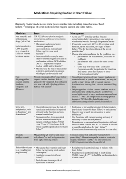


© Copyright 2026