
JMSCR Volume||2||Issue||10||Page 2539-2542||October-2014
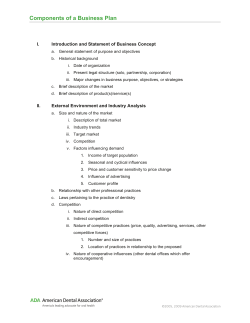
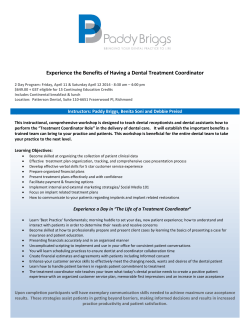
JMSCR Volume||2||Issue||10||Page 2539-2542||October-2014
www.jmscr.igmpublication.org
2014
Impact Factor 3.79
ISSN (e)-2347-176x
Prosthetic Protection of a Healing Bone Defect Caused as a Result of
Chronic Suppurative Osteomyelitis of the Mandible
Authors
1
2
Khurshid A Mattoo 1, Surendra P Singh 2, Krati Mishra3
Assistant Professor, College of Dental Sciences, Gizan University (KSA)
Professor and head, Department of Prosthodontics, Subharti Dental College, Subharti University (India)
3
Post Graduate student, Department of Prosthodontics, Subharti University (India)
Corresponding Author
Dr Khurshid A Mattoo
Assistant Professor, College of Dental Sciences, Gizan University
Email: [email protected]
Work Attributed to Subharti Dental College and Hospital, Subharti University, Meerut
ABSTRACT
Osteomyelitis of the jaws in any of its forms is rarely encountered by a prosthodontist for rehabilitation.
Though acute forms of osteomyelitis are considered as dental emergency, its chronic forms are usually
associated with the destruction of the jawbone in either maxilla or the mandible. Prosthetic protection is
required temporarily till the lesion heals, but dental rehabilitation is required due to tooth loss. This article is
a case report of a patient who had undergone surgery and required a device to protect the area. The design of
such a device is also discussed along with the anatomy of the defect.
Keywords- trabeculae, medically compromised, suppuration, antibiotics
INTRODUCTION
Though bone appears to be a very strong structure
bone.[1] It can be classified as acute, subacute or
yet it is prone to decay due to infections that may
chronic, depending on the clinical presentation, but
range from bacteria to fungal or viral. The word
there are other ways of classifying the condition like
“Osteomyelitis” originates from the ancient Greek
symptoms, cause and presence or absence of
word, osteon (bone) and muelinos (marrow) and
suppuration. The decline in prevalence of this bone
means infection of the medullary portion of the
condition is due to increased availability of
Khurshid A Mattoo et al JMSCR Volume 2 Issue 10 October 2014
Page 2539
JMSCR Volume||2||Issue||10||Page 2539-2542||October-2014
antibiotics and progressively higher standards of
oral and dental health.
[2], [3]
2014
had recently suffered from anemia, which was
corrected before surgery. The patient also gave a
Basically an inflammatory condition of the bone
history of smoking, alcohol and pan chewing.
that begins as an infection of the medullary cavity,
Clinical examination revealed a Kennedy class III
rapidly involves the haversian systems, and extends
situation in relation to left side of the mandible with
[4]
second premolar, first and second molar missing.
Currently osteomyelitis usually presents as a sub-
The edentulous area was trough like extending
chronic condition and is more commonly associated
almost one and a half centimeters in length and 12
with debilitated, immunosuppressed or medically
mm in width (Fig.1).
to involve the periosteum of the affected area.
compromised patients who have an existing
infection in the area.
[5], [6]
In its acute form, the
condition presents symptoms which force the
patient to seek medical attention, but in its chronic
form the condition called chronic suppurative
osteomyelitis causes abscess and sequestration of
the bone.
[7]
Medically compromised patients like
those with underlying diabetes or malnutrition
suffer delayed healing, especially when surgical
intervention has been done. Surgery is indicated to
remove the dead bone along with sequestra and
leave normal healthy bone that is capable of
Figure 1: Intra oral view of the large defect left
healing. This clinical case report is of a similar
over after the surgery
patient and was advised prosthetic protection of the
area that was surgically corrected.
The lingual cortical plate was higher than the buccal
cortical plate and the tissues in the area were flabby
CASE REPORT
and tender to touch. Palpation revealed collected
An elderly male patient in his late forties was
food debris inside with the release of bad odor. The
referred to the department of Prosthodontics by
treatment plan for the patient included a protective
department of oral surgery for possible protective
splint that would cover the entire area, including the
device in relation to surgery on the left side of the
area within the trough and would be retained by a
mandible due to chronic suppurative osteomyelitis.
19 gauge stainless steel Adams clasp.
The aim and objective of surgeons were to protect
Impressions were made for both maxillary and
the area from trauma, food collection, debris while
mandibular dentulous arches with irreversible
at the same time provide a conducive environment
hydrocolloid (CA 37; Cavex, Haarlem, Holland),
to promote healing. The patient was diabetic and
following which retrieved casts were put on a dental
Khurshid A Mattoo et al JMSCR Volume 2 Issue 10 October 2014
Page 2540
JMSCR Volume||2||Issue||10||Page 2539-2542||October-2014
2014
cast surveyor on which the relative soft and hard
tissue
undercuts
simultaneously.
were
This
marked
was
then
and
blocked
followed
by
fabrication of an Adams clasp to act as mechanical
form of retention. Auto polymerizing acrylic resin
(Fortex; Lucite Intl, Durham) was then slowly
added till it took the shape of a protective device
that would engage the undercuts and interproximal
embrasures of the teeth (Fig.2). The protective
device was then tried in the patient and necessary
corrections were made (Fig.3). The patient was
Figure 2: Protective device with no teeth on it
demonstrated and taught the insertion and removal
of the device and was put on a strict following up
protocol of every 15 days. During the follow up
visit the inner surface of the device in the area of
healing was reduced by 1mm to accommodate the
growing tissues under the device.
Surgical correction is the treatment of choice when
underlying systemic diseases are present as it allows
less time to heal. After surgery, the patient is
required to keep the area clean and free from any
sort of injury. Mandibular resected areas are not
easy to maintain, and a protective device is a correct
DISCUSSION
Chronic suppurative osteomyelitis (CSO), which is
a bone infection with suppuration, abscess/fistula
formation, and sequestration at some stage of the
disease, is due to a defined, infectious etiology.
Underlying systemic diseases like diabetes, anemia
and malnutrition that cause concomitant alteration
in host defenses profoundly influences the course of
osteomyelitis.
8
The jaws are unique from other
bones of the body in that the presence of teeth
creates a direct pathway for infectious and
inflammatory agents to invade bone by means of
caries and periodontal disease.9 Oral bone appears to
be particularly resistant
exposure to oral flora.10
to
infection despite
innovation as demanded by surgeons in this case.
The critical part of the healing is during the first 3 to
5 weeks depending upon the extent of surgery.
Forming an acrylic bulb that would overlay the
trough allows complete protection. As the tissues
heal, the bulb can be easily accommodated within
the growing area by simple reduction.
CONCLUSION
Though chronic suppurative lesions of the mandible
are less common in modern times, every clinician
should be able to provide a conducive environment
for healing in such cases. A simple device like one
described is an excellent option in such cases.
Khurshid A Mattoo et al JMSCR Volume 2 Issue 10 October 2014
Page 2541
JMSCR Volume||2||Issue||10||Page 2539-2542||October-2014
2014
5. Eckman MH, Greenfield S, Mackey WC,
Wong JB et al. Foot infections in diabetic
patients. Decision and cost-effectiveness
analyses. JAMA 1995; 273: 712–20.
6. Grayson ML, Gibbons GW, Balogh K,
Levin E, Karchmer AW. Probing to bone in
infected pedal ulcers. A clinical sign of
underlying osteomyelitis in diabetic patients.
JAMA 1995; 273: 721-723.
7. Bevin CR, Inwards CY, Keller EE. Surgical
Figure 3: Protective device in place
management
of
primary
chronic
osteomyelitis: a long-term retrospective
ACKNOWLEDGEMENT
The authors would like to acknowledge the sincere
efforts of department of oral surgery for being
innovative in patient care.
analysis. J Oral Maxillofac Surg 2008; 66:
2073-2085.
8. Topazian RG. Osteomyelitis of jaws. In
Topazian RG, Goldberg MH (eds). Oral and
maxillofacial
REFERENCES
1. Marc Baltensperger and Gerold Eyrich.
Osteomyelitis of the Jaws: Springer Berlin,
Heidelberg. November 07, 2008.
2. Marx RE. Chronic osteomyelitis of the jaws.
In: Laskin D, Strass R, eds. Oral and
maxillofacial surgery clinics of North
America. Philadelphia: Saunders; 1992: 367438
3. Yeoh SC, Mac Mahon S, Schifter M.
Chronic suppurative osteomyelitis of the
infections,
3rd
ed.
Philadelphia, PA: Saunders. 1994: 271-73
9. Lee L. Inflammatory lesions of the jaws. In
White
SC,
Pharoah
MJ
(eds).
Oral
radiology: principles and interpretation,
volume
3,
4th
ed.
Missouri:
Mosby.
2000:338-354..
10. Montonen M, Kalso E, Pylkkaren L et al.
Disodium clodronate in the treatment of
diffuse sclerosing osteomyelitis (DSO) of
the mandible. Int J Oral Maxillofac Surg
2001; 30: 313-317
mandible: Case report. Australian Dental
Journal 2005; 50(3): 200-03.
4. Topazian RG. Osteomyelitis of jaws. In
Topazian R G, Goldberg M H (Eds). Oral
and maxillofacial infections. Philadelphia,
PA: Saunders (3rd ed.) 1994: 257-58
Khurshid A Mattoo et al JMSCR Volume 2 Issue 10 October 2014
Page 2542


© Copyright 2026