
Document 326215
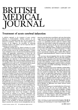
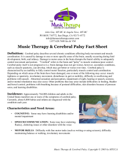
Brain (1996), 119, 1763-1774
Brain development, gender and IQ in children
A volumetric imaging study
Allan L. Reiss,1-2-3-4 Michael T. Abrams, 1 Harvey S. Singer, 34 Judith L. Ross 5 and
Martha B. Denckla 12 - 3
'Kennedy Krieger Institute, Baltimore, the 2Department of
Psychiatry, Division of Child Psychiatry, the ^Department
of Neurology, the ^Department of Pediatrics,
Johns Hopkins University School of Medicine, Baltimore,
the ^Department of Pediatrics, Thomas Jefferson University
School of Medicine, Philadelphia, USA
Correspondence to: Allan L. Reiss, Kennedy Krieger
Institute, 707 North Broadway, Baltimore, MD 21205, USA
Summary
Prominent, age-related changes in grey matter, white matter
and CSF volumes are evident during childhood and appear
to reflect ongoing maturation and remodelling of the CNS.
Both boys and girls show a similar pattern of cerebral
asymmetry; a rightward prominence of cortical and
subcortical grey matter and a leftward prominence of CSF
is observed. IQ is positively correlated with total cerebral
volume in children, in particular, with the volume of cortical
grey matter in the prefrontal region of the brain. Subcortical
grey matter also contributes to the variance in IQ, although
to a lesser extent than cortical grey volume. Quantitative
knowledge of the developing human brain will play an
increasingly greater role in improving sensitivity and
specificity in the interpretation of brain abnormalities in
patients within the clinical environment, as well as in
groups of children with suspected brain dysfunction in the
research setting.
Keywords: brain; development; gender; IQ; MRI; childhood
Abbreviation: NF-1 = neurofibromatosis-1
Introduction
Although a number of investigators have described changes
in cerebral morphology during adult life (Jernigan el al,
1991a; Pfefferbaum et at., 1994; Blatter et al., 1995), there
are relatively few imaging reports describing quantitative
in vivo brain development in a large sample of normal, nonclinically referred children. Obtaining these data in a large
normal population is important in order to unravel the
complex and dynamic biological processes underlying brain
development during childhood, as well as essential to the
valid interpretation of imaging data derived from clinical
paediatric populations with brain disorders.
© Oxford University' Press 1996
Because of the scarcity of normal quantitative paediatric
neuroimaging data, fundamental questions concerning
cerebral development during early childhood remain
unanswered at present. In this study, we utilize quantitativevolumetric analysis of high resolution MRIs to describe brain
development and morphology in 85 normal children and
adolescents ranging in age from 5 to 17 years. Methodological
advantages of the research described in this report over
previous quantitative imaging studies of children (Jernigan
and Tallal, 1990; Jernigan et al., \99\b; Pfefferbaum et al.,
1994) include a large, predominantly non-clinical population
Downloaded from by guest on October 15, 2014
Normal brain development during childhood is a complex
and dynamic process for which detailed scientific information
is lacking. MRI techniques, combined with methods for
advanced image analysis, offer the potential to begin to
construct a quantitative map of normal paediatric brain
development in vivo. In this study we utilize volumetric
analysis of high resolution brain images obtained from MRI
to describe cerebral development and morphology in 85
normal children and adolescents ranging in age from 5 to
17 years. The results show that total cerebral volume is 10%
larger in boys compared with girls. However, both boys and
girls show little change in total cerebral volume after the
age of 5 years. Increased cortical grey matter is the primary
contributor to larger brain volume in boys, thus supporting
the hypothesis that gender associated differences in brain
size are related to differences in cortical neuronal density.
1764
A. L. Reiss et al.
of children, utilization of neuroanatomical imaging data from
the entire cerebrum as opposed to a representative subset of
the brain, use of contiguous slices in the imaging dataset
versus datasets with interslice gaps, incorporation of
standardized methods for tissue segmentation and stereotaxic
division of the brain into more discrete cerebral regions, and
the availability of IQ data on the majority of subjects. In this
study, emphasis is placed on the elucidation of basic issues
related to (i) gender-associated differences in cerebral
structure or development which may be identifiable in young
children, (ii) patterns of age-related changes in specific tissues
or regions of the brain during childhood, and (iii) analysis
of correlations between cerebral morphology and intellectual
abilities. These issues are presented and discussed in light of
our present knowledge of early brain development derived
from post-mortem studies of children.
Methodology
Subjects
Imaging
MRls were acquired on a General Electric 1.5 Tesla Signa
Scanner. Scans utilized for the group analyses were derived
from an axial spin-density, T2-weighted acquisition with the
following parameters: TR = 3000, TE = 30/100, 5 mm
slices angled parallel to the AC-PC (anterior commisureposterior commisure) line. Three subjects had an interslice
gap equal to 2.5 mm. The remaining 82 subjects had
contiguous 5 mm images obtained with an interleaved
protocol. Images were processed, then segmented into grey,
white and CSF compartments as described below.
Procedures for removal of non-brain tissues and correction
of pixel intensity non-uniformity artifact prior to tissue
segmentation were based on methods described by Lim and
Pfefferbaum (1989). Measurement of grey, white and CSF
compartments were made after creating composite images
from paired T2- and proton-weighted images. These
composite images were produced by adding and subtracting,
respectively, the early and late echo (paired) images. Tissue
classification was determined in each slice using histogrambased segmentation algorithms which automatically establish
one or more statistically optimal thresholds for separating
tissue types (Otsu, 1979; Cho etai, 1989). Manual adjustment
of the selected threshold was available to the rater if the
computer-determined values appeared grossly incorrect.
Measurements of grey, white and CSF were made after tissue
and fluid in the posterior fossa (brainstem and cerebellum)
were manually excluded. CSF voxels comprising the lateral
ventricles and subcortical grey voxels comprising the basal
ganglia and thalamus were designated and segmented
separately in each image slice. Lateral ventricle and subcortical regions were included in the overall and hemispheric
grey, white and CSF volume measures but excluded from
the 16 cerebral regional measures described below. The
volume of a region of interest contained in a single slice was
determined by multiplying the number of pixels comprising
the region of interest in the slice by the slice thickness. The
volume of the structure was then determined by summing
the volumes of the region of interest in each slice in which
it occurred.
In order to determine whether neuroanatomical differences
associated with gender or age were localized to specific
regions of the brain, volumes of hemispheric and
subhemispheric regional grey matter, cerebral white
matter, tissue (grey + white) and CSF, were determined
after designation of five planes through the brain of all
subjects. Operational definitions for placement of these planes
were predetermined and based on a previously described
regional stereotaxic coordinate system (Zipursky etal., 1992).
The anatomical location of the planes are further described
and illustrated in Fig. 1. The projection of the five planes
through the brain sections served to divide the brain into 16
Downloaded from by guest on October 15, 2014
MRI scans were originally obtained from 100 normal children
and adolescents between the ages of 5 and 17 years. Fifteen
of these scans were identified as technically sub-optimal due
to failure to include the entire cerebrum within the image
dataset at the time of acquisition. The final subject group
consisted of 64 females (mean age±SD, 10.6 ±2.9 years)
and 21 males (mean age±SD, 10.7±2.8 years). The inclusion
of larger numbers of female subjects relative to males was
due to our recruitment of gender-matched normal controls
for ongoing studies of female children with specific genetic
conditions such as fragile X syndrome and Turner syndrome.
All subjects were in good health and without evidence of
neurological or psychiatric disorders. All subjects had normal
IQ scores as determined by standard cognitive evaluation
(H = 69), or satisfactory performance in a regular classroom
setting (n = 16). The male and female groups were wellmatched for handedness, with ~ 15% in each gender group lefthanded, 83% right handed and 2% having mixed handedness.
Most subjects (n = 42) were recruited from announcements
in public and private schools and area newspapers. Control
subjects were also recruited from the pool of normal siblings
of subjects with fragile X syndrome, Turner syndrome or
neurofibromatosis-1 (NF-1) participating in ongoing research
at the Kennedy Krieger Institute (n = 40). All normal siblings
of individuals with the fragile X syndrome (n = 10) were
tested with DNA analysis (Rousseau el al., 1991) and found
to be negative for the fragile X mutation. All siblings of
children with Turner syndrome or NF-1 were phenotypically
normal; children in the latter group were also examined by
a geneticist and opthalmologist to rule out any physical
stigmata of NF-1. Three subjects in this study were already
scheduled to receive an MRI for evaluation of generalized
headaches and were prospectively asked to participate in the
research protocol (these scans were subsequently read as
clinically normal). Subjects' informed consent was obtained
according to the guidelines of the Johns Hopkins Joint
Commission on Clinical Investigation.
Normal brain development in childhood
1765
neuroanatomical sections. The resulting 16 subhemispheric
brain areas roughly correspond to the following right and
left cerebral regions: prefrontal, posterior frontal, orbitalfrontal, parietal, superior parietal-occipital, inferior parietaloccipital, anterior temporal and mid/posterior temporal
(Fig. 1). Because of the possible influence of slight variations
in head tilt and slice location on volume determination of
subhemispheric cerebral regions, all scans were visually
inspected; 79 scans (60 females and 19 males) were
determined to be correctly aligned to the AC-PC plane and
were subsequently used in the analysis of subhemispheric
regional morphology. The mean ages of the children
comprising this subgroup (females, 10.6±2.9 years; males,
10.5±2.6 years) were very similar to those of the overall
group.
Several procedural details were followed to eliminate
observer bias in determining volumes of regions of interest.
All scans were analysed interspersed among those from
subjects having various developmental disorders (e.g. children
with fragile X or Turner syndrome). Scans were processed
so that images contained no subject information other than
subject number, and the raters performing morphometric
analyses did not know group membership of any subject.
Previous description of specific definitions of neuro-
anatomical regions of interest as well as inter-rater reliabilities
have been reported for all measures described in this study
(Reiss et al., 1993a, b, 1995). The average intraclass correlation corresponding to inter-rater agreement for cerebral
regions of interest was 0.93.
Data analysis
Age was not significantly correlated with total cerebral
volume (Fig. 2); therefore, ANOVA was used in the initial
gender group comparisons for hemispheric and subhemispheric volumes of cerebral tissue (grey + white), grey
matter, cerebral white matter, CSF and subcortical nuclei.
The presence of significant gender differences for total
cerebral volume (Table 1), and the possibility that age would
be related to tissue- or region-specific volumes (Jernigan
et al., 1991ft; Pfefferbaum et al., 1994), led to the use of
ANCOVA with both total cerebral volume and age as
covariates in an additional set of gender comparisons.
MANCOVA was also performed to determine whether there
was a unique pattern of gender differences in the volumes
of the subhemispheric regions.
Regression analyses were used to investigate the association of designated brain regions (or tissue) with age. Total
Downloaded from by guest on October 15, 2014
Fig. 1 The brain parcellation scheme used for analysis of subhemispheric regional volumes is illustrated
in the figure. Images show the location of five planes projected through the brain in a transaxial slice and
a three-dimensional surface rendering. The five planes consisted of (i) a midsagittal plane following the
interhemispheric fissure dividing the brain into right and left hemispheres, (ii) a transaxial plane parallel
to the orientation of the axial sections and along the AC-PC line dividing each hemisphere into superior
and inferior regions, (iii) a coronal plane perpendicular to the transaxial plane and passing through
the most anterior extreme of the genu of the corpus callosum, (iv) a coronal plane perpendicular to the
transaxial plane and passing through the most posterior extreme of the splenium of the corpus callosum,
and (v) a coronal plane perpendicular to the transaxial plane and passing through a point midway between
the anterior and posterior callosal planes.
1766
A. L. Reiss et al.
1600-
X
X
X
1500 -
X
=
1400O
LJUU -
.Q
a
u
1200-
X
o
o
•"""o"o
°x~
^
o
o
O
1000-
0
J
o
X
o
X *
"
O
•©•
8 o
o
o
o
900-
Jx
°
1100o
--^
o
o o
o
o
o
°
o
o
o
o
10
12
14
16
18
Age (years)
Fig. 2 Total cerebral volume in children ranging in age from 5 to
17 years is shown in the figure. Boys (crosses) and girls (open
circles) show little change in this measure over the age range
studied. Regression lines for boys (dashed) and girls (continuous)
are also shown.
Region
Male
(n = 21)
mean±SD
Female
(n = 64)
mean±SD
P
Total cerebrum
Right hemisphere
Left hemisphere
Total grey
Cortical grey
Caudate nucleus
Lenticular nucleus
Thalamus
White matter
Total CSF
Extraventricular CSF
Lateral ventricles
1290.6± 147.4
643.9±72.6
646.7±75.0
707.4±7l.4
674.8±67.8
11.1 ± 1.9
9.9±l.6
11.6+2.2
488.3±92.0
94.9±28.8 •
79.4+23.8
15.5±9.5
1182.5 ±104.7
591.1±52.5
591.4±52.8
637.2+66.1
605.6 ±64.0
IO.7±1.6
9.5±1.6
ll.4±1.8
443.8+61.7
101.5±25.l
89.6±23.7
11.9±5.7
«0.0005
ssO.0005
=s0.0005
=£0.0001*
sso.oooi*
n.s.
n.s.
n.s.
=s0.01
n.s.*
n.s.*
€0.05
Comparison of male and female group volumes for
neuroanatomical regions of interest. All analyses were initially
performed with ANOVA; analyses using ANCOVA with age and
total cerebral volume as covariates were also used for all volume
comparisons (other than total cerebrum) to determine whether a
particular region contributed more heavily to gender differences
in total cerebral volume. The P value shown for all variables
in the table represents the significance level for the main effect of
group (male versus female). *ANCOVA P < 0.005; +ANCOVA
P < 0.0025; *ANCOVA P < 0.0005; n.s. = not significant.
cerebral volume, gender, age and gender by age interaction,
were entered sequentially as independent variables in a series
of hierarchical regression analyses with cortical grey matter,
white matter, CSF and subcortical volumes as dependent
variables. Because of the predictable (positive) correlation
between the volumes of the dependent variables and total
cerebral volume, total cerebral volume was entered into each
regression analysis as an initial independent variable in order
to measure, and remove, the contribution of overall cerebral
Results
Cerebral volume in children-gender effects
Analyses shown in Table 1 indicate that absolute cerebral
volume (grey+white+CSF) in boys is ~10% larger than that
of girls (P = 0.0004). ANCOVA further suggests that in
spite of the magnitudes of gender differences for cortical
grey and white matter volumes being similar (11.4% and
10.0%, respectively), grey matter volume is a more consistent
contributor to larger total cerebral volume in males than
white matter volume (Table 1). Moreover, further examination
of this issue indicated that the standard deviation for male
white matter volume was larger than that for female white
matter volume. Upon visually inspecting the distribution of
this variable across genders, it became apparent that there
was one (10-year-old) male for whom white matter volume
was > 2 SDs (710.6 cm 3 ) from the mean of the entire male
group. (No other volume from this individual was > 2 SDs
from the group mean.) Reanalysis of volumes without this
individual showed that gender differences were 11.0% and
7.5% for cortical grey and white matter, respectively. After
exclusion of this subject, statistical analyses of gender
differences for grey matter volumes remained significant for
both total (P =£ 0.0002) and cortical grey (P =£ 0.0001)
volumes. For white matter volume, the P value decreased to
marginally significant (P = 0.052) when this single subject
Downloaded from by guest on October 15, 2014
Table 1 Cerebral volumes (cm3) in children
size to the volumes of the regions of interest. Gender, age,
and gender by age interaction were entered, respectively, into
the regression analysis in the second through fourth steps,
and the independent (incremental) partial correlation squared
(sr2) determined for each independent variable. The critical
F value corresponding to the contribution of any of these
three variables was set at ^ 4 . 0 0 (corresponding to a P value
of <0.05 for the number of residual degrees of freedom in
the model). As a means of validating the results obtained
from the hierarchical regression analyses for grey and white
matter, simple regression analyses were also performed with
age as the independent variable and the region of interest
volume:total cerebral volume ratio as the dependent variable
in each gender group separately.
For analysis of gender-associated hemispheric differences
in symmetry, a two (gender group)Xtwo (hemisphere: left
versus right) mixed ANOVA was employed, with repeated
measures on hemisphere. In addition, paired / test analyses
were used to analyse left versus right hemisphere differences
in the entire group of 85 subjects. Two-tailed tests of
significance were used for all analyses used unless an a
priori hypothesis had been stated in which case a one-tailed
significance test was used. Alpha was set at P < 0.01 for all
group comparisons in these repeated measures analyses.
The association of cerebral volume with IQ was
investigated with stepwise and hierarchical regression
analyses using specified neuroanatomical regions as
independent variables.
Normal brain development in childhood
Change in cerebral volume with age
There was no observable change in total cerebral volume as
a function of age in either male or female subjects (Fig. 2).
Results from the hierarchical regression analyses (Table 2)
showed that total cerebral volume predicted a significant
proportion of the variance in the volumes of all constituent
tissue and CSF variables evaluated. After removing the effect
of total cerebral volume and gender on region of interest
volume, age was found to predict a significant proportion of
the variance in cortical grey matter volume (22%) and
cerebral white matter volume (16%), such that increasing
age was associated with decreasing grey matter volume and
increasing white matter volume in children. Age was also
observed to predict a small, but significant amount (7%) of
the variance of extraventricular CSF (which increased with
age), and lenticular and thalamic subcortical grey regions
(which decreased with age). Consistent with ANCOVA results
for gender comparisons, gender effects were small, yet
significant, for cortical grey matter (males > females), and
extraventricular CSF (females > males), even after the effect
of total cerebral volume and age had been removed. The
gender-by-age interaction term was not found to be significant
for any of the analyses, indicating that age-related effects for
these brain regions were not different between boys and girls.
Concordant with results from these hierarchical-stepwise
regression analyses, the ratio of cortical grey volume to total
cerebral volume showed a significant negative correlation
with age for both boys (R = -0.62, P =s 0.002) and girls
(R = -0.73, P =£ 0.0001). An opposite pattern was observed
for the ratio of total white matter volume to total cerebral
volume (R = 0.51, P =£ 0.01 for males; R = 0.72, P =£
0.0001 for females) which is observed to increase with age
throughout childhood (Fig. 3).
Age predicted a significant proportion of the variance in
the volumes of both total cortical grey matter and cerebral
white matter. However, based on data suggesting regionspecific differences in rates of cerebral maturation in the
human brain, particularly for white matter (Lemire et al.,
1975), we also investigated whether regional age-related grey
and white volume changes would be consistent with findings
from the 'parent' tissue compartment. This question was
addressed by analysing independent age effects on grey or
white matter volumes of the eight bilateral subhemispheric
cerebral regions; if present, these effects would be distinct
from those explained by age associations with the total
volume of cortical grey or cerebral white matter volume in
the brain, and gender. Accordingly, the effects of gender and
either total cortical grey or cerebral white volume (according
to the variable being analysed) were first measured and
removed in a hierarchical regression analysis. The effects of
age and a gender-by-age interaction term on the grey and
white volumes of each of the subhemispheric regions, were
then examined. The results of these analyses (not shown)
indicated that age, but not age by gender effects, were
significant for white matter volume changes within the frontal
lobe. Specifically, there was an increase in prefrontal white
matter volume (F = 7.0, P < 0.01) and a decrease in orbitalfrontal white matter volume (F = 6.6, P < 0.02) with
increasing age which were independent of age-related changes
in total white matter volume in the brain. No age or age-bygender effects were observed for any regional cortical grey
volume, indicating relative uniformity in age-related changes
for cortical grey throughout the cerebrum in both boys
and girls.
Cerebral asymmetry
The symmetry of right and left hemispheric tissue, grey,
white, CSF (ventricular and extraventricular) and subcortical
nuclei was examined for all 85 subjects; the symmetry of
right and left subhemispheric cerebral regions was also
analysed for the 79 subjects with scans correctly aligned
to the AC-PC plane. No significant gender-by-hemisphere
interactions emerged in these repeated measures ANOVAs,
Downloaded from by guest on October 15, 2014
was excluded. Reanalysis of the data also showed that, similar
to the original analyses in which all subjects were included,
the ANCOVA remained significant for cortical (/> =£ 0.001)
and total (P =£ 0.05) grey matter, and not significant for
white matter (P > 0.20).
Compared with cortical grey and white matter volumes,
the size of subcortical nuclei were not significantly different
between the two gender groups. However, girls did have
proportionally greater amounts of extraventricular CSF, but
not ventricular CSF.
Analyses were carried out to determine whether there was
a unique pattern of gender differences in the volumes of
the subhemispheric regions after statistically removing the
influence of total tissue (or fluid) volume. A MANCOVA for
the tissue (i.e. grey+white) volumes of the eight bilateral
subhemispheric regions (i.e. prefrontal, posterior frontal, etc.)
utilized gender as the grouping variable and total cerebral
tissue volume as a covariate. (Bilateral volumes were used due
to the absence of gender differences in cerebral asymmetry as
shown by subsequent analyses below.) The MANCOVA was
significant [Wilkes F(8,69) = 2.92, P = 0.007]; however,
none of the eight individual ANCOVAs showed a significant
gender effect. Three additional MANCOVAs were carried
out for the eight cortical grey, eight cerebral white, and eight
nonventricular CSF volumes, respectively. Similar to the first
analysis, these three MANCOVAs used the overall volume
of the respective tissue (or fluid) as the covariate. These
analyses (not shown) did not reveal evidence for gender
differences in regional tissue, cortical grey, cerebral white or
non-ventricular CSF volumes of greater magnitude than
those observed for the total tissue or fluid compartments,
respectively. Thus, the relative proportion of tissue and fluid
volumes comprising the brain is similar between males
and females despite the 10% volume difference between
the genders.
1767
1768
A. L. Reiss et al.
Table 2 Influences of total cerebral volume, gender and age on region- and tissue-specific volumes (cm3)
Regression
step
Independent variable(s)
Cortical grey matter
1
2
3
4
1
2
3
4
1
2
3
4
1
2
3
4
1
2
3
4
1
2
3
4
1
2
3
4
Total cerebral
Gender
Age
GenderXage
Total cerebral
Gender
Age
GenderXage
Total cerebral
Gender
Age
GenderXage
Total cerebral
Gender
Age
GenderXage
Total cerebral
Gender
Age
GenderXage
Total cerebral
Gender
Age
GenderXage
Total cerebral
Gender
Age
GenderXage
Cerebral white matter
Ventricular CSF
Extraventricular CSF
Caudate nucleus
Lenticular nucleus
Thalamus
volume
volume
volume
volume
volume
volume
volume
B(p)*
sr2
0.695
-0.087
-0.382
-0.124
0.826
-0.111
0.229
0.241
0.277
0.627
0.812
-1.107
0.485
0.294
0.138
0.122
0.498
0.542
0.416
-0.663
0.433
0.763
0.553
-1.079
0.477
0.487
0.111
-0.508
0.571
0.022
0.223
0.000
0.653
0.002
0.163
0.002
0.110
0.012
0.000
0.038
0.118
0.119
0.052
0.000
0.219
0.008
0.004
0.014
0.182
0.002
0.054
0.038
0.181
0.019
0.067
0.008
<0.00001
<0.05
<0.00001
n.s.
<0.00001
n.s.
<0.00001
n.s.
<0.002
n.s.
n.s.
n.s.
<0.002
<0.001
<0.025
n.s.
<0.0001
n.s.
n.s.
n.s.
<0.0001
n.s.
<0.025
n.s.
<0.0001
n.s.
<0.01
n.s.
The table shows the standardized regression coefficients (P) and incremental partial correlations squared (sr2), corresponding to the
association between the volume of the dependent variable (a designated tissue or region) and the specified independent variables. The srindicates the proportion of variance in the volume of the dependent variable predicted by the independent variable; n.s. signifies that the
F value for the independent variable did not reach statistical significance (P < 0.05). The independent variables were entered
sequentially in individual hierarchical regression analyses in the order shown in the table. *Beta coefficients ((5) for each independent
variable are derived from the final step in the regression analysis.
0.60
0.42
10
12
Age (years)
10
12
Age (years)
14
16
18
Fig. 3 Age-related changes in the proportions of cortical grey matter and cerebral white matter are shown in the figure. Boys (crosses,
dashed line) and girls (open circles, continuous line) show significant changes in both variables over the age range studied.
providing no evidence for gender-associated symmetry
differences for the regions examined.
Following this initial analysis, paired / tests (using right
versus left volumes) were utilized to determine the presence
of brain asymmetries across the entire group of boys and
girls. These analyses revealed a number of cerebral
asymmetries which, although modest in absolute volume,
were statistically significant. These included a right > left
pattern for the caudate (/ = 4.8; P < 0.0001), lenticular
nucleus (t = 4.4; P < 0.0001), and cortical grey (/ = 3.9;
Downloaded from by guest on October 15, 2014
Dependent variable
Normal brain development in childhood
1769
150
70
-300
-200
-100
0
100
200
Total Cerebral Volume-Residual (cm3)
-30
-20
-10
0
10
20
30
Prefrontal Grey Volume-Residual (cm3)
Fig. 4 The scattergrams show the association of full scale IQ with the residual total cerebral volume variable (A) and the residual prefrontal
grey matter volume variable (B). Both neuroanatomical variables represent the 'residual' volume of the respective region after the effect
of gender has been statistically removed.
Association of cerebral volume with IQ
For the 57 girls and 12 boys for whom cognitive testing data
were available (the IQ subgroup), there was no gender
associated statistical difference in mean (±SD) full scale IQ
score (113± 15 for girls; 105± 17 for boys). The ages of the
boys and girls comprising this subgroup were similar to those
of the entire sample (10.6±2.7 for girls, 10.1 ±2.7 for boys).
The volumes of neuroanatomical regions for the known-IQ
subgroup were also analysed to ensure the representativeness
of the sample; these volumes did not significantly differ from
those of the overall group (analyses not shown). So that the
entire group of boys and girls could be analysed together in
subsequent regression analyses focusing on IQ, the effect of
gender on total cerebral volume was statistically removed.
This was accomplished by creating a residual cerebral volume
variable which represents the amount of deviation from the
regression line derived from the correlation of gender (coded
as 0 = male, 1 = female) with total cerebral volume.
In an initial regression analysis, residual total cerebral
volume was observed to predict ~20% of the variance in IQ
(R2 = 0.207; P = 0.0005); the best curve fit for these data
was a second order polynomial (Fig. 4A) suggesting that IQ
increased with larger cerebral volume to a point, then reached
a plateau, and perhaps decreased with the largest volumes.
In order to determine which of the tissue compartments were
most responsible for the IQ/brain volume correlation, residual
variables were also created for total grey matter, white matter
and CSF. These variables were generated from the regression
of the respective tissue compartment with gender and age.
Age was included as an independent variable in the creation
of these three residual volume variables, since we had
previously demonstrated significant age correlations with
grey, white and non-ventricular CSF compartments, as
described above. A stepwise regression was utilized to
determine which of the three residual variables (grey, white,
CSF) would predict a significant portion of the variance in
IQ. Only the residual grey matter variable was included in
the final regression model as a statistically significant
predictor (n = 69, R2 = 0.156; P = 0.0008), indicating that
after removing the effects of gender and age, grey matter
volume predicted ~15% of the variance in IQ in this
population of normal children; specifically, larger total grey
matter volume predicted higher IQ scores.
To further specify the particular tissue components
predicting IQ variance, the stepwise regression was repeated
for the two sub-compartments comprising the total grey
matter volume, cortical grey and subcortical grey. As
described above, residual variables were created with the
respective tissue compartment and age. Since the total grey
matter volume was comprised predominantly of cortical
grey matter (95%), a hierarchical regression was utilized to
determine whether subcortical grey volume would predict
any variance in IQ after cortical grey volume was entered
into the model in an initial step. The results showed that
Downloaded from by guest on October 15, 2014
P = 0.0002), and a left > right pattern for ventricular
(t = 2.6; P = 0.01) and nonventricular CSF (t = 3.5; P =
0.0007). For subhemispheric regions, tissue (grey + white)
volumes showed a right > left pattern for prefrontal (r =
4.0; P = 0.0001), posterior frontal (t = 3.9; P = 0.0002)
and parietal regions (t = 2.8; P = 0.007) and a left > right
pattern for the inferior parietal-occipital region (t = 4.3;
P < 0.0001). When subhemispheric tissue volumes were
analysed separately, cortical grey showed a pattern of regional
asymmetry identical to that observed for regional tissue
volumes (all /s > 4.0; all Ps < 0.0001 for the four regions),
whereas only the inferior parietal-occipital region showed
significant left > right volume in the analysis of white matter
(r = 3.3; P < 0.0014).
1770
A. L. Reiss et al.
Discussion
Gender differences
Imaging and post-mortem studies of adult brains consistently
point to gender differences in overall brain size with adult
males having, on average, a 10% greater volume than females
(Dekaban, 1978; Ho et al, 1980; Filipek et al, 1994;
Pfefferbaum et al, 1994; Blatter et al, 1995). Our study
extends this finding to a large group of children as young as
5 years of age, a result which strongly suggests the presence
of early gender-associated differences in cerebral development and organization.
While the specific learning and behavioural correlates of
gender differences in cerebral volume await elucidation, a
clue as to the cytoarchitectonic basis of this neuroanatomical
finding was recently described by Witelson et al. (1995).
This group reported that the neuronal density of the granular
layers of the cerebral cortex was significantly higher in
females, compared with males, within the portion of the
superior temporal cortex known as the planum temporale. In
our study, increased cortical grey matter was the most
consistent contributor to brain volume differences in our
child subjects, thus supporting the hypothesis of gender
associated differences in brain size being related to differences
in cortical neuronal density.
Unexpectedly, after covarying for overall cerebral volume
and age, extraventricular CSF volume was found to be
increased in the female subject group compared with the
age-matched sample of boys. While an obvious explanation
for this phenomenon is not apparent from our data, this
finding could also have been influenced by the presence, in
girls, of larger amounts of cisternal CSF which was included
in the non-ventricular CSF volume measure in this study.
Future studies will need to separate out the cisternal
compartment from sulcal and ventricular CSF in order to
address this issue.
Age effects
Post-mortem studies show that the human brain attains adult
weight by early childhood, usually by age 5-10 years (Lemire
et al, 1975). Our finding no evidence of growth in total
cerebral volume in either boys or girls between the ages of
5 and 17 years is consistent with these data. In contrast to
total cerebral volume, cortical grey and cerebral white matter
volumes are quite dynamic throughout childhood. Loss of
cortical grey volume and gain in white matter volume with
age were demonstrated in this study, both as a change in the
grey or white matter to cerebral volume ratio, and as a
function of change in absolute (raw) cortical grey or cerebral
white matter volume. Age-related decline in cortical grey
volume in normal children as young as 5 years old is
consistent with the findings of other imaging investigations
of smaller groups of older children and adult populations
(Jernigan et al, 1991a, b; Pfefferbaum et al, 1994; Blatter
et al, 1995). Our findings are also consistent with a recent
imaging study performed on scans obtained retrospectively
from a clinically referred sample of children (Pfefferbaum
et al, 1994).
The finding of decreasing subcortical grey matter volume
with age in our population is consistent with a previous
investigation of a smaller group of normal children (Jernigan
et al, 19916) and suggests that regressive events aimed at
refinement of neuronal connections through cellular and
synaptic pruning (Cowan et al, 1984) occur normally in
cortical and, to a lesser extent, subcortical grey matter
regions during early childhood. Similarly, increase in brain
myelinization in the first and second decades of life is likely
to be a primary contributor to expansion of the volume of
the white matter compartment in the age group sampled in
this study. It is of interest that this expansion was particularly
prominent in the prefrontal region of the brain. This region
is known to undergo considerable myelinization during late
childhood and young adulthood and is implicated in higher
order (executive) regulation of cognitive functions in humans
(Cummings, 1993).
Brain asymmetry
In addition to discrepant cerebral volume, recent evidence
for sexual dimorphism of the human brain with respect to
cerebral asymmetry, has been described in age groups as
young as the foetus (de Lacoste et al, 1991). However,
consistent with data obtained from adults (Zipursky et al,
1990), we found no evidence of brain asymmetry differences
between boys and girls, suggesting that the method for brain
parcellation used in this study may be too unrefined for any
Downloaded from by guest on October 15, 2014
both the cortical grey (R2 = 0.143; P < 0.002) and subcortical
grey (sr2 = 0.073; P < 0.025) residual volumes predicted a
significant portion of the variance in IQ.
To investigate whether particular brain regions were more
highly correlated with general cognitive ability than others,
the association of subhemispheric cortical grey volumes with
IQ were investigated in a subgroup of 64 children (11 boys
and 53 girls) who had both IQ data and correctly aligned
scans. This subgroup was also representative of the overall
group with respect to age and cerebral volumes (analyses
not shown). Residual volume variables were generated from
the regression of the respective subhemispheric regional grey
volume (e.g. orbital-frontal) with both gender and age to
remove the effect of these independent variables. The volumes
of the corresponding right and left grey regions were summed
for these analyses. A stepwise regression was utilized to
determine which of the eight subhemispheric regions would
predict a significant portion of the variance in IQ. Only the
prefrontal cortical grey volume was entered into the final
regression model (n = 64, R2 = 0.186; P = 0.0004).
This indicated that after removing the effects of gender on
subhemispheric volume, the prefrontal regional grey volume
predicted nearly 20% of the variance in IQ (Fig. 4B).
Normal brain development in childhood
volume, suggests that asymmetry of particular tissue types
may be particularly prominent for this region of the brain.
This possibility is being further explored in our laboratory
with probabilistic tissue segmentation procedures which can
assign and measure partial tissue compositions to the same
pixel in an image (Subramanium et ai, 1996). These
discrepancies in study results also point out the need for
close inspection of study methodology as a potential source
of variation in findings from clinical imaging studies of
similar populations of subjects.
Brain volume/IQ correlations
Our data indicate that a modest, yet significant positive
correlation exists between brain volume and IQ in this sample
of young normal children. This result is consistent with
findings reported in normal adults with respect to both the
general magnitude of the correlation (Willerman et ai, 1991;
Andreasen et ai, 1993; Egan et ai, 1994; Wickett et ai,
1994), and the importance of grey matter as the primary
tissue compartment driving the statistical association
(Andreasen et ai, 1993). In addition, our subhemisphericregional analysis further specifies this correlation by
implication of prefrontal grey as the cortical region most
important in the prediction of variance in IQ. This is an
intriguing finding as the prefrontal cortex is believed to be a
regulatory centre for brain activities related to selective
attention, working memory, inhibition and other executive
functions (Cummings, 1993; Williams and Goldman-Rakic,
1995). If confirmed by subsequent studies, the finding of a
particularly strong correlation between prefrontal cortex and
IQ would provide important insights into our current construct
of human cognition and its measurement. Ongoing studies in
our laboratory are aimed at further investigating correlations
between particular brain regions and more specific cognitive
and neuropsychological measures.
The finding that subcortical grey matter volume predicts
a small, yet significant portion of the variance in IQ is not
consistent with a recent study of 67 normal adults (Andreasen
et ai, 1993) in which a statistical association was not found.
Nevertheless, our ongoing imaging studies of paediatric
neurogenetic groups with cognitive deficits (Singer et ai,
1993; Denckla et ai, 1996; Reiss et ai, 1995) consistently
point to the potential importance of subcortical regions in
the pathogenesis of cognitive and neurobehavioural disorders
in children. The study of these homogeneous neurogenetic
groups also reveals circumstances in which larger than
normal brain volume is not associated with superior cognitive
function. Examples include children with NF-1 and the fragile
X syndrome; both disorders are known to be associated with
suboptimal cognitive function and larger-than-normal overall,
or regional brain volumes (Reiss et ai, 1995; von Deimling
et ai, 1995). The possibility that increases in brain size
exceeding an optimal range may be associated with decreased
cognitive function is also suggested by the finding that a
quadratic function best modelled the association between
Downloaded from by guest on October 15, 2014
gender differences in cerebral volume to be identified, if
they exist. However, when analysing the group of children
altogether, evidence for brain asymmetry was present.
Findings of a subtle rightward preponderance of prefrontal
tissue and leftward preponderance of inferior parieto-occipital
tissue is consistent with post-mortem and early imaging
studies describing cerebral hemisphere asymmetry in the
human brain (Chiu and Damasio, 1980; Bear et ai, 1986).
Rightward preponderance of cortical grey (Filipek et ai,
1994) and the caudate nucleus (Castellanos et al., 1994) has
also been reported in adults and children, respectively.
Leftward preponderance of ventricular and extraventricular
CSF was also observed in our child subjects, the former
being consistent with findings from studies of the adult brain
(Zipursky et ai, 1990). Taken together, findings of rightward
preponderance of cortical and subcortical grey, and leftward
preponderance of CSF, support a hypothesis of differential
development and maturation of the two cerebral hemispheres
in humans (de Lacoste et al., 1991).
In contrast to other findings of cerebral asymmetry, our
observation of rightward preponderance of the lenticular
nucleus in normal children is not consistent with previous
studies from our laboratory and others (Peterson et al., 1993;
Singer et ai, 1993). Although variation in scan acquisition
parameters may have contributed to this phenomenon (5 mm
thick, double echo scans in the present study versus 3 mm
thick, inversion recovery scans in Singer et al. (1993), it is
more likely that disparate methods for segmenting and
measuring lenticular nucleus volume is the primary cause of
divergent results. Specifically, manually drawn boundaries
were utilized to circumscribe the lenticular nucleus regions
of interest in both previous studies showing left-prominent
asymmetry of this region (Peterson et ai, 1993; Singer et al.,
1993). In these studies, lenticular nucleus regions of interest
were inclusive of all tissue types (i.e. grey matter, white
matter, grey/white volume-averaged pixels, and vasculature)
contained within the manually drawn boundaries. In contrast,
in the present study, semi-automated, histogram-based
threshold determination was used to classify and measure
only grey matter pixels within the lenticular nucleus region
of interest. Anatomical admixture of grey and white matter
leads to volume averaging of pixels in an MRI and a
decreased likelihood of these volume averaged pixels being
categorized as grey matter in a threshold/histogram-based
segmentation process. Due to the presence of a high
proportion of intermixed grey and white matter and iron
deposits normally occurring within the lenticular nucleus
region (particularly the globus pallidus), the total volume of
the lenticular nucleus would be expected to represent a
different and more restricted tissue composition in the present
study. This hypothesis is supported by the finding that the
mean lenticular nucleus volume in the present study was
-30% less than that reported in our previous study utilizing
manually drawn boundaries (Singer et al, 1993).
The fact that asymmetry measures of the lenticular nucleus
are reversed when measuring grey matter volume versus total
1771
1772
A. L. Reiss et al.
cerebral volume and IQ in the present study (Fig. 4A).
However, this presumption awaits confirmation with a larger
sample as the statistical improvement resulting from a
quadratic fit over a linear correlation, although significant [F
(for R2 change) = 4.41; P < 0.05], appeared to depend on
a small number of subjects with large cerebral volume and
relatively lower IQ.
Consistency of data with previous studies
Subtle differences between our findings and those reported
by Pfefferbaum et al. (1994) were also observed. For example,
they failed to detect gender differences in white matter
volume, or an age-related increase in extraventricular CSF
in subjects aged <20 years. These differences are likely to
be due to methodological problems inherent in retrospective
studies. All of the scans used in the Pfefferbaum study were
obtained post hoc, from patients who had been referred
clinically for an imaging study for suspected neurological
problems, including head trauma, seizures, psychosis and
dehydration. Although these scans were judged to be normal
by standard (qualitative) radiological assessment, many of
the aforementioned indications for imaging are known to be
associated with quantitative morphological abnormalities of
the brain (Cook et al., 1992; Zipursky et al., 1992; Gale
et al., 1995: Yaffe et al., 1995). In addition, because of the
retrospective nature of this study, scan acquisition procedures
could not be standardized across subjects. As a result, scans
included in the analysis had variable pulse sequences, and
had an interslice gap equal to one-half the slice thickness.
Most important, only six representative slices were used for
analysis of each of the brain volume variables. As Pfefferbaum
et al. (1994) also discuss, these factors could have affected
the accuracy of their results
In the only other study of normal neuroanatomy in a large
number of children, Jernigan et al. (1991b) analysed MRI
scans from 39 normal control subjects ranging in age from
Limitations
Although this study was conducted with a predominantly
non-clinical group of children, potential biases resulting
from recruitment strategies utilizing school and newspaper
announcements, and siblings of children with genetic
disorders, cannot be ruled out. Accordingly, conclusions
regarding normal brain development in children must be
considered provisional until confirmed by investigation of a
more random sample of children from the normal population.
Similarly, although the cerebral parcellation procedure used
in this study presents clear advantages over the analysis of
total or hemispheric cerebral volumes only, the definitions
for the cerebral regions of interest are relatively gross and
subject to effects resulting from differences in brain shape
across subjects. Accordingly, studies utilizing finer, more
functionally precise regions of interest are needed to build
upon the results presented here.
Conclusions
Despite attainment of overall adult size by ~5 years of
age, maturation of the human cerebrum is a dynamic and
complex process, with gender differences, structural
asymmetry, and uneven region-specific development being
detectable throughout childhood. Structural components of
the human brain are modestly correlated with cognitive
abilities, a finding that may reflect the action of many
potential influences on brain development, ranging from
environmental to genetic effects. We predict that quantitative
knowledge of normal brain development in children will play
an increasingly greater role in improving sensitivity and
specificity in the interpretation of brain abnormalities in
patients within the clinical environment, as well as in groups
of children with suspected brain dysfunction in the research
setting.
Continued analyses of these and other data are ongoing in
our laboratory and include examination of the association
between more specific neuropsychological measures and
brain volumes, analysis of higher-resolution scans which
Downloaded from by guest on October 15, 2014
Procedures utilized for subject ascertainment, scan acquisition and image analysis in the present study helped to avoid
many of the methodological problems of previous quantitative
imaging studies of children. In the largest of these studies,
Pfefferbaum et al. (1994) analysed MRIs from 88 individuals,
ranging in age from 3 months to 30 years (mean = 14±7
years), for gender differences and age-related changes in
neuroanatomy. Many of the findings of this study were
similar to the results presented here, particularly with respect
to the male > female difference for cortical grey matter
volume, and the presence of age-related changes in grey and
white matter volumes during childhood. This correspondence
in results is of particular interest, as the core methods for
image processing and segmentation were quite similar across
studies. Comparison of cross-study results for many other
variables of interest was not possible, as analyses of
symmetry, subcortical volumes, regional cortical volumes
and brain/IQ correlations were not reported in Pfefferbaum
et al. (1994)
8 to 35 years, including 26 subjects in the age range of the
study presented here. Although these investigators used
different methods for cortical and subcortical parcellation
and tissue segmentation, many similarities in study results
exist. For example, cerebral volume was significantly smaller
in female subjects in the Jernigan et al. (1991/?) study. In
addition, cortical and subcortical grey matter volumes were,
after adjusting for brain volume and gender, observed to
decrease with age; however, cortical changes were restricted
to the superior portion of the cerebrum, a finding not
replicated in the present study (analyses not shown).
Extraventricular CSF volumes were observed to increase
with age, but only in the inferior regions of the cerebrum.
White matter volumes, symmetry and IQ correlations were
not reported.
Normal brain development in childhood
permit a more fine-grained stereotaxic parcellation technique
(Subramanium et al., 1996), longitudinal analysis with serial
brain scans, and analyses of gender differences, age effects
and IQ correlations for structures within the posterior fossa.
Acknowledgements
We wish to thank Drs Lisa Freund, Michele Mazzocco and
Gary Chase for their statistical consultation. This research
was supported by grants MH01142, HD31715, HD24061 and
NS35359 from the National Institutes of Health.
References
Andreasen NC, Flaum M, Swayze V, O'Leary DS, Alliger R, Cohen
G, et al. Intelligence and brain structure in normal individuals [see
comments]. Am J Psychiatry 1993; 150: 130-4. Comment in: Am
J Psychiatry 1994; 151: 456-7.
Bear D, Schiff D, Saver J, Greenberg M, Freeman R. Quantitative
analysis of cerebral asymmetries. Arch Neurol 1986; 43: 598-603.
Castellanos FX, Giedd JN, Eckburg P, Marsh WL, Vaituzis AC,
Kaysen D, et al. Quantitative morphology of the caudate nucleus
in attention deficit hyperactivity disorder. Am J Psychiatry 1994;
151: 1791-6.
Chiu HC, Damasio AR. Human cerebral asymmetries evaluated by
computed tomography. J Neurol Neurosurg Psychiatry 1980; 43:
873-8.
Cho S, Haralick R, Yi S. Improvement of Kittler and Illingworth's
minimum error thresholding. Pattern Recognit 1989; 22: 609-17.
Cook MJ, Fish DR, Shorvon SD, Straughan K, Stevens JM.
Hippocampal volumetric and morphometric studies in frontal and
temporal lobe epilepsy. Brain 1992; 115: 1001-15.
Cowan WM, Fawcett JW, O'Leary DD, Stanfield BB. Regressive
events in neurogenesis. Science 1984; 225: 1258-65.
Cummings JL. Frontal-subcortical circuits and human behavior.
[Review]. Arch Neurol 1993; 50: 873-80.
de Lacoste M-C, Horvath DS, Woodward DJ. Possible sex
differences in the developing human fetal brain. J Clin Exp
Neuropsychol 1991; 13: 831^6.
Dekaban AS. Changes in brain weights during the span of human
life: relation of brain weights to brain heights and body weights.
Ann Neurol 1978; 4: 345-56.
Filipek PA, Richelme C, Kennedy DN, Caviness VS. The young
adult human brain: an MRI-based morphometric analysis. Cereb
Cortex 1994; 4: 344-60.
Gale SD, Johnson SC, Bigler ED, Blatter DD. Trauma-induced
degenerative changes in brain injury: a morphometric analysis of
three patients with preinjury and postinjury MR scans. J Neurotrauma
1995; 12: 151-8.
Ho K-C, Roessmann U, Straumfjord JV, Monroe G. Analysis of
brain weight. I. Adult brain weight in relation to sex, race, and age.
Arch Pathol Lab Med 1980; 104: 635-9.
Jernigan TL, Tallal P. Late childhood changes in brain morphology
observable with MRI. Dev Med Child Neurol 1990; 32: 379-85.
Jernigan TL, Archibald SL, Berhow MT, Sowell ER, Foster DS,
Hesselink JR. Cerebral structure on MRI, Part I: localization of
age-related changes. Biol Psychiatry 1991a; 29: 55-67.
Jernigan TL, Trauner DA, Hesselink JR, Tallal PA. Maturation of
human cerebrum observed in vivo during adolescence. Brain 1991b;
114: 2037^19.
Lemire RJ, Loeser J, Leech R, Alvord E. Normal and abnormal
development of the human nervous system. Hagerstown: Harper &
Row, 1975.
Lim KO, Pfefferbaum A. Segmentation of MR brain images into
cerebrospinal fluid spaces, white and gray matter. J Comput Assist
Tomogr 1989; 13: 588-93.
Otsu N. A threshold selection method from gray-level histograms.
IEEE Trans Syst Man Cybern 1979; SMC-9: 62-6.
Peterson B, Riddle MA. Cohen DJ, Katz LD, Smith JC, Harding
MT, et al. Reduced basal ganglia volumes in Tourette's syndrome
using three-dimensional reconstruction techniques from magnetic
resonance images [see comments]. Neurology 1993; 43: 941-9.
Comment in: Neurology 1993; 43: 859-61.
Pfefferbaum A, Mathalon DH, Sullivan EV, Rawles JM, Zipursky
RB, Lim KO. A quantitative magnetic resonance imaging study of
changes in brain morphology from infancy to late adulthood. Arch
Neurol 1994; 51: 874-87.
Reiss AL, Faruque F, Naidu S, Abrams M, Beaty T, Bryan RN,
et al. Neuroanatomy of Rett syndrome: a volumetric imaging study.
Ann Neurol 1993a; 34: 227-34.
Reiss AL, Freund L, Plotnick L, Baumgardner T, Green K, Sozer
AC, et al. The effects of X monosomy on brain development:
monozygotic twins discordant for Turner's syndrome. Ann Neurol
1993b; 34: 95-107.
Reiss AL, Abrams MT, Greenlaw R, Freund L, Denckla MB.
Neurodevelopmental effects of the FMR-I full mutation in humans.
Nat Med 1995; 1: 159-67.
Denckla MB, Hofman K, Bryan RN, et al. Relationship between
T2 weighted hyperintensities and lowered IQ in children with
neurofibromatosis-1. Neuropsychiatric Genet 1996. In press.
Rousseau F, Heitz D, Biancalana V, Blumenfeld S, Kretz C, Boue
J, et al. Direct diagnosis by DNA analysis of the fragile X syndrome
of mental retardation [see comments]. N Engl J Med 1991; 325:
1673-81.
Egan V, Chiswick A, Santosh C, Naidu K, Rimmington JE, Best
JJK. Size isn't everything: a study of brain volume, intelligence
and auditory evoked potentials. Person Individ Diff 1994; 17:
357-67.
Singer HS, Reiss AL, Brown JE, Aylward EH, Shih B, Chee E,
et al. Volumetric MRI changes in basal ganglia of children with
Tourette's syndrome [see comment]. Neurology 1993; 43: 950-6.
Comment in: Neurology 1993; 43: 859-61.
Downloaded from by guest on October 15, 2014
Blatter DD, Bigler ED, Gale SD, Johnson SC, Anderson CV, Burnett
BM, et al. Quantitative volumetric analysis of brain MR: normative
database spanning 5 decades of life. AJNR Am J Neuroradiol 1995;
16: 241-51.
1773
1774
A. L. Reiss et al.
Subramanium B, Hennessey JG, Rubin MA, Beach LS, Reiss AL.
Software and methods for quantitative imaging in neuroscience: the
Kennedy Krieger Institute Human Brain Project. In: Koslow SH,
Huerta MF, editors. Neuroinformatics: an overview of the Human
Brain Project. Hillsdale (NJ): Lawrence Erlbaum, 1996. In press.
von Deimling A, Krone W, Menon AG. Neurofibromatosis type 1:
pathology, clinical features and molecular genetics. [Review]. Brain
Pathol 1995; 5: 153-62.
Wickett JC, Vernon PA, Lee DH. In vivo brain size, head perimeter,
and intelligence in a sample of healthy adult females. Person Individ
Diff 1994; 16: 831-8.
dopamine Dl receptors in prefrontal cortex [see comments]. Nature
1995; 376: 572-5. Comment in: Nature 1995; 376: 549-50.
Witelson SF, Glezer II, Kigar DL. Women have greater density of
neurons in posterior temporal cortex. J Neurosci 1995; 15: 3418-28.
Yaffe K, Ferriero D, Barkovich J, Rowley H. Reversible MRI
abnormalities following seizures. Neurology 1995: 45: 104-8.
Zipursky RB, Lim KO, Pfefferbaum A. Volumetric assessment of
cerebral asymmetry from CT scans. Psychiat Res: Neuroimag 1990
(35): 70-89.
Zipursky RB, Lim KO, Sullivan EV, Brown BW, Pfefferbaum A.
Widespread cerebral gray matter volume deficits in schizophrenia.
Arch Gen Psychiatry 1992; 49: 195-205.
Willerman L, Schultz R, Rutledge JN, Bigler ED. In vivo brain size
and intelligence. Intelligence 1991; 15: 223-8.
Williams GV, Goldman-Rakic PS. Modulation of memory fields by
Received December 12, 1995. Revised March 20, 1996.
Accepted May 10, 1996
Downloaded from by guest on October 15, 2014


© Copyright 2026