
fluence on breast cancer incidence Nutrition and physical activity in and outcome
The Breast 22 (2013) S30eS37
Contents lists available at SciVerse ScienceDirect
The Breast
journal homepage: www.elsevier.com/brst
Nutrition and physical activity influence on breast cancer incidence
and outcome
Rowan T. Chlebowski*
Los Angeles Biomedical Research Institute at Harbor-UCLA Medical Center, 1124 W. Carson Street, Building J-3, Torrance, CA 90502, USA
a b s t r a c t
Keywords:
Breast cancer
Nutrition
Physical activity
Dietary pattern
Vitamins
Introduction and aims: To provide a current perspective on nutrition and physical activity influence on
breast cancer.
Methods and results: A comprehensive literature review was conducted and selective presentation of
findings follows. While some observational studies have associated higher dietary fat intake with higher
breast cancer incidence, two full-scale randomized, clinical trials of dietary fat intake reduction programs
were negative. However, a lifestyle intervention targeting fat intake reduction in the Women’s Intervention Nutrition Study (WINS), resulted in weight loss and also reduced breast cancer recurrences in
women with early stage disease. Observational studies evaluating specific nutrient intakes and dietary
supplements have provided mixed results. Several observational studies find women with early stage
breast cancer with lower 25-hydroxyvitamin D levels at higher recurrence risk, a finding requiring
cautious interpretation. The lifestyle factor most strongly and consistently associated with both breast
cancer incidence and breast cancer recurrence risk is physical activity. A meta-analyses of observational
studies supports the concept that moderate recreational physical activity (about 3e4 h walking per
week) may reduce breast cancer incidence and that women with early stage breast cancer who increased
or maintain their physical activity may have lower recurrence risk as well. Feasibility of achieving
increased physical activity and weight loss in women with early-stage breast cancer has been established. Two full-scale randomized clinical trials are evaluating weight loss/maintenance and increased
physical activity in relation to recurrence risk in women with early-stage, resected breast cancer.
Discussion/conclusions: Dietary intake may influence breast cancer but influence is difficult to separate from
influence of body weight. A consistent body of observational study evidence suggests higher physical activity
has favorable influence on breast cancer incidence and outcome. While awaiting definitive evidence from
ongoing randomized trials, breast cancer patients can reasonably be counseled to avoid weight gain and
reduce body weight if overweight or obese and increase or maintain a moderate level of physical activity.
Ó 2013 Published by Elsevier Ltd.
Observational studies and emerging clinical trials are evaluating
the role of dietary fat intake, dietary pattern, vitamin supplement
use and physical activity on breast cancer incidence and breast
cancer recurrence risk.
Dietary fat intake
The oldest identified association between lifestyle factors and
breast cancer incidence has been the country-to-country differences
in intake of dietary fat [1]. Subsequent definitive evaluation of this
relationship has been hindered by potential measurement error
* Tel.: þ1 310 222 2219; fax: þ1 310 320 2564.
E-mail address: [email protected].
0960-9776/$ e see front matter Ó 2013 Published by Elsevier Ltd.
http://dx.doi.org/10.1016/j.breast.2013.07.006
associated with methodology unavailable to reliably estimate dietary fat intake by means such as food frequency questionnaires [2].
For example, in a representative cohort report of 13,070 women,
when a seven day food diary was used to determine intake, statistically significant, positive association between higher fat intake and
higher breast cancer risk was seen (RR 1.79, P-trend ¼ 0.05). However, when dietary fat intake was determined using a food frequency
questionnaire, no significant association with breast cancer was
seen [3].
To address the dietary fat and breast cancer hypothesis, the Women’s Health Initiative (WHI) dietary modification (DM) trial randomized 48,835 otherwise healthy postmenopausal women with no
prior breast cancer history to a dietary intervention (N ¼ 19,541)
targeting dietary fat intake reduction and maintenance of nutritional
adequacy or to a comparison group (N ¼ 29,002) receiving no dietary
R.T. Chlebowski / The Breast 22 (2013) S30eS37
intervention. After 8.1 years follow-up, a statistically significant
reduction in dietary fat intake was achieved which was associated
with modest weight loss. While there were fewer breast cancers in the
intervention group, the difference was not statistically significant (HR
0.91, 95% CI 0.83e1.01) [4]. In an unplanned subgroup analyses, statistically significant reductions in breast cancer incidence were seen in
women with higher fat intake at entry (HR 0.78, 95% CI 0.68e0.95) and
those adherent to the visit schedules. Post-intervention follow-up in
the WHI DM trial continues.
In a second primary prevention trial, women 30e65 years of age
without a breast cancer history and with mammographic density
greater than 50%, were randomized to a dietary intervention to
reduce fat intake to 15% of total calories and increase carbohydrates
or to a control condition. Percent calories from fat were 10% lower
and there was a modest 1.6 kg lower body weight seen in intervention participants. However, there was no intervention influence
on invasive breast cancer incidence after 10 years mean follow-up
(HR 1.19, 95% CI 0.91e1.55) [5].
The potential influence of a dietary intervention targeting fat
intake on breast cancer recurrence has also been addressed in randomized clinical trials. The Women’s Intervention Nutrition Study
(WINS) entered 2437 women with early-stage breast cancer who had
received standard cancer management [6]. The dietary intervention,
designed to reduce fat intake while maintaining nutritional adequacy, was implemented with one-on-one visits with Registered
Dietitians and resulted in a statistically significant reduction in dietary fat intake from 29.2% to 20.3% calories from fat at one year
(P < 0.0001) which was maintained through 60 months mean
follow-up. While not an intervention target, a statistically significant
reduction in body weight of approximately 6 pounds was seen
throughout. Relapse-free survival was the primary study endpoint
and was favorably impacted by the dietary intervention (HR 0.76, 95%
CI 0.60e0.98, P ¼ 0.03 from the adjusted Cox proportional hazards
model) [6]. Due to funding issues, detailed follow up for longer
S31
intervals was not possible. However, a survival analysis was
completed at 108 months follow-up based on death registry information. Overall, there were fewer deaths in the intervention group
(HR 0.82, 95% CI 0.64e1.07) but the difference was not statistically
significant (P ¼ 0.146). However, in a subgroup analysis, survival was
significantly greater in women with ER negative, PR negative breast
cancers (HR 0.36, 95% CI 0.18e0.74, P ¼ 0.003) [7].
A second study, the Women’s Healthy Eating and Living (WHEL)
examined a different dietary intervention in early-stage breast
cancer patients [8]. Randomized were 3080 pre-and postmenopausal patients, to dietary intervention targeting increased
vegetable servings, 16 ounces daily vegetable juice, increased fruit
and fiber intake, and a target of reducing fat intake to 15% to 20%
calories from fat. Substantial increase in fruit and vegetable intake
was achieved, however no sustained decrease in percent energy
from fat was seen and, at year 6, intake of dietary fat was nearly
identical to the baseline intake, and no weight change occurred.
There was no effect of the dietary intervention on breast cancer
incidence [8]. The full scale adjuvant lifestyle trials, completed and
ongoing are, outlined in Table 1.
While there are differences between the WINS and the WHEL
trial study populations and interventions, the hypothesis that
emerged from the WINS trial, namely that a lifestyle intervention
targeting dietary fat intake reduction which is associated with
moderate weight loss will reduce breast cancer recurrence, was not
tested in the WHEL trial as neither sustained dietary fat intake
reduction nor weight loss were seen [9]. Ongoing studies, to be
discussed later, are more directly addressing the WINS hypothesis.
Vegetables and fruits
The findings regarding vegetable and fruit intake, assessed
either separately or combined, on breast cancer incidence and
outcome have been mixed [10e12]. Some variability may relate to
Table 1
Randomized Clinical Trials evaluating lifestyle change in early stage, resected breast cancer: study designs and reported outcome.
Eligibility
Stage
Time from surgery
Chemotherapy
Hormonal therapy
Receptor status
Age
Weight/BMI kg/m2
Diet at baseline
Intervention
Number of pts
(randomization type)
Intervention target
Diet
Body weight
Physical activity
Endpoint
Primary breast cancer
outcome result
a
WINS (21)
WHEL (22)
SUCCESS e C (91)
DIANA-5
IeIII A
12 months
AC, CMF, FAC, or
AC / Paclitaxel
Tamoxifen
Any
48e79 yrs
Any
20% caloric from fat
Individual registered dietitian
face-to-face visits
IeIII A
48 months
Any (before randomization)
High riska
<5 y
Any
2437 (3:2 randomization)
3088 (1:1 randomization)
Node þ, high risk node At diagnosis
Randomized to 3
FEC / 3D vs 6 DC
Protocol defined
HER2 negative
Pre and postmenopausal
BMI 24e40
Any
Telephone calls from
personal lifestyle
coach assisted by
tracking team
(nurses, dieticians, physicians
and psychologists)
1000 (estimate)
(1:1 randomization)
Y to 15% cal from fat;
Vegetable: increase;
Fruit: increase
Y to <20% cal from fat; Vegetables:
increase to 5 serving/d and
16 oz vegetable
juice/d; Fruit: increase
to 3 servings/d
N/A
N/A
Y to 20e25% cal from fat;
Vegetable: increase
(no target); Fruit;
increase (no target)
Mediterranean-macrobiotic diet
Loss target
Increase to 150e200 min
moderate PA/wk
Breast cancer recurrence
Pending
Loss target
Increase to 210 min
moderate PA/wk
Breast cancer events
Pending
N/A
N/A
Relapse-free survival
HR 0.76 (95% CI 0.60e0.98,
p ¼ 0.034)
Any
Any
18e70 yrs
Any
Any
Telephone calls and
cooking classes
Breast cancer event free-survival
0.96 (95% CI 0.80e1.14, p ¼ 0.63)
ER negative or metabolic syndrome on high testosterone or insulin.
Any
35e70
Cooking classes,
conferences,
exercise sessions
1214 (1:1 randomization)
S32
R.T. Chlebowski / The Breast 22 (2013) S30eS37
differences in absorption and metabolism between individuals
as, for example, blood concentrations of carotenoids are more
strongly associated with lower breast cancer risk than are carotenoids intake assessed by questionnaire [13]. The available
evidence has been recently summarized in a comprehensive metaanalysis of 20 cohort studies with 993,466 women followed 11e20
years. Those analyses associated higher vegetable intake with
lower estrogen receptor negative breast cancers (RR 0.8; 95% CI
0.74e0.90) but there was no association with estrogen receptor
positive cancers (RR 1.04; 95% CI 0.97e1.11) [14]. Additionally, no
associations with fruit intake and breast cancer incidence were
identified (Table 2). Regarding vegetables/fruit intake influence on
breast cancer recurrence, the negative randomized WHEL study,
where substantial increase in both fruits and vegetables failed to
influence breast cancer recurrence risk, has been discussed previously [8].
Dietary patterns
A number of studies have evaluated the influence of various
dietary patterns on breast cancer incidence and recurrence risk.
The diets are often characterized as Western/unhealthy (those with
high red and or processed meat, potatoes, sweets, high-fat dairy) or
prudent/healthy dietary patterns, those with high fruits and
vegetable intake and include poultry, fish, low-fat dairy and whole
grains. The latter pattern is characterized as Mediterranean if it
includes olive oil emphasis. A number of standardized measures
have been developed to provide various dietary quality scores.
Examples include the Alternate Healthy Eating Index (AHEI), Diet
Quality Index-revised (DQI), Recommended Food Score (RFS), and
adapted Mediterranean Diet Score (arMDS) [15]. In a recent metaanalysis and systematic review, 18 case-control and 17 cohort
studies were identified where prudent/healthy and Western/unhealthy diets were evaluated regarding influence on breast cancer
risk. Breast cancer risk was lower in those with high compared to
low prudent/healthy dietary patterns (OR 0.89, 95%CI 0.81e0.99,
P ¼ 0.02) but there was no difference in breast cancer risk between
those with high compared to low Western/unhealthy dietary patterns (OR 1.09, 95% CI 0.98e1.22, P ¼ 0.12) (Table 2) [16].
Associations of a Mediterranean diet and breast cancer have
been evaluated in several reports [17e19]. In the large European
Prospective Investigation into Cancer and Nutrition (EPIC) cohort of
335,062 women from 10 European countries, the arMDS was used
to examine associations with breast cancer incidence. Overall,
women with high adapted Mediterranean diet score had lower
breast cancer risk (HR 0.94, 95% CI 0.88e1.00, P ¼ 0.048) with
stronger findings related to ER, PR negative cancers [19]. In a casecontrol study in Cyprus, higher consumption of vegetables and fish
and olive oil was associated with lower breast cancer risk [18].
However, findings with the Mediterranean diet are mixed and
other studies find no association [17,20].
The influence of Mediterranean diets on postmenopausal breast
cancer survival has been evaluated in 2729 women with early stage
breast cancer in the Nurse’s Health Study where diet was assessed
one year after cancer diagnosis. In subgroup analyses, women with
low physical activity (<9 Met/h/wk) and high adapted Mediterranean score had lower non-breast cancer related death (RR 0.39, 95%
CI 0.22e0.75, P ¼ 0.004) [15]. While further study is required,
current findings suggest that a Mediterranean dietary pattern
perhaps has only a modest association with breast cancer outcomes
but has greater potential for benefit on other health events.
In summary, studies examining dietary intakes and dietary
patterns find only modest associations with breast cancer incidence and inconsistent results. A remaining question is whether
any specific dietary intake or pattern has an influence on breast
cancer outcomes if body weight is optimal and moderate physical
activity is maintained. Available evidence does not permit separation of these potential influences.
Vitamins and supplements
The findings regarding multivitamin and supplement use on
breast cancer incidence and outcome are mixed. With respect to
breast cancer incidence, in a cohort of 161,808 postmenopausal
women followed in the Women’s Health Initiative, persistent
multivitamin use, compared to nonuse, had no influence on invasive breast cancer incidence (HR 1.00, 95% CI 0.92e1.09) [21]. In
terms of influence on breast cancer recurrence, in a populationbased, prospective cohort of 4877 Chinese women, aged 20e75
years with invasive breast cancer, those who used anti-oxidants
(vitamin E, vitamin C, multivitamins) had 18% lower mortality
risk (HR 0.82, 95% CI 0.65e1.02) and 22% lower breast cancer
recurrence risk (HR 0.78, 95% CI 0.63e0.95) compared to nonusers
[22]. In contrast, in a much smaller study, non-significantly higher
breast cancer mortality (HR 1.75, 95% CI 0.83e2.69) and lower
disease-free survival (HR 1.55, 95% CI 0.94e2.54) was seen in
women following a mega dose vitamin supplement regimen [23].
These results, and related findings, have raised concerns that
Table 2
Selected recent meta-analysis of diet any pattern, physical activity and Breast Cancer incidence and outcome.
Lead author
Study group
Component evaluated
Endpoint
Endpoint
Brennan
Brennan
Jung
10 cohorts
10 cohorts
20 cohorts
993,466 women
Prudent/healthy dietary pattern (high vs low)
Western/unhealthy dietary pattern (high vs low)
Fruit/vegetable intake (high vs low)
OR 0.93 (0.88e0.98)
OR 0.99 (0.90e0.98)
Vegetables
For ER () 0.82 (0.74e0.90)
For ER (þ) 1.04 (0.97e1.11)
Breast cancer incidence
Breast cancer incidence
Breast cancer incidence
Wu
31 cohorts
63,786 women
Physical activity (high vs low)
Obraheim
4 cohorts
10,372 women
Physical activity after cancer diagnosis
Fruits
For ER () 0.94 (0.85e1.04)
For ER (þ) BLANK (BLANK)
Vegetables
Postmenopausal RR 0.87(0.84e0.92)
Premenopausal RR 0.77 (0.72e0.84)
For ER, PR RR 0.50 (0.73e0.87)
For ERþ, PRþ RR 0.92 (0.87e0.98)
Activity Level (MET h/wk)
2.8e8.9 HR 0.76 (0.61e0.95)
>8.0 HR 0.54 (0.40e0.73)
>15.0 HR 0.61 (0.46e0.81)
Breast cancer incidence
Breast cancer mortality
R.T. Chlebowski / The Breast 22 (2013) S30eS37
antioxidants and multi-vitamins may interact negatively with radiation therapy and chemotherapy regimens [24,25].
There are methodological issues involved in comparing clinical
outcomes in pills/supplement users to non-pill users. In the
Women’s Health Initiative hormone therapy trials, the influence of
placebo adherence on various clinical outcomes was evaluated.
Women who were placebo adherent (that is, were taking 80% or
more of assigned study pills) compared to those less adherent to
placebo assignment had significantly lower risk of invasive breast
cancer (HR 0.73, 95% CI 0.53e1.00), myocardial infarction (HR 0.59,
CI 0.50e0.95) and death (HR 0.64, 95% CI 0.51e0.82) in analyses
adjusted for multiple variables associated with these outcomes
[26]. Such findings suggest studies comparing pill users to nonusers
must be interpreted with caution.
Vitamin D
One supplement which has received considerable attention with
respect to breast cancer is vitamin D. Selected observational studies
associate higher vitamin D intake and higher 25-hydrox vitamin D
levels with higher breast cancer risk [27,28]. As a result, some
recommend monitoring 25-hydroxyvitamin D levels and providing
vitamin D supplementation to reduce breast cancer risk [29]. With
respect to associations with breast cancer and vitamin D, the
findings are mixed. In meta-analyses, in most caseecontrol studies,
where 25-hydroxyvitamin D is measured following breast cancer
diagnosis, strong associations between lower 25-hydroxyvitamin D
levels and higher breast cancer risk are seen. However, in the more
reliable cohort studies, where 25-hydroxyvitamin D levels are
measured before breast cancer diagnosis, such associations are
rarely seen [30]. In addition, both low body weight and high
physical activity are associated with both low breast cancer risk and
high 25-hydroxyvitamin D levels representing potential for confounding in analyses not adjusted for these variables [28,31].
The Women’s Health Initiative conducted a randomized clinical
trial involving 36,202 postmenopausal women who received either
supplementation with 1000 mg elemental calcium as calcium carbonate and 400 IU vitamin D3 or placebo. After seven years of
follow-up, breast cancer incidence was similar in the two
randomization groups (HR 0.96, 95% CI 0.85e1.09) [31]. In a nestedcase-control study within this randomized trial, there was no association seen between the 898 women with breast cancer and 895
matched cancer-free controls with respect to 25-hydroxyvitamin D
levels in analyses adjusted for body weight and physical activity. In
analyses attempting to explain differences between individual 25hydroxyvitamin D levels, about 80% of the difference remained
unexplained even after consideration of vitamin D intake from both
diet and supplements as well as influence of body mass index and
physical activity differences [31,32]. Thus, a large component of 25hydroxyvitamin D level differences between individuals likely are
genetically determined making causal attribution of breast cancer
to differences in 25-hydroxyvitamin D levels problematic.
S33
There is an interesting signal which requires further attention
regarding 25-hydroxyvitamin D levels and breast cancer recurrence.
In an initial observation, Goodwin and colleagues [33] found that
women with early-stage breast cancer were more likely to have
recurrence if they have low 25-hydroxyvitamin D levels. Subsequently, there have been six additional reports of this relationship
and they provide mixed findings (Table 3). Four reports support a
significant association between low 25-hydroxyvitamin D levels and
higher recurrence risk [34e37] and two do not [38e40]. With mixed
findings the question remains open. A study to determine whether a
randomized intervention trial of vitamin D as adjuvant therapy in
patients with breast cancer was feasible found that, in metropolitan
populations in Canada and the US, 84% of breast cancer patients were
already taking vitamin D at an average dose of 1000 mg/d, suggesting
a trial of this issue is not feasible, at least in populations receiving
contemporary Western medical intervention [41].
Physical activity
Perhaps the lifestyle factor most strongly and consistently associated with both breast cancer incidence and breast cancer recurrence
is physical activity [9]. In observational studies, a recent meta-analysis
of 76 studies found higher levels of physical activity associated with
lower breast cancer incidence (RR 0.80, 95% CI 0.78e0.84) with
similar findings for pre and postmenopausal women and no differences for relatively low and high intensity physical activity level [42].
In a meta-analysis of 31 prospective studies were reported similar
findings with higher physical activity level associated with lower
breast cancer incidence in both pre-and postmenopausal women
[43].
While many studies have associated moderate physical activity
with lower breast cancer recurrence risk [44e49], of greatest
clinical relevance are studies which compared the timing of the
physical activity in relation to the cancer diagnosis (Table 2). Such
studies address the clinically relevant question of whether a
women who is physically active after diagnosis reduces her risk of
disease recurrence. In the Nurse’s Health Study, 2987 breast cancer
patients with stage 1e3 disease provided self-report of physical
activity prior to diagnosis (done retrospectively) and at about two
years after diagnosis. In multi-variant adjusted analyses, physical
activity over 9 MET/h/wk was associated with lower recurrence risk
[44]. In a study by Irwin and colleagues [49], information on
physical activity was prospectively collected both before and after
breast cancer diagnosis in a total of 2076 early-stage patients. In
multi-variant analyses, breast cancer deaths were lower only
women who maintained an active physical activity pattern or who
increased physical activity post diagnosis but not in those who
were inactive in both or those who were previously active but
decreased their activity post diagnosis. A meta-analysis of such
studies found post-diagnosis physical activity was associated with
34% fewer breast cancer deaths (P < 0.001) and 41% fewer deaths
from all causes (P < 0.001) [50].
Table 3
Cohort studies of 25-hydroxyvitamin D concentration and subsequent Breast Cancer outcome.
Lead author
n
Breast cancer category
Country
Follow-up (mean)
Study outcome
Goodwin [28]
Piura [33], Pritchard [34]
Jacobs [35]
Vrieling [29]
Tretli [30]
Coleman [32]
Kim [31]
512
667
1024
1294
251
872
310
Early stage,
Early stage,
Early stage,
Stage IeIV
Stage IeIV
Early stage,
Early stage,
Canada
Canada
USA
Germany
Norway
International
Korea
11.6 yrs
7.9 yrs
7.3 yrs
5.8 yrs (median)
N/A
4.4 yrs
1.9 yrs
Significant association
No significant association
No significant association
Significant association
Significant association
Significant association
Significant association (luminal cancers)
Brennan et al. AM J Clin Nutr 2010;91:1294e302.
resected
resected
resected
resected
resected
S34
R.T. Chlebowski / The Breast 22 (2013) S30eS37
A number of intervention strategies have been proposed to
increased physical activity. One interesting observation relates to
the association between dog ownership and physical activity. In
two comparable cross-sectional surveys of adults, dog owners
compared to non-dog owners were significantly more likely to
meet the recommended physical activity and walking levels in
analyses adjusted for social-demographic, neighborhood, social
environment and interpersonal factors [51,52].
Potential mechanisms of action mediating lifestyle influence
on breast cancer
A biological model relating potential influence on biomarkers of
exercise with implications for postmenopausal breast cancer risk is
outlined in Fig. 1 [53]. The model also could be reasonably applied
to weight loss and perhaps some dietary pattern differences as well.
As seen, insulin, estrogen, IGF-1, and markers of inflammation are
potential targets mediating the influence of physical activity on
breast cancer incidence and outcome [54]. A series of cohort analyses and small randomized trials have evaluated the effects of
lifestyle interventions on such potential mediating factors with
representative results outlined below. In healthy postmenopausal
women (in DIANA-1) [55,56] and in breast cancer patients (DIANA2) [57], in randomized trials, where the intervention dietary
component was a diet based on traditional Mediterranean and
macrobiotic recipes, intervention group participants had significantly decreased body weight, serum testosterone (18%), and bioavailable estrogen and IGF-1 [55,56]. McTiernan and colleagues
evaluated the influence of 12 months of increased physical activity
compared to control stretching intervention in a randomized trial
in healthy postmenopausal women and found that moderate intensity recreational activity significantly reduced serum insulin and
testosterone, with some influence on estrogen levels seen [58,59].
In the Yale Exercise and Survivorship Study, a program to increase
physical activity lowered IGF-1 levels [60]. In the WHI cohort, lower
insulin levels in 2307 participants were found in women with lower
total caloric intake and higher physical activity [61]. These studies
provide rationale for moving forward with definitive randomized
trials evaluating lifestyle interventions targeting body weight and
physical activity.
Lifestyle feasibility randomized trials in early-stage breast
cancer
Several randomized trials have evaluated the feasibility of conducting lifestyle interventions in full-scale adjuvant breast cancer
trials. The WINS UK (United Kingdom) [62] trial was designed to
evaluate the feasibility of achieving substantial dietary fat intake
reduction in an adjuvant breast cancer setting similar to that in the
WINS US report [6]. While fat intake reduction was achieved, a fullscale adjuvant trial did not receive funding support. A similar pilot
study randomized early stage breast cancer patients to a 6 month
exercise and hypo caloric diet regimen or control. Reductions in fat
intake and waist circumference were seen [63].
In the Reach out to Enhance Wellness (RENEW) study, the efficacy of a telephone-based lifestyle intervention in effecting changes
in diet, physical activity and weight was demonstrated in 641 patients with early-stage colon, prostate, and breast cancer [64].
Similarly, the Nutrition and Exercise for Women (NEW) trial evaluated the influence of lifestyle interventions on biomarkers associated with breast cancer outcomes [65].
The Lifestyle Intervention Study for Adjuvant Treatment of Early
Breast Cancer (LISA) trial is a randomized controlled trial examining
the feasibility of delivering a lifestyle intervention targeting dietary
fat intake reduction, weight loss, or weight maintenance if weight
appropriate, and increase in physical activity using a centrally
mediated intervention. This multicenter clinical trial, involving 328
early-stage breast cancer patients, conducted in Canada and the US
was successful in achieving weight loss and increased physical activity [66] and an expanded feasibility study is under development.
Another ongoing multi-center, randomized vanguard trial in earlystage breast cancer patients is the Exercise and Nutrition to
Fig. 1. Biological model relating proposed biomarkers to long term exercise and postmenopausal breast cancer risk.
R.T. Chlebowski / The Breast 22 (2013) S30eS37
Enhance Recovery and Good health for You (ENERGY) trial. This
study will enter 693 overweight/obese breast cancer patients with
an intervention goal of weight loss [67].
Lifestyle ongoing randomized trials in early-stage breast
cancer
Two randomized clinical trials are evaluating lifestyle interventions in relatively large trials. The SUCCESS-C is a Europeanbased intervention trial targeting increased physical activity and
weight loss/maintenance implemented in a randomized fashion
against the background of a breast cancer adjuvant trial evaluating
several taxane regimens. Approximately 1000 patients will be in
this ongoing trial [68]. The DIANA-5 trial is also an ongoing randomized trial evaluating lifestyle influence on breast cancer
recurrence. The intervention is a diet based on Mediterranean and
macrobiotic recipes and principles together with moderate physical
activity increase. The study has randomly assigned 1208 patients
between 2008 and 2010 to be followed through 2015 [69]. The
study design and available results from the two completed adjuvant lifestyle trials and the 2 ongoing trials are outlined in Table 1.
Completion of these studies will allow more definitive assessment
of the hypothesis that lifestyle interventions targeting weight and
increased physical activity can influence breast cancer recurrence.
Summary
Given available information, it is unclear whether any specific
dietary component or pattern can influence breast cancer outcome
if weight is optimal and moderate physical activity maintained.
However, the evidence for a potentially important role for weight
loss/maintenance (reviewed elsewhere) and moderate intensity
physical activity in impacting breast cancer outcome is strong and
compelling. Perhaps results from ongoing and planned randomized
trials will lead to greater incorporation of lifestyle interventions in
routine clinical practice.
In the era of increasing complexity and attention to development of targeted interventions for breast cancer therapy, the lifestyle interventions reviewed in this document have demonstrated
S35
influence on mediators of several pathways strongly associated
with breast cancer incidence and recurrence including insulin, estrogen, IGF-1, and inflammatory mediators. In this regard, lifestyle
change can be felt to represent a multi-targeted therapy. More
importantly, the lifestyle changes have been successfully implemented in multi-center clinical trials at relatively low cost using
central, telephone-based intervention strategies with minimal side
effects as the behaviors return women to their evolutionary norms.
In terms of balance regarding research allocation and implementation in clinical practice, perhaps history regarding tobacco
and lung cancer risk provide an informative lesson. For nearly a half
century concerted research attention was extended in the pursuit
of the exact mechanism mediating the effect of tobacco use on lung
cancer risk. At some point a decision was made to emphasize
stopping tobacco use and programs to implement smoking cessation were evaluated and introduced in public health practice with
great influence on the target disease [70]. Perhaps lifestyle intervention and breast cancer could be considered in the same light.
There is now strong evidence regarding the relation between lifestyle factors and breast cancer risk when we examine the substantial increase in breast cancer risk in Asian countries including
Japan, China and Korea from 1975 to the present where breast
cancer mortality has increased by over 100% in association with
adaptation of Western lifestyles (Fig. 2) [71]. During the same
period in the USA, the introduction of screening mammography
and adjuvant breast cancer therapy has been associated with a
comparatively modest 28% decrease in breast cancer mortality [72].
While the individual components responsible for the increase in
breast cancer mortality in Asia cannot be definitively evaluated, it is
highly likely that change in diet, obesity, and physical activity play a
major role. It may be time to re-examine the relative importance
afforded lifestyle influence on breast cancer incidence and outcome
relative to other research approaches.
While waiting definitive evidence from ongoing randomized
trials how should the current weight of evidence impact clinical
practice? Given available information, breast cancer patients can
reasonably be counseled to avoid weight gain and reduce body
weight if overweight or obese and, perhaps most importantly, to
increase or maintain a moderate level of physical activity.
Fig. 2. Age-adjusted Breast Cancer Mortality in Korea and Japan (1960e2010).
S36
R.T. Chlebowski / The Breast 22 (2013) S30eS37
Conflict of interest statement
None declared.
Acknowledgments
Funding/support: Studies from the Women’s Health Initiative
(WHI) program reported here were funded by the National Heart,
Lung, and Blood Institute with additional support from the National
Cancer Institute.
Disclosure: Dr. Chlebowski has received consulting fees from
AstraZeneca, Novartis and Pfizer and lecture fees from Novartis.
References
[1] Carroll KK, Gammal EB, Plunkett ER. Dietary fat and mammary cancer. Can
Med Assoc J 1968;98:590e4.
[2] Shaw PA, Prentice RL. Hazard ratio estimation for biomarker-calibrated dietary exposures. Biometrics 2012;68:397e407.
[3] Freedman LS, Potischman N, Kipnis V, Midthune D, Schatzkin A, Thompson FE,
et al. A comparison of two dietary instruments for evaluating the fat-breast
cancer relationship. Int J Epidemiol 2006;35:1011e21.
[4] Prentice RL, Cann B, Chlebowski RT, Patterson R, Kuller LH, Ockene J, et al.
Women’s Health Initiative trial of a low-fat dietary pattern and breast cancer.
JAMA 2006;295:629e42.
[5] Martin LJ, Li Q, Melnichouk O, Greenberg C, Minkin S, Hislop G, et al.
A randomized trial of dietary intervention for breast cancer prevention.
Cancer Res 2011;71:123e33.
[6] Chlebowski RT, Blackburn G, Thomson CA, Nixon DW, Shapiro A, Hoy MK,
et al. Dietary fat reduction and breast cancer outcome: Interim efficacy results
from the Women’s Intervention Nutrition Study (WINS). J Natl Cancer Inst
2006;98:1767e76.
[7] Chlebowski RT, Blackburn GL, Hoy MK, Thomson CA, Giuliano AE,
McAndrew P, et al. Survival analyses from the Women’s Intervention Nutrition Study (WINS) evaluating dietary fat reduction and breast cancer outcome.
J Clin Oncol 2008 May 20 [suppl., abst 522].
[8] Pierce JP, Natarajan L, Caan BL, Parker BA, Greenberg ER, Flatt SW, et al. Influence of a diet very high in vegetables, fruit, and fiber and low in fat on
prognosis following treatment for breast cancer: the Women’s Healthy Eating
and Living (WHEL) randomized trial. JAMA 2007;298:289e98.
[9] Chlebowski RT, Ligibel JA. Lifestyle issues in breast cancer survivors. In:
Harris JR, Lippman ME, Morrow M, Osborne CK, editors. Diseases of the breast.
4th ed.. Lippincott Williams & Wilkins; 2010. p. 676e82.
[10] Thomson CA. Diet and breast cancer: understanding risks and benefits. Nutr
Clin Pract 2012;27(5):636e50.
[11] Thomson CA, Thompson PA. Fruit and vegetable intake and breast cancer risk:
a case for subtype-specific risk? J Natl Cancer Inst 2013 Jan 24. [Epub ahead of
print].
[12] Aune D, Chan DS, Vieira AR, Rosenblatt DA, Vieira R, Greenwood DC, et al.
Fruits, vegetables and breast cancer risk: a systematic review and metaanalysis of prospective studies. Breast Cancer Res Treat 2012;134(2):479e93.
[13] Aune D, Chan DS, Vieira AR, Nararro Rosenblatt DA, Vieira R, Greenwood DC,
et al. Dietary compared with blood concentrations of carotenoids and breast
cancer risk: a systematic review and meta-analysis of prospective studies. Am
J Clin Nutr 2012;96(2):356e73.
[14] Jung S, Spiegelman D, Baglietto L, Bernstein L, Boggs DA, van den Brandt PA,
et al. Fruit and vegetable intake and risk of breast cancer by hormone receptor
status. J Natl Cancer Inst 2013 Jan 24. [Epub ahead of print].
[15] Kim EHJ, Willett WC, Fung T, Rosner B, Holmes MD. Diet quality indices and
postmenopausal breast cancer survival. Nutr Cancer 2011;63:381e8.
[16] Brennan SF, Cantwell MM, Cardwell CR, Velenzis LS, Woodside JV. Dietary
patterns and breast cancer risk: a systemic review and meta-analysis. Am J
Clin Nutr 2010;91:1294e302.
[17] Cade JE, Taylor EF, Burley VJ, Greenwood DC. Does the Mediterranean dietary
pattern or the Healthy Diet Index influence the risk of breast cancer in a large
British cohort of women? Eur J Clin Nutr 2011;65:920e8.
[18] Demetriou CA, Hadjisavvas A, Loizidou MA, Loucaides G, Neophytou I, Sieri S,
et al. The Mediterranean dietary pattern and breast cancer risk in GreekCyproit women: a caseecontrol study. BMC Cancer 2012;12:113.
[19] Buckland G, Travier M, Cottet V, Gonzalez CA, Lujan-Barroso L, Agudo A, et al.
Adherence to the Mediterranean diet and risk of breast cancer in the EPIC
cohort study. Int J Cancer 2012 Nov 26. http://dx.doi.org/10.1002/ijc.27958.
[Epub ahead of print].
[20] Fung TT, Hu FB, McCullough ML, Newby PK, Willet WC, Holmes MD. Diet
quality is associated with the risk of estrogen receptor negative breast cancer
in postmenopausal women. J Nutr 2006;136:466e72.
[21] Neuhouser ML, Wassertheil-Smoller S, Thomson C, Aragaki A, Anderson GL,
Manson JE, et al. Multivitamin use and risk of cancer and cardiovascular
disease in the Women’s Health Initiative cohorts. Arch Intern Med 2009;169:
294e304.
[22] Nechuta S, Lu W, Chen Z, Zheng Y, Gu K, Cai H, et al. Vitamin supplement use
during breast cancer treatment and survival: a prospective cohort study.
Cancer Epidemiol Biomarkers Prev 2011;20:262e71.
[23] Lesperance ML, Olivotto IA, Forde N, Zhao Y, Speers C, Foster H, et al. Megadose vitamins and minerals in the treatment of non-metastatic breast cancer:
an historical cohort study. Breast Cancer Res 2002;76:137e43.
[24] Velicer CM, Ulrich CM. Vitamin and mineral supplement use among US adults
after cancer diagnosis. A systemic review. J Clin Oncol 2008;26:665e73.
[25] Lawenda BD, Kelly KM, Ladas EJ, Sagar SM, Vickers A, Blumberg JB. Should
supplemental antioxidant administration be avoided during chemotherapy
and radiation therapy? J Natl Cancer Inst 2008;100:773e83.
[26] Curtis JR, Larson JC, Delzell E, Brookhart MA, Cadarette SM, Chlebowski R, et al.
Placebo adherence, clinical outcomes, and mortality in the Women’s Health
Initiative randomized hormone therapy trials. Med Care 2011;49:427e35.
[27] Chlebowski RT. Vitamin D and breast cancer: interpreting current evidence.
Breast Cancer Res 2011;13:217.
[28] Chlebowski RT. Vitamin D and breast cancer incidence and outcome. AntiCancer Agents Med Chem 2012 Oct 23. [Epub ahead of print].
[29] Garland CF, Gorham ED, Mohr SB, Garland FC. Vitamin D and cancer prevention: global perspective. Ann Epidemiol 2009;19:468e83.
[30] Amir E, Cecchini RS, Ganz PA, Costantino JP, Beddows S, Hood N, et al. 25hydroxyvitamin D, obesity, and associated variables as predictors of breast
cancer risk and tamoxifen benefit in NSABP-P1. Breast Cancer Res Treat 2012
Mar 14. [Epub ahead of print].
[31] Chlebowski RT, Johnson KC, Kooperberg C, Pettinger M, Wactawski-Wende J,
Rohan T, et al., Women’s Health Initiative Investigators. Calcium plus vitamin
D supplementation and the risk of breast cancer. J Natl Cancer Inst 2008;100:
1581e91.
[32] Millin AE, Wactaswki-Wende J, Pettinger M, Melamed ML, Tylavsky FA, Liu S,
et al. Predictors of serum 25-hydroxyvitamin D concentrations among postmenopausal women: the Women’s Health Initiative Calcium plus Vitamin D
clinical trial. Am J Clin Nutr 2010;91:1324e35.
[33] Goodwin PJ, Ennis M, Pritchard KI, Koo J, Hood N. Prognostic effects of 25hydroxyvitamin D levels in early breast cancer. J Clin Oncol 2009;27:2757e3763.
[34] Vrieling A, Hein R, Abbas S, Schneeweiss A, Flesch-Janys D, Chang-Claude J.
Serum 25-hydroxyvitamin D and postmenopausal breast cancer survival: a
prospective patient cohort study. Breast Cancer Res 2011;13. R74.
[35] Tretli S, Schwartz GG, Torjesen PA, Robsahm TE. Serum levels of 25hydroxyvitamin D and survival in Norwegian patients with cancer breast,
colon, lung and lymphoma: a population-based study. Cancer Causes Control
2012;23:363e70.
[36] Kim HJ, Lee YM, Ko BS, Lee JW, Yu JH, Son BH, et al. Vitamin D deficiency is
correlated with poor outcomes in patients with luminal-type breast cancer. Ann
Surg Oncol 2011;18:1830e6.
[37] Coleman RE, Rathbone EJ, Marshall HC, Wilson C, Brown JE, Gossiel F, et al.
Vitamin D, not bone turnover markers, predict relapse in women with early
breast cancer: an AZURE Translational Study. San Antonio Breast Cancer
Symposium December 7, 2012.
[38] Piura E, Chapman JW, Lipton A, Zhu L, Leitzel K, Wilson CF, et al. Serum 1-OH
vitamin D and prognosis of postmenopausal breast cancer patients: NCIC-CTG
MA14 trial [abstract]. J Clin Oncol 2009;27:15s.
[39] Pritchard KI, Shepherd LE, Chapman LA, Norris BD, Cantin J, Goss PE, et al.
Randomized trial of tamoxifen versus combined tamoxifen and octreotide
LAR therapy in the adjuvant treatment of early stage breast cancer in postmenopausal women: NCIC CTG MA 14. J Clin Oncol 2011;29:3869e76.
[40] Jacobs ET, Thomson CA, Flat SW, Al-Delaimy WK, Hibler EA, Jones LA, et al.
Vitamin D and breast cancer recurrence in the Women’s Healthy Eating and
Living (WHEL) study. Am J Clin Nutr 2011;93:108e17.
[41] Cescon DW, Ganz PA, Beddows S, Ennis M, Mills BK, Goodwin PJ. Feasibility of a randomized controlled trial of vitamin D vs. placebo in women
with recently diagnosed breast cancer. Breast Cancer Res Treat 2012;134:
759e67.
[42] Hardefeldt PJ, Edirimanne S, Eslick GD. Physical activity reduces the risk of
breast cancer. San Antonio Breast Cancer Symposium December 5, 2012.
[43] Wu Y, Zhang D, Kang S. Physical activity and risk of breast cancer: a metaanalysis of prospective studies. Breast Cancer Res Treat 2013;137(3):869e82.
[44] Holmes MD, Chen WY, Feskanich D, Kroenke CH, Colditz GA. Physical activity
and survival after breast cancer diagnosis. JAMA 2005;293:2479e86.
[45] Irwin M, Smith A, McTiernan A, Ballard-Barbash R, Cronin K, Gilliland FD, et al.
Influence of pre and post diagnosis physical activity on mortality in breast
cancer survivors: the health, eating, activity and lifestyle study. J Clin Oncol
2008;26:3958e64.
[46] Pierce J, Stefanick M, Flatt S, Natarajan L, Sternfeld B, Madlensky L, et al.
Greater survival after breast cancer in physically active women with high
vegetableefruit intake regardless of obesity. J Clin Oncol 2007;17:2345e51.
[47] Holick C, Newcomb P, Trentham-Dietz A, Titus-Ernstoff L, Bersch AJ,
Stampfer MJ, et al. Physical activity and survival after diagnosis of invasive
breast cancer. Cancer Epidemiol Biomarkers Prev 2008;17:379e86.
[48] Chen X, Lu W, Zheng W, Gu K, Matthews CE, Chen Z, et al. Exercise after
diagnosis of breast cancer in association with survival. Cancer Prev Res (Phila)
2011;4:1409e18.
[49] Irwin ML, McTiernan A, Manson JE, Thomson CA, Sternfeld B, Stefanick ML,
et al. Physical activity and survival in postmenopausal women with breast
cancer: results from the Women’s Health Initiative. Cancer Prev Res 2011;4:
522e9.
R.T. Chlebowski / The Breast 22 (2013) S30eS37
[50] Ibrahim EM, Al-Homaidh A. Physical activity and survival after breast cancer
diagnosis: a meta-analysis of published studies. Med Oncol 2011;28(3):
753e65.
[51] Reeves MJ, Rafferty AP, Miller CE, Lyon-Callo SK. The impact of dog walking on
leisure-time physical activity: results from a population-based survey of
Michigan adults. J Phys Act Health 2011;8:436e44.
[52] Cutt H, Giles-Corti B, Knuiman M, Timperio A, Bull F. Understanding dog
owners’ increased levels of physical activity: results from RESIDE. Am J Public
Health 2008;98:66e9.
[53] Neilson HK, Friedenreich CM, Brockton NT, Millikan RC. Physical activity and
postmenopausal breast cancer: proposed biologic mechanisms and areas for
future research. Cancer Epidemiol Biomarkers Prev 2009;18:11e27.
[54] Ulrich CM, Wiskemann J, Steindorf K. Physiologic and molecular mechanisms
linking physical activity to cancer risk and progression. Bundesgesunheitsblatt
Gesundheitsforschung Gesundheitsschutz 2012;55:3e9.
[55] Berrino F, Bellati C, Secreto G, Camerini E, Pala V, Panico S, et al. Reducing
bioavailable sex hormones through a comprehensive change in diet: the diet
and androgens (DIANA) randomized trial. Cancer Epidemiol Biomarkers Prev
2001;10:25e33.
[56] Kaaks R, Bellati C, Venturelli E, Rinaldi S, Secreto G, Biessy C, et al. Effects of
dietary intervention on IGF-I and IGF-binding proteins, and related alterations of
sex steroid metabolism: the Diet and Androgens (DIANA) Randomized Trial. Eur
J Clin Nutr 2003;57:1079e88.
[57] Berrino F, Villarini A, De Petris M, Raimondi M, Pasanisi P. Adjuvant diet to
improve hormonal and metabolic factors affecting breast cancer prognosis.
Ann N Y Acad Sci 2006;1089:110e8.
[58] McTiernan A, Tworoger SS, Rajan KB, Yasui Y, Sorenson B, Ulrich CM, et al.
Effect of exercise on serum androgens in postmenopausal women: a 12
month randomized clinical trial. Cancer Epidemiol Biomarkers Prev 2004;13:
1099e105.
[59] McTiernan A, Tworoger SS, Ulrich CM, Yasui Y, Irwin ML, Rajan KB, et al. Effect
of exercise on serum estrogens in postmenopausal women: a 12 month
randomized clinical trial. Cancer Res 2004;64:2923e8.
[60] Irwin M, Varma K, Alvarez-Reeves M, Cadmus L, Wiley A, Chung GG, et al.
Randomized controlled trial of aerobic exercise on insulin and insulin-like
growth factors in breast cancer survivors: the Yale Exercise and Survivorship Study. Cancer Epidemiol Biomarkers Prev 2009;18:306e13.
[61] Chlebowski RT, Pettinger M, Stefanick ML, Howard BV, MossavarRahmani Y, McTiernan A. Insulin, physical activity, and caloric intake in
[62]
[63]
[64]
[65]
[66]
[67]
[68]
[69]
[70]
[71]
[72]
S37
postmenopausal women: breast cancer implications. J Clin Oncol 2004;22:
4507e13.
Parry BM, Milne JM, Yadegarfar G, Rainsbury RM. Dramatic dietary fat
reduction is feasible for breast cancer patients: results of the randomized
study, WINS (UK) e stage 2. Eur J Surg Oncol 2011;37:848e55.
Scott E, Daley AJ, Doll H, Woodroofe N, Coleman RE, Mutrie N, et al. Effects of
an exercise and hypocaloric healthy eating program on biomarkers associated
with long-term prognosis after early0stage breast cancer: a randomized
controlled trial. Cancer Causes Control 2013;24:181e91.
Demark-Wahnefried W, Morey MC, Sloane R, Snyder DC, Miller PE,
Hartman TJ, et al. Out to enhance wellness home-based diet exercise intervention promotes reproducible and sustainable long-term improvements in
health behaviors, body weight and physical functioning in older, overweight/
obese cancer survivors. J Clin Oncol 2012;30:2354e61.
Campbell KL, Foster-Schubert KE, Alfano CM, Wang CC, Wang CY, Duggan CR,
et al. Reduced-calorie dietary weight loss, exercise and sex hormones in
postmenopausal women: randomized controlled trial. J Clin Oncol 2012;30:
2314e26.
Ligibel JA, Segal R, Pond G, Dion MJ, Pritchard KI, Levine M, et al. Impact of the
Lifestyle Intervention Study in Adjuvant Treatment of Early Breast Cancer
(LISA) weight loss intervention upon physical activity. Cancer Res April 26,
2011;70:S2e6.
Rock CL, Byers TE, Colditz GA, Demark-Wahnefried W, Ganz PA, Wolin KY,
et al. Reducing breast cancer recurrence with weight loss, a vanguard trial: the
Exercise and Nutrition to Enhance Recovery and Good Health for You (ENERGY) Trial. Contemp Clin Trials 2012;34(2):282e95.
Rack B, Andergassen U, Neugebauer J, Salmen J, Hepp P, Sommer H, et al.
The German SUCCESS C Study-The first European lifestyle study on breast
cancer. Breast Care 2010;5:395e400.
Villarini A, Pasanisi P, Traina A, Mano MP, Bonanni B, Panico S, et al. Lifestyle
and breast cancer recurrences: the DIANA-5 trial. Tumor 2012;98:1e18.
Hymowitz N. Smoking and cancer: a review of public health and clinical
implications. J Natl Med Assoc 2011;103:695e700.
Shin HR, Boniol M, Joubert C, Hery C, Haukka J, Autier P, et al. Asian populations: Hong Kong, Japan, Korea, Singapore and Taiwan. Cancer Sci
2010;101:1241e6.
Zhang J, Dhakal IB, Zhao Z, Li L. Trends in mortality from cancers of the breast,
colon, prostrate, esophagus, and stomach in East Asia: role of nutrition
transition. Eur J Cancer Prev 2012;21:480e9.
The Breast 22 (2013) S38eS43
Contents lists available at SciVerse ScienceDirect
The Breast
journal homepage: www.elsevier.com/brst
Sex hormones and breast cancer risk and prognosis
Elizabeth Folkerd*, Mitch Dowsett
The Academic Department of Biochemistry, The Royal Marsden NHS Foundation Trust, Wallace Wing, Fulham Road, London SW3 6JJ, UK
a b s t r a c t
Keywords:
Breast cancer
Risk
Prognosis
Sex hormones
The study of large prospective collections of plasma samples from women prior to the development of
breast cancer has firmly established certain sex steroids as being significantly associated with risk. The
strongest associations have been found in postmenopausal women in whom the within person variability of most hormones is markedly reduced but some positive associations have also been seen in
premenopausal women. Plasma estrogens show the strongest correlations with risk and these are
strengthened by measurement or calculation of the proportion of estradiol that circulates free of sex
hormone binding globulin (SHBG), consistent with this being the most active fraction. The relationships
have been reported to potentially explain virtually all of the association of breast cancer with body mass
index in postmenopausal women; this is likely to be due to non-ovarian estrogen synthesis being
prominent in subcutaneous fat. These strong relationships have led to plasma and urine estrogen levels
being used as intermediate end-points in the search for genes that affect breast cancer risk via their role
in steroid disposition. Plasma androgen levels also show a relationship with breast cancer risk that is
weakened but not eliminated by ‘correction’ for estrogen levels. This has been argued to be evidence of
the local production of estrogens being important in the etiology of breast cancer. Given that plasma
steroid levels do not correlate closely with mammographic density, which is strongly associated with
risk, the opportunity exists to combine the two factors in assessing breast cancer risk but the low
availability of suitable estrogen assays is a major impediment to this. In established breast cancer, plasma
estrogens have been found to correlate with gene expression of estrogen dependent genes and the
expression of these varies across the menstrual cycle of premenopausal women.
There is infrequently a need for routine measurement of plasma estrogen levels but it has been
important in the comparative pharmacology and dose-related effectiveness of aromatase inhibitors.
Measurement may be needed to identify residual ovarian function in women who have amenorrhea
subsequent to cytotoxic chemotherapy indicating their unsuitability for aromatase inhibitor treatment.
Use of highly sensitive assays has also revealed that the association between BMI and plasma estrogen
levels persists in patients on 3rd generation aromatase inhibitors and that measurable increments in
plasma estrogen levels occur with some vaginal estrogen preparations that are of concern in relation to
treatment efficacy.
Ó 2013 Elsevier Ltd. All rights reserved.
Introduction
The observation by Sir George Beatson [1] over a century ago
that in premenopausal women some breast cancers regressed in
response to oophorectomy was the first of many observations that
link reproductive physiology to the risk of developing breast cancer
and to the treatment and prevention of this disease. Evidence that
associates sustained exposure to higher levels of estrogens with the
development of breast cancer, have led to the successful application
* Corresponding author. Tel.: þ44 (0)2078082885; fax: þ44 (0)2073763918.
E-mail address: [email protected] (E. Folkerd).
0960-9776/$ e see front matter Ó 2013 Elsevier Ltd. All rights reserved.
http://dx.doi.org/10.1016/j.breast.2013.07.007
of endocrine agents in chemoprevention trials and the development of strategies for the prevention of breast cancer using such
agents [2e4]. A strong dependence of many breast cancers on estrogen for their growth underpin the successful use of endocrine
agents in the adjuvant setting, contributing to a marked improvement in prognosis.
This review will consider factors that influence circulating sex
steroids, with an emphasis on estradiol levels, in both pre and
postmenopausal women, together with the consequences of the
hormonal milieu on the development, treatment and prognosis of
breast cancer. Some of the scientific impact of research in this
sphere is limited by the technical limitations of the methodology
used to quantitate steroids, particularly estradiol, and consideration
E. Folkerd, M. Dowsett / The Breast 22 (2013) S38eS43
will be given to the utility of estrogen measurements and the
problems associated with technological difficulties.
Sex steroid levels in pre and postmenopausal women
In premenopausal women estrogen is produced in the granulosa
cells of the ovary. The aromatase enzyme converts testosterone and
androstenedione, to estradiol and estrone respectively. This ovarian
production of estradiol is regulated by feedback control on the
levels of follicle stimulating hormone. The secretion of estradiol is
cyclical in premenopausal women, with large fluctuations in the
concentration of circulating estradiol and progesterone throughout
the menstrual cycle as oocytes mature and are released. Aromatase
is also expressed, albeit at lower levels, in peripheral tissues such as
adipose tissue and skin, where activity is under control of other
factors including c-AMP, prostaglandin E2 and glucocorticoids [5].
After the menopause when estrogen and progesterone output
from the ovary declines, production of circulating estradiol continues
in the peripheral tissues. Levels of circulating estradiol are not subject to large fluctuations in postmenopausal women, remaining fairly
constant within an individual [6] and low (10e60 pmol/l) in comparison to those found in younger women (70e1500 pmol/l). The
adrenal production of androgens continues after the menopause but
output declines with age [7].
Despite the plethora of breast cancer research over the years
using circulating estradiol measurements as an end point, some researchers have questioned the value of such measurements in
postmenopausal women and argued that circulating estrogens are
only a modest reflection of tissue metabolism and that tissue levels
should be measured to reflect exposure. Estrogen levels in tissues
such as the breast are higher than circulating levels in postmenopausal women. However recent reviews [8,9] have concluded
that in benign breast tissue plasma estrogens are a good surrogate
for tissue levels because of the rapid exchange between the tissue
and the extracellular matrix. Breast tumors have higher levels of
estrogen than surrounding tissues [10] and this is thought to be
largely a consequence of an enhanced uptake from the circulation
rather than an increase in aromatization within the tumor tissue [11].
Circulating levels of estradiol reflect overall production and the
success of systemic aromatase inhibition in the treatment of breast
cancer underlines the validity and utility of steroid measurements
as a tool in epidemiology and breast cancer care.
Circulating steroid levels and the risk of breast cancer
Sex steroids stimulate the growth and division of breast cells
and the observation that endogenous levels of sex steroids are
associated with breast cancer risk and sustained tumor growth in
postmenopausal women is well established [12]. Remarkably
Zhang et al. have reported that one measurement of endogenous
hormone levels in a postmenopausal woman can predict risk of
hormone responsive breast cancer for up to 16e20 years [13]. The
major problems associated with getting precise estimates of relative risks of breast cancer are the need for large sample sizes and
the heterogeneity between laboratories in the estimation of the
hormones. The Endogenous Hormones and Breast Cancer Collaborative Group conducted an overview of nine prospective studies of
risk [12] and for estradiol found an overall increase in relative risk
of 1.29 (95% confidence interval, 1.15e1.44; p < 0.001) for every
doubling of estradiol concentration. They found considerable between laboratory variations particularly for estradiol measurements and therefore had to carry out the analysis of risk by
allocating each measured hormone concentration into five groups
with cut-off points defined by study-specific quintiles i.e. they
looked at the distribution of the hormone concentrations not the
S39
measured values. This approach allowed the group to make valuable epidemiological observations but probably limited their ability
to determine the strength of the relationships accurately.
For most androgens and estrogens there is an approximate
doubling of the risk of breast cancer between women in the lowest
quintile of circulating levels of sex hormones and those in the
highest quintile. Conversely, increasing levels of SHBG are associated with a decrease in breast cancer risk. More than 97% of the
most potent sex steroids, estradiol and testosterone, in blood, is
circulated bound to albumin and SHBG. Only the small unbound
fraction is able to enter the cell and bind to steroid receptors and
therefore this free fraction is often considered to be most active and
most closely allied to breast cancer risk. The concentration of SHBG
is intrinsically related to the bioavailability of sex steroids and
hence risk of developing disease.
In premenopausal women, the cyclical secretion of hormones
complicates comparative interpretation of circulating hormone
levels between women. However this problem can be overcome by
collecting samples at defined phases of the menstrual cycle. In a
large caseecontrol study nested within the Nurses’ Health Study II,
where they collected samples in the early follicular and mid-luteal
phases, women with the highest quartiles of follicular total and free
estradiol levels had a higher risk of breast cancer (RR ¼ 2.1[95%
CI ¼ 1.1e4.1] and RR ¼ 2.4 [95%CI ¼ 1.3e4.5], respectively [14].
Other studies, where the collection of samples has been across the
whole menstrual cycle, have not reported any associations of
estradiol and breast cancer risk although the European Prospective
Investigation into Cancer and Nutrition (EPIC) did find a modest
association between high levels of androgens and risk, and a
reduction in risk with increasing progesterone concentrations [15].
Other than body weight, the determinants of the levels of circulating steroids are not known, although in a recent review the
modest effects of alcohol, exercise, diet and smoking were described
[16]. The highly variable rates of breast cancer incidence worldwide
suggest that ethnic groups may exhibit genetic heterogeneity with
respect to steroid disposition and disease susceptibility. There is
evidence that plasma levels of sex steroids are heritable [17]. Studies
of monozygotic and dizygotic twins have examined the familial associations of circulating hormone levels and found both heritability
and shared environmental factors influence the associations
demonstrated between the endogenous sex steroids levels found in
siblings [18e20]. It has been possible to increase the power of
genome-wide association studies to detect variants with modest
effects, by targeting genes that might influence hormone synthesis
and regulation and hence risk of disease. Using steroid concentrations as intermediate end-points for breast cancer risk it has been
possible to identify highly statistically significant associations between common genetic variants of both the CYP19 (aromatase) and
SHBG genes and circulating sex hormone levels [21e23]. Dunning
et al. [21] reported that CYP19 SNPs (rs10046 and [TCT]þ/) were
associated with differences in plasma estradiol levels in postmenopausal women. They also found SHBG SNPs (50 un-translated
region [50 UTR] g-a and D356N) were associated with SHBG levels.
The SNPs explained 2.4% and 0.6% of the variance in SHBG levels
respectively but in a supplementary caseecontrol study none of the
SNPs were associated with breast cancer risk.
In premenopausal women Johnson et al. [24] using a protocol
that was developed to allow for the cyclical secretion of hormones,
found a tag SNP (rs10273424) mapping 50 kb centromeric to the
cytochrome P450 3A (CYP3A) gene cluster at chromosome 7q22.1,
was associated with a 21.8% reduction in estrone glucuronide levels
in urine. The SNP was associated with a moderate reduction in the
risk of breast cancer in patients who were under fifty at diagnosis
(OR ¼ 0.91, 95% CI ¼ 0.83e0.99; P ¼ 0.03). The results may have
wider implications for breast cancer patients with the SNP since
S40
E. Folkerd, M. Dowsett / The Breast 22 (2013) S38eS43
CYP3A4, which is the most frequently expressed CYP3A gene, has a
role in the metabolism of endogenous and exogenous hormones
and hormonal agents used in the treatment of breast cancer [24].
The magnitude of the effect on hormone levels for a particular
polymorphism is quite modest. Consequently, very large casee
control studies would be necessary to quantify the risk associated
with a particular SNP accurately. Higher levels of risk may be
conferred according to the combined accumulation of polymorphisms in low penetrance genes associated with hormone
metabolism in a particular individual.
Effect of BMI
In postmenopausal women increasing body mass index (BMI) is
associated with a concomitant rise in the risk of breast cancer [25].
It has been estimated that for every 1 kg/m2 increase in BMI there is
a 3% increase in the risk of breast cancer [26]. In obesity the amount
of adipose tissue available for estrogen production increases while
the amount of SHBG goes down thus increasing the bioavailable
free fraction of circulating estradiol. A meta-analysis by the
Endogenous Hormones and Breast Cancer Collaborative Group
looking at whether sex hormone levels explain the relationship
between BMI and breast cancer risk in postmenopausal women,
demonstrated that adjusting for free estradiol reduced the excess
risk for breast cancer associated with a 5 kg/m2 increase in BMI,
from 19% to 2% [25]. There was a moderate effect after adjusting for
SHBG, but adjusting for androgens had a negligible impact. Androgens have been shown to be associated with breast cancer risk
to the same degree as estrogens [12] but in contrast, the impact of
high levels of endogenous androgens on the etiology of breast
cancer appears to be unrelated to BMI [25]
The influence of estrogens and BMI on breast cancer extends
beyond the risk of developing disease to treatment efficacy, progression and outcome [27e30]. In the Arimidex, Tamoxifen, Alone
or in Combination (ATAC) Trial [31] postmenopausal women with
hormone responsive breast cancer who had a BMI > 35 kg/m2
before treatment had a higher risk of recurrence and death than
those with a low BMI (<23 kg/m2). Aromatase inhibitors (AIs) are
more effective in postmenopausal women than tamoxifen but
recent studies have observed a reduced efficacy of adjuvant treatment with aromatase inhibitors relative to tamoxifen in patients
with a high BMI [31]. It has been suggested that the usual 1 mg per
day dose of an AI such as anastrozole may not have the equivalent
efficacy in an obese patient compared to a lean patient [31]. It is
equally possible that the agonisteantagonist balance of activity of
tamoxifen may be disrupted under conditions in which the BMI and
hence estradiol levels are high [32].
Although there is a modest disparity [33,34] between the levels
of estrogen suppression observed as a result of treatment with
different 3rd generation aromatase inhibitors, all achieve greater
than 97% suppression of circulating estradiol. It has been demonstrated, using a sensitive estradiol assay, that in postmenopausal
women with early breast cancer the residual levels of plasma
estradiol and estrone sulphate, after treatment with letrozole or
anastrozole, are related to BMI [35]. Whether these modest but
relatively higher residual levels of circulating estradiol in obese
patients on treatment with an AI have any clinical significance is
unknown. However it is probable that the relationship between
BMI and poor outcome in breast cancer is influenced by factors in
addition to sex steroid levels. Obesity is known to have myriad
metabolic consequences impacting on many metabolic and inflammatory mediators that may act in association with estrogens,
or independently, to influence recurrence of disease. Successful
Implementation of weight loss strategies in breast cancer patients
with high BMI is likely to be more effective in influencing prognosis
for patients than trying to specifically target the crosstalk between
the many metabolic pathways that stimulate and drive the factors
that result in a poor outcome for these patients, but such successful
implementation faces substantial challenges.
Hormones and gene expression
Estradiol and progesterone, acting through cognate receptors,
have an important role in breast development particularly at puberty, pregnancy and lactation. A recent publication by Pal et al.
described how the mammary epigenome can change in response to
changes in hormonal stimuli and in particular the possibility that
the high levels of progesterone during pregnancy act to promote
histone methylation and hence modify gene activity [36]. Histone
methylation has been implicated in silencing the expression of
tumor suppressor genes and this suggests that sustained progesterone exposure could have a role in oncogenesis through disruption of the normal balance of the methylation of chromatin and
specific gene expression.
Other studies have indicated that serum estradiol levels can
feedback at a genetic level, influencing the expression of estrogen
sensitive genes in breast tissue [37]. Importantly this behavior
suggests that there could be a functional link between the clinical
expression of some breast cancers and hormone levels and this is
likely to be an important factor in the progression of disease.
Dunbier et al. demonstrated, in postmenopausal women, a significant association between plasma estradiol levels and gene
expression of known estrogen responsive genes (trefoil factor 1
(TFF1), growth regulation by estrogen in breast cancer 1 (GREB1),
PDZ domain containing 1 (PDZK1) and progesterone receptor
(PGR)) in ER-positive breast tumors [37].
Haakensen et al., looking at normal breast tissue and carcinomas
also identified genes associated with serum estradiol levels [38].
The genes identified were associated with estradiol signaling,
breast carcinogenesis and mammographic density. All had contrasting expression profiles in normal tissue compared with that
observed in tumors, with the gene expression in the tumors from
women with low circulating estradiol levels resembling that
observed in normal tissue. Intratumoral levels of estrogens have
also been found to correlate with the tumor gene expression of
enzymes responsible for estrogen metabolism and the estrogen
receptor gene (ESR1) [39].
In a recent paper it has been demonstrated that in premenopausal women, the level of expression of estrogen regulated genes in
endocrine responsive tumors may be linked with normal cyclical
changes in circulating hormone levels during the menstrual cycle
[40]. Haynes et al. demonstrated that the expression of estrogen
responsive genes (PGR, GREB1, TFF1 and PDZK1) was higher at times
in the menstrual cycle when the estrogen levels would be expected
to be high and low when estrogen levels would be at their nadir [40].
An increasing drive toward personalized treatments for breast
cancer has led to considerable clinical interest in identifying ER
positive tumors that will benefit from endocrine therapy from
those less endocrine responsive cancers that might benefit from an
alternative therapeutic approach. If the expression of responsive
genes is affected by circulating hormone levels then measurements
of sex hormones may ultimately provide a marker for endocrine
sensitivity, and hence disease outcome, after treatment with
endocrine agents.
Influence of sex steroids on breast cancer progression
Approximately 80% of breast cancers are estrogen receptor
positive and the large majority of these tumors are influenced by
plasma estrogens to some degree. However few studies have been
E. Folkerd, M. Dowsett / The Breast 22 (2013) S38eS43
conducted looking for direct evidence for a relationship between
sex steroids and outcome of treatment although Lønning et al. have
reported that time to progression was shorter in breast cancer
patients who had the highest estrogen levels [41].
The influence of sex hormones on breast cancer progression is
evident from the benefits derived from hormone withdrawal
treatment and especially the evidence of a correlation of
completeness of hormone withdrawal with clinical benefit. In
contrast to the first and second generation inhibitors such as aminoglutethimide and fadrozole which inhibited estradiol production
by 90%, third generation inhibitors, letrozole, anastrozole and
exemestane all inhibit by more than 97% and are more effective at
minimizing progression of disease in the adjuvant setting [33,34].
Tumors differ however in their relative initial responsiveness to
sex steroids and further, after long-term estrogen deprivation may
become sensitized to residual levels of estrogens and progress
[42,43]. Treatment regimes are being developed to attempt to gain
longer-term remissions and slow progression to advanced disease
by targeting the interactions between resistance mechanisms and
the estrogen receptor. It is probable that such strategies may be
most effective when given in combination with an AI.
It is the studies linking sex steroid levels with gene expression
profiles that are starting to unravel the complexities of breast
cancer biology. This is particularly important where it relates to
signaling and the molecular factors that contribute to the variable
response of ER-positive tumors to hormone stimulation and hence
to predicting benefit from estrogen deprivation therapy. It is in this
area of research that advances are likely to be made that translate in
the clinical setting to the limitation of disease progression.
Estrogen, mammographic density and breast cancer
prevention
Mammographic density, which is a measure of the relative
amount of stroma and glandular tissue, is a notable risk factor for
breast cancer incidence [44] and possibly progression [45]. Women
with the densest breasts have a risk four to six times greater than
those with the most lucent. It is possible that hormone exposure is
an influence on mammographic density because density changes in
response to treatment with tamoxifen, with exogenous hormonal
influences such as HRT and also at the time of the menopause.
However the findings of studies examining the relationship between pre- and postmenopausal estrogen levels and mammographic density have been inconsistent. Walker et al. measuring sex
hormone levels across the menstrual cycle found modest associations of urinary estrone glucuronide with mammographic density
[46]. A cross sectional analysis of data from the Postmenopausal
Estrogen/Progestin Interventions (PEPI) trial found that higher
levels of estrone, estradiol and free estradiol were related to
mammographic density [47]. However in a large cross sectional
study of postmenopausal women from the European Prospective
Investigation into Cancer and Nutrition (EPIC) where serum
levels of 7 sex hormones (estradiol, testosterone, SHBG, androstenedione, 17-OH-progesterone, estrone and estrone sulphate)
were compared with mammographic density there were no associations with hormones other than a weak positive association with
SHBG levels (p ¼ 0.09) [48]. Similarly in a study of mammographic
density in Afro-Caribbean women and Caucasian women, there
were no associations observed between these biomarkers and
mammographic density after controlling for the effect of BMI. The
authors suggest that mammographic density may be influenced by
sex hormones but the crucial period of exposure may be during the
premenopausal years when the circulating steroid levels are much
higher [49]. The results suggest that mammographic density and
sex hormones may be independent predictors of breast cancer risk
S41
and as such could be used together to formulate an algorithm of
breast cancer risk in postmenopausal women. Women identified at
high risk according to the predictors in the algorithm could be
monitored and advised on strategies for lowering risk.
The utility of the measurement of low concentrations of
estradiol
Estradiol measurements have always been an important tool for
defining menopausal status. In this regard modern treatments for
breast cancer have afforded new situations where estradiol measurements are valuable. For women taking aromatase inhibitors,
estradiol measurements can be used to demonstrate applicability,
compliance and efficacy. Estradiol measurements have also been
used in many epidemiology studies looking at a range of diseases
such as osteoporosis and many diverse hormonally driven cancers,
but particularly in the evaluation of breast cancer risk and in the
development of risk reduction strategies. Specific instances where
the measurements of circulating estradiol are advantageous are
considered in more detail below.
1. As an indicator of ovarian function
The standard treatment for women with early ERþ breast cancer
is surgery followed by chemotherapy if necessary and then hormone therapy, tamoxifen for premenopausal women and aromatase inhibitors for postmenopausal women. As such the assessment
of menopausal status is a determinant of treatment. Treatment
with aromatase inhibitors is very effective in postmenopausal
women with hormone sensitive breast cancer leading to substantial
benefit in disease outcome [50,51]. However in premenopausal
women who have become amenorrheic following chemotherapy
the profound inhibition of estrogen production as a result of
treatment with an AI can act on the hypothalamicepituitary axis
resulting in a stimulation of gonadotrophin secretion and a return
of ovarian function. It is therefore important to verify menopausal
status with measurements of gonadotrophins and also estradiol
using methodology sensitive enough to quantitate estradiol at
levels to 10 pmol/l or below, before deciding on appropriate hormone therapy and for a period after administration of an AI [52,53].
2. Vaginal estrogens
In postmenopausal women AIs are used as adjuvant therapy in
the treatment of hormone sensitive breast cancer. These agents
inhibit the activity of the enzyme responsible for the peripheral
conversion of androgens to estrogens decreasing circulating estrogen levels from around 10e60 pmol/l to below 3 pmol/l. The
profound estrogen suppression that occurs as a result of AI treatment can lead to the development of atrophic vaginitis. This condition is associated with urinary incontinence, dyspareunia,
dryness and pain leading to a diminished quality of life. Treatment
with systemic estrogen replacement therapy is contraindicated but
the use of localized vaginal estrogen preparations such as creams,
rings or tablets with minimal systemic absorption can provide
symptomatic relief in this setting. However it has been reported
that the use of vaginal estrogens can significantly raise systemic
estrogen levels thereby reversing the suppression achieved by AIs
in women with breast cancer [54]. Any ‘spill over’ effect has been
reported by some to be transitory and linked to the degree of
maturation of the vaginal epithelium. However, if vaginal estrogens
are administered in combination with AIs, even for a limited
duration, it is important to monitor the plasma estradiol levels
using a sensitive estrogen assay so that a judgment can be made as
to whether the efficacy of the AI is being compromised.
S42
E. Folkerd, M. Dowsett / The Breast 22 (2013) S38eS43
Standardization of estradiol assays
Consideration of the utility of measuring circulating estradiol
levels particularly at the low concentrations found in postmenopausal women and women taking aromatase inhibitors necessitates a discussion of the problems of methodology.
One of the major impediments to the use of estradiol measurements to predict risk both in epidemiology studies and the putative
use in risk algorithms is the lack of robust technology for the
quantification of estradiol in the range commonly found in postmenopausal women (10e60 pmol/l). Monitoring effectiveness of AI
treatment needs yet more sensitive assays. This has been discussed
in detail elsewhere [55,56] but for immunoassays, accuracy relies on
pre-purification of the estradiol to disassociate the steroid from
SHBG and eliminate interferences from water soluble conjugated
steroids. There is no doubt that the lack of sensitivity and antibody
specificity in many of the currently available assays reduce the capacity for accurate interpretation and limit the prognostic detail
leading to widespread inconsistencies and erroneous conclusions.
Mass spectrometry is the emerging technology for estradiol
analyses but it is by no means definitive as yet. There appear to be
as many between laboratory differences in equipment, methodology and results obtained using mass spectrometry as there are
between laboratories using immunoassay techniques. The term
‘gold standard’ cannot be applied to estradiol measurements made
by mass spectrometry until there is a consensus on quality [57].
Unless the procedures for estradiol measurement improve such
that excellence is easily achievable with a widely available technology or there is a collective will to standardize measurements it is
difficult to see how the current situation will improve.
Conflict of interest statement
MD has received consultancy and research grants from Astra
Zeneca, and a research grant from Novartis.
Acknowledgments
We would like to thank the Royal Marsden National Institute for
Health Research Biomedical Research Centre and Breakthrough
Breast Cancer for their support.
References
[1] Beatson GT. On the treatment of inoperable cases of carcinoma of the
mamma: suggestions for a new method of treatment with illustrative cases.
Lancet 1896;148:162e5.
[2] Cuzick J, Powles T, Veronesi U, Forbes J, Edwards R, Ashley S, et al. Overview
of the main outcomes in breast-cancer prevention trials. Lancet 2003;361:
296e300.
[3] Fisher B, Costantino JP, Wickerham DL, Cecchini RS, Cronin WM, Robidoux A,
et al. Tamoxifen for the prevention of breast cancer: current status of the
National Surgical Adjuvant Breast and Bowel Project P-1 study. J Natl Cancer
Inst 2005;97:1652e62.
[4] Vogel VG, Costantino JP, Wickerham DL, Cronin WM, Cecchini RS, Atkins JN,
et al. Effects of tamoxifen vs raloxifene on the risk of developing invasive
breast cancer and other disease outcomes. JAMA 2006;295:2727e926.
[5] Simpson ER, Mahendroo MS, Means GD, Kilgore MW, Corbin CJ, Mendelson CR.
Tissue-specific promotors regulate aromatase cytochrome P450 expression.
J Steroid Biochem Mol Biol 1993;44:321e30.
[6] Hankinson SE, Manson JE, Spiegelman D, Willett WC, Longcope C, Speizer FE.
Reproducibility of plasma hormone levels in postmenopausal women over a
2e3 year period. Cancer Epidemiol Biomarkers Prev 1995;4:649e54.
[7] Labrie F, Luu-The V, Bélanger A, Lin SX, Simard J, Pelletier G, et al. Is dehydroepiandrosterone a hormone? J Endocrinol 2005;187:169e96.
[8] Lønning PE, Haynes BP, Straume AH, Dunbier A, Helle H, Knappskog S, et al.
Exploring breast cancer estrogen disposition: the basis for endocrine manipulation. Clin Cancer Res 2011;17:4948e58.
[9] Lønning PE, Haynes BP, Straume AH, Dunbier A, Helle H, Knappskog S, et al.
Recent data on intratumor estrogens in breast cancer. Steroids 2011;76:
786e91.
[10] Lønning PE, Helle H, Duong NK, Ekse D, Aas T, Geisler J. Tissue estradiol is
selectively elevated in receptor positive breast cancers while tumour estrone
is reduced independent of receptor status. J Steroid Biochem Mol Biol
2009;117:31e41.
[11] Haynes BP, Straume AH, Geisler J, et al. Intratumoral estrogen disposition in
breast cancer. Clin Cancer Res 2010;16:1790e801.
[12] The Endogenous Hormones and Breast Cancer Collaborative Group. Endogenous sex hormones and breast cancer in postmenopausal women: reanalysis
of nine prospective studies. J Natl Cancer Inst 2002;94:606e16.
[13] Zhang X, Tworoger SS, Eliassen AH, Hankinson SE. Post menopausal plasma
sex hormone levels and breast cancer risk over 20 years of follow-up. Breast
Cancer Res Treat 2013. [Epub ahead of print].
[14] Eliassen AH, Missmer SA, Tworoger SS, Spiegelman D, Barbieri RL, Dowsett M,
et al. Endogenous steroid hormone concentrations and risk of breast cancer
among premenopausal women. J Natl Cancer Inst 2006;98:1406e15.
[15] Kaaks R, Berrino F, Key T, Rinaldi S, Dossus L, Biessy C, et al. Serum sex steroids
in premenopausal women and breast cancer risk within the European Prospective Investigation into cancer and nutrition (EPIC). J Natl Cancer Inst
2005;97:755e65.
[16] Kendall A, Folkerd EJ, Dowsett M. Influences on circulating oestrogens in
postmenopausal women: relationship with breast cancer. J Steroid Biochem
Mol Biol 2007;103:99e109.
[17] Collaborative group on hormonal factors in breast cancer. Familial breast
cancer: collaborative reanalysis of individual data from 52 epidemiological
studies including 58,209 women with breast cancer and 101,986 women
without the disease. Lancet 2001;358:1389e99.
[18] Stone J, Folkerd E, Doody D, Schroen C, Treloar SA, Giles GG, et al. Familial
correlations in postmenopausal serum concentrations of sex steroid hormones and other mitogens: a twins and sisters study. J Clin Endocrinol Metab
2009;94:4793e800.
[19] Peto J, Mack TM. High constant incidence in twins and other relatives of
women with breast cancer. Nat Genet 2000;26:411e4.
[20] Lichtenstein P, Holm NV, Verkasalo PK, Iliadou A, Kaprio J, Koskenvuo M, et al.
Environmental and heritable factors in the causation of cancer-analyses of
cohorts of twins from Sweden, Denmark, and Finland. N Engl J Med 2000;343:
78e85.
[21] Dunning AM, Dowsett M, Healey CS, Tee L, Luben RN, Folkerd E, et al. Polymorphisms associated with circulating sex hormone levels in postmenopausal
women. J Natl Cancer Inst 2004;96:936e45.
[22] Prescott J, Thompson DJ, Kraft P, Chanock SJ, Audley T, Brown J, et al. Genomewide association study of circulating estradiol, testosterone and sex hormone
binding globulin in postmenopausal women. PLoS One 2012;7:e37815.
[23] Coviello AD, Haring R, Wellons M, Vaidya D, Lehtimäki T, Keildson S, et al.
A genome-wide association meta-analysis of circulating sex hormonebinding globulin reveals multiple loci implicated in sex steroid hormone
regulation. PLoS Genet 2012;8:e1002805.
[24] Johnson N, Walker K, Gibson LJ, Orr N, Folkerd E, Haynes B, et al. CYP3A
variation, premenopausal estrone levels, and breast cancer risk. JNCI 2012:
657e69.
[25] Key TJ, Appleby PN, Reeves GK, Roddam A, Dorgan JF, Longcope C, et al. Body
mass index, serum sex hormones, and breast cancer risk in postmenopausal
women. J Natl Cancer Inst 2003;95:1218e26.
[26] Bergström A, Pisani P, Tenet V, Wolk A, Adami HO. Overweight as an avoidable
cause of cancer in Europe. Int J Cancer 2001;91:421e30.
[27] Ewertz M, Jensen M-B, Gunnarsdóttir KÁ, Højris I, Jakobsen EH, Neilsen D,
et al. Effect of obesity on prognosis after early-stage breast cancer. J Clin Oncol
2011;29:25e31.
[28] Protani M, Coory M, Martin JH. Effect of obesity on survival of women with
breast cancer: systematic review and meta-analysis. Breast Cancer Res Treat
2010;123:627e35.
[29] Kwan ML, Chen WY, Kroenke CH, Weltzien EK, Beasley JM, Nechuta SJ, et al.
Pre-diagnosis body mass index and survival after breast cancer in the after
breast cancer pooling project. Breast Cancer Res Treat 2012;132:729e39.
[30] Pfeiler G, Königsberg R, Fesl C, Mlineritsch B, Stoeger H, Singer CF, et al. Impact
of body mass index on the efficacy of endocrine therapy in premenopausal
patients with breast cancer: an analysis of the prospective ABCSG-12 trial.
J Clin Oncol 2011;18:2653e9.
[31] Sestak I, Distler W, Forbes JF, Dowsett M, Howell A, Cuzick J. Effect of body
mass index on recurrences in tamoxifen and anastrozole treated women: an
exploratory analysis from the ATAC trial. J Clin Oncol 2010;21:3411e5.
[32] Wakeling AE, Valcaccia B. Antioestrogenic and antitumour activities of a series
of non-steroidal antioestrogens. J Endocrinol 1983;99:455e64.
[33] Geisler J, Haynes B, Anker G, Dowsett M, Lønning PE. Influence of letrozole
and anastrozole on total body aromatization and plasma estrogen levels in
postmenopausal breast cancer patients evaluated in a randomized cross-over
study. J Clin Oncol 2002;20:751e7.
[34] Geisler J, King N, Anker G, Ornati G, Di Salle E, Lønning PE, et al. In vivo inhibition of aromatization by exemestane, a novel irreversible aromatase inhibitor, in postmenopausal breast cancer patients. Clin Chem Res 1998;4:2089e93.
[35] Folkerd EJ, Dixon JM, Renshaw L, A’Hern R, Dowsett M. Suppression of plasma
estrogen levels by letrozole and anastrozole is related to body mass index in
patients with breast cancer. J Clin Oncol 2012;30:2977e80.
[36] Pal B, Bouras T, Shi W, Vaillant F, Sheridan JM, Fu N, et al. Global changes in
the mammary epigenome are induced by hormonal cues and coordinated by
Ezh2. Cell Rep. http://dx.doi.org/10.1016/j.celrep.2012.12.020; 2013.
E. Folkerd, M. Dowsett / The Breast 22 (2013) S38eS43
[37] Dunbier AK, Anderson H, Ghazoui Z, Folkerd EJ, A’Hern R, Crowder RJ, et al.
Relationship between plasma estradiol levels and estrogen-responsive gene
expression in estrogen receptor-positive breast cancer in postmenopausal
women. J Clin Oncol 2010;28:1161e7.
[38] Haakensen VD, Bjøro T, Lüders T, Riis M, Bukholm IK, Kristensen VN, et al.
Serum estradiol levels associated with specific gene expression patterns in
normal breast tissue and in breast carcinomas. BMC Cancer 2011;11:332.
[39] Kristensen VN, Sörlie T, Geisler J, Yoshimura N, Linegjærde O-C, Glad I, et al.
Effects of anastrozole on the intratumoral gene expression in locally advanced
breast cancer. J Steroid Biochem Mol Biol 2005;95:105e11.
[40] Haynes BP, Viale G, Galimberti V, Rotmensz N, Gibelli B, A’Hern R, et al.
Expression of key estrogen-regulated genes differs substantially across the
menstrual cycle in estrogen receptor-positive primary breast cancer. Breast
Cancer Res Treat 2013;138:157e65.
[41] Lønning PE, Helle SI, Johannessen DC, Ekse D, Adlercreutz H. Influence of plasma
estrogen levels on the length of the disease-free interval in postmenopausal
women with breast cancer. Breast Cancer Res Treat 1996;39:335e41.
[42] Johnson SR, Saccani-Jotti G, Smith IE, Salter J, Newby J, Coppen M, et al.
Changes in estrogen receptor, progesterone receptor, and pS2 expression in
tamoxifen-resistant human breast cancer. Cancer Res 1995;55:3331e8.
[43] Martin LA, Farmer I, Johnson SR, Ali S, Marshall C, Dowsett M. Enhanced
estrogen receptor (ER) alpha, ERBB2, and MAPK signal transduction pathway
operate during the adaptation of MCF-7 cells to long term estrogen deprivation. J Biol Chem 2003;278:30458e68.
[44] McCormack VA, dos Santos Silva I. Breast density and parenchymal patterns as
markers of breast cancer risk: a meta-analysis. Cancer Epidemiol Biomarkers
Prev; 15: 1159e69.
[45] Maskarinec G, Pagano IS, Little MA, Conroy SM, Park S-Y, Kolonel LN.
Mammographic density as a predictor of breast cancer survival: the Multiethnic Cohort. Breast Cancer Res 2013;15:R7.
[46] Walker K, Fletcher O, Johnson N, Coupland B, McCormack VA, Folkerd E, et al.
Premenopausal mammographic density in relation to cyclic variations in
endogenous sex hormone levels, prolactin, and insulin-like growth factors.
Cancer Res 2009;69:6490e9.
[47] Greendale GA, Palla SL, Ursin G, Laughlin GA, Crandall C, Pike MC, et al. The
association of endogenous sex steroids and sex steroid binding proteins with
[48]
[49]
[50]
[51]
[52]
[53]
[54]
[55]
[56]
[57]
S43
mammographic density: results from the postmenopausal estrogen/progestin interventions mammographic density study. Am J Epidemiol 2005;162:
826e34.
Warren R, Skinner J, Sala E, Denton E, Dowsett M, Folkerd E, et al. Associations
among mammographic density, circulating sex hormones, and polymorphisms in sex hormone metabolism genes in postmenopausal women.
Cancer Epidemiol Biomarkers Prev 2006;15:1502e8.
McCormack VA, Dowsett M, Folkerd E, Johnson N, Palles C, Coupland B, et al.
Sex steroids, growth factors and mammographic density: a cross-sectional
study of UK postmenopausal Caucasian and Afro-Caribbean women. Breast
Cancer Res 2009;11:R38.
ATAC Trialists’ Group. Results of the ATAC (Arimidex, Tamoxifen, alone or in
Combination) trial after completion of 5 years’ adjuvant treatment for breast
cancer. Lancet 2005;365:60e2.
Breast International Group (BIG) 1-98 Collaborative Group. A comparison of
letrozole and tamoxifen therapy in postmenopausal women with primary
breast cancer. N Engl J Med 2004;350:1081e92.
Smith IE, Dowsett M, Yap Y-S, Walsh G, Lönning PE, Santen RJ, et al. Adjuvant
aromatase inhibitors for early breast cancer after chemothapy-induced
amenorrhoea: caution and suggested guidelines. J Clin Oncol 2006;24:
2444e7.
Torino F, Barnabei A, De Vecchis L, Appetecchia M, Strigari L, Corsello SM.
Recognizing menopause in women with amenorrhea induced by cytotoxic
chemotherapy for endocrine-responsive early breast cancer. Endocr Relat
Cancer 2012;19:R21e33.
Kendall A, Dowsett M, Folkerd E, Smith I. Caution: vaginal estradiol appears to
be contraindicated in postmenopausal women on adjuvant aromatase inhibitors. Ann Oncol 2006;17:584e7.
Dowsett M, Folkerd E. Deficits in plasma oestradiol measurements in studies
and management of breast cancer. Brest Cancer Res 2004;7:1e4.
Lee JS, Ettinger B, Stanczyk FZ, VittInghoff E, Hanes V, Cauley JA, et al. Comparison of methods to measure low serum estradiol levels in postmenopausal
women. J Clin Endocrinol Metab 2006;91:3791e7.
Stanczyk FZ, Lee JS, Santen RJ. Standardization of steroid hormone
assays: why, how and when? Cancer Epidemiol Biomarkers Prev 2007;16:
1713e9.
The Breast 22 (2013) S44eS47
Contents lists available at SciVerse ScienceDirect
The Breast
journal homepage: www.elsevier.com/brst
Obesity and endocrine therapy: Host factors and breast cancer
outcome
Pamela J. Goodwin a, b, *
a
Department of Medicine, Division of Medical Oncology and Hematology, Samuel Lunenfeld Research Institute, Mount Sinai Hospital, University of Toronto,
Toronto, Ontario, Canada
b
Division of Clinical Epidemiology, Samuel Lunenfeld Research Institute, Mount Sinai Hospital, University of Toronto, Toronto, Ontario, Canada
a b s t r a c t
Keywords:
Breast cancer
Adjuvant
Body mass index
Aromatase inhibitor
Effectiveness
Obesity is becoming increasingly prevalent and it has been linked to poor breast cancer outcomes.
Because obesity is associated with increased adipose tissue mass and aromatase activity [the target of
aromatase inhibitors (AIs)], there is concern that these agents may be less effective in women who are
overweight or obese. Four of the randomized trials of AIs vs. tamoxifen conducted in the adjuvant breast
cancer setting (ATAC, BIG 1-98 and TEAM in the postmenopausal setting and ABCSG-12 in the premenopausal setting) have reported effects of body mass index (BMI) on the relative effectiveness of an AI
vs. tamoxifen. Obesity was confirmed as an adverse prognostic factor in ATAC and BIG 1-98 but not the
TEAM study; in ABSCG-12, obesity was associated with poor outcomes in the anastrozole arm only. In the
three postmenopausal trials, the use of an AI vs. tamoxifen was associated with better outcomes at all
levels of BMI [all hazard ratios for recurrence <1, although 95% confidence intervals often included 1 due
to lower power and smaller reductions in risk]. Of note, there was no significant interaction of BMI with
letrozole (vs. tamoxifen) in the BIG 1-98 trial; while ATAC investigators concluded that the relative
benefit of anastrozole (vs. tamoxifen) might be better in thinner (vs. heavier) women. In ABCSG-12, the
use of anastrozole (vs. tamoxifen) was associated with significantly worse outcomes in women with BMI
25 kg/m2 (similar to the detrimental effect of anastrozole on overall survival seen in the parent trial).
These findings do not support the use of BMI as a predictor of AI (vs. tamoxifen) benefit in the adjuvant
setting in postmenopausal breast cancer.
Ó 2013 Elsevier Ltd. All rights reserved.
Introduction
Overweight and obesity are becoming increasingly prevalent in
adult populations in most of the developed world. The association
of body mass index [BMI ¼ weight (kg)/height (m)2] with breast
cancer outcomes has been examined in over 50 studies. A recent
meta-analysis [1] has reported a hazard ratio (HR) of 1.30e1.35 for
mortality in obese vs. non-obese subjects; this association was seen
whether obesity was measured as BMI or waist-to-hip ratio, in preand postmenopausal women, and in women diagnosed prior to or
after the widespread use of anthracyclines and taxanes in the
adjuvant setting. Our group [2] has shown that prognostic associations of obesity for breast cancer specific and overall survival are
similar in women with hormone receptor positive vs. hormone
receptor negative tumors, although some individual studies have
yielded discordant results [3].
* Mount Sinai Hospital, 1284-600 University Avenue, Toronto, Ontario M5G 1X5,
Canada. Tel.: þ1 416 586 8605; fax: þ1 416 586 8659.
E-mail address: [email protected].
0960-9776/$ e see front matter Ó 2013 Elsevier Ltd. All rights reserved.
http://dx.doi.org/10.1016/j.breast.2013.07.008
How could obesity impact biologic processes and aromatase
inhibitor activity?
Obesity is a complex physiologic state that is associated with
metabolic changes including hyperinsulinemia, hyperglycemia,
hyperlipidemia, altered adiopokine profile (higher leptin and lower
adiponectin) and generalized inflammation [4]. Perhaps of greatest
relevance to breast cancer is the association of adiposity with
higher estrogen levels in postmenopausal women. In these women,
aromatization of androstenedione and testosterone to estrone and
estradiol in adipose tissue is the major source of estrogen (in
contrast to an ovarian source in premenopausal women). An
increasing volume of adipose tissue in obesity is associated with an
increase in total body aromatase activity. Increasing age and higher
leptin levels have also been associated with higher aromatase activity [5,6]. In mice, obesity-associated inflammation is associated
with increased aromatase activity in breast tissue [7]. It is also
possible that non-estrogen related physiologic changes in obesity
may be relevant in breast cancer. For example, higher insulin levels,
P.J. Goodwin / The Breast 22 (2013) S44eS47
associated with poorer breast cancer outcomes [8], may activate
fetal insulin/IGF-1 receptors on breast cancer cells to stimulate
signaling through PI3K and RaseRaf pathways. Cross-talk exists
between IGF-1/insulin signaling pathways and estrogenic signaling
pathways; enhanced signaling through the insulin IGF-1 pathway
in obese women may activate estrogen signaling pathways [9], a
mechanism that is of particular concern in the absence of estrogen
receptor blockade by tamoxifen. A recent report from the Women’s
Health Initiative that insulin may be a more important mediator of
obesity-associated postmenopausal breast cancer risk than estradiol [10] lends support to this concern.
In postmenopausal women, BMI is significantly correlated with
serum levels of estrone and estradiol (r ¼ 0.38, p < 0.001 and r ¼ 0.41,
p < 0.001 respectively) [11]; this is important as blood levels of
estradiol, bioavailable estradiol and free estradiol have been associated with increased risk of breast cancer recurrence [HR 1.41, 95%
Confidence Interval (CI) 1.01e1.97; HR 1.26, 95% CI 1.03e1.53; HR
1.31, 95% CI 1.03e1.65 per unit increase in log concentration,
respectively] [12]. This has raised concerns that aromatase inhibitors (AIs), which target the aromatase enzyme to lower blood
estrogen levels, may be have reduced effectiveness in postmenopausal women who are overweight or obese.
AIs have been shown to lower plasma estradiol, estrone and
estrone sulfate levels in postmenopausal women by up to 99%;
there is some evidence that letrozole may be more effective than
anastrozole in this regard [13]. Emerging evidence suggests that
suppression of estrone and estradiol by anastrozole and letrozole
may be less complete in overweight and obese women [14,15]. The
clinical relevance of these observations is unclear given that major
levels of suppression were identified even in obese women.
Furthermore, it is unclear whether a higher dose of an AI might lead
to greater aromatase suppression in heavier women; one recent
report suggests that plasma letrozole levels are lower in women
with higher BMI, lending some support to more individualized
dosing of AIs [16].
AIs in the adjuvant breast cancer setting e What do we know
about the impact of BMI?
The potential for BMI to impact AI efficacy is an important
consideration as randomized trials have demonstrated benefits of
the three commonly available anti-estrogens (letrozole, anastrozole
and exemestane) over tamoxifen on disease (or recurrence) free
survival in postmenopausal women; evidence of overall survival
benefits is emerging. Together, these results have led to current
American Society of Clinical Oncology (ASCO) Practice Guidelines
[17] which recommend that “Postmenopausal women with hormone receptor positive breast cancer consider incorporating AI
therapy at some point during adjuvant treatment, either as up front
or as sequential treatment after tamoxifen”.
At least four of the randomized trials (ATAC, BIG 1-98, TEAM,
ABCSG-12) comparing an AI to tamoxifen in the adjuvant breast
cancer setting have explored the effect of BMI on treatment efficacy
[18e21]. A brief summary of the overall clinical results of these trials
is shown in Table 1. With the exception of ABCSG-12 [22], each was
conducted in postmenopausal women and two showed a benefit for
the AI (in comparison to tamoxifen alone) in terms of recurrence/
disease-free survival. The fourth trial, ABCSG-12 [22], was conducted in premenopausal women e anastrozole combined with goserelin
(to suppress ovarian function) was compared to tamoxifen combined
with goserelin and half the subjects received zoledronic acid (ZA). ZA
improved disease-free survival, however, overall survival was lower
in those receiving anastrozole (HR 1.75, 95% CI 1.08e2.83).
In the original publication of the ATAC trial [23], inclusion of BMI
in a Forrest plot revealed that a BMI <26.7 kg/m2 was associated
S45
Table 1
Trials of aromatase inhibitors vs. tamoxifen by BMI.
Trial
Comparison(s)
Overall breast cancer results
ATAC [18]
A vs. T
BIG 1-98 [20]
L vs. T vs. L / T vs.
T/L
E vs. T / E
A þ G vs. T þ G
A better than T (DFS, TTR, TTDR,
contralateral BC, not OS)
L better than T (DFS, DDFS, OS)
L / T or T / L ¼ L
E ¼ T / E (DFS, RFS, DDFS, OS)
A þ G worse than T þ G (OS)
A þ G ¼ T þ G (DFS)
TEAM [21]
ABCSG-12 [19]
(premenopausal)
Abbreviations: BMI, body mass index; A, Anastrozole; T, Tamoxifen; L, Letrozole; E,
Exemestane; G, Goserelin; DFS, Disease-free survival; DDFS, Distant disease-free
survival; TTR, Time to recurrence; TTDR, Time to distant recurrence; BC, breast
cancer; RFS, Relapse-free survival; OS, overall survival.
with a non-significantly greater relative benefit for anastrozole (vs.
tamoxifen) on time to recurrence compared to a BMI >26.75 kg/m2.
This was explored in greater detail in a recent publication [18] in
which higher BMI was associated with a higher risk of recurrence in
the overall study population and in those randomized to anastrozole (BMI >35 vs. < 23 kg/m2 HR 1.53, 95% CI 1.01e2.32,
p ¼ 0.001); a similar pattern was not seen in those randomized to
tamoxifen (comparable HR 1.18, 95% CI 0.90e1.84, p ¼ 0.23, p
interaction 0.04). When the relative efficacy of anastrozole vs.
tamoxifen was examined by BMI category (<23, 23e25, 25e28,
28e30, 30e35, >35 kg/m2), HRs for all recurrences (and for distant
recurrences separately) were below 1 for all BMI categories (favoring anastrozole), however, HRs were numerically smaller when
BMI was lower, a finding interpreted by the authors as “the relative
benefit of anastrozole vs. tamoxifen was non-significantly better in
thin women compared to overweight women”. The authors reported that the effects of tamoxifen were similar across BMI categories (p ¼ 0.54) while those of anastrozole were lower in women
with higher BMI (p ¼ 0.01).
BIG 1-98 investigators [20] examined BMI associations in the
two monotherapy arms of the four arm parent trial (i.e. tamoxifen
for 5 years vs. letrozole for 5 years). Combining these 2 arms, a
significant adverse association of higher BMI with overall survival
(p ¼ 0.003) was identified; a non-significant association of obesity
with distant recurrence (p ¼ 0.12) but not with breast cancer
recurrence (p ¼ 0.81) was also reported. There was no evidence of a
significant interaction of BMI with the relative efficacy of letrozole
vs. tamoxifen (interaction p ¼ 0.74). HRs comparing letrozole to
tamoxifen ranged from 0.68 to 0.82 for all BMI subgroups (<25, 25<30, 30 kg/m2) for all outcomes (disease-free survival, overall
survival, breast cancer free interval or distant recurrence free interval), reflecting better outcomes with letrozole over the range of
BMI.
In the TEAM trial [21], tamoxifen for 2.5 years followed by
exemestane for 2.5 years was compared to exemestane monotherapy for five years. At 2.75 years (comparing exemestane to
tamoxifen prior to switching), there was a borderline increased risk
of relapse in obese women receiving tamoxifen (12.5% vs. 9.1% in
normal weight, p ¼ 0.06) but not in those receiving exemestane
(12.5% vs. 9.1%, p ¼ 0.57). At 5.1 years BMI was not associated with
risk of relapse in either arm. When the relative efficacy of
exemestane vs. tamoxifen on risk of relapse was examined at 2.75
years, HRs favored exemestane for all BMI categories and a significant benefit was seen in women with BMI >30 kg/m2 (HR 0.57, 95%
CI 0.39e0.84). At 5.1 years, comparing exemestane monotherapy to
a tamoxifen to exemestane switch, HRs for relapse free or overall
survival favored exemestane for all BMI categories, although 95%
CIs included 1 for all comparisons.
ABCSG-12 [22] differs from the other trials in that it was conducted in premenopausal women. All women received goserelin to
S46
P.J. Goodwin / The Breast 22 (2013) S44eS47
Table 2
Aromatase inhibitor efficacy in the adjuvant setting: BMI effects.
Trial
Comparison
N (BMI)
Follow up
(BMI)
Prognostic associations of higher BMI
Predictive effects by BMI
ATAC [18]
A vs. T
(post)
4939
36.7% OW
27.3% OB
8.3 yrs
All subjects e adverse
(RFS, DDFS, BCSS)
BIG 1-98 [20]
L vs. T
(post)
4760
36% OW
23% OB
8.7 yrs
TEAM [21]
E vs. T / E
(post)
4700
36.9% OW
23.3% OB
(exclude if BMI < 18.5)
2.75 yrs
5.1 yrs
All subjects e adverse
(DFS, DDFS, OS, n/s)
OS: Obese vs. Normal
HR 1.18 Tamoxifen
HR 1.21 Letrozole
2.75 yrs e trend to adverse in
T (p ¼ 0.06)
but not A (p ¼ 0.57)
5.1 yrs e no association in E
or T
A better than T at all BMI levels
A less effective when BMI 30 for DDFS
(p heterogeneity 0.01)
Relative benefit of A vs. T better when
BMI low (p ¼ n/s)
Treatment by BMI interactions not significant
L vs. T e HR 0.77 NW, 0.68 OW, 0.78 OB
ABCSG 12 [19]
A vs. T
(þgoserelin)
(pre)
1684
23.2% OW
10.8% OB
5.2 yrs
A e adverse (DFS, OS)
T e no association
E better than T (2.5 yrs) at all BMI levels,
significant in BMI >30 (DDFS HR 0.57,
95% CI 0.39e0.84)
E better than T / E (5.1 yrs) for any
BMI (n/s), BMI > 30 (DDFS HR 0.75,
95% CI 0.56e1.01 and OS HR 0.71,
95% CI 0.51e1.01)
BMI <25: A ¼ T e (DFS, OS)
BMI 25: A worse than T
HR 1.49, 95% CI 0.93e2.03 DFS
HR 3.03, 95% CI 1.35e6.82 OS
Abbreviations: A, Anastrozole; T, Tamoxifen; L, Letrozole; E, Exemestane; NW, normal weight; OW, overweight; OB, obese; BMI, body mass index; RFS, recurrence-free
survival; DDFS, distant disease-free survival; DFS, disease-free survival; BCSS breast cancer-specific survival; OS, overall survival; n/s, not stated; HR, hazard ratio; CI, confidence interval.
suppress ovarian function and were then randomized to receive
tamoxifen vs. anastrozole (half were also randomized to zoledronic
acid). Prior research [24] had demonstrated these approaches
reduced estradiol levels and, in the case of anastrozole plus goserelin, led to increases in FSH, consistent with ovarian suppression. A
benefit of anastrozole over tamoxifen (both with goserelin) was not
identified in this trial, in fact, overall survival was worse in those
receiving anastrozole (vs tamoxifen) (HR 1.75, 95% CI 1.08e2.83).
Overweight and obese subjects receiving anastrozole had an
increased risk of disease recurrence (HR 1.60, 95% CI 1.06e2.41) and
a significantly increased risk of death (HR 2.14, 95% CI 1.17e3.92); a
similar pattern was not seen in those receiving tamoxifen (HR 0.94,
95% CI 0.60e1.64 and HR 0.83, 95% CI 0.35e1.93 respectively) [19]. In
keeping with the results of the parent trial, in women with BMI
>25 kg/m2, the use of anastrozole plus goserelin, compared to
tamoxifen plus goserelin, was associated with non-significantly
worse disease-free survival (HR 1.49, 95% CI 0.93e2.38) and significantly worse overall survival (HR 3.03, 95% CI 1.35e6.82).
These observations are summarized in Table 2. It can be seen
that there is little evidence that obesity is associated with significantly reduced aromatase inhibitor efficacy (compared to tamoxifen) in the three trials conducted in postmenopausal women. In
the ABCSG-12 trial [22], conducted in premenopausal women, with
the addition of goserelin to anastrozole or tamoxifen, there was
some evidence of worse outcomes in women with BMI >25 kg/m2
when anastrozole plus goserelin was administered.
Two early trials [25,26] explored AI efficacy in relation to BMI in
the advanced breast cancer setting. Michaud et al. [25] found no
evidence that BMI was associated with relative benefit of anastrozole over tamoxifen with respect to time to disease progression
or response rates. Schmid et al. [26] compared letrozole 0.5 mge
2.5 mg and found no significant differences in overall response
rates (ORR) in women with BMI below and above and below 30 kg/
m [2] although in subgroup analyses of women with dominant
visceral metastases (with or without concomitant bone metastases)
those who received letrozole 2.5 mg had a higher ORR if their BMI
was <30 vs >30 kg/m2. Finally, in a neoadjuvant study [27], women
with high BMI had higher rates of response (WHO criteria) [28] to
exemestane than those with lower BMI (60.0% when BMI 25 kg/
m2 vs. 56.0% when BMI was 22e25 kg/m [2] and 21.7% when BMI
was below 22 kg/m [2]). These results persisted in multivariate
analyses e they do not lend support to the hypothesis that high BMI
is associated with lower AI benefit.
Conclusions
Despite observational evidence suggesting that suppression of
aromatase activity by AIs may be less complete in heavy postmenopausal women, evidence from randomized trials conducted in
the adjuvant and metastatic settings provides little evidence that
this leads to clinically worse outcomes, at least in postmenopausal
women. The available evidence suggests that the use of AIs
(particularly letrozole as evidenced by the BIG 1-98 results) vs.
tamoxifen in these women is associated with better breast cancer
outcomes, regardless of BMI, although relative benefits may be
smaller in overweight and obese women. Put in other words,
although higher BMI may be associated with prognosis in early
breast cancer, the current evidence does not suggest it is significantly predictive of AI benefit over tamoxifen. Because power to
examine treatment effects by BMI category was low in individual
trials, consideration should be given to further exploring these issues in meta-analyses using individual patient data. It should be
noted that treatment selection is based not only on efficacy, but also
on toxicity [29,30]. As a result, such meta-analyses would ideally
also investigate the balance of risks and toxicities of AIs (vs.
tamoxifen) across BMI categories. One report [31] has suggested
that AI associated joint symptoms are more severe in obese women
(possibly reflecting mechanical factors). The use of AIs may be
associated with increased cardiovascular risk [29] and this risk may
be greatest in heavier women. Thus, the net benefit of AI vs.
tamoxifen may be lower in heavier women. Nonetheless, the current evidence does not support use of BMI to select adjuvant
endocrine therapy in postmenopausal women with breast cancer,
nor does it suggest that BMI related changes to the most recent
ASCO Clinical Practice Guideline are warranted.
P.J. Goodwin / The Breast 22 (2013) S44eS47
Conflict of interest statement
I have no conflicts of interest to disclose.
References
[1] Protani M, Coory M, Martin JH. Effect of obesity on survival of women with
breast cancer: systematic review and meta-analysis. Breast Cancer Res Treat
2010;123:627e35.
[2] Niraula S, Ocana A, Ennis M, Goodwin PJ. Body size and breast cancer prognosis in relation to hormone receptor and menopausal status: a meta-analysis.
Breast Cancer Res Treat 2012;134:769e81.
[3] Sparano JA, Wang M, Zhao F, Stearns V, Martino S, Ligibel JA, et al. Obesity at
diagnosis is associated with inferior outcomes in hormone receptor-positive
operable breast cancer. Cancer 2012;118:5937e46.
[4] Khandekar MJ, Cohen P, Spiegelman BM. Molecular mechanisms of cancer
development in obesity. Nat Rev Cancer 2011;11:886e95.
[5] Geisler J, Haynes B, Ekse D, Dowsett M, Lonning PE. Total body aromatization
in postmenopausal breast cancer patients is strongly correlated to plasma
leptin levels. J Steroid Biochem Mol Biol 2007;104:27e34.
[6] Simpson ER, Mendelson CR. Effect of aging and obesity on aromatase activity
of human adipose cells. Am J Clin Nutr 1987;45(1 Suppl.):290e5.
[7] Morris PG, Hudis CA, Giri D, Morrow M, Falcone DJ, Zhou XK, et al. Inflammation and increased aromatase expression occur in the breast tissue of obese
women with breast cancer. Cancer Prev Res (Phila) 2011;4:1021e9.
[8] Goodwin PJ, Ennis M, Pritchard KI, Trudeau ME, Koo J, Madarnas Y, et al.
Fasting insulin and outcome in early-stage breast cancer: results of a prospective cohort study. J Clin Oncol 2002;20:42e51.
[9] Fagan DH, Yee D. Crosstalk between IGF1R and estrogen receptor signaling in
breast cancer. J Mammary Gland Biol Neoplasia 2008;13:423e9.
[10] Hvidtfeldt UA, Gunter MJ, Lange T, Chlebowski RT, Lane D, Farhat GN, et al.
Quantifying mediating effects of endogenous estrogen and insulin in the
relation between obesity, alcohol consumption, and breast cancer. Cancer
Epidemiol Biomarkers Prev 2012;21:1203e12.
[11] Cauley JA, Gutai JP, Kuller LH, LeDonne D, Powell JG. The epidemiology of serum
sex hormones in postmenopausal women. Am J Epidemiol 1989;129:1120e31.
[12] Rock CL, Flatt SW, Laughlin GA, Gold EB, Thomson CA, Natarajan L, et al.
Reproductive steroid hormones and recurrence-free survival in women with a
history of breast cancer. Cancer Epidemiol Biomarkers Prev 2008;17:614e20.
[13] Geisler J, Helle H, Ekse D, Duong NK, Evan DB, Nordbø Y, et al. Letrozole is
superior to anastrozole in suppressing breast cancer tissue and plasma estrogen levels. Clin Cancer Res 2008;14:6330e5.
[14] Folkerd EJ, Dixon JM, Renshaw L, A’Hern RP, Dowsett M. Suppression of
plasma estrogen levels by letrozole and anastrozole is related to body mass
index in patients with breast cancer. J Clin Oncol 2012;30:2977e80.
[15] Diorio C, Lemieux J, Provencher L, Hogue JC, Vachon E. Aromatase inhibitors in
obese breast cancer patients are not associated with increased plasma
estradiol levels. Breast Cancer Res Treat 2012;136:573e9.
[16] Desta Z, Kreutz Y, Nguyen AT, Li L, Skaar T, Kamdem LK, et al. Plasma letrozole
concentrations in postmenopausal women with breast cancer are associated
with CYP2A6 genetic variants, body mass index, and age. Clin Pharmacol Ther
2011;90:693e700.
S47
[17] Burstein HJ, Prestrud AA, Seidenfeld J, Anderson H, Buchholz TA, Davidson NE,
et al. American Society of Clinical Oncology clinical practice guideline: update
on adjuvant endocrine therapy for women with hormone receptor-positive
breast cancer. J Clin Oncol 2010;28:3784e96.
[18] Sestak I, Distler W, Forbes JF, Dowsett M, Howell A, Cuzick J. Effect of body
mass index on recurrences in tamoxifen and anastrozole treated women: an
exploratory analysis from the ATAC trial. J Clin Oncol 2010;28:3411e5.
[19] Pfeiler G, Konigsberg R, Fesl C, Mlineritsch B, Stoeger H, Singer CF, et al. Impact
of body mass index on the efficacy of endocrine therapy in premenopausal
patients with breast cancer: an analysis of the prospective ABCSG-12 trial.
J Clin Oncol 2011;29:2653e9.
[20] Ewertz M, Gray KP, Regan MM, Ejlertsen B, Price KN, Thürlimann B, et al.
Obesity and risk of recurrence or death after adjuvant endocrine therapy with
letrozole or tamoxifen in the Breast International Group 1-98 trial. J Clin Oncol
2012;30:3967e75.
[21] Seynaeve C, Hille E, Hasenburg A, Rea D, Markopoulos C, Hozumi Y, et al. The
impact of body mass index on the efficacy of adjuvant endocrine therapy in
postmenopausal hormone sensitive breast cancer patients: exploratory
analysis from the TEAM study. Cancer Res 2010;70(24 Suppl.). Abstr nrS2eS3.
[22] Gnant M, Mlineritsch B, Stoeger H, Luschin-Ebengreuth G, Heck D, et al.
Adjuvant endocrine therapy plus zoledronic acid in premenopausal women
with early-stage breast cancer: 62-month follow-up from the ABCSG-12
randomised trial. Lancet Oncol 2011;12:631e41.
[23] Baum M, Budzar AU, Cuzick J, Forbes J, Houghton JH, Klijn JG, et al. Anastrozole
alone or in combination with tamoxifen versus tamoxifen alone for adjuvant
treatment of postmenopausal women with early breast cancer: first results of
the ATAC randomised trial. Lancet 2002;359:2131e9.
[24] Forward DP, Cheung KL, Jackson L, Robertson JF. Clinical and endocrine data
for goserelin plus anastrozole as second-line endocrine therapy for premenopausal advanced breast cancer. Br J Cancer 2004;90:590e4.
[25] Michaud LB, Buzdar AU, Rubin S, Steinberg M, Yin H, Aaronson L, et al. The
efficacy of anastrozole is not dependent upon body mass index in postmenopausal women with advanced breast cancer. Proc Am Soc Clin Oncol
2002;21 [abstr 219].
[26] Schmid P, Possinger K, Bohm R, Chaudri V, Verbeek V, Grosse Y, et al. Body
mass index as predictive parameter for response and time to progression in
advanced breast cancer patients treated with letrozole or megestrol acetate.
Proc Am Soc Clin Oncol 2000;19 [abstr 398].
[27] Takada M, Saji S, Masuda N, Kuroi K, Sato N, Takei H, et al. Relationship between body mass index and preoperative treatment response to aromatase
inhibitor exemestane in postmenopausal patients with primary breast cancer.
Breast 2012;21:40e5.
[28] Miller AB, Hoogstraten B, Staquet M, Winkler A. Reporting results of cancer
treatment. Cancer 1981;47:207e14.
[29] Amir E, Seruga B, Niraula S, Carlsson L, Ocana A. Toxicity of adjuvant endocrine therapy in postmenopausal breast cancer patients: a systematic review
and meta-analysis. J Natl Cancer Inst 2011;103:1299e309.
[30] Eisen A, Trudeau M, Shelley W, Messersmith H, Pritchard KI. Aromatase inhibitors in adjuvant therapy for hormone receptor positive breast cancer: a
systematic review. Cancer Treat Rev 2008;34:157e74.
[31] Sestak I, Cuzick J, Sapunar F, Eastell R, Forbes JF, Bianco AR, et al. Risk factors
for joint symptoms in patients enrolled in the ATAC trial: a retrospective,
exploratory analysis. Lancet Oncol 2008;9:866e72.


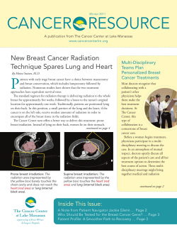

© Copyright 2026