
Vedtægter - Borgparken
Association between Obesity and Outcome after Total Knee and Hip Replacement PhD Thesis Anette Liljensøe Health Aarhus University 2014 PhD Thesis by Anette Liljensøe Health, Aarhus University Submitted: 31. Marts 2014 Public defence: 3. July 2014 2 Association between Obesity and Outcome after Total Knee and Hip Replacement PhD Thesis Anette Liljensøe Aarhus University, Health Department of Orthopaedics Aarhus University Hospital 3 Supervisors Kjeld Søballe, Professor, MD, DMSc (Principal supervisor) Department of Orthopaedics Aarhus University Hospital, Denmark Inger Mechlenburg, Associate Professor, MSc, PhD Department of Orthopaedics Aarhus University Hospital, Denmark Jens Ole Laursen, Clinical Associate Professor, MD Department of Orthopaedics Hospital Southern Jutland, Denmark Evaluation committee Leif Dahlberg, Professor, MD, PhD Department of Orthopaedics Skåne University Hospital, Lund University, Sweden Henrik Husted, Chief Consultant, MD, PhD, DMSc Department of Orthopaedics Hvidovre Hospital, University of Copenhagen, Denmark Sten Rasmussen, Associate professor (chairman) Orthopaedic surgery Research Unit Aalborg University Hospital, Denmark Correspondence Anette Liljensøe, MSc (health) Department of Orthopaedics Aarhus University Hospital Tage-Hansens Gade 2, Building 9A DK – 8000 Aarhus C E-mail: [email protected] or [email protected] www.orthoresearch.dk 4 Acknowledgments I am deeply indebted to a number of people who have made this work possible. First of all, I want to thank the 400 patients who generously donated their time and made this project possible. To my principal supervisor Kjeld Søballe for inviting me into the inspiring Clinical Orthopaedic Research Unit. For supporting my ideas and allowing me to carry out the studies that I wanted to perform, for your guidance whenever needed, and for giving me extensive freedom and responsibility. To co-supervisor Inger Mechlenburg for your faith in my ability to carry out research and for encouraging me to push my limits. For always being available, day and nights, giving me excellent scientific guidance, criticism, and advice. Without co-supervisor Jens Ole Laursen, it would not have been possible to conduct the clinical studies in the Hospital of South Jutland. Thank you for your never-ending support and for always being available for advice and help. Thank you Henning Bliddal from the Parker Institute, for your invaluable contribution in the weight loss study. All the staff at the orthopaedic outpatient clinic in Sønderborg is thanked for enrollment of patients in this study and for helping out with many other practical chores. The orthopaedic ward for letting me "borrow" patients for DXA scan just prior to their operation. I wish to express a special thanks to the WITAK study team for their dedicated work, and for keeping the wheels running at the Hospital of South Jutland, Sønderborg: Lone Sass, Heidi Thuesen, Tanja Fangel Christensen, and to Kinne Møller Nielsen who unfortunately passed away too soon. And what should I have done without clinical dietician Randi Fogtmann and project nurse Inge Boisen. My colleagues at the Clinical Orthopaedic Research Unit in Aarhus in 9A and 10A are thanked for great companionship, scientific discussions, and company at conferences. Especially, Lone Ramer Mikkelsen for always keeping me updated with the latest scientific articles, Maiken Stilling for answering all my questions about DXA scanning, Inger Krog-Mikkelsen for contributing with your knowledge on weight loss and for help with extracting DXA results, and Tina Stenumgaard for keeping track of our fundings. Anders Odgaard are thanked for providing me with the Procordo software, which has made it easy for me, my supervisors, and our sponsors to keep track of the study at anytime and anywhere in the world. Mogens Erlandsen has kindly offered first class statistical guidance with all studies in this thesis. I would also like to thank Kim Hørslev-Petersen and every one from the research group at Kong Christian X's Gigthospital in Gråsten, for inviting me in as a part of the group, for taking an interest in my research, for constant support, and for scientific discussions. My friends and relatives are thanked for taking an interest and for helping out in Nørremølle whenever needed. Birgitte Horst, my friend from the university, has often 5 put me up for the night when I have stayed over in Aarhus. Above all, I wish to thank my beloved husband Ulrich Johannsen and father of our children Tilde and Valdemar, for his constant support and encouragement that made it possible for me to achieve my goals. Thank you for managing everything at home when I was busy, and for always letting me know that you love me. A lot of money went in to this project and I am very grateful for the support from the Danish Rheumatism Association; Linak A/S; Fabrikant Mads Clausens foundation Danfoss; Johs. M. Klein and wife foundation; Knud and Edith Eriksens foundation; Peter Ryholts foundation; Jeppe Juhl og wife Ovita Juhls foundation; Cambridge Weight Plan, Northants., UK; Aarhus University; the Hospital of South Jutland; the Danish Orthopaedic Foundation and Inge & Per Refhalls Research Foundation. Anette Liljensøe, 2014 6 Preface This thesis is based on 3 clinical studies performed at the Clinical Orthopaedic Research Unit at Aarhus University Hospital and the Hospital of South Jutland during my employment as a PhD student at the Aarhus University Hospital during the period 2010-2014. The following pages contain an introduction describing the current knowledge regarding: overweight and obesity and the treatment with total knee and hip arthroplasty, a description of the applied methods, presentation of the findings, discussion and conclusion followed by perspectives. Finally, the results are presented in 3 individual papers. List of papers I. Liljensøe A, Laursen JO, Søballe K, Mechlenburg I. Overweight preoperatively impairs clinical outcome after knee arthroplasty: A cohort study of 197 patients 3–5 years after surgery. Acta Orthop. 2013;84(4):392–7. II. Liljensøe A, Laursen JO, Søballe K, Mechlenburg I. Is high Body Mass Index a potential risk factor for poor quality of life and physical function after hip arthroplasty: A cohort study of 98 patients 1 year after surgery. Submitted. III. Liljensøe A, Laursen JO, Bliddal H, Søballe K, Mechlenburg I. Feasibility and safety of intensive weight loss before total knee replacement in obese patients: A randomized controlled trial. Submitted. 7 Contents THESIS AT A GLANCE 10 ABBREVIATIONS 11 INTRODUCTION 12 OVERWEIGHT AND OBESITY OSTEOARTHRITIS TREATMENT OF KNEE AND HIP OA 12 12 13 OBJECTIVES AND HYPOTHESIS 17 METHODS 18 PATIENTS DESIGN SAMPLE SIZE OUTCOMES WEIGHT LOSS INTERVENTION STATISTICAL ANALYSIS 18 19 20 21 24 24 RESULTS 26 DISCUSSION 35 MAIN FINDINGS INTERPRETATION OF RESULTS AND COMPARISON WITH THE LITERATURE METHODOLOGICAL CONSIDERATION AND LIMITATIONS 35 35 40 CONCLUSION 43 PERSPECTIVES AND FUTURE RESEARCH 44 ENGLISH SUMMARY 45 DANISH SUMMARY 47 REFERENCES 49 APPENDICES 59 THESES FROM THE RESEARCH GROUP 59 PAPER I 63 8 PAPER II 70 PAPER III 81 9 Thesis at a glance Paper Objective Patients Methods Results Interpretation I Association between the preoperative BMI of patients who underwent TKA and their QoL and physical function 3–5 years after surgery. 197 primary TKA patients from a department of orthopedics. Patients came to a clinical follow-up 3-5 years after surgery. The outcome measures were the SF-36 and the KSS. 9 of the 14 endpoints were statistically significantly correlated with BMI. For all outcome measures ORs were < 1. The results indicate that a TKA patients’ pre-operative BMI is a predictor of the clinical effect and patients’ QoL 3–5 years postoperatively. A high BMI increases the risk of poor QoL (SF-36) and physical function (KSS). II Association between the preoperative BMI of patients who underwent THA and their QoL, physical function, and body composition before surgery and 1 year after surgery. 98 primary THA patients from a department of orthopedics. Data were collected at baseline preoperatively and at follow-up 1 year after surgery. The outcome measures were SF-36, HOOS and body composition. The OR was < 1 for all PRO in the obese group compared with the normalweight group. In contrast, the overweight group had a OR > 1 compared with the normalweight group, in improvement of all PRO. Obesity increases the risk of poor general health and hip-related health 1 year after THA. However, the overweight group achieved a greater improvement in score than did the normal-weight group. III Is it feasible and safe to implement an intensive weight loss program in order to reduce TKA patients’ preoperative body weight, before surgery? 76 primary TKA patients from a department of orthopedics. Participants were randomized to either standard care and surgery for TKA or a lowenergy diet (810 kcal/day) and nutritional education for 8 weeks before surgery. Kg, body composition, BP, and lipids were assessed: before intervention for the weight loss group, and within 1 week preoperatively for both groups. The average weight loss was 10.7 kg (10% of baseline body weight), 6.7 kg reduction in fat mass, 3kg reduction in lean mass, and the lean% increased with 2.3%. The intensive diets gave few and mild adverse effects. It is feasible and safe to implement a weight loss program shortly before TKA. 10 Abbreviations 6MWT ADL BMC BMD BMI BP BP CHOL CI DKR DXA GH HDL HOOS KOOS KSS LDL LOS MCS MH mSv OA OR PCS PF PJI POM PRO QoL RE ROM RP SD SF SF-36 TGLY THA TKA VT WHO WITKA 6-Minute Walk Test Activities of daily living Bone mineral content Bone mineral density Body mass index Blood pressure Bodily pain Total cholesterol Confidence interval Danish Knee Replacement Register Dual energy X-ray absorptiometry General health HDL cholesterol Hip disability and osteoarthritis outcome score Knee injury and osteoarthritis outcome score American Knee Society score LDL cholesterol Length of stay Mental component score Mental health MilliSievert Osteoarthritis Odds ratio Physical component score Physical functioning Prosthetic joint infection Proportional odds model Patient-reported outcome Quality of life Role limitation, emotional Range of motion Role limitation, physical Standard error Social functioning Short-Form 36 Triglyceride Total hip arthroplasty Total knee arthroplasty Vitality The World Health Organization The Weight loss Intervention before Total Knee Arthroplasty study 11 Introduction Overweight and obesity According to the World Health Organization (WHO), worldwide obesity has nearly doubled since 1980, with more than 35% of adults aged 20 and over being overweight (body mass index (BMI) > 25 to < 30; calculated in kg/m2), and 11% being obese (BMI > 30).1 WHO BMI calcifications are presented in Table 1. At least 2.8 million adults die each year as a result of being overweight or obese, and overweight and obesity are the fifth leading risk for global deaths, and are linked to more deaths worldwide than underweight.1 For example, 65% of the world's population live in countries where overweight and obesity kill more people than underweight, including all high-income and most middle-income countries.1 In addition, with respect to general health, overweight and obesity are considered a risk factor for hypertension,2–4 type 2 diabetes, coronary heart disease,5,6 coronary heart disease, gallbladder disease, respiratory problems, and certain cancer burdens are attributable to overweight and obesity.7 Table 1. WHO BMI calcifications BMI categories Underweight Normal weight Overweight Obese class I Obese class II (morbid obese) Obese class III (super obese) Kg/m2 <18.49 >18.5 <25 >25 <30 >30 <35 >35 <40 >40 Osteoarthritis Osteoarthritis (OA) is the most common form of arthritis in developing countries and one of the most prevalent age-related musculoskeletal conditions causing significant pain, functional loss, and disability, leading to a substantial impairment in patients’ ability to perform activities of daily living a large impact on health-related quality of life.8,9 OA is characterized by the progressive degenerative breakdown of articular cartilage, associated with inflammation, osteophyte formation, and joint deformity (Figures 1 and 2). Risk factors for OA include advanced age, female gender, genetic predisposition, obesity, and joint injury (including trauma, repetitive use, and prior inflammation), and OA can develop as a secondary consequence of congenital or developmental joint disorders and of metabolic or endocrine diseases.8–10 The etiology of OA is unclear. Mechanical, biochemical, genetic factors, inflammation,11 and hormones,12 seem to play a role.13 According to the National Health Profile 2013, almost 900,000 Danes have some degree of OA14, and each year OA costs the Danish society 11.5 billion DKK. The annual loss to society (lost production and treatment costs) is 6.8 billion.15 12 Obesity and knee/hip OA Obesity is a major risk factor for knee OA,16 and the association has been recognized for several years.17,18 The lifetime risk of symptomatic knee OA rises with increasing BMI, with a risk of 2 in 3 among those who are obese.19 Although the association is not as strong as between obesity and knee OA, there is also an association between obesity and hip OA.8,9,20,21 Obesity has been thought to affect development of OA through mechanical loading of weight-bearing joints. However, mechanical factors alone cannot explain the positive association between hand OA and obesity.8,9,22,23 Figure 1. a. Normal knee anatomy. b. Severe knee osteoarthritis. c. Knee arthroplasty. Treatment of knee and hip OA Joint replacement surgery THA and TKA are a successful and widely applied treatment for advanced hip and knee osteoarthritis.24 The treatment is recommended in all existing treatment guidelines, and THA and TKA are generally accepted as reliable and appropriate surgical procedures to restore function and improve health-related quality of life in patients with hip and knee OA who are not obtaining adequate pain relief and functional improvement with a combination of pharmacological and nonpharmacological treatments.25,26 In 2012, approximately 9000 primary THAs,27 and 8,500 primary TKAs,28 were performed in Denmark. The number has increased from about 5000 primary THAs,27 and 2,200 total TKAs,28 in 2000. In addition, the number of THAs and TKAs has increased in all developed countries, and it is estimated that the number of TKAs in the United States may gendertuple by the year 2030.29 Pain on function, pain at night, severe radiographic OA, and reduced level of mobility are common indications for THA,30 and TKA,31. The TKA surgical procedure involves removal of damage cartilage surfaces at the ends of the femur and tibia along with a small amount of underlying bone. The removed cartilage and bone are replaced with metal components that recreate the surface of the joint, and a medical-grade plastic 13 spacer is inserted between the metal components to create a smooth gliding surface. The metal parts are cemented into the bone. If needed, the undersurface of the patella is cut and resurfaced with a plastic button, and of joint deformities are corrected,32 (Figure 1). In THA, the damaged femoral head is removed and replaced with a metal stem that is placed into the hollow center of the femur. The femoral stem may be either cemented or "press fit" into the bone, and the damaged cartilage surface of the acetabulum is removed and replaced with a metal socket. Screws or cement is sometimes used to hold the socket in place. For a smooth gliding surface, a plastic, ceramic, or metal spacer is inserted between the new femoral head and the socket,32 (Figure 2). Figure 2. a. Normal hip anatomy. b. Severe hip osteoarthritis. c. Hip arthroplasty. TKA and obesity The association between obesity and outcome following TKA is ambiguous. Some studies have shown that overweight and obesity have no effect on pain and mobility after TKA.33–35 Other observational studies have shown that obese patients had worse QoL,36,37 poorer mobility,38 physical function,36,39 and less range of motion (ROM) after surgery than non-obese.40 In addition, obese patients also had more pain after surgery than non-obese41, overweight patients were less satisfied with their treatment than non-overweight patients were 22 years after TKA,42,43 and they were less active and tended to gain weight after TKA.44–46 Moreover, it has been reported that the hospital costs were higher in obese patients than in non-obese.47–49 Thus, surgery in obese patients is associated with several problems:29,50 practical problems with operation tables and instruments, increased operative time,51,52 and increased mortality,53 increased use of analgesics, problems with scarring,54,55 prosthetic joint infection (PJI),47,56 and a poorer prosthetic survival have been reported. By contrast, Suleiman et al.,57 found no difference in perioperative 14 complication rates in patient undergoing TKA or total hip arthroplasty across BMI categories. It is a common recommendation in Danish orthopedic departments that patients undergoing TKA surgery should increase their intake of protein the week leading up to the surgery. Additionally, patients are advised to avoid body weight reduction immediately before TKA surgery because weight reduction often leads to loss of muscle mass and thus loss of protein. THA and obesity The association between obesity and physical functioning and QoL after THA is debated. Some studies have shown that obese patients experienced a reduction in pain,58,59 improvement in function,58,60,61 and QoL,61 after THA comparable with nonobese patients. In contrast, other studies have indicated that overweight and obesity were statistically significantly associated with general health and QoL,62,63 and that obese patients had a lesser ROM than non-obese patients after THA.64 Peri- and postoperative complications in obesity after primary THA have been reported in several studies. Obese THA patients occupy more intraoperative time (total room time, anesthesia induction time, surgery time) than non-obese patients, which reflects the burden obesity poses to the hospitals;65,66 and obesity increases the length of admission,67,68 and direct medical costs.67 Moreover, obesity is independently associated with a high risk of PJI,69–71 thromboembolic complications,70,72 and risk of dislocation,47,64,70,73,74 and increasing BMI is associated with superficial infection.62 Non-surgical treatment Patient information and education, excise, and weight loss are recommended by the Osteoarthritis Research Society International (OARSI) and the European League Against Rheumatism (EULAR) as optimal treatment guidelines for the management of early hip or knee osteoarthritis (Figure 3).75–79 Figure 3. The knee- and hip OA treatment pyramid. 15 A reduction in fat mass in overweight knee OA patients before treatment with TKA is presumed to be beneficial with regard to several factors that can affect the patients’ general health and outcome after TKA. As far as we are aware, weight reduction before joint replacement has not yet been investigated. 16 Objectives and hypothesis The overall objective of this thesis was to investigate the association between patients’ preoperative body weight and the outcome after primary TKA and THA. Paper I The objective of this study was to investigate whether there was an association between the preoperative BMI of patients who underwent TKA and their QoL and physical function 3–5 years after surgery. The hypothesis was that a higher BMI increases the risk of poor physical function and poor QoL following TKA relative to the risk in lean TKA patients. Paper II The objective of this study was to investigate whether there was an association between the preoperative BMI of patients who underwent THA and their QoL, physical function, and body composition before surgery and 1 year after surgery. The hypothesis was that a high BMI increases the risk of poor physical function and poor QoL after THA. Paper III This study is part of the WITKA study. The objective of these preliminary results was to investigate whether it was feasible and safe to implement an intensive weight loss program in order to reduce TKA patients’ preoperative body weight, before surgery. The hypothesis was that it would be feasible and safe to reduce obese patients preoperatively body weight with 5–10% 17 Methods Patient inclusion, data collection, and clinical examinations took place at the Hospital of South Jutland, a non-university hospital specialized in orthopedic surgery and treating the regional patient population. About 300 TKA,28 and 400 THA,27 surgeries are performed at the hospital annually. The department works according to the Joint Care principles, namely: 1. Consider the patient as a healthy person who just needs a joint replacement; 2. it is important that the patient participates actively in treatment; 3. the patient is part of a group of patients who all need joint replacement, and the groups support each other during hospitalization. All patients in the 3 studies (studies I, II, and III) received the Southern Jutland Hospital's standard perioperative and postoperative care. Ethics All study protocols were planned and implemented in accordance with the Helsinki Declaration80, and all participants were provided with both oral and written information about the procedures of the study and informed consent was obtained. Thus, all studies were registered with the Danish Data Protection Agency. Ethical approval was granted by the regional Committee on Biomedical Research Ethics (study II journal number: S-201110124, study III journal number: S-201001309), and studies II and III were registered at www.ClinicalTrials.gov (Study II: NCT01496716, study III: NCT01469403). Patients Study I All patients who had undergone primary TKA during 2005 and 2006 were eligible. During the study period, 297 TKAs were performed in 255 patients. The 42 patients who had undergone primary TKA in both knees during the study period participated in the study only once. Twenty-one patients died before the follow-up, and 37 patients did not wish to participate in the study. Thus, 197 participants completed follow-up. Study II 103 consecutive hip OA patient scheduled for primary THA were recruited between December 2011 and May 2012. Among these, THA was canceled or postponed in 3, 1 patient was lost to follow-up, and 1 patient was seriously ill (not related to THA). Thus, 98 patients completed the 1-year follow-up. Exclusion criteria were rheumatoid arthritis. Patients who underwent surgery on both hips during the project period participated only once. Study III Patients were recruited between August 2011 and April 2013. Those eligible for inclusion were knee OA patients scheduled for primary TKA. The patients had to be obese (BMI > 30 kg/m2), and the patients had to be motivated for weight loss. Exclusion criteria were patients with rheumatoid arthritis and planned bariatric surgery. Patients who underwent surgery on both knees during the project period participated only once. 168 patients were eligible for enrollment, 73 were excluded 18 (Figure 4). 77 patients underwent randomization; 38 were assigned the intervention group, and 39 were assigned to the control group. 1 control group patient was lost to follow-up. Figure 4. Flow diagram of randomization of patients in the RCT weight loss intervention study (study III). Design Study I This study was a cohort study in primary TKA patients. The primary outcome was self-reported health status measured by the SF-36 and the KSS score. Baseline characteristics were collected from patients’ records, and the preoperative KSS scores were collected from the Danish Knee Replacement Register (DKR). 3-5 years after surgery, the patients came to the outpatient clinic for a clinical examination. The 19 patients completed the SF-36, and knee stability, ROM, pain, and functional ability were assessed, and the KSS scores were calculated. Study II This study was a cohort study in primary THA patients. The primary outcome was self-reported health status measured by the SF-36. Secondary outcome was the hip disability and osteoarthritis outcome score 2.0 (HOOS), body composition (fat mass, lean mass, and bone mass), bone mineral content (BMC), and bone mineral density (BMD) measured by DXA scan, and length of hospital stay. Outcomes were assessed at baseline 1 week preoperatively, and at follow-up 1 year after surgery. Study III The study was a single-blind, single-center, pragmatic randomized controlled trial. Patients who met the inclusion criteria and accepted to participate were randomly assigned to either intensive weight loss therapy 8 weeks preoperative and 52 weeks postoperative (the weight loss group) or to non-intervention (the control group) following standard TKA care. Randomization was performed in a 1:1 ratio with a block size of 10. The randomization sequence involved stratification according to gender (M/F) and BMI (<35/>35) to ensure homogeneity between the groups. A statistical computing web program in the project database using the Procordo.com software (Aarhus, DK) generated the randomization. Study III is part of the Weight Loss Intervention before Total Knee Arthroplasty (WITKA) study, investigating whether it is feasible and safe to implement an intensive weight loss program in order to reduce TKA patients’ preoperative weight before surgery. Outcomes were measured before intervention for the weight loss group, and within 1 week preoperatively for both the weight loss group and the control group. Outcomes were body weight, body composition (fat mass, lean mass, and bone mass), patients’ blood lipid status, glucose, blood pressure, and pulse. Safety outcomes were adverse events possibly related to the diet, and preoperative complications. Sample size Study I No priori power analysis was performed. In this study, all patients who had undergone a TKA in a 2-year period were included in the study. During this period, 297 TKAs were performed in 255 patients. A retrospective calculation of the power of this study based on the actual standard deviations and differences shows that the study has a power of 0.90. Study II A priori power analysis was performed to determine the sample size (n) required to detect a 5% difference in PCS in patients with a difference in BMI of 1 kg/m2. To achieve a power of 80%, it was determined that 80 patients would be required in the study group. Study III Patients included in this study were followed for 1 year in a randomized controlled trial investigating group differences in patient-reported health-related quality of life 1 20 year after intensive weight loss intervention and TKA. The sample size of 51 participants per group allowing a drop-out rate of 20% was calculated based on the primary outcome to obtain 80% power to detect an 8% difference between groups in SF-36 physical component score 1 year after TKA. The significance level was set to 5% using a 2-sided analysis. No specific sample size calculation was performed for this feasibility and safety study. Outcomes Short-Form 36 (SF-36) The SF-36 is a patient-reported generic health status measure that contains 36 items, including 8 health domains81: physical functioning (PF), role-physical (RP), bodily pain (BP), general health (GH), vitality (VT), social functioning (SF), role-emotional (RE), and mental health (MH). 2 component scores aggregate the 8 sub-domains into 2 component scales: physical component (PCS) and mental component (MCS) (Table 2).82 Each score was transformed into a 0–100 scale, with higher scores indicating better status. In study I the SF-36 version 1 was used, and in study II version 2 was used. In version 1: 7 questions can only be answered yes/no. In version 2: the options have been extended to 5 possible answers. This means that the SF-36 version 2 is more sensitive than the version 1. The questionnaire has been translated into Danish and has been validated and tested for reliability in healthy Danes and includes a Danish reference material.81 Hip disability and Osteoarthritis Outcome Score 2.0 (HOOS) The HOOS is a hip-related health score, based on the Western Ontario and McMaster Universities Osteoarthritis Index (WOMAC).83 The HOOS is a 40-item questionnaire assessing 5 separate dimensions: pain, hip symptoms, ADL, function in sport and recreation (Sport/Rec), and hip-related QoL. Each score is transformed into a 0–100 scale, with higher scores indicating better status. HOOS is a valid and reliable PRO when evaluating patients with undergoing THR.83,84 The questionnaire has been translate from the original Swedish version into a Danish version, according to existing guidelines.85 However, testing of validity, reliability and responsiveness in a Danish population has not been done yet. American Knee Society score (KSS) The KSS consists of an objective knee score and a function score.86 The knee score measures knee pain (50%), ROM (25%), and alignment and stability of the knee (25%). The function score examines walking distance (50%) and ability to walk up and down stairs (50%), and points are subtracted for use of walking aids. Each score is transformed into a 0–100 scale, with higher scores indicating better status. The KSS is a valid87 questionnaire and reliable for the evaluation of individuals who have undergone TKA. 88 Although the SF-36 are a more responsive measure of outcome of TKA.87 Dual energy X-ray absorptiometry (DXA) DXA devices were primarily developed for the diagnosis of osteoporosis. Bone mineral density (BMD) measurements in the proximal femur, the lumbar spine, or the forearm provide a means for the diagnosis of osteoporosis, and the prediction of 21 fracture risk.89 In addition to BMD assessment, DXA devices are capable of measuring body composition of the total body or body regions (figure 5). Today, body composition is a widely accepted method to assess body composition (bone mineral content, fat mass, and lean mass), either in clinical, research, or athletic settings.90–93 In addition, numerous studies have shown evidence that DXA can accurately assess changes in body composition in obese subjects following weight reduction. 94–99 Table 2. Interpretation of low scores Significant limitations in performing physical activities. Problems with work or other activities as a result of physical problems. Interpretation of high scores Physical Functioning Little limitations in performing (PF) physical activities or no limitations. Role-Physical (RP) Little or no problems with work or other daily activities stemming from physical problems. Bodily Pain (BP) High levels of pain that impact normal No pain and no related impact on activities. normal activities. General health (GH) Indicate evaluation of general-health Indicate that the respondent as poor and likely to get worse. evaluates his or her health most favorably. Vitality (VT) Feelings of tiredness and being worn Feeling full of energy all or most of out. the time. Social Functioning (SF) Extreme or frequent interference with The individual performs normal normal social activities due to physical social activities without interference and emotional problems. from physical or emotional problems. Role-Emotional (RE) Problems with work or other activities No problems with work or other as a result of emotional problems. activities due to emotional problems. Mental Health (MH) Frequent feelings of nervousness and Feelings of peace, happiness, and depression. calm all or most of the time. Physical Component Limitations in physical functioning, Little or no physical limitations, Score (PCS) role participation due to physical disabilities or decrements in wellproblems, a high degree of bodily pain being; a high energy level; and good and poor general health. general health. Mental Component Indicative of frequent psychological Frequent positive affect, little or no Score (MCS) distress, social and role disability due psychological distress or limitations to emotional problems and poor in usual social/role activities due to general health. emotional problems and good general health. The SF-36 questioner includes one favorably scored scale that measures 8t different health domains, and 22 component scores aggregate the 8 sub-domains into 22 component scales. Each score is transformed into a 0–100 scale, with higher scores indicating better status. DXA measurements are rapidly performed (10 sec hip, 4 to 10 min total body), noninvasive, precise (<1% error),92 operator independent, and expose the patient to a low radiation dose of 0.009–0.037 mSv,100 which is very low in comparison with the yearly background radiation of 4 mSv.101 The main DXA body composition measurement technique is based on the differential attenuation by bone, fat, and lean tissue of transmitted photons at 2 energy levels.100 The transmission at 2 energy levels allows the derivation of 2 different components such as fat and lean mass in regions without bone. In regions with bone, the 2 components, bone and soft tissue, are measured, and 22 the composition of the soft tissue needs to be estimated with respect to the adjacent tissue values. Therefore, a body-composition scan of the total body measures 3 compartments of the body: fat and lean body mass, as well as total body bone mineral. Proteins, glycogen, mineral, and water (including water and organic materials of the bone) are included in the component of lean tissue.100,102 Figure 5. Illustration of a DXA total body scan and results of the total body composition measurements. Throughout studies II and III, the same DXA scanner was used: GE Lunar Prodigy Advance (Figure 6) using a narrow-angle fan beam technology. All scans were analyzed using the enCORE Software, version 13.60 (GE Healthcare, USA). Automatic edge detection was used for scan analyses. The scanner’s calibration was checked on a daily basis before each scanning session, using the GE Lunar calibration phantom. The manufacturer’s guidelines for patient positioning and for scan acquisition were followed. Metabolic syndrome Besides measuring the changes in body weight and body composition in study III, we wanted to examine the effect of the intervention on other health factors related to obesity. Therefore, we examined several biochemical changes and monitored patients’ 23 blood pressure. The blood samples (non-fasting) were collected to assess patients’ lipid status (total cholesterol, HDL cholesterol, LDL cholesterol, and triglyceride) and glucose. The blood samples were analyzed at the Department of Clinical Biochemistry at the Hospital of Southern Jutland, using standardized laboratory procedures. Figure 6. Dual energy X-ray absorptiometry (DXA) GE Lunar Prodigy Advance Weight loss intervention In the WITKA intervention program, we had, in contrast to a conventional hypocaloric diet, the opportunity to monitor patients’ intake of vitamins, minerals, and proteins, and we therefore anticipated that patients’ health condition would be better than prior to the intervention. Because we used formula foods, the patients were expected to achieve a faster weight reduction and a greater reduction in fat mass than if a conventional dietetic hypocaloric diet had been used.103–105 Moreover, rapid weight loss improves the prognosis for the sustained weight loss.105,106 During the first 8 weeks, the patients received a low-energy diet (810 kcal/day) using commercially available formula foods (Cambridge Weight Plan, Northants., UK) and nutritional education, before surgery.107 The patients attended weekly individual and group sessions. To avoid more than 8 weeks of waiting for surgery, the patients started in a group immediately after randomization. Statistical analysis Study I In study I, a dropout analysis was performed. The differences between the groups were assessed using the Student´s t-test for continuous variables. The Mann-Whitney rank sum test was used when data were not normally distributed or when there was no homogeneity of variances. The chi-square test was performed for the categorical variables, and Fisher's exact test for the variables that had few observations. For the main results, the ordinal logistic regression (proportional odds model (POM)) was applied. In our POM, all continuous response variables (SF-36, KSS) were generated to 4 ordered categorical variables. 24 Study II As in study I, the ordinal logistic regression (POM) was applied for the patientreported outcomes. Analysis of variance (one-way ANOVA) was used for comparison of mean body composition between the BMI groups. For non-parametric statistics, the Kruskal-Wallis test was used for comparison of mean admission days between the BMI groups. Study III Demographics and baseline characteristics were analyzed with the use of descriptive statistics presented as means with their 95% confidence intervals (CI) and numbers (%). Differences in preoperative outcomes between the groups were assessed using Student’s t-test. All the observations in the samples (n) in study I to III were independent, had the same probability of event, and the sample sizes (n) were determined in advance. For the statistical analysis, the software program Stata 10 was used in study I and Stata 12 was used in study II and III. P values <0.05 were considered statistically significant. 25 Results Study 1 Overweight preoperatively impairs clinical outcome after knee arthroplasty: A cohort study of 197 patients 3–5 years after surgery. The study population is presented with baseline demographics and a drop out analysis in Table 3. More than 80% were overweight, obese, or morbidly obese. Only 15% were of normal weight, and none were underweight. The average age in the population was 67 years, and 73% were female. Table 3. Demographics and drop out analysis Study population (n=197)* Gender; n (%) Female Male Age (years) mean (range) Age groups; years (%) < 49 50-64 65-79 > 80 BMI (kg/m2) mean (range) BMI (kg/m2); n (%) Normal (18.5-24.9) Overweight (25.0-29.9) Obese (30.0-34.9) Morbidly obese (>35.0) Primary disease; n (%) Osteoarthritis Rheumatoid arthritis Injuries Deep infection1; n (%) Revision; n (%) Knee score3 mean (range) Function score3 mean (range) Smoking; n (%) Alcohol4; n (%) Dead (n=21)* P value# Lost to followup (n=37)* 0.3a 144 (73) 53 (27) 67 (37-86) 13 (62) 8 (38) 72 (54-84) 13 (7) 68 (35) 100 (51) 16 (8) 30 (20-47) 0 5 (24) 9 (43) 7 (33) 28 (20-45) 32 (15) 76 (39) 58 (30) 31 (16) 6 (30) 7 (35) 4 (20) 3 (15) 0.3a 0.03c 24 (65) 13 (35) 62 (35-80) 0.02c 0.1c 7 (19) 18 (49) 10 (27) 2 (5) 29 (21-38) 0.6c 4 (11) 18 (49) 12 (32) 3 (8) 0.1b 169 (86) 8 (4) 20 (10) 3 (2) 7 (4) 42 (0-69) 52 (0-90) 18 (9) 6 (3) 17 (81) 3 (14) 1 (4) 0 0 35 (0-61) 37 (0-70) 5 (25) 0 P value# 1b 1b 0.3c 0.01c 0.03a 0.8a 0.3b 31 (84) 0 6 (16) 0 0 39 (0-61) 52 (30-70) 11 (30) 1 (3) 1b 0.6b 0.3d 0.8d 0.001a 0.7a * In a population of 255 TKA patients, 197 agreed to participate in the project, 21 patients died before 5 years of follow-up, and 37 patients did not want to participate in the study. 1 Deep infection occurred within one year postoperatively 2 revision surgery performed 1 year postoperatively. 3 preoperative KSS score. 4Female >14 units per week and male >21 units per week (1 unit=1 beer/1 glass of vine). #P values for comparisons between the study population and those that had died, and P values for comparisons between the study population and the lost to follow-up group a - test b Fisher´s exact test c Mann-Whitney rank-sum test d t-test 26 The patient-reported outcome (SF-36) and the KSS results were adjusted for age, gender, primary disease, and surgical approach. 9 of the 14 endpoints were statistically significantly associated with BMI. Results from the SF-36 PCS (Table 4) showed that for 2 people of same age, gender, primary disease and with the same surgical approach, a difference in BMI of 1 was associated with 8% increased risk of a poorer score for the person with the higher BMI. After adjustment for these variables, the MCS remained unchanged, but the risk of a poorer score rose by 4–12% for the 8 SF-36 subdomains. The change of 2 additional variables, GH and SF, were statistically significant after adjustment. This indicates that the person with the high BMI had a poorer general health, and that physical and emotional problems to some extent interfered with the social activities in those with a high BMI. There were no significant correlations between BMI and the variables RP, BP, and RE. After model adjustment, the risk of a poor KSS increased for those with a high BMI. The increase was in the order of 3% to 14% for a difference in BMI of 1. Table 4. The association between preoperative BMI and outcome 3–5 years after TKA. n=197 SF-36 PCS MCS PF RP BP GH VT SF RE MH KSS Knee score Improvement* Function score Improvement# OR 95 % CI P value OR adjusted 95 % CI P value 0.94 0.98 0.93 096 0.96 0.95 0.93 094 0.95 0.95 (0.90-0.99) (0.94-1.03) (0.89-098) (0.89-1.03) (0.92-1.01) (0.91-1.00) (0.89-0.98) (0.88-1.01) (0.88-1.03) (0.91-1.00) 0.01 0.5 0.007 0.3 0.1 0.06 0.006 0.09 0.2 0.04 0.92 0.98 0.90 0.96 0.96 0.94 0.92 0.92 0.95 0.95 (0.88-0.97) (0.93-1.03) (0.85-0.95) (0.89-1.03) (0.91-1.01) (0.90-0.99) (0.87-0.97) (0.86-0.99) (087-1.04) (0.90-1.00) 0.002 0.5 < 0.001 0.2 0.1 0.03 0.002 0.03 0.3 0.04 0.95 0.98 0.92 0.93 (0.90-1.00) (0.94-1.03) (0.88-0.97) (0.88-0.98) 0.04 0.5 0.003 0.005 0.94 0.97 0.87 0.90 (0.90-0.99) (0.92-1.02) (0.82-0.93) (0.86-0.95) 0.02 0.3 < 0.001 < 0.001 The results are presented as odds ratios (OR) and their 95% confidence intervals (CI), calculated using the proportional odds analysis, unadjusted and with adjustment for age, gender, primary disease, and surgical approach. * Knee score improvement. #Function score improvement. Study II Is high Body Mass Index is a potential risk factor for poor quality of life and physical function after hip arthroplasty: A cohort study of 98 patients 1 year after surgery. Patients’ demographics and baseline characteristics are shown in Table 5. 13% of the population were underweight, 49% normal weight, 27% overweight, and just 9% were obese or morbidly obese. Women were overrepresented in the underweight group (85%) and underrepresented in the obese group (22%). In all groups, the average age was 70–73 years. 27 Table 5. Demographics and baseline characteristics of the study population before THA Underweight BMI <18.5 (n=13) 11 (85) 73 (9.3) 57 (4.9) 21.7 (8.2) 40.5 (9.4) 2137.9 (505.0) 1.05 (0.13) 5 (38) Normal BMI (>18.5-<25) (n=49) 27 (55) 71 (9.4) 74 (7.8) 41.9 (8.8) 37.6 (9.8) 2722.7 (585.2) 1.16 (0.13) 24 (49) Overweight BMI (>25-<30) (n=27) 12 (44) 70 (8.3) 93 (7.5) 46.7 (6.9) 38.6 (9.7) 3229.9 (520.0) 1.28 (0.10) 18 (67) Obese (>30-<40) (n=9) 2 (22) 70 (7.1) 112 (12.0) 52.1 (5.1) 45.8 (8.4) 3205.8 (702.6) 1.30 (0.14) 8 (89) Woman, n (%) Age, years Weight, kg Android fat% Gynoid fat% Bone mineral content (BMC), g Bone mineral density (BMD), g/cm2 Comorbidity, n (%) Education, n (%) Unskilled worker 3 (23) 23 (47) 9 (33) 4 (44) Skilled worker 3 (23) 16 (33) 12 (44) 3 (33) Bachelor/master degree 7 (54) 10 (20) 6 (22) 2 (22) Working, n (%) 0 (0) 7 (14) 6 (22) 2 (22) Living alone, n (%) 6 (46) 16 (33) 10 (37) 4 (44) Smoking, n (%) 3 (23) 3 (6) 2 (7) 0 (0) Alcohol, n (%) 3 (23) 7 (18) 2 (10) 1 (17) Values are means, and the numbers in parentheses indicate the standard deviation (SD), unless otherwise indicated. The results for the patient-reported outcome (SF-36 and HOOS) are presented in Table 4, showing the OR in proportion to the normal-weight BMI group and each of the groups: underweight, overweight, and obese. The results were adjusted for age, gender, and comorbidities. Underweight The underweight group had an increased risk of 58% with regard to achieving a poorer PCS and a 17% increased risk of obtaining a lower change in score compared with the normal-weight group (Table 6). The same trend was found for the HOOS score: QoL, Symptoms and Sport/Reg. In addition to the changes in these scores, the underweight had an increased risk of reaching a smaller change in score, compared with the normalweight group. For the MCS, there was an increased probability of 16% that the group achieved a higher score than the normal-weight group, while the underweight group had an increased risk of 38% for achieving a lower change in score (Table 6). For the HOOS subscores ADL and Pain, the group had an increased likelihood of 61% and 40% for an increased postoperative score compared with those of normal-weight. The same trend was found for the change of these scores. None of the results for the underweight group were statistically significant. Overweight The overweight group had an increased risk of 47% for achieving a worse PCS postoperatively, but an increased likelihood of achieving a greater change in PCS compared with the normal-weight group (Table 6). The same trend was found for the HOOS subscores: ADL, Pain, symptoms, and Sport/Reg, where there was an increased risk of between 9-31% of obtaining a worse score postoperatively, and an increased 28 likelihood of between 22-85% to achieve a greater change in scores, compared with those of normal weight. For the MSC and QoL there was an increased probability respectively at 52% and 70% to achieve a higher post-operative score and an increased probability of 99% and 104% of achieving an increased change in scores, compared with the normal-weight patients. None of the results for the overweight group were statistically significant. Table 6. Normal-weight THA patients’ self-reported outcome compared with underweight, overweight, and obese patients’ outcomes. BMI group SF-36 PCS MCS 1 2 3 4 1 2 3 4 1-year follow-up score OR adjusted (95% CI) 1.00 0.42 (0.13-1.32) 0.53 (0.22-1.27) 0.16 (0.04-0.65) 1.00 1.16 (0.35-3.87) 1.52 (0.65-3.54) 0.25 (0.06-1.01) P value Difference in score OR adjusted (95% CI) 1.00 0.83 (0.25-2.74) 1.44 (0.23-3.29) 0.87 (0.23-3.29) 1.0 0.62 (0.21-1.84) 1.99 (0.82-4.81) 0.86 (0.25-2.98) 0.14 0.16 0.01 0.81 0.33 0.05 P value 0.76 0.41 0.84 0.39 0.13 0.81 HOOS ADL 1 1.00 1.00 2 1.39 (0.41-4.66) 0.59 1.58 (0.48-5.19) 0.45 3 0.74 (0.32-1.71) 0.48 1.55 (0.65-3.69) 0.32 4 0.23 (0.06-0.97) 0.04 0.71 (0.19-2.67) 0.61 QoL 1 1.00 1.00 2 0.84 (0.27-2.61) 0.77 0.75 (0.26-2.14) 0.58 3 1.70 (0.71-4.05) 0.23 2.04 (0.84-4.98) 0.12 4 0.54 (0.14-2.89) 0.39 0.80 (0.19-3.37) 0.76 Pain 1 1.00 1.00 2 1.60 (0.48-5.39) 0.45 1.10 (0.36-3-33) 0.97 3 0.91 (0.40-2.09) 0.83 1.85 (0.77-4.45) 0.17 4 0.54 (0.13-2.24) 0.40 0.90 (0.24-3.37) 0.87 Symptom 1 1.00 1.00 2 0.70 (0.21-2.31) 0.56 0.81 (0.28-2.41) 0.71 3 0.84 (0.36-1.96) 0.68 1.22 (0.52-2.87) 0.64 4 0.41 (0.11-1.49) 0.18 0.94 (0.26-3.42) 0.92 Sport/Rec 1 1.00 1.00 2 0.90 (0.30-2.73) 0.86 0.97 (0.33-2.80) 0.95 3 0.69 (0.29-1.61) 0.39 1.40 (0.58-3.38) 0.46 4 0.25 (0.06-1.03) 0.05 0.18 (0.04-0.86) 0.03 Scores for 1-year follow-up and scores for the difference between preoperative and the 1-year follow-up scores arepresented for the Short Form 36 (SF-36) and the Hip disability and osteoarthritis outcome score (HOOS) BMI groups: 1= normal weight, 2=underweight, 3=overweight, 4=obese. The patient-reported outcome measures are presented as odds ratios (OR) and their 95% confidence intervals (CI); calculated using the proportional odds analysis adjusted for age, gender, and comorbidity. Obesity The obese group (Table 4) had a statistically significantly increased risk of obtaining a worse PCS of 84% (P = 0.01) and a ADL score of 77% (P = 0.04), post-operatively, compared with those of normal weight. In addition, the change in pain score was significantly different from the normal weight group, with an increased risk of a 29 poorer improvement of 82% (P = 0.03). For all other patient-reported outcome measures, we found odds ratios (OR) < 1, with an increased risk of obtaining a worse score between 6% and 75% compared with the normal-weight group, which was not statistically significant. The patients’ body composition at the 1-year follow-up is shown in Table 7. Unlike lean mass, the lean% was statistically significantly between the groups. The mean lean% was highest in the underweight group and lowest in the obese group at the 1-year follow-up. All groups experienced an increase in body fat percentage from baseline to follow-up, and a decrease in body lean% that matched their increase in body fat percentage. There were no statistically significant differences between the groups. Table 7. Comparison of body composition at the 1-year follow-up and the difference in body composition from preoperative examination to 1-year follow-up, between the 4 BMI groups Body composition Total body weight (kg) Total body fat% Total body muscle% Fat body mass (kg) Muscle body mass (kg) Bone body mass (kg) BMI group 1 2 3 4 1 2 3 4 1 2 3 4 1 2 3 4 1 2 3 4 1 2 3 4 1-year follow-up Mean (SD) 75.7 (8.0) 58.8 (6.1) 93.3 (11.0) 109.0 (15.3) 34.2 (8.2) 33.3 (8.4) 37.6 (7.5) 44.5 (5.2) 62.2 (8.0) 63.1 (8.0) 59.0 (7.2) 52.5 (4.7) 25.1 (6.5) 19.0 (5.6) 34.9 (6.9) 46.9 (8.9) 45.8 (8.1) 35.9 (5.5) 55.2 (9.5) 55.0 (7.4) 2.7 (0.6) 2.0 (0.4) 3.2 (0.5) 3.1 (0.6) P value <0.001 0.004 0.005 <0.001 <0.001 <0.001 Difference Mean (SD) 0.77 (1.9) 0.73 (3.5) 0.06 (7.2) -3.17 (8.2) 0.35 (1.8) 1.78 (1.8) 0.22 (2.1) 0.17 (3.3) -0.3 (1.8) -1.8 (1.8) -0.2 (2.1) -0.5 (3.2) 3.1 (1.8) 1.2 (1.4) 4.7 (2.7) -1.3 (5.6) -0.2 (1.5) -0.7 (1.1) 0.1 (2.2) -2.4 (3.3) -0.02 (0.1) 0.01 (0.1) -0.03 (0.1) 0.06 (0.2) P value 0.13 0.28 0.27 0.69 0.73 0.36 BMI groups: 1= normal weight, 2=underweight, 3=overweight, 4=obese. Body composition is presented as means and standard deviation (SD). For P value, 1-way-ANOVA analysis of variance was performed. Furthermore, there were statistically significant differences between the groups’ mean number of admission days in relation to the THA surgery. Hospitalization was shortest for the normal-weight group and longest for the obese group. Study III Feasibility and safety with intensive weight loss before total knee replacement in obese: a pragmatic randomized controlled trial. 30 The baseline characteristics of the weight loss group (n=38) and the control group (n=38) were similar (Table 8) with respect to age, gender, height, weight, and BMI. Mean age was 65 years, and 71% were woman, similar as the population in study I. All patients in the intervention group completed the weight loss program. The average weight loss after 8 weeks was 10.7 kg (10% of baseline body weight) (Table 9). Table 8. Demographics and baseline characteristics in the randomizes groups Female, n (%) Age, years (range) Weight, kg (CI) Height, m (CI) BMI, kg/m2 (CI) Hypertension, n (%) Diabetes, n (%) Type I Type II Education, n (%) Unskilled worker Skilled worker Bachelor/master degree Working, n (%) Residence, n (%) Farm/house Apartment Living alone, n (%) Smoking, n (%) Weight loss (n=38) 27 (71) 65 (46-81) 105.4 (101.2-109.6) 1.67 (1.63-1.70) 31.6 (30.6-32.6) 24 (63) 12 (32) 2 (17) 10 (83) Control (n=38) 27 (71) 65 (46-85) 104.4 (99.4-109.3) 1.67 (1.64-1.71) 31.2 (29.8-32.6) 21 (55) 11 (30) 2 (18) 9 (82) 14 (37) 19 (50) 5 (13) 12 (32) 17 (45) 17 (45) 3(10) 10 (27) 29 (76) 9 (24) 8 (21) 5 (13) 23 (61) 15 (39) 14 (37) 3 (8) CI: 95% confidence interval We recorded 3 serious events that occurred in the waiting time between randomization and surgery, for both the weight loss and the control group; due to cardiac arrhythmia one 70-year-old male patient had the TKA postponed for 3 months. This patient had lost more than 13 kg within 8 weeks, while no adjustment was made to his dosage of antihypertensive medication. After cardiologic examination and adjustment of the dosage, there was no contraindication to TKA. Another 76-year-old male patient had his TKA operation postponed 1 year due to a pacemaker surgery. The treating physician did not regard this episode as related to the weight loss intervention. Due to high blood pressure, one 63-year-old male from the control group, known to have hypertension, had the TKA postponed for 6 months. After the medication dosage had been adjusted, there was no contraindication to TKA. 1 male and 1 female from the weight loss group, both 57 years of age, chose to postpone their TKA by 22 and 29 weeks, respectively, because of decreasing pain after the 8-week weight loss intervention. No perioperative complications were recorded in any of the groups. Compared with the control group, the weight loss group achieved a statistically significant lower weight, BMI, fat mass, systolic blood pressure, CHOL, LDL, and TGLY (Table 9). In addition, lean mass was reduced by 3 kg in the weight loss group, 31 from before baseline to the preoperatively test, and mean lean mass was 2.2 kg lower (not statistically significant) in the weight loss group than in the control group. The lean% increased with 2.3% in the weight loss group, from before weight loss to the preoperatively test, and was 2.8% higher compared with the control group. Table 9. Presentation of the weight loss group before the intervention program and comparison between the weight loss group after 8-week intervention and the control group immediately preoperatively Weight, kg BMI, kg/m2 Fat mass, kg Fat% Lean mass, kg Lean% Bone mass, kg Systolic, mm/Hg Diastolic, mm/Hg Hearth rate, rate/min Glucose, mmol/L Total cholesterol, mmol/L HDL cholesterol, mmol/L LDL cholesterol, mmol/L Triglyceride, mmol/L Before weight loss 105.4 (101.2-109.6) 31.6 (30.6-32.6) 47.2 (44.2-50.3) 45.3 (42.8-47.8) 54.3 (50.7-57.9) 51.8 (49.4-54.2) 3.0 (2.8-3.2) 144 (138.2-150.2) 88 (84.0-91.0) 76 (71.7-80.4) 6.16 (5.56-6.76) 5.27 (4.78-5.77) 1.35 (1.22-1.48) 2.99 (2.53-3.46) 2.18 (1.74-2.62) Preoperatively Weight loss Control 94.7 (90.9-98.5) 104.4 (99.4-109.3) 28.4 (27.4-29.4) 31.2 (29.8-32.6) 40.5 (37.2-43.8) 47.8 (44.4-51.3) 42.7 (39.7-45.8) 45.9 (43.4-48.2) 51.3 (47.8-54.8) 53.5 (49.8-57.1) 54.1 (51.2-56.9) 51.3 (48.9-53.6) 3.0 (2.8-3.2) 3.0 (2.8-3.2) 132 (126.8-136.8) 147 (141.1-153.2) 82 (78.7-84.4) 86 (82.4-89.6) 72 (67.5-76.0) 74 (69.6-78.0) 5.70 (5.17-6.23) 6.02 (5.43-6.62) 4.15 (3.72-4.58) 5.10 (4.69-5.50) 1.29 (1.18-1.40) 1.37 (1.23-1.50) 2.30 (1.90-2.70) 2.96 (2.56-3.36) 1.27 (1.06-1.47) 1.81 (1.48-2.15) P value# 0.0025 0.0015 0.0027 0.11 0.38 0.13 0.88 0.0002 0.051 0.49 0.4 0.0019 0.37 0.021 0.0051 Values are means, and the numbers in parentheses indicate the 95% confidence interval (CI). P values for comparisons between the weight loss and the control group preoperatively. # Student’s t-test. 32 Discussion This thesis investigated the association between TKA and THA patients’ preoperative BMI and QoL, physical function, and body composition 1 to 5 years after surgery. In addition, the feasibility and safety of implementing an intensive weight loss intervention before TKA was investigated. Main findings The results indicate that TKA patients’ preoperative BMI is a predictor of the clinical effect and patient’s QoL, and physical function 3 to 5 years after surgery. Patients with increased BMI did not achieve the same QoL and functional capacity as TKA patients with a lower BMI. In addition, obese THA patients do not obtain the same physical function and QoL as normal-weight patients 1 year after THA, and they do not achieve the same improvement in physical function and QoL as the normal-weight group. Furthermore, length of hospital stay was 1 day longer for the obese than for patients with a normal weight. In contrast, the overweight THA group accomplished larger improvement in QoL and physical function compared with the normal-weight group. To our knowledge, this thesis is the first to report on weight loss intervention before joint replacement surgery, and the results suggest that it is feasible and safe to implement an intensive weight loss program shortly before TKA. Patients in the weight loss group achieved an average preoperative weight reduction of 10%, and the TKAs were performed in all participants without perioperative complications. Interpretation of results and comparison with the literature There was a great difference in demographic characteristics between the TKA (study I) and the THA cohort (study II). The average age in the THA cohort was 10 years older than in the TKA; there were 20% more males in the THA cohort; 13% underweight in the THA cohort and none in the TKA cohort; and 49% more overweight and obese in the TKA than in the THA cohort. Compared with the Danish population, the Southern Denmark population108 (Figure 7), from which both the TKA and the THA population in these studies were recruited, differed in the way that the THA cohort in general was more underweight and less obese, and the TKA cohort was more overweight and obese. The Danish Knee and Hip Alloplastic registers do not report the distribution of BMI in TKA and THA patients in Denmark, and to our knowledge, there are no Danish population-based studies reporting on BMI distribution. In the Canadian Joint Replacement Registry,109 (figure 8), a similar BMI distribution in TKA patient as in study I is reported. In the Canadian population, there were <1% underweight and 87% overweight or obese. In contrast, the Canadian THA population was less underweight and more overweight and obese compared with our THA cohort (study II). In a American study from 2007, Fehring et al.,110 report that just 6% of a TKA cohort were normal weight (BMI < 24), 34% were overweight, 43% obese, and 17% were morbidly obese. In the same study, 10% of a THA population were normal weight (BMI < 24), 43% were overweight, 34% obese, and 6% were morbidly obese. 35 Figure 7. TKA THA South Denmark Denmark 60% 49% 50% 37% 34% 40% 10% 46% 39% 27% 30% 20% 49% 47% 15% 13% 14%13% 9% 3% 3% 0% Underweight Normal Overweight Obese TKA and THA patients (studies I and II), the South Denmark, and the Danish population by BMI category. Figure 8. Hip and knee replacement patients by BMI category, Canada, 2003–04. Canadian Joint Replacement Registry, CIHI. High preoperative BMI before TKA In the TKA cohort study (study I), the SF-36 physical component score (PCS) was statistically significantly associated with BMI. There was thus an inverse association between BMI and performing physical activity like climbing stairs and walking. Patients with a high BMI are more limited than patients with a lower BMI with regard to performing all kinds of physical activities without limitations. The higher the BMI, the more nervous 36 and sad the patient was, and the more difficult it was for the patient to maintain social functioning. These patients also felt more tired and worn out, and they assessed their general health to be worse than the patients’ with the lower BMIs. The association between BMI and the mental component score (MSC) was not statistically significant. There was no statistically significant association between BMI and problems with work or other activities as a result of physical problems. In addition, no association between BMI and pain was found, and the data do not suggest that the BMI had an impact on problems at work or in doing other daily activities because of emotional problems. The follow-up KSS function score and the improvement of function score showed a clear association. Patients with increased BMI did not achieve the same functional capacity as TKA patients with a lower BMI. They were not able to walk as long distances and to climb stairs at the same level as patients with a normal BMI, similar to the SF-36 results. The KSS knee score, including pain, ROM, and stability, was also inversely associated with BMI, whereas the improvement of the knee score from baseline to follow-up showed no statistically significant association, suggesting that patients with a high BMI achieve the same improvement in pain and ROM as patients with a lower BMI. Our results are consistent with other studies.35,36,39–42,111,112 A prospective study with 1011 primary TKA patients found similar negative differences at 12-month follow-up in absolute PCS or MCS values between non-obese and obese patients.36 Naylor et al. found obesity associated with several SF-36 domains at 12-month follow-up.41 This indicates that obesity influences the patients’ generic (overall bodily) QoL after TKA. There is disagreement about the impact of obesity on health-related outcomes after TKA.68,74,113–115 Stickles et al. found significant differences in the absolute WOMAC at 12month follow-up between obese and non-obese patients treated with TKA.36 There were, however, no differences in improvements of the WOMAC between obese and non-obese patients. A case-control study by Nunez et al. found no differences in the WOMAC score at 12-month follow-up,37 or in pain 2 to 5 years after TKA,115 between obese and non-obese. In contrast, a case-control study by Krushell et al. with 5–14 years of follow-up found that patients with a BMI > 40 had lower KSS knee and function scores postoperatively than controls with a BMI < 30.35 Likewise, Foran et al. found that a BMI > 30 had a negative impact on KSS score 7 years after TKA and that obese patients had a lower rate of improvement than non-obese patients did.39 In addition, in a meta-analysis from 2012,116 it was concluded that obesity has a negative influence on the outcome of patients treated with TKA, with more short-term complications and poorer long-term outcome compared with non-obese patients. Like Singh et al. we found no statistically significantly association between high BMI and pain measured with SF-36 in our TKA cohort study.115 One explanation for this may be that TKA is, in general, a highly successful treatment option in patients with incapacitation due to advanced knee osteoarthritis, and studies show good outcomes in terms of pain in abut 80–100% of patients.43,117,118 Another explanation may be that patient with a high BMI are less physically active than patients with a lower BMI. 37 High preoperative BMI before THA In our THA cohort study (study II), the obese group had a statistically significantly increased risk of obtaining a worse PCS than normal-weight patients, and they had a higher risk (statistically non-significant) of achieving a smaller improvement in physical function. Our study also demonstrates that the obese had a statistically significantly increased risk of obtaining a lower activity in daily living 1 year after THA than normal-weight patients, and that the obese had an increased risk of lower hip-related QoL, more pain, more hip symptoms, and lower function in sports and recreation. In addition, the obese group had an overall increased risk of a smaller improvement in HOOS score than did the normal-weight group. Moreover, the underweight group had an increased risk of not achieving the same improvement in general health and hip-related health score as those of normal weight, and with regard to mental general health and ADL, the underweight had an increased risk of not achieving the same follow-up score as the normal-weight patients. In addition, the overweight group had an increased risk of not achieving the same 1-year follow-up score with regard to mental general health and hip-related QoL compared with the normal weight group. In contrast, the overweight group experienced a larger improvement in physical and mental general-health score from pre-surgery to 1-year follow-up than the normal weight group and hence experienced a larger relative improvement than all other groups in the present study. Furthermore, the overweight group had a greater improvement in all hiprelated health scores than did the normal weight group. The literature is unanimous that overweight and obesity are a risk factors for several complications during primary THA. Contrary to this, it is unclear whether overweight and obese patients achieve a boost in health-related QoL and physical function similar to that of normal-weight THA patients, and if they therefore experience a good effect of THA treatment, despite their increased risk of complications. Our results indicate that obese patients do not obtain the same physical function and QoL as normal-weight patients and these results are consistent with other studies that have used patient-reported, generalhealth, and hip-related health outcomes.58,63,68,119,120 A cohort study of 1,617 primary THA patients reported that in all of the SF-36 domains (8 sub-domains) other than mental health, the scores decreased statistically significantly with increasing BMI 5 years after surgery.62 As in our analysis model, these results were adjusted for age, gender, and comorbidities. Rajgopal et al.,68 demonstrated similar results in super obese THA patients (BMI>50) who were compared with normal-weight patients. The former had a statistically significantly lower postoperative SF-12 PCS and smaller improvement in score compared with the normal-weight patients.68 In contrast, Jones et al. found statistically significantly lover WOMAC pain and function scores (better disease) in morbidly obese patients than in a nonobese (BMI<30) at the 6-month follow-up, but no statistically significant differences between BMI groups at the 3-year follow-up.58 In a 2-year follow-up study, McCalden et al. found that morbidly obese patients did not differ statistically significantly in mean postoperative WOMAC scores from underweight/normal-weight patients. Thus, this group had the largest improvement in WOMAC score compared with the non-morbidly obese groups.61 38 In a recently published cohort study,121 with combined data from 4 large European prospective cohorts of patients receiving primary THA,122–126 patients achieved substantial change in the Oxford Hip Score after THA across all BMI categories. In contrast, a 5-unit increase in BMI in the obese was associated with a decrease in the 12-month OHS of 0.78 points 95%CI (0.27-1.28) compared with normal-weight THA patients. In conclusion, the authors suggest that BMI should not present a barrier to access THA in terms of PROs. Although, there was a statistically significant decrease in 12-month OHS, this effect was small and not clinically meaningful in contrast to the substantial change in OHS seen across all BMI groupings. Obesity in THA patients is associated with longer hospital stays and higher costs of THA, even among patients without comorbidities.67 In our study, we demonstrated comparable results. The obese patients’ hospitalization was 1 day longer than that of normal-weight patients. At follow-up, the obese group had accomplished a mean weight loss of 3.17 kg. In the non-obese group, weight increased <1 kg. Similarly, Paans et al. concluded that no clinically relevant reduction of weight occurred 1 year after THA.127 Our study indicates that the THA treatment has no clinically relevant effect on body composition 1 year after surgery. In a systematic review, no conclusive evidence was found regarding whether weight or body composition (weight and BMI) increase, decrease, or remain the same after THA.128 Additionally, Wolf et al. recognized no changes in lean mass or fat mass 3 months, 1 year, and 5 years after THA.129 Obesity has been thought to affect the development of OA through mechanical loading of weight-bearing joints. However, the association between obesity and hip OA is not as strong as the association between obesity and OA of the knees and the fingers. The reason for the association between obesity and hip and knee OA is not fully understood, but in addition to mechanical and biomechanical factors, inflammation and hormones, are likely to be part of the explanation. It is important to note that although we demonstrated an association between obesity and outcome after TKA and THA, we did not established causality. A number of factors might affect the outcome: environment, genes, psychological factors, metabolic syndrome, and hormones. Feasibility and safety with intensive weight loss before TKA A typical TKA patient in the Southern Jutland Orthopedic Department, where this study was, is a female aged 65 years, and more than 80% of the patients are overweight, obese, or morbidly obese. We have no knowledge of whether the TKA patients in this study were offered non-surgical treatment such as patient education, training, and weight loss before they were referred to the orthopedic department. In a small survey among 41 Danish orthopedic surgeons,130 less than 50% of the surgeons required that weight loss should be tried as a treatment prior to TKA. The weight loss group of patients achieved a mean preoperative weight reduction of 10% and a statistically significant reduction in fat mass compared with the control group. At the same time, it was not possible to avoid a reduction in lean mass in the weight loss group, whereas lean%, increased after weight loss. There was no statistically significant difference 39 in preoperative lean mass or lean% between the groups. In addition, the TKAs were performed safely in all participants, without perioperative complications. These results indicate that the diet program does not increase the risk of TKA complications because of, for example, loss of muscle mass just prior to surgery. By using low-energy formula foods, we assured that the patients received adequate vitamins, minerals, and proteins corresponding to the recommendations by the Danish National Board of Health. 131 Our short-term weight intervention results are similar to other RCT studies,103,132,133 conducted in clinical rheumatology departments in patients with primary knee OA. In addition, the intervention in study III improved the patients’ overall health. The metabolic syndrome decreased, the lipid levels decreased, and the average blood pressure and pulse decreased after the completion of the preoperative intervention program. In general, the intensive diet resulted in few and mild adverse events, comparable to adverse events in similar diet programs132. One serious adverse event presumably happened due to a too large dose of antihypertensive in a patient. A large challenge for the treating physician is the reduction of antihypertensive and antidiabetic medications during intensive diets.134 In a yet unpublished study by a New Zealand research group, a weight loss intervention program similar to ours was conducted in patients with severe knee OA. Their results showed that all TKA operations were postponed after the intervention. In our study there were only 2 patients who postponed their operation. The reason for this is probably due to the organization of the patient treatment in our orthopedic department. At the first visit to the clinic, all TKA-scheduled patients are made "operation ready": blood tests, ECG, and anesthesia monitoring. In addition, the patients’ surgery dates are booked at this visit, and the patient begins therefore to prepare mentally for having to undergo surgery already at that time. Methodological consideration and limitations Because of differences in methods applied in studies examining the association between BMI and the outcome after TKA and THA, it is difficult to compare the results. Classification of BMI groups is varied; some studies use the WHO classification of BMI groups as we did in our THA cohort study (study II). Other studies dichotomize BMI at 30, and some use BMI as a continuous variable, which we consider a strength, and hence is what we did in our TKA cohort study (study I). In addition, different endpoints, follow-up times, and statistical methods have been used in the studies, and most importantly, adjustment for confounding has varied. A strength in study I and study II was that we adjusted for confounding in the analyses models, even though we did not adjust for co-morbidities in study I. Moreover, we used of a reliable, valid, and responsive instrument for assessing outcomes of general health,81 TKA, and THA.84 A limitation in study I is that causes of death were not recorded. However, based on data collected at baseline, it is reasonable to assume that the patients who died before follow-up was completed were not systematically more or less healthy than the study population in general. The deceased group had a higher mean age at baseline and counted more smokers 40 than the study population. The deceased group also had a lower mean baseline KSS function score, maybe because they were older and hence had a lower level of functioning. The average BMI of the deceased group was slightly though not statistically significantly lower than the average BMI of the study population. We do not think that this difference is caused by selection bias. Among those who declined the invitation to participate in the study, 2/3 were below 65 years, which meant that they were probable still in active employment and therefore unable to take time off from work to participate in the study. In comparison, less than half were younger than 65 years in the group who completed the follow-up. It is possibly that those who declined participation were actually doing well and therefore uninterested in participating in a 5-year clinical follow-up. This might give rise to selection bias, but there was no difference in baseline BMI or baseline KSS scores between the groups. Secondly, the preoperatively data were collected retrospectively, and we did not have preoperatively SF-36 responses. Thus, improvement in general health was not reported in this study. On the other hand, we report the improvement in KSS, and the follow-up data were collected prospective in the clinic, which is a strength of the study. We also acknowledge certain limitations in study II. Some of groups categorized according to BMI were very small. The underweight group comprised only 13% of the cohort population, the overweight group 27%, the obese group only 7%, and the morbidly obese 2%. This might indicate that the study sample size was too small to say anything about obese THA patients and that the strength of the study was not optimal. On the other hand, all the results in the obese group point in the same direction and indicate a relation between obesity and poorer physical function and QoL compared with normal-weight patients; hence, an OR <1 for all outcomes. It is known that underweight and malnourished THA patients,135 and hip fracture patients136 are at increased risk of complications, which is the reason why we chose to use BMI as a categorical variable. Underweight is apparently not considered a risk factor for hip-related QoL and physical function in other studies. This introduces the possibility that the underweight group pulls the results toward the 0 hypothesis. In study II, all data were collected prospectively with a good rate of follow-up, which we consider a strong point. PROs are the favored measure of outcome used to examine whether surgery has been successful from the patients’ perspective although it is preferred to supplement the PROs with an objective measure. I study II, we used body composition as an objective instrument to measure changes in body composition as a consequence of higher physical function and possibly greater physical activity. We did not detect a statistically significantly change in lean mass at follow-up in any BMI group, although all BMI groups achieved a higher self-reported physical function. More sensitive methods to measure physical function would be 6-MWT, sit to stand in 30 sec, and the timed up and go test. The long inclusion period of 21 months was a limitation in study III, and it was the reason that the sizes of the dietary advice groups ranged from 2 to 8 participants. The goal of the preoperative intervention program was to achieve 5–10% reduction in body weight. This goal was achieved, and it is therefore believed that diet group sizes had no short-term effect on the intervention. In light of organizational changes at the orthopedic department, it was not possible to include 51 patients in each group as estimated in the sample size. The 41 estimations allowed a 20% dropout rate. There were no dropouts in the weight loss group, and only 1 dropout in the control group, which is a clear strength of this study. The lower sample size had no effect on the preoperative results, but if the dropout rate increases in the 1-year maintenance period, the study may turn out to have a low power, which can lead to a type II error. A second limitation of study III, might be that the patients' surgical preparations and surgery date had already been set before the intervention started. If the surgery preparation was performed after the 8-week intervention, it is possible that more patients would request to postpone their TKA, which would have important implications for the individual patient and considerable economic importance to the community. 42 Conclusion In Denmark, there is limited knowledge of TKA and THA patients' preoperative BMI status and thus an inadequate knowledge of the implication of overweight and of obesity for the TKA and THA treatment. In this thesis, we conclude that the prevalence of overweight and obesity is > 80% in the TKA population at the Hospital of South Jutland, additionally, the prevalence of overweight and obesity is 49% lower in the THA population. Thirteen % of the THA population were underweight, whereas there were no underweight in the TKA population. However, we conclude that obesity is a risk factor for outcome after both primary TKA and THA. High BMI in primary TKA patients is a predictor of the outcome; it increases the risk of poor QoL and the risk of low physical function. Although the association is not as strong as the association between obesity and outcome after TKA, our results suggest that obesity in primary THA patients also increases the risk of poor QoL and physical function. Conversely, the results suggest that overweight has no impact on patients' QoL and physical function after THA. In Denmark and in the rest of the world, the number of people suffering from obesity continues to increase. At the same time, the number of people suffering from knee and hip OA, and the number of people who need TKA or THA are increasing. With this knowledge in mind and with the knowledge about the impact of obesity on outcome after TKA and THA, it should be an option for the treating physician, to refer this group of patients to a structured weight loss program. In study III, we have demonstrated that it is feasible and safe, in just 8 weeks to improve obese TKA patients' metabolic syndrome by reducing 10% of their body weight. With the use of formula foods, we were able to control the patient's intake of vitamins, minerals and proteins, resulting in a statistically significant reduction in fat mass compared to the control. In addition, we were able to reduce the loss of lean mass, which after the intervention was not statistically significantly different from the control group. Finally, TKAs were performed in all participants without perioperative complications. 43 Perspectives and future research Our and others results of the increased risk for complications in joint replacement surgery in obesity, suggest that obese patients should be encouraged to reduce weight before surgery. The preoperative results in the intervention study suggest that it is feasible and safe to implement an intensive weight loss program shortly before TKA, and they suggests that there are several advantages of using the waiting time before surgery to encourage weight loss in obese patients. For the control group the median waiting time between randomization and surgery was 6.9 weeks and therefore carrying out a similar weight loss intervention would only postpone their TKA by 1 week. We have continued along this research direction and are conducting following studies: Weight loss Intervention before Total Knee Arthroplasty (WITKA) study; o To investigate whether weight loss intervention before primary TKA will improve QoL, physical function (6MWT, accelerometer), body composition, BMD, and reduce complications 1 year after surgery. o Comparison of changes in metabolic syndrome and serum-leptin between the intervention and the control group 1 year after surgery, and comparison of leptin measure in the knee fat pad before surgery. o Longitudinal study: Comparison of general health, knee-related health, and body composition between the groups 2 years after TKA. Cost-effectiveness analysis (CEA) from the WITKA study. The following future studies are planned: A Danish or Scandinavian population-based TKA cohort study investigating the demographic of the population and the association between patients’ preoperatively BMI and the short- and long-term outcome after surgery. A Danish or Scandinavian population-based THA cohort study investigating the demographic of the population and the association between patients’ preoperatively BMI and the short- and long-term outcome after surgery. 44 English summary In Denmark, about 15,000 primary total knee arthroplasty (TKA) and total hip arthroplasty (THA) are performed annually, and in all countries there seems to be a rapid increase in the number of TKAs and THAs. At the same time, the prevalence of overweight and obesity has increased markedly over the past 50 years, and today 47% of the adult Danish population are overweight or obese. The main aim of this PhD thesis was 1. to investigate whether there was an association between the preoperative BMI of patients who underwent TKA and their QoL and physical function 3–5 years after surgery, 2. to investigate whether there was an association between the preoperative BMI of patients who underwent THA and their QoL, physical function, and body composition before surgery and 1 year after surgery, and 3. to investigate whether it was feasible and safe to implement an intensive weight loss program in order to reduce TKA patients’ preoperative body weight, before surgery. 197 patients who had undergone primary TKA participated in a 3–5 year follow-up study. The outcome measures were the patient-reported Short Form 36 (SF-36) and the American Knee Society score (KSS). The results were adjusted for age, gender, primary disease, and surgical approach and showed a statistically significant negative association between BMI and 9 of 14 endpoints. For all outcome measures, we found an odds ratio (OR) of <1. A difference in BMI of 1 kg/m2 increased the risk of 8% of obtaining a lower score for the person with the higher BMI. 98 consecutive THA patients were included in the study. Data were collected at baseline preoperatively and at follow-up 1 year after surgery. The outcome measures were SF-36, HOOS, and body composition measured with DXA. The results were adjusted for age, gender, and co-morbidities and showed that obese patients had a statistically significant increased risk of 84% of obtaining a poorer physical function and a 77% increased risk of obtaining a worse activity in daily living 1 year after THA, compared with normal-weight patients. In addition, the obese patient’s hospitalization was 1 day longer than that of the normal-weight patients. In contrast, the results suggest that the overweight group accomplished a larger improvement in scores from preoperative to 1 year after surgery, compared with patients with a normal weight. There were no clinically significant changes in patients’ body composition 1 year after THA. 77 consecutive TKA patients with BMI > 30 were randomized to either a control group following the standard treatment for TKA or an intervention group following a low-energy diet (810 kcal/day) (weight loss group) and nutritional education for 8 weeks before surgery. Outcomes were assessed: before intervention for the weight loss group, and within 1 week preoperatively for both the weight loss group and the control group. The average weight loss was 10.7 kg (10% of body weight), and a decrease in fat mass of 6.7 kg and 3 kg lean mass. However, there was an increase of 2.3% in lean%. There was no statistically significant difference in lean mass and lean% between the groups. In addition, cholesterol 45 decreased and systolic blood pressure decreased by 12 mm/Hg. One serious adverse event presumably happened due to a too large dose of antihypertensive medication in a patient. 2 patients postponed their TKA with 6 months after their weight loss. All patients underwent surgery, and no perioperative complications were recorded in any of the groups. In conclusion, our results suggest that obesity is a risk factor for outcome after both primary TKA and THA. A high BMI in primary TKA patients is a predictor of the outcome, and it increases the risk of poor QoL and the risk of low knee-related health 3 to 5 years after TKA, and poor improvement in QoL and knee-related health. Obesity in primary THA patients also increases the risk of poor QoL and hip-related health 1 year after THA, and obesity retards improvement in general-health and QoL during the first year after surgery. Moreover, the obese THA patients’ hospitalization was 1 day longer than that of patients with normal weight. However, the results indicate that the overweight THA group accomplished the largest improvement of physical and mental general health and hiprelated health compared with normal-weight patients. The preoperative results in the intervention study suggest that it is feasible and safe to implement an intensive weight loss program shortly before primary TKA, and they also suggests that there are several advantages of using the waiting time for surgery to encourage weight loss in the obese patients. 46 Danish summary I Danmark er antallet af primære totale knæalloplastik (TKA) og total hoftealloplastik (THA) hvert år stigende. Således blev der i år 2000 udført ca. 5000 operationer og i 2012 blev der udført mere end 15.000 operationer. Samtidig er forekomsten af overvægt og fedme steget markant gennem de seneste 50 år og i dag er 47% af den voksne befolkning overvægttig eller svært overvægtige Hovedformålet med denne ph.d.-afhandling var 1. at undersøge, om der var en sammenhæng mellem TKA patienternes præoperative BMI og patienternes livskvalitet, samt den kliniske effekt 3-5 år efter operationen, 2. at undersøge om der var en sammenhæng mellem THA patienternes præoperative BMI og patienternes livskvalitet, samt den kliniske effekt 1 år efter operationen, 3. at undersøge om det var muligt og sikkert, at gennemføre et intensivt vægttabsprogram korttid føre TKA operation. 197 patienter der alle havde fået foretaget primær TKA kom til en klinisk kontrol 3-5år efter operationen. Alle patienterne besvarede spørgeskemaet SF-36 og der blev foretaget en objektiv undersøgelse af knæet og beregnet en knæscore (KSS). Desuden blev der indsamlet præoperativdata; demografiske variable og KSS. Resultatet blev justeret for alder, køn, grundlidelse og operativadgang og viste at der var en statistisk signifikant negativ sammenhæng imellem BMI og 9 af 14 effektmål. For alle effektmålene var odds ratio (OR) <1. Resultatet for den fysiske funktionsscore viste, at der mellem 2 personer med en forskel på 1kg/m2, var 8% øget risiko for at opnå en dårligere score for personen med den højere BMI. 98 patienter der var indstillet til THA besvarede spørgeskemaerne SF-36 og HOOS. Desuden blev der foretaget en måling af kropssammensætning med DXA helkropsscanning. Alle målingerne blev foretaget præoperativt og 1år efter THA operationen. Resultaterne blev justeret for alder, køn og co-morbiditet og viste at svært overvægtige patienter havde en statistisk signifikant øget risiko på 84% for at opnå en dårligere fysisk funktion og 77% øget risiko for at opnå en dårligere aktivet i daglig livet, 1år efter THA i forhold til normalvægtige. Desuden var de svært overvægtige patienter gennemsnitligt indlagt 1 dag længere end de normalvægtige. Derimod tyder resultaterne på at den overvægtige gruppe opnår en større forbedring af score, fra præoperativt til 1års kontrol, i forhold til den normalvægtige gruppe. Der var ingen klinisk relevant ændring i patienternes kropssammensætning 1år efter THA. 77 patienter der var indstillet til TKA med BMI > 30 blev randomiseret til enden en kontrolgruppe der fulgte standard behandlingen for TKA eller en interventionsgruppe der skulle gennemgå et intensivt vægttabsprogram 8-uger inden TKA operationen. Der blev foretaget en måling af kropssammensætning med DXA helkropsscanning, før intervention (interventionsgruppen) og umiddelbart før operation (begge grupper). Det gennemsnitlige vægttab var 10,7 kg (10% af kropsvægten), samt en reduktion i fedt masse på 6,7 kg og 3 kg muskelmasse. Derimod var der en stigning på 2,3% i muskel%. Der var ingen statistisk 47 signifikant forskel i muskelmasse og muskel% i mellem grupperne. Desuden reduceredes alle kolesterol tal og det systoliske blodtryk faldt med 12mm/Hg. Præoperativt blev der registreret 1 alvorlig bivirkning til vægttabsprogrammet som opstod fordi patienten, efter 13 kg vægttab, ikke havde fået justeret sin blodtrykssænkende medicin. 2 patienter udsatte deres TKA med hver 6 måneder, efter deres vægttab. Alle patienter blev opereret og der blev ikke registreret nogen perioperative komplikationer. Som konklusion tyder resultaterne på, at svær overvægt er en risikofaktor for effekten efter både TKA og THA. Høj BMI hos primære TKA patienter er en indikator for resultatet, og øger risikoen for at opnå en dårlig livskvalitet og fysiskfunktion 3 til 5 år efter TKA, samt en dårligere forbedring af livskvaliteten og den fysiskfunktion. Svær overvægt hos primære THA patienter øger ligeledes risikoen for dårlig livskvalitet og fysiskfunktion 1år efter THA, samt dårligere forbedring af livskvalitet og fysiskfunktion. Desuden var de svært overvægtige THA patienter indlagt 1 dag længere end de normalvægtige. Derimod tyder det på, at de overvægtige THA patienter opnået den største forbedring af livskvalitet og fysiskfunktion, i forhold til de normalvægtige. De præoperative resultater i interventionsstudie, tyder på, at det er muligt og sikkert at gennemføre et intensivt vægttabs program kort før TKA, og det tyder på, at der er flere fordele ved at bruge ventetiden på operation til vægttab hos svært overvægtige patienter. 48 References 1. World Health Organization. 2008-2013 Action Plan for the Global Strategy for the Prevention and Control of Noncommunicable Diseases The six objectives of the 2008-2013 Action Plan are : 2008. Available at: http://www.who.int/mediacentre/factsheets/fs311/en/. 2. Singer GM, Setaro JF. Secondary hypertension: obesity and the metabolic syndrome. J Clin Hypertens (Greenwich). 2008;10(7):567–74. Available at: http://www.ncbi.nlm.nih.gov/pubmed/18607142. 3. Redon J, Cifkova R, Laurent S, et al. The metabolic syndrome in hypertension: European society of hypertension position statement. J Hypertens. 2008;26(10):1891–900. doi:10.1097/HJH.0b013e328302ca38. 4. Hall ME, do Carmo JM, da Silva A a, Juncos L a, Wang Z, Hall JE. Obesity, hypertension, and chronic kidney disease. Int J Nephrol Renovasc Dis. 2014;7:75–88. doi:10.2147/IJNRD.S39739. 5. Scrivo R, Vasile M, Müller-Ladner U, Neumann E, Valesini G. Rheumatic diseases and obesity: adipocytokines as potential comorbidity biomarkers for cardiovascular diseases. Mediators Inflamm. 2013;2013:808125. doi:10.1155/2013/808125. 6. Graham I, Atar D, Borch-Johnsen K, et al. European guidelines on cardiovascular disease prevention in clinical practice: executive summary: Fourth Joint Task Force of the European Society of Cardiology and Other Societies on Cardiovascular Disease Prevention in Clinical Practice (Constituted by r. Eur Heart J. 2007;28(19):2375–414. doi:10.1093/eurheartj/ehm316. 7. Kones R. Primary prevention of coronary heart disease: integration of new data, evolving views, revised goals, and role of rosuvastatin in management. A comprehensive survey. Drug Des Devel Ther. 2011;5:325–80. doi:10.2147/DDDT.S14934. 8. Glass GG. Osteoarthritis. Dis Mon. 2006;52(9):343–62. doi:10.1016/j.disamonth.2006.09.001. 9. Das SK, Farooqi A. Osteoarthritis. Best Pract Res Clin Rheumatol. 2008;22(4):657–75. doi:10.1016/j.berh.2008.07.002. 10. Suri P, Morgenroth DC, Hunter DJ. Epidemiology of osteoarthritis and associated comorbidities. PM R. 2012;4(5 Suppl):S10–9. doi:10.1016/j.pmrj.2012.01.007. 11. Martelpelletier J. Pathophysiology of osteoarthritis. Osteoarthr Cartil. 2004;12:31–33. doi:10.1016/j.joca.2003.10.002. 12. Anandacoomarasamy a, Caterson I, Sambrook P, Fransen M, March L. The impact of obesity on the musculoskeletal system. Int J Obes (Lond). 2008;32(2):211–22. doi:10.1038/sj.ijo.0803715. 13. Sinkov V, Cymet T. Osteoarthritis: understanding the pathophysiology, genetics, and treatments. J Natl Med Assoc. 2003;95(6):475–82. Available at: http://www.pubmedcentral.nih.gov/articlerender.fcgi?artid=2594535&tool=pmcentrez&rendertype =abstract. 14. Sundhedsstyrelsen. Den Nationale Sundhedsprofil 2013.; 2014. Available at: www.sst.dk. 49 15. Statens Institut for Folkesundhed. De samfunds- mæssige omkostninger ved artrose.; 2014. Available at: www.si-folkesundhed.dk. 16. Felson D. Osteoarthritis of the knee. N Engl J Med. 2006;354(8):841–8. Available at: http://www.nejm.org/doi/full/10.1056/NEJMcp051726. Accessed December 19, 2013. 17. Felson D, Anderson J, Naimark A, Walker A, Meenan R. Obesity and knee osteoarthritis. The Framingham Study. Ann Intern Med. 1988;109(1):18–24. 18. Niu J, Zhang YQ, Torner J, et al. Is obesity a risk factor for progressive radiographic knee osteoarthritis? Arthritis Rheum. 2009;61(3):329–35. doi:10.1002/art.24337. 19. Murphy L, Schwartz T a, Helmick CG, et al. Lifetime risk of symptomatic knee osteoarthritis. Arthritis Rheum. 2008;59(9):1207–13. doi:10.1002/art.24021. 20. Felson D. EPIDEMIOLOGY OF HIP AND KNEE OSTEOARTHRITIS. Epidemiol Rev. 1988;10:1–28. doi:10.1038/nrrheum.2010.191. 21. Lohmander LS, Gerhardsson de Verdier M, Rollof J, Nilsson PM, Engström G. Incidence of severe knee and hip osteoarthritis in relation to different measures of body mass: a population-based prospective cohort study. Ann Rheum Dis. 2009;68(4):490–6. doi:10.1136/ard.2008.089748. 22. Grotle M, Hagen KB, Natvig B, Dahl F a, Kvien TK. Obesity and osteoarthritis in knee, hip and/or hand: an epidemiological study in the general population with 10 years follow-up. BMC Musculoskelet Disord. 2008;9:132. doi:10.1186/1471-2474-9-132. 23. Kalichman L, Hernández-Molina G. Hand osteoarthritis: an epidemiological perspective. Semin Arthritis Rheum. 2010;39(6):465–76. doi:10.1016/j.semarthrit.2009.03.001. 24. OECD. Health at a Glance 2011 Hip and knee replacement.; 2011. Available at: http://www.oecdilibrary.org/docserver/download/8111101ec035.pdf?expires=1392665043&id=id&accname=guest&c hecksum=86B60D071A68FD0F0DA0B31C1B596C65. 25. Harris WH, Sledge CB. Total hip and total knee replacement (First of two parts). N Engl J Med. 1990;323(11):725–31. 26. Harris WH, Sledge CB. Total hip and total knee replacement (Second of two parts). N Engl J Med. 1990;323(12):801–7. 27. Danish Hip Arthroplasty Register. Annual report 2013. 2013;(december 2012):2–97. Available at: www.dhr.dk. 28. Danish Knee Arthroplasty Register. Annual report 2013.; 2013. Available at: www.dkar.dk. 29. Issa K, Pivec R, Kapadia BH, et al. Does obesity affect the outcomes of primary total knee arthroplasty? J Knee Surg. 2013;26(2):89–94. doi:10.1055/s-0033-1341408. 30. Levine ME, Nace J, Kapadia BH, et al. Treatment of Primary Hip Osteoarthritis for the Primary Care Physician and the Indications for Total Hip ArthroplastyNo Title. J Long Term Eff Med Implants. 2013;23(4):323–30. 50 31. Wright JG, Hawker G a, Hudak PL, et al. Variability in physician opinions about the indications for knee arthroplasty. J Arthroplasty. 2011;26(4):569–575.e1. doi:10.1016/j.arth.2010.04.028. 32. American Academy of Orthopaedic Surgeons. OrthoInfo. Available at: www.AAOS.org. 33. Amin AK, Patton JT, Cook RE, Brenkel IJ. Does obesity influence the clinical outcome at five years following total knee replacement for osteoarthritis? J Bone Jt Surg Br. 2006;88(3):335–40. 34. Hamoui N, Kantor S, Vince K, Crookes PF. Long-term outcome of total knee replacement: does obesity matter? Obes Surg. 2006;16(1):35–8. doi:10.1381/096089206775222140. 35. Krushell RJ, Fingeroth RJ. Primary Total Knee Arthroplasty in Morbidly Obese Patients: a 5- to 14year follow-up study. J Arthroplasty. 2007;22(6 Suppl 2):77–80. doi:10.1016/j.arth.2007.03.024. 36. Stickles B. Defining the relationship between obesity and total joint arthroplastyTitle. Obes Res. 2001;9(3):219–23. 37. Núñez M, Lozano L, Núñez E, Sastre S, Luis Del Val J, Suso S. Good quality of life in severely obese total knee replacement patients: a case-control study. Obes Surg. 2011;21(8):1203–8. doi:10.1007/s11695-010-0197-9. 38. Mulhall KJ, Ghomrawi HM, Mihalko W, Cui Q, Saleh KJ. Adverse effects of increased body mass index and weight on survivorship of total knee arthroplasty and subsequent outcomes of revision TKA. J Knee Surg. 2007;20(3):199–204. Available at: http://www.ncbi.nlm.nih.gov/pubmed/17665781. 39. Foran JRH, Mont M a., Rajadhyaksha AD, Jones LC, Etienne G, Hungerford DS. Total knee arthroplasty in obese patients. J Arthroplasty. 2004;19(7):817–824. doi:10.1016/j.arth.2004.03.017. 40. Gadinsky NE, Ehrhardt JK, Urband C, Westrich GH. Effect of body mass index on range of motion and manipulation after total knee arthroplasty. J Arthroplasty. 2011;26(8):1194–7. doi:10.1016/j.arth.2010.12.004. 41. Naylor JM, Harmer AR, Heard RC. Severe other joint disease and obesity independently influence recovery after joint replacement surgery: an observational study. Aust J Physiother. 2008;54(1):57–64. Available at: http://www.ncbi.nlm.nih.gov/pubmed/18298360. 42. Järvenpää J, Kettunen J, Soininvaara T, Miettinen H, Kröger H. Obesity has a negative impact on clinical outcome after total knee arthroplasty the knee and since the incidence of obesity is increasing , the need for total knee arthro- the relationship between obesity and TKA . It has been shown in several studies , t. 2012:198–203. 43. Merle-Vincent F, Couris CM, Schott A-M, et al. Factors predicting patient satisfaction 2 years after total knee arthroplasty for osteoarthritis. Joint Bone Spine. 2011;78(4):383–6. doi:10.1016/j.jbspin.2010.11.013. 44. McClung CD, Zahiri C a, Higa JK, Amstutz HC, Schmalzried TP. Relationship between body mass index and activity in hip or knee arthroplasty patients. J Orthop Res. 2000;18(1):35–9. doi:10.1002/jor.1100180106. 51 45. Heisel C, Silva M, dela Rosa M a, Schmalzried TP. The effects of lower-extremity total joint replacement for arthritis on obesity. Orthopedics. 2005;28(2):157–9. Available at: http://www.ncbi.nlm.nih.gov/pubmed/15751370. 46. Lachiewicz AM, Lachiewicz PF. Weight and activity change in overweight and obese patients after primary total knee arthroplasty. J Arthroplasty. 2008;23(1):33–40. doi:10.1016/j.arth.2007.01.023. 47. Malinzak R a, Ritter M a, Berend ME, Meding JB, Olberding EM, Davis KE. Morbidly obese, diabetic, younger, and unilateral joint arthroplasty patients have elevated total joint arthroplasty infection rates. J Arthroplasty. 2009;24(6 Suppl):84–8. doi:10.1016/j.arth.2009.05.016. 48. Vincent HK, Vincent KR, Lee LW, Alfano AP. Effect of obesity on inpatient rehabilitation outcomes following total knee arthroplasty. Clin Rehabil. 2007;21(2):182–90. doi:10.1177/0269215506069245. 49. Dowsey MM, Liew D, Choong PFM. Economic burden of obesity in primary total knee arthroplasty. Arthritis Care Res (Hoboken). 2011;63(10):1375–81. doi:10.1002/acr.20563. 50. Estes CS, Schmidt KJ, McLemore R, Spangehl MJ, Clarke HD. Effect of body mass index on limb alignment after total knee arthroplasty. J Arthroplasty. 2013;28(8 Suppl):101–5. doi:10.1016/j.arth.2013.02.038. 51. Liabaud B, Patrick D a, Geller J a. Higher body mass index leads to longer operative time in total knee arthroplasty. J Arthroplasty. 2013;28(4):563–5. doi:10.1016/j.arth.2012.07.037. 52. Liu JZ, Desai P, Barsoum WK. Surgical considerations in total knee replacement for the obese patient. J Knee Surg. 2013;26(2):77–81. doi:10.1055/s-0033-1341406. 53. Cheah MH, Kam PC a. Obesity: basic science and medical aspects relevant to anaesthetists. Anaesthesia. 2005;60(10):1009–21. doi:10.1111/j.1365-2044.2005.04229.x. 54. Nuñez M, Lozano L, Nuñez E, Segur J-M, Sastre S. Factors influencing health-related quality of life after TKA in patients who are obese. Clin Orthop Relat Res. 2011;469(4):1148–53. doi:10.1007/s11999010-1671-3. 55. Patel a D, Albrizio M. Relationship of body mass index to early complications in knee replacement surgery. Arch Orthop Trauma Surg. 2008;128(1):5–9. doi:10.1007/s00402-007-0364-1. 56. Chesney D, Sales J, Elton R, Brenkel IJ. Infection after knee arthroplasty a prospective study of 1509 cases. J Arthroplasty. 2008;23(3):355–9. doi:10.1016/j.arth.2007.05.052. 57. Suleiman LI, Ortega G, Ong’uti SK, et al. Does BMI affect perioperative complications following total knee and hip arthroplasty? J Surg Res. 2012;174(1):7–11. doi:10.1016/j.jss.2011.05.057. 58. Jones C a, Cox V, Jhangri GS, Suarez-Almazor ME. Delineating the impact of obesity and its relationship on recovery after total joint arthroplasties. Osteoarthritis Cartilage. 2012;20(6):511–8. doi:10.1016/j.joca.2012.02.637. 59. Dowsey MM, Liew D, Stoney JD, Choong PFM. The impact of obesity on weight change and outcomes at 12 months in patients undergoing total hip arthroplasty. Med J Aust. 2010;193(1):17–21. Available at: http://www.ncbi.nlm.nih.gov/pubmed/20618108. 52 60. Dowsey MM, Liew D, Stoney JD, Choong PF. The impact of pre-operative obesity on weight change and outcome in total knee replacement: a prospective study of 529 consecutive patients. J Bone Joint Surg Br. 2010;92(4):513–20. doi:10.1302/0301-620X.92B4.23174. 61. McCalden RW, Charron KD, MacDonald SJ, Bourne RB, Naudie DD. Does morbid obesity affect the outcome of total hip replacement?: an analysis of 3290 THRs. J Bone Joint Surg Br. 2011;93(3):321–5. doi:10.1302/0301-620X.93B3.25876. 62. Davis a M, Wood a M, Keenan a CM, Brenkel IJ, Ballantyne J a. Does body mass index affect clinical outcome post-operatively and at five years after primary unilateral total hip replacement performed for osteoarthritis? A multivariate analysis of prospective data. J Bone Joint Surg Br. 2011;93(9):1178–82. doi:10.1302/0301-620X.93B9.26873. 63. Stevens M, Paans N, Wagenmakers R, et al. The influence of overweight/obesity on patientperceived physical functioning and health-related quality of life after primary total hip arthroplasty. Obes Surg. 2012;22(4):523–9. doi:10.1007/s11695-011-0483-1. 64. Hayashi S, Nishiyama T, Fujishiro T, et al. Obese patients may have more soft tissue impingement following primary total hip arthroplasty. Int Orthop. 2012;36(12):2419–23. doi:10.1007/s00264-0121701-8. 65. Wang JL, Gadinsky NE, Yeager AM, Lyman SL, Westrich GH. The increased utilization of operating room time in patients with increased BMI during primary total hip arthroplasty. J Arthroplasty. 2013;28(4):680–3. doi:10.1016/j.arth.2012.06.037. 66. Raphael IJ, Parmar M, Mehrganpour N, Sharkey PF, Parvizi J. Obesity and operative time in primary total joint arthroplasty. J Knee Surg. 2013;26(2):95–9. doi:10.1055/s-0033-1333663. 67. Maradit Kremers H, Visscher SL, Kremers WK, Naessens JM, Lewallen DG. Obesity Increases Length of Stay and Direct Medical Costs in Total Hip Arthroplasty. Clin Orthop Relat Res. 2013. doi:10.1007/s11999-013-3316-9. 68. Rajgopal R, Martin R, Howard JL, Somerville L, MacDonald SJ, Bourne R. Outcomes and complications of total hip replacement in super-obese patients. Bone Joint J. 2013;95-B(6):758–63. doi:10.1302/0301-620X.95B6.31438. 69. Font-Vizcarra L, Tornero E, Bori G, Bosch J, Mensa J, Soriano A. Relationship between intraoperative cultures during hip arthroplasty, obesity, and the risk of early prosthetic joint infection: a prospective study of 428 patients. Int J Artif Organs. 2011;34(9):870–5. doi:10.5301/ijao.5000026. 70. Haverkamp D, Klinkenbijl MN, Somford MP, Albers GHR, van der Vis HM. Obesity in total hip arthroplasty--does it really matter? A meta-analysis. Acta Orthop. 2011;82(4):417–22. doi:10.3109/17453674.2011.588859. 71. Bozic KJ, Ward DT, Lau EC, et al. Risk Factors for Periprosthetic Joint Infection Following Primary Total Hip Arthroplasty: A Case Control Study. J Arthroplasty. 2013;94(9):794. doi:10.1016/j.arth.2013.04.015. 72. Boisgard S, Descamps S, Bouillet B. Complex primary total hip arthroplasty. Orthop Traumatol Surg Res. 2013;99(1 Suppl):S34–42. doi:10.1016/j.otsr.2012.11.008. 53 73. Sadr Azodi O, Adami J, Lindström D, Eriksson KO, Wladis A, Bellocco R. High body mass index is associated with increased risk of implant dislocation following primary total hip replacement: 2,106 patients followed for up to 8 years. Acta Orthop. 2008;79(1):141–7. doi:10.1080/17453670710014897. 74. Elkins JM, Daniel M, Pedersen DR, et al. Morbid obesity may increase dislocation in total hip patients: a biomechanical analysis. Clin Orthop Relat Res. 2013;471(3):971–80. doi:10.1007/s11999-0122512-3. 75. Zhang W, Doherty M, Arden N, et al. EULAR evidence based recommendations for the management of hip osteoarthritis: report of a task force of the EULAR Standing Committee for International Clinical Studies Including Therapeutics (ESCISIT). Ann Rheum Dis. 2005;64(5):669–81. doi:10.1136/ard.2004.028886. 76. Zhang W, Moskowitz RW, Nuki G, et al. OARSI recommendations for the management of hip and knee osteoarthritis, Part II: OARSI evidence-based, expert consensus guidelines. Osteoarthritis Cartilage. 2008;16(2):137–62. doi:10.1016/j.joca.2007.12.013. 77. Zhang W, Nuki G, Moskowitz RW, et al. OARSI recommendations for the management of hip and knee osteoarthritis: part III: Changes in evidence following systematic cumulative update of research published through January 2009. Osteoarthritis Cartilage. 2010;18(4):476–99. doi:10.1016/j.joca.2010.01.013. 78. Fernandes L, Hagen KB, Bijlsma JWJ, et al. EULAR recommendations for the non-pharmacological core management of hip and knee osteoarthritis. Ann Rheum Dis. 2013;72(7):1125–35. doi:10.1136/annrheumdis-2012-202745. 79. Roos E, Juhl C. Osteoarthritis 2012 year in review: rehabilitation and outcomes. Osteoarthr Cartil. 2012. Available at: http://www.sciencedirect.com/science/article/pii/S1063458412009600. Accessed June 23, 2014. 80. World Medical Association. Ethical principles for medical research involving human subjects. Eur J Emerg Med. 2001;8(3):221–3. Available at: http://www.ncbi.nlm.nih.gov/pubmed/24141714. 81. Bjorner JB, Thunedborg K, Kristensen TS, Modvig J, Bech P. The Danish SF-36 Health Survey: translation and preliminary validity studies. J Clin Epidemiol. 1998;51(11):991–9. Available at: http://www.ncbi.nlm.nih.gov/pubmed/9817117. 82. Ware JE, Gandek B. Overview of the SF-36 Health Survey and the International Quality of Life Assessment (IQOLA) Project. J Clin Epidemiol. 1998;51(11):903–12. Available at: http://www.ncbi.nlm.nih.gov/pubmed/9817107. 83. Nilsdotter AK, Lohmander LS, Klässbo M, Roos EM. Hip disability and osteoarthritis outcome score (HOOS)--validity and responsiveness in total hip replacement. BMC Musculoskelet Disord. 2003;4:10. doi:10.1186/1471-2474-4-10. 84. Collins NJ, Roos EM. Patient-reported outcomes for total hip and knee arthroplasty: commonly used instruments and attributes of a “good” measure. Clin Geriatr Med. 2012;28(3):367–94. doi:10.1016/j.cger.2012.05.007. 85. Beyer N, Thorborg K, Vinther A. Translation and Cross-Cultural Adaptation of the Danish Version of the Hip Dysfunction and Osteoarthritis Outcome Score 2.0 (HOOS 2.0).; 2007:0–4. Available at: www.koos.nu. 54 86. Insall JN, Dorr LD, Scott RD, Scott WN. Rationale of The Knee Society Clinical Rating System. Clin Orthop Relat Res. 1989;(248):13–14. 87. Lingard E a, Katz JN, Wright RJ, Wright E a, Sledge CB. Validity and responsiveness of the Knee Society Clinical Rating System in comparison with the SF-36 and WOMAC. J Bone Joint Surg Am. 2001;83-A(12):1856–64. Available at: http://www.ncbi.nlm.nih.gov/pubmed/11741066. 88. Martimbianco ALC, Calabrese FR, Iha LAN, Petrilli M, Lira Neto O, Carneiro Filho M. Reliability of the “American Knee Society Score” (AKSS). Acta Ortop Bras. 2012;20(1):34–8. doi:10.1590/S141378522012000100007. 89. Characteristics I, Internal G, Francisco S. Bone density at various sites for fractures prediction of hip. :72–75. 90. Santos D a, Gobbo L a, Matias CN, et al. Body composition in taller individuals using DXA: A validation study for athletic and non-athletic populations. J Sports Sci. 2013;31(4):405–13. doi:10.1080/02640414.2012.734918. 91. Bridge P, Pocock N a., Nguyen T, et al. Validation of Longitudinal DXA Changes in Body Composition From Pre- to Mid-Adolescence Using MRI as Reference. J Clin Densitom. 2011;14(3):340– 347. doi:10.1016/j.jocd.2011.04.005. 92. Hind K, Oldroyd B, Truscott JG. In vivo precision of the GE Lunar iDXA densitometer for the measurement of total body composition and fat distribution in adults. Eur J Clin Nutr. 2011;65(1):140– 2. doi:10.1038/ejcn.2010.190. 93. Albanese C V, Diessel E, Genant HK. Clinical applications of body composition measurements using DXA. J Clin Densitom. 2003;6(2):75–85. Available at: http://www.ncbi.nlm.nih.gov/pubmed/12794229. 94. Fogelholm GM, Sievänen HT, van Marken Lichtenbelt WD, Westerterp KR. Assessment of fat-mass loss during weight reduction in obese women. Metabolism. 1997;46(8):968–75. Available at: http://www.ncbi.nlm.nih.gov/pubmed/18271695. 95. Evans EM, Saunders MJ, Spano M a, Arngrimsson S a, Lewis RD, Cureton KJ. Body-composition changes with diet and exercise in obese women: a comparison of estimates from clinical methods and a 4-component model. Am J Clin Nutr. 1999;70(1):5–12. Available at: http://www.ncbi.nlm.nih.gov/pubmed/10393132. 96. Ritz P, Sallé a, Audran M, Rohmer V. Comparison of different methods to assess body composition of weight loss in obese and diabetic patients. Diabetes Res Clin Pract. 2007;77(3):405–11. doi:10.1016/j.diabres.2007.01.007. 97. Weyers AM, Mazzetti S a, Love DM, Gómez AL, Kraemer WJ, Volek JS. Comparison of methods for assessing body composition changes during weight loss. Med Sci Sports Exerc. 2002;34(3):497–502. Available at: http://www.ncbi.nlm.nih.gov/pubmed/11880815. 98. Funkhouser a B, Laferrère B, Wang J, Thornton J, Pi-Sunyer FX. Measurement of percent body fat during weight loss in obese women. Comparison of four methods. Ann N Y Acad Sci. 2000;904:539–41. Available at: http://www.ncbi.nlm.nih.gov/pubmed/10865801. 55 99. Verdich C, Barbe P, Petersen M, et al. Changes in body composition during weight loss in obese subjects in the NUGENOB study: comparison of bioelectrical impedance vs. dual-energy X-ray absorptiometry. Diabetes Metab. 2011;37(3):222–9. doi:10.1016/j.diabet.2010.10.007. 100. Laskey MANN, Phil D, Dunn MRC. Dual-Energy X-Ray Absorptiometry and Body Composition. 1996;12(1). 101. Sundhedsstyrelsen. strålingsguiden Ioniserende stråling. 2013. Available at: http://sundhedsstyrelsen.dk/publ/Publ2013/01jan/Straalingsguiden2udg.pdf. 102. Mazess R, Barden H, Bisek J, Hanson J. Dual-energy x-ray absorptiometry for total-body and regional bone-mineral and soft-tissue composition. Am J Clin Nutr. 1990;(51):1106–12. 103. Christensen R, Astrup a, Bliddal H. Weight loss: the treatment of choice for knee osteoarthritis? A randomized trial. Osteoarthritis Cartilage. 2005;13(1):20–7. doi:10.1016/j.joca.2004.10.008. 104. Abete I, Astrup A, Martinez J, Thoradottir I, Zulet M. Obesity and the metabolic syndrome: role of different dietary macronutrient distribution patterns and specific nutritional components on weight loss and maintenance. Nutr Rev. 2010;68(4):214–31. 105. Elfhag K, Rössner S. Initial weight loss is the best predictor for success in obesity treatment and sociodemographic liabilities increase risk for drop-out. Patient Educ Couns. 2010;79(3):361–6. doi:10.1016/j.pec.2010.02.006. 106. Fabricatore AN, Wadden T a, Moore RH, Butryn ML, Heymsfield SB, Nguyen AM. Predictors of attrition and weight loss success: Results from a randomized controlled trial. Behav Res Ther. 2009;47(8):685–91. doi:10.1016/j.brat.2009.05.004. 107. Christensen P, Bartels EM, Riecke BF, et al. Improved nutritional status and bone health after dietinduced weight loss in sedentary osteoarthritis patients: a prospective cohort study. Eur J Clin Nutr. 2012;66(4):504–9. doi:10.1038/ejcn.2011.201. 108. Danmarks Statistik. 2013. Available at: www.dst.dk. 109. De Guia N, Zhu N, Keresteci M, Shi JE. Obesity and joint replacement surgery in Canada: findings from the Canadian Joint Replacement Registry (CJRR). Healthc Policy. 2006;1(3):36–43. Available at: http://www.pubmedcentral.nih.gov/articlerender.fcgi?artid=2585344&tool=pmcentrez&rendertype =abstract. 110. Fehring TK, Odum SM, Griffin WL, Mason JB, McCoy TH. The obesity epidemic: its effect on total joint arthroplasty. J Arthroplasty. 2007;22(6 Suppl 2):71–6. doi:10.1016/j.arth.2007.04.014. 111. Collins R a, Walmsley PJ, Amin a K, Brenkel IJ, Clayton R a E. Does obesity influence clinical outcome at nine years following total knee replacement? J Bone Joint Surg Br. 2012;94(10):1351–5. doi:10.1302/0301-620X.94B10.28894. 112. Vulcano E, Lee Y-Y, Yamany T, Lyman S, Valle AG Della. Obese patients undergoing total knee arthroplasty have distinct preoperative characteristics: an institutional study of 4718 patients. J Arthroplasty. 2013;28(7):1125–9. doi:10.1016/j.arth.2012.10.028. 56 113. Baker P, Petheram T, Jameson S, Reed M, Gregg P, Deehan D. The association between body mass index and the outcomes of total knee arthroplasty. J Bone Joint Surg Am. 2012;94(16):1501–8. doi:10.2106/JBJS.K.01180. 114. Baker P, Muthumayandi K, Gerrand C, Kleim B, Bettinson K, Deehan D. Influence of body mass index (BMI) on functional improvements at 3 years following total knee replacement: a retrospective cohort study. PLoS One. 2013;8(3):e59079. doi:10.1371/journal.pone.0059079. 115. Singh J a, Gabriel SE, Lewallen DG. Higher body mass index is not associated with worse pain outcomes after primary or revision total knee arthroplasty. J Arthroplasty. 2011;26(3):366–374.e1. doi:10.1016/j.arth.2010.02.006. 116. Kerkhoffs GMMJ, Serverin E, Dunn W, Dahm D, Bramer JAM, Haverkamp D. The Influence of Obesity on the Complication Rate and Outcome of Total Knee Arthroplasty. J BONE Jt Surg. 2012;94:1839–44. 117. Dieppe P, Basler HD, Chard J, et al. Knee replacement surgery for osteoarthritis: effectiveness, practice variations, indications and possible determinants of utilization. Rheumatology (Oxford). 1999;38(1):73–83. Available at: http://www.ncbi.nlm.nih.gov/pubmed/10334686. 118. NIH Consensus Statement on total knee replacement. NHI Consens State Sci Statements. 2003;20(1):1– 34. 119. Chee YH, Teoh KH, Sabnis BM, Ballantyne J a, Brenkel IJ. Total hip replacement in morbidly obese patients with osteoarthritis: results of a prospectively matched study. J Bone Joint Surg Br. 2010;92(8):1066–71. doi:10.1302/0301-620X.92B8.22764. 120. Shea MK, Houston DK, Nicklas BJ, et al. The effect of randomization to weight loss on total mortality in older overweight and obese adults: the ADAPT Study. J Gerontol A Biol Sci Med Sci. 2010;65(5):519– 25. doi:10.1093/gerona/glp217. 121. Judge a, Batra RN, Thomas GE, et al. Body mass index is not a clinically meaningful predictor of patient reported outcomes of primary hip replacement surgery: prospective cohort study. Osteoarthritis Cartilage. 2014;22(3):431–439. doi:10.1016/j.joca.2013.12.018. 122. Arden NK, Kiran a, Judge a, et al. What is a good patient reported outcome after total hip replacement? Osteoarthritis Cartilage. 2011;19(2):155–62. doi:10.1016/j.joca.2010.10.004. 123. Judge a, Arden NK, Price a, et al. Assessing patients for joint replacement: can pre-operative Oxford hip and knee scores be used to predict patient satisfaction following joint replacement surgery and to guide patient selection? J Bone Joint Surg Br. 2011;93(12):1660–4. doi:10.1302/0301-620X.93B12.27046. 124. Judge a, Arden NK, Kiran a, et al. Interpretation of patient-reported outcomes for hip and knee replacement surgery: identification of thresholds associated with satisfaction with surgery. J Bone Joint Surg Br. 2012;94(3):412–8. doi:10.1302/0301-620X.94B3.27425. 125. Judge A, Arden NK, Batra RN, et al. The association of patient characteristics and surgical variables on symptoms of pain and function over 5 years following primary hip-replacement surgery: a prospective cohort study. BMJ Open. 2013;3(3). doi:10.1136/bmjopen-2012-002453. 57 126. Dieppe P, Judge A, Williams S, et al. Variations in the pre-operative status of patients coming to primary hip replacement for osteoarthritis in European orthopaedic centres. BMC Musculoskelet Disord. 2009;10:19. doi:10.1186/1471-2474-10-19. 127. Paans N, Stevens M, Wagenmakers R, et al. Changes in body weight after total hip arthroplasty: short-term and long-term effects. Phys Ther. 2012;92(5):680–7. doi:10.2522/ptj.20110176. 128. Inacio MCS, Kritz-Silverstein D, Paxton EW, Fithian DC. Do patients lose weight after joint arthroplasty surgery? A systematic review. Clin Orthop Relat Res. 2013;471(1):291–8. doi:10.1007/s11999-012-2537-7. 129. Wolf O, Mattsson P, Milbrink J, Larsson S, Mallmin H. Effects of postoperative weight-bearing on body composition and bone mineral density after uncemented total hip arthroplasty. J Rehabil Med. 2013;45(5):498–503. doi:10.2340/16501977-1140. 130. Troelsen A, Schrøder H, Husted H. Opinions among Danish knee surgeons about indications to perform total knee replacement showed considerable variation. Dan Med J. 2012;59(8):A4490. Available at: http://www.ncbi.nlm.nih.gov/pubmed/22849983. 131. Sundhedsstyrelsen. Knæartrose - nationale kliniske retningslinjer og faglige visitationsretningslinjer. København; 2012. 132. Riecke BF, Christensen R, Christensen P, et al. Comparing two low-energy diets for the treatment of knee osteoarthritis symptoms in obese patients: a pragmatic randomized clinical trial. Osteoarthritis Cartilage. 2010;18(6):746–54. doi:10.1016/j.joca.2010.02.012. 133. Bliddal H, Christensen R. The management of osteoarthritis in the obese patient: practical considerations and guidelines for therapy. Obes Rev. 2006;7(4):323–31. doi:10.1111/j.1467789X.2006.00252.x. 134. Makris A, Foster G, Astrup A. Diet composition and weight loss. In: Handbook of Obesity. 3rd edn. New York: Informa Healthcare USA, Inc.; 2008:269–90. 135. Marks R. Body mass characteristics of hip osteoarthritis patients experiencing aseptic loosening, periprosthetic fractures, dislocation, and infections after total hip replacement. Clinicoecon Outcomes Res. 2009;1:7–16. Available at: http://www.pubmedcentral.nih.gov/articlerender.fcgi?artid=3169987&tool=pmcentrez&rendertype =abstract. 136. Chong CPW, Savige J a, Lim WK. Medical problems in hip fracture patients. Arch Orthop Trauma Surg. 2010;130(11):1355–61. doi:10.1007/s00402-009-1038-y. 58 Appendices Theses from the Research Group PhD and Doctoral Theses from the Orthopaedic Research Group, www.OrthoResearch.dk, University Hospital of Aarhus, Denmark PhD Theses 1. In vivo and vitro stimulation of bone formation with local growth factors Martin Lind, January 1996 www.OrthoResearch.dk 2. Gene delivery to articular cartilage Michael Ulrich-Vinther, September 2002 www.OrthoResearch.dk 3. 4. The influence of hydroxyapatite coating on the peri-implant migration of polyethylene particles Ole Rahbek, October 2002 www.OrthoResearch.dk Stimulation and substitution of bone allograft around non-cemented implants Thomas Bo Jensen, October 2003 www.OrthoResearch.dk 6. The influence of RGD peptide surface modification on the fixation of orthopaedic implants Brian Elmengaard, December 2004 www.OrthoResearch.dk 7. DEXA-scanning in description of bone remodeling and osteolysis around cementless acetabular cups Mogens Berg Laursen, November 2005 www.OrthoResearch.dk 9. Studies based on the Danish Hip Arthroplasty Registry Alma B. Pedersen, 2006 www.OrthoResearch.dk 10. Reaming procedure and migration of the uncemented acetabular component in total hip replacement Thomas Baad-Hansen, February 2007 www.OrthoResearch.dk Surgical technique's influence on femoral fracture risk and implant fixation. Compaction versus conventional bone removing techniques Søren Kold, January 2003 www.OrthoResearch.dk 5. 8. 11. On the longevity of cemented hip prosthesis and the influence on implant design Mette Ørskov Sjøland, April 2007 www.OrthoResearch.dk 12. Combination of TGF-β1 and IGF-1 in a biodegradable coating. The effect on implant fixation and osseointegration and designing a new in vivo model for testing the osteogenic effect of micro-structures in vivo Anders Lamberg, June 2007 www.OrthoResearch.dk 13. Evaluation of Bernese periacetabular osteotomy; Prospective studies examining projected load-bearing area, bone density, cartilage thickness and migration Inger Mechlenburg, August 2007 Acta Orthopaedica (Suppl 329) 2008;79 Biological response to wear debris after total hip arthroplasty using different bearing materials Marianne Nygaard, June 2005 www.OrthoResearch.dk 59 14. Rehabilitation of patients aged over 65 years after total hip replacement - based on patients’ health status Britta Hørdam, February 2008 www.OrthoResearch.dk 22. Step-by-step development of a novel orthopaedic biomaterial: A nanotechnological approach. Thomas H.L. Jensen, September 2009 www.OrthoResearch.dk 15. Efficacy, effectiveness, and efficiency of accelerated perioperative care and rehabilitation intervention after hip and knee arthroplasty Kristian Larsen, May 2008 www.OrthoResearch.dk 23. Osteoclastic bone resorption in chronic osteomyelitis Kirill Gromov, November 2009 www.OrthoResearch.dk 24. Use of medications and the risk of revision after primary total hip arthroplasty Theis Thillemann, December 2009 www.OrthoResearch.dk 16. Rehabilitation outcome after total hip replacement; prospective randomized studies evaluating two different postoperative regimes and two different types of implants Mette Krintel Petersen, June 2008 www.OrthoResearch.dk 25. Different fixation methods in anterior cruciate ligament reconstruction Ole Gade Sørensen, February 2010 www.OrthoResearch.dk 17. CoCrMo alloy, in vitro and in vivo studies Stig Storgaard Jakobsen, June 2008 www.OrthoResearch.dk 26. Postoperative pain relief after total hip and knee replacement; prospective randomized studies evaluating two different peri- and postoperative regimes Karen V. Andersen, June 2010 www.OrthoResearch.dk 18. Adjuvant therapies of bone graft around noncemented experimental orthopaedic implants. Stereological methods and experiments in dogs Jørgen Baas, July 2008 Acta Orthopaedica (Suppl 330) 2008;79 27. A comparison of two types of osteosynthesis for distal radius fractures using validated Danish outcome measures Jesper O. Schønnemann, September 2010 www.OrthoResearch.dk 19. The Influence of Local Bisphosphonate Treatment on Implant Fixation Thomas Vestergaard Jakobsen, December 2008 www.OrthoResearch.dk 28. Optimizing the cementation of femoral component in hip arthroplasty Juozas Petruskevicius, September 2010 www.OrthoResearch.dk 20. Surgical Advances in Periacetabular Osteotomy for Treatment of Hip Dysplasia in Adults Anders Troelsen, March 2009 Acta Orthopaedica (Suppl 332) 2009;80 29. The influence of parathyroid hormone treatment on implant fixation Henrik Daugaard, December 2010 www.OrthoResearch.dk 21. Polyethylene Wear Analysis. Experimental and Clinical Studies in Total Hip Arthroplasty. Maiken Stilling, June 2009 Acta Orthopaedica (Suppl 337) 2009;80 30. Strontium in the bone-implant interface Marianne Toft Vestermark, January 2011 www.OrthoResearch.dk 61 31. The applicability of metallic gold as orthopaedic implant surfaces – experimental animal studies Kasra Zainali, April 2011 www.OrthoResearch.dk 39. Results of total joint arthroplasty and joint preserving surgery in younger patients evaluated by alternative outcome measures Jakob Klit, 2013 www.OrthoResearch.dk 32. Gene transfer for bone healing using immobilized freeze-dried adeno-associated viral vectors Mette Juul Koefoed, June 2011 www.OrthoResearch.dk Doctoral Theses 33. Mobile or fixed bearing articulation in TKA? A randomized evaluation of gait analysis, implant migration, and bone mineral density Michael Tjørnild, December 2011 34. Hip resurfacing arthroplasty. Failures and complications investigated by a meta-analysis of the existing literature, and clinically by microdialysis, laser doppler flowmetry, RSA, DXA and MRI Nina Dyrberg Lorenzen, March 2012 www.OrthoResearch.dk 35. Manipulation of the mevalonate pathway in the bone-implant interface Mette Sørensen, September 2012 www.OrthoResearch.dk 36. Bone allograft and implant fixation tested under influence of bio-burden reduction, periosteal augmentation and topical antibiotics Jeppe Barckman, January 2013 www.OrthoResearch.dk 37. Sternal healing characteristics. Animal and clinical experimental investigation Rikke Vestergaard, March 2013 www.OrthoResearch.dk 38. Assessment of factors influencing the surgical outcome of periacetabular osteotomy for treatment of hip dysplasia in adults Charlotte Hartig-Andreasen, June 2013 www.OrthoResearch.dk 62 1. Hydroxyapatite ceramic coating for bone implant fixation. Mechanical and histological studies in dogs Kjeld Søballe, 1993 Acta Orthop Scand (Suppl 255) 1993;54 2. Growth factor stimulation of bone healing. Effects on osteoblasts, osteomies, and implants fixation Martin Lind, October 1998 Acta Orthop Scand (Suppl 283) 1998;69 3. Calcium phosphate coatings for fixation of bone implants. Evaluated mechanically and histologically by stereological methods Søren Overgaard, 2000 Acta Orthop Scand (Suppl 297) 2000;71 4. Adult hip dysplasia and osteoarthritis. Studies in radiology and clinical epidemiology Steffen Jacobsen, December 2006 Acta Orthopaedica (Suppl 324) 2006;77 5. Gene therapy methods in bone and joint disorders. Evaluation of the adeno-associated virus vector in experimental models of articular cartilage disorders, periprosthetic osteolysis and bone healing Michael Ulrich-Vinther, March 2007 Acta Orthopaedica (Suppl 325) 2007;78 6. Assessment of adult hip dysplasia and the outcome of surgical treatment Anders Troelsen, February 2012 www.OrthoResearch.dk Paper I 63 64 65 66 67 68 69 Paper II 70 Is high Body Mass Index a potential risk factor for poor quality of life and physical function after hip arthroplasty? - A cohort study of 98 patients 1 year after surgery. Authors Anette Liljensøe, MSc, PhD-stud.1 Jens Ole Laursen, Clinical Associate Professor, MD 2 Kjeld Søballe, Professor, MD, DMSc 1 Inger Mechlenburg, Associate Professor, MSc, PhD 1 1. Department of Orthopaedic Surgery Aarhus University Hospital, DK 2. Department of Orthopedics Hospital Southern Jutland2, DK Corresponding author Anette Liljensøe Aarhus University Hospital Orthopaedic Research Unit Tage-Hansens Gade 2 8000 Aarhus C, DK Phone: +45 22 96 93 01 [email protected] 71 Abstract The association between obesity and outcome after hip arthroplasty is controversial. We investigated whether there was an association between the preoperative body mass index in primary total hip arthroplasty patients and their quality of life and physical function 1 year after surgery. 98 patients were included in the study. The results were adjusted for age, sex, and comorbidities. The obese group had an increased risk of obtaining a worse physical score and a lower activity in daily living score at the 1-year follow-up than compared with the normalweight group. In addition, the obese patients’ hospitalization was 1 day longer than that of the normal-weight patients. However, the overweight patients accomplished the largest improvement of general health and hiprelated health compared with compared with the normal-weight group. Background The association between obesity, morbidity, as well as peri- and postoperative complications after primary total hip arthroplasty (THA) has been clarified in several studies. Obese THA patients occupy more intraoperative time (total room time, anesthesia induction time, surgery time) than normal-weight patients, which reflects the burden obesity poses to the hospitals; (1,2) and obesity increases the length of admission (3,4) and direct medical costs. (4) Moreover, obesity is independently associated with a high risk of prosthetic joint infection (PJI), (5–7) thromboembolic complications, (7,8) and risk of dislocation; (7,9–12) and increasing body mass index (BMI) is associated with superficial infection (13). The association between obesity and physical functioning and quality of life (QoL) after THA is, however, controversial. One study showed that obese patients experienced a reduction in pain and improvement in function after THA comparable to that of non-obese patients (14). McCalden et al. suggested that obese and non-obese patients enjoyed similar improvements in performance assessed by the Western Ontario and McMaster Universities Arthritis Index (WOMAC), the Harris Hip Score (HHS), and the Short-Form 12 (SF-12) in a 2-year follow-up study. (15) Jones et al. documented that severe obesity was a statistically significant risk factor for worse pain and functional recovery measured with the WOMAC at 6 months, but no longer at 3 years after THA (16). A cohort study with 653 patients (17) showed that overweight and obesity were statistically significantly associated with general health measured with the SF-12. Davis et al. also found that obesity was associated with poorer HHS and SF-36 scores at 5 years after THA. In addition, obese subjects had a lesser range of motion (ROM) than non-obese subjects after THA, even when the implant positioning was performed correctly. (10) In the present study, we investigated whether there was an association between the preoperative BMI of patients who underwent THA and their QoL, physical function, and body composition before surgery and 1 year after surgery. Our hypothesis was that a high BMI increases the risk of poor physical function and poor QoL after THA. Patients and methods Study design and setting The study was a prospective cohort study with a 1-year follow-up conducted at the Department of Orthopedics, Hospital of Southern Jutland, Denmark. ClinicalTrials.gov identifier: NCT01496716. The Local Committee on Biomedical Research Ethics approved the study on 6 September 2011; Journal number: S-201110124. Patients 103 consecutive hip osteoarthritis (OA) patient scheduled for primary THA were recruited between December 2011 and May 2012. Among these, 1 patient did not want to participate at follow-up, 1 patient was seriously ill (not related to THA), and 3 patients THA was canceled or postponed. Thus, 98 patients completed the 1-year follow-up. Exclusion criteria were rheumatoid arthritis. Variables Outcome measures: At baseline preoperatively and at the 1-year follow-up, self-reported health status was measured by the Short-Form 36 (SF-36) version 2 acute (1-week recall) (primary outcome) and the Hip disability and Osteoarthritis Outcome Score 2.0 (HOOS). The SF-36 includes 8 health domains, and 2 component scores aggregate the 8 sub-domains into 2 component scales: physical component (PCS) and mental component (MCS). 72 (18) For registration and calculation of the SF-36 score, we used the Quality Metric Health Outcomes Scoring Software 4.5. The HOOS includes 5 health domains: Function in daily living (ADL), hip-related quality of life (QoL), pain, symptoms, function in sport and recreation (Sport/Rec). (19) Each SF-36 and HOOS score is transformed into a 0–100 scale with higher scores indicating better status. In addition, body composition (fat mass, muscle and bone mass), bone mineral content (BMC), and bone mineral density (BMD) were measured with dual energy X-ray absorptiometry (DXA). The length of hospital stay in days was recorded. Height and weight were measured at baseline and generated to BMI (kg/m2), the exposure variable. BMI is categorized according to the World Health Organization’s (WHO) BMI classification: <18.5 kg/m2 (underweight), >18.5 – 24.9 kg/m2 (normalweight), >25 – 29.9 kg/m2 (overweight), >30 - 34.9 kg/m2 (obese), >35 – 39.9 kg/m2 (morbid obese). In this paper obese and morbid obese are consider as one group: obese. Blood pressure and heart rate were measured at baseline. Known confounder variables (20) were collected at baseline: sex, date of birth, and comorbidities (heart disease, hypertension, diabetes, respiratory disease, digestive disease, psychiatric disease). Potential confounder variables were collected: educational level, working status (yes/no), living alone (yes/no), smoking (yes/no), and alcohol status (female > 7 units per week yes/no, male > 14 units per week yes/no (1 unit = 1 beer/1 glass of wine)). Moreover, postoperative variables were collected from the patient records: operating surgeon (8 staff surgeons), type of prosthesis (uncemented (Corail stem and Pinnacle cup)/cemented (Exeter stem and cup, Palacos cement with gentamycin), perioperative complication (yes/no), in-hospital complications before discharge (yes/no), complications after discharge (yes/no), and other knee or hip replacement (yes/no) (data not shown). At follow-up, the patients were asked if they were doing daily exercise (yes/no) (data not shown). All surgical procedures were performed using a posterior approach with the patient in lateral position. The main author, AL, performed all variable measurements and registration. Statistics The ordinal logistic regression (proportional odds model (POM)) was applied for the patient-reported outcomes. All the response variables (SF-36, HOOS) and the exposure variable (BMI) were continuous, but a linear regression analysis could not be performed since there were ceiling-effects for several of the response variables or because they were not consistent with a normal distribution of residuals. The POM gives a little more information than the binary logistic regression method that applies when we have a categorical response of the simplest possible form - dichotomous. In our POM, all continuous response variables (SF-36, HOOS) were generated to 4 ordered categorical variables. In the POM, logistic regressions were made corresponding to the internal cut-points made for the response variables. The estimates from the regression models were then pooled to provide just one set of estimates, presented as odds ratios (OR) and their 95% confidence intervals (CI). The POM assumption that the relationship between any 2 pairs of response variable groups is statistically the same was tested using a log likelihood test. Analysis of variance (one-way ANOVA) was used for comparison of mean body composition between the BMI groups. Before the ANOVA test, the assumptions of the model were tested. A normal distribution of the residual for each BMI group was checked with a histogram, and a probability plot and a Bartlett's test were performed for homogeneity of variance. For non-parametric statistics, Kruskal-Wallis test was used for comparison of mean admission days between the BMI groups. The assumption: identically shaped and scaled distribution for each group was tested with a histogram and a probability plot. All the observations in the sample (n) were independent and had the same probability of events. A priori power analysis was performed to determine the sample size (n) required to detect a 5% difference in PCS in patients with a difference in BMI of 1 kg/m2. To achieve a power of 80%, it was determined that 80 patients would be required in the study group. For the statistical analysis, the Stata 12 software was used. All p-values < 0.05 were considered statistically significant. Results Patients Patient characteristics are shown in Table 1. Women were overrepresented in the underweight group and underrepresented in the obese group. In all groups, the mean age was 70-73 years. 73 Patient-reported outcome The results in Table 2 show the OR in proportion to the normal-weight BMI group and each of the groups: underweight, overweight, and obese. The results were adjusted for known confounders: age, gender, and comorbidities. Hence, the results show the odds between 2 THA patients of the same age, same sex, and both patients with or without comorbidities and with the difference that one patient is of normal-weight and the other patient is underweight, overweight or obese. The analytical model was tested for other potential confounders (educational level, working status, living alone, smoking, alcohol status, weight at follow-up, work status, surgeon, type of prosthesis, perioperative complication, in-hospital complications before discharge, complications after discharge, other knee or hip replacement, and daily exercise), but these adjustments did not affect the results of the analyses (data not shown). Primary outcome: SF-36 All BMI groups had a 47-84% increased risk (Table 2) of obtaining a worse PCS at the 1-year follow-up than the normal-weight group. The increased risk was statistically significant for the obese patients. The underweight and the obese groups had a 13-17% increased risk of achieving a smaller, statistically non-significant difference in PCS from baseline to the 1-year follow-up than other subjects. In addition, the obese group had a 75% increased risk of obtaining a worse MCS at the 1-year follow-up than the normal-weight group. Moreover, from baseline to the 1-year follow-up, the change in MCS was 14% lower in the obese group than in the normal-weight group (p=0.81).The overweight group had an increased likelihood of achieving a larger, but non-statistically significant improvement in PCS (44%) and MCS (99%) than the normal-weight group. HOOS The risk of obtaining a worse ADL score at follow-up was increased by 77% for the obese group (p = 0.04) (Table 2) compared with the normal–weight group. In addition, the difference in Sport/Rec. score from baseline to follow-up was statistically significantly lower in the obese group than in the normal–weight group. The obese group had a 82% risk of improving less than the normal-weight group (p=0.03). For all other HOOS results, ORs < 1 were found; a 6-75% increased risk of obtaining a worse score for the obese group compared with the normal– weight group was seen. The overweight group had an increased, non-statistically significant likelihood of 22-85% of achieving a larger improvement in scores than normal-weight patients’. Body composition The patients’ body composition at the 1-year follow-up is shown in Table 3. As expected, an increase was observed in weight, fat percentage, fat mass, and muscle mass between the BMI groups. The lowest values were seen in the underweight group and the highest values in the obese group. Unlike muscle mass, the body muscle percentage decreased statistically significantly between the groups. The mean muscle percentage was highest in the underweight group and lowest in the obese group at the 1-year follow-up. The obese group had a mean weight loss from baseline to the 1-year follow-up of -3.17kg (Table 3). For all other BMI groups, the weight increased <1kg from baseline to the 1-year follow-up. All groups experienced an increase in body fat percentage from baseline to follow-up, and all groups experienced a decrease in body muscle percentage that matched their increase in body fat percentage. The obese group had the largest reduction of muscle mass of -2.4kg from baseline to follow-up. There was no statistically significant difference between the groups. Admission Table 4 shows the number of admission days. There were statistically significant differences between the groups’ mean number of admission days in relation to the THA surgery. Hospitalization was shortest for the normalweight group and longest for the obese group. Discussion The literature is unanimous that obesity is a risk factor for several complications during primary THA. Contrary to this, it is unclear whether obese patients achieve a boost in health-related QoL and physical function similar 74 to that of normal-weight THA patients and if they therefore experience good effect of THA treatment, despite their increased risk of complications. Our results indicate that obese patients do not obtain the same physical function and QoL as normal-weight patients 1 year after surgery, and they do not achieve the same improvement as the normal-weight group. These results are consistent with other studies that have used patient-reported, general-health, and hip-related health outcomes. (3,13,16,17,21) Additionally, obesity is associated with longer hospital stays and higher costs of THA, even among patients without comorbidities. (4) In our study, we demonstrated comparable results. The obese patients’ hospitalization was 1 day longer than that of normalweight patients. General-health score In our study, the obese group had a statistically significantly increased risk of obtaining a worse PCS than normalweight patients, and they had an increased, statistically non-significant risk of achieving a smaller change in PCS. A cohort study of 1,617 primary THA patients reported that in all of the SF-36 domains (8 sub-domains) other than mental health, the scores decreased statistically significantly with increasing BMI 5 years after surgery. (13) As in our analysis model, these results were adjusted for age, sex, and comorbidities. Rajgopal et al. demonstrated similar results in super-obese THA patients (BMI> 50kg/m2) who were compared with normalweight patients. The former had a statistically significantly lower postoperative SF-12 PCS and smaller change in score from pre- to postoperative examination compared with the normal-weight patients. (3) In our study, also the underweight group had an increased risk of not achieving the same change in score as the normal-weight group. The overweight group experienced the largest improvement in PCS and MCS from pre-surgery to postsurgery and hence experienced a larger relative improvement in physical and mental health hence outperformed than all other groups in the present study. Hip-related health score Our study also demonstrates that the obese group had a statistically significantly increased risk of obtaining a lower ADL 1 year after THA than the normal-weight group. Additionally, they had an increased risk of lower hiprelated QoL, more pain, more hip symptoms, and lower function in sports and recreation. The obese group had an increased risk of a smaller improvement in HOOS score than the normal-weight group. As far as we know, none of the studies that have investigated the relationship between obesity and the effect after THA have used HOOS as an outcome measure, whereas the WOMAC score is commonly used. Jones et al. found statistically significantly lover pain scores and function scores in morbidly obese patients than in a non-obese group at the 6-month follow-up, but no statistically significant differences between different BMI groups at the 3-year followup. (16) In a 2-year follow-up study, McCalden et al. (2011) found that morbidly obese patients did not differ statistically significantly in mean postoperative WOMAC scores from underweight/normal-weight patients. Thus, this group had the largest change in WOMAC score compared with the non-morbidly obese groups. (15) Like the SF-36 results for overweight patients in our study, overweight patients had the largest improvement in all HOOS scores; this indicates that the overweight group achieved a larger improvement in hip-related physical and mental health than all other BMI groups. Body composition At follow-up, the obese group had accomplished a mean weight loss of 3.17 kg. In the non-obese group, the weight had increased <1 kg. One obese patient had a large weight loss of 23.8 kg and this person’s weight loss alone increased the average weight loss of the group by 1.7 kg. Similarly, one patient in the obese group had a large weight loss of 33.3kg which reduced the average weight gain in the group by 1.3 kg. Paans et al. concluded that no clinically relevant reduction of weight occurred 1 year after THA. (22). Our study indicates that the THA treatment has no clinically relevant effect on body composition 1 year after surgery. In a systematic review, no conclusive evidence was found that weight or body composition (weight and BMI) increase, decrease or remain the same after THA. (23) Additionally, Wolf et al. recognized no changes in muscle mass or fat mass 3 months, 1 year, and 5 years after THA. (24) Limitations We acknowledge certain limitations in our study. Some of groups categorized according to BMI were very small. The underweight group comprised only 13% of the cohort population, the obese group only 9%. This might 75 indicate that the study sample size was too small and that the strength of the study was not optimal. On the other hand, all the results in the obese group point in the same direction and indicate a link between obesity and poorer physical function and QoL compared with normal–weight patients; hence, OR<1 for all outcomes. It is known that underweight THA patients (25) and hip fracture patients (26) are at increased risk of complications, which is the reason why we chose to use BMI as a categorical variable. Underweight is apparently not considered a risk factor for hip-related QoL and physical function in other studies. Several studies placed underweight and normal-weight patients in the same category, (15) used BMI to dichotomize patients into an obese and a nonobese group,(21) or used BMI as a continuous variable. (17) This introduces the possibility that the underweight group pulls the results against the 0 hypothesis. The study population was consecutive included in the study, and only 2 patients did not participate in the follow-up and were therefore excluded from the study. Both patients were normal-weight. We do not expect that there is any selection bias in the population. All patients answered standardized questionnaires; height and weight were measured and DXA scans performed with the same apparatuses and the same staff (AL) at baseline and at follow-up. We therefore assess that there is no information bias in the study. In conclusion, our results suggest that obesity increases the risk of poor general-health and hip-related health 1 year after THA, and obesity retards improvement in general health and QoL during the first year after surgery. In addition, the obese patients’ hospitalization was 1 day longer than that of the normal-weight patients. In light of this knowledge and the existing knowledge about the association between obesity and the complications in relation to THA, obese patients should be made aware of the increased risk of THA. Furthermore, obese patients should be encouraged to reduce weight before surgery. However, the overall results indicate that the overweight BMI group accomplished the largest improvement of physical and mental general health and hip-related health compared with all other BMI groups. This suggests that overweight patients have much to gain from primary THA. There is a need for further studies examining whether underweight is a risk factor for patient-reported QoL and physical function after THA. THA treatment has no clinically relevant effect on body composition1 year after surgery. Contribution of authors KS and IM conceived the study. All authors contributed to the design of the study, the interpretation of the data, and revision of the final manuscript. AL performed the clinical controls, collected data, analyzed the data, and drafted the paper. The analyses of data were supervised by IM. Acknowledgements We wish to express our special thanks to the staff in the orthopedic outpatient clinic in Sønderborg, the biostatistician Mogens Erlandsen from Aarhus University, the Hospital Southern Jutland, and to our sponsors: the Danish Rheumatism Association, LINAK a/s, Fabrikant Mads Clausens foundation Danfoss, Johs. M. Klein and Spouse foundation, Knud and Edith Eriksens foundation, Peter Ryholts foundation. Conflict of interest No conflict of interests. 76 References 1. Wang JL, Gadinsky NE, Yeager AM, Lyman SL, Westrich GH. The increased utilization of operating room time in patients with increased BMI during primary total hip arthroplasty. J Arthroplasty [Internet]. Elsevier Inc.; 2013 Apr [cited 2013 Dec 2];28(4):680–3. Available from: http://www.ncbi.nlm.nih.gov/pubmed/23142454 2. Raphael IJ, Parmar M, Mehrganpour N, Sharkey PF, Parvizi J. Obesity and operative time in primary total joint arthroplasty. J Knee Surg [Internet]. 2013 Apr;26(2):95–9. Available from: http://www.ncbi.nlm.nih.gov/pubmed/23359399 3. Rajgopal R, Martin R, Howard JL, Somerville L, MacDonald SJ, Bourne R. Outcomes and complications of total hip replacement in super-obese patients. Bone Joint J [Internet]. 2013 Jun [cited 2013 Dec 12];95-B(6):758–63. Available from: http://www.ncbi.nlm.nih.gov/pubmed/23723268 4. Maradit Kremers H, Visscher SL, Kremers WK, Naessens JM, Lewallen DG. Obesity Increases Length of Stay and Direct Medical Costs in Total Hip Arthroplasty. Clin Orthop Relat Res [Internet]. 2013 Oct 8 [cited 2013 Nov 22]; Available from: http://www.ncbi.nlm.nih.gov/pubmed/24101527UEMS 5. Font-Vizcarra L, Tornero E, Bori G, Bosch J, Mensa J, Soriano A. Relationship between intraoperative cultures during hip arthroplasty, obesity, and the risk of early prosthetic joint infection: a prospective study of 428 patients. Int J Artif Organs [Internet]. 2011 Sep [cited 2013 Dec 2];34(9):870–5. Available from: http://www.ncbi.nlm.nih.gov/pubmed/22094568 6. Bozic KJ, Ward DT, Lau EC, Chan V, Wetters NG, Naziri Q, et al. Risk Factors for Periprosthetic Joint Infection Following Primary Total Hip Arthroplasty: A Case Control Study. J Arthroplasty [Internet]. Elsevier Inc.; 2013 May 20 [cited 2013 Dec 2];94(9):794. Available from: http://www.ncbi.nlm.nih.gov/pubmed/23702271 7. Haverkamp D, Klinkenbijl MN, Somford MP, Albers GHR, van der Vis HM. Obesity in total hip arthroplasty-does it really matter? A meta-analysis. Acta Orthop [Internet]. 2011 Aug [cited 2013 Dec 12];82(4):417–22. Available from: http://www.pubmedcentral.nih.gov/articlerender.fcgi?artid=3237030&tool=pmcentrez&rendertype=abstract 8. Boisgard S, Descamps S, Bouillet B. Complex primary total hip arthroplasty. Orthop Traumatol Surg Res [Internet]. Elsevier Masson SAS; 2013 Feb [cited 2013 Dec 2];99(1 Suppl):S34–42. Available from: http://www.ncbi.nlm.nih.gov/pubmed/23375960 9. Elkins JM, Daniel M, Pedersen DR, Singh B, Yack HJ, Callaghan JJ, et al. Morbid obesity may increase dislocation in total hip patients: a biomechanical analysis. Clin Orthop Relat Res [Internet]. 2013 Mar [cited 2013 Dec 2];471(3):971–80. Available from: http://www.ncbi.nlm.nih.gov/pubmed/22907474 10. Hayashi S, Nishiyama T, Fujishiro T, Hashimoto S, Kanzaki N, Nishida K, et al. Obese patients may have more soft tissue impingement following primary total hip arthroplasty. Int Orthop [Internet]. 2012 Dec [cited 2013 Dec 2];36(12):2419–23. Available from: http://www.pubmedcentral.nih.gov/articlerender.fcgi?artid=3508038&tool=pmcentrez&rendertype=abstract 11. Malinzak R a, Ritter M a, Berend ME, Meding JB, Olberding EM, Davis KE. Morbidly obese, diabetic, younger, and unilateral joint arthroplasty patients have elevated total joint arthroplasty infection rates. J Arthroplasty [Internet]. Elsevier B.V.; 2009 Sep [cited 2013 Sep 21];24(6 Suppl):84–8. Available from: http://www.ncbi.nlm.nih.gov/pubmed/19604665 12. Sadr Azodi O, Adami J, Lindström D, Eriksson KO, Wladis A, Bellocco R. High body mass index is associated with increased risk of implant dislocation following primary total hip replacement: 2,106 patients followed for up to 8 years. Acta Orthop [Internet]. 2008 Feb [cited 2013 Dec 7];79(1):141–7. Available from: http://www.ncbi.nlm.nih.gov/pubmed/18283586 13. Davis a M, Wood a M, Keenan a CM, Brenkel IJ, Ballantyne J a. Does body mass index affect clinical outcome post-operatively and at five years after primary unilateral total hip replacement performed for osteoarthritis? A multivariate analysis of prospective data. J Bone Joint Surg Br [Internet]. 2011 Sep [cited 2013 Dec 2];93(9):1178–82. Available from: http://www.ncbi.nlm.nih.gov/pubmed/21911527 77 14. Dowsey MM, Liew D, Stoney JD, Choong PFM. The impact of obesity on weight change and outcomes at 12 months in patients undergoing total hip arthroplasty. Med J Aust [Internet]. 2010 Jul 5;193(1):17–21. Available from: http://www.ncbi.nlm.nih.gov/pubmed/20618108 15. McCalden RW, Charron KD, MacDonald SJ, Bourne RB, Naudie DD. Does morbid obesity affect the outcome of total hip replacement?: an analysis of 3290 THRs. J Bone Joint Surg Br [Internet]. 2011 Mar [cited 2013 Nov 21];93(3):321–5. Available from: http://www.ncbi.nlm.nih.gov/pubmed/21357952 16. Jones C a, Cox V, Jhangri GS, Suarez-Almazor ME. Delineating the impact of obesity and its relationship on recovery after total joint arthroplasties. Osteoarthritis Cartilage [Internet]. Elsevier Ltd; 2012 Jun [cited 2013 Dec 2];20(6):511–8. Available from: http://www.ncbi.nlm.nih.gov/pubmed/22395039 17. Stevens M, Paans N, Wagenmakers R, van Beveren J, van Raay JJ a M, van der Meer K, et al. The influence of overweight/obesity on patient-perceived physical functioning and health-related quality of life after primary total hip arthroplasty. Obes Surg [Internet]. 2012 Apr [cited 2013 Dec 2];22(4):523–9. Available from: http://www.pubmedcentral.nih.gov/articlerender.fcgi?artid=3297740&tool=pmcentrez&rendertype=abstract 18. Bjorner JB, Thunedborg K, Kristensen TS, Modvig J, Bech P. The Danish SF-36 Health Survey: translation and preliminary validity studies. J Clin Epidemiol [Internet]. 1998 Nov;51(11):991–9. Available from: http://www.ncbi.nlm.nih.gov/pubmed/9817117 19. Nilsdotter AK, Lohmander LS, Klässbo M, Roos EM. Hip disability and osteoarthritis outcome score (HOOS)-validity and responsiveness in total hip replacement. BMC Musculoskelet Disord [Internet]. 2003 May 30;4:10. Available from: http://www.pubmedcentral.nih.gov/articlerender.fcgi?artid=161815&tool=pmcentrez&rendertype=abstract 20. Lübbeke A, Katz JN, Perneger T V, Hoffmeyer P. Primary and revision hip arthroplasty: 5-year outcomes and influence of age and comorbidity. J Rheumatol [Internet]. 2007 Feb;34(2):394–400. Available from: http://www.ncbi.nlm.nih.gov/pubmed/17143967 21. Chee YH, Teoh KH, Sabnis BM, Ballantyne J a, Brenkel IJ. Total hip replacement in morbidly obese patients with osteoarthritis: results of a prospectively matched study. J Bone Joint Surg Br [Internet]. 2010 Aug [cited 2013 Dec 2];92(8):1066–71. Available from: http://www.ncbi.nlm.nih.gov/pubmed/20675748 22. Paans N, Stevens M, Wagenmakers R, van Beveren J, van der Meer K, Bulstra SK, et al. Changes in body weight after total hip arthroplasty: short-term and long-term effects. Phys Ther [Internet]. 2012 May [cited 2013 Dec 2];92(5):680–7. Available from: http://www.ncbi.nlm.nih.gov/pubmed/22228604 23. Inacio MCS, Kritz-Silverstein D, Paxton EW, Fithian DC. Do patients lose weight after joint arthroplasty surgery? A systematic review. Clin Orthop Relat Res [Internet]. 2013 Jan [cited 2013 Dec 13];471(1):291–8. Available from: http://www.ncbi.nlm.nih.gov/pubmed/22956233 24. Wolf O, Mattsson P, Milbrink J, Larsson S, Mallmin H. Effects of postoperative weight-bearing on body composition and bone mineral density after uncemented total hip arthroplasty. J Rehabil Med [Internet]. 2013 May [cited 2013 Dec 19];45(5):498–503. Available from: http://www.ncbi.nlm.nih.gov/pubmed/23571688 25. Marks R. Body mass characteristics of hip osteoarthritis patients experiencing aseptic loosening, periprosthetic fractures, dislocation, and infections after total hip replacement. Clinicoecon Outcomes Res [Internet]. 2009 Jan;1:7–16. Available from: http://www.pubmedcentral.nih.gov/articlerender.fcgi?artid=3169987&tool=pmcentrez&rendertype=abstract 26. Chong CPW, Savige J a, Lim WK. Medical problems in hip fracture patients. Arch Orthop Trauma Surg [Internet]. 2010 Nov [cited 2013 Dec 10];130(11):1355–61. Available from: http://www.ncbi.nlm.nih.gov/pubmed/20049603 78 Table 1. Demographics and baseline characteristics of the study population before total hip arthroplasty Underweight BMI <18.5 (n=13) 11 (85) 73 (9.3) 57 (4.9) 21.7 (8.2) 40.5 (9.4) 2137.9 (505.0) 1.05 (0.13) 5 (38) Normal BMI (>18.5-24.9) (n=49) 27 (55) 71 (9.4) 74 (7.8) 41.9 (8.8) 37.6 (9.8) 2722.7 (585.2) 1.16 (0.13) 24 (49) Overweight BMI (>25-29.9) (n=27) 12 (44) 70 (8.3) 93 (7.5) 46.7 (6.9) 38.6 (9.7) 3229.9 (520.0) 1.28 (0.10) 18 (67) Obese (>30-39.9) (n=9) 2 (22) 70 (7.1) 112 (12.0) 52.1 (5.1) 45.8 (8.4) 3205.8 (702.6) 1.30 (0.14) 8 (89) Woman, n (%) Age, years Weight, kg Android fat% Gynoid fat% Bone mineral content (BMC), g Bone mineral density (BMD), g/cm2 Comorbidity, n (%) Education, n (%) Unskilled worker 3 (23) 23 (47) 9 (33) 4 (44) Skilled worker 3 (23) 16 (33) 12 (44) 3 (33) Bachelor/master degree 7 (54) 10 (20) 6 (22) 2 (22) Working, n (%) 0 (0) 7 (14) 6 (22) 2 (22) Living alone, n (%) 6 (46) 16 (33) 10 (37) 4 (44) Smoking, n (%) 3 (23) 3 (6) 2 (7) 0 (0) Alcohol, n (%) 3 (23) 7 (18) 2 (10) 1 (17) Values are means and the numbers in parentheses indicate the standard deviation (SD), unless otherwise indicated. Table 2. Normal-weight THA patients' self-reported outcome compared with underweight, overweight, and obese patients’ outcomes. Scores for 1-year follow-up and scores for the difference between preoperative and the 1-year follow-up scores are presented for the Short Form 36 (SF-36) and the Hip disability and Osteoarthritis Outcome Score (HOOS) BMI group SF-36 PCS MCS 1 2 3 4 1 2 3 4 1-year follow-up score OR adjusted (95% CI) 1.00 0.42 (0.13-1.32) 0.53 (0.22-1.27) 0.16 (0.04-0.65) 1.00 1.16 (0.35-3.87) 1.52 (0.65-3.54) 0.25 (0.06-1.01) P-value 0.14 0.16 0.01 0.81 0.33 0.05 Difference in score OR adjusted (95% CI) 1.00 0.83 (0.25-2.74) 1.44 (0.23-3.29) 0.87 (0.23-3.29) 1.0 0.62 (0.21-1.84) 1.99 (0.82-4.81) 0.86 (0.25-2.98) HOOS ADL p-value 0.76 0.41 0.84 0.39 0.13 0.81 1 1.00 1.00 2 1.39 (0.41-4.66) 0.59 1.58 (0.48-5.19) 0.45 3 0.74 (0.32-1.71) 0.48 1.55 (0.65-3.69) 0.32 4 0.23 (0.06-0.97) 0.04 0.71 (0.19-2.67) 0.61 QoL 1 1.00 1.00 2 0.84 (0.27-2.61) 0.77 0.75 (0.26-2.14) 0.58 3 1.70 (0.71-4.05) 0.23 2.04 (0.84-4.98) 0.12 4 0.54 (0.14-2.89) 0.39 0.80 (0.19-3.37) 0.76 Pain 1 1.00 1.00 2 1.60 (0.48-5.39) 0.45 1.10 (0.36-3-33) 0.97 3 0.91 (0.40-2.09) 0.83 1.85 (0.77-4.45) 0.17 4 0.54 (0.13-2.24) 0.40 0.90 (0.24-3.37) 0.87 Symptom 1 1.00 1.00 2 0.70 (0.21-2.31) 0.56 0.81 (0.28-2.41) 0.71 3 0.84 (0.36-1.96) 0.68 1.22 (0.52-2.87) 0.64 4 0.41 (0.11-1.49) 0.18 0.94 (0.26-3.42) 0.92 Sport/Rec 1 1.00 1.00 2 0.90 (0.30-2.73) 0.86 0.97 (0.33-2.80) 0.95 3 0.69 (0.29-1.61) 0.39 1.40 (0.58-3.38) 0.46 4 0.25 (0.06-1.03) 0.05 0.18 (0.04-0.86) 0.03 BMI groups: 1= normal-weight, 2=underweight, 3=overweight, 4=obese. The patient-reported outcome measures are presented as odds ratios (OR) and their 95% confidence intervals (CI); calculated using the proportional odds analysis adjusted for age, gender ,and comorbidity. 79 Table 3. Comparison of body composition at the 1-year follow-up and the difference in body composition from preoperative examination to 1-year follow-up, between the 4 BMI groups Body composition BMI 1-year follow-up p-value Difference group Mean (SD) Mean (SD) Total body weight (kg) 1 75.7 (8.0) <0.001 0.77 (1.9) 2 58.8 (6.1) 0.73 (3.5) 3 93.3 (11.0) 0.06 (7.2) 4 109.0 (15.3) -3.17 (8.2) Total body fat% 1 34.2 (8.2) 0.004 0.35 (1.8) 2 33.3 (8.4) 1.78 (1.8) 3 37.6 (7.5) 0.22 (2.1) 4 44.5 (5.2) 0.17 (3.3) Total body muscle% 1 62.2 (8.0) 0.005 -0.3 (1.8) 2 63.1 (8.0) -1.8 (1.8) 3 59.0 (7.2) -0.2 (2.1) 4 52.5 (4.7) -0.5 (3.2) Fat body mass (kg) 1 25.1 (6.5) <0.001 3.1 (1.8) 2 19.0 (5.6) 1.2 (1.4) 3 34.9 (6.9) 4.7 (2.7) 4 46.9 (8.9) -1.3 (5.6) Muscle body mass (kg) 1 45.8 (8.1) <0.001 -0.2 (1.5) 2 35.9 (5.5) -0.7 (1.1) 3 55.2 (9.5) 0.1 (2.2) 4 55.0 (7.4) -2.4 (3.3) Bone body mass (kg) 1 2.7 (0.6) <0.001 -0.02 (0.1) 2 2.0 (0.4) 0.01 (0.1) 3 3.2 (0.5) -0.03 (0.1) 4 3.1 (0.6) 0.06 (0.2) BMI groups: 1= normal-weight, 2=underweight, 3=overweight, 4=obese. Body composition is presented as means and standard deviation (SD) For p-value, one-way-ANOVA analysis of variance was performed. Table 4. Comparison of admission days between the 4 BMI groups BMI Mean (SD) p-value group Admission days 1 3.2 (0.7) 0.02 2 3.5 (0.5) 3 3.7 (1.2) 4 4.3 (1.6) BMI groups: 1= normal-weight, 2=underweight, 3=overweight, 4=obese. Admission days are presented as means and standard deviation (SD). For p-value, Kruskal-Wallis test was used. 80 p-value 0.13 0.28 0.27 0.69 0.73 0.36 Paper III 81 Feasibility and safety of intensive weight loss before total knee replacement in obese patients: A randomized controlled trial Authors Anette Liljensøe1 [email protected] Jens Ole Laursen2 [email protected] Henning Bliddal3 [email protected] Kjeld Søballe1 [email protected] Inger Mechlenburg1 [email protected] 1Department of Orthopedics Aarhus University Hospital, Denmark 2Department of Orthopedics Hospital Southern Jutland, Denmark 3The Parker Institute Department of Rheumatology Copenhagen University Hospital, Bispebjerg-Frederiksberg, Denmark Corresponding author Anette Liljensøe Aarhus University Hospital Orthopedic Research Unit Tage Hansens Gade 2 8000 Aarhus C Denmark Phone: +45 22 96 93 01 [email protected] 83 Summary Objective: To investigate whether it is feasible and safe to implement an intensive weight loss program in order to reduce preoperative body weight of obese patients before total knee replacement (TKR) surgery. Design: We conducted a pragmatic, single-blind, single-center randomized study. Eligible patients were scheduled for TKR due to osteoarthritis (OA) of the knee and obesity (BMI > 30kg/m2). Participants were randomized to either a control group that followed the standard care or a weight loss group that followed a low-energy diet (810 kcal/day) 8 weeks before TKR. Outcomes were assessed before intervention for the weight loss group, and within 1 week preoperatively for both the weight loss group and the control group. Results: Included were 77 patients (weight loss group n=38; control group n=39), 71% were females, the mean age was 65 years (range 46-85), and the average BMI was 31. The average weight loss after 8 weeks was 10.7 kg. According to dual energy X-ray absorptiometry (DXA), the weight loss consisted of a 6.7 kg reduction in fat mass, a 3.0 kg reduction in lean body mass, and lean body mass increased by 2.3%. The intensive diets had few and mild adverse effects. Serious cardiac complications were found in two cases in the intervention group and in one case in the control group. All three patients later underwent TKR without complications. No perioperative complications were recorded in any group. Conclusions: Our results show that it is feasible and safe to implement a weight loss program shortly before TKR. ClinicalTrial.gov: NCT01469403 Keywords: Obesity, osteoarthritis, knee, weight loss, joint replacement, feasibility, safety. Running title: Weight loss before TKA. Introduction Total knee replacement (TKR) is a successful and widely applied treatment for advanced osteoarthritis (OA) of the knee. The total incidence of TKRs was 213 per 100,000 person years in the US and 168 per 100,000 person years in Denmark in – 20091. The knee is the most commonly OA-affected weight-bearing joint with symptoms of pain and loss of function2. Obesity is a major risk factor for knee OA3, and the lifetime risk of symptomatic knee OA rises with increasing body mass index (BMI). Obese persons have a two- to three-fold increased risk of OA4. According to the World Health Organization (WHO), worldwide obesity has nearly doubled since 1980 with more than 35% of adults aged 20 and over being overweight BMI > 25 to < 30 kg/m2), and 11% being obese (BMI > 30). With respect to general health, overweight and obesity are considered risk factors for hypertension, type 2 diabetes, coronary heart disease, stroke, gallbladder disease, and respiratory problems5. Overweight and obesity also increasingly lead to problems in patients after TKR. Several observational studies have shown that overweight and obesity are associated with poor health-related quality of life (QoL) 6–8, physical function6,8–10, and mobility11; and obese patients have more pain after surgery than patients with a BMI < 3012. Moreover, surgery in obese patients is associated with several practical problems with operation tables and instruments as well as increased operative time and increased morbidity and mortality 13. In addition, increased use of analgesics, problems with scarring7,14, and an association between obesity and deep infection after TKA15,16 have been reported. According to evidence-based consensus recommendations for the treatment of knee OA, overweight patients should be encouraged to lose weight and maintain their weight loss17–19. Numerous studies have shown that weight loss is very important in the treatment of knee OA20–24. By losing > 5% of their body weight, overweight and obese patients with OA of the knee experience a decrease in their symptoms, better function, and less pain 25. A reduction in fat mass in overweight patients with OA of the knee before treatment with TKR is presumed to be beneficial in relation to several factors that can affect the patients’ general health and outcome after TKR. Weight reduction before joint replacement, however, remains to be investigated; therefore, the aim of the Weight Loss Intervention before Total Knee 84 Arthroplasty (WITKA) study is to investigate whether weight loss interventions before primary TKR will improve QoL, physical function, and body composition, and reduce complications 1 year after surgery. It is a common recommendation in Danish orthopedic departments that patients undergoing TKR surgery should increase their intake of protein during the week leading up to the surgery. Patients are also advised to avoid body weight reduction immediately before TKR surgery as weight reduction often leads to loss of muscle mass and thus a loss of protein. In contrast to the conventional hypo-caloric diet, the WITKA intervention program afforded us with the opportunity to control the patients’ intake of vitamins, minerals, and proteins. Previous results with this diet have indicated an improvement in the patients’ health condition26, which would be beneficial to the subsequent surgery. According to these studies, by using formula foods, patients are expected to achieve a faster weight reduction and a larger reduction in fat mass than by using the conventional hypo-caloric-diet20,26–28. Moreover, fast weight loss improves the prognosis for a sustained weight loss28,29. The present data are part of the WITKA study, which investigates whether it is possible to implement an intensive weight loss program in order to reduce TKR patients’ preoperative weight. In this article, we present the preoperative results in relation to feasibility and safety. Patients and methods Study population The study was a single-blind, single-center, randomized controlled trial. Patients were recruited from the outpatient clinic of the Department of Orthopedics, Hospital of Southern Jutland, Denmark, between august 2011 and April 2013. Those eligible for inclusion were knee OA patients scheduled for primary TKR. The inclusion criteria were obesity (BMI > 30; calculated in kg/m2) and motivation for weight loss. Exclusion criteria were rheumatoid arthritis and planned bariatric surgery. Patients operated on both knees during the project period only participated once. Patients interested in participating in the study were provided with both oral and written information in the outpatient clinic. Patients’ contact information and information about whether the inclusion and exclusion criteria were met were recorded in the project's online database using the Procordo.com software (Denmark). Within 3 working days, the primary investigator (AL) telephoned the patients who either consented to participate in the project or refused participation. Ethical approval was granted by the regional Committee on Biomedical Research Ethics (Journal number: S-201001309), and the study was registered at www.ClinicalTrials.gov (NCT01469403). Randomization and blinding Patients who met the inclusion criteria and accepted participation were randomly assigned to either intensive weight loss therapy 8 weeks preoperatively and 52 weeks postoperatively (the weight loss group) or to non-intervention (the control group). Randomization was performed in a 1:1 ratio with a block size of 10. The randomization sequence involved stratification according to gender (M/F) and BMI (<35/>35) to ensure homogeneity between the groups. The randomization was generated by a statistical computing web program in the project database when a participant was included. The primary investigator informed the patients of their allocation. The attending surgeon and the nursing staff on the ward had no knowledge of allocation of study participants. Intervention Weight loss group As previously described in detail22, patients received a low-energy diet (810 kcal/day) using commercially available formula foods (Cambridge Weight Plan, Northants, UK) and nutritional education during the first 8 weeks before surgery. The patients attended both weekly individual and group sessions of 1.5 hours consisting of weighing, provision of formula products, and nutritional and dietetic instructions given by an experienced dietician. To avoid more than 8 weeks of waiting for surgery, the patients started in a group immediately after randomization. The number of participants in the groups varied from two to eight. The group treatment provided a combination of empathy, social support, and friendly competition. The study dietician 85 provided nutritional instructions and behavioral therapy to reinforce patients’ decision about weight reduction and to encourage a high degree of compliance. The education included: energy expenditure and energy balance, macronutrients, satiety, digestion, motivation, and diet planning. At these group meetings, the diet formula products were handed out to the participants and instructions for their use were given. The formula diets consisted of ready-to-use meal bars and sachets to mix with water or skimmed milk (7.5 dL a day) to make shakes, soups, or porridge, which were consumed four times a day. The program met all recommendations for daily nutrient intake of vitamins and minerals. Daily intake of protein was at least 79.7 g, fat 12.0 g, and fiber intake was at least 13.3 g a day. Patients were advised to use a fiber supplement to avoid constipation. The goal of the dietary program was to reduce body weight by 5-10% preoperatively and thereby improve patients’ health before surgery. The median duration of waiting time between randomization and surgery was 10.5 (minimum 8.8 - maximum 61.6) weeks. During the second phase of the study, regular meals were introduced and combined with one formula diet per day, which increased the daily calory intake to approximately 1200 kcal. The principles of the diet were: low-fat, low-sugar, and high-fiber. The participants were followed for 52 weeks after surgery and came in for approximately eight group sessions (1.5 h/session) which were led by the study dietician. The long-term goal was lifestyle changes and modifications. The results of this phase of the weight loss program will be presented in another paper. Control group The control group followed the standard care and surgery for TKR. The median duration of waiting time between randomization and surgery was 6.9 (minimum 1.9 - maximum 21.8) weeks. The participants in the control group were not provided with any nutritional instructions from the study staff, but were given the hospital’s standard information booklet about TKR surgery in which weight loss immediately before surgery is not recommended. Measurements and procedures Demographic characteristics were recorded before the intervention (weight loss group) and preoperatively (control group). Data on educational level (unskilled worker, skilled worker, bachelor’s/master’s degree), work status (yes/no), type of residence (farm/house or apartment), and whether the patient was living with a partner (yes/no) were recorded online by the staff in the outpatient clinic. Patients were asked whether they were being treated for: hypertension (yes/no) or diabetes (type I/type II). Finally, patients were asked whether they were smokers or not. For all patients, body weight was measured in kg on the same decimal scale (Stand weight, Kern Capacity 0-200 kg, class III, approved) wearing light clothing. Body height was measured with a digital altimeter (Soehnle 5003), and BMI was calculated (kg/m2). The patient-reported outcome measure Short-Form 36 (SF-36) was the primary outcome in the WITKA study (data not shown). Secondary outcomes were Knee injury and Osteoarthritis Outcome Score (KOOS), 6 Minutes’ Walk Test (6MW), and VAS pain score (data not shown). In addition, body composition (fat mass, lean mass, and bone mass) and bone mineral density (BMD) were measured using dual energy X-ray absorptiometry (DXA) (Lunar Prodigy Advance), and blood pressure and heart rate were measured using a digital sphygmomanometer (UA-852). All measurements were assessed and recorded online in the project database by the project nurse or the project manager (AL). Blood samples (non-fasting) were collected to assess patients’ lipid status (total cholesterol (CHOL), HDL cholesterol (HDL), LDL cholesterol (LDL), triglyceride (TGLY)), and glucose. The blood samples were analyzed at the Department of Clinical Biochemistry at the Hospital of Southern Jutland, Denmark, using standardized laboratory procedures. The European recommendations of signal values for lipids30 are: CHOL < 5 mmol/L, LDL <3 mmol/L, HDL > 1mmol/L, and TGLY < 2 mmol/L. In addition, one blood sample was collected, processed, and stored at −80°C for analysis of serum leptin after the final clinical control. All outcomes were measured: before intervention for the weight loss group, and for both the weight loss group and the control group within 1 week preoperatively, and after surgery at 8 weeks, 6 and 12 months. The hospitalization data were collected from the patients’ records and included days of admission, duration of anesthesia and surgery time in minutes, mobilization (getting out of bed), getting help with personal care from caregivers, and secretion from the surgical wound at the day of the surgery (day 0). 86 Safety In the weekly sessions with the project dietitian, adverse events possibly related to the low-energy diet spontaneously reported by patients were recorded for the weight loss group. The project nurse and the primary investigator observed if complications occurred in the waiting time between inclusion and surgery, and perioperatively. TKR surgery All patients had a PFC – sigma CR knee prosthesis (DePuy Orthopaedics, Inc., US), and the surgical procedure was midline incision with the medial parapatellar approach. Four highly experienced surgeons performed the operations. Sample size Patients included in this study will be followed for 1 year in a randomized controlled trial investigating group differences in patient-reported health-related QoL 1 year after intensive weight loss intervention and TKR. The sample size of 51 participants per group allowing a drop-out rate of 20% was calculated based on the primary outcome to obtain 80% power to detect an 8% difference between groups in the SF-36 physical component score 1 year after TKR. The significance level was set to 5% using a two-sided analysis. No specific sample size calculation was performed for this feasibility and safety study. Statistics Demographics and baseline characteristics were analyzed using descriptive statistics presented as means with their 95% confidence intervals (CIs) and numbers (%). Differences in preoperative outcomes between the groups were assessed using Student’s t-test. Before the t-test, the assumptions of the model were tested. The distribution of the data was assessed by a histogram, and a Bartlett test was performed for homogeneity of variances. The chi-square test was performed for the categorical variables, and Fisher’s exact test was used for the variables that had few observations. All observations in the sample (n) were independent, had the same probability of events, and the sample sizes (n) were determined in advance. For the statistical analysis, the Stata 12 software was used. All P-values < 0.05 were considered statistically significant. Results Patients Of the 168 patients eligible for enrollment, 91 were excluded (Figure 1). A total of 77 patients underwent randomization; 38 were assigned to the 8-week weight loss intervention program before surgery, and 39 were assigned to standard TKR care. One patient from the control group was lost to follow-up. The baseline characteristics of the two groups were similar (Table I) with respect to age, gender, height, weight, and BMI. The weight loss group had an 8% higher incidence of hypertension, and the control group included 8% more unskilled workers, 15% more living in an apartment, and 15% more living alone. The patients who declined to participate were similar to those who underwent randomization in relation to age, gender, and BMI. Safety All patients in the intervention group completed the weight loss program. The average weight loss after 8 weeks was 10.7 kg (10% of baseline body weight) (Table II), and BMI decreased by 3.2 kg/m 2. In addition, blood pressure, heart rate, and lipids decreased. We recorded only mild adverse events, which were possibly related to the low-energy diet: three patients experienced dry skin, four experienced constipation, one was bothered by wind/flatulence, one had cramps, one felt dizziness, two experienced headaches, two suffered from sleeplessness, two were sensitive to cold, and two experienced bad breath. In addition, we recorded three serious adverse events that occurred in the waiting time between randomization and surgery, for both the weight loss and the control group; due to cardiac arrhythmia, one 70-year-old male patient had the TKR postponed for 3 months. This patient had lost more than 13 kg in 8 weeks, but no adjustment was made to his dosage of antihypertensive medication. After cardiovascular examination and dosage adjustment, there was no contraindication for TKR. Another 76-year-old male patient had his TKR operation postponed for 1 year due to pacemaker surgery. The treating physician did not regard this episode as being related to the weight loss intervention. Due to high blood pressure, one 63year-old male from the control group, who was known with hypertension, had the TKR postponed for 6 months. When the medication dosage had been adjusted, there was no contraindication for TKR. One male and one female from the weight loss 87 group, both 57 years of age, chose to postpone their TKR by 22 and 29 weeks, respectively, because of decreasing pain after the 8-week weight loss intervention. Peri- and postoperative comparison Compared with the control group, the weight loss group achieved a statistically significant lower weight, BMI, fat mass, systolic blood pressure, CHOL, LDL, and TGLY (Table I). Although not statistically significant, fat percentage, diastolic blood pressure, heart rate, and glucose were also lower in the weight loss group. In addition, lean mass was reduced by 3 kg in the weight loss group from before weight loss to the preoperative test, and mean lean mass was 2.2 kg lower (not statistically significant) in the weight loss group than in the control group. The lean percentage increased by 2.3% in the weight loss group from before weight loss to the preoperative test, and was 2.8% higher (not statistically significant) than in the control group. As shown in Table III, 20% (P = 0.043) more patients in the weight loss group were mobilized at the day of surgery (day 0). In the control group, six patients experienced secretion from the wound compared with only one in the weight loss group (not statistically significant). There was no statistically significant difference in the number of admission days and duration of surgery between the groups. No perioperative complications were recorded in any of the groups. Three in-hospital postoperative complications were recorded: one weight loss group patient was diagnosed with quadriceps microfiber blasts using ultra sound, one control group patient was admitted to the intensive care unit and was treated for urosepticemia, and one control group patient had serious surgical wound problems (not statistically significant). Discussion To our knowledge, this study is the first to report on a weight loss intervention before joint replacement surgery. In the orthopedic department, where this study was conducted, typical TKR patients are females aged 65 years, and more than 80% of them are overweight or obese8. The Danish National Board of Health recommends weight loss as a treatment modality in overweight knee OA patients before they undergo TKR therapy. None of the TKR patients in this study had been offered help with systematic weight loss before they were referred to the orthopedic department. In a small survey among 41 Danish orthopedic surgeons31, less than 50% of the surgeons required that weight loss should be attempted before the patient was scheduled for TKR. Our study showed that patients in the weight loss group achieved a mean preoperative weight reduction of 10% and a statistically significant reduction in fat mass compared with the control group. At the same time, it was not possible to avoid a reduction in lean mass in the weight loss group, whereas lean percentage increased after weight loss. There was no statistically significant difference in baseline lean mass or lean percentage between the groups. With respect to the postoperative course, 20% more patients in the weight loss group were out of bed and mobilized on the day of the surgery. More patients from the control group had problems with secretion from the wound; however, this difference did not reach statistical significance. There were no perioperative complications and no difference between the groups in relation to the few reported postoperative complications. Thus, these results indicate that the small loss of muscle mass accompanying the dietary program-induced major weight loss prior to surgery does not increase the risk of TKR complications. The advantages of using low-energy formula foods for weight loss before TKR were: 1) weight reduction was achieved in just 8 weeks, 2) the group achieved a significant reduction in fat mass and an increase in lean percentage, and 3) we ensured that the patients received adequate vitamins, minerals, and proteins corresponding to the recommendations issued by the Danish National Board of Health. If we had used the conventional hypo-caloric diet, we would not expect to achieve similar changes in body composition in a similarly short intervention period. Our short-term results are similar to those of other RCT studies20,22,32 conducted in clinical rheumatology departments. All patients had primary knee OA diagnosed according to the guidelines of the American College of Rheumatology. In a study by Christensen et al.20, the eligibility criteria were knee OA severity grade 2 or 3 on the Kellgren and Lawrence scale, and BMI > 28. After an 8-week dietary intervention, the total body weight loss was 11.1% and a 3.3% reduction in fat percentage. In another study, the eligibility criteria were clinical signs and symptoms as well as radiologically- or arthroscopically-verified OA in one or both knees, and BMI > 3022. In that study, the total body weight loss was 12% after a 16-week dietary intervention. In addition, the lipid levels decreased in the weight loss group and fell below the recommended alert signal 88 values30. Furthermore, the average blood pressure and heart rate decreased after the completion of the preoperative intervention program. These changes show a clear improvement in patients’ overall health. In general, the intensive diet resulted in few and mild adverse events which were comparable to adverse events reported in similar diet programs22. One serious adverse event presumably happened due to a too large dose of antihypertensive medication in one patient. The reduction of antihypertensive and antidiabetic medications during intensive diets represents a challenge for the treating physician33. Finally, the TKR was performed safely in all participants without any perioperative complications. Limitations The long inclusion period of 21 months meant that that the dietary advice group sizes ranged from two to eight participants. Belonging to a group means that the patients have the opportunity to share experiences with others in the same situation. In addition, there will often be friendly competition between members of the group, which has a positive effect on weight loss and weight maintenance25. The goal of the preoperative intervention program was to achieve a 5-10% reduction in body weight. This goal was achieved, and it is therefore believed that the diet group sizes had no short-term effect on the intervention. In contrast, the lack of group affiliation may turn out to be an important factor for the 1-year postoperative weight maintenance period. In light of organizational changes at the orthopedic department, it was not possible to include 51 patients in each group as estimated in the calculation of sample size. The estimations, however, allowed a 20% dropout rate. There were no dropouts in the weight loss group and only one dropout from the control group. Thus, the lower sample size had no effect on the preoperative results, but if the dropout rate increases in the maintenance period, the study may turn out to be underpowered. The number of participants was not sufficiently large to statistically substantiate a tendency towards fewer complications with presurgery weight loss. In conclusion, the preoperative results suggest that it is feasible and safe to implement an intensive weight loss program shortly before TKR, and our study suggests that there are several advantages of using the waiting time for surgery for weight loss in overweight and obese patients. For the control group, the median waiting time between randomization and surgery was 6.9 weeks, and carrying out a similar weight loss intervention would therefore only postpone TKR by one week. Conflict of interest Anette Liljensøe received travel grants from the Cambridge Weight Plan, Northants., UK to attend a scientific meeting. Henning Bliddal has received research grants from Cambridge Weight Plan, Northants, UK. Acknowledgments We wish to express our special thanks to the WITAK study staff for their dedicated work; the staff in the orthopedic outpatient clinic in Sønderborg; the staff at the Clinical Orthopedic Research Unit in Aarhus, the Hospital Southern Jutland, and to our sponsors: the Danish Rheumatism Association; Cambridge Weight Plan, Northants, UK; Linak A/S; Fabrikant Mads Clausen’s foundation Danfoss; Johs. M. Klein og hustru’s foundation; Knud and Edith Eriksen’s foundation; Peter Ryholt’s foundation; Jeppe Juhl og hustru Ovita Juhl’s foundation. Henning Bliddal is sponsored by the Oak Foundation and the Capital Region of Denmark. Role of the funding source None of the sponsors were involved in the design of the study, in the collection, analysis and interpretation of data; in writing of the manuscript; or in the decision to submit the manuscript for publication. Author contributions Conception and design of study: AL, JOL, HB, KS, IM. Acquisition of data: AL. Analysis of data: AL, IM. Interpretation of data: AL, JOL, HB, KS, IM. Drafting, important revisions and final approval: AL, JOL, HB, KS, IM. 89 References 1. OECD. Health at a Glance 2011 Hip and knee replacement.; 2011. Available at: http://www.oecdilibrary.org/docserver/download/8111101ec035.pdf?expires=1392665043&id=id&accname=guest&checksum=86B 60D071A68FD0F0DA0B31C1B596C65. 2. Felson D. Osteoarthritis of the knee. N Engl J Med. 2006;354(8):841–8. Available at: http://www.nejm.org/doi/full/10.1056/NEJMcp051726. Accessed December 19, 2013. 3. Felson D, Anderson J, Naimark A, Walker A, Meenan R. Obesity and knee osteoarthritis. The Framingham Study. Ann Intern Med. 1988;109(1):18–24. 4. Murphy L, Schwartz T a, Helmick CG, et al. Lifetime risk of symptomatic knee osteoarthritis. Arthritis Rheum. 2008;59(9):1207–13. doi:10.1002/art.24021. 5. Kones R. Primary prevention of coronary heart disease: integration of new data, evolving views, revised goals, and role of rosuvastatin in management. A comprehensive survey. Drug Des Devel Ther. 2011;5:325–80. doi:10.2147/DDDT.S14934. 6. Stickles B. Defining the relationship between obesity and total joint arthroplastyTitle. Obes Res. 2001;9(3):219–23. 7. Nuñez M, Lozano L, Nuñez E, Segur J-M, Sastre S. Factors influencing health-related quality of life after TKA in patients who are obese. Clin Orthop Relat Res. 2011;469(4):1148–53. doi:10.1007/s11999-010-1671-3. 8. Liljensøe A, Lauersen JO, Søballe K, Mechlenburg I. Overweight preoperatively impairs clinical outcome after knee arthroplasty: a cohort study of 197 patients 3–5 years after surgery. Acta Orthop. 2013;84(4):392–7. doi:10.3109/17453674.2013.799419. 9. Krushell RJ, Fingeroth RJ. Primary Total Knee Arthroplasty in Morbidly Obese Patients: a 5- to 14-year follow-up study. J Arthroplasty. 2007;22(6 Suppl 2):77–80. doi:10.1016/j.arth.2007.03.024. 10. Foran JRH, Mont M a., Rajadhyaksha AD, Jones LC, Etienne G, Hungerford DS. Total knee arthroplasty in obese patients. J Arthroplasty. 2004;19(7):817–824. doi:10.1016/j.arth.2004.03.017. 11. Mulhall KJ, Ghomrawi HM, Mihalko W, Cui Q, Saleh KJ. Adverse effects of increased body mass index and weight on survivorship of total knee arthroplasty and subsequent outcomes of revision TKA. J Knee Surg. 2007;20(3):199–204. Available at: http://www.ncbi.nlm.nih.gov/pubmed/17665781. 12. Naylor JM, Harmer AR, Heard RC. Severe other joint disease and obesity independently influence recovery after joint replacement surgery: an observational study. Aust J Physiother. 2008;54(1):57–64. Available at: http://www.ncbi.nlm.nih.gov/pubmed/18298360. 13. Cheah MH, Kam PC a. Obesity: basic science and medical aspects relevant to anaesthetists. Anaesthesia. 2005;60(10):1009–21. doi:10.1111/j.1365-2044.2005.04229.x. 14. Patel a D, Albrizio M. Relationship of body mass index to early complications in knee replacement surgery. Arch Orthop Trauma Surg. 2008;128(1):5–9. doi:10.1007/s00402-007-0364-1. 15. Chesney D, Sales J, Elton R, Brenkel IJ. Infection after knee arthroplasty a prospective study of 1509 cases. J Arthroplasty. 2008;23(3):355–9. doi:10.1016/j.arth.2007.05.052. 16. Malinzak R a, Ritter M a, Berend ME, Meding JB, Olberding EM, Davis KE. Morbidly obese, diabetic, younger, and unilateral joint arthroplasty patients have elevated total joint arthroplasty infection rates. J Arthroplasty. 2009;24(6 Suppl):84–8. doi:10.1016/j.arth.2009.05.016. 90 17. Fernandes L, Hagen KB, Bijlsma JWJ, et al. EULAR recommendations for the non-pharmacological core management of hip and knee osteoarthritis. Ann Rheum Dis. 2013;72(7):1125–35. doi:10.1136/annrheumdis-2012-202745. 18. Zhang W, Moskowitz RW, Nuki G, et al. OARSI recommendations for the management of hip and knee osteoarthritis, Part II: OARSI evidence-based, expert consensus guidelines. Osteoarthritis Cartilage. 2008;16(2):137– 62. doi:10.1016/j.joca.2007.12.013. 19. American Academy of Orthopaedic Surgeons. TREATMENT OF OSTEOARTHRITIS OF THE KNEE EVIDENCE-BASED GUIDELINE 2 ND EDITION Adopted by the American Academy of Orthopaedic Surgeons Board of Directors.; 2013. 20. Christensen R, Astrup a, Bliddal H. Weight loss: the treatment of choice for knee osteoarthritis? A randomized trial. Osteoarthritis Cartilage. 2005;13(1):20–7. doi:10.1016/j.joca.2004.10.008. 21. Messier SP, Gutekunst DJ, Davis C, DeVita P. Weight loss reduces knee-joint loads in overweight and obese older adults with knee osteoarthritis. Arthritis Rheum. 2005;52(7):2026–32. doi:10.1002/art.21139. 22. Riecke BF, Christensen R, Christensen P, et al. Comparing two low-energy diets for the treatment of knee osteoarthritis symptoms in obese patients: a pragmatic randomized clinical trial. Osteoarthritis Cartilage. 2010;18(6):746–54. doi:10.1016/j.joca.2010.02.012. 23. Miller G, Nicklas B, Davis C, Loeser R, Lenchik L, Messier S. Intensive weight loss program improves physical function in older obese adults with knee osteoarthritis. Obesity (Silver Spring). 2006;14(7):1219–30. 24. Messier SP, Loeser RF, Miller GD, et al. Exercise and dietary weight loss in overweight and obese older adults with knee osteoarthritis: the Arthritis, Diet, and Activity Promotion Trial. Arthritis Rheum. 2004;50(5):1501–10. doi:10.1002/art.20256. 25. Christensen R, Bartels EM, Astrup A, Bliddal H. Effect of weight reduction in obese patients diagnosed with knee osteoarthritis: a systematic review and meta-analysis. Ann Rheum Dis. 2007;66(4):433–9. doi:10.1136/ard.2006.065904. 26. Christensen P, Bartels EM, Riecke BF, et al. Improved nutritional status and bone health after diet-induced weight loss in sedentary osteoarthritis patients: a prospective cohort study. Eur J Clin Nutr. 2012;66(4):504–9. doi:10.1038/ejcn.2011.201. 27. Abete I, Astrup A, Martinez J, Thoradottir I, Zulet M. Obesity and the metabolic syndrome: role of different dietary macronutrient distribution patterns and specific nutritional components on weight loss and maintenance. Nutr Rev. 2010;68(4):214–31. 28. Elfhang K, Rossner S. Initial weight loss is the best predictor for success in obesity treatment and sociodemographic liabilities increase risk for drop-out. Patient Educ Couns. 2010;79(3):361–6. 29. Fabricatore AN, Wadden T a, Moore RH, Butryn ML, Heymsfield SB, Nguyen AM. Predictors of attrition and weight loss success: Results from a randomized controlled trial. Behav Res Ther. 2009;47(8):685–91. doi:10.1016/j.brat.2009.05.004. 30. Graham I, Atar D, Borch-Johnsen K, et al. European guidelines on cardiovascular disease prevention in clinical practice: executive summary: Fourth Joint Task Force of the European Society of Cardiology and Other Societies on Cardiovascular Disease Prevention in Clinical Practice (Constituted by r. Eur Heart J. 2007;28(19):2375–414. doi:10.1093/eurheartj/ehm316. 31. Troelsen A, Schrøder H, Husted H. Opinions among Danish knee surgeons about indications to perform total knee replacement showed considerable variation. Dan Med J. 2012;59(8):A4490. Available at: http://www.ncbi.nlm.nih.gov/pubmed/22849983. 91 32. Bliddal H, Leeds AR, Stigsgaard L, Astrup A, Christensen R. Weight loss as treatment for knee osteoarthritis symptoms in obese patients: 1-year results from a randomised controlled trial. Ann Rheum Dis. 2011;70(10):1798– 803. doi:10.1136/ard.2010.142018. 33. Makris A, Foster G, Astrup A. Diet composition and weight loss. In: Handbook of Obesity. 3rd edn. New York: Informa Healthcare USA, Inc.; 2008:269–90. 92 Table I. Demographics and baseline characteristics in the randomized groups Female, n (%) Age, years (range) Weight, kg (CI) Height, m (CI) BMI, kg/m2(CI) Hypertension, n (%) Diabetes, n (%) Type I Type II Education, n (%) Unskilled worker Skilled worker Bachelor’s/master’s degree Working, n (%) Residence, n (%) Farm/house Apartment Living alone, n (%) Smoking, n (%) Weight loss (n=38) 27 (71) 65 (46-81) 105.4 (101.2-109.6) 1.67 (1.63-1.70) 31.6 (30.6-32.6) 24 (63) 12 (32) 2 (17) 10 (83) Control (n=38) 27 (71) 65 (46-85) 104.4 (99.4-109.3) 1.67 (1.64-1.71) 31.2 (29.8-32.6) 21 (55) 11 (30) 2 (18) 9 (82) 14 (37) 19 (50) 5 (13) 12 (32) 17 (45) 17 (45) 3(10) 10 (27) 29 (76) 9 (24) 8 (21) 5 (13) 23 (61) 15 (39) 14 (37) 3 (8) CI indicates the 95% confidence interval. Table II. Presentation of the weight loss group before the intervention program and comparison between the weight loss intervention group after 8 weeks and the control group immediately before surgery Preoperatively Before weight loss Weight loss Control P-value# Weight, kg 105.4 (101.2-109.6) 94.7 (90.9-98.5) 104.4(99.4-109.3) 0.0025 BMI, kg/m2 31.6 (30.6-32.6) 28.4 (27.4-29.4) 31.2 (29.8-32.6) 0.0015 Fat mass, kg 47.2 (44.2-50.3) 40.5 (37.2-43.8) 47.8 (44.4-51.3) 0.0027 Fat% 45.3 (42.8-47.8) 42.7 (39.7-45.8) 45.9 (43.4-48.2) 0.11 Lean mass, kg 54.3 (50.7-57.9) 51.3 (47.8-54.8) 53.5 (49.8-57.1) 0.38 Lean% 51.8 (49.4-54.2) 54.1 (51.2-56.9) 51.3 (48.9-53.6) 0.13 Bone mass, kg 3.0 (2.8-3.2) 3.0 (2.8-3.2) 3.0 (2.8-3.2) 0.88 Systolic, mm/Hg 144 (138.2-150.2) 132 (126.8-136.8) 147 (141.1-153.2) 0.0002 Diastolic, mm/Hg 88 (84.0-91.0) 82 (78.7-84.4) 86 (82.4-89.6) 0.051 Heath rate, rate/min 76 (71.7-80.4) 72 (67.5-76.0) 74 (69.6-78.0) 0.49 Glucose, mmol/L 6.16 (5.56-6.76) 5.70 (5.17-6.23) 6.02 (5.43-6.62) 0.4 Total cholesterol, mmol/L 5.27 (4.78-5.77) 4.15 (3.72-4.58) 5.10 (4.69-5.50) 0.0019 HDL cholesterol, mmol/L 1.35 (1.22-1.48) 1.29 (1.18-1.40) 1.37 (1.23-1.50) 0.37 LDL cholesterol, mmol/L 2.99 (2.53-3.46) 2.30 (1.90-2.70) 2.96 (2.56-3.36) 0.021 Triglyceride, mmol/L 2.18 (1.74-2.62) 1.27 (1.06-1.47) 1.81 (1.48-2.15) 0.0051 Values are means and the numbers in parentheses indicate the 95% confidence interval (CI). P-values for comparisons between the weight loss and the control groups preoperatively. # Student’s t-test. 93 Table III. Comparison of hospitalization in the weight loss group and the control group Weight loss Control P-value# Admission, days 3.1 (2.6-3.6) 3.3(2.6-4.0) 0.71 Anesthesia, min 128 (121-136) 125 (118-133) 0.55 Surgery, min 63(58.0-69.0) 65 (59.7-69.7) 0.74 Admission day 0, n (%): Mobilization 32 (84) 24 (63) 0.043 Help with personal care 33 (87) 31 (82) 0.44 Secretion from the wound, n (%) 1 (3) 6 (16) 0.26¶ Complications, n (%) 1 (3) 2 (5) 1.00¶ Values are means and the numbers in parentheses indicate the 95% confidence interval (CI) unless otherwise indicated. # Student’s t-test for continues variables, chi2 test was used for categorical variables, and Fisher’s exact test was used for the variables that had few observations. Figure 1. Flow diagram depicting the inclusion and analysis process. 94


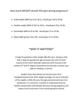
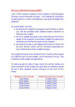


© Copyright 2026