
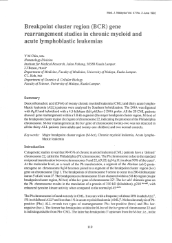
Extensive cross-homology between the long and the short arm of
From www.bloodjournal.org by guest on October 21, 2014. For personal use only.
1992 79: 1299-1304
Extensive cross-homology between the long and the short arm of
chromosome 16 may explain leukemic inversions and translocations
JG Dauwerse, EA Jumelet, JW Wessels, JJ Saris, A Hagemeijer, GC Beverstock, GJ van Ommen
and MH Breuning
Updated information and services can be found at:
http://www.bloodjournal.org/content/79/5/1299.full.html
Articles on similar topics can be found in the following Blood collections
Information about reproducing this article in parts or in its entirety may be found online at:
http://www.bloodjournal.org/site/misc/rights.xhtml#repub_requests
Information about ordering reprints may be found online at:
http://www.bloodjournal.org/site/misc/rights.xhtml#reprints
Information about subscriptions and ASH membership may be found online at:
http://www.bloodjournal.org/site/subscriptions/index.xhtml
Blood (print ISSN 0006-4971, online ISSN 1528-0020), is published weekly by the American
Society of Hematology, 2021 L St, NW, Suite 900, Washington DC 20036.
Copyright 2011 by The American Society of Hematology; all rights reserved.
From www.bloodjournal.org by guest on October 21, 2014. For personal use only.
Extensive Cross-Homology Between the Long and the Short Arm of Chromosome
16 May Explain Leukemic Inversions and Translocations
By J.G. Dauwerse, E.A. Jumelet, J.W. Wessels, J.J. Saris, A. Hagemeijer, G.C. Beverstock, G.J.B. van Ommen,
and M.H. Breuning
Specific rearrangementsof chromosome 16 are well known
in acute nonlymphocytic leukemia with abnormal eosinophils. While mapping cosmids relative to breakpoints in
chromosome 16 in leukemic cells with fluorescence in situ
hybridization (FISH), we have identified three areas of extensive cross-homology between 16p and 16q. Three cosmids
among 99 tested showed two large signals on the short arm
and one signal on the long arm of chromosome 16. A fourth
cosmid showed mainly two signals on the short arm. With
the 16p-specific cosmid we can demonstrate that the break-
points of a pericentric inversion and a reciprocal (16;16)
translocation, both of which are characteristic for acute
leukemia, map to the most distal of two blocks on the short
arm. We suggest that there may be at least two distinct
repetitive elements specific for chromosome 16 interdigitated on 16p. The presence of a similar repeat in the short, as
well as the long arm of the chromosome, may play a role in
the origin of chromosome 16 rearrangementsin acute leukemia.
o 1992by The American Society of Hematology.
C
inferior banding quality, which is a common feature of
metaphase chromosomes from leukemic cells. We have
designed a sensitive method for rapid detection of inv(l6)
using two strategically chosen cosmid probes and fluorescence in situ hybridization6 (FISH). We have also established that the breakpoint in the short arm of chromosome
16 of the inv(l6) and the t(16;16) map to be the same
. ~ breakpoint of another translocation
subregion of 1 6 ~The
involving the short arm of chromosome 16, the t(8;16),
associated with acute monoblastic leukemia (AMoL or M5
in the FAB classification) with prominent erythrophagocytosis and thrombophagocytosis:.” although cytogenetically
indistinguishable from the breakpoint of inv( 16) or t( 16;
16), was mapped to a distinct subregion of 16p.I’ Using
radioactive in situ hybridization techniques, LeBeau et all3
reported the splitting of the metallothionein gene cluster
(MT) mapped on 16q by the inversion breakpoint. Since
this cluster of genes had been cloned, the isolation of the
breakpoint seemed a straightforward procedure. However,
later studies by Sutherland et all4also using in situ hybridization techniques on high resolution banding chromosomes,
indicated that the MT gene cluster actually appears to map
at 16q13, in fact proximal to the breakpoint. We have
investigated the 16p breakpoint of the inversion, because a
detailed map of this chromosomal region was already
a~ailab1e.I~
The serial mapping of cosmids on chromosome
16 showed extensive cross-homology between the p and q
arms of this chromosome.
ANCER IS NOW known to be caused by changes in
genes. These changes can be brought about by chromosomal rearrangements, such as translocations, deletions,
and inversions. A large number of chromosomal rearrangements specific for certain types of malignancy have been
delineated.‘
In 1983, Arthur and Bloomfield’ and LeBeau et a13
reported on specific changes of chromosome 16, ie,
de1(16)(q22) and inv( 16)(p13q22) in acute myelomonocytic
leukemia (AMML or M4 in the French-American-British
[FAB] classification4),which is characterized by the presence of bone marrow eosinophils with abnormal granulation, often referred to as M6eo. In 1984, Testa et aI5
described t(16;16) in a patient with a similar type of
leukemia. The inv(l6) is a subtle alteration that is difficult
to detect with certainty on contracted chromosomes with
-
G Band ing
/a19
telomere
PK75.4
Lovcy
-
-
--
e60
c55
c129
PK31.2
\
~
c36
c36
centromere
Fig 1. Schematic representation of the results obtained after
hybridizationof 99 cosmids to a number of cell lines with breakpoints
in the 1 6 ~ 1 3region. Hybrid CY 19 contains the der(l6) of a t(13;
16)(q12.1;p13.1).u PK75.4,46,XY.t(9;16)(ql2;p13) (kindly provided by
Dr D.M. Carr, Drew Medical School, Lor Angeles, CA) is a fibroblast
cell line. Loucy is a 45,X,5q-,t(16;20) (p12;q13) cell line from a patient
with T-cell acute lymphoblastic leukemia.” Cell line PK31,2,46,XY,t(7;
16) (p22;p13) is from the father of a patient with partial trisomy 16p.%
C55, C60. C129. and C36 are cosmids that map to the region of
interest. C36 shows double signals on prometaphase chromosomes,
and may recognize a duplication; the breakpoint of PK31.2 separates
the two signals (data not shown).
Blood, Vol79, No 5 (March 1). 1992: pp 1299-1304
~
~
~
From the Department of Human Genetics, State University Leiden,
Sylvius Laboratories, Leiden, The Netherlands; and the Department of
Cell Biology and Genetics, Erasmus Universiy, Rotterdam, The
Netherlands.
Submitted July 18,1991; accepted October 22,1991.
Supported in part by the Dutch Kidney Foundation.
Address reprint requests to M A Breuning, MD, PhD, Department
of Human Genetics, State University Leiden, Sylvius Laboratories,
Wassenaarseweg 72, 2333 A L Leiden, The Netherlands.
The publication costs of this article were defrayed in part by page
charge payment. This article must therefore be hereby marked
“advertisement”in accordance with 18 U.S.C. section 1734 solely to
indicate this fact.
O 1992 by TheAmerican Society of Hematology.
0006-4971I92 I 7905-0027$3.00/0
1299
From www.bloodjournal.org by guest on October 21, 2014. For personal use only.
DAUWERSE ET AL
1300
F
m
Fig 2. In situ hybridization of chromosome 16 cosmids on metaphase spreads. (A) Hybridization of cosmid 163 to normal metaphase
chromosomes, showing bright signals on 16q22 and two very broad blocks of signals on 16p. (B) Hybridization of cosmid 177 to normal
metaphase chromosomes, showing weak signals on 16q, two very broad blocks on 16p, and signals on 18p (arrowhead). (C and D) Hybridization
of cosmid 177 to metaphasechromosomesof ANLL MCeO patientswith (C) the pericentric inversion inv(l6)(q22p13) showing signals of the distal
block of 16p. transportedto the q arm of the same chromosome (arrow), and (D) the translocationt(16;16)(q22;p13) showing signals of the short
arm of one chromosome 16 transported to the long arm of the other 16 [arrow pointing at der(16)(16qter-16q22::16p13-16pter]). ID) Shows the
(arrowhead). (Differentiationbetween the p-arm
signals on 16q (arrow). (E) Shows the two blocks on the der(l6)(16qter-16p13::16q22-16qter)
and q-arm was done on basis of the DAPI banding pattern.)
MATERIALS AND METHODS
Patients. Bone marrow samples from patients were obtained
from the diagnostic service of the cytogenetic laboratories. In
addition, samples stored in liquid nitrogen were studied. Histological and immunological analysis of the same samples was performed
in parallel. In total, we have investigated bone marrow from eight
patients with inv(l6) and one with t(16;16).
Cosmids. Chromosome 16 cosmids were obtained from a Library prepared from a mouse hybrid cell line containing chromosome 16 as the only human chromosome.’6
In situ hybridization. Metaphase spreads were prepared according to standard protocol^."^^' Probes were labeled by nick translation’’ in either the presence of biotin-11-dUTP or digoxigenin-lldUTP and further purified and precipitated as described
el~ewhere.’~
Hybridization and detection of the hybrids was performed as described by Pinkel et al” and Kievits et a1.I’ The slides
were mounted in antifade medium containing propidium iodide
(0.5 kg/mL) and DAPI (0.3 pg/mL) for counterstaining of the
chromosomes according to Kievits et a1.I9 Analysis was performed
with a Leitz Aristoplan microscope equipped for fluorescence
From www.bloodjournal.org by guest on October 21, 2014. For personal use only.
1301
HOMOLOGY OF CHROMOSOME 16 ARMS AND LEUKEMIA
A
Fig 3. In situ hybridizationon interphase nucleiwith (A) cosmid 163, showing one time three blocks of separated dots (arrowheads)-the
other
three blocks are clustered in this nucleus; and (E) plasmid pHURl95, a probe for alphoid sequencesin the centromeric region of chromosome 16.showing two homogeneouslystained blocks.
B
Fig 4. (A) Two-color in situ hybridization. Cosmid 163 labeled with biotin (red) and cosmid 36 labeled with digoxigenin (green-yellow) to
normal metaphase chromosomes, showing, after analysis through a double band-pass filter [Omega, Brattleboro, VT), that cosmid 36 (arrow)
hybridizes within the red block detected by cosmid 163. (The red dot marked by the square is a background signal.) (E) The DAPI image of the
same metaphase, with both arrows pointingat the short arms of both chromosomes 16.
microscopy, and suitable metaphases were photographed on 3M
640 ASA film.
Two-color in situ hybridization. For the detection of a biotihylated probe in one color and the digoxigenated probe in a second
color, the following procedure was used*': After hybridization, the
slides were washed three times for 5 minutes in 50% formamide,
2x SSC ( l x SSC = 0.15 mol/L NaCl + 0.015 mol/L sodium
citrate), pH 7.0 at 42"C, then three times for 5 minutes in 0.1 x SSC
at 60"C, followed by a 3-minute wash in 4 x SSC, 0.05% Tween 20.
The slides were preincubated for 10 minutes with 5% nonfat dry
milk in 4x SSC, followed by a 20-minute incubation at room
temperature with avidin Texas Red (20 yg/mL [Vector, Burlingame, CAI). The slides were washed twice for 3 minutes in 4x SSC,
0.05% Tween 20, and once in 0.1 mol/L Tris-HCL, 0.15 mol/L
NaCI, 0.05% Tween 20 (AB buffer) and incubated for 30 minutes
at 37°C with biotinylated goat anti-avidin (Vector) (5 pg/mL) and
monoclonal mouse anti-digoxin (Sigma, St Louis, MO) (1:660
dilution) in AB buffer with 1% blocking reagent (Boehringer
Mannheim, Germany). After three washes with AB buffer, the
slides were incubated for 30 minutes at 37°C with avidin Texas Red
(20 pg/mL) and rabbit anti-mouse fluorescein isothiocyanate
(FITC) (Sigma) (1:1,500 dilution) in AI3 buffer with 1%blocking
reagent. After three washes, the final 30-minute incubation at 37°C
with goat anti-rabbit FITC (Sigma) (1:2,000 dilution) in AB buffer
with 1% blocking reagent was performed. After three 3-minute
washes with AB buffer, the slides were mounted in antifade
medium,n containing 0.3 yg/mL DAPI.
RESULTS
Chromosome 16 cosmids that had been previously isolatedl63l9were regionally mapped with FISH using metaphase preparations of cell lines from carriers of constitutional rearrangements involvingband 1 6 ~ 1 3and
, from eight
patients with inv(l6) (Fig 1). Among the 99 cosmids tested,
four showed a peculiar pattern of hybridization. The
cosmids 152, 163, and 175 showed bright signals on 16q22
and two very broad blocks of signals on 16p (Fig 2A). The
fourth cosmid, cosmid 177, showed very weak signals on
16q, but very bright blocks on 16p (Fig 2B). In most of the
cosmid 177 hybridizations, the signals on 16q could hardly
be seen. In addition, all four cosmids also show small signals
on the short arm of chromosome 18. Hybridizing and
washing under more stringent conditions (60% formamide)
produced the same signals, showing close homology between probe and target sequences. Although the cosmids
contain inserts of approximately 45 kb (data not shown),
From www.bloodjournal.org by guest on October 21, 2014. For personal use only.
1302
DAUWERSE ET AL
the hybridization signals cover almost half the short arm. In
interphase nuclei, the signals of the cosmids 152, 163, and
175 could be seen as two sets of three clusters of distinct
dots (Fig 3A). Cosmid 177 showed two sets of two clusters
of signals in interphase nuclei (data not shown). No
homogeneous painting of certain areas in interphase nuclei
can be seen, such as that observed after hybridization with
alphoid sequences specific for the centromere region of
chromosome 16 (Fig 3B). This leads us to the conclusion
that the painted blocks do not consist of contiguous
repeated sequences, but are probably clusters of repeats
interrupted by other DNA. This is also clearly dcmonstrated by a double hybridization experiment with cosmid
36, which shows in this metaphase a single signal on 16p in
green-yellow, and cosmid 163, which shows large blocks in
red, extendingbeyond cosmid 36 into 1 6 ~ 1(Fig4A
3
and B).
As cosmid 36 was previously localized proximal to the short
arm breakpoints of inv(l6) and the t(16;16),”’ we tested
whether the clusters of repeated sequences on 16p spanned
these breakpoints. Cosmid 177, which gives very weak
signals on 16q, was hybridized to slides containing the
inv(l6) and t(1616). In both cases, the distal part of the
signal on the short arm was connected to the long arm of
chromosome 16. while part of the signal remained on 16p
(Fig 2C, D. and E).Illustrations depicting the rearrangements in a schematic way are given in Fig 5. In metaphase
spreads of the t(1616). the transported part of 16p seemed
i
rather small (Fig 2, D and E). However, on these contracted
chromosomes, the cosmid 177consistently showed no signal
on the long arm of one chromosome 16, and distinct dots on
the other. Therefore, the breakpoints of both inv(l6) and
t( 1616) must be situated within the distal cluster of
repeated sequences. With additional cosmids lacking the
repeat sequences and showing single spots in FISH on 16p,
the breakpoints of inv(l6) and t(16;16) were localized
between cosmids C55 and C129 (Fig 1). The differences in
size of the hybridization signals on 16q illustrated by the
cosmids 152,163, and 175 and cosmid 177 must be due to a
divergence in the repeated sequences present in the different cosmids. Although subcloning of the cosmids and
sequencing of the repetitive elements is still in progress, we
can assume that at least two repeat units must be involved:
one responsible for the bright signalson 16q and one for the
combination of broad blocks on 16p with weak signals on
16q and a signal on 18p.
DISCUSSION
The serial mapping of cosmids on chromosome 16
showed extensive cross-homology in both the p and q arms
of this chromosome. Similar FISH-positive blocks have
been observed on chromosome 16 by R.L. Stallings et al
(manuscript submitted). While characterizing 3,145 cosmid
clones for chromosome 16” by repetitive sequence finger-
‘-7
bq P13
I
L
p13
1
,
16
16
m pi3
m
16
\
I
q22
m
16
D
/--)
I
I
- \-
922
\.
Fig 5. illustrations of (A) inv(l6) in Giemra banding, (B) inv(l6) after FISH with -mid
(C) t(16;16) in G l m u banding, and (D) t(16;lS) .*or FISH with cosmld 177.
-
i
.
.
16
,
16
t(16;16)(p13q22)
177 rhowing two blacks of repetitive sequences on 16p.
From www.bloodjournal.org by guest on October 21, 2014. For personal use only.
HOMOLOGY OF CHROMOSOME 16 ARMS AND LEUKEMIA
1303
printing, 98 cosmids appeared to belong to one very large
overlapping set of cosmids (contig). This would seem to
suggest the presence of repeated elements in a subset of 4%
to 5% of the chromosome 16 cosmids, which yields similar
restriction fragments in the “fingerprint” analysis. Approximately 1,000 sequence families with a relatively low number
of interspersed repeats are believed to be present in the
human genome.24 However, only a handful of the most
abundant repeats has been cloned and sequenced. Therefore, the existence of a new class of interspersed repeats
clustered on chromosome 16 is not unexpected. We propose that these interspersed repeats facilitate “illegitimate”
homologous recombination events leading to the inv( 16)
and t( 1616). It is clear that repetitive sequences are
involved in the origin of some chromosomal rearrangements, such as deletions, translocations, and inversions. In
many cases, the recombinations have occurred within, or
next to, Alu repeats,25s26
while in other cases, local primary
or secondary DNA structure facilitates homologous recombination leading to
The origin of an acute
nonlymphocytic leukemia with eosinophilic granulation
with inv(l6) and t(16;16) is probably similar to myelodysplastic syndrome and t(1;7). In this syndrome, a whole-arm
translocation involving the short arm of chromosome 7 and
the long arm of chromosome 1 occurs by breakage through
the centromeric heterochromatin of both
The resulting monosomy for 7q may contribute to the
neoplastic transformation of the cell. For the ANLL M4 eo,
characterized by inv(l6) or t(16;16), we hypothesize the
following sequence of events. Cross-homology between 16p
and 16q facilitates rearrangements of the chromosome. If
the rearrangement leads to juxtaposition of critical sequences on either arm, a (pre)leukemic cell clone will be
formed.
We describe rearrangements involving the distal block of
repeats on 16p. It is likely that the homology between 16q
and the proximal part of 16p leads to the structural
rearrangements too. Presumably, there is no growth advantage provided to a bone marrow cell with a breakpoint at
this particular location.
ACKNOWLEDGMENT
We thank the clinicians for sending bone marrow samples of
patients with leukemia, and Drs D. Callen, D. Carr, A.G.M.
Hunter, and H. Ben Basset for cell lines. We thank Dr J.
Weissenbach for critical reading of the manuscript. We thank Bert
Eussen for the fine chromosome drawing program.
REFERENCES
1. Sandberg A The Chromosomes in Human Cancer and
Leukemia (ed 2). New York, NY, Elsevier, 1991, p 1
2. Arthur DC, Bloomfield CD: Partial deletion of the long arm
of chromosome 16 and bone marrow eosinophilia in acute nonlymphocytic leukemia: A new association. Blood 61:994,1983
3. LeBeau MM, Larson RA, Bitter MA, Vardiman JW,Golomb
HM, Rowley JD: Association of an inversion of chromosome 16
with abnormal marrow eosinophils in acute myelomonocytic leukemia: A unique cytogenetic-clinicopathologicassociation. N Engl J
Med 309:630,1983
4. Bennett JM, Catovsky D, Daniel MT, Flandrin G, Galton
DAG, Gralnick HR, Sultan C: Proposals for the classification of
the acute leukemias. Br J Haematol33:451,1976
5. Testa JR, Hogge DE, Misawa S, Zandparsa N Chromosome
16 rearrangements in acute myelomonocytic leukemia with abnormal eosinophils. N Engl J Med 310468,1984
6. Dauwerse JG, Kievits T, Beverstock GC, van der Keur D,
Smit E, Wessels HW, Hagemeijer A, Pearson PL, Van Ommen
G-JB, Breuning MH: Rapid detection of chromosome 16 inversion
in acute nonlymphocytic leukemia, subtype M4, regional localization of the breakpoint in 16p. Cytogenet Cell Genet 53:126,1990
7. Wessels JW,Dauwerse JG, Breuning MH, Beverstock GC:
The inversion 16 and the translocation (1616) in ANLL M4EO
break in the same subregion of the short arm of chromosome 16.
Cancer Genet Cytogenet 57225,1991
8. Powell BL, McNay JW,Brown S, Cooper MR, Pettaniti MJ:
Translocation (8;16) (pll;p13) in patients with acute monocytic
leukemias. Cancer Genet Cytogenet 36:109,1988
9. Lai JL, Zandecki M, Jouet JP, Savary JB, Lambiliotte A,
Bauters F, Cosson A, Deminatti M: Three cases of translocation
(8;16) (pll;p13) observed in acute myelomonocytic leukemia: A
new specific subgroup? Cancer Genet Cytogenet 27101,1987
10. Smadja N, De Gramont A, Gonzalez-Canali G, Krulik M,
Deloup J: Another case of acute myelomonocytic leukemia with
t(8;16) (pll;p13). Cancer Genet Cytogenet 36:137,1988
11. Heim S, Avanzi G, Billstrom R, Kristoffersson U, Mandahl
N, Bekassy AN, Ganvichz S, Wiebe T, Pegoraro L, Falda M,
Resegotti L, Mitelman F A new specific chromosomal rearrangement, t(8;16) (pll;p13), in acute monocytic leukaemia. Br J
Haematol66:323,1987
12. Wessels JW,Mollevanger P, Dauwerse JG, Cluitmans FHM,
Breuning MH, Beverstock GC: Two distinct loci on the short arm
of chromosome 16 are involved in myeloid leukemia. Blood
77:1555,1991
13. LeBeau MM, Diaz MO, Karin M, Rowley JD: Metallothionein gene cluster is split by chromosome 16 rearrangements in
myelomonocytic leukemia. Nature 313:709,1985
14. Sutherland GR, Baker E, Callen DF, Carson OM, West AK:
The human methallothionein gene cluster is not disrupted in
myelomonocytic leukemia. Genomics 6144,1990
15. Breuning MH, Snijdewint FGM, Brunner H, Verwest A,
IJdo JW, Saris JJ, Dauwerse JG, Blonden LAJ, Keith T, Callen
DF, Hyland VJ, Xiao GH, Scherer G, Higgs DR, Reeders ST,
Germino GG, Van Ommen G-JB, Pearson P L Map of 16
polymorphic loci on the short arm of chromosome 16 close to the
polycystic kidney disease gene (PKD1). J Med Genet 27:603, 1990
16. Breuning MH, Saris JJ, Wapenaar MC, den Dunnen JT,
Van Ommen G-JB, Pearson PL: The isolation of DNA probes from
chromosome 16 for the diagnosis of polycystic kidney disease, in
Carone FA, Dobbie JW (eds): Pathogenesis of Polycystic Kidney
Disease. Chicago, IL, TP&T, 1990, p 17
17. Landegent J, Jansen in de Wal N, Fisser-Groen YM, Bakker
E, van der Ploeg M, Pearson P L Fine mapping of the Huntington
disease linked D4S10 locus by non-radioactive in situ hybridization. Hum Genet 73:354, 1986
18. Langer PR, Waldrop AA, Ward D C Enzymatic synthesis of
biotin labeled polynucleotides: Novel nucleic acid affinity probes.
Proc Natl Acad Sci USA 78:6633,1981
19. Kievits T, Devilee P, Wiegant J, Wapenaar MC, Cornelisse
CJ,Van Ommen G-JB, Pearson P L Direct nonradioactive in situ
From www.bloodjournal.org by guest on October 21, 2014. For personal use only.
1304
hybridization of somatic cell hybrid DNA to human lymphocyte
chromosomes. Cytometry 11:105,1990
20. Pinkel D, Straume T, Gray JW:Cytogenetic analysis using
quantitative, high-sensitivity, fluorescence hybridization. Proc Natl
Acad Sci USA 83:2934,1986
21. Arnoldus EPJ, Wiegant J, Noordermeer IA, Wessels JW,
Beverstock GC, Grosveld G, van der Ploeg M, Raap AK: Detection
of the Philadelphia chromosome in interphase nuclei. Cytogenet
Cell Genet 54108,1990
22. Johnson GD, Nogueria Araujo GM de: A simple method of
reducing the fading of immunofluorescence during microscopy. J
Immunol Methods 43:349,1981
23. Stallings RL, Tomey DC, Hildebrand CE, Longmire JL,
Deaven LL, Jett JH, Doggett NA, Moyzis R K Physical mapping of
human chromosomes by repetitive sequence fingerprinting. Proc
Natl Acad Sci USA 87:6218,1990
24. Sun L, Paulson KE, Schmid CW,Kadyk L, kinwand L
Non-Alu family interspersed repeats in human DNA and their
transcriptional activity. Nucleic Acids Res 12:2669,1984
25. De Klein A, Van Agthoven T, Groffen C, Heisterkamp N,
Groffen J, Grosveld G: Molecular analysis of translocation products of a Philadelphia-positive CML patient. Nucleic Acids Res
14:7071,1986
26. Nicholls RD, Fischel-Ghodsian N, Higgs DR: Recombination at the human alpha-globin gene cluster: Sequence features
and topological constraints. Cell 49369,1987
27. Henthorn PS, Smithies 0, Mager D L Molecular analysis of
deletions in the human beta-globin gene cluster: Deletion junctions
and locations of breakpoints. Genomics 6:226,1990
DAUWERSE ET AL
28. Fodde R, Losekoot M, Casula L, Bernini L Nucleotide
sequence of the Belgian Ggamma+(Agamma,delta,beta)O-thalassemia deletion breakpoint suggests a common mechanism for a
number of such recombination events. Genomics 8:732,1990
29. Krawczak M, Cooper DN: Gene deletions causing human
genetic disease: Mechanisms of mutagenesis and the role of the
local DNA sequence environment. Hum Genet 86425,1991
30. Vnencak-Jones CL, Phillips J A Hot spots for growth hormone gene deletions in homologous regions outside Alu repeats.
Science 2501745,1990
31. Alitalo T, Willard HF, De la Chapelle A Determination of
the breakpoints of 1;7 translocations in myelodysplastic syndrome
by in situ hybridization using chromosome-specific alpha satellite
DNA from human chromosomes 1 and 7. Cytogenet Cell Genet
5049,1989
32. Callen DF, Hyland VJ, Baker EG, Fratini A, Gedeon AK,
Mulley JC, Femandez KEW, Breuning MH, Sutherland GR:
Mapping the short arm of chromosome 16. Genomics 4348,1989
33. Ben-Bassat H, Shlomai Z, Kohn G, Prokocimer M: Establishment of a human t-acute lymphoblastic leukemia cell line with a
(16;20) chromosome translocation. Cancer Genet Cytogenet 49:
241,1990
34. Hunter AGW, Rimoin DL, Koch UM, MacDonald GJ, Cox
DM, Lachman RS, Adomian G: Chondrodysplasia punctata in an
infant with duplication 16p due to a 7;16 translocation. Am J Med
Genet 21:581,1985
35. Moyzis RK, Albright KL, Bartholdi MF, Cram LS,Deaven
LL, Hildebrand CE, Joste NE, Longmire JL, Meyne J: Human
chromosome-specific repetitive DNA sequences: Novel markers
for genetic analysis. Chromosoma 95:375,1987


© Copyright 2026