
Probing the pathobiology of response to all-trans retinoic acid in
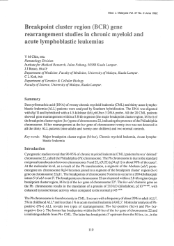
From www.bloodjournal.org by guest on October 15, 2014. For personal use only.
1996 87: 218-226
Probing the pathobiology of response to all-trans retinoic acid in
acute promyelocytic leukemia: premature chromosome
condensation/fluorescence in situ hybridization analysis
RC Vyas, SR Frankel, P Agbor, WH Jr Miller, RP Jr Warrell and WN Hittelman
Updated information and services can be found at:
http://www.bloodjournal.org/content/87/1/218.full.html
Articles on similar topics can be found in the following Blood collections
Information about reproducing this article in parts or in its entirety may be found online at:
http://www.bloodjournal.org/site/misc/rights.xhtml#repub_requests
Information about ordering reprints may be found online at:
http://www.bloodjournal.org/site/misc/rights.xhtml#reprints
Information about subscriptions and ASH membership may be found online at:
http://www.bloodjournal.org/site/subscriptions/index.xhtml
Blood (print ISSN 0006-4971, online ISSN 1528-0020), is published weekly by the American
Society of Hematology, 2021 L St, NW, Suite 900, Washington DC 20036.
Copyright 2011 by The American Society of Hematology; all rights reserved.
From www.bloodjournal.org by guest on October 15, 2014. For personal use only.
Probing the Pathobiology of Response to All-Trans Retinoic Acid in Acute
Promyelocytic Leukemia: Premature Chromosome Condensation/
Fluorescence In Situ Hybridization Analysis
By Rohini C. Vyas, Stanley R. Frankel, Phylisha Agbor, Wilson H. Miller, Jr,
Raymond P. Warrell, Jr, and Walter N. Hittelman
The response of acute promyelocytic leukemia(APL) peripheral blood and bone marrow cells to trans-retinoic acid (RA)
was cytogenetically characterized during RA treatment using thetechniques of premature chromosome condensation
(PCC) and fluorescence in situ hybridization (FISH). Before
treatment, the predominant immature bone marrow cells
were found to have t(15;17), whereas the residual mature
granulocytes were diploid and lacked evidence of the translocation. In response to RA treatment, an increase in the
leukocyte count was noted. The majority of these cells exhibited a t(15;17). Subsequently (eg, between days 6 and
23). 32% to 91% of the maturing myeloid cells still exhibited
t(15;17). The appearance of t(15;17) in gradually maturing
elements suggests that RA contributed to a release of the
maturation block of the leukemic elements. As responding
patients obtained complete remission, diploid elements
without evidence of the translocation prevailed in the blood
and bone marrow. In 16 patients studied after 1 month in
complete remission, all but 2 showed all diploid cells. The
residual t(15;17) cells disappeared 18 days later in 1 patient,
whereas the second patient exhibited clinical evidence of
relapse 20 days later. These results suggest that response
of patients with APL to RA is associated with maturation,
subsequent loss of the matureleukemic elements, and preferential regeneration of normal diploid hematopoietic elements.
0 1996 by The American Society of Hematology.
A
RA administration (ie, between days 5 and 20) in which
peripheral white blood cell (WBC) counts sometimes increased strongly to high levels. Second, upon continued RA
treatment, a gradual decrease in the fraction of immature
elements along with a concomitant increase in the fraction
of maturing elements both in the blood and bone marrow
was o b ~ e r v e d . ' ~All
. ' ~ this occurred without an interceding
hypoplastic bonemarrow phase commonly observed after
cytotoxic chemotherapy. Finally, complete remission was
obtained with full restoration of granulocyte andplatelet
count.
The pathobiology of response of APL to RA is not well
understood. However, because in vitro studies indicated that
RA could induce leukemic cell maturation, it was assumed
that this was a case of true-differentiation therapy."." Indeed, preliminary studies have supported the case for induced cyto-differentiation,".'" and fluorescence in situ hybridization (FISH) studies showed that maturing cells during
RA treatment were derived from leukemic cells bearing
t( 15; 17). Similar findings were also obtained after RA treatment of a patients with promyelocytic variant of chronic
myelogeneous leukemia."
Although the preliminary results were exciting in that they
suggested that RA treatment resulted in the differentiation
of leukemic elements, we now extended the cytogenetic studies to include 30 patients with APL who have been treated
with RA to better characterize the pathobiology of response
and complete remission.
CUTE PROMYELOCYTIC leukemia (APL) is characterized by a distinct translocation between chromosomes 15and 17,[t(15;17)(q22;q12-21)], in 70% to 100%
of the reported cases. Thus, the translocation can usually be
used as a marker for the leukemic compartment."' Recent
molecular characterization of the breakpoint on chromosome
17 has identified the involved region as the gene for nuclear
retinoic acid (RA) receptor a.',' The breakpoint on chromosome 15 has been identified as the PML gene, whichis
thought to encode a transcriptional regulatory factor that
might be active in early myeloid cells.'.''
Treatment of APL patients with RA has been associated
with a 64% to 90% rate of complete remission and reduced
coagulopathy and bone marrow hypoplasia."-I4 The clinical
nature of response to RA wasfound to besomewhat different
from that normally observed cytotoxic chemotherapy. First,
most patients experience a period of leukocytosis early after
From the Department of Clinical Investigation, The University o j
Texas M.D. Anderson Cancer Center, Houston: and Leukemia and
Developmental Chemotherapy Services, the Department of Medicine, Memorial Sloan-Kettering Cancer Center, New York, NY.
Submitted December 27, 1994: accepted August IO, 3995.
Supported in part by Grants No. POI CA-55164, CA-27931, CA57645, and CA-45746 from the National Institutes of Health, National Cancer Institute and grants fromthe Food and Drug Administration (FD-R-000674), the American Cancer Society (PDT-381,
and the Markey Trust (88-23). S.R.F. was
EDT-47, and IM-551),
supported by the Mortimer J. Lacher Research Fundandisthe
recipient of a Cancer Chemotherapy Training Grant (CA-09207-14)
from the National Cancer Institute. W.N.H. is a Sophie Caroline
Steves Professor in Cancer Research.
Address reprint requests to Rahini C. Vyas, PhD, 6323 Vedanta
Terrace, Los Angeles, CA 90068.
The publication costsof this article were defrayedin part by page
chargepayment. This article must therefore be hereby murked
"advertisement" in accordance with 18 U.S.C. section 1734 solely to
indicate this fact.
0 1996 by The American Society of Hematology.
0006-4971/96/8701-0128$3.00/0
218
MATERIALS AND METHODS
Patient population. Thirty patients with APL who received
RA
treatment (45 mg/m2/d)attheMemorialSloan-KetteringCancer
Center (New York, NY) form the basis of the present study. The
clinical trial was an extension of that trial previously described in
greater detail.'? APL patients undergoing a phase I1 trial of RA for
treatment gave informed consent for serial testing of bone marrow
and blood during therapy.
Peripheral blood and bone marrow specimens. Peripheral blood
specimens were obtained by venipuncture and placed into heparinized tubes. Bone marrow aspirates wereplaced into tubes containing
Blood, Vol 87, No 1 (January l ) , 1996: pp 218-226
From www.bloodjournal.org by guest on October 15, 2014. For personal use only.
219
NATURE OF RESPONSE OF RA IN APML BY PCC/FISH
phosphate-buffered saline (PBS), 5,000 IUlmL preservative-free
heparin (Fisher, Houston, TX), and 1 % fetal calf serum (Flow, Costa
Mesa, CA). The specimens were obtained from patients with APL
receiving 45 mg/m2/d RA at the Memorial Sloan-Kettering Cancer
Center. The samples were shipped in cold boxes by overnight courier
to M.D. Anderson Cancer Center (Houston, TX). Mononuclear and
polymorphonuclear fractions were enriched using a two-step FicollHypaque gradient system” in which mononuclear cells were collected at the top interface, polymorphonuclear cells were collected
at the intermediate interface, and erythrocytes were collected in the
pellet. Fractionated cells were washed twice inHanks’ Balanced
Salt Solution (HBSS; GIBCO, Grand Island, NY), and aliquots were
placed on slides using a cytocentrifuge (Shandon, Pittsburgh, PA),
fixed in methanol, and stained with Wright’s-Giemsa for morphologic characterization. The remaining cells were used in cell fusion
studies.
Cell fusionandpremature
chromosome condensation (PCC).
The procedures for cell fusion between mitotic Chinese hamster
cells (CHO) and peripheral blood or bone mmow cells have been
previously described in detail.” Briefly, CH0 cells were grown in
Hsu-modified McCoy’s 5A medium containing 10% calf serum,
100 UlmL penicillin (GIBCO, Santa Clara, CA), and 100 pg/mL
streptomycin (GIBCO). Before their use as mitotic inducers, CH0
cells were grown in the presence of 120 pg/mL BrdUrd for one cell
cycle time and then incubated in colcemid for 3.5 hours. Mitotic
cells were selectively detached. Mononuclear or polymorphonuclear
fractions were mixedwith approximately equal numbers of CH0
mitotic cells and washed twice in HBSS. The cell pellet was resuspendedin 0.5 mL serum-free medium containing approximately
3000 HAU UV-inactivated Sendai virus. The fusion mixture was
incubated at 4°C for 15 minutes to promote cell agglutination, and
then 0.05 mL 20 mmoVL MgCI, and 0.05 mL of 5 pg/mL Colcemid
were added. The fusion reaction was performed at 37°C for 75
minutes in a humidified 5% CO2 incubator.
Slide preparation and storage. At theendofthe
incubation,
the fusion mixtures were treated with 0.075 molL KCL hypotonic
solution for 10 minutes at room temperature and then fixed twice in
a 1O:l mixture of methano1:glacial acetic acid (vol:vol) for IO minutes each at room temperature. The pellet was resuspended in a
small volume of fresh fixative (3:1, methano1:glacial acetic acid),
and drops of cell suspension were placed on clean wet slides and
allowed to air dry. The slides were then washed in PBS to remove
traces of residual acetic acid (3 times each for 3 minutes), dehydrated
in an ethanol series (70%, 90%, and loo%), air-dried, and stored in
a box at -2O’C.
FISH with chromosome 17probe. A phage DNA library derived
from sorted human chromosome 17 (LN17NS03) was obtained from
the American Type Culture Collection (ATCC; Rockeville, MD).
The phage library was amplified using Escherichia coli LE 392 as
the bacterial host, as described by Maniatis et al.’’ Purification of
phage and extraction of phage DNA was performed as described by
Ziai et al.” Briefly, intact bacteriophage particles were precipitated
with ammonium sulfate, digested with proteinase K, and treated with
an alkaline solution. Phage DNA containing human library inserts
were labeled with biotinylated 11-dUTP by nick translation according to the manufacturer’s directions (Fisher, Houston, TX). In
some cases, a polymerase chain reaction (PCR)-generated probe
from sorted human chromosome 17” was used. The efficiency of
biotin labeling was checked using a dot blot procedure withan
immunoperoxidase and diaminobenzidine reaction.
Hybridization. Slide preparations were first baked at 8O‘C for 5IO minutes to maintain chromatin morphology during subsequent
hybridization procedures. The chromosome substrates on slides were
denatured in 70% formamide/2~SSC, pH 7.0 at 80°Cfor 5 minutes,
dehydrated in an ice-cold ethanol series, and air-dried. The hybridization probe mixture contained 1 pg/mL labeled chromosome 17
probe DNA, 20-fold excess of unlabeled competitor total human
genomic DNA, IO-fold excess sonicated salmon sperm DNA, 9%
dextran sulfate, and 2 x SSC in 50% formamide, pH 7.0. The hybridization mixture wasplacedunderan
18 X 18 mm glass coverslip
and the edges were sealed with rubber cement. Hybridization was
performed for 12 to 72 hours at 37’C in a humidified chamber. The
coverslips were then removed. The slides were washed three times
for 5 minutes each in 50% formamide, 2X SSC, pH7.0at 45’C,
three times for 5 minutes each in 2X SSC, pH 7.0 at 45’CC, and then
in 4X SSC, 0.1% Tween 20, pH 7.5 for 5 minutes.
Visualization of hybridizationproduct.
The hybridized slides
were incubated with fluoresceinated avidin (DCS grade, 5 pglmL in
4X SSC with 5% nonfat dry milk) for 20 minutes at room temperature in the dark. Signal amplification was accomplished, when necessary, by successive treatments with biotin-labeled goat antiavidin
(Vector, Burlingame, CA; 5 pg/mL in 4X SSC with 5% nonfat dry
milk) for 20 minutes at roomtemperature and fluoresceinated avidin.
Nonspecific binding wasblocked with 5% nonfatdry milk. After
the last washin 4X SSC and tween 20, the slides were counterstained with 0.5 pg/mL propidium iodide (Sigma, St Louis, MO)
and mounted inan antifade solution containing 1,4-diazibicyclo[2,2,2]-octane (Sigma) and glycerol. The preparations were visualized on a Nikon (Tokyo, Japan) epifluorescence microscope
equipped with the appropriate filters for visualizing both fluorescein
isothiocyanate (FITC) and propidium iodide. Photographs were
taken with Kodakektachrome 400 ASA film(Eastman Kodak, Rochester, NY).
Reverse transcription-PCR (RT-PCR) assay
for PMLJRAR expression. Leukemic cells from APL patients were examined for the
presence of PMLRARa fusion mRNA. Experimental protocol remains the same as described earlier.23
RESULTS
A summary of the clinical response of the 30 patients
studied is shown in Table 1. The patients ranged in age from
9 to 75 years of age and included 17 male and 13 female
patients. Complete remission (CR) was obtained in 26 of the
30 patients studied (87% CR). Four patients did not respond
to RA and died during the early treatment course. The time
to clinical evidence of CR in this set of patients was highly
variable and occurred between days 23 and 77.
Cytogenetic studies before treatment. Of the 30 patients
studied by conventional cytogenetic analysis at mitosis, 20
patients exhibited the common t(l5; 17) and 2 patients
showed a complex translocation still involving chromosomes
15 and 17. In 3 patients, conventional cytogenetic analysis
failed to show any results and only diploid cells were observed in the remaining 5 patients (Table 1). In contrast,
molecular analysis (by Northern blot, Southern blot, and RTPCR) showed either an aberrant transcript or a rearrangement
of the RARa gene in all but 2 of the patients (ie, patients
no. 10 and 11; Table 1). Using the techniques of PCC and
FISH with a chromosome 17 probe, cells with rearrangements of chromosome 17 could be easily distinguished from those with a diploid karyotype (Fig 1A and
B). Using this approach, all patients examined showed evidence of rearrangements of chromosome 17 in at least a
fraction of their cells. Patient no. 26 exhibited a consistent
isol7q abnormality, which is sometimes found in patients
From www.bloodjournal.org by guest on October 15, 2014. For personal use only.
220
W A S ET AL
Table 1. Summary of Patients' Characteristics and Response to RA
~~~
Patient
Age1
No.
Sex
Conventional
Cytogenetics
1
66/M 46XY,t(15;17)+8
2
10/M 46XY,t(15;17)
3
35/F
46XX
4
21/F
46XX
5
17/F
46XX,t(15;17)
6 46X,-X,t(4,5,1
38/F
7
75/F
46XY,t(15;17)
8
16/M 46XY.t(6,15,17
9
52/M Failure
10
51/F
46XX
11
53/M 46XY
12
29/M 46XY,t(15;17)
13
43/M 46XY,t(15;17)
14
44/F
46XX,t(15;17)
34/M 46XY,t(15; 17)
15
9/M 46XY,t(15;17)
16
33/M 46XY,t(l5;17)
17
61/F
46XY,t(l5;17),
18
-2,de17q
27/M
Failure
19
65/F
46XX,t( 15; 17)
20
17/F
46XX
21
54/M 46XY,t(15;17),+8
22
61/F
46XX,t(15;17),+20
23
66/M 46XY,t(15;17)
24
29/M 46XY,t(15; 17)
25
28/M 46XY,t(15;17),
26
der1,delllq.
+Marl,
+MarZ,
+Mar3
17/M 46XY,t(15;17)
27
28
50/M
29
47/F
30
61/F
46XY,(15;17),
+Marl;46XY,
t(15;17)
45XY,t(15;17),
-22
Failure
~~
~
Molecular
Analysis*
RARa Gene
PCC/FISH
+
+
+
+
+
+
+
+
+
+
+
+
+
-
+
+
+
+
+
+
+
+
+
+
+
+
+
+
+
t(15;171
Analysis
+
+
+
+
+
+
+
+
+
+
+
+
+
+
+
+
+
+
+
+
+
is0 17q
CR on
Day
43
ED
23
49
41
35
46
35
56
ED
44
37
29
41
28
43
31
24
33
24
53
?77
47
33
28
ED
+
Complex
t(15;17;A?)
50
+
+
29
+
+
28
+
+
ED
Abbreviation: ED, early death.
* Molecular analyses include Southern, Northern, and
RT-PCR analyses.
with APL.24As shown in Fig 2A, using a total chromosome
17 probe, the centromere in the isol7q chromosome appeared more central than on a normal chromosome 17. This
centromere location was confirmed using an cy satellite DNA
centromere-specific probe for chromosome 17 (Fig 2B). Patient no. 27 was found to have a complex translocation involving one intact and three broken parts of chromosome
17. Using a total chromosome 17 probe, the PCC of these
cells could still be easily distinguished from diploid cells
(Fig 2C).
In patients no. 3, 4, and 21, molecular analysis showed
rearrangements within the RARa gene, whereas conventional cytogenetic techniques failed to show cells with
t( 15; 17). However, PCCFISH analysis successfully identi-
fied a t( 15;17) subpopulation in all 3 cases. These 3 cases
were positive for the PML/RARa rearrangements by RTPCR (Table l). For patients no. 10 and 11, conventional
metaphase analysis had shown only diploid cells and molecular analysis showed no detectable arrangement within the
RARcy gene. On the other hand, PCC/FISH analysis showed
a chromosome 17 rearrangement, but only in a minority of
cells examined (2 of 39 cells and 2 of 24 cells examined
from patients no. 10 and 1 l, respectively). Thus, it appears
likely that this low level of abnormal signals represents background noise and sets a limit as to the specificity of IOW
frequency of positive signals. Alternatively, cytogenetic
analysis of interphase cells by PCC mayaugment the genetic
evaluation of patients with leukemia.
A proportion of the patients withAPLat
presentation
exhibited a residual fraction of apparently fully mature granulocytes in the peripheral blood. Because APL represents a
condition in which partial maturation is apparent, it was of
interest to determine whether these morphologically normal
granulocytes were derived from the leukemic clone or from
residual normal elements. Because mature granulocytes are
nondividing, conventional mitotic analysis was not feasible.
However, the PCC technique allows visualization of the
chromosomes of these nondividing cells and the FISH technique allows an easy distinction between leukemic and normal karyotypes.
We used these techniques to determine the origin of normal granulocytes in 6 patients (ie, patients no. 3, 6, 15, 23,
2.5, and 28) who exhibited sufficient numbers of granulocytes
for analysis (Table 2). Because the peripheral blood samples
contained a mixture of immature and mature elements, a
two-step Ficoll-Hypaque density gradient was used to enrich
each fraction before PCC." Overall cytogenetic analysis of
the light-density fractions (mononuclear cells) from these 6
patients showed that 6% to 89% of the scored PCC exhibited
a t(l.5; 17). In contrast, as shown in Table 2, the mature
granulocyte fractions in these patients were dominated by
diploid cells, and the minor fraction of cells in patients no.
25 and 28 exhibiting a t(l5; 17) could be accounted for by
morphologic evidence of contaminating immature cells in
the heavy density fraction. These results thus suggest that
the mature granulocytes observed in these patients before
treatment were derived from residual normal elements and
that the maturation block of leukemic elements before treatment was intact.
Therapy-associated leukocytosis. In these patients, increased WBC counts were first noted between days 4 and
23 of treatment, and the counts remained increased for 4 to
16 days during RA treatment. Earlier, similar findings have
been reported by
To determine the origin of the
circulating cells during the leukocytosis phase, peripheral
blood specimens were obtained from l 1 patients during this
period, 6 of whom showed white blood counts greater than
40 X IO9 cells& (Table 3).
An example of the leukocytosis phase is shown for patient
no. 1.5 (Fig 3A). Before treatment, the WBC count was 1.6
x lo9&. WithRA treatment, theWBC count reached a
maximum of 17.4 X 10⁹L on day 16, remained increased
From www.bloodjournal.org by guest on October 15, 2014. For personal use only.
221
NATURE OF RESPONSE OF RA IN APML BY PCC/FISH
Fig 1. PCC painting using a chromosome 17-specific probe. (A) PCC with a t(15;17) showing one intact and two translocated products of
chromosome 17 (arrows) exhibit yellowish-greenflourescence. (B)Diploid PCC with two intact chromosome 17s.
for 2 days, and then decreased back to
the normal range
within1week. The pretreatmentperipheralblood sample
showeda predominance of blastsand promyelocytes, of
which 50% exhibited a t(l5; 17). Bone marrow specimens
on days 4and8showedthat90%and98%
of the cells
harbored the t(l5; 17). Similarly, at the time of maximum
-i
leukocytosis on day 18, 82% of the bone marrow cells and
72% of theanalyzed peripheral blood cells werederived
from cells exhibiting t (15; 17).
As illustrated in Fig 3B(and Table 3), patient no.5 experienced ahigh degree of leukocytosis after RA treatment.
Before treatment,theWBC
count was9.4 X lo9& was
is0 17q
I
6
3
e
Fig 2. Chromosome abnormalities observed other than t(15;17). (A)
isol7q detected by a whole chromosome 17 painting probe. Note the
centromeric location differences on thetwo chromosome 17s (patient
no. 26). (B)Chromosome 17 centromeric probe confirming the isol7q
in patient no. 26. (C) Complex translocation (arrows) in patient no. 27
involving chromosomes 15 and 17 and an 'A'
group (7) chromosome.
From www.bloodjournal.org by guest on October 15, 2014. For personal use only.
VYAS ET AL
222
Table 2. PCC-FISH Analysis Before Treatment
Mononuclear Cells
Patient No.
73
6
1550
23
25
28
7
% Diploid Cells
Cells Scored
93
94
50
11
22
93
16
100
100
8
Polymorphonuclear Cells
%Cells
With
100
100
100
92
t(15;17)
Cells Scored
% Diploid Cells
16
20
50
23
100
10
100
6
89
78
20
dominated by blasts and promyelocytes, and exhibited
t( 15; 17) in 67% of the cells analyzed, whereas too few cells
(TFC) were available for cytogenetic analysis from highdensity gradient fraction. By day 9, the count increased to
71.9 X IOy/L, of which 92% of the cells exhibited t(l5; 17).
By day 25 of treatment, the WBC count had returned to 8.9
X 10~1~.
All of the patients with leukocytosis showed a preponderance of cells with t(15; 17) during the height of leukocytosis.
Although the time of occurrence and degree of leukocytosis
varied from patient to patient, the cells were nearly always
of leukemic clone origin. These observations suggest that
RA might selectively allow egress of leukemic cells from
tissue stores and their transient proliferation.
Therapy-associated maturation of leukemic elements.
Continued treatment with RA was associated with a gradual
decrease in the frequency of immature bone marrow cells
and an increase in the frequency of cells exhibiting morphologic features of maturation (ie, the nucleus became lobulated and the cytoplasmic granular content was altered; Fig
4A through D). Auer rods were often observed in maturing
cells. This apparent maturation process occurred at variable
times (range, 6 to 60 days) after the initiation of RA treatment, and it appeared to be a continuous process culminating
in the disappearance of immature cells and the emergence
of fully mature granulocytes in the peripheral blood.
To determine the origin of the maturing myeloid elements
% Cells With t(15;17)
0
0
0
0
80
during this response transition, mononuclear cells and maturing granulocytes from the peripheral blood of four patients
were enriched by a two-step Ficoll Hypaque gradient and
examined by the PCC/FISH technique. As shown in Table
4, the mononuclear fractions exhibited 17% to94% cells
with t(1S; 17),whereas
the polymorphonuclear fraction
showed 33% to 91% of the cells with t( 15; 17). For example,
onday 6, the peripheral blood cell count of patient no. 4
was 14.4 X 10⁹/Lwith 95% immature blasts and promyelocytes. After fractionation, 85% of the mononuclear cells exhibited t( IS; 17), whereas 50% of the polymorphonuclear
fraction exhibited this translocation. Similarly, for patient
no. 23 on day 18 of treatment, 83% of the lobulated cells
exhibited the t(15; 17). These observations suggest that RA
treatment was associated with a gradual maturation of the
leukemic clone. As RA treatment continued, the fraction of
immature cells decreased and the fraction of mature elements
increased. However, the fraction of both the immature and
mature elements exhibiting a t(l5; 17) also decreased.
The morphology of the cells examined also exhibited
unique changes during this transition. For example, on day
4 of treatment, the bone marrow of patient no. 15 contained
mostly blasts and promyelocytes, of which 90% harbored a
t(l5; 17) (Fig 4A). By day 8 of treatment, although 98% of
the cells still showed a t( 15;17), the majority of cells began
to show some lobulation of the nucleus and granulation in
the cytoplasm (Fig 4B). By day 22, the degree of lobulation
Table 3. Patient's WBC Count and PCC-FISH AnalvsisDurina Leukocvtosis
PCC-FISH Analysis at Peak of Leukocytosis
(mononuclear cells)
WBC 1 x 1 0 ~ ~ )
Patient
No.
Before
Treatment
2
4
5
10
14
15
16
19
23
25
26
31
1.5
Abbreviation: NE, not evaluated.
Maxtmum
(on day)
166.0 (71
31.5 (8)
71.9 (9)
36.4 (18)
14.0 (16)
17.4 (16)
26.0 (23)
47.7 (16)
41.3 (3)
41.3 (11)
70.5 (13)
Normal
(on day)
10.0(14)
6.0 (13)
8.9 (25)
5.9 (23)
8.1 (23)
5.1 (36)
5.5 (28)
3.3 (27)
7.5(8)
8.6 (17)
% Diploid
Cells
% Cells With
t(15;17)
Duration of
Leukocytosis
Cells
Scored
07
05
16
NE
-
53
96
91
06
08
95
94
92
05
NE
-
-
29
25
100
100
28
16
09
11
72
84
91
89
NE
-
65
00
07
07
13
12
16
05
04
l00
From www.bloodjournal.org by guest on October 15, 2014. For personal use only.
NATURE OF RESPONSE OF RA IN APML BY PCCFISH
10
-
-
223
WBC
GRANULOCYTES
BLASTS +PROS
1-
.l7
Q
!
I
-1 5
15
.01
I
I
75
45
135105
T
a
CI
a
W
Fig 3. Demonstration of the leukocytosis
phase
during RA treatment. The fractionof cells exhibiting
t115;171 is highlighted at different times &er treatment. (AI Patient no. 15. Note a moderate degreeof
leukocytosis (WBC count, 17 x 10s/L) on day 16. (B)
Patientno.5. Note a highdegree of leukocytosis
(WBC count, 71 x l@/L) on day 9.
0
.014
-15
I
0
15
30
45
I
I
60
75
90
Time on treatment(Days)
had increased, but 82% of the
cells still exhibited a t(15; 17).interest, 1of these 4 patients (no. 26) did not exhibit
t( 15; 17).
Moreover,thecellsappearedvacuolated,and
2%of the
Patient no. 26, who had an
isol7q abnormality, exhibited
cells had a morphology characteristic of cells undergoing
evidence for maturationof the leukemic clone on day 16of
apoptosis (Fig 4C and D). Similarly, 7.4% of the peripheral treatment (Table 4) but did not achieve CR.
blood cells of patient no. 26 appeared to be apoptotic on day Of the remaining 26 patients,
PCC analysis was performed
8 of treatment, and gel electrophoresis of the cellular DNA
on the peripheral blood of 17 patients during CR. In 13 of
showed a fragmentation pattern typical of populations under-these 17 patients, CR was associated with disappearance of
going apoptosis (data not shown). These results therefore
cells with t( 15; 17) and a return of diploid granulocytes to
suggest that RA-induced maturation of the leukemic clone
the peripheral blood. However, 4 patients who entered cliniis associated with an upregulation of the apoptotic pathway
cal CR showed a different pattern of response. Data from
and eventual elimination of these
cells from the bone marrow RT-PCR and PCC/FISH analyses to detect minimal residual
and blood.
disease at respective time after CR in 4 patients are shown
Restoration of n o m 1 hematopoiesis. As a result of RA
in Table 5. Patient no. 1 achieved a CR on day 43, but on
treatment (Table l), 87% of this patient group obtained CR. day 48, PCC/FISH analysis of the mononuclear peripheral
Three patients (no. 2, 26, and 30) died during induction, andblood fraction still showed 1.4% of the cells with t(l5; 17),
1 patient (no. 10) was taken off study without response. Of
whichwasconfirmedbyRT-PCRanlysis.Patientno.14
From www.bloodjournal.org by guest on October 15, 2014. For personal use only.
W A S ET AL
224
B
Fig 4. Morphologic changes observed in bone marrow cells during RA treatment (patient no. 15). (A) Day 4. Note the predominance of
blasts, of which 90% harbored the 15;17 translocation. (B)Day 8. A majoramof cells exhibit a slightly perturbed cytoplasm; 9846 of the cells
analyzed exhibited the 15; 17 translocation. No cells with typical apoptosis-like morphologywere observed. (C and Dl Day 22. Note thepresence
of vacuolated and lobulated cells. Two percent of the cells showed a morphology typical of apoptosis (arrow).
achieved CR on day 41, but on day 58, 2% of the mononuclear fraction still exhibited the leukemic karyotype and presence of PML-RARa transcript detected by RT-PCR. In patient no. 24, 13 days after CR.14%of
the mononuclear
fraction exhibited t(15; 17) was confirmed by RT-PCR data;
however, these abnormal karyotypes were not found on subsequent bone marrowand peripheral blood specimens on
days 59,71, 85, 121, 126, 160, and 174. Similarly, in patient
no. 28, RT-PCR data showed the presence of abnormal cells
and PCC/FISH analysis showed 6% of the mononuclear cells
from bone marrow with t( 15; 17), even after 12 days of CR,
but subsequent granulocyte specimens exhibited only diploid
karyotypes. Taken together, these results suggest that,
whereas early granulocyte maturation after RA treatment
of APL patients produces maturing leukemic cells, normal
hematopoietic elements predominate in the bone marrow and
blood by the time CR is achieved.
DISCUSSION
Although the administration of RA to APL patients likely
results in differentiationof malignant cells and ultimately restoration of normal hematopoiesis, the exact mechanisms responsi-
Table 4. Evidence for Leukemic Maturation During RA Treatment
Mononuclear Cells
Patient
No.
4
14
23
26
Days on
Treatment
14
23
18
Polymorphonuclear Cells
Cells
% Diploid
% Cells With
Cells
% Diploid
96 Cells With
Scored
Cells
t(15;17J
Scored
Cells
t(15;17)
10
50
67
17
09
50
33
83
91
40
52
23
29
42
34
15
83
13
06
85
17
87
94
12
12
From www.bloodjournal.org by guest on October 15, 2014. For personal use only.
225
NATUREOF RESPONSE OF RA IN APML BY PCC/FISH
Table 5. Detection of Minimal Residual Disease by PCClFlSH and
RT-PCR Analysis in Patiants With CR
~~~~~~
~~~
Patient
No.
1
14
24
28
~
~
Day
After CR
5
17
13
~
PCC-FISH % Cells
With t(15;17)
RT-PCR PMURARo
Transcript
1.4
2.0
14.0
6.0
+
+
+
+
bers of maturing granulocytes was observed. Subsequently,
the matured leukemic cells were replaced by cells derived
from the residual normal hematopoietic elements. Thus, response to RA can be considered to be a result of a release of
the maturation block experienced by the leukemic elements
before treatment. Although this type of response might be
considered differentiation therapy, it needs to be noted that
maturation of the leukemic cells was associated with preferential cell loss of these cells, possibly through an apoptotic
pathway."
Although RA can induce remission in patients with APL,
patients who receive RA therapy develop resistance to RA
and ultimately relapse. This scenario suggests that residual
leukemic cells remain in the body. Although it is likely that
the induction of terminal differentiation and apoptosis of the
majority of leukemic cells by RA allows the restoration of
normal hematopoiesis, the detection of some mature diploid
cells before the disappearance of the leukemic population
suggests that RA might induce the repression factors from
the leukemic cells that negatively influence normal hematopoiesis.
This group of patients was notfollowed-up closely enough
by PCC/FISH to determine the technique's value in the detecting minimal residual disease or in predicting the reemergence of cells with t(15; 17). Moreover, this group of patients
was treated in a heterogeneous fashion during remission.
Nevertheless, although PCR techniques are highly sensitive
to detect the presence of abnormal cells in population, it is
still difficult to quantitate disease trends using bulk analyses
of cell populations. Thus, just as the PCC/FISH techniques
was found to be extremely useful for defining the pathobiology of response in patients with undergoing RA treatment, it
might also be useful for defining the pathobiology of relapse,
especially in those patients whose cells are difficult to recognize by other means.
The response of APL patients to RA is a multifactorial
process involving upregulation of leukemic cells, induction
of maturation of leukemic cells, upregulation of cell loss in
the maturing leukemic cells, and preferential repopulation
with normal diploid elements. A better understanding of
these processes will hopefully lead to the development of
new therapeutic strategies to overcome the inhibitory activities of the leukemic burden that cause bone marrow failure as
well as induce preferential loss of the dysregulated leukemia
cells.
ble for this process are undefined. The results reported here
using the techniques of PCC and FISH supportthis contention
and offer a wider understanding
of the regulatory processes
occurring in APL patients duringRA treatment.
PCC/FISH is a useful technique to evaluate and followup patients with APL undergoing therapy. PCC/FISH as well
as molecular analyses show the presence of genetically abnormal cells in cases in which they were missed by conventional cytogenetic analysis. PCC/FISH analysis was able to
detect cells witha t(l5; 17)in 3 cases (patients no. 3, 4,
and 21) that were reported to be diploid by conventional
cytogenetics.
The above finding points out several positive attributes
of the PCC/FISH approach. First, one can cytogenetically
analyze cells in interphase that might not reach metaphase
and would not be available for detection by conventional
cytogenetic analysis. Second, PCC/FISH allows the recognition of a subpopulation of cells that might not be detectable
by Southern blot analysis (due to their dilution by cells without genetic rearrangement) or by RNA analysis (due to lack
of transcriptional activity of the rearranged gene at the time
of study). Third, one can study chromosome translocation
using a single painting probe that might not be determined by
interphase cytogenetics because it is difficult to distinguish
chromosome translocation from chromosome break in the
interphase cell. However, more recently, RT-PCR technique
has opened a new avenue in exploring the genetic alterations
and can be used as a molecular tool for diagnosis of APL
in t( 15; 17)-negative cases.
By using PCC/FISH analysis, we were able to determine
the origin of granulocytes in patients with APL at diagnosis
and during the course of RA therapy. The absence of the
t(l5; 17) in granulocytes found in APL patients before therapy indicates that the maturation block in the leukemic component is intact before treatment, and the limited degree of
maturation observed in some patients was due to an incomplete leukemic inhibitory activity on normal hematopoiesis.
ACKNOWLEDGMENT
The increase in leukocyte count that accompanies RA
We thank DrSureshJhanwarforsupervisingthekaryotyping
therapy may reflect the need of an additional cell division
analyses and Saroj Vadhan-Raj and Jordan Gutterman for their supbefore maturation event.I5 In vitro studies following the cell
port.
growth patterns of leukemia cells that have been induced to
differentiate lend support for this p ~ s s i b i l i t y . " ~At' ~the
~~~~~~
REFERENCES
same time, it is also possible that treatment with RA alters
1 . Bennett JM, Catovsky D, Daniel MT. Flandrin G, Galton DAG,
the adhesive properties of the leukemic cells and allows the
the classification of acute
GranlnickHR.SultanC:Proposalsfor
release of these cells from tissue stores, including the bone
leukemias. Br J Haematol 33:451, 1976
After the leukocytosis, a gradual decrease in the immature
blasts and promyelocytes and a gradual increase in the num-
2. Rowley JD, Golomb HM, Dougherty C: 15; 17translocation:
A consistent chromosomal change in acute promyelocytic leukemia.
Lancet 1549, 1977
From www.bloodjournal.org by guest on October 15, 2014. For personal use only.
226
3. Berger R, Bemheim A, Daniel MT, Valensi F, Flandrin G:
Une nouvelle variet6 de leukemie aigue non promyelocytaire avec
translocation t(15;17). C R Acad Sci 288:177, 1979
4. Berger R, Bemheim A, Daniel MY, Valensi F, Flandrin G:
t(15;17) translocation in acute promyelocytic leukemia (M3) and
cytological “M3 variant”. Nouv Rev Fr Hematol 23:27, 1981
5. Golomb MM, Rowely JD, Vardiman J W , Testa JR. Butler A:
Microgranular acute promyelocytic leukemia: A distinct clinical,
ultrastructural and cytogenetic entity. Blood 55:253, 1980
6. Hagemeijers A, Lowenberg B, Abels J: Analysis of breakpoints
in translocation t(15; 17) observed in 4 patients with acute promyelocytic leukemia. Hum Genet 61:223, 1982
7. de The H, Chomienne C, Lanotte M, Degos L, D’ejean A: The
t(15; 17) translocation of acute promyelocytic leukemia fuses the
retinoic acid receptor gene with a novel transcribed locus. Nature
3475.58, 1990
8. Biondi A, Rambaldi A, Acalay M, Pandolfi PP, LoCoco F,
Diverio D, Rossi V, Mencarelli A, Longo L, Zangrilli D, Masera
G, Barbui T, Mandelli F, Grignani F, Pelicci PG:RAR gene rearrangement as a genetic marker for diagnosis and monitoring in
acute promyelocytic leukemia. Blood 77:1418, 1991
9. Kakizuka A, Miller WH Jr, Umesono K, Warrell RP Jr, Frankel
SR, Murty VVS, Dmitrovsky E, Evano RM: Chromosomal translocation t(l5; 17) in human promyelocytic leukemia fuses RAR with
a novel putative transcription factor, PML. Cell 66:663, 1991
IO. de The’ H, Lavay C, Marchia A, Chomienne C, Degos L,
D’ejean A: The PML-RAR fusion mRNA generated by the t(l5; 17)
translocation acute promyelocytic leukemia encodes a functionally
altered RAR. Cell 65:675, 1991
11. Huang ME, Ye YC, Chen SR, Chai JR, Lu JX, Zhoa L, Gu
LJ, Wang ZY: Use of all-trans retinoic acid in the treatment of acute
promyelocytic leukemia. Blood 72567, 1988
12. Castaigne S , Chomienne C, Daniel MT, Ballerini P, Bengen
R, Fenaux P, Degos L: All trans retinoic acid as a differentiation
therapy for acute promyelocytic leukemia. I. Clinical results. Blood
76:1704, 1990
13. Warrell RP Jr, Frankel SR. Miller WH Jr, Scheinberg DA,
Itri L, Hittelman WN, Vyas R, Andreef M, Tafuri A, Jakubowski
A, Gabrilove J, Gordon M, Dmitrovsky E: Differentiation therapy
of acute promyelocytic leukemia with tretinoin (all trans retinoic
acid). N Engl J Med 324:1385, 1991
14. Frankel S , Eardley A, Hellar G, Miller WH Jr, Berman E,
Dmitrovsky E, Warrell RP Jr: Treatment of patients with acute promyelocytic leukemia using all-trans retinoic acid. Ann Intern Med
120:278, 1994
15. Breitman TR, Selonick SE, Collins SJ: Induction of differentiation of human promyelocytic leukemia cell line HL-60 by retinoic
acid. Proc Natl Acad Sci USA 771:2936, 1980
16. Breitman TR, Keene BV, Hemmi H: Retinoic acid induced
differentiation of fresh human leukemic cells and the human myelomonocytic leukemia cell lines HL-60, U-937and THP-1. Cancer
Surv 2:263, 1983
W A S ET AL
17. Wiernick PH, Dutcher JP, Paietta E, Hittelman WN, Vyas
RC, Strack M, Castaigne S , Degos L, Gallagher RE: Treatment of
promyelocytic blast crisis of chronic myelogeneous leukemia with
all trans retinoic acid. Leukemia 5504, 1991
18. English D, Andersen BR: Single-step separation of red blood
cells, granulocytes and mononuclear leukocytes on discontinuous
density gradients of Ficoll-Hypaque. J Immunol Methods 5:249,
1974
19. Hittelman WN, Petkovic I, Agbor P: Improvements inthe
premature chromosome condensation technique for cytogenetic analysis. Cancer Genet Cytogenet 30:301, 1988
20. Maniatis T, Fristsch EF, Sambrook J: Molecular Cloning: A
Laboratory Manual. Cold Spring Harbor, NY, Cold Spring Harbor
Laboratory, 1982
21. Ziai RM, Giordano A, Armandola E, Ferrone S: Purification
by ammonium sulfate precipitation of bacteriophage gt 11 DNA for
restriction analysis of cloned cDNA inserts. Anal Biochem 171:192,
1988
22. Chang KS, Vyas RC, Deaven LL, Trujillo JM, Stass SA,
Hittelman WN: PCR amplification of chromosome specific DNA
isolated from flow cytometry sorted chromosomes. Genomics
12:307, 1992
23. Miller WH Jr. Kakizuka A, Frankel SR, Warrell RP Jr. DeBlasio A, Levine K, Evans RM, Dmitrovsky E: Reverse transcription
polymerase chain reaction for the rearranged retinoic acid receptor
alpha clarifies diagnosis and detects minimal residual disease in
acute promyelocytic leukemia. Proc Natl Acad Sci USA 89:2694,
1992
24. Sandberg AA: Chromosome changes and their significance
in acute nonlymphocytic leukemia, in Sandberg AA (ed): Chromosomes in Leukemia and Solid Tumors: Introduction and Methodological Aspects. New York, NY, Elsevier, 1990, p 225
25. Pinto A, Aldinucci D, Zagonel V, Di Noto R, Gattei V, Juzbasic S , Improta S , Perin V, Del Vecchio L, Canale V, Grignani F,
Pellicci PG: In vitro and in vivio regulation of adhesion molecules
in acute promyelocytic leukemia (APL) cells by the PMLJRARa
gene product and all-trans retinoic acid. Proc Am SOCClin Oncol
14:389, 1995 (abstr no. 1215)
26. Koeffler HP, Hirji K, Itri L, Stang H, Moore D, Henderson
M, Weinstein I, Resenbloom B, MacIntyre R, Rigberg S, Wilson L,
Dreisbach P, Cone L, Bluming A, Newman S , Orestein A, Rosen
P, Dosik G, Kidder W, Casciato D: 1, 25-Dihydroxy vitamin D3: In
vivo and in vitro effects on human preleukemic and leukemic cells.
Cancer Treat Rep 69:1399, 1985
27. Van Roozendaal KEP, Darling D, Farzaneh F:DMSO and
retinoic acid induce HL-60 differentiation by different but converging pathways. Exp Cell Res 190:137, 1990
28. Wu X, Shao G, Chen S, Wang X, Wang ZY: Studies on the
relationship between protein kinase C and differentiation of human
promyelocytic leukemia cells induced by retinoic acid. Leuk Res
13:869, 1989


© Copyright 2026