
CASE REPORT
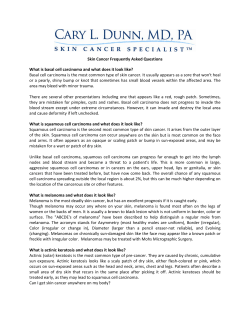
DOI: 10.14260/jemds/2014/3652
CASE REPORT
LARGE NONFUNCTIONAL ADRENO CORTICAL CARCINOMA
MASQUERADING AS RENAL CELL CARCINOMA: A CASE REPORT WITH
REVIEW OF LITERATURES
Ahsan Ahmad1, Kaushal Kunar2, Nadeem Ahmad3, Mumtaz Hussain4, Mahendra Singh5
HOW TO CITE THIS ARTICLE:
Ahsan Ahmad, Kaushal Kunar, Nadeem Ahmad, Mumtaz Hussain, Mahendra Singh. “Large Nonfunctional
Adreno Cortical Carcinoma Masquerading as Renal Cell Carcinoma: A Case Report with Review of Literatures”.
Journal of Evolution of Medical and Dental Sciences 2014; Vol. 3, Issue 54, October 20; Page: 12492-12495,
DOI: 10.14260/jemds/2014/3652
INTRODUCTION: Adrenocortical carcinoma is a rare malignant neoplasm affecting only 1 or 2
person per million population1.Nonfunctional malignant tumors particularly present at advanced
stage and are associated with poor prognosis.2 They are difficult to differentiate from renal cell
carcinoma (RCC) especially if they are very large and nonfunctional compressing its native kidney.
However, with good preoperative planning in such cases innocuous kidney can be saved.
CASE REPORT: A 50 years old female presented to us with complaints of left upper abdomen lump
associated with mild dull ache at same site for four months and irregular fever for three months. She
had history of right nephrolithotomy three years back elsewhere. She had no associated comorbidity.
Physical examination revealed non-tender, firm mass with lobulated surface occupying whole left
hypochondrium. The upper border of mass could not be reached. The hematological and biochemical
parameter including renal and liver function test were within normal range.
We had preoperative suspicion of adrenal tumor so we did glucocorticoid, aldosterone, sexual
steroid (17 OH progesterone, testosterone) and urinary metanephrine assay which were within
normal range. Computed tomography (CT) of abdomen revealed solid contrast enhancing
heterogeneous mass in upper pole of left kidney of about 14cm x 12 cm size. Even CT reporting it as
renal mass we added adrenal mass in our differential diagnosis because of large upper polar renal
mass. Kidney was approached with 11th rib cutting incision as we had doubt of adrenal gland
involvement. The approach also gave us a good exposure of tumor. Intraoperative dissection revealed
large suprarenal mass compressing left kidney.
Therefore renal pedicle was carefully separated with sharp and blunt dissection and the
adrenal mass was excised after controlling its pedicle. Patient showed good postoperative recovery
and did not show any abnormal biochemical profile due to adrenal gland involvement. She is doing
well at almost one year of follow up. We did clinical examination, LFT.
Chest X ray and USG at six month and one year which were within the normal range. We have
planned for CECT abdomen at two year because tumor was nonfunctional adrenocortical carcinoma.
Her histopathological examination of slides showed nuclear atypia, necrosis and more compact cells
with low percentage of clear cells in both low and high power magnification. High power
magnification also showed diffuse architecture of cells (trabecular pattern).
DISCUSSION: Adrenocortical carcinoma is a rare adrenal cancer of which 80% are functional 3.
Functional tumor is common in young females and children whereas nonfunctional tumor occurs
more frequently in elderly.
J of Evolution of Med and Dent Sci/ eISSN- 2278-4802, pISSN- 2278-4748/ Vol. 3/ Issue 54/Oct 20, 2014
Page 12492
DOI: 10.14260/jemds/2014/3652
CASE REPORT
Early diagnosis of adrenal cortical carcinoma is very difficult especially in nonfunctioning
carcinoma.4 Klausner et al reviewed the literature and found that fever is encountered in 6- 10% of
cases with adrenal tumors sometimes as first manifestation of disease.5 Flammingo et al reported a
case of large nonfunctioning adrenal cortical carcinoma with a 24x 18 cm mass.6
The patient had pre-operative fine needle biopsy suggestive of RCC without any clinical sign
of adrenal carcinoma. However, per operative the mass seemed to be of adrenal origin and was
distinct from kidney which was preserved. Azhar A Khanet al reported a case of giant adrenal cortical
carcinoma 30cmx 25cm mass mimicking as RCC in which radical nephrectomy specimen
histopathology revealed diagnosis of adrenocortical carcinoma.2
Kuneida et al reported a patient in whom a giant adrenal cortical carcinoma was found to
have recurred in the contralateral adrenal gland 6 years after the operation.7 They also concluded
that large tumor are associated with better prognosis because aggressive tumors give rise to distant
metastasis and therefore symptomatic at an early stage.
CT and magnetic resonance imaging can be used for diagnosis and provide useful information
about local invasion or spread to kidney with a sensitivity of more than 90%. However, sometimes a
large tumor compressing renal parenchyma, the diagnosis can be missed. Tumor size 7-8 cm has 47%
like hood of malignancy.8 Open adrenalectomy with en bloc excision if needed has been the main stay
for the treatment of primary and recurrent tumors. Neo adjuvant chemotherapy or adjuvant
chemotherapy in such cases is not very effective.9
REFERENCES:
1. Copeland PM. The incidentally discovered adrenal mass. Ann Intern Med1983; 98: 940-47.
2. Khan A A, Iqbal S, Sergill, Rizwan H, Sandeep, S Gujral. Giant nonfunctioning carcinoma of the
adrenal cortex mimicking renal cell carcinoma. a diagnostic dilemma. Urology2007; 70: 178.
e1-2.
3. Luttom JP, Cerdas S, Billaud L.clinical features of adrenocortical carcinoma, prognostic factors.
4. Nq L, Libertino JM. adrenal cortical carcinoma diagnostic evaluation. J urol 2003; 169: 5-11.
5. Klausner JM, Nakash R, Inbar M, Gutman M, Leleusk S, Rozin R.Prolong fever as a presenting
symptoms in adrenal tumors.Oncology1988; 45: 15-7.
6. Flammingo A, Pettinato G. Giant nonfunctioning carcinoma of adrenal cortex. N Eng J Med 2001;
345: 700.
7. Kunieda K, Saji S, recurrence of giant adrenal cortical carcinoma in the contralateral adrenal 6
year after surgery, report of a case.Surg today 2003; 30: 294-97.
8. Sturgeon C, Shen WT, Clarkoff. Risk assessment in 457 adrenal cortical carcinoma. How much
the tumor size predict the likelihood of malignancy. J Am Coll surg 2006; 202: 423-30.
9. Kuraba R, Gallagher SF. Current management of adrenal tumor.Oncology2008; 20: 34-46.
J of Evolution of Med and Dent Sci/ eISSN- 2278-4802, pISSN- 2278-4748/ Vol. 3/ Issue 54/Oct 20, 2014
Page 12493
DOI: 10.14260/jemds/2014/3652
CASE REPORT
High magnification histopathology showing acc
Heterogeneously enhancing left renal area mass
Non-contrast CT scan showing large lump in left renal area
J of Evolution of Med and Dent Sci/ eISSN- 2278-4802, pISSN- 2278-4748/ Vol. 3/ Issue 54/Oct 20, 2014
Page 12494
DOI: 10.14260/jemds/2014/3652
CASE REPORT
AUTHORS:
1. Ahsan Ahmad
2. Kaushal Kunar
3. Nadeem Ahmad
4. Mumtaz Hussain
5. Mahendra Singh
PARTICULARS OF CONTRIBUTORS:
1. Assistant Professor, Department of
Urology, Indira Gandhi Institute of Medical
Sciences.
2. Resident, Department of Urology, Indira
Gandhi Institute of Medical Sciences.
3. Assistant Professor, Department of
Urology, Mithola University.
4. Assistant Professor, Department of
Anaesthesiology, Indira Gandhi Institute of
Medical Sciences.
5.
Professor, Department of Urology, Indira
Gandhi Institute of Medical Sciences.
NAME ADDRESS EMAIL ID OF THE
CORRESPONDING AUTHOR:
Dr. Ahsan Ahmad,
Indira Gandhi Institute of Medical
Sciences, Sheikhpura,
Patna-800014, India.
Email: [email protected]
Date of Submission: 14/09/2014.
Date of Peer Review: 15/09/2014.
Date of Acceptance: 14/10/2014.
Date of Publishing: 18/10/2014.
J of Evolution of Med and Dent Sci/ eISSN- 2278-4802, pISSN- 2278-4748/ Vol. 3/ Issue 54/Oct 20, 2014
Page 12495


© Copyright 2026