ABC
docz
Explore
Log in
Create new account
Download
Report
No category
Interleukin-4 receptor regulation in human monocytic cells
Sample Testing Order Form
H o w
✓ 10 FOLLOW-UP
X-10.0 Product Datasheet 39100.100.43000 Standard Universal OHP Films for Mono Copiers
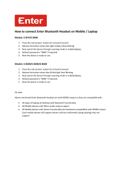
How to connect Enter Bluetooth Headset on Mobile / Laptop
Function of the Interleukin-2 (IL-2) Receptor y
PHARMACOLOGY The Journal of And Experimental Therapeutics Contents
PrepX RNA-Seq Sample and Library Preparation Kits for Illumina
Ataxia and Speech Jennifer Legge M.S., CCC-SLP University of Chicago Medical
gannett ell arn w o le
124 (15)
© Copyright 2026
About abcdocz
DMCA / GDPR
Report