
John M Le, Cristiane H Squarize, Rogerio M Castilho
World J Stem Cells 2014 November 26; 6(5): 511-525
ISSN 1948-0210 (online)
© 2014 Baishideng Publishing Group Inc. All rights reserved.
Submit a Manuscript: http://www.wjgnet.com/esps/
Help Desk: http://www.wjgnet.com/esps/helpdesk.aspx
DOI: 10.4252/wjsc.v6.i5.511
REVIEW
Histone modifications: Targeting head and neck cancer
stem cells
John M Le, Cristiane H Squarize, Rogerio M Castilho
influence gene expression independent of alterations
to the DNA sequence. Recent findings from our group
have shown that pharmacological induction of posttranslational modifications of tumor histones dynamically modulates CSC plasticity. These findings suggest
that a better understanding of the biology of CSCs in
response to epigenetic switches and pharmacological inhibitors of histone function may directly translate to the
development of a mechanism-based strategy to disrupt
CSCs. In this review, we present and discuss current
knowledge on epigenetic modifications of HNSCC and
CSC response to DNA methylation and histone modifications. In addition, we discuss chromatin modifications
and their role in tumor resistance to therapy.
John M Le, Cristiane H Squarize, Rogerio M Castilho, Laboratory of Epithelial Biology, Department of Periodontics and Oral
Medicine, University of Michigan School of Dentistry, Ann Arbor, MI 48109-1078, United States
Author contributions: Le JM, Squarize CH and Castilho RM
contributed equally to this work; all authors were involved in reviewing the literature and writing the manuscript.
Supported by University of Michigan, School of Dentistry startup
Correspondence to: Rogerio M Castilho, DDS, MS, PhD,
Laboratory of Epithelial Biology, Department of Periodontics and
Oral Medicine, University of Michigan School of Dentistry, 1011
N University Ave, Room 3323B, Ann Arbor, MI 48109-1078,
United States. [email protected]
Telephone: +1-734-6472150 Fax: +1-734-7635503
Received: August 29, 2014 Revised: September 10, 2014
Accepted: September 17, 2014
Published online: November 26, 2014
© 2014 Baishideng Publishing Group Inc. All rights reserved.
Key words: Head and neck squamous cell carcinoma;
Chromatin remodeling; Histone deacetylases inhibitor;
Histone acetylation; Cancer-initiating cell; Epigenetic
target; Epigenetic marker; Oral squamous cell carcinoma; Tumor resistance
Abstract
Head and neck squamous cell carcinoma (HNSCC) is
the sixth most common cancer worldwide, and is responsible for a quarter of a million deaths annually. The
survival rate for HNSCC patients is poor, showing only
minor improvement in the last three decades. Despite
new surgical techniques and chemotherapy protocols,
tumor resistance to chemotherapy remains a significant
challenge for HNSCC patients. Numerous mechanisms
underlie chemoresistance, including genetic and epigenetic alterations in cancer cells that may be acquired
during treatment and activation of mitogenic signaling
pathways, such as nuclear factor kappa-light-chain-enhancer-of activated B cell, that cause reduced apoptosis.
In addition to dysfunctional molecular signaling, emerging evidence reveals involvement of cancer stem cells
(CSCs) in tumor development and in tumor resistance
to chemotherapy and radiotherapy. These observations
have sparked interest in understanding the mechanisms involved in the control of CSC function and fate.
Post-translational modifications of histones dynamically
WJSC|www.wjgnet.com
Core tip: Stem cells are long-lived, therefore their genome is subject to more stress from genetic mutations
and epigenetic factors than their short-lived, differentiated progeny. Recent evidence strongly indicates that a
subpopulation of tumor initiating cells, termed “cancer
stem cells”, play a fundamental role in tumor heterogeneity, growth, and preservation. Cancer stem cell
behavior is influenced by epigenetic events comprised
primarily of DNA methylation and histone modifications
that dynamically regulate gene expression and silencing.
Le JM, Squarize CH, Castilho RM. Histone modifications:
Targeting head and neck cancer stem cells. World J Stem Cells
2014; 6(5): 511-525 Available from: URL: http://www.wjgnet.
com/1948-0210/full/v6/i5/511.htm DOI: http://dx.doi.org/10.4252/
wjsc.v6.i5.511
511
November 26, 2014|Volume 6|Issue 5|
Le JM et al . Epigenetics and cancer stem cells
(long non-coding RNA)][30-33]. Consequently, by identifying the molecular mechanisms that drive progression and
recurrence of HNSCC, novel cancer therapeutics can be
developed to improve the effectiveness of treatment and
the rate of long-term survival in patients. In this review,
we highlight the current understanding on cancer stem
cells and the effects of epigenetic modifications on tumor
behavior. We also discuss the latest findings on pharmacological manipulation of epigenetic circuitries that may
result in the development of novel therapeutic strategies
that target cancer stem cells.
INTRODUCTION
There are approximately 560000 cases of head and neck
cancer diagnosed worldwide each year and approximately
300000 deaths annually. This cancer type occurs in the
head and neck region, involves the nasal and oral cavity,
pharynx, and larynx and primarily occurs as squamous
cell carcinoma (HNSCC)[1-4]. Although HNSCC has well
recognized risk factors, including tobacco use, excess
alcohol consumption, and infection by high risk papillomaviruses[5,6], we do not fully understand the mechanisms
underlying its malignant progression[5]. Our understanding of the molecular biology of HNSCC has significantly
improved in the last few decades, contributing to the
development of novel therapies targeted against prosurvival signaling circuitries, including the epidermal
growth factor receptor (EGFR), vascular endothelial
growth factor, receptor tyrosine kinases, interleukins, and
phosphoinositide 3-kinase (PI3K) pathways, among others. Unfortunately, the long-term survival rate for HNSCC patients, which is 50% at five years after diagnosis,
has remained consistent over the past thirty years[3,7-9].
The incidence of HNSCC is much higher in developing
nations, where it is the third most common malignancy
in Asian countries compared to the sixth most common
malignancy in Western countries[10-12]. This discrepancy in
incidence of HNSCC is associated with varying risk factors, such as chewing Betel quid in the Asia-Pacific region
compared to consumption of tobacco and alcohol and/
or human papillomavirus infection outside Asia[1,13-17].
The poor long-term survival rates in HNSCC patients
may be due to diagnosis of disease at an advanced-stage
and development of chemoresistance[8,18]. Numerous
mechanisms underlie chemoresistance, including genetic
and epigenetic alterations in cancer cells that may be acquired during treatment[19,20] and the activation of mitogenic
signaling pathways, such as nuclear factor kappa-light-chainenhancer-of activated B cell NFκB, that result in reduced
apoptosis[21]. Furthermore, the recurrence of cancers depend on a subpopulation of cancer stem cells (CSCs) that
possess the unique and exclusive ability to self-renew and
differentiate into nontumorigenic heterogenous cell types
that maintain the tumor[7,22-24]. Therefore, many factors play
a critical role in the maintenance of tumor heterogeneity
and CSC behavior, including the tumor microenvironment,
genomic instability and the effect of genetic mutations and
epigenetic changes on gene expression[22,25-27].
In a significant number of HNSCC, tumor progression results from mutations in genes, such as TP53,
CDKN2A, HRAS, PTEN, and PIK3CA. This causes
alterations in cell signaling cascades (e.g., PI3K/mTOR,
NFκB, ERK, p53), resulting in aberrant cell growth, migration, and survival[3,8,23,28,29]. Epigenetic changes also play
a key role in regulating gene expression through histone
modifications, DNA methylation, miRNA silencing and
DNA repair mechanisms [HMT (Histone methyltransferases), HAT (Histone acetyltransferases), HDAC (Histone
deacteylases) ncRNA (non-coding RNA), and lncRNA
WJSC|www.wjgnet.com
CANCER STEM CELLS
Because normal stem cells are long-lived, their genome
is subject to more stress from genetic mutations and
epigenetic factors than their short-lived, differentiated
progeny. The majority of oncogenic mutations in stem
cells perturb central cellular processes that regulate cellular division, DNA damage repair, and signal transduction
pathways[24,25,34]. Certain HNSCC-related phenotypes that
arise from mutations in oncogenes and tumor suppressor genes, such as PIK3CA, TP63, PTEN, EGFR, and
MET, result in limitless replication potential, insensitivity to apoptotic signals, angiogenesis, invasion and metastasis[28,35-38]. Therefore, tumors arise when stem cells
lose their ability to regulate and maintain tissue form
and function and when they show reduced control over
apoptosis, cellular senescence and cellular proliferation.
Additionally, although tumors are a population of malfunctioning cells, they are commonly characterized by
histological features that resemble normal tissue[39]. Similarly, hematopoietic cancers are comprised of identical
neoplastic cells, but solid tumors from HNSCC consist
of non-identical cells, resulting in phenotypic heterogeneity[25,27,40-42]. Within the polyclonal tumor, there is a cellular
hierarchy in which a small subpopulation of neoplastic
cells with the highest potential for tumorigenesis and selfrenewal are positioned at the top. The remaining bulk of
the tumor primarily consists of well-differentiated nontumorigenic cells that are susceptible to chemotherapy
and radiation[43-45]. In addition to HNSCC, solid tumors
of the breast, brain, colon, lung and prostate also demonstrate a diverse array of cellular heterogeneity that increases genomic instability and adaptability of the tumor
to its microenvironment[25,46,47]. Recent evidence strongly
indicates that a subpopulation of tumor initiating cells,
termed “cancer stem cells” play a fundamental role in
tumor heterogeneity, growth, and preservation[25,44,48,49].
The cancer stem cell hypothesis, first conceptualized by
Bonnet et al[44] in 1997, established that a subpopulation
of human leukemic cells, positive for CD34 and negative
for CD38 cell surface markers, initiates human acute myeloid leukemia in Non-obese diabetic/Severe combined
immunodeficient (NOD/SCID) mice. The following
observations support the cancer stem cell hypothesis: (1)
only a subpopulation of tumor cells within a tumor mass
grow in immunodeficient mice; (2) the subpopulation of
51
November 26, 2014|Volume 6|Issue 5|
Le JM et al . Epigenetics and cancer stem cells
tumor cells generate both CSCs and heterogeneous nontumorigenic cancer cells; and (3) cancer stem cells selfrenew, as revealed by serial transplantation assays[22,44,50].
The frequency of CSCs is relatively low in HNSCC, lung
squamous cell carcinoma, lung adenocarcinoma, and human pancreatic adenocarcinoma, but xenotransplantation
assays greatly increase their frequency[51].
anisms underlying cancer, DNA methylation is the most
commonly studied epigenetic alteration[86-88]. DNA methylation patterns occur in early and precancerous stages
and most frequently discovered in tumors compared to
normal tissues[89,90]. Methylation occurs sporadically and
is globally distributed in mammals throughout the genome at cytosine-phospho-guanine (CpG) dinucleotide
sequences, as revealed by immunofluorescent labeled
5-methylcytosine. Without considering CpG-rich islands
(approximately 1 kilobase in length), there is a low, but
global level of methylation in specific CpG sequences
throughout the entire mammalian genome[26,91]. Therefore, aberrant DNA methylation of these CpG islands
or specific sequences can lead to oncogenic activation via
silencing of tumor suppressor gene expression[92,93]. Hypomethylation is associated with activation of oncogenes,
while hypermethylation is associated with the silencing
of tumor suppressor genes. Both mechanisms induce
genomic instability and play a dominant role in tumor
initiation and progression[90,94]. The most common types
of DNA methylation in tumors are hypermethylation of
CpG islands and global hypomethylation[89]. Hypermethylated CpG islands are often associated with gene promoters; thus, methylation results in a transcriptionally inactive gene. In contrast, methylation of DNA sequences
further from promoter sequences has less of an effect on
transcription[26].
Cancer stem cell surface markers
CSCs were first discovered in solid tumors in 2003[52], and
the isolation of CSCs in HNSCC, based on the CD44+
cell surface marker, occurred in 2007[18]. In that study, approximately 70% of NOD/SCID mice receiving CD44+
tumor cell xenografts showed tumor formation compared
to 1% of mice receiving CD44- xenografts. In addition to
their association with CSCs in HNSCC[53-56], CD44+ cells
also play a role in chemoresistance. Genes associated with
chemoresistance, including ABCB1, ABCG2, CYP2C8
and TERT, are upregulated in CD44+ cells compared
to CD44- cells[57]. Furthermore, CD44+ HNSCC cells
express high levels of B lymphoma Mo-MLV insertion
region 1 homolog (Bmi-1), a self-renewal and oncogenic
protein associated with poor survival and tumor aggressiveness[18,58-62]. Different isoforms of CD44 differentially
modify the behavior of HNSCC. For instance, the v3,
v6, and v10 isoforms of CD44 promote HNSCC tumor
migration, invasion, and metastasis[63,64] and confer chemoresistance in other solid tumors, attributes commonly associated with the chemo- and radio-resistant fractions of
cancer stem cells[65]. Therefore, CD44 is used to identify
CSCs, and it promotes many of the biological characteristics associated with cancer “stemness”. These characteristics include tumorsphere formation in suspension,
unrestricted cellular proliferation, enhanced migration,
tumor invasion, and resistance to chemotherapy and ionizing radiation therapy. CD24 and CD133 (also known as
Prominin 1) are also CSC cell surface markers[66-68].
The increased enzymatic activity of aldehyde dehydrogenase 1 (ALDH1) is commonly used to identify normal pluripotent cells and tumor cells harboring “stemness” potential
in various solid tumors, including HNSCC[51,69-75]. ALDH is a
detoxifying enzyme involved in the oxidation of intracellular
aldehydes and was initially described for its role in hematopoietic stem cell self-renewal via reduction of retinoic acid
activity[76,77]. The presence of ALDH1-positive tumor cells
correlates with poor clinical outcome in breast cancer[69],
ovarian cancer[78], papillary thyroid carcinoma[79], and pancreatic adenocarcinoma[80], among other solid tumors[70,81-83].
It is believed that HNSCC progression and invasion,
in addition to resistance to non-surgical therapies, may be
regulated by the rare population of CSCs[18,43,84,85]. Therefore, to effectively treat this type of cancer, we must develop a therapy that can target and eliminate CSCs.
Histone modifications: In addition to DNA methylation, the chromatin architecture can be remodeled by a
network of protein mediators called histones that play an
important role in gene regulation by compacting DNA.
Histones can be post-translationally modified at the
amino-terminal ends by acetylation, methylation, phosphorylation, sumoylation, ubiquitination, and ADP-ribosylation[95]. These modifications result in gene transcription through the uncoiling of chromatin or gene silencing
through compacting DNA[96]. HAT, HMT, and HDAC
are key co-factors that modify histones and produce the
epigenetic changes observed in cancer. Histone acetylation, deacetylation and methylation are the major marks
associated with transcriptional activity. Histone acetylation results in chromatin decondensation, promotion of
transcription, and inhibition of DNA methylation, and
is often correlated with the formation of euchromatin.
In contrast, histone deacetylation is the predominant epigenetic influence in transcriptional gene silencing[95,97,98].
In general, histone modifications modulate a diverse array
of biological processes, including gene regulation, DNA
repair, mitosis and meiosis via chromosome remodeling[99].
Histone acetylation and deacetylation: Dysregulation of the exquisite interplay between acetylation and
deacetylation controlled by HAT and HDAC is coupled
to the initiation and progression of cancer, cellular
plasticity, inflammation, and dynamic transformation in
metabolic cascades[100,101]. In addition to the histone sub-
EPIGENETICS OF HEAD AND NECK
CANCER AND ITS STEM CELLS
Basic concepts of epigenetic regulation
DNA methylation: When exploring the molecular mech-
WJSC|www.wjgnet.com
51
November 26, 2014|Volume 6|Issue 5|
Le JM et al . Epigenetics and cancer stem cells
strate peptides described in[102], HAT is associated with
non-histone proteins, transcription co-factors, such as
p53, p65, c-MYC, NFκB, STAT3 (signal transducer and
activator of transcription 3) and BRCA1 (breast cancer
1), among others[30,103]. In particular, acetylation of the
p53 tumor suppressor and pro-apoptotic protein by the
CBP (CREB-binding protein)/p300 family of HATs has
been extensively reviewed in[104,105]. Modification of p53
is associated with increased DNA binding affinity, transcriptional activity[106,107] and protein stability[108]. Similar
to p53, CBP/p300 is associated with the pro-proliferative
and oncoproteins previously listed, and its expression impacts a variety of human diseases, such as leukemia[109,110],
lung cancer[111], colon cancer[112], bladder cancer[113] and
prostate cancer[114-116]. CBP/p300 is also associated with
transcription factors involved in heart disease[117,118], diabetes[119,120] and neurological disorders[121,122].
In summary, epigenetic modifications constitute the
next frontier in tumor biology research. Post-translational
modification of histones dynamically influences gene
expression independent of alterations to the DNA sequence. These mechanisms are often mediated by histone linkers, proteins associated with the recruitment of
DNA-binding proteins, HDAC Ⅰ and ⅠⅡ interacting
proteins and transcriptional activators, coactivators or corepressors. Therefore, histones are molecular markers of
epigenetic changes[136].
Epigenetic regulation of HNSCC
In HNSCC and other carcinomas, the combination of
genetic and epigenetic factors affect gene expression, resulting in altered downstream cellular signaling pathways
that regulate tumor growth, anti-apoptosis, DNA repair,
resistance to extrinsic factors, angiogenesis, and epithelialmesenchymal transition (EMT)[31,137-140]. Although both
genetics and epigenetics may affect the initiation and
progression of HNSCC, epigenetic factors regulate gene
expression in the absence of genomic mutations[19,141,142].
Therefore, epigenetics is defined as a stable heritable
phenotype passed on through either mitosis or meiosis,
resulting in changes in chromosome characteristics without inducing genome alterations, as proposed by Conrad
Waddington in the early 1940s[143-145].
Tumor development is a multi-stage process that
requires the accumulation of numerous genetic mutations and often results in gain-of-function in oncogenes
and loss-of-function in tumor suppressor genes[146-150].
In addition to genetic mutations, tumor development
and progression is extensively influenced by changes in
gene expression independent of alterations in the DNA
sequence, a mechanism known as epigenetic modification. Epigenetic events are comprised primarily of DNA
methylation and histone modifications that dynamically
regulate gene expression and silencing[19,31,141,142,151]. These
dynamic processes occur within the chromatin that is
packed into the nucleus through interactions with core
histone proteins.
The effect of chromatin on cellular behavior depends
on how tightly DNA is spooled around H2A, H2B, H3
and H4 core histones[152]. Together, histones and DNA
form nucleosomes, the fundamental units of chromatin.
Gene expression is driven by the ability of chromatin to
fold and unfold in a process that requires rapid acetylation/deacetylation of the histone core, resulting in alterations in the cellular response to environmental cues[153].
Histone methylation: Histone methylation is the third
major epigenetic process that affects transcriptional activation via chromatin remodeling. Similar to previously described post-translational histone modifications, methylation and demethylation of amino acids at different sites
on histones either promotes or prevents transcriptional
activity[123]. For example, methylation of lysine residues is
associated with transcription and DNA repair, but methylation of arginine residues is only associated with transcription[95,124,125]. Histone H3 is methylated at different
lysine sites, including K4, K9, K27, K36, and K79, that
experience various methylated states, including monomethylated, dimethylated, and trimethylated. Therefore,
the epigenetic modification of the chromatin depends on
the location and state of methylation[126,127]. K9 and K27
methylation is associated with heterochromatin formation and inactive transcription. In contrast, K4 methylation is associated with euchromatin formation and active
transcription[128,129].
HAT and HDAC inhibitors: The development of
HAT inhibitors (HATi) are in the early stages of preclinical studies. Although drugs that regulate HDAC activity
are being used for cancer treatment, there is great interest
in developing HAT inhibitors as a potential treatment
for cancer and other human diseases[130]. Several natural
compounds effectively inhibit HAT activity. For example,
Marcu et al[131] demonstrated that curcumin inhibits HAT
activity by promoting proteasome-dependent degradation of CBP/p300 in both prostate cancer cells and in
HDAC inhibitor-induced peripheral blood lymphocytes.
In addition, epigallocatechin-3-gallate and plumbagin are
selective inhibitors of CBP/p300[132-134]. The potential
for HDAC inhibitors (HDACi) to serve as cancer chemotherapeutics has been examined in clinical trials due
to the role of HDAC in genome stability, proliferation,
differentiation, apoptosis, and metabolism. A current list
of HDACi under clinical investigation can be found in a
review by Li et al[135] that focuses on HDAC and its clinical implications in cancer therapy.
WJSC|www.wjgnet.com
DNA methylation in HNSCC: In Demokan et al[89]
extensive review[89] of DNA methylation in head and
neck cancers, they provide a list of the most frequently
methylated genes. In this list, the hypermethylated genes
include the following: (1) Adenomatous polyposis coli
(APC), which is the most common gene methylated in
HNSCC[154,155]; (2) p16, a cell cycle controller encoded by
the CDKN2A gene, which plays a critical role in inducing
cellular senescence in tumor cells and is downregulated
51
November 26, 2014|Volume 6|Issue 5|
Le JM et al . Epigenetics and cancer stem cells
chromatin condensation[178-181]. Interestingly, overexpression
of EZH2 is associated with malignancy and prognosis of
a variety of cancers, including breast[182,183], prostate[184-186],
gastric[187], hepatic[188], bladder[189,190] and oral squamous cell
carcinoma[129,191]. Wei et al showed that increased expression of EZH2 is associated with dysplasia and malignant
transformation. Similarly, Kidani et al[191] revealed that overexpression of EZH2 is associated with tumor progression,
malignancy and poor prognosis in OSCC. Collectively, these
data reveal that different histone methylation patterns can
greatly influence gene expression in cancer, thereby affecting malignant behavior.
via promoter hypermethylation[156-167]; and (3) p14, also
known as ARF, that in combination with p16 is involved
in regulating the cell cycle and in activating the p53 tumor
suppressor gene by inhibiting MDM2[168]. Surprisingly,
in 96 human samples of oral squamous cell carcinoma,
methylation of p14ARF is associated with a good prognosis, methylation of MINT1 and MINT31 is associated
with poor prognosis, and DCC methylation is associated
with increased bone invasion by squamous cell carcinoma
from the gingiva[169]. Notably, Carvalhoet al[159] and Ogi et
al[169] also identified methylated MINT31 as an independent predictor of outcome and showed its association
with the T4 disease group, according to the Union for
International Cancer Control classification. RASSF1A
is a tumor suppressor gene that is frequently silenced in
tumors, including HNSCC. RASSF1A is involved in the
maintenance of genomic stability and is highly mutated
in poorly differentiated HNSCC compared to moderate and well-differentiated HNSCC[154, 159,160,163,165,167,170,171].
RASSF2 is a novel Ras-associated protein that negatively
regulates Ras signaling[172]. RASSF2 binds directly to
K-Ras in a GTP-dependent manner promoting apoptosis
and cell cycle arrest; however, RASSF2 weakly interacts
with H-Ras. In solid tumors, including human colorectal
cancer and HNSCC, RASSF2 is frequently silenced by
DNA methylation at 5′ CpG islands[167,173].
Other interesting genes methylated in head and neck
cancer include EDNRB, a member of the G proteincoupled receptor family that encodes endothelin receptor
type B protein; EDNRB is methylated in 97% of primary
HNSCC tissues[174]. EDNRB is involved in the development and function of blood vessels, cellular growth
and mitosis[174]. Another gene methylated in HNSCC is
RARB, which encodes retinoic acid receptor beta and
restricts cell growth by altering gene expression. Hypermethylation of RARB results in loss of function and reduced control of transcription[154,162,163,167,175,176]. Currently,
only a few methylated genes can predict the clinical outcome of HNSCC patients. It is unknown how methylated genes correlate with cancer therapy, patient response
and tumor progression and behavior. Methylation analysis techniques have revealed that methylation patterns are
not affected by external factors and are increased during
cancer progression. Therefore, as with stem cell surface
markers, increased sensitivity and specificity of quantitative methodologies for DNA methylation analyses will
allow scientists to develop prognostic tools for clinical
evaluation of head and neck cancer.
Histone acetylation in HNSCC: Early evidence suggested that histones and their modifiers are involved in
sophisticated processes that modulate tumor behavior
and cellular phenotype. We recently reported that chromatin folding in HNSCC during tumor response to environmental cues dynamically modulates tumor behavior
and cellular phenotype[151]. We found that HNSCC cell
lines are hypoacetylated compared to normal mucosa
controls (Figure 1A). Furthermore, we found that endothelial cell-secreted factors, but not fibroblast cell-secreted factors, are able to trigger the acetylation of histones
in tumor cells (also referred to as tumor histones) (Figure
1B). In fact, paracrine-induced histone modifications resulted in enhanced expression of Bmi-1, a transcriptional
repressor upregulated in a variety of cancers and associated with tumor aggressiveness, and poor survival along
with the expression of vimentin, a canonical marker of
EMT (Figure 1B)[192-199]. Similar to our in vitro findings,
human HNSCC samples presented coexpression of acetylated histone 3 and vimentin in the proximity of normal
endothelial cells (Figure 1C-white dashed line) next to the
tumor invasion front in human HNSSC samples (Figure
1C-yellow dashed line). Therefore, acetylation of tumor
histones are associated to changes in cellular behavior,
phenotype and associated to increased invasion. In fact,
malignant tumors derived from epithelial cells (carcinomas) are known to undergo EMT that precedes local invasion and metastasis of cancer cells[200-204]. EMT is characterized by the loss of cell adhesion, increased motility,
aggressive behavior, acquisition of an elongated fibroblastoid morphology and expression of vimentin[200,205,206],
similar to what we observe with pharmacological inhibition of HDAC in HNSCC cell lines (Figure 2-HN6 and
HN13 cells). Interestingly, cellular morphology is not
altered and vimentin is not induced in normal epithelial
cells (NOK-SI) treated with HDAC inhibitors, suggesting
that hyperacetylation of chromatin differentially modulates normal and neoplastic cells (Figure 2). However,
changes in the acetylation of HNSCC chromatin also
triggered an unexpected phenotype, which was the loss
of CSCs. HNSCC treated with Trichostatin A, a histone
deacetylase inhibitor, lose the ability to generate and
maintain tumor spheres and experience rapid reduction
in the enzymatic activity of ALDH1 (Figure 3)[151]. It has
been suggested that epigenetic signals play a major role
Histone methylation in HNSCC: Mancuso et al[177]
showed that the level of H3K4 methylation is significantly
different in normal mucosa compared to oral squamous cell
carcinoma (OSCC) tissues, with dimethylated K4 increased
and trimethylated K4 decreased. A similar trend was observed in oral leukoplakias compared to the pathological
sample[177]. H3K9 and H3K27 are targets for methylation
by enhancer of zeste homolog 2 (EZH2), a member of
the Polycomb-group family, resulting in gene silencing via
WJSC|www.wjgnet.com
51
November 26, 2014|Volume 6|Issue 5|
Le JM et al . Epigenetics and cancer stem cells
A
acetylation and deacetylation play a critical role in transcriptional activation and gene expression. Aside from the
physiological maintenance of cellular homeostasis, aberrant alterations in histone methylation proteins and/or an
imbalance in the HAT/HDAC network results in dysfunctions in cellular processes, such as proliferation, differentiation, DNA repair and apoptosis. Importantly, posttranslational histone modification and DNA methylation
can have similar patterns in the same cancer type. For example, a study by Piyathilake et al[209] revealed that patterns
of global DNA and histone methylation are similar in different human mucosal tissues (e.g., normal, dysplastic and
squamous cell carcinoma). Using immunohistochemical
analysis, they also found that global DNA methylation and
H3 methylation at lysine 4 and lysine 9 are significantly
higher in dysplastic lesions and carcinoma cells compared
to normal oral epithelium[209]. Therefore, when developing
methods and techniques for identifying epigenetic markers in premalignant cells, we must consider analyzing both
global DNA and histone methylation levels concurrently
in the progression of cancer. In conclusion, the previously
described epigenetic alterations are closely associated with
tumorigenesis and malignancy in many types of cancers.
As a result, genomic instability affects numerous intracellular signaling cascades. We will discuss the NFκB signaling pathway in the next section.
Ac. H3
B
2
He
p
Ca
l2
7
0
HN
3
3
HN
1
2
HN
1
NO
KS
I
HN
6
GAPDH
Conditioned media
Fibroblast
-
+
Endothelial
-
+
CM
Ac.H3
Ac.H4
BMI-1
Vimentin
GAPDH
C
Vimentin/Ac.H3
Normal mucosa
HNSCC
TUMOR HISTONE MODIFICATIONS:
EVIDENCE FOR AN EPIGENETIC
MECHANISM RESPONSIBLE FOR
ACQUIRED TUMOR RESISTANCE TO
THERAPY
NFκB is an epigenetic modifier that plays a major role in
malignant transformation[210], and this pathway serves as
a target for epigenetic drugs[211-213]. We, along with others,
have previously reported that constitutive activation of
NFκB signaling is often observed in HNSCC, suggesting a common epigenetic mechanism in HNSCC biology[214,215]. Indeed, activation of NFκB signaling in HNSCC induced chromatin compaction and acquisition of
resistance to chemotherapy[216]. NFκB is active following
its translocation to the nucleus, a process that is regulated
by the IκB kinase (IKK) complex. IκB proteins are targeted for degradation by phosphorylation, which permits
nuclear translocation. Nuclear NFκB binds to target
DNA sequences and modulates the expression of target
genes involved in immune response, cell growth, and cell
survival[217]. Targeted inhibition of NFκB through IKKα
and IKKβ silencing resulted in disrupted accumulation
of nuclear phospho-p65, increased acetylation of histone
3 and accumulation of BRCA1. Collectively, we showed
that NFκB epigenetically modulates chromatin organization and recruits BRCA1 to the nucleus. Indeed, histone
3 is acetylated following loss of NFκB, resulting in decondensation of tumor chromatin and sensitization of
Figure 1 Data represents acetylation status of histone 3 in Head and Neck
Squamous Cell Carcinoma by Giudice et al[151]. A: Tumor cells present hypoacetylation of histone 3 (ac.H3) in a panel of Head and Neck Squamous Cell
Carcinoma (HNSCC) compared to control cells (NOK-SI); B: Endothelial cellsecreted factors are capable of inducing ac.H3 while fibroblast cell-secreted
factors cannot. Also, endothelial cell-secreted factors induce increased expression of BMI-1 and vimentin compared to the fibroblast counterpart; C: Representative examples of human samples of normal oral mucosa and HNSCC.
Note acetylated tumor cells (Ac. H3-FITC) with high levels of the epithelialmesenchymal transition marker vimentin (TRICT) are localized at the invasion
front of HNSCC (arrow). Normal mucosa display acetylated cells distributed
throughout the epidermis but do not express vimentin. ac. H3: Acetyl histone 3;
GAPDH: Glyceraldehyde-3-phosphate dehydrogenase; CM: Conditioned Media; BMI-1: B lymphoma Mo-MLV insertion region 1 homolog; NOK-SI: Normal
oral epithelial keratinocytes.
in stem cell control through deacetylation of histones,
which promotes chromatin condensation and reactivation
of stem cell-like transcription programs[34]. These striking findings suggest that chromatin acetylation selectively
disrupts the physiological requirements for maintenance
of CSC. Indeed, chromatin acetylation has long been
known to induce cellular differentiation and restrict cellular transformation of normal cells[34,207,208].
In summary, histone modifications via methylation,
WJSC|www.wjgnet.com
51
November 26, 2014|Volume 6|Issue 5|
Le JM et al . Epigenetics and cancer stem cells
CK14/Vimentin
HN6
HN13
Vehicle
TSA
NOK-SI
Vehicle
TSA
Vehicle
TSA
Figure 2 Figure from Giudice et al[151] depicting chemically-induced chromatin acetylation leading to activation of the epithelial-mesenchymal transition
phenotype. Inhibition of HDAC induces vimentin expression in HNSCC cells and EMT. Vehicle treated HNSCC cells (HN6 and HN13) present an epithelioid shape
and express CK14. Administration of TSA result in acquisition of a fusiform morphology and expression of vimentin. Normal keratinocytes (NOK-SI) are not sensitive
to EMT upon administration of TSA. TSA: Trichostatin A; CK14: Cytokeratin 14; EMT: Epithelial-mesenchymal transition; HDAC: Histone deacteylases; HNSCC: Head
and Neck Squamous Cell Carcinoma; NOK-SI: Normal oral epithelial keratinocytes.
A
HN6
Vehicle (k)
250
150
150
100
100
50
50
2
10
3
10
4
10
94.28%
0
5
10
4.3%
200
150
150
100
100
50
50
2
10
3
10
4
10
2
3
10
10
0
5
10
6.13%
4
2
3
10
10
5
10
98.01%
250
200
0
91.82%
250
200
250
TSA (k)
11.38%
200
0
SSC
HN13
86.23%
10
0.59%
4
5
10
10
ALDH
Vehicle
TSA
10
HN13
HN6
Number of spheres (per 20 × field)
B
0
h
Vehicle
b
TSA
8
6
a
4
2
0
HN6
HN13
5
Figure 3 Data from Giudice et al[151] showing the impact of histone deacetylases inhibitor on the population of CICs. A: Fluorescence-activated cell sorting
of ALDH+ cells demonstrates that HNSCC cell lines have a high number CICs, and that administration of TSA reduced total number of ALDH+ cells; B: HDACi (TSA)
disrupts tumor spheres as depicted in representative images of tumor spheres and by quantification of spheres (HN6 bP < 0.01, HN13 aP < 0.05). TSA: Trichostatin A;
ALDH: Aldehyde dehydrogenase; SCC: Side scatter of light; HDACi: Histone deacteylases inhibitors; HNSCC: Head and neck squamous cell carcinoma.
WJSC|www.wjgnet.com
51
November 26, 2014|Volume 6|Issue 5|
Le JM et al . Epigenetics and cancer stem cells
dynamically alters gene expression, thereby influencing
tumor behavior following changes in the microenvironment as observed during administration of secreted factor from endothelial cells[151] and expression of tumor
aggressiveness markers[222-225].
Decondensed chromatin
(Acetylation)
Chromatin compaction
(Deacetylation)
CONCLUSION
NFkB
The role of epigenetic modifications in HNSCC warrants
further investigation. Compared to histone modifications,
the role of DNA methylation in regulating gene expression is better characterized. Nonetheless, recent studies
have correlated the effects of histone acetylation in the
dynamic process of tumor adaptation to its microenvironment and the acquisition of a resistant phenotype[151].
The identification of the NFκB signaling pathway as an
epigenetic modulator of tumor behavior and resistance
to chemotherapy further improved our knowledge in the
intricate molecular mechanism of HNSCC and further
clarified our understanding of the NFκB signaling pathway[216]. Novel therapeutic strategies can now be developed that target epigenetic alterations driven by histone
modifications, and the NFκB signaling may serve as an
ideal coadjuvant target for therapy. The development of
personalized therapies specific for tumor subtypes, in this
case tumors with active NFκB signaling, holds the promise of preventing tumor resistance and sensitizing tumors
to chemotherapy. Recent advances in genome sequencing, including next-generation sequencing (NGS), have
also improved our understanding of altered molecular
signaling in HNSCC. NGS was used to identify singlebase changes and larger structural variants characterized
by insertions, deletions, translocations and viral insertions in HNSCC[3,226]. Interestingly, NGS also revealed
that HNSCC have a significant number of mutations in
histones, histone modifiers, transcriptional activators and
coactivators, and transcription regulators, further emphasizing the complexity of tumor signaling[30]. Collectively,
emerging knowledge about tumor behavior and how it
correlates with dynamic changes in gene expression mediated by epigenetic events have substantially clarified the
concept that successful therapeutic strategies will require
targeting of both genetic and epigenetic pathways.
Tumor cells
(EMT)
CSCs
Figure 4 Data from Almeida et al proposing the mechanism for nuclear
factor kappa-light-chain-enhancer of activated B cells driven resistance
to chemotherapy in head and neck squamous cell carcinoma. Chromatin
undergo normal compaction and decondensation through the acetylation of
core histones organized in nucleosomes. Acetylation of tumor histones driven
by expression of NFκB influences tumor behavior and plasticity of Cancer Stem
Cells. NFκB: Nuclear factor kappa-light-chain-enhancer of activated B cells;
EMT: Epithelial to mesenchymal transition.
head and neck tumors to chemotherapy. This indicates
that the effect of NFκB on chromatin organization directly influences tumor response to therapy. As proof of
concept, administration of HDAC inhibitors recapitulate
the effects of NFκB targeted inhibition by promoting
chromatin decondensation and sensitizing tumor cells to
chemotherapy, resulting in increased sensitivity of tumor
cells to chemotherapy (Figure 4).
In addition to chemoresistance, activation of NFκB
signaling increases the number of tumor spheres, indicating a broader role of NFκB as an epigenetic switch
in CSCs. Notably, NFκB signaling is required for the
development of tumor spheres in breast, cervical and
head and neck cancers[218] (Almeida and Castilho, submitted). We established that by controlling tumor histones,
we can dynamically regulate the behavior and number of
HNSCC and its CSCs[151]. Epigenetic signals may play a
major role in stem cell control through deacetylation of
histones, which promotes chromatin condensation and
reactivation of stem cell-like transcription programs[34].
Aligned with previous reports[219-221], we showed that HNSCC tumor cell lines have a subpopulation of CSC, as detected by elevated ALDH activity, and clonogenic potential[151]. This subpopulation of CSCs is highly tumorigenic
and can self-renew, as observed by serial transplantation
assays[37]. By inhibiting HDAC and inducing acetylation
of tumor histones, we found that CSCs lose their “stemness”, as evidenced by a reduction in ALDH+ cells and
progressive disruption of tumor spheres. These findings
indicate that HDAC inhibition disrupts the physiological
requirements for CSC maintenance. Indeed, chromatin
acetylation induces cellular differentiation and restricts
cellular transformation[207,208].
Altogether, HNSCC behavior appears dependent on
dynamic changes in chromatin organization and subsequent gene transcription. Unlike stable DNA modifications mediated by methylation, acetylation of histones
WJSC|www.wjgnet.com
ACKNOWLEDGMENTS
We thank Dr. Luciana Almeida Oliveira for the CSC images used to illustrate Figure 4.
REFERENCES
1
2
3
51
Boyle P, Bernard L. World cancer report 2008. IARC Press,
International Agency for Research on Cancer, 2008
Franceschi S, Bidoli E, Herrero R, Muñoz N. Comparison of
cancers of the oral cavity and pharynx worldwide: etiological clues. Oral Oncol 2000; 36: 106-115 [PMID: 10889929 DOI:
10.1016/S1368-8375(99)00070-6]
Stransky N, Egloff AM, Tward AD, Kostic AD, Cibulskis
K, Sivachenko A, Kryukov GV, Lawrence MS, Sougnez C,
McKenna A, Shefler E, Ramos AH, Stojanov P, Carter SL,
November 26, 2014|Volume 6|Issue 5|
Le JM et al . Epigenetics and cancer stem cells
4
5
6
7
8
9
10
11
12
13
14
15
16
17
18
19
20
21
Voet D, Cortés ML, Auclair D, Berger MF, Saksena G, Guiducci C, Onofrio RC, Parkin M, Romkes M, Weissfeld JL,
Seethala RR, Wang L, Rangel-Escareño C, Fernandez-Lopez
JC, Hidalgo-Miranda A, Melendez-Zajgla J, Winckler W, Ardlie K, Gabriel SB, Meyerson M, Lander ES, Getz G, Golub
TR, Garraway LA, Grandis JR. The mutational landscape of
head and neck squamous cell carcinoma. Science 2011; 333:
1157-1160 [PMID: 21798893 DOI: 10.1126/science.1208130]
Parkin DM, Bray F, Ferlay J, Pisani P. Global cancer statistics, 2002. CA Cancer J Clin 2005; 55: 74-108 [PMID: 15761078
DOI: 10.3322/canjclin.55.2.74]
Mao L, Hong WK, Papadimitrakopoulou VA. Focus on
head and neck cancer. Cancer Cell 2004; 5: 311-316 [PMID:
15093538 DOI: 10.1016/S1535-6108(04)00090-X]
Forastiere A, Koch W, Trotti A, Sidransky D. Head and neck
cancer. N Engl J Med 2001; 345: 1890-1900 [PMID: 11756581
DOI: 10.1056/NEJMra001375]
Mackenzie IC. Cancer stem cells. Ann Oncol 2008; 19 Suppl 5:
v40-v43 [PMID: 18611898 DOI: 10.1093/annonc/mdn306]
Molinolo AA, Amornphimoltham P, Squarize CH, Castilho
RM, Patel V, Gutkind JS. Dysregulated molecular networks
in head and neck carcinogenesis. Oral Oncol 2009; 45:
324-334 [PMID: 18805044 DOI: 10.1016/j.oraloncology.2008.
07.011]
Partridge M, Li SR, Pateromichelakis S, Francis R, Phillips E,
Huang XH, Tesfa-Selase F, Langdon JD. Detection of minimal residual cancer to investigate why oral tumors recur
despite seemingly adequate treatment. Clin Cancer Res 2000;
6: 2718-2725 [PMID: 10914716]
Parkin DM, Läärä E, Muir CS. Estimates of the worldwide
frequency of sixteen major cancers in 1980. Int J Cancer 1988;
41: 184-197 [PMID: 3338870 DOI: 10.1002/ijc.2910410205]
Palme CE, Gullane PJ, Gilbert RW. Current treatment options in squamous cell carcinoma of the oral cavity. Surg
Oncol Clin N Am 2004; 13: 47-70 [PMID: 15062361 DOI:
10.1016/S1055-3207(03)00123-6]
Takiar R, Nadayil D, Nandakumar A. Projections of number of cancer cases in India (2010-2020) by cancer groups.
Asian Pac J Cancer Prev 2010; 11: 1045-1049 [PMID: 21133622]
Blot WJ, McLaughlin JK, Winn DM, Austin DF, Greenberg
RS, Preston-Martin S, Bernstein L, Schoenberg JB, Stemhagen A, Fraumeni JF. Smoking and drinking in relation to
oral and pharyngeal cancer. Cancer Res 1988; 48: 3282-3287
[PMID: 3365707]
Johnson NW. Orofacial neoplasms: global epidemiology,
risk factors and recommendations for research. Int Dent J
1991; 41: 365-375 [PMID: 1800387]
Weinberg RA. Mutistep tumorigenesis. The biology of cancer. New York: Garland Science, Taylor & Francis Group
LLC, 2014: 439-506
Graham S, Dayal H, Rohrer T, Swanson M, Sultz H, Shedd
D, Fischman S. Dentition, diet, tobacco, and alcohol in the
epidemiology of oral cancer. J Natl Cancer Inst 1977; 59:
1611-1618 [PMID: 926184]
Dayal PK, Mani NJ, Bhargava K. Prevalence of oral cancer
and precancerous lesions in ‘pan’/’supari’ chewers. Indian J
Public Health 1978; 22: 234-245 [PMID: 569642]
Prince ME, Sivanandan R, Kaczorowski A, Wolf GT, Kaplan
MJ, Dalerba P, Weissman IL, Clarke MF, Ailles LE. Identification of a subpopulation of cells with cancer stem cell
properties in head and neck squamous cell carcinoma. Proc
Natl Acad Sci USA 2007; 104: 973-978 [PMID: 17210912 DOI:
10.1073/pnas.0610117104]
Song SH, Han SW, Bang YJ. Epigenetic-based therapies in
cancer: progress to date. Drugs 2011; 71: 2391-2403 [PMID:
22141383 DOI: 10.2165/11596690-000000000-00000]
Khan O, La Thangue NB. HDAC inhibitors in cancer biology: emerging mechanisms and clinical applications. Immunol Cell Biol 2012; 90: 85-94 [PMID: 22124371 DOI: 10.1038/
icb.2011.100]
WJSC|www.wjgnet.com
22
23
24
25
26
27
28
29
30
31
32
33
34
35
36
37
38
39
51
Ben-Neriah Y, Karin M. Inflammation meets cancer, with
NF-κB as the matchmaker. Nat Immunol 2011; 12: 715-723
[PMID: 21772280 DOI: 10.1038/ni.2060]
Clarke MF, Dick JE, Dirks PB, Eaves CJ, Jamieson CH,
Jones DL, Visvader J, Weissman IL, Wahl GM. Cancer stem
cells--perspectives on current status and future directions:
AACR Workshop on cancer stem cells. Cancer Res 2006;
66: 9339-9344 [PMID: 16990346 DOI: 10.1158/0008-5472.
CAN-06-3126]
Weinberg RA. Maintenance of genomic integrity and the
development of cancer. The biology of cancer. New York:
Garland Science, Taylor and Francis Group, LLC, 2014:
511-572
Reya T, Morrison SJ, Clarke MF, Weissman IL. Stem cells,
cancer, and cancer stem cells. Nature 2001; 414: 105-111
[PMID: 11689955 DOI: 10.1038/35102167]
Lobo NA, Shimono Y, Qian D, Clarke MF. The biology of cancer stem cells. Annu Rev Cell Dev Biol 2007; 23:
675-699 [PMID: 17645413 DOI: 10.1146/annurev.cellbio.22.010305.104154]
Weinberg RA. Tumor suppressor genes. The biology of
cancer New York: Garland Science, Taylor & Francis Group,
LLC, 2014: 231-272
Shackleton M, Quintana E, Fearon ER, Morrison SJ. Heterogeneity in cancer: cancer stem cells versus clonal evolution.
Cell 2009; 138: 822-829 [PMID: 19737509 DOI: 10.1016/
j.cell.2009.08.017]
Squarize CH, Castilho RM, Abrahao AC, Molinolo A, Lingen MW, Gutkind JS. PTEN deficiency contributes to the
development and progression of head and neck cancer. Neoplasia 2013; 15: 461-471 [PMID: 23633918]
Stadler ME, Patel MR, Couch ME, Hayes DN. Molecular
biology of head and neck cancer: risks and pathways. Hematol Oncol Clin North Am 2008; 22: 1099-1124, vii [PMID:
19010262 DOI: 10.1016/j.hoc.2008.08.007]
Martins MD, Castilho RM. Histones: Controlling tumor
signaling circuitry. J Carcinogene Mutagene 2013. Available
from: URL: http: //omicsonline.Org/histones-controllingtumor-signaling-circuitry-2157-2518-s5-001.Php?Aid=19129
González-Ramírez I, Soto-Reyes E, Sánchez-Pérez Y, Herrera LA, García-Cuellar C. Histones and long non-coding
RNAs: the new insights of epigenetic deregulation involved
in oral cancer. Oral Oncol 2014; 50: 691-695 [PMID: 24844984
DOI: 10.1016/j.oraloncology.2014.04.006]
Sharma S, Kelly TK, Jones PA. Epigenetics in cancer. Carcinogenesis 2010; 31: 27-36 [PMID: 19752007 DOI: 10.1093/carcin/bgp220]
Esteller M. Epigenetic changes in cancer. F1000 Biol Rep
2011; 3: 9 [PMID: 21655338 DOI: 10.3410/B3-9]
Pardal R, Clarke MF, Morrison SJ. Applying the principles
of stem-cell biology to cancer. Nat Rev Cancer 2003; 3: 895-902
[PMID: 14737120 DOI: 10.1038/nrc1232]
Hanahan D, Weinberg RA. The hallmarks of cancer. Cell 2000;
100: 57-70 [PMID: 10647931 DOI: 10.1016/S0092-8674(00)81683-9]
Leemans CR, Braakhuis BJ, Brakenhoff RH. The molecular
biology of head and neck cancer. Nat Rev Cancer 2011; 11:
9-22 [PMID: 21160525 DOI: 10.1038/nrc2982]
Krishnamurthy S, Dong Z, Vodopyanov D, Imai A, Helman
JI, Prince ME, Wicha MS, Nör JE. Endothelial cell-initiated
signaling promotes the survival and self-renewal of cancer
stem cells. Cancer Res 2010; 70: 9969-9978 [PMID: 21098716
DOI: 10.1158/0008-5472.CAN-10-1712]
Neiva KG, Zhang Z, Miyazawa M, Warner KA, Karl E, Nör
JE. Cross talk initiated by endothelial cells enhances migration and inhibits anoikis of squamous cell carcinoma cells
through STAT3/Akt/ERK signaling. Neoplasia 2009; 11:
583-593 [PMID: 19484147]
Weinberg RA. The nature of cancer. The biology of cancer.
New York: Garland Science, Taylor & Francis Group, LLC,
2014: 31-67
November 26, 2014|Volume 6|Issue 5|
Le JM et al . Epigenetics and cancer stem cells
or.2011.1322]
57 Okamoto A, Chikamatsu K, Sakakura K, Hatsushika K,
Takahashi G, Masuyama K. Expansion and characterization of cancer stem-like cells in squamous cell carcinoma
of the head and neck. Oral Oncol 2009; 45: 633-639 [PMID:
19027347 DOI: 10.1016/j.oraloncology.2008.10.003]
58 Dalley AJ, Pitty LP, Major AG, Abdulmajeed AA, Farah CS.
Expression of ABCG2 and Bmi-1 in oral potentially malignant lesions and oral squamous cell carcinoma. Cancer Med
2014; 3: 273-283 [PMID: 24415717 DOI: 10.1002/cam4.182]
59 Major AG, Pitty LP, Farah CS. Cancer stem cell markers in
head and neck squamous cell carcinoma. Stem Cells Int 2013;
2013: 319489 [PMID: 23533441DOI: 10.1155/2013/319489]
60 Zhang P, Zhang Y, Mao L, Zhang Z, Chen W. Side population in oral squamous cell carcinoma possesses tumor
stem cell phenotypes. Cancer Lett 2009; 277: 227-234 [PMID:
19185988 DOI: 10.1016/j.canlet.2008.12.015]
61 Yamazaki H, Mori T, Yazawa M, Maeshima AM, Matsumoto F, Yoshimoto S, Ota Y, Kaneko A, Tsuda H, Kanai Y.
Stem cell self-renewal factors Bmi1 and HMGA2 in head
and neck squamous cell carcinoma: clues for diagnosis. Lab
Invest 2013; 93: 1331-1338 [PMID: 24145240 DOI: 10.1038/labinvest.2013.120]
62 Nör C, Zhang Z, Warner KA, Bernardi L, Visioli F, Helman
JI, Roesler R, Nör JE. Cisplatin induces Bmi-1 and enhances
the stem cell fraction in head and neck cancer. Neoplasia
2014; 16: 137-146 [PMID: 24709421]
63 Franzmann EJ, Weed DT, Civantos FJ, Goodwin WJ, Bourguignon LY. A novel CD44 v3 isoform is involved in head
and neck squamous cell carcinoma progression. Otolaryngol
Head Neck Surg 2001; 124: 426-432 [PMID: 11283501 DOI:
10.1067/mhn.2001.114674]
64 Wang SJ, Wong G, de Heer AM, Xia W, Bourguignon LY.
CD44 variant isoforms in head and neck squamous cell
carcinoma progression. Laryngoscope 2009; 119: 1518-1530
[PMID: 19507218 DOI: 10.1002/lary.20506]
65 Bates RC, Edwards NS, Burns GF, Fisher DE. A CD44 survival pathway triggers chemoresistance via lyn kinase and
phosphoinositide 3-kinase/Akt in colon carcinoma cells.
Cancer Res 2001; 61: 5275-5283 [PMID: 11431370]
66 Yang JP, Liu Y, Zhong W, Yu D, Wen LJ, Jin CS. Chemoresistance of CD133+ cancer stem cells in laryngeal carcinoma.
Chin Med J (Engl) 2011; 124: 1055-1060 [PMID: 21542968]
67 Pellacani D, Oldridge EE, Collins AT, Maitland NJ. Prominin-1 (CD133) Expression in the Prostate and Prostate Cancer: A Marker for Quiescent Stem Cells. Adv Exp Med Biol
2013; 777: 167-184 [PMID: 23161082 DOI: 10.1007/978-1-461
4-5894-4_11]
68 Meng X, Li M, Wang X, Wang Y, Ma D. Both CD133+ and
CD133- subpopulations of A549 and H446 cells contain
cancer-initiating cells. Cancer Sci 2009; 100: 1040-1046 [PMID:
19385971 DOI: 10.1111/j.1349-7006.2009.01144.x]
69 Ginestier C, Hur MH, Charafe-Jauffret E, Monville F,
Dutcher J, Brown M, Jacquemier J, Viens P, Kleer CG,
Liu S, Schott A, Hayes D, Birnbaum D, Wicha MS, Dontu
G. ALDH1 is a marker of normal and malignant human
mammary stem cells and a predictor of poor clinical outcome. Cell Stem Cell 2007; 1: 555-567 [PMID: 18371393 DOI:
10.1016/j.stem.2007.08.014]
70 Clay MR, Tabor M, Owen JH, Carey TE, Bradford CR, Wolf
GT, Wicha MS, Prince ME. Single-marker identification
of head and neck squamous cell carcinoma cancer stem
cells with aldehyde dehydrogenase. Head Neck 2010; 32:
1195-1201 [PMID: 20073073 DOI: 10.1002/hed.21315]
71 Keysar SB, Jimeno A. More than markers: biological
significance of cancer stem cell-defining molecules. Mol
Cancer Ther 2010; 9: 2450-2457 [PMID: 20716638 DOI:
10.1158/1535-7163.MCT-10-0530]
72 Sano A, Kato H, Sakurai S, Sakai M, Tanaka N, Inose T,
Saito K, Sohda M, Nakajima M, Nakajima T, Kuwano H.
40 Park CH, Bergsagel DE, McCulloch EA. Mouse myeloma
tumor stem cells: a primary cell culture assay. J Natl Cancer
Inst 1971; 46: 411-422 [PMID: 5115909]
41 Kreso A, Dick JE. Evolution of the cancer stem cell model.
Cell Stem Cell 2014; 14: 275-291 [PMID: 24607403 DOI:
10.1016/j.stem.2014.02.006]
42 Gerlinger M, Rowan AJ, Horswell S, Larkin J, Endesfelder
D, Gronroos E, Martinez P, Matthews N, Stewart A, Tarpey
P, Varela I, Phillimore B, Begum S, McDonald NQ, Butler
A, Jones D, Raine K, Latimer C, Santos CR, Nohadani M,
Eklund AC, Spencer-Dene B, Clark G, Pickering L, Stamp
G, Gore M, Szallasi Z, Downward J, Futreal PA, Swanton C.
Intratumor heterogeneity and branched evolution revealed
by multiregion sequencing. N Engl J Med 2012; 366: 883-892
[PMID: 22397650 DOI: 10.1056/NEJMoa1113205]
43 Szafarowski T, Szczepanski MJ. Cancer stem cells in head
and neck squamous cell carcinoma. Otolaryngol Pol 2014; 68:
105-111 [PMID: 24837904 DOI: 10.1016/j.otpol.2013.10.010]
44 Bonnet D, Dick JE. Human acute myeloid leukemia is organized as a hierarchy that originates from a primitive hematopoietic cell. Nat Med 1997; 3: 730-737 [PMID: 9212098]
45 Meacham CE, Morrison SJ. Tumour heterogeneity and cancer cell plasticity. Nature 2013; 501: 328-337 [PMID: 24048065
DOI: 10.1038/nature12624]
46 Grady WM, Markowitz S. Genomic instability and colorectal cancer. Curr Opin Gastroenterol 2000; 16: 62-67 [PMID:
17024019]
47 Heng HH, Stevens JB, Liu G, Bremer SW, Ye KJ, Reddy PV,
Wu GS, Wang YA, Tainsky MA, Ye CJ. Stochastic cancer
progression driven by non-clonal chromosome aberrations. J Cell Physiol 2006; 208: 461-472 [PMID: 16688757 DOI:
10.1002/jcp.20685]
48 Dontu G, Al-Hajj M, Abdallah WM, Clarke MF, Wicha MS.
Stem cells in normal breast development and breast cancer.
Cell Prolif 2003; 36 Suppl 1: 59-72 [PMID: 14521516]
49 Yang T, Rycaj K, Liu ZM, Tang DG. Cancer stem cells: constantly evolving and functionally heterogeneous therapeutic targets. Cancer Res 2014; 74: 2922-2927 [PMID: 24713433
DOI: 10.1158/0008-5472.CAN-14-0266]
50 Prince ME, Ailles LE. Cancer stem cells in head and neck
squamous cell cancer. J Clin Oncol 2008; 26: 2871-2875 [PMID:
18539966 DOI: 10.1200/JCO.2007.15.1613]
51 Ishizawa K, Rasheed ZA, Karisch R, Wang Q, Kowalski J,
Susky E, Pereira K, Karamboulas C, Moghal N, Rajeshkumar NV, Hidalgo M, Tsao M, Ailles L, Waddell TK, Maitra
A, Neel BG, Matsui W. Tumor-initiating cells are rare in
many human tumors. Cell Stem Cell 2010; 7: 279-282 [PMID:
20804964 DOI: 10.1016/j.stem.2010.08.009]
52 Al-Hajj M, Wicha MS, Benito-Hernandez A, Morrison SJ,
Clarke MF. Prospective identification of tumorigenic breast
cancer cells. Proc Natl Acad Sci USA 2003; 100: 3983-3988
[PMID: 12629218 DOI: 10.1073/pnas.0530291100]
53 Baumann M, Krause M. CD44: a cancer stem cell-related
biomarker with predictive potential for radiotherapy.
Clin Cancer Res 2010; 16: 5091-5093 [PMID: 20861165 DOI:
10.1158/1078-0432.CCR-10-2244]
54 Chikamatsu K, Ishii H, Takahashi G, Okamoto A, Moriyama M, Sakakura K, Masuyama K. Resistance to apoptosis-inducing stimuli in CD44+ head and neck squamous
cell carcinoma cells. Head Neck 2012; 34: 336-343 [PMID:
21472882 DOI: 10.1002/hed.21732]
55 Perez A, Neskey DM, Wen J, Pereira L, Reategui EP, Goodwin WJ, Carraway KL, Franzmann EJ. CD44 interacts with
EGFR and promotes head and neck squamous cell carcinoma initiation and progression. Oral Oncol 2013; 49: 306-313
[PMID: 23265944 DOI: 10.1016/j.oraloncology.2012.11.009]
56 Faber A, Barth C, Hörmann K, Kassner S, Schultz JD, Sommer U, Stern-Straeter J, Thorn C, Goessler UR. CD44 as a
stem cell marker in head and neck squamous cell carcinoma.
Oncol Rep 2011; 26: 321-326 [PMID: 21617876 DOI: 10.3892/
WJSC|www.wjgnet.com
520
November 26, 2014|Volume 6|Issue 5|
Le JM et al . Epigenetics and cancer stem cells
73
74
75
76
77
78
79
80
81
82
83
84
85
86
87
CD24 expression is a novel prognostic factor in esophageal
squamous cell carcinoma. Ann Surg Oncol 2009; 16: 506-514
[PMID: 19050962 DOI: 10.1245/s10434-008-0252-0]
Lim SC. CD24 and human carcinoma: tumor biological
aspects. Biomed Pharmacother 2005; 59 Suppl 2: S351-S354
[PMID: 16507407 DOI: 10.1016/S0753-3322(05)80076-9]
Neuzil J, Stantic M, Zobalova R, Chladova J, Wang X, Prochazka L, Dong L, Andera L, Ralph SJ. Tumour-initiating
cells vs. cancer ‘stem’ cells and CD133: what’s in the name?
Biochem Biophys Res Commun 2007; 355: 855-859 [PMID:
17307142 DOI: 10.1016/j.bbrc.2007.01.159]
Chen YC, Chen YW, Hsu HS, Tseng LM, Huang PI, Lu KH,
Chen DT, Tai LK, Yung MC, Chang SC, Ku HH, Chiou SH,
Lo WL. Aldehyde dehydrogenase 1 is a putative marker
for cancer stem cells in head and neck squamous cancer.
Biochem Biophys Res Commun 2009; 385: 307-313 [PMID:
19450560 DOI: 10.1016/j.bbrc.2009.05.048]
Sophos NA, Vasiliou V. Aldehyde dehydrogenase gene superfamily: the 2002 update. Chem Biol Interact 2003; 143-144:
5-22 [PMID: 12604184 DOI: 10.1016/S0009-2797(02)00163-1]
Chute JP, Muramoto GG, Whitesides J, Colvin M, Safi R,
Chao NJ, McDonnell DP. Inhibition of aldehyde dehydrogenase and retinoid signaling induces the expansion of
human hematopoietic stem cells. Proc Natl Acad Sci USA
2006; 103: 11707-11712 [PMID: 16857736 DOI: 10.1073/
pnas.0603806103]
Silva IA, Bai S, McLean K, Yang K, Griffith K, Thomas D,
Ginestier C, Johnston C, Kueck A, Reynolds RK, Wicha MS,
Buckanovich RJ. Aldehyde dehydrogenase in combination with CD133 defines angiogenic ovarian cancer stem
cells that portend poor patient survival. Cancer Res 2011;
71: 3991-4001 [PMID: 21498635 DOI: 10.1158/0008-5472.
CAN-10-3175]
Xing Y, Luo DY, Long MY, Zeng SL, Li HH. High ALDH1A1 expression correlates with poor survival in papillary thyroid carcinoma. World J Surg Oncol 2014; 12: 29
[PMID: 24485040 DOI: 10.1186/1477-7819-12-29]
Rasheed ZA, Yang J, Wang Q, Kowalski J, Freed I, Murter
C, Hong SM, Koorstra JB, Rajeshkumar NV, He X, Goggins
M, Iacobuzio-Donahue C, Berman DM, Laheru D, Jimeno A,
Hidalgo M, Maitra A, Matsui W. Prognostic significance of
tumorigenic cells with mesenchymal features in pancreatic
adenocarcinoma. J Natl Cancer Inst 2010; 102: 340-351 [PMID:
20164446 DOI: 10.1093/jnci/djp535]
Tirino V, Desiderio V, Paino F, De Rosa A, Papaccio F, La
Noce M, Laino L, De Francesco F, Papaccio G. Cancer stem
cells in solid tumors: an overview and new approaches for
their isolation and characterization. FASEB J 2013; 27: 13-24
[PMID: 23024375 DOI: 10.1096/fj.12-218222]
Ucar D, Cogle CR, Zucali JR, Ostmark B, Scott EW, Zori R,
Gray BA, Moreb JS. Aldehyde dehydrogenase activity as a
functional marker for lung cancer. Chem Biol Interact 2009;
178: 48-55 [PMID: 18952074 DOI: 10.1016/j.cbi.2008.09.029]
Sun S, Wang Z. ALDH high adenoid cystic carcinoma
cells display cancer stem cell properties and are responsible for mediating metastasis. Biochem Biophys Res Commun 2010; 396: 843-848 [PMID: 20450887 DOI: 10.1016/
j.bbrc.2010.04.170]
Sayed SI, Dwivedi RC, Katna R, Garg A, Pathak KA, Nutting CM, Rhys-Evans P, Harrington KJ, Kazi R. Implications
of understanding cancer stem cell (CSC) biology in head
and neck squamous cell cancer. Oral Oncol 2011; 47: 237-243
[PMID: 21382740 DOI: 10.1016/j.oraloncology.2011.02.009]
Allegra E, Trapasso S. Cancer stem cells in head and neck
cancer. Onco Targets Ther 2012; 5: 375-383 [PMID: 23189032
DOI: 10.2147/OTT.S38694]
O’Sullivan E, Goggins M. DNA methylation analysis in
human cancer. Methods Mol Biol 2013; 980: 131-156 [PMID:
23359152 DOI: 10.1007/978-1-62703-287-2_7]
Esteller M. Cancer epigenomics: DNA methylomes and
WJSC|www.wjgnet.com
88
89
90
91
92
93
94
95
96
97
98
99
100
101
102
103
104
105
106
107
521
histone-modification maps. Nat Rev Genet 2007; 8: 286-298
[PMID: 17339880 DOI: 10.1038/nrg2005]
Beck S, Rakyan VK. The methylome: approaches for global
DNA methylation profiling. Trends Genet 2008; 24: 231-237
[PMID: 18325624 DOI: 10.1016/j.tig.2008.01.006]
Demokan S, Dalay N. Role of DNA methylation in head
and neck cancer. Clin Epigenetics 2011; 2: 123-150 [PMID:
22704334 DOI: 10.1007/s13148-011-0045-3]
Jones PA, Baylin SB. The fundamental role of epigenetic
events in cancer. Nat Rev Genet 2002; 3: 415-428 [PMID:
12042769]
Phillips T. The role of methylation in gene expression.
Electronic Article, 2008; 1(1). Available from: URL: http: //
www.nature.com/scitable/topicpage/the-role-of-methylation-in-gene-expression-1070
Miranda TB, Jones PA. DNA methylation: the nuts and
bolts of repression. J Cell Physiol 2007; 213: 384-390 [PMID:
17708532 DOI: 10.1002/jcp.21224]
Weber M, Schübeler D. Genomic patterns of DNA methylation: targets and function of an epigenetic mark. Curr Opin
Cell Biol 2007; 19: 273-280 [PMID: 17466503 DOI: 10.1016/
j.ceb.2007.04.011]
Momparler RL, Bovenzi V. DNA methylation and cancer. J
Cell Physiol 2000; 183: 145-154 [PMID: 10737890 DOI: 10.10
02/(SICI)1097-4652(200005)183: 2<145: : AID-JCP1>3.0.CO;
2-V]
Kouzarides T. Chromatin modifications and their function.
Cell 2007; 128: 693-705 [PMID: 17320507 DOI: 10.1016/
j.cell.2007.02.005]
Alberts B, Johnson A, Lewis J, Raff M, Roberts K, Walter
P. DNA, chromosomes, and genomes. Molecular biology
of the cell. New York: Garland Science, Taylor and Francis
Group, LLC, 2008: 222-226
Kim JK, Samaranayake M, Pradhan S. Epigenetic mechanisms in mammals. Cell Mol Life Sci 2009; 66: 596-612 [PMID:
18985277 DOI: 10.1007/s00018-008-8432-4]
Eberharter A, Ferreira R, Becker P. Dynamic chromatin:
concerted nucleosome remodelling and acetylation. Biol
Chem 2005; 386: 745-751 [PMID: 16201869 DOI: 10.1515/
BC.2005.087]
Zentner GE, Henikoff S. Regulation of nucleosome dynamics by histone modifications. Nat Struct Mol Biol 2013; 20:
259-266 [PMID: 23463310 DOI: 10.1038/nsmb.2470]
Ellis L, Atadja PW, Johnstone RW. Epigenetics in cancer:
targeting chromatin modifications. Mol Cancer Ther 2009;
8: 1409-1420 [PMID: 19509247 DOI: 10.1158/1535-7163.
MCT-08-0860]
Dekker FJ, Haisma HJ. Histone acetyl transferases as
emerging drug targets. Drug Discov Today 2009; 14: 942-948
[PMID: 19577000 DOI: 10.1016/j.drudis.2009.06.008]
Furdas SD, Kannan S, Sippl W, Jung M. Small molecule inhibitors of histone acetyltransferases as epigenetic tools and
drug candidates. Arch Pharm (Weinheim) 2012; 345: 7-21
[PMID: 22234972 DOI: 10.1002/ardp.201100209]
Glozak MA, Sengupta N, Zhang X, Seto E. Acetylation and
deacetylation of non-histone proteins. Gene 2005; 363: 15-23
[PMID: 16289629 DOI: 10.1016/j.gene.2005.09.010]
Brooks CL, Gu W. The impact of acetylation and deacetylation on the p53 pathway. Protein Cell 2011; 2: 456-462
[PMID: 21748595 DOI: 10.1007/s13238-011-1063-9]
Wang F, Marshall CB, Ikura M. Transcriptional/epigenetic regulator CBP/p300 in tumorigenesis: structural and
functional versatility in target recognition. Cell Mol Life
Sci 2013; 70: 3989-4008 [PMID: 23307074 DOI: 10.1007/
s00018-012-1254-4]
Gu W, Roeder RG. Activation of p53 sequence-specific
DNA binding by acetylation of the p53 C-terminal domain. Cell 1997; 90: 595-606 [PMID: 9288740 DOI: 10.1016/
S0092-8674(00)80521-8]
Luo J, Li M, Tang Y, Laszkowska M, Roeder RG, Gu W.
November 26, 2014|Volume 6|Issue 5|
108
109
110
111
112
113
114
115
116
117
118
119
120
121
122
Acetylation of p53 augments its site-specific DNA binding
both in vitro and in vivo. Proc Natl Acad Sci USA 2004; 101:
2259-2264 [PMID: 14982997 DOI: 10.1073/pnas.0308762101]
Ito A, Kawaguchi Y, Lai CH, Kovacs JJ, Higashimoto Y, Appella E, Yao TP. MDM2-HDAC1-mediated deacetylation
of p53 is required for its degradation. EMBO J 2002; 21:
6236-6245 [PMID: 12426395 DOI: 10.1093/emboj/cdf616]
Shima Y, Kitabayashi I. Deregulated transcription factors in
leukemia. Int J Hematol 2011; 94: 134-141 [PMID: 21823042
DOI: 10.1007/s12185-011-0905-9]
Kida A, Kahn M. Hypoxia selects for a quiescent, CML
stem/leukemia initiating-like population dependent on
CBP/catenin transcription. Curr Mol Pharmacol 2013; 6:
204-210 [PMID: 24720539 DOI: 10.2174/187446720766614021
9121219]
Gao Y, Geng J, Hong X, Qi J, Teng Y, Yang Y, Qu D, Chen G.
Expression of p300 and CBP is associated with poor prognosis in small cell lung cancer. Int J Clin Exp Pathol 2014; 7:
760-767 [PMID: 24551300]
Ionov Y, Matsui S, Cowell JK. A role for p300/CREB binding protein genes in promoting cancer progression in colon
cancer cell lines with microsatellite instability. Proc Natl
Acad Sci USA 2004; 101: 1273-1278 [PMID: 14732695 DOI:
10.1073/pnas.0307276101]
Takeuchi A, Shiota M, Tatsugami K, Yokomizo A, Tanaka
S, Kuroiwa K, Eto M, Naito S. p300 mediates cellular resistance to doxorubicin in bladder cancer. Mol Med Rep 2012; 5:
173-176 [PMID: 21935574 DOI: 10.3892/mmr.2011.593]
Debes JD, Sebo TJ, Lohse CM, Murphy LM, Haugen DA,
Tindall DJ. p300 in prostate cancer proliferation and progression. Cancer Res 2003; 63: 7638-7640 [PMID: 14633682]
Ianculescu I, Wu DY, Siegmund KD, Stallcup MR. Selective
roles for cAMP response element-binding protein binding protein and p300 protein as coregulators for androgenregulated gene expression in advanced prostate cancer
cells. J Biol Chem 2012; 287: 4000-4013 [PMID: 22174411 DOI:
10.1074/jbc.M111.300194]
Bouchal J, Santer FR, Höschele PP, Tomastikova E, Neuwirt
H, Culig Z. Transcriptional coactivators p300 and CBP stimulate estrogen receptor-beta signaling and regulate cellular
events in prostate cancer. Prostate 2011; 71: 431-437 [PMID:
20859991 DOI: 10.1002/pros.21257]
Davidson SM, Townsend PA, Carroll C, Yurek-George A,
Balasubramanyam K, Kundu TK, Stephanou A, Packham G,
Ganesan A, Latchman DS. The transcriptional coactivator
p300 plays a critical role in the hypertrophic and protective
pathways induced by phenylephrine in cardiac cells but
is specific to the hypertrophic effect of urocortin. Chembiochem 2005; 6: 162-170 [PMID: 15593114 DOI: 10.1002/
cbic.200400246]
Chen G, Zhu J, Lv T, Wu G, Sun H, Huang X, Tian J. Spatiotemporal expression of histone acetyltransferases, p300 and
CBP, in developing embryonic hearts. J Biomed Sci 2009; 16:
24 [PMID: 19272189 DOI: 10.1186/1423-0127-16-24]
Stanojevic V, Habener JF, Thomas MK. Pancreas duodenum homeobox-1 transcriptional activation requires interactions with p300. Endocrinology 2004; 145: 2918-2928 [PMID:
15001545 DOI: 10.1210/en.2003-1188]
He L, Naik K, Meng S, Cao J, Sidhaye AR, Ma A, Radovick S, Wondisford FE. Transcriptional co-activator
p300 maintains basal hepatic gluconeogenesis. J Biol Chem
2012; 287: 32069-32077 [PMID: 22815486 DOI: 10.1074/jbc.
M112.385864]
Caccamo A, Maldonado MA, Bokov AF, Majumder S, Oddo
S. CBP gene transfer increases BDNF levels and ameliorates
learning and memory deficits in a mouse model of Alzheimer’s disease. Proc Natl Acad Sci USA 2010; 107: 22687-22692
[PMID: 21149712 DOI: 10.1073/pnas.1012851108]
Rouaux C, Loeffler JP, Boutillier AL. Targeting CREBbinding protein (CBP) loss of function as a therapeutic strat-
123
124
125
126
127
128
129
130
131
132
133
134
135
136
137
138
139
140
522
egy in neurological disorders. Biochem Pharmacol 2004; 68:
1157-1164 [PMID: 15313413 DOI: 10.1016/j.bcp.2004.05.035]
Gallinari P, Di Marco S, Jones P, Pallaoro M, Steinkühler C.
HDACs, histone deacetylation and gene transcription: from
molecular biology to cancer therapeutics. Cell Res 2007; 17:
195-211 [PMID: 17325692 DOI: 10.1038/sj.cr.7310149]
Di Lorenzo A, Bedford MT. Histone arginine methylation. FEBS Lett 2011; 585: 2024-2031 [PMID: 21074527 DOI:
10.1016/j.febslet.2010.11.010]
Völkel P, Angrand PO. The control of histone lysine methylation in epigenetic regulation. Biochimie 2007; 89: 1-20
[PMID: 16919862 DOI: 10.1016/j.biochi.2006.07.009]
Binda O. On your histone mark, SET, methylate! Epigenetics
2013; 8: 457-463 [PMID: 23625014 DOI: 10.4161/epi.24451]
Chi P, Allis CD, Wang GG. Covalent histone modifications-miswritten, misinterpreted and mis-erased in human cancers. Nat Rev Cancer 2010; 10: 457-469 [PMID: 20574448 DOI:
10.1038/nrc2876]
Lachner M, Jenuwein T. The many faces of histone lysine
methylation. Curr Opin Cell Biol 2002; 14: 286-298 [PMID:
12067650 DOI: 10.1016/S0955-0674(02)00335-6]
Cao W, Younis RH, Li J, Chen H, Xia R, Mao L, Chen W,
Ren H. EZH2 promotes malignant phenotypes and is a
predictor of oral cancer development in patients with oral
leukoplakia. Cancer Prev Res (Phila) 2011; 4: 1816-1824 [PMID:
21697275 DOI: 10.1158/1940-6207.CAPR-11-0130]
Manzo F, Tambaro FP, Mai A, Altucci L. Histone acetyltransferase inhibitors and preclinical studies. Expert Opin
Ther Pat 2009; 19: 761-774 [PMID: 19473103 DOI: 10.1517/13
543770902895727]
Marcu MG, Jung YJ, Lee S, Chung EJ, Lee MJ, Trepel J,
Neckers L. Curcumin is an inhibitor of p300 histone acetylatransferase. Med Chem 2006; 2: 169-174 [PMID: 16787365
DOI: 10.2174/157340606776056133]
Choi KC, Jung MG, Lee YH, Yoon JC, Kwon SH, Kang HB,
Kim MJ, Cha JH, Kim YJ, Jun WJ, Lee JM, Yoon HG. Epigallocatechin-3-gallate, a histone acetyltransferase inhibitor,
inhibits EBV-induced B lymphocyte transformation via suppression of RelA acetylation. Cancer Res 2009; 69: 583-592
[PMID: 19147572 DOI: 10.1158/0008-5472.CAN-08-2442]
Ravindra KC, Selvi BR, Arif M, Reddy BA, Thanuja GR,
Agrawal S, Pradhan SK, Nagashayana N, Dasgupta D,
Kundu TK. Inhibition of lysine acetyltransferase KAT3B/
p300 activity by a naturally occurring hydroxynaphthoquinone, plumbagin. J Biol Chem 2009; 284: 24453-24464 [PMID:
19570987 DOI: 10.1074/jbc.M109.023861]
Yuan H, Marmorstein R. Histone acetyltransferases: Rising
ancient counterparts to protein kinases. Biopolymers 2013; 99:
98-111 [PMID: 23175385 DOI: 10.1002/bip.22128]
Li Z, Zhu WG. Targeting histone deacetylases for cancer
therapy: from molecular mechanisms to clinical implications. Int J Biol Sci 2014; 10: 757-770 [PMID: 25013383 DOI:
10.7150/ijbs.9067]
Bártová E, Krejcí J, Harnicarová A, Galiová G, Kozubek S.
Histone modifications and nuclear architecture: a review. J
Histochem Cytochem 2008; 56: 711-721 [PMID: 18474937 DOI:
10.1369/jhc.2008.951251]
Towle R, Garnis C. Methylation-mediated molecular dysregulation in clinical oral malignancy. J Oncol 2012; 2012:
170172 [PMID: 22645611 DOI: 10.1155/2012/170172]
García MP, García-García A. Epigenome and DNA methylation in oral squamous cell carcinoma. Methods Mol Biol
2012; 863: 207-219 [PMID: 22359295 DOI: 10.1007/978-1-617
79-612-8_12]
Mascolo M, Siano M, Ilardi G, Russo D, Merolla F, De Rosa
G, Staibano S. Epigenetic disregulation in oral cancer. Int J
Mol Sci 2012; 13: 2331-2353 [PMID: 22408457 DOI: 10.3390/
ijms13022331]
Lingen MW, Pinto A, Mendes RA, Franchini R, Czerninski
R, Tilakaratne WM, Partridge M, Peterson DE, Woo SB.
November 26, 2014|Volume 6|Issue 5|
Published by Baishideng Publishing Group Inc
8226 Regency Drive, Pleasanton, CA 94588, USA
Telephone: +1-925-223-8242
Fax: +1-925-223-8243
E-mail: [email protected]
Help Desk: http://www.wjgnet.com/esps/helpdesk.aspx
http://www.wjgnet.com
© 2014 Baishideng Publishing Group Inc. All rights reserved.


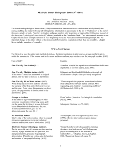
© Copyright 2026