
27783_157950_3_PB - VBN
Aalborg Universitet
Extracellular vesicles from glioblastoma cells as potential drug-delivery vehicles to
isotypic tumours
Gudbergsson, Johann Mar; Skov, Martin N.; Karred, Nichlas; Stolberg, Christian B.; Elbæk,
Kirstine J.; Pilgaard, Linda; Stensballe, Allan; Christiansen, Gunna; Gurevich, Leonid; Duroux,
Meg
Published in:
Journal of Extracellular Vesicles
DOI (link to publication from Publisher):
10.3402/jev.v4.27783
Publication date:
2015
Document Version
Publisher's PDF, also known as Version of record
Link to publication from Aalborg University
Citation for published version (APA):
Gudbergsson, J. M., Skov, M. N., Karred, N., Stolberg, C. B., Elbæk, K. J., Pilgaard, L., ... Duroux, M. (2015).
Extracellular vesicles from glioblastoma cells as potential drug-delivery vehicles to isotypic tumours. Journal of
Extracellular Vesicles, 4(Suppl.), 52, No. P-V-2. [27783]. 10.3402/jev.v4.27783
General rights
Copyright and moral rights for the publications made accessible in the public portal are retained by the authors and/or other copyright owners
and it is a condition of accessing publications that users recognise and abide by the legal requirements associated with these rights.
? Users may download and print one copy of any publication from the public portal for the purpose of private study or research.
? You may not further distribute the material or use it for any profit-making activity or commercial gain
? You may freely distribute the URL identifying the publication in the public portal ?
Take down policy
If you believe that this document breaches copyright please contact us at [email protected] providing details, and we will remove access to
the work immediately and investigate your claim.
Downloaded from vbn.aau.dk on: December 30, 2015
The Fourth International Meeting of ISEV
ISEV2015
Organized by ISEV and ISEV-Americas
Washington, D.C., USA, 23 – 26 APRIL, 2015
Abstracts
ISEV 2015 is organized by
The Local Organizing Committee:
Kenneth Witwer (Chair, Baltimore), Shilpa Buch (Omaha), Prasun Datta (Philadelphia),
Dolores Di Vizio (Los Angeles), Uta Erdbrügger (Charlottesville), Steven Jay (College Park),
Dimitrios Kapogiannis (Baltimore), Leonid Margolis (Bethesda) & Susmita Sahoo (New York)
Together with the Executive ISEV Board (2014 – 2016)
President: Jan Lötvall
Secretary General: Clotilde Théry
Treasurer: Fred Hochberg
Executive Chair Science / Meetings: Marca Wauben
Executive Chair Education: Yong Song Gho
Executive Chair Communication: Andrew Hill
Members at Large: Peter Quesenberry, Kenneth Witwer, Susmita Sahoo, Dolores
Di Vizio, Chris Gardiner, Edit Buzas, Hidetoshi Tahara, Suresh Mathivanan, Igor Kurochkin
Gold sponsors
Silver sponsors
Funding statement
Funding for this conference was made possible in part by R13 TR001278-01 (PI: Witwer,
Kenneth) from the NIH Common Fund, through the Office of Strategic Coordination/Office
of the NIH Director and National Center for Advancing Translational Sciences (NCATS).
The views expressed in written conference materials or publications and by speakers and
moderators do not necessarily reflect the official policies of the Department of Health and
Human Services; nor does mention by trade names, commercial practices, or organizations
imply endorsement by the U.S. Government.
Funding for this conference was provided in part by a grant from the National Science Foundation
(PI: Jay, Steven).
ABSTRACT
Scientific Program ISEV 2015 meeting
Thursday April 23, 2015
Oral Presentations
Registration
08:30-10:00
Welcome & networking coffee
08:30-10:00
Setting up posters (Poster sessions I, II, III, IV, V, VI, VII)
08:30-09:00
Sponsor exhibition
10:00-18:00
Ballroom D-E
Plenary Opening Session
Chairs: Jan Lötvall and Kenneth W. Witwer
10:00-12:00
James E. Rothman, Ph.D.
Wallace Professor of the Biomedical Sciences at Yale University
Principles of vesicle transport
Francis S. Collins, M.D., Ph.D.
Director, United States National Institutes of Health
Nurturing innovative science at NIH
Roundtable discussion
Moderator: Matthew Herper, Forbes
Journal of Extracellular Vesicles 2015. This is an Open Access article distributed under the terms of the Creative Commons Attribution-NonCommercial 4.0
International License (http://creativecommons.org/licenses/by-nc/4.0/), permitting all non-commercial use, distribution, and reproduction in any medium,
provided the original work is properly cited.
Citation: Journal of Extracellular Vesicles 2015, 4: 27783 - http://dx.doi.org/10.3402/jev.v4.27783
1
Thursday April 23, 2015
Networking lunch
12:15-13:15
Poster viewing sessions I, II, III, IV, V, VI, VII
Posters attended by authors
12:45-13:15
Poster walk by chairpersons, sessions I, II, III, IV, V, VI, VII
Posters attended by authors
14:00-15:00
2
Citation: Journal of Extracellular Vesicles 2015, 4: 27783 - http://dx.doi.org/10.3402/jev.v4.27783
Scientific Program ISEV 2015 meeting
Ballroom D
Symposium session 1A - EV biogenesis I
Chairs: Clotilde Théry and Stephen J. Gould
O-1A-1
ApoE regulates ESCRT-independent sorting on exosomes
and endosomal amyloid formation
Guillaume Van Niel1, Ptissam Bergam1, Aurelie Di Cicco2, Ilse Hurbain1,
Alessandra Lo Cicero1, Cecile Fort1, Florent Dingli3, Marie-Claude Potier4,
Leon Schurgers5, Damarys Loew3, Daniel Levy2 and Graça Raposo1
1
Department of Cell Biology, Curie Institute, Paris, France; 2Department of
Physical Chemistry, Curie Institute, Paris, France; 3Department of Mass
Spectrometry, Curie Institute, Paris, France; 4CNRS UMR7225, INSERM U1127,
UPMC, Institut du Cerveau et de la Moelle, Paris, France; 5Department of
BiochemistryVascular Aspects, Faculty of Medicine, Health & Life Science,
Maastricht University, Maastricht, The Netherlands
Introduction: Exosomes are generated within secretory multivesicular endosomes (MVEs) as intraluminal vesicles (ILVs). To serve specific
cellular functions, notably once secreted, ILVs are enriched with defined sets of proteins by various and still elusive sorting mechanisms
within MVEs. Pigment cells have tuned their MVEs to produce amyloid
fibrils derived from the protein PMEL. For this purpose, PMEL the
first protein reported as an ESCRT-independent cargo is sorted in a
CD63-dependent manner on ILVs that likely serve as potential seeding
platforms for PMEL amyloidogenesis. Contrary to amyloids such as
those associated with Alzheimer’s disease, PMEL amyloids are nontoxic and are functional as they serve as a scaffolding structure for the
synthesis of melanin. To better understand the mechanisms exploited
on ILVs to avoid potential toxicity during PMEL amyloidogenesis, we
have used exosomes as reporters of these endosomal processes.
Methods: For this purpose, we have characterized exosomes derived
from pigment cells by cryo-electron microscopy, mass spectrometry
and western blot. We then investigated the role of the intracellular
counterparts of exosomes, ILVS in cell lines and in vivo using siRNA,
western blotting and morphological analysis by electron microscopy.
Results: Characterization of exosomes derived from pigment cells
revealed the association of exosomes and ILVs with apolipoprotein
E (ApoE) and lipoparticles. We could show that ApoE is targeted to
endosomes in a CD63-dependent/ESCRT-independent manner and
facilitates the ESCRT-independent sorting of PMEL amyloidogenic
fragments onto ILVs. At the surface of ILVs, ApoE regulates the formation of mature fibrils in melanocytic cell lines and in pigment cells
in vivo. Summary/conclusion: These results established a clear molecular mechanism for ESCRT-independent sorting of PMEL. Moreover,
the novel evidence that lipoparticles are associated to exosomes
provides a breakthrough that might be exploited to reconsider the
respective roles of each extracellular particle in pathologies. Finally our
study establishes a paradigm for the mechanism by which ApoE, the
first genetic risk for early onset Alzheimer’s disease, regulates the
assembly of mature amyloid fibrils under benign and pathological
conditions.
O-1A-2
Prion protein regulates autophagy and endocytic trafficking
with essential roles for the biogenesis of extracellular vesicles
Marcos Dias, Bianca Teixeira, Bruna Rodrigues, Camila Arantes,
Martin Roffe, Glaucia Hajj and Vilma Martins
International Research Center, AC Camargo Cancer Center, Sao Paulo, Brazil
Introduction: Prion protein (PrPC) plays important roles in neuronal
physiology and in tumour biology. Primary cultures from PrPC knockout astrocytes (Prnp0/0) show a defect in the secretion of neurotropic
13:15-14:00
factors, and the protein and lipids content in conditioned medium
(CM) of these cells were reduced when compared to wild-type (WT)
astrocytes (Lima et al., J Neurochem. 103(6): 2,16476, 2007). Thus,
suggesting that PrPC regulates extracellular vesicles (EV) release.
Methods: EVs were isolated using ultracentrifugation and their size
and concentrations analyzed by NTA. Electron and confocal microscopies and biochemical analysis were performed to evaluate EV
and endocytic components. siRNA was used to knock down beclin1.
Results: Prnp0/0 astrocytes secrete lower EVs in CM than WT cells whilst
the reconstitution of PrPC expression in Prnp0/0 astrocytes restores the
levels of EV secretion. In astrocytes overexpressing PrPC, the number of
secreted EVs is higher than those secreted by WT cells. The same
differences were observed in CM from primary fibroblasts cultures and
in the blood circulation of these mice. The absence of PrPC causes a
delay in caveolin-mediated endocytosis and in the traffic of EGF-EGFR
to late endosomes/multivesicular bodies (LE/MVB). Prnp0/0 astrocytes
present altered MVB formation, LAMP1-stained lysosomal compartments were enlarged and shown an increased number of autophagosomes when compared with WT cells. Autophagy inhibition by beclin1
knockdown restored the levels of EVs released by Prnp0/0 cells.
Conversely, autophagy induction by serum starvation or rapamycin
treatment inhibited exosome release in WT cells. Summary/conclusion:
PrPC modulates endocytic pathways and impairs autophagy. In the
absence of PrPC, endosomes can be directed to fuse with autophagosomes contributing to the impairment in EVs biogenesis/release. The
cellular levels of PrPC can regulate the amount of secreted vesicles with
major roles in heath and disease.
O-1A-3
Rabs involved in extracellular vesicle biogenesis each affect multiple
different steps in endolysosomal and secretory trafficking in
Drosophila secondary cells
Clive Wilson1, Siamak Redhai1, Ben Kroeger1, Laura Corrigan1, Shih-Jung
Fan1, Mark Wainwright1, Ian Dobbie2, Aaron Leiblich1, Carina Gandy1,
John Morris1, Freddie Hamdy3 and Deborah Goberdhan1
1
Department of Physiology, Anatomy and Genetics, University of Oxford,
Oxford, United Kingdom; 2Department of Biochemistry, University of Oxford,
Oxford, United Kingdom; 3Nuffield Department of Surgical Sciences, University
of Oxford, Oxford, United Kingdom
Introduction: We have recently characterized a new in vivo system to
dissect the role of the endolysosomal and secretory pathways in
regulating the biogenesis of extracellular vesicles. It takes advantage
of the Drosophila male accessory gland (AG), which shares similarities
with the human prostate. A small subset of epithelial cells within
the AG called secondary cells (SCs) specifically produces vesicles,
which can be marked by a GFP-tagged form of the putative exosome
marker, human CD63. CD63-positive vesicles are made inside giant
(5 mm diameter) endolysosomal compartments and secreted into
the AG lumen in a process that requires the function of ESCRTs like
ALiX and Hrs, and of different Rabs associated with mammalian
exosome secretion, such as Rab11, Rab27 and Rab35. We investigated
how these Rabs alter membrane trafficking within SCs to affect
extracellular vesicle secretion. Methods: We have used molecular
genetic approaches in the fly combined with both confocal and
super-resolution (3D-SIM) microscopy to determine how knockdown
of specific Rabs alters trafficking and vesicle biogenesis pathways
in SCs. Results: Knockdown of Rab11, Rab27 or Rab35 with at least
two independent RNAi molecules affects EV secretion. However,
each Rab has very different effects on SC intracellular compartments.
Importantly, overexpression of dominant negative Rabs does not
produce these specific defects, suggesting that their effects are not
Citation: Journal of Extracellular Vesicles 2015, 4: 27783 - http://dx.doi.org/10.3402/jev.v4.27783
3
Thursday April 23, 2015
the result of simple loss-of-function. Super-resolution microscopy of
living glands reveals previously unappreciated complexity in endolysosomal trafficking events. For example, highly dynamic intraluminal vesicles and membranes are found inside endolysosomal
and secretory compartments, suggesting that EV trafficking to the
cell surface may not just involve late endosomal compartments.
Although neither Rab11 nor Rab35 appear to be expressed on the
surface of large acidic endolysosomes, when either is knocked down,
these large compartments do not appear to mature properly, and
vesicles accumulate inside. The numbers of non-acidic secretory
compartments are also affected by these two treatments, but in
completely opposite ways, with the secretory compartments being
4
almost entirely lost after Rab11 knockdown. Summary/conclusion:
Our data reveal that knockdown of specific Rabs alters several
different membrane-bound compartments in SCs. Therefore, though
at least one key effect of knockdown may be on a specific endosomal trafficking pathway to the plasma membrane, the defect in EV
secretion may primarily be explained by indirect effects on other
parts of the secretory or endolysosomal systems. The interpretation
of the effects of Rab knockdown on EV secretion, therefore, requires
careful analysis of trafficking in compartments in addition to
endolysosomes.
Citation: Journal of Extracellular Vesicles 2015, 4: 27783 - http://dx.doi.org/10.3402/jev.v4.27783
Scientific Program ISEV 2015 meeting
Ballroom E
Symposium session 1B - EVs and stem cells
Chairs: Susmita Sahoo and Weian Zhao
13:15-14:00
Introductory Lecture: Stefano Pluchino, M.D., Ph.D.
University of Cambridge
O-1B-1
Exosomes for heart failure associated with Duchenne muscular
dystrophy in mdx mice and in human iPS cells
Mark Aminzadeh1, Prasanthi Durvasula1, Rachel Tobin1, Rachel Smith1,
Xuan Guan2, Martin Childers2, Linda Marbán1 and Eduardo Marbán1
1
Cedars-Sinai Heart Institute, Los Angeles, CA, USA; 2Institute for Stem Cell and
Regenerative Medicine, University of Washington, Seattle, WA, USA
Introduction: Cardiosphere-derived cells (CDCs) promote cardiomyogenesis and angiogenesis, while inhibiting oxidative stress, inflammation and fibrosis, in ischemic and non-ischemic cardiomyopathy.
Recent mechanistic investigations implicate exosomes as central
mediators of CDCs’ beneficial effects. The mdx mouse model of
Duchenne muscular dystrophy develops cardiomyopathy due to dystrophin deficiency and the resultant intense oxidative stress, inflammation and apoptosis. We have shown that CDCs are salutary in this
model; here we tested the hypothesis that transplantation of CDC
exosomes (CDC-XO) may generate similar beneficial effects. Methods:
A total of 78 mice, including the mice recruited for assessment of CDC
efficacy, were studied at a point when global cardiac dysfunction was
already evident by echocardiography. CDC-XO (70 mg) or vehicle-only
were injected intramyocardially in 4 left ventricular (LV) sites in 10month-old mdx mice. Results: Similar to CDC-treated mdx mice, LV
ejection fraction markedly improved over 3 months after treatment
with CDC-XO compared to vehicle-treated mice (68.7392.82 vs.
5793.05; p0.011). The functional improvement was associated
with reduced collagen I and III deposition and enhanced cardiomyocyte proliferation in the CDC-XO-treated mouse hearts. To assess the
role of CDC-exosomes on mitochondrial function, which is impaired in
Duchenne cardiomyopathy, cardiomyocytes derived from human
Duchenne iPS cells (hDMD-CM) were primed with CDC-XO and
assessed for mitochondrial respiratory capacity 1 week after. Oxygen
consumption rate was impaired in control hDMD-CM, but normalized in hDMD-CM that had been treated with CDC-XO (p B0.05).
Summary/conclusion: Cardiac function improved in mdx mice treated
with CDC-XO, accompanied by reduced cardiac collagen content and
fibrosis, and augmented cardiomyogenesis. Priming hDMD-CM with
CDC-XO markedly enhanced their respiratory capacity. Thus, CDCexosomes mimic CDCs’ beneficial effects in the heart failure associated with Duchenne muscular dystrophy. CDCs themselves, and
their exosomes, are viable therapeutic candidates for Duchenne
cardiomyopathy.
Introduction: Bone marrow mesenchymal stem cells (MSC) provide
unique environmental cues to support homeostatic and regenerative
function in the hematopoietic stem/progenitor cell (HSPC) pool. MSC
are potent producers of vesicles, but their role in regulating hematopoiesis has not been elucidated. Here, we hypothesized that MSC
exosome trafficking impacts cell fate decision making and proliferation of HSPCs. Methods: Murine bone marrow-derived MSC (plastic
adherent, osteocyte/adipocyte differentiation, CD45, lineage, CD44,
Sca-1, CD105, CD29), HSPC (C-kit/ Sca-1/ Lineage),
CFU-c assay, qRT-PCR, inflammatory cytokine PCR microarray, flow
cytometry, and Ki67 cell cycle. Results: After 48 hours of in vitro
exposure to MSC exosomes, we observed a 2-fold increase among Ckit/ Sca-1/ Lineage- (KSL) progenitors, with an 80% increase in the
KSL/CD48-/CD150 (SLAM) stem cell populations, compared to the
vesicle-free media control. Consistent with expansion of the multipotent progenitor cell pool, combined Hoechst/Ki-67 cell cycle
analysis showed that exosome-exposed cells were more actively
dividing compared to control. That loss of quiescence upon MSC
exosome exposure was reflected in the downregulation of critical selfrenewal genes (Bmi-1, C-myc, Mef/Elf4) and a significant increase
(p 0.01) in the number of progenitor CFU colonies. Furthermore,
colony formation of pluripotent progenitors (CFU-GM) was decreased
by 26%, against an increase in committed myeloid progenitors (CFUG, 74%; BFU-E, 92%) in exosome-exposed cells. Indeed, extended
exosome exposures resulted in comparative exhaustion of the colonyforming progenitors. Correlative experiments are now ongoing. To
understand the mechanistic basis of our observations, we performed
a survey of cytokine activity in exosome-exposed HSPC. Our experiments revealed the activation of inflammatory regulation of HSPC
fate, and qRT-PCR analysis confirmed the upregulation of type I
interferon signaling pathway members (Stat1, Irf1, Irf9) in exosome
exposed HSPCs. Finally, molecular analysis of MSC exosomal content
revealed an enrichment of a panel of microRNA, which we are
currently investigating for dysregulation of interferon signaling.
Summary/conclusion: Taken in aggregate, these data suggest that
HSPC activation occurs via MSC exosome trafficking of microRNA and
involves the upregulation of type I interferon-signaling pathways.
O-1B-2
Activation and reprogramming of hematopoietic stem and progenitor
cell fate by MSC exosomes
Natalya A. Goloviznina, Santhosh Chakkaramakkil Verghese and
Peter Kurre
Department of Pediatrics, Oregon Health and Science University, Portland,
OR, USA
Citation: Journal of Extracellular Vesicles 2015, 4: 27783 - http://dx.doi.org/10.3402/jev.v4.27783
5
Thursday April 23, 2015
Ballroom F-H
Symposium session 1C - Standardization and organization of EV research
Chairs: Alain Brisson and Louise Laurent
13:15-14:00
Introductory Lecture: Rienk Nieuwland, Ph.D.
Medical Centre of the University of Amsterdam
O-1C-1
Bioinformatics analysis of extracellular vesicles using FunRich and
Vesiclepedia
Suresh Mathivanan, Shivakumar Keerthikumar and Mohashin Pathan
Department of Biochemistry, La Trobe University, Melbourne, Australia
Introduction: In the last decade, extracellular vesicles (EVs) have
attracted significant interest among scientists for their proposed role
in intercellular communication, as reservoirs for disease biomarkers
and as targeted drug delivery vehicles. Since its initial discovery,
multiple groups have reported the secretion of EVs and characterized
the transcriptomic, proteomic and lipidomic content of EVs. As
high-throughput techniques including proteomics become more
accessible to individual laboratories, there is an urgent need for a userfriendly bioinformatics analysis system. Here, we describe FunRich, an
open access, standalone functional enrichment and network analysis
tool. FunRich is designed to be used by biologists with minimal or no
support from computational and database experts. Methods: FunRich
tool was developed using Visual C#. Hypergeometric distribution test
was performed to check the statistical significance of enriched and
depleted terms. In addition, Bonferroni and Benjamini-Hochberg (BH)
aka FDR (false discovery rate) method was also implemented to
correct for multiple testing. Results: Using FunRich, users can perform
functional enrichment analysis on background databases that are
integrated from heterogeneous genomic and proteomic resources
(1.5 million annotations). Besides default human-specific FunRich
database, users can download data from the UniProt database which
currently supports 20 different taxonomies against which enrichment
analysis can be performed. Moreover, the users can build their own
custom databases and perform the enrichment analysis irrespective
of organism. In addition to proteomics datasets, the custom database
allows the tool to be used for genomics, lipidomics and metabolomics datasets. Thus, FunRich allows complete database customization and thereby permits the tool to be exploited as a skeleton for
enrichment analysis irrespective of the data type or organism used.
FunRich is user-friendly and provides graphical representation (Venn,
pie charts, bar graphs, column, heatmap and doughnuts) of the data
with customizable font, scale and colour (publication quality).
Summary/conclusion: FunRich is designed to be used by biologists,
biochemists and geneticists with minimal or no support from
computational and database experts. Data sets from EV studies can
be analysed using this software to identify signaling pathways that
may underpin the pathophysiological role of EVs.
Introduction: The development of minimally invasive biomarkers to
assess disease risk, direct treatment or prevent secondary medical
complications has the ability to profoundly change patient care in the
coming decade. To develop, replicate and validate these biomarkers,
we envision the necessary coordination of multiple steps to optimize
yields of mRNAs and miRNAs emerging from SOPs for specimen
acquisition and processing as well as seamless sharing of collected
biospecimens to power these efforts. Each step in this process has the
potential to exhaust scarce disease-specific biofluids. Equally problematic is the comparative analysis of these biomarkers from patients
with specific medical conditions and normative populations. We
hope to use these comparative analyses to identify optimal ways to
enrich exRNAs of particular interest for the central nervous system.
Ultimately this can be best facilitated through the sharing of IRB
consent language, the use of universal MTAs and intellectual property
agreements and biofluids with appropriate acknowledgement of the
authors and funding agencies. We have piloted the database specimen acquisition and distribution of this shared NIH Common Fund
biorepository within centers, whose aims involve the evaluation of
cerebrospinal fluid for the diagnosis of primary tumours of brain
(UCSD, MGH); subarachnoid hemorrhage (BNI, TGen); Alzheimer’s
disease and Parkinsonian syndromes (OHSU). Methods: To implement
this collaboration we have proposed: (a) comparative studies of RNA
yields resulting from 4 preparative approaches applied to pooled CSF
specimens; (b) comparisons of 3 analytic approaches (RNA Seq;
Qiagen qPCR; Nanostring) to detect disease-specific amplifications or
mutations of genes of interest. Refinements to the database and
repository will then be made after which, it will be offered as a
resource to the Data Management and Resource Repository (DMRR)
of the NIH Director’s Office Extracellular RNA Communication
Consortium. Results: The exRNA biospecimen virtual repository is a
collective effort of the Resource Sharing Working Group and the
DMRR to facilitate tracking of biospecimens. The repository is
developed on the existing Genboree KnowledgeBase (GenboreeKB),
a distributed and extensible infrastructure. Genboree REST APIs
provide extensibility and enable controlled data sharing across
physically distributed nodes using Linked Data technologies. The
pilot biorepository provides inventory tracking of various biospecimens from the CSF group. Users see a Dashboard with a grid of
specimens from specific diseases and samples from fluids of donors.
Summary/conclusion: During an oral presentation we will provide the
rationale and structure of the biorepository and patient safety
standards and then demonstrate file-encoding, acquisition, bulk file
acquisition on a ‘‘real-time basis’’ as a model for synergies within the
ISEV community.
O-1C-2
ExRNA Biospecimen virtual repository: the CSF working group
demonstration project
Sai Lakshmi Subramanian1, William E. Butler2, Bob Carter3, Matthew
Huentelman4, Kendall Van Keuren-Jensen4, Aleksandar Milosavljevic1,
Joseph F. Quinn5, Julie Saugstad5 and Fred H. Hochberg2
1
Baylor College of Medicine, Jewish Institute for Research, Houston, TX, USA;
Massachusetts General Hospital, Boston, MA, USA; 3UC San Diego, San Diego,
CA, USA; 4Translational Genomics Research Institute, Phoenix, AZ, USA;
5
Oregon Health & Science University, Portland, OR, USA
2
6
Citation: Journal of Extracellular Vesicles 2015, 4: 27783 - http://dx.doi.org/10.3402/jev.v4.27783
Scientific Program ISEV 2015 meeting
Ballroom D
Oral with poster A
Chairs: Dae-Kyum Kim and Fatemeh Momen-Heravi
P-IV-16
Differential detergent sensitivity of extracellular vesicle
subpopulations
Xabier Osteikoetxea1, Barbara Sódar1, Andrea Németh1, Krisztina Pálóczi1,
Katalin Szabó-Taylor1, Krisztina Vukman1, Ágnes Kittel2, Éva Pállinger1 and
Edit I. Buzas1
1
Department of Genetics, Cell- and Immunobiology, Semmelweis University,
Budapest, Hungary; 2Institute of Experimental Medicine, Hungarian Academy
of Sciences, Budapest, Hungary
Introduction: Earlier work from our research group provided evidence
that extracellular vesicles can be differentiated from protein aggregates (e.g. immune complexes) by low concentration detergent
treatment in flow cytometric experiments. Methods: In the present
study, we investigated the sensitivity of different subpopulations of
extracellular vesicles (including exosomes, microvesicles and apoptotic bodies) to detergent treatment using tunable resistive pulse
sensing (qNano), flow cytometry and transmission electron microscopy. Results: We found that tunable resistive pulse sensing detection
of extracellular vesicle subpopulations was compatible with differential detergent lysis. We determined the detergent concentrations
that lysed each of the different vesicle subpopulations. Our qNano
data show that microvesicles and apoptotic bodies are more sensitive
to detergent lysis, while the signals of exosomes disappear only at
higher detergent concentrations. The qNano findings are also in line
with the disappearance of fluorescent labelling of the extracellular
vesicles in flow cytometry. Summary/conclusion: Taken together, our
data suggest that the combination of differential detergent lysis with
tunable resistive pulse sensing or flow cytometry may provide useful
for simple and fast validation of the vesicular nature of the detected
particles. Furthermore, this approach may also provide information
about the vesicle subpopulation being studied.
P-IV-15
A novel method to validate fluorescence nanoparticle tracking analysis
for phenotyping extracellular vesicles
Rebecca Dragovic1, Gavin Collett1, Patrick Hole2, Christopher Redman1,
Ian Sargent1 and Dionne Tannetta1
1
Nuffield Department of Obstetrics & Gynaecology, University of Oxford,
Oxford, United Kingdom; 2Malvern Instruments, Amesbury,
United Kingdom
Introduction: Fluorescence Nanoparticle Tracking Analysis (fl-NTA) has
the potential to enable phenotyping of extracellular vesicles (EV)
down to 50 nm in size, which is not possible using conventional
flow cytometry. However, unlike flow cytometry, fl-NTA cannot
currently measure EV in scatter and fluorescence modes simultaneously. To accurately determine the number of fluorescence positive
EV, the operator must establish optimal camera levels for each mode.
To do this requires a ‘‘standard’’ containing a known amount of EV
positive for the antigen being studied. We aimed to prepare an EV
standard to enable optimization of fl-NTA settings for quantification
of placental alkaline phosphatase (PLAP a trophoblast marker)
positive EV in preparations of syncytiotrophoblast (STB) microvesicles
(STBMV) and exosomes (STBEX) from human placentas. Methods:
STBMV and STBEX were prepared using a dual placental perfusion
model and differential centrifugation. Pools of STBMV and STBEX were
prepared (4 samples/pool) and the percentage of PLAPve EV
determined using anti-PLAP antibody Dynabead depletion. Pools
were also labelled with anti-PLAP-Qdot605 or IgG1-Qdot605 control
15:00-16:00
and measured using fl-NTA in scatter and fluorescence modes using
camera levels 1214. Scatter and fluorescence concentration measurements were used to determine the percentage of PLAPve EV
and compared to the percentage of PLAPve EV removed by
immunodepletion. Optimal camera settings were defined and used
to analyse individual preparations (n8). Results: Using anti-PLAP
Dynabeads, 59.895.2% and 51.693.4% of EV in STBMV and STBEX
pools respectively were PLAPve. fl-NTA of STBMV and STBEX pools
showed 48.593.0% and 17.993.3% PLAPve respectively. PLAP
ve EV in individuals ranged from 18.8 to 67.0% (STBMV) and 3.2
51.7% (STBEX). A positive correlation (r2 0.737, p B0.01) was found
between STBEX size and PLAP positivity (%). Summary/conclusion: The
use of anti-PLAP immunobead depletion provided the necessary
standard to set fl-NTA settings. We show that fl-NTA is a robust
method for determining the percentage of PLAPve EV in STBMV.
However, fl-NTA is not as sensitive as Dynabead capture for STBEX
detection using this particular marker, possibly due to the smaller
STBEX having less surface PLAP. This same method could be used to
validate settings for all fl-NTA immunolabelling.
P-IV-14
A new fluorescent probe for identifying extracellular vesicles
Joshua Welsh1, Nicola Englyst1, Judith Holloway1, James Wilkinson2 and
David Smith3
1
Department of Medicine, University of Southampton, Southampton,
United Kingdom; 2Department of Engineering, University of Southampton,
Southampton, United Kingdom; 3Anaesthetics, University Hospitals
Southampton, Southampton, United Kingdom
Introduction: Flow cytometry methods for sub-micron particle quantification require higher precision in identifying extracellular vesicles
from various sources of background noise. Currently many extracellular vesicles, particularly microvesicles (1001,000 nm), are commonly identified with procoagulant markers annexin V or lactadherin
but both have limitations. Whilst membrane loading techniques
are possible, long incubation periods and wash steps are required
making their practicality in large studies is questionable. They do
however benefit from being able to use a fluorescent threshold
making higher precision acquisition possible. They also have the
benefit in distinguishing vesicles from noise. Here, we describe the
use of a new generation of fluorescent markers capable of loading
extracellular vesicles with the advantages of a short incubation
period, no wash steps and the unique property of providing a
ratiometric indication of negative and neutral charged phospholipids
simultaneously. Methods: Tewnty microlitres of extracellular vesiclerich samples from 8 ml whole blood isolated in citrated tubes, and
then centrifuged at 1,500g2, were suspended in 50 ml HBS or
HBS2 mM Ca2 and then stained with either 1 ml of 200 mM VRD
and 5 ml of 25 mg/ml Annexin V or 10 mg/ml Lactadherin. Within
these samples, microvesicles were identified with platelet markers
CD41 and CD42, whilst lipoproteins were identified using ApoE
antibodies. Results: Preliminary data show that lactadherin stains a
higher number of microvesicles when compared to annexin V. There
are, however, multiple lactadherin positive populations, most likely
due to the fact that lactadherin binds to integrins such as CD61. As
well as not having phosphatidylserine specificity, lactadherin staining
shows a higher amount of noise when compared to VRD, which has
little/no observable background noise at the 160 nm calibration.
The VRD has a distinct linear correlation of size to fluorescent intensity, unlike lactadherin. Staining of microvesicles is equally as
effective as lactadherin. We have shown that the VRD not only
stains microvesicles but also lipoproteins identified as ApoE positive
Citation: Journal of Extracellular Vesicles 2015, 4: 27783 - http://dx.doi.org/10.3402/jev.v4.27783
7
Thursday April 23, 2015
events. The VRD, due to having dual emission peaks at 530 nm or
580 nm depending on lipid exposure, can indicate whether vesicles
are primarily negatively charged phospholipids or neutrally charged
phospholipids. Summary/conclusion: From the results above, VRD
proves to be superior as a new generation dye for identifying
extracellular vesicles. We believe that VRD has huge potential for
future microvesicle studies due to its short incubation time, little/no
background noise, and membrane specificity. It matches lactadherin
for staining of microvesicles but without the draw backs of unspecific binding and blocking of integrins. Using VRD in future
microvesicle studies will improve detection accuracy and throughput
by allowing use of fluorescent thresholds over SSC/FSC thresholds,
which to date have shown the highest accuracy and allow lower
sizes of extracellular vesicles to be detected.
P-IX-7
Systematic comparative lipidomic and proteomic analysis of exosomes:
a path toward reverse engineering and artificial exosome development
Reka Haraszti1, Marie Didiot1, Andrew Coles1, Matthew Hassler1,
Julia Alterman1, John Leszyk2, Scott Shaffer2, Hannah Rockwell3,
Michael Kiebish3, Neil Aronin4 and Anastasia Khvorova1
1
RNA Therapeutics Institute, University of Massachusetts Medical School,
Worcester, MA, USA; 2UMMS Proteomics Core, University of Massachusetts
Medical School, Worcester, MA, USA; 3BERG LLC, Framingham, MA, USA;
4
Department of Medicine, University of Massachusetts Medical School,
Worcester, MA, USA
Introduction: Extracellular Vehicles (EVs) are believed to be involved
in native small RNA cell-to-cell transfer and signalling and are being
explored as a potential novel formulation for therapeutic oligonucleotide delivery. The mechanistic underpinning of how EVs traffic to
target cells and which components of the vesicles are essential for
trafficking is unknown. EVs are usually derived from cell lines, and
their potential mass production for therapeutic application is limited
by cell-based manufacturing methods, with no quantitative QC
parameters established. To realize the full potential of exosomes as a
therapeutic oligonucleotide formulation, it is necessary to define the
functional characteristics essential and responsible for efficient
cellular delivery. Methods: The exosomes and corresponding cells
were purified (n 3) from different origins, and their lipidomic and
proteomic content was evaluated using LC-MS/MS. Artificial lipid
vesicles based on the exosome lipidomic profile were engineered,
and their cellular trafficking and tissue distribution profiles were
evaluated. Results: Systematic lipidomics detected 19 structural
lipids. Surprisingly, exosomes were characterized by a specific lipidomic profile and were enriched in sphyngomyelin, glycolipids,
ceramide and cholesterol-esters and depleted in triacyl-glycerol and
coenzyme Q. Alteration of the lipid profile affected the cellular
trafficking and tissue distribution profiles of the artificial exosomes
engineered based on native exosome profiles. Proteomics identified
2,394 proteins, with 223 being present only in cells, 85 in exosomes
and 19 in microvesicles. Further studies will be focused on defining
the protein exosome components essential for trafficking. Summary/
conclusion: Preliminary analysis showed distinct protein and lipid
profiles of exosomes. This information may help engineer artificial
exosomes as delivery vesicles of therapeutic oligonucleotides and
help define a QC matrix for exosome manufacturing.
P-III-1
Specific isolation of tumour-derived extracellular vesicles using
microfluidic technologies
Eduardo Reátegui1, Charles P. Lai2, Kristan van der Vos2, Mahnaz Zeinali1,
Xandra O. Breakefield2 and Shannon L. Stott1
1
Department of Surgery, Massachusetts General Hospital, Charlestown, MA,
USA; 2Department of Neurology, Massachusetts General Hospital, Charlestown,
MA, USA
8
Introduction: Extracellular vesicles (EVs), lipid vesicles present in
blood and other biofluids, have sizes that vary from 100 nm to a few
microns and carry rich biological cargo that includes proteins, mRNA,
microRNA and DNA. Through their content, EVs preserve the
transcriptome information of their parent cells. Thus, they serve as a
unique nich of opportunities for the development of cancer diagnostic
tools. However, EVs have not seen widespread clinical testing due to
limitations in isolation technologies that rely on ultracentrifugation
and precipitations processing that yield low-throughput outcomes,
and a lack of tumour-EV specificity. Here we present a microfluidic
platform with a temperature responsive nanointerface for tumourspecific recovery of exosomes through immunoaffinity immobilization. Methods: The microfluidic device consists of a ridged geometry
that induces mixing and promoting collisions between exosomes and
the antibody functionalized walls of the device. The nanointerface
consisted of an ultra-thin (135 nm) gelatin membrane functionalized
with nanoparticles that created a nanotexture to maximize EV surface
interactions. To mimic conditions present in patient samples, we used
engineered exosomes derived from human Gli36 glioblastoma cells
that stably express the fluorescent (PalmGFP/tdTomato) and bioluminescent EV reporter (EV-GlucB), and subsequently spiked them in
healthy human serum. Results: Using confocal microscopy, we
identified tumour-specific exosomes at the surface of the device that
were capture using anti-cetuximab, an antibody for EGFR. Our system
was able to isolate 21% of tumour-derived spiked exosomes at
processing flow rates of 1 ml/h. Moreover, the temperature sensitivity
of the nanocoating was used for the recovery of EVs on demand from
the surface of the microfluidic device after heating at 378C for 10
minutes. We also performed molecular analysis to determine the total
amount of RNA present in the isolated EVs. We detected RNA from the
isolated EVs that indicated the effectiveness and sensitivity of our
method. From this amplified RNA, deep sequencing or transcriptome
analysis is currently being performed to identify tumour-specific
microRNAs. Summary/conclusion: We have developed a microfluidic
technology that can be used for the specific isolation of tumourderived EVs, that can be applied to different type of cancers.
The responsive nanocoating, allowed the recovery of EVs on demand
to perform additional characterizations like imaging or size characterization. This unique system also will allow the identification
of mRNAs or microRNAs as biomarkers due to the purity of the EVs
population.
P-XVI-4
Diversity of extracellular vesicles in human ejaculates identified by
cryo-electron microscopy and tomography
Johanna Hoog and Jan Lotvall
Sahlgrenska Academy, University of Gothenburg, Göteborg, Sweden
Introduction: Extra-cellular vesicles in ejaculate are important for
male fertility as the vesicles capacitates the sperm, increases sperm
motility and modulates female genital tract immune cell response.
This population of extra-cellular vesicles found in ejaculate are mostly
of prostate (prostasomes) and epididymal (epididymosomes) origin
but vesicular contributions of other tissues are also present. Methods:
Ejaculates were plunge-frozen and visualized using cryo-electron
microscopy and tomography. Results: Here, we show that when EVs in
ejaculates are studied in unaltered plunge-frozen ejaculates using
cryo-EM and electron tomography, a high number of morphologically
distinct subgroups of extra-cellular vesicles are revealed. Some vesicles
have a smooth surface, whereas others show spiky membrane protrusions.
Cryo-electron tomographic 3D reconstructions show that vesicles
may contain one or more smaller vesicles and large sacs filled with
extra-cellular vesicles are also detected. A large population of tubular
structures, sometimes branched and up to 12 mm in length, was
also found. Frozen events of vesicular fusion (or budding) with the
spermatozoa were imaged, showing which vesicle sub-populations
can interact or originate from the spermatozoa membrane. Summary/
conclusion: Identification of these EV sub-populations provides
an inventory of the complex composition of extra-cellular vesicles
found in human ejaculate, their morphologies and prevalence.
Citation: Journal of Extracellular Vesicles 2015, 4: 27783 - http://dx.doi.org/10.3402/jev.v4.27783
Scientific Program ISEV 2015 meeting
This inventory is important to quality assure future extracellular
vesicle isolates. We are now faced with many new questions about the
role of the individual extracellular vesicle subgroups in human fertility.
P-XVI-12
Extracellular vesicles and small RNA of the cervicovaginal compartment
in Macaques
Dillon Muth, Melissa McAlexander, Erin L. Buchanan, Grace V. Hancock,
Lauren Ostrenga and Kenneth Witwer
Molecular and Comparative Pathobiology, Johns Hopkins School of Medicine,
Baltimore, MD, USA
Introduction: Macaques are an excellent model for human disease.
Comparatively little is known about extracellular vesicles (EV) and
their RNA cargo in the cervicovaginal compartment, especially, in
macaques. In this study, we compare EV and RNA in cervicovaginal
secretions and cervicovaginal lavage (CVL) of rhesus and pigtailed
macaques and report on a case study of a macaque with endometriosis. Methods: Four total cervicovaginal lavage (CVL) and vaginal
swab (VS) samples from rhesus and pigtailed macaques were utilized.
Nanoparticle tracking analysis was performed on whole CVL and EV
fractions enriched by stepped ultracentrifugation. Total RNA was
obtained with an optimized method for biofluids RNA extraction.
miRNAs were profiled with a medium-throughput stem-loop/hydrolysis probe qPCR platform in CVL, VS, and matched vaginal tissue
samples. Results: As in our work with archived human CVL, miRs-223
and -186 were among the most abundant in CVL of rhesus and
pigtailed macaques. Of more than 60 miRNAs detected consistently in
all samples, only miR-29b and miR-184 were approximately as
abundant in the EV as in total CVL. U6 was present almost exclusively
in whole CVL. On average, 1% of CVL miRNA copies were found in EV
but 2% of VS, likely due to better liberation of EV from mucus during
VS swab processing. No differences were apparent in RNA expression
in CVL or VS fractions from the 2 macaque species, although minor
expression differences were observed at the tissue level, interestingly
in levels of numerous let-7 family members. Comparison of particle
numbers in the various reproductively healthy samples and in an
individual rhesus with severe endometrial disease revealed lower
particle count in the diseased animal. Summary/conclusion: EV
analysis may be useful as a diagnostic tool in the cervicovaginal
compartment, especially in the case of reproductive tract disease.
Controlled studies of cervicovaginal EV and their RNA cargo are
merited to characterize the potential role of EV and specific small
RNAs as markers for any manifestation of reproductive tract disease.
P-IV-8
Pre-preparation of extracellular vesicles by electro-dialysis and
adsorption-elusion methods
Siwoo Cho1, Ju Hun Yeon2, Rhokyun Kwak2, Wonju Jo1, Ji Yoon Kang2,
Jaesung Park1 and Youhee Heo2
1
Mechanical Engineering, POSTECH, Pohang, South Korea; 2Center for
BioMicrosystems, KIST, Seoul, Republic of Korea
Introduction: Currently existing extracellular vesicle (EV) pre-preparation methods have been challenged. Ultracentrifuge has low yield,
consumes time and is laborious. Precipitating methods have coagulation problem on blood plasma and nano-particle contamination. In
immune-affinity-based separation, elimination of attached EV antibody is difficult, and this elimination causes irreversible damages to
whole EV or surface protein. In this study, we introduce electrodialysis
and elution system, which can achieve high yield, low buffer and
nano-particle contamination, and no coagulation. Methods: Separation chamber is composed of 2 poles of electrode at both the ends
and thin planar sandwich partitioned channels between the electrodes.
Polycarbonate track-etched membranes partition the chamber,
and sample flow goes into the channel covered by the membranes.
In the channels outside of the membranes, where electrodes are
located, waste buffer runs tangentially to membranes. As original
sample flow also crosses the channel tangentially, electric field is
applied to the direction of membranes. Particle streamline is deflected and particles approach the membrane surface. Because the
membranes have specific pore diameter, 30 nm, particles bigger than
30 nm get adhered on the membrane, but smaller particles get
through the pores. By this process, larger particles, EVs, remain until
the dialysis process gets over. After the dialysis, remaining sample is
collected and membranes are disassembled. Adhered EV on the
membranes is eluted by pipetting. Results: With protein solution, it
was checked that lower sample flow and higher voltage show higher
protein removal rate, but higher voltage causes heat damage on
the sample. The damage was detected where the average power
dissipation is higher than 0.5 W, so following tests were set to have
power limit and 0.04 ml/min sample flowrate. Comparison of EVs
yield and purity from mouse blood plasma was done for existing
methods: ExoQuick, ultracentrifuge and this system. According to
RNA recovery rate, yield of this system is similar to ExoQuick, but far
higher than ultracentrifugation. This system’s protein removal rate
is slightly better than ExoQuick. By dynamic light scattering test,
ExoQuick showed no clear peak on size distribution, but this system
showed clear size peak at the range of typical EVs, from 50200 nm.
Also, CD9 western blotting and RT-PCR of beta-actin were done. All
methods preserve surface proteins and RNA contents well. Summary/
conclusion: This electrodialysis-elution system showed fast and high
yield milliliter scale EV pre-preparation methods. Because of size
exclusive membrane dialysis, this system showed minimal nanoparticle contamination than existing methods. Also, the buffer used in
this test is a sucrose-based buffer in which cells can be cultured
without immune reaction or pH disturbance. Because of it, this
method may preserve surface property of EV than other chemical
based preparation methods.
P-XX-1
Role of extracellular vesicles and miRNA cargo in the control of
epididymal functions important to sperm maturation
Matthieu Rousseau1, Eric Boilard1, Alain Brisson2 and
Clémence Belleannée3
1
Infectiology and Immunology, Université Laval, Quebec, Canada; 2NaniBio
Technology, University of Bordeaux, Bordeaux, France; 3Obstetric Gynecology
and Reproduction, Université Laval, Quebec, Canada
Introduction: Among male infertility cases, 30% are idiopathic and
may result from impairment of sperm maturation in the epididymis.
This organ is a single tubule of the male reproductive tract in which
spermatozoa transit to acquire their motility and capacity to bind
and fertilize the oocyte. Because of its unique region-specific gene
expression pattern, the epididymis is a model of choice to study the
influence of regulatory factors on gene expression. Among these
factors, we assessed the contribution of extracellular vesicles and their
miRNA cargo from the male reproductive system in the control of
target cell functions via a mechanism of intercellular communication.
Methods: Our multidisciplinary research combines the use of microarray as well as electron microscopy and cytometry technologies on
human epididymal samples and transgenic mouse models, which
display severe male infertility phenotype due to impairment of miRNA
maturation or production. Results: We identified a strong correlation
between the expression of miRNAs and their target transcripts all
along the human epididymis, suggesting the involvement of miRNAs
in the regionalized pattern of expression in this organ. Among these
targets we identified several genes, including Claudin 10, that encode
proteins involved in the blood-epididymis barrier integrity. Furthermore, we explored the possibility that miRNAs could be involved
in a mechanism of extracellular communication between epididymal
epithelial cells via extracellular vesicles, referred to as epididymosomes. We identified 1,645 extracellular miRNAs associated with
extracellular vesicles from the proximal and distal regions of the
epididymidis. Importantly, we showed that extracellular miRNA repertoires from these two epididymal regions are distinct and differ
from the miRNA signature identified in epididymal epithelial cells,
Citation: Journal of Extracellular Vesicles 2015, 4: 27783 - http://dx.doi.org/10.3402/jev.v4.27783
9
Thursday April 23, 2015
suggesting that miRNA populations released from the cells are
selectively sorted in a region-specific manner. Furthermore, in vitro
assays performed on primary cultured epididymal cells indicated that
extracellular vesicles from the proximal region interact with distal
epithelial cells and may transfer their miRNA content to recipient cells,
as evidenced by our recent findings on transgenic mouse models.
Since the population of extracellular vesicles from the epididymis
are highly heterogeneous, we recently optimized the detection and
characterization of these extracellular vesicles by cytometry and cryoelectron microscopy. Summary/Conclusion: Altogether, our findings
reveal that extracellular miRNAs conveyed by extracellular vesicles
throughout the male reproductive tract could be efficient regulators
of epididymal functions and male fertility, and may be used for the
non-invasive diagnostics of idiopathic male infertility cases.
P-XIII-1
Isolation and characterization of Citrus limon L. derived nanovesicles:
potential use as antineoplastic agent
Stefania Raimondo1, Flores Naselli1, Simona Fontana1, Francesca
Monteleone1, Alessia Lo Dico1, Laura Saieva1, Giovanni Zito1, Anna Flugy1,
Mauro Manno2, Maria Antonietta Di Bella1, Giacomo De Leo1 and
Riccardo Alessandro1
Introduction: Circulating microvesicles (MVs) have emerged as a
promising surrogate for tissue-based markers, enabling non-invasive
and real-time disease monitoring. Purifying MVs for analyses, however, remains a challenging task, which often involves time-consuming
and extensive procedures (e.g., ultracentrifugation, multiple filtration). Methods: We herein present a new microfluidic platform for
MV isolation and enrichment from clinical samples. The system
utilizes acoustophoresis to size-selectively separate MVs. Interdigitated electrodes, patterned on piezoelectric substrate, were used to
generate standing surface acoustic wave (SSAW) inside a microfluidic
channel, and the resulting acoustic radiation force separated MVs
according to their size and density. The design and operation of
the device was optimized through numerical simulation. Results: The
system achieved 90% sorting yields, and the size cut-off could
be tuned in situ through controlling the acoustic power. We used the
system to collect MVs from pRBC (packed red blood cell) samples
as well as from cell culture media. The microfluidic-SSAW device
successfully isolated and enriched pure MV population, which was
confirmed by downstream molecular analyses. Summary/Conclusion:
Enabling label-free and continuous in-flow separation, the developed
platform could be an ideal tool for fast preparation of intact MVs.
P-XIII-17
1
Biopatologia e Biotecnologie Mediche, University of Palermo, Palermo, Italy;
Istituto di Biofisica, National Research Council, Palermo, Italy
2
Introduction: A large amount of data has been produced on the
release of extracellular vesicles from mammalians, but only a few
information is available on the presence of nanosize vesicles in plant
species. Here we focused on the isolation and characterization
of nanovesicles released from lemon juice in order to test their
antitumor efficacy. Methods: Nanovesicles were isolated from Citrus
limon L. juice by differential centrifugation and filtration steps.
Vesicles were characterized through electron microscopy, dynamic
light scattering analysis and proteomic approaches. Viability assays
and real-time PCR analysis were performed in cancer (colon, lung,
chronic myeloid leukaemia, multiple myeloma, liver) and normal cells
following treatment with nanovesicles. A tumour xenograft model
was used to test the in vivo effect of isolated nanovesicles. Results:
Isolated nanovesicles were identified as a homogenous population
(5070 nm) of exosomes-like particles and the analysis of vesicles
protein composition revealed the presence of proteins that were
homologous to mammalian exosomal proteins. PKH26-labelled
nanovesicles were internalized in a dose dependent way by cancer
cell lines. Furthermore, different cancer cell lines treated with citrus
nanovesicles showed a dose and time dependent decrease of
cell viability and a simultaneous increase of cell death pathways.
These results were also confirmed in an in vivo tumour xenograft
model. Summary/Conclusion: We isolated and characterized nanovesicles from edible Citrus limon with size and composition similar to
mammalian-derived exosomes. Furthermore we show an in vitro and
in vivo anti-proliferative and pro-apoptotic effect of these vesicles.
This study opens the possibility of using this natural plant-derived
nanovesicles as antineoplastic agents.
Label-free single exosome detection using frequency-locked
microtoroid optical resonators
Judith Su
Biology and Biological Engineering, Caltech, Pasadena, United States
Introduction: Recently exosomes have attracted interest due to
their potential as cancer biomarkers. We report the real time, labelfree sensing of single exosomes in serum using microtoroid optical
resonators. We use this approach to assay the progression of
tumours implanted in mice by specifically detecting low concentrations of tumour-derived exosomes. Methods: Our approach measures
the adsorption of individual exosomes onto a functionalized silica
microtoroid by tracking changes in resonant frequency of the
microtoroid. When exosomes land on the microtoroid, they perturb
its refractive index in the evanescent field and thus shift its
resonance frequency. Results: Through digital frequency locking,
we are able to rapidly track these shifts with accuracies of better
than 10 attometers (one part in 1011). Samples taken from tumourimplanted mice from later weeks generated larger frequency shifts
than those from earlier weeks. Analysis of these shifts shows a
distribution of unitary steps, with the maximum step having a height
of 1.2 fm, corresponding to an exosome size of 44 nm. Summary/
Conclusion: Our results demonstrate the development of a minimally
invasive tumour ‘‘biopsy’’ that eliminates the need to find and access
a tumour.
P-XX-5
Acoustic microfluidic system for microvesicle purification
Kyungheon Lee1, Huilin Shao2, Ralph Weissleder1,3 and Hakho Lee1
1
Center for Systems Biology, Massachusetts General Hospital, Boston, MA, USA;
Institute of Molecular and Cell Biology, Singapore; 3Systems Biology, Harvard
Medical School, Boston, MA, USA
2
10
Citation: Journal of Extracellular Vesicles 2015, 4: 27783 - http://dx.doi.org/10.3402/jev.v4.27783
Scientific Program ISEV 2015 meeting
Ballroom E
Oral with poster B
Chairs: Shivani Sharma and Hakho Lee
P-IX-17
Proteome-wide profiling of circulating exosomes for identification of
scirrhous gastric cancer biomarkers
Koji Ueda
Division of Biosciences, Functional Proteomics Center, Graduate School of
Frontier Sciences, University of Tokyo, Tokyo, Japan
Introduction: Recently biological significance and clinical utility of
exosomes have been extensively investigated. Particularly, in the field
of cancer diagnosis, exosomes are considered as ideal targets for
biomarker discovery due to their molecular characteristics. Indeed, a
set of molecules expressed in original tumour cells can be detectable
from exosomes in blood circulation. However, despite enormous
interest in exosomes, difficulties in exosome isolation from biological
fluids have significantly hindered effective discovery of biomarker
components. In the present study, we developed EV-Second (Extracellular Vesicle isolation by Size Exclusion Chromatography ON Drip
column) technology allowing rapid collection of highly pure exosomes in flow-through fraction by simple gravity drip in order to
identify early detection biomarkers for scirrhous gastric cancer.
Methods: We employed EV-Second columns to perform quantitative
proteome profiling of serum exosomes from 58 individuals (10
normal controls, 17 early-stage gastric cancer patients, 17 advancedstage gastric cancer patients, and 14 scirrhous gastric cancer patients)
using LC-MS/MS shotgun analysis. The proteome-wide datasets of
exosomes were quantitatively compared and statistically evaluated
on the Expressionist proteome server system to extract exosomal
biomarker candidate proteins. The identified biomarker candidates
were biologically assessed in vitro and in vivo. Results: Among 822
identified proteins by LC-MS/MS analysis, 299 serum-derived proteins
were removed based on GO. Student’s t-test (normal group vs. gastric
cancer group) using 523 exosomal proteins revealed that 13 proteins
were significantly up-regulated in gastric cancer-derived exosomes
(p B0.05, fold change 2.0, and valid value80%). We furthermore
focused on 3 of them and investigated for cancer-associated functions. These experiments strongly suggested the existence of an
exosome-driven positive feedback loop regulating tumour microenviroments. Summary/conclusion: Our EV-Second technology (commercially available from GL Science company) can provide the best
way for high-throughput exosomal biomarker screening studies
using multiple clinical specimens. The 13 biomarker candidate proteins in cancer-associated exosomes would be able to explain more
new insights concerning tumour progression or metastasis.
P-XII-7
Characterization of the functional role of rhabdomyosarcoma-derived
exosomes in tumour cell biology and investigation of their miRNA
cargo
Sandra E. Ghayad, Farah Ghamloush, Hussein Basma and Raya Saab
Department of Pediatrics and Adolescent Medicine, American University of
Beirut, Beirut, Lebanon
Introduction: Rhabdomyosarcoma (RMS) is an aggressive childhood
soft tissue tumour, with two distinct subtypes, alveolar (ARMS)
and embryonal (ERMS) histologies. ARMS is characterized by a more
aggressive clinical phenotype and a specific translocation PAXFOXO1, thought to contribute to its aggressive and metastatic
behaviour. Exosomes are small membranous vesicles secreted into
body fluids by multiple cell types, including tumour cells. Tumour
15:00-16:00
exosomes contain intact and functional proteins, mRNA and miRNA
that may alter the cellular environment to favour tumour growth.
Methods: We characterized exosomes from a panel of 5 RMS cell
lines and examined their functional effects on cell migration, invasion,
proliferation and viability. We characterized the miRNA cargo of ERMSand ARMS-derived exosomes using microarray profiling. Results: RMSderived exosomes significantly increased the cellular migration and
invasion of normal fibroblasts and had a positive effect on cell viability
of both fibroblasts and tumour cells. Expression array analysis showed
that exosomal miRNA clustered together well, and to a higher extent
than cellular miRNA, in both ARMS and ERMS cell lines. Commonly
enriched miRNA in exosomes derived from ERMS cell lines were
different from those in exosomes derived from ARMS cell lines. There
were only 2 miRNA in common among both ERMS and ARMS cell lines;
putative targets were found to be implicated in cancer and inflammation. Summary/conclusion: RMS exosomal cargo results in specific
effects on cell biology, enhancing invasive potential of recipient cells.
Commonly enriched miRNA in exosomes of ERMS cells are separate
from those of ARMS cells, possibly defining potential biomarkers for
ERMS and ARMS, respectively. Moreover, these enriched miRNA may
contribute to the paracrine signaling specific to each tumour subtype.
Current work is focused on analysis of the effects of the identified
miRNA on paracrine signaling in RMS, and on their validation as
possible biomarkers.
P-XII-8
Identification of optimal culturing conditions for studying exosomes
from normoxic and hypoxic colorectal cancer cell lines
Nirujah Sivarajah1,2, Anne Hansen Ree1,2, Erta Kalanxhi2, Kathrine Røe
Redalen2 and Karianne Risberg2
1
Clinical Medicine, University of Oslo, Oslo, Norway; 2Department of Oncology,
Akershus University Hospital, Lorenskog, Norway
Introduction: Tumour hypoxia (oxygenation deficiency) contributes
significantly to treatment resistance, metastatic progression and poor
survival in colorectal cancer (CRC) and evidence supports a central
role of exosomes in the aggravated biology caused by tumour
hypoxia. In this study, we aimed to identify the optimal culturing
conditions for the study of exosomes in normoxic and hypoxic CRC
cell lines. Methods: Growth characteristics were investigated in the
CRC cell lines HCT 116, RKO, LoVo, and HT-29 under normoxia
(21% O2) and hypoxia (0.2% O2), using 4 different cell culture media:
RPMI-1640 with 10% fetal bovine serum (FBS), RPMI-1640 with 10%
exosome-deprived FBS, RPMI-1640 with bovine serum albumin (BSA),
and serum-free RPMI-1640. Adherent cells were washed thrice with
phosphate-buffered saline before media were added and cells were
exposed to normoxia or hypoxia for 2448 hours. Cell confluency was
determined by visual inspection; cell viability by a proliferation assay
(MTS); cell doubling-time by cell count; and cell cycle progression by
flow cytometry. Results: After 24 hours, the viability of normoxic and
hypoxic HCT 116, RKO and LoVo cells was increased at all conditions.
In contrast, for normoxic and hypoxic HT-29 cells, both serum-free
and BSA-containing medium caused a decrease in cell viability. The
doubling time of normoxic and hypoxic RKO cells was 1623 hours
and for LoVo cells 1935 hours depending on the condition. For
normoxic HCT 116 and HT-29 cells the doubling time was 1734
hours depending on the condition. For hypoxic HCT 116 and HT-29
cells cultured in serum-free medium, estimated doubling time was 96
and 66 hours. Cell cycle analysis revealed that normoxic and hypoxic
RKO and LoVo cells in serum-free medium arrested in G1 phase.
Summary/conclusion: Bovine exosomes (in FBS) and exosomes from
cells arrested in the G1 phase of the cell cycle may impinge on
Citation: Journal of Extracellular Vesicles 2015, 4: 27783 - http://dx.doi.org/10.3402/jev.v4.27783
11
Thursday April 23, 2015
downstream analyses. Hence, our data suggested that RPMI-1640
with BSA is the optimal culturing medium for studying exosomes in
normoxic and hypoxic CRC cells. The HT-29 cell line was found
ineligible for exosome studies.
P-XII-11
Tumour cells and macrophages: can mutant p53 be the matchmaker
using exosomes?
Tomer Cooks1, Ioannis Pateras2, Keval Patel3, Nitzan Rosenfeld3, Vassilis
Gorgoulis2 and Curtis C. Harris1
1
Lab of Human Carcinogenesis, NCI, Bethesda, MA, USA; 2University of Athens,
Athens, Greece; 3Cambridge University, Cambridge, United Kingdom
Introduction: Both epithelial mutations in p53 as well as the presence
of tumour-associated macrophages (TAMs) are hallmarks of solid
tumours. We hypothesized that an epithelial cell harboring mutant
p53 (mutp53) can educate the adjacent macrophages to be polarized towards a specific macrophage subset promoting tumorigenesis, eventually resulting with a TAMs population inside a tumour.
Methods: By designing a co-culture system, we incubated human
primary monocytes together with colorectal cancer (CRC) cells
differing in their p53 status. Relevant macrophages markers were
evaluated on RNA level (qPCR) and protein level (ELISA, Flow
cytometry). In addition, co-cultured macrophages were subjected
to various functional assays (phagocytosis, migration, invasion). In an
attempt to confirm clinical relevance, samples from a cohort of
human CRC patients were analyzed using genomic and immunohistochemical methods. To identify the interaction between the tumour
cells and the macrophages, we isolated exosomes from the CRC cells
and subjected them to a Nanostring analysis to learn about their
microRNAs composition. Results: When monocytes were matured to
become macrophages while exposed to tumour cells harbouring
mutp53, they became polarized towards a distinguished subset of
macrophages characterized by TAMs-related markers. These findings
were in high correlation with samples taken from CRC patients
sequenced with mutp53 tumours but not wild-type (WT) p53
tumours. Investigating the exchange of messages between the
2 entities via exosomes revealed a mutp53-specific miRs signature
led by miR-1246 which could be incorporated into adjacent macrophages promoting the TAM phenotype and creating an invasive
front together with tumour cells. Summary/conclusion: This study is
the first to show a non-cell-autonomous role played by mutant p53
the most common form of mutation found in human cancers.
Deciphering the intricate microenvironmental regulation shared by
the tumour cell and its surrounding macrophages may lead to
breakthroughs in prognosis, diagnosis (bio-markers) and therapeutics aiming to target the key molecules involved.
P-XII-15
Functional analysis of extracellular vesicles as a novel regulatory agent
of scirrhous type gastric cancer microenvironment
Yutaka Naito1, Masakazu Yashiro2, Kosei Hirakawa2, Wataru Yasui3 and
Takahiro Ochiya1
1
Division of Molecular and Cellular Medicine, National Cancer Center Research
Institute, Tokyo, Japan; 2Department of Surgical Oncology, Osaka City
University Graduate School of Medicine, Osaka, Japan; 3Department of
Molecular Pathology, Hiroshima University Institute of Biomedical and Health
Sciences, Hiroshima, Japan
Introduction: Scirrhous type gastric cancer (GC) is highly metastatic
and characterized clinically by rapid disease progression and poor
prognosis. Therefore, better understanding of the pathological and
biological basis of scirrhous type GC is necessary to improve
diagnosis and treatment. Extracellular vesicles (EVs) are small membrane vesicles that are secreted by various cell types including
cancer cells and surrounding stromal cells. Evidence support that
some components of EVs derived from cancer stroma, including
12
microRNAs (miRNAs), function to promote the progression, growth
and spread of cancer. However, the precise mechanisms controlling
cancer microenvironment are not well understood. In the present
study, we aimed to investigate the difference of miRNA expression in
EVs between cancer stromal fibroblasts and normal fibroblasts (NFs)
and identify the specific extracellular miRNAs of cancer stromal
fibroblasts (CaFs). Methods: To check the differences of amounts
and characters between NF derived EVs and CaF derived EVs, we
performed nanoparticle tracking analysis (NTA) and western blot
analysis of EV markers. Furthermore, we also investigated the effect
of EV derived from NF and CaF on the proliferative activity of GC cell
lines by MTS assay. Results: NTA showed that the amount of EVs was
not changed between NF and CaF. CD9 and CD63 expression, as
markers of EVs, were detected on each fibroblasts derived EVs. There
was no difference of these marker expression between NF derived
EVs and CaF derived EVs. However, CaF derived EVs could affect on
GC cell proliferation, but not NF derived EVs. Summary/conclusion:
These data suggested that CaF-derived EVs might be effective on
cancer progression. Now, we will perform miRNA microarray analysis
and the results of expression analysis of miRNAs in each EVs will be
discussed on the basis of function on the intercellular communication.
P-XII-18
Role of extracellular vesicles derived from mesenchymal stem/stromal
cells in breast cancer progression and metastasis
Krishna C. Vallabhaneni1, Fei Xing2, Yoni Hassler1, Kounosuke Watabe2
and Radhika Pochampally1,3
1
Cancer Institute, University of Mississippi Medical Center, Jackson, MS, USA;
Wake Forest Baptist Medical Center, Winston-Salem, NC, USA; 3Biochemistry,
University of Mississippi Medical Center, Jackson, NC, USA
2
Introduction: Recent studies have demonstrated that extracellular
vesicles (EVs) play a supportive role in cancer initiation, progression,
angiogenesis and metastasis. Human mesenchymal stem cells
(hMSCs) are shown to act as stromal cells in solid tumours and
promote tumour progression, partly through their secretome. Our lab
has shown that hMSCs are resilient to serum deprivation and have the
ability to survive for longer periods using autophagy. This in vitro
model is used to study solid tumour core. The secretome of the
stressed cells is tumour supportive, demonstrating that this model
mimics solid tumour core. EVs are secreted by various types of cells
and are made up of a double membrane of phospholipids that
contain proteins, mRNA and microRNAs (miRNA). miRNAs act as posttranscriptional regulators and affect the regulation of protein-coding
mRNA. In our previous studies, we characterized the EV cargo from
serum deprived hMSCs (SD-hMSCs) and their roles in breast tumour
progression. In this study, we have developed an invivo model to
investigate the role of EVs in breast tumour metastasis. Methods:
hMSCs were cultured in a-MEM without serum for a period of 1530
days. Culture supernatant was centrifuged to remove large floating
debris and was concentrated 120 times using an ultrafiltration
cellulose membrane (cutoff 1 kDa) mounted on a N2 positive pressure
system (Amicon). EVs were purified from concentrated supernatant
after a series of ultra-centrifugation steps and were quantified by
protein content. For internalization studies, EVs were labelled with
PKH26 red fluorescence cell linker. miRNA silencing in hMSCs was
performed using locked nucleic acids (LNAs) from Exiqon. In vivo
orthotopic tumour progression modelling was performed on nude
mice and tumour size was measured by vernier calipers. For invivo
metastatic mouse model, nude mice were administered with luciferase expressing breast tumour cells with or without EVs into their left
ventricle. Different organ metastasis was monitored by luciferase
signal detected by IVIS imaging station. Results: Next-Gen sequencing
assays for non-coding RNAs in EVs from SD-MSCs indicated the
presence of tumour supportive miRNAs. As a proof of concept, miRNA
silencing studies were performed which confirmed the role of miRNA21 and -34a as tumour supportive miRNAs. The orthotopic xenograft
mouse model using breast cancer cells (MCF-7) demonstrated the
tumour supportive function of SD-MSC derived EVs. On the contrary,
in vivo metastatic mouse model demonstrated that EVs from SD-MSCs
Citation: Journal of Extracellular Vesicles 2015, 4: 27783 - http://dx.doi.org/10.3402/jev.v4.27783
Scientific Program ISEV 2015 meeting
suppresses breast cancer (MDA-MB-231) metastasis. Summary/
conclusion: These findings suggests that EVs transfered miRNAs
from hMSCs may promote breast cancer progression but inhibit
metastasis. Further studies to identify-specific factors responsible are
in progress.
This work was supported in part by NIH grant CA1515851 and the
UMMC Cancer Institute startup funds.
P-VII-8
Newly designed size exclusion chromatography columns for
isolation and purification of extracellular vesicles in clinical
samples
Sigrid Marie Lund1, Allan Stensballe2, Gunna Christiansen3,
Morten K. Nielsen1, Søren R. Kristensen1 and Shona Pedersen4
1
Department of Clinical Biochemistry, Aalborg University Hospital, Aalborg,
Denmark; 2Department of Health Science and Technology, Aalborg University,
Aalborg, Denmark; 3Department of Biomedicine, Aarhus University, Aarhus,
Denmark; 4Department of Clinical Medicine, Aalborg University Hospital,
Aalborg, Denmark
Introduction: Isolation of pure extracellular vesicles (EV) from body
fluids is crucial for the development of novel diagnostic and
therapeutic tools. Ultracentrifugation and EV isolation kits are
currently the methods of choice but recently Size Exclusion Chromatography (SEC) has shown to be a simple and efficient method for
isolating EVs. In this study, we have designed 2 SEC columns to
improve reproducibility, separation efficiency and purity of EVs in
plasma. Methods: Platelet free plasma (PFP) from healthy donors and
patients was applied to 3 different sepharose CL-2B columns; a 10 ml
commercially available and an in-house 31 ml column, both separating
by gravity, and an in-house 130 ml column separating by Fast Protein
Liquid Chromatography (FPLC). The fractions were analyzed by:
electrophoresis and spectrophotometer to verify the columns efficiency and vesicle purity, Nanoparticle Tracking Analysis (NTA) for total
vesicle concentration and size distribution, Transmission Electron
Microscopy (TEM) for morphology and quantitative Mass Spectrometry (MS) to annotate the identified proteins associated to specific
EVs according to JEV guidelines. Results: NTA, spectrophotometry and
electrophoresis data show a distinct separation of particles and
proteins for all columns. Furthermore, all columns separate particles
by size. TEM and MS data demonstrated that SEC holds potential in
isolating EVs from lipoproteins. EV isolation on the commercial
available, and the in-house 31 ml column showed analogous separation and purification profiles. However, the in-house FPL-connected
column improved the purity and isolation of EVs in plasma substantially with excellent reproducibility. Summary/conclusion: Our
results accentuate that our newly designed SEC columns hold
potential in the research field of EVs, and a 130 ml column using FPLC
clearly improves separation. SEC may add to the study of EVs in
diseased states enabling recognition of novel biomarkers for diagnosis
and treatment.
P-XI-9
Astrocyte-derived exosomes regulate dendritic complexity and
synaptic protein expression in neurons
Amanda Trout1, Alex M. Dickens1, Joelle M. Dorskind1, Jacqueline Lovett1,
Ken Witwer2 and Norman J. Haughey1,3
1
Neurology, Johns Hopkins University, Baltimore, MD, USA; 2Molecular and
Comparative Pathobiology, Johns Hopkins University, Baltimore, MD, USA;
3
Psychiatry, Johns Hopkins University, Baltimore, MD, USA
Introduction: Bi-directional communication between astrocytes and
neurons regulates synaptic formation, synaptic strength and participates in the regulation of neural circuitry by coordinating activity. To
date, relatively few mediators of this network have been identified.
Recent advancements in the biology of extracellular vesicles suggest
that microparticles may function as mediators of glia to neuron
communication. Methods: To address this question, we developed a
method that selectively isolates exosomes from astrocytes stimulated
with ATP (10 mM, 2 hours). To evaluate the functional effect on
neurons, a dose response of exosomes (1100 particles/cell) were
applied to a primary rat neuronal culture. Results: These isolated
particles were 70930 nM in diameter, were enriched in CD63 and
contained a ceramide content that was characteristic of endosomal
origin. When applied onto neurons, these astrocyte-derived exosomes produced a dose dependent increase in dendritic complexity,
increased the expression of the postsynaptic marker PSD95 and the
requisite NMDA receptor subunit NR1. To determine the molecular
mechanisms for these effects, we performed whole genome shotgun
sequencing in neurons treated with exosomes for 6 and 12 hours. A
total of 11,194 genes that contained a minimum of 4-hits per
transcript were analyzed, and we identified 546 transcripts showing
expression changes ]2 SD (192 increased and 354 decreased).
Summary/conclusion: A number of these transcriptional products are
known to regulate PSD95, receptor trafficking or function as
scaffolding proteins that regulate the localization and function of
receptors to post synaptic specifications. These data suggest that
exosomes released from astrocytes may regulate neuronal function
through the modulation of synaptic structures.
P-VI-11
Altered compartmentalization of KIT enhances death receptor 5 and
KIT-enriched exosome release by imatinib-treated gastrointestinal
stromal tumours
Safinur Atay, Ziyan Pesseto and Andrew K. Godwin
The University of Kansas Medical Center, KUMC, Kansas City, MO, USA
Introduction: Gastrointestinal stromal tumours (GISTs) are oncogene
addicted tumours that are driven by activating mutations in the
receptor tyrosine kinase KIT. The use of imatinib mesylate, a tyrosine
kinase inhibitor, as first line therapy in metastatic GIST patients has
been shown to delay tumour progression. However, most patients
who initially respond to the drug eventually develop resistance
through activation of autophagy and/or quiescence. The lack of
curative strategies, emergence of secondary resistance and ultimately
tumour progression underscore the need for additional therapeutic
targets other than inhibition of KIT. To address the role of autophagy
mediated survival on KIT receptor expression and its subcellular
localization during imatinib treatment, we identify the molecular
events involved in this process and the qualitative/quantitative
impact on exosomes release and composition. Methods: Exosomes
were isolated by differential ultracentrifugation from the conditioned
medium (CM) of untreated or vehicle (DMSO), imatinib mesylate (IM),
balfilomycin A1 or the drug combination-treated GIST882 and GISTT1 and analyzed by Nanoparticle tracking analysis and electron
microscopy. Cell viability after treatment and combination index
calculation were performed using CellTiter-Blue reagent according to
the manufacturer’s protocol (Promega). Apoptosis and autophagyrelated proteins and transcript were analyzed by protein array,
western blotting, flow cytometry and qRT-PCR. Effects of the drugs
on KIT compartmentalization was performed using both cellular
fractionation and confocal microscopy. All experiments were performed in triplicate. Results: Treatment of GIST cells with bafilomycin
A1 alone impaired recycling and significantly increased KIT at the
plasma membrane (PM) and in the late endosomal compartment.
Although, the combination of bafilomycin A1 and IM enhanced KIT
expression at the PM it synergistically inhibited GIST cells viability,
impaired AKT activation and sensitized GIST cells to imatinib-induced
apoptosis. In addition, we observed a significant increase in the
expression of death receptor 5 (DR5) and phosphorylation of p53 in
GIST cells after combination treatment using bafilomycin A1 and IM.
Finally, analysis of exosomes released during this dual therapy
showed copious amount of KIT, DR5 and autophagy-related proteins,
suggesting the possibility to follow these markers systemically on
circulating exosomes. Summary/conclusion: Our findings provide
novel insights on how endocytic receptor trafficking regulates
Citation: Journal of Extracellular Vesicles 2015, 4: 27783 - http://dx.doi.org/10.3402/jev.v4.27783
13
Thursday April 23, 2015
activation of downstream signaling pathways and exosomes composition. Our study provides new evidence to prove that targeting
molecules involved in KIT compartmentalization could be used
therapeutically to augment the cytotoxicity of imatinib in GIST.
Finally, since combination treatment enhances expression of several
autophagy-related proteins on exosomes, this finding further suggests the possibility to use exosomes as markers of response.
P-VII-10
Exosome fractions of conditioned media from normal and
pre-eclamptic placental villi are selectively anti-angiogenic and
reversed by low molecular weight heparin
Kalpana Pillai1, Dora Baczyk1 and John Kingdom1
1
Lunenfeld Tanunbaum Research Institute, Toronto, Canada
Introduction: Severe pre-eclampsia (sPE) is a placenta-mediated
disorder characterized by systemic endothelial cell (ET) dysfunction.
The syncytiotrophoblast (SCT) layer, which is the outer surface of
the human placenta, communicates with the maternal circulation
via the secretion of soluble proteins, microvesicles and exosomes.
Media conditioned by sPE or first trimester placental villi are antiangiogenic. This effect is reversed by low molecular weight heparin
(LMWH). Clinical trials demonstrate that LMWH significantly reduces
the recurrence risk of sPE via unknown mechanisms. We hypothesize
that LMWH treatment will modulate the intracellular signalling
mechanisms and block the uptake of placenta-derived vesicles by
maternal endothelium. Methods: Conditioned media was obtained
from floating human villus explant (across gestation and in sPE;
n 5/category) over 72 hours; the exosome-enriched fraction (ExF)
was derived using differential centrifugation and characterized using
Nanoparticle tracking analysis, Western blotting and electron microscopy. ExF was labelled using PHK67 to track entry into target ET
cells using confocal microscopy. Angiogenic properties of ExF were
assessed using the endothelial (ET) tube formation assay (branch
point/capillary tube length) in human umbilical vein (HUVEC) and
uterine microvascular (UTMEC) cell lines in the presence/absence
of LMWH (enoxaparin; 0.12550 IU/ml). Results: Exosome release
increased throughout gestation with a further increase in sPE as
compared to first trimester (3.51e 00991.2e 009 vs 2.98e 00891.11e 008 particles, p 0.02). ExF derived from first trimester (tube length 19063 9235.1 vs 238379246.2 pixels, p 0.001)
and sPE (172939541 vs 238379246.2 pixels, p 0.0001) villi significantly inhibited angiogenesis. Cytoplasmic uptake of labelled ExF,
from normal and sPE villi, was observed for 24 hours in both ET
cell types. Pre-incubation of ExF with LMWH (2.5 IU/ml, 30 minutes
at 378C) blocked labelled ExF entry into both ET cell lines and
significantly reversed the anti-angiogenic effect of first trimester
(201249318.2 vs 190639235.1 pixels, p 0.04) and sPE exosomes (246809630.7 vs 172939541 pixels, p 0.0009). Summary/
Conclusion: The anti-angiogenic effect of first trimester and sPE
media is concentrated in the ExF and is reversed by therapeutic
LMWH via blockade of exosome entry. This may explain the beneficial effect of LMWH to prevent recurrent pre-eclampsia via a nonanticoagulant mechanism. Evaluation of their composition may
prove useful in the development of more specific exosomes derived
biomarkers to identify women at risk of developing sPE.
P-VII-11
Cancer-associated urinary extracellular vesicles as novel bladder cancer
biomarker
Yusuke Yoshioka1, Yuki Konishi2, Hideo Sasaki3, Nobuyoshi Kosaka2,
Hideki Ohta4, Hiroyuki Okamoto4, Hikaru Sonoda4 and Takahiro Ochiya2
1
Division of Molecular and Cellular Medicine, National Cancer Center Research
Institute, Tokyo, Japan; 2Division of Molecular and Cellular Medicine, National
Cancer Center Research Institute, Tokyo, Japan; 3Department of Urology,
14
St. Marianna University School of Medicine, Kawasaki, Japan; 4Diagnostic
Division, Shionogi & Co., LTD, Osaka, Japan
Introduction: Small membranous extracellular vesicles (EVs) are
naturally secreted by almost all cell types including cancer cells
and have been found in various body fluids. Recently, EVs attract
much attention as potential biomarker because tumour cells have
been shown to release EVs into circulation which mirror their cellular
origin. Therefore, detection of tumour-associated EVs in body fluids
from cancer patients could serve as a non-invasive liquid biopsy
for diagnosis and monitoring of cancer. The main objective of this
study is to investigate the potential use of urinary EVs from bladder
cancer patients. Methods: We have developed a bead-based proximity assay named ExoScreen, which is based on AlphaLISA
technique (Yoshioka et al., 2014 Nat Commun). In this assay, EVs
are captured by two antibodies modified in distinct ways. One is a
biotinylated antibody, and the other is an antibody conjugated with
AlphaLISA acceptor beads. To characterize the membrane components of tumour-associated EVs, we performed proteomic analysis
using urinary EVs of bladder cancer patients and those of healthy
donors. Using ExoScreen system, we then explored the feasibility of
the identified membrane proteins as biomarker for bladder cancer
patients. Results: We first confirmed that ExoScreen using anti-CD9
and -CD63 antibodies enabled us to detect EVs present in 5 ml of
healthy donor urine. Our proteomic analysis using clinical samples
identified several transmembrane proteins. Of these, we selected
3 proteins as candidate biomarkers, and performed ExoScreen using
antibodies against these urinary EV proteins. Urine from bladder
cancer patients contained EVs that are double positive for one of
these three proteins and a general EV marker, CD9 or CD63, more
abundantly than that from healthy donors. Summary/Conclusion:
ExoScreen propose a novel liquid biopsy technique to detect bladder
cancer-specific urinary EVs.
P-XI-4
Lysosome status modulates exosome function in intercellular signalling
and intracellular protein disposal
Erez Eitan1, Shi Zhang1, Emmette R. Hutchison1, Dimitrios Kapogiannis1,
Edward J. Goetzl2, Kenneth W. Witwer3 and Mark P. Mattson1
1
Laboratory of Neurosciences, National Institute on Aging, Baltimore,
University of California-San Francisco Medical Center, San Francisco, CA, USA;
2
Department of Medicine, University of California-San Francisco Medical Center,
San Francisco, CA, USA; 3Department of Molecular and Comparative Pathobiology,
The Johns Hopkins University School of Medicine, Baltimore, MD, USA
Introduction: Exosomes are extracellular nanoscale vesicles derived
from multivesicular bodies (MVBs) fusion with the plasma membrane. In the endosomal pathway, MVBs fuse with lysosomes, resulting
in degradation of their content. Fusion of MVBs with autophagosomes has also been reported. The mechanisms that regulate the
distinct fates of MVBs are unknown. Here, we asked how lysosome
inhibition affects MVB sorting, exosome release and functions.
Methods: Sequential centrifugations were used to isolate exosomes
from culture media and ExoQuick† followed by immunoaffinity from
plasma samples. Exosomes were quantified using NanoSight† and
the content was measured by immunoblot or ELISA. Results: Etoposide/temozolomide increased exosome concentration in U87 glioblastoma culture medium by 2030%. Naı̈ve U87 cells, treated with
exosomes released from treated cells, exhibited resistance to the
cytotoxicity of these drugs. Intriguingly, lysosome inhibition with
bafilomycin further increased exosomes concentration and the level
of Lysosomal-associated membrane protein 1 (LAMP1) and ubiquitinated proteins in them, but abolished their cytoprotective effect.
Lysosome inhibition in primary cortical neurons increased exosomemediated transfer of neurotoxic aggregated forms of a-synclein
between neurons. When human neuroglioma cells that express
mutated forms of presenilin 1 that cause familial Alzheimer’s disease
Citation: Journal of Extracellular Vesicles 2015, 4: 27783 - http://dx.doi.org/10.3402/jev.v4.27783
Scientific Program ISEV 2015 meeting
were treated with bafilomycin, they released exosomes containing
higher amounts of pathogenic Ab42, tau, cathepsin D and ubiquitinated proteins. We found these proteins increased in neuronalenriched exosomes isolated from plasma of 56 Alzheimer’s patients
compared to age-matched controls. Summary/conclusion: Exposure
to cytotoxic agents, lysosome functional status and genetic factors
(presenilin 1 mutations) alter the protein content of exosomes
released by neural cells, impacting the vulnerability of other neurons
to cytotoxicity related to Alzheimer’s and Parkinson’s disease.
P-XX-4
Source cell microenvironment impacts extracellular vesicle cargo
composition
Divya Patel, Tek Lamichhane and Steven Jay
Bioengineering, University of Maryland, College Park, MD, USA
Introduction: Extracellular vesicles (EVs) have been implicated in
vascular intercellular communication and remodelling and thus
have potential as biotherapeutics and/or drug delivery vehicles for
therapeutic vascular regeneration applications. However, before
this potential can be fully exploited, an increased understanding of
the influence of cell source and cargo composition on EV bioactivity
is necessary. Here, we investigated how EVs isolated from endothelial cells (ECs) derived from distinct microenvironments human
umbilical vein EC (HUVEC) and human dermal microvascular EC
(HDMEC) differ with regard to their cargo composition, which
may be determinant of their bioactivity. In particular, we analyzed
microRNA (miRNA) profiles of EVs, as miRNA has been identified as
critical in EV-mediated stimulation of vascularization. Methods: EVs
were isolated from cultured ECs via differential ultracentrifugation
and analyzed by NanoTracking Analysis using a Nanosight LM10.
The expression of the 1,066 most abundantly expressed miRNAs
in the human transcriptome were analyzed in EVs derived from
HDMECs or HUVECs using a qPCR-based whole miRNome array
approach. Results: Human miRNome array results revealed differential expression of more than 50 miRNAs between EVs from HUVECs
and HDMECs. Interestingly, our results indicate that HUVEC-derived
EVs contain higher amounts of miR-328, miR-137 and miR-573,
which are associated with an anti-angiogenic phenotype, than EVs
from HDMECs. The average difference in Ct values of miR-328, miR137 and miR-573 between HDMEC and HUVEC EV miRNAs was 11.6,
16.0 and 18.3, respectively. In addition, HDMEC-derived EVs contained significantly higher amounts (delta Ct values 10) of miR-9,
miR-135a and miR-135b, all associated with pro-angiogenic gene
regulation activity, than in EVs obtained from HUVECs. Summary/
Conclusion: Our results show that HDMEC-derived EVs contain higher
amounts of known pro-angiogenic miRNAs and lower amounts of
known anti-angiogenic miRNAs relative to HUVEC-derived EVs,
indicative of relatively greater pro-vascularization potential. The
disparity in miRNA cargo between these EV subsets from similar cell
types may be due to the fact that HDMECs experience a microenvironment that typically undergoes continuous remodelling and
thus may play an active role in the formation of new blood vessels
via EV-mediated communication. Further in vitro and in vivo analysis
of the therapeutic potential of EVs from ECs is ongoing in order
to better understand the effect of source cell phenotype-specific
differences on the potential of EVs to stimulate vascularization.
Citation: Journal of Extracellular Vesicles 2015, 4: 27783 - http://dx.doi.org/10.3402/jev.v4.27783
15
Thursday April 23, 2015
Ballroom F-H
Oral with poster C
Chairs: John Nolan and Matias Ostrowski
P-XIV-15
Isolation, characterization and procoagulant role of platelet-derived
extracellular vesicles
René Weiss1, Carla Tripisciano1, Tanja Eichhorn1, Andreas Spittler2,
Michael Fischer1,3 and Viktoria Weber1,3
1
Christian Doppler Laboratory for Innovative Therapy Approaches in Sepsis,
Danube University Krems, Krems, Austria; 2Core Facility Flow Cytometry,
Medical University Vienna, Vienna, Austria; 3Center for Biomedical Technology,
Danube University Krems, Krems, Austria
Introduction: We aimed to study the procoagulant activity of microvesicle (MV) and exosome (EX) fractions enriched from platelet
concentrates. Methods: Platelet concentrates were produced using a
Trima Accel blood collection system (Version 5.0, Gambro BCT) and
stored for a maximum of 2 hours at RT before use. After removal of
platelets by centrifugation (1,500 g, 15 minutes, RT), MVs were
obtained by centrifugation at 20,000 g (230 minutes, 48C). To
enrich exosomes (EX), the supernatant after MV removal was centrifuged at 1,00,000 g (260 minutes, 48C). Alternatively, a commercial
exosome isolation kit (Invitrogen) was used. Size distribution of
isolated EVs was assessed by nanoparticle tracking analysis (Nanosight, Malvern). Flow cytometry was performed using CytoFLEX
(Beckman Coulter, detection limit 100 nm) with Annexin V (AV) as
MV marker, CD41 as platelet marker and CD63 as EX marker. EV
preparations were standardized with respect to protein content (DC
assay, Biorad). Tissue factor (TF) expression was assessed by western
blotting (TF9-10H10 antibody). EV-induced thrombin generation was
studied using a thrombin generation assay (Technoclone). Results:
Mean particle sizes were 156 nm for MVs versus 135 nm for EX
(centrifugation) and 80 nm for EX (kit). Flow cytometry of the MV
preparation showed 55% CD41AV and 0.5% CD63 events in the
MV gate. For the EX preparations, the distribution of markers depended on the isolation protocol with 4% CD41AV and 2% CD63
events in the MV gate for EX (centrifugation) versus 0.5% CD41AV
and 10% CD63 events for EX (kit). Western blotting revealed the
presence of TF in all preparations with an approximate signal ratio of
1:3:10 for MVs versus EX(centrifugation) versus EX(kit). All EV preparations induced thrombin generation in MV depleted plasma with the
strongest effect for MVs. Summary/conclusion: Our data support the
procoagulant role of EVs and reveal differences between MV and EX
preparations with respect to TF content and thrombin generation.
P-X-1
Role of miRNA-132 and extracellular vesicles in liver fibrosis
Shashi Bala, Fatemeh Momen Heravi, Donna Catalano, Gyongyi Szabo
and Kaimin Li
Medicine, University of Massachusetts Medical School, Worcester, MA, USA
Introduction: miRNAs are small regulatory RNAs and they are frequently deregulated in liver disease. Extracellular vesicles (EVs) are
small vesicles shed from almost all types of cells. Circulating EVs
and miRNAs have promising potentials for biomarker discovery
and treatment monitoring. miRNAs are also associated with EVs in
circulation. Liver fibrosis is characterized by excessive scarring caused
by chronic inflammatory processes. In this study, we determined the
role of miR-132 using LNA-anti-miR approach in liver fibrosis. Methods:
For in vivo study C57BL/6 mice (n 8) were injected either with
scrambled (control) or miR-132- lock nucleic acid (LNA) inhibitor. Mice
received either corn oil or CCl4 for 2 weeks. Total liver, hepatocytes and
Kupffer cells (KCs) were isolated. EVs were characterized from plasma
16
15:00-16:00
using Nanosight, electron microscopy and western blot analyses.
Results: Total number of EVs in plasma was increased after CCl4
treatment. Size and morphology characterization of EVs revealed
increased number of exosomes than microvesicles. A significant
induction of miR-132 was found in the livers of mice treated with
CCl4. Inhibition of miR-132 function in mice with LNA-anti-miR-132
caused a decrease in CCl4-induced collagen deposition and a smooth
muscle actin and induction of macrophage metalloelastase (MMP12).
CCl4 treatment increased caspase-3 activity in anti-control but not in
anti-miR-132 treated mice. At cellular level, miR-132 was increased in
hepatocytes and KCs isolated after CCl4 treatment. Inhibition of miR132 in KCs and not in hepatocytes was associated with augmentation
of MMP12, suggesting a potential role of miR-132 in KCs. Mice that
received anti-miR-132 did not show any increase in EVs indicating miR132 inhibition was able to prevent CCl4-induced EVs release. Plasma
levels of miR-122, miR-21 and miR-132 were increased in CCl4 treated
mice. Sorting of miRNAs into the EVs showed a specific pattern as miR122 levels were increased while miR-21 was decreased in exosomes.
The data suggest that anti-miR-132 treated mice are protected from
CCl4-induced liver fibrosis. Summary/conclusion: Results from this
study suggest a functional role of miR-132 in liver fibrosis. Therapeutic
inhibition of miR-132 might be a new approach to alleviate liver
fibrosis and efficacy of treatment can be monitored with EVs count.
P-X-3
High-resolution flow cytometric analysis of synovial fluid-derived
extracellular vesicle populations during joint inflammation
Janneke Boere1, Chris H.A. van de Lest1,2, Janny C. de Grauw1,
Jos Malda1,3, P. René van Weeren1 and Marca H.M. Wauben2
1
Equine Sciences, Faculty of Veterinary Medicine, Utrecht, The Netherlands;
Biochemistry & Cell Biology, Faculty of Veterinary Medicine, Utrecht
University, Utrecht, The Netherlands; 3Orthopaedics, University Medical Center
Utrecht, Utrecht, The Netherlands
2
Introduction: Extracellular vesicles (EVs) in the synovial fluid are likely
to play a role in the communication between articular cells and
tissues during health and disease. Gaining insight into this form of
intercellular communication may be of great benefit for the progress
of cartilage regenerative medicine and treatment of joint disease.
In this study, synovial fluid-derived EVs from equine joints with and
without LPS-induced inflammation were isolated and analyzed using
high-resolution flow cytometry. Methods: Synovitis was induced in
middle carpal joints of Warmblood horses by injection of 0.5 ng
lipopolysaccharide (LPS) from E. coli into each joint. Synovial fluid
samples were collected at 0-, 8-, 24- and 168-hour post LPS injection.
Synovial fluid EVs were isolated using an optimized protocol and
pelleted at 10,000 g and 1,00,000 g respectively, labelled with PKH67
and separated according to buoyant density by iodixanol gradientultracentrifugation. Concentrations of PKH67-labelled EVs were
analyzed by high-resolution flow cytometry (BD Influx). To further
characterize the different EV subsets, lipidomics using HPLC/LCMS is
currently performed. Results: Using high-resolution flow cytometry,
differences between EVs present in the 10,000 g and 1,00,000 g
pellets were readily observed based on the light scattering patterns
of individual EVs. Quantitative EV analysis revealed that the highest
concentration of EVs, derived from both the 10,000 g and 1,00,000 g
pellets were found at 8-hour post LPS injection while concentrations gradually returned to baseline at 168 hours. These findings
are in line with previous measurements of inflammation markers
(prostaglandin E2, substance P, bradykinin, MMP activity) and leukocyte
and neutrophil infiltration in the same samples, which also showed
a peak at 8-hour post LPS injection (De Grauw et al. Arthritis Res Ther
Citation: Journal of Extracellular Vesicles 2015, 4: 27783 - http://dx.doi.org/10.3402/jev.v4.27783
Scientific Program ISEV 2015 meeting
2009;11:R35). Differences in kinetics were observed between the
10,000 g and 1,00,000 g EVs, suggesting subset-specific characteristics for production/infiltration or clearance of these vesicles in
the joint after an acute inflammatory insult. Summary/conclusion:
These data show that EV concentrations in synovial fluid increase
during inflammatory responses in the joint, suggesting a role for
EV-mediated signalling in this process. To characterize the different
EV subsets, comprehensive lipid analysis is currently performed to
define specific lipid profiles for EV subpopulations. Further studies
will be undertaken to examine the cellular origin of these EVs and
their specific function during inflammation.
P-XI-6
Stress-induced changes in exosomal histone secretion
Uma Muthukrishnan1, Balasubramanian Natarajan2, Sophie Rome3,
Henrik J. Johansson4, Joel Z. Nordin5, Oscar Wiklander5, Mattias Hällbrink6,
Ivan Nagaev7, Valdimir Baranov7, Lucia Mincheva-Nilsson7, Janne Lehtiö4,
Samir EL Andaloussi5 and Jonathan D. Gilthorpe2
1
Umeå Centre for Molecular Medicine, Umeå University, Umeå, Sweden;
Department of Pharmacology and Clinical Neuroscience, Umeå University,
Umeå, Sweden; 3CarMeN Laboratory, University of Lyon, Oullins, France;
4
Department of Oncology-Pathology, Karonlinska Institutet, Stockholm,
Sweden; 5Department of Laboratory Medicine, Karolinska Institutet;
6
Department of Neurochemistry, Stockholm University, Stockholm, Sweden;
7
Department of Clinical Microbiology, Umeå University, Umeå, Sweden
2
Introduction: Histones are often isolated as a protein component of
purified exosomes from a wide range of sources. However, they are
thought to represent a contaminant of extracellular chromatin or
apoptotic bodies. Conversely, histones also have a number of nonchromatin, extranuclear or extracellular functions such as antimicrobial proteins and neurodegenerative signaling factors. The linker
histone H1, but not the core histones (H2A, H2B, H3 and H4), is toxic
to cortical neurons in vitro at low nM concentrations. Furthermore,
H1 promotes glial reactivity suggesting a specific role for secreted,
extracellular histone H1, in at least two conserved aspects of the
neurodegenerative process: neuronal death and gliosis. Methods: We
have investigated the exosome pathway to understand how histones
are secreted. Exosomes from the oligodendroglial progenitor cell line
OLN-93 have been isolated to a high degree of purity using OptiPrep
and sucrose equilibrium density, as well as sucrose step, gradient
centrifugation and verified by nanoparticle tracking analysis and
transmission electron microscopy (EM). Results: Western blotting and
immuno-EM verified the presence of all histones in purified exosome
preparations. Exosomal histones do not appear to be associated with
exosomal DNA in a chromatin-like configuration. Surprisingly, histones are strongly associated with the exosomal membrane, and
trypsin mapping and western blotting confirms that H3 and H4
are integrated into the exosome membrane. To understand this in
the context of neurodegeneration, we have investigated exosomal
histones in response to both oxidative and heat stresses by western
blotting and proteomic analyses. Summary/conclusion: Histones are
exosomal proteins and their membrane association, together with
the fact that they are specifically up-regulated in response to cellular
stress, indicates a novel function for exosomal histones.
P-XVI-2
Exosomes isolated from trophoblast cells modulate the response of
skeletal muscle cells to insulin under diabetic conditions
Katherin Scholz1, Miharu Kobayashi1, Hassendrini Peiris1, Gregory
Duncombe1, Sherri Longo2, Murray Mitchell1, Gregory Rice1 and
Carlos Salomon1
1
UQ Centre for Clinical Research, University of Queensland, Brisbane, Australia;
Department of Obstetrics and Gynecology, Ochsner Baptist Hospital, New
Orleans, LA, USA
2
Introduction: Gestational diabetes mellitus (GDM) is a pregnancy
complication associated with hyperglycaemia and insulin resistance in
maternal peripheral tissues, including skeletal muscle cells (SKMC).
Previously, we established that the release of exosomes from
placental cells into maternal plasma is higher in women with GDM
than normoglycaemic women. The aim of this study was to determine
the effect of trophoblast-derived exosomes (exo-CT) on SKMC migration and glucose uptake. Methods: Exosomes were isolated by
differential and buoyant density centrifugation from conditioned
media of trophoblast cells cultured with normal (5 mM, EXO-CT5) and
high (25 mM, EXO-CT25) D-glucose concentration under 8% O2 for 48
hours. Exosomes were enriched by sucrose-continuous-gradient and
characterized by western blot (CD63), size distribution (NanoSight)
and electron microscopy. Primary SKMC from normal and diabetes
type 2 (D-SKMC) patients were obtained from LONZA (Lonza Pharma
& Biotec). The effect of exosomes on cell migration and glucose
uptake (2-NBDG, a fluorescent glucose analog) were quantified using
a real-time, live-cell imaging system (IncucyteTM). Results: Trophoblast
exosomes significantly increased the migration of skeletal muscle cells
from diabetic patients (p B0.05, 1.2 for EXO-CT5 and 1.5-fold,
EXO-CT25) but did not affect the migration of skeletal muscle cells
from normoglycemic patients. Insulin (10 nM) alone increased SKMC
migration (p B0.05, 1.3-fold) was inhibited by high D-glucose and
independent of trophoblast-derived exosomes. Insulin also increases
D-SKMC migration (p B0.05) but was not independent of exo-CT
(EXO-CT5 increased and EXO-CT25 decreased insulin-induced migration). Insulin increased glucose uptake in SKMC, an effect inhibited
in the presence of EXO-CT25. Insulin-induced glucose uptake was
significantly higher in D-SKMC in the presence of EXO-CT5 but
inhibited in the presence of EXO-CT25. Sonication completely abolished the effect of exosomes on SKMC migration and glucose uptake.
Summary/conclusion: Skeletal muscle cells migration and glucose
uptake is regulated by exosomes released from the placenta. Thus,
under diabetic conditions a placental exosomal signaling pathway
may contribute to the skeletal muscle insulin resistance, an event
associated with GDM.
P-XVI-3
Follicular fluid extracellular vesicles regulate cumulus-oocyte-complex
expansion through preferential uptake by cumulus cells
Wei-Ting Hung, Raphatphorn Navakanitworakul, Lynda K. McGinnis,
Sumedha Gunewardena and Lane K. Christenson
Molecular and Integrative Physiology, University of Kansas Medical Center,
Kansas City, MO, USA
Introduction: Cumulus-oocyte-complex (COC) expansion is critical
for ovulation and female fertility. It is achieved by well-coordinated
signals within the ovarian antral follicle. The antral follicle houses a
rich and complex fluid which contains numerous proteins, nucleic
acids and other macromolecules, thus generating an environment for
storing and exchanging cell communication signals. Recent studies
have shown that extracellular vesicles (EVs) are abundant within antral
fluid. The importance of EVs in female health arose because microRNA
contents in follicular fluid EVs were different in polycystic ovarian
syndrome. To date, no functions of EVs have been demonstrated in
the ovary. In our study, the effect of follicular fluid EVs on COC
expansion was assayed. Methods: First, EVs were isolated using
ultracentrifugation from follicular fluid of early antral (35 mm
diameter) and late antral (9 mm) bovine follicles (n3) and then
analysed by nanoparticle tracking analysis (NTA), electron microscopy
and western blot analysis. RNA isolated from EVs were made into
libraries by TruSeq Small RNA kit and sequenced on an Illumina HiSeq
2500. To test EV bioactivity, mouse COC were cultured with or without
EVs (100 mg/ml) from different sized follicles and expansion was
measured 16 hours later. Uptake of EVs was evaluated by observing
PKH67 labelled EVs in intact COC, cumulus-free (zona intact) and
denuded (zona removed) oocytes. Results: Electron microscopy
indicated that the ultra-centrifuged pellet contained numerous bilipid
membrane enclosed vesicles (50200 nm) and negligible amounts of
protein aggregates. The concentration of EVs decreased from
19.41012 in early follicle to 2.8 1012 in late antral follicles ( 7
fold) as determined by NTA. Exosomal markers, CD81 and Alix were
enriched in EV preparations while gp96 (endoplasmic reticulum
Citation: Journal of Extracellular Vesicles 2015, 4: 27783 - http://dx.doi.org/10.3402/jev.v4.27783
17
Thursday April 23, 2015
marker) was only evident in cell lysates. Small RNAseq indicated that
52 or 32 miRNAs were over-represented (p B0.05) in early or late
antral follicle EVs, with differences ranging from 1.8883-fold. EVs
from early or late antral follicles induced COC to expand 20% or 15%
in diameter (compared with 0 hour) (p B0.05). Uptake of PKH67
labelled EVs was observed in cumulus cells as numerous green
punctate spots. In contrast, the oocyte had no detectable EV uptake in
COC intact, cumulus free or denuded oocytes. Summary/conclusion:
The number of EVs changed as follicular development proceeded as
did their miRNA contents. Uptake of EVs by cumulus cells was
dramatic, yet no uptake was observed in oocytes. For the first time our
study demonstrates a biological effect of a highly purified follicular
fluid EV preparation on COC expansion, an important event in the
process of ovulation.
P-V-1
Powering the production of extracellular vesicles as drug delivery
vectors using microfluidics
Max Piffoux1, Zoran Marinkovic1, Jean Baptiste Lugagne1, Pascal Hersen1,
Stéphanie Descroix2, Claire Wilhelm1, Florence Gazeau1 and
Amanda Silva1
(EC) and its influence on neutrophil recruitment is not well understood. We aimed to determine the binding kinetics of PMV to EC and
the resultant effect on neutrophil recruitment flow. Methods: PMV
were generated from stimulated CD41 labelled platelets stimulated
with collagen-related peptide (CRP-XL, 1 mg/ml) and were incubated
with EC. PMV mediated stimulation of EC was assessed by flow
cytometry of adhesion receptors. Flow based adhesion assay assessed
neutrophil recruitment on PMV-coated on glass capillaries or on EC
grown in flow chambers and treated with combinations of PMV and
different concentrations of TNF-a. Results: PMV binding to EC was
detected within 1 h and maximal by 4 hours with 60% dual
positivity for CD41 and VE-cadherin on EC. The PMV uptake resulted
in upregulation of endothelial activation markers (ICAM-1, VCAM-1
and E-selectin). Neutrophils bound directly to PMV enabling frequent
inflow capture and low levels of stable adhesion to a PMV-coated
surface. Similar effects of PMV were observed on unstimulated or
minimally stimulated (1U/ml TNF-a) EC, where capture was significantly enhanced by 3.6 and 1.8-fold, respectively, compared to
untreated EC. An additive effect of PMV was not observed at higher
concentrations of TNF-a. Summary/conclusion: Surface-bound PMV
can directly capture flowing neutrophils and also activate endothelial
cells. Thus PMV may promote neutrophil recruitment in inflammation,
by potentiating effects of low levels of cytokines acting on EC.
1
UMR 7057 Matière et systèmes complexes, Institut Curie, Paris, France;
Macromolécules et Microsystèmes en Biologie et en Médecine, Institut Curie,
UMR 168, CNRS, Paris, France
2
Introduction: Cell-released microvesicles and exosomes are recently
considered as the nature’s own carrier system to deliver biological
informations in an horizontal way. In order to take advantage from
this cell communication pathway for therapeutic purposes, we engineered extracellular vesicles (EVs) to encapsulate a set of multifunctional nanoparticles and drugs. Cell microvesicles encapsulating
iron oxide nanoparticles were magnetically responsive, readily manipulated and isolated by magnetic forces, and they could be
monitored by MRI in vivo. We also demonstrated that vesicles could
be loaded with different therapeutic agents such as doxorubicin, tPA, TPCS2a and mTHPC in addition to nanoparticles. Anti-tumoral
action of photosensitizer-loaded EVs was demonstrated in vitro as
well as in vivo. Importantly, EVs uptake by cancer cells could be
spatially controlled with magnetic field and cancer cell death was
enhanced by magnetic targeting. In studies above, EVs were released
from serum-starved cells previously labelled with drug and nanoparticles. Here, we aim to optimize the production of extracellular drugdelivery vectors using a microfluidic method. Methods: Microfluidic
channels were used to induce a mechanical stress on cells loaded
with drug and iron oxide nanoparticles. The passage of cells through
microchannels for a few seconds induced the release of EVs. EVs were
characterized and quantified by nanoparticle tracking analysis (NTA),
diffusion light scattering, imaging flow cytometry. Parent cells were
also analysed before and after passage in microchannels. Results:
Microfluidic device successfully triggered vesiculation of loaded cells.
Loaded vesicles of a controlled size (100 nm) were produced in large
quantity (100 times more efficient than serum starvation method)
and in a very short time. Summary/conclusion: This microfluidic
strategy may be very promising for increasing the availability and
standardization of cell-derived drug delivery vectors.
P-XV-10
The role of platelet-derived microvesicles in neutrophil recruitment to
endothelium during inflammation
Sahithi J. Kuravi1,2, George E. Rainger2 and Gerard B. Nash1,2
1
NIHR SRMRC, University of Birmingham, Birmingham, United Kingdom;
Centre for Cardiovascular Sciences, University of Birmingham, Birmingham,
United Kingdom
2
Introduction: Platelet microvesicles (PMV) account for 80% of circulating MVs and have been suggested to promote leukocyte recruitment to the vessel wall. The rate of PMV binding to endothelial cells
18
P-XIII-9
Vesicle flow cytometry of extracellular vesicles in cerebral spinal fluid
John Nolan1, E Duggan1, KJ Van Keuren-Jensen2, M Huentelman2,
R Reiman2, J Arango3, PD Adelson3, Y Kalani3, R Spetzler3, JC Akers4,
RY Kim4, K Messer5, JF Quinn6, JI Phillips7, JA Saugstad7, BS Carter4 and
FH Hochberg4
1
Scintillon Institute, San Diego, CA, USA; 2Translational Genomics Research
Institute; 3Barrow Neurological Institute at Phoenix Children’s Hospital,
Phoenix, AZ, USA; 4Division of Neurosurgery, University of California at San
Diego, CA, USA; 5Division of Biostatistics & Bioinformatics, University of
California at San Diego, San Diego, CA, USA; 6Department of Neurology,
Oregon Health & Science University, Portland, OR, USA; 7Department of
Anesthesiology & Perioperative Medicine, Oregon Health & Science University,
Portland, OR, USA
Introduction: We present data on the enumeration, sizing and surface
marker characterization of individual extracellular vesicles (EVs)
in cerebrospinal fluid (CSF) using vesicle flow cytometry (VFC). VFC
employs a fluorogenic membrane probe and fluorescent antibodies
to stain EVs for analysis with a high sensitivity flow cytometer (HSFC).
We analyzed CSF from patients diagnosed with five different neurological disorders and from normal subjects. Methods: CSF was obtained and pooled from patients with high grade glioma (HGG)
and low-grade glioma (LGG), Alzheimer’s disease (AD), Parkinson’s
disease (PD), or subarachnoid haemorrhage (SAH) as well as normal
relatives of patients. Aliquots were analyzed by nanoparticle tracking
analysis (NTA) or stained with a fluorogenic membrane probe (di-8ANEPPS) and DyLight488-labeled surface markers (annexin V or antiCD41), and then analyzed by a custom HSFC. Results: In the pool
of normal samples, NTA reported a total nanoparticle concentration
of 4.32 106/ml, while VFC reported a membranous nanoparticle
concentration of 2.16 106/ml. NTA distribution ranged from B50 nm
to 250 nm, with a mean of 118 nm. VFC size distribution was from
75 nm to 400 nm, with a mean of 192 nm. In general, particle
concentrations by VFC were lower than NTA, and the sizes were
larger, perhaps due to VFC’s selectivity for membranous nanoparticles. Pools of patient-derived samples showed 2- to 7-fold increases in
EVs compared to normals, with no notable differences in size,
indicating that EV concentrations in CSF may have diagnostic value.
Staining EVs with fluorescently labelled surface !markers revealed a
variable fraction annexin V vesicles in all samples, with some
showing a significant fraction of CD41 EVs. Summary/Conclusion:
EVs in CSF can be characterized with high sensitivity compared to
NTA, while VFC providing fluorescent antibody based speciation of
EVs. Funded by NIH EB003824 (JPN), NIH TR000931 (BSC, FHH),
TR000891 (KJ, MH) and TR000903 (JAS, JFQ).
Citation: Journal of Extracellular Vesicles 2015, 4: 27783 - http://dx.doi.org/10.3402/jev.v4.27783
Scientific Program ISEV 2015 meeting
P-XX-9
Engineered glycosylation stabilizes exosome targeting peptides and
enables targeted exosome delivery
Michelle Hung1 and Joshua Leonard1,2,3,4
by recipient cells were identified. This strategy could be applied
generally to improve both safety and efficacy of therapeutic vesicles
by enabling targeted uptake to specific recipient cells.
P-XX-11
1
Interdisciplinary Biological Sciences, Northwestern University, Evanston, WY,
USA; 2Chemical and Biological Engineering, Northwestern University, Evanston,
WY, USA; 3Chemistry of Life Processes Institute, Northwestern University,
Evanston, WY, USA; 4Member, Robert H. Lurie Comprehensive Cancer Center,
Northwestern University, Evanston, WY, USA
Introduction: Extracellular vesicles (EVs) show great promise as therapeutic delivery vehicles with cargo versatility, immune compatibility
and, in some cases, inherent therapeutic activity. Several reports
indicate that targeting EVs to specific recipient cells can enhance
functional cargo delivery, yet in general, targeting EVs has proven
challenging. This work investigated one potential explanation that
in the case of exosomes, which are endosomally derived vesicles,
targeting peptides expressed on the vesicle surface are degraded
during exosome biogenesis. This work also explored a strategy
for achieving robust display of targeting peptides on exosomes.
Methods: Peptides were fused to the N-terminus of the endosomal
membrane protein, Lamp2b. Presence of N-terminal peptides on
exosomes was analyzed by western blotting, and targeting peptide
functionality was assessed by uptake of fluorescently labelled exosomes by target cells. Results: Peptides fused to the N-terminus of
Lamp2b were not displayed robustly on the surface of exosomes.
This loss of N-terminal peptides was mediated by acid-dependent
proteolysis, which can occur during exosome biogenesis. Engineered
glycosylation motifs were added at various positions with respect to
the targeting peptide, and protection of targeting peptides was
observed for several constructs. Moreover, engineered glycosylation
was required to achieve targeting peptide-enhanced delivery of
exosomes to neuroblastoma cells. Delivery of targeted exosomes
to prostate cancer cells was also investigated. Summary/Conclusion:
Engineered glycosylation is a robust method for protecting targeting
peptides on the surface of exosomes from degradation, and glycosylation strategies that did not impair targeted uptake of vesicles
Characterization of extracellular nucleic acids inside and outside
exosomes
Dmitry Ter-Ovanesyan1,2,3, Emma Kowal2, Aviv Regev3 and
George Church2
1
Molecular and Cellular Biology, Harvard university, Cambridge, MA, USA;
Wyss Institute, Boston, MA, USA; 3Broad Institute, Cambridge, MA, USA
2
Introduction: Exosomes contain a variety of RNAs, including both
protein-coding messenger RNAs (mRNAs) and non-coding RNAs.
There have also been reports of DNA in exosomes. Previous reports
have found about extracellular microRNAs that some exist inside
vesicles whereas others are contained outside the vesicles in protein
complexes. It is unclear that in what proportion other extracellular
RNA or DNA resides inside versus outside of vesicles. Methods:
We used differential ultracentrifugation to isolate exosomes from
the K562 Leukemia cell line. We then performed various enzymatic
treatments (with proteinase, DNase or RNase) to get rid of nucleic
acids not protected by intact lipid membranes, and analyzed the
resulting nucleic acids. Results: We found that double stranded DNA
is present in the exosome pellet. However, upon treatment with
DNase, this DNA is all digested, suggesting that it is outside of
vesicles as opposed to inside. We also treated the exosome pellet
with RNase or Proteinase and then RNase. We are analysing the
resulting RNA by a variety of Methods, including qRT-PCR and RNASeq. Summary/Conclusion: The exosome pellet contains both DNA
and RNA, but some of these nucleic acids (particularly doublestranded DNA) are not inside the exosome but rather appear to be
stuck to the outside. Identifying nucleic acids that are truly inside
vesicle has important implications for studying the role of exosome
cargo in intercellular communication.
Networking coffee
16:00-16:30
Poster sessions I, II, III, IV, V, VI, VII continued
Posters not attended by authors
16:00-16:30
Citation: Journal of Extracellular Vesicles 2015, 4: 27783 - http://dx.doi.org/10.3402/jev.v4.27783
19
Thursday April 23, 2015
Ballroom D
Biotechnology: sponsored session A
Chairs: Yong Song Gho and Joanne Lannigan
16:30-17:00
O-SSA-1
O-SSA-2
Measurement and analysis of vesicles for clinical use
Hans van der Voorn
Exploring sub-populations in EV samples with transient nanoparticle
tracking analysis (t-NTA)
Hanno Wachernig1, Matthias Dorfer2, Thomas Hoch2 and
Clemens Helmbrecht1
Izon Science Ltd, Christchurch, New Zealand
Extracellular vesicles (EVs) are the subjects of wide ranging research
leading to many new medical and biological insights. There is a high
level of excitement and anticipation around the potential medical
benefits of improved diagnostic capabilities, therapeutics and understanding of medical biology. The EV research may or may not prove
to be the start of a revolution in medicine, but it is clear that moving
the field from a research base to routine clinical use will require
substantial improvements in separation, measurement and analysis
of EVs. The requirements for measurement include basic accuracy,
but also data detail or resolution, repeatability and calibration back to
known physical standards. Objective measurements that can be
reliably compared across different research groups and ultimately,
different patients, are fundamental to progress. A primary thesis is
that the medical understanding around EVs cannot develop effectively unless measurement and analysis improves. That means
continual development of the core measurement tools and close
working relationships between EV researchers, clinical practitioners
and technology providers including Izon Science. TRPS offers a
reliable measurement solution for the field, but uptake has been
hampered by unfamiliarity and difficulty with learning how to use it
well. That means a step in change in practicality and time to
measurement has been required. The use of SEC columns helps to
address accuracy, repeatability and practicality. The time taken
to separate and measure EVs has reduced by about 75% in the last
12 months, partly through the use of SEC columns for EV separation.
Phenotyping of the EVs is a key problem that does not have a
workable solution as yet. A development road map that attempts to
cover all of these requirements will be proposed.
20
1
R&D, Particle Metrix GmbH, Diessen, Germany; 2R&D, Software Competence
Center Hagenberg, Hagenberg, Austria
Introduction: In conventional Nanoparticle Tracking Analysis (NTA)
small particles such as EVs are visualized by an ultra-microscopy set
up and analyzed for size distribution from their Brownian motion
(translational diffusion). Generally, sub-populations with different
behaviour and appearance can be distinguished by the naked eye
but are difficult to quantify and to discriminate. Methods: We present
the transient NTA (t-NTA) method, which is derived from the
statistical learning theory t-SNE [1] and projects a multidimensional
solution into a two-dimensional scatter plot. Results: From analyzing
image features in a series of images such as intensity, area, size or
fluctuation characteristics of the visualized objects, similar sample
appearances are grouped in clusters. The discrimination and quantification of sub-populations of a sample with the t-NTA method
will be discussed on exosome examples. Summary/conclusion: The
clustering of visually similar particles by t-SNE helps to gain a deeper
understanding of the sample composition, that is, the particle size
distribution of several sub-populations can be calculated. In addition,
this algorithm is capable of identifying artifacts like rotational diffusion making the NTA particle size analysis more robust.
Citation: Journal of Extracellular Vesicles 2015, 4: 27783 - http://dx.doi.org/10.3402/jev.v4.27783
Scientific Program ISEV 2015 meeting
Ballroom E
Biotechnology: sponsored session B
Chairs: Sai Kiang Lim and Steven M. Jay
16:30-17:00
O-SSB-1
O-SSB-2
Beads for your extracellular vesicles needs
Kenneth Henry1, Tetsuji Yamaguchi2, Hiroaki Sagawa1, Marybeth George1,
Dennie Magcase1, Hiroya Fujii3, Motoaki Mizuuchi3, Kimiko Hasegawa3,
Hiroki Abe3, Manami Matsukawa4, Yoshihiro Fujii4 and Satoshi Katayose1
From basic research to clinical setting: adapting methods for EV
culturing, enrichment and analysis
Brian Paszkiet and Axl Neurauter
ThermoFisher Scientific, Waltham, MA, USA
1
Division of Research and Diagnostics Reagents, JSR Life Sciences, Sunnyvale,
CA, USA; 2Division of Research and Development, JSR Life Sciences, La Jolla,
CA, USA; 3Tsukuba Research Laboratories, JSR Life Sciences, Tsukuba, Ibaraki,
Japan; 4Medical & Biological Laboratories CO., LTD., Nagoya-shi, Aichi, Japan
Extracellular vesicle (EV) area is no longer the domain for just the
membrane traffic and vesicle biogenesis researchers. Today, translational, clinical and basic researchers recognize the importance of EVs
and their growing potential in vaccine development, diagnostics and
therapeutics. As origins, roles, destinations and vast utilizations of
EVs are discovered, improvements in isolation techniques, downstream applications and standards are needed. Innovative solutions
and scientists are working to make these improvements. We have
beads to help you with your innovation. We have latex and magnetic
particle product lines with various surface chemistries for your
specific needs. Immutex, our latex beads ranging from 50 nm to 1
mm in size, have been used in the immunoassay development and
fungal adherence. OligotexTM-dT30 Super Latex beads are being
used for mRNA isolation. MagnosphereTM, our magnetic beads, have
been used for immunoassay development, bioseparations, circulating tumor cells (CTC) isolation, cell separation, immunoprecipitation,
nucleic acid isolation and most recently EV isolation. ExoCapTM, an
immuno-affinity magnetic beads based exosome isolation kit, was
developed to ease the complications of exosome isolation. ExoCapTM
Isolation and Enrichment Kits consist of magnetic particles coupled
to antibodies that recognize antigens on the exosome surface, a
diluent/washing solution and a reagent that releases the captured
intact exosomes for analysis. Antibodies against 3 tetraspanins (CD9,
CD63 and CD81) and EpCAM (an adherent protein associated with
various cancer derived exosomes), are preferentially chosen for the
kits. In addition, the Composite ExoCapTM Kit, composed of the 4
selected antibodies, may better serve your research needs. ExoCapTM
can easily separate antigen-specific exosomes, without an ultracentrifuge or any special equipment. In exosome rich samples, 100
1000 mL will be sufficient for the exosome isolation. Exosomes
isolated from various body fluids (such as human serum, plasma,
urine) and cell culture media using ExoCapTM ensures compatibility
with downstream applications such as western blot, particle size
distribution, RNA analysis and electron microscopy. We have beads
for your innovative EV needs.
The isolation and characterization of exosomes from body fluids and
cell culture systems will provide important information that may be
useful for early disease detection monitoring of disease status and
the development of effective treatments for cancer, inflammation
and autoimmune disease. Further insight into exosome biology may
also accelerate the use of these microvesicles in regenerative
medicine and vaccination research and increases the efficacy of
therapeutic antibodies. The aim of this presentation is to provide
information on how to optimize experimental conditions
for study†
ing cell culture exosomes by (a) introducing a Gibco ExosomeDepleted FBS and (b) giving an overview over a direct method for
fast, efficient and selective isolation of exosomes from cell culture
supernatant that is compatible with a wide range of downstream
applications. Currently, researchers using cell culture to study
exosomes still incorporate fetal bovine serum (FBS) into their culture
medium, even though FBS possesses extremely high levels of bovine
exosomes that can confound downstream analyses. To circumvent
the problems arising from contaminating bovine exosomes in FBS,
researchers typically deplete FBS of exosomes by ultra-centrifugation. However, this process is not very efficient, generally achieving
only about 50% depletion of exosomes, while adding considerable
time and effort to the cell culture workflow. To address these issues,
we have developed an exosome-depleted FBS to achieve a more
consistent cell culture reagent for exosome research as well as
improve the exosome workflows. Pilot and manufacturing-scale
depletion studies have exhibited 90% exosome depletion from
FBS while maintaining cell culture performance at 90% of the
parental FBS lot for multiple relevant cell lines. Further, we describe
the use of magnetic beads coated with antibodies against the
tetraspanins CD9 and CD81 common exosomal markers to
isolate and characterize pre-enriched exosomes (pre-enriched by
ultracentrifugation and/or precipitation) derived from adherent and
suspension cells. Critical factors such as volume, time and exosome
concentrations are addressed in order to establish optimal and
comparable isolation conditions. In addition, we have developed a
method for isolating exosomes directly from cell culture medium
using magnetic beads. Direct isolation methods have the potential
not only to shorten the workflow, but also to reduce any artifacts or
contamination that can result from the enrichment procedure.
Citation: Journal of Extracellular Vesicles 2015, 4: 27783 - http://dx.doi.org/10.3402/jev.v4.27783
21
Thursday April 23, 2015
Ballroom D
Symposium session 2A - EV isolation and characterization
Chairs: Yong Song Gho and Joanne Lannigan
17:00-18:30
O-2A-1
Quantitative light scattering of extracellular vesicles for flow cytometry
standardization
Edwin van der Pol1, Rienk Nieuwland2, Auguste Sturk2,
Ton G. van Leeuwen1 and Frank A. Coumans1
1
Biomedical Engineering & Physics, University Of Amsterdam, Amsterdam, The
Netherlands; 2Laboratory Experimental Clinical Chemistry, Academic Medical
Center, University Of Amsterdam, Amsterdam, The Netherlands
Introduction: Although flow cytometry is the most widely used
method to study single vesicles, comparison of flow cytometry
results between laboratories remains challenging. At present, most
laboratories select vesicles by setting an inclusion gate based on the
scatter signal from 2 polystyrene bead sizes, resulting in a coefficient
of variation (CV) of the vesicle concentration of 91%. This moderate
reproducibility is attributed to the variety of optical configurations in
flow cytometers and the refractive index difference between vesicles
and polystyrene. Due to this refractive index difference, polystyrene
beads scatter light 10-fold more efficiently than vesicles. Methods:
A well-defined polystyrene beads mixture (metves.eu) and 2 vesicle
standards are measured on 45 flow cytometers in 31 laboratories
worldwide. The relation between particle size, refractive index and
scatter is obtained for the specific optical configuration of each
instrument by describing the data from beads with Mie theory. This
relation is used to set 3 vesicle size gates, taking into account the
latest insights on the refractive index of vesicles. Results: Preliminary
data show that our calibration procedure reduces the CV of the
vesicle concentration with 21% compared to the current standard.
Circa 30% of all instruments is unable to detect 400 nm FITC-labelled
polystyrene beads. For comparison, a 400 nm polystyrene bead
scatters light more efficiently than a 1 mm urinary vesicle. In addition,
the size of the smallest detectable vesicle differs 2-fold between
instruments of the same type. Final data and results are expected in
February and June 2015, respectively. Summary/conclusion: Welldefined beads and Mie theory are used to standardize the size range
detected by flow cytometry, thereby, improving the CV of the vesicle
concentration with 21% compared to the current standard. This
unique approach provides a profound understanding of vesicle
detection by flow cytometry, which is essential to inter-laboratory
data comparison.
O-2A-2
Fluorescence-activated vesicle sorting (FAVS): a novel method for
analysis of individual extracellular vesicles and their EGF receptor
(EGFR) activation state
James N. Higginbotham, Qin Zhang, Dennis K. Jeppesen,
Jeffrey L. Franklin and Robert J. Coffey
Medicine, Vanderbilt University Medical School, Nashville, TN, USA
Introduction: Various methods exist for analyzing the size, constituents and number of secreted extracellular vesicles (ECVs). However,
analysis of small 30120 nm ECVs, termed exosomes, by flow cytometry represents a formidable challenge. We previously developed
FAVS to isolate a subset of exocytic vesicles (MCP 7: 1,6511,667,
2008) and subsequently adapted this method to isolate exosomes
from MDCK cells individually overexpressing the EGFR ligands AREG,
TGFA and HBEGF (Curr Biol 21: 779786, 2011). Methods: In the
present study, exosomes were isolated by sequential ultracentrifugation. We employed FAVS to analyze endogenous human EGFR on
22
exosomes released from a human colorectal cancer cell line, DiFi,
both in vitro and from the plasma of mice bearing DiFi xenografts.
Results: Using human-specific antibodies to EGFR and the exosomal
marker CD9, we isolated DiFi EGFR/CD9 double-positive exosomes
mixed with mouse exosomes purified from mouse plasma. The
presence of individual double-positive 100 nm vesicles was confirmed by super-resolution microscopy. Using antibodies that recognize total and activated EGFR, respectively, we employed FAVS to
assess EGFR activation in individual exosomes from DiFi and A431
epidermoid carcinoma cells. Although both cell lines express high
levels of total EGFR, DiFi-derived exosomes were selectively enriched
for activated EGFR compared to exosomes derived from A431 cells;
these results were confirmed by immunoblotting with the relevant
antibodies. Summary/conclusion: FAVS is a novel method for isolation
and characterization of individual exosomes.
O-2A-3
nanoFACS for EV isolation, purification and functional studies
Jennifer Jones1, Thomas Musich2, Aizea Morales Kastresana1, William
Telford2, Kathy McKinnon2, Roy Overton3, Aparna Kesarwala2, Ionita
Ghiran4, John Tigges4, Vasilis Toxavidis4, Marty Bigos5 and Jay Berzofsky2
1
NCI/CCR/NIH, Bethesda, MA, USA; 2National Institutes of Health, Bethesda,
MA, USA; 3Quantacyte, Mullica Hill, New Jersey, USA; 4BIDMC, Boston, MA, USA;
5
Stanford University, Stanford, CA, USA
Introduction: Cytometric separation of individual biological submicron particles is limited both by the small and overlapping sizes of
these biological particles and by the overlap of particle-associated
signals with background instrument noise. In order to identify, sort
and study distinct subsets of submicron extracellular vesicles (EVs)
and other submicron particles, we developed nanoscale Fluorescence
Activated Cell Sorting (nanoFACS) methods. Whereas, other groups
have pioneered methods for nanoparticle flow cytometric analysis
that rely on coupling EVs to beads (PMID 23925291) and require the
use of a fluorescent label with a corresponding threshold/trigger
(PMID 22722367) or use cytometric image analysis (PMID 24903900).
Our nanoFACS method uses a scatter threshold parameter and
fluorescent labels to enable the analysis and sorting of EVs or virions
as single, functional particles. Methods: We configured jet-in-air
sorters with high-resolution optics and high-speed digital electronics
with 2 side scatter (SSc) detectors: one as the trigger and the other
as a detector. With this configuration, we defined and used two
informative regions of interest in the data the ‘‘parallel subthreshold’’ events and the background instrument noise, or ‘‘reference noise,’’ events. We used a side scatter threshold and fluorescent
labels to discriminate and sort distinct EV and viral preparations.
Because there are no well-defined EV subset standards for demonstrating the biological activity of sorted particle (EV) subsets, we used
2 HIV 100 nm viral preparations (each with distinct co-receptor
tropism) and matched reporter cell lines to quantify preservation
of biological activity (infection) and purity (specific tropism) among
the sorted nanoparticles. Results: The nanoFACS method detected
EVs, liposomes and other particles as small as 2040 nm and sorted
particles as small as 80 nm. We were able to sort 10100 million
particles per hour, as individual particles without swarming or coincidence, in two-way sorts. nanoFACS sorting achieved purity ( 96%,
based on flow cytometric re-analysis) and preserved the biological
activity of sorted particles. Crossover testing of sorted viroins with
reporter cell lines demonstrated preservation of co-receptor integrity and clean separation of the 2 virion populations. Summary/
conclusion: With precise configuration and close attention to the
Citation: Journal of Extracellular Vesicles 2015, 4: 27783 - http://dx.doi.org/10.3402/jev.v4.27783
Scientific Program ISEV 2015 meeting
reference noise distribution and experimental controls, nanoFACS
can extend the range of high-speed particle sorting to an order-ofmagnitude smaller than standard flow cytometry. Our goal is to use
nanoFACS to identify, sort and study relevant subsets of EVs arising
from tumours and the immune system. Between ISEV 2013 and 2014,
the focus of many basic science studies shifted from isolation/
purification to characterization. To our knowledge, nanoFACS is the
first method that can be used to analyze, identify and isolate specific
EV subsets for further functional studies.
O-2A-4
Definition of extracellular vesicle subtypes released simultaneously by
dendritic cells
Joanna Kowal1, Guillaume Arras2, Marina Colombo1, Mabel Jouve3, Jakob
Paul Morath1, Florent Dingli2, Damarys Loew2, Mercedes Tkach1 and
Clotilde Théry1
1
INSERM U932, Institut Curie, Paris, France; 2Laboratory of Proteomic Mass
Spectrometry, Institut Curie, Paris, France; 3CNRS UMR3215, Genetic and
Developmental Biology, Paris, France
Introduction: The heterogeneous nature of extracellular vesicles (EVs)
is now clearly documented in the literature where EVs of various
sizes and origins are frequently reported. However, the field of
EVs still lacks tools to fully separate or characterize different EV
subpopulation. In this study, we define diverse subtypes of EVs
based on their protein composition and we propose to use a set of
markers to estimate the heterogeneity of any bulk preparation of
EVs. Methods: We applied differential ultracentrifugation with or no
subsequent floatation into density gradients to purify EVs from
conditioned medium of human primary monocyte-derived dendritic
cells or of 7 widely used cell lines. EVs recovered at each step of the
process were analyzed by western blotting, electron microscopy and
NTA. Quantitative LC-MS/MS analyses were performed in 4 different
fractions recovered after iodixanol separation. We also analyzed the
effect of GW4869, an inhibitor of ceramide formation, in secretion of
all EV subtypes. Results: DCs secrete a wide range of EVs recovered at
each step of differential ultracentrifugation. HSC/P70, MHC class I/II,
flotillin-1and CD63, often used as ‘‘exosome-markers,’’ were equally
present in all of them. GW4869 decreased secretion of exosomes
but also of larger microvesicles. Our quantitative proteomic comparison of 4 subfractions separated by iodixanol gradient, identified
new proteins uniquely present in large pelleting at 2 and 10,000 g,
and small pelleting at 1,00,000 g, vesicles, and showed that the
latter contain at least 2 distinct populations: endosome-derived and
extracellular matrix-containing EVs. Summary/conclusion: Our data
unravel new specific protein markers, whose systematic analysis in a
bulk pellet could reveal the proportion of different types of EVs
present in the preparation.
O-2A-5
Biofluid-derived exosomes: a challenge for proteomics
Joanne Welton1, Juan M. Falcon-Perez2 and Aled Clayton1
1
Institute of Cancer and Genetics, Exosome Biology, Cardiff University, Cardiff,
United Kingdom; 2Metabolomics Research Unit, CICbioGUNE, Bizkaia, Spain
Introduction: There is much interest in biofluid exosomes as a biomarker
source, however, a major challenge for proteomics is isolating sufficient
high purity exosomes for analysis. When using mass spectrometry (MS),
removal of abundant non-exosomal protein is imperative to limit
genuine exosomal protein masking. The aims were to develop biofluid
exosome isolation methods, assess sample purity and perform
preliminary proteomics analysis. Methods: Healthy donor plasma and
urine was used as an exosome source to test isolation methods,
including ultracentrifugation (UC), filtration, gradient UC and size
exclusion chromatography (SEC). Purity was tested using: ELISA-like
assays, cryo-electron microscopy, western-blot and nanoparticle tracking. We used LC/MALDI MS or a novel aptamer based protein array
(SOMAscanTM) for proteomics analysis. Results: Isolating exosomes from
plasma using UC and gradient UC insufficiently removed plasma
proteins resulting in inadequate purity when measured by particle/
protein (P/P) ratio. The use of SEC, however, removed 95% of
contaminating protein and effectively separated EV associated tetraspanins from serum albumin. Unfortunately, even with an increase in
sample purity this was still insufficient to generate high quality datasets
by LC/MALDI MS analysis (21 identifications, mostly plasma proteins).
However, with SOMAscanTM hundreds of proteins were identified from
urine- and plasma-exosome samples. Some abundant non-vesicular
proteins were still identified, particularly in plasma-exosome samples
but this did not impede the identification of many vesicle associated
proteins. Summary/conclusion: SEC removed 95% of the most
abundant biofluid proteins. Urine exosome preparations exhibited
higher purity and increased numbers of protein identifications by
SOMAscanTM. This workflow will now be used to examine the protein
profiles of matched patient plasma and urine to identify potential
biomarkers associated with prostate cancer.
O-2A-6
Monoclonal antibodies with specificity for subfractions of extracellular
vesicles derived from human mast cells
Niels H. H. Heegaard1, Aleksander Cvjetkovic2, Zanne Henriksen1, Ewa
Kogutowska1, Ole Østergaard1, Cecilia Lässer2 and Jan Lötvall2
1
Autoimmunology & Biomarkers, Statens Serum Institut, København S,
Denmark; 2Krefting Research Centre, Sahlgrenska Academy, Göteborg, Sweden
Introduction: Despite the widespread use of proteins such as TSG101,
Rab-5b, annexin 2, flotillin 2, CD63, CD9 and CD 81 as markers of
extracellular vesicles (EVs), these proteins are not consistently
detected in all EVs of different origin. Also, even though some (CD9
and CD81) are more universally present and enriched in EVs of
different origin they are not absent from the parent cells or from nonvesicular cell fragments. This hampers their use as specific tools, for
example, when monitoring purification procedures and characterizing EV depletion or yield from cell culture supernatants or biofluids.
Finally, there are a number of distinct EV subfractions in biofluids and
even in cell line-derived EVs that have physical characteristics that
allow them to be subfractionated but where little is known about
specific markers characterizing these EV subsets. Thus, specific
immunological reagents are needed for EV research. Methods: We
hypothesize: (a) that subsets of EVs display subset-specific markers
and that (b) some EV markers will be common for groups of EV
representing more than one subset, that is, there are EV-specific and
EV-subset-specific epitopes that may be targeted by immunological
reagents and thus be highly valuable tools for EV characterization.
Using EV subfractions (EV1 and EV2) purified by differential centrifugation of the supernatant of a human mast cell line (HMC-1), we here
report on the establishment and characterization of a panel of
monoclonal antibodies against these EVs. Results: We have developed
monoclonal antibodies that bind specifically to either EV1 or EV2 or
both EV1 and EV2 in a screening assay. We present results on the
epitope characterization of these monoclonal antibodies using
immunoblotting, mass spectrometry, flow cytometry and electron
microscopy. Summary/conclusion: We have developed monoclonal
antibodies specific for EVs and EV subfractions. The antibodies
represent the first group of immunological reagents that have been
specifically developed for EV characterization based on primary
immunization with highly purified EV preparations.
Citation: Journal of Extracellular Vesicles 2015, 4: 27783 - http://dx.doi.org/10.3402/jev.v4.27783
23
Thursday April 23, 2015
Ballroom E
Symposium session 2B - EV therapeutics I: protein and drug delivery
Chairs: Sai Kiang Lim and Steven M. Jay
17:00-18:30
O-2B-1
O-2B-2
HEK293 cell toolbox for exosome engineering: small-scale screening
and bioreactor scale-up of exosomes carrying additional functional
proteins
Dionysios C. Watson1, Avinash Srivatsan2, Rami Doueiri1,
Cristina Bergamaschi3, Antonio Valentin1, Candido Alicea3,
Gang Niu2, Xiaoyuan Chen2, Barbara K. Felber3 and
George N. Pavlakis1
Comparison of two novel strategies for (re)targeting of extracellular
vesicles with anti-EGFR nanobodies
Sander A. A. Kooijmans1, Lies A. Fliervoet1, Roy van der Meel1,
Marcel H. Fens1, Paul M. van Bergen en Henegouwen2, Pieter Vader1
and Raymond M. Schiffelers1
1
Human Retroviruses Section/Vaccine Branch, National Cancer Institute,
National Institutes of Health, Frederick, MD, USA; 2Laboratory For Molecular
Imaging & Nanomedicine, National Institute of Biomedical Imaging and
Bioengineering, National Institutes of Health, Bethesda, MA, USA; 3Human
Retrovirus Pathogenesis Section/Vaccine Branch, National Cancer Institute,
National Institutes of Health, Frederick, MD, USA
Introduction: Exosomes have significant attractive therapeutic potential, given their effect on cellular function via their cargo and
surface proteins. In addition, they can be engineered and modified
with molecular biology methods. This study soughts to establish
a simplified toolbox for screening of DNA constructs expressing
exosome-associated proteins with therapeutic potential, and to
scale-up production of bioactive exosomes, enabling in vitro and
in vivo studies. Interleukin-15 (IL-15), based on its ability to stimulate
NK and cytotoxic T-cells and its potential as an anti-cancer immunotherapeutic agent, was used as a model protein in this study.
Methods: We designed a DNA construct encoding murine IL15
(muIL15) fused via a flexible linker to the exosome-associated protein human lactadherin. To assess the presence of muIL15/lactadherin on exosomes, HEK293 cells were transfected with the muIL15/
lactadherin-expressing plasmid, and cultured in 500 kDa ultrafiltered
(exosome-free) DMEM10%FBS antibiotics for 48 hours. For
development of scale-up protocol, a stable HEK293 cell line overexpressing heterodimeric IL-15 (IL-15IL-15 Receptor a [hetIL15])
was grown in a hollow fiber bioreactor, using protein-free medium.
Exosomes were purified from supernatants by filtration and differential centrifugation, and their bioactivity was assessed in vitro by
measuring IFNg production from NK92 cells stimulated with several
concentrations of purified hetIL-15 exosomes. Results: Triplicate
dynamic light scattering of purified exosomes showed a monomorphic particle population, with a mean diameter of 119 nm
(standard deviation 410 nm). Enrichment of the exosome markers
CD63 and Alix was confirmed by western blot as compared to cell
lysates. muIL-15/lactadherin fusion protein and hetIL-15 were also
detected on the purified exosomes. Molecular weight of exosomal
muIL-15/lactadherin was slightly higher than that of cell-associated
protein, perhaps, as the result of additional glycosylation within the
endosomal pathway. The presence of hetIL-15 on exosomal outer
surface was confirmed on exosomes immobilized on CD63-binding
beads by flow cytometry. Finally, hetIL-15 exosomes showed comparable IL-15 bioactivity versus purified protein. Summary/conclusion:
The novel muIL-15/lactadherin construct was well expressed on
exosomes, despite previous findings that mammalian expression of
IL-15 without its receptor a results in an unstable protein. Thus, this
linker-fusion approach may be useful to express even unstable
proteins on exosomes. Transfection with engineered constructs and
culture of HEK293 in 500kDa ultrafiltered media allows for rapid
screening of protein exosome-localization. Finally, the protein-free
hollow fiber bioreactor system comprises a platform for continuous
scalable production of engineered exosomes by HEK293 cells stably
expressing exosome-associating proteins, completing the HEK293
exosome engineering toolbox.
24
1
Department of Clinical Chemistry and Haematology, University Medical
Center Utrecht, The Netherlands; 2Division of Cell Biology, Utrecht University,
Utrecht, The Netherlands
Introduction: Extracellular vesicles (EVs) facilitate cargo transfer
between cells and could potentially be exploited for the delivery of
therapeutics. Their applicability could be improved with universal
methods to target them to cells other than those reached through
their native tropism. In this work, 2 novel strategies to decorate the EV
surface with targeting ligands are presented, and their effects on EVcell interactions are compared. Methods: EVs from EGFR-negative
Neuro2A cells or platelets were isolated by differential (ultra)centrifugation. Anti-EGFR nanobodies, which serve as targeting ligands,
were either covalently conjugated to EV membrane proteins using
chemical crosslinkers or inserted into EV membranes through the
use of nanobody-polyethylene glycol(PEG)-phospholipid micelles
(post-insertion). After purification of EVs, modification efficiency was
analyzed by western blotting and immuno-EM. Cell uptake by EGFRpositive tumour cells was assessed using flow cytometry. Results:
Regardless of EV source, covalent conjugation resulted in high ligand
density and impaired functions of EV proteins responsible for cell
interactions, whereas, post-insertion was more subtle and yielded EVs
with decreased unspecific interactions with cells due to PEGylation.
In both methods, EV size and morphology was maintained as
determined by NTA and immuno-EM. Importantly, cell association
and uptake of (re)targeted EVs by EGFR-positive tumour cells was
significantly increased when compared to unmodified EVs or EVs
modified with control nanobodies. In addition, EV uptake became
largely EGFR-dependent after decoration with nanobodies. Summary/
conclusion: In order to employ EVs as drug delivery systems, it is
essential that they can be (re)targeted to specific cell types or tissues.
Covalent conjugation and post-insertion techniques can be used to
modify the tropism of EVs while maintaining their integrity. Currently,
biodistribution studies are ongoing to assess the effect of both
methods on EV behaviour in vivo.
O-2B-3
Recombinant exosomes for immunotherapy of chronic lymphocytic
leukemia (CLL)
Kathrin Gärtner and Reinhard Zeidler
Department of Gene Vectors, Helmholtz Zentrum München, München,
Germany
Introduction: CLL continues to be an incurable disease that is characterized by progressive accumulation of malignant B cells and abnormalities of T-cell functions. CLL cells are poor antigen-presenting
cells because they lack costimulatory molecules. However, immunogenicity of the tumour cells is increased upon CD40 stimulation with
its ligand, CD40L. Our work aims at the development of a novel
immunotherapeutic approach for CLL based on the transfer of
CD40L together with immunodominant Epstein-Barr-Virus (EBV) and
Cytomegalovirus (CMV) proteins to malignant CLL cells by means of
Citation: Journal of Extracellular Vesicles 2015, 4: 27783 - http://dx.doi.org/10.3402/jev.v4.27783
Scientific Program ISEV 2015 meeting
engineered exosomes, re-targeting the strong herpesviral immunity
to CLL cells. Methods: Recombinant exosomes were generated in
HEK293 cells overexpressing the proteins of interest and isolated from
cell culture supernatants by centrifugation and density gradient
fractionation. Exosome preparations were characterized by western
blot. Primary CLL cells were loaded with exosomes and binding was
analyzed by flow cytometry. Functionality of exosomal CD40L was
validated by measuring induction of costimulatory molecules on CLL
cells. CLL cells were loaded with exosomes overnight and incubated
with specific T-cell clones for 24 hours. T-cell activation was analyzed
by IFNg ELISA. Results: We showed that CD40L, the EBV protein gp350
and the CMV protein pp65 are efficiently packed into exosomes
released by HEK293 cells and retain their functionality. Gp350
increased the exosomal binding capacity to CD21 CLL cells, while
exosomal CD40L induced the expression of the costimulatory molecules CD54, CD80, CD86 and CD95. Loading of CLL cells with CD40L/
gp350 or CD40L/pp65 exosomes also led to efficient restimulation of gp350-specific T cells or pp65-specific T cells, respectively.
Summary/conclusion: Our findings suggest that modified exosomes
that transfer functional proteins to specific target cells and further
trigger virus-and tumour-specific immune responses are powerful
tools for various immunotherapeutic approaches and might facilitate
new strategies for treatment of CLL and other B-cell malignancies.
O-2B-4
Exosomes as drug delivery vehicles for Parkinson’s disease therapy
Elena Batrakova1, Matthew Haney1, Natalia L. Klyachko2, Yuling Zhao1,
Marina Sokolsky1 and Alexander V. Kabanov1
1
Pharmaceutical Sciences, University of North Carolina at Chapel Hill, Chapel
Hill, NC, USA; 2Chemical Enzymology, M. V. Lomonosov Moscow State
University, Moscow, Russian Federation
Introduction: Exosomes are naturally occurring nanosized vesicles that
have recently attracted significant attention as drug delivery vehicles.
We posit that exosomes secreted by monocytes and macrophages
can provide an unprecedented opportunity to avoid entrapment
in mononuclear phagocytes (as a part of a host immune system) and
at the same time enhance delivery of incorporated drugs to target
cells ultimately increasing drug therapeutic efficacy. In light of this,
we developed a new exosomal-based delivery system of a potent
antioxidant, catalase, to treat Parkinson’s disease (PD). Methods: Catalase
was loaded to exosomes ex vivo using different methods: permeabilization with saponin, freeze-thaw cycles, sonication and extrusion.
A reformation of exosomes upon sonication and extrusion or
permeabilization with saponin resulted in high loading efficiency,
sustained release and catalase preservation against proteases degradation. Results: The size of the obtained catalase-loaded exosomes
(exoCAT) was in the range of 100200 nm. Exosomes were readily
taken up by neuronal cells in vitro. A considerable amount of exosomes was detected in PD mouse brain following intranasal administration. ExoCAT provided significant neuroprotective effects in in
vitro and in vivo models of oxidative stress. Summary/conclusion:
Overall, exosome-based catalase formulations have a potential to be
a versatile strategy to treat inflammatory and neurodegenerative
disorders.
O-2B-5
Targeted therapeutic extracellular vesicles produced by liposomebased cellular engineering
Ji-Ho Park1,2, Junsung Lee2, Jiyoung Kim1, Hyoungjin Lee1, Moonkyoung
Jeong1 and Unbyeol Goh1
1
Department of Bio and Brain Engineering, Daejeon, Republic of Korea;
Graduate School of Medical Science and Engineering, KAIST, Daejeon,
Republic of Korea
2
Introduction: Extacellular vesicles (EVs), such as exosomes anc
microvesicles, are secreted from most cell types and play a critical
role in intercellular communications involved in various pathophy-
siological processes. However, efficient engineering of these EVs for
their functional applications remains a great challenge. Current
methods for EV engineering are divided into largely 2 approaches:
loading of exogenous agents directly to isolated EVs and genetic
engineering of parental cells to produce functional EVs. However,
these methods have shown limitations such as membrane damage,
aggregation, low yield of EVs, laborious procedures and restriction of
loaded cargo. Additionally, none of these previous methods allows
simultaneous loading of compounds with disparate characteristics.
Methods: Various liposomal formulations loaded with hydrophilic or
hydrophobic/lipophilic cargo were prepared using a film hydration/
extrusion method. Functional (cargo-loaded) EVs were isolated from
the supernatant collected from the liposome-engineered cells by a
series of centrifugations. For transwell experiments, the cells on the
transwell filter were treated with cargo-loaded liposomes for 30
minutes and then placed on the lower chamber onto which fresh
cancer cells were plated and incubated for 12 hours to allow
production of functional EVs and their subsequent internalization
into the cells in the lower chamber. To visualize the cellular uptake of
functional EVs, the cells in the lower chamber were stained with
Hoechst 33342 and imaged using confocal microscopy. To evaluate
the therapeutic effect of functional EVs, the cell viability in the lower
chamber was evaluated using MTT assay. Results: We report here a
liposome-based EV engineering method, inspired by the biogenesis
of EVs, to equip EVs with an arsenal of functional agents. This
method allows the individual or simultaneous packaging of EVs with
functional agents including, but not limited to fluorophores, drugs,
lipids and bio-orthogonal chemicals in an efficient and controlled
manner by delivering the agents to parental cells via synthetic
membrane fusogenic liposomes. We further utilized this method
to produce versatile EVs that can be customized with any desired
functional agent using copper-free click chemistry. We demonstrated
that the EVs co-loaded with anti-cancer drugs and azide-lipids using
this method enhanced targeting efficacy to cancer cells after in situ
conjugation of EVs with targeting peptides. Importantly, the EVs
engineered using this method preserve their native transmembrane
proteins and lipids, thus likely maintaining their biological functions.
Summary/conclusion: We believe that our method has great potential
to help elucidate the biological roles of EVs, utilize them as selforiginated drug carriers, and engineer site-specific EVs in vivo
through delivery of synthetic liposomes to desired sites.
O-2B-6
Therapeutic potentials of mesenchymal stem cell-derived extracellular
vesicles
Kyra de Miroschedji, Verena Börger, Michel Bremer, André Görgens,
Peter Horn and Bernd Giebel
Institute for Transfusion Medicine, Essen University Hospital, Essen, Germany
Introduction: MSCs have been used to treat a variety of different diseases such as myocardial infarction, stroke and graft-versus-host
disease (GvHD). Despite contrary reports regarding the outcome of
MSC treatments, increasing evidence suggests that MSCs exert their
beneficial effects via small extracellular vesicles (EVs, 80160 nm),
such as exosomes and microvesicles, rather than by intercalating into
affected tissues. Indeed, we observed beneficial therapeutic effects
following MSC-EV administration in a steroid-refractory GvHD patient
(Kordelas et al., 2014) and in animal models for stroke and preterm
brain injury. At the functional level MSC-EVs were shown to exert
immunosuppressive functions in vivo and in vitro. Due to the contrary reports regarding the outcome of MSC treatments and the fact
that MSCs are a very heterogeneous, ill-defined class of fibroblastlike cells, we consider that not all human MSCs release therapeutic
effective EVs. Methods: To compare the immunomodulatory features
of different MSC lineages, MSCs were raised from BM samples of 20
different stem cell donors. Their cell surface phenotype was analyzed
by flow cytometry and their differentiation potential in conventional
differentiation assays. EVs were harvested from MSC conditioned
media using the PEG method followed by ultracentrifugation.
Obtained MSC-EV fractions were characterized by western blot and
NTA. The MSC-EVs’ immunomodulatory properties were studied in
Citation: Journal of Extracellular Vesicles 2015, 4: 27783 - http://dx.doi.org/10.3402/jev.v4.27783
25
Thursday April 23, 2015
T-cell proliferation and activation assays in which T cells were
stimulated with the lectin phytohemagglutinin (PHA). Results: Bona
fide MSCs were obtained from all 20 donor samples. All of them
expressed the cell surface antigens CD44, CD73, CD90, CD105,
CD146 and CD166 and were negative for CD14, CD31, CD34 and
CD45. They revealed osteogenic and adipogenic and as far as tested
chondrogenic differentiation potentials. Huge differences regarding
the averages cell size of the different MSC lineages were observed.
All MSC-EV fractions were positive for the tetraspanins CD9, CD63
and CD81 as well as for Tsg101 and HSP70 and revealed average size
distributions ranging between 110 and 150 nm. However, only a
proportion of the MSC-EVs revealed immunosuppressive features
in the T-cell readouts. Summary/conclusion: MSC-EV preparations
26
of different MSC lineages differ in their in vitro immunomodulatory
capabilities, suggesting that only a proportion of MSCs produce
therapeutically active EVs. This might explain the controversy reports
of MSC therapies in a variety of clinical settings. Even though, all
MSC-EV fractions which we had used in in vivo studies were selected
according to their capabilities to exert immunosuppressive functions
in vitro, it will be important to also study the therapeutic impact of
MSC-EV fractions lacking in vitro immunosuppressive functions.
Currently, we are improving the platform to produce MSC-EVs
for the clinical setting and search for surrogates to discriminate
therapeutic effective and non-effective MSC-EV samples.
Citation: Journal of Extracellular Vesicles 2015, 4: 27783 - http://dx.doi.org/10.3402/jev.v4.27783
Scientific Program ISEV 2015 meeting
Ballroom F-H
Symposium session 2C - EVs in immune regulation
Chairs: Adrian Morelli and Huang-Ge Zhang
O-2C-1
Passage of clusters of donor-derived exosomes promotes T-cell
immunity against donor MHC molecules after heart transplantation
Adrian E. Morelli1, Adriana T. Larregina2, William J. Shufesky1, Donna Beer
Stolz3, Geza Erdos2, Mara L. Sullivan3, Simon C. Watkins3 and Quan Liu1
1
Surgery, Pittsburgh, PA, USA; 2Dermatology, Pittsburgh, PA, USA; 3Cell
Biology, University of Pittsburgh, Pittsburgh, PA, USA
Introduction: It is assumed that, after heart transplantation, T cells are
primed against donor intact MHC molecules by donor dendritic
cells (DCs) mobilized from the graft to the graft-draining secondary
lymphoid tissues. Nevertheless, growing evidence suggests that donor
DCs from the allografts home in relatively low numbers in recipient
secondary lymphoid tissues, they are short-lived and they are killed by
recipient NK cells and CTLs. Thus, an old question in transplantation is
how so few donor DCs can efficiently prime recipient T-cells against
intact donor MHC molecules. One possibility is that donor DCs, once
homed in lymphoid tissues, transfer donor MHC molecules to recipient
Ag-presenting cells, which in turn prime donor-reactive T cells.
Interestingly, the mechanism of transfer of MHC molecules and the in
vivo relevance in transplantation is unknown. Thus, we investigated the
role of extracellular vesicles (EVs) in such phenomenon. Methods:
Heterotopic (abdomen) heart allograft transplantation in mice, quantitative RT-PCR, FACS, cytoline ELISPOTs, in vitro generation of DCs,
and electron and high-resolution confocal microscopy. Results: After
transplantation of CD45.2 BALB/c hearts in CD45.1 C57Bl/6 (B6) mice,
the few donor DCs that migrated to the recipient spleen (11279372
cells per spleen on post-operative day 3) transferred donor MHC
molecules to a higher number recipient splenic conventional DCs
(cDCs). Transfer of donor MHC molecules was mediated through
clusters of exosome-like EVs of 75932 nm in size. The transferred MHC
molecules were functional, since FACS-sorted recipient cDCs induced
proliferation and effector cell differentiation of 2C CD8 T cells, specific
for the BALB/c intact H2Ld molecule. Cultures of CFSE-labelled BALB/c
splenic DCs with acceptor B6 splenic DCs in presence of inhibitors of
release of exosomes (GW4869 or Rab27a siRNA), micropartices shed
from the cell surface (imipramine), or apoptotic cell-derived EVs (DEVD),
confirmed that the passage of BALB/c MHC molecules occurred via
exosomes transferred in clusters. The passage of MHC molecules did
not occur when donor BALB/c DC were separated from the acceptor B6
DCs by 0.4 mm pore transwells. By high resolution confocal microscopy,
BALB/c DCs, genetically engineered to release RFP (red fluorescent
protein)-tagged exosomes and injected (s.c. or i.v.) in CD11c-YFP
(yellow fluorescent protein) B6 mice, transferred RFP exosomes in
clusters (which were above the limit of resolution of the imaging
system) to recipient YFP DCs, both in lymph nodes and spleen.
Accordingly, depletion of recipient DCs in diphtheria toxin (DT)-treated
CD11c-DTR recipients prevented presentation of donor MHC molecules
to T cells and acute rejection of BALB/c cardiac allografts. Summary/
conclusion: Transfer of exosome clusters bearing donor MHC molecules
explains an old enigma in transplantation, which is, how a few donor
migratory DCs promote such a strong anti-donor T-cell response
against donor MHC molecules after transplantation?
O-2C-2
Differential effect of microparticles and exosomes isolated from
mesenchymal stem cells on T-cell proliferation and experimental
arthritis
Daniele Noel, Stella Cosenza, Karine Toupet, Patricia Luz-Crawford and
Christian Jorgensen
Inserm, Montpellier, France
16:30-18:30
Introduction: Mesenchymal stem cells (MSC) are multipotent cells that
possess regenerative and immunomodulatory functions that are of
high interest for therapeutic purposes in osteoarticular diseases such
as rheumatoid arthritis (RA). These functions are primarily mediated by
soluble mediators that are released in the extracellular milieu or within
extracellular vesicles (EV). EVs consist of 3 main populations: exosomes, microparticles and apoptotic bodies that mirror the effect of
parental cells. But little is known about the respective role of the
various subsets of EVs secreted by a specific cell type. The aim of this
study was to compare in vitro and in vivo the immunomodulatory
effects of exosomes and microparticles secreted by MSCs. Methods:
Exosomes and microparticles were isolated from culture supernatants
of murine primary MSCs by differential ultracentrifugation. Size and
structure of exosomes and microparticles were evaluated by Dynamic
Light Scattering and/or electron microscopy. Expression of membrane
and endosomal markers was tested by flow cytometry or western blot.
Proliferation of freshly isolated murine splenocytes or isolated CD4 T
cells, activated with concanavalin A was quantified after 72 hours of
incubation with different quantities of EVs, using the Cell TiterGlo
assay. In vivo, EVs (10 mg) were injected systemically in the collageninduced arthritis (CIA) model. Results: We first confirmed that MSCderived exosomes were less than 100 nm in diameter and expressed
CD9, CD81 and TSG101 while microparticles were around 400 nm in
diameter and expressed CD29, CD44 and Sca1 MSC membrane
markers. In vitro functional analysis of the 2 EV subsets indicated that
addition of microparticles in proliferative assays significantly inhibited
the proliferation of total splenocytes and CD4 T cells in a dosedependent manner, whereas exosomes were not able to exert a
suppressive effect. This immunomodulatory function of microparticles
was also observed in vivo in the CIA model of inflammatory arthritis
with a reduced incidence and significant reduction of clinical
symptoms: paw swelling and inflammation. Summary/conclusion:
Our in vitro and in vivo data indicated that the immunosuppressive
effect of MSCs is, at least in part, mediated by EVs and microparticles
seemed to play a major role in T-cell proliferation inhibition.
O-2C-3
Extracellular Vesicles released from primary mucosal and connective
tissue type mast cells differ in composition and are capable of
modulating adaptive immunity
Tom Groot Kormelink1, Susanne G. van der Grein1, Ger J. Arkesteijn1,2,
Esther N. Nolte-‘t Hoen1 and Marca H. Wauben1
1
Biochemistry and Cell Biology, Utrecht University, Utrecht, The Netherlands;
Infectious Diseases and Immunology, Utrecht University, Utrecht, The
Netherlands
2
Introduction: Murine mast cells can be divided into 2 main subtypes
that differ in tissue distribution, protein content, mediator release
and function: mucosal mast cells (MMC) and connective tissue mast
cells (CTMC). We here characterized EV release from both MC phenotypes at resting and activated conditions and analyzed their capacity
to modulate the induction of adaptive immune responses. Methods:
Primary murine MMC and CTMC were obtained from spleen cell or
peritoneal cell cultures, respectively. EVs were collected from culture
supernatants by differential centrifugation and subsequently floated
into a density gradient and analyzed by western blotting. For
multiparameter analysis by high-resolution flow cytometry, which
allows for single nano-sized particle analysis, EVs were stained with
antibodies and CFDA-SE prior to floatation. For functional analysis,
isolated EVs were added in a primary T-cell stimulation system using
SEA superantigen. Results: Both MMC and CTMC released distinct
EV populations after stimulation as indicated by changes in buoyant
Citation: Journal of Extracellular Vesicles 2015, 4: 27783 - http://dx.doi.org/10.3402/jev.v4.27783
27
Thursday April 23, 2015
density, light scattering and CD9 and CD63 contents. Unstimulated
cells released primarily CD9 EVs with buoyant densities of 1.13
1.19 g/ml, whereas IgE-mediated activation induced a massive and
rapid release of CD9CD63 and CD63CD9- EVs from MMC and
CTMC, respectively. EV released by the activated CTMC were
characterized by lower buoyant densities (1.211.23 g/ml). Functional analysis indicated that MC-derived EVs had a modulatory
effect on dendritic cells and on cytokine production during antigendriven CD4 T-cell priming. Summary/conclusion: The two major
mast cell phenotypes MMC and CTMC release extracellular vesicles
that differ in phenotypic characteristics after IgE-mediated activation
and which show the ability to shape adaptive immune responses.
O-2C-4
Exosomes coated with antibody light chains bind antigen peptides
on APC to antigen specifically suppress effector T-cells by delivery
of miRNA-150
Phil Askenase1, Katarzyna Nazimek2, Wlodzimierz Ptak2, Tom Groot
Kormelink1 and Krzyszt of Bryniarski2
1
Immunology, Yale University, New Haven, CT, USA; 2Immunology, Jagiellonian
University Medical College, Kracow, Poland
Introduction: We previously described antigen (Ag)-specific suppressive exosomes derived from T & B cells in hapten-induced contact
sensitivity (CS). We studied similar regulation of ovalbumin (OVA)
protein-induced delayed-type hypersensitivity (DTH), focusing on
the mechanism of suppression. In CS, the hapten-self peptide
complexes on the Ag presenting cells (APC) cannot be analyzed.
In contrast, OVA protein DTH allows analysis of the APC controlling
the targeted effector T cells. Methods: According to Bryniarski et al.
(J Allergy Clin Immunol 2013;132:170181) inhibitory T-cell exosomes produced by CD8 suppressor T (non-Treg) cells were induced
by Ag high dose tolerance. They had a coating of B1a cell-derived
Ag-specific antibody free light chains (Ab FLC) and a cargo of inhibitory miRNA-150. Suppressor B cell exosomes, that already expressed surface anti-OVA Ab FLC, were produced by B1a cells induced
by intradermal Ag immunization and associated with miRNA-150.
Results: Both suppressor T-cell exosomes containing miRNA-150 and
B cell exosomes associated with miRNA-150 strongly inhibited DTH
when injected systemically into mice actively sensitized with OVA.
When the T or B cell exosomes were injected at the 24-hour peak
of DTH, subsequent responses at 48120 hours were inhibited by
6080%. Importantly, and shown for the first time, orally administered T-cell exosomes caused even stronger inhibition, with 6799%
suppression of 48120 hours DTH. To determine the nature of the
Ag on the APC that Ab FLC coated exosomes binds, we studied
4 anti-OVA monoclonal Ab IgG antibodies that bound native OVA
protein with markedly different strengths (strong, medium and
weak) as did, but to a lesser extent, their FLC. Then we tested the
suppressive ability of originally non-suppressive T-cell exosomes
induced by Ag high dose tolerance in Ab deficient JH-/- mice.
Separate groups of JH-/- tolerized exosomes were rendered Agspecific by coating in vitro with FLC from each anti-OVA mAb.
Resulting suppression of DTH via Ag-specific targeting likely of the
Ag peptides of OVA on the APC surface was the inverse of their
ability to bind native Ag. Two mAb FLC that hardly bound native
OVA were the strongest mediators of exosome suppression. This
suggested that the FLC coated exosomes may suppress by binding
OVA peptides on the APC. Summary/conclusion: These results
showed that Ag-specific T and B cell exosomes coated with Ab FLC
can suppress OVA-induced DTH. Further, they were remarkably
inhibitory orally. Interestingly, the mechanism of suppression seems
to involve exosome-surface Ab FLC binding to Ag peptides complexed with MHC molecules on the targeted APC in an inverse
manner to mAb ability to bind native OVA protein. Thus, AB FLCcoated exosomes carrying and delivering inhibitory miRNA-150 seem
to bind OVA peptides on the APC surface and then subsequently to
Ag to specifically suppress responses of their companion OVA/MHCspecific DTH-effector T cells.
28
O-2C-5
Defined breast milk EV subsets boost the immune response and
skew the T-cell balance towards a regulatory phenotype
Marijke Zonneveld1,2, Martijn van Herwijnen1, Jos Brouwers1, Johan
Garssen2,3, Frank Redegeld2, Esther Nolte - ‘t Hoen1 and Marca Wauben1
1
Biochemistry & Cell Biology, Faculty of Veterinary Medicine, Utrecht
University, Utrecht, The Netherlands; 2Pharmaceutical Sciences, Faculty of
Science, Utrecht University, Utrecht, The Netherlands; 3Nutricia Research
Centre for Specialized Nutrition, Utrecht, The Netherlands
Introduction: In the past years it has become clear that cell-derived
extracellular vesicles (EV) are present in human breast milk and that
these EV can play a role in the instruction of the immune system.
Since breast milk impacts the development of the neonatal immune
system by conveying environmental and maternal information to the
child, we investigated the EV composition of human breast milk and
evaluated the potential of these EV to modulate immune responses.
Methods: For efficient and reliable recovery of naturally occurring EV
from human breast milk, we applied a recently developed protocol,
based on differential centrifugation and density gradient separation.
Isolated EV were characterized by western blotting, EM, highresolution flow cytometry, and lipidomics. For functional analysis
of breast milk EV, removal of density gradient medium by column
filtration was essential to avoid gradient medium-induced sideeffects. EV-induced modulation of immune responses were analyzed
in a T-cell stimulation assay with PBMC by profiling T-cell activation
and cytokine release. Results: EV subsets were identified that differed
in protein composition, light scattering, size and lipid composition.
Furthermore, breast milk EV were also found to be highly enriched
for several immune modulatory molecules, such as MHC class II, HSC/
HSP-70, MFG-E8, butyrophilin 1A1 and MUC-1. Addition of breast
milk EV to a T-cell stimulation assay revealed that milk EV skew T cells
towards a regulatory phenotype, while boosting the release of the
pro- and anti-inflammatory cytokines, such as IFN-g, TNF-a, IL-17, IL10 and IL-6. Summary/conclusion: These data indicate that breast
milk EV have a broad potential to steer immune responses, and
could be transporters of (antigen-specific) immune information. As
such, these EV may be involved in inducing immunogenicity, as well
as oral tolerance in the neonate via the gastrointestinal tract.
O-2C-6
CD47 modulates T-cell micro-RNA expression and sorting into
extracellular vesicles
Sukhbir Kaur1, Abdel G. Elkahloun2, Weiwei Wu2, Satya P. Singh3, Lisa M.
Jenkins4 and David D. Roberts1
1
Laboratory of Pathology, CCR, National Insitute of Cancer, National Institutes
of Health, Bethesda, MA, USA; 2Cancer Genetics Branch, National Human
Genome Research Institute, National Institutes of Health, Bethesda, MA, USA;
3
Inflammation Biology Section, Laboratory of Molecular Immunology, National
Institute of Allergy and Infectious Diseases, National Institutes of Health,
Bethesda, MA, USA; 4Laboratory of Cell Biology, National Insitute of Cancer,
National Institutes of Health, Bethesda, MA, USA
Introduction: Extracellular vesicles (EVs) mediate cell to cell communication in part by transferring mRNAs, miRNAs and other noncoding RNAs into recipient cells. The thrombospondin-1 receptor
CD47 plays an important role in regulating communication between
T cells and endothelial cells. EVs released by T cells alter gene
expression and angiogenic signaling of recipient human umbilical
vein endothelial cells (HUVEC) in a CD47-dependent manner (Matrix
Biol 2014;37:49). Gene enrichment analysis of HUVEC treated with
Jurkat T-cell-derived EVs also showed induction of T-cell signaling
genes. Methods: To further examine the role of transferred RNAs,
we performed global mRNA expression profiling of WT parental and
CD47-deficient (JinB8) Jurkat T cells and their EVs and performed
proteomic profiling of EVs. Results: Comparing Jurkat and JinB8 cells
and EVs, 590 and 178 transcripts, respectively, were differentially
expressed. EVs contained less mRNA than their cells of origin but
Citation: Journal of Extracellular Vesicles 2015, 4: 27783 - http://dx.doi.org/10.3402/jev.v4.27783
Scientific Program ISEV 2015 meeting
were enriched in miRNAs and non-coding RNAs. miRNA microarray
analysis revealed 257 differentially expressed miRNAs between
Jurkat and JinB8 cells. Of these, 95 were confirmed to be CD47
dependent by treating Jurkat cells with CD47 siRNA. Notably, miRNA
expression profiles of EVs derived from WT and JinB8 T cells were
distinct from those of their parental cells. Recognition of specific
miRNA sequence motifs by heterogeneous nuclear ribonucleoproteins (HNRNPs) can mediate selective sorting of miRNA into EVs
(Nat Commun. 2011;2:282). MEME and JASPAR Bioinformatics and
computational analysis of the miRNA data identified 3 motifs that are
enriched in JinB8 EV miRNAs (AGAAAA, GGGG and GGGAAGG) and 1
motif (AGGCAG) that is enriched in Jurkat EV miRNAs. Differential
expression of HNRNPD and HNRNPAB between EVs derived from WT
and JinB8 cells revealed 2 RNA binding proteins that could mediate
CD47-dependent miRNA sorting into EVs. Summary/conclusion: We
currently are validating our miRNA and proteomics results to define
the role of specific HNRNPs and sequence motifs in CD47-dependent
miRNA sorting but the present data demonstrate that the presence
of CD47 at the surface of T cells alters their cellular miRNA expression
as well as the specific sorting of those miRNAs into EVs that mediate
communication between T cells and endothelial cells.
O-2C-7
Immunosuppressive exosomes present in human ovarian tumour
microenvironments rapidly and reversibly arrest T-cell activation
Gautam N. Shenoy1, Raymond J. Kelleher Jr1, Sathy Balu-Iyer2, Jenni Loyall1,
Anthony Sacca1, Vandana Iyer2, Anas Fathallah2, Charles S. Berenson3,
Paul K. Wallace4, Joseph Tario4, Kunle Odunsi5 and Richard B. Bankert1
1
Microbiology & Immunology, University at Buffalo, Buffalo, NY, USA;
Pharmaceutical Sciences, University at Buffalo, Buffalo, NY, USA; 3Infectious
Disease Division, University at Buffalo, Buffalo, NY, USA; 4Flow Cytometry,
Roswell Park Cancer Institute, Buffalo, NY, USA; 5Gynecologic Oncology,
Roswell Park Cancer Institute, Buffalo, NY, USA
2
Introduction: One of the conundrums in tumour biology is that
tumours grow despite the presence of infiltrating anti-tumour T cells.
T cells present within tumour microenvironments have been found to
be hyporesponsive to activation via the T cell receptor. Both cellular
and acellular checkpoints have been believed to contribute to the loss
of function of tumour-associated T cells, and some of them have been
identified. The identification of these immunosuppressive factors
within human tumour microenvironments and the ability to block
these factors would be expected to enhance patients’ anti-tumour
immune responses by re-activating hypo-responsive tumour-associated T cells. Methods: Ovarian tumour ascites fluids were received
from Roswell Park Cancer Institute. The ascites fluids were delipidated
by the addition of methanol:diethyl ether (20:80 v/v) in an extraction
funnel. Exosomes were isolated by ultracentrifuged at 200,000 g for
90 minutes. Human NDPBL or sorted T cells were activated with
immobilized anti-huCD3/CD28 with or without exosomes and the
percentage of activated T cells was determined by monitoring the
translocation of NFkB from the cytosol into the nucleus using
fluorescence confocal microscopy. PS expressing exosomes were
depleted magnetically using anti-PS antibody conjugated magnetic
beads. Immunophenotyping of exosomes was carried out using an 18parameter LSR Fortessa that was mechanically and electronically
adapted in order to optimize the detection of submicron particles.
Results: We have identified that one of the inhibitory factors present in
ovarian tumour ascites fluids that reversibly inhibits the activation of
T cells is a phospholipid, phosphatidylserine (PS). We report here that
the inhibitory activity is mediated by small, 50120 nm, extracellular
vesicles (exosomes) that are present in ovarian tumour ascites fluids
and in solid tumour tissues. Tumour ascites fluids act directly upon T
cells to induce a rapid and reversible arrest in their activation. The
exosomes present in the ascites fluids are surrounded by a lipid bilayer
in which PS is expressed in its outer leaflet. Phenotyping by flow
cytometry reveals that these tumour-associated exosomes are heterogeneous and express surface markers associated with both tumour
and tumour-associated non-malignant cells, indicating that they are
derived from multiple cellular sources. We report that the tumour-
associated exosome-induced T cell signalling arrest is dependent upon
PS, as a blockade of PS with anti-PS antibodies significantly reverses
the inhibitory activity of the exosomes. The inhibitory activity is
overcome by the addition of diacylglycerol kinase inhibitors, suggesting a possible mechanism for the T cell inhibition and also potential
molecules to be targeted in developing approaches to reverse it.
Summary/conclusion: Exosomes present within ovarian tumour microenvironments represent a novel T cell checkpoint and are a potentially
viable target to block and thereby enhance anti-tumour immune
responses in ovarian cancer patients.
O-2C-8
Modulation of monocyte activation by retinal pigment
epithelium-derived exosomes
Jared Knickelbein1, Anush Arakelyan2, Susan Hannes1, Baoying Liu1,
Jean-Charles Grivel2, Sonia Zicari2, Arvydas Maminishkis3,
Sheldon S. Miller3, Leonid Margolis2 and Robert B. Nussenblatt1
1
Laboratory of Immunology, National Eye Institute, NIH, Bethesda, MD, USA;
Program in Physical Biology, Eunice Kennedy-Shriver National Institute of
Child Health and Human Development, NIH, Bethesda, MD, USA; 3Section on
Epithelial and Retinal Physiology and Disease, National Eye Institute, NIH,
Bethesda, MD, USA
2
Introduction: Exosomes are important mediators of intercellular
communication and have been implicated in modulation of the
immune system. We sought to investigate if exosomes released from
retinal pigment epithelial (RPE) cells, which are critically important to
the normal function of the retina, alter the activation status of
immune cells in vitro. Methods: ARPE-19 cells were stimulated or not
with the inflammatory cytokines IFN-g, TNF-a, and IL-1b for 48 hours.
Exosomes were isolated from the culture supernantant using
ExoQuick TC isolation kit, and ARPE-19 cells were assayed by flow
cytometry for the expression of surface and intracellular CD81 and
CD107b (LAMP-2). Isolated exosomes were quantified with a NanoSight NS300 nanoparticle analyzer and incubated for 24 hours with
either THP-1 or enriched human donor monocytes, which were then
stained for the expression of ICAM-1 (THP-1) or CD14 and CD16
(human monocytes) and analyzed with flow cytometry. Results:
Stimulation of ARPE-19 cells with IFN-g, TNF-a, and IL-1b reduced
the expression of surface and intracellular CD81, while levels of
intracellular CD107b were unaltered, compared to non-stimulated
controls. There was no difference in the number of exosomes secreted
from ARPE-19 between stimulated and non-stimulated cultures. THP1 monocytes upregulated ICAM-1 expression upon exposure to
exosomes isolated from both non-stimulated and cytokine-stimulated
ARPE-19 cells, however those from cytokine-stimulated ARPE-19 cells
caused significantly higher ICAM-1 expression per THP-1 cell.
Exposure to exosomes from non-stimulated ARPE-19 cells induced
undifferentiated human monocytes into a more regulatory phenotype with a significantly higher percentage of CD14CD16 cells
compared to human monocytes exposed to exosomes from ARPE-19
cells stimulated with cytokines. Summary/conclusion: RPE constitutively secrete exosomes. The quality but not the quantity of exosomes
secreted from ARPE-19 cells is altered by cytokine stimulation.
Exosomes from cytokine-stimulated ARPE-19 cells promoted expression of ICAM-1 in THP-1 cells, while exosomes from non-stimulated
ARPE-19 cells skewed human monocytes toward a regulatory
phenotype. These findings suggest that the normal RPE milieu may
skew monocyte differentiation towards a regulatory phenotype and
the inflammatory milieu of the RPE reverses this regulatory phenotype
in particular through exosomes.
Citation: Journal of Extracellular Vesicles 2015, 4: 27783 - http://dx.doi.org/10.3402/jev.v4.27783
29
Thursday April 23, 2015
Evening poster viewing
A cash bar will be available for drinks and snacks
19:00-21:00
Bioinformatics Workshop: Exploring exRNAs using Web-based RNA-Seq
pipelines/tools and public data resources of the exRNA Communication
Consortium (ERCC)
19:30-21:00
Ballroom D
This bioinformatics workshop will introduce the Genboree Workbench, a Web and cloud-based
platform for collaborative genome-centric research, virtual integration of data, bioinformatics
tools, and other resources across the Web. Demonstrations will focus on the exRNA-seq analysis
pipelines (‘RSEQTools’ and ‘exceRpt’) developed by members of the NIH Common Fund ExRNA
Communication Consortium (ERCC), in addition to the incorporation of pathway visualization/
analysis tools such as Cytoscape, Wikipathways, etc for exRNA analysis. ERCC researchers will
demonstrate through use cases how to upload data and run the RNA-seq analysis tools, which
will include explanation of the available parameters and how to interpret results.
Presenters: Joel Rozowsky (intro/overview), Roger Alexander (use case), Sai Lakshmi
Subramanian (exRNA Atlas), Rob Kitchen (RNA-Seq pipelines/WikiPathways/Cytoscape).
30
Citation: Journal of Extracellular Vesicles 2015, 4: 27783 - http://dx.doi.org/10.3402/jev.v4.27783
Scientific Program ISEV 2015 meeting
Poster Presentations
Poster session I - EV biogenesis
Chairs: Paola de Candia and Valentina Minciacchi
P-I-1
Polarized epithelial cells secrete exosomes through distinct membrane
domains that carry different exosomal cargo
Leslie Morton
Internal Medicine, Division of Gastroenterology and Hepatology, Mayo Clinic,
Rochester, NY, USA
Introduction: Recent data from our lab have shown that: (a) cholangiocytes, the polarized epithelia lining bile ducts in the liver, secrete
exosomes from their apical surface into the bile duct lumen; and
(b) these exosomes influence signalling pathways and functional
behaviour of target, neighbouring cholangiocytes via interactions
with primary cilia. Based on these observations, and recognizing that
polarized epithelia, including cholangiocytes, also have a basolateral
domain facing a complex, extracellular matrix that includes many cell
types, we began to test the hypothesis that cholangiocytes and other
polarized epithelia release exosomes from their apical and basolateral
domains that differ both in their content and in the target cells with
which they communicate. Methods: Culture media from above (apical)
and below (basolateral) the polarized epithelia was collected and
exosomes isolated by differential ultracentrifugation. Transmission
electron microscopy, nanoparticle tracking, western blots and flow
cytometry confirmed exosomal shape, particle size and the presence
of the exosomal membrane protein marker, CD63, on exosomes
collected from both apical and basolateral cell poles. Results: Normal
human cholangiocytes (NHCs) were grown on membrane inserts.
Their confluency was confirmed by microscopy, and the integrity of
their tight junctions supported by the increase in trans-epithelial
electronic resistance measurements over the course of 56 days.
While exosomes from both domains were similar in size, the 2
exosome populations differed in number (apical basolateral) as well
as protein and miRNA content. Summary/conclusion: The data support
the concept that polarized epithelia release exosomes from both
apical and basolateral membrane domains that differ in number and
content. Studies underway are focusing on the (a) mechanisms of
biogenesis and secretion of exosomes originating from both domains
of polarized epithelia, (b) the target cells that these subpopulations of
exosomes interact with and (c) the functional consequences of these
forms of intercellular communication.
P-I-2
Live-visualization of tumour exosome release dynamics with a
pH-sensitive fluorescent reporter
Frederik Verweij1,2, Anoek Zomer3, Serena R. Baglio1, Juan Garcia-Vallejo4,
Tamara Sequeiros Fontán1, Marc Coppolino5, Florence De Groen1,
Hans Janssen6, Jacques Neefjes6, Matthijs Verhage7, Jaap Middeldorp1,
Guillaume Van Niel2, Jacco Van Rheenen3, Ruud Toonen7 and
Dirk M. Pegtel1
1
Pathology, VUmc Cancer Center Amsterdam, Amsterdam, The Netherlands;
UMR144, Curie Institute, Paris, France; 3Cancer Biophysics, Hubrecht Institute,
Utrecht, The Netherlands; 4Department of Molecular Cell Biology &
Immunology, VUmc, Amsterdam, The Netherlands; 5Department of Molecular
and Cellular Biology, University of Guelph, Guelph, Canada; 6Division of Cell
Biology, NKI, Amsterdam, The Netherlands; 7CNCR Neuroscience Campus, VU
University, Amsterdam, The Netherlands
2
Introduction: Individual cancer cells may secrete thousands of
40100 nm membrane vesicles a day many of which are presumed
to originate from multivesicular bodies (MVBs). However, what drives
MVBs to fuse with the plasma membrane (PM) leading to exosome
release is not understood. Methods: We labelled intraluminal vesicles
of acidic MBVs with a pH-sensitive optical reporter (CD63-pHluorin)
to reveal fusion dynamics of peripheral MVBs with the PM using livecell imaging. Results: In contrast to rapid transport vesicle-PM fusion,
MVB-PM fusion events are rare and under control by endosomal
cholesterol trafficking. MVB-PM fusion increases upon stimulation
with cAMP-dependent protein kinase (PKA) and histamine treatment,
supporting a direct role for G protein-coupled receptor (GPCR)
signalling in MVB-fusion and exosome release. We identified the
non-neuronal t-SNARE SNAP23 and Syntaxin-4 as mediators of
MVB-PM fusion, and inactivation of SNAP23 in invasive cancer cells
reduced extracellular matrix remodelling and invasion. Moreover,
increased SNAP23 expression was observed in tumour tissues and
correlated with increased incidence of metastasis. Summary/
conclusion: Together, our findings demonstrate that MVB fusion with
the PM is a dynamic, physiologically relevant process in cancer cells
that can be modulated by soluble factors present in the tumour
microenvironment.
P-I-3
Alix regulates epithelial cell polarity by bridging the actin cytoskeleton
with tight junction proteins
Yvan Campos1, Xiaohui Qiu1, Simon Moshiach1, Elida Gomero1, Randall
Wakefield2, Wojciech Brutkowski3, Antonella Bongiovanni4 and
Alessandra d’Azzo1
1
Genetics, St Jude Children’s Research Hospital, Memphis, TN, USA;
Cellular Imaging Shared Resource, St Jude Children’s Research Hospital,
Memphis, TN, USA; 3Neurobiology Center, Nencki Institute of Experimental
Biology, Warsaw, Poland; 4Institute of Biomedicine and Molecular Immunology
(IBIM), Palermo, Italy
2
Introduction: The multivalent scaffold protein Alix, first identified as
a binding partner of ALG-2, has since been implicated in multiple
cellular processes, due to its ability to interact with proteins in
different cell compartments, which are themselves components of
large complexes. For example, by interacting with ESCRT I and III, Alix
synergistically coordinates endocytosis and recycling of membrane
receptors, extracellular vesicle/exosome biogenesis, viral budding
and cytokinesis. By binding to F-actin Alix can also influence the
maintenance and remodelling of the actin cytoskeleton. These multitasking properties of Alix have largely been inferred from the
functions of proteins with which it interacts. The aim of this study
was to elucidate the role(s) of Alix in vivo by investigating the
consequences of Alix loss of function in a novel knockout model.
Methods: We generated the first Alix-KO mouse and characterized the
phenotype by: (a) MRI scans; (b) 3D-reconstruction of scanning
electron microscopy (3D-SEM) images, and transmission electron
microscopy (TEM); (c) immunofluorescence and co-immunoprecipitation of protein complexes; (d) purification of actomyosin; (e) tunnel
assays. These experiments were performed on whole mount preparations of the choroid plexus (CP) and lateral wall from WT and Alix-KO
brains; ex vivo cultures of tracheal epithelial cells; cultured CP cells.
Results: Alix-KO mice were viable and had a normal lifespan, but were
Citation: Journal of Extracellular Vesicles 2015, 4: 27783 - http://dx.doi.org/10.3402/jev.v4.27783
31
Thursday April 23, 2015
often identifiable at young age by the dome-shape of their head.
Sequential MRI scans of WT and mutant crania starting at P8 and
ending at 44 weeks of age revealed enlargement of the lateral
ventricles, resulting in progressive hydrocephalus. This phenotype is
linked to 2 major defects: abnormal production/absorption of the
cerebral spinal fluid by the CP; abnormal development and function
of the cilia. SEM of the Alix-KO CP identified overt morphological
changes, that is, irregular cell shape and size, abnormal positioning of
the primary cilia, blebbing of the microvilli, and incorrect alignment
and organization of the epithelial cell layer. By IF analyses, we found
that these defects were accompanied by increased extrusion of
apoptotic cells and aberrant structural organization of the tight
junctions. We further demonstrate that loss of Alix in the CP affected
the correct assembly and maintenance of the actomyosin and tight
junction complexes. Together these phenotypic alterations were
indicative of loss of epithelial cell polarity in the Alix-KO CP.
Summary/conclusion: We demonstrate for the first time in an in vivo
model that Alix plays a central role in the maintenance of epithelial
cell polarity, by bridging the F-actin actomyosin with tight junction
protein complexes. Combined the CP defects in the Alix-KO mice
justify the occurrence of the progressive hydrocephalus phenotype in
this mouse model.
P-I-6
Proteomic characterization of outer membrane vesicles derived from
antimicrobial resistant and susceptible Escherichia coli
Jung-Seok Lee, Si-Won Kim, Seong-Bin Park, Jae-Sung Kim, Young-Shin
Chon, Se-Pyeong Im, Jassy Marry Lazarte and Tae-Sung Jung
Laboratory of Aquatic Animal Diseases, College of Veterinary Medicine,
Gyeongsang National University, Jinju-si, Republic of Korea
P-I-4
Role of cytoskeletal organization in the biogenesis of matrix vesicles
during odontoblast-supported mineralization
Sandeep C. Chaudhary1, Maria Kuzynski1, Morgan Goss1, Callie G.
Mobley1, Anne Poliard2, Odile Kellermann3, Jose-Luis Millan4 and
Dobrawa Napierala1
1
Department of Oral and Maxillofacial Surgery, University of Alabama
at Birmingham, Birmingham, AL, USA; 2EA2496 UFR d’Odontologie,
Université Paris Descartes, Montrouge, France; 3INSERM UMR-S 1124,
Université René Descartes Paris 5, Centre Universitaire des Saints-Pères,
Paris, France; 4Sanford Children’s Health Research Center, Sanford-Burnham
Medical Research Institute, La Jolla, CA, USA
Please see Symposium Session 5C
P-I-5
Regulation of a metal transporter via membrane budding
Kimberly Mackenzie1, Natalie Foot1, Hazel Dalton1, Brett Collins2 and
Sharad Kumar1
1
Centre for Cancer Biology, University of South Australia, Adelaide, Australia;
Institute of Molecular Bioscience, University of Queensland, Brisbane, Australia
2
Introduction: The regulation of plasma membrane proteins is crucial
for maintaining optimal functional responses. Ubiquitination often
acts as a major regulatory mechanism of internalization, intracellular
sorting and degradation of these proteins. Here we present evidence
that an iron transporter, highly expressed in duodenal enterocytes,
is regulated in an unexpected manner that involves membrane
shedding as microvesicles (MVs) involving a ubiquitin ligase and 2
a-arrestin family members. Methods: We investigated the role of aarrestins in the regulation of an iron transporter through functional
and biochemical assays using cell lines transiently expressing tagged
constructs. MVs were purified by differential centrifugations from
culture media of cell lines and gut explants cultured under normal and
32
low iron conditions. RT-PCR assessed a-arrestin expression in the
duodenum of mice fed a standard iron versus high iron rodent diet.
Results: Two of the a-arrestins act as ubiquitin ligase adaptors to
negatively regulate the activity of the iron transporter. Surprisingly,
instead of leading to endocytosis, the transporter is released from the
plasma membrane in a-arrestin-positive MVs. Both a-arrestins are
similarly dependent on the ubiquitin ligase to efficiently target the
transporter into MVs; however, they bud through different mechanisms. The release of the endogenous transporter in MVs is significantly
increased from mouse gut explants cultured under high iron conditions, and the expression of an a-arrestin is significantly upregulated
in the duodenum of mice fed a high iron diet. Summary/conclusion:
The release of an iron transporter in MVs may represent a novel
mechanism for the maintenance of iron homeostasis which may also
be important for the regulation of other membrane proteins.
Introduction: Outer membrane vesicles (OMVs) have been observed
to be released from gram-negative bacteria. These released OMVs
help in defending cells against outer membrane-acting antibiotics
based on the nearly identical surface ingredients of the OMVs and
the bacterial outer membrane. In this way, they are suspected to be
involved in antimicrobial resistance, thus antimicrobial sensitive
bacteria would survive in antimicrobial media by adding the OMVs
released from antimicrobial resistant bacteria. Methods: Antimicrobial
resistant bacteria (RC85) were established by conjugation assay
using multidrug resistant bacteria (Sal45) and antimicrobial sensitive
bacteria (RC85). The OMVs from RC85 or RC85 were purified by
ultracentrifugation followed by QuixStand. The morphology of their
OMVs was monitored by transmission electron microscopy. To
evaluate the effects of the OMVs, the growth rates of RC85 treated
with the OMVs from RC85 or RC85 were determined. The OMVs
from RC85 or RC85 were analyzed using LC-ESI-MS/MS to compare
their respective protein compositions. Results: As a result of the
antibiotic resistance test, we found that the OMVs from RC85
diminished the activity of the antibiotics to inhibit the growth rate of
RC85 and guess that the OMVs from RC85 could consume the
antibiotics in the media, thus let RC85 keep growing. From the result
of the protein analysis by LC-ESI-MS/MS, total 453 proteins were
detected in the OMVs from both RC85 and RC85. Among them,
103 and 163 proteins were uniquely found in antibiotic-susceptible
E.coli (RC85) and -resistant E.coli (RC85), respectively. The OMVs
released from RC85 solely possessed chain O and chain I proteins,
which are part of structural proteins of bacterial ribosome. On the
other hand, only the OMVs released from RC85 possessed longchain-fatty-acid-CoA ligase and fimbrial protein prsG. Summary/
conclusion: We demonstrated that the survival rate of RC85 in the
antibiotic media was improved with the treatment of the purified
OMVs released from RC85. Furthermore, we compared the protein
compositions of the OMVs from RC85 or RC85 using gel free LCESI-MS/MS in order to evaluate the proteomes involved in the
antimicrobial resistance. With the information, we suggest that the
presence of these proteins found in the OMVs from RC85 is
essential for the bacterial growth and survival in an environment
with antibiotics.
Citation: Journal of Extracellular Vesicles 2015, 4: 27783 - http://dx.doi.org/10.3402/jev.v4.27783
Scientific Program ISEV 2015 meeting
Poster session II - EVs and stem cells
Chairs: Susmita Sahoo and Thomas Würdinger
P-II-1
Mesenchymal stem cell-derived exosomes mediate angiogenesis
Johnathon Anderson
Stem Cell Program, UC Davis Medical Center, Sacramento, CA, USA
Introduction: Elucidating the mechanisms of new blood vessel formation (angiogenesis) has important implications for numerous diseases
including cardiovascular disease and cancer. Bone marrow-derived
mesenchymal stem cells (MSC) have been well characterized for their
immunomodulatory, tissue healing and pro-angiogenic capabilities.
Studies have shown MSCs mediate angiogenesis through the secretion of pro-angiogenic factors. Studies to date have focused on
canonical secretory proteins such as VEGF as the mediators of MSC’s
ability to induce angiogenesis. However, recent studies have shown
MSC also secrete significant amounts of secreted vesicles called
exosomes, which can transport biologically active non-secretory
proteins and miRNA from their cell of origin to target cells. We aimed
to investigate the potential role of MSC exosomes in MSC induced
angiogenesis. Methods: Exosomes were isolated from MSC conditioned media. MSC-exosomes were used to stimulate endothelial cells
[human umbilical vein endothelial cell (HUVEC)] in vitro. miRNA
expression in MSC-exosomes was quantified via qPCR. Results: We
show that MSC exosomes induce angiogenesis-like tubule formation
in endothelial (HUVEC) cells in vitro. We show that MSC-derived
exosomes contain pro-angiogenic miRNAs. Summary/conclusion:
These findings indicate that MSC exosomes have the potential to
deliver miRNA payloads and suggest that this capability may be
further enhanced through genetic engineering, perhaps leading to
new therapeutic avenues involving the delivery of extracellular RNAs.
P-II-2
A GMP-grade standard protocol for exclusively human mesenchymal
stromal cell-derived extracellular vesicles
Thomas Lener1,2, Doris Streif1, Karin Pachler1, Alexandre Desgeorges1,
Eva Rohde1,2 and Mario Gimona1
1
Spinal Cord Injury and Tissue Regeneration Center Salzburg (SCI-TReCS),
Salzburg, Austria; 2Department of Blood Group Serology and Transfusion
Medicine, Paracelsus Medical University (PMU) Salzburg, Salzburg, Austria
Introduction: Production and application of extracellular vesicles (EVs)
for regenerative, immune-modulatory or neuroprotective therapies
requires stringent control of identity, purity and potency. Vesicles
present in bovine serum (FBS) containing growth media interfere with
the purification of pure cell-derived EVs and lead to heterogeneous EV
preparations. As an alternative to FBS, human platelet lysate (HPL) can
be used as a growth medium supplement for the propagation of
human mesenchymal stromal cells (MSCs). However, fibrinogen that is
present in HPL negatively affects the immune-modulatory functions
of MSCs and high Ca2 concentrations, commonly used for fibrinogen clotting and removal negatively influence osteogenic differentiation. Moreover, heparin (used to prevent fibrin clotting) impairs the
uptake of EVs into recipient cells and the trilineage differentiation of
MSCs under HPL conditions. We have addressed the apparent need
for heparin and fibrinogen-reduced growth media devoid of serumderived EVs and present a method for the production of such growth
media. Methods: We have developed a novel method to avoid heparin
and to physically remove fibrin without addition of Ca2 that results
in a clear and stable growth medium which supports MSC growth
comparable to formulations containing heparin. Prior to use HPLderived vesicles are depleted from the medium in a single ultracentrifugation step. EVs are purified from the conditioned growth
medium by sequential ultracentrifugation and filtration. Results:
Applying minimal physical interventions, we were able to generate
a growth medium suitable for human MSCs in vitro culture. MSCs
retain their in vitro potential for adipogenic and osteogenic differentiation, and surface marker expression, cell morphology, migration,
contractility and cell death rate are unaltered compared with cells
grown in heparin-containing medium. The proliferation rate is
decreased by 1590.3%, and consequently the doubling time is
increased by 1591.1%. Bioanalyzer-based miRNA profiling revealed
significant differences between EVs from cells grown in heparin-free
and fibrinogen-reduced growth media compared to EVs isolated from
cells propagated in standard HPL- or FBS-containing media. Summary/
conclusion: We present a GMP-grade protocol for the purification of
exclusively human MSC-derived EVs from cells propagated in heparinfree and fibrinogen-reduced growth medium avoiding FBS and
lacking contaminating serum-derived EVs. With this protocol, a
thorough characterization and establishment of protein and miRNA
profiles from MSC-derived EVs can now be achieved to identify the
active components of therapeutic EVs for future clinical application.
P-II-3
Mesenchymal stem cell-derived microvesicle therapy for stroke:
neurogenic/angiogenic effects and biodistribution in a rat stroke
model
Gyeong Joon Moon, Ji Hee Sung, Dong Hee Kim, Yeon Hee Cho and Oh
Young Bang
Samsung Medical Center, Seoul, Republic of Korea
Introduction: We hypothesized that mesenchymal stem/stromal cells
(MSCs) exert their action via microvesicles in the ischemic brain and
that MSC-derived microvesicles could minimize cell trapping within
organs that filter the bloodstream of systemically introduced stem
cells. Thus, we evaluated the neurogenic and angiogenic potential of
microvesicles and their biodistribution in a rat stroke model. Methods:
Microvesicles were obtained from supernatants of MSC cultures after
treatment with ischemic brain extracts. MSC-derived microvesicles
were injected stereotaxically or intravenously in a rat stroke model.
Results: When neural stem cells and endothelial cells were treated with
100 mg/ml microvesicles, neurogenesis and angiogenesis increased
in a dose-dependent manner (p B0.01). MSC-derived microvesicles
stimulated neurogenesis and angiogenesis. To test microvesicle
biodistribution, microvesicles and MSCs were labelled with either
fluorescent dyes carboxyfluorescein succinimidyl ester (CFSE) or green
fluorescent protein (GFP) transfection and intravenously injected. On
western blots, higher GFP levels were observed in the infarcted brain
of microvesicle-treated rats than MSC-treated rats, whereas GFP levels
in the lung and liver were higher in MSC-treated rats than microvesicletreated rats. Mortality rates were higher in MSC-treated rats than
microvesicle-treated rats (50% vs. 5%, pB0.0001). Microvesicle-treated
rats exhibited greater behavioural improvements than control rats
(p B0.05). Cargo protein analysis showed that microvesicles included
VEGF/VEGFR-2, HGF/c-Met, SDF-1/CXCR-4, synaptophysin and TGFbeta. Additionally, microvesicles contained various miRNAs associated
with neurogenesis and angiogenesis. Summary/conclusion: MSCderived microvesicles promote neurogenesis and angiogenesis in
the injured brain. Stem cell-derived microvesicle therapy could be a
novel, feasible and safe strategy that avoids cell-associated problems.
P-II-4
Experimental conditions of exosomes derived from adipose-derived
mesenchymal stem cells effectively protect glutamate-induced PC-12
cell injury
Xiaofei Ji1, Jing Liu1, Yu Sun1, Chao Li1, Rutao Liu2, Jingjing Zhang1,
Yan Ren1 and Wenjuan Wei1
Citation: Journal of Extracellular Vesicles 2015, 4: 27783 - http://dx.doi.org/10.3402/jev.v4.27783
33
Thursday April 23, 2015
1
The First Affiliated Hospital of Dalian Medical University, Xigang, Dalian;
Shandong Provincial Qianfoshan Hospital, Shandong University, Jinan, China
2
Introduction: Exogenous exosomes delivered from stem cells are
considered to have cytoprotection in cell injury. However the
optimal exosomal concentration and co-culture time target to injury
cells are still not clear. Our previous research indicated exosomes
enriched adipose-derived mesenchymal stem cells conditioned
medium could reduce the glutamate induced cell injury, which
prompted adipose-derived mesenchymal stem cells exosomes might
involved in cytoprotection. Methods: In this study, we co-cultured
exosomes delivered from adipose-derived mesenchymal stem cells
with glutamate-treated PC12 cells to confirm the exosomal cytoprotection by LDH detection and Trypan blue staining, and then
determined the optimal experimental conditions by comparing
different exosomal concentration and co-culture time. Finally, we
dynamically observed the process of PKH67 dyeing exosomes target
to PC12 cells in the Live Cell Imaging System. Results: The results
showed that exosomes delivered from adipose-derived mesenchymal stem cells significantly decreased LDH level and improved cell
survive in glutamate-treated cells, especially in 40 ng/ml exosomal
concentration and 12 hours co-cultured. Furthermore, PKH67 dyeing
exosomes were most obvious in PC12 cells after 1012 hours cocultured in the Live Cell Imaging System. Summary/conclusion: Our
study indicated exosomes delivered from adipose-derived mesenchymal stem cells had cytoprotection to glutamate induced cell injury.
The optimal exosomal concentration was 40 ng/ml, and co-culture
time was 12 hours.
P-II-6
Extracellular vesicles from mesenchymal stem/stromal cells contain a
novel miRNA that inhibits polycomb protein SUZ12
Patrice Penfornis1, Joseph D. Fernandes1 and Radhika R. Pochampally2
1
Cancer Institute, University of Mississippi Medical Center, Jackson, CA, USA;
Department of Biochemistry, University of Mississippi Medical Center, Jackson,
CA, USA
2
Introduction: Current knowledge in cancer biology shows that human
mesenchymal stem/stromal cells (hMSCs) play a role in cancer
progression through their secretome. Our lab has shown that hMSCs
are resilient to serum deprivation using autophagy. The secretome of
these stressed cells is tumour supportive, demonstrating that this
model mimics solid tumour core. It has been demonstrated that
extracellular vesicles (EVs) secreted by cells contain microRNAs
(miRNAs), which in turn affect a recipient cell by modifying its protein
translation, thus inducing a cascade of signalling events. In this study,
we focused on a new and unknown miRNA contained in EVs, its
potential RNA targets in recipient cells and its regulation in parent
cells. Methods: EVs were isolated from serum free hMSCs using
concentration followed by serial ultracentrifugation. EVs small RNA
were isolated using the miRVANA kit, and a small RNA library was
generated. The purified cDNA library was used for cluster generation,
then sequenced on Illumina GAIIx. Human cancer cells (osteosarcoma
and breast cancer) miRNA knockdown models were obtained using
agomirs and shRNA strategies. Cell proliferation assays were performed using trypan blue and Cyquant DNA detection. In vivo tumour
progression in nude mice was tracked by caliper measurement and
relative GFP signal was detected by in vivo imaging station. miRNA
targets were revealed by Affimetrix Human gene 2.0 ST array analysis
and confirmed by 3’UTR luciferase assays. Results: Next generation
sequencing of EVs from hMSCs identified an unknown microRNA and
here we report the characterization of this miRNA provisionally
named miR-G665A. miR-G665A is highly conserved among mammals
and its expression is confirmed in most cell types. Both in vivo and
in vitro knockdown studies using agomirs and shRNA strategies
demonstrate that miR-G665A plays a role in cell proliferation and
tumour progression. Microarray data from these knockdown studies
revealed potential end targets of miR-G665A (e.g. interleukins, TNF,
MMPs), some are confirmed using 3’UTR luciferase assays. One of
these targets, SUZ12, is a component of the polycomb repressive
34
complex PRC2, which play a role in conferring the neoplastic
phenotype to adult cells. Summary/conclusion: The role of this novel
miRNA contained in EVs on the regulation of an essential transcription
factor could lead to a better understanding of the crosstalk
mechanisms between mesenchymal stem/stromal and cancer cells.
This work was supported in part by NIH grant CA1515851 and the
UMMC Cancer Institute startup funds.
P-II-7
Mesenchymal stem cell exosomes are enriched in complement and
coagulation cascade proteins
Luis Ortiz, George Leikauf, Michelangelo DiGiuseppe, James P. Fabisiak,
Joel Njah and Bryan Brockway
EOH, University of Pittsburgh, Pittsburgh, PA, USA
Introduction: Complement is a highly conversed innate immune
response that becomes activated during tissue injury and remodelling. Although mesenchymal stem cell (MSC) exosomes are currently
being employed in several clinical trials, the ultimate utility of this
therapy may dependent a full characterization of the transferred
exosomal cargo of proteins, mRNA and microRNA. Methods: We
examined the proteome of exosomes from human MSCs procured
by the PACT program at the University of MN and currently being
used in clinical trials. Results: We identified 845 proteins, which was
comparable to the 856 identified by Lai et al. (PMID: 22852084) with
346 proteins present in both analyses. Typical exosome-associated
cytoskeletal proteins (actins, moesin and tubulin) co-purified with
MSC exosomes (1.101.18 g/ml fraction). Pathway enrichment analysis highlighted cytosolic and mitochondrial proteins with enzymatic activity. Among these enzymes the entire 20S proteasome
were detected, which was accompanied by several chaperons (heat
shock 40, 70 and 90 kDa proteins) confirming possible proteolytic
enhancement and maintenance of protein homeostasis. Another
predominant pathway was ‘‘complement and coagulation cascades’’
that contain proteins in the classic (C1Q, C1R, C1S, C2-6, C8 and C9),
lectin (MASP2) and alternative (C3, CFB) complement activation
cascades that also can promote phagocytosis (C1Q and C3) or
anaphylatoxn responses (C3, C4A and C5). Coagulation factors
included F2, F3, F5, F8, F10 and F12, and F2R and kallirein-kinin
system proteins included plasminogen, kininogen 1, plasminogen
activators (PLAT, PLAU) and plasminogen receptor (PLAUR). In
addition, inhibitory proteins that can modulate complement (CFH,
CFI, SERPING1 and CD59), coagulation (SERPINA1, C1 and A2M)
and plasminogen (SERPINF2) pathways were identified. Summary/
conclusion: This study indicates that complement proteins are
enriched in MSC-derived exosomes. Recently, an emerging role of
complement proteins in promoting tissue reprogramming, pattern
formation and regeneration has gain support. Future investigations
of the functional consequences of complement proteins in MSCderived exosomes are warranted.
P-II-8
Impact of purification methodologies on the characterization of
extracellular vesicles from pluripotent stem cell sources
Yi Xin Fiona Lee1, Imre Mager1, Matthew J. A. Wood1 and
Samir El Andaloussi1,2
1
Physiology, Anatomy and Genetics, University of Oxford, Oxford, United
Kingdom; 2Laboratory Medicine, Karolinska Institutet, Stockholm, Sweden
Introduction: Differential centrifugation (UC) is the standard method
used for purifying extracellular vesicles (EVs) from all biological
sources. However, there have been some recent debates regarding
the low yields, purity and biophysical properties of these purified EVs.
We have recently demonstrated in a systematic comparison study
that an alternative strategy size-exclusion liquid chromatography
(LC), is more efficient than UC in deriving higher yields of pure EVs
Citation: Journal of Extracellular Vesicles 2015, 4: 27783 - http://dx.doi.org/10.3402/jev.v4.27783
Scientific Program ISEV 2015 meeting
from standard cell culture media. Here, we extend this comparison
study on complex media types from pluripotent stem cells and
discuss how different purification methodologies can have a profound impact on the characterization of these EVs. Methods:
Conditioned stem cell media was collected from mouse embryonic
(ES) and induced pluripotent stem (iPS) cells grown on gelatin-coated
plates. EVs were then purified, either by UC or LC. Purified EVs were
subsequently quantified and characterized with nanoparticle tracking
analysis, transmission electron microscopy, sucrose gradient separation, western blotting, RNA bioanalyzer and proteomics. Results:
Generally, EVs isolated by both UC and LC had similar size distribution
with a modal size of 8090 nm. Interestingly, more particles were
isolated by UC than LC, opposite to our previous findings with
conventional conditioned media. Further analysis showed that the
general protein expression of vesicle markers and pluripotency
factors as well as the RNA profiles differed greatly between the UC
and LC-purified EV sample, which we speculate is due to the presence
of protein contaminations pelleted with the UC technique. Summary/
conclusion: EVs are reported to be potent source of communication
between cells through the transfer of their vesicular content of
proteins and RNA. Depending on the type of purification method
used, the resultant deduction of vesicular phenotype differs greatly.
Selecting the least biased methodology for purification is crucial for
more accurate characterization of vesicles from complex biological
fluids.
P-II-10
Melanoma cell-derived exosomes alter the microenviroment of
malignant tumours via re-education of mesenchymal stem cells by
miRNAs
Edina Gyukity-Sebestyen1, Maria Harmati1, Gabriella Dobra1,
Annamaria Marton1, Robert L. Katona1, Peter Horvath1, Istvan Nagy1,
Csaba Vizler1, Katalin Medzihradszky1, Eva Hunyadi-Gulyas1,
Sandor Kormondi2 and Krisztina Buzas1,3
1
Biological Research Centre, Hungarian Academy of Sciences, University of
Szeged, Szeged, Hungary; 2Department of Traumatology, University of Szeged,
Szeged, Hungary; 3Faculty of Dentistry, University of Szeged, Szeged, Hungary
Please see Symposium Session 4A
P-II-11
Exosomes expressing the CIC marker tandem EpCAM and Claudin-7
promote epithelial-mesenchymal transition in non-metastatic tumour
cells
Sarah Heiler, Margot Zöller and Florian Thuma
Tumor Cell Biology, University Hospital of Heidelberg, Heidelberg, Germany
P-II-9
Mast cell-derived exosomes induce migration and matrix
metalloproteinase expression in human bone marrow mesenchymal
stem cells
Yanan Yin1,2, Ganesh Shelke2, Milad Riazifar3, Yong Song Gho4, Cecilia
Lässer2 and Jan Lötvall2
1
Shanghai First People’s Hospital, Shanghai Jiao Tong University, Shanghai,
China; 2Krefting Research Centre, Gothenburg University, Gothenburg,
Sweden; 3Stem Cell Research Center, University of California, Irvine, CA, USA;
4
Department of Life Sciences, Pohang University of Science and Technology,
Pohang, Republic of Korea
Introduction: Mesenchymal stem cells (MSCs) have been shown to
play important roles in inflammatory lung diseases, such as acute lung
injury, asthma and chronic obstructive pulmonary disease. Many
regulatory factors released during the process of these diseases, can
induce migration of MSC to the target tissue. However, many of the
mechanism underlying MSCs trafficking still remains to be understood. When mast cells get activated during inflammation they
release several soluble factors as well as extracellular vesicles such as
exosomes which are intricately involved in communication between
cells. The aim of this study was to determine whether mast cell
exosomes can induce migration of human bone marrow-derived
MSCs. Methods: Human bone marrow-derived MSCs were expanded
in vitro and used for experiments in early passages (less than passage
4). Mast cell exosomes were isolated from 3 days conditional media
using differential centrifugation protocols which include a long
centrifugation (3 hours). In vitro migration and invasion assays toward
exosomes were performed with a Boyden chamber. The expression of
matrix metalloproteinase (MMP) was analyzed by gelatin zymography. Levels of TGF-beta in supernatants were detected by ELISA assay.
Gene expression in MSC with and without treatment of exosomes was
analyzed by real time PCR. Results: MSCs migrated toward mast cellderived exosomes in a dose dependent manner. The release of MMPs
and TGF-beta into the supernatant by the MSC was increased after
treatment with exosomes compared with untreated cell. Furthermore, the stimulation of MSC by mast cell exosomes influenced the
TGF beta-mRNA expression in the MSCs. Summary/conclusion: Mast
cell-derived exosomes can induce migration of MSC and induce
release of MMPs involved in the invasion process. The TGF-beta
production in MSC was up-regulated by treatment of mast cell
exosomes, which might be associated with the immune regulatory
function of MSC in inflammatory lung diseases.
Introduction: In colorectal cancer (CoCa) EpCAM is frequently associated with claudin-7. There is evidence that tumour-promoting
EpCAM activities are modulated by the association with claudin-7.
Methods: To support this hypothesis, claudin-7 was knocked-down
(kd) in HT29 and SW948 cells. Results: HT29-cldkd and SW948-cld7kd
cells display decreased anchorage-independent growth and the
capacity for holoclone-, respectively, sphere-formation is reduced.
Tumour growth is delayed and cld7kd cells poorly metastasize. In line
with this, migratory and invasive potential of cld7kd clones is strongly
impaired, migration being inhibited by anti-CD49c, but not antiEpCAM, although motility is reduced in EpCAM siRNA-treated cells.
This is due to claudin-7 recruiting EpCAM in glycolipid-enriched
membrane fractions towards claudin-7-associated TACE and presenilin2, which cleave EpCAM. The cleaved intracellular domain, EpIC,
promotes epithelial-mesenchymal transition (EMT-associated transcription factor expression, which together with fibronectin and
vimentin are reduced in claudin-7kd cells. Importantly, uptake of
HT29wt and SW948wt exosomes by non-metastatic, non-CIC sufficed
for EMT-related transcription factor and mesenchymal protein
upregulation, which promoted metastatic capacity. We are currently
controlling whether cld7 facilitates recruitment of miRNA, which
regulate EMT genes, into exosomes. Summary/conclusion: Taken
together, claudin-7 contributes to motility and invasion and is
required for recruiting EpCAM towards TACE/presenilin2. EpIC
generation further supports motility by promoting a shift towards
EMT, where EMT features of claudin-7-competent metastatic CoCa
cells can be transferred via exosomes to poorly metastatic cells. We
hypothesize, that cancer-initiating cell exosomes reprogram neighbouring non-CIC towards the EMT phenotype. This could well
explain the MET reversion after reprogrammed non-CIC leave the
primary tumour and settle in distant organs.
P-II-12
Approaching the functionality of CIC markers in pancreatic and
colorectal cancer via exosomes
Anja von Au, Ulrike Erb, Zhe Wang and Margot Zöller
Tumor Cell Biology, University Hospital of Heidelberg, Heidelberg,
Germany
Introduction: Pancreatic and colorectal cancer initiating cells express
CD44v6, EpCAM, claudin7 and Tspan8, which are recovered in
exosomes. Downregulation of either of these markers sufficed for a
striking downregulation of circulating immune complexes (CIC)
Citation: Journal of Extracellular Vesicles 2015, 4: 27783 - http://dx.doi.org/10.3402/jev.v4.27783
35
Thursday April 23, 2015
features like anchorage independent growth, drug resistance, invasiveness and metastatic capacity, while exosomes expressing this set
of CIC markers sufficed to restore motility, invasiveness, drug
resistance, reprogramming of distinct host cells and EMT induction
in non-CIC. Thus, the question arose, how a cell-matrix adhesion
molecule that signals via associating kinases (CD44v6), a homophilic
cell-cell adhesion molecule, whose intracellular domain acts as a
cotranscription factor (EpCAM), a tight junction protein that looses
contact to other tight junction components upon palmitoylation
(cld7) and a tetraspanin acting via associating laminin-binding
integrins (Tspan8) display indispensable converging activities in CIC
exosomes? Methods: Exosome composition and activity of CIC was
evaluated in dependence on the presence of CD44v6, Tspan8, EpCAM
and claudin7. Results: (a) All 5 proteins associate, where the cld7EpCAM association is the only direct protein-protein association that
depends on cld7 palmitoylation, which is accompanied be recruitment into GEM/TEM (glycolipid/tetraspanin enriched membrane
microdomains). (b) Within GEM/TEM the 5 CIC markers associate with
additional molecules that are constitutive GEM/TEM components or
become recruited upon activation by individual CIC markers like
activated ERM proteins binding to cld7 or CD44. (c) TEM are prone for
internalization, recruitment into MVB and release in exosomes, where
tetraspanins are associated with fission, scission and transporter
complexes. Target cell structures are also located in internalization
prone GEM/TEM. By recognizing protein complex rather than
individual molecule, some selectivity is conferred to exosomebinding. (d) Target modulation can be a direct consequence of
exosome binding as ECM degradation by exosomal proteases or T-cell
activation by exosomal peptide-loaded MHC complexes, where the T
cell receptor complex and exosomal MHC complexes are GEM located.
Alternatively exosome uptake promotes reprogramming by signal
transduction like EMT induction via targeting Notch. Reprogramming
also relies on exosomal miRNA, our data pointing towards a major
contribution of Tspan8 and CD151 in miRNA recruitment. Summary/
conclusion: Though CIC exosomes are suggested to drive tumour
progression, the functionality of CIC protein markers is still disputed,
and their contribution to exosome assembly and activity is largely
neglected. We provide evidence that CIC markers working in concert
in CIC exosomes guarantees tumour progression; Notably all CIC
markers described so far are GEM-located or can be driven into GEM
and are recovered on exosomes. According to our studies, distorting
the CIC marker network is sufficient to break the vicious circle of
tumour progression.
P-II-13
Leukaemia cell microvesicles promote survival in umbilical cord blood
hematopoietic stem cells
Farnaz Razmkhah1, Masoud Soleimani1, Davood Mehrabani2, Mohammad
Hossein Karimi3 and Sedigheh Amini Kafi-abad4
1
Department of Hematology, Tarbiat Modares University, Tehran, Iran;
Transgenic Research Center, Shiraz University of Medical Sciences, Shiraz, Iran;
3
Transplant Research Center, Shiraz University of Medical Sciences, Shiraz, Iran;
4
Department of Pathology, Blood Transfusion Research Center, High Institute
for Research and Education in Transfusion Medicine, Tehran, Iran, Islamic
Republic of Iran
2
Introduction: Microvesicles can transfer their contents, proteins and
RNA, to target cells and thereby transform them. This may induce
apoptosis or survival depending on cell origin and the target cell. In
this study, we investigate the effect of leukemic cell microvesicles on
umbilical cord blood hematopoietic stem cells to seek evidence of
apoptosis or cell survival. Methods: Microvesicles were isolated from
both healthy donor bone marrow samples (written consent form was
obtained) and Jurkat cells by ultra-centrifugation and were added to
hematopoietic stem cells sorted from umbilical cord blood samples
(written consent form was obtained) by magnetic associated cell
sorting (MACS) technique. After 7 days, cell count, cell viability test,
flow cytometry analysis for hematopoietic stem cell marker (CD34)
and qPCR for P53 gene expression were performed. Results: Our
Data showed higher cell number, higher cell viability rate and
36
lower P53 gene expression in leukaemia group in comparison with
normal and control groups (p B0.05). Also, CD34 expression as the
most important hematopoietic stem cell marker, did not change
during the treatment and lineage differentiation was not observed.
Summary/conclusion: This study showed anti-apoptotic effect of
leukaemia cell-derived microvesicles on umbilical cord blood hematopoietic stem cells.
P-II-14
Isolation of extracellular vesicles from murine induced pluripotent stem
cells and their potential role as carriers of complex molecular
information
Marta Adamiak1, Sylwia Bobis-Wozowicz1, Elzbieta Karnas1,
Michal Sarna2,3, Zbigniew Madeja1 and Ewa Zuba-Surma1
1
Department of Cell Biology, Faculty of Biochemistry, Biophysics and
Biotechnology, Jagiellonian University, Krakow, Poland; 2Laboratory of Imaging
and Atomic Force Spectroscopy, Malopolska Centre of Biotechnology,
Jagiellonian University, Krakow, Poland; 3Department of Biophysics, Faculty
of Biochemistry, Biophysics and Biotechnology, Jagiellonian University, Krakow,
Poland
Introduction: Cells release nano-sized vesicles into their milieu either
by shedding from the plasma membrane or through secretion from
late endosomal compartments. Extracellular vesicles (EVs) contain
bioactive molecules and are present in cell culture-conditioned
medium where they function as vehicles for intercellular communication. EVs participate in physiological and pathological processes
and have potential applications in diagnostics or therapeutics.
Although EVs from stem cell populations have been already studied,
the phenotypical and biological profile of EVs derived from murine
induced pluripotent stem cells (miPSCs) has not been investigated
yet. Thus, we characterized EVs collected by differential centrifugation of supernatant harvested from the serum- and feeder-free
culture of miPSCs obtained in our laboratory. Methods: Atomic force
microscopy (AFM) in liquid environment was used to reveal the
morphology and size of EVs. The level of transcripts related to
pluripotency, angio- and cardiomyogenesis (e.g. Oct4, Nanog, Gata4)
in miPSCs and their EVs was compared by real-time RT-PCR. Further
analysis of miPSC-EV molecular composition was performed by
Western blot, high-sensitivity flow cytometry as well as mass
spectroscopy. Results: AFM analysis showed that miPSC-EV samples
contain homogenous spherical vesicles with a diameter smaller than
100 nm. We established that miPS-EVs are rich in mRNAs, miRNAs
and proteins carried by donor cells. We also evaluated the expression
of selected exosomal markers in iPSC-EVs, including CD9, CD63 and
CD81. Our preliminary data demonstrated that miPSC-EVs may
represent natural nanocarriers of genetic material to target cells.
Summary/conclusion: This study brings novel insights on iPSCderived EVs and provides a reference for their analysis as therapeutic
tools for tissue regeneration. Moreover, we optimized harvesting of
EVs from serum- and feeder-free cultures of murine iPSCs that may
be further used for studying their potential regenerative capacity in
models of ischemic tissue injury that is currently on-going.
P-II-16
Isolation and characterization of extracellular vesicles of different
cellular origins
Golam Kibria1, Katelyn Lee1, Jaffre J. Athman1, Xia Liu1, Pamela A.
Wearsch1, Clifford V. Harding1, Jan Lötvall2 and Huiping Liu1
1
Department of Pathology, School of Medicine, Case Western Reserve
University, Cleveland, OH, USA; 2Krefting Research Centre, Department of
Internal Medicine and Clinical Nutrition, University of Gothenburg,
Gothenburg, Sweden
Introduction: Extracellular vesicles (EVs) including exosomes, secreted
by numerous cells in the body, carry different kinds of functional
molecules, including proteins, mRNAs and miRNAs. Research on
Citation: Journal of Extracellular Vesicles 2015, 4: 27783 - http://dx.doi.org/10.3402/jev.v4.27783
Scientific Program ISEV 2015 meeting
EVs is a rapidly growing scientific field, but the characterization of
EVs derived from human cells is not sufficiently explored. For the
applications of EVs in therapeutics, the yield of EVs produced by cells
is a great concern. In this study, we semi-quantitate and characterize
EVs that are isolated from different cell types, including mesenchymal stem cells, breast cancer cells (primary patient xenograft cells
and cell lines), acute monocytic leukaemia cells (THP-1), embryonic
kidney cells HEK293FT) and others. Methods: The cells were cultured
in medium with foetal bovine serum depleted of EVs (18 hours,
100,000 g) for 48 hours under an atmosphere of 5% CO2 at 378C.
After 48 hours with a cell-confluency of approximately 80%, EVs were
isolated by differential centrifugation. The isolated vesicles were
characterized based on their size, shape and the exosome-specific
surface markers (e.g. CD63 and Lamp2B). The size and shape of
the vesicles were evaluated using a Particle Size Analyzer (90plus,
Brookhaven Instruments Corporation, USA), and Electron microscopy. Yields of vesicles were determined by measuring the total
amount of vesicular protein and RNA content using NanoDrop 1000
Spectrophotometer (Thermo Fisher Scientific, USA). Results: A wide
range (50500 nm in diameter) in the distribution of size was
observed in vesicles derived from the cultured cells, where a
population of vesicles produced by the primary patient xenograft
cells was found comparatively larger. The isolated vesicles were
found positive for CD63 and Lamp2B irrespective of their cellular
origins. Based on the analysis of the protein, the THP-1 cells produce
about 4 folds higher amount of vesicles as compared to the
HEK293FT cells. The mesenchymal stem cells produce significantly
lower quantity of vesicles. Summary/conclusion: Tumour xenografts
produced larger EVs than cell lines, but the total yield was higher
with the THP-1 cells. Extended, this study will provide insight into the
degree of EVs production by different cells and their expression of
different surface proteins.
P-II-18
Microparticles produced by wound-healing myofibroblasts cells
stimulate endothelial cells
Véronique J. Moulin1,2, Amélie Langlois1,2, Sébastien Larochelle1,2,
Mays Merjaneh1,2, Hervé Genest3 and Chanel Beaudoin-Cloutier4
1
Centre de Recherche d’Organogénèse Expérimental de l’Université Laval,
Quebec, Canada; 2LOEX, CHU de Quebec, Université Laval, Quebec, Canada;
3
CHU de Québec, Department of Surgery, Quebec, Canada; 4CHUM, Montreal,
Canada
Introduction: Myofibroblasts (Wmyo) are differentiated cells appearing during wound healing process that play a central role into the
extracellular matrix reconstitution. Production of microparticles
(MPs) by Wmyo is a new finding in the field of cellular communication. We have determined the proteome of MPs produced by Wmyo
and selected only highly expressed and extremely conserved
proteins between 6 different populations of WMyo’s MPs. Using
mass spectrometry, 292 different proteins were identified with a 95%
probability of being peptides unique to one protein. Methods: Using
DAVID software, we have identified clusters of proteins involved in
extracellular matrix production (19 proteins, enrichment score: 3;
pB2.3 10 6) but also into blood vessel development/angiogenesis (19 proteins, enrichment score: 2.83, p B6.7 10 5). WMyo
were cultured during 48 hours in DMEM20% FBS. MPs were
collected and washed using differential centrifugations. MPs were
quantified using protein assay before to be added on microvascular
skin endothelial cells during 6 days before being trypsinized and
counted. Migration of endothelial cells in presence of MPs was also
evaluated using scratch test. Results: Presence of MPs produced by
Wmyo in culture medium induced a significant increase of MVEC
growth as well as migration rate. Both mechanisms are involved into
capillary formation during wound healing. In addition of the proteins
detected by mass spectrometry, ELISA performed on MPs preparation showed that they contained VEGF and FGF2, 2 cytokines that
have been described as highly pro-angiogenic. Summary/conclusion:
MPs produced by myofibroblasts could stimulate angiogenesis
during skin healing enhancing the potential role of these cells
during wound healing process.
Research Support: The cell network-FRQS; NSERC.
P-II-19
Extracellular vesicles as mediators of immune-mesenchymal stem/
stromal cells crosstalk
Andreia Silva1,2,3, Maria I. Almeida2,3, Mário A. Barbosa1,2,3 and
Susana G. Santos2,3
1
Instituto de Ciências Biomédicas Abel Salazar, Universidade do Porto, Porto,
Portugal; 2INEB Instituto de Engenharia Biomédica, Universidade do Porto,
Porto, Portugal; 3Instituto de Investigação e Inovação em Saúde, Universidade
do Porto, Porto, Portugal
Introduction: Bone repair depends on interactions between cells, like
immune cells and Mesenchymal Stem/Stromal Cells (MSC) and
mediators in injury microenvironments. Previous results show that
immune cells recruit MSC via paracrine factors. The hypothesis
underlying this work is that extracellular vesicles (EVs) can be major
players in this crosstalk. Methods: Human monocyte-derived macrophages (MO) and dendritic cells (DC) were obtained from buffy coats,
and MSC isolated from bone marrow of patients undergoing hip
arthroplasty. EVs were isolated from cell culture media (using EVdepleted FBS) by differential (ultra)-centrifugation (2,000, 10,000,
100,000 g), and characterized by TEM, flow cytometry and DLS. For
internalization assays, PKH26-labelled EVs were added to cell cultures
for 6 hours. Cells were then fixed and stained, and internalization was
evaluated by confocal microscopy. For cell migration assays, MSC
were cultured on transwell inserts, with DC-EVs in bottom wells.
After 8 hours, cells on the outer surface of insert membranes were
fixed, stained and counted. For MSC-EVs content characterization,
RNA was isolated and miRNA expression analyzed by quantitative
real-time PCR with Taqman-based assays. Results: DC-EVs are
internalized by MSC and able to specifically promote their migration
in a dose-dependent manner. Conversely, MSC-EVs contain microRNAs like miR-29b and miR-20a, and can be internalized by MO.
Summary/conclusion: These results show that immune cells produce
EVs capable of recruiting MSC, while MSC-EVs contain microRNAs
known to mediate MO inflammatory responses. Also, MSC internalize
immune-EVs, and immune cells internalize MSC-EVs. Thus, EVs can
mediate immune cells-MSC bidirectional communication and are
promising targets for bone repair therapies.
Acknowledgements: The authors thank Centro Hospitalar São João
for donating buffy coats and bone marrow samples. This work was
funded by Programa Operacional Factores de Competitividade
COMPETE, FCTFundação para a Ciência e a Tecnologia (PEst-C/
SAU/LA0002/2013; SFRH/BD/85968/2012 (AMS); SFRH/BPD/91011/
2012 (MIA)) and North Region Operational Program-ON.2 (Project on
Biomedical Engineering for Regenerative Therapies; Cancer-NORTE07-0124-FEDER-000005), through QREN.
P-II-20
Replicative senescence impacts mesenchymal stem cell-derived
extracellular vesicle bioactivity for therapeutic vascularization
Tek N. Lamichhane, Sonja Sokic and Steven M. Jay
Fischell Department of Bioengineering, University of Maryland, College Park,
MD, USA
Introduction: Mesenchymal stem cells (MSCs) have demonstrated
efficacy in inducing vascularization in a variety of models, with
substantial evidence supporting secretion of paracrine factors and
extracellular vesicles (EVs) as a primary mechanism. Moreover, MSCderived EVs have been specifically identified as potential mediators of
therapeutic vascularization in numerous studies. Thus, MSC-derived
EVs may represent an alternative to MSC transplantation for therapeutic
Citation: Journal of Extracellular Vesicles 2015, 4: 27783 - http://dx.doi.org/10.3402/jev.v4.27783
37
Thursday April 23, 2015
vascular regeneration. However, critical biological parameters of
MSC-derived EVs remain undefined. For example, replicative senescence can have dramatic effects on MSC protein and microRNA
(miRNA) expression, yet it is unknown how MSC-derived EV cargo and
bioactivity might be affected by this phenomenon. Better understanding these effects might yield design parameters to enable
scalable biomanufacturing of MSC-derived EVs for therapeutic
vascularization and myriad other therapeutic applications. Methods:
We isolated EVs via differential ultracentrifugation and their sizes and
concentrations were determined by nanoparticle tracking analysis
(NTA) with a NanoSight LM10. EVs were isolated from passages 2 to 5
(P2 to P5). We analyzed the effect of EVs on tubule formation in
human umbilical vein endothelial cells (HUVECs) using a Matrigel
angiogenesis assay. EV-associated microRNA was analyzed via
quantitative RT-PCR. Results: In 3 independent experiments, we
38
observed a decrease in EV production per cell in MSCs after P3.
Vessel formation parameters in HUVECs were analyzed as a function
of EV passage number (P2 to P5) as well as EV concentration (1, 5, 10
and 25 mg/ml). Overall, MSC-derived EVs of all passages showed an
increase in activity over the positive control (EGM media with growth
factors). However, pro-vascularization bioactivity significantly decreased after P2, suggesting a change in EV composition or cargo.
Summary/conclusion: The phenomenon of replicative senescence in
MSCs correlates with changes in MSC-derived EV vascularization
bioactivity. The mechanism of this change is currently under
investigation. These data have significant implications for potential
production of MSC-derived EVs for therapeutic applications.
Citation: Journal of Extracellular Vesicles 2015, 4: 27783 - http://dx.doi.org/10.3402/jev.v4.27783
Scientific Program ISEV 2015 meeting
Poster session III - EV isolation
Chairs: Casey Maguire and Cecilia Lässer
P-III-1
Specific isolation of tumour-derived extracellular vesicles using
microfluidic technologies
Eduardo Reátegui1, Charles P. Lai2, Kristan van der Vos2, Mahnaz Zeinali1,
Xandra O. Breakefield2 and Shannon L. Stott1
1
Department of Surgery, Massachusetts General Hospital, Charlestown, MA,
USA; 2Department of Neurology, Massachusetts General Hospital, Charlestown,
MA, USA
Please see Oral with Poster A
exosomes as a delivery vehicle. Thus, this methodology may provide
a unique means to manipulate exosome content for a variety of
diagnostic and therapeutic uses.
P-III-4
Exosomes in the blood plasma: characterization, isolation and stability
Zoltan Giricz1, Tamás Baranyai1, Kata Herczeg2, István Voszka2, Károly
Módos2, Nikolett Marton3, György Nagy3,4, Ágnes Kittel5, Edit I. Buzás3
and Péter Ferdinandy1,6
1
Department of Pharmacology, Semmelweis University, Budapest, Hungary;
Department of Biophysics and Radiation Biology, Semmelweis University,
Budapest, Hungary; 3Department of Genetics, Cell- and Immunobiology,
Semmelweis University, Budapest, Hungary; 4Department of Rheumatology,
Polyclinic of the Hospitaller Brothers of St John of God, Budapest, Hungary;
5
Department of Pharmacology, Institute of Experimental Medicine, Hungarian
Academy of Sciences, Budapest, Hungary; 6PharmaHungary Group, Budapest,
Hungary
2
P-III-3
In situ loading, labelling and manipulation of exosome content via
HDL-like gold nanoparticles
Nicholas L. Angeloni1,2,3, Kaylin M. McMahon1,2,3,4 and
Colby S. Thaxton1,2,3,5
1
Simpson Querrey Institute for BioNanotechnology in Medicine, Northwestern
University, Chicago, IL, USA; 2Robert H. Lurie Comprehensive Cancer Center,
Northwestern University, Chicago, IL, USA; 3Department of Urology,
Northwestern University, Chicago, IL, USA; 4Driskill Graduate Program,
Northwestern University, Chicago, IL, USA; 5International Institute for
Nanotechnology, Northwestern University, Evanston, IL, USA
Introduction: Exosomes are 30100 nm diameter lipid vesicles
produced by most cells and contain a diverse variety of cargo
including DNA, RNA, proteins and lipids. In particular, primary tumour
cells produce an abundant amount of exosomes, which are released
into the tumour microenvironment and the circulation. These
exosomes may be intrinsically targeted to certain cell types and
tissues to influence cancer progression and metastasis. It is not
currently possible to specifically identify tumour-derived exosomes in
complex mixtures such as serum. The ability to load exosomes at time
of synthesis for tracking and isolation would provide a better
understanding of their contribution to tumourigenesis and cancer
progression and potentially provide a means of specifically delivering
therapeutic cargo. Our group previously developed a synthetic highdensity lipoprotein-like gold particle (HDL NP), which we utlized for
this purpose. Methods: Cultured CWR22Rv1 prostate cancer cells were
treated with HDL NPs containing a lipid conjugated to a rhodamine
fluorophore. Exosomes were isolated using ultracentfugation or
ExoQuick-TC and characterized via spectroscopy, transmission electron microsopy, dynamic light scattering, western blot and flow
cytometry using the ExoFlow kit. Exosomes from cells treated with
HDL NPs were incubated with naive CWR22Rv1 cells and the cells
then analyzed via flow cytometry. Results: Exosomes from prostate
cancer cells treated with HDL NPs are associated with gold particles.
HDL NP treatment does not affect exosome production or morphology. Additionally, exosomes from cells treated with rhodamine HDL
NPs are fluorescent for rhodamine, and a portion of the rhodamine is
on the exterior of the exosome and can be used for antibody or
fluorescence based isolation. Finally, naive CWR22Rv1 cells treated
with exosomes containing rhodamine HDL NPs become fluorescent
for rhodamine. Summary/conclusion: Cells treated with rhodamine
HDL NPs produce exosomes with a functionalized fluorophore which
can then be used for isolation and tracking of exosomes from a
specific cell type of origin in complex matrices such as serum using
antibody- or fluorescence-based isolation methods. Exosomes incorporate the HDL NP with a minimum of processing or effects on
cells or exosomes and has the potential for use in both in vitro and in
vivo applications. Furthermore, lipids associated with the HDL NPs are
delivered back to the cell type of origin via exosomes, suggesting that
HDL NPs may be able to deliver cargo conjugated to them using
Introduction: Although exosomes are emerging targets for novel
diagnostic and therapeutic approaches, isolation and storage of
blood plasma-derived exosomes have not yet been optimized.
Therefore, we investigated the effect of duration of ultracentrifugation (UC) and different storage conditions on qualitative and
quantitative properties of exosomes isolated from rat and human
blood. Methods: Exosomes were isolated from rat and human blood
plasma by differential centrifugation, filtration and UC at 120,000 g
for different time periods (1, 3, 6 or 14 hours). Size distribution of
exosomes was assessed by dynamic light scattering (DLS) and
transmission electron microscopy. Isolated exosomes were stored at
48C and 808C in PBS for 4 and 8 weeks. The amount of intact
isolated exosomes was estimated by Western blot for flotillin-1 and
CD63. Results: DLS showed that exosomes after 1 hour UC had a mode
diameter of 98.49 nm. Although longer UC time increased exosome
yield, it also altered exosome size distribution and increased
contamination, as assessed by transmission electron microscopy
and immunoblotting. Using 1 hour UC, exosomes were also successfully isolated from human blood plasma. Morphology of exosomes
was preserved after 8 weeks at 808C, but not at 48C. However, CD63
levels were decreased in exosome preparations after storage at
808C for 4 or 8 weeks indicating compromised exosome quality.
Summary/conclusion: We conclude that the optimal duration of UC is 1
hour for the isolation of pure blood plasma-derived exosomes, and
that the exosomes should be analyzed immediately after isolation to
minimize artefacts and avoid deterioration.
P-III-5
A practical comparison of exosome isolation kits for primary
endothelial and monocytic THP-1 cell samples with low working
volumes
Benjamin D. Zeitlin1, Osamu Iwamoto2, Wei Ye Yu1 and Takahiro Chino1
1
Biomedical, University of the Pacific Dugoni School of Dentistry,
San Francisco, CA, USA; 2Dental and Oral Medical Center, Kurume University
School of Medicine, Kurume, Japan
Introduction: Use of proprietary exosome isolation kits may be driven
by lack of an ultracentrifuge, limited sample volume, high sample
number or even unfamiliarity with the field of exosome research.
Using commonly available commercial kits, we examined the relative
recovery of product from low volume samples of cell culture
supernatant media from primary human endothelial cells (HEC) and
monocytic THP-1 cells. Samples of 4 ml and under are realistic in vitro
working volumes and are within the advertized ranges of all kits,
Citation: Journal of Extracellular Vesicles 2015, 4: 27783 - http://dx.doi.org/10.3402/jev.v4.27783
39
Thursday April 23, 2015
indeed 4 ml is the maximum suggested volume for one tested kit.
Methods: Cells were grown to subconfluency then washed and
exposed to serum-free media for 48 hours. Conditioned media was
collected by low speed centrifugation, cell number and viability
was determined. To standardize samples, media from several flasks
was pooled for each cell line. Samples were processed according to
manufacturer guidelines. Relative recovery was determined by
protein content, using a standard colorimetric assay. Protein content
is a primary sample determinant for downstream studies such as
western blot. T-test was performed, n5 for all samples. Results:
Mean sample protein content was highest for the Invitrogen kit in
both HEC, 11.3 mg/sample and THP-1 cells, 26.28 mg/sample. Recovery
from each kit was significantly different from the others (p 50.005)
for both HEC and THP-1, following the order Total Exosome Isolation
(Invitrogen) Exoquick (System Biosciences) PureExo (101Bio). Notably mean total recovery from a 4 ml sample was mostly lower than
the normal limit for downstream protein assays such as western blot.
To compare relative recoveries from both cell lines, protein mass
per million cells was calculated; HEC sample levels were markedly
higher than THP-1 levels. Summary/conclusion: The decision to use a
commercial kit for exosome recovery may have many influences but
low protein recoveries for all kits, from sample volumes common to
cell culture studies, lends caution to that choice.
P-III-7
Analyzing the ultracentrifugation productivity in extracellular vesicle
isolation from HEK293T cells grown in suspension culture
Thomas Schmittgen, Ola A. Elgamal, Mitch Phelps, Clayton Deighan and
Jeffrey Chalmers
Department of Pharmaceutics and Pharmaceutical Chemistry, The Ohio State
University, Columbus, OH, USA
Introduction: Due to their ability to transport cargo in vivo, extracellular
vesicles (EVs) have been proposed as useful drug delivery vehicles. Our
long term plan are to genetic modify HEK293 cells to produce
therapeutically loaded EVs. Methods: In this investigation, we adapted
HEK293T cells to grow in suspension cultures and characterized the
exosome yield following the standard sequential ultracentrifugation
protocol. Using 10 different 400 ml suspension cultures/batches, we
measured the yield of EVs using nanoparticle tracking analysis at 3
different steps in the isolation protocol; (a) after 10 Kg spin, (b) the
EV cleared supernatant after first 110 Kg ultracentrifugation spin,
and (c) PBS wash step. Following the isolation, we compared the EV
yield isolated from 10 Kg spin using Invitrogen isolation kit. Results:
We found that following a 110 Kg spin (supposedly cleared of
exosomes) the supernatant still contained a measurable concentration
of EVs within the size range of exosomes (mode size B100 nm), and
that the recovery of the whole ultracentrifugation process is on average 40%. The recovery of exosomes by the commercial kit appeared
to increase the recovery to 80%. In some samples the kit appeared to
have added measureable quantities of EVs to the system. Summary/
conclusion: In conclusion, the implications of ultracentrifugation on
recovery and productivity of EV isolation is evaluated for optimizing a
protocol for large scale production of therapeutic exosomes.
P-III-8
Costs of manufacturing extracellular vesicles
Kelvin S. Ng1,2,3, James A. Smith1,2,3,4, Benjamin E. Mead1,2,3, Kim Bure5,
David A. Brindley1,4,6,7,8 and Jeffrey M. Karp1,2,3
1
Harvard Stem Cell Institute, Harvard Medical School, Cambridge, MA, USA;
Harvard-MIT Division of Health Sciences and Technology, Harvard Medical
School, Cambridge, MA, USA; 3Division of Biomedical Engineering, Department
of Medicine, Center for Regenerative Therapeutics, Brigham and Women’s
Hospital, Harvard Medical School, Cambridge, MA, USA; 4The Oxford-UCL
Centre for the Advancement of Sustainable Medical Innovation, Oxford, United
Kingdom; 5TAP Biosystems, Royston, Hertfordshire, United Kingdom; 6Centre
for Behavioural Medicine, UCL School of Pharmacy, University College London,
London, United Kingdom; 7Oxford Business School, University of Oxford,
Oxford, United Kingdom; 8Nuffield Department of Orthopaedics,
Rheumatology and Musculoskeletal Sciences, Nuffield Orthopaedic Centre,
University of Oxford, Oxford, United Kingdom
Introduction: As interest in extracellular vesicles (EVs) as a therapeutic
or research tool continues to rise, there is a growing demand for largescale production of EVs. To academia and industry alike, minimizing
costs of scaling up is imperative. In this study, we modelled EV manufacturing based on existing protocols and estimated the associated
costs. Key cost determinants were identified. Methods: Using mesenchymal stem cells as a model cell type, an equation describing the
kinetics of EV accumulation was developed after tracking cell and EV
numbers over time in serum-free media, permitting prediction of total
EV output as cells are expanded. A range of manufacturing processes
were simulated in MATLAB by combining 15 distinct technologies for
cell expansion with 2 distinct technologies for EV harvest, namely
ultracentrifugation and precipitation. Costs were obtained from
literature and commercial suppliers. Results: 3D culture (e.g. in
stirred-tank bioreactors) as opposed to 2D culture (e.g. in T-flasks
and cell factories) can reduce overall costs by an order of magnitude.
The choice of harvest technology does not significantly affect overall
costs but changes the cost structure: labour and equipment costs
dominate when EVs are harvested by ultracentifugation, while costs of
consumables dominate when EVs are harvested by precipitation.
Summary/conclusion: Cost modelling is a swift and affordable evaluation of the economic feasibility of an envisioned manufacturing
process as well as the commercial value of a new expansion or harvest
technology. Our model provides an initial framework for academic and
industrial parties seeking to develop or invest in EV products.
P-III-9
Hyaluronic acid-based enrichment of extracellular vesicles: lessons
from the extracellular matrix
Anirban Ghosh1, Simi Chacko1, Ian C. Chute1, David Barnett1, Sébastien
Fournier1, M. Vicki Meli2, Stephen M. Lewis1 and Rodney J. Ouellette1
1
Atlantic Cancer Research Institute, Moncton, Canada; 2Chemistry &
Biochemistry, Mount Allison University, Sackville, Canada
Introduction: Extracellular vesicles (EVs) are a group of nano-scale
membrane-bound compartments that actively transport cargoes of
molecules, including genetic materials, to proximal and distal cells.
EVs have emerged as an organelle-based intercellular communication
network and represent real-time physiological and pathological
status of their cells of origin. Thus EVs have the potential to be an
important source of materials for minimally-invasive diagnostics and
other clinical applications. Most cells release EVs into their surrounding extracellular matrix (ECM) where they make their way to
neighbouring cells, distal cells or body fluids. Similarly, circulating
EVs encounter the ECM of recipient cells. Methods: During the course
of our research, we found that long-chain Hyaluronic acid (HA), a
major component of ECM, has specific affinity for a subpopulation of
EVs and can be used as a method for the enrichment of HA-specific
EVs (HA-EVs) from cell culture-conditioned media and human body
fluids using standard laboratory set-ups. Results: We profiled and
validated the above-described methodology using electron microscopy, atomic force microscopy, proteome-based cellular component
ontology analysis, nanoparticle tracking analysis, immunoblotting
and cellular uptake assays. Furthermore, our results also indicate that
HA-EVs are biologically active and capable of inducing transformation
when the source of HA-EVs is aggressive cancer cells. Summary/
conclusion: HA can be used to enrich and profile EVs from cell cultureconditioned media and human body fluids.
2
40
P-III-10
Detection of cancer-derived extracellular vesicles from body fluid by
aqueous two-phase system
Jiyoon Kim1, Hyunwoo Shin Shin2 and Jaesung Park2
Citation: Journal of Extracellular Vesicles 2015, 4: 27783 - http://dx.doi.org/10.3402/jev.v4.27783
Scientific Program ISEV 2015 meeting
1
Interdisciplinary Bioscience and Bioengineering, Pohang University of Science
and Technology, Pohang, Republic of Korea; 2Mechanical Engineering, Pohang
University of Science and Technology, Pohang, Republic of Korea
Introduction: Extracelluar vesicles are released by various cell
types, particularly tumour cells, and may be potential targets for
body fluid-based cancer diagnosis. However, studies of extracellular
vesicles from body fluid have been relatively undermined by time
consuming process of isolating extracellular vesicles and lack of
effective purification strategies. In this study, we isolated extracellular
vesicles by a new method on specific time intervals and detected
tumour-derived extracellular vesicles from 2 body fluids, blood and
saliva. Methods: Six-week old C57BL6 mice had been grown after
subcutaneous injection of 106 B16BL6 melanoma cells into their
bodies. Blood and saliva were collected on specific time intervals from
each subject. Extracellular vesicles were isolated from the fluids by
ultracentrifugation or aqueous two phase system, a new method
which uses a mixed solution of polyethylene glycol and dextran.
Isolated melanoma exosome was identified by western blots, and
expression of tumour-specific marker was compared. Results: In the
case of ultracentrifugation, tumour-specific marker was not detected
from the great part of body fluid samples because of low isolation
efficiency. On the other hand, aqueous two phase system efficiently
isolated extracellular vesicles, thus tumour-specific maker was
detected with high probability. Especially, aqueous two phase system
showed about 10 times higher cancer detection chances than
ultracentrifugation when extracellular vesicles was isolated from the
body fluids in 8-week old melanoma injected mice. Summary/
conclusion: To date, it was impossible to use extracellular vesicles
for practical cancer diagnosis system because of small amount of
extracellular vesicles. We overcame the restriction by using aqueous
two phase system. High isolation efficiency of aqueous two phase
system gave a new direction for practical cancer diagnosis system.
P-III-11
Comparison of different extraction methods for exosome and exoRNA
from cell culture supernatants and body fluid
Lei Zheng, Yueting Tang and Yiyao Huang
Department of Laboratory Medicine, Nanfang Hospital, Southern Medical
University, Guangzhou, China
Introduction: Exosomes are cell-derived vesicles presenting in many
biological fluids. RNAs contained in exosomes could serve as a novel
platform for diagnosis. To obtain reliable results, efficient and
reproducible methods to isolated exoRNA are mandatory requirements. Methods: In our study, nanoparticle tracking analysis was used
to measure and characterize exosomes from cell culture supernatants
and body fluid isolated by ultracentrifugation, ExoQuick and Total
Exosome Isolation Reagent. The quantity and quality of the exoRNA
isolated by Trizol-LS, SeraMir ExoRNA Amplification Kit and HiPure
Liquid RNA Kit were determined by Bioanalyzer 2100, Nanodrop and
Qubit. Results: Both ExoQuick (6.94E10 particles/ml serum) and
Exosome Isolation Reagent (4.56E10) had higher extraction efficiency than ultracentrifugation (1.23E09). Particles isolated by
ExoQuick were more homogeneous distributed than other methods.
Total Exosome Isolation Reagent and SeraMir had highest extraction
yield of exoRNA (Qubit result: 273 ng/10 ml supernatants) than other
combinations of exosome and exoRNA methods. Though ExoRNA
concentrations of 500 ul serum were below the detection limits of
Quibit (20 ng/ml), Bioanalyzer 2100 result showed that HiPure Liquid
RNA Kit had higher extraction efficiency (112.5 ng/500 ml serum) than
SeraMir (88.56 ng) and Trizol-Ls (37.38 ng). Supernatants exosome
isolated by ultracentrifugation had better RNA size distributions than
kits, which may have cellular 18sRNA contamination. However, no
contamination was found in exoRNA from serum isolated by kits.
Summary/conclusion: For cell culture supernatants, 2 new nanomaterial exosome Isolation kits have higher extraction quantity than
ultracentrifugation, but exoRNA extracted by kits have lower stability
and quality. With respect to serum, the convenient and efficient
exosome and exoRNA kits maybe best choice for exosome research.
P-III-12
Scalable isolation and purification of a therapeutic stem cell line
exosome product by tangential flow filtration
Hayley Mulhall, Randolph Corteling, Caroline Hicks and John Sinden
ReNeuron, Guildford, United Kingdom
Introduction: The ability to produce a commercially valuable therapeutic product from stem cell-derived exosomes demands a standardized stem cell producer line appropriately sourced and isolated,
manufactured at scale under GMP and grown in serum-free conditions.
ReNeuron’s proprietary CTX neural stem cell line fulfils all of these
requirements and is a highly efficient producer of therapeutically
active exosomes. Current manufacturing scale produces in excess of 25
l of GMP produced conditioned medium from which exosomes can be
harvested. However, the use of conventional methods used to isolate
and purify exosomes is restricted due to limitations in scalability. We
have therefore developed a completely scalable process based on
tangential flow filtration (TFF) which enables the isolation and
purification of exosomes from large scale production of conditioned
medium. Methods: A TFF system, comprising of an initial 0.1 mm filter
step to separate extracellular vesicles on the basis of size followed by a
300 kDa filter step to remove contaminating protein, was used to
isolate and purify an exosome population. Shear stress was maintained
at a level of 3,000 s 1 or less in order to minimize exosome damage.
The concentration and size of the exosome population were measured
using nanoparticle tracking analysis, and the protein, DNA and RNA
concentrations were quantified. An in vitro model of wound healing
and a xenograph model of glioblastoma were used to assess the
efficacy of TFF sourced exosomes. Results: A yield in excess of
3.0 1013 particles per litre of starting material has been achieved
with an exosome purity of up to 3.6 109 particles/mg protein and an
average modal size of 98 nm. TFF isolated exosomes maintained
efficacy in both in vitro and in vivo models of disease. Summary/
conclusion: A scalable TFF based process has been developed to isolate
and purify an exosome product as part of the GMP manufacture of a
standardized stem cell line with demonstrated patient safety. The
resultant exosome product shows efficacy as a therapeutic product in
a number of disease models, both in vitro and in vivo.
P-III-13
Comparative analysis of physical-chemical precipitation methods
of circulating exosome isolation from human biofluids
Simona Taverna1, Laura Saieva1, Francesco LoZupone2, Giorgia Radano3
and Riccardo Alessandro1
1
Biopatologia e Biotecnologie mediche e forensi, University of Palermo,
Palermo, Italy; 2Dip del Farmaco, Isituto Superiore di Sanità, Roma, Italy;
3
Exosomics Siena spa, Siena, Italy
Introduction: Exosome isolation from complex biofluids, such as
plasma/serum or urine, is the critical step for downstream applications
in diagnostic and therapeutic areas. Differential ultracentrifugation is
still the most appreciated method for obtaining an enriched exosome
pellet, although is time consuming, requires capital equipment and
large sample handling. Numerous commercially reagents have been
produced in order to separate exosomes via chemical precipitation.
This technique allows a fast and easy microvesicle isolation and
is especially useful when small volumes of samples are available.
Methods: Ultracentrifugation and commercial reagents were used to
precipitate exosomes from different small volumes of human biofluids
(plasma, serum, urine). The exosome isolation efficiency of chemical
reagents was compared with the ultracentrifugation yield, and
isolated exosomes were tested for common protein or nucleic acid
exosome markers with various techniques (WB, ELISA, qRT-PCR).
Results: Chemical precipitation revealed consistent advantages compared to ultracentrifugation in terms of time and exosome yield from
small volumes of samples. Main disadvantage has been reported in
exosome pellet solubilization, very hard for some reagent tested.
Remarkable, a chemical reagent showed high efficiency in isolating
Citation: Journal of Extracellular Vesicles 2015, 4: 27783 - http://dx.doi.org/10.3402/jev.v4.27783
41
Thursday April 23, 2015
exosomes from only 100 ml of plasma/serum, easy pellet solubilization
and isolated exosomes were suitable for all downstream performed
analyses. Summary/conclusion: In this study, we compared the
efficiency of different commercial reagents in isolating exosomes
from small volumes of complex human biofluids (plasma, serum,
urine). Exosome chemical precipitation is an efficient method for
isolating exosomes from small volumes of samples, when pellet
solubilization is an easy step. This method is really useful as a source of
exosome biomarkers for proteomic and trascriptomic with potential
development to diagnostic and therapeutic area.
P-III-14
Newly designed size exclusion chromatography columns for
isolation and purification of extracellular vesicles in clinical
samples
Sigrid Marie Lund1, Allan Stensballe2, Gunna Christiansen3,
Morten K. Nielsen1, Søren R. Kristensen1 and Shona Pedersen4
1
Department of Clinical Biochemistry, Aalborg University Hospital, Aalborg,
Denmark; 2Department of Health Science and Technology, Aalborg University,
Aalborg, Denmark; 3Department of Biomedicine, Aarhus University, Aarhus,
Denmark; 4Department of Clinical Medicine, Aalborg University Hospital,
Aalborg, Denmark
Please see Oral with poster B
P-III-15
Exosomes isolation by differential centrifugation: theoretical analysis
and the experiment
Elena Khomyakova1, Mikhail Livshits2, Evgeniy Evtushenko3,
Nikolay Kulemin4, Vasily Lasarev1, Edward Generozov1 and Vadim Govorun1
1
Research Institute of Physical Chemical Medicine, Moscow, Russian
Federation; 2Engelhardt Institute of Molecular Biology, RAS, Moscow,
Russian Federation; 3Lomonosov Moscow State University, Moscow,
Russian Federation; 4Department of Molecular and Chemical Physics, Moscow
Institute of Physics and Technology, Moscow, Russian Federation
Introduction: Differential centrifugation though recognized as ‘‘gold
standard’’ method of exosomes isolation may give dissimilar and
improper results. It is rather common practice to apply the same
centrifugation protocols for different types of rotors, which leads to
significant differences in yield and purity of exosome preparations. In
present work, we perform the detailed theoretical consideration of the
sedimentation process in the 2 types of rotors swinging bucket (SW)
and fixed-angle (FA) and apply the theory to the sedimentation
behaviour of extracellular vesicles (EV). Methods: HT29 cell culture
supernatant was centrifuged at 500 g for 5 minutes, at 2,000 g for 10
minutes, at 10,000 g for 30 minutes and at 100,000 g for 70 minutes.
Vesicles sizes and concentrations were measured by NTA. Results:
General equation, describing the velocity of a particle under centrifugal acceleration was adapted to the form convenient for calculation of
EV sedimentation profile. The proportion of pelleted vesicles of a given
size and the ‘‘cut-off’’ size of completely sedimented vesicles were
presented as dependent on centrifugation force and duration and
sedimentation path length for both SW and FA rotors. The theoretical
analysis shows that the application of common centrifugation protocol without the account of rotor’s sedimentation pathlength may be
misleading. The usage of K-factors for adjustment of the centrifugation
duration at a change of rotor is reasonable in case of SW rotors, but
for FA rotors K-factor makes no sense. Experimentally obtained NTA
particle size distributions rather well coincide with theoretically
predicted vesicles size distributions. Summary/conclusion: We demonstrate for a number of commonly used rotors how the proper
centrifugation conditions can be selected using rather simple theoretical estimates of ‘‘cut-off’’ sizes of vesicles. To make easy the adjustment of centrifugation protocol for any rotors in use we present a
specially designed web-calculator.
42
P-III-16
Effective and gentle isolation of extracellular vesicles in human
and bovine milk without ultracentrifugation
Kristine Blans1, Maria S. Hansen1, Laila Sørensen2, Michael L. Hvam3,
Lotte B. Larsen4, Lars Wiking4 and Jan T. Rasmussen1
1
Molecular Biology and Genetics, Aarhus University, Aarhus, Denmark; 2Arla
Foods Ingredients, Nr. Vium, Aarhus, Denmark; 3iNANO, Aarhus University,
Aarhus, Denmark; 4Food Science, Aarhus University, Foulum, Denmark
Introduction: Milk has over millions of years evolved to provide
offspring with crucial developmental components adapted for
survival under extreme aspects of early life. A multitude of health
promoting milk components supports infant development. Extracellular vesicles in milk represent most likely an additional health
promoting component though with a less understood diverse and
complex nature. In understanding the molecular- and bio-functional
diversity of extracellular vesicles in milk, and their physiological
function, gentle isolation procedures and well isolated vesicle
fractions are crucial. We have identified and validated a novel and
effective milk-EV isolation procedure applicable on both human and
bovine milk with focus on purity, and gentle treatment, therefore steps
including ultracentrifugation and sedimentation/resuspension are
omitted. Methods: Untreated fresh human or bovine milk is centrifuged to remove milk fat globules. The resulting skim milk is subjected
to centrifugation at 30,000 g. Resulting supernatant is subjected to
size exclusion chromatography to remove remaining casein and whey
proteins from the vesicles. The isolated EV fraction, as well as control
fractions, are investigated by; SDS-PAGE for primary protein components, EV markers by western blotting and mass spectrometry,
phospholipid- and neutral lipid profiles by thin layer chromatography,
particle size by nanoparticle tracking analysis and total RNA profiles
using a Bioanalyzer. Results: Using this purification method extracellular vesicles in milk can be gently and effectively isolated from all
major milk proteins without the use of ultracentrifugation. Isolated
vesicle fractions are highly enriched in lactadherin, CD63, CD9, MUC1
and Hsc70, compared to control fractions. Isolated milk-EV fractions
contain moreover a phospholipid composition similar to the plasma
membrane and do not contain triglycerides as opposed to milk fat
globules. Particles in the vesicle fraction show a mean diameter of
200 nm. Finally, vesicle fractions are also associated with various
RNAs and low amounts of ribosomal RNA. Summary/conclusion: The
described method enables a successful and gentle isolation of
extracellular vesicles from both human and bovine milk without the
use of potential detrimental methods. The obtained vesicle fraction
shows several extracellular vesicle characteristics and represents a
good starting material for further analysis of the bio-diversity and function of milk vesicles.
P-III-17
From basic research to clinical setting: adapting methods for EV
enrichment and analysis
Axl Neurauter, Bente Kierulf, Anette Kullmann and Ketil W. Pedersen
Bio Science Division, ThermoFisher, Oslo, Norway
Introduction: Exosomes are small (30100 nm) vesicles secreted by all
cell types in culture and found in most body fluids such as urine.
Depending on the disease and state of progression, the number
of exosomes may vary significantly. Urine represents an easy and
accessible exosome source for downstream analysis. The main
purpose of this study was to establish a workflow for exosome
enrichment and characterization applicable to a clinical lab setting.
Methods: A platform for pre-enrichment and analysis of cell culture
exosomes has previously been developed in collaboration with Dr.
Oksvold (University of Oslo) and Dr. Vlassov (ThermoFisher, Austin,
Texas). This platform has here been optimized for direct capture and
analysis of exosomes from cell culture media and clinical urine sample
including flow cytometry, western blotting, qRT-PCR. Results: Here we
demonstrate parameters important for efficient capture kinetics of
Citation: Journal of Extracellular Vesicles 2015, 4: 27783 - http://dx.doi.org/10.3402/jev.v4.27783
Scientific Program ISEV 2015 meeting
exosomes from cell culture includes pre-clearing by centrifugation,
incubation time, capture volume, exosome sample input, nature and
concentration of solid support. The flexibility built into the method
allows for reduced incubation time, increased sample volume and
omitting pre-enrichment. Reduced incubation time (down to 1 hour)
was associated with reduced signal to noise level but well within
acceptable levels. This optimized method also applies to clinical urine
samples. Summary/conclusion: We demonstrate efficient direct capture and analysis of exosomes from 25 to 800 ml of cell culture media
and up to 400 ml from urine with linear correlation between sample
input and output signal in flow analysis. Direct capture ensures no
loss of exosomes during sample preparation which may occur during
the different pre-enrichment methods commonly used.
P-III-18
Impact of trehalose on isolation, storage and biological activities of
extracellular vesicles released from beta-cells
Steffi Bosch1,2, Laurence de Beaurepaire1,2, Marie Allard1,2, Maria Alhajj1,2,
Dominique Jegou1,2 and Jean-Marie Bach1,2
approach. Methods: Blood from healthy donors was collected in
different collection tubes. After generation of plasma and removal
of residual cells and fragments, vesicular RNA was generated and
relative abundance of selected RNAs compared by qPCR. In addition,
effects of the time between blood collection and generation of
plasma were tested on the RNA level. Finally, we compared different
approaches to remove residual cells and cell fragments by additional
centrifugation and filtration steps prior to vesicle isolation and
analyzed the resulting vesicle size distribution and RNA content.
Results: In all tested preprocessing workflows, intact vesicles and
vesicular RNA could be isolated. However, the choice of collection
tube, anticoagulant, etc. does have an influence on RNA representation, so it is strongly recommended to not switch between different collection tubes within the same study. Summary/conclusion:
Storage of blood prior to plasma generation can result in release
of additional vesicular RNA from blood cells, which in most cases
represent unwanted background. Centrifugation and filtration steps
affect representation of different classes of vesicles.
P-III-20
1
INRA, Nantes, France; 2Lunam Universite, ONIRIS, EA 4644 IECM, Nantes, France
Introduction: Pancreatic beta-cells release extracellular vesicles (ßEVs) carrying autoantigens and microRNAs among which miR-29b.
We have previously shown that microRNA-29b modulates innate and
antigen-specific immune responses in mouse models of autoimmunity suggesting a possible role for ß-EVs in autoimmune diabetes.
Trehalose is a natural non-toxic sugar known to stabilize protein
structures and to prevent exosome aggregation. In addition, trehalose may suppress inflammatory responses and reduce insulin
resistance. Here we studied the influence of trehalose for isolation
and storage on ß-EV stability, cryoconservation and biological
activity. Methods: ß-EVs were harvested from murine insulinoma
MIN6 cells through differential centrifugation and ultrafiltration using
either PBS or PBS 25 mM trehalose (TRE) for washing, recovery and
storage. EVs were analyzed by nanoparticle tracking analysis, automated electrophoresis, western blot and assessed for cryoconservation and biological activity on murine immune cells. Results:
Both ß-EVs expressed CD63 CD81 tetraspanins and were approximately 100 nm in size. Compared to PBS, TRE isolated ß-EVs showed
a narrowed unimodal distribution and a higher number of particles
per microgram of protein indicative of increased colloidal stability.
PBS and TRE purification of ß-EVs yielded 0.490.2 and 0.790.2 mg/ml
of culture supernatant, respectively. We are currently investigating
whether use of trehalose affects EV uptake, activation and cytokine
secretion by murine immune cells. Summary/conclusion: This study
assessed the utility of intrinsic properties of trehalose for the development of EV-based approaches for immune therapy.
P-III-19
Comparison of different preanalytical workflows for isolation of intact
exosomes and other extracellular vesicles
Constanze Kindler1, Daniel Enderle2, Johan Skog3, Mikkel Noerholm2,
Markus Sprenger-Haussels1 and Martin Schlumpberger1
1
QIAGEN GmbH, Hilden, Germany; 2Exosome Diagnostics GmbH, Martinsried,
Germany; 3Exosome Diagnostics Inc., Cambridge, MA, USA
Introduction: The scientific literature on exosomes and other extracellular vesicles (EVs) continues to be characterized by a wide variety
of vesicle isolation and characterization procedures, as well as
nomenclature, resulting in considerable difficulty comparing results
between independent studies. An additional layer of variability is
added by sample handling and pretreatment. This may include the
type of blood collection tube, time between blood draw and generation of plasma or serum, as well as measures taken to remove residual
cells and cell fragments, such as pre-centrifugation or filtration steps.
In this study, we compare different handling and pretreatments, and
how they affect physical characteristics, as well as RNA content of
vesicle preparations resulting from a spin column-based purification
Immuno-magnetic separation assay: a new tool for the measurement
of microparticle-associated plasmin generation capacity
Bouriche Tarik1, Poncelet Philippe1, Judicone Coralie1, Lacroix Romaric2,
Harti Souab Karim3, Cointe Sylvie2, Albanese Jacques3 and Dignat-George
Françoise2
1
Research & Technology, BioCytex, Marseille, France; 2VRCM, INSERM UMRS1076, Marseille, France; 3Intensive care unit, CHU la conception, APHM,
Marseille, France
Introduction: Blood cells-derived microparticles (MPs) may become
clinically relevant biomarkers in several processes such as coagulation,
inflammation and angiogenesis. Plasmin generation capacity (PGC)
was recently assigned to MPs as an additional, pro-fibrinolytic, function. A new Immuno-Magnetic Separation (IMS) assay is described
here. Circulating MPs are directly captured from plasma samples, and
their PGC is then measured in a purified system. Methods: Plasma
models were prepared by spiking different amounts of purified MP
from various origins into first MP-free and later platelet-free plasma
(PFP). MP subsets were selectively extracted using MAb-coated
magnetic Dynabeads and incubated overnight at 378C in the presence
of plasminogen to generate plasmin whose activity was measured
using its chromogenic substrate. PGC was also measured on MPs
extracted by high-speed centrifugation (24,000 g, 1 hour). Efficacy and
specificity of IMS was monitored by flow cytometry using MPoptimized settings standardized with Megamix-Plus. Results: MP-PGC
was solely linked to leukocyte- and not to platelet- nor erythrocytederived MPs since 90% PGC was specifically associated with CD15
MP after IMS with various MAb-coated beads. This IMS-based MP-PGC
assay displayed superior sensitivity and reproducibility when compared to using centrifugation. Samples from SSK patients displayed
highly variable MP-PGC values, from sub-normal to 100normal
levels, indicating heterogeneity in the fibrinolytic capacity of their MPs.
A significant association was found between MP-PGC and the risk of
premature death in a small cohort of SSK patients (n34). Summary/
conclusion: This new assay measuring MP-PGC in human plasma
samples in a specific, sensitive and reproducible manner showed
encouraging prognostic value in a small-sized pilot study of sepsis.
These preliminary results warrant extensive clinical studies in Septic
Shock and other pathologies where outcome may be linked to the fine
balance between procoagulant and profibrinolytic functions of MPs.
P-III-21
Optimization of extracellular vesicle isolation directly from melanoma
metastases
Rossella Crescitelli1, Taral R. Lunavat1, Johanna Höög1, Valerio Belgrano2,
Su Chul Jang3, Cecilia Lässer1, Yong Song Gho3, Roger Olofsson Bagge2
and Jan Lötvall1
Citation: Journal of Extracellular Vesicles 2015, 4: 27783 - http://dx.doi.org/10.3402/jev.v4.27783
43
Thursday April 23, 2015
1
Department of Internal Medicine, Krefting Research Centre, University of
Gothenburg, Gothenburg, Sweden; 2Department of Surgery, Institute of
Clinical Sciences, University of Gothenburg, Gothenburg, Sweden;
3
Department of Life Science and Division of Molecular and Life Sciences,
Pohang University of Science and Technology (POSTECH), Pohang,
Republic of Korea
Introduction: Recent studies argue that melanoma-derived extracellular vesicles (EVs) promote tumour metastasis, and EV cargo may
function as biomarkers. Studies focusing on tumour tissue-EVs are
limited, because EV isolation protocols are not validated. The aim of
this study was therefore to optimize the isolation protocol of human
melanoma metastasis tissue -EVs. Methods: Subpopulations of EVs
were isolated from melanoma metastasis acquired by surgery (n5)
using a centrifugation-based protocol. Tumours were chopped into
approximately 2 mm pieces and were incubated in medium for 30
minutes at 378C. After a 70 mm filtration, cells and tissue debris were
eliminated by centrifugation at 300 g. Subsets of vesicles were
collected from the supernatant with validated differential centrifugation steps. Tumour pieces or EV pellets showing entrapment in fibrotic
material were treated with collagenase and DNase. Vesicles were
characterized by protein quantity, electron microscopy and RNA
profiles. Results: Melanoma metastasis-derived EVs showed typical
morphology and RNA profiles compatible with extracellular vesicle
subpopulations. Smaller vesicles (40100 nm) exhibited RNA profiles
similar to exosomal RNA profiles, without prominent ribosomal RNA
peaks. Larger vesicles (200800 nm) showed a RNA profile comparable to microvesicles with the presence of 18S and 28S ribosomal RNA
peaks. Collagenase and DNase treatment did not affect EV morphology or RNA profiles. Summary/conclusion: We here optimized a
centrifugation based protocol to isolate vesicles directly from tumour
tissue. Collagenase and DNase treatments are useful to isolate EVs
from dense fibrotic tumours, without any apparent effect on
characteristics.
P-III-22
A new method to capture exosomes for diagnosis of glomerular
diseases
Noritoshi Kato, Fumitoshi Nishio, Takuji Ishimoto, Tomoki Kosugi,
Naotake Tsuboi, Shoichi Maruyama and Seiichi Matsuo
Nephrology, Nagoya University, Nagoya, Japan
Introduction: Many efforts have been made to discover novel exosomal
biomarkers from plasma/serum, but it is sometimes very difficult to
detect target cell’s small difference from tremendous amount of
exosomes, which are originated from various types of cells. In cancer
research field, to obtain exosomes from cancer cells, epithelial
adhesion molecule (EpCAM) antibody is used to narrow down the
target exosomes. The aberrant miRNA expressions in mesangial cells
are reported in diabetic nephropathy, IgA nephropathy and lupus
nephritis. If we could collect exosomes form mesangium cells, we
might be able to diagnose kidney diseases without biopsy. The aim of
this study is to establish the method for capturing exosomes in plasma
from kidney mesangial cells and to use captured exosomes for
diagnosis of kidney diseases. Methods: We employed 2-step-method.
First, pre-enrich the exosomes by standard ultracentrifugation. Then
we incubated the sample with magnetic beads that are coated with
alpha integrin antibody beforehand. Finally, beads-bound exosomes
were isolated with magnetic separator and analyzed. Using this
method, we purified exosomes from culture medium of human
primary mesangial cells, and human plasma. Results: Immunofluorescent staining of human kidney biopsy specimen revealed that alpha
integrin was exclusively positive in mesangium area. According to the
immuno-electron microscopy, captured exosomes had alpha integrin
on them. Using magnetic beads that were coated by alpha integrin
antibody, we could confirm captured exosomes from human plasma
by electron microscopy. miRNAs could be extracted from these plasma
exosomes. Bioanalyzer result indicated these miRNAs were in good
condition, and we could amplify miRNA by PCR technique using
appropriate primers. Summary/conclusion: We could successfully
44
collect exosomes from cultured human mesangial cells and from
human plasma using alpha integrin antibody. To date, there is no
specific surface marker for kidney mesangium cells. But instead of
collecting all exosomes from plasma, it is a better way to discard the
disinterested exosomes and narrow down the target.
P-III-23
To the standardization of exosome isolation and characterization
workflow
Chad Schwartz
Beckman Coulter, Indianapolis, IN, USA
Introduction: Standardization of exosome isolation and characterization methods is critical to the advancement of this exciting,
emerging field. Currently, the most popular approach for exosome
isolation uses ultracentrifugation, which generates highly pure
exosomes; however, the workflow is lengthy and often not
reproducible. The most tedious step involves layering and fractionating from a density gradient and often contributes the most disparity.
Despite new, commercially available isolation kits, depending on
your downstream needs, the classical approach of ultracentrifugation for exosome isolation offers significant advantages. Methods:
Here, we present an automated method for exosome isolation that
works towards standardizing the workflow to reduce discrepancy in
results. An automated workflow that begins with measuring cell
number and viability with a Vi-CELL then proceeds into benchtop
pelleting and later ultracentrifugation for sample cleanup. Higher
purity is achieved by density gradient separation in which both
layering and fractionation is achieved by automation. Finally, size
characterization is achieved by dynamic light scattering. Results:
Automation of the layering and fractionation technique for density
gradient separation generated a more reproducible isolation than
manual preparation. Furthermore, the pervasive Théry protocol for
preparation was improved upon by time and efficiency using
shortened pathlengths. Summary/conclusion: Exosomes are becoming a major research area in science as both diagnostic and
therapeutic biomarkers. Additionally, exosomes have been shown
to be part of intercellular communication networks and implied in
tumor regulation. However, the field is still in its infancy, and often
times, the size range and especially exosome RNA content are
misreported or vary from publication to publication. In order to
advance this field, it is essential to standardize the workflow to purify
and better characterize the entire exosome population for a more
representative sample.
Sponsored poster
P-III-24
Optimized solutions for isolation, extraction and analysis of nucleic
acids shuttled by extracellular vesicles in body fluids
Davide Zocco1, Paolo Guazzi2, Pietro Ferruzzi1, Julia Gavrilova2,
Antonio Chiesi1 and Natasa Zarovni1
1
Exosomics Siena, Siena, Italy; 2HansaBioMed, Tallinn, Estonia
Introduction: Extracellular vesicles (EVs) carry a plethora of nucleic
acids with potential roles in health and disease and are emerging as
a novel platform for biomarker discovery and therapeutics. Currently,
EV-derived nucleic acids are extracted with commercial kits following
EV isolation with physical and/or chemical methods. These methods
are time-consuming and co-isolate contaminants such as protein
aggregates and lipoproteins that may affect downstream analyses.
Additionally, physical-chemical methods do not allow for analyses of
nucleic acids from cell/tissue-specific EV subpopulations. HansaBioMed has developed kits that couple isolation of either total or
specific EV subpopulations with an efficient extraction of EV-carried
nucleic acids. Methods: EVs were isolated from cell supernants or
body fluids with the appropriate HBM kit and isolation methods
optimized according the nucleic acid type. miRNA and messenger
Citation: Journal of Extracellular Vesicles 2015, 4: 27783 - http://dx.doi.org/10.3402/jev.v4.27783
Scientific Program ISEV 2015 meeting
RNAs (mRNAs) were retro-transcribed, and expression of selected
targets was evaluated through qRT-PCR. Wild type and mutated
copies of the BRAF gene from EV-derived DNA were amplified by
allele-specific locked nucleic acid-(LNA)-PCR. Results: Following EV
immunocapture with HBM beads, vesicle-derived miRNAs and mRNA
were successfully amplified from 100 and 500 ml of plasma or serum
respectively. HBM beads for tumor-derived EVs capture enriched for
miR-21 in cancer patients samples, while beads for overall capture
did not. EV-derived DNA was extracted from 1 ml of serum or plasma
with ultracentrifugation, chemical precipitation or immunoprecipitation. LNA-PCR confirmed that extracted DNA is suitable for analysis
of point mutations in cancer patients. Finally, EV-derived nucleic
acids were successfully extracted and amplified by RT-qPCR from
diluted matrices such as urine and cell supernatants. Conclusions:
HBM kits for isolation and extraction of EV-derived nucleic acids are
optimized to yield EV-derived mRNA, miRNA and DNA from cell
supernatants and complex body fluids. The kits isolate high-yield/
high-quality nucleic acids from low volumes of starting sample and
the extracted nucleic acids can be used for all the most common
molecular analyses.
Sponsored poster
Citation: Journal of Extracellular Vesicles 2015, 4: 27783 - http://dx.doi.org/10.3402/jev.v4.27783
45
Thursday April 23, 2015
Poster session IV - EV characterization
Chairs: Martijn van Herwijnen and Cherie Blenkiron
P-IV-2
Tumour-derived vesicle identification by Raman microspectroscopy
combined with resistive pulse sensing
Edwin van der Pol1, Dayna E. Every1, Frank A. Coumans1, Chi M. Hau2,
Cees Otto3, Aufried T. Lenferink3, Auguste Sturk2, Ton G. van Leeuwen1
and Rienk Nieuwland2
1
Biomedical Engineering & Physics, Academic Medical Center, University of
Amsterdam, Amsterdam, The Netherlands; 2Laboratory Experimental Clinical
Chemistry, Academic Medical Center, University of Amsterdam, Amsterdam,
The Netherlands; 3Medical Cell BioPhysics, University of Twente, Enschede,
The Netherlands
Introduction: The cellular origin of extracellular vesicles is usually
established by fluorescent antibody labelling, which is laborious,
expensive and involves practical problems. We are developing an
ultra-modern setup capable of determining (a) size, (b) refractive
index (RI) and (c) chemical composition of single vesicles without
labelling. Methods: Resistive pulse sensing (RPS) determines the size
from the increase in resistance caused by the passage of a vesicle
through a sub micrometre orifice. A laser beam is focused on top of
the orifice to optically trap the vesicles. The elastically scattered light
of an optically trapped vesicle is measured to derive the RI from Mie
theory. The Stokes shift of Raman scattered light is measured by a
spectrograph to obtain chemical information. Results: We have
studied the performance of each task of the setup separately. (a)
RPS was capable of sizing vesicles 60 nm with an uncertainty B5%.
(b) The RI of vesicles 100 nm was determined with an uncertainty
B3% using nanoparticle tracking analysis. (c) We have applied
Raman microspectroscopy to distinguish tumour-derived vesicles
from normal vesicles. The Raman spectra of single optically trapped
vesicles showed spectral transitions characteristic of phospholipids.
Moreover, optically trapped tumour-derived vesicles showed unique
Raman transitions compared to normal vesicles. Summary/conclusion:
(a) The sensitivity and accuracy of RPS are sufficient to distinguish
vesicles from small cells, such as platelets. (b) The accuracy of determining the RI with elastic light scattering is sufficient to distinguish
vesicles (RIB1.38) from lipoproteins (RI1.42). (c) For the first time,
single tumour-derived vesicles were distinguished from normal
vesicles without fluorescent antibody labelling using Raman microspectroscopy. Thus, Raman microspectroscopy combined with RPS
has great potential for tumour-derived vesicle identification in
plasma.
P-IV-3
Detection of specific exosomes using in situ proximity ligation with
flow cytometry as a read-out
Liza Löf, Lotta Wik, Ulf Landegren and Masood Kamali-Moghaddam
Department of Immunology, Genetics and Pathology, Uppsala University,
Uppsala, Sweden
Introduction: Capturing, detecting and identifying microvesicles and
exosomes using conventional flow cytometry are challenging due to
the small size of the particles, and the correspondingly low amounts
of proteins present. To identify and distinguish the true signals over
any background is one of the major limitations. The need to specify
different subpopulations in heterogeneous populations of microvesicles raises the bar even higher. Methods: We have developed a
method that can be used to detect and distinguish different
exosomes in a sample, based on the in situ proximity ligation assay
(PLA) (1). By using a multiplex version of the in situ PLA, we can
simultaneously detect 5 different antigens on the exosome surfaces.
Results: The in situ PLA utilizes DNA oligonucleotides that give rise to
46
a local DNA amplification reaction via rolling circle amplification
(RCA). Our multiplex assay produces 3 classes of RCA products each
of which is labelled with a distinct fluorophore. The RCA products,
when bound to the surfaces of the microparticles, increase their
apparent size. Summary/conclusion: This enhances side scatter in the
flow cytometer and serves to separate true exosome signals from
background. The different colours can be used to distinguish
subpopulations of exosomes according to the presence of target
molecules on their surfaces.
Reference
1. Söderberg O, Gullberg M, Jarvius M, Ridderstråle K, Leuchowius
KJ, Jarvius J, et al. Direct observation of individual endogenous
protein complexes in situ by proximity ligation. Nat Methods. 2006;3:
9951000.
P-IV-4
Microchip-based exosome analysis system: 2D-histogram of size and
protein expression
Takanori Ichiki1, Nami Hanamura1, Takanori Akagi1, Tamiko Minamizawa2
and Kiyotaka Shiba2
1
School of Engineering, The University of Tokyo, Tokyo, Japan; 2Cancer
Institute, Japanese Foundation for Cancer Research, Tokyo, Japan
Introduction: Surface protein profiling using antibodies is the most
desirable approach for characterizing extracellular vesicles (EVs) due
to their heterogeneity. Although recent improvements of highresolution fluorescent flow cytometry have enabled the detection
and analysis of fluorescence-labelled EVs of 100 nm, the requirement for an experienced operator and expensive apparatus will limit
their widespread use. We have proposed an analysis method for
profiling surface proteins of individual EVs employing the principle of
on-chip particle immunoelectrophoresis and have been developing
a prototype analysis system. In this paper, we will present the
observation of exosomes with a diameter of 50 nm or smaller.
Methods: The system for on-chip immunoelectrophoresis comprises a
microcapillary electrophoresis (mCE) chip and a laser dark-field
microscope with a CMOS camera. To achieve more sensitive detection, a chip design and laser beam irradiation were improved for
reducing background noise. Exosome samples were extracted from
supernatant of human fibrosarcoma cell line (H1080) after cultivation
with a serum-free MEM medium for 24 hours. After the differential
centrifugation and filtration using a 220 nm-pore-size filter, the
samples were further purified using Opti-prep density-gradient
ultracentrifugation. The fractions were collected and ultracentrifuged,
and the final pellet was suspended in PBS. Results: To examine the
detection limit of our system, artificial polymeric vesicles (average
diameter of 100 nm) were used. After introduced into the flow
channel of a mCE chip, motion of the vesicles was analyzed, and its
hydrodynamic diameter was calculated using the Stokes-Einstein
equation. The vesicles with diameter between 55 and 233 nm could
be observed. For obtaining images of smaller vesicles, optimization of
the optics of the system is now under intense study. To examine the
applicability of our system to exosome analysis, exosomes secreted
from H1080 cells were analyzed. A fraction of 1,065 g/ml Opti-prep
solution was determined for the experiment by immunoblotting to
compare the abundance of 2 exosome markers of CD81 and CD63 for
the fractions. After introduced into the mCE chip, both diameter and
zeta potential of each exosome were measured simultaneously.
The exosomes with the diameter between 53 and 404 nm and with
the zeta potential between 3 and 17 mV were detected. Since
surface protein expression level of exosomes can be estimated from
the zeta potential values before and after immunoreaction, 2dimensional-histogram of size and protein expression level of each
exosome can be obtained. Summary/conclusion: A microchip-based
Citation: Journal of Extracellular Vesicles 2015, 4: 27783 - http://dx.doi.org/10.3402/jev.v4.27783
Scientific Program ISEV 2015 meeting
exosome analysis system has been developed to provide a 2Dhistogram of size and protein expression level of each exosome. Using
a prototype system, small exosomes with a diameter up to 53 nm are
reliably detected. This system is expected to make a substantial
contribution to the ever-growing exosomal biomarker research.
P-IV-6
Development of high-sensitivity flow cytometry for rapid and
high-resolution characterization of single exosomes
Ling Ma, Shaobin Zhu, Ye Tian, Chaoxang Chen and Xiaomei Yan
Department of Chemical Biology, College of Chemistry and Chemical
Engineering, Xiamen University, Xiamen, China
Introduction: Exosomes are cell-secreted nanometre-sized vesicles
(30100 nm) that hold great potential in disease diagnosis and
therapeutics. Rapid and high-resolution analysis of single exosomes
is critical to illustrating their biological functions and to promoting
applications. Flow cytometry has been widely used for vesicle analysis
in clinical and research laboratories. However, the small size, low
refractive index and extreme heterogeneity of exosomes render it
challenging to measure single exosomes by conventional flow
cytometry. Adopting strategies for single-molecule fluorescence
detection in a sheathed flow, our laboratory has developed highsensitivity flow cytometry (HSFCM) that allows light scattering
detection of single silica and gold nanoparticles as small as 24 and 7
nm in diameter, respectively. In this report, we will demonstrate how
HSFCM can be applied to the analysis of single exosomes. Methods:
Exosomes were extracted from human platelet free plasma (PFP)
through a 2-step ultracentrifugation process (100,000 g for 3 and 2
hours, respectively). Employing a set of silica nanoparticles with known
diameters as the size references, high-resolution size distribution
analysis of exosomes can be rapidly achieved. By using fluorescent
nanoparticles of known concentration as the internal standard,
particle concentration of exosomes was determined via single particle
enumeration at a rate up to 10,000 particles per minute. Results: For
exosomes isolated from human PFP, it was found that particle size fell
in the range of 32110 nm. The median size of exosomes was
measured to be 44 nm, with more than 90% of them distributed
between 32 and 70 nm, which agrees well with TEM data. Meanwhile,
the concentration was determined to be 1.7 1011 particles/ml, higher
than the values reported by other approaches such as scanning ion
occlusion sensing (2.0 108 particles/ml) and nanoparticle tracking
analysis (0.5 5 1010 particles/ml). It’s worth noting that besides
the sensitivity issue of instruments, different exosomes extraction
methods also contributes to the variance. Summary/conclusion: We
demonstrate that with a sensitivity 45 orders of magnitude higher
than that of conventional FCM in light scattering detection, the
laboratory-built HSFCM is capable of providing much needed analysis
of single exosomes. Moreover, HSFCM offers concurrent assessment of
biochemical properties via sensitive fluorescence detection. Thus,
quantitative and multiparameter analysis can be obtained to reveal
more detailed information of exosome populations.
P-IV-7
Capturing extracellular vesicles on solid surfaces
Kiyotaka Shiba, Tamiko Minamisawa, Kanako Suga, Kazuhiro Hibino,
Satoshi Yamamoto, Mitsutaka Yoshida and Sachiko Matsumura
Protein Engineering, Cancer Institute, Japanese Foundation for Cancer
Research, Tokyo, Japan
Introduction: Extracellular vesicles (EVs) carry mRNAs, microRNAs,
lncRNAs, proteins, metabolites and lipids originating in the parental
cells and are involved in epigenetic communication between cells.
For these reasons, the development of the EV-based diagnostic
and therapeutic devices have been attracting much attention. The
successful development of these types of devices requires an indepth understanding of the interactions between EVs and surfaces of
inorganic materials; however, our knowledge of these interactions
remains limited. Here, we use atomic force microscopy (AFM) to
examine the adsorption behaviour of EVs from various sources onto
surfaces of different materials (including peptide modified surfaces).
Methods: EVs were prepared from several cell lines by a densitygradient centrifugation method. The EVs adsorbed onto the surfaces
of various materials were observed in aqua by MFP-3D (Asylum
Research) with an AC (tapping) mode. Results: Adsorption behaviours
of EVs onto the surfaces varied substantially depending on the
substrate used. The coating of surfaces with zwitterionic polymers
such as 2-methacryloyloxyethyl phosphorylcholine (MPC) copolymer
substantially suppressed the adsorption of EVs. The presence of some
EV markers on the vesicles was confirmed by gold particle-modified
antibodies and AFM observation. Summary/conclusion: AFM revealed
distinct adsorption behaviours of EVs on different materials.
P-IV-8
Pre-preparation of extracellular vesicles by electro-dialysis and
adsorption-elusion methods
Siwoo Cho1, Ju Hun Yeon2, Rhokyun Kwak2, Wonju Jo1, Ji Yoon Kang2,
Jaesung Park1 and Youhee Heo2
1
Mechanical Engineering, POSTECH, Pohang, South Korea; 2Center for
BioMicrosystems, KIST, Seoul, Republic of Korea
Please see Oral with poster A
P-IV-9
Characterization and functional evaluation of distinct subpopulations
of extracellular vesicles released by melanoma cells
Pieter Vader1,2, Eduard Willms1, Henrik Johansson3, Imre Mäger1, Yi Lee1,
Janne Lehtiö3, Samir El Andaloussi1,3 and Matthew Wood1
1
University of Oxford, Oxford, United Kingdom; 2University Medical Center
Utrecht, Utrecht, The Netherlands; 3Karolinska Institutet, Stockholm, Sweden
Introduction: Extracellular vesicles (EVs) are heterogeneous populations of nano-sized cell-derived membrane vesicles involved in
intercellular communication by transferring biological information
between cells. However, it remains to be determined whether subpopulations of EVs serve different physiological functions. Here, we
sought to characterize and evaluate the biological effects of distinct
subpopulations of melanoma cell-derived EVs. Methods: EVs were
isolated from conditioned medium of B16F10 melanoma cells using
differential ultracentrifugation followed by sucrose gradient separation. Vesicles were analyzed by Nanosight analysis, Western Blotting
and electron microscopy. EV subpopulations were further characterized for protein and RNA composition using LC-MS/MS and Bioanalyzer analysis. Effects on gene expression in recipient H5V endothelial
cells were assessed using an Affymetrix gene array. Results: Using
Nanosight analysis, Western Blotting and electron microscopy, 3 EV
subpopulations were identified with different sedimentation and
floatation characteristics. EV biogenesis was found to be ceramidedependent. LC-MS/MS analysis allowed quantification of 1,8842,893
proteins (1% FDR), of which 533, 354 and 110 proteins were
exclusively found in 1 of the 3 subpopulations. Exposure of H5V cells
to EVs induced changes in gene expression, some of which were
specific to 1 EV subpopulation. Summary/conclusion: Cells release
different subpopulations of EVs that differ in composition and
biological effects on recipient cells. Discrimination between subpopulations may be important for studies on EV biology and function,
and the development of EV-based diagnostics and therapeutics.
P-IV-11
Detection of epithelial antigens on tumour vesicles by flow cytometry:
selection of antibodies
Anita N. Böing, Anita E. Grootemaat, Auguste Sturk and Rienk Nieuwland
Citation: Journal of Extracellular Vesicles 2015, 4: 27783 - http://dx.doi.org/10.3402/jev.v4.27783
47
Thursday April 23, 2015
Laboratory of Experimental Clinical Chemistry, Academic Medical Center,
Amsterdam, The Netherlands
Introduction: Most tumours are of epithelial origin. To identify
tumour-derived vesicles in plasma of cancer patients by flow
cytometry, we compared several antibodies for their ability to identify
such vesicles. Methods: We isolated total extracellular vesicles (EV)
populations from human cell-free urine containing vesicles originating from normal epithelial cells and from conditioned culture media
of a pancreatic cell line (BXPC3). Antibodies tested against epithelial
antigens were (anti) EpCAM (Dako, ThermoFisher and Miltenyi), MUC-1
(Pharmingen and BioMeda) and CD24 (MACS and BioConnect).
Stained samples were analyzed by flow cytometry on a FACS Calibur
(BD) and an A50 micro (Apogee), and data were analyzed by Cellquest
Pro version 4.02 and FlowJo, respectively. Results: In all experiments,
the A50 micro detected 30200 fold higher numbers of positive
events than the Calibur. In urine-derived EV, only large vesicles
stained positive for EpCAM (8.0105 vesicle/ml; A50 micro) with all 3
tested antibodies. In contrast, large as well as small vesicles stained
positive for MUC-1 with both antibodies (9.0 106/ml). In BxPC3derived EV, small and large vesicles both stained positive for EpCAM
(1.0106/ml) with all tested antibodies, whereas lower numbers of
both populations stained positive for MUC-1 (2 105/ml) with both
tested antibodies. In urine and BxPC3, low numbers of small as well as
large vesicles stained for CD24 (1.2105/ml) with both antibodies.
Summary/conclusion: Due to the detection of smaller vesicles, the A50
micro detects more than 30 fold higher numbers of epithelial-derived
EV. The exposure of epithelial antigens of small and large vesicles
differs and seems to be dependent on the source of the material.
Therefore, to study tumour-cell-derived vesicles, we advise to use
both anti-EpCAM and anti-MUC-1.
P-IV-13
Size exclusion chromatography of extracellular vesicles: comparison of
different stationary phases
Zoltán Varga, Róbert Deák and Tibor Kremmer
Biological Nanochemistry Research Group, Research Centre for Natural
Sciences, Budapest, Hungary
Introduction: The purity of vesicle samples is a key issue in basic
research in the EV field but also in clinical investigations. As it was
shown previously, the commonly used isolation techniques often coisolate non-vesicular materials like lipoproteins and protein aggregates. Size exclusion chromatography (SEC) is a powerful tool for the
separation of different macromolecules and biological nanoparticles,
and it was found to be very useful for the isolation of EVs. Despite the
wide variety of macroporous stationary phases in SEC, only limited
number of them was applied in the EV research. The aim of our study
was to compare different stationary phases in SEC regarding their
ability of separation and size characterization of EVs. Methods: EV
samples isolated by differential centrifugation from Jurkat cell line
and from erythrocyte concentrates were investigated in this study.
Separation and size characterization of the studied EV samples was
performed by HPLC-SEC with the following pre-packed columns: TSK
G6000PW (Tosoh Corp.), PL aquagel-OH 60 (Agilent Technologies),
Nucleogel GFC 4000-8 (Macherey-Nagel). Additionally, a Tricorn 5/200
glass column filled with Sepharose CL-2B (GE Healthcare) cross-linked
agarose gel was also investigated. SEC was performed on a Jasco
HPLC system consisting of a PU-2089 pump with UV-2075 UV/Vis
detector supplemented with an on-line coupled W130i DLS instrument (Avid Nano Ltd.) for the identification of the vesicle fraction.
Synthetic liposomes, lipoprotein particles and different soluble
proteins were also measured for reference. Results: Based on the
fractionation ranges of the used columns (TSK G6000PW: up to 8,000
kDa for polyethylene glycol, PL aquagel-OH 60: 20010,000 kDa for
polyethylene glycol, Nucleogel GFC 4000-8: 10020,000 kDa for
dextrans and Sepharose CL-2B: 7040,000 kDa for dextrans) all of
them was found to be suitable for the separation of EVs from soluble
proteins and lipoprotein particles. On the other hand, only slight
differences were found in the elution profiles of synthetic liposomes
48
and EVs with different sizes, hence the applicability of SEC for
discrimination of different vesicle fractions by size is limited. However,
liposomes with different surface characteristics and EVs from different
origin resulted slightly different elution times and profiles, which
indicates that not only steric interactions governs the retention of
these biological nanoparticles in SEC. Summary/conclusion: SEC using
different macroporous stationary phases was found to be suitable for
the characterization of the purity of different EV preparations. The use
of SEC is limited for the size determination of the vesicles, although,
more investigations are needed to reveal the physicochemical
background of the observed elution profiles.
P-IV-14
A new fluorescent probe for identifying extracellular vesicles
Joshua Welsh1, Nicola Englyst1, Judith Holloway1, James Wilkinson2 and
David Smith3
1
Department of Medicine, University of Southampton, Southampton,
United Kingdom; 2Department of Engineering, University of Southampton,
Southampton, United Kingdom; 3Anaesthetics, University Hospitals
Southampton, Southampton, United Kingdom
Please see Oral with poster A
P-IV-15
A novel method to validate fluorescence nanoparticle tracking analysis
for phenotyping extracellular vesicles
Rebecca Dragovic1, Gavin Collett1, Patrick Hole2, Christopher Redman1,
Ian Sargent1 and Dionne Tannetta1
1
Nuffield Department of Obstetrics & Gynaecology, University of
Oxford, Oxford, United Kingdom; 2Malvern Instruments, Amesbury,
United Kingdom
Please see Oral with poster A
P-IV-16
Differential detergent sensitivity of extracellular vesicle
subpopulations
Xabier Osteikoetxea1, Barbara Sódar1, Andrea Németh1, Krisztina Pálóczi1,
Katalin Szabó-Taylor1, Krisztina Vukman1, Ágnes Kittel2, Éva Pállinger1 and
Edit I. Buzas1
1
Department of Genetics, Cell- and Immunobiology, Semmelweis University,
Budapest, Hungary; 2Institute of Experimental Medicine, Hungarian Academy
of Sciences, Budapest, Hungary
Please see Oral with poster A
P-IV-17
Simplified protocol for flow cytometry analysis of fluorescently labelled
microvesicles and exosomes using dedicated flow cytometer
Vendula Pospichalova1, Jan Svoboda2, Zankruti Dave1, Anna Kotrbova1,
Dobromila Klemova3, Ladislav Ilkovics3, Ales Hampl3, Igor Crha4,
Eva Jandakova4, Lubos Minar4, Vit Weinberger4 and Vitezslav Bryja1
1
Institute of Experimental Biology, Masaryk University, Brno, Czech Republic;
Bio-port Europe, Svinare, Czech Republic; 3Faculty of Medicine, Masaryk
University, Brno, Czech Republic; 4Department of Obstetrics and Gynecology,
Faculty Hospital Brno, Brno, Czech Republic
2
Introduction: Flow cytometry is a powerful method, which is widely
used for high-throughput quantitative and qualitative analysis of cells.
However, its straightforward applicability for extracellular vesicles and
Citation: Journal of Extracellular Vesicles 2015, 4: 27783 - http://dx.doi.org/10.3402/jev.v4.27783
Scientific Program ISEV 2015 meeting
mainly exosomes is hampered by several challenges reflecting mostly
small size of these vesicles (exosomes: 80200 nm, microvesicles:
2001,000 nm), their polydispersity and low refractive index.
Methods: The currently best and widely used protocol employs
ultracentrifugation coupled to floatation in sucrose gradient for
isolation of exosomes, labelling with lipophilic dye PKH67 and
optimized version of commercial high-end cytometer. However, this
approach requires experienced flow cytometer operator capable of
manual hardware adjustments and calibration of cytometer. Here we
provide novel and fast approach for quantification and characterization of both microvesicles and exosomes suitable for multiuser labs by
utilizing flow cytometer especially designed for small particles, which
can be used without adjustments prior to data acquisition. Results:
Extracellular vesicles can be fluorescently labelled with protein- (CFSE)
and/or lipid- (FM) specific dyes, without the necessity to remove
unbound fluorescent dye by ultracentrifugation, which further
facilitates and speeds up characterization of microvesicles and
exosomes using dedicated flow cytometry. Additionally, double
labelling with protein- and lipid- specific dyes enables to separate
extracellular vesicles from common contaminants of sample preparations, such as protein aggregates or micelles formed by unbound
lipophilic styryl dyes, thus not leading to overestimation of their
numbers. Moreover, our protocol is compatible with antibody labelling using fluorescently conjugated primary antibodies. Summary/
conclusion: Our methodology opens possibility for routine quantification and characterization of extracellular vesicles from various
sources. Finally, it has the potential to bring desired level of control
into routine experiments and non-specialized labs.
P-IV-18
Immunocapture and analysis of tumour-derived extracellular vesicles
Roy van der Meel, Romy L. Verschoor, Robbert P. Zuidema,
Marcel H. Fens, Pieter Vader and Raymond M. Schiffelers
Laboratory of Clinical Chemistry and Haematology, University Medical Center
Utrecht, Utrecht, The Netherlands
Introduction: Analysis of extracellular vesicles (EVs) in biological fluids
offers exciting possibilities for the development of diagnostic tests.
The use of antibody-coated magnetic beads is a straightforward
method for isolation and subsequent analysis of EVs amenable to
routine laboratory analysis. This approach was applied to analyze
specific subpopulations of tumour-derived EVs in cell culture medium
and in platelet-free plasma (PFP). Methods: EV size distribution and
concentration were determined by NTA. Antibody-coated (CD9,
CD63, CD81, EpCam) magnetic beads were used to capture tumour
cell-derived EVs in cell-free culture medium, in PFP containing spikedin EVs and in PFP obtained from tumour-bearing mice. EV-bead
complexes were incubated with fluorescently-labelled antibodies
directed at tetraspanins and tumour markers, followed by 96-well
plate-based flow cytometry analysis. Alternatively, EV tetraspanins
and tumour-markers in culture medium and PFP were analyzed by
ELISA. Results: Analysis of cell-free tumour cell culture medium and
PFP spiked with EVs revealed linear correlations between number of
EVs and mean fluorescent intensity. Semi-quantitative flow cytometry
analysis of tetraspanins on tumour cell-derived EVs showed distinct
profiles for various tumour cell lines. ELISA development and analysis
of EVs in PFP derived from tumour-bearing mice are currently ongoing, and our latest results will be presented. Summary/conclusion:
Immunocapture is a promising start for parallel diagnostic analysis of
EV surface markers, proteins and RNA, which can be performed in a
high-throughput manner using the same sample.
1
Department of Clinical Immunology, Aalborg University Hospital, Aalborg,
Denmark; 2Department of Clinical Biochemistry, Aarhus University Hospital,
Aarhus, Denmark; 3Department of Oncology, Aarhus University Hospital,
Aarhus, Denmark
Introduction: Currently, no proteins are known to be constitutively
sorted into extracellular vesicles (EVs) independently of the subcellular origin and the producing cells’ activation status. This lack of
invariant ‘‘housekeeping’’ markers hampers the quantitative analysis
of these EVs, and all antibody-based EV detection assays suffer from
this. In the discovery of, for example, cancerous biomarkers, selecting
the right EV markers for detection is essential. The Extracellular Vesicle
(EV) Array (Jørgensen et al., 2013, JEV) facilitates the ability to detect
and profile EVs for the presence of multiple surface-exposed antigens
simultaneously. The aim of the study was to illustrate the impact of
changing the EV markers for cancerous biomarker discovery by using
the EV Array. Methods: The novel, multiplexed platform of the EV Array
was used for capturing, detecting and profiling EVs in plasma from 10
healthy donors and 10 lung cancer patients (NSCLC). The assay is
based on the antibody capture of EVs and subsequent detection by
biotin-labelled antibodies. Antibodies against 30 different EV biomarkers were used to capture the EVs. Subsequently, 9 different
antibodies (including CD9, CD63, CD81, EGFR and EpCAM) were used
as detection. Results: In general, the known exosomal markers CD9,
CD63 and CD81 showed great variation in their ability to distinguish
healthy from cancerous patients. Unexpectedly, the healthy donors
tend to have a higher protein load of CD9 and CD81 in relation to the
cancerous patients. Illustrated by the use of the EV Array this study
clearly shows that the phenotype is affected by the choice of
detection marker. Summary/conclusion: The use of various detectors
and antibodies clearly revealed the importance of selecting the right
EV markers when exploring novel markers in NCSLC.
P-IV-20
Zeta potential measurement of different sub-populations of
extracellular vesicles
Su Chul Jang, Aleksander Cvjetkovic, Taral R. Lunavat and Jan Lotvall
Krefting Research Centre, University of Gothenburg, Gothenburg, Sweden
Introduction: Extracellular vesicles (EVs) are secreted from most cell
types and are involved in the intercellular communication. Our group
has identified several different sub-population of EVs from a single
cell type, based on the EV density. However, the physiochemical
properties of each EV sub-population are unknown. In this study, we
measured the zeta potential of different sub-population of EVs with/
without proteinase K treatment using ZetaView analyzer. Methods:
Two different sub-populations of EVs (based on density) were isolated
from HMC-1 cells, and both sub-populations were treated with
proteinase K to remove surface proteins. Each sample was diluted
with phosphate-buffered saline with appropriated concentration and
measured the zeta potential using the ZetaView analyzer. Results: The
zeta potential of lower density EVs showed a greater negative value
than higher density EVs. The zeta potential of higher density EVs is
shifted to a greater negative value after proteinase K treatment, but
lower density EVs are not affected by proteinase K treatment, even
though surface proteins are removed. Summary/conclusion: Our
results show that zeta potential differ between sub-population of
EVs. Importantly, 2 different sub-populations of EVs show different
patterns of zeta potential shift after proteinase K treatment, implying
that these EVs may have different surface proteins or lipids, which can
influence functions and behaviours.
P-IV-19
P-IV-21
The importance of selecting the right EV markers for analytical
detection in the discovery of biomarkers
Rikke Bæk1, Birgitte S. Paulsen2,3, Boe S. Sørensen2, Kim Varming1 and
Malene M. Jørgensen1
Violet SSC: an alternative to FSC PMT or fluorescence in the detection
of extracellular vesicles
John Tigges, Virginia Camacho and Vasilis Toxavidis
Flow Cytometry, Beth Israel Deaconess, Boston, MA, USA
Citation: Journal of Extracellular Vesicles 2015, 4: 27783 - http://dx.doi.org/10.3402/jev.v4.27783
49
Thursday April 23, 2015
Introduction: Over the past decade, there has been a rapid growth in
studies of secreted membrane vesicles, collectively called extracellular
vesicles (EVs). Although advances in various fields, including microscopy, have addressed some of the preliminary hindrances, flow
cytometry remains the dominant approach for the characterization of
submicron cell-derived particles. In this independent study, several of
those technologies are evaluated and compared. As most of the
hardware adjustments are accomplished by enhancements to the FSC
parameter, the study will also evaluate the use of Violet SSC on
Beckman Coulter’s Cytoflex as a novel approach to small particle
detection. Methods: For this flow cytometric assay, particles were
chosen at specific sizes for later cell tracking. By using beads of
differing sizes and fluorescent intensities, one can optimize the flow
cytometer for cellular analysis. Bangs Labs’ Dragon Green Beads (DG)
were acquired on Beckman Coulter’s Astrios EQ, MoFlo XDP with
Propel Labs NanoView attachment, Gallios and Cytoflex flow cytometers. All instruments were peaked to maximize resolution and
separation of populations (192, 520 and 780 nm). To further
investigate Violet SSC as an alternative to the conventional mechanisms of EV detection, PCS control beads of 100, 200, 300 and 500 nm
were evaluated. Results: The Cytoflex showed similarities in both
resolution and dynamic range for all particles. Particles in the size
range of 100200 nm are not easily separated from one another
on a Scatter Plot. Measuring differences in fluorescent intensity is the
only means of separation for detection below 200 nm. Summary/
conclusion: The interest in the identification and detection of
submicron particles has increased in recent years. Flow Cytometry
has become an important tool in EV research with instrumentation
being developed to identify particles at the submicron level; such
as cytometers optimized to improve light scattering collection and
image cytometers. In this comparison study, it has been shown that
the Cytoflex is compatible to the results obtained from the AstriosEQ,
NanoView and Gallios. The ability to resolve and distinguish the
populations as effectively as its counterparts, has proven the Cytoflex
Violet SSC to be a viable alternative to the FSC PMT to detect EVs.
P-IV-22
Isolation and characterization of exosomes using flow cytometry
and analytical ultracentrifugation
Carley Ross and Thomas Ramin
Research and Development, Beckman Coulter Life Sciences, Fort Collins,
CO, USA
Introduction: Historically, differential centrifugation has been the
primary method for isolating extracellular vesicles (EVs), while additional technologies, such as filtration and polymer precipitation, have
more recently entered the field. Recent advances in flow-cytometric
detection technologies now allow for nanoparticles to also be
detected by flow cytometry at the higher end of the exosome size
range using side scatter (20130 nm). Using flow cytometry-based
sorting, exosomes can not only be characterized by their scatter
profiles but also isolated at a rate of 2550 K events per second (EPS).
The goal of this research is to compare the exosome isolations from
blood plasma, and precipitation; focusing on the optimization of
exosome isolation by flow sorting. Subsequent analysis of the
exosomes by analytical ultracentrifugation (AUC) provides additional
information, such as mass distribution and the presence of debris.
Methods: HeLa cells were cultivated using exosome-depleted foetal
bovine serum (FBS) prepared by ultracentrifugation. HeLa and bloodplasma exosomes were initially separated by slow centrifugation and
were then purified using flow sorting (Astrios EQ1, Beckman Coulter),
ultracentrifugation or Total Exosome Isolation Reagent (Life Technologies). Afterward, the purities of the exosome isolations were
confirmed using CD63 Dynabeads (Life Technologies), analyzed by
flow cytometry. The AUC optical interference module was standardized using polystyrene beads between 20 and 100 nm (MagnaBead)
in order to confirm its ability to separate large particles, as well as to
provide a reference for exosomes. Finally, the size and structure of the
exosomes were confirmed using the AUC to measure the mass of the
isolated exosomes. Results: All 3 methods can be used for purposes of
50
isolation of exosomes with distinctive limitations and constraints for
each method. The ultra-centrifugation is a long process requiring
several rotors and steps. The flow cytometric method had issues with
sub-threshold populations. The polymer precipitation has reagent to
volume costs. The exosomes were able to be distinguished from
optical noise by side scatter, and the purities of the exosome
populations were confirmed using CD63 Dynabeads. The exosome
isolations were further analyzed by AUC for exosome versus debris
concentration for the 3 extraction methods. The AUC optical interference module was confirmed to be able to detect and distinguish the
20100 nm polystyrene particles. Summary/conclusion: Flow cytometric sorting on a high-speed multiscatter enabled sorter allows for
exosome sorting. The AUC was a complimentary method to use the
sorted exosomes samples to determine its mass, mass distribution and
presence of debris.
P-IV-23
Improvements to the characterization of extracellular vesicles by
Nanoparticle Tracking Analysis: size and concentration
Clayton Deighan1, Ragy Ragheb1, Pauline Carnell2, Andrew Malloy2,
Agnieszka Siupa2 and Patrick Hole2
1
Malvern Instruments Limited, Westborough, MA, USA; 2Malvern Instruments
Limited, Amesbury, United Kingdom
Introduction: Extracellular vesicles (EVs, 30100 nm) are released
from endosomes in a wide variety of cells. It is increasingly accepted
that exosomes are implicated and appear in a multitude of
pathological conditions and show much promise as diagnostics for
many different diseases such as cancer, heart disease, diabetes,
Alzheimer’s, pre-eclampsia, etc. However, developments in this area
are constrained by limitations in the technology available for their
measurement. Nanoparticle Tracking Analysis (NTA) (Malvern Instrument Ltd, Amesbury) offers the potential to both enumerate and
speciates (through fluorescent markers) these microparticles in a
rapid manner. Methods: In this method, the particles in suspension
scatter laser light which is collected by a CCD (or sCMOS) camera via a
microscope-type configuration. Particles (sized between 10 and 2000
nm) are tracked individually and their diffusion coefficient, and
therefore size, calculated directly from their speed. This characterization gives a direct measurement of the concentration and size
distribution of the particles in the field of view. Results: Here we
present the improvement to concentration measurement through
using a calibration algorithm (patent pending) to compensate for user
settings and thus make the measurement independent of the user,
halving the variability. We also demonstrate through the application
of controlled flow and improved statistics that the precision can be
further improved by a factor of 2. Implementing these changes
significantly reduces the variability in concentration measurement.
Sizing capability is also improved by compensating for the stochastic
motion of Brownian motion allowing resolution of better than 1:1.25.
Summary/conclusion: NTA is demonstrated to be a robust technology
capable of measuring EVs down to a size of 30 nm to a sufficiently high
resolution and delivering a system and user-independent concentration measurement.
Sponsored poster
P-IV-24
Use of ApogeeMix beads to assess the performance of flow cytometers
for small particle analysis
Oliver Kenyon
Apogee Flow Systems Ltd, Hemel Hempstead, United Kingdom
Introduction: The complex relationship between particle size and the
amount of light scattered at different collection angles makes it
difficult to infer particle size from a flow cytometer’s light scatter data.
A population may be described as scattering an amount of light equal
Citation: Journal of Extracellular Vesicles 2015, 4: 27783 - http://dx.doi.org/10.3402/jev.v4.27783
Scientific Program ISEV 2015 meeting
to a reference particle (e.g. a latex or silica bead of known size) but
same sized particles of different refractive index can give very different
signal strengths (see data below). When comparing data between
flow cytometers, the difficulties are compounded by differences in
light scatter collection angles. Ideally, it would be possible to produce
stable reference particles of known size and of a refractive index and
structure similar to the bacteria or microvesicles of interest but such
particles are not commercially available. Due to the refractive index
difference, latex beads do not offer a precise means to assess a flow
cytometer’s light scatter performance for the study of biological
particles. Instead, silica beads can be used as a better reference
particle because silica’s refractive index is closer to the refractive index
of biological vesicles. Methods: The ApogeeMix product (Apogee Flow
Systems Ltd, Cat #1493) is a convenient mixture of non-fluorescent
silica beads and fluorescent latex beads with sizes from 110 to 1300
nm, which can be used to prepare flow cytometers for the analysis of
small biological particles. It can be used to determine a region of
interest on the flow cytometer’s datagrams within which to count
microvesicles. An Apogee A50-Micro flow cytometer has been used
to provide data on a range of sizes of silica and latex beads. Results:
The flow cytometer’s sensitivity and resolution are of interest to
scientists wishing to study sub-micron biological particles which may
be close to the instrument’s noise limit. Data from the high sensitivity
A50-Micro flow cytometer demonstrates the ability of flow
cytometry to count and resolve sub-micron particles. Summary/
conclusion: The ApogeeMix beads offer a fast and convenient method
to assess your flow cytometer’s optical performance.
Disclosure of Interest: O. Kenyon Conflict with: Director of Apogee
Flow Systems Ltd.
1
Hitachi Chemical Co. America, Ltd., Irvine, CA, USA; 2UCI Genomics High
Throughput Facility, Irvine, CA, USA; 3Nanosomix, Aliso Viejo, CA, USA;
4
Division of Nephrology-Hyptertension, University of California, San Diego,
San Diego, CA, USA
Diabetic kidney disease (DKD) takes many years to develop. The
earliest sign of kidney disease is indicated by the presence of small
amounts of albumin in the urine, called microalbuminuria. Not all
individuals with microalbuminuria, however, progress to end stage
renal disease. Diagnostic biomarkers with improved sensitivity are
necessary. We initiated this study in order to evaluate urine exosome
mRNA as potential biomarkers in screening Type II diabetes patients
for kidney disease. Exosomes and microvesicles (EMVs) from 10 mL
urine (n = 2 control, n = 3 DKD) were captured and collected by a filter
device called ExoComplete Isolation Tube. The EMV mRNAs were
released by a lysis buffer and hybridized to a T7 promoter-linked
oligo(dT) coated plate. The RNA was captured, amplified by in vitro
transcription in solid phase, and then used as starting material for next
generation RNA sequencing library preparation. Using CyberT software for data analysis, the most highly significant differentially
expressed mRNA were those encoding for cytosolic ribosomes and
mitochondrial components involved in translational elongation and
oxidative phosphorylation, respectively. Validation by qPCR using 22
control and 18 DKD patient urine samples confirmed 6 potential mRNA
biomarkers. Several of these biomarkers have been implicated to play
a role in mediating oxidative stress. Further studies are necessary to
validate their roles in the progression of end stage renal disease.
Sponsored poster
Sponsored poster
P-IV-25
Urine exosome mRNA in diabetic kidney disease
Cindy Yamamoto1, Melanie Oakes1,2, Mieko Ogura1, Taku Murakami1,
Masato Mitsuhashi3 and Kumar Sharma4
Citation: Journal of Extracellular Vesicles 2015, 4: 27783 - http://dx.doi.org/10.3402/jev.v4.27783
51
Thursday April 23, 2015
Poster session V - EV therapeutics
Chairs: Jason Aliotta and Su Chul Jang
P-V-1
P-V-3
Powering the production of extracellular vesicles as drug delivery
vectors using microfluidics
Max Piffoux1, Zoran Marinkovic1, Jean Baptiste Lugagne1, Pascal Hersen1,
Stéphanie Descroix2, Claire Wilhelm1, Florence Gazeau1 and
Amanda Silva1
Characterization of exosome-encapsulated paclitaxel for the treatment
of neoplasms
Myung Soo Kim, Matthew Haney, Yuling Zhao, Richa Gupta, Zhijian He,
Phi Phua, Aleksandr Piroyan, Marina Sokolsky, Alexander Kabanov and
Elena Batrakova
1
UMR 7057 Matière et systèmes complexes, Institut Curie, Paris, France;
Macromolécules et Microsystèmes en Biologie et en Médecine, Institut Curie,
UMR 168, CNRS, Paris, France
2
Please see Oral with poster C
P-V-2
Extracellular vesicles from glioblastoma cells as potential drug-delivery
vehicles to isotypic tumours
Johann M. Gudbergsson1, Martin N. Skov1, Nichlas Karred1,
Christian B. Stolberg1, Kirstine J. Elbæk1, Linda Pilgaard1, Allan
Steensballe2, Gunna Christiansen3, Leonid Gurevich4 and Meg Duroux1
1
Laboratory of Cancer Biology, Department of Biomedicine, Aalborg University,
Aalborg, Denmark; 2Laboratory for Medical Mass Spectrometry, Department of
Biomedicine, Aalborg University, Aalborg, Denmark; 3Department of
Biomedicine, Aarhus University, Aarhus, Denmark; 4Department of Physics
and Nanotechnology, Aalborg University, Aalborg, Denmark
Introduction: Glioblastoma Multiforme (GBM) is a highly aggressive
tumour of the central nervous system, with only 5% surviving the 5th
year post diagnosis. Extracellular vesicles (EVs) have been investigated
over the past years for use in drug delivery systems with promising
results. Since the composition of EVs resembles that of their cell of origin,
a tendency for cancer EVs to be preferentially internalized by cancer cells
exists. Thus, the source of GBM EVs might be the ideal choice as a drugdelivery vehicle in GBM treatment. This study seeks to investigate
whether EVs isolated from primary GBM cell lines qualify as drug-delivery
vehicles in GBM treatment with respect to their effects on cell proliferation. Methods: EVs were isolated from supernatants of 6080%
confluent cell cultures by sequential centrifugation followed by ultracentrifugation. EV quantification and size distribution were assessed with
NTA, and EV morphology was characterized with TEM. To evaluate the
proliferative potential of a primary GBM cell line (C16) EVs on C16 cells,
electroporated and non-electroporated EVs were added to the culture
medium and analyzed by flow cytometry. Protein analysis of GBM cells
and EVs was done with mass spectrometry. Lastly, EVs were stained
and uptake was visualized by confocal microscopy. Results: EV size
distribution and quantities were comparable to those reported for
other cancer cells. The morphology of EVs appeared round in shape
and confirmed the general size distribution obtained from NTA.
Electroporated and non-electroporated EVs showed no effects on cell
proliferation compared to controls. In addition, GBM EVs were
successfully internalized by GBM cells. Protein analysis revealed
several proteins only present in the EV fraction that interact with
surface molecules present in the cell fraction. Summary/conclusion:
GBM EVs have a potential to be used as drug-delivery vehicles in the
treatment of GBM with respect to their lack of stimulatory growth
effects on the autologous cell line. GBM EVs were expected to
stimulate proliferation of GBM cells, but no proliferative effect from
electroporated or non-electroporated EVs was seen. To further
establish GBM EVs as drug-delivery vehicles, internalization of GBM
EVs in GBM cells was confirmed. The EV protein composition identified
proteins only present in the GBM EV fraction which interact with
proteins present on the surface of GBM cells. This might explain the
interactions of GBM EVs with GBM cells with regards to cellular uptake.
In order to fully conclude that GBM EVs qualify to be used clinically as
drug-delivery vehicles in treating GBM, further functional analysis
needs to be addressed, and an efficient way of loading EVs with
relevant therapeutics needs to be optimized.
52
Molecular Pharmaceutics, University of North Carolina at Chapel Hill, Eshelman
School of Pharmacy, Chapel Hill, NC, USA
Introduction: Exosomes are naturally occurring membrane-derived
extracellular vesicles 100 nm in size; they are produced by many cell
types and are involved in intracellular communication by delivering
cargo (e.g. proteins, nucleic acids) to recipient cells. Exosomes have
recently generated interest as possible drug delivery vehicles due
to their ability to be loaded with various cargo [e.g. siRNA, curcumin]
and because of the absence of toxic excipients (e.g. Cremophor
EL in the commercial formulation of paclitaxel, Taxol). Our lab has
previously shown that macrophages loaded with a therapeutic are
able to hone to sites of inflammation such as the tumour microenvironment. Exosomes released by macrophages may possess a
similar ability to hone to sites of inflammation and thus represent a
promising drug delivery platform. Paclitaxel (PTX), a water-insoluble
small molecule chemotherapeutic commonly used for the treatment
of various cancers, was incorporated into exosomes to increase its
solubility and enhance its therapeutic efficacy against pulmonary
metastases. The objective of this study was to explore the feasibility of
an exosome based drug delivery platform for water insoluble
chemotherapeutics, for example paclitaxel (PTX), for the treatment
of solid tumours and tumour metastases. Methods: Herein, we
compare different methods of loading exosomes derived from RAW
264.7 macrophages with PTX and characterize their size by nanoparticle tracking analysis (NTA) and transmission electron microscopy
(TEM), uptake, release, stability and in vitro efficacy. Because the
exosomal membrane is composed of a lipid bilayer, we believed that
mild sonication would allow for paclitaxel to insert into the hydrophobic inner layer of the exosomal membrane without major
disruption of exosomal membrane components. Results: We found
that mild sonication conditions allows for the greatest drug loading of
paclitaxel into exosomes (30.9591.38% loading capacity by mg
protein). ExoPTX was taken up by 3LL-M27 Lewis Lung Carcinoma
cells in significantly greater numbers than either liposomes or
polystyrene nanoparticles in vitro and delivered PTX more efficiently
than Taxol to MDCK WT and MDCK MDR1 (Pgp) cells. ExoPTX was
shown to exhibit a burst release profile followed by sustained release
and good long-term stability. ExoPTX demonstrated significantly
greater cytotoxicity against 3LL-M27 cells (13.5791.33 ng/ml) and
MDCK WT (23.3393.77 ng/ml), as compared to Taxol (23.1691.88
and 69.54911.50 ng/ml, respectively). Furthermore, incorporation of
PTX into exosomes appeared to somewhat mitigate resistance to PTX
in Pgp cells (exoPTX IC50 for PgpMDCK MDR1 cells was found to
be 187.5938.65 ng/ml, for Taxol 1708.679299.93 ng/ml); the exact
mechanism behind this phenomenon remains to be elucidated.
Summary/conclusion: Our results demonstrate that this platform
may provide a novel platform for the delivery of water insoluble
chemotherapeutics to Pgp drug resistant cancer cells.
P-V-4
Hybrid exosomes by fusion with functional liposomes for drug delivery
system
Yoko Sato1, Kaori Umezaki1, Shin-ichi Sawada1,2, Sada-atsu Mukai1,2,
Naozumi Harada1,3, Hiroshi Shiku1,3 and Kazunari Akiyoshi1,2
1
Department of Research Project, JST ERATO, Tokyo, Japan;
Bio-macromolecular Science, Kyoto University, Kyoto, Japan; 3Mie University,
Mie, Japan
2
Citation: Journal of Extracellular Vesicles 2015, 4: 27783 - http://dx.doi.org/10.3402/jev.v4.27783
Scientific Program ISEV 2015 meeting
Introduction: Exosomes are of great interest to drug delivery systems
of biological molecules. In general, engineered exosomes have been
prepared by genetic modification of parent cells with functional
fusion membrane proteins. On the other hand, functional liposomes
have been a focus of constant attention as drug carrier for
therapeutically active agents. Hybrids of exosomes with liposomes
are interesting to prepare functional exosomes. In this report, we
propose a new method for modification of exosomes by exosomeliposome fusion. Methods: Exosomes were isolated form culture
supernatants using ultracentrifugation protocol from CMS7wt cells
and CMS7-HE cells which overexpress human HER2 receptor. The
exosomes were characterized by FACS analysis, western blot analysis
and nanoparticle tracking analysis (NTA). Interaction of exosomes
with various liposomes (fusion between exosome and liposome) after
freezing-thawing treatments was investigated by lipid mixing assay
(FRET assay) and particle size measurement by NTA. Fluorescent
labelled DOPC (non-ionic), DOPS (anionic), DOTAP (cationic) and
DOPC: EPC (7:3) (cationic) liposomes were prepared by extrusion
method through a polycarbonate filter with 100 nm pores. Results: The
average sizes of exosomes were 141947 nm in the CMS7Wt and
127945 nm in the CMS7-HE. Phospho-HER2 was detected only CMS7HE exosome. HER2 is activated through auto-phosphorylation at
tyrosine residues, thus, the detection of phospho-HER2 provides an
indication of HER2 activity. After freezing-thawing treatments of the
mixture of exosomes and liposomes, effective lipid mixings and
increases in sizes were observed. The results suggested that exosomes
and liposome fused by freezing-thawing treatments. The cationic
liposomes such as DOTAP and DOPC: EPC (7:3) liposomes showed
high fusion efficiency. Phospho-HER2 was observed in the hybrid of
exosome and liposome. Summary/conclusion: We prepared engineering exosome from CMS7-HE cell overexpressing human HER2
receptor and the HER2-containing exosome fused with various
liposomes by freezing-thawing treatments. The hybrids of exosomes
with functional liposomes offer a new option of wide utilization of
exosomes in DDS.
P-V-5
Chemobiological and mechanobiological effects on endothelial
cell-derived extracellular vesicle biogenesis
Lampouguin Y. Douti, Tek N. Lamichhane and Steven M. Jay
Fischell Department of Bioengineering, University of Maryland, College Park,
MD, USA
Introduction: Extracellular Vesicles (EVs) are 30100 nm lipid vesicles
released by many cell types into the extracellular space or into
biological fluids. EVs hold great potential as novel therapeutic delivery
vehicles, however no scalable biomanufacturing processes currently
exist to enable their widespread use. The first step in establishing such
processes is to understand how EV production by cells is influenced by
common parameters known to affect cell function. Here, we examined
how mechanical substrate stiffness and cell media content effect EV
biogenesis and cargo. Methods: Human umbilical vein endothelial cells
(HUVEC) were used as a model EV-producing cell type. To determine
effects of mechanical substrate stiffness, HUVEC were plated on 80mm-thick polyacrylamide gels of varying physiological stiffnesses
attached to glass coverslips and cultured in EV-depleted EGM-2
medium. After 24 hours, EVs were isolated by differential ultracentrifugation and were quantified and sized via a Nanosight LM10.
Separately, HUVECs were cultured on typical polystyrene plates in the
presence or absence of 50 uM ethanol, known to stimulate gene
regulation changes in HUVEC. EVs were again charactertized, and RNA
was isolated and assessed via agarose gel electrophoresis. miRNA
content was analyzed using RT- qPCR, and a PCR-based microarray
was performed to evaluate any EV-associated miRNA alteration after
exposure to ethanol. Results: EV production by HUVECs increased with
decreasing substrate stiffness. After ethanol exposure, HUVECs
produced 9% less EVs than the unstimulated controls; however,
the RNA content of these EVs was almost 2.5 fold higher than in
control cells. mIRnome analysis indicated a decrease in miRNAs
associated with anti-angiogenic effects in ethanol-exposed HUVECs,
consistent with the observation that ethanol increases angiogenesis in
HUVECs. Summary/conclusion: We found that substrate stiffness and
contents of the cell medium can influence EV production and cargo.
Further exploration of these parameters may yield optimal conditions
for EV production at large scale with selected desired cargo.
P-V-6
Optimization of exosome loading with the cytotoxic small molecule
drug doxorubicin by electroporation
Christina Schindler and Natalie Tigue
ADPE, Medimmune, Cambridge, United Kingdom
Introduction: Exosomes loaded with small molecule drugs or siRNA are
promising therapeutic agents as they are well tolerated by the
immune system and can be directed to specific cells or tissues using
peptide targeting motifs on their surface. In vitro loading by a number
of methods such as soaking and electroporation has been previously
described, however a strategy to optimize protocols for small
molecule drugs has not been published so far. Methods: Herein, we
describe a systematic approach to improve the loading of the
hydrophobic small molecule drug doxorubicin into isolated exosomes
using electroporation. The parameters analyzed were electroporation
buffer composition, doxorubicin and exosome concentration, electroporation conditions, recovery times and strategies for the removal of
unincorporated doxorubicin. The readouts used were particle number,
doxorubicin fluorescence and cell viability. Results: We were able to
show that high salt buffers containing sugars such as sucrose or
trehalose support efficient incorporation of doxorubicin into exosomes while preventing doxorubicin precipitation occurring in low
salt buffers. Longer recovery times promoted doxorubicin precipitation and hampered efficient removal of the non-incorporated
doxorubicin from electroporated samples. Of the doxorubicin depletion procedures tested, only fast removal by spin desalting columns
was able to retain the cytotoxic effects of the loaded exosomes.
Additionally, we were able to show that the cytotoxic effect of drugloaded exosomes correlate with exosome numbers and drug concentration although we report saturation effects in the latter.
Summary/conclusion: Here we present a strategy to successfully
incorporate a small molecule into exosomes by means of electroporation which may have a broader application.
P-V-7
Delivery of therapeutic agents by exosome-like nanoparticles made of
grapefruit exosome-derived lipids
Huang-Ge Zhang and Qilong Wang
James Brown Cancer Center, University of Louisville, Louisville, KY, USA
Introduction: Exosomes have been found to contain more than 100
different types of miRNAs alone and can be taken up by different
types of cells. Therefore, the same exosomes miRNA could have
different biological effects on the different types of exosomes recipient cells. Addressing these issues will require tools to target and
deliver specific miRNA for investigational purposes. Although the
use of nanotechnology for the delivery of a wide range of medical
treatments including miRNAs has potential to reduce adverse effects
associated with drug therapy, tissue-specific delivery remains challenging.
Recently, exosomes released from mammalian cells have been utilized
for encapsulating drugs and siRNAs to treat brain-related diseases in
mouse models. Although this approach is promising, production of
large quantities of mammalian cell exosomes and evaluation of their
potential biohazards has been challenging. Methods: Exosome-like
nanoparticles were isolated in large quantities from the tissue of
grapefruit using a standard sucrose gradient centrifugation method. A
grapefruit-derived nano-vector (GNV) is assembled from grapefruit
exosome-like derived lipids. Results: Exosome-like nanoparticles can
Citation: Journal of Extracellular Vesicles 2015, 4: 27783 - http://dx.doi.org/10.3402/jev.v4.27783
53
Thursday April 23, 2015
be isolated in large quantities from the tissue of grapefruit. We have
demonstrated that exosome-like nanoparticles from grapefruit are
composed of small RNAs, proteins and lipids. An grapefruit-derived
nano-vector (GNV) assembled from grapefruit exosome-like derived
lipids is capable of encapsulating siRNAs, and miRNAs, DNA expression vectors and proteins to different types of cells. We demonstrate
the in vivo targeting specificity of grapefruit-derived nanovectors by
co-delivering therapeutic agents with folic acid, which in turn leads to
significantly increasing targeting efficiency to cells expressing folate
receptors. The therapeutic potential of grapefruit-derived nanovectors was further demonstrated by enhancing the chemotherapeutic
inhibition of tumour growth in 2 tumour animal models. Grapefruitderived nanovectors are less toxic than nanoparticles made of
synthetic lipids and, suggesting that they may be a useful tool for
miRNAs and drug delivery. Summary/conclusion: Developing edible
plant-derived exosomes like nanovectors, that is, the capability to
deliver miRNAs in a tissue-specific manner, will move the exosomal
miRNA biology field forward by providing opportunities to identify
clear roles for specific exosomal miRNAs, not only in exosomes but in
other vesicles. Based on the lipidomic profile of GNV, we propose that
GNVs would be a versatile therapeutic carrier agent being capable of
entrapping and delivering hydrophilic, hydrophobic and amphipatic
agents.
P-V-8
Scalable production of exosomes and their potential use as a
therapeutic for tendinopathy
John W. Ludlow1, Benjamin Buehrer1 and John Cadwell2
1
Zen Bio Inc., Research Triangle Park, NC, USA; 2FiberCell Systems Inc.,
Frederick, MD, USA
Introduction: Adipose-derived mesenchymal stem cells (MSCs) are
characterized by their self-renewing capacity and their ability to
differentiate into chondrocytes, adipocytes and osteocytes. This
makes them attractive starting materials for tendon tissue engineering and regenerative medicine applications. While the recognized
regenerative properties of stem cells show promise in bioengineering
tendon constructs for repairs, there is limited evidence that direct
injection of stem cells for tendon healing has beneficial effects. It has
been suggested that much of the observed benefit of these stem cell
injections arises from stem cell-secreted factors carried in discreet
microvesicles called exosomes. These small vesicles contain bioactive
components related to wound healing and present a potential new
allogeneic therapy for tendinopathies. Here, we present our findings
on the physical and functional characteristics of MSC exosomes
relevant to wound healing and tissue regeneration. Methods: Data are
presented on the proteomics of these exosomes, their ability to
mediate cell migration and incorporate into the recipient cell
membrane, and their ability to down regulate STAT3 phosphorylation
as determined by western blotting. Scalable production of exosomes
was accomplished using a hollow-fibre bioreactor. Results: A global
analysis of the proteomic data identified a2-macroglobulin, which has
been shown to inhibit MMP activities and improve tendon healing in
animal models of rotator cuff and ACL injury, Lactoferrin (LTF), an iron
binding protein with inflammation modulatory effects, can enhance
fibroblast migration and proliferation, and an LTF-derived peptide has
shown efficacy in reducing adhesions in a flexor tendon model
system. Lipophilic dye transfer from labelled exosomes to cultured cell
membranes indicated that the particles can deliver their payload to
recipient cells. Treatment with exosomes significantly increases
tenocyte proliferation to the level induced by FBS containing base
medium, whereas the exosome depleted conditioned medium (Ex
free) was ineffective. In addition, tenocytes treated with exosomes
significantly increased collagen I production. Total bioreactor yield
was approximately 10-fold greater than T-225 flask controls based on
exosome number in addition to being at a 10-fold higher concentration.
Summary/conclusion: Having successfully developed scalable exosome production, isolation procedures and in vitro assays to functionally characterize these particles which are secreted by the cultured
MSCs, our goal is to leverage the regenerative and healing properties
54
of adult stem cells by developing exosomes as a non-surgical and
non-cellular treatment for tendon repair.
P-V-9
Establishment of an in vivo exosomal transfection as a new gene
therapy
Hiroshi Ageta, Keisuke Hitachi, Masashi Nakatani and Kunihiro Tsuchida
Institute for Comprehensive Medical Science, Fujita Health University, Toyoake,
Japan
Introduction: From genomic analyses, various diseases have been
found to be caused by the mutation of specific genes. However, we
do not have an effective treatment for these human genetic diseases
because of the difficulty to apply the gene therapy to human
patients. To overcome this, we now performed the establishment of
an in vivo exosomal transfection strategy as a new gene therapy.
Methods: We detected specific miRNA expression on the exosomal
fraction by using real-time PCR when we transfected miRNA
expression vector to HEK293T cells. Furthermore, we were able to
significantly raise the production of exogenous miRNAs in exosomal
fraction when the cell culture condition was modified and optimized.
Results: When exosomes produced by large-scale preparation were
checked by electron microscope, we did observe normal exosomes
although some impurities on this exosomal fraction were evident.
Summary/conclusion: Previously, our research group found that the
inhibition of myostatin in muscle increases skeletal muscle mass and
ameliorates dystrophic pathology in a model for Duchenne muscular
dystrophy, decreases adipose tissue mass and prevents hepatic
steatosis. We generated large-scale exosome which contains myostatin-shRNA and are ready to analyze their efficiency. We hope that
this exosomal genetic silencing system will be a new therapy for
muscle wasting diseases and even obesity.
P-V-10
Non-invasive imaging of radiolabelled exosome-mimetic nanovesicles
using 99mTc-HMPAO
Do Won Hwang1, Hongyoon Choi1, Su Chul Jang2, Yong Song Gho2 and
Dong Soo Lee1
1
Nuclear Medicine, Seoul National University, Seoul, South Korea; 2Department
of Life Sciences, Pohang University of Science and Technology, Pohang,
Republic of Korea
Introduction: Exosomes known as nano-sized extracellular vesicles
have attracted huge amount of interest due to its potential usefulness
in drug delivery. Amid remarkable interest of exosome towards
biomedical application, it is crucial to understand in vivo distribution
and behaviour of exosome. Here, we developed a new method for
radiolabelling of macrophage-derived exosome-mimetic nanovesicle
(ENV) with 99mTc-HMPAO under physiologic condition and monitored
in vivo distribution of 99mTc-HMPAO-ENVs using SPECT/CT in living
mice. Methods: ENVs were produced from the mouse RAW264.7
macrophage cell line using a serial extrusion through filters with
different pore sizes (10, 5 and 1 mm). The ENVs were labelled with
99m
Tc-HMPAO for 1 hour incubation, followed by removal of free
99m
Tc-HMPAO. SPECT/CT images were serially acquired at 30 minutes,
3 and 5 hours after intravenous injection of 99mTc-HMPAO-ENVs to
BALB/c mouse. In vivo distribution study was also performed after
sacrificing 99mTc-HMPAO-ENVs injected mice. Results: When ENVs
were incubated with 99mTc-HMPAO at room temperature followed
by purification, the radiochemical purify of 99mTc-HMPAO-ENVs
was more than 90%. The expression of exosome-specific protein
(CD63) in 99mTc-HMPAO-ENVs was not changed after labelling
procedure. 99mTc-HMPAO-ENVs showed high serum stability (90%)
which was similar with stability in phosphate buffered saline until
5 hours. SPECT/CT images of 99mTc-HMPAO-ENV injected mice
exhibited high uptake in liver and no uptake in brain, whereas
99m
Tc-HMPAO only injected mice showed high brain uptake until
Citation: Journal of Extracellular Vesicles 2015, 4: 27783 - http://dx.doi.org/10.3402/jev.v4.27783
Scientific Program ISEV 2015 meeting
5 hours. Summary/conclusion: We firstly reported the radiolabelling
method and non-invasive radionuclide imaging of ENVs. In vivo
distribution of 99mTc-HMPAO-ENVs was investigated in living animals.
The radiolabelled ENV imaging promises to provide useful information to understand in vivo behaviour of exosome for upcoming
biomedical application.
P-V-11
Knockdown of human c-Myc by therapeutically exosome-mimetic
nanovesicles in lymphoma
Taral R. Lunavat1, Su Chul Jang1, Lisa M. Nilsson2, Hui Xiao1, Gabriela
Repiska1, Cecilia Lässer1, Jonas Nilsson2, Yong S. Gho3 and Jan Lötvall1
1
Krefting Research Centre, Department of Internal Medicine, University of
Gothenburg, Göteborg, Sweden; 2Department of Clinical Sciences, Sahlgrenska
Cancer Centre, University of Gothenburg, Göteborg, Sweden; 3Department
of Life Sciences, Pohang University of Science and Technology, Pohang,
Gyeongbuk, Republic of Korea
Introduction: Myc (c-Myc) belongs to the class of transcription factors
that regulates cellular growth, cell proliferation and differentiation.
This proto-oncogene is activated by chromosomal translocation, and
it is frequently deregulated in human cancers, such as lymphoma.
The aim of this study was therefore to determine the knockdown
efficiency of human Myc in the mouse lymphoma cell line l820
driven by a human Myc transgene by therapeutic exosome-mimetic
nanovesicles containing siRNA against human Myc. Methods: The
NIH3T3 mouse cell line was lentivirally transduced with shRNA
specifically designed to recognize human Myc and therefore overexpressing siRNA against human Myc. Exosome-mimetic nanovesicles from NIH3T3 were prepared by serial extrusion with different
filters to get the desired sized of the vesicles containing the siRNA of
interest. Vesicles were assessed for their size by Nanoparticle tracking
analysis and were characterized by electron microscopy. Knockdown
of Myc at the transcript level as well as at the protein level in l820
cells were assessed by qPCR and western blot respectively. Results:
Characterization of vesicles by Nanoparticle tracking analysis showed
particle size around 170200 nm. Electron microscopy revealed that
the particles had intact vesicular structures that retain the properties
of plasma membrane similar to exosomes. Treatment with exosomemimetic nanovesicles to l820 cells showed reduction of Myc at both
the transcript and protein level indicating that siRNA against the Myc
has been captured in the vesicles during the extrusion procedure.
This treatment also reduced the number of viable cells in culture.
Summary/conclusion: Taken together, cell-derived therapeutic nanovesicles that mimics exosomes could serve as vehicles carrying RNAi
to efficiently knockdown several oncogenes to treat malignant
tumours. Further assessment of effects of this cMyc targeting
therapy in recipient cells will be pursued.
P-V-12
Mesenchymal stem cell EVs modulate cell adhesion homeostasis
through its proteome
Sai-Kiang Lim1, Ruenn Chai Lai1, Ronne Yeo1, Soon Sim Tan1, Andre Choo1
and Jakub Tolar2
1
ASTAR Institute of Medical Biology, Singapore, Singapore; 2Stem Cell Institute,
University of Minnesota, Minneapolis, MN, USA
Introduction: Mesenchymal stem cells (MSCs) are intimately associated
with stem cell niches where they help regulate the local microenvironment to promote the engraftment of stem cells and calibrate their
proliferation to the needs of the body. In recent years, MSC is increasingly shown to exert its biological activity through secreted EVs.
Here we postulate that MSC’s role in the maintenance of stem niches is
partially mediated by EVs, and MSC EVs help regulate cell adhesion
homeostasis in the niche through proteomic complementation.
Methods: Purified MSC EVs were analyzed by mass spectrometry. The
proteome were interrogated for proteins that are important for cell
adhesion. The presence of some of these proteins was validated by
immunoblotting, ELISA and enzymatic assays. The biological activity of
some EV proteins was evaluated in cell assay systems or in animal
models. Results: Proteomic analysis revealed that MSC EVs have 70
extracellular matrix (ECM) proteins, 20 cell adhesion molecules
(CAMs) and 20 ECM enzymes. In particular, we detected an
abundance of collagen 7, a key protein that is mutated in epidermolysis bullosa, a disorder of epithelial adhesion. MSC EVs increased
cellular adhesion in vitro and alleviate disease severity in collagen 7
deficient mice. Summary/conclusion: MSC EVs can modulate cell
adhesion homeostasis through proteomic complementation.
P-V-13
Evaluation of therapeutic effect of neural stem cells and exosome
mimetic nanovesicles derived from neural stem cell in 6-OHDA-induced
mouse model of Parkinson’s disease
Hyung-Jun Im1, Su chul Jang2, Do Won Hwang1, Yong Song Gho2 and
Dong Soo Lee1
1
Seoul National University, Seoul, South Korea; 2Pohang University of Science
and Technology, Pohang, Republic of Korea
Introduction: Transplantation of neural stem cells (NSCs) has shown
efficacy in animal models of Parkinson’s disease (PD). Exosomes have
been emerged as active component of the paracrine effect in stem
cell therapy, yet have not been evaluated in PD model. The purpose
of the study was to evaluate therapeutic effect of NSC and exosomemimetic nanovesicles (NVs) derived from NSC in 6-OHDA induced
mouse model of PD. Methods: The mouse model of PD was induced
by unilateral injection of 6-OHDA in right striatum. Apomorphine
induced rotation tests were conducted before, and 2, 4 weeks after
the treatment. Human NSC line, HB1.F3, is transduced with an
enhanced firefly luciferase retroviral vector (F3-effLuc cells). F3effLuc-derived exosome-mimetic NVs (F3-NV) were obtained by serial
extrusion and density gradient ultracentrifugation and labelled with
Cy7. F3-effLuc cells or F3-NV were injected into the right striatum of
mouse model of PD. Human foreskin fibroblasts transduced with
effLuc (HFF-effLuc cells) and PBS were used as controls. In vivo
bioluminescence imaging for cells and fluorescence imaging for F3NVs were done. Mice were sacrificed 1 hour after injection of
apomorphine at 4 weeks after transplantation, and immunostain
for tyrosine hydroxylase (TH), dopamine transporter (DAT) and cFOS
were done at the level of caudateputamen. Results: After transplantation of F3-effLuc or HFF-effLuc cells in PD mice, bioluminescence
signals were visualized. Quantified bioluminescence intensity of the
transplanted F3-effLuc and HFF-effLuc cells gradually decreased until
it was undetectable by 10 days. Injected Cy7 labelled F3-NVs were
visualized by in vivo fluorescence imaging at 3 hours after injection,
but not visualized at 24 hours after injection. Side biased motor
impairment was attenuated by F3-effLuc cell transplantation at 4
weeks after the treatment but not by HFF-effLuc, or PBS. F3-NVstreated mice showed a tendency of improvement of side biased
behaviour at 2 weeks after injection, but no significant improvement
at 4 weeks after the treatment. There was no significant difference in,
TH and DAT stain of PD models regardless of types of treatment (F3effLuc, F3-NVs, HFF-effLuc and PBS). Striatal cFOS expression was
lower in F3-effLuc-treated striatum than F3-NVs, HFF-effLuc or PBStreated one. Summary/conclusion: Transplanted F3-effLuc cell and F3NVs were successfully visualized by in vivo optical imaging. Side
biased motor impairment and striatal supersensitivity of PD mouse
was improved by F3-effLuc transplantation. F3-NVs showed tendency
of behavioural improvement initially however the change was not
sustained until 4 weeks after treatment.
P-V-14
Novel tools for labeling and manipulation of extracellular vesicle cargo
to engineer intercellular delivery shuttles
Archana Gupta, Jasmine Sethi, Maureen Peterson, Nicole Otoc and
Travis Antes
System Biosciences, Mountain View, CA, USA
Citation: Journal of Extracellular Vesicles 2015, 4: 27783 - http://dx.doi.org/10.3402/jev.v4.27783
55
Thursday April 23, 2015
Introduction: Extracellular vesicles (EVs) are nano-sized vesicles that
are shed by a variety of cell types in the body. Several lines of
evidence suggest that exosomes function as messengers for
intercellular communication by the transfer of functional proteins
and RNA species. Their potential to serve as biomarkers for several
pathologic conditions as well as vehicles for delivery of therapeutic
agents has only begun to be explored. Development of tools that
allow manipulation of the exosomal cargo and tracking exosome
cargo delivery are key to optimizing EVs as therapeutic shuttles.
Methods: Double-stranded DNA as well as RNA species ranging from
as little as 22 and up to 5,000 nucleotides were successfully
transfected into EVs using Exo-Fect technology (SBI). Functionality
of the transfected nucleic acids in exosomes and their subsequent
delivery in the recipient cells were evaluated by fluorescence
microscopy. EV proteins were labeled using Exo-Glow (SBI) which
is based on CFSE chemistry while trafficking of exosomal RNA was
achieved by using Exo-Red (SBI), a fluorescent cationic dye that
preferentially binds to single stranded RNA. EVs from various cell
types including cancer cells, stem cells and neurological cells were
harvested using ExoQuick-TC (SBI) and used for cargo labeling and
transfection in these studies. Results: We show that EVs can be
effectively transfected with siRNAs, mRNAs as well as plasmid DNA
using a simple 2-step protocol developed for Exo-Fect. Importantly,
we show that the transfected nucleic acids are translated into
protein when delivered to recipient cells suggesting that they retain
their functionality within the vesicles. EVs labeled with ExoGlow
or ExoRed exhibited robust fluorescent signals at their emission
wavelengths and were visible in cells after being internalized.
Various cells types including cancer cells (MCF-7, MDA-MB-231),
neurological cells (Schwann cells) as well as stem cells (Bone marrow
and Adipocyte-derived MSCs) shed EVs in culture media and
are available for biomarker discovery or cargo manipulation and
tracking. Summary/conclusion: Taken together, the tools presented
offer a comprehensive system for cargo labeling and manipulation to
enhance applications in using EVs for therapeutic delivery. Furthermore, investigation of protein and/or RNA content in available, premade EVs harvested from various cell types will allow for the easy
tracking of EVs through cargo labeling as well as accelerate the
discovery of novel biomarkers.
Sponsored poster
P-V-15
tissue and organ types) within the body. There is great potential for
EVs to be utilized in clinical settings to deliver specific RNAs, proteins,
small molecule drugs and other biologics. To this end, we have
developed novel technologies to package specific RNAs and proteins
into engineered EVs. We have also built a system to efficiently
display cell type specific surface ligands on EVs to program their
delivery destinations. Methods: To achieve EV loading of miRNAs and
anti-miRNAs into exosomes, a short nucleic acid motif was added to
the 3’ end of a transfection ready mature miRNA oligo or the 3’ end
of a hairpin miRNA sequence expressed from a lentivector. EV
loading was validated using qRT-PCR, and target cell delivery was
verified by 3’ UTR-linked luciferase reporter assays and Western blot
analysis of cognate targets. To achieve EV loading of proteins, a
sequence encoding a membrane inner leaflet targeting peptide was
fused to the protein’s open reading frame encoded in a lentivector,
resulting in N-terminal fusion of the targeting peptide with the
protein of interest. EV loading was validated using Western blots and
enzymatic assays, and target cell delivery was confirmed using
fluorescence microscopy, Western blots and enzymatic as well as
functional assays. To achieve presentation of ligands on the EV
membrane surface, the C1C2 domain of the MFG-E8 gene was fused
to the ligand’s open reading frame encoded in a lentivector,
resulting in C-terminal fusion of the C1C2 domain to the ligand of
interest. Cellular specific targeting was confirmed in vitro by analyses
of the efficiency of labeled exosomal cargo delivery to various
human cell lines and in vivo by mouse tail vein injection of luciferase
containing exosomes displaying various surface ligands followed by
whole body imaging. Results: We have confirmed EV loading and
bioactivity within target cells of several miRNAs and anti-miRNAs.
Additionally, we have shown EV loading of various reporter proteins
(GFP, RFP, Luciferase and HRP) as well as confirmed target cell
delivery. Further, we have shown that loading of the puromycin
resistance gene in EVs and subsequent target cell delivery confers
puromycin resistance to target cells, indicating that delivered
proteins are functional within the target cell environment. We also
show efficient EV coating of surface ligands, which specify target cell
delivery by analysis of exosomal cargo delivery in vitro as well as
in vivo. Summary/conclusion: Taken together, this complete system
to engineer EV cargo and program target cell delivery enables the
creation of designer EVs, which will deliver designated RNA and
protein cargo to specific cell types, allowing steps forward in
realizing exosomes as therapeutic shuttles.
Sponsored poster
Methods for engineering exosome cargo and programming target cell
delivery
Maureen Peterson, Archana Gupta, Jasmine Sethi, Nicole Otoc and Travis
Antes
SBI, Mountain View, CA, USA
Introduction: Extracellular vesicles (EVs) are naturally occurring nanosized shuttles used by cells to communicate at both short range (i.e.
across neural synapses) and at long range (i.e. between different
56
Citation: Journal of Extracellular Vesicles 2015, 4: 27783 - http://dx.doi.org/10.3402/jev.v4.27783
Scientific Program ISEV 2015 meeting
Poster session VI - EVs in cancer I
Chairs: Jason Webber and Kenneth W. Witwer
P-VI-1
Immunosuppressive exosomes present in human ovarian tumour
microenvironments rapidly and reversibly arrest T-cell activation
Gautam N. Shenoy1, Raymond J. Kelleher Jr1, Sathy Balu-Iyer2, Jenni Loyall1,
Anthony Sacca1, Vandana Iyer2, Anas Fathallah2, Charles S. Berenson3,
Paul K. Wallace4, Joseph Tario4, Kunle Odunsi5 and Richard B. Bankert1
1
Microbiology & Immunology, University at Buffalo, Buffalo, NY, USA;
Pharmaceutical Sciences, University at Buffalo, Buffalo, NY, USA; 3Infectious
Disease Division, University at Buffalo, Buffalo, NY, USA; 4Flow Cytometry,
Roswell Park Cancer Institute, Buffalo, NY, USA; 5Gynecologic Oncology,
Roswell Park Cancer Institute, Buffalo, NY, USA
2
Please see Symposium Session 2C
P-VI-4
Inhibition of uptake of tumour-derived EVs by monocytic cells:
‘‘eat me’’ versus ‘‘don’t eat me’’ signals
Xi Wen Zhao1, Jordi Berenguer1, Tonny Lagerweij2, Matheus H.
Crommentuijn1, Olaf van Tellingen3 and Thomas Wurdinger4,5
1
Department of Neurosurgery, VUmc-CCA, Amsterdam, The Netherlands;
VUmc-CCA, Amsterdam, The Netherlands; 3Department of Clinical Chemistry,
Netherlands Cancer Institute, Amsterdam, The Netherlands; 4Department of
Preclinical Pharmacology, Netherlands Cancer Institute, Amsterdam,
The Netherlands; 5Neuro-oncology Research Group, VUmc-CCA, Amsterdam,
The Netherlands
2
Introduction: Extracellular vesicles (EVs) are released by most tumours
types, including glioblastoma (GBM) and play a key role in the creation
of an immunosuppressive microenvironment via uptake by monocytic
cells. GBM-derived EVs are considered to enable recruitment and
modulation of monocytic cells to support tumour proliferation,
invasion, vascularization and immune-evasion. Therefore, identifying
EV ‘‘eat me’’ and ‘‘don’t eat me’’ signals on monocytes and blocking
selective EV internalization pathways may provide a new immunotherapeutic strategy against GBM. Methods: Using a druggable siRNA
screen we explored potential ‘‘eat me’’ and ‘‘don’t eat me’’ signals for
GBM-derived EVs. We developed a flow cytometric based GBM-derived
EVs uptake assay that allowed the monitoring of the uptake of PKH67labelled GBM-derived EVs by CD14 human monocyte-derived
macrophages in vitro. Results: These cells efficiently take up primary
stem-like GBM-derived EVs in a dose-dependent manner. Notably,
antagonist of a chemokine receptor inhibited EVs uptake by macrophages, whereas several ‘‘don’t eat me’’ signals are subject of study.
Summary/conclusion: These findings indicate that specific receptors
can function as ‘‘eat me’’ or ‘‘don’t eat me’’ signals for GBM-derived EVs
on monocytic immune cells. Small molecule and antibodies directed
against these receptors are currently being evaluated for their
propensity to evoke immune attacks against GBM in a EVs-dependent
manner in vitro and in vivo.
P-VI-5
Cell damage results in altered exosome profile of nasopharyngeal
carcinoma cells
Maria Harmati1, Edina Gyukity-Sebestyén1, Okay Saydam2, Laszlo
Janovak3, Imre Dekany4, Gabor Decsi5, Sandor Kormondi6, Zsofia Tarnai5,
Anna Faragó1, Katalin Nagy5 and Krisztina Buzas1,5
1
Biological Research Centre, Hungarian Academy of Sciences, Szeged, Hungary;
Molecular Neuro-Oncology Research Unit, Medical University of Vienna, Wien,
Austria; 3Deparment of Physical Chemistry and Materials Sciences, University of
Szeged, Szeged, Hungary; 4MTA-SZTE Supramolecular and Nanostructured
Materials Research Group, University of Szeged, Szeged, Hungary; 5Faculty of
2
Dentistry, University of Szeged, Szeged, Hungary; 6Department of
Traumatology, University of Szeged, Szeged, Hungary
Introduction: Head and neck cancers are the sixth most common
cancer worldwide. One of the most remarkable malignancy of the
head and neck is the cancer of the nasopharynx, with a tendency for
spreading metastasis in the early stage. Beside the conventional
pathways of metastasis formation, the information content of
exosomes produced by the cancer cells may play a key role in the
metastatic disease. The purpose of this study was to investigate how
cytostatic therapy changes the characteristics of the tumour-derived
exosomes. Methods: In our experimental model we compared the
quantity and content of exosomes produced by a nasopharyngeal
carcinoma cell line (58F) under conventional (chemoterapy) and
alternative (AgTiO2 catalyzed reactive oxygen species generation)
cytostatic treatment. MicroRNA content of the nasopharyngeal cellderived exosomes was analyzed with SOLiD 5500xl technology. The
sequences were annotated in CLC Genomics Workbench version 5.5.1.
Exosome quantitation was performed with Nanosight NS500 device.
Results: The cytostatic activity of AgTiO2 in a photo-catalytic process
was commensurable with a classic chemotherapeutic agent (doxorubicin). We have proven that the tumour cell devastation altered both
the number and the content of the exosomes. We have found
significant changes of the expression rate of mir-205, mir-451a, mir125a, mir-30d, mir-30c-1, mir-30c-2, mir-425 and mir-17. Summary/
conclusion: The significantly increased quantity of the exosomes may
potentiate the information transfer from tumour cells to the surrounding stroma cells, perhaps this could promote metastasis formation
during cytostatic treatment. Since the microRNA profiles showed
significant differences after cytostatic versus photocatalytic treatment,
our results point to the role of cell damage in of exosome production.
P-VI-6
Melanoma-derived EVs as drivers of immunosuppression
Rachel L. Maus1, Wendy Nevala1,2 and Svetomir N. Markovic1,2,3
1
Immunology, Mayo Clinic, Rochester, NY, USA; 2Hematology, Mayo Clinic,
Rochester, NY, USA; 3Oncology, Mayo Clinic, Rochester, NY, USA
Introduction: Evolution of metastatic melanoma from a primary
tumour of the skin to widespread dissemination is crucially dependent on early regional lymph node metastases. Characterization of
tumour-draining, sentinel lymph nodes (SLNs) in patients reveals an
immunosuppressed state amenable to tumour growth and progression. Importantly, this immune profile was detected in the SLN
irrespective of metastatic status. The observation that regional
immunosuppression is independent of nodal involvement suggests
the lymphatic microenvironment is altered prior to clinical evidence
of metastasis and, therefore, an alternative mechanism independent
of tumour cells is responsible for initiating this process. In the current
study, we evaluated melanoma-derived EVs for their potential to
polarize immunity towards an immunosuppressive, tumour-promoting state. Methods: EVs were isolated from human melanoma cell lines
(A375, SKMEL28, C32TG), cultured under normoxic or hypoxic
conditions and characterized by protein and RNA composition, size
distribution and concentration. Proteomic and RNA profiling was
analyzed by Metacore pathway analysis and comparative analysis
using the PlateletWeb proteomic database. For functional analysis,
melanoma-derived EVs were co-cultured with human monocytederived dendritic cells and dendritic cell maturation and activation
was assessed by phenotyping surface marker expression and mixed
lymphocyte reaction proliferation assays. Results: Hypoxic stress
increased EV production and size distribution in all melanoma cell
lines assessed. The proteomic and genomic analysis of vesicular cargo
identified immune signatures both unique to each and shared among
Citation: Journal of Extracellular Vesicles 2015, 4: 27783 - http://dx.doi.org/10.3402/jev.v4.27783
57
Thursday April 23, 2015
all melanoma cell lines including ICAM1 and Galectin 3 binding
protein. Of note, striking homology exists between proteomes of
melanoma-derived EVs and platelets, suggesting platelets serve as a
valuable model to study EV functions. Melanoma-derived EVs
demonstrate a functional capacity to dramatically suppress dendritic
cell maturation and activation in vitro. Summary/conclusion: The
enrichment of melanoma-derived EVs for immune mediators and
ability of the vesicles to suppress dendritic cell function in vitro
supports our proposed model in which melanoma-derived EVs traffic
to regional lymph nodes in advance of nodal involvement to foster an
immunosuppressive environment promoting metastasis. We are
currently translating these findings into SLNs of patients to identify
evidence of melanoma-derived EVs in metastasis.
P-VI-7
The adenosine pathway in ovarian carcinoma: tumour cells and
tumour-derived exosomes express CD39 and CD73 ectonucleotidases,
produce adenosine and mediate immune suppression
Malgorzata Czystowska-Kuźmicz1, Marta Szajnik2, Marcin Michalak2,
Hanna Drzewiecka2, Andrzej Nowakowski2 and Theresa L. Whiteside3
1
Immunology Department, Medical University of Warsaw, Warsaw, Poland;
Poznań University of Medical Sciences, Poznań, Poland; 3Department of
Pathology, University of Pittsburgh Cancer Institute, Pittsburgh, PA, USA
2
Introduction: Ectonuleotidases CD39/CD73 have been reported to play
an important role in functional suppression of various immune cells via
adenosine that is generated locally in the tumour microenvironment.
In patients with ovarian cancer (OvCa) exosomes released by tumour
cells (TEX) are abundant in plasma or ascites and may be involved in
tumour progression. Since TEX usually carry proteins that are
expressed in tumour cells from which TEX originate, we hypothesized
that CD39 and CD73 TEX could deliver these enzymes to distant
immune cells and suppress their functions or elevate suppressor
activity of regulatory T cells (Treg). Methods: We investigated the
expression and clinical significance of CD39, CD73, adenosine
deaminase (ADA) and CD26 in OvCa tissues by immunohistochemistry
(IHC) and in TEX isolated from patients’ plasma by Western blots. ATP
hydrolysis by TEX was measured using a luciferase detection assay. The
phenotype of NK cells and Treg separated from PMBC of normal
donors was evaluated by flow cytometry after co-culture with TEX.
Results: By IHC in tissue sections, 70% of tumour cells were CD39,
77% were CD73 and 100% were CD26ADA. Expression levels of
the ectonucleotidases varied from strong to moderate, and patients
with a more advanced disease stage had tumours showing strongest
CD73 expression (p B0.05). Exosomes isolated from plasma of OvCa
patients, in contrast to TEX from healthy donors, carried CD39, CD73,
ADA and TGF-b1 as well as the tumour markers MAGE3/6 and Ep-CAM.
They hydrolyzed more exogeneous ATP and produced more adenosine (p B0.05) than did TEX from OvCa cell line supernatants and from
healthy donors (p B0.05). After co-incubation with TEX, normal NK
cells downregulated expression of NKG2D, NKp44 and NKp46 (p B
0.05) and Tregs up-regulated expression of perforin, FasL, CCR7
(p B0.05) and showed increased suppression of responder cells
(p B0.01). Summary/conclusion: Exosomes isolated from the plasma
of OvCa patients were found to carry enzymatically-active ectonucleotidases and to produce extracellular adenosine. These exosomes
were capable of down-regulating NK cell functions and up-regulating
Treg activity in vitro. These TEX-mediated mechanisms could contribute to tumour-induced immune suppression characteristic of
OvCa, resulting in tumour immune escape and OvCa progression.
P-VI-8
Exosomes confer pro-survival signals to alter the phenotype of
prostate cells in their surrounding environment
Elham Hosseini-Beheshti1, Wendy Choi1, Louis-Bastien Weiswald2,
Geetanjali Kharmate1, Mazyar Ghaffari1, Mani Roshan-Moniri1,
58
Mohamed D. Hassona1, Leslie Chan1, Mei Yieng Chin1, Paul S. Rennie1,
Isabelle T. Tai2, Ladan Fazli1 and Emma S. Guns1
1
The Vancouver Prostate Centre-University of British Columbia, Vancouver,
Canada; 2BC Cancer Agency, Vancouver, Canada
Introduction: Since their discovery in 1969 by Anderson, EV research
has grown exponentially. Cancer cell-derived exosomes (CEV) in
particular have been one of the main areas of interest for EV
scientists. These effects occur via blocking or inducing specific
pathways and are possible as a result of their extensive range of
bioactive molecules. It is very well known that cancer cells produce
many of their own growth factors to sustain independent proliferative growth signalling. MAPK and PI3K/Akt pathways are recognized as the main cytoplasmic signalling pathway that play a central
role in growth signalling. Due to their large array of proteins as well
as their role in disease progression, numerous studies have already
looked into the effects of CEV on different signalling pathways within
the neighbouring cells in their microenvironment. Methods: Exosomes were purified from the conditioned media of AR/ ve PCa
cell lines. Further analysis using nanosight, western blot and
transmission electron microscopy validated the size, purity and
integrity of isolated exosomes. Different functional assay including
apoptosis, real Time cell analysis, 3D migration and cell motility were
performed to investigate the role of PCa-derived exosomes in PCa
progression. Finally LNCaP xenografted nude mice were treated with
2 different dosage of PCa-derived exosomes, and the level of PSA
and tumour growth were assessed. Results: We investigate the role of
AR / ve, PCa CEV on PCa tumour growth and progression. We
have also reported the effects of exosomes derived from PCa cells on
the PSA level and tumour growth of mice bearing human PCa
tumour xenografts when they have been systemically introduced via
IV injection. While the primary emphasis of this research was to
understand the effects of different PCa-derived exosomes, with
distinct AR phenotypes, on cell-cell communication as they confer
changes in cellular properties of neighbouring cells in a tumour
population, further studies are required to achieve a deeper and
more precise understanding of the role of exosomes at the
molecular level as it pertains to cancer progression and metastasis.
Summary/conclusion: Our experimental evidence indicates that
exosomes with different AR phenotype attribute positively in most
of mechanisms that contribute to prostate cancer progression.
P-VI-9
Identification of exosomal markers for taxane-resistance and
progression of prostate cancer
Masafumi Ito1, Taku Kato2, Kyojiro Kawakami1, Kosuke Mizutani3,
Yasunori Fujita1, Koji Kameyama3 and Takashi Deguchi3
1
Research Team for Mechanism of Aging, Tokyo Metropolitan Institute of
Gerontology, Tokyo, Japan; 2Department of Informative Clinical Medicine, Gifu
University Graduate School of Medicine, Gifu, Japan; 3Department of Urology,
Gifu University Graduate School of Medicine, Gifu, Japan
Introduction: In order to allow a specific diagnosis of cancer by
examining blood exosomes, it is a prerequisite to identify exosomal
markers associated with disease states. We have recently reported that
exosomes derived from prostate can be isolated from blood using
magnetic beads conjugated with anti-PSMA (prostate-specific membrane antigen) antibody. In the present study, we aimed to identify
exosomal protein markers related to taxane-resistance and progression of prostate cancer. Methods: Exosomes were isolated by
differential centrifugation of culture media from docetaxel-resistant
prostate cancer PC-3 cells (PC-3R) and their parental PC-3 cells.
Exosomes were subjected to Western blot analysis and iTRAQ-based
quantitative proteomic analysis. Silencing of protein expression was
conducted by siRNA transfection. Exosomes were isolated from the
serum of patients by differential centrifugation and subjected to
Western blot analysis. This study was approved by the Bioethics
Committees and written informed consent was obtained from all
patients. Results: P-glycoprotein (P-gp) encoded by multidrug resistance
protein 1 (MDR1) gene plays an important role in drug-resistance.
Citation: Journal of Extracellular Vesicles 2015, 4: 27783 - http://dx.doi.org/10.3402/jev.v4.27783
Scientific Program ISEV 2015 meeting
The expression of P-gp was higher in exosomes as well as cell extracts
from PC-3R cells than in those from PC-3 cells. Unlike other taxanes
such as docetaxel and paclitaxel, a novel taxane, cabazitaxel, has
poor affinity for P-gp and is thereby effective in docetaxel-resistant,
castration-resistant prostate cancer (CRPC). Indeed, cabazitaxel effectively killed PC-3R cells and MDR1 knockdown improved the sensitivity
of PC-3R cells to docetaxel but not to cabazitaxel. The P-gp level
in blood exosomes was relatively higher in patients with clinically
docetaxel-resistant patients than in therapy-naı̈ve patients. We also
performed proteomic analysis of exosomes isolated from cells and
found that expression of Integrin, beta 4 (ITGB4) was increased in PC3R cells compared with PC-3 cells. Silencing of ITGB4 expression
inhibited migration and invasion, but did not affect proliferation and
taxane-resistance in PC-3R cells, suggesting its importance in progression and metastasis. Summary/conclusion: We identified P-gp and
ITGB4 as exosomal markers for docetaxel-resistance and progression
of prostate cancer, respectively, and also demonstrated that P-gp
expression in blood exosomes was elevated in docetaxel-resistant
prostate cancer patients than in therapy-naı̈ve patients. Increased
ITGB4 expression has been shown to be associated with progression
and poor prognosis in various cancers including prostate and
pancreatic cancer. When combined with a surface marker specific for
a certain type of cells (such as PSMA for prostate epithelial cells),
detection of exosomal P-gp and ITGB4 in blood would allow us to
select treatment (such as docetaxel or cabazitaxel) and predict
prognosis as well as to diagnose drug-resistance and the stage of
progression.
P-VI-11
Altered compartmentalization of KIT enhances death receptor 5 and
KIT-enriched exosome release by imatinib-treated gastrointestinal
stromal tumours
Safinur Atay, Ziyan Pesseto and Andrew K. Godwin
The University of Kansas Medical Center, KUMC, Kansas City, MO, USA
Please see Oral with poster B
P-VI-12
The effect of extracellular vesicles (EVs) from cancer-associated
fibroblasts (CAFs) on oral/mobile tongue cancer cell (HSC3)
invasion in vitro
Johanna Korvala1, Maurı́cio R. Dourado2, Sami Yokoo3, Flavia V. Winck3,
Adriana F. Paes Leme3, Tuula Salo4,5 and Ricardo D. Coletta2
1
Medical Research Center Oulu, Oulu University Hospital, Team of Cancer and
Translational Medicine, University of Oulu, Oulu, Finland; 2Department of
Diagnosis, School of Dentistry, State University of Campinas (UNICAMP),
Piracicaba, Brazil; 3Laboratório Nacional de Biociências, LNBio, CNPEM,
Campinas, Brazil; 4Medical Research Center Oulu, Oulu University Hospital,
Team of Cancer and Translational Medicine, University of Oulu, Oulu, Finland;
5
University of Helsinki, Helsinki, Finland
Introduction: Oral cancer is among the most common malignancies
worldwide. Although treatment has improved, a major challenge lies
in its early-diagnosis. Cancer microenvironment and cancer-associated fibroblasts (CAFs) are shown to regulate cancer growth and
invasion by producing for example growth factors and cytokines.
CAFs may load these molecules into extracellular vesicles (EVs) and
mediate their effects this way. The current project studies if EVs of
CAF origin affect invasion of tongue cancer cell line, HSC3, differently
compared to EVs of normal fibroblasts (NFs). Methods: Four CAF
(CECGA, CECORO, MCA, MCD) and four NF cell lines (MNFB, MNF3,
MNF4, TGN1) were used. To produce EVs, cells were plated at
0.5 106/175 cm2. After 24 hours serum-free medium was exchanged
on cells and conditioned media were collected 48 hours later. EVs
were isolated with ultracentrifugations of 10,000 g; 90 minutes and
100,000 g; 90 minutes at 48C. Composition and number of EVs was
assessed using nanoparticle tracking instrument (NanoSight NS300).
The effect of EVs on HSC3 cell invasion was studied in transwell
invasion assay. HSC-3s in serum-free medium were plated with
5.76106 EVs (determined using NanoSight) into 24-well transwells,
pre-coated with gelatinous protein mixture and allowed to invade for
48 hours. At the end of invasion assay cells were fixed and stained,
and the number of invading cells was studied using a microplate
reader. We are yet to characterize EVs using transmission electron
microscopy (TEM) and to finish mass spectrometry analyses for all cell
lines (LC-MS/MS). Results: The transwell invasion assay showed that
EVs of CAF origin induced invasion of HSC3s significantly compared
to EVs of NFs. Preliminary results show differences between
proteomes of CAF and NF EVs. Summary/conclusion: EVs of CAF
origin induce invasion of HSC3s compared to EVs of NF. The final
results will enlighten the interaction between CAFs and HSC3s, and
the significance of CAF produced EVs in HSC3 invasion.
P-VI-13
Marrow stromal cell exosomes frustrate temozolomide treatment of
glioblastoma
Mark Katakowski1, Elizabeth Pikula1, Benjamin Buller1 and
Michael Chopp1,2
1
Neurology, Henry Ford Hospital, Detroit, MI, USA; 2Physics, Oakland University,
Rochester, NY, USA
Introduction: Bone marrow stromal cells (MSCs) produce exosomes
which can be found in circulating blood. Temozolomide (TMZ) is an
alkylating agent used for the treatment of glioblastoma multiform. As
TMZ is distributed systemically, and therapy can result myelosuppression, we tested the response of TMZ upon MSC exsomes, and the
profile of non-coding RNAs within the exosomes. Furthermore, we
tested if MSC exosomes altered the effect of TMZ upon glioblastoma
cells. To this end, we treated MSCs with TMZ in vitro and performed
miRNA PCR arrays upon exosomes produced by the MSCs (tMSC-exo).
We also treated glioblastoma cell lines with tMSC-exo and measured
the effect upon glioblastoma cell viability, growth and response to
TMZ. Methods: Exosomes were harvested from primary human MSCs
using ExoQuick-TC precipitation solution. Real-time PCR was employed to detect miRNA or vault RNAs in MSC exosomes or U87
human glioblastoma cells. BrdU, LDH and MTT assays were performed
to test tumour cell proliferation, TMZ cytotoxicity and cell viability,
respectively. The Izon qNano system was used to analyze and quantify
harvested exosomes. MSCs were treated with a dose range of TMZ of
5100 mM. Results: tMSC-exo elicit a protective effect upon U87
glioblastoma cells treated with TMZ. TMZ treatment significantly
alters the miRNA profile of MSC exosomes and increases exosome
vault RNA. The protective effect of tMSC-exo is likely mediated by
inducing senescence in tumour cells. Summary/conclusion: Our
findings indicate that MSCs exosomes may diminish the efficacy of
TMZ treatment of glioblastoma.
P-VI-14
Proteomics of prostate cancer stroma reveal the onset of similar
myofibroblasts in response to exosomes or soluble TGF-beta as a
stimulating factor
Jason P. Webber1, Ian Brewis2 and Aled Clayton1
1
Institute of Cancer & Genetics, Cardiff University, Cardiff, United Kingdom;
Central Biotechnology Services, Cardiff University, Cardiff, United Kingdom
2
Introduction: In prostate cancer, the stroma surrounding the tumour
becomes perturbed, resulting in the chronic presence of myofibroblastic cells supporting tumour growth and ultimately disease
progression. Cancer cell TGF-beta1 is a documented factor responsible for driving stromal differentiation to myofibroblasts. We recently
demonstrated cancer exosomes can also activate this phenotypefrom primary normal prostatic stromal cells. It is currently unclear
which stimulus (exosomes or TGF-beta) gives a myofibroblast
phenotype that best reflects the perturbed, cancer-educated stromal
Citation: Journal of Extracellular Vesicles 2015, 4: 27783 - http://dx.doi.org/10.3402/jev.v4.27783
59
Thursday April 23, 2015
myofibroblast in vivo. We hypothesized that exosomes and not
soluble TGF-beta are key to generating disease-relevant myofibroblasts, and we used a proteomic profiling approach to address this
question. Methods: Pairs of prostate biopsies were taken at the cancer
site (representing disease) or from the other half of the prostate with
no clinical disease (representing normal stroma). Primary stromal cell
cultures were generated from these (6 patients). Normal stroma was
stimulated with soluble TGF-beta or prostate cancer (Du145 cell)
exosomes. Following 3 day differentiation whole cell lysates were
prepared, and tryptic digests performed with a 4-plex iTRAQ labelling
protocol (AB Siex). Samples were analyzed by LC/MALDI and the
protein profiles were compared. Differentially expressed proteins
were identified, and some confirmed by western blotting. Results: TGFbeta or exosomes successfully triggered myofibroblastic differentiation of normal stroma in 6/6 donors. Comparing the proteome of
normal versus naturally occurring diseased cells revealed differential
expression of over 40 proteins, including elevated CALD1, TAGLN,
CALM1 and reduced BASP1, ALDH1A1 and CAV1 (FC ]91.5,
pB0.001). Changes triggered by TGF-beta or exosomes showed
some overlap in phenotype with downregulated BASP1 and ALDH1A1. Robust FN elevation was however unique to TGF-beta
stimulation and not a property of exosome-induced or natural
myofibroblasts. Summary/conclusion: We document changes in several proteins related to cytoskeleton and extracellular matrix, which
may show utility for defining cancerous-stroma in future studies.
Exosomes or TGF-beta induce a similar profile however that is not
readily distinguishable.
P-VI-15
Extensive analysis of four ovarian cancer cell lines and their production
of extracellular vesicles during normoxic and hypoxic conditions
Evo L. Søndergaard, Lotte H. Pugholm, Rikke Bæk,
Anne Louise S. Revenfeld, Kim Varming and Malene M. Jørgensen
Clinical Immunology, Aalborg University Hospital, Aalborg, Denmark
Introduction: Extracellular vesicles (EVs) are one of several tools cells
use to communicate with each other. It is therefore expected that cell
culture supernatants contain EVs with specific phenotypes reflecting
the cells producing them. The hypoxia-induced release of EVs from
cancer cells has been hypothesized to cause the malignant transformation of normal recipient cells, which results in malignant cell
proliferation and migration. Taken together this suggests that EVs play
a part in cancer in general and in the spread of cancer. Understanding
the EVs signalling in cancer cell communication and the phenotype of
cancer cells and cell-derived EVs may help to overcome the
therapeutic challenges in cancer treatment. Methods: Different concentrations of the ovarian cancer cell lines COV 504, SKOV 3, OAW 42
and Pt 4 were subjected to normoxic or hypoxic conditions. The cells
and the cell culture supernatants were harvested after 12 and 24
hours. The cellular phenotype was analyzed by flow cytometry using 5
cellular markers (CAIX, CAXII, CD9, CD81 and CD151). The EVs were
phenotyped (for 31 protein markers) and the amount estimated (aCD9, -CD63 and -CD81) using the EV Array (Jørgensen et al., 2013, JEV).
Results: Data showed that independent of cell line, confluence and
cellular concentration, cells subjected to hypoxic conditions for 24
hours produce more EVs than cells subjected to normoxic conditions,
even though, there were more living cells after 24 hours incubation in
normoxia versus hypoxia. Furthermore, the cellular phenotype is only
slightly affected by time, cellular concentration and hypoxia, whereas
the EV phenotype seems to be considerably more affected by these
conditions. Summary/conclusion: These data indicate that cells subjected to normoxia focus mostly on proliferation whereas the cells
subjected to hypoxia focus on EV release. In addition, data suggests
that, EVs are used by the cells for fast and effective communication
with their surroundings.
Reference
1. Jørgensen M, Bæk R, Pedersen S, Søndergaard EKL, Kristensen SR,
Varming K. Extracellular Vesicle (EV) Array: microarray capturing of
exosomes and other extracellular vesicles for multiplexed phenotyping. Journal of Extracellular Vesicles. June 2013.
60
P-VI-16
Platelets and extracellular vesicles in hostglioblastoma
communication
Matheus H. W. Crommentuijn1,2,3, Tonny Lagerweij1, Xi Wen Zhao1, Mark
C. De Gooijer2, Jordi Berenguer de Felipe1, Bakhos A. Tannous3, Olaf Van
Tellingen2 and Thomas Würdinger1,3
1
Department of Neurosurgery, Neuro-Oncology Research Group, Cancer
Center Amsterdam, VU University Medical Center, Amsterdam, The
Netherlands; 2Department of Preclinical Pharmacology and Clinical Chemistry,
Netherlands Cancer Institute, Amsterdam, The Netherlands; 3Department of
Neurology, Neuroscience Center, Massachusetts General Hospital, Harvard
Medical School, Boston, MA, USA
Introduction: Extracellular vesicles (EVs) are released from many cell
types, including platelets. In recent years, it has become increasingly
clear that both EVs and platelets play a role in cancer progression
and metastasis. In addition, platelets are known to release high
numbers of EVs upon activation (platelet EVs are also called platelet
microparticles or PMPs). Activation of platelets and subsequent
release of their EVs may contribute to tumour growth. While this has
been suggested for some tumour types, many aspects are still
unknown, especially in the case of glioblastoma (GBM). In this
project we attempt to study the interactions that take place between
platelets, GBM and EVs. Methods: We created mT/mG reporter cell
lines of both human GBM8 and mouse GBM cells. These reporter
cells carry a floxed allele for membrane-targeted tdTomato (mT) that
can be excised by expression of Cre-recombinase (Cre). Excision
results in alternate expression of eGFP, effectively switching red
fluorescent cells to green. EVs of Cre-expressing cells are expected to
contain Cre mRNA that may drive Cre expression in target cells. We
used the PF4-Cre transgenic mouse model that expresses Cre in
megakaryocytes and platelets. Nude mice were intracranially (i.c.)
injected with human GBM8-mT/mG cells. Platelets were isolated
from the blood of PF4-Cre mice and injected intravenously (i.v.) via
the tail vein into the GBM8-mT/mG recipient mice. As a negative
control, we injected isolated platelets from wildtype mice. For
syngeneic mouse GBM models, we also performed PF4-Cre bone
marrow transplantation in FVB:129/Ola (F1) recipient mice followed
several weeks later by i.c. injection of mouse GBM-mT/mG tumour
cells. Results: Fluorescence imaging of brains of nude mice bearing
human GBM8-mT/mG tumours showed switching of tumour cells
from tdTomato (red) to GFP (green) in the mice injected with PF4-Cre
platelets, but not in control mice. Studies in syngeneic GBM mouse
models are on-going and will be reported at the ISEV 2015 meeting.
Summary/conclusion: This red-to-green switch in recipient GBM
reporter cells suggests that platelets home to the tumour site,
where they are activated and release their EV payload. These mT/mG
reporter models will allow functional studies to determine the effect
of platelets and their EVs on GBM in physiologically relevant in vivo
tumour models.
P-VI-17
Cetuximab treatment alters effects of tumour cell-derived extracellular
vesicles on recipient cells
Susan van Dommelen1, Roy van der Meel1, Maria Coimbra2, Pieter Vader1
and Raymond Schiffelers1
1
University Medical Center Utrecht, Utrecht, The Netherlands; 2Utrecht
University, Utrecht, The Netherlands
Introduction: Due to overexpression or mutations, epidermal growth
factor receptor (EGFR) is overactive in several cancer types. Hyperactivity of EGFR leads to excessive growth of tumours and is
associated with increased angiogenesis and metastasis. EGFR is a
validated target for cancer therapy with several inhibitors in the clinic.
Cetuximab is a monoclonal antibody that binds to EGFR and thereby
competes with its natural ligand epidermal growth factor (EGF).
Extracellular vesicles (EVs) released from tumour cells bear oncogenic
Citation: Journal of Extracellular Vesicles 2015, 4: 27783 - http://dx.doi.org/10.3402/jev.v4.27783
Scientific Program ISEV 2015 meeting
proteins such as EGFR and promote angiogenesis in endothelial cells.
Here, we assessed the effects of cetuximab treatment on content and
function of tumour cell-derived EVs. Methods: Viability of A431 cells
after cetuximab treatment was assessed using MTS assay. EVs were
isolated using differential (ultra)centrifugation. Nanoparticle tracking
analysis (NTA) and microBCA protein assay were used to determine
EV number and protein content. EV markers and EGFR were detected
by Western Blotting. Effects of EVs on endothelial cells were assessed
in angiogenesis assays and using Western Blotting for downstream
signalling pathways. Results: Cetuximab inhibited viability of A431
cells in a dose-dependent manner. EVs released from cetuximabtreated A431 cells differed from EVs released by non-treated cells
in content, but not in number. Levels of Akt, pAkt, MAPK and
pMAPK were lower in EVs derived from cetuximab-treated cells.
When endothelial cells were treated with EVs originating from
cetuximab-treated A431 cells, cellular activation was reduced.
Summary/conclusion: Our results indicate that besides its direct
effect on tumour cells, cetuximab may have indirect effects on
endothelial cells by altering the content and function of tumour cellderived EVs.
P-VI-18
The role of extracellular vesicles in oral cancer progression
Mark Ofield, Daniel Lambert and Stuart Hunt
School of Clinical Dentistry, Sheffield University, Sheffield,
United Kingdom
Introduction: Oral cancer includes cancers of the throat, tongue and
mouth. The latest Cancer Research UK figures place the annual
number of new cases and deaths at 6,767 and 2,119 respectively;
worryingly oral cancer mortality rates are slowly on the rise increasing
by 10% in the last decade. Efforts to improve survival rates are
hampered by a limited understanding of the molecular complexity of
the disease. Recently, interest has grown in the contribution of
extracellular vesicles (EVs) to cancer pathogenesis. EV’s are produced
by most cell types but are produced in higher quantities by cancer
cells. Since the discovery of mRNA and miRNA in EVs they have been
considered as a major signalling system capable of exerting powerful
behaviour changing effects on local or distant cells. Developing
tumours exist as a complex milieu comprising multiple cell types each
capable of producing a range of EVs with pleiotropic functions.
The aim of this work is to explore the role of EV’s and in particular
their miRNA cargo, in oral cancer progression. Methods: EV’s were
extracted from the culture media of oral cancer cell lines using
ultracentrifugation and then characterized using TEM and western
blotting. EV’s were labelled with fluorescent markers and transferred
to cells of a different line to visualize transfer of RNA. Results: We have
successfully isolated EVs from a panel of cell lines representative of
the stages of oral cancer development and confirmed their presence
by western blot and transmission electron microscopy. Using
fluorescently labelled EV’s the horizontal transfer of RNA between
oral cancer cells and stromal cells that would be present in the
tumour microenvironment has been visualized. Summary/conclusion:
Using a combination of techniques the beginnings of an EV mediated signalling network in the oral cancer microenvironment has been
revealed. Future work will identify the RNA and protein cargo of the
isolated EVs in order to identify their roles in oral cancer progression.
P-VI-19
On the role of breast cancer exosomes in platelet activation and
aggregation
Fausto Gomes1, Ana Carolina Leal1, Eugenio Hottz2, Vitor Hugo Luna1,
Tainá Gomes1, Patricia Bozza2 and Robson Monteiro1
1
Laboratório de Trombose e Câncer, Instituto de Bioquı́mica Médica
Leopoldo de Meis, Federal University of Rio de Janeiro, Rio de Janeiro, Brazil;
2
Laboratório de Imunofarmacologia, Instituto Oswaldo Cruz, Rio de Janeiro,
Brazil
Introduction: Thrombosis is a major cause of mortality in cancer
patients. There is increasing evidence supporting the participation of
tumour-derived extracellular vesicles in cancer-associated thrombosis. Tumour-derived vesicles may carry tissue factor (TF), the clotting
initiator protein, as well as to modulate platelet function. In this
study we evaluated the ability of tumour-derived exosomes in
promoting platelet activation, aggregation and plasma coagulation.
Methods: We employed 2 human mammary carcinoma cell lines:
MCF-7 (non-metastatic) and MDA-MB231 (highly metastatic). Exosomes were isolated from conditioned media using ExoquickTM, and
particle size was further confirmed on a LM10 nanoparticle analyzer.
Platelet/exosome interaction was evaluated by confocal microscopy
and flow citometry analyses. Platelet aggregation was measured on a
Chronolog aggregometer. Results: The mean size of tumour-derived
extracellular vesicles ranged from 146 to 151 nm for both cell lines.
Incubation of fluorescently labelled exosomes with platelets promoted P-selectin exposure, a known platelet activation marker. Such
effect was more pronounced upon incubation with MDA-MB231derived exosomes. On the other hand, exosomes did not induce
platelet aggregation. Exosomes from MDA-MB231 cells showed a
higher TF level and procoagulant activity as compared to MCF-7derived exosomes. Accordingly, MDA-MB231-derived exosomes
accelerated platelet aggregation on platelet rich plasma in a TFdependent fashion. Summary/conclusion: Our results suggest that
mammary carcinoma-derived exosomes interact with platelets,
mediate their activation, and also favour plasma coagulation and
platelet aggregation through TF-dependent thrombin generation.
This work was supported by CAPES, CNPq and FAPERJ.
P-VI-20
Development of a biomarker-based microvesicle assay for prostate
cancer prognosis
Desmond Pink1, Robert Paproski1, Ryan Lee1, Deborah Sosnowski1, Andriy
Chepesh1, Catalina Vasquez1, Andries Zijlstra2 and John D. Lewis1
1
Oncology, University of Alberta, Edmonton, Canada; 2Department of
Pathology, Microbiology, and Immunology, Vanderbilt University, Nashville,
TN, USA
Introduction: The use of PSA as the gold standard for diagnosing
prostate cancer (PCa) has been challenged recently, and the search
for new PCa biomarkers has increased. Although the PSA test has
acceptable sensitivity, it lacks the necessary specificity to discriminate
benign prostatic diseases, resulting in overdiagnosis and overtreatment. New, more specific biomarkers for PCa are needed to prevent
unnecessary surgical biopsies. The analysis of extracellular microvesicles in biofluids is an exciting new area of investigation. Methods:
A diagnostic test must differentiate microvesicles originating from
PCa cells versus non-cancerous origins and also define a biomarker
which is enriched in the PCa microvesicle population. Three prostatespecific membrane antigen (PSMA) antibodies, each of highly specific
for PCa cells, were tested; 3/E7, 3/A12 and J591. As a potential
biomarker, the CD151 antibody 1A5, which recognizes a subset of the
tetraspanin family, integrin-free CD151 was tested. All antibodies
were directly conjugated to fluorophores and tested for functionality
by assessing surface expression in LnCap cells. Plasma and urine were
collected from non-PCA, treated PCa and drug-naı̈ve metastatic PCa
patients. Initial studies examined plasma preparation and storage as a
source of variation. Next, assay conditions were optimized. Finally
plasma was sequentially centrifuged to provide platelet rich, plate
poor and exosomal fractions and then assayed to determine the
levels of PSMA, PSCA and CD151free microparticles using the
Apogee A50 micro-flow cytometer. Results: Plasma processing had
significant effects on PSMA populations. Fresh plasma had more
PSMA microvesicles than any other treatment group. Assaying
fresh patient plasma is not practical, and plasma fully processed
within 2 hours and frozen to 808C was considered standard. Plasma
was sequentially centrifuged to provide platelet rich, platelet poor,
platelet depleted and exosomal fractions. No differences between
PSMA levels were detected between the platelet-rich plasma of nonPCA and drug-naı̈ve metastatic patients but PSMA and 1A5
microvesicles were significantly enriched in platelet poor plasma
Citation: Journal of Extracellular Vesicles 2015, 4: 27783 - http://dx.doi.org/10.3402/jev.v4.27783
61
Thursday April 23, 2015
microvesicles. Further centrifugation did not improve this enrichment. The 3/E7 and J591 PSMA antibodies had similar enrichments of
PSMA and 1A5 in the drug-naı̈ve metastatic plasma samples. PSCA
and 3/12 did not yield any significant differences between groups.
Summary/conclusion: Using PSMA antibodies to identify prostatederived microvesicles, we have shown that variations in plasma
processing can significantly change the PSMA microvesicle population. To this end, we have standardized a 2 hours blood processing
protocol for all prospective sampling and have determined that the
platelet poor plasma fraction is optimal to see differences in PSMA/
1A5 microvesicles between non-PCA and metastatic PCa patients. In
summary, we have developed an assay that can distinguish metastatic PCa from non-PCa using a simple flow cytometry assay.
P-VI-21
Differential profiling of smoker versus non-smoker broncho alveolar
lavage extracellular vesicles: towards tobacco smoke exposition
biomarkers identification?
Amélie Héliot1, Françoise Roy Saint-Georges2, Pierre Gosset3, Dominique
Courcot1, Pirouz Shirali1 and Perrine J. Martin1
1
Unité de Chimie Environnementale et Interactions sur le Vivant (UCEIV),
EA4492, Université du Littoral Côte d’Opale (ULCO), Dunkerque, France;
2
Pneumology Service, Groupement des Hôpitaux de l’Institut Catholique de
Lille, Lille, France; 3Anatomo-Pathology Service, Groupement des Hôpitaux de
l’Institut Catholique de Lille, Lille, France
Introduction: Evaluation and understanding of tobacco health effects
are of major interest worldwide and answer to important societal
concerns. Identification of new biomarkers of exposure to tobacco
62
smoke potentially implicated in lung carcinogenesis would allow
a better observation of tobacco exposed population, thanks to
screening establishment at reversible stages of pathological processes. In this study, Bronchial Alveolar Lavages (BALs) were carried
out on 10 smokers and 10 non-smokers, and Extracellular Vesicles
(EVs) were isolated from the supernatants, for identification of
characteristic RNAs or specific membrane markers. Our purpose
was to evaluate EVs as potential biomarkers of tobacco exposure.
Methods: BALs obtention: The experimental design was approved by
the ethical committee (Comité de Protection des Personnes NordOuest, France; ECH11/03 DC-2011-1393). BALs were performed by
bronchoscopic procedure. EVs isolation: BALs were submitted to
differential centrifugations, 0.2 mm filtration and ultracentrifugations.
RNA extraction and RT-qPCR: Small and large RNAs were obtained
separately from BAL EVs thanks to columns extraction kit. RNAs
quantity and quality were measured by a Bioanalyzer. RT-qPCR was
performed with Taqman procedure. Flow cytometry: BAL EVs were
fixed to magnetic beads thanks to exosome-specific antibodies and
-specific markers were quantified. Results: Expression of miR-26b,
miR-223 and miR-30a, miRNAs known as induced by environmental
pollutants or expressed in lung cancers, was shown to be higher in
EVs isolated from smokers versus non-smokers. In parallel, specific
surface markers like EGFR, identified as potential lung cancer
biomarker on blood exosomes surface, or MMP-14, a membrane
protein found on EVs derived from tobacco smoke exposed
macrophages were measured. Summary/conclusion: This preliminary
study revealed that EVs could be used as biomarkers of exposure to
tobacco smoke. Other parameters will be assessed to confirm these
results. The most relevant biomarkers could be then used to
exposure profiling in blood samples.
Citation: Journal of Extracellular Vesicles 2015, 4: 27783 - http://dx.doi.org/10.3402/jev.v4.27783
Scientific Program ISEV 2015 meeting
Poster session VII - Late breaker, EVs in disease
Chairs: Norman Haughey and Pamela Wearsch
P-VII-1
Expression of microRNAs in plasma exosomes collected in the first
trimester of pregnancy complicated by preeclampsia, intrauterine
growth restriction or gestational diabetes
Virginie Gillet1, Annie Ouellet2, Larissa Takser1 and Andrea Baccarelli3
1Pediatrics, University of Sherbrooke, Sherbrooke, Canada; 2Obstetrics and
Gynecology, University of Sherbrooke, Sherbrooke, Canada; 3Environmental
Health, Harvard University, Boston, USA
Introduction: Common pregnancy complications such as preeclampsia, intrauterine growth restriction and gestational diabetes are related
to placental dysfunction. Recent case-control studies have reported
that placenta from pregnancies complicated by preeclampsia and/or
intrauterine growth restriction differentially expressed certain microRNAs compared to those from uncomplicated pregnancies. Most of
these studies have been conducted on full term placenta samples
following birth, which is not appropriate for early biomarker discovery.
A recent study reported that placental-specific miRNAs were released
into maternal circulation through exosomes, tissue-specific nanovesicles of 3010 nm diameter secreted by all types of cells including
trophoblastic cells. Our objective is to examine the microRNA profile of
exosomes in pregnancies complicated by preeclampsia or intrauterine
growth restriction or gestational diabetes using blood samples
collected in early pregnancy. Methods: We performed a case-control
study nested in a prospective cohort of pregnant women enrolled at
the first trimester of pregnancy, in Centre Hospitalier Universitaire,
Sherbrooke, Canada. Five cases of preeclampsia, five cases of
intrauterine growth restriction and twenty-three cases of gestational
diabetes were selected and each case was matched for parity and
gestational age at sampling with 2 controls (uncomplicated pregnancy). Based on literature review, we selected 18 microRNAs
(placenta-specific or not) reported with placenta altered expression
in preeclampsia and/or intrauterine growth restriction pregnancy or
gestational diabetes. Plasma exosomes (including placental exosomes) and microRNAs were purified using ExoRNeasy method.
Presence of placental exosomes was verified with immuno-electron
microscopy and microRNAs quantification was done using small-RNA
chip. The analysis of relative expression of the 18 microRNAs was done
by quantitative real-time polymerase chain reaction with home-made
assays. MicroRNA C. elegans miR-39 was used as ‘‘spike-in’’ to allow
normalisation of the results. Results: We have shown the presence of
placental exosomes in blood sample collected between 6 and 15
weeks of pregnancy. For preeclampsia pregnancy we have overexpression of 8 microRNAs (fold change 1,35), for intrauterine
growth restriction we have shown underexpression of 4 microRNAs
(fold change B-1,23) and for gestational diabetes we have overexpression of 7 microRNAs. Summary/Conclusion: Exosomes as well as
miRNAs represent a new avenue in the area of early diagnostic of
pregnancy complications. The miRNA profile of placental exosomes
may be used as an early biomarker of placental dysfunction. To
validate our results we currently conduct a larger prospective clinical
study of placental exosomes using blood and urine repeated sampling
during first and second trimesters of gestation.
P-VII-2
Organ targeting and enhancement of therapeutic effect of exosome by
surface modification with cationized polysaccharide
Ryo Tamura1,2, Yasuhiko Tabata2 and Shinji Uemoto1
Pediatric Surgery; 2Field of Tissue Engineering, Kyoto University, Kyoto, Japan
1
Introduction: Exosome is distributed to many part of a body through
the bloodstream once it is intravenously injected. Surface modification of exosome with a technique of bioengineering would add
the some ability of tissue targeting and enhancement of effect of
exosome. Pullulan is a polysaccharide polymer consisting of maltotriose units. It has an ability to be taken-up by hepatocytes through
asialoglycoprotein receptor (AGPR) and its application as an efficient
transporter of plasmid DNA to hepatocytes has been reported.
This research is undertaken to evaluate an application of cationized
pullulan as a biomaterial for surface modification of exosome.
Methods: Cationized pullulan (CP) was made from pullulan with
molecular weight 47,300 and spermine. Exosome was collected from
the supernatant of mesenchymal stem cell from bone marrow
of C57B6 mice with ultracentrifugation. CP was introduced on the
surface of exosome by simple mixing. The change of diameter and
zeta potential of exosome with CP was analyzed. Presence of CP
on the surface of exosome was confirmed by the aggregation
induced by Ricinus communis Agglutinin that could cause aggregation of pullulan. HepG2 was used for the evaluation of taken-up
of fluorescence-labelled exosome with CP in vitro. Accumulation of
exosome with CP was also observed under the presence of
asialofetuin, a competitive inhibitor of AGPR, to confirm the involvement of the receptor. Animal experiments were conducted under a
protocol approved by our institutional review board. Exosome with or
without CP was injected intravenously into liver injury C57B6 male
mice prepared by the intravenous injection of concanavalin-A. Liver
and serum samples were collected 24 hours. later to evaluate the
histological findings, the alanine aminotransferase (ALT) and cytokine
mRNAs expression levels. Phenotypical change of non-parenchymal
liver cells was evaluated by flow cytometry. Liver localization of
fluorescence-labelled exosome after the injection was also observed.
Results: Gradual increase of diameter and positive change of zeta
potential was observed along with the increase of the concentration
of CP added. Increase of number of fluorescent particles with HepG2
was observed in a group of exosome with CP while the number was
decreased by asialofetuin treatment. The ALT level was suppressed
and histological finding of liver inflammation was improved in the
group of exosome with CP in vivo. More fluorescent particles were
observed in the same group than a group of exosome without CP.
The proportion of regulatory T cell was also increased. Summary/
Conclusion: Surface modification of exosome with a polysaccharide
polymer and the evaluation of modified exosome were done and the
enhancement of liver accumulation and the effect of modified exosome was observed. This study would shed light on the novel usage
and future potentials of biomaterials for the modification of exosome.
P-VII-3
Lung cancerderived extracellular vesicles triggered oncogenic signals
and increased vascular permeability in an autocrine/paracrine fashion
Wei-Lun Huang1,2,3, Chien-Chung Lin2,4, Shang-Rung Wu1 and
Wu-Chou Su2,3,4
1
Institute of Oral Medicine; 2Department of Internal Medicine; 3Cancer Center;
Institute of Clinical Medicine, National Cheng Kung University College of
Medicine and Hospital, Tainan, Taiwan, Republic of China
4
Introduction: Extracellular vesicles (EVs) have been shown to play
important roles in many diseases including tumour. However, the role
of EVs in lung cancer is still largely unknown. In this study, we tried to
find out the biological functions of EVs in lung cancer. Methods: EVs
were isolated from culture supernatants, serum, and malignant
pleural effusion (MPE) using ultra-centrifugation and ultra-filtration
and then evaluated by TEM, cryo-EM, Nanosight, and western
blotting. The biological functions of EVs were analyzed in both in
vitro cell line model and in vivo animal model. Results: EVs could be
isolated from culture supernatants, serum, and MPE samples using
these two methods with different capacity revealed by EM and
Nanosight. Specific EV markers including Alix, CD63, and, Tsg101
were detected in the isolated EVs. The EVs carried various RNA species
Citation: Journal of Extracellular Vesicles 2015, 4: 27783 - http://dx.doi.org/10.3402/jev.v4.27783
63
Thursday April 23, 2015
that small RNAs seemed to be enriched. Furthermore, the EVs could
be taken up by lung cancer cells and trigger oncogenic signals such
as Stat3 and Akt in an autocrine/paracrine fashion. Previously, we
have shown that IL-6/Stat3/tissue factor (TF)/VEGF pathway plays an
important role in lung cancer angiogenesis and metastasis as well as
the formation of MPE. Here, we showed that EVs from lung cancer
samples carried high level of VEGF and TF and triggered vascular
permeability changes in mice. Summary/Conclusion: Using these
methods, we isolated EVs not only from culture supernatants but
also various lung cancer associated clinical samples. Furthermore, the
EVs triggered oncogenic signals and increased vascular permeability
in an autocrine/paracrine fashion. These results may help the
understanding of the biological functions of EVs in lung cancer and
also the discovery of novel biomarkers and potential drug targets.
P-VII-4
Dendritic cell-derived exosomes/EVs as maintenance immunotherapy
after first line chemotherapy in NSCLC
Clotilde Théry1, Benjamin Besse2, Melinda Charrier3, Valérie Lapierre4,
Eric Dansin5, Olivier Lantz6, David Planchard7, Alain Livartosky8,
Fabrice Barlesi9, Laurence Zitvogel10 and Nathalie Chaput11
1
INSERM U932/CICBT1418, Institut Curie, Paris, France; 2Department
of Medical Oncology/INSERM U981; 3Immune Monitoring in Oncology;
4
Cell Therapy Unit, Institut Gustave Roussy, Villejuif, Paris, France; 5General
Oncology Department, CLCC Oscar Lanbret, Lille, France; 6INSERM U932/
CICBT1418, Institut Curie, Paris; 7Medical Oncology Department, Institut
Gustave Roussy, Villejuif, Paris, France; 8Medical Information Service, Institut
Curie, Paris, France; 9Multidisciplinary Oncology and Therapeutic Innovations
Department, AP Hôpitaux de Marseille, Marseille, France; 10INSERM U1015/
CICBT 1418; 11Immune Monitoring in Oncology UMS3655 CNRS/CICBT 1418,
Institut Gustave Roussy, Villejuif, France
Introduction: Dendritic cellderived exosomes (Dex) are small extracellular vesicles (EV) secreted by viable dendritic cells (DC). In the two
phase I trials that we conducted using the first generation of Dex (IFNg-free) in end-stage cancer (1,2), we reported that Dex exerted NK cell
effector functions in patients (3). A second generation of Dex (IFN-gDex) was manufactured from matured DC with the aim of boosting T
cell immune responses (4). Methods: We carried out a phase II clinical
trial testing the clinical benefit of IFN-g-Dex loaded with MHC class
I and class IIrestricted cancer antigens as maintenance immunotherapy in HLA-A2 patients bearing inoperable non-small cell lung
cancer (NSCLC) responding to or stabilized after induction chemotherapy. Informed consent was obtained and the trial was registered (CSET 2008/1437 IDRCB 2008-A1171-54; NCT01159288). The
primary endpoint was to observe at least 50% of patients with
progression-free survival at 4 months after chemotherapy cessation.
Results: Feasibility of IFN-g-Dex manufacturing was 89% with 75% of
products exhibiting a ‘‘mature’’ phenotype. Twenty-two patients
received IFN-g-Dex and only one patient exhibited grade 3 toxicity.
The median time to progression was 2.2 months and median overall
survival was 15 months. Seven patients (32%) experienced stabilization of 4 months. The primary endpoint was not reached. One
patient with disseminated metastases became operable after 2 years
of IFN-g-Dex maintenance therapy. Although no significant induction
of Dex antigen-specific T cells was observed, an increase in NKp30dependent NK cell functions was evidenced after IFN-g-Dex treatment
in the NSCLC patients presenting with defective NKp30 expression.
Summary/Conclusion: Corroborating the findings of the two phase I
studies, this phase II trial confirmed the capacity of Dex to boost the
NK cell arm of antitumor immunity in patients with advanced NSCLC.
References
1. Escudier B, Dorval T, Chaput N, André F, Caby MP, Novault S, et al.
Vaccination of metastatic melanoma patients with autologous
dendritic cell (DC) derived-exosomes: results of thefirst phase I
clinical trial. J Transl Med. 2005 Mar 2;3(1):10.
2. Morse MA, Garst J, Osada T, Khan S, Hobeika A, Clay TM, et al.
A phase I study of dexosome immunotherapy in patients with
advanced non-small cell lung cancer. J Transl Med. 2005 Feb
21;3(1):9.
64
3. Viaud S, Terme M, Flament C, Taieb J, André F, Novault S, et al.
Dendritic cell-derived exosomes promote natural killer cell activation
and proliferation: a role for NKG2D ligands and IL-15Ralpha. PLoS
One. 2009;4(3):e4942.
4. Viaud S, Ploix S, Lapierre V, Théry C, Commere PH, Tramalloni D.
Updated technology to produce highly immunogenic dendritic cellderived exosomes of clinical grade: a critical role of interferon-g.
J Immunother. 2011 Jan;34(1):6575.
P-VII-5
Severe injury alterations in plasma microparticle phenotypes are
associated with transfusion requirements and mortality
Nena Matijevic1, Yao-Wei Wang2, John Holcomb1, Rosemary Kozar3 and
Charles Wade1
1
Surgery and Center for Translational Injury Research; 2Center for Translational
Injury Research, University of Texas Health Science Center, Houston, TX; 3Shock
Trauma Center, University of Maryland, Baltimore, MD, USA
Introduction: Microparticles (MP) are small vesicles shed from damaged or activated cells. They express phosphatidylserine, adhesion
receptors, and tissue factor, rendering them highly procoagulant.
Circulating MPs are predominantly of platelet origin and support
haemostasis and vascular function. Severe injury often presents with
increased bleeding (requiring transfusion of blood products) and
mortality. Injury provokes cellular activation and release of MPs. The
roles of MPs in survival after severe injury are largely unknown.
We hypothesized that altered MP phenotypes would be associated
with transfusion requirements and poor outcomes. Methods: This is a
single centre study that was approved by the Institutional Review
Board. The study cohort consisted of 166 patients with major trauma
who required a blood product transfusion and 26 normal blood
donors. Plasma samples for MPs were collected at admission to
emergency department (n 166), and post resuscitation (n 44)
upon admission to the ICU. Samples were analyzed by flow cytometry
for MP counts and cellular origin using antibodies for platelet (PMP;
CD41), leukocyte (LMP; CD45), erythrocyte (RMP; CD235a), endothelial
cell (EMP; CD146), tissue factor (TFMP; CD142), and Annexin V (AVMP).
These data were compared over time (pre vs. post resuscitation), and
analyzed with comprehensive demographic, injury, mortality, and
other clinical data. Results: The median cohort age was 34 (IQR 23, 51),
72% were male, Injury Severity Score was 29 (IQR 19, 36), and 24 hours
mortality was 13% (22/166). During the first 24 hours of admission
patients received a median of 11 (IQR 5, 23) units of total blood
products (median RBC 5U, plasma 6U, platelets 12U). MP levels and
phenotypic distribution were different between patients and controls.
Highly elevated admission EMPs were found both in survivors (409/ml)
and non-survivors (393/ml) compared to controls (23/ml, p B 0.001),
and remained elevated over time. Admission levels of PMP, AVMP,
RMP, and TFMP were significantly lower in patients who died within 24
hours of admission compared to survivors (PMP 1828/ml vs. 2448/ml,
p=0.003; AVMP 1753/ml vs. 2719/ml, p 0.001; RMP 230/ml vs. 481/ml,
p0.005; TFMP 180/ml vs. 251/ml, p 0.03). Patients with lower MP
levels at admission were transfused with the highest volumes of blood
products within the first 24 hours. PMP levels increased over time in
patients who survived and decreased in non-survivors, resulting in
significantly lower levels at ICU admission in non-survivors compared
to survivors (1245 PMP/ml vs. 2655 PMP/ml, p 0.005). Summary/
Conclusion: Severe injury results in endothelial activation and altered
MP phenotypes. Significant differences in specific MP phenotypes were
associated with blood product requirements and 24-hour mortality.
P-VII-6
Hypoxia-induced lipid accumulation in prostate cancer cells controls
extracellular vesicles biogenesis promoting growth and invasiveness
Gagan Deep, Isabel Schlaepfer, Dhanya Nambiar, Anand Ramteke, Rahul
Kumar, Deepanshi Dhar, Chapla Agarwal, Bryan Bergman, Michael Graner,
Paul Maroni, Rana Singh and Rajesh Agarwal
University of Colorado Denver, Aurora, USA
Citation: Journal of Extracellular Vesicles 2015, 4: 27783 - http://dx.doi.org/10.3402/jev.v4.27783
Scientific Program ISEV 2015 meeting
Introduction: Hypoxia in prostate cancer (PCA) is associated with an
aggressive phenotype and poor prognosis; however, the precise
underlying mechanism is still unclear. Here, we studied the role of
cellular lipids in extracellular vesicles (EVs) biogenesis as well as
growth and invasiveness of hypoxic PCA cells. Methods: Ultracentrifugation, NTA and GC-MS techniques were used to isolate and
characterize EVs; Immunoblotting to assess protein levels; and shRNA
to stable knock-down carnitine palmitoyltransferase 1A (CPT1A)
expression. Results: Human PCA cells and secreted EVs were enriched
in lipids under hypoxic (1% O2) condition due to the activation of
lipogenesis-related enzymes and signalling molecules. Importantly,
omega-6 fatty acids linoleic acid (18C:2) and arachidonic acid
(20C:4) were strongly represented in the EVs triglycerides (5-fold,
p B 0.01) underscoring their role as bioactive lipid carriers. These
fatty acids were also increased in the triglycerides of hypoxic PCA
cells, creating a reservoir of bioactive lipids that could stimulate
growth following oxygen exposure. As expected, higher proliferation
was observed in hypoxic PCA cells (LNCaP, C4-2B and DU145)
following reoxygenation associated with rapid use of the lipids.
Notably, inhibition of lipid -oxidation pathway by CPT1 inhibitor
etomoxir or shRNA-mediated CPT1A knockdown compromised hypoxic
PCA cells proliferation following reoxygenation. Also, COX2 inhibitor
celecoxib caused strong reduction in the growth and invasiveness of
hypoxic PCA cells following reoxygenation, and strongly inhibited the
invasiveness induced by hypoxic PCA EVs, establishing the vital role
of COX2 enzymatic products therein. Importantly, concentration and
loading of EVs secreted by hypoxic PCA cells were strongly reduced
under delipidized serum condition and by lipogenesis inhibitors
(fatostatin and silibinin). Summary/Conclusion: Present study suggests
an important role of cellular lipids in EVs biogenesis and in PCA
aggressiveness under hypoxia.
P-VII-7
Plasma exosome profile in patients with cardiovascular diseases
Bing Wang1, Hendrik Zimmet1, Maria Jose Torres2, Henry Krum1,
Gregory Rice2 and Carlos Salomon2
1
Centre of Cardiovascular Research and Education in Therapeutics,
Department of Epidemiology and Preventive Medicine, Monash University,
Melbourne; 2Exosome Biology Laboratory, Centre for Clinical Diagnostics
(CCD), The University of Queensland Centre for Clinical Research, Brisbane,
Australia
Introduction: Recent studies highlight the putative utility of exosomes in the diagnosis of disease onset and treatment monitoring.
To date, there is a paucity of data defining changes in the release,
role and diagnostic utility of exosomes in various stress and disease
conditions, however, the role of these nanovesicles in subjects with
cardiovascular disease remains to be fully elucidated. The aim of this
study was to quantify the exosomes vesicles in plasma from patients
with Chronic and Acute Heart Failure compared with healthy
volunteers. Methods: Plasma was obtained from patients with acute
(ADHF, n 12), chronic decompensated heart failure (CDHF, n 10)
and healthy volunteers (control, n 10). Exosomes were isolated
by differential and buoyant density centrifugation using a sucrose
continuous gradient and characterised by their size distribution and
morphology using the nanoparticles tracking analysis (NTA; NanosightTM) and electron microscopy (EM), respectively. The total number
of exosomes and placenta-derived exosomes were determined by
quantifying the immunoreactive exosomal marker, CD63 using an
ELISA kit (ExoELISA kit, System Biosciences). Results: Exosomes were
identified as spherical vesicles and diameters ranging from 50 to
100 nm and were positive for CD63. The number of exosomes
particles in ADHF subjects averaged 4.891.8 109 number of
exosomes vesicles per ml plasma (NEV/ml) and was significantly
higher (p B 0.05) than that observed in the plasma of subjects with
CDHF (0.8790.88 109 NEV/ml) and control (1.491.3 109 NEV/
ml). No significant difference between control and CDHF was
identified (p 0.39). Exosomes in patients with ADHF was 5-fold
higher compared to CDHF. Summary/Conclusion: This study demonstrated that patient who experience ADHF have higher amount of
plasma exosomes compared with CDHF. While the role of exosomes
in the onset and development of cardiovascular diseases has not
been established, the quantification of these nanovesicles would
provide an opportunity to develop and evaluate appropriate intervention strategies to limit acute adverse sequel.
P-VII-9
Regulation of astrocyte EAAT2 gene expression by EV derived from
HIV-1-infected cells
Satish Deshmane, Kiarols Hanna and Prasun Datta
Neuroscience, Temple University, Philadelphia, USA
Introduction: Neuronal dysfunction and degeneration are the causative mechanisms for the HIV-associated neurocognitive disorders
(HAND). In this study, we hypothesize that EVs (exosomes) released
by HIV-1 infected macrophages down regulate expression of astrocyte excitatory amino acid transporter 2 (EAAT2), whose dysregulation is implicated in the pathogenesis of HAND by glutamate
mediated excitotoxicity. Methods: Exosomes were isolated from
conditioned media of U1 cells and U937 cells by ultracentrifugation
and Iodixanol (optiprep) gradient centrifugation. The size and concentration of vesicles were analyzed with nanoparticle tracking
analysis (NTA) and scanning electron microscopy. The EVs were
characterized for markers of exosomes such as Tsg101 and Alix, and
Acetylcholinesterase enzyme (AchE) activity. Human foetal brain
astrocytes were prepared from brain tissues of gestational age
week 16. The expression level exosomal miRNAs were examined by
microarray and real time PCR. The expression level of target proteins
was detected by western blot. Results: The expression levels of
numerous miRNA were upregulated in U1 cell-derived exosomes in
comparison to the parental cell-derived exosomes. We detected
down regulation in the expression of EAAT2, which was predicted
in silico using a miRNA-targeting mRNA prediction algorithm, as
a target of miRNAs, miR-200b and miR-30a-5p. These two miRNAs
were up-regulated in U1 cell-derived exosomes. Furthermore, these
miRNAs were validated to target EAAT2 experimentally using
luciferase assays. Summary/Conclusion: Collectively, these observations demonstrate that miRNA cargo in exosomes derived from HIV1-infected cell dysregulate the expression of EAAT2 a predominantly
astroglial glutamate transporter responsible for the majority of
synaptic glutamate clearance in the mammalian brain.
P-VII-10
Exosome fractions of conditioned media from normal and
pre-eclamptic placental villi are selectively anti-angiogenic and
reversed by low molecular weight heparin
Kalpana Pillai1, Dora Baczyk1 and John Kingdom1
1
Lunenfeld Tanunbaum Research Institute, Toronto, Canada
Please see Oral with poster B
P-VII-11
Cancer-associated urinary extracellular vesicles as novel bladder
cancer biomarker
Yusuke Yoshioka1, Yuki Konishi2, Hideo Sasaki3, Nobuyoshi Kosaka2,
Hideki Ohta4, Hiroyuki Okamoto4, Hikaru Sonoda4 and Takahiro Ochiya2
1
Division of Molecular and Cellular Medicine, National Cancer Center Research
Institute, Tokyo, Japan; 2Division of Molecular and Cellular Medicine, National
Cancer Center Research Institute, Tokyo, Japan; 3Department of Urology, St.
Marianna University School of Medicine, Kawasaki, Japan; 4Diagnostic Division,
Shionogi & Co., LTD, Osaka, Japan
Introduction: Small membranous extracellular vesicles (EVs) are
naturally secreted by almost all cell types including cancer cells
Citation: Journal of Extracellular Vesicles 2015, 4: 27783 - http://dx.doi.org/10.3402/jev.v4.27783
65
Thursday April 23, 2015
and have been found in various body fluids. Recently, EVs attract
much attention as potential biomarker because tumour cells have
been shown to release EVs into circulation which mirror their cellular
origin. Therefore, detection of tumour-associated EVs in body fluids
from cancer patients could serve as a non-invasive liquid biopsy for
diagnosis and monitoring of cancer. The main objective of this study
is to investigate the potential use of urinary EVs from bladder cancer
patients. Methods: We have developed a bead-based proximity assay
named ExoScreen, which is based on AlphaLISA technique (Yoshioka
et al., 2014 Nat Commun). In this assay, EVs are captured by two
antibodies modified in distinct ways. One is a biotinylated antibody,
and the other is an antibody conjugated with AlphaLISA acceptor
beads. To characterize the membrane components of tumourassociated EVs, we performed proteomic analysis using urinary EVs
66
of bladder cancer patients and those of healthy donors. Using
ExoScreen system, we then explored the feasibility of the identified
membrane proteins as biomarker for bladder cancer patients. Results:
We first confirmed that ExoScreen using anti-CD9 and -CD63
antibodies enabled us to detect EVs present in 5 ml of healthy donor
urine. Our proteomic analysis using clinical samples identified several
transmembrane proteins. Of these, we selected 3 proteins as candidate biomarkers, and performed ExoScreen using antibodies
against these urinary EV proteins. Urine from bladder cancer patients
contained EVs that are double positive for one of these three
proteins and a general EV marker, CD9 or CD63, more abundantly
than that from healthy donors. Summary/Conclusion: ExoScreen
propose a novel liquid biopsy technique to detect bladder cancerspecific urinary EVs.
Citation: Journal of Extracellular Vesicles 2015, 4: 27783 - http://dx.doi.org/10.3402/jev.v4.27783
ABSTRACTS
Scientific Program ISEV 2015 meeting
Friday April 24, 2015
Oral Presentations
Registration
08:00-09:00
Setting up posters (Poster sessions VIII, IX, X, XI, XII, XIII)
08:00-09:00
Ballroom D
Meet the experts I: Diversity of EVs
Chair: Clotilde Théry
Speakers: Jan Lötvall and Stephen J. Gould
08:00-08:45
Ballroom E
Meet the experts II: Stem cells and EVs as therapeutics
Chair: Steven M. Jay
Speakers: Bernd Giebel and Sai Kiang Lim
08:00-08:45
Ballroom F-H
Meet the experts III: EVs in cardiovascular disease
Chair: Chantal Boulanger
Speakers: Eduardo Marban and Dominique de Kleijn
08:00-08:45
Sponsor Exhibition
10:00-18:00
Citation: Journal of Extracellular Vesicles 2015, 4: 27783 - http://dx.doi.org/10.3402/jev.v4.27783
67
Friday April 24, 2015
Ballroom D-E
Plenary session - EVs and cancer
Chairs: Fred Hochberg and Janusz Rak
09:00-10:15
Xandra O. Breakefield, Ph.D.
Harvard Medical School, Massachusetts General Hospital
The evil little things about cancer: EVs as infiltrators and informants
O-PL-1
Tumour-derived large oncosomes contain kinase activity and can
modulate transcription factor activation in the tumour
microenvironment
Valentina René Minciacchi1,2, Lorenzo Cavallini3, Cristiana Spinelli1,2,
Mandana Zandian1,2, Rosalyn M. Adam4, Emanuele Cocucci5,6,
Michael R. Freeman1,2,4,7 and Dolores Di Vizio1,2,4,7
1
Division of Cancer Biology and Therapeutics, Department of Surgery,
Biomedical Sciences and Pathology, Samuel Oschin Comprehensive Cancer
Institute, Cedars-Sinai Medical Center, Los Angeles, CA, USA; 2Department of
Laboratory Medicine, Samuel Oschin Comprehensive Cancer Institute,
Cedars-Sinai Medical Center, Los Angeles, CA, USA; 3Department of
Experimental and Clinical Biomedical Science, University of Florence, Florence,
Italy; 4The Urological Diseases Research Center, Boston Children’s Hospital,
Boston, MA, USA; 5Cellular and Molecular Medicine Program, Boston Children’s
Hospital, Boston, MA, USA; 6Department of Cell Biology and Pediatrics, Harvard
Medical School, Boston, MA, USA; 7Department of Surgery, Harvard Medical
School, Boston, MA, USA
Introduction: Tumour-derived extracellular vesicles (EVs) can mediate
intercellular communication within the tumour microenvironment
through activation of signal transduction pathways as well as through
direct transfer of their molecular cargo. We recently demonstrated
that rapidly migratory ‘‘amoeboid’’ prostate cancer (PCa) cells shed
large, bioactive EVs, termed large oncosomes (LO), whose abundance correlates with tumour aggressiveness. The goal of this study
was to investigate the role of LO in the tumour-stroma cross-talk.
Methods: We used high-speed centrifugation, iodixanol gradient
centrifugation and filtration for EV isolation; flow cytometry and
confocal microscopy for EV quantitation and imaging; western blotting (WB); immunoprecipitation and kinase assay; promoter-reporter
assay for transcription factor (TF) activity; and tube formation as an
angiogenesis assay. Results: We demonstrated that LO derived from
PCa cells overexpressing MyrAkt1, which harbour significantly higher
levels of a constitutively active form of Akt1 in comparison with
exosomes, can be internalized by different cell types at different
rates. Inhibition of LO internalization by selective block of endocytic
pathways suggested that LO uptake occurs via phagocytosis. LO
internalization in fibroblasts resulted in altered TF activity, including
Myc and Ets factor TFs. Treatment of endothelial cells with conditioned media from fibroblasts previously exposed to LO significantly increased the rate of tube formation, an effect that was
potentiated by direct exposure of HUVEC cells to LO. Summary/
conclusion: Our results demonstrate that tumour-derived LO contain
active kinases and can modulate TF activation in the tumour
microenvironment. These novel findings suggest important functional roles for LO as mediators of stromal responses to the presence
of aggressive cancers.
This study was supported by grants from the National Institutes of
Health (NCI NIH R00 CA131472 (to D.D.V.); NIH UCLA SPORE in
Prostate Cancer award P50 CA092131 (to D.D.V.); Avon Foundation
Fund 02-2013-043 (D.D.V.).
O-PL-2
Extracellular vesicle-mediated transfer of functional RNA in the tumour
microenvironment
Stefan Momma
Neurological Institute (Edinger Institute), University Hospital Frankfurt,
Frankfurt am Main, Germany
Introduction: Extracellular vesicles (EVs) have been shown to transfer
various molecules including functional RNA between cells and this
process has been suggested to be particularly relevant in tumourhost interactions. However, data on EV-mediated RNA transfer has
been obtained primarily by in vitro experiments or involving ex vivo
manipulations likely affecting its biology, leaving its physiological
relevance unclear. Methods: We engineered glioma and carcinoma
tumour cells to express Cre recombinase showing their release of EVs
containing Cre mRNA in various EV subfractions including exosomes.
Transplantation of these genetically modified tumour cells into mice
with a Cre reporter background leads to frequent recombination
events at the tumour site. Results: In both tumour models the
majority of recombined cells are CD45 leukocytes, predominantly
Gr1CD11bmyeloid-derived suppressor cells (MDSCs). In addition, multiple lineages of recombined cells can be observed in the
glioma model. In the lung carcinoma model, recombined MDSCs
display an enhanced immunosuppressive phenotype and an altered
miRNA profile compared to their non-recombined counterparts.
Summary/conclusion: Cre-lox-based tracing of tumour EV RNA transfer in vivo can therefore be used to identify individual target cells in
the tumour microenvironment for further mechanistical or functional
analysis.
Networking coffee
10:15-10:45
Poster viewing sessions VIII, IX, X, XI, XII, XIII
Posters not attended by authors
10:15-10:45
68
Citation: Journal of Extracellular Vesicles 2015, 4: 27783 - http://dx.doi.org/10.3402/jev.v4.27783
Scientific Program ISEV 2015 meeting
Ballroom D
Symposium session 3A - Novel developments in EV characterization
Chairs: Erez Eitan and Edwin van der Pol
10:45-12:00
O-3A-1
A methodology for comparable measurements and characterization of
extracellular vesicles in plasma as an international standardization
collaboration
Shona Pedersen1, Sigrid M. Lund1, Reink Nieuwland2, Murray Broom3,
Raymond Schiffelers4, Marike Broekman5, Andrew Hill6, Meta Kuehn7,
Anthony de Maio8, Sybren Maas5, Edit Buzas9, Xabier Osteikoetxea9,
Benjamin Scicluna6, Mitch Shambrook6 and Amy Phillips3
1
Clinical Biochemistry, Centre for Cardiovascular Research, Aalborg University
Hospital, Aalborg, Denmark; 2Clinical Chemistry, Academy of Medical Sciences,
Amsterdam, The Netherlands; 3Bionanotechnolgy, Izon Science, Christchurch,
New Zealand; 4Pharmaceutical Sciences, UMC Utrecht University, Utrecht, The
Netherlands; 5Neurosurgery, UMC Utrecht University, Utrecht, The Netherlands;
6
Biochemistry and Molecular Biology, Bio21 Institute, University of Melbourne,
Melbourne, Australia; 7Biochemistry, Duke University School of Medicine,
North Carolina, NC, USA; 8Cancer Biology and Signaling, Medical Center
Moores Cancer Center, San Diego, CA, USA; 9Genetics, Cell and
Immunobiology, Semmelweiss University, Semmelweiss, Hungary
Introduction: Recently, novel platforms for the detection of extracellular vesicles (EVs) have emerged but clinical evaluations of these
new technologies are lacking. To promote effective research of EVs, a
crucial pre-requisite is the ability to compare work across different
groups using optimized and standardized methods of characterization, analysis and reporting. We report on the results of such a
methodology being developed to allow consistent analysis of vesicles
isolated from plasma and other complex body fluids. Methods: The
isolation method is based on size exclusion chromatography (SEC)
which is gentle and takes less than 20 minutes. The characterization
technique used is Tunable Resistive Pulse Sensing (TRPS), as it is
possible to standardize operation to provide repeatable measurements across a defined size range and analyze concentration over a
defined population size-range. Results: The protocols have been
tested using a liposome control and citrated plasma sample measured
by 7 groups. Each group will perform 5 measurements to demonstrate
the reproducibility across SEC isolation, TRPS measurement and users.
Preliminary evaluation of the liposome model across users showed
that the TRPS method was capable of 40% coefficient of variation (CV).
An improved protocol that mitigates the variation of the nanopore
and biological sample interactions has been developed and tested
using a double blind study involving 8 participants where a CV of 30%
was calculated. Summary/conclusion: Accurate, high-resolution characterization of EVs is critical to understanding their properties, function and potential use as predictive markers or therapeutic agents.
Isolating EVs using SEC combined with TRPS as high-resolution
particle-by-particle analysis technique can provide much needed
detail to better understand the use of EVs. We have presented results
of this standard measurement protocol that allows for comparable
characterization, analysis and reporting of clinical samples.
levels in various diseases, yet methods used until now for quantifying EVs suffer from well-known drawbacks. It is, therefore, critical to
design simple methods for determining precisely EV concentrations.
We have shown recently that a simple flow cytometry (FCM) method,
based on fluorescence triggering, enabled to detect 50more
Annexin-5-positive (Anx5) EVs than the conventional FCM methods based on light scattering triggering (1). Here, we will present
the extension of this approach to the phenotyping of the main EV
populations in plasma, namely those derived from erythrocytes and
platelets, as well as their sub-populations that bind or do not bind
Anx5. Methods: Platelet free plasma (PFP) samples were either single
labelled with Anx5-Cy5, anti-CD41-PE or anti-CD235a-PE mAb or
double labelled with Anx5-Cy5 and either 1 of anti-CD41-PE or antiCD235a-PE mAb. EV concentrations were determined by triggering
detection either on a fluorescence signal or on the forward scatter
intensity, using a Gallios flow cytometer. In addition, EVs were
labelled with 10 nm gold particles conjugated with Anx5, anti-CD41
or CD235a, and were enumerated after sedimentation on EM grids,
as described in (2). Results: Using the fluorescence triggering approach,
the following EV concentrations were obtained: 30,00096,000 (n5)
Anx5 EVs, 22,00096,000 CD41 EVs and 13,00095,000 CD235a
EVs, expressed per mL pure PFP. These concentrations are significantly higher than those determined by conventional light scattering
triggering, namely 40for Anx5 EVs, 75 for CD41 EVs and
15 for CD235a EVs. About 30% of Anx5 EVs were found to be
of platelet origin and only 3% of erythrocyte origin. These studies
were performed at saturating concentration for each of the 3 markers.
In addition, we verified that there was no contribution of coincidence
effect, by showing that EV concentrations decreased linearly for a
series of two-fold dilutions of PFP, while their mean fluorescence
intensity remained constant. EV concentrations determined by EM
after on-grid sedimentation were close to those determined by
fluorescence triggering in FCM. The fluorescence detection limit was
found to correspond to about 1,000 Anx5-Cy5 and anti-CD41-PE or
anti-CD235a-PE molecules. Summary/conclusion: The method of EV
detection and phenotyping by fluorescence triggering in FCM is
reliable, sensitive and simple. The catalogue of EVs in normal plasma
constitutes a baseline level for future studies of EVs in diseases. This
study constitutes an important first step towards the development of
a standardized approach for quantifying EVs.
References
1. Arraud N, Gounou C, Linares R, Brisson AR. A simple flow
cytometry method improves the detection of phosphatidylserine-exposing extracellular vesicles. J. Thromb. Haemost. 29 Nov
2014.
2. Arraud N, Linares R, Tan S, Gounou C, Pasquet JM, Mornet S, et al.
Extracellular vesicles from blood plasma: determination of their
morphology, size, phenotype and concentration. J. Thromb.
Haemost. 2014;12(5):61427.
O-3A-3
O-3A-2
Determination of the concentrations of the main populations of
extracellular vesicles in normal plasma
Alain R. Brisson, Nicolas Arraud, Céline Gounou, Romain Linares and
Delphine Turpin
CNRS-UMR CBMN, University of Bordeaux, Pessac, France
Introduction: Extracellular vesicles (EVs) are proposed to play major
roles in health and disease. Many papers have reported increased EV
Nano-plasmonic exosome (nPLEX) platform
Hyungsoon Im1, Huilin Shao1, Yongil Park1, Vanessa M. Peterson1,
Cesar M. Castro1, Ralph Weissleder1,2 and Hakho Lee1
1
Center for Systems Biology, Massachusetts General Hospital, Boston, MA, USA;
Harvard Medical School, Boston, MA, USA
2
Introduction: Exosomes, membrane-bound phospholipid nanovesicles actively secreted by mammalian cells, have emerged as novel
biomarkers for clinical diagnostics as they carry molecular constituents reflective of originating cells. Despite the clinical potential of
Citation: Journal of Extracellular Vesicles 2015, 4: 27783 - http://dx.doi.org/10.3402/jev.v4.27783
69
Friday April 24, 2015
exosomes, detecting and molecularly profiling exosomes has been a
challenging task that often requires time-consuming sample purification and labelling processes. Here, we describe a label-free, highthroughput approach for real-time quantitative analyses of exosomes
directly from clinical samples. Methods: We specifically developed a
nano-plasmonic exosome (nPLEX) sensor, which comprised multiple
arrays of periodic nanoholes (200 nm in diameter and 450 nm in
periodicity) patterned in a thin gold film (200 nm) on a conventinal
glass microscope slide. With a multichannel microfluidic flow cell
constructed on top of the sensor arrays, each nanohole array was
functionalized with a different type of antibody. Target-specific
binding of exosomes on the sensor array induced a spectral shift in
the optical resonance wavelength. Such a shift was proportional to
the number of bound exosomes and correlated sensitively with
overall cancer antigen abundance, thereby, enabling quantitative molecular profiling. Results: Compared to conventional protein analysis
methods (e.g. Western blot and enyzme-linked immunosorbent
assay/ELISA), this platform offers improved sensitivity at least by
100 times and assay speed ( B60 minutes) and enables portable
operation when integrated with miniaturized optics. We profiled the
expression levels of molecular markers in exosomes isolated from
cancer cell culture, whose profiling results showed an excellent
match with those of parental cells. This indicates that cancer-derived
exosomes reflect the molecular signature of their primary tumour.
We also used the nPLEX assay to detect ovarian cancer exosomes in
ascites samples. Our study showed that cancer-derived exosomes can
be identified by the elevated expression of specific protein markers.
In addition, changes in exosomal protein profiles further showed
potential as indicators of treatment response. Summary/conclusion:
With its capacity for sensitive, label-free molecular detection, the
nPLEX platform would be a promising new tool for comprehensive
exosomal molecular analyses for clinical trial testing. These efforts
could ultimately enhance patient care opportunities through clinical
trial testing and improve disease monitoring and rational therapy
selection to derive greater clinical benefits.
O-3A-4
Visualization and tracking of tumour extracellular vesicle delivery and
RNA translation using multiplexed reporters
Charles P. Lai1, Edward Y. Kim2, Christian E. Badr1, Ralph Weissleder3,
Thorsten R. Mempel4, Bakhos A. Tannous5 and Xandra O. Breakefield5
1
Program in Neuroscience, Department of Neurology, Massachusetts General
Hospital/Harvard Medical School, Boston, MA, USA; 2Department of Medicine,
Center for Immunology and Inflammatory Diseases, Massachusetts General
Hospital/Harvard Medical School, Boston, MA, USA; 3Department of Systems
Biology, Center for Systems Biology, Massachusetts General Hospital/Harvard
Medical School, Boston, MA, USA; 4Program in Immunology, Department of
Medicine, Center for Immunology and Inflammatory Diseases, Massachusetts
General Hospital/Harvard Medical School, Boston, MA, USA; 5Program in
Neuroscience Department of Neurology and Radiology, Massachusetts General
Hospital/Harvard Medical School, Boston, MA, USA
Introduction: Accurate spatiotemporal assessment of extracellular
vesicle (EV) delivery and cargo RNA translation requires specific and
robust live-cell imaging technologies. For fluorescent proteins conjugated to proteins enriched in EVs, labelling may be restricted to
subpopulations of EVs, limiting their wider use to observe multiple EV
types. Meanwhile, fluorescent dyes for EV lipid labelling including
PKH may not reflect true half-life of EVs and can be retained in
association with other lipid entities for long periods, thus misguiding
spatiotemporal assessment of EV dynamics, especially over extended
periods. Methods: Here we engineered optical reporters to label multiple EV populations to visualize tumour EVs both in culture and in vivo.
Enhanced green fluorescence protein (EGFP) and tandem dimer
Tomato (tdTomato) were fused at their NH2-termini with a palmitoylation signal (PalmGFP, PalmtdTomato) for inner EV membrane labelling.
70
To monitor EV-RNA cargo, transcripts encoding PalmtdTomato were
tagged with MS2 RNA binding sequences and detected by coexpression of bacteriophage MS2 coat protein fused with EGFP. By
multiplexing the fluorescent and a bioluminescent EV membrane
reporters (GlucB), we further examined the dynamics of EV uptake and
translation of EV-delivered cargo mRNAs in cancer cells. Results:
PalmGFP/PalmtdTomato enabled visualization and tracking of tumour
EV release, uptake and exchange between cell populations in culture
by confocal microscopy, as well as in vivo via multiphoton intravital
microscopy. The reporters also allowed microscopic semi-quantification of EVs and flow cytometry analysis of EV uptake by the recipient
cells. We further visualized EV-mRNA cargo and revealed the rapid
dynamics of tumour EV uptake and translation of EV-delivered mRNAs
that occurred within 1-hour post-horizontal transfer between cells.
Summary/conclusion: These studies confirm that EV-mediated communication is dynamic and multidirectional between cells with delivery of
functional mRNA.
This work was supported by NIH grants CA069246 (XOB, BAT and
RW), U19CA179563 (supported by the NIH Common Fund, through
the Office of Strategic Coordination/Office of the NIH Director; XOB
and TRM), and CA150975, CA179563, and AI073457 (TRM). C.P.L was
supported by the Canadian Institutes of Health Research (CIHR).
O-3A-5
Assessment of small RNA sorting into different extracellular fractions
revealed by high-throughput sequencing of breast cell lines
Juan P. Tosar1,2, Fabiana Gámbaro1, Julia Sanguinetti1, Braulio Bonilla1,
Kenneth W. Witwer3 and Alfonso Cayota1,2
1
Functional Genomics Unit, Institut Pasteur de Montevideo, Montevideo,
Uruguay; 2Department of Medicine, Universidad de la República, Montevideo,
Uruguay; 3Molecular and Comparative Pathobiology, Johns Hopkins University,
Baltimore, MD, USA
Introduction: Intercellular communication can be mediated by extracellular small regulatory RNAs (sRNAs). Circulating sRNAs are intensively studied as potential minimally invasive disease biomarkers.
To date, most attention is centred on exosomes (small extracellular
vesicles and EVs) and microRNAs as the vectors and the secreted
species, respectively. However, an increased understanding of the
plethora of sRNAs secreted by different cell types in different
extracellular fractions is needed. It is still not clear to what extent
specific sRNAs are selected for secretion or whether sRNA secretion is
mostly passive. Methods: Using high-throughput sequencing, we
compared the intracellular sRNA content of breast epithelial cell lines
(MCF-7 and MCF-10A) with extracellular fractions enriched in microvesicles (large EVs), exosomes and ribonucleoprotein complexes
via standard and widely used differential centrifugation methods.
Lung and cervical lines were also evaluated. Selected results were
confirmed by droplet digital PCR. Results: Our results are consistent
with a non-selective secretion model for most microRNAs. Nevertheless, a small number of microRNAs were statistically significantly
enriched in the vesicle or protein complex enriched fractions, consistent with preferential secretion. In contrast with most microRNAs,
5’ tRNA halves and 5’ RNA Y4-derived fragments of 3133 nucleotides in length were greatly and significantly enriched in the extracellular space. This was the case even in non-mammary cell lines.
Specific tRNA halves were predominantly detected as part of ribonucleoprotein complexes of approximately 45 kDa. Vault RNAs were
detected mostly in the EV fractions. Summary/conclusion: Overall, we
show that, while many microRNAs do not appear to be selectively
released from the examined cell lines different sRNA families have
characteristic secretion patterns. These findings should prompt further
investigations of the role of these sRNAs in the extracellular space.
Citation: Journal of Extracellular Vesicles 2015, 4: 27783 - http://dx.doi.org/10.3402/jev.v4.27783
Scientific Program ISEV 2015 meeting
Ballroom E
Symposium session 3B - EVs in cardiovascular disease
Chairs: Peter Quesenberry and Costanza Emanueli
O-3B-1
Exosomes from human cardiac progenitor cells preserve cardiac
function long term after myocardial infarction
Lucio Barile1, Elisabetta Cervio1, Vincenzo Lionetti2, Marco Matteucci2,
Tiziano Torre3, Tiziano Moccetti3 and Giuseppe Vassalli1
1
Laboratory of Molecular and Cellular Cardiology, Fondazione Cardiocentro
Ticino, Lugano, Switzerland; 2Laboratory of Medical Science, Institute of Life
Sciences, Scuola Superiore Sant’Anna, Pisa, Italy; 3Cardiology and Heart
Surgery, Fondazione Cardiocentro Ticino, Lugano, Switzerland
Introduction: Recent evidence suggests cardiac progenitor cells (CPC)
may improve cardiac function after injury. The underlying mechanisms are indirect but their mediators remain unidentified. Exosomes
(Exo) act as paracrine signalling mediators. Here we report that Exo
secreted by human CPC are crucial cardioprotective agents that
improve left ventricular ejection fraction (LVEF%) in long-term animal
model of infarct. Methods: Medium from CPC or normal human
dermal fibroblasts (NHDF) was conditioned for 57 days and subjected to differential centrifugation for Exo isolation. Exo from CPC
(Exo-CPC) were tested in-vitro for their functional activity such as antiapoptotic and pro-angiogenic effects and compared with Exo from
NHDF (Exo-F). The content of micro-RNA (miRNA) has been analysed
by real-time PCR in Exo-CPC versus Exo-F. Exo-CPC derived from 6
patients were pooled and intramyocardially injected in-vivo in animal
model of permanent left anterior descending (LAD) coronary ligation.
One and four weeks after injection LVEF was evaluated by echocardiography and hearts were processed for histological analysis. Results:
Exo-CPC inhibited apoptosis in cardiomyocytes enhance tube formation in human endothelial cells in-vitro compared to Exo-F. Exo-CPC
were enriched in miR-210, miR-132, miR-146a and miR-181a compared to Exo-F. In gain-of-function studies, miR-210 and miR-146a
inhibited apoptosis in cardiomyocytes by downregulating their
targets ephrinA3/PTP1b and Nox4 respectively. miR-132 downregulated its target RasGAP-p120 and enhanced angiogenesis. Moreover,
Exo-CPC, but not Exo-F, downregulated anti-apoptotic factors in
cardiomyocytes. Infarcted hearts injected with Exo-CPC significantly
preserved the LVEF after 1 week (84.0091.6%) and the effect was preserved after 4 weeks (80.5792.3%) compared with animals injected
with Exo-F (60.7197.4%; 48.0094.6% 1 and 4 weeks respectively).
Moreover Exo-CPC injected hearts showed significantly reduced scar
size (6.792.0% Exo-CPC vs. 19.2593.4% Exo-F). Summary/conclusion:
Exo are the active component of the paracrine secretion of human
CPC. They are enriched in miRNA with cardioprotective and proangiogenic activities. Exo-CPC preserves heart function in a long-term
animal model of permanent LAD ligation. As a cell-free approach, Exo
could circumvent many of the limitations of cell transplantation.
O-3B-2
Selective internalization of human stem cell-derived exosomes
mediates angiogenesis and ischemic tissue repair
Susmita Sahoo1, David Kim1, Douglas Losordo2 and Roger Hajjar1
1
Cardiovascular Research Center (CVRC), Icahn School of Medicine,
Mount Sinai, New York, NY, USA; 2Northwestern University, Chicago, IL, USA
Introduction: Locally transplanted human CD34 cells have been
shown to improve exercise tolerance in patients with myocardial
ischemia and promote angiogenesis in animal models. Earlier, we
have demonstrated a novel mechanism that CD34 cells secrete
pro-angiogenic exosomes (CD34Exo), which constitute a critical
10:45-12:00
component of the paracrine activity of the cells. Methods: We used
a murine model of myocardial ischemia to determine the therapeutic
efficacy of CD34Exo. Langendorff digestion was used to prepare
single cell suspensions; confocal microscopy and flow cytometry was
used to study uptake of exosomes; RNA was analyzed by Taqman
assays. Results: Cell-free CD34Exo mimicked the therapeutic activity
of CD34 cells by significantly improving myocardial ischemia (ejection
fraction 4294 vs. 2296%; capillary density 11397 vs. 6696/HPF;
fibrosis 2792 vs. 4897%; p B.005, n 712) compared with PBS
control. Trafficking studies revealed that CD34Exo and CD34Exosomal
miRNAs such as proangiogenic miR126 was selectively internalized
to endothelial cells and cardiomyocytes, but not to fibroblasts
in the CD34Exo-injected ischemic hearts. CD34Exo injection induced
the expression of miR126 and several pro-angiogenic genes in
mouse ischemic myocardium, but did not affect the endogenous
synthesis of miR126, suggesting a direct transfer of miR126. CD34Exo
lacking in miR126 had decreased angiogenic and therapeutic activity
both in vitro and in vivo, indicating that exosomes-mediated transfer
of miR126 was important for the therapeutic function of CD34Exo.
Our recent studies suggest that surface proteins of CD34Exo are
involved in the internalization into endothelial cells and induction of
angiogenic activity. Summary/conclusion: Our results reveal a novel
molecular mechanism of CD34Exo that involves selective uptake of
stem cell-derived exosomes in the ischemic heart. Exosomes-shuttled
miRNAs may signify amplification of stem cell function and may explain
therapeutic benefits associated with human CD34 cell therapy.
O-3B-3
Characterisation of the human pericardial fluid microRNAs and
exosome content
Cristina Beltrami1, Saran Shantikumar2, Andrew Shearn2, Abas Laftha1,
Gabor Foldes1, Cha Rajakaruna2, Nishit T. Patel1, Aled Claydon3, Gianni D.
Angelini2 and Costanza Emanueli2
1
National Heart & Lung Institute, Imperial College, London, United Kingdom;
School of Clinical Sciences, University of Bristol, Bristol, United Kingdom;
3
Institute of Cancer & Genetics, Cardiff University, Cardiff, United Kingdom
2
Introduction: The pericardial fluid (PF) has been proposed as a biologically active compartment able to induce myocardium responses.
Nonetheless, this novel concept has not been sufficiently tested.
Recently, it has been shown that cells release functionally active
microRNAs (miRs) often contained into exosomes (nanovesicles)
or other kinds of extracellular vesicles (EVs). In vitro, EVs shuttle
miRs other molecules from cells to cell, thus helping in cell-to-cell
communications. The functional relevance of EVs has not been yet
proved in vivo, especially in human studies. In this study, we have
aimed at (a) providing the first characterization of the miRs and
exosomes present in the human PF; (b) investigating the biological
function elicited by PF-exosomes on cultured cardiac cells. Methods:
A series of samples were collected (under ethical approval) as
leftover from aortic valve (AV) surgery: PF, plasma, thoracic aorta and
right atrium appendage (RAA). The PF miR content was determined
by an Exiqon miR array. The top expressed miRs were validated
by RT-PCR and also measured in plasma, thoracic aorta and RAA.
Exosomes in PF and plasma were quantified using a nanoparticle
tracking analysis system (Nanosight) and the presence of exosomes
in both biological fluid was confirmed by TEM. Next, exosome preparations were enriched from the PF and plasma (Exoquick method)
and validated by TEM before being employed in cell biology
experiments. Following confirmation of florescence-conjugated exosome incorporation by human endothelial cells (ECs) and human
iPSC-derived cardiac myocytes, cells were treated with either
Citation: Journal of Extracellular Vesicles 2015, 4: 27783 - http://dx.doi.org/10.3402/jev.v4.27783
71
Friday April 24, 2015
exosomes, exosome-free PF or vehicle and submitted to cell death/
survival and cell migration assay (limited to ECs). Results: Analyses of
miR array data and single RT-PCR revealed that a cohort of miRs,
known to be highly expressed by vascular cells and cardiomyocytes
are enriched in the PF in comparison to plasma samples. Exosomes
are abundant in the PF of cardiac surgery patients, even if less
concentrated than in plasma. In pilot experiments, PF exosomes
appear to regulate cell death in both cardiac myocytes and ECs and
they also regulated EC migration. More experiments are ongoing.
Summary/conclusion: This study provides the first characterization
of miRs and exosomes in the human PF, showing that PF is an
important source of miRs produced by cardiac cells. Finally, for the
first time, we provided evidence of a functional role of PF-derived
exosomes harvested from cardiovascular patients.
vs. 0.07190.014 mg/g; 0.12190.011, 0.12390.012 vs. 0.062
0.009%, p B0.05, n 5/cohort). RV/BW and WD/T ratios were similar
in mice infused with MCT MVs and control mice. Reversal study: RV/
BW and WT/D ratios were higher in MCT-injured mice infused with
mMSC MVs versus control mice (0.14190.02 vs. 0.07390.012 mg/g;
0.12490.018 vs. 0.0590.011%, pB0.05, n 5/cohort). RV/BW and
WD/T ratios were similar in MCT-injured mice infused with mMSC
EXs, mMSC MVsEXs and control mice. Summary/conclusion: The EX
subpopulation but not the MV subpopulation of EVs induces RV
remodelling and pulmonary vascular remodelling in healthy mice
when isolated from MCT-injured mice and reverses RV remodelling
and pulmonary vascular remodelling in MCT-injured mice when
isolated from mMSC. These studies serve as the basis for characterizing EVs involved in these important biological observations.
O-3B-4
O-3B-5
The exosome subpopulation of extracellular vesicles is central to the
pathogenesis and reversal of monocrotaline-induced pulmonary
hypertension in mice
Jason Aliotta, Mandy Pereira, Mark Dooner, Michael Del Tatto, Elaine
Papa, Yan Cheng, Laura Goldberg, Sicheng Wen, James Klinger and
Peter Quesenberry
Systemic delivery of microRNA-146a mimics in lipid microparticles
substitutes for ApoE in suppressing inflammation and atherosclerosis
in hyperlipidemic mice
Robert Raffai, Daniel Ching, Fu Sang Luk and Kang Li
Rhode Island Hospital/Alpert Medical School of Brown University, Providence,
RI, USA
Introduction: Apolipoprotein E (apoE) exerts anti-inflammatory properties that protect against atherosclerosis and other inflammatory
diseases beyond reducing blood lipid levels. We tested the hypothesis that apoE enhances levels of microRNA-146a to control NF-kB
signalling in leukocytes. We also tested whether the systemic
delivery of miR-146a mimics emulsified in lipid microparticles could
substitute for apoE in suppressing leukocyte activation and thereby
the progression of atherosclerosis in hyperlipidemic Apoe / mice.
Methods: Male wildtype and Apoe / mice served as a source
of bone marrow monocytes and peritoneal macrophages. Chow-fed
16 week-old male Apoe / mice were infused via tail vein twice a
week for 6 weeks with 1 nmol miR-146a mimics or scrambled control
miRNA emulsified in cationic phospholipids. At sacrifice, levels of
miR-146a and its mRNA targets IRAK-1 and TRAF-6 along with the
inflammatory cytokines IL-6 and TNF-alpha were detected in isolated
monocytes and macrophages, and the extent of atherosclerosis was
assessed in the aortic root and abdominal aorta. Results: An absence
of Apoe expression in macrophages and monocytes resulted in
reduced levels of miR-146a and enhanced NF-kB signalling and
inflammatory responses upon stimulation with LPS. Accordingly,
the cellular enrichment of miR-146a through the systemic delivery
of miR-146a mimics into Apoe / mice substantially attenuated
systemic inflammation and atherosclerosis in the absence of plasma
lipid reduction. Summary/conclusion: Our findings demonstrate that
apoE suppresses NF-kB-mediated inflammation and atherosclerosis
by enhancing miR-146a levels in monocytes and macrophages. Our
findings also demonstrate that the systemic delivery of miR-146a can
reproduce the beneficial effects of apoE to control inflammation and
atherosclerosis. On-going studies are exploring the impact of apoE
on the cellular release of miR-146a into exosomes, and HDL that
can be communicated to leukocytes and vascular cells to suppress
inflammation and atherosclerosis.
Introduction: We have demonstrated that extracellular vesicles (EVs)
isolated from mice with monocrotaline (MCT)-induced pulmonary
hypertension (PH) induce right ventricular (RV) hypertrophy and
pulmonary vascular remodelling when infused into healthy mice
(Aliotta et al., Cardiovasc. Res. 2013). These changes can be completely reversed in MCT-injured mice after infusion of EVs isolated
from murine bone marrow-derived mesenchymal stem cells (mMSCs)
(Aliotta et al., 2014 ATS abstract). As the EVs used in these studies
represent a mixture of micro vesicles (MVs) and exosomes (EXs), we
wished to determine which EV subpopulation influenced pulmonary
hypertensive changes. Methods: Platelet-free plasma isolated from
MCT-injured and vehicle-injected mice was ultracentrifuged at 10,000g
for 1 hour. The pelleted material (MVs) was saved. The remaining
supernatant was then ultracentrifuged at 100,000 for 1 hour and the
pelleted material (EXs) was saved. Alternatively, platelet-free plasma
was ultracentrifuged at 100,000g (without the 10,000g step) and
the pelleted material was saved (whole population: MVsEXs). The
same EV subpopulations were isolated from mMSC cell-free conditioned media. Injury study: cohorts of healthy mice were infused via
tail vein with an equal amount of plasma-derived MVs, EXs and
MVsEXs from MCT-injured and vehicle injected mice. Twenty eight
days after injections, recipient mouse right ventricular (RV) hypertrophy was assessed by RV-to-body weight (RV/BW) ratio (mg/g) and
pulmonary vascular remodelling by blood vessel wall thicknessto-diameter (WT/D) ratio. Reversal study: cohorts of mice with
established MCT-induced PH were infused via tail vein with an equal
amount of mMSC MVs, EXs and MVsEXs. Twenty-eight days after
injections, mice were analyzed as previously described. Results: Injury
study: RV/BW and WT/D ratios were higher in mice infused with MCT
EXs and MVsEXs versus control mice (0.13990.015, 0.13590.011
72
Surgery, University of California San Francisco, San Francisco, CA, USA
Citation: Journal of Extracellular Vesicles 2015, 4: 27783 - http://dx.doi.org/10.3402/jev.v4.27783
Scientific Program ISEV 2015 meeting
Ballroom F-H
Symposium session 3C - EVs and viral infection
Chairs: V. Craig Bond and Jennifer Jones
O-3C-1
Extracellular vesicles in HIV-1 isolates and their contribution to
infection
Leonid Margolis, Anush Arakelyan, Wendy Fitzgerald,
Victor Barreto-de-Souza and Sonia Zicari and Jean-Charles Grivel
National Institute of Child Health and Human Development, National Institutes
of Health, Bethesda, MA, USA
Introduction: It is known that HIV preparations contain not only infectious viruses but predominantly non-infectious ‘‘defective’’ particles.
These particles seem not to be inert ballast but rather to be implicated
in viral infection. Here, using a recently developed flow technique
(‘‘flow virometry’’) that permits analysis of the antigenic composition
of small ( B300 nm) cell-derived particles, we studied the composition of HIV-1 preparations and the role of EVs in HIV infection.
Methods: Membrane vesicles and viral particles were captured with
15 nm magnetic nanoparticles (MNPs) coupled to ‘‘capture’’ antibodies against membrane antigens of interest. MNPs were separated
on magnetic columns from non-bound antibodies and particles that
do not carry a capture antigen and analyzed by a flow cytometer
triggered on fluorescence. The role of EVs in HIV infection was studied
in a TZM-bl cells and in human tonsillar explants. Results: Using
capture MNPs decorated with antibodies against HIV gp120 in various
conformations, or against surface antigens of non-HIV particles
(CD45) as well as against various cellular tetraspanins that are
incorporated in EVs, we analyzed HIV-1 preparations and identified
HIV virions with functional gp120, HIV virions that carry gp120 in
a non-functional conformation, and, as well, non-HIV CD45 EVs.
Some of these EVs nevertheless carry HIV protein gp120 representing
a hybrid between an EV and a virion. Also, there were gp120
particles that did not carry CD45 but carried cellular proteins that are
typical for EVs: CD81 and/or/CD63. By depleting or enriching HIV
preparations with EVs, we found that EVs modulate HIV infection.
Summary/conclusion: What we call ‘‘HIV suspensions’’ are mixtures of
different particles: Some of them are true viruses carrying gp120 in
different conformations, some are not viruses but rather EVs carrying
gp120 and some are intermediates between viruses and EVs. We
found that EVs play an important role in HIV infection.
O-3C-2
An analysis of extracellular vesicles and small RNA in cervicovaginal
lavage of HIV-infected and uninfected individuals
Grace Hancock1, Dillon Muth1, Kenzie Birse2, Melissa McAlexander1,
Diego Espinoza1, Joshua Kimani3, Charles Wachihi3, Blake T. Ball2,
Adam Burgener4 and Kenneth Witwer1
1
Molecular and Comparative Pathobiology, Johns Hopkins University,
Baltimore, MD, USA; 2Department of Medical Microbiology, University of
Manitoba, Winnipeg, Canada; 3Department of Medical Microbiology,
University of Nairobi, Nairobi, Kenya; 4Texas Tech University Health Sciences
Center, El Paso, TX, USA
Introduction: We recently conducted a pilot study of extracellular
vesicles (EV) and miRNA in cervicovaginal lavage (CVL) from sex
workers with frequent exposure to HIV. The results of this study
indicated that HIV positive individuals had fewer liberated EV in CVL
and differential expression of miRNAs including miR-223. We have
now expanded this study from 5 to 15 individuals per category and
have added an additional exposure group, hypothesizing that EV
10:45-12:00
and their cargo may be an indicator of mucosal conditions associated with HIV infection and/or reduced susceptibility to HIV.
Methods: Forty-five archived CVL samples from the Pumwani sex
worker cohort were obtained, representing 15 individuals each in
the categories HIV, HIV highly exposed seronegative (HESN or
resistant), and HIV negative. Based on our pilot study, we selected 48
miRNAs with high probability of expression in CVL and CVL EVs and
prepared a custom low-density array. RNA isolated from differential
centrifugation-enriched EV pellets was profiled in duplicate for each
of 45 CVL samples. Nanoparticle tracking analysis was performed
to assess number of particles liberated. Analyses were performed
by group and also accounted for donor characteristics such as age,
antiretroviral drug status, infections and sexual and hygiene practices.
Results: Thirty two of the 48 miRNAs were detected in most CVL EV
samples. The most abundant miRNAs in CVL EVs included miRs-186,
-205 and -223 which were detected in all replicates of all samples.
While fewer particles were found in CVL of HIV-infected than in
resistant or negative individuals, there was substantial variability of
counts and the differences did not reach statistical significance.
Differentially expressed miRNAs included several that have characterized roles in inflammation and/or that had been found in our
previous studies of HIV-infected individuals, for example, miR-223,
miR-29 family members, and miR-328. Summary/conclusion: Our
results support the idea that EV in cervicovaginal mucus contain
small RNAs and that certain RNAs associate with HIV infection status.
However, initial enthusiasm that EV counts in CVL could be an
indicator of infection status has been dampened by the high variability observed in mucosal samples.
O-3C-3
Role of exosomes from HIV-1 infected cells in neurodegeneration
Satish L. Deshmane, Kamel Khalili and Prasun K. Datta
Neuroscience, Temple University, Philadelphia, PA, USA
Introduction: Neuronal dysfunction and degeneration are the causative mechanisms for the HIV-associated neurocognitive disorders
(HAND) in the era of combined antiretroviral therapy (cART). In this
study, we assessed the effects of exosomes derived from PMA
activated promonocytic cells U1 that are latently infected with HIV-1,
U1 cells treated with HIV-1 protease inhibitor, Ritonavir and parental
cells U937 on human fetal neurons. Methods: Exosomes were derived
from conditioned media of U1 cells and U1 cells cultured in the presence of HIV-1 protease inhibitor ritonavir and U937 cells by ultracentrifugation and Exoquick techniques. Human fetal brain neurons
were prepared from brain tissues of gestational age week 16. Results:
Neuronal cultures treated with U1 cell and U1/ritonavir-derived exosomes were found to be severely compromised in their ability to
maintain existing neuronal network as well as their ability to form
neurites in a scratch-wound assay. In addition, neuronal cultures
treated with U1 exosomes demonstrated increase in reactive oxygen
species (ROS) generation and low levels of superoxide dismutase
(SOD) activity indicating heightened oxidative stress. Analysis of the
phosphorylation status of protein kinases in neurons treated with U1
exosomes using phosphoproteome array demonstrated dysregulation of regulators of signal transduction, cell proliferation and survival.
Specifically, we observed dysregulation of cAMP response element
binding protein (CREB)-target gene expression and proteins involved
in actin assembly. Summary/conclusion: Collectively, these observations demonstrate that exosomes derived from HIV-1 infected cell
and HIV-1 cells treated with HIV-1 protease inhibitor Ritonavir can
cause neuronal dysfunction and degeneration.
Citation: Journal of Extracellular Vesicles 2015, 4: 27783 - http://dx.doi.org/10.3402/jev.v4.27783
73
Friday April 24, 2015
O-3C-4
Activated monocyte-derived exosomes have a decrease in miR-223 and
stimulate ICAM-1 in endothelial cells: implications of chronic immune
activation in cardiovascular disease
Lynn Pulliam1,2, Archana Gupta1, Norina Tang1, Bing Sun1 and
Hans Rempel1
1
Laboratory Medicine, Veterans Affairs Medical Center, University of
CaliforniaSan Francisco, San Francisco, CA, USA; 2Laboratory Medicine,
University of CaliforniaSan Francisco, San Francisco, CA, USA
Introduction: Widespread use of antiretroviral therapy (ART) by HIVinfected subjects has improved their health and extended their lives.
However, with increased longevity cardiovascular disease has become
a significant comorbidity. HIV-infected individuals continue to have
chronic immune activation in spite of effective therapy lowering viral
load to undetectable. We recently reported that individuals with HIV
infection have a type 1 interferon (IFN alpha) phenotype with elevated
circulating lipopolysaccharide (LPS). We modelled this activation in
vitro to determine the effect of activated monocyte-derived exosomes
on intracellular adhesion molecule (ICAM-1) expression in human
endothelial cells. Stimulation of ICAM-1 would facilitate monocyte
migration through the endothelium and promote the development of
atherosclerosis by plaque formation. Methods: TaqMan low-density
human miRNA arrays were performed on human monocyte-derived
exosomes from controls (N 3) and HIV-infected subjects (N 5).
Monocyte exosomes were harvested using a polymer-based exosome
precipitation method, labelled with DiI-C16 and incubated with
human umbilical vein endothelial cells (HUVECs) to confirm entry.
To determine the effect on HUVECs, monocytes were treated with IFN,
LPS or a combination for 3 hours before media exchange and further
incubated for 24 hours. Exosome-treated HUVECs were lysed to
isolate total RNA and probed by RT-PCR for miR- 223 and expression
levels of ICAM-1 were determined. Results: miR arrays on monocyte
exosomes from HIV-infected and control subjects showed miR-223 to
be the most abundant miR. However, miR-223 was decreased in HIVinfected subjects compared to controls. Monocytes treated with a
combination of IFN/LPS released exosomes with a marked decrease in
miR-223 compared to IFN or LPS alone or untreated monocytes.
Normal HUVECs did not express miR-223. However, when exosomes
were internalized, miR-223 was expressed. Monocytes treated with
LPS or IFN/LPS released exosomes with low miR-223 expression
that when incubated with HUVECs stimulated ICAM-1 expression.
Summary/conclusion: Normal human monocyte-derived exosomes
have abundant miR-223 that is suppressed when treated with IFN/
LPS, an activation pattern we find in HIV-infected subjects. When
untreated monocyte exosomes were incubated with endothelial cells,
they did not stimulate ICAM-1. However, exosomes from IFN/LPSstimulated monocytes activated ICAM-1 expression, suggesting that
miR-223 is protective and a decrease in this miR may enhance
monocyte migration into the endothelium and contribute to the
development of cardiovascular disease.
O-3C-5
Phosphatidylserine vesicles enable efficient en bloc transmission of
multiple enteroviruses
Nihal Altan-Bonnet
Cell Biology and Physiology Center, National Institutes of Health, Bethesda,
MA, USA
Introduction: A central paradigm within virology is that each viral
particle largely behaves as an independent infectious unit. Methods:
Here we investigate the assembly, exit and subsequent infection
processes of enteroviral particles using a combination of imaging
techniques including confocal microscopy, super-resolution light
microscopy, correlative light electron microscopy along with single
molecule RNA fluorescence in situ hybridization (FISH), proteomic
and biochemical approaches. Results: We demonstrate that clusters
of enteroviral particles are packaged within phosphatidylserine (PS)
lipid-enriched vesicles that are non-lytically released from cells and
provide greater infection efficiency than free single viral particles.
We show that vesicular PS lipids are co-factors to the relevant enterovirus receptors in mediating subsequent infectivity and transmission, in particular to primary human macrophages. We demonstrate
that clustered packaging of viral particles within vesicles enables
multiple viral RNA genomes to be collectively transferred into single
cells. Summary/conclusion: This study reveals a novel mode of viral
transmission, where enteroviral genomes are transmitted from cellto-cell en bloc in membrane-bound PS vesicles instead of single
independent genomes. This has implications for facilitating genetic
cooperativity among viral quasispecies as well as enhancing viral
replication.
Networking lunch
12:00-13:00
Poster viewing sessions VIII, IX, X, XI, XII, XIII
Posters not attended by authors
12:30-13:00
Poster walk by chairperson, Session VIII, IX, X, XI, XII, XIII
Posters not attended by authors
13:00-14:00
74
Citation: Journal of Extracellular Vesicles 2015, 4: 27783 - http://dx.doi.org/10.3402/jev.v4.27783
Scientific Program ISEV 2015 meeting
Ballroom D
Symposium session 4A - EVs in tumour progression
Chairs: Takahiro Ochiya and Dolores Di Vizio
O-4A-1
Extracellular vesicle induced phenotype switching in malignant and
non-malignant colon cells
Devasis Chatterjee, Hillary Mulvey, Michael DelTatto and
Peter Quesenberry
Rhode Island Hospital, Providence, RI, USA
Introduction: Colorectal (CRC) cancer is the second most prevalent
cancer in women and the third most prevalent cancer among men,
globally. Recent declines in mortality can be attributed to improved
screening processes and treatment plans including surgical resection
of tumour tissue, adjuvant chemotherapy and radiation. However,
even with medical advances, the average 5-year survival rate of
patients is a moderate 65%. There is a need for a more expansive body
of targeted therapies to improve clinical outcome. An emerging body
of research focuses heavily on understanding the tumour microenvironment, specifically soluble factors secreted and taken up by cells.
Extracellular vesicles (EVs) are secreted from many cells carrying
cargoes including proteins and nucleic acids. Studies have shown that
EVs play a role in a variety of biological processes including immunity,
bone formation and recently they have been implicated in promotion
of a metastatic phenotype. Methods: EVs were isolated from HCT116
colon cancer cells, 1,459 non-malignant colon epithelial cells and
tumour and normal colon tissue from a patient sample. Co-cultures
were performed with 1,459 cells and malignant vesicles, as well as
HCT116 cells and non-malignant vesicles. Malignant phenotype
was measured using soft agar colony formation assay. Co-cultures
were also analyzed for protein levels using mass spectrometry. The
importance of 14-3-3 zeta/delta in transfer of malignant phenotype
was explored using siRNA. Additionally, luciferase reporter assay was
used to measure transcriptional activity of NF-kB. Results: This study
demonstrates the ability of EVs derived from malignant colon cancer
cell lines and malignant patient tissue to induce the malignant
phenotype in non-malignant colon cells. Similarly, EVs derived from
non-malignant colon cell lines and normal patient tissue reversed the
malignant phenotype of HCT116 cells. Cells expressing an EV-induced
malignant phenotype showed increased transcriptional activity of NFkB, a protein implicated in both anti-apoptotic and inflammatory
processes in the development and progression of CRC. We also used
proteomic analysis to identify an up regulation of 14-3-3 zeta/delta
resulting from an EV-mediated switch to the malignant phenotype.
Further study demonstrated that knock down of 14-3-3 zeta/delta
reduced anchorage-independent growth of HCT116 cells and 1,459
cells co-cultured with HCT-derived EVs. Summary/conclusion: Evidence of EV-mediated induction of malignant phenotype, and
reversal of malignant phenotype, provides rational basis for further
study of the role of EVs in tumorigenisis. Identification of 14-3-3 zeta/
delta as up-regulated in malignancy suggests its potential as a
putative drug target for the treatment of colorectal cancer.
14:00-15:30
Australia; 4Central Analytical Research Facility, Institute for Future
Environments, Queensland University of Technology, Brisbane, Australia;
5
Diamantina Institute, University of Queensland, Brisbane, Australia; 6Erasmus
MC, Josephine Nefkens Institute, Rotterdam, The Netherlands
Introduction: Androgen hormones are required for cellular proliferation and maintenance in prostate cancer. Treatment for advanced
prostate cancer has focused on inhibiting the action of androgens in
activating a nuclear receptor, an androgen receptor (AR), which upon
binding with androgens can mediate the DNA transcription process.
However, androgens can also induce non-genomic effects by facilitating the activation of plasma membrane receptors that lead to
the activation of kinase signalling cascades, bypassing the genomic
AR activity. Methods: We investigated the effect of androgens in
regulating extracellular vesicle (EV) secretion in AR prostate cancer
cells. EVs were isolated using differential ultracentrifugation method
and characterised using EM and qNANO. We performed mass spectrometry LC MS/MS followed by quantitative analysis using Scaffold4,
bioinformatic analysis using R and analysed with Ingenuity Pathway
Analysis to investigate activated pathways. Changes of EV markers
under various treatments were investigated using qRT PCR and
confocal microscopy. CD9 were analysed using western blot and TRFIA assay. Functional analysis was performed using a life imaging
Incucyte assay. Plasma samples from patients under therapy were
collected longitudinally and analysed using CD9 TR-FIA assay. Results:
We found that treatment with androgen, androgen deprivation or
using androgen agonists altered the secretion of 150 nm vesicles in
LNCaP cell lines. Western blot followed by quantitative analysis
showed that the increased secretion of these vesicles corresponded
to an increased amount of CD9 in isolated EVs. This effect was not due
to AR activation, as androgens did not regulate CD9 expression. In
contrary, androgen manipulation altered mRNA expression of another
EV marker, TSG101. The CD9-enriched EVs isolated from androgentreated LNCaPs were able to increase the proliferative rate of
androgen-deprived cells, implying a role of CD9 in mediating cellular
proliferation via EVs. Comparison of mass spec data and microarray
gene expression profiling showed that the effect of androgen upon
AR transcription activity only minimally influenced the EV content,
suggesting an alternative pathway(s) is responsible for this process
and requires further investigation. Summary/conclusion: While we
have just begun to unravel the role of EV in prostate cancer progression, our data confirms that androgens can regulate the secretion
of subpopulations of EVs, the CD9-enriched EVs, leading to modulation of paracrine signalling in LNCaP cells. Isolation and characterisation of CD9 vesicles may have prognostic value in advanced
prostate cancer patients.
This material is based upon work supported by: the United States
Department of Defense Congressionally Directed Medical Research
Program Prostate Cancer Research Program Postdoctoral Training
Award for Carolina Soekmadji; the Australian Government Department of Health and Ageing; and the Movember Global Action Plan
for Exosome Biomarkers.
O-4A-2
O-4A-3
Androgen-stimulated extracellular vesicles regulate cellular
proliferation in androgen-deprived prostate cancer cells
Carolina Soekmadji1,2,3, Pamela J. Russell1,2,3, James D. Riches4,
Jayde Ruelcke3,5, Chenwei Wang1,2,3, Stephen McPherson1,2,3,
Michelle M. Hill3,5, Guido W. Jenster6 and Colleen C. Nelson1,2,3
1
Australian Prostate Cancer Research Centre, Queensland, Brisbane, Australia;
Institute of Health and Biomedical Innovation, Queensland University of
Technology, Brisbane, Australia; 3Translational Research Institute, Institute for
Future Environments, Queensland University of Technology, Brisbane,
2
Exosome-mediated transfer of alphaV integrins promotes cell-cell
communication in prostate cancer
Amrita Singh1, Carmine Fedele1, Rachel DeRita1, Alexander N. Duffy1,
Adam M. Hawkins2, Wm K. Kelly2, Renato V. Iozzo3 and Lucia R. Languino1
1
Cancer Biology, Thomas Jefferson University, Philadelphia, PA, USA; 2Medical
Oncology, Thomas Jefferson University, Philadelphia, PA, USA; 3Pathology,
Thomas Jefferson University, Philadelphia, PA, USA
Citation: Journal of Extracellular Vesicles 2015, 4: 27783 - http://dx.doi.org/10.3402/jev.v4.27783
75
Friday April 24, 2015
Introduction: We have investigated whether integrins are transferred
among different subsets of prostate cancer (PrCa) cells through
exosomes and have the ability to induce functional aberrations in
recipient cells. Recent studies have characterized integrin expression
in exosomes but have not investigated whether integrins are actually
transferred to recipient cells and whether they modulate the phenotype of these cells. We have focused our study on the alphaVbeta3
and alphaVbeta6 integrins since they are highly up-regulated in
cancer and metastasis. Methods: Exosomes from cell cultures were
purified by differential ultracentrifugation. The quality of our preparations was tested by electron microscopy, continuous sucrose gradient
and biochemical characterization using antibodies to CD63, CD81
and Flotillin-1. Exosome internalization was determined by confocal
microscopy of red PKH26-labeled exosomes followed by Z-stack
image analysis. Immunoblotting, FACS and RT-PCR analysis were used
to investigate whether these integrins are transferred as proteins to
recipient cells and are localized to the cell surface. Cell migration
assays on integrin specific substrates were also performed. Exosomes
from sera were purified using the ExoQuick kit (SBI). Results: We show
here that the alphaV integrins are present in exosomes of several PrCa
cells and are transferred as proteins from donor to recipient cells.
The size range (40100 nm), the characteristic cup shape of these
vesicles, the density range of 1.151.17 g/ml as well as CD63, CD81
and Flotillin-1 enrichment were also confirmed. Furthermore, FACS
analysis demonstrated that they are localized to the cell surface indicating they are functional. The active state of alphaVbeta6 in recipient
cells was confirmed in cell migration assays on an alphaVbeta6
specific ligand, latency-associated peptide-TGFBeta. To evaluate the
physiological relevance of our findings, we purified exosomes from
sera of PrCa patients as well as TRAMP mice which had developed
PrCa. The results show that the alphaVbeta3 integrin is expressed in
these exosomes. Summary/conclusion: Overall, this study shows that
alphaV integrins are transferred among different subsets of PrCa cells
through exosomes and promote cell migration through interaction
with specific ligands, suggesting that this novel pathway may lead to
increased PrCa metastasis in distinct distant sites. In addition, the
results show that the alphaVbeta3 integrin is expressed in exosomes
purified from serum of PrCa patients and is a potential biomarker
for PrCa.
AS and CF., Co-first Authors
AS, Biochemistry Molecular Pharmacology Graduate Program
RDR, Genetics Cancer Biology Graduate Program
This study was supported by NIH-P01CA140043 to LRL; an AmericanItalian Cancer Foundation Post-Doctoral Fellowship to CF; JGSBS
Alumni Association Travel Fellowships to AS and CF. Sidney Kimmel
Cancer Center Bioimaging and Flow Cytometry Facilities were
supported by the NCI under Award number P30CA056036.
O-4A-4
Exosomes as microenvironmental cue for engaging mesenchymal stem
cells in osteosarcoma progression
Serena Rubina Baglio1,2, Tonny Lagerweij2, Frederik Verweij2,
Laura Roncuzzi1, Gloria Bonuccelli1, Nicoletta Zini1,3, Michelina Greco1,
Amir Avan2, Elisa Giovannetti2, Giulia Grisendi4, Massimo Dominici4,
Benno Naaijkens2, Hans Niessen2, Thomas Wurdinger2, Nicola Baldini1
and Dirk Michiel Pegtel2
1
Rizzoli Orthopaedic Institute, Bologna, Italy; 2VU University Medical Center,
Amsterdam, The Netherlands; 3CNR National Research Counsil of Italy,
Bologna, Italy; 4University-Hospital of Modena and Reggio Emilia, Modena, Italy
Introduction: Osteosarcoma (OS) is a very aggressive malignant bone
tumour mainly affecting children and adolescents. No major breakthrough in the treatment of OS has been reported in the last 3
decades, and survival in the presence of metastases has stagnated at
a dismal 30%. Thus, alternative therapeutical approaches urgently
need to be explored. Because OS onset occurs during the adolescent
76
growth spurt at sites of rapid bone growth, microenvironmental
signals provided by stromal cells could have a defining role in OS
development and progression. We hypothesized that tumour cellsecreted exosomes play a pro-tumorigenic role in OS progression by
controlling mesenchymal stem cell (MSC) physiology. To test this
hypothesis, we explored the properties of OS-exosomes ‘‘educated’’
MSCs in a xenograft model of OS. Methods: To study the effect of
OS exosomes on MSC behaviour, we injected OS exosome-educatedMSCs in a bioluminescent orthotopic OS xenograft model and
monitored tumour growth and metastasis formation by bioluminescence imaging. The effects of OS exosomes on MSC cell cycle and
cytokine expression were assessed in vitro by FACS, qPCR and ELISA.
To investigate the role of exosome-induced IL-6 production we used
a specific IL-6 blocking agent in vivo that is assayed for its antiinflammatory effects in clinical trials. Results: We found that OSsecreted exosomes promote tumour progression by reprogramming
MSCs into a pro-tumorigenic and pro-metastatic phenotype. In vitro,
OS exosomes stimulate cell cycle and induce the expression of
IL-6, a cytokine associated with tumour-inflammation. The use of an
anti-IL-6 antibody in OS-bearing mice completely abrogated the
tumour- and metastasis-promoting effects of exosome-educatedMSCs. Summary/conclusion: Our study suggests that interfering with
exosome-mediated tumour-MSCs communication may represent a
new therapeutic option to fight OS, and underscore the importance
for studying the mechanism behind exosome-mediated IL-6 activation in human tumours.
O-4A-5
Exosomal TSPAN8 and CD151 drive tumour progression in host and
non-metastatic tumour cells
Wei Mu, Shijing Yue and Margot Zöller
Department of Tumour Cell Biology, University Hospital of Heidelberg,
Heidelberg, Germany
Introduction: Tspan and CD151 are metastasis-promoting tetraspanins
and a knockdown (kd) of Tspan8 or CD151 and most pronounced of
both tetraspanins affects the metastatic potential of the rat pancreatic
adenocarcinoma line ASML. We elaborated the impact of ASMLwt,
-CD151kd and/or Tspan8kd exosomes on metastatic progression as
exosomes play a major role in tumour progression and tetraspanins
are suggested to be engaged in exosome targeting. Methods: In vitro
and in vivo control of exosome activity of a metastasizing tumour line
depending on Tspan8 and CD151 expression. Results: ASML-CD151/
Tspan8kd cells poorly metastasize but regain metastatic capacity
when rats are pretreated with ASMLwt, but not with ASML-CD151kd
and/or Tspan8kd exosomes. Both exosomal CD151 and Tspan8 contribute to host matrix remodelling due to exosomal tetraspaninintegrin and tetraspanin-protease associations. ASMLwt exosomes
also support stroma activation with upregulation of cytokines,
cytokine receptors and proteases and promote inflammatory cytokine
expression in leukocytes. Finally, CD151-/Tspan8-competent exosomes support EMT gene expression in poorly-metastatic ASMLCD151/Tspan8kd cells. These effects are not seen or are weakened
using ASML-CD151kd or Tspan8kd exosomes. Thus, multiple activities
of exosomal Tspan8 and CD151 cooperate in promoting metastasis.
(a) binding and uptake of ASML-CD151/Tspan8kd exosomes is severely
impaired due to defects on tetraspanin-integrin complexes; (b) by
the reduced recruitment of MMP2, MMP9 and TACE into the Tspan8
or CD151 exosomal tetraspanin web, matrix degradation becomes
inefficient, which together with the reduced integrin expression
hampers mobility and strikingly affects invasiveness; (c) the strong
impact of exosome-uptake on host cells is at least in part due to
exosomal miRNA that affects target cell-specific mostly cell cycle
regulators, chemokines and chemokines receptors; (d) induction
of EMT though not exclusively relying on Tspan8 and CD151 may
become initiated be the impact of Tspan8 on the recruitment of
E-cadherin targeting miRNA into exosomes. Summary/conclusion:
We interpret these findings that the contribution of Tspan8 and
CD151 to tumour progression mostly relies on exosomal activities of
these 2 tetraspanins.
Citation: Journal of Extracellular Vesicles 2015, 4: 27783 - http://dx.doi.org/10.3402/jev.v4.27783
Scientific Program ISEV 2015 meeting
O-4A-6
Melanoma cell-derived exosomes alter the microenviroment of
malignant tumours via re-education of mesenchymal stem cells by
miRNAs
Edina Gyukity-Sebestyen1, Maria Harmati1, Gabriella Dobra1,
Annamaria Marton1, Robert L. Katona1, Peter Horvath1, Istvan Nagy1,
Csaba Vizler1, Katalin Medzihradszky1, Eva Hunyadi-Gulyas1,
Sandor Kormondi2 and Krisztina Buzas1,3
1
Biological Research Centre, Hungarian Academy of Sciences, University of
Szeged, Szeged, Hungary; 2Department of Traumatology, University of Szeged,
Szeged, Hungary; 3Faculty of Dentistry, University of Szeged, Szeged, Hungary
Introduction: The mechanism of its rapid metastasis formation, high
genetic variability and effective immune escape of malignant
melanoma are not explained yet. Exosomes are among the potential
mediators of communication between melanoma cells and their
environment. To explore the interaction of melanoma cell-derived
exosomes (mcde) and their microenvironment, we investigated their
effect on syngeneic mesenchymal stem cells (MSC). Methods: Murine
primary MSCs from adipose tissue were pretreated with B16F1
melanoma cell-derived exosomes. Exosomes were stained by lipidophilic dyes, and their uptake into recipient cells was visualized. The
rate of apoptosis and expression of multipotent stromal cell markers
were analyzed by flow cytofluorometry. To explore the potential
in vivo tumour-promoting effect of exosomes, tumour bearing mice
were injected with mcde-conditioned MSCs i.v.; the control mice
received untreated MSC. Because we hypothesized that exosomal
information might be carried by their miRNA content, we sequenced
the whole miRNA spectra by SOLiD 5500xl technology, and then
the sequences were annotated in CLC Genomics Workbench
version 5.5.1. Results: After 1 hour incubation the labelled exosomes
were internalized into MSCs. The internalization was the highest after
4 hours. We have found marked differences in the expression CD44,
a4 integrin, CD29, CD106, CD73 and Sca-1 after induction. The ratio
of late apoptotic or necrotic/early apoptotic cells decreased after
mcde treatment (0.43/1 vs. 0.98/1). In animal experiments, the
survival rate at day 42th was 38% after mcde-conditioned MSC
treatment versus 85% non-conditioned MSCs. We have found
highest expression levels of mir205, mir31, mir21a, mir15b, respectively. Summary/conclusion: Our data suggest that melanoma cellderived exosomes could re-educate mesenchymal stem cells, giving
rise to a cell population that supports metastasis formation. The
potential inducer of MSC re-education might be the miRNA content
of exosomes.
Citation: Journal of Extracellular Vesicles 2015, 4: 27783 - http://dx.doi.org/10.3402/jev.v4.27783
77
Friday April 24, 2015
Ballroom E
Symposium session 4B - EVs and the nervous system
Chairs: Shilpa Buch and Paula Saá
O-4B-1
The choroid plexus epithelium plays a crucial role in both systemic and
brain inflammation via the production of extracellular vesicles
Roosmarijn Vandenbroucke, Sriram Balusu, Elien Van Wonterghem and
Claude Libert
VIB Ghent University, Ghent, Belgium
Introduction: The blood-cerebrospinal fluid barrier (BCSFB) forms a
unique interface between blood and brain. It consists of a single cell
layer, called choroid plexus epithelium (CPE), situated at the interface
of blood and cerebrospinal fluid (CSF). The CPE forms a barrier to
protect the brain from fluctuations in peripheral blood thereby
assuring brain homeostasis, produces CSF and is responsible for the
active removal of toxic molecules from the brain. In recent years, the
BCSFB has gained increasing attention, especially its role in inflammatory and age-related diseases. Methods: We made use of different
sepsis mouse models, namely endotoxemia, that is, intraperitoneal
(ip) injection of lipopolysaccharide (LPS), caecal ligation and puncture
and ip injection of the cytokine TNF. As mouse models for Alzheimer’s
disease, we used the transgenic APP/PS1 mice and intracerebroventricular (icv) injection of amyloid b oligomers. We performed miRNA
and mRNA profiling, using the NanoString and Illumina technologies,
respectively. Both TEM and 3D SEM were used to visualize the CP
tissue. Primary CPE cells and mixed cortical cultures were used to
study the effects in vitro. Results: We observed that both sepsis and
Alzheimer’s disease are associated with an increase in amount of
miRNA containing extracellular vesicles (EVs) in the CSF. Interestingly,
we identified the CPE as main source of these EVs. Indeed, different
inflammatory triggers were able to activate the exosome machinery
in the CPE cells, reflected by an increase in exosome containing
multivesicular bodies (MVBs), which could be blocked by an exosome
inhibitor. Additionally, the CPE-derived EVs were able to cross the
ependymal layer that lines the ventricles, were taken up by astrocytes
and microglia, and induced eventually miRNA target repression and
inflammatory gene up-regulation. Analysis of the EV content revealed
the presence of several pro-inflammatory miRNAs such as miR146
and miR155 and proteins involved in vesicular transport and
inflammation. Summary/conclusion: We found that the CPE releases
EVs into the CSF in response to inflammatory stimuli, thereby
affecting brain homeostasis.
O-4B-2
Neuronal origin plasma exosomes provide novel biomarkers for
neuronal intracellular dysfunction in Alzheimer’s disease
Dimitrios Kapogiannis1,2, Maja Mustapic2, Erez Eitan2, Adam Boxer3,
Janice Schwartz3, Erin Abner4, Ronald Petersen5, Bruce Miller3 and
Edward Goetzl3
1
Department of Neurology, Johns Hopkins University, Baltimore, MD, USA;
Laboratory of Neurosciences, National Institute on Aging (NIA/NIH), Baltimore,
MD, USA; 3University of California, San Francisco, CA, USA; 4University of
Kentucky, Lexington, KY, USA; 5Mayo Clinic, Rochester, NY, USA
2
Introduction: Using a methodology for enriching blood exosomes
for neuronal origin, we introduced a series of novel biomarkers
for Alzheimer’s disease (AD), including pathogenic proteins (p-tau,
Ab(142)) and insulin signalling factors. Here, we turn our attention
to lysosomal function and cellular stress responses, both previously
implicated in AD pathogenesis. Methods: Blood exosomes were
obtained from patients with AD (AD24) and controls (AC 24),
and from 20 others when cognitively normal and 110 years later
78
14:00-15:30
when diagnosed with AD. Exosomes were enriched for neuronal
origin by immunoabsorption with anti-L1CAM antibody. Exosomal
cathepsin D (CD), type-1 lysosome-associated membrane protein
(LAMP1), ubiquitinylated proteins (UBP), heat-shock protein-70
(HSP70), low-density lipoprotein receptor-related protein-6 (LRP6),
heat-shock transcription factor-1 (HSF-1) and repressor element 1silencing transcription factor (REST) were quantified by immunoabsorption assays. We examined their performance in diagnostic
classification of AD using Stepwise Discriminant Classification (crossvalidated using leave-1-out) and Receiver Operating Characteristic
(ROC) analyses. Results: AD patients had significantly higher mean
exosomal CD, LAMP1, and UBP and lower exosomal HSP70, HSF-1,
LRP6, and REST versus controls (e.g. for CD, 18 vs. 8 ng/ml; for REST, 67
vs. 667 pg/ml; all p B0.001; Figure displays CD and REST values for AD
and AC). Stepwise Discriminant Classification models combining
these markers achieved 100% diagnostic accuracy. In the longitudinal
cohort, preclinical and clinical levels of all proteins were indistinguishable and different versus controls (all pB0.001). Summary/conclusion:
Exosomal levels of lysosomal proteins and transcription factors that
mediate neuronal defences against stresses distinguish patients with
AD from controls and predict AD diagnosis. Besides their potential
implications for early disease detection and therapeutic monitoring,
these results confirm in living AD patients early neuronal lysosomal
dysfunction and abnormal responses to cellular stresses.
O-4B-3
Extracellular vesicles from amyotrophic lateral sclerosis tissue have
misfolded SOD1 cargo and are implicated in propagation of protein
misfolding
Sarah M. Fernando1, Judith M. Silverman2, Leslie I. Grad2 and
Neil R. Cashman2
1
Department of Pathology and Laboratory Medicine, University of British
Columbia, Vancouver, Canada; 2Department of Medicine, University of British
Columbia, Vancouver, Canada
Introduction: Amyotrophic Lateral Sclerosis (ALS) is a fatal neurodegenerative disease wherein motor neurons progressively degenerate
over time, and pathology spreads spatiotemporally through the
neuroaxis from one contiguous area to the next. Mutations in the
Cu/Zn superoxide dismutase (SOD1) gene are linked to inherited
cases of ALS, and misfolded SOD1 is found in neuronal tissues from
sporadic ALS patients. We have previously shown that wild-type
human SOD1 in cultured cells can be induced to misfold by physical
contact with mutant misfolded SOD1, and that cell-to-cell transmission of misfolding can occur via the uptake of exosomes collected
from misfolded SOD1-expressing cells. However, the mechanism by
which misfolded SOD1 is propagated in vivo remains ambiguous. We
hypothesized that vesicles found in the extracellular spaces of ALS
neuronal tissues would bear misfolded SOD1 and participate in
propagation of SOD1. Methods: We isolated EVs from frozen neuronal
tissues of ALS mouse models and human ALS patients by serial
centrifugation and purification by density gradient. Vesicles were
examined by transmission electron microscopy (TEM), nanoparticle
tracking analysis (NTA), immunoblotting, and a high-throughput
fluorescence quantitation immunoassay, cellomics. Results: We found
that vesicle populations pelleted at 10,000 g and 100,000 g and
localized to the 30% sucrose density fraction displayed characteristic circular cup-shaped morphology by TEM. Both EV populations
were positive for neuronal EV-specific markers prion protein, flotillin1,
and SOD1. Markers of intracellular contamination were largely absent
in the isolations, confirming cellular integrity during isolation. Vesicles
under TEM displayed a highly variable size distribution, which was
Citation: Journal of Extracellular Vesicles 2015, 4: 27783 - http://dx.doi.org/10.3402/jev.v4.27783
Scientific Program ISEV 2015 meeting
confirmed using NTA technology. Immunoprecipitation using conformation-specific antibodies that detect misfolded SOD1 showed the
enrichment of misfolded SOD1 on human and mouse ALS tissuederived EVs compared to control tissues. We applied the secreted EVcontaining fraction from mutant-SOD1 expressing cells onto wild-type
cells in culture and observed an induction of SOD1 misfolding, a
phenomenon that was abolished by heat-denaturation of the EVcontaining fraction. Finally, our preliminary results suggest that the
application of ALS mouse model tissue-derived EVs onto fresh HEK
cell cultures also causes misfolding of wild-type SOD1 in those cells.
Summary/conclusion: In summary, we have successfully isolated EV
populations from ALS patient and murine neuronal tissues, and shown
these vesicles to specifically carry misfolded SOD1. Our results suggest
that EVs bearing misfolded SOD1 are competent to induce misfolding
of wild-type SOD1, implicating EV dissemination in the propagation of
SOD1 misfolding seen in ALS.
O-4B-4
Characterization of blood-derived exosomes from glioblastoma (GBM)
patients following oral loading with Gliolan† (5-ALA)
Alan Ezrin1, Costas Hadjipanayis2 and Walter Stummer3
1
NX Development Corp, Miami, FL, USA; 2Department of Neurosurgery, Emory
University School of Medicine, Atlanta, GA, USA; 3Department of Neurosurgery,
Universitätsklinikum, Munster, IN, USA
Introduction: Gliolan† (5-aminolevulinic acid hydrochloride) is currently approved in Europe for the intraoperative visualization of
malignant tissue during glioma surgery (WHO grade III and IV). The
agent is currently being studied as an adjunct to fluorescent-guided
surgery to maximize tumour resection. The present study was undertaken to define if tumours loaded with Gliolan† (5-ALA) could
shed circulating microparticles containing PPIX derived fluorophore
as a novel tool to endogenously label, track and quantify tumour
derived microparticles. Methods: Serum samples from GBM patients
(n31) undergoing surgery were collected prior to, and at different
time points up to 48 hours following oral dosing with Gliolan†
(20 mg/kg). Microparticles were isolated by gel filtration and characterized using Nanoparticle Tracking Analysis (NTA) and BCA for
microparticle size/number and protein content. Endogenous fluorescence from the microparticles was assessed using NTA in the
fluorescence detection mode (lex 405 nm, lem 430 nm). Western
blot analysis for canonical protein and CPOX converting enzyme
forming PPIX were utilized. Results: Microparticles (mode diameter of
50100 nm) expressing Tenascin-C, CD63 and CD9 are present at a
concentration of 1011 particles/ml of serum (protein content 283.547 mg/ml of sera). Multiple microparticle phenotypes based
upon size (20 nm to 200 nm) were observed under fluorescence
mode implying capture of cytosolic fluorophore during biogenesis of
at least 2 major populations of shed microparticles. Microparticles
from GBM patients administered Gliolan† contain an fluorescent
species unique to PPIX that is observed in a small ( 0.1%) fraction
of the total number of microparticles after dosing suggesting that
cellular cytosol may circulate with shed microparticles. Summary/
conclusion: The evidence demonstrates that a small molecule drug
following oral dosage can be uptaken by tumour cells, enzymatically
modified and detected in the macroscopic tumour guiding resection
and shed back into circulating microparticles within hours of dosing.
This direct measure of tumour function affords potential diagnostic
opportunities for the early detection of tumour recurrence using a
‘‘liquid biopsy’’ procedure.
O-4B-5
Plasma extracellular vesicles (EVs) as biomarkers in a new murine
model of variant Creutzfeldt-Jakob disease (vCJD)
Paula Saá, Oksana Yakovleva, Jorge de Castro, Irina Vasilyeva,
Anton Cervenak and Larisa Cervenakova
Scientific Affairs, American Red Cross, Rockville, MD, USA
Introduction: vCJD belongs to the group of human transmissible
spongiform encephalopathies (TSEs). Transfusion of non-leukoreduced red blood cells from asymptomatic donors that subsequently
developed vCJD resulted in disease transmission to 4 recipients.
Recently, disease-associated prion protein, PrPres, was detected in
whole blood of vCJD cases. These findings and the uncertainty about
the number of vCJD-infected people question the safety of bloodderived products and stress the need of reliable tests for early
detection of infected individuals. We reported the colocalization of
PrPres, with plasma extracellular vesicle (EV) preparations of preclinical and clinical wild-type (wt) mice infected with mouse (mo)adapted-vCJD and Fukuoka. In this study, we used another model,
transgenic Tga20 mice overexpressing mo-PrP, to reinforce our
previous findings that blood-derived EVs may serve as vehicles for
the transfusion transmission of the TSEs. Methods: Groups of Tga20
mice were intracerebrally inoculated with 1% mo-vCJD, or normal
brain homogenate as control. They were euthanized periodically
during the preclinical and clinical phases. Blood, brains and
spleens were removed for biochemical, histological and PMCA
studies. EVs were isolated from plasma with ExoQuick. Results: We
established a new mo-vCJD model, with Tga20 mice manifesting
clinical signs after shorter incubation time than wt mice (104 vs. 140
days). We confirmed PrPres in plasma EV preparations by PMCA and
determined the shortest post-infection period for detection. The
temporal pattern of PrPres in EVs has been compared with PrPres
occurrence in spleen (potential source of prions in blood) and
brain. Summary/conclusion: Our data confirm the potential of our
strategy as a novel diagnostic approach for vCJD detection in
blood, contribute to elucidate the prionemia origin, and establish a
model to assess the effectiveness of prophylactic or therapeutic
interventions.
Acknowledgements: The study was partly funded by FABS.
O-4B-6
HIV Tat-induced miR-9 released from astrocyte EVs contributes to
microglial migration: Implications for HAND
Shilpa Buch, Guo Hu and Lu Yang
Department of Pharmacology & Experimental Neuroscience, University of
Nebraska Medical Center, Omaha, NE, USA
Introduction: Increased microglial activation and migration are contributing hallmark features of HIV-associated neurological disorders
(HAND). HIV-1 Tat protein is toxic for various CNS cells often resulting
in cellular apoptosis, activation and/or migration. Molecular mechanism(s) by which Tat mediates its effects is the focus of this study.
MiRNA-mediated regulation of disease pathogenesis represents an
evolving area of research that has ramifications for identification of
potential therapeutic targets for various neurodegenerative disorders. The highly conserved brain enriched miR-9 plays critical roles
in neurogenesis as well as axonal extension. Its role in microglial
migration, however, remains poorly understood. The goal of the
current study was to examine how HIV Tat mediates induction and
release of miR-9, which in turn, regulates the cross-talk between
astrocytes and microglia, thereby contributing to disease pathogenesis.
Methods: Using real-time PCR we demonstrated increased expression
of miRNA-9 in the frontal cortices of HIV/SIV-infected humans/
macaques. Transmission electron microscopy and western blot
analyses was used to characterize EVs. Functional end point of cell
migration was assessed by examining migration of cells in Boyden
chambers. To validate the specificity of miR-9, target protector
transfection using lipofectamine was performed. Results: Our findings
demonstrated that HIV Tat exposure resulted in increased induction/
release of miR-9 in the EVs isolated from astrocytes. MiR-9-enriched
EVs, were in turn, taken up by the microglia, resulting in increased
migration of these cells. Treatment of microglia with Dotap liposomal formulations containing miR-9, resulted in increased microglial
migration and reciprocally, formulations containing anti-miR-9 failed
to mediate migration. Mir-9 mediated migration of microglia involved downregulated expression of the key target protein, monocyte
Citation: Journal of Extracellular Vesicles 2015, 4: 27783 - http://dx.doi.org/10.3402/jev.v4.27783
79
Friday April 24, 2015
chemotactic protein-induced protein 1 and downstream signalling
via the b-catenin pathway. In vivo validation of these findings further confirmed the role of miR-9. Summary/conclusion: HIV-Tat can
induce the expression and release of miR-9 in astrocytes, which
are subsequently taken up by microglial cells via the EVs, leading
80
ultimately to migration of these latter cells. These studies are likely
to reveal new mechanism(s) and regulatory strategies in the
paracrine-mediated regulation of miRNAs with relevance to microglia dysfunction. EV-loaded anti-miRs could be developed as a
potential therapeutic strategy for HAND.
Citation: Journal of Extracellular Vesicles 2015, 4: 27783 - http://dx.doi.org/10.3402/jev.v4.27783
Scientific Program ISEV 2015 meeting
Ballroom F-H
Symposium session 4C - EV proteomics and lipidomics
Chairs: Hidetoshi Tahara and An Hendrix
O-4C-1
In-depth proteomics analysis of human breast milk-derived
extracellular vesicles to reveal their origin and targets in the infant’s
developing immune system
Martijn Van Herwijnen1, Marijke Zonneveld1,2, Soenita Goerdayal3,4,
Maarten Altelaar3,4, Esther Nolte ’t Hoen1, Johan Garssen5,
Frank Redegeld2 and Marca Wauben1
1
Department of Biochemistry & Cell Biology, Utrecht University, Utrecht,
The Netherlands; 2Department of Pharmaceutical Sciences, Utrecht University,
Utrecht, The Netherlands; 3Biomolecular Mass Spectrometry and Proteomics
Group, Bijvoet Center for Biomolecular Research, Utrecht University, Utrecht,
The Netherlands; 4Utrecht Institute for Pharmaceutical Sciences, Utrecht
University, Utrecht, The Netherlands; 5Nutricia Research Centre for Specialized,
Utrecht, The Netherlands
Introduction: Besides providing nutrition, breast milk delivers important signals that stimulate the infant’s developing immune
system. It has been postulated that extracellular vesicles (EV) in milk
support the instruction and/or development of neonatal immunity.
However, little is known about the composition of milk-derived EV,
partly due to the difficulty to purify EV from other components in
milk. Methods: In this study, an extensive LC-MS/MS proteomic
analysis was performed, whereby EV were isolated from breast milk
of 7 individual donors using our recently established optimized
density gradient-based isolation protocol [1]. High-density, nonfloating complexes were included to compare the contents of EV to
other macromolecular structures in milk. A comprehensive protein
network was composed tracing the possible cellular origins of milkderived EV and the potential targets in the gut. Results: An average
of 579 proteins was identified in EV, compared to 205 proteins in
the non-floating fraction. Interestingly, EV associated proteins like
ANXA5 and Flotillin were exclusively identified in EV, while CD9,
CD63 and CD81 were also present in non-floating protein complexes.
Additionally, MHC-II was identified in the EV fraction only, suggesting
that antigenic epitopes may be delivered via EV released from
antigen-presenting cells. Besides MHC-I, the mammary epithelial cell
marker beta-1,4-galactosyltransferase (lactose subunit) was identified
in the EV fraction only, demonstrating EV of epithelial origin.
Furthermore, several adhesion molecules (ICAM-1, CEACAM-1) were
associated to EV which could allow EV binding to gut epithelial
cells and gut resident immune cells. Summary/conclusion: In-depth
proteomic analysis and compilation of an extensive network of EV
proteins involved in immunity demonstrates that milk-derived EV
originate from multiple cellular sources and have the ability to target
various cell types in the gut.
O-4C-2
Verification of predictive biomarker candidates of colorectal cancer
metastasis in serum extracellular vesicles by selected reaction
monitoring-based targeted proteomics
Takeshi Tomonaga, Hideaki Kume
Laboratory of Proteome Research, National Institute of Biomedical Innovation,
Osaka, Japan
Introduction: Recently, the release of extracellular vesicles such as
exosomes into biological fluids calls attention to their promise
as circulating biomarkers in the surveillance of disease state such as
cancer progression. Since proteins in the extracellular vesicles are
14:00-15:30
very stable, they are the ideal resource for proteomics based
biomarker discovery. In addition, high abundance serum/plasma
proteins can be removed during isolation process of the extracellular
vesicles, which can eliminate a huge potential source of interference
in mass spectrometric analysis. Methods: A quantitative proteomic
analysis for biomarker discovery was performed by iTRAQ labelling
using membrane protein of colorectal cancer tissue. Serum extracellular vesicles were prepared by ultracentrifugation. The biomarker
candidates identified in colorectal cancer tissue were verified in
serum extracellular vesicles obtained from health controls (n20),
colorectal cancer patients with and without metastasis (n18 each)
by SRM/MRM targeted proteomic approach using stable isotopelabelled reference peptides. Results: We have previously identified
and verified biomarker candidates of colorectal cancer in membrane
fractions of colorectal cancer tissues using iTRAQ and SRM/MRM
methods. Among about 5,500 proteins identified, 105 membrane
and extracellular proteins were shown to be differentially expressed
between adenomas, cancer without metastasis and with metastasis.
These biomarker candidates were verified by SRM/MRM using stable
synthetic isotope-labelled peptides as an internal control (Kume
et al., Mol Cell Proteomics 13: 147184, 2014). Then, we investigated
if we can detect and quantify the biomarker candidates in the
extracellular vesicles containing fraction of serum by SRM/MRM.
Among 100 biomarker candidates, we could quantitate more than 20
proteins in the extracellular vesicles containing fraction of serum.
Moreover, we were able to verify 3 predictive biomarker candidates
for colorectal cancer metastasis by SRM/MRM. Intriguingly, genes
corresponding to the 3 biomarker candidates in extracellular vesicles
have been reported to reside in 19q13 region that is amplified in
advanced colorectal cancer. Further validation of the predictive
biomarker candidates are currently under investigation. Summary/
conclusion: These results suggest that targeted proteomic technology is a powerful tool to identify and verify novel promising
biomarkers for diagnosis in the extracellular vesicles of biological
fluids.
O-4C-3
Selection of extracellular vesicle subsets markers by proteomics
Aizea Morales-Kastresana1, Lisa M. Jenkins2, Katherine M. McKinnon1,
Thorsten Demberg1, Thomas A. Musich1, Masaki Terabe1, William T.
Arscott3, Aparna H. Kesarwala4, Kevin A. Camphausen4, Jay A. Berzofsky1
and Jennifer C. Jones1
1
Vaccine Branch, National Cancer Institute, National Institutes of Health,
Bethesda, MD, USA; 2Laboratory of Cell Biology, National Cancer Institute,
National Institutes of Health, Bethesda, MD, USA; 3Hospital of University of
Pennsylvania, Philadelphia, PA, USA; 4Radiation Oncology Branch, National
Cancer Institute, National Institutes of Health, Bethesda, MD, USA
Introduction: Extracellular vesicles (EV) are a heterogeneous population of nano-sized vesicles with important regulatory roles. EV
attributes depend on the type and status of the cell secreting them.
We hypothesize that the targets and functions of EVs are determined
by their constituent protein, lipid, and RNA components, and that EV
subsets produced by different cells (and under different conditions)
have functional differences that are due to and identifiable by their
distinctive component repertoires. Although certain proteins (such
as CD9, TSG101, and Alix) are recognized as common components
among EVs, we hypothesize that subset-specific components contribute to subset-specific functions. In order to determine whether
EV subsets are relevant in performing regulatory effects, we must
be able to isolate EV subsets from the bulk population of EVs and
Citation: Journal of Extracellular Vesicles 2015, 4: 27783 - http://dx.doi.org/10.3402/jev.v4.27783
81
Friday April 24, 2015
perform functional studies. Whether we isolate EV subsets by
preparative nanoFACS sorting, or by coupling to beads, we need
subset-specific surface markers. In this study we present the use of
high sensitivity proteomics to identify surface markers for further
analysis and sorting of EV subsets. Methods: We isolated exosomes
by ultracentrifugation from irradiated (25 Gy) and non-irradiated 4T1
mammary carcinoma cell line, after 48 hours culture in exosomedepleted medium. Exosome isolation was performed by serial centrifugation and ultracentrifugation. 10 mg of protein were used for
proteomic analyses (Thermo Orbitrap Fusion Tribrid Mass Spectrometer). Results: Based on redundant peptide fragments, we identified
a total of 549 proteins. 160 proteins were uniquely expressed on
irradiated samples. Among the proteins uniquely identified in the
irradiated EV preparation, 41 were plasma membrane associated,
and 3 of these (Mcam, Plexin A1, and Stom) were very highly represented in the analysis. Of the proteins expressed in both irradiated
and unirradiated EV samples, 204 proteins were overrepresented
at least 2-fold in the irradiated EVs, 82 were plasma membrane
proteins. In total, 37 proteins were overrepresented by more than
10-fold, and 6 were plasma membrane proteins with high peptide
numbers. These were Rap2b, Rab8a, Stxbp3a, Slc9a3r1, CD109, Clic4.
Mcam, PlexinA1 and Stom have been selected for further testing as
radiation-specific EV markers. Summary/conclusion: EVs have tremendous potential as biomarkers and regulators of disease. However,
to leverage this potential, we need ways to identify relevant EV
(sub)populations. One of our goals is to understand the impact of
radiation on the immune system, and, to this end, we sought to
identify markers for radiation-associated EVs that could be used to
investigate the functions of radiation-induced EVs. We identified
candidate radiation-specific markers with protein identification mass
spectrometry and are proceeding to test the utility of these markers
for subset discrimination.
O-4C-4
Evaluation of the intravesicular versus extravesicular protein cargo in
extracellular vesicles
Aleksander Cvjetkovic, Cecilia Lässer, Johanna Höög and Jan Lötvall
Krefting Research Centre, Department of Internal Medicine, University of
Gothenburg, Gothenburg, Sweden
Introduction: The protein cargo of extracellular vesicles (EVs) is
important to determine their biogenesis and function, as well as
their putative biomarker roles in disease. Isolation of these vesicles
often leads to co-isolation of likely non-vesicular proteins. The aim of
this study is therefore to dissect the exosomal protein cargo to
determine which proteins that are truly intravesicular and which are
co-isolated together with the EVs, or present on the EV surface.
Methods: Human mast cell (HMC-1) EVs were isolated by ultracentrifugation and were further separated on density gradient, resulting
in 2 fractions, 1 with lower density (LD-EV) and 1 with higher density
(HD-EV) respectively. Enzymatically digestions with proteinase K (PK)
was used to remove proteins not protected by a lipid membrane. PK
and not treated (NT) samples were analyzed by LC-MS/MS. Western
blot and electron microscopy was used in validation experiments.
Results: Electron microscopy revealed intact vesicles after PK treatment and Western blot confirmed protection of ‘‘exosome-markers.’’
Together this suggests that the PK treatment used was not harmful
to the vesicle integrity. Mass spectrometry revealed 1,826, 1,628,
1,480 and 1,001 proteins for the LD-EV NT, LD-EV PK, HD-EV NT and
HD-EV PK samples respectively. Thus, several proteins were found to
be protected from PK digestion, suggesting that these are intravesicular. In addition a number of proteins were found to be digested
by PK treatment which is indicating that they are present outside the
EV lipid membrane. Summary/conclusion: These data indicate that
proteins not protected by a lipid bilayer are co-isolated with EVs
even after a density separation. Presence of proteins uniquely found
in the PK treated samples indicate a masking of certain proteins by
the more abundant shared and co-isolated proteins. These data can
be useful to identify-specific intravesicular proteins.
82
O-4C-5
Proteogenomic analysis of exosomes and ectosomes reveals that
exosomes are more oncogenic
Shivakumar Keerthikumar, Lahiru Gangoda and Suresh Mathivanan
Department of Biochemistry, La Trobe University, Melbourne, Australia
Introduction: Exosomes (40100 nm) are produced through the
endocytic pathway via the multivesicular bodies while ectosomes
(1001,000 nm in diameter) are released through budding of the
plasma membrane. Despite the differences in the mechanism of
biogenesis, there are no well-defined molecular markers that can
distinguish between the 2 types of extracellular vesicles (EVs). In
recent years, many studies have focused on the role of exosomes in
various disease states; however, ectosomes remains poorly characterized. With the goal of finding a better demarcation for these 2 types
of vesicles in terms of their physical, biochemical and functional
properties, a comprehensive proteogenomic analysis was done with
exosomes and ectosomes isolated from 3 different human cell types.
Methods: The EVs were isolated by ultracentrifugation and OptiPrep
density gradient centrifugation. In addition, 10,000 g centrifugation
(10K pellet) was performed to isolate larger EVs. Fraction similar to the
density of exosomes (1.10 g/ml), higher density fraction (1.141.20 g/ml)
and 10K pellet were analysed by transmission electron and atomic
force microscopic techniques. Quantitative proteomics analysis was
performed on the isolated exosomes, ectosomes (1.141.20 g/ml),
10K pellet and whole cell lysate of 3 different cell types (neuroblastoma cells SH-SY5Y and SK-N-BE2; colorectal cancer cells LIM1215).
Exome sequencing analysis was performed on SH-SY5Y cells and a
proteogenomic analysis was performed to identify mutant proteins
secreted via exosomes and ectosomes. To characterize the functional
attributes of exosomes and ectosomes, MTS-based cell proliferation
and wound healing-based migration assay was performed. Results:
Microscopic analysis confirmed the enrichment of smaller EVs in
exosome fractions and larger EVs in ectosome fractions. Western
blotting confirmed that many of the so-called exosomal markers
are also present in ectosomes. However, the markers are exosome
enriched. Quantitative proteomics analysis highlighted that exosomes are enriched or uniquely contain the ESCRT machinery
proteins, annexins, flotillins, tetraspanins and integrins. For the first
time, the analysis also highlighted the presence of an ectosome
protein signature that can be used to discriminate between the EV
types. Proteogenomic analysis revealed the secretion of multiple
mutant proteins that are implicated in neuroblastoma via exosome
and ectosomes. A follow up bioinformatics analysis based on proteogeomics data, COSMIC database and EST expression data revealed
that exosomes contain more oncogenic potential than ectosomes.
Treatment of recipient cells with exosomes induced significant cell
proliferation and migration compared to ectosomes. Summary/
conclusion: This study will help us not only decode the biogenesis
and cargo-sorting mechanisms during different types of vesicle
formation but also elucidate the pathophysiological roles of these
EVs, especially the less studied ectosomes.
O-4C-6
The ether lipid precursor hexadecylglycerol stimulates the release and
changes the composition of exosomes derived from PC-3 cells
Santosh Phuyal1, Tore Skotland1, Nina Hessvik1, Helena Simolin2, Anders
Øverby1, Andreas Brech1, Robert G. Parton3, Kim Ekroos2, Kirsten
Sandvig1 and Alicia Llorente1
1
Institute for Cancer Research, Department of Biochemistry, Oslo University
Hospital, Oslo, Norway; 2Zora Biosciences, Espoo, Finland; 3Institute for
Molecular Bioscience, Centre for Microscopy and Microanalysis, The University
of Queensland, Brisbane, Australia
Introduction: The molecular machinery involved in the formation
and release of exosomes is still not well understood. Interestingly,
lipids are emerging as important constituents of this machinery.
Citation: Journal of Extracellular Vesicles 2015, 4: 27783 - http://dx.doi.org/10.3402/jev.v4.27783
Scientific Program ISEV 2015 meeting
Ether lipids are important structural components of membranes and
have several biological functions, for example, in signalling and
membrane fusion and trafficking. In this study we have analyzed the
changes produced in the cellular and exosomal lipidome after
addition of an ether lipid precursor, and we have investigated
whether the release of exosomes is affected in these conditions.
Methods: Prostate cancer PC-3 cells were treated with or without the
ether precursor hexadecylglycerol (HG) for 24 hours in complete
medium, and then for 1719 hours with similar substance concentrations in serum-free medium. Exosomes were isolated by sequential centrifugation (1,000 g, 10,000 g, 100,000 g) and subjected to
several analyses: Western blot, electron microscopy, silver staining,
nanoparticle tracking analysis. The lipidome of cells and exosomes
were analyzed by mass spectrometry. Results: The ether lipid
precursor HG was used to increase the cellular levels of ether lipids
in PC-3 cells. Lipidomic analyses confirmed that the cellular levels of
ether lipids were increased in HG-treated cells compared to control
cells. Furthermore, increased levels of ether lipids were also found in
exosomes released by these cells. Interestingly, cells containing high
levels of ether lipids released more exosomes per cell than control
cells, and these exosomes were similar in size to control exosomes.
Moreover, silver staining and Western blot analyses showed that the
protein composition of exosomes released in the presence of HG
was changed. Finally, we used quantitative electron microscopy in an
attempt to get more information about the stage of exosome release
that was affected by HG. A lower number of multivesicular bodies
(MVBs) per cell profile were observed in HG-treated cells. This could
indicate that more MVBs have fused with the plasma membrane in
HG-treated cells and more intraluminal vesicles have been released.
Considering that ether lipids have previously been implicated in
membrane fusion, it is possible that the addition of HG increases the
fusion of MVBs with the plasma membrane. Summary/conclusion:
These results show that an increase in cellular ether lipids is
associated with changes in the release and composition of exosomes. It is at the moment unclear how HG alters exosome release
and at which step the compound affects exosome release. However,
our result may indicate that the addition of HG affects the fusion of
MVBs with the plasma membrane.
Networking coffee
15:30-16:00
Poster sessions VIII, IX, X, XI, XII, XIII
Posters not attended by authors
15:30-16:00
Citation: Journal of Extracellular Vesicles 2015, 4: 27783 - http://dx.doi.org/10.3402/jev.v4.27783
83
Friday April 24, 2015
Ballroom D
Symposium session 5A - EVs as cancer biomarkers I
Chairs: Suresh Mathivanan and Göran Ronquist
O-5A-1
Identification of vesicular RNA and protein that may contribute to the
radiation-induced bystander effect
David Carter1, Laura A. Jacobs1, Munira Kadhim1, Craig H. Kerr2,
Leonard J. Foster2 and Ryan C. Pink1
1
Department of Biological and Medical Science, Oxford Brookes University,
Oxford, United Kingdom; 2Department of Biochemistry and Molecular Biology,
University of British Columbia, Vancouver, Canada
Introduction: Tumours are often treated with ionizing radiation, which
induces DNA damage and leads to apoptosis of cancer cells.
However, DNA damage can also be observed in neighbouring cells
that were not directly treated with radiation. This curious phenomenon, known as the bystander effect (BE), can lead to long term
genomic instability in bystander cells. We have recently shown that
the radiation-induced BE is caused by extracellular vesicles (EVs)
released by irradiated cells. We also demonstrated that treatment of
EVs with RNase abrogated the ability of EVs to induce BE, suggesting
an RNA molecule is involved. In order to identify potential RNA and
protein species involved in the bystander effect we performed transcriptomic and proteomic screens. Methods: Human MCF-7 breast
carcinoma cells were irradiated with 0 Gy or 2 Gy of X rays and the EVs
were harvested from conditioned media. Total RNA and protein was
extracted from EVs and differential incorporation was assessed using
RNA-seq and mass spectrometry, respectively. Results: Our preliminary data highlight the differences in the proteome and transcriptome
of EVs released by cells under normal and stressed conditions. The
proteomic screen reveals a number of proteins that are up-regulated
EVs released following irradiation. The transcriptomic study shows a
number of coding and non-coding RNAs to be enriched in radiationinduced EVs. Summary/conclusion: The results of these unbiased
approaches have identified a number of candidate molecules that
may be carried by EVs and mediate the radiation-induced bystander
effect. This work offers insight into the responses of tumour cells to
treatment and has implications for cancer radiotherapy.
O-5A-2
Development and validation of non-invasive blood-based vesiclemiRNA assays for response monitoring to support personalized
treatment in lymphoma patients
Michiel Pegtel1, Monique V. Eijndhoven1, Danijela Koppers-Lalic1, Nils
Groenewegen1, Anke van den Berg2, Josee Zijlstra3 and Daphne de Jong1
1
Pathology, VUmc University Medical Center, Amsterdam, The Netherlands;
Pathology, UMCG, Groningen, The Netherlands; 3Hematology, VUmc
University Medical Center, Amsterdam, The Netherlands
2
Introduction: Upcoming treatment strategies for malignant lymphoma patients are aimed towards personalized therapy, increasing
survival and reducing chemotherapy-induced side effects. In patients
that respond poorly or still progress after initial cycles of chemotherapy, clinicians may decide for treatment intensification (escalation)
or a switch to alternative ‘‘targeted’’ therapies. However, for patients
with an excellent response, de-escalation should be considered.
Ideally, clinicians are fully aware of the tumour status and clinical
response allowing them to modulate treatment in a timely and
adequate fashion. For lymphoma this is most pertinent in classical
Hodgkin lymphoma (cHL) and diffuse large B-cell lymphoma (DLBCL).
Methods: We developed a single-step size-exclusion chromatography
method that enables detection of miRNAs enriched in plasma
84
16:00-17:45
vesicles using self-prepared size-exclusion chromatography columns.
We extracted RNA from the vesicle fractions and measured 10
miRNAs that were identified as candidate biomarkers by RNAseq.
Results: We discovered that lymphoma cells actively secrete small
vesicles (exosomes) (8) carrying a defined pattern of 22 nt microRNA
(miRNAs) transcripts. Using a customized size-exclusion chromatography approach, we isolated uncontaminated vesicles directly from
plasma and performedRNAseq and/or RT-PCR. We measured elevated levels of defined miRNAs in plasma vesicles fromcHL patients
compared to healthy donors. In a first clinical evaluation of cHL
samples before and during treatment, we observed significant
decreases in several circulating miRNAs that corresponded with
FDG-PET images. Importantly, miRNA levels remain ‘‘low’’ in remission
patients during follow-up after the end of treatment, precluding a
direct chemotherapy-related effect. Increase of miRNA levels were
seen at relapse. We concluded that vesicle-bound tumour-miRNAs
may be suitable for non-invasive treatment monitoring in lymphoma
patients. Summary/conclusion: We demonstarted a strong correlation
between defined circulating vesicle-miRNAs with the presence (or
absence) of vital tumour in cHL patients determined by interim FDGPET and via serum TARC levels, a chemokine produced exclusively
Hodgkin tumour cells.
O-5A-3
Circulating hTERT (human telomerase) mRNA: mechanism of action
and potential use for early diagnosis of malignancy
Anna Gutkin1,2, Orit Uziel1,2, Einat Beery2, Jardena Nordenberg2,
Hadar Goldvaser2, Steven Henick3 and Meir Lahav1,2
1
Tel-Aviv University, Tel Aviv, Israel; 2Beilinson Medical Center, Petach Tikva,
Israel; 3Cornell University, Ithaca, NY, USA
Introduction: In contrast to current impressive advances in biological
knowledge and achievements in the therapy of cancer, the field of
early diagnosis lags behind and presents an unmet need. We hereby
present the human telomerase as a potential tool for early diagnosis
of cancer. Telomerase activation is a prerequisite for the perpetuation of the malignant clone during cancer progression as it elongates
telomeres in each cell division. We established a method for the
detection and quantification of hTERT mRNA products in exosomes
derived from cancer cell lines growth media and human sera for
future use as a diagnostic tool for the early detection of cancer.
Methods: Exosomes were isolated by exosome isolation kit. qPCR was
performed to assess the hTERT mRNA levels in exosomes and in cells.
Verification of exosome isolation, hTERT transcription factors and
telomerase levels were estimated by Western blotting. Telomerase
activity was assessed by qPCR-based TRAP assay. Results: In 4 cancer
cell lines, exosomal hTERT mRNA expression was detected and
correlated with telomerase activity and intracellular hTERT mRNA
expression, compared to a non-telomerase expressing cells. We
studied the crosstalk between T cell leukemia derived exosomes and
primary human fibroblast cells and revealed that the secreted
exosomes transfer the hTERT mRNA from the ‘‘donor’’ cancer cell
into the ‘‘recipient’’ non-telomerase expressing fibroblasts. These
transcripts were successfully translated into a mature and fully active
telomerase 24 hours post exposure. To establish a method for early
diagnosis of malignancies, we screened sera from 130 patients with
various malignancies and compared their exosomal hTERT mRNA
levels to that of healthy volunteers. The results have shown that the
expression of hTERT is variable among malignancies and between
different patients with the same cancer type. To exclude some
variables, such as drugs, that may influence exosome secretion on
our chosen experimental cell lines, we examined the effect of the
Citation: Journal of Extracellular Vesicles 2015, 4: 27783 - http://dx.doi.org/10.3402/jev.v4.27783
Scientific Program ISEV 2015 meeting
chronically administered drugs: aspirin, simvastatin and captopril.
While simvastatin significantly decreased hTERT mRNA in exosomes
derived from 1 cell line, neither aspirin nor captopril effected the
secretion of exosomes, indicated by a similar hTERT expression in the
relevant treated cells. Furthermore, no change in the intracellular
hTERT expression and telomerase activity after drug exposure was
evident. Summary/conclusion: In light of these results we confirmed
that exosomes derived from tumour cells can affect the surrounding
microenvironment by exploiting the recipient cell mechanism and
promoting the activation of telomerase in those cells. Understanding these mechanisms may have a strong impact on deciphering
metastases formation. Exosomal hTERT may serve as a valid marker
for the detection of malignancy, also in patients treated with commonly used medications. Hopefully, these results will be translated
into the development of a new diagnostic tool for the early diagnosis
of cancer.
O-5A-4
Glioblastoma-specific miRNA signature in the extracellular vesicle and
soluble fractions of clinical cerebrospinal fluid specimens
Johnny C. Akers1, Valya Ramakrishnan1, Shirley Phillips2, Vivek Kaimal2,
Ying Mao3, Wei Hua3, Martin Beaulieu2, Bob S. Carter1 and Clark C. Chen1
1
Center for Theoretical and Applied Neuro-Oncology, University of California,
San Diego, CA, USA; 2Regulus MicroMarkersTM Division, Regulus Therapeutics,
San Diego, CA, USA; 3Department of Neurosurgery, Fudan University,
Shanghai, China
Introduction: Glioblastoma is the most common form of primary
brain neoplasm and remains one of the deadliest of human cancers.
While glioblastoma tumour burden has been shown to predict
therapeutic response, the opacity of the skull creates technical
challenges in the quantitative assessment of tumour burden. Moreover, the inherent eloquence of the brain translates into potentially
devastating consequences for repeated biopsies. In this context,
minimally invasive biomarkers of glioblastoma tumour are urgently
needed as a foundation for meaningful therapeutic development.
There have been significant advances toward the development of
Extracellular Vesicles (EVs) as a platform for fulfilling this critical need.
The underlying premise of this biomarker platform is observation
that EVs isolated from glioblastoma cell lines contain tumour-specific
mRNA and miRNAs. Here we provide a comprehensive profile of
miRNAs derived from the cerebrospinal fluid (CSF) of 108 glioblastoma patients and 50 patients diagnosed with non-neoplastic
conditions. Methods: The CSF was fractionated into a soluble fraction
and an EV fraction that were profiled separately. miRNA profiling was
performed using the TaqMan OpenArray platform, where 754 miRNA
TaqMan assays were immobilized onto a solid state cartridge. Results:
Distinct miRNA signatures were derived from the soluble and
EV fractions of CSF that discriminated glioblastoma patients from
patients with non-neoplastic diseases. Interestingly, these profiles
are distinct from those miRNA that are most over-expressed in
the glioblastoma tumour specimen. Summary/conclusion: This work
represents the largest effort of CSF miRNA profiling for glioblastoma
patients to date and lays the foundation for CSF EV miRNA as a
platform for glioblastoma biomarker development.
O-5A-5
Detection of exosomal EGFRvIII mutation and EGFR amplification in CSF
of glioblastoma patients report of the Exosomal Glioma Biomarker
Consortium
Bob Carter1, Johan Skog2, Ryan Kim3, Rick Komotar4, Johnny Akers5,
Reid Thompson6, Xandra Breakefield7, Randy Jensen8, Clark Chen5,
Steven Kalkanis9, Isaac Yang10, Karen Messer11, Linda Liau12,
Max Wallace13, David Sandak13, Emily Berghoff2, William Curry14,
Daniel Cahill14, Lei Bao11, Chetan Bettegowda15, Florian Ringel16,
Ennio Chiocca17, Lori LoGuidice2, Andrew Parsa18, John Henson19,
Frederick Lang20 and Fred Hochberg1
1
Neurosurgery, UC San Diego, San Diego, USA; 2Exosome Diagnostics,
New York, USA; 3Neurosurgery, University of California, San Diego (UC San
Diego), San Diego, USA; 4University of Miami, Miami, USA; 5Neurosurgery,
UC San Diego, La Jolla, USA; 6Neurosurgery, Vanderbilt University Medical
Center, Nashville, USA; 7Harvard Medical School, Boston, USA; 8University of
Utah, Salt Lake City, USA; 9Henry Ford Health System, Detroit, USA;
10
Neurosurgery, UCLA, Los Angeles, USA; 11UC San Diego, La Jolla, USA;
12
UCLA, Los Angeles, USA; 13Accelerate Brain Cancer Cure, Washington, DC,
USA; 14Massachusetts General Hospital, Boston, USA; 15Johns Hopkins
University, Maryland, USA; 16Neurochirurgische Klinik und Poliklinik, Munich,
Germany; 17Brigham and Women’s Hospital, Boston, USA; 18Northwestern
Medical Center, Chicago, USA; 19Swedish Medical Center, Seattle, USA;
20
MD Anderson Cancer Center, Houston, USA
Introduction: A non-invasive way of diagnosis glioblastoma and glioblastoma genetic subtypes may provide the opportunity to diagnose
patients at less cost and at lower risk than open or stereotactic brain
biopsy. It may also provide the opportunity to follow patients in a
minimally invasive manner over time for changes in tumour genetic
content. We sought to test this paradigm of minimally invasive diagnosis based upon analysis of exosomal EGFRvIII mutation present
in preoperative cerebrospinal fluid (CSF). Methods: A consortium
provided GBM tissue and CSF under stringent ascertainment. CSF,
before operation, followed by tumour tissue was obtained from
70 patients who then had confirmation of glioblastoma histology.
Patients were typed for wild-type EGFR (EGFRwt) expression as well
as EGFRvIII status in both tumour and CSF exosome RNA relative
to the housekeeping genes GAPDH and 18S rRNA. Concordance of
EGFRvIII detection in CSF exosome RNA and tissue was determined.
Results: Of the 70 patients, 25% were positive for EGFRvIII in tissue.
The sensitivity of CSF detection of EGFrvIII was 50% and specificity
of 98%. CSF of cisternal origin had a higher sensitivity of 67%.
Interestingly, a new EGFR deletion (now named EGFRv6) was discovered in this project and could be detected in the CSF exosomes.
EGFrvIII expression in tumour tissue was correlated with amplification of EGFRwt in tumour tissue (p-value B0.001). Expression of
EGFrvIII in CSF was also correlated with amplification of EGFRwt in
CSF (p-value0.006). 6 of the patients had RNA levels that were
too low to detect EGFRwt and were deemed ‘‘sample not sufficient’’
for analysis. Five additional patients with EGFrvIII tumours had
EGFrvIII in CSF upon tumour recurrence. Summary/Conclusion: Exosomes
from preoperative CSF contain tumour RNA of diagnostic value with
a sensitivity over 50% and specificity 98%. These finding support the
utility of CSF EGFRvIII detection to provide tumour-specific diagnosis.
This mutation is strongly associated with the classical subtype
of GBM that can be targeted by chemotherapy and immunization
therapies.
O-5A-6
Characterization of functional cargo of large oncosomes
Cristiana Spinelli1, Valentina R. Minciacchi1, Andrew Conley1,
Sungyong You1, Mandana, Zandian1, Mirja Rotinen1, Elham Hosseini
Beheshti 2, Emma Tomlinson Guns3, Hisashi Tanaka4, Michael R.
Freeman1,5,6 and Dolores Di VIzio1,5,6
1
Division of Cancer Biology and therapeutics, Department of Surgery,
Biomedical Sciences and Pathology and Laboratory Medicine, Samuel Oschin
Comprehensive Cancer Institute, Cedars-SInai Medical Center, Los Angeles, CA,
USA; 2Vancouver Prostate Centre, Department of Urologic Sciences, University
of British Columbia, Canada; 3Vancouver Prostate Centre, Department of
Urologic Sciences, University of British Columbia, Vancouver, Canada;
4
Department of Surgery, Cedars-SInai Medical Center, Los Angeles, CA, USA;
5
The Urological Diseases Research Center, Boston Children’s Hospital, MA, USA;
6
Department of Surgery, Harvard Medical School, Boston, MA, USA
Introduction: Tumour cells can release a typically large extracellular
vesicles (EVs) (110 mm), called large oncosomes (LO), which may
play an important role in cell-cell communication. Studies from our
group and others have shown that EVs are bioactive extracellular
organelles. Here we profiled LO protein and DNA cargo. Methods: EVs
were purified by differential centrifugation and density gradient
Citation: Journal of Extracellular Vesicles 2015, 4: 27783 - http://dx.doi.org/10.3402/jev.v4.27783
85
Friday April 24, 2015
centrifugation, imaged by confocal microscopy, and profiled by
mass spectrometry, next generation sequencing (NGS) and qRTPCR. Results: Quantitative mass spectrometry (SILAC) demonstrated
that, in comparison to exosomes, LO from prostate cancer cells were
enriched in proteins involved in cell cycle regulation, vesiclemediated transport, cell motility and invasion. CK18 emerged as an
LO-enriched protein and was elevated in EV preparations from the
blood of patients with prostate cancer in comparison to healthy
control subjects. LO were also enriched in proteins involved in
glutamine, glucose and amino acid metabolism and altered glutamine metabolism in recipient cells. LO cargo was enriched in
chromatin proteins, including H2B, H2A, and NPM1 (FDR B 0.05).
Imaging demonstrated that H2B was exported into LO and colocalized with DNA, suggesting that chromatin complexes are
transferred by LO into recipient cells. LO DNA demonstrated a single
strand (ssDNA) to double strand (dsDNA) ratio of 5:1. Whole genome
sequencing of glioma and prostate cancer cells, and derived LO,
demonstrated that DNA in LO encompassed all 23 chromosomes. LO
DNA contained genetic aberrations of the cell of origin, including
point mutations and copy number variations. MYC gain and PTEN
loss, important features of prostate cancer, as well as EGFR gain,
typically found in glioma, were detectable in LO. Finally, LOtransferred drug resistance to sensitive cells. Summary/Conclusion:
Our data suggest that LO-derived DNA and protein can be used as a
circulating source of tutor-derived information. They further support
the hypothesis that LO transfer from tumour cells might mediate
functional alterations in trans. Whether this process is mediated by
DNA or protein requires further investigation.
O-5A-7
Identification of markers of endometrial cancer in exosome-like vesicles
of uterine aspirates using a super SILAC quantitative proteomics
approach
Irene Campoy1, Evgeny Kanshin2, Lucia Lanau1, Tamara Sequeiros1, Elena
Martı́nez1, Laura Devis1, Peter Kubiniok2, Montserrat Cubo-Albert3, Silvia
Cabrera3, Jordi Temprana4, Xavier Matias-Guiu5, Jaume Reventos1,6,7,
Antonio Gil-Moreno3$, Pierre Thibault2$ and Eva Colás1,5$
1
Biomedical Research Group in Gynaecology, Vall Hebron Research Institute
(VHIR), Barcelona, Spain; 2Proteomics and Bioanalytical Mass Spectrometry
86
Research Unit, IRIC (Institute of Research in Immunology and Cancer),
Montréal, Canada; 3Department of Gynecological Oncology, Vall Hebron
University Hospital, Barcelona, Spain; 4Department of Pathology, Vall Hebron
University Hospital, Barcelona, Spain; 5Department of Pathology and Molecular
Genetics/Oncologic Pathology Group, Hospital Universitari Arnau de Vilanova,
Universitat de Lleida, IRBLleida, Lleida, Spain; 6Departament de Ciències
Bàsiques, Universitat Internacional de Catalunya, Barcelona, Spain; 7Bellvitge
Biomedical Research Institute (IDIBELL), Barcelona, Spain
Introduction: Endometrial cancer (EC) accounts for 8,500 deaths per
year in the US alone. There is an urgent need to develop noninvasive tests that can provide early detection of ECs. This study
focuses on the identification of markers in exosome-like vesicles
(ELVs) isolated from uterine aspirates by combining subcellular
fractionation and quantitative proteomics. Uterine aspirates have
advantages over other body fluids, as it is the closest body fluid to
the neoplastic endometrium cells, and those cells are likely secreting
key proteins during disease progression. Methods: This study
obtained approval from the institutional review board. Written
informed consent was obtained from all patients. Protein extracts
from purified ELVs were obtained following ultracentrifugation of
uterine fluid from age-matched groups of control, endometrioid and
serous EC patients (10 patients/group). The quality of isolated ELVs
was monitored by Nanoparticle Tracking Analysis, and immunoblots,
and only ELVs of diameter 100950 nm showing exosomal markers
were selected for subsequent experiments. To profile protein
abundance across different groups, we develop a super SILAC
approach where ELV proteins from 3 different EC cell lines grown
in heavy Lys and Arg amino acids were combined with ELV protein
extracts of each patient. Proteins were separated by SDS-PAGE, and
gel-isolated bands were digested with trypsin and analyzed by
Mass Spectrometry. Results: Our proteomics analyses identified key
exosome markers including CD81, CD63, CD9 and TSG101, thus
confirming the quality of subcellular fractionation. Quantitative
proteomics in ELVs also enabled the identification of protein
biomarkers previously identified at tissue level and novel biomarkers
for endometrioid and serous EC diagnosis. Summary/conclusion: Our
study describes, for the first time, the proteome of uterine aspirate
ELV and enabled the identification of several putative biomarkers to
improve EC diagnosis.
$
These authors have contributed equally to the work.
Citation: Journal of Extracellular Vesicles 2015, 4: 27783 - http://dx.doi.org/10.3402/jev.v4.27783
Scientific Program ISEV 2015 meeting
Ballroom E
Symposium session 5B - EVs and the immune system
Chairs: Marca Wauben and Prasun Datta
O-5B-1
Immunogenic and tolerogenic dendritic cell subsets release
phenotypically different extracellular vesicle populations with distinct
immune-modulatory properties
Susanne G. van der Grein1, Tom Groot Kormelink1, Ger J. Arkesteijn2,
Marca H. Wauben1 and Esther N. Nolte-‘t Hoen1
1
Department of Biochemistry and Cell Biology, Utrecht University, Utrecht,
The Netherlands; 2Department of Infectious diseases and immunology,
Utrecht University, Utrecht, The Netherlands
Introduction: Antigen presenting dendritic cells (DC) are key regulators
of the adaptive immune system. Different subsets of DC can either act
as potent stimulators of T cell-mediated immune responses or function
to maintain immunological tolerance. Previous data indicate that DCderived extracellular vesicles (EV) can contribute to DC-T cell communication and regulation of immune responses. Furthermore, it is
known that the molecular composition of EV can be very heterogeneous and depends on the differentiation and activation state of the
producer cell. We therefore aimed to comprehensively characterize
the molecular contents of EV from immunogenic and tolerogenic DC in
order to understand how these EV may differentially affect adaptive
immune responses. Methods: We investigated differences in the
quantity, quality, and function of EV released by lipopolysaccharide
(LPS)-stimulated immunogenic murine bone marrow-derived DC and
1a, 25-dihydroxyvitamin D3 (VitD3)-treated tolerogenic DC. To this
end, we developed a velocity gradient centrifugation-based method
to segregate the pool of released EV into subpopulations, which were
characterized by high-resolution flow cytometry and western blotting.
Results: The EV subpopulations segregated on velocity gradients
differed in light scatter profiles as well as CD9 surface protein levels.
Although major histocompatibility (MHC) class II levels on the cell
surface of VitD3-treated tolerogenic DC remained unchanged, we
observed substantially reduced surface MHC class II levels on EV
released by these cells. Furthermore, functional characterization of the
various EV populations indicated that EV from VitD3-treated and LPSstimulated DC have different modulatory effect on cytokine production during antigen-driven DC-CD4 T cell interactions. Summary/
conclusion: This study sheds more light on how phenotypically
different EV populations may contribute to the immune-modulatory
function of immunogenic and tolerogenic DC subsets.
O-5B-2
Epithelial-to-mesenchymal transition by mast cell extracellular vesicles:
Mechanism of epithelial damage
Ganesh Shelke1, Yanan Yin1,2, Cecilia Lässer1 and Jan Lötvall1
1
Krefting Research Centre, Department of Internal Medicine, Gothenburg
university, Gothenburg, Sweden; 2Shanghai First Peoples Hospital, Shanghai
Jiao Tong University, Shanghai, China
Introduction: Disruption of lung epithelium is a major attribute of
several lung diseases. Mast cells have been reported to be involved
in many lung diseases, and are known to release significant quantities of different extracellular vesicles (EVs). The aim of this study is
to determine how the epithelial to mesenchymal transition is
induced by mast cell derived EVs. Methods: EVs were isolated from
human a mast cell line (HMC-1) with differential centrifugations. Cell
migration related to EV exposure was evaluated using a reversed
Boyden chamber assay. The expression of mRNA involved in
epithelial to mesenchymal transition was evaluated with qRT-PCR.
Protein level validation of matrix metalloproteinase activity and
TGFb-1 was performed using zymography and ELISA respectively.
16:00-17:30
Results: Mast cells derived EVs induce epithelial to mesenchymal
(EMT) like phenotype in A549 cells associated with increased MMP
transcripts and MMP release. HMC-1 EV-exposed epithelial cells
showed significantly elevated levels of the classical EMT markers
TWIST1 and N-cadherin. A dose dependent increase in cell migration
was observed. This feature was also associated with release of TGFb1 from epithelial cells after EVs exposure. Summary/conclusion: Taken
together our results suggest that EVs from mast cells exhibit potent
features to instruct epithelial cancer cells to shift to a mesenchymal
phenotype. Experiments are underway to dissect which molecules
mediate this cellular effect.
O-5B-3
Nanovesicles released by apoptotic endothelial cells and characterized
by an active 20S proteasome trigger autoimmunity and enhance
vascular rejection
Mélanie Dieudé1, Christina Bell2, Julie Turgeon1, Deborah Beillevaire1,
Luc Pomerleau1, Katia Hamelin1, Shijie Qi1, Matthieu Rousseau3,
Diane Gingras4, Eric Boilard3 and Marie-Josée Hébert1
1
Research Centre, Centre hospitalier de l’Université de Montréal (CRCHUM),
Montreal, Canada; 2Institute for Research in Immunology and Cancer (IRIC),
Montreal, Canada; 3Centre Hospitalier de l’Université Laval (CHUL), Quebec,
Canada; 4Université de Montréal, Montreal, Canada
Introduction: The causal and associative role of autoantibodies to LG3/
perlecan with acute vascular rejection was recently described in renal
transplant patients. One of the most intriguing observations is the
appearance of anti-LG3 autoantibodies before transplantation in
absence of an autoimmune disease context. Since we know that LG3
fragment is produced by apoptotic endothelial cells (apoEC) and that
uremic patients show enhanced endothelial apoptosis, we aimed at
evaluating whether membrane vesicles (MV) released by apoEC
contribute to autoimmunity to LG3 and risk of rejection. Methods:
MV released by apoptotic endothelial cells, apoptotic bodies (apobodies) and apoptotic nanovesicles (apoNano), were purified by
sequential centrifugation and analysed by small particle flow cytometry, electron microscopy and comparative proteomics. C57Bl/6
mice transplanted with an aortic graft from a MHC-mismatched BALB/c
donor or non-transplanted C57Bl/6 mice were injected intravenously
with apoptotic MV for up to 3 weeks. Results: Injection of apobodies
(size 500 to 7,000 nm), characterized by the presence of histones, failed
to induce an immunogenic response. Injection of apoNano (30100
nm) characterized by the presence of the autoantigen perlecan/LG3,
induced a strong anti-LG3 IgG response, both in non-transplanted
(n11, p B0.001) and transplanted (n 12, pB0.05) mice. Aorta
recipients injected with apoNano significantly increased neointima
formation (n6, p B0.01), compared to recipients injected with apobodies or vehicle. Proteomic analyses identified all sub-components of
the 20S proteasome specifically in apoNano. All 3 proteolytic activities
of the 20S proteasome were significantly increased in apoNano
(trypsin-like (p B0.05), chymotrypsin-like (p B0.01) and caspase-like
(p B0.01)) compared to apobodies or cell extracts. Proteasome
inhibition in endothelial cells with bortezomib, did not reduce the
secretion of apoNano but significantly impeded their proteolytic
activity (p B0.001). Injection of proteasome-inhibited apoNano led to
reduced circulating levels of anti-LG3 IgG (n 11, p B0.05). Summary/
conclusion: These results identify endothelial cell-derived apoptotic
nanovesicles characterized by active 20S proteasome as novel
inducers of autoimmunity and accelerators of vascular remodelling.
This work is supported by the Canadian Institute of Health Research
(CIHR) and the Canadian National Transplantation Research Program
(CNTRP)
Citation: Journal of Extracellular Vesicles 2015, 4: 27783 - http://dx.doi.org/10.3402/jev.v4.27783
87
Friday April 24, 2015
O-5B-4
The role of MSC-derived EVs in immunomodulation: the role of
purinergic signalling
Erja Kerkelä1, Anita Laitinen1, Sami Valkonen2, Kaija Alfthan3,
Matti Höyhtyä4, Petri Lehenkari5, Pia Siljander2 and Saara Laitinen1
1
Advanced Therapies & Product Development, Finnish Red Cross Blood
Service, Helsinki, Finland; 2Department of Biosciences, University of Helsinki,
Helsinki, Finland; 3Cell Services, Finnish Red Cross Blood Service, Helsinki,
Finland; 4Medix Biochemica, Helsinki, Finland; 5Department of Anatomy and
Cell Biology, University of Oulu, Oulu, Finland
Introduction: Recent observations suggest that 1 important function
of mesenchymal stem/stromal cells (MSCs) is to protect against
excessive inflammatory responses. MSCs have a promising therapeutic potential in many immunological disorders and in tissue
repair/engineering. The functional mechanism of these cells is not
fully understood. Currently the main therapeutic route is considered
to be mediated via paracrine interactions with immune cells.
We investigated the production of extracellular vesicles (EVs) from
2 different sources: human umbilical cord blood derived MSCs
(hUCBMSC) and bone marrow derived MSCs (hBMMSC) and further
the role of purinergic signalling in immune cell interactions.
Methods: hMSCs were cultured in serum free media and EVs were
collected from conditioned media by ultracentrifugation. Production
of the vesicles was characterized by NTA and EM. The content of the
exonuclease important in purinergic signalling (CD39 and CD73)
was analyzed using indirect immune fluorescence (IIF) and flow
cytometry (FACSAria and Apogee). The measurement of ectonuclease activity of the MSCs and MSC-EVs was performed using coculture assays and the production of different nucleotides analyzed
by high performance liquid chromatography (HPLC). The immunological effects on proliferation of different T-cell subtypes were
analyzed by co-culture assays and flow cytometry. Results: Both MSCs
and the EVs derived from them were able to generate adenosine
from adenosine 5?-monophosphate (AMP). The extracellular vesicles
secreted from both hBMMSCs and hUCBMSCs (MSC-EVs) highly
expressed active CD73, but low amount of CD39. We show that
the adenosine production by MSCs was specific to CD73 since it was
inhibited by a specific CD73 inhibitor. We also show that the
interplay between MSCs and T cells is important for the adenosine
mediated immunosupression. The CD39 expression is higher in Tcells. They produce more efficiently adenosine 5’-monophosphate
(AMP) from adenosine 5?-triphosphate (ATP) compared to MSCs. On
the other hand AMP is hydrolyzed to adenosine less efficiently by Tcells alone, but this is enhanced in presence of MSCs or MSC-derived
EVs. Summary/conclusion: We demonstrate that the purinergic
signalling especially the adenosine production is important functional pathway that MSCs use to regulate immune cells. Part of this
regulation is mediated via MSC-EVs. This route may be important
route in situations of tissue damage when there are high amounts of
ATP liberated from tissue. The interplay between T-cells and MSCs is
needed and we show for the first time how purinergic signalling is
working in concert between these cells.
O-5B-5
Platelet microparticles reprogram macrophage gene expression and
function
Benoit Laffont, Aurélie Corduan, Matthieu Rousseau, Anne-Claire Duchez,
Éric Boilard and Patrick Provost
CHUQ Research Center University Laval, Quebec, Canada
Introduction: Platelets contain an abundant and diverse array of
messenger RNA (mRNA)-regulatory microRNAs that can be released in
microparticles (MPs) upon activation. The relative abundance of
microRNAs in platelet MPs, the crosstalk between platelets and
macrophages as well as the capacity of macrophages to mediate
clearance of platelet MPs prompted us to investigate if platelet MPs
can regulate gene expression and influence the phenotype of macro-
88
phages through the delivery of functional microRNAs. Methods: MPs
isolated from thrombin-activated platelets were counted and characterized by flow cytometry prior to co-culture with human primary
macrophages. Internalization of MPs was analyzed by confocal
microscopy, whereas the mRNA and microRNA profiling of macrophages was performed by micro-array and quantitative PCR (qPCR).
Results: We demonstrate that platelet MPs can be internalized by
primary human macrophages and deliver functional miR-126. The
increase in macrophage miR-126 levels was not prevented by
actinomycin D, suggesting that it was not due to de novo gene
transcription. Platelet MP-derived miR-126 downregulated expression
of 4 predicted gene targets at the mRNA level. These effects were
abrogated by expression of a miR-126 sponge, implying that the
mRNA downregulatory effects of platelet MPs are mediated by miR126. Platelet MPs induced a significant upregulation of 34 microRNAs
and a concomitant downregulation of 277 RNAs, including mRNAs
encoding for cytokines/chemokines CCL4, CSF1 and TNF. These
changes in macrophage gene expression were associated with a
significant reduction in macrophage CCL4, CSF1 and TNF mRNA levels
and cytokine/chemokine release and accompanied by a marked
increase in the phagocytic capacity of macrophages. Summary/
conclusion: These findings suggest that platelet MPs can modify the
microRNA and mRNA transcriptomes of macrophages, and reprogram
macrophages towards a phagocytic phenotype. Platelet MPs may
thus act as a natural vehicle for microRNAs, contribute to intercellular
signalling and condition the circulatory system under specific health
conditions associated with platelet activation.
O-5B-6
Vesicle-associated microRNAs released by different human CD4 T-cell
subsets can mediate intercellular communication
Paola de Candia1, Anna Torri1, Donatella Carpi2, Maria Cristina Crosti2,
Monica Moro2, Mirjam Hoxha3, Valentina Bollati3, Maria Cristina Gagliani4,
Carlo Tacchetti4 and Sergio Abrignani1
1
Immunology, 2Istituto Nazionale Genetica Molecolare INGM ‘‘Romeo ed Enrica
Invernizzi’’, 3Molecular Epidemiology and Environmental Epigenetics
Laboratory, Department of Clinical Sciences and Community Health, University
of Milan, Milan, Italy; 4Department of Experimental Medicine, University of
Genoa, Genoa, Italy
Introduction: CD4 T helper (Th) lymphocytes can differentiate into
various cell subsets, with defined biological functions in orchestrating adaptive immune responses. The aim of the present work has
been to analyse nature and microRNA content of extracellular
vesicles (EV) they release upon activation. Methods: To this goal,
human CD4 Th1, Th2, Th17 (T conventional) and T regulatory cells
were purified from peripheral blood of healthy donors and activated
in vitro. EVs released after activation were purified by microfiltration,
differential precipitation and differential centrifugation in parallel;
characterized by nanoparticle tracking analysis, fluorescence activated cell sorting and transmission electron microscopy to evaluate
size distribution and concentration, membrane integrity and RNA
content, and obtain visual validation. We also observed the actual
passage of RNA material from cell to cell through SYTO RNAselect
fluorescent staining of vesicle-releasing cells and then profiled EVs
derived from the different human primary lymphocyte subsets for
microRNA content by RT-qPCR. Results: We showed that T regulatory
vesicles are characterized by a very distinct pattern of microRNAs
compared to T conventional, as assessed by Pearson correlation
analysis and Spearman’s rank-based hierarchical clustering. Descriptive Principal Component Analysis enforced these findings allowing
the identification of a T regulatory-derived vesicle-associated microRNA signature. Moreover, GO biological processes linked to lymphocyte activation and immune response were significantly over
represented in the list of literature-curated validated targets of T
regulatory (but not T conventional) vesicle-enriched microRNAs and
our preliminary data support the hypothesis that these microRNAs
down-modulate messenger RNAs in receiving cells. Summary/
Conclusion: In conclusion, our results suggest that the inhibitory
activity of human T regulatory cells can, at least in part, utilize the
regulatory function of vesicle-associated microRNAs.
Citation: Journal of Extracellular Vesicles 2015, 4: 27783 - http://dx.doi.org/10.3402/jev.v4.27783
Scientific Program ISEV 2015 meeting
Ballroom F-H
Symposium session 5C - EV biogenesis II
Chairs: Guillaume van Niel and Alissa Weaver
O-5C-1
Deciphering the biogenesis of extracellular miRNAs-Argonaute
complexes using the RUSH-RISC system
Bogdan Mateescu, Veronica Barragan-Borrero, Carmel Peterhans and
Olivier Voinnet
RNA biology laboratory, Department of Biology, ETH Zurich, Zurich,
Switzerland
Introduction: miRNAs associate with Argonaute (AGO) proteins as part
of RNA-induced silencing complexes (miRISCs) that repress expression
of mRNAs displaying miRNA sequence-complementarity. Over the last
years, several studies demonstrated the presence of microRNAs (exmiRNAs) and/or AGO proteins in several mammalian biofluids, either
associated with extracellular micro- vesicles/particles or as free
protein complexes. Although there is a raising interest in these
extracellular miRNAs as biomarker or as potential cell-to-cell mediator,
their origins and biogenesis pathways remains mostly unknown.
Interestingly, our laboratory and others previously uncovered the
tight association of RNA silencing machinery with cell endo-membranes such as Endoplasmic Reticulum (ER), Golgi or MultiVesicular
Bodies (MVB). This association is notably important for regulating
miRISCs loading, turnover and activity but might also be key to drive
RNA silencing complexes toward secretion pathways. Here we
present the RUSH-RISC model, an innovative system allowing us to
manipulate the endomembrane association of AGO in living cells to
investigate its intra- and extra-cellular dynamic. Methods: RUSH-RISC is
based on the RUSH system that relies on the co-expression of a
membrane-specific hook (e.g. golgi, endoplasmic reticulum ER,
multivesicular bodies, or MVB) fused to streptavidin, along with a
protein bait (here Argonaute 2 AGO2) tagged with streptavidinbinding protein (SBP) and a fluorescent protein (FP). The streptavidinSBP complex is stable in cells, allowing trapping of the bait in a specific
compartment (here specific endomembranes). Upon biotin addition,
a potent streptavidin binder, the tagged-Argonaute is released,
allowing us to monitor its cellular trafficking using microscopy but
also to purify associated complexes using affinity resins followed by
mass-spectrometry (IP-MS). Results: We successfully established RUSHRISC stable cell lines in which AGO2 accumulate respectively on the
cytoplasmic face of ER-, Golgi- or MVB-compartments. Using this
system, we not only identified differences in the trafficking dynamic of
these ‘‘subcellular-specific’’ pools of AGO2, but also characterized
proteins specifically associated with these fractions using IP-MS.
Moreover, we demonstrate that specific tethering of AGO2 onto the
cytoplasmic face of MVB (e.g. key organelle for exosomes formation)
dramatically enhances the production of extracellular EV-associated
pools of AGO2. Summary/conclusion: In conclusion, the RUSH-RISC
allows, for the first time, to investigate dynamic and composition of
endomembrane-bound Argonaute fraction. This system will also not
only be key in deciphering the mechanisms associated with exmiRNAs/Argonaute complexes biogenesis, topology and composition, but also has great potential to engineer extracellular vesicles
dramatically enriched in bioactive AGO2-bound miRNA/siRNA that
could be used for therapeutic purposes.
O-5C-2
Sortilin mediates the release and transfer of exosomes in concert with
two tyrosine kinase receptors
Cornelia Wilson1, Thomas Naves1, François Vincent1, Fabrice Lalloue2 and
Marie-Odile Jauberteau1
16:00-17:30
1
Faculté de Médecine, Université de Limoges, Limoges. France; 2Service de
Pathologie Respiratoire, Centre Hospitalier et Universitaire de Limoges,
Limoges, France
Introduction: Cancer cells can influence their cellular microenvironment through intercellular communication. Growing evidence indicates that microenvironment control is supported by the release of
extracellular microvesicles, called exosomes. Exosomes have the
capacity to transfer their cellular content to neighbouring cells and
modify their microenvironment to promote tumour-induced immune suppression, angiogenesis, and pre-metastatic niche formation. Still, the function of proteins found in the exosomal cargo and
their mechanisms in membrane transport and the signalling events
is not clearly defined. The multifaceted receptor, neurotensin (NT)
receptor-3, also called sortilin plays a multitude of roles in the cell as
a receptor or a co-receptor, in protein transport to the plasma membrane and to lysosomes, and in the regulated secretion. Numerous
studies indicate that sortilin expression is elevated in several human
cancers. In this study, we examined closely the secretion mechanism
utilized for the extracellular domain of sortilin from human lung
cancer cells (A549) and the effect on the microenvironment. Methods:
We use a number of experimental approaches in our studies including lentivirus-mediated RNAi, metabolic labelling, immunoprecipitation, immunofluorescence (indirect and confocal), Western blot,
exosome purification, EM, FACs analysis, co-culture using AMNIS,
microarray, invasion assays, ELISA and the chicken embryo chorioallantoic membrane (CAM) model. Results: Here, we show for the
first time that sortilin uses a ‘‘canonical pathway’’ and can be found
in exosomes. We demonstrate that sortilin is a key component of
exosomes mediating communication between A549 and endothelial
cells. Sortilin is already known to play a prime function in cancer
cells; however we report herein that it plays new role in both
assembly of a tyrosine kinase complex and its exosome release. This
novel complex (TES complex) expressed by exosomes results in the
linkage of 2 tyrosine kinase receptors, TrkB and EGFR with sortilin.
Using in vitro and ex vivo models, we demonstrate that this complex
containing sortilin exhibits a control on endothelial cells and angiogenesis activation through exosome transfer. Summary/conclusion:
Taken together our data suggest a paracrine function for sortilin and
its partners in exosome transfer and the control of the microenvironment. This novel complex containing sortilin could play the role of
as a molecular switch in cancer progression by promoting angiogenesis.
O-5C-3
Exosome secretion by vascular smooth muscle cells is regulated by the
cytoskeleton
Alexander Kapustin1, Joy Askew1, Maddy Parsons1, Sergio Bertazzo2,
Chris Molenaar1 and Catherine M. Shanahan1
1
Department of Cardiology, King’s College London, London, United Kingdom;
Department of Materials, Imperial College London, London, United Kingdom
2
Introduction: Vascular smooth muscle cells (VSMCs) normally regulate
vascular tone however pathological stresses such as inflammation or
calcium and phosphate imbalance induce VSMC trans differentiation
to a ‘‘repair’’ phenotype characterized by proliferation and secretion
of exosomes. Exosomes form the nidus for vascular calcification
which is an independent risk factor for cardiovascular morbidity
and mortality. VSMC phenotypic conversion is associated with
cytoskeletal re-arrangements so in the present study we tested
how components of the cytoskeleton impact on exosome secretion.
Methods: Exosomes were quantified by a CD63-bead capture assay.
Citation: Journal of Extracellular Vesicles 2015, 4: 27783 - http://dx.doi.org/10.3402/jev.v4.27783
89
Friday April 24, 2015
VSMCs were imaged by Scanning electron microscopy (Gemini 1525
FEGSEM). VSMCs transfected with DNA vectors encoding fluorescently-labelled organelle markers and actin tracer by electroporation
were imaged by time-lapse acquisition of optically-sectioned zvolumes captured using spinning disk confocal microscopy (Nikon).
Results: PDGF treatment induced loss of VSMC contractile marker
expression, increased proliferation and stimulated exosome secretion and calcification. Using scanning electron microscopy we found
organized outward budding vesicular structures at the cell surface
following cytoskeletal filaments. Using immunofluorescent staining
we also observed colocalisation between CD63-positive multivesicular bodies (MVB) and F-actin cables. Disassembling of the actin
cytoskeleton triggered exosome secretion by VSMCs whilst inhibition
of ROCK kinase using Y27632 prevented MVB tethering to F-actin
cables and inhibited exosome secretion. Finally by using time-lapse
microscopy we observed fast trafficking of CD63-positive organelles
along F-actin cables associated with extensive organelle fusion and
fission. Summary/conclusion: Our findings suggest that phenotypic
conversion increases exosome production by affecting actin filaments. MVB destined for secretion are tethered to F-actin in a Rhokinase dependent manner and can be transported to exosytosis
sites where local disassembly of F-actin filaments triggers exosome
secretion.
O-5C-4
Rab27a controls HIV-1 assembly by regulating plasma membrane levels
of phosphatidylinositol 4,5-bisphosphate
Matias Ostrowski1, Pehuen Pereyra Gerber1, Mercedes Cabrini1, Lorena
Sigaut2, Catalina von Bilderling2, Claudia Banchio3 and Lia Pietrasanta2
1
Institute INBIRS, University of Buenos Aires, Buenos Aires, Argentina; 2Physics,
School of Science, University of Buenos Aires, Buenos Aires, Argentina;
3
Instituto de Biologia Molecular y Celular de Rosario-CONICET, Rosario,
Argentina
Introduction: During the late stages of the HIV-1 replication cycle, the
viral polyprotein Pr55Gag is recruited to the plasma membrane (PM),
where it binds phosphatidylinositol 4,5-bisphosphate [PI(4,5)P2] and
directs HIV-1 assembly. The cellular mechanisms underlying PI(4,5)P2
synthesis at the site of HIV assembly are not known. We hypothesized
that exosome secretion and HIV-1 assembly are functionally coupled,
and therefore, we undertook the analysis of the role played by Rab27a
in HIV-1 assembly. Methods: Rab27a expression was silenced in Jurkat
cells as well as in primary CD4 T lymphocytes and macrophages.
Cells were subsequently infected with HIV-1, and viral production was
analyzed by ELISA. Alterations in exosome secretion were analyzed by
immunoblot. Modifications in endosome trafficking and Gag distribution were analyzed by confocal microscopy. Results: We show that
Rab27a controls the trafficking of multivesicular endosomes (MVEs)
carrying phosphatidylinositol 4-kinase type 2 alpha (PI4KIIa) towards
the PM of CD4 T cells, promoting exosome secretion, high levels
of PM phosphatidylinositol 4-phosphate and the localized production
of PI(4,5)P2. This phosphoinositide subsequently promotes Gag
recruitment and HIV-1 assembly. Rab27a also controls PI(4,5)P2 levels
at the virus-containing compartments of macrophages. Summary/
conclusion: We conclude that by directing the trafficking of PI4KIIapositive endosomes towards the PM, Rab27a controls exosome
secretion and PI (4,5)P2 production at the site of HIV-1 assembly.
O-5C-5
Role of cytoskeletal organization in the biogenesis of matrix vesicles
during odontoblast-supported mineralization
Sandeep C. Chaudhary1, Maria Kuzynski1, Morgan Goss1, Callie G.
Mobley1, Anne Poliard2, Odile Kellermann3, Jose-Luis Millan4 and
Dobrawa Napierala1
1
Department of Oral and Maxillofacial Surgery, University of Alabama
at Birmingham, Birmingham, AL, USA; 2EA2496 UFR d’Odontologie,
Université Paris Descartes, Montrouge, France; 3INSERM UMR-S 1124,
Université René Descartes Paris 5, Centre Universitaire des Saints-Pères,
Paris, France; 4Sanford Children’s Health Research Center, Sanford-Burnham
Medical Research Institute, La Jolla, CA, USA
Introduction: Matrix vesicles (MVs), a specific population of extracellular vesicles, are initial sites of mineralization of bone, dentin and
cartilage. Mineralization-competent MVs are enriched in tissue-non
specific alkaline phosphatase (TNAP), PHOSPHO1 phosphatase and
PO43 and Ca2 transporters, which support mineralization. Earlier,
we demonstrated that 17AII11 preodontoblast cell line deficient for
the Trps1 transcription factor showed impaired mineralization and
produced fewer MVs than the parental cell line. However, the
mechanisms of MVs biogenesis are not clear. Here, we investigated
the role of cytoskeletal organization and activation of Rho/Rac1/
Cdc42 GTP-ases in the biogenesis of mineralization-competent MVs.
Methods: Preodontoblast-derived 17IIA11 cell line was used as a
model of MV-initiated mineralization. MVs induced by osteogenic
medium (10 mM sodium phosphate and 50 mg/ml ascorbic acid)
were analyzed by Nanosight NS300 and Western blot. Remodelling
of cytoskeleton components was inhibited by cytochalasin D,
nocodazole, dynasore and forchlorfenuron. Results: Ten-fold increase
of the number of MVs/cell in extracellular matrix was observed 12
hours after induction of osteogenic differentiation in comparison
with untreated cells. MVs release was accompanied by activation
of Rac1 and Cdc42, but not RhoA. The Rac1 and Cdc42 activation
was diminished in Trps1-deficient cells, in which MVs biogenesis and
mineralization is impaired. MVs release was increased by noncytotoxic levels of inhibitors of cytoskeletal organization, with the
strongest effect of nocodazole. Comparative Western analyses of
MVs and cells under osteogenic and non-osteogenic conditions
revealed differential distribution of TNAP and PHOSPHO1 isoforms
between MVs and cells. Summary/conclusion: Our results suggest
that cytoskeletal changes and activation of Rac1 and Cdc42 play a
role in MVs biogenesis and that osteogenic properties of 17AII11 cell
line rely in MVs released by these cells.
Poster viewing (not attended by authors)
17:15-18.00
ISEV networking event - Dinner Cruise
19:30-22:30
Buses to the dinner location leave from the Marriott Conference Center
entrance at 18:00
90
Citation: Journal of Extracellular Vesicles 2015, 4: 27783 - http://dx.doi.org/10.3402/jev.v4.27783
Scientific Program ISEV 2015 meeting
Poster Presentations
Poster session VIII - EVs in cardiovascular disease
Chairs: Dylan Burger and Dimitrios Kapogiannis
P-VIII-1
Microvesicles from activated endothelial cells are decreased by weight
loss and correlate with dyslipidaemia and cardiovascular risk in severe
obesity
Maja H. Christensen1, Morten H. Nielsen1, Stine B. Thomsen2,
Henrik Vestergaard2 and Aase Handberg1
1
Department of Clinical Biochemistry, Aalborg University Hospital, Aalborg,
Denmark; 2Faculty of Health and Medical Sciences, Novo Nordisk Foundation
Center for Basic Metabolic Research, Copenhagen, Denmark
Introduction: Obesity, elevated triglycerides (TG) and low HDLcholesterol are all factors related to the metabolic syndrome (MetSy),
a state that includes increased cardiovascular risk. Endothelial CD36
is involved in TG clearance from TG-rich lipoproteins and upregulated by oxidative stress, both potentially activating endothelial
cells. Microvesicles (MVs) may be markers and mediators of endothelial dysfunction having impact on the cardiovascular state during
obesity. The aim of this study was to evaluate the effect of weight
loss on levels of total MV number, activated endothelial MVs (EMVs)
and CD36 positive EMVs, and their correlation to HDL, TG and MetSy.
Methods: Twenty obese individuals (BMI of 43.095.4 kg/m2) were
included. Informed consent was obtained, and the study was
approved by the local ethical committee. Fasting blood samples
were collected at baseline and 3 months after weight loss. Plasma
MV profile was analyzed in accordance to a published flow cytometric method by this group that includes the whole MV size range
of 1001,000 nm. MVs were stained with lactadherin-FITC, CD62e-PE
and CD36-APC antibodies. Statistical analysis included the Wilcoxon
signed rank test and Spearman’s Correlation test. Results: Gastric
bypass reduced BMI with 20% (p B0.001). Total MV number decreased by 39% (p 0.025), total EMV number by 47% (p 0.002)
and CD36 positive EMVs by 73% (p 0.004). At baseline, total MV
count correlated with TG (rho0.5, p 0.015) and TG/HDL ratio
(rho0.6, p0.009). Total EMVs correlated with HDL (rho0.6,
p0.005), TG (rho 0.5, p0.02) and TG/HDL ratio (rho0.8,
pB0.001). When data from baseline and after weight loss were
pooled, total MV number was increased by 1.7 (p B0.012), total EMV
by 1.74 (p B0.067) and CD36EMV by 3.45 fold (p B0.01) in
participants with MetSy. Summary/conclusion: Significant weight loss
in obese individuals leads to a decrease in total MV number. The
decrease in EMVs and CD36 EMVs may reflect reduced endothelial
dysfunction and lower cardiovascular risk.
(MCT)-induced pulmonary hypertension (PH) cause pulmonary hypertensive changes when transplanted into lethally-irradiated mice
(Aliotta et al., 2014, ATS abstract). Endothelial progenitor cells (EPCs)
have been implicated in the pathogenesis of human PH as more
have been noted to be present in the circulation and remodelled
pulmonary vasculature of these patients compared with healthy
humans. We wished to determine if bone marrow-derived EPCs
isolated from mice with MCT PH were the cells responsible for inducing PH in healthy mice. Methods: Sca-1/ckit/flk1 cells (EPCs) and
sca1/ckit/flk1 cells (non-EPCs) were isolated from the bone
marrow of MCT-injured and vehicle-injected mice. Cohorts of lethallyirradiated mice were transplanted with EPCs (2,000 EPCs200,000
helper WBM cells/recipient) or non-EPCs (200,000 non-EPCs
200,000 helper WBM cells/recipient) from the bone marrow of
MCT-injured and vehicle-injected mice. Twenty-eight days after
transplantation, recipient mouse right ventricular (RV) hypertrophy
was assessed by RV-to-body weight (RV/BW) ratio (mg/g) and
pulmonary vascular remodelling by blood vessel wall thickness-todiameter (WT/D) ratio. Results: RV/BW ratios of mice transplanted
with EPCs from MCT-injured mice (0.13690.032 mg/g) were similar
to those of MCT-injured mice (0.14490.031 mg/g, p NS, n 5/
cohort) but elevated compared to mice transplanted with EPCs and
non-EPCs from vehicle-injected mice (0.07690.011 and 0.08190.024
mg/g, pB0.05, n 5/cohort). RV/BW ratios of mice transplanted
with non-EPCs from MCT-injured mice (0.10890.028 mg/g, n 5/
cohort) were lower than those of MCT-injured mice but elevated
compared to mice transplanted with cells from vehicle-injected mice.
WT/D ratio measurements were not completed by the time of this
abstract submission. Summary/conclusion: These findings suggest
that EPCs are among the pathogenic bone marrow cells in MCTinjured mice that are responsible for inducing RV hypertrophy, a key
feature of PH, upon transplantation into healthy mice.
P-VIII-3
Modulation of exosome release from endothelial cells
causes alterations in cell proliferation and miRNA-126
expression
Langni Liu1, Chuanfang Cheng1,2, Jinju Wang1, Xiang Xiao1, Shiming Liu2
and Yanfang Chen1
1
Pharmacology and Toxicology, Wright State University, Dayton, OH, USA; 2The
Institute of Cardiovascular Disease, The Second Affiliated Hospital of
Guangzhou Medical University, Guangzhou, China
P-VIII-2
Transplanted bone marrow-derived endothelial progenitor cells
isolated from mice with monocrotaline-induced pulmonary
hypertension cause pulmonary hypertension in healthy mice
Jason Aliotta, Mandy Pereira, Mark Dooner, Michael Del Tatto,
Elaine Papa, Yan Cheng, Laura Goldberg, Sicheng Wen, James Klinger
and Peter Quesenberry
Rhode Island Hospital, Alpert Medical School of Brown University, Providence,
RI, USA
Introduction: We have demonstrated that whole, undifferentiated
bone marrow (WBM) cells isolated from mice with monocrotaline
Introduction: Exosomes are usually defined as extracellular particles
released from cell membranes via exocytosis with size between
30 and 100 nm in diameter. For exosome release, there are 2 major
mechanisms: Endosomal Sorting Complex Required for Transport
(ESCORT) mechanism and ESCORT-independent mechanism. Ceramide is one of the most important factors in ESCORT-independent
mechanism. Neutral sphingomyelinase (nSMase) is an enzyme that
breaks sphingomyelin into ceramide which can upregulates exocytosis in plasma membrane. In this study, we used an nSMase
inhibitor (GW4869) and an analogue of ceramide (C6-ceramide) to
modulate the release of exosomes in endothelial cells (ECs), and
to test the effects of these modulations on EC proliferation and
miRNA126 expression. Methods: Endothelial cells were treated with
Citation: Journal of Extracellular Vesicles 2015, 4: 27783 - http://dx.doi.org/10.3402/jev.v4.27783
91
Friday April 24, 2015
GW4869 in 5, 10, 15 mM and C6-ceramide in 10, 15, 20 mM for
24 hours. Exosomes were isolated form cell culture medium by
ultracentrifuge at 120,000 g for 2 hours after filtration with 100 nm
filter. Exosome size and concentration were analyzed by Nanoparticle Tracking Analysis (NTA) technique. Cell proliferation was
detected by MTT. The levels of miRNA126 expression in ECs and
isolated exosomes were detected by RT-PCR. Results: GW4869 significantly decreased exosome release (by 33%) and C6-ceramide
significantly increased exosome release (by 42%) in ECs. C6-ceramide
significantly decreased the cell proliferation rate in a dose-dependent
manner but GW4869 had no effects on it. GW4869 decreased
miRNA126 expression (by 9%) in ECs and their released exosomes
(by 57%). C6-ceramide showed the opposite effects in ECs, increased
miRNA126 expression (by 18%) in cells but also decreased the
miRNA126 expression (by 56%) in exosomes. Summary/conclusion:
Inhibition of EC exosome release results in reduction of miRNA126
expression in both ECs and their released exosomes without affects
the cell proliferation, whereas increase in exosome release showed
opposite effects on ECs but not EC derived exosomes and also
decreased the cell proliferation rate. Our data demonstrate that
exosome release could affect EC function which may imply in
vascular physiology and pathology.
P-VIII-4
Increased miR-107 in pulmonary vascular endothelial cells cultured
with extracellular vesicles known to induce pulmonary hypertension in
mice
Jason Aliotta, Mandy Pereira, Mark Dooner, Michael Del Tatto,
Elaine Papa, Yan Cheng, Laura Goldberg, Sicheng Wen, James Klinger
nd Peter Quesenberry
Rhode Island Hospital, Alpert Medical School of Brown University, Providence,
RI, USA
Introduction: We have demonstrated that lung, plasma and bone
marrow cell-derived extracellular vesicles (LEVs, PEVs, BEVs) from
mice with monocrotaline (MCT)-induced pulmonary hypertension
(PH) induce features of PH when infused into healthy mice. In
addition, murine pulmonary vascular endothelial cells (mPVECs)
cultured with LEVs, PEVs and BEVs from MCT-injured mice have
lower rates of apoptosis and have increased expression of antiapoptotic genes compared with mPVECs cultured with EVs from
healthy mice. We wished to determine if changes in miR expression
of mPVECs exposed to EVs from MCT-injured mice might be
connected to this apoptosis-resistant phenotype. Methods: mPVECs
(Lonza) were cultured with EVs from MCT-injured and vehicle (PBS)injected mice for 7 days. miRNA microarray analysis was performed
on cultured mPVEC. miRNA species of interest were defined as
miRNAs uniquely up- or down-regulated in mPVECs after culture
with EVs from MCT-injured mice (up- or down-regulated 2-fold in
mPVECs cultured with MCT-EVs vs. mPVECs but not up- or downregulated in mPVECs cultured with vehicle mouse EVs vs. control
mPVECs). Results: We identified 15, 35 and 26 distinct miR species
that were up-regulated, and 19, 28 and 24 distinct miR species that
were down-regulated in mPVECs cultured with MCT-LEVs, PEVs
and BEVs but not in mPVECs cultured with those EVs from vehicle
mice. miRs-107 and -449a were commonly up-regulated in mPVECs
exposed to all 3 MCT-EV populations (125-, 213-, 124-fold increase in
mPVECs cultured with MCT-LEVs, PEVs and BEVs vs. control mPVECs).
Summary/conclusion: miR-107 is commonly up-regulated in mPVECs
after culture with 3 EV populations previously demonstrated to
induce features of PH in healthy animals and an anti-apoptotic
phenotype in cultured mPVECs. It has been reported that miR-107 is
among the hypoxia-induced miRNAs and its over expression leads to
the decrease in pro-apoptotic signalling in a variety of cancer cell
lines. Although these studies did not determine if EV-based miR-107
is delivered to mPVECs in culture or if miR-107 is expressed de novo
in response to exposure to these EVs, they suggest that mPVEC
expression of miR-107 may contribute to pathogenesis of MCT EVinduced PH.
92
P-VIII-5
Use of outgrowth endothelial cell vesicles to modulate retinal
angiogenesis
Margaret Dellett, Eoin Brown, Jasenka Guduric-Fuchs, Christina L. O’Neill,
Reinhold J. Medina and David A. Simpson
Centre for Experimental Medicine, Queen’s University Belfast, Belfast,
United Kingdom
Introduction: When the blood supply to the retina is impaired this
can result in uncontrolled proliferation of new fragile blood vessels,
which leak blood and fluids. This causes the loss of vision experienced in several eye diseases such as diabetic retinopathy, retinal
vein occlusion and retinopathy of prematurity. Current therapeutic
strategies aimed at blocking the proliferation include inhibiting
VEGF, however there are mounting concerns over the effects of
chronic VEGF inhibition. Our strategy is to tackle the cause of the
initial impaired blood flow and therefore prevent the growth of the
leaky vessels. A subtype of endothelial progenitor cells (EPCs),
termed outgrowth endothelial cells (OECs) provide an attractive
alternative therapy to address the underlying pathology and promote revascularization of the ischaemic retina. We have previously
demonstrated the selective extracellular export of specific regulatory
miRNAs into extracellular vesicles (EVs) which may contribute to this
response. We aim to harness the vesicles released from OECs to
promote vascular regeneration. Methods: OECs were isolated from
umbilical cords using established protocols. EVs were isolated from
conditioned OEC culture medium by ultracentrifugation and characterized by electron microscopy and flow cytometry. OEC and OEC
EV miRNA content was investigated by deep sequencing. EVs were
labelled with CellTrackerTM CM-DiI and delivered by intravitreal
injection to a mouse model of oxygen-induced retinopathy (OIR
model) at P13 and P20. Various markers of vascular cells were used
to identify recipient cells. Results: The population of EVs ranged in
size from 50 to 2,000 nm and were positive for CD9 and CD63.
Specific miRNAs were enriched in OEC EVs. The EVs are taken up by
retinal microvascular endothelial cells (HRMECs) in culture. Intravitreal injection of OEC EVs in the OIR model showed a trend towards
decreased retinal avascular area. Following intravitreal injection,
labelled EVs can be detected in the retina for 7 days and predominantly target perivascular cells. Co-localization studies suggest
that these may be macrophage/microglia. Summary/conclusion: EVs
derived from OECs can be successfully delivered to the retina and
become localized around vessels. They therefore have the potential
to modulate revascularization and reduce pathologic angiogenesis.
We postulate future modification of EV miRNA content may enhance
this activity.
P-VIII-7
Endothelial-derived extracellular vesicles in inflammation
stimulate monocyte migration in vitro and is mediated by
Endoglin/CD105
Janet Digby1, Abhijeet Tavare1, Naveed Akbar1, Alaa Aljabali1, Rebecca
Dragovic2 and Robin Choudhury1
1
Cardiovascular Medicine, University of Oxford, Oxford, United Kingdom; 2The
Nuffield Department of Obstetrics & Gynaecology, University of Oxford,
Oxford, United Kingdom
Introduction: Endothelial cell-derived extracellular vesicles (EVs) are
released under basal conditions and in response to inflammatory
stimuli. Since endothelial cells are activated in a range of inflammatory pathologies from atherogenesis to metastatic cancer, it is
plausible that EVs may act as a signal for monocyte mobilization
and recruitment. The aims of this study were to assess the effect of
endothelial-derived EV on monocyte chemotaxis/migration in vitro.
This study also looked at the effect of Endoglin/CD105, a RGD-containing transmembrane glycoprotein highly expressed on endothelial EV, reported to play a role in leucocyte migration. Methods: EVs
Citation: Journal of Extracellular Vesicles 2015, 4: 27783 - http://dx.doi.org/10.3402/jev.v4.27783
Scientific Program ISEV 2015 meeting
were generated from human umbilical endothelial cells stimulated
with TNF-a, IL-1b, IL-4 or IL-6 for 24 hours. EVs were isolated using
differential ultracentrifugation, re-suspended in chemotaxis buffer
(RPMI, HEPES and 0.1% BSA) and characterized according to
established markers. Chemotaxis studies were performed using a
transwell assay (Corning FluroblokTM) with calcein AM fluorescently
stained THP-1 monocytes. To look at effects of CD105 on THP-1
migration, EVs were pre-incubated with a blocking antibody to
CD105 (R&D systems) for 1 hour prior to chemotaxis assays. Fluorescence in the lower chamber was measured over 2 hours, and the
number of cells migrated quantified by comparison to a standard
curve. Results: EV generation was significantly increased with TNF-a
(15.5 108/ml) and IL-1b (14.3 108/ml), compared to basal (7.1 108/ml). Monocyte migration was significantly increased (2.5-fold,
pB0.01) towards EV generated after TNF-a and IL-1b treatment,
compared to basal. When TNF-a EV were incubated with the
monocytes prior to chemotaxis assays, migration was significantly
increased compared to that seen in response to EV alone (3-fold,
pB0.01). THP-1 migration was also increased by 3.5-fold (p B0.01)
to recombinant CD105 (10 ng/ml) as well as to TNF-a-derived EV,
pre-incubation of EV with a CD105 blocking Ab abolished this
response. Summary/conclusion: This study demonstrates that under
conditions of inflammation, the generation of endothelial EV is
increased. Exposure to EV increased monocyte movement/migration
which is likely due to an increase in motility, rather than exerting a
chemoattractant effect. The increase in monocyte movement may in
part be mediated by CD105.
P-VIII-8
Monocyte-derived microvesicles are elevated in morbidly obese
individuals and associated with oxidized LDL
Morten H. Nielsen1, Maja H. Christensen1, Stine B. Thomsen2, Henrik
Vestergaard2 and Aase Handberg1
1
Department of Clinical Biochemistry, Aalborg University Hospital, Aalborg,
Denmark; 2Faculty of Health and Medical Sciences, Novo Nordisk Foundation
Center for Basic Metabolic Research, Copenhagen, Denmark
Introduction: The main cause of increased mortality among obese
individuals is cardiovascular disease. Early atherosclerosis involves
foam cell formation in the vessel wall induced by CD36-induced
accumulation of oxidized LDL (oxLDL) in macrophages. Obesity is a
state of insulin resistance and increased oxidative stress. Insulin
resistance increases monocyte CD36 expression, and we propose
that the interaction between oxLDL and circulating monocyte CD36
induce release of monocyte-derived microvesicles (MMVs). In this
study, we aimed to investigate the association between oxLDL and
MMVs and CD36MMVs, respectively, and to evaluate the effect of
weight loss on MMV and CD36MMV number. Methods: Twenty
morbidly obese individuals were studied before and 3 months after
weight loss due to gastric bypass. The study was approved by the
local ethical committee, and informed consent was obtained before
inclusion. MVs stained with lactadherin, anti-CD14 and anti-CD36
were analyzed by flow cytometry. A mix of calibrated fluorescent
beads was used to define the MV region (1001,000 nm). Circulating
oxLDL concentration was determined by ELISA. Statistical analysis
included the Wilcoxon signed-rank test and Spearman’s Correlation
test. Results: Gastric bypass reduced BMI from 42.795.4 to 34.395.2
kg/m2 (p B0.0001). Total number of MMVs and CD36MMVs were
reduced by 77% (p 0.073) and 80% (p 0.002), respectively. At
baseline oxLDL correlated with MMVs (Rho 0.49, p0.028) as well
as with CD36MMVs (Rho 0.49, p0.029), whereas total MV
number was unrelated to oxLDL. When data from baseline and after
weight loss were pooled, CD36MMV number correlated with
oxLDL (Rho 0.36, p0.023), hsCRP (Rho 0.38, p 0.015) and
insulin resistance estimated as HOMA-IR (Rho 0.37, p0.018).
Summary/conclusion: MMVs and CD36MMVs are increased in
obesity possibly induced by monocyte CD36 interaction with oxLDL
and may be an early marker of the on-going pathophysiological
process of atherosclerosis.
P-VIII-9
Pro-inflammatory microvesicle release by insulin-resistant human
macrophages in vitro
Charlotte Lawson, Grazielle Silva Balbi and Mark Cleasby
Comparative Biomedical Sciences, Royal Veterinary College, London,
United Kingdom
Introduction: The prevalences of pre-diabetes and Type 2 diabetes
(T2D) have increased dramatically recently and both are underpinned by insulin resistance (IR). These conditions are both important risk factors for the development of atherosclerosis, which is a
chronic inflammatory disease of the large arteries, involving activation of the vascular endothelium, vascular inflammation and oxidative stress. Trans-endothelial migration of monocyte subsets and
their differentiation into macrophages is an important component of
the inflammatory phenotype. Both endothelial inflammation and
oxidative stress are stimulated by the hyperglycaemia associated
with both T2D and pre-diabetes. The aim of this study was to
determine if IR induces microvesicle (MV) release from human
macrophages and if these can directly cause endothelial dysfunction
in vitro. Methods: Conditioned medium was collected from Phorbol
myristate acetate-differentiated human THP-1 macrophages that had
been treated with a saturated fatty acid [palmitic acid (PA); 250750
mM] to induce IR. Numbers of B1 mm MVs released into the supernatant were counted by flow cytometry using enumeration beads
and 1.1 mm sizing beads, after labelling of particles with annexin V.
Human umbilical vein endothelial cells (HUVEC) were incubated with
concentrations of glucose (between 6 and 50 mM) for 24 hours,
followed by collection of medium to measure MV numbers. After
culture in increasing[mec1] glucose concentrations, HUVEC were
loaded with dihydrorhodamine-1,2,3 and treated with MVs isolated
from PA-treated THP-1 supernatants. Production of reactive oxygen
species was measured by fluorescence changes for up to 24 hours.
Results: Treatment of THP-1 with 500 or 750 mM PA stimulated a moderate increase in annexinV MV release (p0.052 one way ANOVA).
There was no alteration of MV release after culture of HUVEC in high
glucose medium. ROS production by HUVECs cultured for up to
48 hours in high glucose concentrations (12.5, 25, 50 mM) was
increased compared to normal levels (6 mM). However, addition of
MVs isolated from THP-1s treated with 500 or 750 mM PA led to a
significant further increase in ROS production in HUVECs cultured
in high glucose concentrations versus those in 6 mM glucose
(p0.0062 one way ANOVA). Summary/conclusion: These data suggest
that high glucose concentrations may prime EC for oxidative stress
upon further pro-inflammatory insult. Insulin resistant macrophages
may provide this insult by release of MV, as well as by cytokines and
other pro-inflammatory molecules. This may provide a novel mechanism whereby hyperglycaemia and elevated plasma lipids jointly
contribute to the development of atherosclerosis in pre-diabetic and
T2D patients.
P-VIII-11
Mitral valve interstitial cell growth is improved by canine Wharton’s
Jelly mesenchymal stem cell exosomes
Vicky Yang, Dawn Meola, Sarah K. Crain, Kristen Thane, Airiel Davis and
Andrew Hoffman
Tufts University Cummings School of Veterinary Medicine, North Grafton, MA,
USA
Introduction: Mitral valve prolapse is the most common adult valvular
heart disease in industrialized countries. Histologic changes include
disarray of collagen and elastin fibres, which are known as myxomatous changes. In dogs with myxomatous mitral valvular disease
(MMVD), the histological changes and functional consequences are
virtually identical to those in humans. Canine valve interstitial cells
(VICs) from diseased heart valves have a greater prevalence of cells
with a myofibroblastic phenotype, growth retardation and lower
Citation: Journal of Extracellular Vesicles 2015, 4: 27783 - http://dx.doi.org/10.3402/jev.v4.27783
93
Friday April 24, 2015
viability in vitro. The changes seen in VICs in diseased valves suggest
a progressive fibrotic process. Given that MSC and its exosomes can
exert anti-fibrotic effects, we investigated the effects of exosomes
derived from pooled Wharton’s Jelly MSC lines (WJ-MSC, n7) on
the growth potential of canine VICs. Methods: Histopathology was
used to confirm the disease state of the valves. VICs were isolated by
enzymatic digestion from normal (n2), mildly affected (n3) and
severely affected valves (n2) and then cultured in DMEMF1215% FBS. Immunohistochemistry and RT-PCR were used to
evaluate the expression of aSMA, vimentin, elastin, fibronectin,
tenascin, collagen and CTGF. Conditioned media (CM) was collected
from WJ-MSC cultured in aMEM10% FBS for 24 hours. Exosomes
were isolated with centrifugation at 300 g for 10 minutes and
2,000g for 10 minutes, followed by either Vivaspin 6 100 kD
ultrafiltration or by ultracentrifugation at 100,000 g for 90 minutes
for further concentration. NanoSight (NTA) was used to characterize
particle size distribution, and the purity of exosome pellets was
analyzed using size exclusion HPLC and flow cytometry. Internalization of WJ-MSC stained with either PKH or SYTO RNASelect by
VICs was evaluated. Results: Culturing with WJ-MSC CM resulted in
an increased number of viable VICs with both fibroblastic and
myofibroblastic phenotypes compared to non-CM. When CM was
depleted of exosomes, cell growth was markedly reduced, and the
opposite effect was seen when the exosome fraction was added
back to non-CM. PKH and RNA staining confirmed EV uptake into VIC
cytoplasm and nucleus, although RNA containing exosomes were
internalized by B50% of VICs. Summary/conclusion: Exosomes from
WJ-MSC CM improve both cell growth and viability for fibroblastic
and myofibroblatic VICs and have potential therapeutic benefits for
the treatment of MMVD.
P-VIII-12
Exosomes in cardiac preconditioning with isoflurane and hypoxia
Kraemer Sandra1, Eva Dahmen1, Sebastian Borosch1, Christian Beckers1,
Christian Stoppe2 and Andreas Goetzenich1
1
Department of Thoracic, Cardiac and Vascular Surgery, RWTH Aachen,
University Hospital, Aachen, Germany; 2Department of Anesthesiology, RWTH
Aachen, University Hospital, Aachen, Germany
Introduction: Cardiac preconditioning with brief cycles of hypoxia or
volatile anaesthetics like isoflurane leads to a significant protection
from subsequent ischemia/reperfusion injury. Lately, the crosstalk
between different cardiac cell types (cardiomyocytes, fibroblasts,
endothelial cells) has come into focus of current research. One
possibility is the intercellular exchange of cardioprotective mediators
like heat shock proteins (HSP). In this context, exosomes have
emerged as possible communication mediators. Recent publications
indicate that exosomes might be involved in hypoxic preconditioning, but mechanistic data are still missing. In addition, it remains to
be investigated, whether other preconditioning stimuli, like volatile anaesthetics also trigger the release of vesicles and if these
exosomes differ in their protein composition and play a role in preconditioning. Methods: Primary cardiac cells (fibroblasts and
cardiomyocytes) from neonatal rats were exposed to different preconditioning stimuli like isoflurane (1.5 Vol%, 4 hours) hypoxia ( B1%
O2, 1 hour) or argon (50%, 1 hour). Supernatants were collected at different time points after preconditioning, and exosomes were isolated
via differential centrifugation with a final ultracentrifugation step.
Isolated exosomes were quantified and characterized via different
methods: (a) protein quantification using micro BCA assay, (b)
western blot analysis with detection of the exosomal marker proteins
CD63, Flotilin-1 and HSC70, (c) nanoparticle tracking (NTA), (d)
electron microscopy. Results: Preconditioning with isoflurane and
hypoxia triggers the release of extracellular vesicles from cardiomyocytes and fibroblasts. Interestingly, the composition of these vesicles
differs depending on the applied stimulus. Preconditioning of
fibroblasts with isoflurane leads to the release of CD63, HSC70 and
Flotilin-1 positive vesicles. In contrast, treatment with hypoxia
triggers CD63 and Flotilin-1 positive but HSC70 negative vesicles
whereas vesicles after argon treatment are only Flotilin-1 positive.
94
This implies that different preconditioning stimuli indeed trigger the
release of vesicles with varying protein compositions. Summary/
conclusion: We were able to show, that primary cardiac cells actively
secrete exosomes after different preconditioning stimuli. The protein
composition of these vesicles differs, depending on the stimulus used
and might indicate differential roles during cardiac preconditioning.
P-VIII-13
Systemic delivery of microRNA-146a mimics in lipid microparticles
substitutes for ApoE in suppressing inflammation and atherosclerosis
in hyperlipidemic mice
Robert Raffai, Daniel Ching, Fu Sang Luk and Kang Li
Surgery, University of California San Francisco, San Francisco, CA, USA
Please see Symposium Session 3B
P-VIII-14
The predictive role of circulating microparticles in patients with chronic
heart failure
Alexander E. Berezin1 and Alexander A. Kremzer2
1
Internal Medicine, State Medical University, Zaporozhye, Ukraine; 2Clinical
Pharmacology, State Medical University, Zaporozhye, Ukraine
Introduction: The study aim was to evaluate whether circulating
microparticles with apoptotic or none-apoptotic phenotypes are
useful for risk assessment of 3-year cumulative fatal and non-fatal
cardiovascular events in CHF patients. Methods: It was studied
prospectively the incidence of fatal and non-fatal cardiovascular
events, as well as the frequency of occurrence of death from any
cause in a cohort of 388 patients with CHF during 3 years of
observation. Circulating levels of NT-pro brain natriuretic peptide
(NT-pro-BNP), high-sensitivity C-reactive protein (hs-CRP), endothelial
apoptotic microparticles (EMPs) were measured at baseline. Results:
Median follow-up was of 2.32 years (IQR 1.83.1). During followup, 110 cardiovascular events (including 43 fatal cases) were determined. Additionally, 74 subjects were hospitalized repetitively due
to worsening CHF and also 16 subjects were readmitted in the
hospital due to other cardiovascular reasons. In the univariate
logistic regression analysis, the main factors independently related
with cumulative end-points were creatinine, fasting glucose, HbA1c,
total cholesterol, uric acid various types of EPMs, NT-pro-BNP, hsCRP, NYHA class, decreased left ventricular ejection fraction (LVEF)
less 45% and type 2 diabetes mellitus. In multivariate model NYHA
class, decreased LVEF (less 45%), NT-pro-BNP, hs-CRP, CD144/
CD31/annexin V EMPs and CD31/annexin V EMPs remained
statistically significant for cumulative end-point. Adding of CD144/
CD31/annexin V EMCs and CD31/annexin V EMCs to the
standard ABC model may improve the relative IDI for cumulative
end-point by 11.4 and 10.5% respectively. Summary/conclusion:
Apoptotic phenotype of circulating microparticles may relate 3-year
combined clinical outcomes in CHF patients.
P-VIII-15
Muscle-released exosomes act as paracrine signal during the
development of lipid-induced insulin-resistance
Sophie Rome1, Hala Aswad1, Alexis Forterre1, Oscar Wiklander2, Guillaume
Vial1, Emmanuelle Berger-Danty1, Audrey Jalabert1, Antonin Lamazière3,
Emmanuelle Meugnier1, Sandra Pesenti1, Samir El-Andaloussi2,4, Hubert
Vidal1, Jennifer Rieusset1 and Etienne Lefai1
1
CarMeN Laboratory (INSERM 1060, INRA 1362, INSA), Faculté de Médecine
Lyon-Sud, University of Lyon, Oullins, France; 2Department of Laboratory
Medicine, Clinal Research Center, Karolinska Institut, Stockholm, Sweden;
Citation: Journal of Extracellular Vesicles 2015, 4: 27783 - http://dx.doi.org/10.3402/jev.v4.27783
Scientific Program ISEV 2015 meeting
3
Laboratory of Mass Spectrometry-APLIPID, Faculty of Medicine Pierre et Marie
Curie, Paris, France; 4Department of Physiology, Anatomy and Genetics,
University of Oxford, Oxford, United Kingdom
Introduction: Skeletal muscle (SkM), the largest organ in the human
body, is responsible for whole-body metabolism. Although communication between SkM and other tissues appears of importance in
the development of diabetes, the possibility that SkM-derived
exosomes act as a mode of communication has hitherto never been
described. In this study, we have tested the hypothesis that SkMreleased exosomes might transmit specific signals during insulinresistance (IR) induced by saturated fatty acids. Methods: C57/black6
mice were fed either with a standard diet (SD mice) or with SD
enriched with 20% palm oil (palmitic acid 44.5%, oleic acid 38.5%)
(HP mice). In vitro, C2C12 cells were incubated with 0.5 mM
palmitate (C16:0), the most abundant saturated fatty acid of palm
oil (EXO-Palm) or with BSA as control (EXO-BSA). In addition, C57/
black6 mice were given single i.v. injection of labelled exosomes
from control- or palmitate-exposed muscle cells to determine their
biodistribution in vivo. Results: (a) Feeding mice with palm oil
modified the exosome secretion rate from SkM. This result was
confirmed in vitro as increasing the amount of palmitate in the
medium of C2C12 induced an increase in exosome release. (b) HP
mice had reduced expressions of markers of muscle differentiation
indicating that palm oil had a deep impact on muscle homeostasis in
addition to IR. All these alterations were reproduced in vitro when
muscle cells were treated with EXO-Palm compared with EXOBSA suggesting that muscle released exosomes could transfer the
deleterious action of palmitate between muscle cells. In agreement,
EXO-palm was enriched in palmitate compared with EXO-BSA
indicating that muscle-released exosomes likely transfer lipids in
recipient cells. (c) Fluorescent labelled muscle-exosomes injected
into mice were incorporated into 8 different tissues including
pancreas and liver. This result suggested that SkM might transfer
specific signals through the exosomal route to key metabolic tissues.
Summary/conclusion: We provide a proof-of-concept that SkM cells
might communicate to other insulin-sensitive tissues through
exosome secretion. Alterations in this signalling system during
high fat diet may eventually contribute to the development of IR
and type 2 diabetes. We also demonstrated that muscle-released
exosomes act as ‘‘endocrine-like’’ signals which can locally affect
muscle homeostasis during lipid-induced IR.
P-VIII-16
Extracellular vesicles secreted from adipocytes exposed to hypoxia and
their effects on macrophage chemotaxis and phenotype
Katie Connolly1, Irina A. Guschina2, Parbata Chauhan3, Andrew Devitt3,
Nurudeen Hassan4, Keith Morris4, Aled Clayton5, Aled Rees1 and
Philip E. James1
1
Institute of Molecular and Experimental Medicine, Cardiff University, Cardiff,
United Kingdom; 2School of Biosciences, Cardiff University, Cardiff, United
Kingdom; 3School of Life and Health Sciences, Aston University, Birmingham,
United Kingdom; 4School of Health Sciences, Cardiff Metropolitan University,
Cardiff, United Kingdom; 5Institute of Cancer Genetics, Cardiff University,
Cardiff, United Kingdom
Introduction: Localized areas of hypoxia are associated with expanding adipose tissue, particularly in obesity. This hypoxia is a strong
stimulus for angiogenesis to supply the expanding tissue bed with
new blood vessels. Expanding adipose tissue can also become
inflamed resulting in further recruitment of inflammatory cells such
as macrophages therefore driving obesity-related metabolic disorders. Adipocytes have been shown to release extracellular vesicles
(EVs) in vitro that can promote angiogenesis in vivo. The aim of this
work was to determine whether adipocyte EV production was
altered following exposure to a hypoxic episode, and whether these
hypoxic EVs could stimulate macrophages to a pro-inflammatory
state. Methods: 3T3-L1 adipocytes were incubated in either 1 or 95%
oxygen for 24 hours in serum-free media which was then collected
for EV isolation. EV size and concentration were measured using
nanoparticle tracking analysis, and changes in fatty acid composition
were analyzed using gas chromatography with flame ionization
detection. Changes in EV protein content were measured using a
sticky plate ELISA. Results: Hypoxia (1% oxygen) was shown to
increase EV production by 3T3-L1 adipocytes compared to 95%
oxygen controls (1%; 391990 EVs/viable cell, 95%; 158923 EVs/
viable cell, p0.05). Hypoxic adipocyte-derived EVs were then
analyzed for changes in their lipid composition and their protein
content compared to 95% oxygen controls. Data will be presented
showing the functionality of hypoxic adipocyte-derived EVs tested
using THP1 macrophages in a Dunn chamber, and their ability to
stimulate a phenotypic shift towards an M1 phenotype using qPCR.
Summary/conclusion: EV production is increased in adipocytes in
response to hypoxia. These EVs may aid a phenotypic shift in tissueresident macrophages towards a more pro-inflammatory phenotype
and also promote further migration of macrophages into adipose
tissue. Together, this may help to enhance the inflammation of
adipose tissue and exacerbate obesity-related metabolic disorders.
P-VIII-17
Age- and sex-specific differences in blood microvesicles from healthy
humans
Callie M. Gustafson1, Alex J. Shephered1, Virginia M. Miller1 and
Muthuvel Jayachandran2
1
Surgery, Physiology and Biomedical Engineering, Mayo Clinic, Rochester, NY,
USA; 2Physiology and Biomedical Engineering, Mayo Clinic, Rochester, NY, USA
Introduction: Incidence and prevalence of cardiovascular disease
(CVD) varies by sex and age. Cellular activation associated with
progression of CVD could induce shedding of cell membrane-derived
bioactive microvesicles (MV) into the blood. Concentrations of cellspecific MV in blood have the potential to be a diagnostic/prognostic
marker of pathology, but ranges of MV must first be established in
healthy humans. This study identified cellular origin of blood-borne
MV in blood of healthy women and men by decades of life. Methods:
Venous blood was collected into protease inhibitor anticoagulant
(hirudin plus soybean trypsin inhibitor) from age- and sex-matched
apparently healthy blood donors (n153; 65 men and 88 women;
2070 years of age) participating in the Mayo Clinic Individualized
Medicine Biobank. Blood MV ( 0.2 micron) were isolated by
differential centrifugation and analyzed by digital flow cytometry.
Cellular origin of MV was verified by 2 different marker antibodies
(e.g. r2 0.96; CD41 vs. CD42a for platelets) and analyzed as MV/ml
plasma. Results: Platelet-derived MV were the most abundant type of
MV in blood from men and women in all age groups. Total numbers
of platelet- and endothelium-derived MV were significantly (p B0.05)
greater in women than men. In women there was a positive
relationship between age and erythrocyte-derived MV (p 0.009);
while in men adipocyte-derived MV increased with age (p 0.007).
Summary/conclusion: This study provides ranges for various cellderived blood-borne MV 0.2 microns in apparently healthy men
and women from 20 to 70 years of age. Future studies need to
validate these reference ranges in other healthy individuals for
comparison to patient populations or asymptomatic individuals at
risk for cardiovascular, thrombotic or other diseases.
P-VIII-18
Future potential of human cell-derived extracellular vesicles in
cardiovascular research
Baharak Hosseinkhani1, Katrijn Vanschoenbeek1, Tom Struy1, Nynke Van
den Akker2, Daniel Molin2, Marcel Ameloot1 and Luc Michiels1
1
BIOMED, Hasselt University, Diepenbeek, Belgium; 2Cardiovascular Research
Institute Maastricht, Maastricht University, Maastricht, The Netherlands
Introduction: Healthcare systems are facing increasing costs for the
diagnosis and treatment of patients with cardiovascular disease.
Citation: Journal of Extracellular Vesicles 2015, 4: 27783 - http://dx.doi.org/10.3402/jev.v4.27783
95
Friday April 24, 2015
This demands for more extensive development of personalized diagnostics and therapy. Current advances in biosensors, bio-imaging
technology and functional biomarker discovery hold potential for
a new generation of personalized diagnostics. A wide range of
functional biomarkers such as lipid, protein, mRNA and miRNA are
principally carried through biological fluids by secreted extracellular
vesicles (EVs) for intercellular communication and autocrine signalling. Moreover, it has become clear that the cargo of EVs is tightly
linked to the stage of a disease. Hence, the molecular profiling of EVs
to identify a cell-specific signature has emerged as a new tool in
personalized diagnostics. Our aim is to identify disease-associated
biomarkers and to develop label free methodology for biomarker
profiling and quantification of EVs in relation to the risk for
cardiovascular disease. Methods: Different analysis tools such as
Transmission electron microscopy (TEM) and dynamic light scattering were used to study the size distribution and the morphology
of isolated EVs from different cell lines and from primary cultures
of endothelial cells derived from patients with different risks for
cardiovascular disease. Biomarker profiling and quantifying of
96
isolated EVs were carried out using different label dependent and
label free methodologies. Results: Vesicles having an approximate
size range between 25 and 100 nm were successfully isolated using
different isolation processes. We observed that stress applied to cells
is resulting in changes in the number and biomarker profile of
secreted EVs. The number of isolated EVs from conditioned cells with
TNF-a were 1.21.4 fold higher as compared to untreated cells. The
observed changes in the biomarker profile of EVs were verified using
electron immune-gold labelling, FACs, ELISA and SPR techniques. In
addition, we discovered that CD63 also has potential to discriminate
between EVs derived from normal or from stressed endothelial cells.
Summary/conclusion: Our results support the idea that labelled and
unlabelled biomarker based approaches for EV profiling can be used
to study stress related responses in cells of patients with different
risk profiles.
This work was supported by the EU through the VaRiA- Vascular Risk
Assessment (IVA-VLANED-3.65) project.
Citation: Journal of Extracellular Vesicles 2015, 4: 27783 - http://dx.doi.org/10.3402/jev.v4.27783
Scientific Program ISEV 2015 meeting
Poster session IX - Protein analysis of EVs
Chairs: Eric Boilard and Tommaso Leonardi
P-IX-1
Simplified protocol for Tamm-Horsfall protein depletion from urinary
extra dimensional electrophoresis-based proteome analysis
Aki Nakayama1, Akihiro Masukawa2, Yuki Arai2 and Shiro Iijima2
1
Clinical Laboratory Medicine, Bunkyo Gakuin University, Tokyo, Japan; 2Health
Care Science, Graduate School of Bunkyo Gakuin University, Tokyo, Japan
Introduction: The main problem encountered during purification of
urinary extracellular vesicles (EVs), including exosomes, is that TammHorsfall protein (THP), which is the most abundant protein in urine of
healthy subjects secreted from the thick ascending limb of Henle’s
loop, co-precipitates in the EV fraction because of its aggregation
property. The aim of this study was to design a simple and easy
protocol for THP depletion using polyvinylidenedifluoride (PVDF)
membrane filters for urinary EVs for proteomic analysis based on
2 dimensional electrophoresis (2DE). Methods: Urinary EVs were purified from a pooled urine sample of healthy subjects by 2-step ultracentrifugation. Isolated EVs were analyzed by 2DE. PVDF membrane
filtration was performed (a) before centrifugation at 17,000 g, (b)
before ultracentrifugation at 200,000 g or (c) after ultracentrifugation at 200,000g. The resulting 2DE protein profiles were compared
with that of the unfiltered sample. Protein spots were subsequently
trypsin-digested and analyzed by liquid chromatography-tandem
mass spectrometry (LC-MS/MS). Results: The most appropriate timing
for PVDF membrane filtration was step (c), which resulted in the
detection of 20695 proteins. Compared to the unfiltered EV sample
(18396), the protein pattern after (c) showed clear background and
no loss of the detected spots, resulting in increased number of
visible protein spots than with the unfiltered sample. In steps (a and
b), the numbers of urinary EV protein spots were 4097 and 4896,
respectively, which were significantly lower than those in both the
unfiltered sample and filtered sample after step (c). Among the 89
proteins identified in the present proteome analysis, 35 proteins
have not been published or listed in the major database of urinary
EVs, indication that these 35 proteins were newly identified in this
study. Summary/conclusion: Highly aggregated THP molecules were
trapped in the PVDF membrane upon filtration, leading to better
resolution of urinary EV proteins in 2DE analysis. We determined the
appropriate timing for PVDF membrane filtration for THP removal,
and this easy-to-use approach facilitated improved identification of
urinary EVs proteins.
P-IX-2
An enzyme-linked immunosorbent assay to quantify water and salt
transporters in urinary extracellular vesicles
Mahdi Salih1, Jeroen Knipscheer1, Joost Janssen1, Mirella Vredenbregt-van
den Berg2, Guido Jenster2, Robert Zietse1 and Ewout Hoorn1
1
Internal medicine - Division of Nephrology & Transplantation, Erasmus MC,
Rotterdam, The Netherlands; 2Urology, Erasmus MC, Rotterdam,
The Netherlands
Introduction: Urinary extracellular vesicles (uEVs) are nanometre sized
particles that contain many disease-related proteins from renal
epithelial cells, including water channels and sodium transporters.
Protein abundance in uEVs may reflect the physiological or
pathophysiological state of the kidney. uEVs are usually isolated
using ultracentrifugation, making it less suitable for high throughput
clinical application. For this reason, we developed an enzyme linked
immune sorbent assay (ELISA) that requires less starting material,
allows more simultaneous measurements and could ultimately be
applicable for clinical use. As proof of principle, the ELISA was
designed to quantify the water channel aquaporin-2 (AQP-2) and the
sodium chloride co-transporter (NCC) in uEVs. Methods: The sandwich ELISA that we constructed consists of a primary antibody that
captures uEVs, a secondary antibody directed against the protein of
interest (AQP-2 or NCC) and a tertiary antibody for luminescent
detection (horse radish peroxidase). The capture antibody is directed
against the tetraspanin CD9, which is abundantly expressed on the
plasma membrane of uEVs. Because our secondary antibodies are
directed against intracellular domains of AQP-2 and NCC, the use of
detergents to permeabilize uEVs is required, for example using
sodium dodecyl sulphate (SDS) or triton X-100. Results: Our uEV
ELISA setup allowed sensitive detection of AQP-2 and NCC in a
concentration-dependent matter. Intra-assay coefficient of variation
for AQP-2 and NCC was estimated to be 8%. As expected, permeabilization of uEVs was required to obtain a signal. Compared to
other detergents, SDS yielded the highest signal to noise ratio.
Subsequently, thirsting and water loading experiments in healthy
volunteers were performed. This allowed verification whether physiological changes in the water balance regulating hormone
vasopressin were reflected in AQP-2 and NCC abundance in uEVs.
Indeed, normalized for urinary creatinine, fasting increased the
abundance of AQP-2 and NCC in uEVs, while water loading decreased the abundance of both proteins in uEVs. The abundance of
CD9, however, remained constant in both experiments. Summary/
conclusion: We successfully developed an ELISA to capture and
quantify uEV content and validated this technique for analysis of
AQP-2 and NCC. The abundances of AQP2 and NCC in uEVs changed
with thirsting and water loading, recapitulating their known regulation by vasopressin. The uEV-marker CD9, however, paralleled
urinary creatinine, suggesting that the number of excreted uEVs
remained constant. Our uEV ELISA set-up may be used as platform
for other uEV proteins of interest.
P-IX-3
Proteomic profiling of exosomes from cancer cell lines to identify
biomarker candidates for colon cancer
Chanhyeong Lee, Pyong-Gon Moon, Jeong-Eun Lee and Moon-Chang
Baek
Department of Molecular Medicine, Kyungpook National University, Daegu,
Republic of Korea
Introduction: Despite current biomarkers such as CEA and CA 19-9 are
used in hospital, there is an increasing demand for biomarkers for
early detection of colon cancer because of their low sensitivities and
specificities. Exosomes are informative vesicles harbouring unique
information about the physiological or pathophysiological state of
cells and provide early biomarker candidates. In this study, we focus
on identification of specific biomarkers for diagnosis of colon cancer
through differential proteomic approaches to exosomes from colon
cancer and normal cell lines. Methods: We isolated exosomes from
colon cancer cell lines (HT-29 and HCT-116) and colon normal cell line
(CRL-1541) by a differential ultracentrifugation method. These exosomes are characterized by western blot and FACS analysis using
exosomal markers, and nanoparticle tracking analysis (NTA). Exosomal
proteins were trypsinized and subsequently analyzed using liquid
chromatography coupled to mass spectrometry (LC-MS/MS). Results:
We have obtained the exosomes from cultured media of colon cancer
cell lines and identified a profile of differentially expressed proteins
from 3 cell lines. Most of the proteins identified in colon cancer cells
exosomes compared to normal cell exosomes were part of biological
process involved in signal transduction and protein metabolic
process. Some candidates are selected among 316 differentially expressed proteins and confirmed by western blot analysis. Summary/
conclusion: Some exosomal proteins identified from cell lines will be
confirmed in a large-scale of clinical samples to increase the potential
of early diagnostic biomarker for colon cancer.
Citation: Journal of Extracellular Vesicles 2015, 4: 27783 - http://dx.doi.org/10.3402/jev.v4.27783
97
Friday April 24, 2015
P-IX-4
Inadequate isolation methods mask the genuine exosomal proteome
Jan Van Deun1, Dawn Maynard2, Marc Bracke1, Olivier De Wever1 and
An Hendrix1
1
Radiation Oncology and Experimental Cancer Research, Ghent University
Hospital, Ghent, Belgium; 2Medical Genetics Branch, National Human Genome
Research Institute, Bethesda, MD, USA
Introduction: Despite the enormous interest in the role of exosomes
in cancer, one important challenge remains the lack of standard
methods to obtain highly pure exosome populations for transcriptome and proteome studies. We recently compared the performance
of 4 exosome isolation protocols and determined the transcriptome
of the resultant samples. We found OptiPrep density gradient (ODG)
producing the purest exosomal isolates, while commercially available
kits such as ExoQuick (EQ) co-isolate contaminants and introduce
bias when identifying the exosome-specific RNA content (Van Deun
et al. ‘‘The impact of disparate isolation methods for extracellular
vesicles on downstream RNA profiling.’’ JEV. 18 Sept 2014). In this
follow-up study, we examine proteomics data obtained from both
OptiPrep and EQ samples to determine to what extent a suboptimal
isolation procedure affects the identified proteome. Methods: Liquid
chromatography tandem mass spectrometry was performed on
lysates of exosomes isolated from breast cancer cell-conditioned
medium via the ODG or EQ method. To validate the data, the
Vesiclepedia database was used whereby we selected for human
proteins identified in vesicles designated as ‘‘exosomes.’’ Data were
further analyzed using the DAVID or UniProt database for Gene
Ontology enrichment analysis. Results: Comparison of ODG-exo and
EQ-exo against the Vesiclepedia database revealed that 90% of ODGexo and only 72.5% of EQ-exo were already identified previously.
When selecting for proteins in exosomes isolated through density
gradient, this percentage dropped for EQ-exo but stayed high for
ODG-exo. DAVID GO Cellular Compartment analysis annotated ODGexo proteins most significantly to compartment categories of
vesicular nature, while for EQ-exo this was not the case. Performing
GO Molecular Function analysis, the most significant categories
identified for ODG-exo were GTPase activity and GTP binding, while
for EQ-exo these categories were (poly-A) RNA binding and structural
constituent of ribosome. This is in agreement with previous
observations where we found an enrichment of the RNA-binding
protein Argonaute-2 in EQ-exo samples compare to ODG-exo.
Although Ago2 itself was not identified in EQ-exo, members from
the same family were. Summary/conclusion: We have shown that the
ODG sample is much more enriched in proteins related to exosomes
than the EQ sample. The presence of RNA-binding proteins in EQ-exo
provides an explanation for the previously identified discrepancy in
mRNA profile. Our combined results indicate that both transcriptome and proteome of exosome samples are highly dependent on
the performance of the isolation technique. This has considerable
implications for functional and biomarker studies performed with
exosomes isolated using an inadequate method.
P-IX-5
transform infrared spectroscopy (ATR-FTIR) was used for the first
time to characterize the protein-to-lipid ratio of different EV
preparations. EVs isolated from Jurkat cell line and from erythrocyte
concentrates were studied, and the calibration of the method was
performed by using synthetic liposome protein mixtures, lipoproteins and erythrocyte ghost membrane samples. To describe the
amount of proteins in each sample, the Amide I bond at 1,690 to
1,600 cm 1 was used, while the C0 band (at 1,765 to 1,710 cm 1)
and the CH2 bands (at 3,000 to 2,800 cm 1) characteristic to lipid
moieties was used for the phospholipids. Results: The characteristic
lipid and protein bands can be clearly identified on the IR spectra of
the studied EV preparations. Using the above mentioned reference
samples, we could relate the measured band intensities to concentration values. In accordance with previous investigations applying
classical biochemical assays, we found characteristic protein-to-lipid
ratio values for the subtypes of EVs. Moreover, we found that the IR
spectra of EV preparations also carry information about the presence
of non-vesicular materials, like lipoproteins and protein complexes.
Summary/conclusion: ATR-FTIR was found to be an effective method
for the rapid and reliable characterization of the protein-to-lipid ratio
of a vesicle preparation. Moreover, the difficulties associated to the
interfering compounds in biochemical assays can be overcome by
using this method.
P-IX-6
Meta-EVO: a meta-analysis with extracellular vesicular ontology to
identify biomarkers
Jennifer Jones, Aizea Morales-Kastresana, Aparna Kesarwala and Jay
Berzofsky
National Institutes of Health, Bethesda, MD, USA
Introduction: Extracellular vesicles (EVs) have major biological effects
and tremendous potential as biomarkers. There is a prevailing
understanding that different cells, under different conditions, release
distinctive types EVs that have specific effects that are due to specific
protein, lipid or RNA repertoires of those different types of EVs.
However, we do not yet know what the relevant protein, lipid and
RNA repertoires are for most of the types of EVs that have been
identified. Dexosomes, Texosomes, Prostasomes and countless other
types of EVs have been reported, but we lack relevant subset-specific
annotation for EVs, and most mixed samples of such EVs, could not
be precisely sorted to separate one EV type (or subset) from another.
In order to facilitate the development of the necessary bioinformatics infrastructure that would enable EV researchers to identify
relevant subset-specific EV components, we propose a draft EV
Ontology (EVO) framework that identifies biological parameters that
are important to the classification of EV subsets. Furthermore, we
analyzed ExoCarta data to determine what information relating to
(candidate) subset-specific markers can be drawn from public
databases at this time. Methods: We defined the following EVO
attributes, below, as characteristics that we would want to include in
analyses of EV meta-data, in order to identify EV subset-specific
biomarkers. EVO terms 1, 2 and 6 are annotated in ExoCarta.
1. Cell/tissue source of the EV preparation?
Characterization of the protein-to-lipid ratio of extracellular vesicles by
infrared spectroscopy
Zoltán Varga1, Róbert Deák1, Imola Szigyártó1, Judith Mihály1, Xabier
Osteikoetxea2 and Edit I. Buzás2
1
Hungarian Academy of Sciences, Budapest, Hungary; 2Department of
Genetics, Cell- and Immunobiology, Semmelweis University, Budapest,
Hungary
Introduction: Extracellular vesicles (EVs) attract great interest due to
their potential use as non-invasive biomarkers. However, comparison
of results in the field and evaluation of clinical studies are hampered
by the lack of standardization and characterization of vesicle preparations. The total protein and lipid content of a preparation is a
key property that characterizes the subtype and quality of the EV
sample. Methods: In this study, attenuated total reflection Fourier
98
2. How were the EVs prepared & analyzed?
3. What was the state of the source cells/tissue? (ischemic, irradiated,
etc.)
4. What were the target tissues/cells where the EVs had an effect?
5. What effects did the EVs have?
6. Route(s) of EV travel (serum, urine, cerebrospinal fluid, etc.)?
7. Any genetic variations known in the source?
We extracted the December 2014 ExoCarta database and analyzed
the data, with basic methods, such as Venn Diagrams and exploratory analyses, including t-SNE multidimensional data visualization,
to identify structure in the underlying dataset. Results: Our analyses
identified a limited number of candidate subset-specific proteins,
and we found clusters of proteins associated with specific cell types
Citation: Journal of Extracellular Vesicles 2015, 4: 27783 - http://dx.doi.org/10.3402/jev.v4.27783
Scientific Program ISEV 2015 meeting
(adenocarcinoma of the prostate and pancreatic cancer) and with
pleural effusions. However, multidimensional analysis demonstrated
that some of the ‘‘weights’’ of selected subset markers are attributable, in part, to expected similarities within data sets from
individual studies. Summary/conclusion: Although ExoCarta is the
most comprehensive data repository that exists for tissue-, diseaseor treatment-related EV biomarkers, additional annotation and input
from the EV community is needed to capture relevant information
for future researchers. We put forward this proposed EV Ontological
framework as a starting point, and our meta-analysis of the ExoCarta
database as one example of the potential uses of this valuable
resource.
P-IX-7
Systematic comparative lipidomic and proteomic analysis of exosomes:
a path toward reverse engineering and artificial exosome development
Reka Haraszti1, Marie Didiot1, Andrew Coles1, Matthew Hassler1,
Julia Alterman1, John Leszyk2, Scott Shaffer2, Hannah Rockwell3,
Michael Kiebish3, Neil Aronin4 and Anastasia Khvorova1
1
RNA Therapeutics Institute, University of Massachusetts Medical School,
Worcester, MA, USA; 2UMMS Proteomics Core, University of Massachusetts
Medical School, Worcester, MA, USA; 3BERG LLC, Framingham, MA, USA;
4
Department of Medicine, University of Massachusetts Medical School,
Worcester, MA, USA
Please see Oral with Poster A
P-IX-8
Proteomic profiling of detergent resistant membranes (lipid rafts) of
prostasomes and their revesiculation
Louise Dubois1, Göran K. Ronquist1, Bo Ek2, Gunnar Ronquist1 and Anders
Larsson1
1
Medical Sciences, Clinical Chemistry, Uppsala University, Uppsala, Sweden;
Medical Sciences, Analytical Chemistry, Uppsala University, Uppsala, Sweden
2
Introduction: Prostasomes are exosomes derived from prostate
epithelial cells. The membrane lipid composition has a high contribution of sphingomyelin at the expense of phosphatidylcholine
and has a high cholesterol/phospholipid ratio. Lipid rafts are liquidordered domains that are more tightly packed than the surrounding
bilayer. Lipid rafts are proposed to be submicroscopic assemblies
that float freely within the liquid membrane bilayer and some
proteins preferentially partition into the ordered raft domains. We
asked the question whether lipid rafts do exist in prostasomes and
which proteins might be associated with them. Methods: Prostasomes were subjected to ultracentrifugation in a sucrose density
gradient containing PBS plus 1% Triton X-100 with capacity for
banding at 1.10 g/mL, that is the classical density of lipid rafts.
Prepared prostasomal lipid rafts were analyzed by mass spectrometry and electron microscopy. Lipid raft associated proteins were
identified and several of them were involved in intraluminal vesicle
formation for example tetraspanins, ESCRTs and Ras-related proteins.
Results: This is the first comprehensive LC-MS/MS profiling of
proteins in lipid rafts derived from exosomes. Prostasomal lipid rafts
and control prostasomes displayed similar spherical shapes but the
former were more electron lucent than controls. Summary/conclusion:
Prostasomal lipid rafts also presented a bilayered membrane, and
we hypothesized that they underwent revesiculation. Accordingly,
prostasomes contain lipid rafts that may be functional vesicular entities.
P-IX-9
Exosomes produced by myeloid-derived suppressor cells contain
ubiquitinated proteins
Meghan C. Burke1, Maria S. Oei1, Nathan J. Edwards2,
Suzanne Ostrand-Rosenberg3 and Catherine Fenselau1
1
Chemistry and Biochemistry, University of Maryland College Park, College
Park, MD, USA; 2Biochemistry and Molecular & Cellular Biology, Georgetown
University Medical Center, Washington, DC, USA; 3Biological Sciences, UMBC,
Baltimore, MD, USA
Introduction: Myeloid-derived suppressor cells (MDSC) are present
in most cancer patients where they promote tumour growth by
inhibiting anti-tumour immunity. We have recently shown that exosomes from MDSC polarize macrophages towards a tumour promoting phenotype and mediate their chemotaxis of MDSC (1). The
current study uses proteomic techniques to test the presence of
ubiquitinated proteins in exosomes produced by MDSC (2). Although
ubiquitination has been shown to signal both the internalization of
surface proteins and the sorting of endosomal proteins into luminal
vesicles, the basis for differentiation between lysosomal degradation
and exocytosis is not yet understood in exosome formation. Ubiquitin
forms an isopeptide bond through its C-terminus to the o-amino
group of a lysine on a substrate protein. Tryptic digestion cleaves
ubiquitin at Arg74, which leaves a diglycine remnant on the modified
lysine of the substrate protein. Methods: MDSC were harvested from
BALB/c mice carrying 4T1 mammary carcinoma tumours secreting
interleukin-1b and plated overnight in serum-free medium. MDSCderived exosomes were purified from culture supernatants. Ubiquitinated proteins from an exosome lysate were enriched using
agarose beads coupled to anti-ubiquitin antibodies (cell signalling).
Following tryptic digestion, enrichment was repeated using antibodies specific for gylcyl-glycyl-lysine. LC-MS/MS analyses were
performed on a Shimadzu Prominent nanoHPLC (Shimadzu BioSciences) in-line with an LTQ-orbitrap XL (Thermo Fisher Scientific).
Peptide identifications were made by the meta-search engine
PepArML with the UniProt Mouse database. The mass corresponding
to diglycine-modified lysine was used as a variable modification.
Results: A total of 65 tryptic peptides containing modified lysines
correspond to 50 ubiquitinated proteins. Each protein reported as
ubiquitinated has been identified with at least one peptide containing a diglycine remnant. From this total, only 10 proteins have
previously been identified in MDSC-derived exosomes. Additionally, a
polyubiquitin fragment was characterized with multiple branch sites.
Several histones were observed to be conjugated at multiple unique
and non-overlapping sites, and the proinflammatory high mobility
group protein B1 (HMG B1) was found to be ubiquitinated. Among
the cohort of 50 proteins, 34 have not been previously reported to be
ubiquitinated. Summary/conclusion: Mass spectrometry-based proteomics has been used successfully to identify 50 ubiquitinated
proteins from MDSC-derived exosomes. Based on protein assay
results, approximately 10% of the MDSC-derived exosome lysate is
composed of ubiquitinated proteins. Among the list of identified
proteins, 5 are associated with endosome and exosome formation.
These proteins of interest include sorting nexin 13, keratin type I
cytoskeletal 14 and 42, leucine zipper EF hand-containing transmembrane protein 1 (LETM1) and endoplasmin.
References
1. Burke M, Choksawangkarn W, Edwards N, Ostrand-Rosenberg S,
Fenselau C. Exosomes from myeloid derived suppressor cells
carry biologically active proteins. J Proteosome Res. 2014;13(2):
83643.
2. Burke MC, Oei MS, Edwards NJ, Ostrand-Rosenberg S, Fenselau C.
Ubiquinated Proteins in Exosomes Secreted by Myeloidderived Suppressor Cells. J Proteosome Res. 2014;13(12):596572.
P-IX-10
Comparative biophysical and lipidomic analysis of mammalian and
nematode exosomes
Fabio Simbari1, Jana J. McCaskill 1, Gillian Coakley1, Marissa Lear1,
Rick Maizels1, Josefina Casas2 and Amy Buck1
1
Institute of Infection and Immunology Research, University of Edinburgh,
Edinburgh, United Kingdom; 2The Institute of Advanced Chemistry of
Catalonia, Barcelona, Spain
Citation: Journal of Extracellular Vesicles 2015, 4: 27783 - http://dx.doi.org/10.3402/jev.v4.27783
99
Friday April 24, 2015
Introduction: Exosomes and micro-vesicles represent a rapidly expanding field with an explosion over the last few years in the number
of publications showing their role in intercellular transfer of proteins
and genetic information. Studies in a diverse range of organisms
suggest that vesicles secretion is ubiquitous across Bacteria, Eukaryotes and Archaea. A number of reports on exosomes secreted by
various parasitic organisms also suggest their role in host-pathogen
interactions. We recently identified vesicles secreted by a gastrointestinal nematode, Heligmosomoides polygyrus, which infects mice.
These vesicles contain small RNAs and proteins that are homologs to
mammalian exosomes proteins. Furthermore, these vesicles are taken
up into mouse epithelial cells, presumably through a conserved uptake pathway. Since very little is known about nematode exosomes,
here we examine the lipid content and biophysical properties of
these vesicles in comparison to mammalian exosomes from epithelial
and macrophage cells. Methods: Exosomes isolation by ultracentrifugation and TEM imaging, western blot analysis of exosomes markers,
silver staining of exosomes proteins, LC-MS analysis of exosomes
lipids content and dynamic light scattering analysis of exosomes
size and charge. Results: H. polygyrus exosomes show similar size,
morphology and protein content to mammalian-derived exosomes.
Nonetheless, biophysical characterization highlights subtle differences in the surface electrostatic charge of the vesicles (Z-potential).
Nematode and mammalian exosomes present very similar sphingolipids contents with Sphingomyelin as the most abundant species.
However, the global phospholipids analysis reveals some differences
in the enrichment of unique phospholipids species. All together these
results suggest that H. polygyrus secreted exosomes have similar
but distinct lipid composition compared to mammalian exosomes.
They also display some subtle differences in their size and charge.
Summary/conclusion: We speculate that the unique lipid profile of
H. polygyrus exosomes might mediate their stability and functional
properties within the host environment. This work suggests that, in
general, the lipid content of mammalian exosomes from different cell
types is similar to one another but very distinct from nematodederived vesicles.
P-IX-11
Proteomic analysis of Phytomonas serpens-derived microvesicles
Pryscilla F. Wowk1, Rosiane V. da Silva2, Patricia A. Mörking1,
Maria Luisa Zardo1, Michel Batista1, Fabrı́cio K. Marchini1,
Paulo C. Carvalho1, Sueli Fumie Y. Ogatta2, Phileno Pinge-Filho2 and
Samuel Goldenberg1
1
Instituto Carlos Chagas/Fiocruz-PR, Curitiba, Brazil; 2Universidade Estadual de
Londrina, Londrina, Brazil
Introduction: Phytomonas serpens is along with other members of the
genus Phytomonas an economically important trypanosomatid parasite that causes crop diseases in coconut, oil palms, cassava and coffee
tree, being highly abundant in tomato fruits. The transmission to
plants occurs through the saliva of phytophagous hemipteran.
Methods: P. serpens strain 15T isolated from tomato fruit (Lycopersicum esculentum) was cultured in GYPMI medium (glucose,
yeast extract, peptone and meat infusion) at 288C. Three days after
peal, a billion parasites were washed with PBS and cultured in RPMI
medium for 2 hours at 288C. Supernatant free from parasites were
filtered in a 0.45 mm membrane, and extracellular microvesicles
isolation was performed by ultracentrifugation at 120,000 g during
2 hours. Sample characterization was performed using NanoSight
LM10. Protein levels were measured by Qubit fluorometric quantification. Three microvesicle sample preparations, independently obtained from P. serpens were separated by SDS-PAGE. The gel lanes
were excised, sliced and digested with trypsin. Five micrograms of
protein were analyzed in triplicate by LC-MS/MS in a Thermo Scientific
Easy-nLC 1000 system coupled to a LTQ Orbitrap XL ETD. Peaklist
picking, protein identification, were done using the MaxQuant version
1.5.0.25 and Pattern Lab platform. Results: Microvesicles medium size
was 150.796.6 nm, mode 134.397.3 nm, with a concentration of
1.00e012 particles/sample. Proteomics analysis identified 1,411
100
proteins and 5,495 peptides. In the absence of Phytomonas genome
data, we compared the peptides with those from other trypanosomatids (NCBI/Taxon ID 5654) with available genome data. The results
showed and enrichment for peptides corresponding to heat shock
proteins. Summary/conclusion: This is the first characterization of P.
serpens microvesicles. Proteomic analysis indicates that there is an
enrichment in proteins involved in stress response.
Financial Support: CNPq, PRONEX Fundação Araucaria.
P-IX-12
Proteome analysis of extracellular vesicles from patients with systemic
sclerosis
Ole Ostergaard1, Line Iversen1, Soren Jacobsen2 and
Niels H. Heegaard1
1
Department of Autoimmunology and Biomarkers, Statens Serum Institut,
Copenhagen, Denmark; 2Department of Rheumatology, Rigshospitalet,
Copenhagen University Hospital, Copenhagen, Denmark
Introduction: Systemic sclerosis (SSc) is a complex systemic autoimmune disease of unknown aetiology characterized by extensive
fibrosis of the skin and of inner organs. In SSc, vascular damage may
lead to local inflammation and activation of endothelial cells
resulting in changed EV levels which may be associated with the
disease phenotype. The current study was initiated to characterize
EVs from SSc patients for comparison with EVs from healthy controls
(HCs) in the search for new disease biomarkers and improved understanding of disease mechanisms. Methods: We profiled proteins from
EVs isolated from a total of 61 samples including 37 patients
diagnosed with SSc (n 37) and 24 HC (n24). EVs were isolated
from platelet-poor plasma by 4 times wash at 18,890g, 30 minutes.
Washed EVs were precipitated, resolubilized in urea and digested
with endo-Lys C followed by continued digestion using trypsin.
Digested samples were analyzed by nano-LC-tandem mass spectrometry on an Orbitrap XL. Peptide data were processed for label-free
protein quantification and protein identification and were correlated
with diagnosis and measures of disease severity. Results: In total,
1,029 unique proteins were identified and quantified. The obtained
protein list was subjected to Gene Ontology analysis using DAVID
and showed a significant overrepresentation of proteins associated
with membrane-bound vesicles, platelet alpha-granules, mitochondria and extracellular matrix. The MannWhitney U-test was applied
to identify proteins showing statistically significant different levels
between SSc patients and HCs followed by a Benjamini-Hochberg
correction of p-values to correct for multiple hypothesis testing. This
resulted in a list of 222 proteins showing significant differences
between HCs and SSc patients (p B0.05). Proteins increased in
abundance in EVs from SSc patients included TGF-beta, extracellular
matrix proteins (fibulin-1, fibronectin), complement proteins and
proteins related to coagulation (von Willebrand factor, fibrinogen,
plasminogen) in agreement with disease pathology. Proteins decreased in abundance included proteins of mitochondrial origin
(maleate dehydrogenase, isocitrate dehydrogenase, several ATP synthase subunits). Summary/conclusion: Circulating EVs isolated from
SSc patients consistently show altered protein profiles relative to
healthy controls in accordance with disease pathology and severity.
P-IX-13
Exosomal proteomic profiling of Niacin-treated Type 2 Diabetes (T2D)
patients by isobaric tags for relative and absolute quantitation (iTRAQ)
labelling mass spectrometry
Tan Soon Sim1, Kaing Woon2 and Lim Sai Kiang1
1
Institute of Medical Biology, Singapore, Singapore; 2Khoo Teck Puat Hospital,
Singapore, Singapore
Introduction: Type 2 diabetes (T2D) is associated with an increased
risk of atherosclerotic cardiovascular disease. Current available
Citation: Journal of Extracellular Vesicles 2015, 4: 27783 - http://dx.doi.org/10.3402/jev.v4.27783
Scientific Program ISEV 2015 meeting
treatment in managing atherosclerosis remains limited with niacin
being the most potent but poor patient tolerance due to side effects.
Exosomes are cell-derived vesicles thought to facilitate cellular
communication and have been exploited as novel reservoir for
disease biomarker discovery. Niacin effect on exosomal proteomic
profiles has not been described. This study aimed to compare the
proteome profiles of exosomes isolated from plasma of niacintreated T2D patients. Methods: Seven male subjects with T2D and
low high-density lipoprotein-cholesterol (HDL-C) were recruited.
Fasting plasma was collected before and after 12-week of niacin
therapy. Exosomes were isolated using 2 ligands, Annexin-V (AV) and
Cholera Toxin B (CTB). Pooled exosomes were lysed for proteomics
profiling using iTRAQ mass spectrometry. Only identified proteins
with fold-change ]1.2 compared to baseline and p-value B0.05
were included in pathway analysis using IPA. Results: Niacin treatment raised HDL-C from 0.8 mmol/l (95% CI: 0.70.9) to 0.9 mmol/l
(95% CI: 0.81.1). In AV-isolated exosomes, 131 differential proteins
were identified, of which 38% were found to be signal peptideassociated and 11% were involved in acute-phase response signalling. In CTB-isolated exosomes, among the 110 differential proteins
identified, 25% were involved in intracellular transport. They were
also found to be associated to integrin-linked kinase, 2-ketoglutarate
dehydrogenase complex, acute-phase response signalling. Summary/
conclusion: Proteomic compositions of 2 exosome isolation methods
may represent 2 distinct signalling pathways. Enriched pathways
identified are related to known and novel mechanistic actions of
niacin. Future studies may reveal the pleiotropic effects of niacin.
P-IX-14
Hepatocyte-secreted extracellular vesicles in endogenous and
xenobiotics metabolism
Juan M. Falcon-Perez
Exosomes Lab.3.-Metabolomics Unit, CIC bioGUNE, CIBERehd, IKERBASQUE
Research Foundation, Derio, Spain
Introduction: Hepatic function is essential for homeostasis of the
organism and outcome of different endogenous and external stresses
greatly depends on the integrated response ejected by this organ.
Liver injury ranging from mild infection to life-threatening liver failure
is a serious worldwide health issue, and a major goal in liver pathology is the identification of molecular markers for its early detection,
that is before clinical manifestations are produced. Methods: By
applying metabolomics, proteomics and transcriptomics technologies as well as specific biochemical tools, our group is studying the
physiological role of extracellular vesicles in the hepatic function in
normal and pathological conditions to identify novel low-invasive
markers for liver injury. Results: Our group demonstrated that hepatocytes are able to secrete exosome-like vesicles enriched in metabolic enzymes. We are currently achieving a thorough qualitative and
quantitative analysis by transcriptomics, proteomics and metabolomics of hepatocyte-derived extracellular vesicles challenged to
different model toxins as well as the effect that these vesicles have
on blood homeostasis. We have detected a significant number of
RNAs, proteins and enzymatic activities in these vesicles that are
altered by the liver toxins. Summary/conclusion: Our work provides a
repertoire of low invasive candidate markers for liver damage. In
addition, we have detected a number of metabolites and enzymatic
activities that are enriched in hepatocytes-released vesicles that support a physiological role of these vesicles in several cellular pathways.
P-IX-15
Extensive analysis of plasma exosomes in healthy donors does
gender, age or smoking influence the contents of plasma exosomes?
Rikke Bæk, Kim Varming and Malene M. Jørgensen
Department of Clinical Immunology, Aalborg University Hospital, Aalborg,
Denmark
Introduction: Plasma-derived exosomes are raising increased attention as they potentially serve as a novel, inexpensive and minimally
invasive biomarker reservoir to screen for various diseases and
thereby might increase the chance of early detection of chronic
diseases or cancers especially. In order to use exosomes as biomarkers, it is important to know the biological variance of healthy
individuals. The purpose of the study was to examine the exosomal
contents and their distribution of various proteins in plasma of a
healthy cohort of 160 persons with focus on gender, age and
smoking status. Methods: The novel multiplexed platform of the
extracellular vesicle array (EV Array, 1) was used for profiling the
exosomes. The assay is based on the antibody capture of exosomes
and subsequent detection of the captured exosomes by biotinlabelled anti-tetraspanin antibodies (CD9, CD63 and CD81). Antibodies against 37 different exosome biomarkers were used to
capture the exosomes. The panel of antibodies contained the wellknown exosome markers (CD9, CD63, CD81 and HLA-ABC), and 33
other membrane or membrane-associated markers related to, for
example, cancer and inflammation. Results: Using the EV Array, it was
possible to detect and profile exosomes for 37 analytes simultaneously using only 10 ml of plasma. In the cohort of 160 healthy
donors, the amount of exosomes varied greatly. The distribution of
the 37 exosomal markers varied greatly among the donors, and all
data were analyzed with respect to the gender, age and smoking
status of the donors. Smokers had a significantly higher amount of
CD9 and CD151 on their exosomes compared to the non-smoking
cohort. However, none of the analyzed antigens tends to be
influenced by the gender of the donor. Concerning age, it was
observed that the amount of AREG significantly lowers by age.
Summary/conclusion: Generally, a large variation (in both amount and
expressed antigens) was seen in the healthy cohort, which should be
taken into consideration when looking for disease biomarkers.
Reference
1. Jørgensen M, Bæk R, Pedersen S, Søndergaard EKL, Kristensen SR,
and Varming K. Extracellular Vesicle (EV) Array: microarray capturing of exosomes and other extracellular vesicles for multiplexed
phenotyping. J. Extracell. Vesicles. June 2013.
P-IX-16
Redefining the breast cancer exosome proteome using unbiased
quantitative proteomics and multivariate cluster analysis
David Clark, William Fondrie and Austin Yang
Greenebaum Cancer Center, University of Maryland Baltimore School of
Medicine, Baltimore, MD, USA
Introduction: Exosomes are microvesicles of endocytic origin constitutively released by multiple cell types into the extracellular
environment. With evidence that exosomes can be detected in the
blood of patients with various onco-malignancies, the development
of a platform that uses exosomes as a diagnostic tool has been
proposed. However, it has been difficult to truly define the exosome
proteome due to the challenge of contaminant proteins identified
via mass spectrometry from various exosome enrichment strategies.
Methods: We combined differential and density ultracentrifugation
exosome enrichment together with Tandem-Mass-Tag based proteomic analysis. Three conditioned media derived fractions corresponding to a 10,000 g cellular debris pellet, a 100,000 g crude
exosome pellet and an Optiprep exosome enriched pellet, were
isobaric labelled and analyzed via LC-MS/MS. Reporter ion intensities
of identified peptides, from the 3 fractions in our analysis, enabled
calculation of enrcihment ratios as a result of our exosome isolation
strategy. To unbiasedly determine which proteins localized within
exosomes, a Support Vector Machine (SVM) cluster analysis of our
enrichment ratios was performed using known exosome markers
(ALIX, TSG101 and Syndecan) and non-exosomes markers (PARP1,
ATP synthase subunit d, ATP citrate synthase). Results: Employing
SVM cluster analysis allowed us to classify 455 proteins as confident
exosomal cargo proteins out of 4,124 quantified proteins in our
proteomic analysis. Annotation revealed breast cancer derived
exosomes were enriched in plasma membrane proteins with known
Citation: Journal of Extracellular Vesicles 2015, 4: 27783 - http://dx.doi.org/10.3402/jev.v4.27783
101
Friday April 24, 2015
associations in breast cancer oncogenesis. Summary/conclusion: Our
analysis shows our ability to define the exosome proteome by
discriminating between contaminant proteins and high-confidence
exosome cargo proteins. This study represents a robust proteomic
pipeline and conceptual framework for the future development of
using exosomes as potential multiprotein marker phenotyping tool
in breast cancer diagnosis.
P-IX-17
Proteome-wide profiling of circulating exosomes for identification of
scirrhous gastric cancer biomarkers
Koji Ueda
Division of Biosciences, Functional Proteomics Center, Graduate School of
Frontier Sciences, University of Tokyo, Tokyo, Japan
Please see Oral with poster B
P-IX-18
Identification and characterization of invasive-associated EV proteins
by using proteomics analysis
Satoshi Muraoka, Toshiki Ueda, Masaki Kinehara, Megumi Okada,
Naoki Sako, Akira Shimamoto and Hidetoshi Tahara
Graduate School of Biomedical & Health Sciences, Hiroshima University,
Hiroshima, Japan
Introduction: Exosomes are extracellular vesicles (EVs) released from
cancer cells and various cells into the extracellular space. They are
small particle membrane proteins with a diameter 50100 nm and
contain microRNA, and protein that could be transferred to target
cells. Their EVs were speculated to facilitate local invasion and
metastatic spread. To elucidate biological function and role of EV on
cancer cell, we performed a shotgun proteomic analysis of EVs
released from pancreatic cancer cell line. Methods: Firstly, we investigated the invasive potential of 5 pancreatic cancer cells in matrigel
invasion. Secondly, EVs were isolated from culture medium of
their pancreatic cancer cell lines by differential ultracentrifugation
and profile of their EVs-proteins was analyzed by a quantitative
proteomics approach using SWATH MS analysis. Results: The matrigel
invasion assay indicated that their cell lines have a differential
invasive potential. In the SWATH MS experiments, a total of 645
proteins were identified with high confidence by a quantitative
proteomics approach in 5 pancreatic cancer cell lines. A total of 91
proteins were found high correlation and reversed correlation
between invasion rate and fold change of proteins and 13 proteins
of them were annotated as membrane proteins by Gene Ontology
analysis. Moreover, the High invasive pancreatic cancer cell line
derived EVs enhanced invasive potential of Low invasive pancreatic
cancer cell line and knockdown of EV-protein X by siRNA lead to
reduction of invasive potential. Summary/conclusion: Our results
suggested that EV-protein X was a key protein involved in the
invasion activity of cancer-derived EVs. These results provide not
only understanding the molecular biology of invasion mechanism
but will novel diagnostic biomarkers or therapeutic targets of
pancreatic cancer.
P-IX-19
Proteomic study of prostate cancer extracellular vesicles for
identification of potential diagnostic markers
Gloria Polanco, Emma Arigi, Marc Cox and Igor Almeida
College of Science, University of Texas at El Paso, El Paso, TX, USA
Introduction: Prostate cancer is the most common non-cutaneous
malignancy and the second deadliest form of cancer in American
102
men. Currently, there is a need for more reliable prostate cancer
diagnostic markers. Our study uses a proteomic approach to identify
potential diagnostic marker candidates using extracellular vesicles
(EVs) derived from 2 prostate cancer cell lines and 1 prostate-derived
non-cancerous cell line. Methods: Prostate cancer cell lines LNCaP
and 22RV1, and prostate-derived non-cancerous cell line RWPE1,
were seeded in 150 cm dishes and allowed to shed EVs into serumfree media for 48 hours. The cell supernatant was then collected
and centrifuged at 300, 3,000 and 10,000 g to eliminate cell debris
and apoptotic bodies. The samples were then ultracentrifuged at
100,000 g for 2 hours (EV2) followed by an ultracentrifugation at
100,000 g for 16 hours of the resulting supernatant (EV16). The
presence of EVs was confirmed for both fractions from each cell line
using transmission electron microscopy. The enriched EV fractions
were lysed, and proteins were subjected to reduction and alkylation
prior to trypsin digestion. LC-MS/MS analysis of the resulting
peptides was done using an Ultimate 3000 RSLC nano system coupled
to a Thermo Q Exactive Orbitrap mass spectrometer. Bioinformatic
analysis was conducted using Proteome Discoverer with Sequest HT
algorhythm and Scaffold software programs. Proteins were identified
with a 5% false discovery rate and a minimum of 2 unique peptides
per protein. Results: A total of 339, 362, 403, 532, 386, 408 proteins
were identified for 22RV1 EV2, 22RV1 EV16, LNCaP EV2, LNCaP EV16,
RWPE1 EV2 and RWPE1 EV16 samples, respectively. Our results show
a difference in the proteome of EVs derived from prostate cancer
cell-lines LNCaP and 22RV1 in comparison to prostate-derived noncancerous cell line RWPE, with a total of 186 and 209 cancer-cellunique proteins in EV2 and EV16, respectively. Among the proteins
identified uniquely in prostate cancer cell lines are PTK2, angiopoietin1, both of which are important for cell proliferation, migration and
angiogenesis, and b-catenin, which has been shown to increase
androgen receptor activity important for cancer cell survival and
proliferation. Summary/conclusion: This study shows that there is a
difference in EV proteomes from prostate cancer and healthy cell
lines. Further testing of biofluids from healthy and prostate cancer
positive patients is planned in order to validate our identified
proteins as promising diagnostic marker candidates.
P-IX-20
A multiplex bead platform for the protein characterization of cell
culture and plasma-derived exosomes
Nina Koliha, Andreas Bosio and Stefan Wild
Miltenyi Biotec GmbH, Bergisch Gladbach, Germany
Introduction: Exosomes are released by a variety of cell types either
constitutively or stimulation-induced. Depending on the originating
cell, exosomes are loaded with a specific set of proteins, lipids and
nucleic acids. To investigate the origin, composition and function
of exosomes in biological fluids (e.g. plasma), specific markers are
needed. We established a multiplex bead-based array consisting of
capture and detection antibodies to analyze the composition of
exosomal surface proteins in a given sample by flow cytometry.
Methods: To produce exosomes from defined origins, blood cell
populations were isolated, cultivated in exosome-free medium and
stimulated if required. Exosomes were isolated from conditioned
cell media or healthy plasma by serial centrifugation, filtration and
ultracentrifugation. Isolated exosomes were incubated with a mix of
39 distinct fluorescently labelled capture antibody beads. The bead
populations were discriminated by flow cytometry, and specifically
bound exosomes were detected by a second set of fluorescently
labelled antibodies recognizing the exosome markers CD9, CD63 and
CD81. To compare the surface protein profiles from different exosome sources, signals were normalized to the mean signal intensity
of anti-CD9, anti-CD63 and anti-CD81 coupled beads. Results: The
multiplex bead platform allows the analysis of exosome surface
markers and their relative abundance. Capture antibodies were
chosen with focus on blood cell markers. Well characterized cell
markers were detected on the originating cells as well as on the
secreted exosomes, for example CD42a on platelet exosomes and
Citation: Journal of Extracellular Vesicles 2015, 4: 27783 - http://dx.doi.org/10.3402/jev.v4.27783
Scientific Program ISEV 2015 meeting
CD3 on T cell exosomes. We could show that the common exosome
markers CD9, CD63 and CD81 were not equally distributed on the
investigated populations. CD9 was underrepresented on NK exosomes, the signal intensity for CD63 was relatively low on plasma
exosomes and platelet exosomes bound to a smaller extent on antiCD81 beads. Summary/conclusion: Exosomes harbour specific sets of
proteins on their surface depending on the originating cell type and
its status. The composition of exosomes in biological fluids can be
deduced from this specific protein set under different physiological
and pathological conditions, for example to monitor disease progression or to evaluate immune responses after therapy.
Citation: Journal of Extracellular Vesicles 2015, 4: 27783 - http://dx.doi.org/10.3402/jev.v4.27783
103
Friday April 24, 2015
Poster session X - EVs in immune and inflammatory disorders
Chairs: Saara Laitinen and Paola de Candia
P-X-1
Role of miRNA-132 and extracellular vesicles in liver fibrosis
Shashi Bala, Fatemeh Momen Heravi, Donna Catalano, Gyongyi Szabo
and Kaimin Li
Medicine, University of Massachusetts Medical School, Worcester, MA, USA
Please see Oral with poster C
P-X-2
Circulating extracellular vesicles and microRNAs in hereditary
angioedema
Pei-Song Gao1, Kenneth Witwer2 and Huamin H. Li3
1
Johns Hopkins Asthma and Allergy Center, Baltimore, MD, USA; 2Molecular
and Comparative Pathobiology, Johns Hopkins University, Baltimore, MD, USA;
3
Institute for Asthma and Allergy, Chevy Chase, MD, USA
Introduction: Hereditary angioedema (HAE) with C1INH deficiency
has tremendously diverse clinical presentation. Despite the recent
advances in treatment, no reliable biomarkers are available for
identifying patients with higher risk/potential for frequent attacks.
We assessed feasibility of using extracellular vesicles (EVs) and
microRNAs (miRNAs) as potential biomarkers for HAE attack frequency. Methods: Peripheral blood was collected from 3 separate
groups (8 each): HAE patients with more than 12 attacks in the past
12 months; HAE patients with less than 6 attacks in the last 12
months; and healthy controls. Circulating miRNA was isolated
from plasma. Profiling of 754 human miRNAs was done with the
QuantStudio OpenArray platform. The top 10 differentially expressed
miRNAs were selected for validation by individual qPCR assays. EVs
were sized and counted by nanoparticle tracking analysis. Results:
Members of the paralogous miR-17-92 clusters are estrogen-sensitive
and were identified to be differentially expressed between cases and
controls. The largest differences were observed between controls
and the HAE with frequent attack group. miR-384 was detected by
array almost exclusively in healthy controls but not in HAE patients
with frequent attacks. Individuals with higher attack frequency had
lower EV counts than individuals with lower attack frequency
(p 0.04). Summary/conclusion: Circulating EVs and miRNAs can
potentially serve as biomarkers for predicting HAE attack frequency.
The presence of certain miRNA(s) may affect the clinical presentation
of HAE patients. Larger studies, as well as studies including longitudinal sampling, are warranted.
P-X-3
High-resolution flow cytometric analysis of synovial fluid-derived
extracellular vesicle populations during joint inflammation
Janneke Boere1, Chris H.A. van de Lest1,2, Janny C. de Grauw1,
Jos Malda1,3, P. René van Weeren1 and Marca H.M. Wauben2
1
Equine Sciences, Faculty of Veterinary Medicine, Utrecht, The Netherlands;
Biochemistry & Cell Biology, Faculty of Veterinary Medicine, Utrecht
University, Utrecht, The Netherlands; 3Orthopaedics, University Medical Center
Utrecht, Utrecht, The Netherlands
2
Please see Oral with poster C
P-X-4
Circulating miR-21 in extracellular vesicles could serve as a biomarker
of b cell death in Type 1 Diabetes Mellitus
Emily Sims, Ivan Restrepo, Xin Tong, Tatsuyoshi Kono, Raghu Mirmira and
Carmella Evans-Molina
Indiana University School of Medicine, Indianapolis, IN, USA
104
Introduction: Recent emphasis has been placed on the role of
intrinsic b cell stress pathways that may activate or accelerate
autoimmune-mediated b cell death during the progression of Type 1
diabetes (T1D). Altered b cell and serum microRNA (miRNA) profiles
have been shown in T1D, suggesting a role for these small, noncoding RNAs in b cell pathology. Specifically, b cell miR-21 expression is increased in models of T1D, but whether miR-21 promotes b
cell death or survival in developing T1D remains controversial. b cells
release extracellular vesicles (EVs) containing multiple miRNAs.
Therefore, interrogation of EV cargo could yield insights regarding
activation of b cell intrinsic stress pathways and provide clinically
useful biomarkers of inflammatory stress within the islet during T1D
development. Methods: To test this, INS-1 832/13 b cells were treated
with a cytokine mix to mimic the early T1D milieu, combined with
or without transfection of a miR-21 mimic. Exoquick TC was used
to isolate EVs from treated cell media. C57BL6/J mice were treated
with multiple low doses of streptozotocin to induce diabetes, and
Exoquick was used to isolate EVs from terminal plasma 1 week after
treatment. Results: Cytokine treatment increased b cell miR-21 levels
and, consistent with previous data, miR-21 overexpression decreased
the proapoptotic protein Programmed Cell Death 4. However,
transfection of a miR-21 mimic with or without cytokines decreased
cell count and viability (via AO/PI staining) and increased cleaved
caspase 3 levels. Interestingly, cytokine treatment increased miR-21
levels in treated cell media 5-fold. This effect was doubled in EVs
isolated from media. However, in mice with chemically induced T1D,
no increase in EV miR-21 was seen compared to controls. Summary/
conclusion: Our findings show that in contrast to a prosurvival role in
other systems, b cell miR-21 increased apoptosis. Our results also
suggest that cytokine-induced miR-21 production in b cells could
increase circulating miR-21 levels during T1D development. Lack of
increase in circulating EV miR-21 in diabetic mice may have resulted
from our choice of T1D model, or may reflect a need for further
selection of b cell-specific EVs. Future studies will verify elevations
of exosomal miR-21 in serum from mice and humans with developing T1D.
P-X-6
Urinary exosome-associated miR-146a as a potential lupus nephritis
biomarker
Javier Perez-Hernandez1,2, Maria J. Forner2,3, Felipe J. Chaves1,4,
Josep Redon2,3 and Raquel Cortes1,2
1
Genotyping and Genetic Diagnosis Unit INCLIVA Biomedical Research
Institute, Valencia, Spain; 2Cardiometabolic and Renal Unit, INCLIVA Biomedical
Research Institute, Valencia, Spain; 3CIBER Physiopathology of Obesity and
Nutrition (CIBEROBN), Institute of Health Carlos III, Madrid, Spain;
4
CIBER of Diabetes and Associated Metabolic Diseases (CIBERDEM), Institute of
Health Carlos III, Barcelona, Spain
Introduction: Changes in urinary miRNAs have been reported in
several renal diseases, having a strong potential to be biomarkers of
renal injury. Here we investigated if miRNAs in urine are concentrated in exosomes and whether a glomerular disease as lupus
nephritis (LN) modifies the distribution pattern. Methods: We used
urine of patients with systemic lupus erythematosus (SLE) (6 active,
10 inactive and 12 absence of LN) and healthy controls (n12),
quantifying miRNAs by RT-qPCR in cell-free urine (CFU), exosomedepleted supernatant (Sn) and exosome pellet (Exo). Selected miRNA
were: ubiquitously detected in urine (miR-302d and miR-335*), a
urine kidney damage biomarker (miR-200c) and altered in glomeruli
of LN (miR-146a). Vesicles were characterized by electron microscopy, western blot and NanoSight. Results: In active LN, all miRNAs
were significantly higher in urinary Exo compared to Sn fraction,
especially miR-146a (57-fold change, p B0.01). Furthermore, when
Citation: Journal of Extracellular Vesicles 2015, 4: 27783 - http://dx.doi.org/10.3402/jev.v4.27783
Scientific Program ISEV 2015 meeting
we compared miRNAs of each urinary fraction among pathological
groups with controls, we observed that miRNAs of exosome pellet in
active LN were significantly increased compared to that in controls,
being the miRNA-146a the most augmented (103-fold change,
pB0.001). Inactive LN only showed a significant increase for miR146a (p B0.05). Finally, urinary exosomal miRNA-146a had the
highest diagnostic role of active LN compared to SLE in the absence
of LN (AUC 0.960, p B0.01) and between active and inactive LN
(AUC 0.867, pB0.05). In addition, logistic regression was performed,
and a significant odds ratio for exosomal miR-146a was obtained (OR
24.00, pB0.05). Summary/conclusion: This study confirms that urinary
miRNAs are enriched in exosome-containing pellet. In the presence
of active LN, the quantity of miRNAs was increased, especially
in isolated exosomes. The exosomal miRNA 146a showed a high
diagnostic accuracy of active LN. These results remark the attractiveness of exosome-associated miRNAs in urine as renal disease
markers.
P-X-7
Dendritic cell-derived exosomes carry the major cat allergen fel d 1 and
induce allergic immune response
Annika Scheynius1, Helen Vallhov1, Cindy Gutzeit1, Kjell Hultenby2,
Rudi Valenta3 and Hans Grönlund4
1
Translational Immunology Unit, Department of Medicine Solna, Karolinska
Institutet, Stockholm, Sweden; 2Department of Laboratory Medicine,
Karolinska Institutet, Stockholm, Sweden; 3Department of Pathophysiology
and Allergy Research, Medical University of Vienna, Vienna, Austria;
4
Department of Clinical Neuroscience, Karolinska Institutet, Stockholm,
Sweden
Introduction: Exosomes are nano-sized membrane vesicles released from a wide variety of cells, and they can be found in several
body fluids. Depending on their cellular origin, they have been demonstrated to have immune stimulatory-, inhibitory- or tolerance-inducing
effects. However, it is still unclear what role exosomes play during
human inflammatory diseases. We here explored whether dendritic
cell (DC)-derived exosomes can carry aeroallergens, such as the major
cat allergen Fel d 1, and if they thereby contribute to the pathogenesis
of allergic disease. Methods: Monocyte derived DCs from cat allergic
and healthy blood donors were co-cultured with rFel d 1 (Exos-rFel
d 1) or cultured alone. Exosomes from cultures were isolated by
ultracentrifugation and characterized by iEM, flow cytometry and
NanoSight. ELISA and TEM were used for detection of rFel d 1 on
exosomes. ELISpot co-cultures of peripheral blood mononuclear cells
and exosomes were used to verify whether Exos-rFel d 1 are able to
induce IL-4 responses in cat allergic donors. The study was approved
by the local ethics committee at Karolinska Institutet and informed
consent was obtained from the participants. Results: Isolated exosomes had a size with a diameter ranging from 135 to 150 nm and
exhibited a typical phenotype of DC-derived exosomes, showing the
presence of HLA-DR, CD63 and CD81 on the surface. Results showed
that Exos-rFel d 1 carry rFel d 1, which was both verified by ELISA and
TEM. ELISpot results showed that Exos-rFel d 1 are able to induce IL-4
responses in the cat allergic donors. Summary/conclusion: Our results
demonstrate a novel pathway for distribution of aeroallergens via
exosomes derived from DC. These exosomes are able to present
allergens and thereby induce T-cell T(H)2-like cytokine production in
allergic donors. Thus, these exosomes may be important immunestimulatory factors in allergic immune responses and important
targets or engineered tools in immunotherapy.
P-X-8
Distribution of phospholipase A2 isoforms in nanovesicles from
bronchoalveolar lavage fluid
Eleftheria Kazepidou1, Aikaterini Nelli1, Apostolos Avgeropoulos2, George
Nakos3 and Marilena Lekka1
1
Department of Chemistry, University of Ioannina, Ioannina, Greece;
Department of Materials Science and Engineering, University of Ioannina,
Ioannina, Greece; 3Intensive Care Unit, University Hospital of Ioannina,
Ioannina, Greece
2
Introduction: Nanovesicles, including exosomes, play a significant role
in intercellular communication. They are of endosomal origin, present
in almost all biological fluids. They carry proteins including active
enzymes, mRNAs, miRNAs and bioactive lipids. Bronchoalveolar
Lavage Fluid (BALF) is a useful diagnostic tool for studying lung
pathology, but the presence and the role of nanovesicles in this fluid
is still unclear. The aim of our work is to investigate the phospholipases A2 (PLA2) distribution in nanovesicles fractions isolated from
BALF and their association with different lung pathologies. These
enzymes hydrolyse the sn-2 acyl chain of phospholipids giving rise to
bioactive molecules, such as arachidonic acid and platelet-activating
factor. They are associated with inflammatory conditions or even to
signal transduction processes. Methods: In this regard, we isolated
BALF nanovesicles with differential ultracentrifugations from mechanically-ventilated patients with acute lung inflammation (ARDS)
and controls, without cardiopulmonary disease. Purification of
nanovesicles was performed using continuous sucrose gradients.
The fractions were collected and characterized morphologically by
TEM and DLS, as well as by blotting CD63 and Grp78 as positive and
negative markers, respectively. sPLA2-IIA, cPLA2 and p-cPLA2 were
analysed in the different vesicular fractions by Western Blotting.
Results: We found that exosomes are present in BALF from both ARDS
and our control patients without obvious infection, indicating
constitutive or induced exosomal production. sPLA2-IIA was mainly
detected in BALF exosomes from patients with acute lung inflammation. This possibly suggests a novel way of sPLA2 transportation.
cPLA2 was not detected in any sample, whereas we identified traces
of its active form, p-cPLA2. Summary/conclusion: The localization of
different PLA2 isoforms in exosomes could indicate a direct delivery
of the enzymes to target cells in order to alter the microenvironment
conditions in the lung under acute inflammation.
P-X-9
Oral administration of bovine milk-derived extracellular vesicles
ameliorates arthritis in two mouse models
Onno Arntz, Bartijn Pieters, Marina Chavez de Oliveira, Miranda Bennink
and Fons vandeLoo
Experimental Rheumatology, Department of Rheumatology, Radboud
University Medical Center, Nijmegen, The Netherlands
Introduction: Development of rheumatoid arthritis is associated with
environmental factors, and several studies show a connection with
diet. In this study, we investigated the effect of oral intake of bovine
milk-derived extracellular vesicles (BMEVs) on collagen-induced
arthritis (CIA) and the spontaneous polyarthritis model in IL1Ra /
mice. Methods: Isolated BMEVs from semi-skimmed milk were
characterized by nanoparticle tracking analysis, electron microscopy,
anti-CD63 staining and PCR. Cellular uptake of PKH-67 labelled
BMEVs was analyzed by flow cytometry and microscopy. Active TGFb
was measured using a (CAGA)12-fLuc reporter assay. Naı̈ve T cells
were cultured for 5 days with an inflammatory cocktail in the
presence of BMEVs, and Th17 or Treg differentiation was assessed by
RORgT or FoxP3 mRNA expression, respectively. IL-1Ra / mice
received a daily dose of BMEVs by oral gavage starting at week 5 of
age. CIA mice received BMEVs via drinking water a week before
immunization. Arthritis was scored macroscopically and histologically. Serum IgG levels were measured by ELISA and T-cell specific
gene expression in LPS stimulated splenocytes by Luminex and RTqPCR. Results: BMEVs expressed CD63 and contained milk-specific
mRNA, immunoregulatory miRNAs and bioactive TGFb. Incubation of
naı̈ve T cells with BMEVs induced Th17 that could be blocked by antiTGFb1,2,3 antibodies. Also induction of Treg differentiation was
observed. BMEVs treatment of mice showed a delayed onset of
arthritis in both the IL-1Ra / and CIA model and diminished
Citation: Journal of Extracellular Vesicles 2015, 4: 27783 - http://dx.doi.org/10.3402/jev.v4.27783
105
Friday April 24, 2015
cartilage pathology and bone marrow inflammation. BMEVs also
reduced the circulation levels of MCP1 and IL-6 levels and their production by splenic cells. In CIA, serum anti-collagen type II IgGa level
and splenic Th1 and Th17 numbers were reduced. Summary/conclusion:
BMEVs are immunoregulatory possibly by their TGFb content, and oral
delivery ameliorates disease in two autoimmune arthritis models.
P-X-10
LL37-associated extracellular vesicles regulate Behçet’s disease severity
Ihsan Gursel1, Tamer Kahraman1, Ismail Simsek2, Mayda Gursel3 and
Ayhan Dinc2
1
Molecular Biology and Genetics, Bilkent University, Bilkent, Turkey;
Rheumatology, GATA, Ankara, Turkey; 3Molecular Biology and Genetics,
Middle East Technical University, Ankara, Turkey
2
Introduction: The relationship between LL37 associated plasma microparticle (MP) level and severity of Behcet’s disease (BD) is unresolved.
This study aimed to identify the inflammatory action of LL37-MPs to
different clinical states of BD. Methods: Plasma MPs of 72 BD and 22
healthy subjects were analyzed by FACS to determine the cell types
beget MP to plasma. Intracellular localization, binding and internalization kinetics of MP by PBMCs were assessed either by confocal
microscopy or by FACS. Differential cytokine secretion from PBMCs
or mouse spleen cells in response to MPs or LL-37 associated MPs
were studied either by intracellular cytokine staining or by ELISA.
Results: Data revealed 65% of all MPs derived from platelets in active
BD patients. The plasma MP concentration in active BD patients was
4.8-folds higher than healthy individual MP concentration. In culture,
40% of PBMCs became MP by 8 hours post-incubation. Active BD
patient’s plasma contained 5-fold more LL37 than healthy plasma,
whereas MP associated fraction constituted 65% of all LL37. Treatment of healthy PBMCs or mouse splenocytes with BD MPs induced
differential IL-6, IL12 and IFNg production. Cytokine inductive capacity of BD patients was dependent on disease severity. When healthy
MPs were mixed with LL-37, their activity reproduced active BD
patients’ MPs. LL37 mediated MP internalization and induced 2X
more IL1b from monocytes and 20X IFNa from pDC. Summary/
conclusion: This study established that BD severity is correlated with
elevated circulating LL37 associated MPs. LL37 mediates enhanced
MP internalization by immune cells thereby led to an exacerbated
immune milieu and this contributed to sustained BD activity.
P-X-11
Neutrophil microvesicles resolve acute gout by inhibiting C5a priming
of the inflammasome
Arun Cumpelik1,2, Barbara Ankli3, Daniel Zecher4 and Jurg A. Schifferli1,2
1
Department of Internal Medicine, University Hospital Basel, Basel, Switzerland;
Department of Biomedicine, University Hospital Basel, Basel, Switzerland;
Department of Rheumatology, University Hospital Basel, Basel, Switzerland;
4
Department of Nephrology, University Medical Center Regensburg,
Regensburg, Germany
2
3
Introduction: Gout is a highly inflammatory but self-limiting joint
disease induced by the precipitation of monosodium urate crystals
(MSU). While it is well established that inflammasome activation by
MSU mediates acute inflammation, little is known about the mechanism controlling its spontaneous resolution. Methods: The peritoneum
of C57BL/6, MerTK / and C5aR / mice was stimulated with MSU.
The peritoneal compartment was assessed for the number of infiltrating neutrophils (PMN), neutrophil microvesicles (PMN-Ecto),
concentration of cytokines (IL-1b, TGFb) and complement factors
(C5a). To determine whether PMN-Ecto have an anti-inflammatory
effect, the peritoneum was pre-treated with PMN-Ecto prior to MSU
stimulation. Alternatively, liposomes containing either phosphatidylserine (PS) or control phosphatidylcholine (PC) were used to implicate
PS expression in the regulatory mechanism of PMN-Ecto. Furthermore,
PMN-Ecto were isolated from synovial exudates of patients undergoing an acute gouty attack. Their ability to inhibit inflammasome
106
priming and IL-1b release was tested in vitro on human macrophages.
Results: Intraperitoneal injection of MSU induced a rapid rise in
complement factor C5a, which subsequently primed the inflammasome of resident macrophages for IL-1b release. Neutrophils (PMN)
infiltrated the peritoneum and released phosphatidylserine (PS)positive microvesicles (PMN-Ecto) in response to C5a early on in the
course of inflammation. Treatment of the peritoneum with these
PMN-Ecto resulted in the suppression of C5a priming of the
inflammasome and consequently 3-fold inhibition of IL-1b release
and PMN influx. Ectosome-mediated suppression required the
presence of the PS-receptor MerTK and could be reproduced using
PS-expressing liposomes. In addition, ectosomes triggered the release
of TGFb independent of MerTK. TGFb, however, was not sufficient to
control MSU-driven inflammation in vivo. Finally, PMN-Ecto were
found to be present in significant amounts in joint aspirates of
patients with gouty arthritis and had anti-inflammatory properties
in vitro. Summary/conclusion: These results show that C5a simultaneously initiates and limits gouty inflammation by inducing the
release of PMN-Ecto. Ectosome-mediated control of inflammasomedriven inflammation is a compelling concept of autoregulation
initiated early on during PMN activation in gout.
P-X-12
Microparticle subtypes in decompensated liver cirrhosis using imaging
flow cytometry
Nicolas Intagliata1, Stephen Caldwell1, Christine Rudy2, Michael D.
Bennett2, Patrick Northup1, Joanne Lannigan3 and Uta Erdbrugger2
1
Gastroenterology and Hepatology, University of Virginia, Charlottesville, VA,
USA; 2Nephrology, University of Virginia, Charlottesville, VA, USA;
3
Microbiology, University of Virginia, Charlottesville, VA, USA
Introduction: The origin of circulating microparticles in cirrhosis and
portal hypertension remains unclear. We set out to measure platelet-,
endothelial- and leukocyte-derived MPs in patients with decompensated cirrhosis with a more sensitive detection technique utilizing imaging flow-cytometry. We also evaluated the effect of freezing
and thawing on resulting sample data. Methods: Plasma was
obtained from 9 adult hospitalized patients with decompensated
cirrhosis and 8 healthy controls (HC). Enumeration and phenotyping
of MP were accomplished with imaging flow cytometry (Imagestream XMKII) and surface markers: annexin V (AV), platelets (CD41),
monocytes (CD14, CD45) and endothelial cells (CD105, CD31). Levels
of MPs were compared between groups directly after phlebotomy.
An aliquot was frozen at 808C and MPs were measured again
4 weeks later after thawing. Results: Biochemical profiles differed
significantly between patients and controls, including peripheral
platelet levels (p B0.0001). PMP constituted the largest proportion
of AV and AV phenotypes in both groups. Levels of AV MP
were not significantly different between patients and controls (3.9 106/ml patients and 3.5 106/ml, p0.84). Phenotypes of AV MP
revealed no significant differences between groups: PMP (2.6106/ml
patients and 2.5 106/ml controls, p 0.94), EMP (4.0103/ml
patients and 1.9103/ml controls, p0.44) and MMP (1.6104/ml
patients and 1.1104/ml in controls, p0.26). Levels of MMP were
significantly higher in the entire cohort when measured after freezing and storing plasma for 1 month (mean difference 1.0 104/ml,
p0.0054). Summary/conclusion: Levels of AV and AV MPs,
especially PMP, appear similar between decompensated cirrhosis
patients and HC. Freezing and storing plasma might alter numbers of
subtypes of MPs. Further study is warranted to explore other MP
phenotypes and function, which may relate to mechanisms underlying the pathophysiology of portal hypertension and cirrhosis.
P-X-15
Characterization of murine cytotoxic T-cell-derived exosomes in the
prevention of tumour growth
Naohiro Seo1,2, Yoshitaka Shirakura1, Daisuke Muraoka1, Yoshiro Tahara2,3,
Shin-ichi Sawada2,3, Kazunari Akiyoshi2,3 and Hiroshi Shiku1,2
Citation: Journal of Extracellular Vesicles 2015, 4: 27783 - http://dx.doi.org/10.3402/jev.v4.27783
Scientific Program ISEV 2015 meeting
1
Department of Immuno-Gene Therapy, Mie University Graduate School of
Medicine, Tsu, Japan; 2ERATO Bio-Nanotransporter Project, Japan Science and
Technology Agency, Tokyo, Japan; 3Gradurate School of Engineering, Kyoto
University, Kyoto, Japan
Introduction: Activated CD8 T cells recognize tumour-associated
antigen peptides on the context of major histocompatibility complex
class I molecules and are capable to regress tumour progression via
their-producing cytokines and cytotoxic agents in favourable condition. Thus, although direct interaction of CD8 T cells with tumour
cells has been well received, the characterization of CD8 T-cellreleased exosomes on the tumour formation and growth is not fully
understood. Methods: Splenocytes of TCR gene transgenic DUC18
mice whose CD8 T cells recognize the mutated ERK2 proteinderived 9-mer (QYIHSANVL) tumour antigen peptide/H-2Kd molecule
were used as CD8 cytotoxic T-lymphocyte (CTL) source. After 7-day
cultivation of DUC18 splenocytes with 9-mer peptide, the obtained
culture medium was subjected to the microfiltration (0.45 and
0.22 mm) and the super centrifugation (100,000 g) for exosome
purification. At 1014 days after inoculation of mERK2 CMS5a, or
mERK2 tumour cells (CMS7 or B16) s.c. in respective BALB/c or B6
mice, DUC18 CD8 CTL-released exosomes were injected in each
tumour. Results: DUC18 CD8 CTL-released exosomes were confirmed to be present in the culture medium at approximately
1 mg/ml concentration, and characterized as 100150 nm diameter,
and CD8/ , CD9, CD63 , CD90 and TCRVbeta8.3 surface
phenotypes. DUC18 CD8 CTL-released exosomes attenuated the
growth of not only specifically relevant CMS5a but also unrelated
CMS7 or B16. Furthermore, non-specific CD8 T cells obtained from
CD3-mediated activation of BALB/c splenocytes also released exosomes with similar property to DUC18 CTL exosomes, indicating that
CD8 T-cell-released exosomes function as a tumour inhibitor nonspecifically. Interestingly, CD4 T cells obtained from CD3-activated
BALB/c splenocytes did not produce inhibitory exosomes for tumour
growth. Tumour growth inhibition by the activated CD8 T-cellreleased exosomes seemed to be mediated by the depletion of
mesenchymal stroma cells rather than the direct attenuation of
tumour cells. Summary/conclusion: In this study, we demonstrated
for the first time the inhibitory roles of CD8 T-cell-released
exosomes in the progression of tumours. This information on the
inhibitory roles has a possibility for the development of therapeutic
strategy against cancer.
P-X-17
Exosomes from human semen impair antigen-presenting cell function
and decrease antigen-specific T-cell responses
Lucia Vojtech1,2, Sean Hughes1,2, Claire Levy1 and Florian Hladik2
1
Vaccine and Infectious Disease, Fred Hutchinson Cancer Research Center,
Seattle, WA, USA; 2Obstetrics and Gynecology, University of Washington,
Seattle, WA, USA
Introduction: Exposure to semen is the primary route of transmission
for many sexually transmitted infections, including HIV. Accumulating evidence suggests that components in semen directly impair
leukocytes, which could compromise the protective efficacy of
vaccine-induced immune responses in the mucosa. Seminal plasma
contains large numbers of extracellular microvesicles or exosomes
(SE). Thus, we explored the effects of SE on blood and mucosal
immune responses. Methods: SE isolated from semen donated by
healthy men were pooled from multiple donors. Dendritic cells (DC)
were derived from peripheral blood mononuclear cell (PBMC)
monocytes. DC and PBMC cultures were exposed to SE and assayed
for SE uptake, immune function and cytokine production by confocal
microscopy, flow cytometry and quantitative PCR. Results: Extracellular vesicles from semen are present at an average concentration
of 2.2 1013 particles per ejaculate (n18 donors). These SE were
taken up extensively by peripheral and vaginal DCs, to a lesser
degree by B cells, and scarcely at all by T cells after 2 hours. In PBMC
cultures, SE impaired memory T-cell function, reducing the production of TNFa and/or IFNg in response to CMV, EBV or influenza-
derived peptides by 73% for CD4 and 55% for CD8 T cells, in a
dose-responsive manner (n4 donors). SE also impaired vaginal Tcell responses to a superantigen. CD8 T-cell responses were also
inhibited (by 51%, n 3 donors) when just DC, as opposed to bulk
PBMC, were exposed to SE. This effect occurred even when only a
fraction of DCs (as low as 20%) were exposed to SE. Summary/
conclusion: Exposure to SE inhibits adaptive immune responses,
apparently by interfering with antigen presentation, as exposing
only DCs to SE recapitulates the effect of exposing mixed cultures.
Understanding how semen alters programmed immune responses is
important to develop the next generation of vaccine and preventative treatments against sexually transmitted diseases.
P-X-18
Murine mesenchymal stem cell-derived exosomes have
immunomodulatory effects on T-cells, especially suppressing Th17, but
not Th1 differentiation
Lee Sunho and Park Chung-Gyu
Immunology & Microbiology, Seoul National University in Medicine, Seoul,
Republic of Korea
Introduction: Mouse mesenchymal stem cells (MSCs) have been
reported to alleviate the disease severity in the neurodegenerative
disease models such as experimental autoimmune encephalitis
(EAE). Although increasing evidences suggest that these cells can
contribute to modulate immune responses, the underlying mechanisms are not fully understood. Methods: Exosomes which derived
from mMSC were isolated by ultracentrifuge (100,000 g). Then these
nanovesicles were used at in vitro and in vivo model. 1. Exosomes
were treated at Th17&Th1 differentiation condition using naive
CD4 T cells. 2. Exosomes were injected into mouse tail intravenous,
after induced EAE (experimental autoimmune encephalomyelitis).
Results: In the present study, the exosomes were purified from the
culture supernatant of MSCs using ultracentrifugation and tested
for their immunosuppressive effects on T cells. Exosomes exhibited
potent suppressive effects on T-cell proliferation through the cell
cycle arrest. Specifically, treatment with MSC-derived exosome
significantly reduced the level of RORgt expression at Th17 condition. However, the expression level of T-bet showed no difference
between the exosome-treated and control group under Th1 condition. To prove the effect of the exosomes in vivo, EAE was induced in
four cohorts of mice and each group was given with exosome,
exosome-enriched, exosome-lowered culture supernatant or saline
in the disease initiation phase. The development of EAE was suppressed by exosome or exosome-enriched culture supernatant.
Importantly, the level of IL-17 was 2 times lower in exosome-treated
group than that of control mice, but IFN-g level was not different
between 2 groups in ELISPOT analyses. Furthermore, proteasome
20s were detected in MSC-exosomes using immune-blot, and
microarray data showed that even T cell’s proteasomes were
increased after treatment of exosomes. Interestingly, Th17 suppressive effects of exosomes were reversed in the presence of proteasome inhibitor (MG132). These mean that it could be one of
candidates about suppression of RORgt. Summary/conclusion: In
conclusion, exosomes secreted from MSCs can suppress T-cell activation and specifically abrogate Th17 T-cell differentiation in vitro
and in vivo.
P-X-19
Platelet microparticles reprogram macrophage gene expression and
function
Benoit Laffont, Aurélie Corduan, Matthieu Rousseau, Anne-Claire Duchez,
Éric Boilard and Patrick Provost
CHUQ Research Center University Laval, Quebec, Canada
Please see Symposium Session 5B
Citation: Journal of Extracellular Vesicles 2015, 4: 27783 - http://dx.doi.org/10.3402/jev.v4.27783
107
Friday April 24, 2015
P-X-20
Binding of exosomal miR-138 to TLR7 promotes microglial activation
and contributes to morphine-associated neuroinflammation
Guoku Hu1, Lu Yang1,2, Howard fox1,2 and Shilpa Buch1
1
Pharmacology and Experimental Neuroscience at the University of Nebraska
Medical Center, Omaha, NE, USA; 2University of Nebraska Medical Center,
Omaha, NE, USA
Introduction: Opiate abuse and HIV-1 have been described as 2 linked
global health crises, and despite the advent of anti-retroviral therapy,
abuse of opiates is on a rise and has been shown to result in increased neuroinflammation in HIV-associated neurological disorders
(HAND). This finding was corroborated in a morphine-dependent
model of rhesus macaques (RMs) infected with CCR5-utilizing
SIVR71/17E. Mechanism(s) by which opiates such as morphine
contribute to increased neuroinflammatory responses remains less
understood. In recent years, microRNAs (miR), known regulators of
translational regulation of genes, have emerged as key paracrine
signalling mediators, regulating disease pathogenesis and cellular
crosstalk specifically, via the transfer of miRs via extracellular vesicles
(EV). In this regard, we have demonstrated that EV-mediated
shuttling of miR-29 from the HIV protein Tat & morphine-exposed
astrocytes regulated neuronal dysfunction. Methods: Using realtime
PCR, expression of miRNA-138 was examined in the basal ganglia of
SIV-infected RMs with or without morphine dependence. Transmission electron microscopy and western blot analyses were used to
characterize EVs. RNA-immunoprecipitation assay was used to verify
miRNA-TLR7 interaction in microglial cells. Microglial activation
was assessed by realtime PCR and ELISA for cytokines such as IL-6
& TNF-a both in wild type and TLR7 / mice. Results: MiR-138 was
upregulated in the basal ganglia of the morphine-dependent, SIVinfected RMs compared with SIV-infected RMs. Exposure of astrocytes in culture to morphine resulted in increased induction and
release of miR-138 in the EVs isolated from the culture media of
these cells. Released EVs, in turn, were taken up with microglia,
resulting in their activation, via the TLR7-dependent pathway.
Mutation of the GUUGUGU motif in miR-138 that is homologous
to a TLR7 binding domain ablated this activation. Corroboration of
these cell culture findings was further demonstrated in vivo. In wildtype mice morphine administration resulted in activation of microglia, and in TLR7 / mice morphine failed to mediate this effect.
Summary/conclusion: Exposure of astrocytes to morphine leads to
increased expression and release of miR-138, which when taken up
by microglial cells results in their activation via the TLR-7 dependent
pathways. These findings have ramifications for the development of
EV-loaded anti-miRNAs as therapeutics for neuroinflammation associated with opiate abuse.
P-X-21
The innate immune response to Plasmodium falciparum-infected
erythrocytes and parasite-derived exosomes
Natalia G. Sampaio1,2, Xavier Sisquella1, Neta Regev-Rudzki3,
Emily Eriksson1 and Louis Schofield1
1
Infection and Immunity, Walter and Eliza Hall Institute, Parkville, Australia;
Medical Biology, The University of Melbourne, Parkville, Australia; 3Biological
Chemistry, Weizmann Institute of Science, Rehovot, Israel
2
Introduction: The malaria parasite Plasmodium falciparum infects
erythrocytes in the human host during the asexual stage of its lifecycle. Recent findings demonstrate that during this stage, parasites
can use extracellular vesicles as a mode of cellcell communication.
We have shown that these parasite-derived vesicles are distinct from
uninfected erythrocyte vesicles, and contain exosome- and parasite-
108
specific proteins. Methods: We investigated the innate immune
response to parasite exosomes using human primary cells, multiparameter flow cytometry and multiplex cytokine ELISA. In addition,
the immune response to parasite exosomes was compared to the
response to infected erythrocytes. Results: We found that when
peripheral blood mononuclear cells were stimulated with parasite
exosomes, CD14 monocytes were activated, whereas no responses
from either NK cells or gd T cells were detected. When analyzing total
cytokine release by isolated monocytes, the response to exosomes
was comparatively lower than the response to infected erythrocytes.
Summary/conclusion: Our findings suggest that although parasite
exosomes activate host monocytes, the low immunogenicity of
exosomes compared to infected erythrocytes may allow cellcell
communication via exosomes to proceed without major immune
interference. Further investigations into the potential immunomodulatory effects exerted by these parasite exosomes on the host are
underway.
P-X-22
Circulating microparticles associate to severe radiation proctitis
consecutive to abdomino-pelvic radiotherapy
Stephane Flamant1, Alexandre Ribault1, Sophie Jacob1, Claire Squiban1,
Laurent Arnaud2, Aurélie Leroyer3, Florence Sabatier3, Jean-Marc Simon4,
Marc Benderitter1 and Radia Tamarat1
1
Institut de Radioprotection et Surete Nucleaire IRSN, Fontenay-aux-Roses,
France; 2CHU La Conception, Marseille, France; 3INSERM UMR-1076, Marseille,
France; 4CHU la Pitié-Salpétrière, Paris, France
Introduction: Microparticles (MPs) are membrane fragments with
biological activities shed from damaged or activated cells. MPs have
been studied as biomarkers in several inflammatory diseases and
as central players in intercellular communication. In this study,
we investigated the potential use of MPs as biomarkers in patients
suffering from radiation proctitis consecutive to complications of
abdomino-pelvic radiotherapy. In addition, we evaluated the relationship between circulating MPs and endothelium-dependent
responses. Methods: We included 217 patients who were overexposed to ionizing radiation during their treatment against prostate
cancer. According to their rectal bleeding grade, patients were
divided into 4 severity grades, 0, 1, 2 and 3, which included 77, 81, 39
and 21 patients, respectively. Platelet-free plasma was obtained from
blood samples after centrifugation at 1,500 g for 15 minutes to
remove cell debris. After centrifugation at 13,500 g for 5 minutes,
MPs from the supernatant were quantified, and their phenotype was
analysed by flow cytometry. For functional assays, MPs were isolated
by 3 consecutive centrifugations at 20,000 g for 120 minutes, and
tested individually or in pools according to patients’ grade. Results:
Flow cytometry analysis of platelet-free plasma indicated that
circulating levels of annexin V MPs displayed a 3-fold increase in
grade 3 patients (SOMALENT scale) as compared to patients with
grade 0, 1 and 2. Moreover, platelet-derived CD41 MPs constituted
the major sub-population compared to leukocyte, monocyte,
endothelial and red blood cells in all groups. Using a clotting assay,
we measured the procoagulant activity of MPs, and we found that
thrombin generation velocity tended to decrease in grade 3 patients
compared to other groups. Finally, MPs from grade 3 patients did
not affect endothelium activation when compared to other grades.
Summary/conclusion: Our data demonstrate that high level of circulating MPs is correlated to the grade 3 patients with radiation
proctitis. These results suggest that detection of circulating MPs may
be valuable for the prognostic of radiotherapy complications.
Eventually, this study could contribute to propose a new anti-MPs
therapeutic approach for the treatment of radiation-induced pelvic
disease.
Citation: Journal of Extracellular Vesicles 2015, 4: 27783 - http://dx.doi.org/10.3402/jev.v4.27783
Scientific Program ISEV 2015 meeting
Poster session XI - EVs in CNS and kidney diseases
Chairs: Norman Haughey and Uta Erdbruegger
P-XI-1
Molecular characterization of neurally enriched exosomes from human
plasma
Maja Mustapic1, Erez Eitan1, Edward J. Goetzl2, Mark P. Mattson1 and
Dimitrios Kapogiannis1
1
Intramural Research Program, National Institute on Aging, Baltimore, MD,
USA; 2Department of Medicine, UCSF Medical Center and the Jewish Home of
San Francisco, San Francisco, CA, USA
Introduction: Neural cell adhesion molecules L1CAM and NCAM are
highly expressed in brain tissues. We have developed a methodology
for enriching blood exosomes for expression of these surface
markers and have successfully implemented it for neurodegenerative
disease biomarker discovery. Here, we further characterize subsets of
plasma exosomes isolated from the total population by sequential
immunoabsorption: first with anti-L1CAM antibody (L1CAM) and
then with anti-CD81 antibody (CD81/L1CAM ). Methods: Total
exosomes were extracted from half of a millilitre of human plasma
from n 16 healthy control subjects with age range 2070 years
using ExoQuick exosome isolation kit (System Biosciences, Mountain
View, CA, USA), followed by enrichment of exosomes from neural
sources using immunoabsorption with anti-L1CAM and anti-NCAM,
in 12 subjects. Exosomes in suspensions were counted with a NS500
nanoparticle tracking system (Nanosight, Amesbury, United Kingdom).
For identification and comparison of known surface markers in
different populations of exosomes (L1CAM, L1CAM /CD81), we
used Exo-Check exosome Antibody Array (System Biosciences,
Mountain View, CA, USA) and western blot. Finally, we performed
real-time PCR analysis for relatively neuronal-specific HuD mRNA.
Results: Exosomes expressing L1CAM or NCAM similarly comprised
5% of total exosomes. When visualized with electron microscopy,
there were no differences in size or morphology between L1CAM,
L1CAM /CD81 and total exosomes. No exosome population expressed GM130 (mitochondria) or EEA1 (early endosome) suggesting
lack of cellular contamination. L1CAM CD81 exosomes expressed
significantly higher levels of ICAM1 (endothelial), and EpCAM (epithelial) and L1CAM showed much higher levels of NCAM, higher
TSG101 and ALIX (endosomal sorting complex). L1CAM exosomes
contained significantly higher levels of HuD mRNA. Summary/
conclusion: We provide strong circumstantial evidence that we are
able to isolate a subpopulation of exosomes enriched for neural
origin, on the basis of co-expressing functionally and structurally
unrelated L1CAM, NCAM and HuD mRNA. The distinctive set of
proteins and RNAs in neural-enriched blood exosomes create unique
opportunities for studying normal and diseased brain functions.
P-XI-2
Methods for quantifying extracellular vesicles (EVs) in clinical
cerebrospinal fluids (CSF)
Johnny C. Akers1, Valya Ramakrishnan1, Chia-Chun Fu2, Erika Duggan3,
John Nolan3, Bob S. Carter1 and Clark C. Chen1
1
Center for Theoretical and Applied Neuro-Oncology, University of California,
San Diego, La Jolla, CA, USA; 2Izon Science Ltd, San Diego, CA, USA; 3Scintillon
Institute for Biomedical and Bioenergy Research, San Diego, USA
Introduction: Extracellular vesicles (EVs) have emerged as a promising
biomarker platform for glioblastoma patients. However, the optimal
method for quantitative assessment of EVs in clinical bio-fluids
remains a point of contention. Multiple high-resolution platforms for
quantitative EV analysis have emerged, including methods grounded
in diffraction measurement of Brownian motion (Nanosight), Tunable
Resistive Pulse Sensing (qNano), vesicle flow cytomery (VFC) and EV
particle count based on Transmission Electron Microscopy (TEM).
Here, we compared quantitative EV particle assessment using cerebrospinal fluids derived from glioblastoma patients using these
methods. Methods: EVs were isolated from CSF by differential centrifugation and resuspended in PBS. Nanoparticle Tracking Analysis
(NTA) was performed on a Nanosight LM-10HS equipped with a
405 nm laser. TRPS analysis was performed using an Izon qNano. VFC
was performed using a fluorogenic lipid probe (di-8), fluorescently
labelled antibodies and a custom high sensitivity flow cytometer.
Results: For particles with median sizes of 100 nm, Nanosight and
qNano yielded comparable particle count and distribution. VFC
particle counts were consistently 2- to 3-fold lower than Nanosight
and qNano, suggesting that non-lipid aggregates contributed to
particle count while using these platforms. TEM yielded meaningful
data in terms of EV morphology; however, particle counts were
consistently under-estimated by an order of magnitude relative to
Nanosight and qNano. The number of larger particles (diameter of
200 nm or greater) detected by Nanosight were underestimated by
an order of magnitude relative to qNano. Summary/conclusion: These
results unveil the strength and pitfalls of each quantitative method
for assessing EVs derived from clinical cerebrospinal fluids and
suggest that thoughtful synthesis of multiplatform quantitation will
be required to guide meaningful clinical investigations.
P-XI-3
MicroRNAs in human cerebrospinal fluid as biomarkers for Alzheimers
disease
Theresa Lusardi1, Jay Phillips2, Jodi Lapidus3, Christina Harrington4,
Babette Lind5, Joseph Quinn5 and Julie Saugstad2
1
RS Dow Neurobiology Labs, Legacy Research Institute, Portland, OR, USA;
Anesthesiology & Perioperative Medicine, Oregon Health & Science University,
Portland, OR, USA; 3Public Health & Preventive Medicine, Division of
Biostatistics, Oregon Health & Science University, Portland, OR, USA;
4
Integrated Genomics Lab, Gene Profiling Shared Resource, Oregon Health &
Science University, Portland, OR, USA; 5Neurology, Layton Aging and
Alzheimer’s Center, Oregon Health & Science University, Portland, OR, USA
2
Introduction: Alzheimer’s disease (AD) is the most common form of
dementia. There are currently no clinical biomarkers to confirm the
onset of AD, but such a tool would allow earlier initiation of
treatments that can slow disease progression. Here, we examined
the utility of microRNAs (miRNAs) in cerebrospinal fluid (CSF) to
serve as biomarkers for AD. Methods: We examined miRNA expression in CSF of living donors (47 healthy, 47 AD) obtained from the
Oregon Alzheimer’s Disease Center using TaqMan Human MicroRNA
Arrays. Total RNA was isolated from CSF using the mirVana PARIS kit
and concentrated using Zymo Clean & Concentrator-5 columns.
RNAs were reverse transcribed, pre-amplified and run on Taqman
miRNA arrays. Ct values were calculated using automated baseline
and threshold calculations in QuantStudio Software. Results: We
analyzed the data using a 2-step approach. First, we considered
presence/absence of miRNAs utilizing a Fisher’s Exact test (p B0.05)
of the 215 miRNAs detected in 2080% of the CSF samples. We
found 18 miRNA biomarker candidates: 17 with decreased presence
in AD and 1 with increased presence in AD, relative to control.
Second, we ran inferential tests based on the log-rank statistic and
Kaplan Meier curves, which utilize Ct and DCt relative to reference:
U6 small RNA. Of 643 miRNAs evaluated, 74 had log-rank p B0.05
and 28 had pB0.01. The top 20 most significantly differentially
expressed miRNAs (p B0.0061) had false discovery rate of 20%.
We also evaluated linear combinations of subsets of the 20 top
miRNAs via best subsets logistic regression, and computed the area
under the receiver operating characteristic (ROC) curve to ascertain
classification performance. Top-performing linear combinations of 3,
Citation: Journal of Extracellular Vesicles 2015, 4: 27783 - http://dx.doi.org/10.3402/jev.v4.27783
109
Friday April 24, 2015
4 and 5 miRNA have areas under the ROC of 0.800.87. Summary/
conclusion: These studies highlight the potential of miRNAs in human
CSF as clinical biomarkers for AD, and suggest that the use of
multiple miRNAs improves the sensitivity and specificity of performance. Future analyses will expand the pilot cohort, refine the
statistical approach and validate identified candidate miRNA biomarkers in an independent set of banked CSF samples. Funded by
NIH NCATS UH2 TR000903 (JAS & JFQ).
1
Umeå Centre for Molecular Medicine, Umeå University, Umeå, Sweden;
Department of Pharmacology and Clinical Neuroscience, Umeå University,
Umeå, Sweden; 3CarMeN Laboratory, University of Lyon, Oullins, France;
4
Department of Oncology-Pathology, Karonlinska Institutet, Stockholm,
Sweden; 5Department of Laboratory Medicine, Karolinska Institutet;
6
Department of Neurochemistry, Stockholm University, Stockholm, Sweden;
7
Department of Clinical Microbiology, Umeå University, Umeå, Sweden
2
Please see Oral with poster C
P-XI-4
P-XI-7
Lysosome status modulates exosome function in intercellular signalling
and intracellular protein disposal
Erez Eitan1, Shi Zhang1, Emmette R. Hutchison1, Dimitrios Kapogiannis1,
Edward J. Goetzl2, Kenneth W. Witwer3 and Mark P. Mattson1
1
Laboratory of Neurosciences, National Institute on Aging, Baltimore,
University of California-San Francisco Medical Center, San Francisco, CA, USA;
2
Department of Medicine, University of California-San Francisco Medical Center,
San Francisco, CA, USA; 3Department of Molecular and Comparative Pathobiology,
The Johns Hopkins University School of Medicine, Baltimore, MD, USA
Please see Oral with poster B
P-XI-5
Age-related alterations in the number of exosomes derived from APP
transgenic mouse CSF
Kohei Yuyama, Hui Sun and Yasuyuki Igarashi
Faculty of Advanced Life Science, Hokkaido University, Sapporo, Japan
Introduction: Increased levels of amyloid-b peptide (Ab) in human
brain are linked to the pathogenesis of Alzheimer’s disease (AD). We
have previously reported that intracerebral administration of neuronal exosomes into brains of AD model mice resulted in reductions in
Ab levels, suggesting that the exogenously treated exosomes have
potential ability to sequestering Ab. However, it remains unclear
whether the exosomes, which originate from brain resident cells,
also contribute to Ab metabolism. In this study, we evaluated tissue
Ab levels and exosome densities in the cerebrospinal fluid (CSF)
during aging of AD model mice. Methods: Transgenic mice expressing the human APP (amyloid precursor protein) bearing the
Swedish and Indiana (KM670/671NL, V717F) mutations (J20 strain)
were used as AD model mice. The exosomes were isolated from CSF
of the mice at ages 223 months by sequential ultracentrifugation.
The particle size and densities of the exosomes were analyzed using
qNano System (Izon Science, Ltd). Ab levels were determined with an
ELISA kit. Results: The CSF-derived exosomes were confirmed by
electron microscopy to mainly consist of small membrane vesicles
50200 nm in diameter. Ab was detectable in the CSF-derived
exosome fractions, but the levels of exosome-associated Ab were
markedly lower in the aged brains compared to the young subjects.
Particle analysis revealed that the densities of exosomes in the CSF at
67 months old decreased compared to 23 months old, these low
levels were maintained through 1213 months of age and thereafter
drastically declined in 23-month-old mice. In contrast, tissue levels of
Ab continued increasing during aging. Summary/conclusion: Our data
demonstrated that exosomes in the mouse brains declines during
aging, inversely correlated with Ab levels. This raised the possibility
that endogenous exosomes may play a role in modulating Ab
metabolism.
P-XI-6
Stress-induced changes in exosomal histone secretion
Uma Muthukrishnan1, Balasubramanian Natarajan2, Sophie Rome3,
Henrik J. Johansson4, Joel Z. Nordin5, Oscar Wiklander5, Mattias Hällbrink6,
Ivan Nagaev7, Valdimir Baranov7, Lucia Mincheva-Nilsson7, Janne Lehtiö4,
Samir EL Andaloussi5 and Jonathan D. Gilthorpe2
110
Organotypic brain slices for studying the role of microglial-derived
microvesicles in neonatal stroke
Sophorn Chip1, Alexander Klibanov2, Joanne Lannigan2 and
Zinaida Vexler1
1
Department of Neurology, University California San Francisco, San Francisco,
CA, USA; 2University of Virginia School of Medicine, Charlottesville, VA, USA
Introduction: Neonatal stroke is as frequent as stroke in adults and
causes life-long debilitating effects. Neuroinflammation plays a major
role in stroke, with microglial cells serving as injury modifiers. Using
age-appropriate model of neonatal stroke, a transient middle cerebral
artery occlusion (tMCAO) in postnatal day 7 (P7) rats, we previously
showed that microglial cells protect rather than harm (Faustino et al.,
J. Neurosci 2011), but the role of microglia-derived microvesicle
particles (MMVP) as means of cellcell communication after neonatal
stroke has not been explored. Methods: P9 Cx3cr1-eGFP/Ccr2-RFP
mice were subjected to tMCAO, and organotypic brain slices were
dissected 4 hours after reperfusion from injured and contralateral
cortex and maintained for up to 5 days. GFP-MMVP were isolated
by differential centrifugation from slice supernatants and characterized by flow cytometry (ImageStreamX) using markers specific for
microvesicles, such as phosphatidylserine (PS). RFP allowed detection
of Ccr2-RFP monocytes. Results: Injury (Nissl and presence of
cleaved caspase-3) and lack of RFPmonocytes were confirmed in
slices from individual mice. The number of MMVP collected from
contralateral organotypic slices 35 days after plating was sufficient
for MMVP measurements (n6). Our preliminary data show that the
number of MMVP and the numbers of CD45/RFP-, CD45/GFP
and CD45/PS MMVP in injured regions increased significantly
compared to those in contralateral regions. Structural characteristics
of MMVP were affected by injury. Summary/conclusion: Our preliminary results indicate that stroke increases the number and characteristics of MMVP in organotypic slice cultures obtained from injured
regions, suggesting that slice cultures can be used to study cellular
interactions and signals after stroke to better understand the role of
microglial activation in neonatal stroke pathology.
Funding agency: NINDS NS44025, NINDS NS76726.
P-XI-8
Blood-spinal cord barrier disruption after contusive spinal cord injury
rapidly recovers following intravenous infusion of bone marrow
mesenchymal stem cells (MSCs) or MSC-derived exosomes
Karen L. Lankford1, Takashi Matsushita1, Philip W. Askenase2 and
Jeffery D. Kocsis1
1
Neurology, Yale University, West Haven, CT, USA; 2Immunology,
Yale University, New Haven, CT, USA
Introduction: Traumatic spinal cord injury (SCI) damages the blood
spinal cord barrier (BSCB) and allows immune cells and toxic molecules to infiltrate into the cord, contributing to the neuronal loss,
axon severing and demyelination that can lead to paralysis. Intravenous infusions of mesenchymal stem cells (MSCs) can reduce the
severity of experimental spinal cord injury (SCI), but mechanisms are
not fully understood. Methods: Young adult rats were subjected to a
moderate contusive spinal cord injury (SCI) at the T9 level, i.v. infused
Citation: Journal of Extracellular Vesicles 2015, 4: 27783 - http://dx.doi.org/10.3402/jev.v4.27783
Scientific Program ISEV 2015 meeting
with 1106 rat MSCs or media 1 week post-SCI, and assessed for functional recovery, BSCB permeability, or distribution of transplanted
MSCs. Alternatively, a small number of rats were infused with exosomes isolated from MSC-conditioned media. Locomotor function
was assessed using the BassoBeattieBresnahan (B-B-B) rating scale.
Spatial and temporal changes in BSCB integrity were assessed by i.v.
infusions of Evans blue (EvB) with ex vivo optical imaging and
spectrophotometric quantitation of EvB leakage into the parenchyma.
Distribution of DiR-labelled GFP-expressing MSCs was assessed by
in vivo and ex vivo imaging of organs and fluorescence microscopy of frozen sections. Results: SCI resulted in diffuse and persistent
BSCB leakage. BSCB leakage was reduced in MSC transplanted rats,
and locomotor function was improved beginning 1 week post-MSC
infusion. However, i.v infused MSCs were not detected within the
spinal cord at any time point, but appeared to traffic transiently to the
lungs. Preliminary data showed that exosomes isolated from MSC
conditioned media also reduced BSCB permeability and improved
locomotor functioning 1 week post -infusion. Summary/conclusion:
Infusions of either MSCs or MSC-derived exosomes 1 week after
contusive SCI reduced BSCB permeability and improved functional
recovery. Our preliminary data suggest that the therapeutic effect of
MSCs on SCI may be mediated by MSC-derived exosomes.
P-XI-9
Astrocyte-derived exosomes regulate dendritic complexity and
synaptic protein expression in neurons
Amanda Trout1, Alex M. Dickens1, Joelle M. Dorskind1, Jacqueline Lovett1,
Ken Witwer2 and Norman J. Haughey1,3
1
Neurology, Johns Hopkins University, Baltimore, MD, USA; 2Molecular and
Comparative Pathobiology, Johns Hopkins University, Baltimore, MD, USA;
3
Psychiatry, Johns Hopkins University, Baltimore, MD, USA
Please see Oral with Poster B
P-XI-10
Extracellular vesicles from the choroid plexus propagate proinflammatory message in CNS upon peripheral inflammatory stimulus
Sriram Balusu1, Elien V. Van Wonterghem1, Riet D. Rycke1, Koen
Raemdonck2, Stephan Stremersch2, Kris Gevaert3, Marjana Brkic4,
Delphine Demeestere1, Valerie Vanhooren1, An Hendrix5, Claude Libert1
and Roosmarijn E. Vandenbroucke1
1
Department of Biomedical Molecular Biology, VIB-Inflammation Research
Center, Zwijnaarde, Ghent, Belgium; 2Laboratory of General Biochemistry &
Physical Pharmacy, Ghent University, Ghent, Belgium; 3Medical Protein
Research, VIB-Medical Protein Research, Ghent, Belgium; 4Faculty of Biology,
University of Belgrade, Belgrade, Serbia; 5Department of Radiation
Oncology and Experimental Cancer Research, Ghent University Hospital,
Ghent, Belgium
Introduction: The main enigmatic question regarding the central
nervous system (CNS) is how periphery communicates with CNS and
vice versa, in normal as well as pathophysiological conditions. Here,
we hypothesized that the choroid plexus epithelium (CPE), a unique
single layer of epithelial cells situated at the interface of the blood
and the cerebrospinal fluid (CSF), forming the blood-CSF barrier
(BCSFB), might be equipped to do this. In recent years, the BCSFB has
gained increasing attention, especially its role in inflammatory and
neurodegenerative diseases. Our lab recently showed that systemic
inflammatory conditions such as sepsis compromise BCSFB barrier
functionality in vivo, allowing components of the blood to gain
access into the CNS via the CSF. Methods: Several high throughput
technologies, such as NanoString and advanced mass spectrometry
(MS) were used, together with in vitro and in vivo qPCR, western blot
and immunohistochemistry analyses. Results: Here, we found that
systemic inflammation induced a fast decrease in miRNA expression
levels in the CPE, and this was inversely correlated with increased
miRNAs levels in the CSF, such as the pro-inflammatory miRNAs
miR146, miR155, miR9 and miR1a. This was linked with an increase
in the amount of extracellular vesicles (EVs) in the CSF. Using
transmission electron microscopy (TEM), we also observed in the
CPE cells a time-dependent increase in multivesicular bodies (MVBs)
filled with EVs, called exosomes, upon inflammatory stimulation in
vivo. In vitro studies revealed that these secreted EVs are taken up by
brain parenchymal cells and are able to transfer a message from the
blood to the CNS. In vitro and in vivo pharmacological inhibition of
the exosome production reduced the inflammation-induced exosome release and resulted into accumulation of miRNAs in the CPE
cells. Proteomic analysis of EVs isolated from the CSF revealed that
they carry myriad number of protein molecules that act as key
signalling molecules in the recipient cells. Summary/conclusion: In
conclusion, we identified CPE-derived EVs as a new mechanism of
blood-CNS communication during peripheral inflammation by
transferring a pro-inflammatory message from the BCSFB to the
recipient brain parenchymal cells.
P-XI-11
Specific population of urinary extracellular vesicles identifies structural
pathology of the kidney as detected by computed tomography and
biopsy among healthy humans
Muthuvel Jayachandran1, Aleksandar Denic2, John C. Lieske2, Virginia M.
Miller3 and Andrew D. Rule2
1
Physiology and Biomedical Engineering, Mayo Clinic, Rochester, NY, USA;
Internal Medicine and Division of Nephrology and Hypertension, Mayo Clinic,
Rochester, NY, USA; 3Surgery, Physiology and Biomedical Engineering, Mayo
Clinic, Rochester, NY, USA
2
Introduction: Diagnosis of chronic kidney disease relies almost
exclusively on glomerular filtration rate (GFR) and albuminuria
without invasive assessment of structural pathology, thus occult
injury can be missed. This study assessed whether populations of
urinary extracellular vehicles (EVs) reflect underlying renal structural
features of adults with normal GFR and urinary albumin. Methods:
Computer tomography (CT) angiogram and random urine samples
were obtained from age-stratified (1870 years) healthy kidney
donors (n69 men and 69 women) prior to kidney donation. A
kidney biopsy was obtained during transplantation. Macro-structural
CT analyses included indices of nephron hypertrophy (increased
cortical volume) and nephrosclerosis (decreased cortical volume and
surface roughness). Micro-structural biopsy analyses included indices
of nephron hypertrophy (glomerular volume, cortical volume per
glomerulus and mean profile tubular area) and nephrosclerosis (%
fibrosis, % glomerulosclerosis, arteriosclerosis and arterial hyalinosis).
EVs ( 0.2 micron) in cell-free urine were analyzed by digital flow
cytometry using fluorophore conjugated cell-specific antibodies with
or without annexin-V, and normalized to urine creatinine. Results:
Nephron hypertrophy negatively correlated (p B0.05) with urinary
EVs positive for phosphatidylserine (PS) and monocyte chemoattractant protein-1 (MCP-1), and those derived from mesangial cells
and parietal epithelium of Bowman’s capsule. Nephrosclerosis positively correlated (p B0.05) with urinary EVs positive for PS and
MCP-1 and negatively (p B0.05) with exosome marker and EVs
derived from podocytes or parietal cells. Summary/conclusion:
Specific markers positive populations of urinary EVs from cells in
the nephron may identify underlying structural kidney pathology.
P-XI-12
Exosomal RNA isolated from kidney transplantation preservation fluid
provides a biomarker source for organ quality
Bas W. M. van Balkom1, Raechel J. Toorop2, Arjan D. van Zuilen1,
Olivier G. de Jong1, Alexander V. Vlassov3 and Marianne C. Verhaar1
1
Nephrology and Hypertension, UMC Utrecht, Utrecht, The Netherlands;
Vascular Surgery, UMC Utrecht, Utrecht, The Netherlands; 3ThermoFisher,
Austin, TX, USA
2
Citation: Journal of Extracellular Vesicles 2015, 4: 27783 - http://dx.doi.org/10.3402/jev.v4.27783
111
Friday April 24, 2015
Introduction: Kidney transplantation is the preferred option for
chronic kidney disease patients, and the shortage of donor kidneys
is an increasing problem. Molecular and histological analysis of
0-hour biopsies can assess kidney quality and have predictive value
for short- and long-term outcome after transplantation but require
an invasive procedure and may not reflect the quality of the entire
organ. Alternative, non-invasive and more representative methods to
assess graft quality are required. We hypothesized that donor kidney
preservation fluid contains exosomes which harbour biomarkers
associated with graft quality, allowing prediction of kidney function
and risk for complications after transplantation. Methods: We developed a protocol to collect transplant preservation fluid. Preservation
fluid from donor kidneys from different donor types (living, heartbeating or non-heartbeating) which are associated with different
risks for adverse kidney function after transplantation was collected
and analyzed for the presence of exosomes by ultracentrifugation or
commercially available reagents followed by nanoparticle tracking,
electron microscopy and sucrose density gradient analyses. Small
RNA content was analyzed by next generation sequencing. Results:
Exosomes of around 132 nm with a density of 1.11 g/ml could be
isolated from the donor kidney preservation fluid. RNA was isolated
and analyzed by next-generation sequencing, identifying 2,000
small RNAs, including miRNAs and tRNAs. The most abundant
miRNAs included many miRNAs known to be highly expressed in
endothelial cells, suggesting that isolated exosomes are derived from
the renal vascular endothelium. Comparing small RNAs derived from
exosomes from different donor types identified a panel of 83 small
RNAs that distinguish between donor types. Summary/conclusion:
Here, we present the proof of concept for a novel approach to assess
donor kidney quality and potentially post-transplantation function.
This exosome-based approach is non-invasive, easy to implement
and can be easily translated to other donor organs.
P-XI-13
Significant expansion of plasma bandwidth for biomarker discovery
with plasma extracellular vesicles
Kok Hian Tan1, Soon Sim Tan2, Mor Jack Ng3, Eng King Tan4 and
Sai Kiang Lim2
1
Maternal Fetal Medicine, KK Women’s & Children’s Hospital, Singapore,
Singapore; 2Institute of Medical Biology, A*STAR, Singapore, Singapore;
3
KK Women’s and Children’s Hospital, Singapore, Singapore;
4
National Neuroscience Institute, Singapore, Singapore
low abundance disease biomarker discovery in plasma necessitates
either enrichment of low abundance proteins or depletion of high
abundance proteins. Cholera toxin B chain (CTB) and annexin V (AV)
bind 2 distinct groups of extracellular vesicles (EVs) in the plasma via
their respective receptors, namely GM1 ganglioside and phosphatidylserine. We compared the effectiveness of using whole plasma,
plasma CTB- or plasma AV-binding EVs or their various combinations
in identifying biomarker candidates. Methods: Plasma from 5 preeclamptic (PE) patients and 5 matched healthy control; 6 spontaneous pre-term labour (PTL) patients and 6 matched healthy controls
and 6 stroke patients and 6 matched healthy controls were
respectively pooled into 3 sets of patients’ and matched controls’
plasma pools. CTB- and AV-EVs were extracted using magnetic-bead
technology. Proteins extracted from plasma and EVs were analyzed
by an array of 656 antibodies of which 475 targeted human antigens.
GFP was used as the background control. Results: In the PE group,
276 antigens were detected (118 in AV-EVs, 153 in CTB-EVs and 200
in plasma) in the patient group versus a total of 272 in control group
(190 in AV-EVs, 168 in CTB-EVs and 170 in plasma). Of all the
detected antigens, 40 were in the patient only and 36 in the control
only, thus representing candidate biomarkers. In the PTL group, 143
antigens were detected (113 in AV-EVs, 116 in CTB-EVs and 120 in
plasma) in the patient group versus a total of 205 in the control
group (120 in AV-EVs, 171 in CTB-EVs and 158 in plasma). Of all the
detected antigens, 12 were in the patient only and 74 in the control
only, thus representing candidate biomarkers. In the stroke group,
375 antigens were detected (352 in AV-EVs, 330 in CTB-EVs and 238
in plasma) in the patient group versus a total of 279 in control group
(190 in AV-EVs, 168 in CTB-EVs and 170 in plasma). Of all the
detected antigens, 131 were in the patient only and 35 in the control
only, thus representing candidate biomarkers. The candidate biomarkers for the 3 diseases that were exclusively in the plasma were
51% (39/76) for PE, 24% (21/86) for PTL and 1.8% (3/166) for stroke.
Those that were exclusively in the EVs were 45% (35/76) for PE, 56%
(48/86) for PTL and 66.3% (110/166) for stroke. This exclusivity in EVs
enhanced plasma bandwidth for discovery. Summary/conclusion:
Using plasma EVs in addition to whole plasma enhanced the
bandwidth of plasma for biomarker discovery by 2- to 30-fold. The
unique distribution of a biomarker in 3 different plasma fractions
(whole plasma, plasma CTB-EV & plasma AV-EV) suggests that
the presence of a candidate biomarker, in one but not any of the
fractions, could be diagnostic in itself. The level and distribution of a
biomarker in plasma EVs with respect to whole plasma, could
enhance the rigour, robustness and specificity of the biomarker.
Introduction: Plasma as the main conduit of secreted molecules, and
clinically accessible, is the ideal source of biomarkers. However,
112
Citation: Journal of Extracellular Vesicles 2015, 4: 27783 - http://dx.doi.org/10.3402/jev.v4.27783
Scientific Program ISEV 2015 meeting
Poster session XII - EVs in cancer II
Chairs: Michael Graner and Devasis Chatterjee
P-XII-1
Leptin modulates exosome release and composition in epithelial
ovarian cancer
Lorena Abarzua-Catalan, Sumie Kato and Mauricio Cuello
Obstetrics and Gynecology, Faculty of Medicine, Pontificia Universidad Católica
de Chile, Santiago, Chile
Introduction: Leptin has been identified as a ‘‘cytokine-like hormone’’
acting as growth factor in different cancers. Leptin exerts its effects
through activation of OB-Rb isoform and its overexpression has
been associated with worse prognosis in several cancers, including
epithelial ovarian cancer. Recently, exosome release has been
identified as one of the key events regulating cellcell communication and the interaction between cancer cells and their hosting
microenvironment, particularly during metastasis process. What
factors contribute to modulate exosome release and composition
in ovarian cancer is almost unknown. Here, we propose that leptin
could modulate exosome release and miRNA content in ovarian
cancer cells. Methods: Two ovarian cancer cell lines (HEY and SKOV3)
maintained in free-exosome culturing media were treated with or
without leptin (100 ng/ml for up to 48 hours). Upon leptin or MOCK
exposure, exosomes floating in the medium were isolated using
ExoQuick-TC according to manufacturer instructions. To confirm
proper isolation, exosomes were characterized using TSG101, HSP70
and CD63 markers by immunoblot. To assess exosome release and
detecting any change in number or morphology of exosomes,
induced by MOCK or leptin exposure, vesicles were characterized
and counted using images taken by electron microscopy to
extraction aliquots. To investigate changes in exosome composition,
we analyzed miRNA exosome levels of 5 representative miRNAs
previously characterized in ovarian cancer (miR-21, miR-200a, -200b, 200c and let-7a) by Taqman-Real time PCR. RNU48 expression we
used as internal control. Results: Isolated exosomes from both cell
lines expressed TSG101, HSP70 and CD63 as measured by immunoblot. An increase in TSG101 expression was found HEY cells but not
in SKOV3 cells upon leptin treatment. HSP70 levels increase in both
cell lines. In contrast, CD63 levels did not change in both cell lines. By
analyzing images captured by electron microscopy, we detected an
increase in the number but not change in morphology of exosome
vesicles released upon leptin treatment in both cell lines. We also
found changes in miRNA expression in both cell lines. Specifically, we
found an increase in miR-200a and miR200b expression in HEY cells
and increase in miR-21, miR-200a and miR-200c in SKOV3 cells.
Summary/conclusion: Here, we demonstrated that leptin indeed
increases exosome release and modifies the expression pattern of
exosome markers and miRNA composition in the cell lines tested.
These results suggest a leptin role in modulating exosome cellcell
communication and a potential explanation for leptin participation
in cancer progression.
Grants: Fondecyt Postdoctorado 3140335, Fondecyt 1120292.
P-XII-2
Analysis of the specific microRNA motifs and basic properties of the
activated human T-cell-released extracellular vesicles
Fumiyasu Momose1,2, Naozumi Harada1,2, Yasushi Akahori1,2, Naohiro
Seo1,2, Shinichi Sawada2,3, Kazunari Akiyoshi2,3 and Hiroshi Shiku1,2
1
Department of Immuno-Gene Therapy, Mie University Graduate School of
Medicine, Tsu, Japan; 2ERATO Bio-Nanotransporter Project, Japan Science and
Technology Agency (JST), Tokyo, Japan; 3Department of Polymer Chemistry,
Kyoto University Graduate School of Engineering, Kyoto, Japan
Introduction: CD8 T cells play a central role in the rejection of established tumours in an appropriate antigen-restricted manner. The
activated CD8 T cells have been well-known to produce cytokines
such as IFN-g and TNF-a in addition to cytotoxic nature; however, the
characterization of CD8 T-cell-released extracellular vesicles (EVs),
including exosomes, is not fully understood. Recently our group has
revealed by murine study that EVs obtained from the activated
CD8 T cells attenuate the growth of various tumours in association
with microRNAs (miRNA) by intratumoral administration (unpublished data), indicating the importance of exosomal miRNAs on the
deep understanding of CD8 T cells. In this study, we investigated
the miRNA profile and basic characters of the activated human CD8
T-cell-derived EVs. Methods: PBMCs isolated from healthy donors
(who obtained an informed consent) were expanded and activated
with anti-human CD3 mAb (OKT-3), IL-2 and RetroNectin. The
obtained culture supernatant was subjected to microfiltration (0.45
and 0.22 mm) and ultracentrifugation (120,000 g, 70 minutes) for
EV purification. Diameter and protein concentration of the PBMCreleased EVs were measured by NTA and BCA assay, respectively.
The expression of tetraspanins and T-cell markers was observed by
flow cytometric analysis of the latex beads bound with EVs. The
miRNA profile in EV cargos was examined by miRNA microarray.
Results: CD8 T cells enriched in the culture process of PBMCs
produced EVs of approximately 160 nm size. The CD8 T-cellreleased EVs showed the expression of not only tertaspains (CD9,
CD63, CD81) but also CD8 and HLA-I molecules. By the analysis of the
EV miRNAs in comparison with the parent CD8 T-cell-containing
miRNAs, large parts of EV dominant miRNAs showed the guanine
rich motifs including GGGG, likely GGAG with a result from other
group. In addition, the CD8 T-cell-released EVs contained miRNAs
with antitumor property. Our results suggest that EVs are released
from CD8 T cells under the precise regulation to exert cytotoxicity.
Summary/conclusion: Our results regarding the guanine rich motifs as
GGGG and cytotoxic nature of miRNAs in CD8 T-cell-released EVs
exhibit a new possibility for the treatment of patients with cancer.
P-XII-3
Mechanisms and impacts of exosomal microRNAs on lung
adenocarcinoma tumourigenesis
James Lawson1, Chris Dickman2 and Cathie Garnis1,3
1
Integrative Oncology, BC Cancer Research Centre, Vancouver, Canada;
Integrative Oncology, BC Cancer Research Centre, Vancouver, Canada;
3
Department of Surgery, University of British Columbia, Vancouver, Canada
2
Introduction: microRNAs (miRNA) are small non-coding RNAs that
modulate the activity of protein coding genes. The miRNAs have been
shown to have a number of functions in cancer cells including tumour
suppression and oncogenesis. The miRNAs have recently been shown
to be selectively packaged into exosomes which may be a tumour
specific mechanism. Exosomes and their contents have been shown
to affect many cancers, however, the mechanism behind this selection
process and the function of the miRNAs are not well understood. We
aim to determine novel mechanisms driving exosome packaging and
to determine the function of miRNAs enriched in exosomes in lung
adenocarcinoma. Methods: The miRNA profiles were generated from
exosomal and cellular fractions of 5 lung adenocarcinoma cell lines,
using qRT-PCR for a panel of 742 different miRNAs. Candidate miRNAs
with at least a 4-fold change between the 2 fractions were selected for
further analysis. The miRNAs up-regulated in the exosomal fractions
were then analyzed with MEME suite, a motif-based sequence analysis
tool, to determine if selected mature miRNA sequences shared a
common motif. To assess the biological role of the miRNA candidates
selectively packaged into exosomes, lentiviral miRNA inhibitors and
miRNA mimics were used. Results: On average, we detected the
expression of 264 miRNAs in the exosomes and 258 miRNAs in
the cells. Of those miRNAs, an average of 15 were observed to be
Citation: Journal of Extracellular Vesicles 2015, 4: 27783 - http://dx.doi.org/10.3402/jev.v4.27783
113
Friday April 24, 2015
selectively released in exosomes and 14 were selectively retained in
cells. Comparing across all cell lines, we noted that miRNAs 223-3p,
142-3p, 451a, 144-3p and 150 were up-regulated in 5/5 exosome
samples tested, and 145-5p and 605-5p were found to be upregulated in 4/5 exosome samples relative to the cells they were
derived from. The miR-502-5p was the only miRNA found to be
selectively retained in 5/5 cell lines relative to the exosomes. The
miRNAs up-regulated in exosomes regulate key oncogenes, including
EGFR and c-Myc. Exosomal miRNAs were enriched for a known RNA
binding motif UGUA. Summary/conclusion: We have identified a set of
miRNAs that are commonly enriched in lung adenocarcinoma cell line
exosomes. These miRNAs appear to regulate key oncogenes in lung
adenocarcinoma. The discovery of novel exosomal miRNA function
and mechanisms could directly impact patient care through the
development of novel therapeutics.
P-XII-4
Extracellular vesicles as a potential mediator of microRNA-linked
ovarian cancer drug resistance
Ryan Pink1, Priya Samuel1, Davide Massa1, Daniel P. Caley2,
Susan A. Brooks1 and David R. F. Carter1
1
Department of Biological and Medical Sciences, Oxford Brookes University,
Oxford, United Kingdom; 2Genome Science Centre, British Columbia Cancer
Agency, Vancouver, Canada
Introduction: Ovarian cancer is the deadliest gynaecological cancer.
A major contributor to the poor survival rate is the development of
chemoresistance to platinum-based therapies such as cisplatin and
carboplatin. We aimed to test the role of miRNAs in the acquisition of
drug resistance in ovarian cancer and whether vesicular transfer
between cells could be a route by which resistance can develop.
Methods: We used microarrays to measure miRNA levels in the
ovarian cancer cell line A2780 and its cisplatin-resistant derivative
CP70. The role of miRNAs and mRNA targets were tested using
transfected miRNA mimics and siRNAs, respectively. CP70-derived
extracellular vesicles were added to cisplatin sensitive A2780 cells,
and the effect on drug resistance was measured. Delivery of miRNAs
was assessed by qRT-PCR. Results: We identified several miRNAs that
are increased in cisplatin-resistant cells. We show that most of these
do not directly contribute to cisplatin resistance. Interestingly, miR21-3p, the passenger strand of the known oncomiR, directed
increased resistance to cisplatin in a range of ovarian cell lines. This
effect was specific to the star strand, as miR-21-5p had the opposite
effect and actually increased sensitivity of A2780 cells to cisplatin.
We identify NAV3 as a potential target of miR-21-3p and show
that knockdown of NAV3 increases resistance. Extracellular vesicles
released by CP70 cells were also capable of increasing resistance in
A2780 cells, which may be contributed by the delivery and increase
in miR-21-3p. Finally, we use publically available transcriptomic data
to demonstrate that miR-21-3p is raised, whilst NAV3 is reduced, in
ovarian tumours that are resistant to platinum treatment. Summary/
conclusion: Our data suggest that miR-21-3p can induce cisplatin
resistance in ovarian tumours, potentially by targeting the NAV3
gene, which could be promoted by the localized release of drugresistant cell-derived extracellular vesicles.
P-XII-5
Curcumin induces selective packaging of miR-21 in exosomes released
by chronic myelogenous leukaemia cells
Simona Taverna1, Marco Giallombardo1, Anna Flugy1, Christian Rolfo2,
Marzia Pucci1, Giacomo De Leo1 and Riccardo Alessandro1
1
Biopatologia e Biotecnologie mediche e forensi, University of Palermo,
Palermo, Italy; 2Phase I Early Clinical Trials Unit, Oncology Department,
Antwerp University Hospital, Antwerp, Belgium
Introduction: Chronic myeloid leukaemia (CML) is characterized by
the clonal expansion of myeloid precursors. Exosomes are nanove-
114
sicles able to modulate intercellular communication and tumour
microenvironment. Exosomes contain miRNAs that can influence
gene expression in target cells. The miRNAs, such as miR-21, with
tumour-suppressor functions are often lost in cancer. Some observation indicate a possible cellular disposal of miRNAs by exosomes.
Curcumin affects the expression of microRNAs in CML cells and
according to our data may play a role in this process. Methods:
Exosomes were collected by K562 and LAMA84 conditioned medium
by ultracentrifugation. CML cells were treated with Curcumin. The
miR-21 expression, PTEN and VEGF mRNA were assessed by real time
PCR. VEGF secretion and pAKT were evaluated by ELISA. The anticancer effects of Curcumin, in vivo, were evaluated with a xenograft
CML tumour model. Results: Nanovesicles of CML cells treated with
curcumin were characterized by physical and biochemical methods.
DLS analyses indicated that isolated exosomes had an average size of
80 nm and contained Alix and TSG 101. Curcumin treatment caused
miR-21 decrease in CML cells, but a greater amount was observed in
exosomes. In order to support our hypothesis that decrease of miR21 was determined by a selective enrichment of this miRNA in CML
exosomes, we treated CML cells with GW4869, an inhibitor of
exosome release. GW4869 treatment induced an increase of miR-21
in CML cells compared with curcumin-treated cells. The addition of
curcumin, to CML cells, caused a dose-dependent increase in PTEN,
well-known target of miR-21, at mRNA and protein level. Curcumin
treatment decreased AKT phosphorylation and VEGF expression. The
effects of curcumin on a xenograft CML tumour model, confirmed
the in vitro results and the anticancer effects of curcumin. Summary/
conclusion: Our data suggest that curcumin caused a decrease of
miR-21 in CML cells through a selective packaging of miR-21 in
exosomes.
P-XII-6
Identification of specific miRNA expression pattern in exosomes
of invasive urinary bladder cancer cell lines
Sophie Baumgart1, Joana Heinzelmann1, Michael Stoeckle1,
Marie Stampe2 and Kerstin Junker1
1
Department of Urology, University Hospital of Saarland, Homburg/Saar,
Germany; 2Department of Molecular Medicine, University Hospital Aarhus,
Aarhus, Denmark
Introduction: Interaction of tumour cells and tumour microenvironment (TME) plays an important role in tumourigenesis and progression. Thereby, exosomal microRNAs (miRNAs) can affect cellcell
communication at the site of origin as well as the TME. The aim of
the project is the identification of a specific miRNA expression
pattern from in-vitro obtained tumour-derived exosomes of urinary
bladder cancer (UBC) cell lines in correlation to their malignant
potential. Furthermore, we want to analyze the effect of these exosomal miRNAs on tumour-associated fibroblasts (TAFs). Methods:
Exosomes were isolated from invasive (T-24, 253J-BV and J82) and
non-invasive (RT-112, 5637) UBC cell lines. The number and size of
vesicles were measured by NTA. Exosomal and contamination
markers were analyzed by Western blotting. Total RNA was isolated
from cells and their exosomes (upon treatment with RNase). MiRNA
expression pattern of UBC cells and exosomes was analyzed using
miRNA microarray and qPCR. Exosome-mediated miRNA transfer
between cancer cells and TAFs was verified by 1) transfection of
donor UBC cells with non-human miRNA, cel-miR-39, 2) Exosome
isolation and RNAse treatment, 3) Transfer to recipient TAFs and 4)
qPCR analysis using total RNA from the recipient TAFs. Results: The
isolated exosomes exhibited a high amount of exosomal markers
(CD63, CD81, syntenin). Sixteen miRNAs were identified, which distinguish invasive UBC cells from non-invasive cells. Exosomes
secreted by invasive UBC cells are characterized by a specific miRNA
signature of 25 miRNAs. Six differently expressed miRNAs were
validated by qPCR. After successful transfection of RT-112 and T-24
with cel-miR-39, cel-miR-39 was detected in RT-112 and T-24
exosomes as well as in recipient TAFs cultivated in the presence of
these exosomes. Summary/conclusion: Exosomes secreted by UBC
cells exhibit a specific miRNA signature depending on the invasive
Citation: Journal of Extracellular Vesicles 2015, 4: 27783 - http://dx.doi.org/10.3402/jev.v4.27783
Scientific Program ISEV 2015 meeting
potential of the originating cells. We could prove an exosome-mediated
transfer of miRNAs between tumour cells and TAF. These results
emphasize the role of exosomal miRNAs for the interaction between
tumour cells and the tumour microenvironment. Further studies have
to show the functional relevance of selected exosomal miRNAs.
produced using B16 WT cells. Based on this result, we can deliver
protein of interest to recipient cells by generating nanovesicles using
cells that transfected with the protein of interest.
P-XII-10
P-XII-7
Characterization of the functional role of rhabdomyosarcoma-derived
exosomes in tumour cell biology and investigation of their miRNA
cargo
Sandra E. Ghayad, Farah Ghamloush, Hussein Basma and Raya Saab
Department of Pediatrics and Adolescent Medicine, American University of
Beirut, Beirut, Lebanon
Please see Oral with poster B
P-XII-8
Identification of optimal culturing conditions for studying exosomes
from normoxic and hypoxic colorectal cancer cell lines
Nirujah Sivarajah1,2, Anne Hansen Ree1,2, Erta Kalanxhi2,
Kathrine Røe Redalen2 and Karianne Risberg2
1
Clinical Medicine, University of Oslo, Oslo, Norway; 2Department of Oncology,
Akershus University Hospital, Lorenskog, Norway
Please see Oral with poster B
P-XII-9
Delivery of functional protein using cell-engineered nanovesicle
Chungmin Han1, Junho Kim2 and Jaesung Park1,2
1
Department of Mechanical Engineering, Pohang University of Science and
Technology, Pohang, Republic of Korea; 2School of Interdisciplinary Bioscience
and Bioengineering, Pohang University of Science and Technology, Pohang,
Republic of Korea
Introduction: Delivering a functional protein to the cytoplasm of
recipient cells is a major goal of many drug delivery-related fields.
Although liposomes and polymeric vesicles have widely been used for
this purpose, some characteristics such as use of organic solvents in
manufacturing process and absence of membrane-bound proteins
were often problematic in scientific and medical applications. Recently,
as extracellular vesicles become famous, techniques that utilize exosome as delivery vehicle were also developed. However, their
application is also limited because of the low productivity of natural
exosomes. To overcome this limitations, we assessed the protein
delivery ability of cell engineered nanovesicles we had developed. As a
result, they not only can be produced in a large quantity but also
capable of delivering functional cytoplasmic proteins. Methods: B16Bl6
melanoma cell line was transfected with pMSCV-puro-IRES-GFP
plasmid, and stable cell line was established with puromycin selection.
Cultured wild type B16 (B16 WT) and transfected B16 (B16-PIG) cells
were extruded through micro porous membranes to generate
nanovesicles. Nanovesicles in extruded solution were separated using
density gradient ultra-centrifugation and quantified using Bradford
protein assay. B16 WT and B16-PIG nanovesicles were then treated to
NIH-3T3 cells for 1 day, and puromycin resistance was assessed by
measuring survival rates under puromycin condition. Survival rate
was measured using CCK-8 cell viability assay kit. Results: Treatment
of nanovesicles produced using puromycin resistance cells (B16-PIG)
significantly increased the survival rates of treated cells when it
compared to the B16 WT vesicle treated control groups (2-fold
higher survival rate, pB0.01). Summary/conclusion: We confirmed the
functional protein delivery ability of cell-engineered nanovesicle which
also can be produced in a large quantity (in this study, puromycin
resistance protein). Nanovesicles produced using B16-PIG cells almost
doubled the survival of recipient cell compare to nanovesicles
Impact of extracellular vesicles from mesenchymal stromal cell-based
microenvironment on chronic lymphocytic leukaemia B-cells
Emerence Crompot1, Michael Van damme1, Karlien Pieters1, Philippe
Mineur2, Nathalie Meuleman3, Dominique Bron3, Laurence Lagneaux1 and
Basile Stamatopoulos1
1
LTCC, Jules Bordet Institute/ULB, Bruxelles, Belgium; 2Grand Hopital de
Charleroi, Gilly, Belgium; 3Jules Bordet Institute, Bruxelles, Belgium
Introduction: The interactions between chronic lymphocytic leukaemia (CLL) cells and the microenvironment (primarily composed by
mesenchymal stromal cells MSC) play an important role in
promoting the increased survival of leukaemic B cells. Extracellular
vesicles (EVs) produced by leukaemic cells and the microenvironment
may be implicated in this cross-talk. EVs, including microparticles and
exosomes, are small plasma membrane fragments with sizes ranging
from 0.01 to 1 mm, and contain products specific to the original cell,
such as microRNA, mRNA and proteins. Our objective is to assess
the role of EVs in the cross-talk between malignant cells and
their microenvironment. Methods: Ultracentrifugation at 150,000 g
during 1 hour was applied to isolate EVs from supernatant of MSC
culture. Protein concentration was measured by BCA kit and
Nanodrop. Different concentrations of EVs were added to CLL-B-cells
to evaluate their impact on cell survival. PKH67 labelling and qRTPCR
were performed to prove the inclusion of EVs in CLL B-cells (n18).
Results: We first demonstrated that EVs from MSCs are able to enter in
CLL B-cells. By flow cytometry with PKH67-labelled EVs, we observed
that 44.2, 93.8 and 100% of CLL B-cells had integrated fluorescent EVs
after 1, 3 and 24 hours, respectively. A total of 2 highly expressed
mRNA (collagen and fibronectin) in MSC, also detected in MSCderived EVs by qRTPCR, were increased in CLL-B cells after 24 hours of
incubation with EVs confirming EV-mediated mRNA transfer to target
cells. Further analysis of apoptosis in CLL cells were assessed by flow
cytometry using an annexin/7AAD staining: addition of increasing
concentrations of EVs showed a protective effect on CLL B-cells from
cell death (mean decrease of 15.02% of apoptotic cells, n 13/
p-value 0.0002). Summary/conclusion: We demonstrated, by two
methods, that MSC-derived-EVs enter into CLL B-cells. These vesicles
protect CLL cells from spontaneous apoptosis and affect mRNA
expression involved in CLL cell functions. This study provides
evidence of the critical role played by EVs in the interactions between
leukaemic cells and their microenvironment.
P-XII-11
Tumour cells and macrophages: can mutant p53 be the matchmaker
using exosomes?
Tomer Cooks1, Ioannis Pateras2, Keval Patel3, Nitzan Rosenfeld3,
Vassilis Gorgoulis2 and Curtis C. Harris1
1
Lab of Human Carcinogenesis, NCI, Bethesda, MA, USA; 2Molecular
Carcinogenesis Group, Department of Histology and Embryology, School of
Medicine, University of Athens, Athens, Greece
Please see Oral with poster B
P-XII-12
Identification of specific exosome-associated markers as early
detectors of malignant transformation from neurofibroma
to MPNST
Natasa Zarovni1, Pietro Ferruzzi1, Paolo Guazzi2, Davide Zocco1, Costanza
Fondelli1, Steven Carroll3, Jodie Longo3 and Antonio Chiesi1
Citation: Journal of Extracellular Vesicles 2015, 4: 27783 - http://dx.doi.org/10.3402/jev.v4.27783
115
Friday April 24, 2015
1
Exosomics Siena, Siena, Italy; 2HansaBioMed, Tallinn, Estonia; 3Department of
Pathology and Laboratory Medicine, Medical University of South Carolina,
Charleston, SC, USA
Introduction: Neurofibromatosis type 1 (NF1) is a hereditary syndrome
characterized by the development of benign nerve-sheath tumours.
These neoplasms represent a major risk factor for further transformation to malignant peripheral nerve sheath tumours (MPNST). There is
an urgent need to develop non-invasive methods for the screening
and early detection of NF to MPNST transition, as well as for
monitoring of the disease and therapeutic outcome. Exosome-based
diagnostics represents a cutting-edge frontier of cancer research and
clinical management. Increasing body of evidence confirms the
correlation of exosomes release and molecular composition with
fundamental mechanisms driving cancer onset and progression,
making them early sentinels of tissue alterations and appealing
source for identification of novel disease-relevant biomarkers.
Methods: We performed comparative analysis of candidate protein
biomarkers through FACS, WB and ELISA assays using exosomes
purified from MPNST, plexiform NF and dermal NF cell lines
conditioned supernatants. RNA and miRNA markers were identified
through qRT-PCR and miRNA array upon exosomes immunocapture
from cell media and healthy plasma. Bio-luminescent MPNST cells
injected in nude mice provided an in vivo model for best candidate’s
validation in plasma obtained by endpoint retro-orbital bleeding.
Results: Exosomes originating from different cell models displayed
differential exosome associated protein, RNA and miRNA markers
clearly distinguishing MPNST and benign NFs in vitro. In vivo models
confirmed that our assays can specifically detect exosome associated
tumour markers in complex biological fluids. Summary/conclusion:
Candidate exosome markers identified in in vitro and in vivo MPNST
models pave the way to their validation in NF clinical samples with
the aim of developing a non-invasive, sensitive and specific multimarker assay for timely identification of NF1 patients with risk of
MPNST transition.
P-XII-15
Functional analysis of extracellular vesicles as a novel regulatory agent
of scirrhous type gastric cancer microenvironment
Yutaka Naito1, Masakazu Yashiro2, Kosei Hirakawa2, Wataru Yasui3 and
Takahiro Ochiya1
1
Division of Molecular and Cellular Medicine, National Cancer Center Research
Institute, Tokyo, Japan; 2Department of Surgical Oncology, Osaka City
University Graduate School of Medicine, Osaka, Japan; 3Department of
Molecular Pathology, Hiroshima University Institute of Biomedical and Health
Sciences, Hiroshima, Japan
Please see Oral with poster B
P-XII-16
Melanoma-derived exosomes stimulate endothelial sprouts and
metastatic invasion in a dose-dependent manner
Ju Hun Yeon1, Hong Nam Kim1, Youhee Heo1, Siwoo Cho2, Nakwon Choi1,
Jaesung Park2 and Ji Yoon Kang1
1
Center for BioMicrosystems, Korea Institute of Science and Technology (KIST),
Seoul, Republic of Korea; 2Mechanical Engineering, Pohang University of
Science and Technology, Pohang, Republic of Korea
Introduction: Melanoma-derived exosomes are known as important
players in the formation of tumour microenvironment by triggering
angiogenic processes and enhancing metastatic evolution. However,
it has not been studied if exosomes are able to determine a different
cell fate for tumourigenesis. Here, we show that melanoma-derived
exosomes can determine the fate of HUVECs to develop a tumour in
a dose-dependent manner. Methods: Circular and perfusable collagen channel was fabricated by an array of microneedles placed
116
into a collagen gel as a template. Isolated exosomes from mouse
melanoma or human skin melanoma cell lines were injected to the
collagen channel composed of HUVECs. Various amounts of
exosomes containing 2.550 mg/ml of proteins were flowed through
an artificial blood vessel and metastatic effects were monitored over
time. Directional flow was continuously triggered by a hydraulic
head to mimic interstitial flow on endothelial cells. Results: HUVECs
were cultured on a collagen channel mimicking blood vessel.
HUVECs internalized by melanoma-derived exosomes were observed
for their cell fate over time. At low concentration of exosomes, some
of HUVECs were converted into tip cells, which are leading cells of
the vascular sprouts. Within 3 days, HUVECs proliferated into stalk
cells, which are the initial cell fate to make lumen for angiogenensis.
On the other hand, high concentration of exosomes induced HUVECs
to develop into cancerous mesenchymal cells following the endothelial to mesenchymal transition (EMT) as enhanced malignancy
induced by exosomes. EMT single cells derived from HUVECs acquired motility to migrate into the collagen gel. Summary/conclusion:
Recent studies have shown that melanoma-derived exosomes
internalized by endothelial cells promote angiogenesis and metastasis. Here, we show that melanoma-derived exosomes induce the
different cell fate of endothelial cells in a dose-dependent manner:
vascular formation or EMT.
P-XII-17
Investigating the role of exosomes in ovarian cancer metastasis
Laura Mulcahy, Priya Samuel, Ryan C. Pink and David R. F. Carter
Health and Life Sciences, Oxford Brookes University, Oxford,
United Kingdom
Introduction: Ovarian cancer is accountable for more deaths than all
other gynaecological cancers combined. Cancer metastasis involves
breakaway of tumour cells from the primary site and spread to other
organs of the body. Metastasis is one of the main causes of death
in ovarian cancer; hence, elucidation of this mechanism, at least in
part, will support development of effective therapies to improve the
morbidity and mortality of ovarian cancer sufferers. Exosomes are
released into the organ microenvironment or directly into the blood
stream by both normal cells and tumour cells. Cancer cell-derived
exosomes are selectively loaded with specific proteins and RNA molecules; upon delivery, these exosomes convey signals that advocate
oncogenesis and promote cancer progression. The project aim is
to determine how exosomes derived from different ovarian cancer
cell lines contribute to metastasis. Methods: We established the
invasive potential of 9 ovarian cancer cell lines using the scratch
motility assay. SKOV-3 cells are the most motile; OVCAR-5 cells are
intermediately motile and IGROV-1 cells are the least motile.
Exosomes were extracted from SKOV-3, OVCAR-5 and IGROV-1 cells,
and their ability to increase motile capacity of OVCAR-5 cells was
investigated. Results: OVCAR-5 cell motility was increased by ovarian
cancer exosome treatment, independent of the motile capacity of
the cells from which they were derived. Despite their slower migratory phenotype, IGROV-1 and OVCAR-5 cell-derived exosomes were
able to increase motility of OVCAR-5 cells to a similar extent as SKOV3 cell-derived exosomes. Summary/conclusion: These results suggest
that ovarian cancer cell-derived exosomes have motility increasing
capabilities in recipient cells, irrespective of the motile phenotype of
the cell of origin. This indicates that ovarian cancer cells can transfer
motility promoting signals via exosomes regardless of the cancer cell
of origin. This work provides new insight into the role of exosomes in
ovarian cancer metastasis.
P-XII-18
Role of extracellular vesicles derived from mesenchymal stem/stromal
cells in breast cancer progression and metastasis
Krishna C. Vallabhaneni1, Fei Xing2, Yoni Hassler1, Kounosuke Watabe2
and Radhika Pochampally1,3
Citation: Journal of Extracellular Vesicles 2015, 4: 27783 - http://dx.doi.org/10.3402/jev.v4.27783
Scientific Program ISEV 2015 meeting
1
Cancer Institute, University of Mississippi Medical Center, Jackson, MS, USA;
915 Wake Forest Baptist Medical Center, Winston-Salem, NC, USA;
3
Biochemistry, University of Mississippi Medical Center, Jackson, NC, USA
2
Please see Oral with poster B
P-XII-19
Role of extracellular vesicles on multidrug resistance transfer between
leukaemia cells
Céline Bouvy1, Damien Gheldof1, Christian Chatelain2, François Mullier2
and Jean-Michel Dogné1
1
Department of Pharmacy, University of Namur, Namur, Belgium;
Department of Hematology, CHU Dinant-Godinne, Yvoir, Belgium
2
Introduction: Chemoresistance of tumour cells is a major issue for
the treatment of acute myeloid leukaemia (AML). This chemotherapeutics resistance is established by clonal selection of resistant
leukaemic cells. However, a horizontal transfer of chemoresistance
has recently been discovered between tumour cells via extracellular
vesicles (EVs). Several researches demonstrated a transfer of multidrug resistance proteins, miRNA and mRNA from multiresistant cells
to sensitive ones via EVs. The aim of this research was to investigate
the role of EVs in chemoresistance transfer in AML. For this purpose,
the promyelocytic leukaemia HL60 cell line have been compared to
its multiresistant strain: HL60/AR. The latter overexpresses multidrug resistance-associated protein 1 (MRP1) conferring resistance
to anthracyclins including daunorubicin. Methods: The production of
EVs by HL60/AR (EVs/AR) was first investigated by transmission
electron microscopy, and the expression of MRP1 was evaluated by a
flow cytometry drug retention assay. The miRNA content of EVs/AR
was also analyzed by qRT-PCR. The chemoresistance transfer via EVs/
AR was then analyzed by treating HL60 with EVs/AR isolated by
ultracentrifugation. Viability of HL60 after daunorubicin treatment
was then assessed by a MTT cytotoxicity assay. Results: At the highest
doses of daunorubicin investigated (0.5 to 1 mM), EVs/AR treatment
confers a benefit to HL60, 48 and 72 hours after treatment. Indeed,
the viability of EVs/AR-treated cells was significantly higher than
viability of non-treated cells at these concentrations. Summary/
conclusion: This study demonstrates a real benefit of EVs from
resistant cells on sensitive cells viability. A potential increase of MRP1
expression via EVs/AR will be investigated in EVs/AR-treated HL60
by flow cytometry, and the contribution of a miRNA transfer will
be analyzed.
P-XII-20
Exosomes released by chronic myelogenous leukaemia cells modulate
bone marrow stromal cells (BMSC) phenotype by EGFR activation
Laura Saieva, Chiara Corrado, Stefania Raimondo, Giacomo De Leo and
Riccardo Alessandro
Biopatologia e Biotecnologie Mediche e Forensi, University of Palermo,
Palermo, Italy
Introduction: Chronic myelogenous leukaemia (CML) is a myeloproliferative disorder characterized by Bcr-Abl oncoprotein endowed with
a constitutive tyr-kinase activity. Exosomes (exo) are nanovesicles
secreted from different cell types, including cancer cells. Exo shed by
cancer cells are considered messengers in intercellular communication and can affect tumour microenvironment. We previously
demonstrated that CML-derived exo modulate bone marrow microenvironment by stimulating IL8 secretion in BMSC. EGFR, as well as IL8,
regulates cell proliferation and survival. Exo expressing EGFR ligands
can initiate signalling that affect cancer progression. We hypothesized
that the release of IL8 from BMSC is mediated by CML-exo through
EGFR pathway. Methods: Human cell lines used were LAMA84 (CML
cells) and HS5 (BM stromal cells). Exosomes were collected by
LAMA84-conditioned medium and CML patients’ serum by ultracentrifugation. Gene expression analysis was performed by real-time-PCR,
and western blot assay used antibodies to EGFR, pEGFR, Snail and
AREG. IL8 secretion was evaluated by ELISA. Results: We demonstrated
that exo mediate the activation of EGFR signalling pathway in BM
stromal cells in a dose- and time- dependent manner. The EGFR
inhibitor, gefitinib, reverts the effects mediated by exo on IL8 mRNA
expression and IL8 secretion by BMSC, suggesting a role of EGFR in
this pathway. Exo derived in vitro from LAMA84 cells and in vivo from
CML patients have exposed on their surface the EGFT ligand
amphiregulin (AREG). Since IL8 is a direct downstream target of snail,
we studied in BMSC the effects of exo treatment on the expression of
snail, slug and their downstream targets. We observed an increase of
snail and slug levels after exo treatment, and this effect was reverted
by gefitinib. Summary/conclusion: In conclusion, we demonstrated
that CML-exo displayed EGFR ligands and are able to activate EGFR
signalling pathways in BMSC leading to IL8 release.
P-XII-21
Procoagulant activity of extracellular vesicles from patient with acute
leukaemia has a predictive value for thrombotic events
Céline Bouvy1, Damien Gheldof1, François Mullier2, Bernard Chatelain2,
Jean-Michel Dogné1 and Christian Chatelain2
1
Department of Pharmacy, University of Namur, Namur, Belgium; 2Department
of Hematology, CHU Dinant-Godinne, Yvoir, Belgium
Introduction: Thrombosis induced by a hypercoagulable state is a
common complication of patients with malignancies. Recent studies
suggest that extracellular vesicles (EVs) procoagulant activity (PCA)
may have prognostic value in identifying cancer patients with increased risk of developing venous thromboembolism (VTE) or disseminated intravascular coagulation (DIC). The aim of this study was
to find a link between procoagulant state in patient with acute
leukaemia and EVs-PCA. The other aim was to assess if EVs-PCA could
be a good biomarker to predict thrombotic events. Methods: A total of
27 patients with acute leukaemia (AL) newly diagnosed were taking
off at Day-0 (without treatment), D-3 and D-7 (3 and 7 days after
treatment). All patients signed the informed consent following the
requirements of local ethical committee (number B039201212954).
The platelet-poor plasma was obtained from the supernatant fraction
of the blood tubes after a double centrifugation for 15 minutes at
2,500 g. EVs from patient were isolated by ultracentrifugation for one
and half hours at 100,000 g. EVs were concentrated 6.6-fold.
Procoagulant activity of EVs derived from blood patient was studied
by thrombin generation with an increased sensitivity to tissue factor
(TF) positive EVs. EVs were used as inductor of coagulation, and the
proportion of TF activity was assessed by the use of an antibody
against TF. Results: Among 27 AL patients, 3 patients have an
increased EVs-PCA at D-0 in comparison to healthy subjects. Among
these 3 patients, 2 have a thrombotic event (DIC and clot in pyelon). In
these 2 patients, a significant PCA came from TF. The patient with an
increased PCA without thrombotic event has a minder TF activity in
comparison to patients with thrombotic event. At D-0, the others 24
patients who have no increased PCA in their EVs, did not develop any
thrombotic event. Moreover, the study of EVs-PCA shows an increased
activity in 1 patient at D-3. And this patient developed an induced DIC
at D-5. Summary/conclusion: This study shows the link between
thrombotic events and EVs-PCA and suggests the role of EVs derived
from leukaemic blast and other cells in procoagulant state in AL.
Moreover, EVs-PCA could have a predicting value for VTE and DIC in
patients with AL and could inform haematologists for the thrombosis
prophylaxis.
Citation: Journal of Extracellular Vesicles 2015, 4: 27783 - http://dx.doi.org/10.3402/jev.v4.27783
117
Friday April 24, 2015
Poster session XIII - Late breaker, Advances in isolation and characterization
of EVs and EV subpopulations
Chairs: Bernd Giebel and Anna Banizs
.
P-XIII-1
Isolation and characterization of Citrus limon L. derived nanovesicles:
potential use as antineoplastic agent
Stefania Raimondo1, Flores Naselli1, Simona Fontana1,
Francesca Monteleone1, Alessia Lo Dico1, Laura Saieva1, Giovanni Zito1,
Anna Flugy1, Mauro Manno2, Maria Antonietta Di Bella1, Giacomo De Leo1
and Riccardo Alessandro1
higher CD44v6 and HER-2/neu expression compared to the ones
isolated by PP. Elevated HER2/neu, MUC1 and MAGE1 mRNA expression were observed in GC1401,1415,1436 tumour cells after their
exposure to isolated oncosomes suggesting that these mRNAs maybe
transported by oncosomes and maybe transcribed in the targeted
tumour cells, thus, increasing their metastatic potential.
P-XIII-3
1
Biopatologia e Biotecnologie Mediche, University of Palermo, Palermo, Italy;
2
Istituto di Biofisica, National Research Council, Palermo, Italy
A new flow cytometer particle size calibration method: refractive index
compensated light scatter
Oliver Kenyon
Please see Oral with poster A
Apogee Flow Systems, Hemel Hempstead, United Kingdom
P-XIII-2
Characterization of oncosomes isolated from gastric cancer cell lines
and their potential impact on autologous tumour cells
Rafal Szatanek, Kazimierz Weglarczyk, Monika Baj-Krzyworzeka,
Anna Mueck, Jarek Baran and Maciej Siedlar
Department of Clinical Immunology, Polish-American Institute of Pediatrics,
Jagiellonian University Medical College, Krakow, Poland
Introduction: The discovery of extracellular vesicles (EV) has revised
the interpretation of intercellular communication. It is now well established that EV play a significant role in coagulation, inflammation and
cancer. Several techniques are available for EV isolation, however,
the most widely used method is differential centrifugation. An alternative to this technique is a polymeric precipitation method that
features the formation of mesh-like web, enabling EV capture, and
their later pelleting at low centrifugal speeds. Methods: In this study,
the tumour-derived EV (oncosomes) isolated from supernatants of
newly established gastric cancer cell lines (GC1401,1415,1436) by
differential centrifugation (DC) and polymeric precipitation (PP) were
compared. Their average size (mean), the most frequent oncosome
population (mode) and concentration were assessed by nanoparticle
tracking analysis (NTA). Presence of several surface markers was evaluated by antibody staining and flow cytometry analysis. Additionally,
mRNA and protein expression for chosen neoantigens were assessed
by qRT-PCR and western blotting, respectively. Results: The mean size
was higher for oncosomes isolated by PP, while the mode size was
lower in oncosomes isolated by DC. Concentration was comparable
for both types of isolated oncosomes. CCR6 and CD63 surface expression was higher in oncosomes isolated by PP, while CD44v6 and
HER-2/neu expression was higher in oncosomes isolated by DC. The
HER-2/neu and MUC1 mRNA expression was higher in tumour cells
exposed to their respective autologous oncosomes isolated by DC.
MAGE1 mRNA expression was slightly higher in GC1415 and 1436
tumour cells exposed to oncosomes isolated by PP. Western blot
analyses revealed the presence of EMMPRIN, panCEA, MAGE1 and
CD63 proteins in all three cell lines, whereas their respective oncosomes showed only EMMPRIN and CD63 protein expression. Summary/
Conclusion: NTA analyses of the isolated oncosomes revealed a substantial difference in the mode values. The smaller mode values for the
oncosomes isolated by DC may suggest that this method enables
the isolation of the whole oncosome population (sizewise) or that
the consecutive centrifugation steps may cause rapture of the larger
oncosomes. Higher expression of CD63 and CCR6 on oncosomes isolated by PP may suggest the presence of a dominant exosome population capable of transporting CCR6 chemokine receptor to the
targeted cell(s), which in turn, may lead to their increased chemotactic
capabilities. All three kinds of oncosomes isolated by DC showed
118
Introduction: The complex relationship between particle size and the
amount of light scattered at different collection angles makes it difficult to infer particle size from a flow cytometer’s light scatter data.
A population may be described as scattering an amount of light
equal to a reference particle (e.g. a latex or silica bead of known size)
but same sized particles of different refractive index can give very
different signal strengths. When comparing data between flow cytometers the difficulties are compounded by differences in light scatter
collection angles. Ideally it would be possible to produce stable
reference particles of known size and of a refractive index and structure similar to the bacteria or microvesicles of interest. Submicron
latex or silica beads of known size are widely available but stable
particles of a refractive index more similar to bacteria or microvesicles
are not commercially available. Methods: Apogee has developed
a range of samples containing a continuum of particle size and
of known refractive index capable of giving a ‘‘snapshot’’ of a flow
cytometer’s light scatter performance. From this data Apogee has
developed a method (patent pending) to perform a refractive index
compensated particle size calibration. The method requires a flow
cytometer with sufficient sensitivity to measure sub-micron particles
(e.g. small bacteria and extracellular vesicles) by more than one light
scatter detector (e.g. the large angle (SSC) and small angle (FSC) light
scatter detectors). The method does not require detailed knowledge
of the flow cytometer’s design and it may be applied to particles of a
size similar to bacteria and extracellular vesicles. Results: This method
predicts the position on a light scatter datagram of particles of a
known size and refractive index. Due to their different refractive indexes,
a quick and easy validation of the method has been performed by
analysing a mixture of latex and silica bead populations of known
size. Further work is needed to validate the method for biological
samples. Summary/Conclusion: This new method allows particle size
assessment from a flow cytometer’s light scatter data and therefore
standardisation of light scatter data between different flow cytometers.
P-XIII-4
Changes of microvesicle distribution in whole blood in dependence of
storage time, temperature and anticoagulants
Andreas Spittler1,2, Christian Lamm2, Julia Hartmann2, Falk Preißing2,
Klaus Dragosits2, Annica Bee3 and Lukas Wisgrill4
1
Core Facility Flow Cytometry; 2Department of Surgery, Res Lab, Medical
University of Vienna, Vienna; 3Department of Surgery, University Clinic,
St. Pölten, Austria; 4Department of Pediatrics and Adolescent Medicine,
Medical University of Vienna, Vienna, Austria
Introduction: Microvesicles (MV) are cell-derived vesicles in the size of
100 nm up to 1000 nm. They are produced by a variety of cells into
Citation: Journal of Extracellular Vesicles 2015, 4: 27783 - http://dx.doi.org/10.3402/jev.v4.27783
Scientific Program ISEV 2015 meeting
body fluids to interfere in coagulation, inflammation, communication
and transport. It is known that the storage and preparation of plasma
samples significantly influence the analysis of MVs. Thus, this study
evaluated the microparticle release in whole blood samples under
the influence of different anticoagulants, storage time and various
temperature conditions. Methods: Samples were collected from healthy
probands (n 7 for each anti-coagulant) and subsequently stored for
0, 4, 8, 24 and 48 hours at room temperature (RT) or 48C. Samples
were centrifuged in two steps to harvest platelet-free plasma (PFP)
which was immediately stored at 808C until analysis. To identify
MVs, PFP was stained with Annexin V (Pacific Blue) and calcein (FITC
channel). To detect MV subpopulations, PFP was stained for MVs
from red blood cell origin with an anti-CD235a (APC Alexa Fluor 750),
for platelet-derived MVs with an anti-CD41 (APC) and for myelomonocytic cell (MMV) source with an anti-CD15 (Krome Orange).
Measurement was performed on a CytoFLEX (Beckman Coulter) flow
cytometer. In addition MVs were quantified with TruCount beads to
determine their absolute number. To show the procoagulatory effect
of the PFP, a tissue factor MV-activity (TF-MV) assay was performed.
Results: Without prior storage, sodium citrate showed the lowest MV
count compared to heparin and EDTA. Interestingly, EDTA showed a
significant release of MMVs compared to sodium citrate. In addition,
sodium citrate and heparin showed a stable MV count at RT or 48C
in the first 8 hours after blood collection. Overall, the MV count
massively increased after 24 hours of storage independent of storage
condition or anticoagulant which was related to all subpopulations.
In addition, the procoagulatory (TFMV) potential increased significantly after 8-hour storage. Summary/Conclusion: Based on both, this
work and literature data, sodium citrate seems to be the best working
anticoagulants to analyse MV count and function. Motionless storage
did not significantly influence MV count for 8 hours indicating a
potential longer storage time prior to analysis. Large-scale studies are
needed to evaluate the precise influence of storage time on MV count
and, even more interesting, on MV subpopulations.
P-XIII-5
Improved criteria of MVs size discrimination allow better characteristics
of patients with controlled and uncontrolled diabetes mellitus
Martyna Durak1, Kamila Rawojć1, Alekszandra Tokarz2,
Agnieszka Opalińska3, Witold Łojkowski3 and Ewa L. Stepien1
1
Deartment of Medical Physics, Jagiellonian University; 2Department of Clinical
Biochemistry, Jagiellonian University Medical College, Krakow, Poland;
3
Nanotechnology Group, Institute of High Pressure Physics Polish Academy
of Science, Warsaw, Poland
Introduction: Cell released microvesicles (MVs) vary in size and other
parameters (i.e. surface markers, molecular properties, RNA and DNA
content). MVs may reflect the diverse functional and dysfunctional
states of the releasing cells and tissues along the complete individual
pathways in health and disease and hyperglycemic conditions are
considered to be important. C-reactive protein and triglyceride (TG)
concentrations are commonly considered as secondary cardiovascular
risk factors. We tested the hypothesis that hyperglycemia stimulates
the formation of MVs diverse in size and nanoparticle tracking analysis
(NTA) might be a useful tool for their detection. Methods: Type 2
diabetes mellitus patients T2DM uncontrolled (n3), T2DM controlled (n3) and healthy controls HC (n3) were enrolled to the study.
T2DM group was divided according to haemoglobin A1c criteria.
Citrate blood was centrifuged twice at 2500 g for 15 minutes to
obtain platelet poor plasma (PPP). Next PPP was centrifuged at
16,000 g for 90 minutes at 48C to obtain MV-enriched fraction and
MV-depleted fraction. The size of plasma MVs was determined by the
NTA method (Nanosight Ltd., Amesbury, Great Britain). Before analysis
samples were diluted 1 000 times in buffer. The time of NTA analysis
for a single sample was 30 seconds and the images were captured
every 30 ms by EMCCD camera. Number of MVs was presented in
6 MV-classes according to their diameter: 100, 200, 300, 400, 500,
600 nm. The analysis was performed at room temperature (23.58C).
The two-sample variance test and r-Pearson correlation coefficient (rPCC) were calculated using Origin Pro 9.0 64 bit program (OriginLab Co.,
Northampton, MA, USA). Results: The distribution of MVs size into
two main MVs ranges (1299 nm and 300600 nm) indicated
increased levels of smaller MVs in UD T2DM patients (452.29*E6/ml,
pB0.1) compared to control group (261.24*E6/ml, pB0.1) and CD
T2DM (257.41*E6/ml, p B0.1). The distribution of MV size revealed
that UD T2DM patients had two times higher concentration of 100 nm
MVs than CD T2DM ones (2.71 *E6/ml vs. 1.28*E6/ml, pB0.1). In
control subjects we also found decreased number of 100 nm MVs
(1.19*E6/ml, pB0.1). Additionally, we observed the positive correlation between hsCRP levels and 100 nm MV concentrations in all groups
(r-PCC for UD was 0.99, p B0.05; CD 0.97, pB0.05 and controls 0.68, pB0.05), whereas TG levels were positively correlated in the
case of UD patients only (r-PCC 0.91, pB0.05). Summary/Conclusion:
Our preliminary results demonstrate the role of size discrimination of
MVs in better characteristics of diabetes mellitus patients. Proposed
analysis showed that 100 nm MVs may be important in evaluation
of dysfunctional or physiological state in patients with UD. Positive
correlation between MV levels and hsCRP concentrations may suggest the relationship between inflammatory state and the number of
small sized MVs.
The study is supported by the Polish National Science Center, grant
number 2012/07/B/NZ5/02510.
P-XIII-6
Characterization of vesicles in human cerebrospinal fluid
Julie Saugstad1, Jay Phillips1, Joseph Quinn2, Amy Phillips3, Chia-Chun Fu3,
Kendall Van Keuren-Jensen 4, Rebecca Reiman4, Matt Huentelman4, Jorge
Arango5, P. David Adelson5, Yashar Kalani6, Robert Spetzler6, Karen
Messer7, Johnny Akers8, Ryan Kim8, Bob Carter8 and Fred Hochberg8
1
Department of Anesthesiology & Perioperative Medicine; 2Department of
Neurology, Oregon Health & Science University, Portland, USA; 3Izon,
Christchurch, New Zealand; 4Neurogenomics, Translational Genomics Research
Institute; 5Barrow Neurological Institute, Phoenix Children’s Hospital; 6Barrow
Neurological Institute, Saint Joseph’s Hospital and Medical Center, Phoenix;
7
Division of Biostatistics & Bioinformatics; 8Division of Neurosurgery, University
of California at San Diego, San Diego, USA
Introduction: The extracellular RNAs (exRNAs) within extracellular
vesicles (EVs) in the cerebrospinal fluid (CSF) can be interrogated to
provide diagnostic and prognostic biomarkers of neurologic diagnoses. We present the first systematic quantitative and qualitative
examination of CSF microvesicles in normal individuals. Methods: We
examined exRNA in CSF obtained from normal donors using the Izon
qNano analysis system. To establish a ‘‘signature’’ of normal CSF, we
characterized vesicle size and concentration using tunable resistive
pulse sensing (TRPS). CSF pooled and individual samples, both unpurified and purified using qEV columns, were evaluated. Results: EVs
in pooled CSF samples evaluated by TRPS showed bimodal distribution of 100 nm and 200 nm diameter vesicles. The 100 nm EVs
may be in addition to peaks with possible dimeric or trimeric forms of
particles, while the 200 nm EVs did not represent contaminants, nor
‘‘dimers.’’ The individual, non-pooled CSF specimens also had the
100 nm EVs, but had lower concentrations of the 200 nm EVs.
Mean diameters for the two major peaks identified in normal CSF
(113 nm and 251 nm) and Zeta potentials were obtained, and will be
presented. Summary/Conclusion: TRPS qNano measures of EV serve
the communities evaluating CSF analytics in normal as well as neurodegenerative and acquired brain diseases. Funded by NIH grants
TR000903 (JAS, JFQ), NIH TR000931 (BSC, FHH), and TR000891 (KJ, MH).
P-XIII-8
Examination of extracellular RNAs in cerebrospinal fluid
Kendall Van Keuren-Jensen1, Rebecca Reiman1, Jay Phillips2,
Molly Malone2, Johnny Akers3, Ryan Kim3, Matthew Huentelman1,
Robert Spetzler4, P. David Adelson5, Jorge Arango5, Yashar Kalani4,
Joseph Quinn6, Julie Saugstad2, Bob Carter3 and Fred Hochberg3
Citation: Journal of Extracellular Vesicles 2015, 4: 27783 - http://dx.doi.org/10.3402/jev.v4.27783
119
Friday April 24, 2015
1
Neurogenomics, TGen, Phoenix, USA; 2Department of Anesthesiology &
Perioperative Medicine, Oregon Health and Science University, Portland, USA;
3
Division of Neurosurgery, University of California at San Diego, San Diego,
USA; 4Division of Neurological Surgery, Barrow Neurological Institute,
Phoenix, USA; 5Barrow Neurological Institute, Phoenix Children’s Hospital,
Phoenix, USA; 6Department of Neurology, Oregon Health and Science
University, Portland, USA
Introduction: The validation of a diagnostic or prognostic biomarker
of human disease involves comparative studies of extracellular RNA
(exRNA) yields and isolation which emerge from preparative techniques.
Methods: We compared exRNA expression in cerebrospinal fluid
(CSF) from six major classes of neurologic diseases (Alzheimer’s
disease, Parkinson’s disease, low-grade glioma, glioblastoma, aneurysmal subarachnoid haemorrhage, and normal subjects). Uniform CSF
pools from each disorder were evaluated using four RNA isolation
kits: Total Exosome Isolation (Life Technologies), ExoRNeasy (Qiagen),
miRCURY (Exiqon), and miRVana PARIS (Life Technologies), in each of
three laboratories. We evaluated exRNA yields using Ribogreen Assay
and Agilent Bioanalyzer chips. Results: We used Agilent small RNA
bioanlyzer chips for exRNA sizing and ribogreen assay for exRNA
yields. We present RNA yields and profiles resulting from the four
compared kits across five neurologic diseases and controls. We will
also present preliminary sequencing and qRT-PCR results for these
fluids. Summary/Conclusion: This is the first comprehensive study of
exRNAs in CSF across neurologic diagnoses. We provide data on the
types of exRNA present in CSF in control subjects and patients with
neurologic diseases.
P-XIII-11
Analysis of sugar chain and extracellular vesicles for evaluation of
complication in multiple myeloma
Akihiro Masukawa1, Aki Nakayama2, Yuma Araki1 and Shiro Iijima1
1
Health Care Science, Graduate School of Bunkyo Gakuin University, Tokyo,
Japan; 2Clinical Laboratory Medicine, Bunkyo Gakuin University, Tokyo, Japan
P-XIII-9
Vesicle flow cytometry of extracellular vesicles in cerebral spinal fluid
John Nolan1, E Duggan1, KJ Van Keuren-Jensen2, M Huentelman2,
R Reiman2, J Arango3, PD Adelson3, Y Kalani3, R Spetzler3, JC Akers4,
RY Kim4, K Messer5, JF Quinn6, JI Phillips7, JA Saugstad7, BS Carter4
and FH Hochberg4
1
Scintillon Institute, San Diego, CA, USA; 2Translational Genomics Research
Institute, Phoenix, AZ, USA; 3Barrow Neurological Institute at Phoenix
Children’s Hospital, Phoenix, AZ, USA; 4Division of Neurosurgery, University of
California at San Diego, CA, USA; 5Division of Biostatistics & Bioinformatics,
University of California at San Diego, San Diego, CA, USA; 6Department of
Neurology, Oregon Health & Science University, Portland, OR, USA;
7
Department of Anesthesiology & Perioperative Medicine, Oregon Health &
Science University, Portland, OR, USA
Please see Oral with poster C
P-XIII-10
Plasma exosome profile from dairy cows with divergent fertility
phenotypes
Katherin Scholz-Romero1, Kanchan Vaswani1, Susanne Meier2,
Chris Burke2, Sara Edwards3, Gregory Rice4, Carlos Salomon4 and
Murray Mitchell4
1
Exosome Biology Laboratory, Centre for Clinical Diagnostics (CCD), The
University of Queensland Centre for Clinical Research, Brisbane, Australia;
2
DairyNZ Limited, Newstead, New Zealand; 3AgResearch, Auckland,
New Zealand; 4Exosome Biology Laboratory, Centre for Clinical Diagnostics
(CCD), University of Queensland, Brisbane, Australia
Introduction: Placental exosomes have been recently recognised to
have a role in human reproduction, being involved in maternal
immune tolerance to foetal development; however, their role in dairy
cow reproduction has not been established. The aim of this study
was to characterise the exosome profile in plasma of dairy cows
differing in their fertility phenotype. Methods: Plasma was obtained
post-parturition (between 50 to 80 days) from cows characterised on
the basis of genetic ancestry as fertile (New Zealand: NZ, n24) or
subfertile (North American: NA, n24). Exosomes were isolated by
120
differential and buoyant density centrifugation and characterised
by size distribution (NTA, NanoSight NS500), the presence of CD63
(western blot), and their morphology (electron microscopy). The total
number of exosomes was determined by quantifying the immunoreactive CD63 (ExoELISA kit, System Biosciences). The exosome pellet
was resuspended in PBS and protein content was established by
mass spectrometry (MS). Results: Enriched exosome fractions were
identified as cup-shape vesicles with diameters around 100 nm and
positive for CD63 marker. Before the exosome purification, the extracellular vesicles (EVs) (i.e. 100,000 g pellet) were quantified. EVs
protein concentration in NZ and NA cows averaged 3146 9 675-and
30469967-g protein/ml (p 0.05, Student’s t-test). After exosome
enrichment, the number of exosomes in NZ was greater than the NA
cows (p B0.01, 1.8 10991.2 108 versus 1.2 10994.4 107
exosomes vesicles/ml plasma). Bioinformatics analysis identified over
130 different proteins in exosomes involved in immunomodulatory
pathways and cell-to-cell communication. Summary/Conclusion:
While the role of exosomes in dairy cow reproduction remains to
be established, their quantification and/or content in models with
divergent fertility phenotypes could provide novel information to
improve dairy cow fertility.
Introduction: Multiple myeloma (MM) is often associated with monoclonal immunoglobulin (M protein), and the complication of MM such
as osteolytic bone disease, anaemia and extramedullary plasmacytoma comes to the whole body. To reveal the relationship between
extracellular vesicles (EVs) and the MM complication, we examined
sugar chain on M-protein, cell surface and EVs in MM cell lines.
Methods: Two cell lines of human myeloma (RPMI8226, KMM-1) were
cultured in RPMI1640 medium with EVs-free FBS or FBS-free medium.
EVs were isolated using ultracentrifugation method or ExoQuick
kit. EVs maker proteins were detected by SDS-PAGE, western blot
(WB), and proteins expressed on the surface of EVs were detected
by immunoelectron microscopy or flow cytometry. Protein binding
sugar chains were detected using lectins. Results: In RPMI8226 and
KMM-1 cells, HSP70 was detected in the WB method. Further, in the
KMM-1 cells, CD63 was also detected. There results suggested that EVs
were secreted in both MM cell lines. Protein amount of EVs was almost
identical in the two separation method, ultracentrifugation and
ExoQuick. FBS in culture medium increased protein content of EVs,
however, sugar chains in the FBS interfered analysis of M-protein sugar
chain. Therefore, FBS-free medium were used for cell culture of MM cell
lines. MM cells were stimulated with a growth factor, IL-6, and change
of sugar chains were examined. As a result, in cell surface sugar chain
of the KMM-1 cells, the reactivity with SSA lectin increased, suggesting
that sialic acid binding sugar chain increased in M protein and cell
surface proteins. Because sialic acids are known to be involved in
tumour metastasis, the increased sugar chains may be involved in
various complications of MM. Effects in EVs are under consideration.
Summary/Conclusion: EVs could be effectively purified from two
MM cell lines by culture in serum-free medium, and isolation by
ultracentrifugation or ExoQuick method. In analysis of sugar chain on
cell surface or M-protein, content of sialic acid associated to metastasis
in the sugar chain increased after IL-6 stimulation.
P-XIII-12
Exosome release and bioactivity is associated with the invasiveness of
ovarian cancer cell lines
Miharu Kobayashi1, Jorge Tapia2, Richard Kline3, Murray Mitchell1,
Gregory Rice1 and Carlos Salomon1
Citation: Journal of Extracellular Vesicles 2015, 4: 27783 - http://dx.doi.org/10.3402/jev.v4.27783
Scientific Program ISEV 2015 meeting
1
Exosome Biology Laboratory, Centre for Clinical Diagnostics (CCD), The
University of Queensland Centre for Clinical Research, Brisbane, Australia;
2
Department of Obstetrics and Gynaecology, Universidad de los Andes,
Santiago, Chile; 3Department of Obstetrics and Gynecology, Ochsner Baptist
Hospital, New Orleans, USA
Introduction: Ovarian cancer is the most lethal gynaecological cancer
with most patients (75%) diagnosed at an advance stage when the
tumour has spread well beyond the ovaries. Recently, exosomes has
received great attention as a potential biomarker. They are actively
released by tumours into the peripheral circulation, playing roles in
tumour progression and metastasis. The aim of this study was to
characterize exosomes from ovarian cancer cell lines and determine
their effect on target cells. Methods: High (SKOV-3) and low (OVCAR-3)
invasive ovarian cancer cell lines were used to characterize their
exosome release. SKOV-3 and OVCAR-3 cells were cultured (RPMI
Media 1640, 10% exosome-free FBS) under an atmosphere of 8% O2
for 24 hour. Cell-conditioned media were collected and exosomes
(exo-SKOV3 and exo-OVCAR) were isolated by differential and buoyant density centrifugation and characterized by their density,
presence of exosome-specific markers (CD63 and TSG101, western
blot), size distribution (NanosightTM) and shape (electron microscopy).
The effect of exo-SKOV-3 and exo-OVCAR-3 on human vein endothelial cell (HUVEC) migration, proliferation and tube formation were
established using an IncucyteTM live-cell imaging system. Results:
Exosomes were identified as vesicles with a typical cup-shape as observed under electron microscopy, diameters ranging from 50 to 100 nm
with the presence of endocytic markers. The release of exosomes
from SKOV-3 and OVCAR-3 cells incubated in vitro was 1.38 mg/106
cells and 0.59 mg/106 cells. Exosome release was 2.3 fold higher in
SKOV-3 than OVCAR-3 cells. Exo-SKOV-3 and exo-OVCAR-3 increased
HUVEC migration by 1.4- and 1.8-fold compared to the control,
without exosomes (p B0.05) respectively. Exosome effect on
migration was associated with increased HUVEC proliferation. Sonication completely abolished the effect of exosomes on HUVEC.
Summary/Conclusion: The results of this study show that exosomes
could play a role in cancer progression through the promotion of
angiogenesis.
P-XIII-13
Characterization of circulating microparticles in colorectal cancer
patients
Kenneth Alibek1, Simion Kreimer2, Alexander R. Ivanov2,
Natasha S. Barteneva3 and Yeldar Baiken4
1
School of Medicine, Nazarbayev University, Astana, Kazakhstan; 2Barnett
Institute of Chemical and Biological Analysis, Northeastern University, Boston,
USA; 3Program in Cellular and Molecular Medicine, Boston Children’s Hospital,
Boston, USA; 4PI "NURIS", Nazarbayev University, Astana, Kazakhstan
Introduction: One of the major plausible roles of extracellular vesicles
(EVs) and their large subset, microparticles (MPs) shed by cancer cells
is to facilitate disease progression. Recent studies demonstrated that
cancer cellderived MPs or cargo enclosed in such vesicles offer new
diagnostic possibilities. This work provides preliminary characterization of circulating MPs isolated from blood of colorectal cancer
patients (CRC) and healthy controls using diverse analytical techniques. Methods: Blood samples ( 5 ml) were collected from five CRC
patients and five healthy controls for preliminary characterization.
MPs were enriched by sequential centrifugation of platelet-poor
plasma. Two dimensional polyacrylamide gel electrophoresis (2-D
PAGE)-based proteomic analysis: MPs were batch-produced, pelleted
and pooled to generate a 50 g of protein. 2-D PAGE gel spots were
cut, destained from Coomassie Blue, trypsin digested, and subjected
to liquid chromatography coupled with tandem spectroscopy (LCMS/MS). NanoLC-MS-based proteomic profiling: platelet free plasma
was filtered through 0.8 mm filters and MPs were enriched using
size exclusion chromatography. MPs isolates were dilapidated and
digested with trypsin following nanoLC-MS/MS. Flow cytometry
(FCM): MPs suspension aliquots were labelled using calcein AM (lipid
marker) and Alexa 647-Annexin V (phosphatidylserine marker). The
number of MPs per mL of plasma was calculated based on the
number of the events (N) in the gate between 0.3 and 1.0 mm beads
and using the Countbright beads ratio. Results: We analyzed MPs
isolated from blood samples of CRC patients and healthy controls
using FCM and proteomic methods. Both types of analytical techniques confirmed the adequate efficiency of MP isolation. Our
preliminary experiments revealed that the FCM techniques were
able to quantitate MPs sized between 300 and 1000 nm and provided preliminary characterization of MP proteomes. 2-D PAGE and
nLC-MS/MS-based comparative proteomic profiling indicated differences in protein composition of patient and control MP samples
that have to be further validated using larger cohorts of donors.
Summary/Conclusion: The described combination of analytical techniques demonstrated the potential to generate new knowledge
about functions of MPs in cancer.
P-XIII-14
Size exclusion chromatography-preclearing of plasma from patient
samples facilitates magnetic bead-based isolation of CD9 or CD81
expressing circulating EVs
Kari Bente Foss Haug1,2, Beate Vestad1,2, Bente Kierulf2,3,
Anette Kullmann2,3, Hans Christian Dalsbotten Aass1,2, Peter Kierulf1,2,
Ketil Winther Pedersen2,3, Elin Kure2,4, Axl Neurauter2,3 and
Reidun Ovstebo1,2
1
Department of Medical Biochemistry, Oslo University Hospital, Oslo, Norway;
Regional Research Network on Extracellular Vesicles, South-Eastern Norway
Regional Health Authority; 3Life Sciences Solutions, Thermo Fisher Scientific;
4
The OncoGenomics Group, Akershus University Hospital and Oslo University
Hospital, Radiumhospitalet, Oslo, Norway
2
Introduction: Plasma is a complex mixture with a diversity of
extracellular vesicles (EVs) derived from different cells, and a large
number of macromolecules. Direct isolation of EVs from plasma by
magnetic beads, based on surface protein expression, is difficult when
compared with other body fluids, probably due to the high levels of
proteins and lipids. Pretreatment of plasma, by size exclusion
chromatography (SEC), may reduce interfering substances and may
allow for the detection of specific EVs. Our aim has been to capture
specific circulating EVs in plasma by combining SEC-preclearing and
target specific magnetic bead isolation. Methods: Plasma samples
(1 ml, EDTA, citrate), from fasting colorectal cancer (CRC) patients and
healthy individuals, were precleared by SEC using PBS-washed
Sepharose CL-2B (GE Healthcare) in custom made 10 ml columns.
EVs were eluted in 0.5 ml fractions, concentrated (2:1) and fractions
were screened for proteins and lipids. Separate SEC-fractions (613)
and a joint fraction (810), depleted of proteins and lipids, were used
to isolate CD9- and CD81-positive EVs with Dynabeads† (Thermo
Fisher Scientific). Bead bound EVs were detected by flow cytometry
(BD Accuri C6) using PE-labelled Ab against CD9 and CD81, read out as
signal to noise ratio (S/N) by median fluorescence intensity to
matched isotype controls. Results were supported by NTA (NS500,
Malvern), Western blot and Transmission electron microscopy. Results:
The effect of SEC-preclearing opens for reproducible bead binding of
plasma EVs, from both citrate and EDTA, in a range of fractions. While
flow cytometry showed that a major amount of CD9 and CD81positive EVs were detected in fractions 7 to 12, the joint fraction
(810) represented the most concentrated in EVs and simultaneously
low in proteins and lipids. Precleared EDTA-plasma from CRC-patients
(n4) showed S/N for CD9-positive EVs ranging from 9.8 to 18.1 and
for CD81-positive EVs from 1.4 to 2.2 in the joint fraction (810).
Healthy individuals demonstrated low S/N for both CD9 and CD81positive EVs. Summary/conclusion: Preclearing of plasma samples by
SEC sufficiently reduces interfering non-vesicular components to
facilitate capture of CD9 of CD81 positive EVs with magnetic beads.
This combined purification and isolation protocol may aid in
obtaining specific circulating EVs from plasma patient samples, for
further downstream characterization.
Citation: Journal of Extracellular Vesicles 2015, 4: 27783 - http://dx.doi.org/10.3402/jev.v4.27783
121
Friday April 24, 2015
P-XIII-15
Glioblastoma cells exposed to 5-ALA release protoporphyrin IX
containing microparticles detectable by high-resolution flow cytometry
Sybren L.N. Maas1,2, Rosalie Schnoor1,2, Ger J.A. Arkesteijn3,
Jeroen de Vrij1,2, Pierre A. Robe1,2, Marca H.M. Wauben4,
Esther N.M. Nolte-’t Hoen4 and Marike L.D. Broekman1,2
1
Neurosurgery, UMC Utrecht, Utrecht, The Netherlands; 2Brain Center Rudolf
Magnus, UMC Utrecht, Utrecht, The Netherlands; 3Infectious Diseases &
Immunology, Veterinary Medicine, Utrecht University, Utrecht, The
Netherlands; 4Biochemistry & Cell Biology, Veterinary Medicine, Utrecht
University, Utrecht, The Netherlands
Introduction: 5-Aminolevulinic acid (5-ALA) is used for fluorescenceguided neurosurgery in glioblastoma multiforme (GBM) patients. 5-ALA
is part of the heme synthesis pathway and after oral intake, fluorescent protoporphyrin IX (PpIX) accumulation is observed in GBMs,
providing the neurosurgeon with visual feedback of the tumour. We
hypothesized that after 5-ALA intake, microparticles derived from
GBMs contain PpIX and thus become fluorescent, which allows
tumour-derived microparticle detection by high-resolution flow
cytometry. Methods: U87-MG/EGFRvIII (GBM) cells were exposed to
500 M 5-ALA and cultured for 24 hours. After 24 hours microparticles
were isolated by sequential (ultra)centrifugation and studied using
high-resolution flow cytometry. Fluorescent microparticles were
added to platelet free plasma (obtained by centrifugation at
1,500g for 10 minutes, followed by 13,000 g for 10 minutes)
or cerebrospinal fluid (CSF) at different concentrations and visualized
using high-resolution flow cytometry. Results: Fluorescent microparticles released by 5-ALA treated GBM cells could be detected by
high-resolution flow cytometry (488 nm excitation; 630/22 nm band
pass filter). Spiked into platelet-free plasma or CSF, these fluorescent
microparticles could be reliably quantified, even at extremely low
concentrations (4e6/ml). Summary/conclusion: High-resolution flow
cytometry of PpIX positive microparticles provides a novel method
to identify tumour-derived particles. Further research should focus
on detection of these particles in GBM patients and their potential
relation to tumour volume and response to therapy.
P-XIII-16
Functional analysis of exosome-mediated cellular motility in cancer
Ainsley Underhill1,2, Mikhail Bashkurov1, Ying Yi Zhang1,2,
Liang Zhang1, Valbona Luga3, Jeffrey Wrana1,2
1
Lunenfeld-Tanenbaum Research Institute, University of Toronto, Toronto,
Canada; 2Molecular Genetics, University of Toronto, Toronto, Canada;
3
Medicine, Weill Cornell Medical College, New York, NY, USA
Introduction: Metastasis is the leading cause of mortality in all cancers.
A key process in metastasis is stimulating cellular motility, which
allows cancer cells to migrate from the tumour to a distal site. Previous
research from our lab determined that one method of stimulation
involved interactions between cancer cells and exosomes produced
by cancer-associated fibroblasts (CAFs). The cancer cells modified the
CAF-exosomes by loading them with Wnt11. These modified exosomes can then activate non-canonical Wnt signalling within the
cancer cells, resulting in increased cellular motility and metastasis
in vivo. Additionally, this function was dependent on the tetraspanin
Cd81 presence on the exosomes. Although extensive work was done
to characterize this phenotype, the exact mechanisms involved in
these interactions are unknown. I plan to further investigate the
function of the CAF-exosome by examining the role of the exosome
proteome in motility stimulation. Methods: Although the addition of
Wnt11 is known, further modifications of the CAF-exosomes are
currently uncharacterized. Through use of mass spectrometry, I am
developing an in-depth proteomic map of the CAF-exosomes before
and after cancer cell interactions. By modifying CAF-exosomes to express tagged proteins, I can isolate specific populations of exosomes
through chromatography and immunoprecipitation. Furthermore, I
am currently optimizing a high-throughput screen to investigate the
role of each exosome protein in its motility-inducing function. CAFs
122
will be treated with a shRNA library to produce CAF-exosomes lacking
specific proteins. Cancer cells will be treated with these exosomes and automatically tracked to monitor their motility response.
Variations in cellular motility will determine which components will
regulate exosome function. Results: I am optimizing my methodology
to produce a functional proteomic map of the CAF-exosomes. I am
utilizing a size-exclusion column coupled to immunoprecipitation
against exosome-specific proteins to purify and isolate whole CAFexosomes for mass spectrometry analysis. Currently, I am utilizing
Cd81 to isolate the motility-inducing CAF-exosomes prior to cancer
cell interactions and a tagged Wnt11 protein to isolate the exosomes
after cancer cell modifications. MDA-MB-231 expressing Wnt11-HA
can successfully modified CAF-exosomes with the tagged Wnt11
protein. To investigate the motility-inducing function of the CAFexosomes, I have produced an MDA-MB-231 cell line with fluorescent
nuclei through H2B-Clover expression. Utilizing manual tracking,
I have determined that these cells can be used to determine changes
in cellular motility when exposed to CAF-exosomes. I am now developing a successful automated tracking algorithm. Summary/conclusion:
Through these approaches, I aim to elucidate the CAF-exosome proteome and determine its necessary functional components. I hope to
identify new mechanisms of exosome interaction in cancer.
P-XIII-17
Label-free single exosome detection using frequency-locked
microtoroid optical resonators
Judith Su
Biology and Biological Engineering, Caltech, Pasadena, United States
Please see Oral with poster A
P-XIII-18
Characterization of exosomes from mammalian circadian clock
Yuhua Farnell1, Dan Zhao1, Jiaxu Li1, Tibor Pechan2, David Earnest3 and
Morgan Farnell4
1
BCH-EPP, Mississippi State University, Mississippi State, MS, USA; 2Institute for
Genomics, Biocomputing and Biotechnology, Mississippi State University,
Mississippi State, MS, USA, 3Department of Neuroscience and Experimental
Therapeutics, Texas A&M Health Science Center, Bryan, TX, USA; 4Department
of Poultry Science, Mississippi State University, Mississippi State, MS, USA
Introduction: Mammalian suprachiasmatic nuclei (SCN) are autonomous clocks that generate coordinated rhythms and drive oscillations
in other peripheral tissues. We have identified that the conditioned
media (CM) of SCN2.2 cells confer molecular rhythmicity to cocultured fibroblasts via some diffusible factors. However, the type of
signal that SCN cells use to coordinate circadian rhythmicity in
fibroblast cells is currently unknown. Exosomes are extracellular
nanoparticles that contain distinct subsets of RNAs and proteins.
They play important roles in cell signalling, and intercellular communication. One potential mechanism of diffusible factors transfer from
the SCN2.2 cells to the other cells is through exosomes. Therefore,
studies were conducted to characterize SCN2.2 cell-derived exosomes. Methods: Exosomes were isolated and purified from CM of SCN
2.2 cells using a differential ultracentrifugation method. The transmission electron microscopy and Western blot analysis were employed to
confirmed SCN-derived exosomes. Exosomal RNA and protein contents were analysed by a Bioanalyzer and 2-dimensional polyacrylamide gel electrophoresis (2-D GE). Results: The purified exosomes
were disc-shaped vesicles with lipid bilayer membranes, and ranged
from 30 to 150 nm in diameter. The exosomes were positive for
exosomal marker CD63 by Western blot analysis. The exosomal RNA
profile was different to those found in SCN2.2 cells, and revealed the
presence of large amounts of small RNAs.There were approximately
50 proteins present in SCN-derived exosomes analysed by 2-D GE.
Summary/conclusion: These studies demonstrated that exosomes
were released from SCN2.2 cells. The characterization of SCN-derived
exosomes is essential in furthering our understanding of the biological role of exosomes in circadian clock.
Citation: Journal of Extracellular Vesicles 2015, 4: 27783 - http://dx.doi.org/10.3402/jev.v4.27783
Scientific Program ISEV 2015 meeting
P-XIII-19
Comparison of the traditional ultracentrifugation techniques and PEG
precipitation for isolating exosomes from ACS (autologous conditioned
serum): a kinetic study
Maria Weisshaar1, Eslam Shehata1, Shadi Amin1, Stephan Irsen2,
Julio Reinecke3, Peter Wehling3 and Shafqat Rasul Chaudhry4
1
Applied Sciences, University of Applied Sciences Bonn-Rhein-Sieg, Rheinbach,
Germany; 2Caesar, Bonn, Germany; 3Orthogen AG; Düsseldorf, Germany;
4
Department of Neurosurgery, University Hospital University of Bonn, Bonn,
Germany
Introduction: In the past few years, there is a renewed interest in the
exosomes after it was known that they play a major role in cellular
communication, not only in normal physiology but also in pathological situations and serve as cargoes of proteins, lipids and nucleic
acids. Various techniques have been utilized for the isolation of
exosomes such as ultracentrifugation, ultrafiltration, size exclusion
chromatography, immuno-magnetic bead utilization and precipitation. In this study we demonstrate the advantage of the PEG precipitation technique over the traditional ultracentrifugation specifically considering the kinetics of ACS incubation at 378C. Methods:
In our study the production of ACS involves the incubation of the
venous blood in the presence of medical glass beads at 378C during
an increasing time scale: 3, 6, 9 and 24 hours. We utilized a slightly
modified polyethylene glycol precipitation method to isolate exosomes in combination with ultracentrifugation as mentioned by (1).
Visualization and quantification was performed using transmission
electron microscopy (TEM) and the nanoparticle tracking analysis
(NTA). The protein content was revealed via SDS gel analysis. Results:
The TEM analysis of the exosome isolated per ultracentrifugation
showed the highest amount of intact exosomes (2.89*108 particles/
ml) with a mean size between 100 and 150 nm after an incubation
time of 6 hours, for the samples isolated per PEG precipitation the
highest amount was also found to be after 6 hours incubation but
with a much higher concentration with 8.22*109 particles per ml and
the mean size been 92.6 nm. In both cases there was an increase in
size of the vesicles at 24 hours incubation as shown by TEM and NTA
measurements. TEM could demonstrate the presence of aggregates
up to 180 nm in the ultra centrifuged sample but only vesicles with a
mean value of 100 nm after PEG precipitation. While the particle
number decreased in the original ACS sample from 7.74*104 at 0 hour
incubation to 2.26*1010 after 24 hours, there is an increase in vesicle
number in the exosome fraction from 7.48*109 at 0 hour incubation
up to 8.22*109 after 6 hours and then decreasing again down to
3.50*109 after 24 hours incubation. Such high amount of particles
could not be observed after ultracentrifugation. The SDS analysis
revealed the same band pattern with both isolation Methods. The
intensity of some bands (e.g. above and below 17 kDa) was increased
with increasing incubation time, as well as the protein concentration,
supporting the hypothesis that the incubation time affects the protein
content. Summary/conclusion: The PEG method of exosome isolation
takes a longer period of time for isolation, with 2 overnight
incubations, but provides a large volume of exosome containing
fluid as compared to the ultracentrifugation. The method provides
high yield of proteins and Exosomes facilitating the following analysis,
as enough material is available for each incubation time.
Reference
1. Kordelas L, Rebmann V, Ludwig AK, Radtke S, Ruesing J, Doeppner
TR, et al. Leukemia. 2014;28(4):9703.
P-XIII-20
Size and shape characterization of hydrated and desiccated exosomes
Vasiliy Chernyshev1, Rakesh Rachamadugu2, Yen Hsun Tseng1,
David Belnap3, Yunlu Jia1, Kyle Branch1, Anthony Butterfield1,
Leonard Pease4, Philip Bernard2,5 and Mikhail Skliar1
1
Department of Chemical Engineering, University of Utah, Salt Lake City, UT,
USA; 2Huntsman Cancer Institute, University of Utah, Salt Lake City, UT, USA;
3
Departments of Biology and Biochemistry, University of Utah, Salt Lake City,
UT, USA; 4Pacific Northwest National Laboratory, Richland, WA, USA;
5
Department of Pathology, University of Utah, Salt Lake City, UT, USA
Introduction: Exosomes can be differentiated from other circulating
vesicles by the markers of the endosomal pathway and their small
size. Though an important differentiator, the size of exosomes is often
reported with substantial variability. Several factors likely contribute
to this variability, including biological origin, isolation technique and
sizing method. Methods: In this work, we quantify the influence of
different analytical techniques on exosome sizing by re-analyzing the
same serum exosomes samples, isolated by ExoQuick precipitation,
by SEM, cryo-TEM and particle mobility. Results: For hydrated
exosomes, our results indicate a surprisingly large degree by which
the measured hydrodynamic sizes exceed their geometric sizes. As
expected, the geometric size of desiccated exosomes was found to
be smaller than their hydrated size. However, the manner in which
desiccation occurs has an influence on the sizing results. Specifically,
exosomes desiccated on a surface (imaged by SEM) appear larger
than exosomes desiccated in aerosol (characterized by their electrical
mobility in the gas phase). Reminiscent of the coffee ring effect, the
surface desiccation was observed to cause the particle size segregation in different spatial locations within the perimeter of the dried
sample. Discussion: Cryo-TEM images are formed by electrons
transmitted through vitrified sample and give 2-dimensional projections of 3-dimensional exosomes in their native hydrated state. Our
results show that these projections are close to circular. This finding is
consistent with only a near spherical shape of exosomes. The shape
distortion away from spherical (e.g. their cup-shaped morphology,
still reported commonly) is likely caused by the sample preparation
steps, such as electrostatic immobilization of hydrated exosomes on a
charged substrate prior to atomic force microscopy (AFM) imaging.
Surface desiccation also leads to shape distortion, caused by nonisotropic drying front and thus unbalanced capillary forces. We find
that airborne desiccation, on the other hand, which progresses
isotropically, preserves close to spherical shape of exosomes and
results in the smallest measured sizes among all techniques. Overall,
we found that sizing results change significantly when different
analytical techniques are used to characterize the size of the
exosomes. The size variability between different methods is significant and consistent with the variability in sizes reported in literature
for the cases when the types of cells that secreted the exosomes, the
body fluids from which they were isolated, and exosome isolation
methods were also the contributing factors. We attribute the
observed large difference in hydrodynamic and geometric sizes to
the presence of membrane-conjugated macromolecules that impede
hydrodynamic mobility of exosomes. The difference between geometric and hydrodynamic sizes may therefore be useful in analyzing
the conjugation of macromolecules to the surface of exosomes.
P-XIII-21
Evaluation of a practical, highly efficient and specific method for singlevesicle fluorescent labelling using nanoparticle tracking analysis (NTA)
to characterize canine extracellular vesicles
Kristen Thane, Airiel Davis and Andrew Hoffman
Cummings School of Veterinary Medicine, Tufts University, North Grafton,
MA, USA
Introduction: A reliable method for characterizing the molecular phenotype of individual extracellular vesicles (EV) utilizing fluorescence
immunolabelling is needed. Current methods are limited to bulk
labelling of vesicles or complex, and inefficient antibody labelling.
We evaluated the efficiency of 2 methods for single-vesicle immunolabeling and detection using commercially available reagents. Methods:
EV were isolated by differential ultracentrifugation from serum-free
conditioned medium of canine placental mesenchymal stromal cells
from multiple donors. We compared the efficiency of CD9 immunolabelling of EV (11011 particles) between 2 Methods. (a) direct
labelling using Qdot conjugated CD9 (1 g) versus (b) indirect
labelling using biotinylated CD9 antibody or isotype (1g) and secondary
Citation: Journal of Extracellular Vesicles 2015, 4: 27783 - http://dx.doi.org/10.3402/jev.v4.27783
123
Friday April 24, 2015
strepavidin-Qdot655. Biotinylated bead incubation, centrifugation,
and filtration were used for removal of bead-unbound Qdot655 complexes. Direct antibody labelling (Life Technologies SiteClick Qdot655)
was performed per manufacturer instructions. NTA in both lightscatter mode (LSM) and fluorescence mode (FM) was performed
in replicates of 5 using a NS300 machine with a 488 nm laser and
500 nm long-pass filter with Nanosight NTA3.0 software. Results:
Fluorescence was readily visible and distinct for labelled EV and free
Qdots using NTA. Detection of CD9 was highly efficient using
indirect labelling (FM:LSM ratio from 8893.8% for particles 55
nm); FM and LSM generated similar size-density histogram profiles.
Indirect isotype labelling showed disparate size profiles in LSM and
FM with 99100% particles 55 nm in LSM and 8086% particles
B55 nm in FM. Unstained EV incubated with streptavidin Qdot655
showed 99100% particles 55 nm in LSM and 7287% particles
B55 nm in FM. In contrast, there was poor CD9 detection on EV
using direct labelling (45.2% FM:LSM for particles 55 nm). Most
(94%) particles in FM were B45 nm corresponding to unbound QdotsCD9 complexes. Bead-facilitated removal of free Qdots decreases EV
concentration without improving the FM:LSM ratio. There is minimal
evidence of non-specific antibody or free Qdot binding to EV.
Summary/conclusion: Direct labelling of EV underperforms compared
to indirect labelling. Direct labelling of antibodies with Qdots may
damage antibody-protein interactions. Indirect labelling and strategic removal of unbound complexes coupled with NTA permits
efficiency surface phenotyping of EV.
P-XIII-22
Diurnal variations of circulating extracellular vesicles
John Tigges1, Ionita Ghiran2, Virginia Camacho1, Vasilis Toxavidis1,
Saumya Das2, Kirsty Danielson2 and Praveen Akuthota2
1
Flow Cytometry, Boston, MA, USA; 2Department of Medicine, Beth Israel
Deaconess Medical Center, Boston, MA
Introduction: The identification of extracellular vesicles (EVs) as
intercellular conveyors of biological information has recently emerged
as a novel paradigm in paracrine signalling. However, whether there
are diurnal variations in the size, number, and tissue of origin of blood
EVs is currently not known, and could have significant implications
when using EVs as biomarkers for disease progression. Methods: To
address this important question, we examined the relative number
and size distribution of EVs at 3 different time points during the day
from cell-free plasma of 6 healthy donors using a specialized flow
cytometer, MoFlo XDP equipped with NanoView technology, that
allows detection and relative quantification of particles as small as
100 nm in diameter. Results: Our results suggest that diurnal variation
of biological systems could impact the number and size distribution of
circulating EV populations. This could further extend to protein and
nucleic acid composition of these particles and is something that
warrants further investigation. Our findings lay the foundation for
future explorations of the functional implications of these diurnal
124
variations in EVs as well as their use as markers for disease progression
and treatment efficacy. Summary/conclusion: In this study, we have
used the MoFlo XDP equipped with NanoView technology to analyse
the EVs size and number distribution in the blood of healthy donors at
different time-points throughout the day. We demonstrated, using
beads and liposomes, that the NanoView can accurately detect events
down to a size of a 100 nm and be a more suitable instrument for
detection and characterization of plasma EVs than certain commercially available Flow Cytometers. While using the NanoView, we
discovered that in healthy donors, the quantity and size distribution of
EVs in blood varies over the course of a day.
P-XIII-23
An in vivo fluorescent sensor for the detection of exRNA
Vanille Greiner1, Julia Kolibachuk1, Hart Dengler1, Ivan Osokine1, Chong
Park1, Robert Lebbink2, Nikki Shariat1 and Michael McManus1
1
Diabetes Center, UCSF, San Francisco, CA, USA; 2Medical Microbiology,
University Medical Center Utrecht, Utrecht, The Netherlands
Introduction: MicroRNAs (miRNAs) are a class of short non-coding
RNAs that downregulate gene expression by binding to complementary sequences in the 3’UTR of their target genes, thereby destabilizing the mRNA transcript and repressing protein translation. By this
mechanism, numerous miRNAs have been shown to play a key role in
many biological decisions including cell fate and cancer progression.
Moreover, the recent discovery that some of these miRNAs are found
in human body fluids gives rise to the possibility that circulating
miRNAs could act as endocrine signals, affecting gene expression in
neighboring cells, and perhaps even distant target organs and cells.
Because of their small size, the detection of miRNAs remains a
challenge. Although many systems allow for the detection of miRNA
in cell populations or in fixed tissues, these standard protocols are
highly invasive and lack the sensitivity for detecting smaller quantities
of miRNAs. Methods: Here we introduce a highly responsive system
for determining the expression of miRNAs in living single cells using
an artificial genetic circuit, producing a positive readout for miRNA
expression. To create a positive miRNA reporter, we created a novel
system that produces a positive signal when an miRNA enters a cell
and is active. Results: Our novel positive miRNA sensor circuit detects
both moderately and highly expressed miRNAs, giving a proportional
change in the EGFP expression level. Moreover, changes in miRNA
expression is detected rapidly and tracked by fluorescent analysis
by flow cytometry and microscopy in an in vitro cell culture system.
The system is robust in that it produces a readily detectable signal
in an in vivo xenograft model system that can be tracked during
tumour progression. Summary/conclusion: In conclusion, our positive
miRNA sensor system allows for the detection of changes in a single
miRNA both in cellular in vitro and in vivo models, constituting a
formidable tool for the study of extracellular miRNAs functionality
and miRNA expression changes in mammalian development and
disease.
Citation: Journal of Extracellular Vesicles 2015, 4: 27783 - http://dx.doi.org/10.3402/jev.v4.27783
ABSTRACTS
Scientific Program ISEV 2015 meeting
Saturday April 25, 2015
Oral Presentations
Registration
08:00-09:00
Setting up posters (Poster sessions XIV, XV, XVI,
XVII, XVIII, XIX, XX)
08:00-09:00
Ballroom D
Meet the experts IV: EVs in biotechnology and therapeutics
Chair: Raymond Schiffelers
Speakers: Janusz Rak and Samir El-Andaloussi
08:00-08:45
Ballroom E
Meet the experts V: Bacterial and parasite EVs
Chair: Pamela Wearsch
Speakers: Yong Song Gho and Antonio Marcilla
08:00-08:45
Ballroom F-H
Meet the experts VI: EVs in inflammation and immunity
Chair: Esther Nolte-’t Hoen
Speakers: Edit Buzás and Stefano Pluchino
08:00-08:45
Sponsor Exhibition
10:00-18:00
Citation: Journal of Extracellular Vesicles 2015, 4: 27783 - http://dx.doi.org/10.3402/jev.v4.27783
125
Saturday April 25, 2015
Ballroom D
Symposium session 6A - EVs in blood
Chairs: Uta Erdbruegger and Leonid Margolis
09:00-10:00
O-6A-1
O-6A-2
Determination of serum microRNA profiles in human stroke
patients
Josie van Kralingen1, Christopher R. Breen1, Ciaran Groome2,
Christopher McCabe3, I. Mhairi Macrae3, Jesse Dawson1 and
Lorraine Work1
Platelet-derived extracellular vesicles in blood of patients with acute
coronary syndrome
Murad Vagida1,2, Jean-Charles Grivel2, Anush Arakelyan2, Nadezhda
Ryazankina1, Anna Lebedeva1, Alexander Shpektor1, Leonid Margolis2 and
Elena Vasilieva1
1
Institute of Cardiovascular and Medical Sciences, University of Glasgow,
Glasgow, United Kingdom; 2School of Medicine, University of Glasgow,
Glasgow, United Kingdom; 3Institute of Neuroscience and Psychology,
University of Glasgow, Glasgow, United Kingdom
1
Atherothrombosis Department, Moscow State University of Medicine and
Dentistry, Moscow, Russian Federation; 2Program in Physical Biology, Eunice
Kennedy-Shriver National Institute of Child Health and Human Development,
National Institutes of Health, Bethesda, MD, USA
Introduction: Stroke is the third leading cause of death in the United
Kingdom and the leading cause of long-term adult disability. There
is only one licensed pharmacological intervention, intravenous
delivery of thrombolytic recombinant tissue plasminogen activator
within 4.5 hours of stroke, beyond which time there are significant
haemorrhagic risks. An alternative intervention is needed and
through their ability to alter the expression of multiple genes
involved in stroke pathophysiology, miRNAs offer a novel therapeutic intervention. miRNA expression is altered in experimental stroke
and in patients with stroke. Recently, active transport of miRNA in
extracellular vesicles (EV), such as exosomes, has been demonstrated
pre-clinically between cells in atherosclerosis. We hypothesized that
miRNAs packaged in EV would differ between patients with stroke
and patients without stroke, raising the potential for novel miRNAs
to be used as biomarkers or therapeutic agents for modulation.
Methods: We recruited 169 patients with suspected stroke and a
blood sample was taken at 48 hours post-stroke. All participants
gave full informed consent and the study was approved by the
Scotland A Research Ethics Committee. A miRNA microarray was
performed (OpenarrayTM platform) on samples from 39 patients
(n10 non-stroke, n 29 with stroke further subdivided by
TOAST classification into large artery (n9), cardioembolic (n10)
or small vessel disease (n 10) stroke). Validation of results was
performed using samples from 169 patients. EVs were isolated from
200 ml serum before RNA was extracted and the concentration
determined using nanodrop spectrophotometry. TaqmanTM real-time
quantitative polymerase chain reaction was used to determine the
expression levels of specific miRNA(s). Results: The microarray
identified 26 miRNAs that were significantly dysregulated between
stroke versus non-stroke patients or between specific TOAST
subtypes and non-stroke control. Of these, changes in 17 miRNA
were validated in the larger cohort: levels of miRNAs-17 (relative
quantification, RQ, vs. non-stroke 1.74*), 20b (RQ, vs. nonstroke 1.95*), 27 (RQ, vs. non-stroke 1.83*), 30a-5p (RQ, vs. nonstroke 1.67*), 93 (RQ, vs. non-stroke 1.80*) and 199a-3p (RQ, vs.
non-stroke 1.91*) were significantly increased in stroke versus nonstroke patients (*p B0.05 by unpaired Student’s t-test). Furthermore,
differences between TOAST subtypes were shown with small vessel
disease consistently having the highest levels of miRNA. Bioinformatics analysis highlighted a number of important target genes
implicated in stroke pathophysiology for each miRNA including
genes involved in the regulation of apoptosis, angiogenesis and cell
migration. Summary/conclusion: We have identified and validated
changes in EV packaged miRNA expression in patients with stroke
across differing stroke subtypes. This will direct future studies
looking into paracrine signalling in the setting of stroke and the
modulation of specific miRNAs as a novel therapy in the setting of
experimental stroke.
126
Introduction: Platelets and endothelium release extracellular vesicles
(EVs) in blood and it has been reported that the pattern of vesicle
repertoire is altered in patients with acute coronary syndrome.
However, the majority of these data come from the analysis of large
particles that constitute less than 10% of total EVs. Flow cytometers,
which are designed to analyze cells, are unable to analyze the
majority of the released EVs, which have a diameter below 300 nm.
Recently, we developed a nanotechnology-based assay that permits
the analysis of the antigenic composition of individual extracellular
vesicles. Here, we used this technology to analyze EV composition in
blood of patients with acute coronary syndrome (ACS). Methods: EVs
from platelet-poor plasma from 15 healthy donors and 15 patients
with ACS were captured with 15 nm magnetic nanoparticles (MNPs)
coupled with mouse antibodies specific for 1 of the 3 antigens CD31,
CD41 and CD63. Captured EVs were stained for the other 2 antigens
and analyzed with a flow cytometer set to trigger on fluorescence.
Results: Plasma from ACS patients contained more EVs captured with
anti-CD31, anti-CD41 or anti-CD63 MNPs than plasma from control
individuals. This increase was not equal for EVs of different antigenic
composition and seems to be the largest among CD41 vesicles:
There was a two-fold increase in the number of CD41-captured EVs
(predominantly of CD31/CD63 phenotype). Also, a two-fold
increase in the number of CD63-captured EVs as well as 1.5-fold increase in CD63-captured EVs was predominantly restricted to CD41
vesicles. Summary/conclusion: Our results show that ACS is associated with an increase in plasma concentration of EVs predominantly of the phenotype CD41, which is in agreement with their
platelet origin. Further analysis of the antigenic composition of
individual EVs with our nanotechnology-based assay may lead to the
development of new diagnostic and prognostic tools.
O-6A-3
Assessment of cardiovascular status in rats using nanovesicle flow
cytometry
John Nolan1, Erika Duggan1, Abraham Guerrero2, James Turk3,4 and
Padma Narayanan5
1
Scintillon Institute, San Diego, CA, USA; 2Comparative Biology and Safety
Sciences, Amgen Inc, San Francisco, CA, USA; 3Comparative Biology and Safety
Sciences, Amgen Inc, Thousand Oaks, CA, USA; 4Cardiac Biomarkers Working
Group, HESI Cardiac Safety Technical Committee, Washington, DC, USA;
5
Comparative Biology and Safety Sciences, Amgen Inc, Seattle, WA, USA
Introduction: Extracellular vesicles (EVs) membranous particles shed
and/or secreted from cells, are thought to play a role in normal
physiology and disease. EVs are generally 100200 nm in diameter
and can be shed from the cell surface (ectosomes) and secreted from
internal compartments (exosomes) of many different cell types.
Citation: Journal of Extracellular Vesicles 2015, 4: 27783 - http://dx.doi.org/10.3402/jev.v4.27783
Scientific Program ISEV 2015 meeting
The combination of small size and heterogeneous origins make EVs
extremely difficult to measure. We have developed a Nanoparticle
Flow Cytometer and associated reagents and protocols to better
detect these small and dim particles. Methods: EVs were obtained
from supernatants of rat aortic endothelial cells (RAEC) cultures or rat
plasma. EVs were separated from cell debris and soluble protein by
low (2 cycles of 2.5K g, 5 minutes) and high (20Kg, 4 hours)
speed centrifugation, respectively, and resuspended in 0.1 mm
filtered dPBS. Purified EVs were stained with an optimized concentration (determined by titration) of the fluorogenic lipid probe, di8ANEPPS (red fluorescence), and DyLight 488-labelled cell surface
markers and analyzed using the NFC with 488 nm excitation (150 mW)
triggered on red fluorescence. Intensity-calibrated reference beads
were used for calibration of fluorescence measurements. Liposomes
and EVs were also analyzed using nanoparticle tracking analysis,
providing an independent estimate of nanoparticle size that can be
used to calibrate NFC measurements. Results: Fluorescence triggered
detection of the membrane-bound lipid probe enabled measurement of individual liposomes and EVs less than 80 nm in diameter.
Dilution experiments show an expected decrease in detected events
with no change in intensity, indicating these measurements are not
compromised by coincidental occurrence of multiple EVs in the
measurement volume. We labelled annexin V and antibodies against
cell surface markers with an F/P of 24 to maximize the brightness of
these reagents and were able to detect their binding to EVs with a
limit of resolution of 800 FITC MESF. We find that only a fraction of
EVs from RAEC cultures are annexin V positive and that neither the
number of EVs nor the AnnV fraction changes significantly with
24-hour treatment with TNF a or IL-1b. By contrast, LPS treatment of
rats did result in a significant increase in plasma EVs. Summary/
conclusion: Fluorescence-triggered detection of EVs stained with a
fluorogenic lipid probe enabled robust and quantitative enumeration and sizing of individual EVs. Serial dilution of the sample can be
used to ensure that the pervasive artefact of coincidence is not
affecting the data, and the surface area staining of the lipid probe
gives results that are comparable to the diameter estimates provided
by NTA. Brightness-optimized reagents can greatly improve the
detection of low abundance surface markers. We are now working to
implement additional fluorescence channels to allow more parameters to be measured on individual EVs.
Introduction: Blood for transfusion can be stored for up to 42 days,
however, during storage a number of biochemical changes occur
leading to release of extracellular vesicles with increased storage
time. A number of serious complications may occur in patients post
transfusion, including transfusion related acute lung injury. The aim
of this study was to test the hypothesis that as red blood cells age
during storage they release increasing numbers of microvesicles that
are potentially damaging to the endothelium, causing oxidative
stress and potentially causing cell death. Methods: Blood was
purchased from National Blood Service and aliquoted before storage
at 48C for up to 42 days. At each time point plasma was prepared by
centrifugation at 2,500g and MV were measured by flow cytometry using enumeration beads and 1.1 mm sizing beads, after
labelling of particles with annexin V. MV were enriched by centrifugation at 17,000 g for 15 minutes followed by measurement of
haemoglobin in the pellet (MV fraction) and the supernatant, using
Drabkin’s reagent. For analysis of MV effect on endothelial cell
function MV were isolated as above and resuspended in cell culture
medium. Human umbilical vein endothelial cells (HUVEC) were preloaded with dihydrorhodamine-1, 2 and 3 and then incubated with
MV for up to 24 hours with measurement of ROS production over
time using a fluorescent plate reader. At the end of the experiment
Alamar Blue was added to measure cell viability. Results: Release of
AnnexinV 1 mmMV peaked at day 14 of storage (p B0.001 twoway ANOVA) whilst release of annexinV- MV peaked at day 28
(p 0.0028 two-way ANOVA). Hb released in MV pellets also peaked
at day 28 (p B0.0001 two-way ANOVA) whilst Hb in the plasma rose
steadily over time of storage until day 42 (p B0.0001). Incubation of
HUVEC with MV from blood stored for 42 days caused a significant
increase in ROS production (p B0.001) and a significant decrease in
cell viability (p B0.01) compared to untreated HUVEC or those
treated with MV isolated from blood stored for 3 days. Summary/
conclusion: These data suggest that the MV released during storage
of blood for transfusion may have directly damaging effects on EC,
possibly by delivering Hb directly onto the cells. Further studies are
underway to determine whether RBC-derived MV activate endothelial cell pro-inflammatory gene expression and whether the inflammatory status of the endothelium prior to contact with RBC-derived
MV alters their responses.
Supported by NIH EB003824 and Amgen.
O-6A-4
Microvesicles released from aged red blood cells are damaging for
human endothelial cells in vitro
Charlotte Lawson, Stephanie Allen and Rob Fowkes
Comparative Biomedical Sciences, Royal Veterinary College, London,
United Kingdom
Citation: Journal of Extracellular Vesicles 2015, 4: 27783 - http://dx.doi.org/10.3402/jev.v4.27783
127
Saturday April 25, 2015
Ballroom E
Symposium session 6B - EVs in inflammation
Chairs: Edit Buzás and Susanne Gabrielsson
O-6B-1
Toxic lipids induce release of proinflammatory extracellular vesicles
from hepatocytes
Petra Hirsova, Steven F. Bronk, Nathan W. Werneburg and
Gregory J. Gores
Division of Gastroenterology and Hepatology, Mayo Clinic, Rochester, NY, USA
Introduction: Hepatocyte lipotoxicity (cell stress and death induced
by lipids) and macrophage-driven inflammation are key features of
non-alcoholic steatohepatitis (NASH). Although toxic lipids activate
hepatocyte-expressed death receptor 5 causing lipotoxicity, how this
process is linked to inflammation during NASH is unclear. We posited
that toxic lipids trigger hepatocyte release of extracellular vesicles
(EV) which induce the proinflammatory phenotype of macrophages.
Methods: Primary mouse and human hepatocytes and Huh7 cells
were treated with lysophosphatidylcholine, a toxic metabolite of
palmitate and released EV were purified by differential ultracentrifugation, quantified by nanoparticle tracking analysis, and were
employed for macrophage treatment. C57BL/6 mice were placed
on chow or NASH-inducing FFC (high saturated fats, fructose and
cholesterol) diet. Two weeks prior to killing, the mice were treated
with a ROCK1 inhibitor fasudil. Results: Lipotoxic treatment of primary
hepatocytes and Huh7 cells induced EV release by 3-fold and 400fold, respectively, with no effect on cell viability. The lipotoxicityinduced release of EV was prevented by pharmacologic inhibition or
genetic deletion of components of the death receptor 5 signalling
cascade mediating hepatocyte lipotoxicity such as death receptor
5, caspase 8, caspase 3 and ROCK1, a kinase activated by caspase
cleavage. Treatment of mouse bone marrow-derived macrophages
with lipotoxic hepatocyte-derived EV induced mRNA levels of
cytokines IL-1beta and IL-6, suggesting that these macrophages
acquired a proinflammatory phenotype. Similarly, EV derived from
hepatocytes isolated from mice with NASH, but not control mice, also
activated mouse macrophages. Finally, in the same mouse model of
NASH, treatment with a ROCK1 inhibitor decreased NASH-induced
serum levels of EV, which was associated with a reduction in liver
injury as assessed by serum ALT levels. Consistent with a reduction in
tissue injury, ROCK1 inhibitor normalized induced hepatic mRNA
levels of TNF-alpha and a variety of macrophage markers to the levels
of control mice and prevented accumulation of galectin-3 (a macrophage marker) positive cells in the liver. Summary/conclusion: In
conclusion, lipotoxicity induces release of hepatocyte EV via death
receptor 5 signalling pathway, which activates macrophages towards
a proinflammatory phenotype. The inhibition of ROCK1-dependent
release of EV by hepatocytes appears to be salutary in NASH.
O-6B-2
Cigarette smoke extract induces the release of extracellular vesicles by
airway epithelial cells via cellular carbonyl stress
Birke J. Benedikter1,2, Charlotte Volgers1, Guido R. M. M. Haenen3,
Paul H. M. Savelkoul1, Emiel F. M. Wouters2, Gernot G. U. Rohde2,
Antje R. Weseler3 and Frank R. M. Stassen1
1
Medical Microbiology, Maastricht University Medical Center, Maastricht,
The Netherlands; 2Respiratory Medicine, Maastricht University Medical Center,
Maastricht, The Netherlands; 3Toxicology, Maastricht University Medical Center,
Maastricht, The Netherlands
Introduction: The airway epithelium of smokers is repeatedly exposed
to cigarette smoke (CS), the major cause of chronic obstructive
pulmonary disease (COPD). Exposure to CS causes oxidative cell
128
09:00-10:00
stress via 2 mechanisms: (a) Reactive oxygen species (e.g. H2O2)
within CSE directly exert oxidative stress. (b) Thiol-reactive carbonyl
compounds (e.g. acrolein) indirectly cause oxidative stress by
depleting thiol-group bearing antioxidants like glutathione (GSH).
Compelling evidence has shown that several types of cell stress
induce the release of extracellular vesicles (EVs). Therefore, we determined whether exposure of airway epithelial cells (AEC) to cigarette
smoke extract (CSE) results in increased EV release. Using H2O2
and acrolein, we determined whether reactive oxygen species or
reactive carbonyl compounds accounted for CSE-induced EV release.
Methods: AEC were exposed for 6 or 24 hours to different concentrations of CSE, H2O2 or acrolein and in some experiments in the
presence of the thiol-group bearing antioxidant N-acetylcysteine
(NAC). Relative levels of CD63CD81 EVs in conditioned media
were measured by bead-coupled flow cytometry. Oxidized and total
GSH were assessed using a GSH reductase cycling assay. Results: Both
basal and CSE-induced EV-release increased over time. At 24 hours,
CSE induced EV release in a concentration-dependent manner up to
2.3-fold at 1.5% CSE. This was paralleled by increases in oxidized
cellular GSH (3.1-fold) and total GSH expression (5.8-fold). Incubation
of 25% CSE with 50 mM GSH for 24 hours resulted in complete GSH
oxidation, confirming direct thiol reactivity of CSE. In line with this,
NAC prevented CSE-induced EV-release likely by scavenging thiolreactive components of CSE. Similar to CSE, acrolein induced EV
release in a NAC-reversible manner. No EV induction was observed
for H2O2. The majority of control and CSE-induced EVs was smaller
than 200 nm, as signal recovery after 200 nm-filtration was 78926%.
Summary/conclusion: AEC release an increased quantity of small,
CD63CD81 EVs when exposed to CSE. This is likely mediated by
reactive carbonyl compounds. Currently, we are characterizing the
composition and functionality of the CSE-induced EVs, focusing on
immune regulatory properties that may be relevant to the pathogenesis of COPD.
O-6B-3
Altered glycosylation of proteins associated with syncytiotrophoblastderived extracellular vesicles in pre-eclampsia
Dionne Tannetta1, Rebecca Dragovic1, Susan Brooks2, Christopher
Redman1 and Ian Sargent1
1
Nuffield Department of Obstetrics & Gynaecology, University of Oxford,
Oxford, United Kingdom; 2Department of Biological and Medical Sciences,
Oxford Brookes University, Oxford, United Kingdom
Introduction: Pre-eclampsia (PE) complicates 3% of pregnancies
and is a leading cause of maternal mortality. One or more placental
factors, released into the maternal circulation, trigger maternal raised
blood pressure, proteinuria and inflammation. In PE, significantly
more placental syncytiotrophoblast extracellular vesicles (STBEV) are
released into the maternal circulation, with altered phenotype, cargo
and biological activity. Our proteomic analysis has shown increased
glycosylation of the sialic acid binding protein Siglec 6 in PE STBEV.
The aim of this study was to further investigate STBEV-associated
protein glycosylation in PE. Methods: Lysates and sections were
prepared from 6 normal (N) and 6 PE placentas. STBEV were prepared
using ex vivo dual placental perfusion and differential centrifugation
(N, n 10 and PE, n10). Size, purity and phenotype of STBEV were
measured using NTA, flow cytometry (FC) and Western blotting (WB).
Increased glycosylated Siglec 6 in PE derived placenta and STBEV
lysates was confirmed by WB and deglycosylation analysis. Global
normal and PE placental glycosylation levels were investigated by
immunohistochemistry (IHC) using a panel of FITC conjugated lectins.
Those lectins showing altered PE placenta staining were then used to
Citation: Journal of Extracellular Vesicles 2015, 4: 27783 - http://dx.doi.org/10.3402/jev.v4.27783
Scientific Program ISEV 2015 meeting
analyse N and PE STBEV by WB and FC. Results: Levels of glycosylated
Siglec 6 were significantly higher in PE placenta (p B0.0001) and
STBEV (p B0.05). PE placenta expressed both unglycosylated (50 kDa)
and glycosylated Siglec 6 ( 70 kDa) whereas STBEV only contained
glycosylated Siglec 6 (60 and 70 kDa forms). IHC results showed
increased staining of PE placenta with lectins Sambucus nigra (SNA;
binds sialic acid) and Ricinus communis agglutinin I (RCA 120; binds
galactose), which was confirmed by STBEV FC (SNA: p0.001 and
RCA 120: pB0.01) but no clear difference in total levels were seen
by WB. Summary/conclusion: As with cellcell interactions, surface
glycans may also play a significant role in vesicle-cell interactions.
In PE, altered surface glycans and glycosylated protein enrichment
in STBEV may affect functions such as cell targeting, clearance
and immune activity. Possible triggers of altered glycosylation are
oxidative and inflammatory stress, therefore, STBEV surface glycans
may also have potential use as markers of placental stress.
O-6B-4
Extracellular vesicles as possible conveyors of tight junction protein
to leukocytes in neuroinflammation
Debayon Paul1, Shujun Ge1, Evan Jellison2, Dritan Agalliu3 and Joel
Pachter1
1
Cell Biology, 2Immunology, UConn Health, Farmington, 3Developmental &
Cell Biology, University of California, Irvine, CA, USA
Introduction: Leukocyte transendothelial migration (TEM) across the
blood-brain barrier (BBB) is a hallmark of neuroinflammatory disease.
Leukocytes are thought to negotiate the tight junctions (TJs) of the
BBB that restricts the passage of soluble and cellular elements into
the central nervous system (CNS). Our recent work shows leukocytes
invading the CNS early in experimental autoimmune encephalomyelitis (EAE), an animal model of multiple sclerosis (MS), are coated with
TJ protein claudin-5 (CLN-5), a BBB determinant. As circulating
leukocytes in MS patients show elevated CLN-5 protein in exacerbation, and ‘‘microparticles’’ released by cultured brain microvascular
endothelial cells (BMEC) stimulate leukocyte TEM, we hypothesize,
extracellular vesicles (EVs) released from BMEC in neuroinflammation
transfer CLN-5 to leukocytes for TEM. Methods: High-resolution 3D
confocal imaging was used to detect CLN-5 leukocytes in vivo
during EAE, and the binding of CLN-5 EVs from EAE mice to
leukocytes in vitro. EVs were FACS purified from plasma or TNF-atreated BMEC cultures of mice expressing eGFP-CLN-5 in endothelial
cells (Tie-2-eGFP-CLN-5). All EVs were co-labelled with PKH-26 dye
to determine the CLN-5 fraction. EV subtypes were isolated by
differential centrifugation and validated by nanoparticle tracking
analysis. Western Blot confirmed CLN-5 expression in EV subtypes.
Results: eGFP-CLN-5 leukocytes were seen in the CNS of Tie-2-eGFPCLN-5 mice by day 9 EAE, consistent with the transfer of endothelial
CLN-5 to leukocytes. eGFP-CLN-5 EVs were also obtained from
plasma and cultured BMEC of these mice. FACS purified, eGFP-CLN5 EVs bound to leukocytes in vitro. CLN-5 protein was expressed
by exosomes and microvesicles. Summary/Conclusion: Results show
CLN-5 endothelial EVs are released in vitro and in vivo in neuroinflammatory conditions, and can bind to leukocytes. Focal transfer
of endothelial- CLN-5 EVs to leukocytes might aid the latter in TEM
across the BBB by forging transient interactions with TJs on BMEC.
Citation: Journal of Extracellular Vesicles 2015, 4: 27783 - http://dx.doi.org/10.3402/jev.v4.27783
129
Saturday April 25, 2015
Ballroom F-H
Symposium session 6C - Cellular uptake of EVs
Chairs: Michiel Pegtel and Muller Fabbri
O-6C-1
Super-resolution imaging of uptake and processing of prostate cancerderived exosomes in living prostate epithelial cells
Thomas Hartjes1, Diederick Duijvesz2, Adriaan Houtsmuller1,
Guido Jenster2 and Martin van Royen1
1
Pathology, Erasmus MC, Rotterdam, The Netherlands; 2Urology, Erasmus MC,
Rotterdam, The Netherlands
Introduction: Prostate cancer (PCa) is the most common malignancy in
men and 1 of the leading causes of cancer-related death in developed
countries. While overall survival of patients with early-diagnosed
localized PCa is high, metastasized PCa decreases survival dramatically. Tumour cells communicate with surrounding stromal and
epithelial cells to enhance tumour progression and metastasis. Using
quantitative imaging approaches, we will provide novel insight into
the exosome-mediated mechanisms by which prostate cancer cells
influence their microenvironment. Methods: Exosomes from a PCa cell
line (DU145N) are isolated by differential ultracentrifugation and
stained with a fluorescent membrane dye (e.g. PKH26 or PKH67). To
provide more insight of its molecular mechanism, we have followed
exosome uptake and further processed it by non-tumorigenic prostate epithelial cells on different time scales, from seconds to multiple
hours, by live cell imaging using conventional confocal microscopy
and, for high- speed imaging, spinning disk microscopy. Several
stages of exosome uptake and processing are studied in more detail
using super-resolution 4Pi and structured illumination microscopy
(SIM). Results: Conventional time-lapse imaging shows a rapidly
initiated and continuous cellular uptake of individual fluorescently
labelled exosomes. High speed spinning disk microscopy shows
binding and subsequent translocation of exosomes over the target
cell membrane. Furthermore, it shows that internalized exosomes are
transported via microtubules. Super-resolution co-localization studies
between exosomes and specific subcellular compartments shows
processing of internalized exosomes through the endocytic pathway.
Summary/conclusion: The combination of several imaging approaches enabled us to visualize subsequent steps and dynamics
of exosome uptake and further processing by target cells. Superresolution imaging (SIM) allows us to unravel the molecular mechanisms of action of exosomes in PCa in more detail.
This project is funded by Prostate Cancer UK (G2012-36).
O-6C-2
Syk is a target of lymphocyte-derived microparticles in the induction of
apoptosis of retinoblastoma cells
Pierre Hardy1,2, Chun Yang2 and Qian Qiu2
1
Research Center CHU Sainte-Justine, Montreal, Canada; 2Pediatrics and
Pharmacalogy, Research Center CHU Sainte-Justine, Montreal, Canada
Introduction: Retinoblastoma (Rb) is an aggressive childhood cancer
of the developing retina that is associated with epigenetic deregulation of several cancer pathways. In addition to the significantly
upregulation of the proto-oncogene spleen tyrosine kinase (SYK), the
angiogenic potential of Rb correlates with invasive growth and
metastasis. Lymphocyte-derived microparticles (LMPs) possess strong
antiangiogenic effect against pathological ocular angiogenesis and
potent inhibitory effect on cell viability. This study is designed to
elucidate the mechanisms underlying the anti-Rb effect of LMPs.
Methods: LMPs were produced from human CEM T cells after 0.5 mg/ml
actinomycin D stimulation. Rb cell line (Y-79) and primary cultured
130
09:00-10:00
Rb cells (isolated from primary site intraocular retinoblastoma of
Rb patients) were subjected to WST-1, apoptotic assay after treated
with LMPs. Quantitative RT-PCR, immunohistostaining and Western
blot were performed to detect interest gene expression and protein
levels in LMPs and in Rb cells. To elucidate the signalling pathways
mediating LMPs-induced cell death, inhibitory experiments using
individual specific inhibitors or siRNA were performed in Rb cells. For
example, SYK inhibitor, Pifithrin-a (PFT, inhibits p53-dependent
transactivation) and siRNA against p21. Results: LMPs significantly
reduced Rb cell viability and induced cell death of human primary
Rb cells in a dose-dependent manner. LMPs treatment caused a
significant decrease of SYK expression in Rb cells at both mRNA and
protein levels. Inhibition of SYK activity reduced Rb cell viability but
up-regulated the expressions of p53 and p21. More interestingly,
inhibition of p53 and/or knockdown of p21 expression abrogated
LMPs-induced cell death and LMPs-induced caspase-3 activity.
Summary/conclusion: The strong proapoptotic effect of LMPs on
Rb cells is associated with the reduction of SYK expression and
up-regulated p53/p21 signalling pathway. These data may open
unexpected avenues for the development of novel therapeutic
strategies that are particularly useful and relevant for the treatment
of Rb cancer.
O-6C-3
Hyaluronan-coated extracellular vesicles as novel biomarkers
Kirsi Rilla1, Uma Thanigai Arasu1, Ashik Jawahar Deen1, Kai Härkönen1,
Arto Koistinen2, Riikka Kärnä1, Sanna Oikari1, Sanna Pasonen-Seppänen1,
Piia Takabe1, Elisa Lazaro-Ibanez3, Marjo Yliperttula3 and Pia Siljander3,4
1
Institute of Biomedicine, University of Eastern Finland, Kuopio, Finland;
SIB Labs, University of Eastern Finland, Kuopio, Finland; 3Division of
Pharmaceutical Biosciences, University of Helsinki, Helsinki, Finland; 4Division
of Biochemistry and Biotechnology, University of Helsinki, Helsinki, Finland
2
Introduction: Hyaluronan is the most abundant polysaccharide of the
extracellular matrix. It is essential for maintenance of normal tissues
but it also promotes cancer progression by creating a favourable
microenvironment for the growth of tumour cells. Hyaluronan is
detectable in normal body fluids like plasma and urine and highly
concentrated in fluids with anti-friction properties like pleural, synovial and peritoneal fluids. In many pathological states like cancer and
inflammation, the amount of HA increases around cells, in the extracellular matrix and in the body fluids. Accumulation of hyaluronan
acts as a poor prognostic factor in many epithelial cancers and noninvasive methods to detect hyaluronan levels in cancer patients
would be valuable in diagnostics. Hyaluronan is synthesized by
specific plasma membrane-bound enzymes, hyaluronan synthases
(HASs) that induce growth of extremely long filopodia coated with
hyaluronan. Furthermore, HAS activity induces shedding of extracellular vesicles (EVs) originating from the tips of filopodia. Interestingly, EVs are suggested to interact with their target cells by utilizing
receptors on the vesicular surface, such as CD44, which is the most
common receptor for hyaluronan. Hyaluronan-CD44 interactions
may act as universal mechanism facilitating cellular binding and
uptake of EVs. Methods: These hyaluronan-coated EVs secreted by
different cell types with HAS overexpression were characterized by
NTA, immunoblotting and qPCR. Their binding and effects on target
cell properties like morphology and hyaluronan staining were
studied by using live cell confocal microscopy and correlative light
and electron microscopy. Hyaluronan secretion by target cells was
measured with ELISA-like assay. Results: EVs were found to contain
both HAS proteins and mRNA. EV uptake regulated the level of
hyaluronan secretion in target cells. Furthermore, the interaction of
Citation: Journal of Extracellular Vesicles 2015, 4: 27783 - http://dx.doi.org/10.3402/jev.v4.27783
Scientific Program ISEV 2015 meeting
hyaluronan with CD44 facilitated both EV binding and uptake into
target cells. Summary/conclusion: Hyaluronan-induced EV shedding and uptake is a universal process regulating progression of
cancer, inflammation and healing of tissues. EVs act as carriers for
hyaluronan on their surface and are potential vehicles in preparing
the premetastatic niches. Furthermore, hyaluronan-EVs could be
utilized as diagnostic tools and targets of therapy.
O-6C-4
Functional RNAi screening identifies a novel and druggable chemokine
receptor that mediates EV uptake by tumor cells
Jordi Berenguer1, Tonny Lagerweij1, Marloes Zoetemelk2, Xi Wen Zhao1,
Jacco van Rheenen2, Michiel Pegtel3 and Thomas Wurdinger1
1
Neurosurgery, VumC-CCA, Amsterdam, The Netherlands; 2Hubrecht institute,
Utrecht, The Netherlands; 3Pathology, VumC-CCA, Amsterdam,
The Netherlands
Introduction: Extracellular vesicles (EVs) are gaining increasing attention as stimulants of tumour growth, including for glioblastoma
(GBM), thereby positioning EVs and their putative receptors as
potential therapeutic targets. However, so far, no specific EV receptors
have been identified that allow inhibition by clinically relevant small
molecule drugs. Methods: To identify putative EV receptors, we
performed a siRNA screen against 700 druggable GPCRs to evaluate
inhibition of PKH67-labelled EV uptake. Results were confirmed in
primary stem-like GBM cells. Expression and uptake analysis were
performed by fluorescence microscopy and/or FACS. A cre-mTmG
model was generated by lentiviral transduction and selection using
standard techniques. Results: siRNA screen identified several EV
receptors. Among them, a chemokine receptor (CR) was selected as
the most potent EV receptor. A genetic Cre-mTmG-based reporter
model for EV uptake was developed resulting in fluorescent colour a
switch upon EV uptake by recipient GBM cells, allowing monitoring
of physiological relevant EV uptake by GBM cells in vitro and in
orthotopic GBM mouse models in vivo. GBM EVs stimulated proliferation of GBM cells. The effect of genetic and pharmacological inhibition
of the newly identified EV receptor in counteracting EV-induced cell
growth was evaluated. In addition, we discovered that the CR ligand
was directly exposed on EVs via coupling of the ligand to vesicular
glycan groups. Displacement of glycan-chemokine interactions as well
as incubation with ligand neutralizing antibodies inhibited EV uptake,
similar as observed when blocking the chemokine receptor. Currently
in vivo studies using Cre-mTmG reporter mouse models are ongoing
to determine the effect of the pharmacological CR inhibitor on EV
uptake and GBM growth, which will be reported at the ISEV meeting.
Summary/conclusion: We have identified a CR involved in EV uptake.
Our results also indicate that EV-mediated tumour crosstalk can be
blocked by receptor-specific targeting.
Networking coffee
10:00-10:30
Poster viewing sessions XIV, XV, XVI, XVII, XVIII, XIX, XX
Posters not attended by authors
10:00-10:30
Citation: Journal of Extracellular Vesicles 2015, 4: 27783 - http://dx.doi.org/10.3402/jev.v4.27783
131
Saturday April 25, 2015
Ballroom D
Symposium session 7A - EVs as cancer biomarkers II
Chairs: Koji Ueda and Evo K.L. Soendergaard
O-7A-1
Large-scale proteome profiling of cell-specific extracellular vesicles and
identification of specific markers by multiplex proximity assays
Lotta Wik, Liza Löf, Ulf Landegren and Masood Kamali-Moghaddam
Department of Immunology, Genetics and Pathology, Science for Life
Laboratory, Uppsala University, Uppsala, Sweden
Introduction: Extracellular vesicles (EVs), present in various biological
fluids such as plasma, urine, cerebrospinal fluid (CSF), etc., hold great
promise as diagnostic biomarkers in a broad range of diseases. The
molecular composition of EVs depends on their cellular origin and
can reflect the condition of the tissues they derive from. To validate
large sets of candidate markers in EVs, multiplex and sensitive
detection technologies with low sample consumption are required.
Methods: Proximity extension assay (PEA) is a homogenous immunoassay with minute sample requirement. The assay uses sets of 92
pairs of matched antibodies equipped with DNA oligonucleotides
that give rise to amplifiable reporter molecules upon binding of the
antibody pairs to target molecules. Results: The results demonstrate
strong variations in the proteome profile between EVs from different
sources, including proteins not previously known to be associated
with EVs. Summary/conclusion: By using the PEA technology we have
been able to screen for the presence of large numbers of proteins
located on the surfaces or internally in EVs isolated from different
source.
O-7A-2
Blood-derived extracellular vesicle mRNA analysis of glioblastoma and
healthy controls
Leonora Balaj1, Sarada Sivaraman1, Bob Carter2, Xandra Breakefield1 and
Fred Hochberg2
1
Department of Neurology, Massachusetts General Hospital, Harvard Medical
School, Boston, MA, USA; 2Department of Neurology, University of California,
San Diego, CA, USA
Introduction: Extracellular vesicles (EVs) are dynamic lipid structure
released by all cells in vivo and in vitro and, frequently, closely reflect
the content of the cell they are released from. We have previously
described enrichment of retrotransposon elements in EVs from several
tumour cell lines, including human glioblastoma (GBM), medulloblastoma and melanoma cells. Specifically, we determined that human
endogenous retroviruses (HERVS mRNAs for HERV-H, HERV- C, HERVK-6
and HERV-W) were enriched up to 16-fold in EVs compared to donor
tumour cells. These sequences are remnants of infections of ancient
germ line cells by retroviruses resulting in insertions that have been
transmitted down through many generations. We now sought to
determine if elevated levels of these sequences are reflected in the
blood of GBM patients as compared to healthy controls. Methods: In
an initial pilot study, we tested serum samples from GBM patients
(n6) and healthy controls (n 8), and more recently we have
analyzed 20 more GBM samples and 10 more healthy controls. These
serum samples were initially filtered through an 0.8 mm filter and
ultracentrifuged at 100,000 g for 90 minutes (ML-55 fixed angle
rotor). RNA was extracted using the miRNeasy kit and analyzed using
the Agilent Bioanalyzer. 1.6 ng of RNA was reverse transcribed into
cDNA using the VILO Superscript (Invitrogen) and L1, HERV-K and Alu
levels were analyzed by qRT-PCR. All data is normalized to the
housekeeping gene GAPDH. Results: We successfully detected these
genes in the serum derived EVs from both GBM and healthy controls
blood samples. Alu sequences are the most abundant repetitive
132
10:30-12:00
elements in humans, followed by L1 and HERV-K and this was clearly
confirmed in the samples that we analyzed. Importantly, the levels of
each gene were significantly higher in GBM samples compared to the
healthy control groups (p50.001). This was reflected in the pilot study
and confirmed on the larger samples cohort. Summary/conclusion:
Taken together, these findings suggest that we have a few new
powerful biomarker candidates for brain tumour detection, diagnosis as
well as follow-up care, which should encourage the use of these markers
in future trials and clinical care.
O-7A-3
Biomarker-containing extracellular vesicle release from tumours into
blood using ultrasound and nanodroplets
Robert Paproski, John Lewis and Roger Zemp
University of Alberta, Edmonton, Canada
Introduction: The diagnosis and prognosis of cancer typically involves
analysis of physical biopsies. These procedures can significantly
increase the risk of lethal infections, limiting the number of biopsies
that can be taken and preventing the analysis of serial cancer samples
throughout disease progression. Extracellular vesicles (EV) shed by
solid tumours contain informative biomarkers such as proteins and
nucleic acids and have been under recent investigation as a source of
diagnostic biomarkers. Our goal was to develop an ultrasound (US)based technique to stimulate the release of tumour EVs into the
bloodstream which could be acquired through blood draws. Methods:
Nanodroplets are 200 nm perfluorobutane particles that, when
exposed to US, can phase change into microbubbles which undergo
energetic reactions. To determine if US9nanodroplets could stimulate EV release, human fibrosarcoma HT10809GFP cells in culture
were incubated with or without nanodroplets and exposed to focused
US (30 MPa pk-pk pressure, 1010,000 cycles). Green fluorescent
(GFP) EVs were quantified in the supernatant after centrifugation
(2,000 g for 5 minutes) using the Apogee A50 micro flow cytometer.
In vivo experiments involved injecting nanodroplets intravenously in
chicken embryos bearing HT10809GFP tumours, applying focused
US to tumours, acquiring serum samples and analyzing GFPparticle
levels using the Apogee A50. miR-21 (miRNA overexpressed in many
cancers), GFP and RAC1 mRNA levels were assessed in serum inside
and outside of EV. Results: Only medium from HT1080GFP cells (and
not HT1080 no GFP cells) contained GFPEV (which were 180240 nm).
When US was applied to cultured HT1080GFP cells, GFP particle
levels in the medium increased 17-fold compared to cells without
US. In the presence of nanodroplets, US-mediated release of GFP
EVs was further enhanced by 3-fold. When the blood from chicken
embryos bearing HT1080GFP tumours was assessed for EVs, very
few GFP particles were detected (B100 GFP particles/ mL).
However, after i.v. injection of nanodroplets and exposure of the
tumours to US, GFP particles in the serum increased dramatically
( 10,000 GFP particles/ mL). This increase in serum GFP particles
was not observed when US was applied away from tumours. When
assessing the impact of US and nanodroplets on the release of
relevant biomarkers in the bloodstream, serum miR-21 and GFP mRNA
levels were significantly increased, with the majority of the tumourspecific mRNA present within EV. Tumour-derived EV in the blood also
contained RAC1 mRNA which, when sequenced, revealed the N92I
mutation known to exist in HT1080 cells. Summary/conclusion: We
have developed a new minimally invasive technique that increases
blood levels of tumour-derived biomarker-rich EV. Focused US can
be used to ensure only specific regions of tumours release EV into
the blood. Tumour-derived EVs in the blood contain a variety of
potentially useful biomarkers which could facilitate the delivery of
personalized medicine approaches.
Citation: Journal of Extracellular Vesicles 2015, 4: 27783 - http://dx.doi.org/10.3402/jev.v4.27783
Scientific Program ISEV 2015 meeting
O-7A-4
Perturbations in extracellular vesicle emission and phosphoproteome
following exposure to oncogene-directed therapeutics implications
for targeting Epidermal Growth Factor Receptor in cancer
Laura Montermini1, Brian Meehan2, Delphine Garnier2, Tae Hoon Lee3 and
Janusz Rak4
1
Research Institute of McGill University Health Centre, Montreal, Canada;
Department of Pediatrics, Research Institute of McGill University Health
Centre; 3Pediatrics, McGill University, Montreal, Canada; 4McGill University,
Montreal, Canada
2
Introduction: Cancer cells emit extracellular vesicles (EVs) containing
active oncoproteins, the abundance and functional state of which
could be biologically and clinically informative in the context of
targeted therapy. For example, oncogenic forms of the epidermal
growth factor receptor (EGFR/EGFRvIII) trigger changes in the
phosphoproteome of cancer cells and in the corresponding cargo
of tumour-derived EVs, both in vitro and in vivo. Methods: To explore
how these changes may be affected by different classes of EGFRtargeting anticancer agents we compared the effects of the EGFR
neutralizing antibody (Cetuximab), and 2 different, irreversible,
second generation EGFR/panERBB kinase inhibitors (EKIs): canertinib
(CI-1033) and dacomitinib (PF-00299804) in EGFR driven A431 cancer
cells. We assessed numerical changes in EV output using NTA
approach and analysed the cargo by Western, ELISA and antibody
arrays. Results: We observed that while these different classes of
targeted agents markedly reduced EGFR activity in the parental cells,
as measured by the levels of phosphorylated EGFR (P-EGFR), they also
induced distinctive (class-specific) changes in numbers, size distributions, protein and nucleic acid contents and EGFR isoform levels.
These agents also affected P-EGFR content and that of EGFR signalling
intermediates (MAPK, AKT) in the cargo of exosome-like EVs emitted
following the drug exposure. Summary/conclusion: Overall, our findings suggest that the EV emission rates, oncoprotein content and
their functional states may reflect the diverse activities of oncogenedirected therapeutics, and we suggest a possibility of exploring these
changes as companion diagnostics in the course of targeted therapy.
O-7A-5
Use of exosomes as surrogate markers of plexiform neurofibroma and
malignant peripheral nerve-sheath tumour transformation
Angela Di Giannatale1, Alberto Benito Martin1, Prajwal Rajappa2, David
Pisapia3, Wu Jianqiang4, Nancy Ratner4, Kaleb Yohay5, David Lyden1 and
Hector Peinado1,6
1
Department of Pediatrics, Weill Cornell Medical College, New York, NY, USA;
Department of Neurosurgery, Weill Cornell Medical College, New York, NY,
USA; 3Department of Pathology and Laboratory Medicine, Weill Cornell
Medical College, New York, NY, USA; 4Department of Pediatrics, Cancer and
Biology and Neural Tumours Program, Cincinnati Children’s Hospital,
Cincinnati, OH, USA; 5Department of Neurology, New York University Langone,
New York, NY, USA; 6Molecular Oncology Program, Spanish National Cancer
Research Centre (CNIO), Madrid, Spain
plasma from 17 patients with NF1, median age 16 years (range 554
years, 13 males). Twelve of Seventeen patients had plexiform
neurofibroma (PN), and among them 3 were associated with MPNST.
Sample collection was approved by Institutional IRBs. We isolated
exosomes using standard serial ultracentrifugation methods and
confirmed their number/size distribution by NanoSight. We further
analyzed protein content by bicinchoninic-acid assay and BMDCs by
flow cytometry. Results: The number and the protein content of
exosomes produced by MPNST cell lines was significantly increased
when compared with medium from immortalized human Schwann
cells. The exosome protein cargo was also increased in plasma from
Nf1fl/fl;DhhCre mice harbouring neurofibromas, and from NPCis mice
with MPNST, compared with non-tumour-bearing controls. We also
compared 2 subgroups, NF1 patients (with PN, with or without
MPNST) and NF1 patients (without PN/MPNST) to healthy controls.
We found that PN patients (with or without MPNST) have increased
exosome protein concentration compared to controls and to NF1
patients without PN (p 0.030). Summary/conclusion: Our data
suggests that analysis of circulating exosomes in NF1 patients may
be useful for early detection of subjects developing PN and/or
MPNST. Furthermore, analysis of tumour-derived exosomes could be
important to understand the role of the NF1 microenvironment in
tumoural transformation.
Funding source: Our work is supported by grants from National
Institutes of Health, Melanoma Research Alliance, Sohn Conference
Foundation, the Mathew Larson Foundation, the Feldstein Foundation,
the Children’s Cancer and Blood Foundation, The Nancy C and Daniel P
Paduano Foundation, Pediatric Oncology Experimental Therapeutic
Investigator Consortium (POETIC), James Paduano Foundation, the Starr
Cancer Consortium and Nuovo-Soldati Foundation (AG). Grants from
the NIH, DOD Program on Neurofibromatosis, NTAP and the Children’s
Tumour Foundation support JQ and NR.
O-7A-6
Extracellular vesicles in human bile from malignant biliary stenoses
contain overexpressed cancer-associated proteins
Valeria Severino1, Yohann Couté2, Solange Moll3, Myriam Delhaye4, JeanMarc Dumonceau5, Jean-Louis Frossard1,6 and Annarita Farina1,7
1
Department of Internal Medicine Specialties, University of Geneva, Geneva,
Switzerland; 2Biologie à Grande Echelle, INSERM, Grenoble, France;
3
Department of Pathology, University Hospital Geneva, Geneva, Switzerland;
4
Department of Gastroenterology, Hepatopancreatology and GI Oncology,
Erasme University Hospital, Université Libre de Bruxelles, Bruxelles, Belgium;
5
Gedyt Endoscopy Center, Buenos Aires, Argentina; 6Division of
Gastroenterology and Hepatology, University Hospital Geneva, Geneva,
Switzerland; 7Department of Human Protein Science, University of Geneva,
Geneva, Switzerland
2
Introduction: The development of neurofibromas and neurofibroma
transformation into MPNST are serious complication of Neurofibromatosis type 1 (NF1). Although it is known that the NF1 microenvironment contributes to the initiation and progression of these
tumours, relevant tumour-stromal interactions remain incompletely
understood. We have recently demonstrated that tumour-derived
exosomes (30100 nm vesicles) can support primary tumour growth
and metastatic progression through bone marrow-derived dendritic
cells (BMDCs) education. Here, we report for the first time evidences
that circulating exosomes may be useful to monitor neurofibroma
and/or MPNST. Methods: To determine the presence of circulating
exosomes in neurofibroma and MPNST, we prospectively isolated
and characterized exosomes from human and murine MPNST cell
lines, Nf1fl/fl;DhhCre neurofibroma-bearing mice and littermate
controls, and from Nf1;Tp53 (NPCis) mice bearing GEM-PNST, and
Introduction: The differential diagnosis of biliary stenosis is critically
challenging for gastroenterologists. The diagnostic value of all
currently available methods (e.g. imaging technics, standard serum
biomarkers) is limited by relatively poor accuracy and negative predictive value. New biomarkers are thus needed to enable rapid and
accurate discrimination of malignant lesions. Extracellular vesicles
(EVs) released in bile from surrounding cancer cells could represent a
rich source of new potential biomarkers. Li et al., recently established
the presence of EVs in human bile from cholangiocarcinoma (CC)
patients and characterized their microRNA content. However, no
data is still available on their protein counterpart. Here we present
the first proteomic study of human bile EVs aimed at identifying new
candidate biomarkers for malignant biliary stenoses. Methods: Bile
samples were obtained from 40 patients presenting with a biliary
stenosis due to pancreatic cancer (PAC), CC and chronic pancreatitis
(CP). The study was approved by Ethics Committee of Geneva and
Erasme Hospitals, and written informed consent was obtained for
each patient. EVs were isolated from 3 pools of 4 samples by using
differential centrifugation, inspected by electron microscopy (EM)
and nanoparticle tracking analysis (NTA), and probed by western
blotting (WB) for exosome markers. EVs proteins were subjected
to proteomic analysis by GeLC-MS/MS and identified against the
Citation: Journal of Extracellular Vesicles 2015, 4: 27783 - http://dx.doi.org/10.3402/jev.v4.27783
133
Saturday April 25, 2015
SwissProt database. Selected cancer-related proteins were finally
verified by WB in the whole cohort. Results: The presence, size, shape
and number of EVs from bile collected from malignant and nonmalignant biliary stenosis were established by EM and NTA. WB
analysis for TSG101, Alix, HSP70 and CD9 confirmed the enrichment
of EVs populations. The subsequent proteomic analysis of bile EVs
allowed the identification of 521 proteins with at least 2 unique
peptides. Among them, all the most known EVs markers stood out.
134
A number of cancer-associated proteins were also identified and
the overexpression of 3 of them (CD133, c-SRC, RAC-1) was finally
verified in malignant samples. Summary/conclusion: This study highlights bile EVs proteins as new ‘‘star players’’ in the differentiation of
malignant biliary stenoses.
Acknowledgements: Work supported by the ‘‘Fondation Ernst et
Lucie Schmidheiny’’.
Citation: Journal of Extracellular Vesicles 2015, 4: 27783 - http://dx.doi.org/10.3402/jev.v4.27783
Scientific Program ISEV 2015 meeting
Ballroom E
Symposium session 7B - EVs and microorganisms
Chairs: Amy Buck and Yong Song Gho
O-7B-1
Bacterial membrane vesicles mediate the secretion of
immunomodulatory factors during Mycobacterium tuberculosis
infection
Pamela Wearsch1, Jaffre Athman1, Ying Wang1, David McDonald2 and
Clifford Harding1
1
Department of Pathology, Case Western Reserve University, Cleveland, OH,
USA; 2Department of Molecular Biology and Microbiology, Case Western
Reserve University, Cleveland, OH, USA
Introduction: Mycobacterium tuberculosis (Mtb) is an intracellular
pathogen that infects lung macrophages and employs immune
evasion strategies to create a niche for survival within modified
phagosomes. Mtb secretes lipoglycans and lipoproteins that disseminate throughout the endocytic network of infected cells and
modulate macrophage effector functions such as phagosome maturation, MHC-II antigen presentation, and cytokine production. These
Mtb molecules also escape from infected cells in extracellular vesicles
(EVs) that can regulate the functions of uninfected immune cells in
vitro and may shape the immune response to Mtb in vivo. Current
models predict that Mtb molecules are secreted via exosomes, but a
mechanism for their trafficking into EVs has not been addressed.
Methods: The aim of this study was to investigate the biogenesis of
EVs released by Mtb-infected cells. Murine macrophages were
infected with Mtb in vitro for 24 hours and EVs were purified by
differential ultracentrifugation. Results: IZON qNano and Western
blotting analysis confirmed the isolation of 80250 nm EVs that
contain exosome markers (e.g. CD63, CD9) and Mtb lipoglycans/
lipoproteins (e.g. LAM, LpqH), consistent with previous reports.
However, electron microscopy with immunogold labelling indicated
that exosome and Mtb molecules were present in distinct subsets
of EVs. This finding was validated using a novel method that we
developed to quantitate EV heterogeneity by immunofluorescence
microscopy (‘‘Single vesicle IF’’). Exosomes and Mtb EVs were
separated on sucrose gradients based on their distinct densities and
only the gradient fractions that contained Mtb EVs stimulated
cytokine production by uninfected macrophages. A microbial origin
of Mtb EVs released from infected macrophages was suggested by
similarities in size, composition and biological activity to bacterial
membrane vesicles (MVs) produced by Mtb in broth culture.
Consistent with this conclusion, the release of Mtb lipoglycans in
EVs was dependent on bacterial viability. Summary/conclusion: Our
data provides evidence for MV-based secretion of lipoproteins/
lipoglycans during Mtb infection as well as the release of Mtb MVs
into the extracellular environment. We therefore propose that Mtb
MVs modulate the effector functions of infected macrophages and
also circulate Mtb components beyond the site of infection to further
regulate immune responses to Mtb. These mechanisms may promote
the survival of Mtb and the pathogenesis of TB infection.
O-7B-2
A novel mechanism of bacterial toxin transfer within host blood
cell-derived microvesicles
Diana Karpman1, Anne-lie Ståhl1, Ida Arvidsson1, Karl Johansson1,
Milan Chromek1, Johan Rebetz1, Sebastian Loos1,
Ann-Charlotte Kristoffersson1, Zivile Békássy1 and Matthias Mörgelin2
1
Division of Pediatrics, Clinical Sciences, Lund University, Lund, Sweden;
Division of Infection Medicine, Lund University, Clinical Sciences, Lund,
Sweden
2
10:30-12:00
Introduction: Shiga toxin (Stx) is the main virulence factor of enterohemorrhagic Escherichia coli (EHEC), which are non-invasive
strains that can lead to hemolytic uremic syndrome (HUS), associated
with renal failure and death. Although bacteremia does not occur,
bacterial virulence factors gain access to the circulation and are
thereafter presumed to cause target organ damage. Stx was previously shown to circulate bound to blood cells but the mechanism
by which it could potentially transfer to target organ cells has not
been elucidated. Methods: Stx within blood cell-derived microvesicles
was detected by flow cytometry in blood samples from EHEC-infected
patients (n18, of which 13 had HUS) and mice (n15) as well as
in blood samples stimulated with toxin in vitro. Toxin containing
microvesicles were also studied by electron microscopy in the kidney
of a patient with HUS, in infected mice and in glomerular endothelial
cells stimulated with microvesicles in vitro. The effect of toxincontaining microvesicles on glomerular endothelial cell viabilty was
assayed by a viability assay and inhibition of protein synthesis. Results:
Blood cell-derived microvesicles, shed during HUS, contain Stx
and are found within patient renal cortical cells. The finding was
reproduced in mice infected with Stx-producing Escherichia coli
exhibiting Stx-containing blood cell-derived microvesicles in the
circulation that reached the kidney where they were transferred into
glomerular and peritubular capillary endothelial cells and further
through their basement membranes followed by podocytes and
tubular epithelial cells, respectively. In vitro studies demonstrated
that blood cell-derived microvesicles containing Stx undergo endocytosis in glomerular endothelial cells leading to cell death
secondary to inhibited protein synthesis. Summary/conclusion: This
study demonstrates a novel virulence mechanism whereby bacterial
toxin is transferred within host blood cell-derived microvesicles in
which it may evade the host immune system.
O-7B-3
Vesicles from different Trypanosoma cruzi strains trigger differential
chronic immune responses by C57BL/6 splenocytes
Rodrigo Soares1, Paula M. Nogueira1, Amanda C. Silveira2, Olindo A.
Martins-Filho2, Samantha R. Bela2 and Ana C. Torrecilhas3
1
Laboratório de Parasitologia Celular e Molecular, Centro de Pesquisas René
Rachou, Belo Horizonte, Brazil; 2Laboratório de Biomarcadores de Diagnóstico
e Monitoração, Centro de Pesquisas René Rachou, Belo Horizonte, Brazil;
3
Laboratório de Imunologia Celular e Bioquı́mica de Fungos e Protozoários,
Universidade Federal de São Paulo, UNIFESP, São Paulo, Brazil
Introduction: Trypomastigote forms of Trypanosoma cruzi vesicles that
are enriched with trans-sialidase (TS)/gp85 glycoproteins and other
alpha-galactosyl (alpha-Gal)-containing glycoconjugates, like mucins.
Previous data from our group have shown that T. cruzi vesicles are
potent agonists of TLR2 in murine macrophages having an important
role during the initial steps of infection especially in the strains Yu-Yu
and CL-14. Here, we evaluated the role of vesicles from different
T. cruzi strains (Y, YuYu, CL-14 and Colombiana) in the modulation
of chronic immune response in splenocytes from C57BL/6 mice.
Methods: C57BL/6 Mice (10 per group) were infected with 50 parasites
from each strain and positive animals were followed for 180 days.
Spleen was removed and splenocytes (1 106 cells) were incubated
with vesicles (5 mg/ml) for 72 hours. Supernatants were removed and
the nitrite was determined by Griess reaction. Cytokines (IFN-gamma,
TNF-alpha, IL-2, IL-4, IL-5, IL-6 and IL-10) were determined using the
CBA kit (BD Biosciences). Results: No production of IL-2, IL-4 and IL-5
was detected after incubation with vesicles from all strains by chronic
spleen cells. Regardless the type of strain used for in vivo infection, in
chronic infected mice, Colombiana and Y strains vesicles exhibited a
Citation: Journal of Extracellular Vesicles 2015, 4: 27783 - http://dx.doi.org/10.3402/jev.v4.27783
135
Saturday April 25, 2015
more pro-inflammatory role than Yu-Yu and CL-14 strains. Interestingly, vesicles purified from Colombian strains have differences in the
expression of a-galactosyl residues compared to Y, YuYu and CL-14.
Summary/conclusion: Those data indicate that polymorphisms in the
vesicles surface are determinant in the immunopathologic events not
only in the early steps of infection but also in the chronic phase.
O-7B-4
Extracellular vesicles secreted by Opisthorchis viverrini promote
tumourigenic changes in cholangiocytes
Javier Sotillo1, Sujittra Chaiyadet2, Michael Smout1, Cinzia Cantacessi3,
Malcolm K. Jones4, Michael S. Johnson5, Lynne Turnbull5, Cynthia
Whitchurch5, Jason Mulvenna4, Paul Brindley6, Jeffrey M. Bethony6,
Thewarach Laha7, Banchob Sripa8 and Alex Loukas1
1
Centre for Biodiscovery and Molecular Development of Therapeutics, James
Cook University, Cairns, Australia; 2Faculty of Biomedical Sciences, Khon Kaen
University, Khon Kaen, Thailand; 3Department of Veterinary Medicine,
University of Cambridge, Cambridge, United Kingdom; 4QIMR-Berghofer
Medical Research Institute, Brisbane; 5The iThree Institute, University of
Technology Sydney, Sydney, Australia; 6Department of Microbiology,
Immunology and Tropical Medicine, George Washington University,
Washington DC, USA; 7Department of Parasitology, Khon Kaen University,
Khon Kaen, Thailand; 8Department of Pathology, Khon Kaen University, Khon
Kaen, Thailand
Introduction: Opisthorchis viverrini is a parasitic liver fluke that causes
major health problems in several Southeast Asian countries such as
Thailand, Cambodia and Laos. This parasite induces chronic biliary
inflammation, often culminating in cancer of the bile ducts, or
cholangiocarcinoma (CCA). Indeed, O. viverrini is the single major
cause of liver cancer in regions of NE Thailand where the fluke is
endemic. The excretory/secretory products (ESP) of O. viverrini have
been characterized using proteomic approaches, but substantially
less is known about the mechanisms by which these ESP interact
with the host and promote tumourigenesis. Methods: We combined
cell biology and proteomics (including xCELLigence† ) approaches
to observe the internalization of O. viverrini secreted extracellular
vesicles (EVs) by human cholangiocytes and assess the impact of
their uptake on host cell protein expression. Results: O. viverrini adult
flukes secrete EVs containing proteins that are diagnostic of
exosomes, including tetraspanin transmembrane proteins. Internalization of EVs resulted in cholangiocyte proliferation and secretion of
IL-6, and induced major changes in expression of proteins associated
with processes such as phagocytosis, wound healing and cancer.
We showed that antibodies to a recombinant O. viverrini surface
tetraspanin blocked the uptake of O. viverrini EVs by cholangiocytes,
highlighting a novel potential approach to vaccine development for
this chronic infectious cancer. Summary/conclusion: These findings
are the first to implicate parasitic helminth EVs in the disease
process, and reveal novel molecular mechanisms of immunopathogenesis and tumourigenesis.
O-7B-5
Fungi extracellular vesicles transriptome
Roberta Peres da Silva1, Rosana Puccia1, Marcio L. Rodrigues2,3,
Leonardo Nimrichter2, Samuel Goldenberg4 and Lysangela Alves4
1
Departamento de Microbiologia, Imunologia e Parasitologia da Escola Paulista
de Medicina, Universidade Federal de São Paulo UNIFESP, São Paulo, Brazil;
2
Instituto de Microbiologia Professor Paulo de Góes, Universidade Federal do
Rio de Janeiro UFPR, Rio de Janeiro, Brazil; 3Centro de Desenvolvimento
Tecnológico em Saúde (CDTS), Fundação Oswaldo Cruz FIOCRUZ, Rio de
Janeiro, Brazil; 4Laboratório de Regulação da Expressão Gênica, Instituto Carlos
Chagas FIOCRUZ PR, Curitiba, Brazil
Introduction: Extracellular vesicles (EVs) play an important role in the
biology of various organisms, including fungi, in which they are
136
required for the trafficking of molecules across the cell wall. Fungal
EVs contain a complex combination of macromolecules, including
proteins, lipids and glycans. In this work, we aimed to describe and
characterize RNA in EV preparations from the human pathogens
Cryptococcus neoformans, Paracoccidiodes brasiliensis and Candida
albicans, and from the model yeast Saccharomyces cerevisiae. Methods:
EVs were isolated from cell-free culture supernatants were recovered
by centrifugation at 4,000g for 15 minutes at 48C and the resulting
supernatants were recentrifuged at 15,000 g for 30 minutes, to
remove smaller debris. The final supernatants were concentrated by
a factor of 20 with an Amicon ultrafiltration system (100-kDa cutoff,
Millipore). Concentrated supernatants were centrifuged at 15,000 g
for 30 minutes, to ensure the removal of aggregates, and the resulting supernatant was then ultracentrifuged at 100,000 g for
1 hour to precipitate vesicles. The RNA was isolated with the RNeasy
mini kit, and RNA-seq 100 ng of purified RNA from 3 independent
biological replicates was used. The RNA-seq was performed in a
SOLiD 4 platform and the sequencing data obtained were analyzed
with CLC Genomics Workbench v 5.5.1. Results: We characterized EV
RNA content, which consisted mostly of molecules less than 250 nt
long. The reads obtained aligned with intergenic and intronic
regions or specific positions within the mRNA. In C. neoformans,
90% of the reads mapped to intronic regions, the remaining 10%
mapping to exons. In P. brasiliensis, 17% of the reads mapped to
intronic regions, 82% to exons and 21% to exon-intron regions. In
S. cerevisiae and C. albicans, which have few introns, 90% of the reads
mapped to exons. We identified 114 ncRNAs, among them, 6 small
nucleolar (snoRNA), 2 small nuclear (snRNA), 2 ribosomal (rRNA) and
1 transfer (tRNA) common to all the species considered, together
with 20 sequences with features consistent with miRNAs. We also
observed some copurified mRNAs, as suggested by reads covering
entire transcripts, including those involved in vesicle-mediated
transport and metabolic pathways. Summary/conclusion: We characterized for the first time RNA molecules present in EVs produced
by fungi. Our results suggest that RNA-containing vesicles may be
determinant for various biological processes, including cell communication and pathogenesis.
Financial support: CNPq, CAPES, FAPESP, FAPERJ, FIOCRUZ and
Fundação Araucária.
O-7B-6
Production rates of extracellular vesicles among marine bacteria
Steven Biller1, Madeline Williams1 and Sallie W. Chisholm1,2
1
Department of Civil and Environmental Engineering, Massachusetts Institute
of Technology, Cambridge, MA, USA; 2Department of Biology, Massachusetts
Institute of Technology, Cambridge, MA, USA
Introduction: Extracellular vesicles are abundant in the oceans, where
they likely play a variety of roles within microbial communities. The
concentration of vesicles varies from place to place in ocean
ecosystems, reflecting the balance between species-specific production rates, degradation rates, and consumption rates by the marine
food web. To gain insights into the forces influencing these patterns,
we have measured vesicle production rates in a variety of cultured
marine bacterial strains as a function of growth rate. Here, we
describe the impact of light intensity on vesicle release in 3 strains of
Prochlorococcus the most abundant cyanobacterium in the oceans.
We compare these results to measurements of vesicle release by 5
diverse marine heterotrophs, and investigate the amount of DNA
each strain exports within the vesicles. Methods: Batch cultures of
each microbe were grown under a range of environmentally relevant
light intensities and temperatures. Vesicle production rates, measured on a per generation basis, were determined by following
the concentration of vesicles (by nanoparticle tracking analysis) and
cells (by flow cytometry) over time. Variation in DNA export was
investigated using dye-based assays, BioAnalyzer, and visual quantitation of SYBR-stained vesicles. Results: We find that average vesicle
production rates can vary by multiple orders of magnitude among
cultured marine bacteria growing at their maximal growth rate.
Citation: Journal of Extracellular Vesicles 2015, 4: 27783 - http://dx.doi.org/10.3402/jev.v4.27783
Scientific Program ISEV 2015 meeting
Within Prochlorococcus strains, we find that vesicle production rates
vary as a function of light limited growth. The average amount of
DNA released in vesicles, and its fragment size distribution, also
differs among strains. Only a small fraction of vesicles contain sufficient DNA to be visualized with fluorescent DNA stains, suggesting
heterogeneous DNA packaging. Summary/conclusion: We have
shown that there is significant variability in vesicle production rates
per generation among model marine bacteria. Thus a microbe’s
relative abundance within a community may not dictate its contribution to the local vesicle population, or the relative frequency
of its DNA within vesicle metagenomes. These data provide a first
step toward improving our understanding of the distribution and
biological roles of vesicles in the marine environment.
Citation: Journal of Extracellular Vesicles 2015, 4: 27783 - http://dx.doi.org/10.3402/jev.v4.27783
137
Saturday April 25, 2015
Ballroom F-H
Symposium session 7C - EV transcriptomics
Chairs: Lorraine O’Driscoll and Esther Nolte-‘t Hoen
O-7C-1
Diabetes Research Institute, Seattle, WA, USA; 5Institute for Systems Biology,
Seattle, WA, USA
Development of a high-throughput workflow for small RNA profiling of
neuronal-cell-derived exosomes
Andrew Hill1, Camelia Quek1, Shayne A. Bellingham1, Chol-Hee Jung2,
Benjamin Scicluna1, Robyn Sharples1 and Lesley Cheng1
1
Department of Biochemistry and Molecular Biology, Bio21 Institute, University
of Melbourne, Melbourne, Australia; 2VLSCI Life Sciences Computation Centre,
University of Melbourne, Melbourne, Australia
Introduction: Extracellular vesicles (EVs) include microvesicles, exosomes and other membrane-bound particles released by cells. EVs
can be isolated from the majority of bodily fluids including blood
and CSF and provide a rich source of protein and RNA biomarkers for
the diagnosis of disease. We have applied the analysis of exosomal
RNA to develop biomarkers for neurodegenerative disorders such
as Alzheimer’s and prion diseases. We sought to determine the
influence of exosome isolation method on the profile of small nonprotein coding RNA (ncRNA) obtained from the vesicle preparations.
Common exosome isolation methods involve differential ultracentrifugation and ultracentrifugation coupled with density gradient
separation. The question of whether a higher purity of exosomes is
required for small ncRNA profiling, especially for diagnostic and
biomarker purposes, has not yet been addressed. Methods: We
investigated using deep sequencing whether there are any differences in the ncRNA profiles using vesicles isolated from neuronal
cells using different techniques. We also established an automated
bioinformatics pipeline to comprehensively characterize small ncRNA
species from cells and exosomes prepared by the 2 exosome isolation methods. Results: By comparing the ncRNA content in exosomes, we identified an enrichment of miRNAs and a diverse range
of ncRNAs including ribosomal RNA, transfer RNA, small nuclear RNA,
small nucleolar RNA and Piwi-interacting RNA as compared to their
cellular counterparts. We show that ultracentrifugation-based methods provide a suitable approach to identify ncRNA biomarkers
in neurological diseases without requiring additional separation.
The use of the high throughput workflow revealed a wealth of
information on the different biotypes of small ncRNAs in exosomes
prepared by density gradients and ultracentrifugation methods.
Summary/conclusion: By comparing the overall small RNA profiles
and their abundance level of individual RNA species, we found that
both exosome isolation methods provided an efficient approach for
exosomal small RNA profiling. Additionally, our findings present a
high coverage approach for determining the optimal exosome
isolation method for small RNA profiling.
Acknowledgements: This study is funded by grants from the
Australian Research Council and National Health and Medical
Research Council to AFH.
O-7C-2
Diurnal dynamics of small RNAs in plasma and plasma-derived
exosomes of healthy human subjects
Dilmurat Yusuf1, Anna Heintz-Buschart1, Bimal Babu Upadhyaya1,
Joelle V. Fritz1, Anubrata Ghosal2, Mahesh Desai1, Pranjul Shah1,
Emilie Muller1, Hugo Roume1, Carine De Beaufort1, Jochen Schneider1,
Angela Hogan3, David Huang4, Kai Wang5, David Galas1,4,5 and
Paul Wilmes1
1
Luxembourg Center for Systems Biomedicine, Esch-sur-Alzette, Luxembourg;
Massachusetts Institute of Technology, Cambridge, MA, USA; 3Integrated
BioBank of Luxembourg, Luxembourg City, Luxembourg; 4Pacific Northwest
2
138
10:30-12:00
Introduction: Extracellular small RNAs (ex-sRNAs) consist of microRNAs (miRNAs) and specific fragments of other RNA classes. Little is
known about the diurnal dynamics in relation to ex-sRNAs in plasma
and plasma-derived exosomes of healthy humans. Methods: To
determine conserved patterns of RNA levels in plasma, we collected
plasma samples from 10 healthy individuals. Additionally, in a time
course experiment (36 hours; total 8 time points), we obtained
plasma samples from 4 healthy individuals. We next isolated
exosomes fractions from all plasma samples obtained in the timecourse experiment. A total of 74 RNA-seq libraries were prepared
from both plasma and plasma-derived exosomes these libraries
were analyzed using a newly developed in-house bioinformatics
pipeline. Results: Selective patterns were found for specific miRNAs,
several Y RNA fragments and few transfer RNA fragments in the
plasma from 10 healthy individuals. In the time-course samples, the
levels of these ex-sRNAs showed considerable intra- and interindividual variability in both plasma and exosomal fractions. Three
clusters of correlated ex-sRNAs were identified in plasma. Ex-sRNA
levels in 1 cluster exhibit strong correlations between plasma and
exosomes. Peak levels of this cluster in plasma were observed at midmorning, while the other 2 clusters both peaked at noon. Interindividual variability of ex-sRNA levels in plasma steadily increased
between morning and noon but dropped towards evening. In
contrast, the inter-individual variability of ex-sRNA levels in the
exosomal fractions peaked at mid-morning but was lowest at noon
and evening. Summary/conclusion: Our findings indicate that diurnal
signatures and variability of ex-sRNA levels may reflect different
physiological states of healthy humans during the course of a day.
Our results further highlight that ex-sRNA concentrations in both
plasma and exosomes are variable over time and between healthy
individuals, which should be taken into account when assessing the
potential of ex-sRNAs as biomarkers.
O-7C-3
Short and long RNA deep sequencing of two sub-populations of nanosized exosome-like extracellular vesicles
Cecilia Lässer1, Ashish Yeri2, Kendall Van Keuren-Jensen2 and Jan Lötvall1
1
Krefting Research Centre, Institute of Medicine, University of Gothenburg,
Gothenburg, Sweden; 2Division of Neurogenomics, TGen, Phoenix, AZ, USA
Introduction: Several different extracellular vesicles can be released
by cells into the surrounding environment. We have recently
identified 2 distinct nano-sized, exosome-like subpopulations of
vesicles that both express common ‘‘exosome-markers,’’ including
CD63, but differ in size and density. The aim of this study was to
evaluate the RNA content of these sub-populations of exosome-like
vesicles. Methods: Exosomes were analysed from the human mast
cell line, HMC-1. Cell, debris and larger vesicles were eliminated by a
300g and a 16,500 g centrifugation. The remaining vesicles were
then separated by different ultracentrifugation, filtration and density
gradient protocols. Different pellets were evident after this separation and the vesicles in the different pellets were named exosomelike vesicles 1 (ELV-1) and ELV-2 and they were analysed for their
RNA content by using both microRNA and whole transcriptome
sequencing. Also the cellular RNA content was analysed for comparison. Results: Samples were analysed in 2 biological replicates with
good reproducibility between them (Pearson 0.910.99). The ELV-2
mRNA correlated better with the cellular mRNA (Pearson 0.95;
ELV-2 vs. cells), than the ELV-1 mRNA (Pearson 0.78; ELV-1 vs. cells).
Citation: Journal of Extracellular Vesicles 2015, 4: 27783 - http://dx.doi.org/10.3402/jev.v4.27783
Scientific Program ISEV 2015 meeting
The correlation between ELV-1 and ELV-2 was relatively weak
(Pearson 0.75). miRNAs and mitochondrial tRNA were more prominent in the ELV-2 vesicles, while vault RNA, Y RNA, snoRNA and snRNA
were more prominent in the ELV-1 vesicles. Summary/conclusion: The
RNA cargo in subsets of exosomes-like EVs is significantly different.
It seems to be more of a random sample of cellular RNA in 1 of the
subsets, while the RNA cargo in the other subset seems to be more
specifically loaded. Distinguishing diverse RNA cargo in subpopulations of EVs may have the capacity to further distinguish EV diversity,
biogenesis and function.
O-7C-4
miRNA in extracellular vesicles derived from cerebrospinal fluid of
glioblastoma patients
Johnny C. Akers1, Valya Ramakrishnan1, Fred H. Hochberg2, Bob S. Carter1
and Clark C. Chen1
1
Center for Theoretical and Applied Neuro-Oncology, University of California,
San Diego, CA, USA; 2Massachusetts General Hospital, Boston, MA, USA
Introduction: Extracellular Vesicles (EVs) are cell-secreted vesicles that
range 302,000 nm in size. These vesicles are secreted by both normal
and neoplastic cells. Because EVs contain genetic and proteomic
contents that reflect the cell of origin, it is possible to detect tumourspecific material in EVs secreted by cancer cells. Importantly, EVs
secreted by cancer cells transgress anatomic compartments and can
be detected in the blood, cerebrospinal fluid (CSF), and other biofluids of cancer patients. There is a growing interest in analyzing EVs
from the bio-fluids of cancer patients as for disease diagnosis and
therapeutic monitoring. We explored the miRNA content of EVs
isolated from the plasma and CSF of patients with glioblastoma, the
most common form of primary brain cancer. Methods: EVs were
isolated from plasma and CSF of glioblastoma patients. The microvesicle subpopulation was isolated by pelleting at 10,000 g for 30
minutes after cellular debris was cleared by a 2,000 g (20 minutes)
spin. The exosome subpopulation was isolated by pelleting the
microvesicle supernatant at 120,000 g (120 minutes). qRT-PCR was
performed to examine the distribution of miR-21, miR-103, miR-24,
and miR-125. Results: We found that irrespective of the source or
methods for EV assessment (Izon, Nanosight, or Electron Microscopy),
there is less than 1 miRNA per 15085,000 EVs based on qPCR
quantitation. These results suggest that most EVs in clinical CSF
samples carry little or no miRNAs and raise questions pertaining to the
biologic plausibility of cell-to-cell communication through EV miRNA.
Moreover, the reference transcripts commonly used as references
for quantitative PCR (including miR-103, miR-24, and miR-125) and
assumed to be invariant between EVs exhibit significant variability in
EVs isolated from clinical CSF. When sub-fractionated into exosomes
and microvesicles by differential centrifugation, we found that
miRNAs reside in the exosome fraction. Summary/conclusion: Despite
their relative rarity, CSF EV miRNAs, when quantitated in absolute
levels, reliably discriminate between glioblastoma patients and
patients with non-oncologic conditions. Our results demonstrate the
potential and challenges of CSF EV as a platform for clinical biomarker
development.
O-7C-5
A comprehensive method for the analysis of extracellular small RNAseq data, including characterization based on cellular expression
profiles and exogenous sequence detection
Robert Kitchen1,2, Sai Lakshmi Subramanian3, Fabio Navarro2, William
Thistlethwaite3, Matthew Roth3, Aleks Milosavljevic3, Joel Rozowsky2 and
Mark Gerstein2,4
1
Department of Psychiatry, Yale University, New Haven, CT, USA; 2Department
of Molecular Biophysics and Biochemistry, Yale University, New Haven, CT,
USA; 3Department of Molecular and Human Genetics, Baylor College of
Medicine, Houston; 4Department of Computer Science, Yale University,
New Haven, CT, USA
Introduction: Analysis of extracellular RNA (exRNA) is a very promising
approach to clinical diagnostics and therapy. However, due mainly to
low yields, wet-lab and biochemical methods for extraction, purification, and sequencing of exRNAs are much more vulnerable to
contamination and artefact than cellular RNA preparations. Furthermore, it has been suggested that exogenous-exRNAs may be present
at detectible levels in some biofluids, but extreme care is required to
ensure these sequences are not in fact derived from endogenous
RNA molecules. To accommodate these considerations, we present
the extra-cellular RNA processing tool (exceRpt), a software package
optimized for the analysis of small-RNA-seq datasets obtained from
exRNA preparations. Methods: We extend beyond existing tools used
to assess cytosolic micro-RNAs (miRNAs) by deliberately addressing
experimental considerations specific to exRNA profiling. The operation of the tool falls into 3 categories, 1) ‘‘pre-processing’’, 2)
‘‘endogenous alignment’’, and 3) ‘‘exogenous alignment’’: 1a) Alignment against synthetic spike-in sequences for calibration or titration;
1b) Explicit removal of common laboratory contaminants and ribosomal RNAs; 2a) Alignment to the full set of annotated, potentially
spliced, endogenous RNA transcripts including all known miRNAs,
tRNAs, piRNAs, snoRNAs, lincRNAs, mRNAs, retrotransposons, and
circular RNAs; 2b) Alignment to the endogenous genome of the
‘‘host’’ organism; 3a) Alignment to all annotated exogenous miRNAs
in miRBase; 3b) Efficient alignment to the full genomes of all
sequenced bacteria, viruses, plants, fungi, protists, and select ‘‘food’’
vertebrates. Results: Our approach to exogenous RNA detection is
deliberately conservative, due to the explicit removal of likely contaminants and prior alignment to endogenous sequences. However
we present a re-analysis of published exRNA-seq data in which we
confidently detect the presence of specific plant species. Using this
software we have also created the first human cellular small-RNA
reference atlas by uniformly re-processing 6.8 billion reads from 528
samples corresponding to 16 healthy tissues. We assess tissuespecific small-RNA expression profiles that can be used to elucidate
exRNA analyses by identifying a single tissue, or even decompose
contributions from multiple tissues, from which extracellular RNAs
may be derived. We use qPCR miRNA expression data to corroborate
these RNA-seq expression profiles and create a ‘‘gold-standard’’ set
of tissue-specific miRNAs. This has been included in exceRpt in the
form of a statistical classifier which, when given expression data of
unknown tissue-of-origin, will predict the most likely percent
contributions of each tissue-type. Summary/conclusion: The exceRpt
software package is freely available to the community in the
Genboree Workbench (www.genboree.org) where a user may upload and perform comparative/integrative analysis of their samples
and store results using only a web browser.
O-7C-6
Whole transcriptome analysis suggests a cytosolic origin and enhanced
shedding during S-phase for human EVs
Andrew Conley1, Valentina Minciacchi1,2,3,4, Cristiana Spinelli1,2,3,4,
Dhong Lee1,2,3,4, Irina Khrebtukova5, Gary Schroth5, Beatrice Knudsen1,2,
Beth Karlan3,6, Francesca Demichelis7, Michael Freeman1,2,3,4,8,9 and
Dolores Di Vizio1,2,3,4,8,9
1
Department of Biomedical Sciences, Cedars-Sinai Medical Center, Los Angeles,
CA, USA; 2Department of Pathology and Laboratory Medicine, Cedars-Sinai
Medical Center, Los Angeles, CA, USA; 3Samuel Oschin Comprehensive Cancer
Institute, Cedars-Sinai Medical Center, Los Angeles, CA, USA; 4Department of
Surgery, Division of Cancer Biology and Therapeutics, Cedars-Sinai Medical
Center, Los Angeles, CA, USA; 5Illumina Inc, Hayward, CA, USA; 6Division of
Gynecologic Oncology, Department of Obstetrics and Gynecology, CedarsSinai Medical Center, Los Angeles, CA, USA; 7Centre for Integrative Biology,
University of Trento, Trento, Italy; 8The Urological Diseases Research Center,
Boston Children’s Hospital, Boston, MA, USA; 9Department of Surgery, Harvard
Medical School, Boston, MA, USA
Introduction: Extracellular vesicles (EVs) are detectable in body fluids
and contain functionally active macromolecules from the cells of
origin, thus are potentially useful as clinical biomarkers and as
analytical disease surrogates. While several reports have analyzed the
Citation: Journal of Extracellular Vesicles 2015, 4: 27783 - http://dx.doi.org/10.3402/jev.v4.27783
139
Saturday April 25, 2015
miRNA profile of EVs, the mRNA content of EV populations remains
poorly characterized. Methods: We performed RNA sequencing (RNAseq) of paired cells and EVs, collected by ultracentrifugation from cell
media and of EVs collected from human plasma from cancer patients
and healthy controls, followed by ad hoc computational analysis.
Results: We found that the mRNA profile of large and nano-sized EVs
from U87 cells differs markedly from the mRNA profile of the source
cells. We show that, in comparison to whole cells, (1) EVs contain a
significantly lower fraction of unspliced mRNA, (2) are enriched for
shorter transcripts, and (3) are depleted for short half-life mRNA.
These differences in mRNA between EVs and cells were strikingly
similar to the difference seen comparing the mRNA of cytosol to
whole cells, suggesting a cytosolic origin for EVs. Further, we show
that mRNAs bearing a signal peptide are depleted from EVs,
suggesting that EV formation in the cytosol may largely exclude
the endoplasmic reticulum. We also show that EVs are enriched for
histone and E2F target mRNAs, both of which are most highly
transcribed during the S-phase of the cell cycle, suggesting that EV
shedding is likely enhanced during S-phase. Lastly, we demonstrate
that we can detect similar RNA enrichment patterns in EVs isolated
from the blood of patients with invasive lobular breast carcinoma,
suggesting the use of circulating EVs as biomarkers. Summary/
conclusion: We have shown that mRNA profiles of U87 EVs are
consistent with a cytosolic origin, and that their shedding may be
cell cycle-regulated. Further, these same signals can be seen in
circulating EVs from breast cancer patients.
This study was supported by grants from the National Institutes of
Health (NCI NIH R00 CA131472 (to D.D.V.); NIH UCLA SPORE in
Prostate Cancer award P50 CA092131 (to D.D.V.); Avon Foundation
Fund 02-2013-043 (D.D.V.)).
Networking lunch
12:00-13:00
Poster viewing sessions XIV, XV, XVI, XVII, XVIII, XIX, XX
Posters attended by authors
12:30-13:00
Poster walk by chairperson, XIV, XV, XVI, XVII, XVIII, XIX, XX
Posters attended by authors
13:00-14:00
140
Citation: Journal of Extracellular Vesicles 2015, 4: 27783 - http://dx.doi.org/10.3402/jev.v4.27783
Scientific Program ISEV 2015 meeting
Ballroom D
Symposium session 8A - EVs in the tumour microenvironment
Chairs: Hector Peinado and Dolores Di Vizio
14:00-15:30
O-8A-1
Glioma-derived extracellular vesicles selectively suppress immune
responses
Michael Graner1, Justin Hellwinkel1 and Tom Anchordoquy2
1
Neurosurgery, University of Colorado Denver, Anschutz Medical Campus,
Aurora, CO, USA; 2Pharmacy, University of Colorado Denver, Anschutz Medical
Campus, Aurora, CO, USA
Introduction: Glioma-related immunosuppression has been well
documented for many years; however, mechanisms by which this
suppression occurs are not fully understood. Here we explore a role
for extracellular vesicles (EVs) released from glioma cells as a means
of immune modulation. Methods: Healthy donor peripheral blood
mononuclear cells (PBMCs) were incubated with mitogenic stimuli
and various concentrations of glioma-derived EVs. Intracellular
signalling and cytokine output were determined by antibody microarrays and phenotypic changes were assessed by flow cytometry.
Recall antigen testing, mixed lymphocyte reactions and migration
assays were utilized to analyze functional capacity of PBMC and T cells.
Results: Protein microarray data revealed induction of an immunosuppressive phenotype and cytokine output at high tumour vesicle
concentrations, and an activated phenotype at low tumour EV
concentrations. T cell activation antigen expression confirmed differential activation profiles between varying concentrations. Functional analyses revealed decreased migratory capacity of PBMCs after
incubation with tumour EVs; however, upon wash-out of tumour EVs,
recall antigen and mixed lymphocyte reactions indicate activation
capacity is still retained in EV-treated cells. Summary/conclusion: The
differential effects of high and low tumour EV concentrations dictate
modulatory effects on PBMCs. These data provide a role for tumour
EVs at high concentrations inducing selective tolerance of an immune
response in a tumour setting. This suggests that lymphocytes in circulation of glioma patients may not irreparably impaired, as previously thought, but might be rescued to augment antitumour
responses.
All use of human cells was approved by COMIRB protocols 95-100
and 13-3007. Funding was from the University of Colorado Cancer
Center, UC Department of Neurosurgery, Cancer League of Colorado,
and NIH/NIBIB 1R01 EB016378.
fibroblasts that determine their unique functional properties and
induce the ‘‘CAF-like state.’’ The elucidation of regulators of this
cellular transition together with a better understanding of the role of
CAF-derived exosomes on tumour progression, and metastasis can
provide better opportunities for cancer treatment. Methods: To
investigate stromal heterogeneity in OSCCs, we isolated matched
pairs of human primary fibroblasts from resected tumours (CAFs) and
adjacent tissue (AFs) and characterized them according to established CAF markers such as, morphology, a-SMA expression and the
ability to degrade extracellular matrix components. We employed
a quantitative shotgun proteomics approach to identify CAFassociated proteins in total lysate, conditioned media and exosomes.
Results: Our comprehensive dataset of 4,160 proteins includes intracellular proteins, signalling factors and receptors associated with a
pro-tumourigenic stroma. We applied quantitative proteomics to
identify differentially expressed protein cargo in purified exosomes
from matched pairs of CAFs and AFs resulting in a short list of 151
exosomal proteins specific to the CAF-like state. Interestingly, significant differences were observed in proteins involved in transmembrane transport, membrane trafficking and metabolic enzymes,
suggesting that metabolic synergy between cancer cells and CAFs,
could involve exosomal cargo. To assess the functional relevance of
exosome-mediated crosstalk, we have shown a significant effect of
CAF-derived exosomes on the motility of OSCC cells, when compared to control fibroblasts and exosome-free media. Summary/
conclusion: Through a proteomics approach, we have identified a
signature of CAF-enriched exosomal proteins potentially involved in
pathways mediating tumour-stromal crosstalk. Our data will provide
a solid foundation to mechanistically investigate how the tumour
microenvironment supports tumour growth including the detection of novel exosomal cargo with functional relevance in tumour
progression. Functional experiments in the context of migration,
metabolic coupling and radiation response are currently in progress.
References
1. Shimoda M, Principe S, Jackson HW, Luga V, Fang H, Molyneux SD,
et al. Loss of the Timp gene family is sufficient for the acquisition of
the CAF-like cell state. Nat Cell Biol. 2014;16(9):889901.
2. Luga V, Zhang L, Viloria-Petit AM, Ogunjimi AA, Inanlou MR, Chiu
E, et al. Exosomes mediate stromal mobilization of autocrine WntPCP signaling in breast cancer cell migration. Cell. 2012;151(7):154256.
O-8A-3
O-8A-2
Exosomal cargo to identify targets within the tumour
microenvironment
Simona Principe1, Salvador Mejia-Guerrero1, Vladimir Ignatchenko1,
Alexander Ignatchenko1, Ankit Sinha2, Keira Pereira2, Laurie Ailles1 and
Thomas Kislinger1,2
1
Princess Margaret Cancer Centre, University Health Network, Toronto, Canada;
Department of Medical Biophysics, University of Toronto, Toronto, Canada
2
Introduction: Cancer-associated fibroblasts (CAFs) represent the
most abundant cell type of the stroma and are a key component
involved in regulating carcinogenesis. During the development of
oral squamous cell carcinoma (OSCC), CAFs create a supporting
niche by maintaining a bidirectional crosstalk with cancer cells,
mediated by extracellular matrix, cell-cell contact, soluble factors and
nanometre-sized membranous vesicles, such as exosomes. Recently,
CAF-secreted exosomes were shown to be important mediators of
paracrine signals that promote motility, and metastasis in breast
cancer (1,2). Little is known about the molecular alteration in
Breast cancer-derived extracellular vesicles promote brain metastasis
via break-down of the BBB
Naoomi Tominaga1,2,3, Nobuyoshi Kosaka3, Makiko Ono3, Hitoshi
Nakagama2,4 and Takahiro Ochiya3
1
Research Fellow of the Japan Society for the Promotion of Science (JSPS),
Tokyo, Japan; 2Graduate School of Medicine, University of Tokyo, Tokyo, Japan;
3
Division of Molecular and Cellular Medicine, National Cancer Center Research
Institute, Tokyo, Japan; 4Division of Cancer Development System, National
Cancer Center Research Institute, Tokyo, Japan
Introduction: A key event in brain metastasis is the invasion of cancer
cells across blood-brain barrier (BBB). However, the molecular mechanism behind the passage of this natural barrier remains unclear.
Methods: We established brain metastatic cell lines bone marrowderived (BMD) from MDA-MB-231-D3H2LN breast cancer cell line. To
assess the effect of extracellular vesicles (EVs) from BMD cell line, BBB
model, which composes primary cultures of brain capillary endothelial cells, pericytes and astrocytes, was employed. Using this model,
we investigated the association of the cancer derived-EVs in BBB
break down and the invasiveness of the cancer cells. For EVs
Citation: Journal of Extracellular Vesicles 2015, 4: 27783 - http://dx.doi.org/10.3402/jev.v4.27783
141
Saturday April 25, 2015
preparation, the conditioned medium from cancer cell incubates in
non-serum medium for 2 days was ultracentrifuged at 110,000g
for 70 minutes at 48C. Results: We showed that cancer-derived EVs
trigger the disruption of BBB. The breakdown of BBB by the BMD
cell-derived EVs was demonstrated by increased permeability of a
fluorescent dye and decreased transendothelial electrical resistance
(TEER) of BBB, an index of barrier integrity. Addition of BMD cellderived EVs to the in vitro BBB model resulted in disruption of
several tight junction proteins and adherence junction protein.
However, it did not change the expression level of these proteins,
suggesting that BMD cell-derived EVs may regulate the localization
of tight junction proteins and adherence junction protein. In
addition, EVs-specific microRNAs promoted the disruption of BBB
through the abnormal localization of actin via the down-regulation
of its target gene, PDPK1. PDPK1 degradation led to down-regulation
of phosphorylated cofilin and the resultant activated cofilin-induced
modulation of actin dynamics. Interestingly, BMD cell-derived EVs
allowed the non-invasive breast cancer cells to invade across the BBB
in vitro. Furthermore, BMD cell-derived EVs promoted brain metastasis of non-BMD cancer cells in vivo. Summary/conclusion: In
conclusion, these results provide new insights regarding the role
of cancer-derived EVs in brain metastasis of breast cancer.
O-8A-4
Extracellular vesicles containing mutant b-catenin activate Wnt
signalling pathway in the tumour microenvironment
Hina Kalra1, Michael Liem1, Ching-Seng Ang2 and Suresh Mathivanan1
1
La Trobe University, Melbourne, Australia; 2Bio21 institute, University of
Melbourne, Melbourne, Australia
Introduction: Colorectal cancer (CRC) arises as a result of accumulated
mutations in key proteins that regulate cell proliferation, differentiation and death. Large scale sequencing studies have established the
predominance of mutations in proteins involved in Wnt signalling
pathway in a large number of CRC patients. It was also shown that
over 90% of sporadic CRC cases, harboured mutation in APC and/or
b-catenin gene both of which are involved in Wnt signalling
pathway. One of the proposed mechanisms of intercellular signalling
involves extracellular vesicles (EVs) which act as messengers carrying
oncoproteins from malignant to non-malignant target cells. Methods:
Conditioned media from LIM1215 CRC cells were differentially centrifuged followed by ultracentrifugation at 100,000 g for 1 hour to
pellet EVs. OptiPrepTM density gradient centrifugation was utilized to
isolate pure population of EVs. Exome sequencing was performed
in LIM1215 cells. The isolated exosomes were subjected to a high
resolution mass spectrometry-based proteomics analysis. A proteogenomics analysis was conducted to identify the mutant proteins
secreted via EVs. The Wnt signalling activity was measured using
TOP/FOP luciferase assay. Further, the cell proliferation was measured using MTS assay. b-catenin knockout and knockdown LIM1215
cells were generated using TALENs and RNAi, respectively. Dimethyl
labelling-based quantitative phosphoproteomics analysis was carried
out using TiO2 columns, and the eluted phosphopeptides were later
analyzed by LC-MS/MS. Results: In this study, an integrative proteogenomic analysis identified the presence of mutated b-catenin
in EVs secreted by CRC cells. Follow up experiments established
that EVs released from LIM1215 CRC cells stimulated Wnt signalling
pathway in a variety of recipient cells in the tumour microenvironment. Additionally, EVs derived from LIM1215 cells promoted proliferation in recipient cells. SILAC-based quantitative proteomics
analysis confirmed the transfer of mutant b-catenin to the nucleus
of the recipient cells. To understand the role of EVs in the tumour
microenvironment, variety of cells including NK, T cells, cancer
associated fibroblasts, endothelial, monocytes and pericytes were
treated with EVs and analyzed by quantitative proteomics and
phosphoproteomics. Even though the same EVs were used, the
analysis highlighted the activation of different signalling pathways
based on the recipient cell type. Summary/conclusion: The role of EVs
bearing mutant b-catenin in context of Wnt signalling pathway has
not been extensively studied yet. This is the first study highlighting
142
the role of EVs carrying mutant b-catenin in increasing Wnt signalling
pathway and proliferation in recipient cells. Here, we also show how
EVs can induce various signalling pathways based on the cell type in
the tumour microenvironment.
O-8A-5
The biodistribution and immune suppressive effects of breast cancerderived exosomes
Shu Wen Wen, Melanie Becker, Jaclyn Sceneay, Christina Wong, Richard
Lobb and Andreas Möller
Tumour Microenvironment, QIMR Berghofer Medical Research Institute,
Brisbane, Australia
Introduction: Metastatic spread is the main cause of breast cancerrelated mortality. Exosomes are important mediators of intercellular
communication between cancer cells and non-cancerous stroma,
promoting tumour progression and metastatic spread. This study
investigated the tissue distribution and immune-modulatory role of
breast cancer exosomes. Methods: Optical imaging was used for
the longitudinal tracking of fluorescently-labelled exosomes derived
from a highly aggressive murine breast cancer cell line (E0771) in
syngeneic, immune-competent mice. Specific uptake of fluorescently-labelled exosomes by various immune cell lineages in different
tissues was assessed by flow cytometry. The effect of cancer-derived
exosomes on T-cell proliferation and Natural Killer (NK) cell cytotoxicity was further confirmed by in vitro co-culture experiments. Results:
Intravenously injected exosomes accumulated predominately in the
lung, spleen and bone marrow. Specific uptake of fluorescentlylabelled exosomes was observed in CD11b/Gr myeloid-derived
suppressor cells (MDSCs) in the lung and in CD4 and CD8 T-cells in
various tissues. Importantly, injection of cancer-derived exosomes
significantly reduced CD4 and CD8 T-cell abundance in mice. This
was accompanied by an increase of CD11b/Gr MDSCs. Proteomic
analysis of tumour-derived exosomes uncovered an enrichment of
TRAIL, B7-H4 and galectin 9 proteins, all of which are capable of
promoting T cell apoptosis and decreased proliferation. Additionally,
tumour-derived exosomes had a direct inhibitory effect on NK cell
cytotoxicity as well as T-cell proliferation and function. Summary/
conclusion: Our findings provide a first insight into the tissue-specific
distribution of breast cancer-derived exosomes and their contribution
to immune suppression by limiting T-cell and NK cell function and
increasing MDSC abundance. This data suggest that immunesuppressive functions of exosomes could be potential targets of
novel anti-cancer therapeutics in breast cancer.
O-8A-6
Senescence-associated EVs act as tumour suppressor at the tumour
microenvironment
Okada Megumi, Satoshi Muraoka, Naoki Sako, Toshiki Ueda,
Masaki Kinehara, Akira Shimamoto and Hidetoshi Tahara
Department of Cellular and Molecular Biology, Hiroshima University Institute of
Biomedical & Health Sciences, Hiroshima, Japan
Introduction: Extracellular vesicles (EVs) including exosomes released
into the extracellular environment from a variety of cells and can be
used for cell-to-cell communication in vivo. Interestingly, we found
that significant level of EVs was secreted from senescent fibroblast.
The senescence-associated secretory phenotype (SASP) implies
cellular communication through SASP factors such as inflammatory
cytokine and chemokine’s, but little is known about the function of
senescence associated EVs (SA-EVs) secreted from senescent fibroblasts in tumour environments. Methods: Characteristics of EVs are
examined with qNano (iZON) and SP6800 (SONY corporation). EVs
from culture media of young and senescence fibroblasts are purified
with ultracentrifuge (HITACHI), and pelleted EVs are suspended with
cultured medium and add to recipient cells. MicroRNA analysis was
purified with MicroRNA Easy kit (Qiagen) and was performed with
Citation: Journal of Extracellular Vesicles 2015, 4: 27783 - http://dx.doi.org/10.3402/jev.v4.27783
Scientific Program ISEV 2015 meeting
next generation sequencing (IonPGM and Hiseq), microarray (Toray
3Dgene) and qRT-PCR (Exiqon). Results: By adding SA-EVs to cancer
cell lines, SiHa and MDA-231MA, we found that SA-EVs significantly
inhibited cell proliferation, colony formation and invasion of cancer
cell lines. This data suggest that SA-EVs have tumour suppressive
activity in tumour microenvironment. To understanding the function
of SA-EVs, we analyzed microRNAs in EVs from young and senescent
fibroblast, and found that several microRNAs are enriched in SA-EVs.
Furthermore, these miRNAs from SA-EVs showed tumour suppressive function in cancers. Summary/conclusion: We found that SA-EVs
contained several significant tumour suppressive microRNA implicating in tumour invasion, EMT and cell growth. These results suggest
that SA-EVs play a major role of tumour suppression in tumour
microenvironments.
Citation: Journal of Extracellular Vesicles 2015, 4: 27783 - http://dx.doi.org/10.3402/jev.v4.27783
143
Saturday April 25, 2015
Ballroom E
Symposium session 8B - EV therapeutics II: nucleic acid delivery
Chairs: Elena Batrakova and Steven M. Jay
14:00-15:30
O-8B-1
Identification and screening of exosome-tropic RNA by Selex method
and its application in loading therapeutic RNA to exosomes
Yuki Takahashi, Haruka Shinotsuka, Kana Kato, Makiya Nishikawa and
Yoshinobu Takakura
Graduate School of Pharmaceutical Sciences, Kyoto University, Kyoto, Japan
Introduction: To efficiently load therapeutic RNAs to exosomes,
exosome-tropic RNAs were screened by using a systematic evolution of ligands by exponential enrichment (SELEX) method. Then,
the identified exosome-tropic RNA was used to deliver siRNA to
exosomes. Methods: Pooled single stranded 80-base DNAs, each of
which contains a randomized 40 base sequence, were used to
transcribe the initial RNA pool. Obtained RNA pool was transfected
into B16-BL6 murine melanoma cells, and exosomes were collected
from the culture medium by ultracentrifugation method. RNAs were
collected from the exosomes and subjected to the next SELEX round.
After 12 rounds of screening, the RNA pools were subjected to
cloning and sequencing. Then, the identified exosome-tropic RNA,
named as ExoApt, was conjugated to the sense strand of siRNA
targeting firefly luciferase (siLuc) to obtain ExoApt-siLuc. B16-BL6
cells were transfected with firefly luciferase-expressing plasmid and
siLuc or ExoApt-siLuc, and the luciferase activity of the cells was
measured. Results: The cloning and sequencing of the RNAs in the
RNA pools after SELEX showed that 29 out of 56 clones had an
identical RNA sequence, which was selected as an exosome-tropic
RNA and named as ExoApt. Transfection of ExoApt resulted in high
recovery of the RNA from exosomes compared with other RNAs
found in the screened RNA pools. Transfection of ExoApt-siLuc as
well as siLuc resulted in reducing the luciferase activity in B16-BL6
cells cotransfected with firefly luciferase-expressing plasmid. Moreover, ExoApt-siLuc was delivered to exosomes more efficiently than
siLuc after the transfection to B16-BL6 cells. Summary/conclusion:
These results indicate that the exosome-tropic RNA, ExoApt, obtained by the SELEX-based screening can be used for the delivery of
siRNA and other therapeutic RNAs to exosomes.
O-8B-2
A comparative analysis between extracellular vesicles and liposomes as
delivery vehicle for small interfering RNAs
Stephan Stremersch1, Kevin Braeckmans1, Roos Vandenbroucke2,
Stefaan De Smedt1 and Koen Raemdonck1
1
Lab of General Biochemistry and Physical Pharmacy, University Ghent, Ghent,
Belgium; 2Inflammation Research Center, VIB-Ghent University, Ghent, Belgium
Introduction: As it is becoming more and more apparent that extracellular vesicles (EVs) play an important role in intercellular communication, interest in using them as a drug delivery vehicle is rising.
For this reason, the potential of these vesicles as a new siRNA delivery approach was evaluated and compared with a classic, fusogenic
liposome with similar physicochemical characteristics. Methods: EVs
were purified from conditioned cell culture medium derived from a
B16F10 melanoma cell line by iodixanol based density gradient
ultracentrifugation and characterized in accordance with ISEV guidelines. SiRNA-EV association was evaluated using an array of methods
based on gel electrophoreses, antigen coated beads for flow
cytometry and density gradient colocalization. Uptake and functional
siRNA delivery was evaluated in a monocyte/DC cell line using flow
cytometry and RT-PCR. Results: In previous work, we showed that the
aforementioned electroporation for intravesicular loading of siRNA in
144
EVs is not feasible. For this reason, a new loading approach, in which
siRNA modified with a cholesterol moiety, was used to ally siRNA
to the EV lipid membrane. Here, we also show the importance of
using chemically stabilized siRNA and high purity vesicles, as strong
nuclease activity was found in vesicular samples purified by traditional UC protocols. Next, we assessed the functional siRNA delivery
capacity in vitro and compared this with a negatively charged, fusogenic liposome. Although the EV-siRNA complex showed efficient
cell uptake, no gene silencing effect could be observed (even using
high EV concentrations). Surprisingly, the same amount of liposomes
induced a strong target downregulation. Summary/conclusion: To
conclude, we can state that electroporation, in contrast to previous
reports, is not a feasible technique for loading siRNA in isolated
EVs. Instead, we developed a new approach based on a cholesterol
modified siRNA to efficiently and reproducibly load EVs with exogenous small nucleic acids. Despite efficient siRNA loading, in the
here presented experimental setup the liposomes clearly outperformed the EVs in functional siRNA delivery.
O-8B-3
Exosome-mediated delivery of hydrophobically modified siRNA for
Huntingtin mRNA silencing
Marie Didiot1, Lauren Hall2, Andrew Coles2, Kathy Chase2, Julia Alterman2,
Reka Haraszti2, Ellen Sapp3, Matthew Hassler2, Marian DiFiglia3,
Neil Aronin2 and Anastasia Khvorova2
1
RNA therapeutics Institute, University of Massachusetts Medical School,
Worcester, MA, USA; 2University of Massachusetts Medical School, Worcester,
MA, USA; 3Massachusetts General Hospital, Boston, MA, USA
Introduction: Neurodegenerative disorders are one of the major
classes of disease with significant unmet medical need, with many of
them, including Huntington, being genetically defined and thus
representing ideal targets for oligonucleotide therapeutics. Huntington’s disease (HD) is caused by the autosomal dominant inheritance
of the mutant Huntingtin (HTT) allele, with direct siRNA based targeting of huntingtin mRNA being considered a valid therapeutic
intervention strategy. However, delivery of oligonucleotides to brain
is challenging and represents the most serious limitation to realize
the full potential of oligonucleotide-based therapies (ONTs) and
advancement of treatments to clinic. Exosomes have been recently
implemented in the trafficking of native small RNAs across cellular
boundaries and are considered as a vehicle for transfer of therapeutic RNAs. Methods: Here, we develop a novel simple method
enabling efficient loading of therapeutic RNAs into exosomes through
hydrophobic modifications (hydrophobically modified siRNAs, hisRNA). Results: We demonstrated that exosome loaded with hsiRNA
efficiently silence huntingtin in primary neurons in vitro, promote
wide distribution in vivo upon brain infusion and cause silencing in
both treated and non-treated side of the brains. Summary/conclusion:
This novel approach opens an opportunity to use exosomes for
delivery of oligonucleotides therapeutics for treatment of wide ranges
of human disease.
O-8B-4
Exosome-associated AAV as a novel platform for gene therapy of
hearing loss
Bence György1,2, Cyrille Sage1, Deborah Scheffer1, Artur A. Indzhykulian1,
Dakai Mu2, Xandra O. Breakefield2, Casey A. Maguire2 and David P. Corey1
Citation: Journal of Extracellular Vesicles 2015, 4: 27783 - http://dx.doi.org/10.3402/jev.v4.27783
Scientific Program ISEV 2015 meeting
1
Neurobiology, Harvard Medical School, Howard Hughes Medical Institute,
Boston, MA, USA; 2Neurology, Massachusetts General Hospital, Charlestown,
MA, USA
Introduction: In recent gene therapy trials, adeno-associated virus
(AAV) vectors for diseases such as blindness and haemophilia were
found to be safe and effective. Gene therapy for hearing and balance
disorders is not as advanced, because gene delivery into the cochlea
(particularly to sensory hair cells) is generally inefficient. Here, we
show that exosome-associated AAV vectors (vexosomes) are highly
effective carriers of transgenes to hair cells. Methods: Vexosomes
from media of AAV-producing cells (293T) were harvested by ultracentrifugation. For in vitro cochlear transductions, we explanted
organs of Corti from P1 CD1 mice. Conventional AAV1 vectors or
AAV1 vexosomes, encoding for GFP, were added to the culture
medium to determine the extent of transgene delivery and expression. For in vivo studies, we injected vectors at P1 into the scala
media through cochleostomy or into the scala tympani through the
round window membrane (RWM). To study whether vexosomes can
rescue a disease phenotype in vitro, we explanted Corti organs from
Tmhs (Lhfpl5) knock-out mice, which lack mechanotransduction (and
hearing and balance) beyond P5. Cultures were transduced with
vexosomes encoding TMHS (tetraspan membrane protein of hair cell
stereocilia) and restoration of function was assessed by FM1-43 dye
uptake, which is trapped inside functional hair cells. Results: In vitro,
AAV1-vexosomes led to almost 100% transduction of inner (IHCs)
and outer hair cells (OHCs), while regular AAV1 was able to transduce
only up to 30% of IHCs and OHCs at equivalent genome copies per
cell. In vivo, vexosomes also outperformed regular AAV. Delivered by
cochleostomy, AAV1-vexosomes transduced 63.796.5 and 30.09
9.8% of IHCs and OHCs, respectively, whereas AAV1 transduced only
35.890.7 and 16.791.9% (mean fraction of transduced cells from 2
experiments with 10 animals in each group). Delivered by RWM,
AAV1-vexosomes transduced 88.092.2 and 25.2910% of IHCs
and OHCs, whereas AAV1 transduced 75.094.4 and 15.690.4%
(2 experiments). In vitro, AAV1-vexosomes encoding TMHS were able
to restore FM1-43 accumulation in TMHS KO hair cells, apparently rescuing mechanotransduction. Summary/conclusion: Exosome-associated
AAV is a powerful gene delivery system to the mammalian cochlea in
vitro and in vivo. Vexosomes outperform all currently used techniques in the delivery of nucleic acids into hair cells, therefore they
may be utilized to study hair cell physiology in vitro, and*in the
future*for in vivo gene therapy.
O-8B-5
Engineering extracellular vesicles for DNA delivery
Tek N. Lamichhane, Rahul S. Raiker and Steven M. Jay
Fischell Department of Bioengineering, University of Maryland, College Park,
MD, USA
Introduction: Extracellular vesicles (EVs) hold immense promise for
utilization as drug delivery vehicles for a variety of reasons. EVs can
potentially be procured from a patient’s own cells, overcoming
problems of immunogenicity associated with synthetic drug delivery
systems. EVs may also directly introduce their cargo into the cytoplasm of a recipient cell via fusion with its membrane. This latter
property would be especially beneficial for nucleic acid delivery,
as many nanoparticle systems are taken up via endocytosis into
endosomes, where RNA and DNA can be degraded. Recent reports
show that small RNAs (siRNA and miRNA) can be loaded into EVs and
targeted to specific tissues, however the potential of EVs to deliver
DNA is not well known. Our goal was to determine whether EVs
could be utilized for delivery of large nucleic acids such as linear
or circular DNA that might be useful in numerous gene therapy
applications. Methods: EVs were harvested from HEK293T cells via
differential ultracentrifugation, and size and concentration were
evaluated using nanotracking analysis (NTA) with a NanoSight LM10.
dsDNA ranging from 250 bp to 10 kb in size were loaded into EVs by
electroporation. DNA was labelled with picoGreen stain either before
or after electroporation, and quantification was determined by
fluorescence. EV-mediated transfer of DNA into cells was confirmed
by PCR. Results: We optimized electroporation conditions for maximum delivery of DNA molecules into EVs and were able to load, on
average, 1000s of copies of DNA per EV. Loading of DNA into EVs can
be modulated based on the initial amount of DNA used, and DNA
loading appears to be saturable. Additionally, the efficiency of DNA
loading into EVs was found to be dependent on DNA size. Linear
DNA is incorporated into EVs at much higher levels than circular
DNA, and linear dsDNA [coding for the tRNA-Ser(CGA) gene
from yeast] can be introduced into recipient cells via EV delivery.
Summary/conclusion: The novel capability that EVs can be employed
to transfer nucleic acids larger than siRNA or miRNA into cells were
demonstrated. These results significantly broaden the potential use
of EVs as non-viral gene delivery vehicles and thus have implications
for therapeutic approaches in a multitude of conditions, including
systemic protein deficiencies, autoimmune disorders and many more.
O-8B-6
G-force loading of virus vectors into vesicles for enhanced gene
therapy vehicles
Zachary Fitzpatrick1, Andreas Maus1, Bence Gyorgy1, Dakai Mu1,
Krisztian Csomos2, Jolan E. Walter2 and Casey Maguire1
1
Neurology, The Massachusetts General Hospital, Charlestown, MA, USA;
Pediatric Medical Services, The Massachusetts General Hospital, Charlestown,
MA, USA
2
Introduction: Adeno-associated virus (AAV) vectors have shown
remarkable efficiency in a number of preclinical models of disease
in several organs including the eye, brain, muscle, heart and liver.
A major limitation to long term transduction, especially via the
systemic route, is pre-existing immunity (humoral and cell-mediated)
as well as the high vector load required to achieve high levels of
gene expression in the desired organ. Natural nanoparticles released
from all cells, called extracellular vesicles (EVs), may have utility in
creating better AAV vectors for human gene therapy. We have
previously shown that harvesting AAV vectors from the media of
293T producer cells contains AAV vectors endogenously associated
with EVs (called ev-AAV). We have gone on to demonstrate that
293T-derived ev-AAV can evade neutralizing anti-AAV antibodies and
greatly increase transduction in mice. An alternative strategy would
be to load purified AAV vectors into separately purified EVs. This
would have the benefit of using EVs from a variety of cellular sources
with desired biological activity and also to use donor-derived
autologous EVs. Here, we demonstrate a simple method for loading
EVs with AAV vectors using high speed centrifugation forces.
Methods: Iodixanol density gradient-purified AAV was mixed with
conditioned media containing EVs from 293T cells and also primary
human peripheral blood mononuclear cells (PBMCs) and then ultracentrifuged to co-pellet EVs and AAV. The pellet was resuspended in
media and used in transduction assays and anti-AAV antibody
neutralization assays. Results: Strikingly, we found that this process
bestowed greatly enhanced transduction of cells in culture ( 36fold) as well as resistance to neutralizing anti-AAV antibodies. To
demonstrate that EVs were essential to this enhancement, we mixed
AAV with plain media (no EVs) or depleted conditioned media of
EVs before addition of AAV using either Triton-X-100 EV lysis or
ultracentrifugation. In all cases, there was a large decrease in the
transduction efficiency and antibody evasion compared to conditioned EV media mixed with AAV. Summary/conclusion: Together,
these data suggest that g-force loading of AAV into EVs represents a
promising method to increase performance of the vector. We will
continue to optimize the protocol and test the gene delivery
functions in vivo.
Citation: Journal of Extracellular Vesicles 2015, 4: 27783 - http://dx.doi.org/10.3402/jev.v4.27783
145
Saturday April 25, 2015
Ballroom F-H
Symposium session 8C - EVs in tissue injury and repair
Chairs: Ewa Stepien and Dimitrios Kapogiannis
O-8C-1
Microparticles derived from human podocytes induce pro-fibrotic
signalling in cultured human proximal tubule epithelial cells
Mercedes N. Munkonda, Shareef Akbari, Maddison Turner and
Dylan Burger
Ottawa Hospital Research Institute, University of Ottawa, Ottawa, Canada
Introduction: Diabetic nephropathy (DN) is a serious complication
arising from longstanding type I and II diabetes and is the leading
cause of end stage kidney disease. While early DN is characterized
by glomerular injury, notably to podocytes (specialized epithelial cells
critical to filtering), progression to renal failure more closely correlates
with development of tubulointerstitial fibrosis. However, mechanisms
linking glomerular injury to tubular fibrosis are not clear. Recently, we
reported that podocytes produce microparticles in response to
diabetic stress conditions. In the present study, we examined whether
podocyte microparticles interact with proximal tubular cells and
influence cell function. Methods: Podocyte MPs were isolated from
the media of cultured human conditionally-immortalized podocytes,
and human proximal tubular epithelial cells were exposed to
podocyte MPs. MP-cell interaction were examined by fluorescence
microscopy. Effects of MPs on intracellular signalling responses (p38,
ERK1/2, JNK) were examined by Western blot analysis. Expression of
the extracellular matrix proteins fibronectin and collagen type IV as
well as hypertension-related calcium regulated gene (hCaRG, a small
intracellular protein involved in tubular repair) were examined by
Western blot analysis. Reactive oxygen species (ROS) production was
assessed by lucigenin chemiluminescence. Results: Fluorescence
microscopy showed cell surface binding of podocyte MPs, suggesting
a paracrine effect. Treatment with podocyte MPs increased p38
phosphorylation ( 3-fold) after 30 minutes (p B0.05, n4), while
JNK and ERK phosphorylation were not altered over 24 hours
(p 0.05, n 4). Expression of fibronectin and collagen type IV were
significantly increased following 72 hours treatment (3- and 5-fold
respectively, pB0.01, n 46), while expression of hCaRG was
significantly decreased ( 75%, p B0.01, n 3). Proximal tubular
ROS production was increased by podocyte MP treatment at 16 and
24 hours (p B0.01, n 4). Summary/conclusion: Our results suggest
that podocyte MP interact with proximal tubule epithelial cells and
induce intracellular signalling, ROS production and fibrosis while
impairing reparative processes. Such effects may play a role in the
development of tubular injury in glomerular disease.
O-8C-2
Fibronectin-dependent exosome adhesion triggers sphingosine
1-phosphate-dependent hepatic stellate cell chemotaxis
Ruisi Wang1,2, Qian Ding1, Sheng Cao1, Usman Yaqoob1,
Thiago De Assuncao1 and Vijay Shah1
1
Department of Gastroenterology and Hepatology, Mayo Clinic, Rochester, NY,
USA; 2Molecular Pharmacology and Experimental Therapeutics, Mayo Clinic,
Rochester, NY, USA
Introduction: Exosomes are cell derived extracellular vesicles (EVs)
thought to promote intercellular communication by delivering
specific content to target cells. However, exosomal contents and
mechanisms of action on target cells remain undefined. The aim of
this study was to determine whether endothelial cell (EC) derived
exosomes could regulate the phenotype of neighbouring pericytes
(hepatic stellate cells; HSC) and if so, then how. Methods: Exosomes
were isolated by differential ultracentrifugation and characterized by
146
14:00-15:30
transmission electron microscopy (TEM), nanoparticle tracking analysis and western blotting (WB). WB was used to detected HSC Akt
phosphorylation levels and transwell assay tested HSC chemotaxis.
Exosome adhesion to HSC was detected by scanning electron microscopy (SEM). Results: Initial microarray studies showed that the
canonical EC growth factor, FGF2 induced a 2.4-fold increase in
mRNA levels of sphingosine kinase1 (SK1), an enzyme that produces
sphingosine-1 phosphate (S1P). Exosomes derived from an SK1
overexpressing EC line increased HSC migration (3.2-fold, n 4,
pB0.05). Incubation of HSC with serum-derived exosomes was associated with increased Akt phosphorylation (8.3-fold, n 6, p B0.05)
and increased chemotaxis (2.5-fold, n 3, p B0.05). Exosomes were
found to express the matrix protein and integrin ligand fibronectin
by WB and TEM. Blockage of FN-integrin interaction through CD29
neutralizing antibody or RGD peptide attenuated exosome induced
HSC Akt phosphorylation by 53 and 56%, respectively (n3, pB0.05)
and chemotaxis by 41 and 38%, respectively (n3, p B0.05). SEM
showed that CD29 antibody and RGD peptide decreased exosome
adhesion by 14 and 38%, respectively (n 6, p B0.05). Summary/
conclusion: EC-derived SK1 containing exosomes regulate HSC
chemotaxis through FN-integrin dependent exosome adherence.
These findings advance our understanding of liver pathobiology and
identify exosomes as a potential target for fibrolytic therapy.
O-8C-3
Antithymocyte globulin-induced platelet prothrombinase activity is a
consequence of complement activation and microvesicle release
Arun Cumpelik1,2, Estelle Gerossier1, Julie Jin1, Dimitrios Tsakiris3,
Michael Dickenmann4, Salima Sadallah1, Jürg Schifferli1,2 and
Daniel Zecher5
1
Department of Biomedicine, University Hospital Basel, Basel, Switzerland;
Department of Internal Medicine, University Hospital Basel, Basel, Switzerland;
3
Department of Laboratory Medicine, University Hospital Basel, Basel,
Switzerland; 4Department of Nephrology and Transplant Immunology,
University Hospital Basel, Basel, Switzerland; 5Department of Nephrology,
University Medical Center Regensburg, Regensburg, Germany
2
Introduction: T cell depletion with antithymocyte globulins (ATG)
during hematopoietic stem cell or kidney transplantation can be
complicated by thrombopenia and hypercoagulability. The underlying mechanism is still unclear. Methods: Platelet-rich plasma and full
blood of healthy donors were incubated with ATG in the presence of
normal human serum or plasma. Platelet aggregation was measured
by light transmission aggregometry, platelet activation and degranulation was assessed by phosphatidylserine (PS) exposure and
p-selectin expression, respectively. Platelet microvesicles (MV) released
during ATG stimulation in vitro or isolated from plasma of transplant
patients receiving ATG were phenotyped and counted by flow
cytometry. Blocking antibodies against FcgRII, complement factors
(C5, C7, C8, C9) and complement-depleted sera were used to implicate or exclude complement involvement. Thrombin generation by
platelet MV was measured by a chromogenic thrombin assay. Results:
We found that binding of ATG to platelets caused platelet aggregation, alpha-granule release, membrane PS-exposure and the rapid
release of procoagulant platelet MV. Platelet activation and MV
release were complement-dependent and required membrane insertion of C5b-8 but not stable lytic pore formation by C5b-9. Platelet
MV had high prothrombinase activity, expressed clotting factors Va
and Xa, but no tissue factor. Although FcgRII contributed to platelet
aggregation and activation by ATG, MV release was FcgRII-independent. Moreover, the blocking of C5 complement factor inhibited
ATG-induced MV release and consequently thrombin generation.
Citation: Journal of Extracellular Vesicles 2015, 4: 27783 - http://dx.doi.org/10.3402/jev.v4.27783
Scientific Program ISEV 2015 meeting
In 10 hematopoietic stem cell and 9 kidney transplant patients, ATG
treatment resulted in thrombopenia, elevated plasma levels of DDimer, thrombin-anti-thrombin-complexes and soluble C5b-9 indicating a state of heightened coagulation and complement activation.
Flow cytometric analysis of complement fragments on platelet MV in
patient plasma confirmed dose-dependent complement activation
and MV release by ATG. Summary/conclusion: These data suggest
that platelet MV release is a clinically relevant link between complement activation and thrombin generation and offer a potential
mechanism underlying ATG-induced hypercoagulability.
O-8C-4
Repeated doses of cardiosphere-derived cell extracellular vesicles are
hypo-immunogenic
Maria Mirotsou1, Agnieszka Blusztanj1, Ileana Tremmel1, Grace Asuelime1,
Ahmed Ibrahim1, Linda Marban1, Eduardo Marban2 and Rachel
Ruckdeschel Smith1
1
Capricor Therapeutics, Los Angeles, CA, USA; 2Cedars-Sinai Heart Institute, Los
Angeles, CA, USA
Introduction: Data from early clinical studies indicate that, while
allogeneic cells can be used in any recipient without adverse safety
effects, some recipients develop low-level immune responses, which
may undermine the efficacy of subsequent administrations of
identical biomaterial. Allogeneic cardiosphere-derived cells (CDCs)
are therapeutic candidates in clinical development for cardiac repair.
CDCs exert their beneficial effects via the release of extracellular
vesicles (EVs), which can be utilized as a distinct regenerative therapy.
EV-based therapy can potentially circumvent some of the limitations
associated with cell transplantation, including concerns over immune sensitization with repeat dosing. Here, we evaluated the
immunogenicity of repeated doses of xenogeneic (human) and
allogeneic CDCs and CDC-derived EVs (CDC-EVs). Methods: Subcutaneous injections of CDCs (106 cells) and CDC-EVs (700 mg) were
delivered in male rats as a general model from which data could be
extrapolated to multiple indications. Doses were delivered every 2
weeks for up to 3 repeats. Sera were collected weekly, and in vitro
and in vivo measures of immune sensitization to CDCs and CDC-EVs
were performed. Results: As expected, repeated human (xenogeneic)
CDC injections resulted in rapid humoral and cell-mediated immune
responses. Delivery of human CDC-EVs did not induce any response
after the first dose. Repeated dosing led to progressively increased
immunogenicity, however, the response was diminished compared
to that observed with human CDCs. Importantly, delivery of rat
(allogeneic) CDC-EVs did not elicit any significant immune response
even after repeated dosing. Summary/conclusion: Allogeneic CDC-EV
delivery without immunosuppression elicits no overt immunogenicity after repeated dosing. These results support the use of a
repeated dose treatment strategy with CDC-EVs, which provides
great flexibility in the further development of this novel cell-free
regenerative therapy. The lack of a robust immune response may
enable safe and effective repeated dosing of CDC-EV, which is
desirable in the treatment of chronic diseases.
O-8C-5
Physical and functional analyses of airway epithelial-derived exosomes
and their role in the airway biology
Richa Gupta, Sabri Abdelwahab, Giorgia Radicioni and Mehmet Kesimer
Department of Pathology and Laboratory Medicine, Cystic Fibrosis/Pulmonary
Research and Treatment Center, Marsico Lung Institute, University of North
Carolina at Chapel Hill, Chapel Hill, NC, USA
Introduction: Exosomes are small membranous vesicles (40100 nm)
released by various cell types. Our previous studies indicated that
exosome-like vesicles from HTBE cell culture secretions carried
specific cargo with and had innate protective properties. In this
study, we physically characterized and compared vesicles isolated
from 2 different cultured human airway epithelial cells, HTBE and
Calu-3, DLS, and a multi-angle laser light scattering combined with
size exclusion chromatography (SEC-MALLS). We further elucidated
their organization, their cargo, and assessed their role in intercellular
communication and airway biology. Methods: Mucus secretions were
obtained from well-differentiated HTBE and Calu3 cells by 1 ml PBS
washes. Washings were subjected to differential sedimentation to
isolate exosomes. Exosomes were characterized by NTA, DLS, SECMALLS, EM and proteomic analysis. For cross-treatment experiments,
HTBE cells were treated with Calu3 isolated exosomes, and Calu3
cells were treated with of HTBE isolated exosomes for 3 days. The
apical secretions of the HTBE and Calu3 cells were collected for the
following 3 days. Isolated exosomes and mucus from the apical
secretions were subjected to biochemical and biophysical characterization including proteome and light scattering analysis. Results: The
results indicated that epithelial cells release vesicles with distinct
physical properties and sizes. HTBE vesicles have a 340 nm hydrodynamic radius (Rh) and a radius of gyration (Rg) 200 nm. Calu-3
derived vesicles have an Rh 165 nm and Rg 145 nm. EM analysis
revealed that the spherical component of both vesicles has a radius
of 40100 nm, and that they carry filamentous entangled membrane
mucins on their surface that increases their overall radius. Proteomic
showed that the exosomal cross-transfer from one cell system to
another significantly altered the protein content inside both cell’s
secretomes and vesicles, while the presence of common proteins
such as the Lamp-1, CD59, Annexins, GTPases and heat shock proteins remained the same. This corresponds with an overall change of
510% in the proteome of both secretions, a change mediated by
exosomes. In addition, the profile of mucins, which is important in
the innate immune protection of the epithelial cells, was altered.
Summary/conclusion: The mucin composition on the surface of
exosomes defines the size and charge of exosome like vesicles as
measured by light scattering techniques, and their surface properties
mirror the properties of their cells of origin. Our data suggest that
cellular information can be carried between the airway epithelial
cells via exosomes and modulate the protein expression. Biophysical
and biochemical characterization of exosomes may provide a unique
tool for investigators to analyze exosome properties and address
unanswered questions regarding normal airway biology, innate
immunity, as well as airway remodelling during infection, inflammation, cancer and metastasis.
O-8C-6
Platelet exosomes, plexosomes, are targeted to platelet alpha-granules
and can modulate canonical WNT signalling in target cells
Patricia Maguire, Feidhlim Dervin and Eugene Dempsey
UCD Conway Institute, Dublin, Ireland
Introduction: Upon activation platelets release the contents of their
preformed granules, the platelet ‘‘releasate’’ into the external milieu,
which includes a myriad of soluble factors as well as exosomal
vesicles. It is well known that the platelet acts as a courier of soluble
granular proteins such as platelet factor 4 and thrombospondin from
megakaryocytes in the bone marrow in to the blood stream and
also whilst circulating, that platelets constantly endocytose soluble
factors such as albumin from the plasma. However, it is currently
unknown where platelet exosomes or ‘‘plexosomes’’ are derived, the
specific factors that they contain and the effects of plexosomes on
target cells following platelet activation. Methods: Nbeal2-knockout
mice display the characteristics of human grey platelet syndrome, a
rare bleeding disorder characterized by macrothrombocytopenia,
with platelets lacking the contents of a-granules. Here we utilized
Nbeal2-deficient mice to establish that plexosomes are specifically
targeted to platelet alpha-granules in megakaryocytes in the bone
marrow. Next, following extensive quality control analysis, we undertook a multiomics approach and characterized the proteomic
and miRNomic contents of human platelet exosomes, isolated by
ultracentrifugation following platelet activation, in a population of
healthy donors. The physiologic effects of these secreted human
plexosomes on target cells such as the endothelium were also
Citation: Journal of Extracellular Vesicles 2015, 4: 27783 - http://dx.doi.org/10.3402/jev.v4.27783
147
Saturday April 25, 2015
determined. Results: Utilizing Nbeal2-knockout mice, we established
that plexosomes are specifically packaged into alpha-granules in the
bone marrow, with Nbeal2 / platelets containing less than 25%
of the exosomes of wildtype platelets. Next, high mass accuracy
quantitative proteomics of secreted plexosomes uncovered a
specific marker of human plexosomes, glycoprotein V, which was
confirmed by immunogold-electron microscopy. Interestingly, proteomic analysis also revealed that secreted plexosomes express
active WNT glycoproteins on their surface. Furthermore, we quantified 300 miRNAs in human plexosomes by microarray profiling and a
statistical comparison between these and the corresponding whole
platelet miRNome revealed a cassette of 32 miRNAs preferentially
secreted in human plexosomes. Interestingly, pathway analysis
identified the WNT signalling network as the top pathway targeted
by these 32 miRNAs. Finally, we demonstrated these secreted human
plexosomes were readily taken up by EA.hy926 endothelial cells,
where they could accelerate wound healing by modulating canonical WNT signalling, in a manner similar to the canonical agonist
WNT3a. Summary/conclusion: Our in-depth analysis has revealed that
secreted plexosomes are packaged specifically into platelet alphagranules in megakaryocytes in the bone marrow. These plexosomes,
released following platelet activation, can serve as powerful modulators of signalling networks within target cells.
Networking coffee
15:30-16:00
Poster viewing sessions XIV, XV, XVI, XVII, XVIII, XIX, XX
Posters not attended by authors
15:30-16:00
ISEV General Assembly
16:00-17:00
J Extracell Vesicles: Editorial Board meeting
17:00-18:00
Kick-off meeting, EV-flow cytometry working group (ISEV-ISAC-ISTH),
hosted by Marca Wauben and Jennifer Jones
18:00-19:30
Evening poster viewing
A cash bar will be available for drinks and snacks
148
19:00-21:00
Citation: Journal of Extracellular Vesicles 2015, 4: 27783 - http://dx.doi.org/10.3402/jev.v4.27783
Scientific Program ISEV 2015 meeting
Poster Presentations
Poster session XIV - EVs in tissue repair and injury
Chairs: Andrew Hill and Jan Simak
P-XIV-1
Role of Thy-1 in the uptake of extracellular vesicles by lung fibroblasts
Simon Wong, Matejka Cernelc-Kohan, Celia Espinoza, Jingjie Zhao and
James S. Hagood
Division of Respiratory Medicine, Department of Pediatrics and Rady Children’s
Hospital of San Diego, University of California-San Diego, La Jolla, CA, USA
Introduction: Thy-1/CD90, a glycophosphatidylinositol (GPI)-anchored
membrane protein that contains an integrin-binding RGD-like motif
(RLD), regulates cell adhesion, migration and differentiation. In this
study, we aim to examine uptake of Thy-1() fibroblast-derived
extracellular vesicles (fEVs) by lung fibroblast. We speculate that this
process is associated with functional presence of Thy-1 and activation/conformations of integrin located in raft-like structure. Methods:
After 24-hour stimulation with TNF-a and IL-1b, fEVs were isolated/
purified from rat lung fibroblasts (RFL-6) stabling expressing either
mouse Thy-1 (CD90.2) or empty vector pcDNA3.1 Zeo and stained
with deepRed (649/666 nm). CFSE-stained normal human lung
fibroblasts (CCL-210, 1103) were cultured with deepRed-stained
fEVs (8 ml) for 30 minutes, fixed with 3.7% PFA and examined using a
Leica SR GSD Microscope (150X). Results: In response to profibrotic IL1b/TNF-a stimulation activated fibroblasts released Thy-1()fEVs, as
indicated by flow cytometry analysis of fEVs labelled with FITCLactadherin and APC anti-Thy-1 Ab. Also we demonstrated that Thy1()fEVRLF-6(Thy-1) did increasingly bind to CCL-210 cognate cell
surface when compared to fEVRLF-6(empty vector) (9.81-fold, pB0.05).
It suggests that this interaction is likely involved in fEVs uptake and/or
associated downstream effects. Further studies to identify molecular
interaction and involvement of lipid rafts are in the process of being
conducted. Summary/conclusion: These data, along with our previous
studies, indicate that Thy-1 is of importance in cell-to-cell communication via interaction with cognate cell surface molecules thus
affecting transfer of EV molecular signals (supported by NIH
#HL082818 & Pulmonary Fibrosis Foundation).
P-XIV-2
Exosomes secreted during adipogenic differentiation of human
adipose-derived stem cells induce adipogenesis of human
adipose-derived stem cells
Kyoung Soo Lee1, In Jae Lee1, Seong Hyun Yang1, Ji Suk Choi1,
Jae Hyung Park2 and Yong Woo Cho1
1
Chemical Engineering, Hanyang University, Ansan, Republic of Korea;
Chemical Engineering, Sungkyunkwan University, Suwon, Republic of Korea
2
Introduction: Loss or damage of adipose tissue that result from
traumatic injury and tumour resection need reconstructive approaches, such as cell/tissue transplantation or tissue engineering.
Although stem cell-based therapies have clear beneficial effects on
adipose tissue regeneration, there are still a number of concerns, such
as limited survival and the reduced regenerative capacity of engrafted
stem cells, as well as immune-mediated rejection. Differentiating
stem cells during adipogenesis secrete extracellular vesicles containing various adipokines and small molecules, which could act as critical
signals of adipogenesis, homeostasis, and lipid metabolism. In this
study, we hypothesized that exosomes secreted during adipogenesis
may contain specific biochemical cues that promote and regulate the
differentiation of human adipose-derived stem cells (hASCs) with
adipogenic potential. We propose that exosomes may provide a cellfree therapeutic approach for regenerative medicine and for adipose
tissue regeneration in particular. Methods: Exosomes were isolated
from conditioned media during adipogenic differentiation by ultracentrifugation, concentration and purification. The isolated exosomes
were analyzed by transmission electron microscopy (TEM), dynamic
light scattering (DLS), western blot and growth factor array.
Adipogenesis of hASCs was monitored in the presence of exosomecontaining medium (Ex-medium) for 14 days. The differentiation
potency of exosomes was analyzed by quantitative analysis of
accumulated lipid droplets in adipogenic-differentiation medium
and Ex-medium. Results: The exosomes were approximately 60 nm
in diameter and expressed exosomal markers such as CD63 and CD81.
The exosomes also contained various soluble factors related to
adipose tissue development, such as ACRP30, ANGPT1, TNF-a, leptin
and MCSF. In the presence of exosomes, hASCs differentiated into
mature adipocyte, accumulating lipid droplets. Overall results suggest that the adipogenic differentiating hASC-derived exosomes
could act as a biochemical cue for control of hASC fate toward
adipogenic lineage. Summary/conclusion: Our findings indicate that
adipogenic differentiating hASC-derived exosomes may act as a
biochemical cue directing adipogenesis of stem cell.
P-XIV-3
Exosome-mimetic nanovesicles derived from insulinoma induce
differentiation of lineage-negative bone marrow mononuclear cells
into insulin-producing cells
Sae Rom Kim1, Dae-Kyum Kim1, Hakmo Lee2, Kyong Soo Park3 and
Yong Song Gho1
1
Department of Life Sciences, Pohang University of Science and Technology,
Pohang, Republic of Korea; 2Biomedical Research Institute, Seoul National
University Hospital, Republic of Korea; 3Department of Internal Medicine, Seoul
National University College of Medicine, Seoul, Republic of Korea
Introduction: Exosome-mimetic nanovesicles are bioinspired cellderived nanocarriers produced by subjecting cells to serial extrusion
through filters with decreasing pore sizes. Here, we used exosomemimetic nanovesicles derived from insulinoma cells (INVs) to induce
the differentiation of stem cells into insulin-producing b cells (IPCs)
for the treatment of diabetes, a well-known metabolic disorder
characterized by autoimmune destruction of IPCs. Methods: INVs
were produced by a serial extrusion of MIN-6 cells and further
characterized by electron transmission microscopy, dynamic light
scattering and nanoparticle tracking analysis. Lineage-negative bone
marrow mononuclear cells (Lin- BMNCs) were isolated from mice by
magnetic-activated cell sorting. To confirm the differentiation of
stem cells into IPCs in vitro and in vivo, insulin and PDX-1 levels were
measured by qRT-PCR, western blot and confocal microscopy.
Results: INVs have the shape of spherical bilayered proteolipids with
an average diameter of 200 nm. After Lin BMNCs were treated
with INVs in vitro and in vivo, their morphologies were changed into
the spread shape. Not only the protein and mRNA levels of insulin
and PDX-1 but also the number of insulin or PDX-1-positive cells
were increased significantly in the INVs-treated Lin- BMNCs. Moreover, most of the differentiated cells co-expressed insulin and PDX-1,
Citation: Journal of Extracellular Vesicles 2015, 4: 27783 - http://dx.doi.org/10.3402/jev.v4.27783
149
Saturday April 25, 2015
which infers that our protocol succeeded in efficiently inducing
stems cells into IPCs. Summary/conclusion: In this study, we showed
that INVs could induce the differentiation of Lin BMNCs into IPCs, in
vitro and in vivo. Considering that there are many hurdles to treat
diabetes due to the donor shortage and immune rejection problems
of islet transplantations, these results imply that exosome-mimetic
nanovesicles can be applied for regenerative medicine as therapeutic options.
P-XIV-4
Ionizing radiation exposure stimulates microparticle production:
bystander effect
Alexandre Ribault, Stephane Flamant, Claire Squiban and Radia Tamarat
Institut de Radioprotection et Surete Nucleaire IRSN, Fontenay-aux-Roses,
France
Introduction: Irradiated cells can communicate with each other by
releasing soluble factors into the surrounding medium. Microparticles
are also known to mediate communication between cells. The main
aim of this study was to establish whether microparticles are involved
in radiation-induced bystander signalling by investigating the direct
effect of radiation exposure on MP release from several cell types.
Methods: Primary human microvasculature endothelial cells from
dermis (HMVEC-d) and lymphatic vessels (HMVEC-L), primary human
keratinocytes (NHEK), as well as human monocytic (U937), fibroblastic
(HLF1) and epithelial (T84) cell lines were used in this study. Confluent
cultures of cells were exposed to 0 (control), 10, 20 or 40 Gy gamma
rays (60Co source), and culture medium was collected at 3, 6, 24 and
72 hours after irradiation. Culture supernatants were centrifuged for
5 minutes at 400 g to remove cell debris. Annexin V MPs were
counted by flow cytometry. Results: All cell types under study
produced Annexin V MP levels above detection threshold in all
conditions, with highest and lowest basal levels being produced by
NHEK cells and HMVECs, respectively (200 vs. 30 MPs/ml). More
importantly, kinetic analysis of MP numbers revealed some variability
according to cell type and time. Interestingly, we observed a time and
dose-effect of irradiation on MP levels released from monocytic cells
at day 1 and day 3 after irradiation. Main MP number changes were
observed in 10 and 40 Gy-irradiated cultures of HMVEC-d (3-fold
increase after 1 and 3 days, respectively) and 40 Gy-irradiated cultures
of HMVEC-L and U937 cells (4- and 2.6-fold increase after 1 day,
respectively) as compared to non-irradiated cells. No significant
differences were obtained in MP production for the other cells.
Although most cells displayed increased MP levels, regardless of
irradiation dose, after 1 and 3 days of culture, NHEK cultures showed
decreased MP numbers, while they were stable in HMVEC-d cultures
(excepted at 40 Gy). Summary/conclusion: Taken together, these
results suggest that irradiation increases MP production from
monocytes in a dose-dependent manner, while endothelial cells,
and to a lesser extent fibroblasts, produce increased numbers of MPs
upon exposure to high dose ionizing radiation in vitro, that may
mediate radiation-induced bystander effect.
P-XIV-5
In vitro assessment of primary hepatocyte-derived extracellular vesicles
and their role in drug-induced liver injury
Natalie Holman1,2, Merrie Mosedale2, Edward L. LeCluyse1,2 and
Paul B. Watkins1,2
1
University of North Carolina at Chapel Hill, Chapel Hill, NC, USA; 2Institute for
Drug Safety Sciences, The Hamner Institutes for Health Sciences, Research
Triangle Park, NC, USA
Introduction: Hepatocytes release extracellular vesicles (EV) that can
diffuse into peripheral blood through the fenestrations within the
sinusoidal endothelium. Evidence suggests that the abundance and
cargo of hepatocyte-derived extracellular vesicles (HDVs) change
during pathological conditions, prior to the elevation of traditional
150
markers of liver injury. The early release and dynamic content of
HDVs during cell stress indicate a potential biological role in
propagating drug-induced liver injury (DILI) reactions. The objective
of this work is to evaluate the role of HDVs in DILI using cultures of
primary rat and human hepatocytes. Methods: The present work
utilized cultures of primary rat and human hepatocytes to explore EV
content and release from control and drug-treated cells. Briefly,
HDVs were isolated from conditioned culture medium via polymer
precipitation. Western blots were performed to test HDV fractions
for enrichment of exosomal proteins. Nanoparticle tracking analysis
was conducted to confirm the size distribution of vesicles in HDV
preparations. Total RNA was extracted from HDVs and hepatocytes
for copy number analysis using absolute qPCR. Cytotoxicity was
assessed by measuring leakage of cellular contents into culture
media. Results: HDV enrichment methods yielded a population of
small vesicles (median of 81.3 and 100.8 nm from human and rat
hepatocytes, respectively) that are enriched for the exosomal protein
markers flotillin and CD81. Under basal conditions, human and
rat HDV fractions contained detectable levels of 9 liver-enriched
transcripts including albumin. The absolute quantity of HDV transcripts varied across human hepatocyte donors. In response to the
prototypical hepatotoxicant acetaminophen, albumin mRNA in rat
HDV fractions exhibited significant dose-dependent increases while
cellular albumin mRNA remained unchanged. These results suggest
that HDV-associated albumin transcript may be actively released in
greater quantities during cell stress. Summary/Conclusion: An improved understanding of HDV release and content is necessary to
evaluate their role in the signalling mechanisms that underlie DILI.
We present novel analysis of primary rat and human HDVs in vitro,
which will provide insight into HDV in human DILI, and will help
evaluate the translational value of HDV studies in rats, a key species
in pre-clinical testing. To our knowledge, this is the first evaluation
of the transcriptional content of exosomes released by cultured
primary human hepatocytes. Our results suggest that HDV secretion
by primary hepatocytes is altered in response to toxicant-induced
injury.
P-XIV-6
Cardiac surgery induces coordinated plasma changes in cardiac
enriched microRNAs and exosomes: possibilities for new biomarkers
and targets for therapeutic interventions
Costanza Emanueli1,2, Saran Shantikumar1, Andrew Shearn1, Abas Laftah2,
Aled Clayton3, Andrew Mumford4 and Gianni Angelini4
1
Department of Vascular Pathology and Regeneration, University of Bristol,
Bristol, United Kingdom; 2National Heart and Lung Institute, Imperial College,
London, United Kingdom; 3Institute of Cancer & Genetics, Cardiff University,
Cardiff, United Kingdom; 4School of Clinical Science, University of Bristol,
Bristol, United Kingdom
Introduction: Cardiac surgery is associated with ischaemia/reperfusion
injury, which is difficult to grade using the available biomarkers
(troponins and cardiac enzymes). Moreover, surgical patients can
develop acute complications, whose mechanisms are not fully
understood and that are difficult to predict, making therapeutic
treatment unsatisfactory. For these reasons, new mechanistic understanding and circulating biomarkers of the response of the myocardium (and distant organs) to cardiac surgery are intensively searched.
MicroRNAs (miRNAs, miRs) are small, non-coding RNAs that are key
regulators of gene expression. Exosomes are nanovesicles (30100
nm) which can be released by cells in response to certain stimuli,
including stress. Exosome-mediated miRNA transfer may be a
mechanism of intercellular signalling and exosome-associated miRNA
in biofluids are potential biomarkers. We performed a translational
study comparing the expression of cardiac-related miRNAs and
plasma exosomes in patients pre- and post-coronary artery bypass
grafting (CABG). Methods: As part of a prospective cohort study of
over 2,000 cardiac surgery cases, peripheral blood was taken from
non-diabetic patients (n20) undergoing CABG both immediately
before anaesthesia and after grafting (after heparin reversal). Plasma
was extracted using a standardised protocol. Exosome concentration
Citation: Journal of Extracellular Vesicles 2015, 4: 27783 - http://dx.doi.org/10.3402/jev.v4.27783
Scientific Program ISEV 2015 meeting
was determined using nanoparticle tracking analysis (Nanosight) and
relative miRNA expression was quantified using qPCR. Results: There
was a significant increase in post-op expression of miR-1, -24, -133a,
-208b, -210 and -223 (p B0.05 vs. pre-op for all, Wilcoxon test). The
mean fold-change increases ranged from 3 (miR-24) to 50 (miR-133a
vs. pre-op). Similarly, the concentration of exosomes in the plasma
increased postoperatively (mean 3.21013/ml pre-op vs. 4.41013/ml
post-op, p 0.03, paired t-test, n 11). Summary/conclusion: CABG
is associated with an increase in plasma exosome concentration.
The corresponding increase in both exosome concentration and
cardiac-associated miRNAs post-CABG suggest that these miRNAs
may be transported within the exosomes. Further work is underway
to determine if this is the case and whether these miRNAs have a
potential functional role in mediating the postoperative complications of cardiac surgery.
P-XIV-7
Extracellular vesicles promote an EMT-like phenotype underlying
fibrosis and skeletal muscle degeneration in NEU1-deficient mice
Diantha van de Vlekkert1, Simon Moshiach1, Elida Gomero1,
Jeroen Demmers2 and Alessandra d’Azzo1
1
Genetics, St Jude Children’s Research Hospital, Memphis, TN, USA;
Proteomics Center, Erasmus University Medical Center, Rotterdam,
The Netherlands
2
Introduction: Lysosomal sialidase NEU1 is the enzyme responsible for
the removal of terminal sialic acids from sialo-glycoproteins. The
degradation/processing of target NEU1 substrates is pivotal for the
homeostatic control of basic cellular processes, as demonstrated by
the severe systemic effects of NEU1 deficiency characteristic of the
lysosomal disease sialidosis. Studies of the molecular mechanisms
of pathogenesis in Neu1-KO mice, a faithful model of the disease,
have identified NEU1 as negative regulator of lysosomal exocytosis.
Deficiency of Neu1 results in excessive exocytosis of lysosomal
contents and extracellular vesicles (EVs) with deleterious consequences for ECM and PM integrity, and tissue homeostasis. In muscle,
excessive lysosomal exocytosis promotes progressive infiltration/
invasion of the muscle bed by abnormally expanded connective
tissue with characteristics of severe fibrosis. Altered composition of
the connective tissue and muscle microenvironment ultimately leads
to muscle degeneration. The scope of this study was to investigate the
molecular cues downstream of Neu1 deficiency that provoke this
fibrotic phenotype, focusing on the excessive release of EVs by Neu1KO myo-fibroblasts. Methods: Primary myo-fibroblasts, isolated from
gastrocnemius (GA) muscles, were used for proliferation, invasion/
migration assays and for EVs purification. EVs from WT and Neu1-KO
myo-fibroblasts were subjected to microarray and proteomic analyses
and used in co-culture experiments with primary murine and human
fibroblasts. Gene expression assays were performed on total RNAs
from EV-treated cells and muscle tissue. IF and IHC analyses were
carried out on WT and Neu1-KO GA’s using markers for fibrosis and
epithelial-mesenchymal transition (EMT). Results: Our results show
that primary Neu1-KO fibroblasts had higher proliferation rate that WT
cells were capable to migrate through and invade ex vivo basement
membranes and had increased exocytosis of EVs. Microarray and
proteomic analyses on WT and Neu1-KO GA muscle RNA’s and EVs
revealed upregulation of genes/proteins specifying the TGFb and bcatenin/WNT signalling pathways and markers of EMT. Induction of
EMT may explain the increased proliferation of the Neu1-KO myofibroblasts and the abnormal expression of ECM components. We
further demonstrate that both human and murine WT fibroblasts
acquired Neu1-KO proliferative and invasive characteristics when cocultured with Neu1-KO EVs and showed increased expression of genes
involved in fibrosis and EMT. Summary/conclusion: We present new
evidence that Neu1-KO EVs from primary myo-fibroblasts are loaded
with high levels of signalling molecules, TGFb and WNT/b-catenin,
capable of inducing an EMT phenotype to primary human and murine
WT fibroblasts. Thus, an EMT-like process likely underlies the fibrosis
and myopathy in the Neu1-KO mice. This unusual fibrotic process may
resemble that occurring in idiopathic pulmonary fibrosis, opening
new possibilities for diagnosis and therapy in this patient population.
P-XIV-8
Effect of oxygen-stop decompression on bubble formation and
microvesicles release after an open-sea air dive
Estelle Balducci1, Romaric Lacroix1, Tarik Bouriche2, Jean-Michel Pontier3,
Coralie Judicone1, Kate Lambrechts4, Laurent Arnaud1,
Stephane Robert1 and Françoise Dignat-George1
1
VRCM, Vascular Research Center of Marseille, INSERM UMR-S 1076,
Aix-Marseille Université, Marseille, France; 2Research & Technology, BioCytex,
Marseille, France; 3Diving and Hyperbaric Department, French Navy Diving
School, Toulon, France; 4ORPHY Laboratory, PHYPODE Project, Université de
Bretagne Occidentale, Brest, France
Introduction: Decompression sickness (DCS) is a systemic pathophysiological process with a wide variety of signs and symptoms related
to dissolved gas. DCS has been associated with endothelial dysfunction, platelet activation and alterations in coagulation pathways.
Extracellular vesicles levels increase in association with the shearing
stress caused by bubbles in the blood. Decompression protocol using
oxygen-stop accelerates the washout of nitrogen loaded in tissues.
We hypothesized that the beneficial effect of oxygen-stop decompression on intravascular bubbles might involve decreased levels of
circulating microvesicles (MV) after SCUBA diving. Methods: Healthy
experienced divers performed 2 protocols in a randomised order: (a)
firstly, an open-sea air dive (sea temperature 17928C) at 400 kPa (30
msw, metres of sea water) for 30 minutes with a 9-minute air-stop
decompression at 3 msw (French Navy MN90 shedule) (n10); (b)
secondly, the same open-sea air dive with a 9-minute oxygen-stop
decompression (n15). Bubble grades were monitored with the KISS
score. We evaluated the plasma levels of platelet-derived MV (defined
as AnnexinVCD41) and endothelial cells-derived MV (defined as
AnnexinVCD31CD41-) by flow cytometry 1 hour before and after
each dive. Results: Mean KISS bubble score was significantly lower
after the dive with oxygen-decompression stop compared to the dive
with air-decompression stop (4.397.3 vs. 32.7919.9, pB0.001). Flow
cytometry analysis demonstrated that, after the dive, the circulating
levels of platelet-derived MV significantly increased in the air-stop
decompression group (753/ml/ 245 vs. 381/ml/ 191, p
0.003) whereas there are no significant changes in the oxygen-stop
decompression group (329/ml/215 vs. 381/ml/191, p 0.2).
Moreover, the circulating levels of endothelial cells-derived MV
trended to increase in the air-stop decompression group (43/ml9
44 vs. 25/ml914, p 0.065) whereas no changes were observed in
oxygen-stop decompression group (21/ml913 vs. 25/ml914, p0.27).
Summary/conclusion: Unlike air-stop decompression, oxygen-stop
decompression does not induce air bubble formation, platelet or
endothelial activation. Oxygen breathing during decompression
stops could prevent the thrombotic events after stressful decompression and, therefore, may be beneficial in reducing the development of DCS.
P-XIV-9
Size distribution and thrombin generation activity of membrane
vesicles in cryopreserved blood platelets
Tseday Tegegn, Silvia De Paoli, Martina Orecna, Oumsalma Elhelu,
Samuel Woodle, Ivan Tarandovskiy, Mikhail Ovanesov and Jan Simak
CBER, FDA, Silver Spring, MD, USA
Introduction: Platelets (PLTs) for transfusion can only be stored for
5 days at 22248C. Freezing was proposed as a promising but as
yet unproven approach to extended PLT storage for transfusion
medicine. The current state of the art method is 6% DMSO
cryopreserved platelets (CPP). Here we investigate platelet membrane changes in CPP, the size distribution of generated platelet
membrane vesicles (PMVs) and their contribution to procoagulant
activity of CPP products. Methods: PMVs in CPP and liquid stored
PLTs (LSP, paired control) were characterized by flow cytometry (FC),
nanoparticle tracking analysis (NTA), laser scanning confocal microscopy (LSCM), field emission scanning electron microscopy (FESEM),
Citation: Journal of Extracellular Vesicles 2015, 4: 27783 - http://dx.doi.org/10.3402/jev.v4.27783
151
Saturday April 25, 2015
transmission electron microscopy (TEM), atomic force microscopy
(AFM), thrombin generation (TG) assay and light transmission
platelet aggregometry (LTA). Results: FESEM and TEM showed
marked disintegration and vesiculation of platelet plasma membrane
in CPP compared to almost resting phenotype of corresponding LSP.
FC revealed that FC counts of CD41a PMVs and annexin V-binding
PMVs in CPP were about 20 and 70 higher, respectively, compared to LSP. Similarly, the NTA showed about 5 higher counts
of PMVs compared to LSP processing control. NTA size distribution
histogram of PMVs in CPP indicated a peak of about 100 nm of
particle hydrodynamic diameter, corresponding to the size range
of exosome-like vesicles. The TG assay showed that CPP possessed
about 5 higher TG activity (thrombin peak height/106 PLTs), compared to LSP. Interestingly, the exosome size PMVs were responsible
for most of the TG activity of CPP product since the activity remained
in the 20,000 g supernatant of CPP but it was substantially decreased
after spinning at 100,000 g. Summary/conclusion: The CPP exhibit
dramatic platelet membrane vesiculation and release of exosome
size PMVs responsible for a substantial part of procoagulant activity
of the CPP product. These results warrant further investigation on
the content, phenotypes and activities of PMVs in CPP products
to achieve standardization of in vitro potency of these products
and optimize their safety and haemostatic efficacy profile in vivo.
These findings and conclusions have not been formally disseminated
by FDA and should not be construed to represent any Agency
determination or policy.
P-XIV-10
Microparticles are increased in human endotoxemia and promote
coagulation via the intrinsic pathway
Micah J. Mooberry1, Robert Bradford2, Feng-Chang Lin3, Eva-Luise Hobl4,
Bernd Jilma4 and Nigel S. Key1
1
Medicine, Hematology/Oncology, University of North Carolina at Chapel Hill,
Chapel Hill, NC, USA; 2McAllister Heart Institute, University of North Carolina at
Chapel Hill, Chapel Hill, NC, USA; 3Biostatistics, University of North Carolina at
Chapel Hill, Chapel Hill, NC, USA; 4Clinical Pharmacology, Medical University of
Vienna, Vienna, Austria
Introduction: Human endotoxemia is characterized by acute inflammation and activation of coagulation as well as increased circulating
microparticles (MP). Much emphasis has been placed on potential MP
procoagulant effects mediated through tissue factor (TF) and the
extrinsic pathway. However, little data exist regarding the procoagulant properties of MP in endotoxemia. Herein, we sought to further
characterize MP and their procoagulant properties using a model
of human endotoxemia, including the relative contributions of
the extrinsic and intrinsic pathways in MP-mediated procoagulant
activity. Methods: LPS 2 ng/kg was infused into 17 healthy subjects
with blood samples obtained at 4 time points (baseline, 3, 6, 24 hours
after infusion). Total MP, platelet MP (PMP), red cell MP (RMP),
monocyte MP (MMP) and endothelial MP (EMP) were enumerated in
plasma using high sensitivity flow cytometry. MP procoagulant
activity was measured with the Zymuphen MP-Activity assay.
MP-dependent clot time was assessed by a re-calcification time.
MP-dependent thrombin generation was measured by calibrated
automated thrombogram (CAT). Relative contributions of the extrinsic and intrinsic pathways in MP-mediated procoagulant activity
were assessed using plasmas deficient in FVII or FXI or with blocking
antibodies to TF or FXIa. Results: Total MPs and PMPs were significantly elevated at 6 hours (p B0.05) with a trend towards a
significant increase in RMPs (p 0.09). In general, MPs obtained at
3 hours following endotoxin infusion were more procoagulant than
baseline MPs, with increased prothrombinase activity (p B0.0001),
decreased clotting time (p B0.0001), and shortened lag time
(p B0.05) and time to peak thrombin (p B0.05) by CAT. The use of
FVII deficient plasma or blocking antibody to TF did not significantly
affect MP-mediated procoagulant effects. In contrast, MP-mediated
clotting time was prolonged in FXI deficient plasma (p B0.0001) and
a FXIa-blocking antibody inhibited all parameters in the CAT assay
(p B0.0001). Summary/conclusion: MP numbers, particularly those
152
from platelets and red cells, are increased in endotoxemia, and the
intrinsic pathway appears to play a more dominant role in the
MP-mediated procoagulant effects in plasma.
P-XIV-11
How can we determine the nanoparticles haemocompatibility
with human blood?
Julie Laloy1, Lutfiye Alpan1,2 and Jean-Michel Dogné1,2
1
Department of Pharmacy, University of Namur, Namur, Belgium;
Namur Nanosafety Centre, Namur, Belgium
2
Introduction: Nanosciences and nanotechnologies are in constant
evolution. Development of new therapeutic and diagnostic agents
using nanotechnologies to reach their pharmaceutical target require
the knowledge of biocompatibility of nanoparticles with the blood
compounds. Haemostasis is the ensemble of physiological phenomena which causes bleeding to stop. It also maintains vascular integrity.
A dysfunction of the haemostasis can lead to slow down or even to
completely stop the circulation of the blood. It is, therefore, primordial
to study the haemocompatibility of nanoparticles NPs. The aim of
this study is to evaluate the biocompatibility of manufactured NPs
on haemolysis, platelet function and coagulation. Methods: Various
techniques assessing erythrocytes integrity, activation and aggregation of the platelets or the impact of NPs on coagulation cascade were
investigated. An approach in transmission and in scanning electronic
microscopy was also accomplished. Five NPs (carbon nanotubes,
carbon black, silicon dioxide, copper oxide and silicon carbide) with
different physicochemical properties were studied. Results: The
Impact-R† with scanning electronic microscopy support and the
calibration thrombin generation tests were the reference method to
investigate the potential impact of NPs on platelet function and the
procoagulant activity of NPs, respectively. Summary/conclusion: We
suggest guidelines for testing NP haemocompatibility which responds
to a request of scientific community due to lack of recommendations
for the evaluation of nanomaterial haemocompatibility.
P-XIV-12
Microparticles, plasminogen activator inhibitor (PAI-1),
and antithrombin: key players of veno-occlusive disease
Andrea Piccin1,2, Maria T. Sartori3, Muriel Van Schilfgaarde4,
Irene Pusceddu5, Daisy Corvetta5, Luigi Marcheselli6,
Alessandra Paratella7, Gianni Bisogno7 and Simone Cesaro8
1
Department of Internal Medicine, University of Medicine, Innsbruck, Austria;
Haematology, San Maurizio Regional Hospital, Bolzano, Italy; 3Department of
Internal Medicine, University of Medicine, Padua, Italy; 4Onze Lieve Vrouwe
Gasthuis OLVG, Amsterdam, The Netherlands; 5Haematology Department, San
Maurizio Regional Hospital, Bolzano, Italy; 6Department of Diagnostic, Clinical
and Public Health Medicine, University of Modena and Reggio Emilia, Modena,
Italy; 7Paediatric Onco-Haematology Department, University of Medicine,
Padua, Italy; 8Paediatric Oncology-Hematology Department, Azienda
Ospedaliera Universitaria University of Medicine, Verona, Italy
2
Introduction: Veno-occlusive disease (VOD) is a severe complication of
haematopoietic stem cell transplantation (HSCT) and chemotherapy
treatment. Early VOD prompt recognition and treatment are imperative to improve patient outcome. The increase of plasminogen
activator inhibitor-1 (PAI-1) has been reported as an early marker of
VOD in patients undergoing HSCT or during chemotherapy for Wilms
tumour. Cell derived microparticles (MP) are involved in the pathophysiology of endothelial damage but their role in the pathogenesis
of VOD has never been investigated. Methods: We retrospectively
analysed plasma samples from 12 paediatric patients with malignancies and 6 of them developed VOD after HSCT or after chemotherapy.
We studied MP cells origin (red cells-, platelets-, white cells- and
endothelium-derived MP) and MP functional markers (tissue factor
and adhesion molecules such as CD144). We correlated the new MP
Citation: Journal of Extracellular Vesicles 2015, 4: 27783 - http://dx.doi.org/10.3402/jev.v4.27783
Scientific Program ISEV 2015 meeting
data with clotting and fibrinolytic parameters. Results: A statistically
significant difference was found for antithrombin (AT) (p 0.021),
D-Dimer (p 0.029) and platelets (p B0.001). MP were higher on early
sampling, but were reduced in VOD. However, MP staining for
CD144 (an endothelial gap junction) were significantly higher in
patients who developed VOD than in those who did not (55 vs. 13;
p0.016). VOD. PAI-1: act showed an inverse association with
MP CD144 ( 0.251, p0.043), MP CD31/CD41 ( 0.004, p 0.026) and antithrombin (0.470, p0.038). Summary/conclusion:
These findings suggest a clear procoagulant status in VOD, with AT-,
PMP- and PLT-consumption over time. The consistent finding of
elevated MP CD144 before VOD suggest that this marker may
become a useful sign of early VOD. Our group is currently performing a
large prospective study to further clarify these findings.
P-XIV-13
Characterization of different populations of platelet membrane
microvesicles released from TRAP-activated platelets
Silvia De Paoli, Tseday Tegegn, Michael Strader, Ivan Tarandovskiy,
Martina Orecna, Oumsalama Elhelu, Mikhail Ovanesov, Abdu Alayash and
Jan Simak
CBER, FDA, Silver Spring, MD, USA
Introduction: Platelet membrane microvesicles (PMVs) circulate in
blood and their elevated counts are associated with various diseases.
A potential role of PMVs in adverse events following platelet (PLT)
transfusion is also of concern, since PMVs are released from PLTs
during processing and storage of PLT transfusion products. Here we
characterized different populations of PMVs released from thrombin
receptor-activating peptide (TRAP)-activated PLTs. Methods: PMVs
were characterized by flow cytometry (FC), nanoparticle tracking
analysis (NTA), confocal microscopy (CM), electron microscopy (SEM
and TEM) and atomic force microscopy (AFM). Proteome and lipid
composition were analyzed by mass spectrometry (MS), and PMVs
activity was assayed by thrombin generation (TG). Results: CM showed
that PMVs were formed in TRAP-activated PLTs from both plasma
as well as internal membranes. The mitotracker labelling detected
released mitochondria within MPV populations. NTA showed that
PMV sizes range from 80 nm to 700 nm. The larger PMVs (PMV20)
were sedimented at 20,000 g followed by 100,000 g centrifugation for
isolation of small exosome-like PMVs (PMV100); PMV20 and PMV100
contained 61 and 94% of PMVs B300 nm, respectively. Phosphatidylethanolamine (PE) and phosphatidylserine (PS) content in PMV100
was approximately twice higher than in PMV20; the cholesterol ester
(CE)/phospholipid ratio was 2.7 times higher in PMV100 compared to
PMV20. Proteome analysis revealed the presence of PLT marker CD41
and exosome markers CD9 and Hsp70 in both PMV20 and PMV100;
the amount of mitochondria-related proteins was more than twice
higher in the PMV20. Phosphatases and membrane trafficking
proteins were found only in PMV20. Both PMV populations were
pro-coagulant in vitro; TG activity (thrombin peak height/109 PMVs)
was 1.8 times higher for PMV20 compared to PMV100. Summary/
conclusion: Activated PLTs release heterogeneous populations of
PMVs differing in size, protein and phospholipid composition. TG
activity of PMVs was observed, including small exosome-like PMV
population, rich in PS. In addition to FC, reliable analysis of PMVs
requires high resolution methods capable to detect and quantify
PMVsB300 nm. These findings and conclusions have not been
formally disseminated by FDA and should not be construed to
represent any agency determination or policy.
P-XIV-14
Revisited role of microvesicles in haemostasis
Romaric Lacroix1, Françoise Dignat-George1,2 and Tarik Bouriche3
1
VRCM, UMR-S1076, INSERM, Aix-Marseille University, Marseille, France;
Haematology and Vascular Biology Department, APHM, Hôpital de la
Conception, Marseille, France; 3Research & Technology, BioCytex, Marseille,
France
2
Introduction: Microvesicles (MVs) represent a heterogeneous population of submicronic vesicles that are released in response to cell
activation or apoptosis. MPs harbour a large repertoire of cell surface
receptors, mRNA and biological activities representative of their
parent cells and related to their involvement in many biological functions such as haemostasis. The aim of these presentations is to discuss
a new vision of MVs as complex and ambivalent structures that
express both activators and inhibitors of coagulation but also convey
fibrinolytic properties. Methods: This vision emerges from the work
of our team during the past 10 years on the procoagulant and proteolytic properties of microvesicles. We propose to briefly review some key results integrated with the literature and our most recent
unpublished data on the fibrinolytic potential of some MV subpopulations as arguments to support our new concept on the role of MVs
in haemostasis. Results: Beyond their well-described procoagulant
property, accumulating data show that specific endothelial cell-,
leukocyte-, tumour-derived MVs bind plasminogen and vectorise
plasminogen activators, leading to an efficient plasmin generation
and matrix metalloproteinases activation. The existence of a measurable MV-dependent plasmin generation capacity (MV-PGC) in the
circulation or in body fluids raises the question about the pathophysiological relevance of this activity. We recently test the hypothesise
that MV-PGC would be conveyed by MVs in septic shock patients and
would determine the disease outcome by counterbalancing the risk of
microthrombosis. An original assay was developed and validated to
reproducibly and selectively measure MV-PGC in human plasma
samples. Interestingly, circulating leukocyte-derived MVs from septic
patients can generate plasmin via the urokinase system. Moreover,
this activity is associated with a better survival and inversely correlates
with coagulopathy, hypoxia and organ failure. We also demonstrated
that MVs generated in inflammatory conditions or from septic
patients contribute to clot dissolution in vitro. Summary/conclusion:
Although, in vivo demonstration is still lacking, accumulating data
suggest this MV-dependent fibrinolytic activity may counterbalance
the systemic pro-coagulant state. Thus, specific MV subpopulations
emerge as a new actor in fibrinolysis. These evidences changed our
procoagulant vision of the role of MVs in haemostasis into systems
regulating the balance between coagulation and fibrinolysis.
P-XIV-15
Isolation, characterization and procoagulant role of platelet-derived
extracellular vesicles
René Weiss1, Carla Tripisciano1, Tanja Eichhorn1, Andreas Spittler2,
Michael Fischer1,3 and Viktoria Weber1,3
1
Christian Doppler Laboratory for Innovative Therapy Approaches in Sepsis,
Danube University Krems, Krems, Austria; 2Core Facility Flow Cytometry,
Medical University Vienna, Vienna, Austria; 3Center for Biomedical Technology,
Danube University Krems, Krems, Austria
Please see Oral with poster C
Citation: Journal of Extracellular Vesicles 2015, 4: 27783 - http://dx.doi.org/10.3402/jev.v4.27783
153
Saturday April 25, 2015
Poster session XV - Cellular targeting and uptake of EVs
Chairs: Raymond Schiffelers and Samir El-Andaloussi
P-XV-1
Effects of macrophage-derived extracellular vesicles on cellular
migration are specific to both source and target cells
Leni Moldovan, Yijie Wang, Duaa Dakhlallah, Amy Gross, Melissa Piper,
Clay B. Marsh and Tim Eubank
Internal Medicine, Ohio State University, Columbus, OH, USA
Introduction: Others and we have previously shown that macrophages
secrete mRNA-, micro RNA- and protein-containing extracellular
vesicles (EVs) that target other cells, impacting on a large span of
functions, from differentiation to motility. However, most of the
studies to date are done in the context of cancer or other pathologies.
Here, we sought to identify functional effects of the proteins that are
present in EV derived from THP1 monocytes, and compare them with
EVs produced by peripheral blood monocyte-derived macrophages
(MDM), in terms of composition and effects upon several types
of target cells. Methods: A preliminary proteomic analysis revealed
within these EVs, a plethora of proteins involved in the cytoskeleton
control, therefore conceivably affecting the migratory behaviour
of target cells. To test this hypothesis, we isolated EVs from (a)
PMA-differentiated and naı̈ve THP1 cells, (b) MDM and (c) monocytes
differentiated in vitro into M1 or M2 macrophages. EVs were
quantified by flow cytometry and physiological amounts were added
to target cells. Starting either immediately after EV addition or after 24
hours of incubation, we performed migration assays in the Radius
96-well plates, in the continuous presence of the same EVs. Results:
EVs from differentiated THP1 cells enhanced the migration of lung
fibroblasts and pulmonary artery endothelial cells to 120 and 114%,
respectively, but reduced the in vitro wound closure in lung epithelial
cells to 75%. Similarly, MDM-derived EV increased fibroblasts migration to 156%. When we used EV derived from M1 (M1-EV) or M2
(M2-EV) in vitro differentiated macrophages, the effects were more
nuanced. M2-EV significantly decreased MCF 10A epithelial cells
motility to 47%, while M1-EV significantly increased their motility to
108%. However, when EVs targeted the transformed MDA-MB-231
epithelial cells, the effects were opposite: M1-EV reduced their
migration to 75%, while M2-EV had no effect. Summary/conclusion:
Our data indicate that EV actions are specific to both the status of
source cells and the type of target cells. These findings are relevant for
the EV-mediated intercellular communication of macrophages with
their tissue microenvironment during normal and pathological
processes.
P-XV-2
Preferential uptake of mantle cell lymphoma exosomes
by B-lymphocytes
Daniel Rosenblum1,2, Inbal Hazan Halevy1,2, Shiri Weinstein1,2,
Osnat Bairey3, Pia Raanani3 and Dan Peer1,2
1
The Center for Nanoscience and Nanotechnology, Tel Aviv University, Tel Aviv,
Israel; 2The Department of Cell Research and Immunology, Tel Aviv University,
Tel Aviv, Israel; 3Institute of Hematology, Rabin Medical Center, Beilinson
Hospital, Petah-Tikva, Israel
Introduction: Mantle Cell Lymphoma (MCL) is an incurable mature B
cell neoplasm. It is relatively rare and aggressive disease with short
median survival. MCL is characterized by the overexpression of cyclin
D1, which is a key regulator of the cell cycle progression. Currently all
treatment regimens are based on chemotherapeutic regimens, which
in most cases ends with tumour resistance and relapse. Therefore,
further understanding of disease pathology and development of
novel therapeutic and diagnostic modalities are needed. Exosomes
are small natural extracellular vesicles, which play an important role in
154
intercellular communication and in various physiological and pathological processes. We aimed to characterize MCL exosomes, decipher
their cell specificity and internalization mechanism. Methods: We have
isolated MCL-derived exosomes from the conditioned media of Jeko1
and Mino, MCL cell lines and MCL patients’ cells by deferential
ultracentrifugation. Exosomes were characterized by immune electron microscopy, Nanoparticle tracking analysis, Western blot and
flow cytometry. Exosomes uptake by subsets of leukocytes was
analyzed by confocal microscopy and flow cytometry. Results:
Internalization of MCL-derived exosomes by MCL cells was visualized
10 minutes post administration and increased thereafter. Only minor
fraction of exosomes internalized into Jurkat, T-cell leukaemia cell
line, when these cells were co-cultured with Jeko1 or Mino cells. MCL
Patients’ derived exosomes were preferentially internalized by both
healthy and malignant B-lymphocytes with no apparent internalization to T lymphocytes. MCL derived exosomes internalization was
not inhibited by specific knock down using siRNA against Caveolin1
and Clathrin suggesting on a non-classical endocytosis mechanism.
Nevertheless, the internalization of MCL exosomes found to be
mediated by lipid raft/cholesterol-dependent pathway using specific
inhibitors to various endocytosis mechanisms. Summary/conclusion:
Our findings demonstrate B-lymphocytes specificity of MCL exosomes and emphasize the potential of harnessing MCL exosomes for
the delivery of therapeutic payloads while exploiting their natural
specifically towards MCL cells.
P-XV-3
Studies of paclitaxel-loaded EVs as drug delivery vehicles
Elisa Lazaro Ibañez1, Heikki Saari1, Marjo Yliperttula1 and Pia Siljander1,2
1
Division of Pharmaceutical Biosciences, Center for Drug Research, Faculty
of Pharmacy, University of Helsinki, Helsinki, Finland; 2Division of Biochemistry
and Biotechnology, University of Helsinki, Helsinki, Finland
Introduction: The poor penetration of anticancer drugs is a challenge
for oncological treatments. Extracellular vesicles (EVs) have gained
interest as drug vehicles due to their natural capacity for cargo
delivery. In this study, prostate cancer (PCa) cell line-derived EVs were
studied as carries of paclitaxel, an anti-mitotic cancer drug. Methods:
EV populations from LNCaP and PC3 cell lines were isolated by
differential centrifugations. EVs were characterized by TEM, NTA and
Western blotting. The importance of EV surface proteins was studied
by trypsin treated control EVs. Uptake of DiD-labelled EVs to PCa cells
was determined in time by confocal microscopy and quantified by
flow cytometry and a novel fluorescence image analysis (FIA). EVs
were loaded with 550 nM paclitaxel, measured by spectrophotometry and confirmed with UPLC-MS. The cytotoxic effect of the
paclitaxel-loaded EVs was assessed by AlamarBlue viability assay after
24 and 48 hours. The cellular localization of the drug and the EVs was
examined by live cell confocal microscopy using Oregon greenlabelled paclitaxel and DiD-labelled EVs. Results: The 3 different
methods (microscopy, flow cytometry and FIA) showed similar
efficacy of EV uptake during 24 hours irrespective of the used EV
population or the cell line. Significant uptake was observed already
by 9 hours. EVs on their own EVs increased the viability of the PCa
cells, whereas the paclitaxel-loaded EVs had a cytotoxic effect at
48 hours. This effect was reduced by trypsin digestion of the EVs to
remove surface proteins as shown by Western blotting. Interestingly,
the localization of the OG-labelled paclitaxel in the recipient cells
differed depending on whether the drug was introduced by the EVs
or by media. Summary/conclusion: Uptake of multiple EV subtypes
and origins can be quickly compared with FIA. EVs may be used as an
efficient drug delivery system to tumour cells, which is influenced by
the EV surface receptors.
Citation: Journal of Extracellular Vesicles 2015, 4: 27783 - http://dx.doi.org/10.3402/jev.v4.27783
Scientific Program ISEV 2015 meeting
P-XV-5
Towards endothelial cell-specific extracellular vesicle isolation
Sam Das1, Brittany L. Dunkerly1, Lauren Ostrenga2, Amanda Auerbach2,
Richard L. Blosser3, Kenneth W. Witwer2 and Marc K. Halushka1
1
Pathology, Johns Hopkins School of Medicine, Baltimore, MD, USA; 2Molecular
and Comparative Pathobiology, Johns Hopkins School of Medicine, Baltimore,
MD, USA; 3Oncology Center Support Services, Johns Hopkins School of
Medicine, Baltimore, MD, USA
Introduction: Extracellular vesicles (EV) in blood are derived from a
heterogeneous collection of cell types. Using EVs and their contents
as biomarkers is potentially challenging as the cell type of interest tumour cell, inflammatory cell, etc. may contribute only a small
percentage of the wider population of EVs. To improve the use of EVs
as biomarkers, EVs or subtypes such as exosomes could be isolated
from a particular cell type based on cell markers. Elevated levels of
EVs from endothelial cells origin were found in plasma from patients
with vascular diseases, where they serve as a surrogate marker of
endothelial function. Methods: We cultured human umbilical vein
endothelial cells (HUVEC). Cell culture media was collected and
exosome-enriched EVs were isolated using differential ultracentrifugation (UC) alone, poly(ethylene glycol) (PEG) precipitation alone or
PEG-based concentration followed by dilution and UC. EVs obtained
from the PEG/UC method were used for flow cytometry studies. We
bound the EVs to 0.22 mm magnetic beads designed for endothelial
origin EV isolation and labelled with an endothelial cell marker. EVs
attached to beads were then co-labelled with fluorescently tagged
anti-CD81, a putative exosomal marker. Results: We compared the
particle populations obtained by the 3 methods using nanoparticle
tracking analysis. PEG or PEG/UC resulted in 3 to 6-fold more particles
than UC alone, as well as slightly larger apparent particle size. We
note that not all of these particles are necessarily EVs. Flow cytometry
identified a CD81 labelled population of magnetic beads, apparently larger vesicle sizes and a particle concentration of 1.6 106 per
ml. Microscopy identified marked clumping of exosomes bound to
or free of the beads. NTA of this same fraction returned a particle
concentration as 3.4 1011 particles/ml, suggesting significant
clumping of vesicles. Summary/conclusion: Enriching for EVs by cell
of origin is challenging but certain to improve biomarker specificity.
UC alone was superior to PEG or PEG/UC. The use of PEG to
concentrate EVs tended to clump and suffer from antigen masking
resulted impractical for flow cytometry, although perhaps a dilution
strategy could overcome this problem.
P-XV-6
Uptake pathways of endothelial extracellular vesicles into
endothelial cells
Anna Banizs1, Joanne Lannigan2, Tao Huang1, Weibin Shi1 and Jiang He1
1
Radiology and Medical Imaging, University of Virginia, Charlottesville,
VA, USA; 2Flow Cytometry Core Facility, University of Virginia, Charlottesville,
VA, USA
Introduction: Endothelium long has been a key target of molecular
therapy. Many different types of extracellular vesicles (EVs) are known
to communicate with endothelium, and the interest is growing
toward the use of EVs as delivery vectors. In order to achieve that goal
a better understanding of the cellular uptake mechanism of EVs is
important since it determines the intracellular fate of the internalized
particle. Cellular uptake of EVs occurs almost entirely through
endocytosis. The endocytotic pathway used by EVs depends on many
factors, that is, origin of the EVs, the type and cell cycle of recipient
cells etc. We previously characterized EVs isolated from endothelial
cells. Here, we explored the uptake kinetics and endocytosis of the
endothelial EVs into parent cells. Methods: DiO-labelled endothelial
EVs were characterized by TEM and quantified by NanoSight particle
size analyzer. Uptake of DiO-EVs into endothelial cells was investigated under 378C and 48C temperatures by imaging flow cytometry.
The effect of pathway specific blocking agents bafilomycin A1,
chlorpromazine and nystatin on the endocytosis of DiO-EVs into
endothelial cells was tested by imaging flow cytometry. Results:
According to our data, the uptake of endothelial EVs was energy
dependent and the kinetics showed a saturable pattern. Among the
pathway inhibiting agents, chlorpromazine exerted the highest
blocking effect, reducing the uptake of EVs by 89%, while bafilomycin
A1 and nystatin had significantly lower effect. Summary/conclusion:
Our results indicate that all examined pathways play a role in the
energy dependent uptake of EVs, but the receptor mediated clathrindependent pathway is the dominant route. Surface epitopes participating in the uptake remain to be identified.
P-XV-7
Targeting and sub-cellular localization of cell-derived microparticles
to macrophages
Defne Bayik1,2, Tamer Kahraman1, Gozde Gucluler1, Dennis Klinman2 and
Ihsan Gursel1
1
Department of Molecular Biology and Genetics, Bilkent University, Ankara,
Turkey; 2Cancer and Inflammation Program, National Cancer Institute,
Frederick, MD, USA
Introduction: Extracellular vesicles can form microparticles (MPs)
capable of acting as intercellular communication vectors. Cell-derived
MPs are thus being explored as potential vehicles for vaccines.
MPs reportedly interact with cells via both active and passive
transport mechanisms including fusion, endocytosis and receptor
binding. This study seeks to clarify the mechanism by which MPs
interact with macrophages and monitor their uptake and internal
localization. Methods: MPs were isolated from culture supernatants of
the follow cell lines: RAW264.7 (macrophage-like), Meg01 (megakaryocytes), E.G7 (T cell) and NIH3T3 (fibroblast). These MPs were
incubated with RAW264.7 in the presence of inhibitors targeting
active transport, clathrin-coat mediated endocytosis or scavenger
receptor mediated uptake to assess the contribution of each
mechanism to vesicle internalization over time. To track the subcellular localization following internalization, MPs were incubated
with HEK293T cells transiently transfected with plasmids encoding
markers of each compartment in the endocytic pathway (RAB 5, 9,
11 and CD63 proteins). Results: MPs derived from fibroblasts but
not T cells were readily taken up by macrophages. This internalization
required the formation of clathrin-coated pits on the surface of target
cells. Scavenger receptor blockage abolished 40% of the internalization of RAW264.7 MPs, which were the most negatively charged
vesicles as determined using a Zeta-Nanosizer. In contrast, megakaryocyte derived MPs were unique in being uptaken by macrophages via passive transport mechanisms. Confocal microscopy
showed that internalized MPs localized to CD63 compartments,
suggesting that the particles accumulated in late endosomes
and did not reach early endosomes. Summary/conclusion: MPs
derived from different cell types gained entry to macrophages via
different routes, with clathrin-coat mediated endocytosis being the
primary mechanism for most MP. In all cases the internalized
MPs homed to the late endosomes. Ongoing studies are designed
to optimize the targeting of MPs to macrophages and establish
whether delivery to their late endosomal compartment will be
effective for loading and processing encapsulated antigens, including
vaccines.
P-XV-8
Haemocompatibility of estetrol and estradiol cyclodextrins complexes
Julie Laloy1,2, Claudio Palazzo3, Lutfiye Alpan1,2, Jean-Michel Foidart4,
Géraldine Piel3 and Jean-Michel Dogné1,2
1
Department of Pharmacy, University of Namur, Namur, Belgium;
Namur Nanosafety Centre, Namur, Belgium; 3Laboratoire de Technologie
Pharmaceutique et Biopharmacie, University of Liège, Liège, Belgium;
4
University of Liège, Liège, Belgium
2
Citation: Journal of Extracellular Vesicles 2015, 4: 27783 - http://dx.doi.org/10.3402/jev.v4.27783
155
Saturday April 25, 2015
Introduction: In order to prevent damages to premature babies, we
want to develop liposomes based estetrol and estradiol to increase
their concentration into the brain. These liposomes will be administered in vivo by iv injection. The study of their haemocompatibility if
of major importance. In fact, a dysfunction of the haemostasis can
lead to slow down or even to completely stop the circulation of the
blood. It is therefore primordial to study the haemocompatibility of
nanoparticles NPs. The aim of this study is the evaluation of the
haemocompatibility of estetrol and estradiol-based liposomes on
haemolysis, platelet function and coagulation. Methods: Crysmeb†
and hydroxypropyl-b-cyclodextrin (substitution degrees of 0.63)
HEPES buffer solutions were prepared at different concentrations.
An excess of estradiol or estetrol was added to the cyclodextrin
solutions, and the dispersion was stirred at 140 rpm at 258C for 24
hours. Then the unsolubilized hormone was separated by filtration.
Various techniques assessing erythrocytes integrity, activation and
aggregation of the platelets or the impact of NPs on coagulation
cascade were investigated on human blood. Results: Estetrol and
estradiol alone or coupled with cyclodextrines induced no haemolysis. Estetrol, with or without clyclodextins had no impact on
platelet function and coagulation. Complexes cyclodextrin-estradiol
avoid platelet aggregation and extend the blood coagulation.
Summary/conclusion: Estetrol and complexes cyclodextrin-estetrol
are haemocompatible. Complexes cyclodextrin-estradiol show antiaggregating and anti-coagulant properties.
P-XV-9
Changes in plasma microvesicle origins and concentrations
over time following traumatic injury
Sahithi J. Kuravi1,2, Clara M. Yates1,2, Mark Foster1, Paul Harrison3,
Steve P. Watson1,2 and Gerard B. Nash1,2
1
NIHR SRMRC, University of Birmingham, Birmingham, United Kingdom;
Centre for Cardiovascular Sciences, University of Birmingham, Birmingham,
United Kingdom; 3Centre for Translational Inflammation Research,
University of Birmingham, Birmingham, United Kingdom
2
Introduction: Elevated levels of circulating plasma microvesicles (MV)
have been described in a number of clinical conditions including
severe traumatic injury. The source of these MV populations and
changes in their release


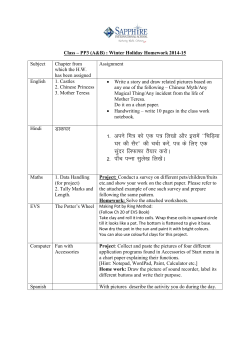


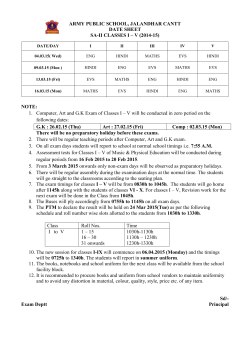


© Copyright 2026