
EQUIPMENT AND TECHNOLOGY IN ROBOTICS. , Lakmal Seneviratne
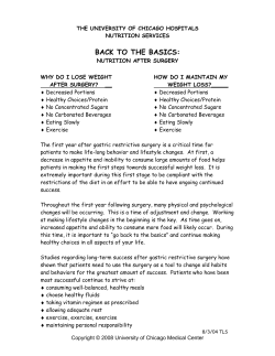
UROLOGY ROBOTIC SURGERY
Arch. Esp. Urol., 60, 4 (349-354), 2007
EQUIPMENT AND TECHNOLOGY IN ROBOTICS.
Declan Murphy, Ben Challacombe, Tim Nedas, Oussama Elhage, Kaspar Althoefer1, Lakmal
Seneviratne1 y Prokar Dasgupta.
Department of Urology, Guy’s & St Thomas’ NHS Foundation Trust, and King’s College School of Medicine
Department of Mechatronics1, King’s College London, UK.
Summary.- We review the evolution and current status of robotic equipment and technology in urology. We
also describe future developments in the key areas of virtual reality simulation, mechatronics and nanorobotics.
The history of robotic technology is reviewed and put
into the context of current systems. Experts in the associated fields of nanorobotics, mechatronics and virtual
reality simulation simulation review the important future
developments in these areas.
Keywords: Robotic. da Vinci™. Technology.
Nanorobotics.
INTRODUCTION
We have reached an interesting time in the
field of surgical robotics. Over the past 5 years the
imagination of patients and surgeons alike has been
captured by the arrival of robotic-assisted surgery,
telesurgical manipulators, and telepresence surgery.
Robotic-assisted laparoscopic surgery in particular,
has dominated headlines and symposium proceedings throughout the world in recent years, and lead
to increasing patient demand for “robotic surgery”.
The bulk of this demand is due to the proliferation of
da Vinci™ surgical systems (Intuitive Surgical, CA),
especially in the USA where over 400 systems have
been installed.
Correspondence
Urologists have been quick to embrace this
technology and robotic-assisted laparoscopic radical
prostatectomy (RALRP) is the most commonly performed robotic procedure performed worldwide. From
766 cases performed in 2002, over 48,000 are projected for 2007 (1). This accounts for 39.5% of the
radical prostatectomy market in the USA.
Declan Murphy, M.D.
Department of Urology
1st Floor Thomas Guy House
Guy’s Hospital
London SE1 9RT (UK)
[email protected]
But the monopoly which the da Vinci™ system
holds has led to stagnation in the development of the
field of robotic-assisted surgery. This technology has
its roots in the Stanford Research Institue (SRI) Green
Telepresence System which was developed in the early 1990’s by Philip Green and other researchers at
Stanford. The commercial licence for this technology
was subsequently acquired by Fredrick Moll, MD, to
found Intuitive Surgical in 1995. The prototype da
Vinci™ system was launched in 1997 and little has
been done to update the system since its Food & Drugs
Administration (FDA) approval in 2000. Therefore the
technology remains very expensive, very bulky, and
somewhat outdated.
350
D. Murphy, B. Challacombe, T. Nedas et al.
This paper outlines the interesting developments which have led to the current status of robotic technology
and equipment today. However, as much of this has
been described before (2,3), we will concentrate on
the very exciting future technologies in development,
especially the areas of virtual reality simulation, mechatronics and nanorobotics.
Definitions and history:
A surgical robot has been defined as “a computer-controlled manipulator with artificial sensing that
can be reprogrammed to move and position tools to
carry out a range of surgical tasks” (4). Strictly speaking, the current popular surgical “robots” do not satisfy this definition, and some authors have suggested
the term “computer-assisted surgery” more accurately
describes the current generation of robotic devices
(5). A description of “off-line” and “on-line” robots
has been used to discriminate between machines carrying out pre-programmed tasks and those carrying
out actions in response to ongoing commands (“master-slave” type devices) (6). Whatever the conclusion
of that pedantic debate, the term “robotic” is in popular use to describe the range of technology under
discussion here.
The early pioneers in this field included Wickham et al from Guy’s Hospital and Imperial College, London, who developed the PROBOT in the late
1980’s. The PROBOT used a robotic frame, which
guided a rotating blade to complete transurethral resection of the prostate (TURP). Initial studies on prostate-shaped potatoes were followed up by clinical trials
in patients to demonstrate safety and feasibility of the
technology (7). This was a truly autonomous device
(“off-line robot”), satisfying the definitions outlined
above. However convincing differences over conventional TURP were not demonstrated.
while ensuring very steady images. They also enable
the concept of solo-surgery, dispensing with the need
for surgical assistants (11).
Master-Slave Systems:
It is the daVinci™ surgical system (Intuitive
Surgical, CA) which has generated the most headlines
with regard to robotic-assisted surgery. It developed
in the mid-1990’s from the SRI Green Telepresence
System, while a competitor, the ZEUS™ system (Compuer Motion initially; now owned by Intuitive Surgical), was also undergoing clinical evaluation. This is
a master-slave system (“on-line” robot) rather than a
true autonomous robot. The surgeon sits at a console
remote from the patient, controlling 3 or 4 robotic
arms, which are docked through the laparoscopic
ports. Three dimensional (3D) vision, 7 degrees of
freedom (DoF) of movement, and intuitive movements
of the robotic instruments are among its proposed benefits over conventional laparoscopic surgery. The da
Vinci™ technology, its advantages and disadvantages are described in detail elsewhere (3).
Force Sensing and Tissue Identification - Current and
Future Developments
With the advent of specialised surgical robots such as the da Vinci™ surgical system, surgeons
are given advanced tools that assist during complex
operations and help to improve the outcome of surgical procedures. These highly sophisticated robotic
devices incorporate advanced technologies such as
precision mechanics, enhanced stereo vision and advanced motion control algorithms enabling tremor-free
and intuitive handling of the operating tools. However,
there are limitations. Most notably, the surgeon loses
all tactile sensation when operating with the aid of a
Other urological robots have included the
percutaneous renal access robot, PAKY-RCM which
demonstrated superior accuracy but longer operating
(access) times when compared to humans in a randomised control trial of transatlantic tele-robotics (8,9).
Robotic laparoscope manipulators:
The development of laparoscope manipulators such as the Automated Endoscopic System for
Optimum Positioning (AESOP™, Intuitive Surgical,
CA) and EndoAssist™ (Armstrong Healthcare, UK)
has certainly found a niche in laparoscopic urological procedures. These devices hold the laparoscope
under voice, pedal or infra-red motion control and
provide steadier images with less instrument collisions
than a human assistant (10). These are particularly
useful in procedures such as laparoscopic radical
prostatectomy, freeing the assistant to use 2 ports,
FIGURE 1. Indentation device employing wheeled
probe for rapid soft tissue characterisation.
EQUIPMENT AND TECHNOLOGY IN ROBOTICS.
robot. The sense of touch which is readily available
during open surgery provides the surgeon with valuable information about the operating site. The inability
to palpate organs during an operation can lead to a
misjudgement of interaction dynamics between tool
and soft tissue. Recent studies have revealed that the
lack of tactile sensation during robot-aided surgery
can lead to an increase in tissue trauma and accidental tissue damage, and surgeons provided with force feedback significantly improve their performance
(12,13).
Current research at a number of research institutes aims at equipping surgical robots with sensors
and feedback mechanisms to re-establish the surgeon
with tactile perception.
Owing to advances in micro-technologies,
there is now a clear trend towards developing miniaturised sensors that can measure the manipulation forces at the point where the tool comes into contact with
soft tissue. Recently, a surgical gripper at the end of
a laparoscopic tool has been integrated with a strain gauge sensor (14). The sensor’s hexapod structure
made from an aluminium alloy provides a light weight
and rigid solution to acquire force and torque signals
along all six axes with a high resolution of 0.05 N
and 0.25 N in radial and axial direction, respectively
with a range of up to 20 N. A MEMS micro gripper driven by a piezoelectric actuator integrated with
semiconductor strain gauges mounted on a microfabricated elastic surface (flexure) has been developed
allowing soft tissue property characterisation and realistic palpation using a haptic interface (15).
Advances have also been made in employing piezoelectric materials to measure the contact
forces at the tip of surgical tools. Micro-machined
force array sensors have been developed that can
be mounted on surgical tools (16,17). The developed
sensors claim to have a high sensitivity and linear behaviour over a wide range of up to 15 N, allowing
realistic palpation feedback.
Very promising results have also been achieved based on fibre optic measuring principles. Miniature force sensors can be created using fibre optic
cables that carry light signals -which are modulated in
response to the applied forces- from a sensing region
to an opto-electronic converter. Recently, a 5-mm diameter force sensor integrating three fibre-optic sensor
elements into the tip of a surgical tool was developed.
This sensor can measure forces along three axes with
a sensitivity of 0.04 N and a range of up to 2.5 N
(18). The main advantages of these sensors are that
they are not affected by electromagnetic interference
and compatible with magnetic resonant imaging sys-
351
tems. Exploiting this, a three degree-of-freedom optical fibre force sensor was used in an MR-compatible
neurosurgery robot to measure tool-tissue interaction
forces (19).
Accurate sensors and appropriate actuators
that reconstruct the measured forces in the user’s
hand are both necessary components of haptic interfaces that realise truthful remote touch sensing. An
alternative feedback mechanism is the use of force
sensors for the identification of soft tissue (i.e. areas
of increased stiffness or softness) providing the surgeon with visual cues on the location and severity of
any organ abnormalities. Attempts to identify tissue
have let to the development of a number of devices.
A uni-axial stretching device is used by Brouwer et
al. to measure porcine tissue response both in-vivo
and ex-vivo (20). Another device was developed to
investigate the in-vivo viscoelastic properties of tissue
under uni-axial small deformations (21). A motorized
endoscopic grasper which was used to test abdominal porcine tissues in-vivo and in-situ with cyclic and
static compressive loadings is also described (22).
A mechanical probe developed by the Harvard Biorobotics Laboratory, attempts to identify the location
and properties of tumours based on static indentation tests (23). Recently conducted research at King’s
College London aims at the development of devices
that consider a series of distributions measured as a
wheeled probe (see figure 1) slides across the tissue
surface. This approach departs from the previous one
of static indentations, allowing the identification of
whole regions of organ tissue in short time (24).
Nanotechnology:
In the same way as the development of microtechnology in the 1980s has led to new tools for surgery, emerging nanotechnologies will similarly permit
further advances providing better diagnosis and new
devices for medicine. Nanorobots are expected to
enable significant new capabilities for diagnosis and
treatment of disease for patient monitoring and minimally invasive surgery (25,26).
The ability to manufacture nanorobots may
result from current trends and new methodologies in
fabrication, computation, transducers and manipulation. The hardware architecture for a medical nanorobot must include the necessary devices for monitoring
the most important aspects of its operational workspace: the human body.
Teams of nanorobots may cooperate to perform predefined complex tasks in medical procedures (27). To reach this aim, data processing, energy
supply, and data transmission capabilities can be
addressed through embedded integrated circuits,
352
D. Murphy, B. Challacombe, T. Nedas et al.
using advances in technologies derived from nanotechnology and Very Large System Integration (VLSI
design) (28). Complementary Metal Semi-Conductor
(CMOS) VLSI design using deep ultraviolet lithography provides high precision and a commercial way
for manufacturing early nanodevices and nanoelectronics systems. The CMOS industry may successfully drive the pathway for the assembly processes
needed to manufacture nanorobots, where the joint
use of nanophotonic, carbon nanotubes and nanocrystals, may even accelerate further the actual levels
of resolution ranging from 248nm to 157nm devices
(29). The appropriate interdisciplinary effort will impact on assembly nanodevices and nanoeletronics to
build nanorobots (30). To validate designs to achieve
a successful implementation, the use of Verification
Hardware Description Language (VHDL) is the most
common methodology utilized in the integrated circuit
manufacturing industry. Nanorobots can be useful in
a large range of biomedical applications for future
drug delivery applications, such as dosage regimens
based on predicted pharmacokinetic parameters for
chemotherapy in anti-cancer treatments (31,32). A
range of different signals are directly correlated to
specific medical problems. Chemical signals can serve for medical target identification and actuation. A
single tumor cell can be characterized as a typical
endothelial cell mutation with profound consequences
for patients suffering from cancer. Endothelial cells
have a large number of functions and may play an
important role in human health. They also serve as
part of the structure forming the inside blood vessels,
which are spread throughout every single organ or
system comprising our body.
FIGURE 2. A nanorobot employing nanosensors and
advanced nanorobot control design features. Courtesy
of Adriano Cavalcanti, www.nanorobotdesign.com
FIGURE 3. Virtual reality simulator for laparoscopic renal surgery. Courtesy of Mentice, Goteborg, Sweden.
Factors like low energy consumption and
high-sensitivity are among some of the advantages
of nanosensors. Nanobioelectronics using nanowires
as material for circuit assembly can achieve maximal
efficiency for applications regarding chemical changes, enabling new medical applications (30). Using
chemical sensors nanorobots can be programmed to
detect different levels of E-cadherin and beta-catenin
as medical targets in primary and metastatic phases.
Integrated nanosensors can be utilized for such a task
in order to find different concentrations of E-cadherin
signals (33-35). Beyond sensors, nanorobots may be
designed with appropriated space to carry chemotherapy for future cancer drug delivery. Such approach allows maintaining the drug carrier for a time
longer as necessary into the bloodstream circulation,
avoiding the current resulting extravasation towards
non reticuloendothelial-located cancers and the high
degenerative side-effects (36).
Figure 2 demonstrates a nanorobot developed by Adriano Cavalcanti at the CAN Center for
Automation in Nanobiotech, Sao Paulo, Brazil.
The role of virtual reality simulation and robotics
Minimally invasive surgery has long been associated with training issues. At their inception novel
surgical techniques must be learned by all grades of
surgeon and once in wide use trainees must be able
EQUIPMENT AND TECHNOLOGY IN ROBOTICS.
to learn techniques safely. From this point of view the
evolution of laparascopic surgery training provides a
good template for robotic techniques.
Robot assisted procedures are complex operations in which precision is vital, a far from ideal
learning environment; hence the need for other training environments. Also with robot assisted surgery
there is a huge infrastructure cost associated with
both the docking robot and the operating console. It
is not within the budget of many institutions to provide
a fully serviced training robot.
Virtual reality has been shown to be effective
for surgical training and has the benefits of providing
a reproducible operating environment in which metric
parameters can be used to monitor operator performance (37,38). The ability to regularly incorporate
individual patient anatomy into a surgical simulator
and practice prior to surgery is only a few years
away.
Virtual reality has shown itself as the premier
training option in laparascopic surgery due to the
evolution of haptic feedback instruments, these instruments provide the operator with tactile sensations
akin to real surgery.
The prospect of using virtual reality to simulate robot assisted procedures is incredibly exciting.
From the development point of view haptic feedback
is not required thereby removing a great deal of programming time and research from any project which
is necessary in order to incorporate the increased
number of degrees of freedom. For a VR robotic simulator only the operating console is required saving the
expense of the robotic device.
Current developments in VR software are progressing towards the fully interactive abdomen and
pelvis; the ability to alter the software to include robotic instruments is not far off. Figure 3 demonstrates a
VR simulator for laparoscopic renal surgery currently
under development by Mentice, Goteborg, Sweden,
and Guy’s Hospital, London.
The future of robotic surgery in conjunction
with VR may include a surgeon “pre-operating” on
the same patient where the VR anatomy has been produced directly from the patients imaging.
CONCLUSIONS
The introduction of robotic technology into
urology in the late 20th and early 21st century has
heralded an exciting time for surgeons. However, it is
353
clear that such technology is in its infancy, at least in
clinical surgery. The exciting potential developments
in mechatronics, nanotechnology and virtual reality
simulation outlined in this paper will lead to a huge
leap to the next generation of robotic technology and
equipment.
ACKNOWLEDGEMENT
Special thanks to Dr. Adriano Calvanti of the
CAN Center for Automation in Biotech, Sao Paulo,
for his contribution on nanotechnology.
REFERENCES AND RECOMMENDED READINGS
(*of special interest, **of outstanding interest)
1. PEPLINSKI, R.: “Past, present and future of the
Da Vinci robot”. 2nd UK Robotic Urology Course, 2006.
2. CHALLACOMBE, B.J.; KHAN, M.S.; MURPHY, D. et al: “The history of robotics in urology”. World J. Urol., 24: 120, 2006.
*3. CHALLACOMBE, B.J.; KHAN, M.S.; MURPHY, D. et al: “Robotic technology in urology”.
Postgrad. Med. J., 82: 743, 2006.
4. DASGUPTA, P.; JONES, A.; GILL, I.S.: “Robotic urological surgery: a perspective”. BJU Int.,
95: 20, 2005.
5. GUILLONNEAU, B.: “What robotics in urology? A current point of view”. Eur. Urol., 43: 103,
2003.
*6. SIM, H.G.; YIP, S.K.; CHENG, C.W.: “Equipment and technology in surgical robotics”. World
J. Urol., 2006.
7. DAVIES, B.L.; HIBBERD, R.D.; TIMONEY,
A.G. et al: “The development of a surgeon robot
for prostatectomies”. Proc. Inst. Mech. Eng., 205:
35, 1991.
8. CHALLACOMBE, B.J.; KAVOUSSI, L.R.;
DASGUPTA, P.: “Trans-oceanic telerobotic surgery”. BJU Int., 92: 678, 2003.
*9. CHALLACOMBE, B.; KAVOUSSI, L.; PATRICIU, A. et al: “Technology insight: telementoring and telesurgery in urology”. Nat. Clin. Pract.
Urol., 3: 611, 2006.
10. KAVOUSSI, L.R.; MOORE, R.G.; ADAMS, J.B.
et al: “Comparison of robotic versus human laparoscopic camera control”. J. Urol., 154: 2134,
1995.
11. ANTIPHON, P.; HOZNEK, A.; BENYOUSSEF,
A. et al: “Complete solo laparoscopic radical
prostatectomy: initial experience”. Urology, 61:
724, 2003.
354
12. WAGNER, C.; STYLOPOULOS, N.; HOWE, R.:
“The role of force feedback in surgery: Analysis
of blunt dissection”. Proc. IEEE 10th symp. on
haptic interfaces for virtual Envir&Teleoperator
systems. 2002.
13. DEML, B.; ORTMAIER, T.; SEIBOLD, U.: “The
touch, and feel in minimally invasive surgery”.
IEEE Int. workshop on haptic audio visual environments, and their applications , 33-38. Ottawa,
Ontario, 2005.
14. SEIBOLD, U.; KUEBLER, B.; HIRZINGER, G.:
“Prototype of instrument for minimally invasive surgery with 6-axis force sensing capability”.
Proc. IEEE Int. conf. on robotics, and automation,
496-501. Barcelona, Spain, 2005.
15. MENCIASSI, A.; EISINBERG, A.; CARROZZA, M. et al: “Force sensing microinstrument for
measuring tissue properties, and pulse in microsurgery”. IEEE/ASME Trans on mechatronics, 8:
10, 2003.
16. OTTERMO, M.; STAVDAHL, O.; JOHANSEN,
T.: “Palpation instrument for augmented minimally invasive surgery”. Proc. IEEE/RSJ Int. conf.
on intelligent robots, and systems, 3960-3964.
Sendai, Japan, 2004.
17. DARGAHI, J.; PARAMESWARAN, M.; PAYANDEH, S.: “A micromachined piezoelectric
tactile sensor for an endoscopic grasper-theory,
fabrication, and experiments”. Journal of microelectromechanical systems, 9: 329, 2000.
18. PEIRS, J.; CLIJNEN, J.; REYNAERTS, D. et al:
“A micro optical force sensor for force feedback
during minimally invasive robotic surgery”. Sensors and actuators A., 115: 447, 2004.
19. SUTHERLAND, G.; McBETH, P.; LOUW, D.:
“NeuroArm: an MR compatible robot for microsurgery”. International congress series, 1256: 504.
Elsevier Science, 2003.
20. BROUWER, I.; USTIN, J.; BENTLEY, L. et al:
“Measuring in-vivo animal soft tissue properties
for haptic modelling in surgical simulation vol. 81
69-74, 2001”. Stud. Health Technol. Inform., 81:
69, 2001.
21. OTTENSMEYER, M.: “In-vivo measurement of
solid organ visco-elastic properties”. Stud. Heatlh
Technol. Inform., 85: 328, 2002.
22. BROWN, J.; ROSEN, J.; KIM, Y, et al: “In-Vivo
and In-Situ Compressive Properties of Porcine
Abdominal Soft Tissues”. Stud. Health Technol.
Inform., Newport Beach, CA., 2003.
23. WELLMAN, P.; HOWE, R.: “Modelling probe
and tissue interaction for tumour feature extraction”. ASME summer Bioengineering conference,
Sun River, Oregon, 1997.
24. NOONAN, D.; LIU, H.; ZWEIRI, Y. et al: “A
dual-function wheeled probe for tissue viscoelastic property identification during minimally inva-
**25.
26.
27.
28.
29.
30.
**31.
32.
33.
34.
35.
36.
*37.
38.
sive surgery”. International Conference on robotics and automation. Rome, Italy, 2007.
CAVALCANTI, A.: “Assembly automation with
evolutionary nanorobots and sensor-based control
applied to nanomedicine”. IEEE Transactions on
Nanotechnology, 2: 82, 2003.
FREITAS, R.A. Jr.: “Nanomedicine - basic capabilities”. www.nanomedicine.com, 1999.
CAVALCANTI, A.; FREITAS, R.A. Jr.: “Nanorobotics control design: a collective behavior approach for medicine”. IEEE Trans. Nanobioscience,
4: 133, 2005.
SRIVASTAVA, N.; BANERJEE, K.: “Performance analysis of carbon nanotube interconnects
for VLSI applications”. IEEE/ACM ICCAD Int.
Conf. on computer-aided design., 383-390, 2005.
BOGAERTS, W.; BAETS, R.; DUMON, P. et al:
“Nanophotonic waveguides in silicon-on-insulator fabricated with CMOS technology”. J. Lightwave Technology, 23: 401, 2005.
CAVALCANTI, A.; SHIRINZADEH, B.; FREITAS, R.A. Jr. et al: “Medical nanorobot architecture based on nanobioelectronics. Recent patents on
nanotechnology”. 1 ed. Bentham Science, 2007.
KAWASAKI, E.S.; PLAYER, A.: “Nanotechnology,
nanomedicine, and the development of new, effective therapies for cancer”. Nanomedicine: nanotechnology, biology & medicine, 101-109, 2005.
MUTOH, K.; TSUKAHARA, S.; MITSUHASHI, J. et al: “Estrogen-mediated post transcriptional down-regulation of P-glycoprotein in MDR1transduced human breast cancer cells”. Cancer
Sci., 97: 1198, 2006.
JANDA, E.; NEVOLO, M.; LEHMANN, K. et al:
“Raf plus TGFbeta-dependent EMT is initiated by
endocytosis and lysosomal degradation of E-cadherin”. Oncogene, 25: 7117, 2006.
SONNENBERG, E.; GODECKE, A.; WALTER,
B. et al: “Transient and locally restricted expression of the ros1 protooncogene during mouse development”. EMBO J., 10: 3693, 1991.
TRUST SANGER INSTITUTE: “Human chromosome 22 project overview”. www.sanger.
ac.uk/HGP/Chr22/ 2007.
COUVREUR, P.; GREF, R.; ANDRIEUX, K. et
al: “Nanotechnologies for drug delivery: application to cancer and autoimmune diseases. Progress
in solid state”. Chemistry, 34: 231, 2006.
SEYMOUR, N.E.; GALLAGHER, A.G.; ROMAN, S.A. et al: “Virtual reality training improves operating room performance: results of a randomized, double-blinded study”. Ann. Surg., 236:
458, 2002.
FRIED, G.M.; FELDMAN, L.S.; VASSILIOU,
M.C. et al: “Proving the value of simulation in
laparoscopic surgery”. Ann. Surg., 240: 518,
2004.


© Copyright 2026