
C S ase tudies
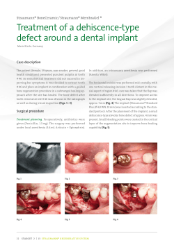
Case Studies
Septodont
No. 09 - October 2014
R.T.R.
ß-tcP for bone regeneration
GIUSEPPE GALVAGNA
PAOLO BRUNAMONTI BINELLO
MASSIMO GALLI
MAURO LABANCA
R.T.R.
alloPlastic grafts - ß-tcP
MARIO ERNESTO GARCÍA-BRISEÑO
BIODENTINE™
PulPotomy on Primary molars
LUC MARTENS & RITA CAUWELS
BIODENTINE™
PulP caPPing
LUC MARTENS & RITA CAUWELS
Collection
since its foundation septodont has developed, manufactured
and distributed a wide range of high quality products for
dental professionals.
© Jeremy allen | Dreamstime.com
septodont recently innovated in the field of gingival preparation, composites and dentine care with the introduction
of racegel, the n’Durance® line and biodentine™, which
are appreciated by clinicians around the globe.
septodont created the “septodont case studies collection”
to share their experience and the benefits of using these
innovations in your daily practice.
this collection consists in a series of case reports and is
published on a regular basis.
this 9th issue is dedicated to two of septodont’s innovative
products:
r.t.r., an easy-to-use synthetic bone grafting material.
in addition to its ability to provide an optimal osteoconductive environment to promote the growth of new dense
bone, r.t.r. comes in 3 different presentations to suit all
the clinical situations.
biodentine™, the first biocompatible and bioactive dentin
replacement material. biodentine™ uniqueness not only
lies in its innovative bioactive and “pulp-protective”
chemistry, but also in its universal application, both in
the crown and in the root.
2
Content
Use of ß-tricalcium phosphate for bone
regeneration in oral surgery
04
Giuseppe Galvagna, Paolo Brunamonti Binello,
Massimo Galli and Mauro Labanca
Alloplastic Grafts Beta-tricalcium phosphate
14
Mario Ernesto García-Briseño
Biodentine™ - Pulpotomy on primary
molars
21
Luc Martens and Rita Cauwels
Biodentine™ - Pulp capping
Luc Martens and Rita Cauwels
26
3
Use of ß-tricalcium phosphate
for bone regeneration
in oral surgery
A multicenter study to evaluate the clinical applications
of R.T.R. (Resorbable Tissue Replacement)
Giuseppe Galvagna: Private practice, catania, italy
Paolo Brunamonti Binello: consultant Professor, university of genoa
Massimo Galli: Private practice, Pistoia, italy
Mauro Labanca: consultant Professor in oral surgery and anatomy, milan, italy
In prosthetic implant rehabilitation, loss of bone volume in atrophic maxillae is one of the
major problems faced by surgeons in their clinical practice. In the presence of horizontal
and vertical bone defects, atrophic ridges need to be restored to make them suitable for
implant placement and for restoration of masticatory and aesthetical functions.
For this reason, in recent years the term “GBR” has been closely associated with the
concept of prosthetically guided implantology.
The purpose of this study is to demonstrate the osteoconductive properties of synthetic
biomaterials, particularly ß-tricalcium phosphate or R.T.R., and its benefit for a suitable GBR.
Introduction
in order to obtain effective bone regeneration
using natural or synthetic fillers, a series of
favourable conditions must occur that allow the
body to perform the bone growth (15) as follows:
- presence of a blood clot with a high concentration of mesenchymal stem cells (msc)
capable of evolving in the osteoblast line and
of endothelial cells forming a rich vascular
network
- presence of vital bone tissue, from which
osteogenic and angiogenic cells originate
4
through adequately prepared surrounding bone
(cortical perforation)
- stabilization and maintenance of volume underneath the membrane
- protection of blood clot with a membrane,
with the function of the stabilization of the
clot, protecting growing vascular structures
and blocking the migration of epithelial cells,
which proliferate faster than bone cells.
in 1980 nyman and Karring first introduced the
concept of guided tissue regeneration (gtr) of
periodontal tissues, showing that the cells of
soft tissue can grow faster than bone tissue (10).
thus, based on the results obtained, the migration
of soft tissue cells above the implanted material
must be stopped, thereby blocking migration
within the porosity of the same material, thus
promoting osseointegration rather than fibrointegration. the main characteristic of a membrane
should be semi-permeability, i.e. the presence of
porosity approximately 22 microns in diameter.
initially, a thin vascular network and a primary
fibrous osteoid tissue -also called primary spongiosa- will begin to form within the clot. the
latter is later mineralized thanks to osteoblasts
that cover its surface, forming a new poorly
calcified cortical bone.
the process stops when intertrabecular spaces
narrow due to the formation of new bone tissue,
until they reach the characteristic dimensions
of Havers channel which, along with concentric
lamellae, originate primary osteomas. all this
occurs during the first 3-4 months, though
actual bone remodelling requires more time, as
it creates secondary spongiosa.
research conducted by Hämmerle in 1996 (11)
on human subjects confirmed what had
previously been observed in animals, i.e. in the
presence of large bone defects, regeneration
can be limited to the more peripheral areas of
the defect, while less activity is observed in the
central area, where granules of biomaterial
remain over time, though less frequently when
tricalcium phosphate is used. the process of
ossification always starts from the walls of the
defect toward the center of the clot, along the
newly formed vessels.
a number of studies clearly describe the regenerative ability of autologous bone compared
with synthetic biomaterials, but unfortunately
not without negative aspects. indeed, collection
from the donor site is very often painful for the
patient; additionally, this results in longer surgical
procedures and postoperative pain ; finally, the
implanted material has a high degree of resorption
(not a negligible factor).
the materials used for bone regeneration are
grouped into: autogenous bone, allogenic bone,
xenogenic bone, alloplastic bone.
Graft biomaterials
the alloplastic biomaterials available on the
market represent an excellent alternative to
autologous bone graft and are classified into
two large groups: bioinert and bioactive, according to their interaction when they come into
contact with the receiving site.
the main requirement of a synthetic biomaterial
is to have a surface porosity that must promote
colonization and development within its structure.
these porosities must measure between 200/400
microns in diameter (lynch et al., 2000; bauer
and muschler, 2000). synthetic biomaterials
have been the subject of many studies, though
their long term results have not always been
considered. (1-2-3)
today, osteoblastic cells or bone morphogenetic
proteins (bmP obtained with in vitro cultures)
can be added to a graft material (4-5) to enhance
its osteoinductive and osteoconductive abilities
and therefore reduce the time required for cells
colonization. (6-7-8)
among alloplastic biomaterials, ß-tricalcium
phosphate is the one that mostly displays a
stable bond with bone neoformation; indeed,
its characteristics have made it suitable for
use in orthopedics since the early 1900s. in
the presence of H2o it becomes instable,
turning into hydroxyapatite, and this characteristic makes it suitable as an osteoconductive
material (coetree, 1980, De leonardis and
Pecora, 1999; 2000).
ß-tcP is characterized by a lower ca/P ratio,
which makes it more soluble than natural apatite.
the beta form is commonly obtained by mixing
calcitis (cc) and dibasic calcium phosphate
anhydrous (DcPa). the product obtained is
rapidly cooled, and alfa-tcP is obtained. conversely, extended repeat baking at 800/950°c
results in the beta form (9-12-13).
multiple studies conducted on the tc and bone
interaction have shown that histological examination at four months shows an initial bone
neoformation in the intergranular spaces and in
the surface porosity that helps guided bone
formation. indeed, the granules are reabsorbed
by phagocytosis, releasing ca/mg and phos-
5
phates in the surrounding bone tissue, thereby
activating alkaline phosphatase, a key ossification
process. between 6 and 18 months, fibroblasts
begin invading the biomaterial, activating the
extracellular dissolution process which ends
with the calcification phase. if this should occur
sooner, graft integration, rather than biodegradation, would happen. (5-6-7-8).
unlike hydroxyapatite, r.t.r. is progressively
and totally reabsorbed, thereby releasing calcium
and phosphate ions that participate actively in
the formation of new bone tissue. over a period
of time between 6 and 9 months, which may
vary according to the patient’s physiological
response, while stimulating bone regeneration,
r.t.r. is progressively reabsorbed, leaving space
for bone neoformation.
R.T.R.
Indications:
• Post-extraction sites
• filling post-extraction sites to maintain the
dimensions of alveolar bone
• implant defects
• sinus lift procedure
• reconstruction of peri-implant defects
• filling periodontal pockets with two or more
walls
• residual cavities after oral surgery (like cyst)
• filling defects after apicectomy
• alveolar filling following extraction of impacted
teeth.
synthetic, biocompatible and totally resorbable
99% pure tricalcium phosphate bone replacement, available in granules and in a cone shape,
for regeneration in periodontal defects, implant,
post-extraction bone defects and bone lesions
following endodontic surgery.
the micro and macro-porous r.t.r. structure,
with macropores measuring between 100 and
400 µm and micropores measuring less than
10 µm.
these morphological characteristics allow excellent osteogenic cell in-depth colonization and
easy compacting.
Materials and methods
We conducted a multicenter study to evaluate
the clinical application of r.t.r. (ß-tricalcium
phosphate).
this study examines the regeneration of bone
defects with r.t.r. (ß-tricalcium phosphate) in
patients eligible for prosthetic implant rehabilitation. Patients were randomly selected,
according to the following key criteria:
- aged between 20 and 60 years
- either male or female
- non-smokers
- in good general health
- having at least one crestal bone defect (no
morphology and etiopathogenesis restrictions).
6
the cases treated were identified in the following
clinical situations:
- Post-extraction sites
- bone regeneration around implants placed in
areas with bone loss or post-extraction.
- gbr (sinus lift or major bone defects).
in all cases, patients received antibiotic therapy
with 1 gr every 8 h of amoxicilline plus clavulanic
acid (starting 24 hours before surgery up to day
5 post-surgery), repeated daily rinses with chlorhexidine and therapy with fans (ibuprofen
800 mg/day in single dose), as necessary.
Case Report no.1
Patient: female
Age: 30
History: odontogenic cyst in maxillary bone at 2.1.
cyst was removed in october 2013. Vertical guided regeneration with resorbable membrane and
r.t.r. was performed. implant placement: bone peak follow-up intraoral X-ray in consecutive
months. 2nd surgery isQ value: 61.
Fig. 1-4: Different projections of radiographic c.t images show the large bone defect in site 2.1.
Fig. 6: X-ray image before
Fig. 7: X-ray image after 5 months.
Fig. 5: temporary prosthesis to cover the
cosmetic defect.
Fig. 8: X-ray after insertion
Fig. 9: the local objective
examination and routine
radiographic examination showed
a good short-term healing.
grafting.
of the implant.
Fig. 10: the isQ test confirms a good stability of the implant.
Fig. 11: to obtain a good aesthetics of the final prosthesis is
important to condition the soft tissue with the healing screws that
favors the emergence profile.
7
Case Report no.2
Patient: female
Age: 60
History: large cyst in upper maxillary bone extending from 2.2 to 1.1.
the cyst was removed and the cavity was filled with r.t.r. enhanced with Prgf (platelet-enriched
plasma) and simultaneous placement of five implants was also performed.
Fig. 12-13: Presence of large cysts of 2.1. the oral cavity examination shows a poor oral hygiene.
Fig. 14-15: removal of the cyst; it is very
Fig. 16-17-18: filling the bone cavity with granular r.t.r.
important to remove all residual epithelial.
Fig. 19-20: During the same surgery were included five implants.
8
Fig. 21: X-ray control after five months.
Case Report no.3
Patient: female
Age: 50
History: severe atrophy of the alveolar ridge, upper right jaw, affecting the area of 1.3, 1.4, 1.5, 1.6.
these teeth were extracted and the alveolar ridge was reconstructed with r.t.r., covering biomaterial
with tabotamp (oxidized cellulose).
Fig. 22-23: resorption of the alveolar process caused by periodontal disease. the local examination shows the class iii mobility of the teeth.
Fig. 24-25-26: resorption of the alveolar process caused by periodontal disease. the local examination shows the class iii mobility of the teeth.
Fig. 27-28: use of tabotamp to cover the
Fig. 29-30: the local examination after
graft.
10 days.
Fig. 31-32: the radiographic image after
4 months showed an increase in the vertical
dimensions.
9
Case Report no.4
Patient: female
Age: 30
History: root fracture of 1.6, previously treated with root canal treatment and large crown composite
reconstruction. the roots were carefully extracted, and an implant was placed, using the interadicular
septum bone and by performing an elevation of the maxillary sinus with osteotomes. the alveoli
were filled with r.t.r. and covered with a collagen membrane.
Fig. 33-34: fracture of the first molar with the insertion of implant with post-extraction procedure.
Fig. 35-36-37: extraction was performed with piezosurgery technique to preserve the alveolar bone.
Fig. 38-39-40: the gap was filled using r.t.r.-size cone.
Fig. 41-42: the intraoral examination and radiographic examination after 4 months showed good integration of the implant.
10
Case Report no.5
Patient: female
Age: 64
History: loss of 2.2 and 2.3 due to trauma.
ct revealed a significant resorption of buccal vestibular alveolar process.
two implants were placed (measuring 3.3 mm in diameter and 13 mm long) and the gap was filled
with r.t.r.
re-opening was done after 5 months and evaluation of osteointegration with osstel.
2.2 showed a value of 22-isQ, and 2.3 –isQ 64.
Fig. 43-44-45: severe post-traumatic atrophy of the alveolar process in place 2.2- 2.3.
Fig. 46-47: insertion of two implants with “split-crest” surgical
Fig. 48-49: the gap between the two margin bone was filled with
technique.
phosphate-tricalcium r.t.r.
Fig. 50: X-ray control after 4 months
showed a good bone density.
Fig. 51-52: the intraoral examination does
Fig. 53-54: the isQ value confirms a good
appreciate a good recovery and an increase
in bone volume.
osseointegration.
11
Case Report no.6
Patient: female
Age: 55
History: residual cyst at 3.6. to proceed to prosthetic molar rehabilitation with an implant, the cyst
was extracted and the cavity was filled with r.t.r.; after a 4-month period for bone regeneration,
the implant was placed.
Fig. 59: Wound at 30 days.
Fig. 55-56: the c.t. examination before
Fig. 57-58: after having removed the cyst,
surgery showed a residual cyst.
to speed up the healing time, the cavity has
been filled with r.t.r. granules.
Fig. 60: X-ray at 3 months.
Conclusion
based on the results obtained in the short term,
the authors confirm the excellent properties of
r.t.r., both in the first weeks of healing and in
the following months, and they consider it an
excellent alternative to autologous bone grafts.
no inflammatory reactions or loss of bone
volume evaluated clinically and radiographically
12
occurred in any of the cases examined. the
most encouraging data came from the observation of the compactness and density of the
bone neo-formation, which easily allowed the
placement of implants with high isQ values
both during and after the placement of r.t.r.
graft.
Authors (left to right)
Giuseppe Galvagna: Dental practitioner, catania, italy
Paolo Brunamonti Binello: consultant Professor, university of genoa. galliera Hospital,
genoa, italy
Massimo Galli: oral surgeon, dental practitioner, Pistoia, italy
Mauro Labanca: Professor of oral surgery and anatomy, milan, italy
References
01. labanca m., leonida a., rodella fl: natural synthetic biomaterial in Dentistry: science and ethics as
criteria for their use. implantologia 2008; 1:9-23
02. ohgushi H, caplan ai: stem cell technology and bioceramics: from cell to gene engineering. J biomed
mater res 48(6): 913-927, 1999
03. anselme K: osteoblast adhesion on biomaterals. biomaterials 2000 apr; 21(7):667-81.
04. toquet J, roahanizadeh r, guicheux J, couillaud s, Passuti n, Daculsi g, Heymann D: osteogenic.
Potential in vitro of human bone marrow cells cultured on macroporous biphasic
05. maddox e, Zhan m, mundy gr, Drohan Wn, burgess WH: optimizing human demineralized bone matrix
for clinical application. tissue eng 6(4): 441-448, 2000 calcium phosphate ceramic. J biomed mater res.
1999 Jan; 44(1):98-108.
06. gauthier o, bouler Jm, aguado e, Pilet P, Daculsi g: macroporous biphasic calcium phosphate ceramics:
influence of macropore diameter and macroporosity percentage on bone ingrowth. biomaterials 1998 Jan
feb;19(1-3):133-9.
07. martinetti r, belpassi a, nataloni a, biasini V, martignani g: idrossiapatite porosa sintetica per sostituzioni
ossee: caratterizzazione chimico-fisica. biomateriali: atti del congresso; 51 55, roma enea, 1999
08. martinetti r, belpassi a et al.: Key engineering materials Vols. 192-195 (2001), 507-510 – Proceeding of
the 13th int. symp. on ceramics in medicine, bologna, italy, 22-26 nov.
09. caplan ai: mesenchymal stem cells. J orthop res 9: 641-650, 199
10. nyman s, lindhe J, Karring t et rylander H: new attachment following surgical treatment of human
periodontal disease. Journal of clinical Periodontology 9: 290-296, 1982; 39
11. Hämmerle cH, brägger u, bürgin W, lang nP: the effect of subcrestal placement of the polished surface
of iti implants on marginal soft and hard tissues. clin oral implants res. 1996 Jun;7(2):111-9
12. chin m: Distraction osteogenesis in maxillofacial surgery. in lynch se, genco rJ, marx re (eds). tissue
engineering – application in maxillofacial surgery and Periodontics. illinois; Quintessence Publishing co,
inc, 1999; 147
13. arun K. garg: grafting materials in repair and restoration. in lynch se, genco rJ, marx re, (eds). tissue
engineering - application in maxillofacial surgery and Periodontics. illinois; Quintessence Publishing co,
inc, 1999; 83
14. scipioni a, bruschi gb, calesini g: the edentulous ridge expansion technique: a five-year study. int J
Periodont rest Dent 14: 451-459, 1994 trans tech Publications, switzerland
15. schenk rK and buser D: osseointegration: a reality. Periodontology 2000, Vol 17. 1998, 22.35
16. bauer tW and muschler gf: bone graft materials. an overview of the basic science. clin orthop relar
res, 2000 feb;(371):10-27.
13
Alloplastic Grafts - Beta-tricalcium
phosphate
Presentation of clinical cases
Mario Ernesto García-Briseño
DDs and Professor in Periodontics - autonomous university of guadalajara (uag), mexico This case report presents a brief review of bone loss due to periodontal infection and
tooth extraction, and evaluates the biological characteristics, description and indications
of an alloplastic graft material, beta-tricalcium phosphate, as an alternative treatment in
two clinical cases.
Introduction
the effects of periodontal infection and its
consequences, attachment and bone losses,
result in the formation of periodontal bone
defects (1,2). tooth extraction involves two
processes, the healing of the alveolus (3) and
any change that may appear during post-extraction healing (4). the clinical consequences of
tooth extraction are the resorption of the alveolar
ridge (5) and the pneumatization of the maxillary
sinus (6). if the effects of periodontal disease
and tooth extraction are combined, the consequences will be even more severe and will
complicate tooth restoration (7). the use of
bone grafting materials prevents and/or repairs
the insufficient bone conditions due to the
previously mentioned situations (8). We will first
evaluate the biological characteristics (9) of the
14
alloplastic graft material, beta-tricalcium phosphate, before presenting clinical cases using a
commercial presentation of beta-tricalcium phosphate. the objective of this presentation is to
demonstrate the clinical applications of betatricalcium phosphate alloplastic graft in
periodontal bone defects and extraction sites.
Modifications in the Alveolar
Process
the most common modifications involving the
bone tissue of the oral cavity are: Horizontal
bone loss due to periodontal infection, bone
defects caused by periodontal disease, tooth
extraction resulting in vertical and horizontal
resorption, pneumatization of the maxillary
sinus and a combination of these. the clinical
consequence of these conditions are inappropriate bone dimensions for prosthetics on
natural abutment teeth and the placement of
its substitutes, dental implants (10).
Periodontal bone defects
as a result of inflammatory and immune reactions to the presence of bacterial plaque and
the way in which it progresses apically over
the cement surface in one of the periodontal
attachment components, the alveolar bone,
specific patterns of destruction can be observed.
these patterns depend mainly on the type of
subgingival bacterial plaque and the anatomical
characteristics of the alveolar process (11).
basically there are two bone loss patterns,
horizontal or vertical forms. the vertical form
of bone loss has been described as intrabony
loss and the resulting defects have been historically classified according to the number of
bone walls lost: one, two or three-wall intrabony
defects. other bone loss patterns have been
described as osseous craters and circumferential
defects. the special anatomy of molars and
premolars involves furcations as a special
condition in periodontal defect (12).
Tooth extraction
the consequences of tooth extraction represent
specific conditions and a special challenge in
conventional restorative dentistry and oral rehabilitation, especially with the use of dental
implants (13). Whether in case of single or
multiple implants - as abutments for fixed
bridges and/or removable prosthesis – or in
complete oral rehabilitation, the bone loss that
follows tooth extraction in alveolar area or in
edentulous arch represents a clinical challenge
(14). the consequences of this biological
process are both functional -which complicates
the prosthesis design - and aesthetic, especially
in the anterior zone. after tooth extraction, the
alveolus has a very high and predictable chance
of healing in a natural and healthy way without
any intervention. the biological principle of
the alveolus repair is based on the formation
of a blood clot that covers it completely. Postextraction healing process has been accurately
described. the stages of this natural process
may be summarised in the following manner:
at 30 minutes: clot; at 24 hours: formation of
blood clot and haemolysis; at 2-3 days: formation of granulation tissue. at 4 days: increase
in fibroblast density and epithelial proliferation
over the edge of the wound and presence of
osteoclasts indicating the alveolus remodelling.
at 1 week: defined vascular network and maturing connective tissue; osteoid formation in
the bottom of the alveolus. at 3 weeks: dense
connective tissue; full epithelial cover. at
2 months: full bone formation is complete but
without reaching the original height (15,16).
Graft Material Characteristics
autogenous bone is the only graft material
that meets the requirement of being osteogenic
activating new bone formation via viable osteogenic cells (Periostium osteoblasts, endostium,
bone marrow cells, perivascular cells and undifferentiated and/or stem cells) which are
transplanted with the material and is considered
as the "gold standard" as it also has osteoinductive and osteoconductive properties (17,18).
the term “osteoinduction” implies the biological
effect of inducing differentiation of pluripotent
undifferentiated cells and/or potentially inducible
cells to express the osteoblast phenotype
leading to new bone growth both within bone
tissue and in ectopic sites. i.e. sites in which
there is no natural bone formation. even though
there are several molecules able of inducing
"de novo" bone tissue formation, the bone
morphogenetic Protein (bmP) is the main protein
involved (19). the term “osteoconduction”
refers to the characteristic of the graft material
to act as a scaffold or mesh on which existing
bone cells can proliferate and form new bone.
in the absence of this supporting structure
15
provided by the material, the defect or bone
surface would be filled or covered by fibrous
soft tissue. the porosity, pore size, shape,
particle size and physical/chemical characteristics influence the biological effects of cell
adhesion, migration, differentiation and vascularisation at the receptor site (20,21).
Classification of graft materials
by origin
autogenous materials or autografts are tissues
from the same individual transplanted from
one site to another. Viable cortical or
spongy/medullar bone is commonly used in
periodontology and maxillofacial surgery. allogenic materials or allografts are tissue from
one individual of the same species; usually via
a freeze-drying process; bone and skin are the
most common ones. Xenografts are tissue
from different species; mainly mineral bone
component or collagen. alloplastic graft materials are synthetic materials, i.e. they are
manufactured by industrial processes and the
most representative in medical dental use are
Hydroxyapatite, ß-tricalcium Phosphate and
bioactive glasses or polymers (22).
Beta-tricalcium phosphate
beta-tricalcium phosphate (ß-tcP) is a synthetic
ceramic bone graft material which has been
used in orthopaedic and dentistry -periodontology and maxillo-facial surgery - for more
than 30 years (23). ß-tcP can be treated during
the manufacturing process so that it has a
structure similar to the bone mineral component,
either in a block or in particles similar to spongy
or trabecular bone (24). this structure has
randomly interconnected pores. Porosity may
range from 20% to 90%. the variation in pore
size ranges from 5µm to 500µm depending on
the particle size. the particle size in dental use
is generally inferior to 1000µm. the mechanism
of action of ß-tcP as a graft material is via
16
osteoconduction with the subsequent resorption
and replacement by host bone. (25) osteoconduction is facilitated by the interconnection
between pores. in the biological process the
material is resorbed and replaced by bone
from the receptor individual. When the graft is
placed in the receptor site, serum proteins are
adsorbed on the surface of the particles, which
later favours cell migration to initiate neovascularisation in the porous structure. over
time, the particles inferior to 1 micron start to
dissolve and are then resorbed in a process
mediated by phagocytic cells, thus allowing
bone deposit over the material. the level of
porosity and the particle size will define the
resorption rate and the bone replacement
process which occurs in 9 to 12 months in
average (26).
Case Report no.1
48-years-old male patient in general good health
conditions with two localized areas presenting
some discomfort since a couple of months
mainly in tooth 15 where the patient refers
recurrent swelling but without need to take
analgesics. a full periodontal examination reveals
a localized distal periodontal probing of 10 mm.
with bleeding and suppuration and a mild
redness (Fig. 1).
Fig. 1: bleeding and suppuration in 10 mm pocket in tooth 15
on X-ray examination a wide distal intrabonytwo walls defect is present(Fig. 2). the previous
root canal treatment seems without complications in the periapical area. the localized
periodontal attachment loss and the overall
periodontal health could support the etiology
of pulpar complication i.e. a lateral canal since
the patient report at least 8 years of root canal
treatment after a painfull episode in the tooth.
Fig. 2: initial X-ray showing the intrabonytwo walls defect.
a flap debridement procedure is indicated and
it is confirmed the bone defect and irregular
bone loss in the vestibular cortical plate. (Fig. 3).
With the pulpar involvement as main etiology
an effort is done to find clinical evidence i.e.
localized area of resorption and/or lateral foramen
without confirmation. it is decided to use
ß-tricalcium phosphate “r.t.r.” (septodont) as
a graft material. (Fig. 4).
Fig. 3: after debridement, scaling and root
planning a complicated bone loss is
present.
Fig. 4: ß-tricalcium phosphate “r.t.r.”
(septodont) as a graft material.
Fig. 5: X-ray taken immediately after the surgical procedure showing
Fig. 6: X-ray six months post-surgery where the particules of
the particles of ß-tricalcium phosphate “r.t.r.” (septodont) in the
defect.
ß-tricalcium phosphate “r.t.r.” (septodont) has been replaced with
recently formed trabecular bone and the bone defect appears with
some lateral reduction.
17
Case Report no.2
a second problem is identified at the periodontal
examination in tooth 46 and confirmed with the
X-ray. an extensive root resorption on the distal
root is present (Fig. 7) with any symptom reported
by the patient except some discomfort and
“bad taste” occasionally. the root canal treatment
was done at the same time than tooth 15 (eight
years before). the clinical condition makes
difficult to try endodontic retreatment or other
treatment options like hemisection, the extraction
18
is indicated. in order to avoid the collapse of
the residual alveolar bone and the socket, particles of ß-tricalcium phosphate “r.t.r.”
(septodont) is used as graft material. (Fig. 8,9).
Conclusion
in concepts of osteogenesis, remains today
still open the debate as to which type of currently
available bone grafting material is the best.
Fig. 7: X-ray showing extensive root
Fig. 8: clinical view showing the extensive
resorption on the distal root of tooth 46.
bone loss in the residual ridge after the
tooth extraction. notice the minimal width
at the crestal area in the buccal and lingual
plates and the attachment loss at the
mesial root of tooth 47.
Fig. 10: X-ray taken immediately after the
surgical procedure showing ß-tricalcium
phosphate “r.t.r.” (septodont) in the bone
defect and mesial root alveoli
Fig. 11: 6 months of clinical healing.
Fig. 9: composite blood clot and
ß-tricalcium phosphate “r.t.r.” (septodont)
used as graft material prior the suture. in the
area of resorption a cone shaped
ß-tricalcium phosphate “r.t.r.” (septodont)
is used and in the mesial root alveolus
particles of ß-tricalcium phosphate “r.t.r.”
(septodont) are used.
Fig. 12: X-ray 6 months post-surgery where
the particles of ß-tricalcium phosphate
“r.t.r.” (septodont) have been replaced by
recently formed trabecular bone in the bone
defect and mesial root alveolar.
Author: Dr Mario Ernesto García-Briseño
DDs at universidad autónoma de guadalajara, méxico, 1976.
certificate program in Periodontology at universidad nacional autónoma de
méxico 1985-87.
Director and Professor of the certificate program in Periodontology since 1992
to date at universidad autónoma de guadalajara school of Dentistry.
Professor of Periodontology at certificate programs in endodontics,
orthodontics, and oral rehabilitation since 1992 at universidad autónoma de
guadalajara school of Dentistry.
founder and President of the mexican association of Periodontology 1996-98.
founding member and secretary of the mexican board of Periodontology 1996/2001.
academy affairs coordinator college of Dental surgeons of Jalisco 2000/2004.
founder and academic coordinator of the college of Periodontics gDl.
national and international lecturer.
Private practice in Periodontics and implant Dentistry.
References
01. listgarten ma. Pathogenesis of periodontitis. J clin Periodontol. 1986; 13: 418-425.
02. Waerhaug J. the angular bone defect and its relationship to trauma from occlusion and down growth of
subgingival plaque. J clin Periodontol 1979: 6: 61-82.
03. boyne PJ. osseous repair of the postextraction alveolus in man. oral surg oral med oral Pathol 1966;
21:805-813.
04. Pietrokovski J, massler m. alveolar ridge resorption following tooth extraction. J Prosthet Dent
1967;17:21-27.
05. seibert Js. reconstruction of deformed, partially edentulous ridges, using full thickness onlay grafts. Part
i. technique and wound healing. compend contin educ Dent 1983;4:437-453.
06. smiler Dg, Johnson PW, lozada Jl, et al. sinus lift grafts and endosseous implants. treatment of the
atrophic posterior maxilla. Dent clin north am 1992;36:151-186.
07. lekovic V, Kenney eb, Weinlaender m, et al. a bone regenerative approach to alveolar ridge maintenance
following tooth extraction. report of 10 cases. J Periodontol 1997;68:563-570.
08. mellonig, Jt. autogenous and allogeneic bone grafts in Periodontal therapy. crit rev oral biol
19923(4):333-352
09. Jarcho m. biomaterials aspects of calcium phosphates. Properties and applications. Dent clin north am
1986;30:25–47.
10. Dhingra K. oral rehabilitation considerations for Partially edentulous Periodontal Patients J
Prosthodontics 21 (2012) 494–513
11. listgarten ma. Pathogenesis of periodontitis. J clin Periodontol. 1986; 13: 418-425.
12. Waerhaug J. the angular bone defect and its relationship to trauma from occlusion and down growth of
subgingival plaque. J clin Periodontol 1979: 6: 61-82.
13. Zitzmann nu, Krastl g, Walter c, et al: strategic considerations in treatment planning: deciding when to
treat, extract, or replace a questionable tooth. J Prosthet Dent 2010;104:80-91
14. craddock Hl, youngsoncc, manogue m, blance a. occlusal changes following Posterior tooth loss in
adults. Part 2. clinical Parameters associated with movement of teeth adjacent to the site of Posterior
tooth loss J Prosthodont 2007;16:485-494.
15. amler mH. the time sequence of tissue regeneration in human extraction wounds. oral surg . 1969 27;
309-318
16. boyne PJ. osseous repair of the postextraction alveolus in man. oral surg oral med oral Pathol 1966;
21:805-813.
19
References
17. american academy of Periodontology. glossary of Periodontol terms, 4th ed. chicago: american academy
of Periodontolgy; 2001;44.
18. reynold ma, aichelmann-reidy me, branch-mays gl. regeneration of periodontal tissue: bone
replacement grafts. Dent clin north am 2010;54:55-71
19. urist mr. bone: formation by autoinduction. science 1965;150:893-899.
20. Jarcho m. biomaterials aspects of calcium phosphates. Properties and applications. Dent clin north am
1986;30:25-47
21. Hollinger Jo, brekke J, gruskin e, lee D. role of bone substitutes. clin orthop 1996;mar(324):55-65
22. gross Js. bone grafting materials for dental applications: a practical guide. compend contin educ Dent.
1997;18:1013-1036
23. labanca m, leonida a, rodella fl natural or synthetic biomaterials in Dentistry: science and ethic as
criteria for their use. implantologia 2008; 1:9-23
24. metsger D.s. et al. (1982). tricalcium phosphate ceramic--a resorbable bone implant: review and current
status. J am Dent assoc.105: 1035-1038.
25. Jensen ss, broggini n, Hjorting-Hansen e, schenk r, buser D. bone healing and graft resorption of
autograft, anorganic bovine bone and beta-tricalcium phosphate. a histologic and histomorphometric
study in the mandibles of minipigs. clin oral implants res 2006; 17:237–243.
26. artzi Z, Weinreb m, givol n, et al. biomaterial resorption rate and healing site morphology of inorganic
bovine bone and beta-tricalcium phosphate in the canine: a 24-month longitudinal histologic study and
morphometric analysis. int J oral maxillofac implants 2004;19:357–368.
Download the complete series
www.septodont.com
20
Biodentine™
Pulpotomy on vital carious
primary molars
Prof. Dr Luc Martens
Sivaprakash Rajasekharan, dental surgeon
Dr Rita Cauwels
Department of Pedodontics - uZgent
In pediatric dental care, we try to keep primary teeth free of caries until they are replaced
naturally. However, extensive caries in contact with the pulp still occurs. In such cases,
a timely endodontic procedure can enable us to avoid early extraction of primary teeth,
thereby preserving the primary objective of dental caries treatment, which is to maintain
the function of chewing, speaking, swallowing and esthetic appearance.
Introduction
Diagnosis in children presenting with pain is not
always easy. Pulpal diagnosis in primary teeth
is imprecise as clinical symptoms do not reflect
the histologic condition of the pulp. age and
behavior may jeopardize the reliability of pain
as an indicator of the degree of pulpal inflammation. also, asymptomatic chronic pulpitis is
seen in primary molars. therefore, adequate
radiographic diagnosis is necessary in cases of
multiple caries.
Why pulpotomy?
in primary teeth, pulpotomy is the most widely
accepted pulpal treatment throughout the world.
above all, it is the complex root canal anatomy
of the primary teeth, the proximity of
permanent teeth and the absence of
filling materials compatible with physiologic root resorption that determine
this choice. a pulpotomy is based on
the hypothesis that the inflammation
and reduced vascularization, caused
by bacterial invasion, are confined to the coronal
pulp, while the root pulp remains vital (Fig. 1)1.
Fig. 1: Pulpotomy: overview of preparation, control of bleeding and product
application
21
An ideal material for pulpotomy
an ideal pulpotomy material should be bactericidal, promote root pulp healing, provide a
relatively stable environment for the dentin-pulp
complex, stimulate dentin-pulp complex regeneration and not disturb the physiologic process
of root resorption2.
What the literature tells us
Despite its limited bactericidal action and potential
toxicity, formocresol is regarded as the reference
standard worldwide. iodoform pastes which
have a greater bactericidal action and are histologically better tolerated are less widespread
and are used more on a regional level. at the
same time, scientific evidence has emerged
showing that pulpotomy performed with mineral
trioxide aggregate (mta) produces better clinical
and radiographic results than formocresol3,4.
(Fig. 2).
meanwhile, biodentine™, a non-metallic, inorganic,
tricalcium-silicate (ca3sio5)-based restorative
cement, marketed and recommended as a
“bioactive dentin substitute”, has been shown
Fig. 2: comparison of percentage
success of formocresol and mta
22
to have much better physical and biologic properties, such as material handling, faster setting
time, better resistance to pressure, greater leak
resistance and faster dentin bridge formation
compared with mta. although clinical studies
(long-term studies) are still rare, biodentine™
appears to be an effective substitute for mta in
pulpotomy.
How to perform pulpotomy with
Biodentine™
- remove any caries and the pulp from the pulp
chamber
- control bleeding at the canal entrances
- mix the material following the manufacturer’s
instructions
- the material can be applied deep within the
cavity with a spatula or amalgam carrier.
the material can be compacted using dry pluggers or cotton pellets.
on no account should the cotton pellets be
moistened with water (!); any residual liquid
from biodentine™ (calcium chloride) may be
used for moistening later, after which the cotton
pellet should be squeezed out thoroughly using
a towel.
Choice of filling material after
pulpotomy
biodentine™ can be used as a temporary filling
material that can be exposed to saliva for 6
months. this can be an advantage for lengthy
treatments or in uncooperative children. if the
dentist wishes to make a permanent filling, he
or she is then advised to wait 6 minutes until the
material has completely hardened, after which
bonding restoration can be performed. studies
have shown that etching the biodentine™ surface
with a H3Po4 gel for 15 seconds and applying a
bonding layer achieves greater adhesive strength
and less micro-infiltration5. it is also possible to
opt for a steel crown.
Case studies
all the cases described below took place under
general anesthesia as part of a randomized
clinical trial (rct).
Case no.1
in a 6-year-old girl, pulpotomy
with biodentine™ was performed
on tooth 75. the radiograph at
one-year follow-up showed no
complications whatsoever
(Fig. 3).
Fig. 3: tooth 75: radiograph after
Fig. 4: tooth 84: radiographic diagnosis
pulpotomy with biodentine™
before pulpotomy
Fig. 5: tooth 84: radiograph immediately
after pulpotomy
Fig. 6: radiograph 1 year after pulpotomy
Case no.2
in a 5-year-old boy in whom
tooth 84 had been treated by
pulpotomy with biodentine™,
the radiograph at 1-year followup shows complete obliteration
(Figs. 4-5-6).
in the rct in progress at the
Pedodontics Department, obliteration is the most common
observation when biodentine™
is used for pulpotomy of
primary molars. it is considered
a positive sign as obliterated
canal pulp usually does not
cause any clinical complications.
23
Case no.3
Fig. 7: radiograph immediately
after pulpotomy
Fig. 9: 1 year after pulpotomy:
process of healing via
obliteration of the mesial root
Fig. 8: radiograph 6 months
after pulpotomy: internal
resorption in the mesial root
Fig. 10: tooth 55: radiograph
after pulpotomy with
tempophore™
a 3-year-old girl had been treated 1 year
previously for multiple caries. Pulpotomy was
then performed with biodentine™ on tooth 75
(Fig. 7). after 6 months of follow-up, there were
no complaints such as spontaneous pain, tenderness on percussion, tenderness on palpation,
increased mobility or swelling. a sign of internal
resorption on the mesial root was visible on the
radiograph (Fig. 8). it was decided to follow up
the tooth every 3 months.
after 9 months of follow-up, the patient had no
pain and the tooth was in order clinically.
after 12 months of follow-up, the tooth was still
not causing any problems. early obliteration
was visible on the radiograph in the place of the
internal resorption (Fig. 9). the appearance of
internal resorption after pulp treatment is attributed to inflammation of the residual pulp
whereas obliteration results from increased activity of odontoblast-like cells. the formation of
tertiary dentin leads to obliteration and should
be considered an attempt by vital pulp tissue to
heal. consequently, in the case presented above,
the incipient inflammation may have been halted,
leading to a healing process.
Remarks
24
Fig. 11: tooth 75: radiograph
after pulpotomy with mta
Fig. 12: tooth 85: radiograph
after pulpotomy with
biodentine™
Fig. 13: Pulpotomy with mta:
clinical follow-up at 1 year
Fig. 14: Pulpotomy with
biodentine™: clinical follow-up
at 1 year
- owing to limited radio-opacity, it can sometimes be difficult to see dentin bridge formation
and it can be confused with natural dentin. in
a 6-year-old girl, 3 pulpotomies were performed
with three different materials. the radio-opacity
of the 3 materials can be compared on the
radiographs (Figs. 10, 11 and 12).
- a major advantage of biodentine™ over mta
is the absence of grayish-black discoloration.
figure 13 shows a clinical view of a primary
tooth treated by mta pulpotomy a year earlier.
figure 14 shows a clinical view of a primary
tooth treated with biodentine™, also at 1-year
follow-up. there is absolutely no discoloration.
Clinical Success
as a pulpotomy material, mta has been shown
to produce better clinical and radiographic results
than formocresol. in the authors’ clinical study,
no clinical or radiographic differences were found
between biodentine™ and mta when used as a
pulpotomy agent in carious primary molars.
Conclusion
the era of formocresol and other mummification
products for the treatment of deep caries of
vital primary molars appears to be past. in
regard to mta, a modified Portland cement,
there is already a fair amount of evidence in
the literature. biodentine™, with its superior
properties in comparison with mta, also appears
to show this in clinical practice. moreover, the
absence of coronal discoloration is a major
additional benefit.
Prof. Dr. Luc Martens (ughent 1980) is full time Professor in the dental
medicine education and research department of the university of ghent and Head
of the Pediatric and special Dentistry department. He is the author and co-author
of more than 100 international publications and supervised 7 doctoral theses. He
implemented and developed, in collaboration with Dr rita cauwels, the clinical
use of biodentine™ and both in vitro and in vivo research. He lectured on these
topics in moscow (russia), Hong-Kong (china) and Perth (australia). He recently
presented a poster in sopot (Poland).
Dr Rita Cauwels (ughent 1980) is currently full time practitioner in the Pediatric
and special Dentistry department at the ghent university Hospital as a chief
resident. she is dedicated to 2nd and 3d line care with a special expertise in
traumatology and laser-therapy. she obtained her thesis on “treatment
improvement of traumatized immature teeth” in 2012 at the ghent university. she
initiated, with Prof. martens, the clinical development of biodentine™. she
lectured on this topic in strasbourg (france) and ljubljana (slovenia) and recently
presented a poster in sopot (Poland).
References
01. guidelines. american academy of Pediatric Dentistry. Pediatric dentistry. 1996;18(6):30 81. epub
1996/11/01.
02. Zhang W, yelick Pc. Vital pulp therapy-current progress of dental pulp regeneration and revascularization.
international journal of dentistry. 2010;2010:856087. epub 2010/05/11.
03. simancas-Pallares ma, Diaz-caballero aJ, luna-ricardo lm. mineral trioxide aggregate in primary teeth
pulpotomy. a systematic literature review. medicina oral, patologia oral y cirugia bucal. 2010;15(6):e942-6.
epub 2010/06/08.
04. Peng l, ye l, tan H, Zhou X. evaluation of the formocresol versus mineral trioxide aggregate primary molar
pulpotomy: a meta-analysis. oral surgery, oral medicine, oral pathology, oral radiology, and endodontics.
2006;102(6):e40-4. epub 2006/12/02.
05. boinon c, bottero-cornillac mJ, Koubi g, Déjou J. evaluation of adhesion between composite resins and
an experimental mineral restorative material. european cells and materials. 2007;13(suppl. 1).
25
Biodentine™
Pulp capping
Prof. Dr Luc Martens
Dr Rita Cauwels
Department of Pedodontics - uZgent
Often, after removal of caries, the pulp becomes transparent or is exposed. Hence
dentists are often faced with the dilemma of starting endodontic treatment or delaying it
in favor of pulp capping. This involves placing a “medicine” on the exposed pulp or over
the ultrafine dentin layer with the specific aim of preserving pulp vitality. Calcium
hydroxide has been used as the reference standard for this for several decades.
Meanwhile, Mineral Trioxide Aggregate (MTA) has become a popular alternative.
Although a Cochrane review reports that the evidence is still insufficient, the recent
literature shows, over a 2-year period, that the success rate for MTA, like that of original
Portland cement, can be as much as 100%. In this article, we compare MTA and calcium
hydroxide and discuss and illustrate the use of BiodentineTM as a capping material in
the treatment of deep caries in children’s permanent molars.
Introduction
it is a well-known fact that calcium hydroxide
has a major antibacterial action and thereby
minimizes or eliminates bacterial penetration.
While this has been recognized for decades,
the healing mechanism has only recently been
described. “bone morphogenetic protein” and
“bone transforming growth factor beta 1” play
a very important role here, in both pulp healing
and new dentin formation.
However, the disadvantages of calcium hydroxide
are that it does not have good adhesion on its
own, resulting in poor sealing, and that it is
highly soluble.
mta, well known as an aggregate of tricalcium
silicate, dicalcium silicate and tricalcium alumi-
26
nate, has, with water, calcium hydroxide as its
principal reaction product. it is to this that mta
owes its biocompatibility. moreover, a single
seal with dental tissue
is found, following a bioactive reaction. another
disadvantage is its high solubility and long
setting time.
three studies were published between 2003
and 2008 in which no clinical differences were
found between calcium hydroxide and mta,
while 4 other studies considered mta to be
more effective. However, histologic studies
revealed that there was less pulpal inflammation
with mta and that a tissue barrier with superior
hardness was formed. a very recent (2013)
practice-based randomized clinical trial has
established the superiority of mta as a direct
capping material.
as regards biodentinetm, various in vitro animal
experiments have established its biocompatibility,
bioactivity and ability to induce pulp healing. in
a study on direct capping in piglets, biodentinetm
showed much faster hard tissue induction than
calcium hydroxide in the short term (1 week).
However, beyond 3 months, no further difference
Fig. 1: indirect capping of tooth 46 with biodentine™
in barrier formation was observed. it was shown,
via a “human dental culture model”, that
biodentine™ and Proroot™ mta both initiated
reparatory dentin synthesis. the possibility of
inducing cell proliferation as well as biomineralization was clearly demonstrated on immortalized
murine pulp cells.
in a study of human pulp in which capping was
performed on premolars due for extraction for
orthodontic reasons, no difference between
mta and biodentine™ was apparent. in both
cases, complete dentin bridges were observed
in the presence of inflammatory cells. moreover,
layers of odontoblasts and pseudo-odontoblastic
cells forming tubular dentin were observed.
besides the good histologic reactions established
since then, biodentine™ has the added advantage
that discoloration never occurs (unlike mta)
and that the material can act as a temporary
filling material and be exposed to saliva for a
certain number of months. this is comforting
for both patient and practitioner.
Case report
Fig. 2: radiograph after treatment (2a) and at 12-month follow-up
(2b)
Fig. 3: Deep caries on tooth 47 (a) and radiograph after treatment
Patient a (7 years) presented for treatment of
caries on tooth 46. He had not previously
complained of pain. During curettage (see fig.
1a), the pulp was almost exposed. after eccentric
removal of all the caries, indirect capping was
performed with biodentinetm, after which the
entire cavity was filled with it. figures 1b and
1c show the radiographic image before and
after. During follow-up, no subjective complaint
was reported. after only a year, the continued
formation of radicular apices was apparent on
radiographs. the formation of hard tissue in
the pulp chamber is also probable (Fig. 2b).
in patient b (14 years) very deep caries was
found on tooth 47 during treatment (Fig. 3a).
there had been no previous symptoms.
biodentine™ was placed inside the entire cavity
(Fig. 3b). figures 3c and 3d illustrate biodentine™
in place immediately after treatment and a few
weeks later, respectively. Due to its special properties, biodentine™ can be used as a temporary
with biodentine™ (b) clinical view after immediate treatment with
biodentine™ (c) and after 4 weeks (d)
27
a
b
c
d
Fig. 4: Deep caries on tooth 16 (a) and clinical view after immediate
treatment with biodentinetm as a cavity liner (b). a composite
restoration (c) was placed at the same visit and a control radiograph
was obtained (d)
filling material, unlike mta. Here also, there were
no complaints of pain during the follow-up period.
in patient c (12 years) who had absolutely no
complaints previously, an occlusal lesion was
treated and the pulp was almost exposed unexpectedly (Fig. 4a). biodentine™ was placed here
as a cap and cavity lining (Fig. 4b) and immediate
restoration with composite was performed
(Fig. 4 c, d). the patient had no complaints of
pain during the follow-up period.
in patient D (11 years) deep caries on tooth 46
was detected radiographically, on which pulp
exposure appeared inevitable (Fig. 5a). Direct
capping with biodentine™ was performed (fig. 5b).
there were no further complaints at follow-up.
the control radiographs suggest the formation
of hard tissue after 4 and 8 months (Fig. 5 c, d).
Conclusion
Fig. 5: Deep caries on tooth 46 (a), treatment with biodentinetm (b)
and control radiographs after 4 and 8 months (c, d). With our thanks
to bieke Kreps, dental surgeon
biodentine™ calcium silicate cement is suitable
for use as a capping material for deep carious
lesions and pulp exposure. the material can be
used as a temporary filler and can be exposed
to saliva for a certain number of months and act
as a cavity liner, on which an immediate composite
restoration can be placed. clinical follow-ups
reveal no complaints from patients at 2-3 years.
radiographs show continued maturation of
immature molars and hard tissue formation can
be seen in pulp chambers.
References
• Hilton tJ. Keys to clinical success with pulpcapping: a review of the literature . oper dent 2009:34:615-625.
• Hilton tJ, ferracane Jl, mancl l., comparison of caoH with mta for Direct Pulp capping: a Prbn
randomized cinical trial. J Dent res 2013:92;1:16-22.
• nowicka a, lipski m, Parafiniuk m eta l., response of Human dental Pulp capped with biodentine™ and
mineral trioxide aggregate; Joe 2013:39:6:743-747.
• ng fK, messer lb mineral trioxide aggregate as a pulpotomy medicament: an evidence based assessment.
eur arch Paed Dent 2008:9(2)58-73.
• oliveira tm, moretti abs, sakai Vt et al., clinical, radiographic and histologic analysis of the effects of pulp
capping materials used in pulpotomies of primary teeth. eur arch Paed Dent 2013:14:65-71.
• shayegan a, Jurysta, atash, Petein, abbeele aV. biodentine™ used as a pulpcapping agent in primary pig
teeth. Pediatr dent 2012:34:e202-8.
• Zanini m, sautier Jm, berdal a, simon s. biodentine™ induces immortalized murine Pulp cell differentiation
into odontoblast-like cells and stimulates biomineralization. Joe2012:38:9-1220-1226.
28
R.T.R.
Full resorption...
...Strong new bone formation
R.T.R. (Resorbable Tissue Replacement) is a highly pure
ß-tricalcium phosphate bone grafting material that helps to
safely create new bone formation following an extraction or
any bone loss (intrabony defect, sinus-lift...).
• Resorbs progressively and fully: R.T.R. releases calcium
and phosphate ions helping to promote strong new bone
formation.
• Regenerates natural bone growth. Osteoconductive micro
and macroporous structures foster dense new bone growth.
• Restores volume: R.T.R. renews the bone integrity within
3-6 months.
• Available in 3 presentations (Cone, Syringe, Granules) to
suit all clinical situations.
Cone
Syringe
Granules
Improve your patients’ extraction therapy and bone loss
repair to promote future implant success with R.T.R.
R.T.R. Cone contains collagen from bovine origin


© Copyright 2026