
Treatment of a dehiscence-type defect around a dental implant with Straumann MembraGel
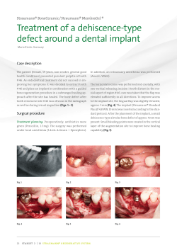
StraUMaNN® reGeNeratiVe SySteM
STARGET 1 I 12
FrANK BröSeLer
Treatment of a dehiscence-type defect around a dental
implant with Straumann ® MembraGel
Summary
dividual situation before or during use. The availability of a
This case report describes the treatment of a dehiscence-
biodegradable membrane which can be customized in-situ for
type alveolar bone defect around a dental implant with GBR
each individual defect thus offers an improvement for GBR pro-
(guided bone regeneration) where Straumann® MembraGel
cedures. Recently, a novel PEG-derived (polyethylene glycol)
is used as a biodegradable barrier. This membrane simp-
membrane for use with GBR indications has become available
lifies clinical handling compared to conventional membra-
on the European and North-American markets. After activation
nes because it is applied as a liquid.1 After application, the
by mixing the four different precursors, the membrane is applied
membrane solidifies within 20 – 50 seconds. This effectively
as liquid and forms a hydrogel within a few seconds after being
stabilizes the bone graft, in order to provide stable bone and
applied. The membrane undergoes hydrolytic degradation du-
peri-implant soft tissue conditions with a completely restored
ring the healing period. PEG has been shown to be biocompa-
emergence profile around the dental implant.
tible 9 and has been studied in other medical disciplines in the
past, for example, as a sprayable adhesion barrier 10,11 and in
Introduction
neurosurgery 12.Several preclinical studies using different animal
Alveolar bone defects in the jaw can be successfully treated
models have been conducted to evaluate the effectiveness of
using GBR techniques. The idea behind this treatment method
the membrane as barrier membrane in GBR procedures 13,14,15.
is to use a resorbable or non-resorbable barrier to prevent in-
First clinical data is available as well.1 Because Straumann®
growth of proliferating connective or soft tissue cells into the
MembraGel represents a completely new technology for usage
hard tissue defect and to create a space for bone tissue-deri-
in GBR procedures the surgical protocol for the augmentation
ved cells to grow 2,3. Barrier membranes can be manufactured
procedure has to be modified slightly over that of conventio-
from various natural or synthetic precursors. Today, the most
nal membranes. This case report illustrates a feasible treatment
frequently used resorbable membranes in dentistry are made
protocol for this new-membrane technology in the treatment of
. These membranes are available in standard
alveolar bone dehiscence around a bone level dental implant
of collagen
4,5,6,7,8
sizes and forms and need to be adapted to the patient’s in-
Fig. 1
Fig. 2
in combination with bone graft substitute material.
Fig. 3
29
30
STARGET 1 I 12
StraUMaNN® reGeNeratiVe SySteM
Patient history
wound healing after tooth extraction, typical horizontal and
The patient (female, 52 years at time of implant surgery,
vertical alveolar bone loss was found at position #41. For
overall in good health, non-smoker) had been treated for
this reason, prior to the planned implant insertion, a ridge
chronic periodontitis and undergone maintenance therapy
augmentation was performed using bovine-derived xenograft
since 1994 (Fig. 1). Due to pulp necrosis resulting from cari-
and a collagen membrane (in january 2010) 7. In November
es, tooth #41 was treated endodontically in 1997. Because
2010, the patient was scheduled to undergo implant insertion
of periapical periodontitis, an apicoectomy was performed.
(bone level) with a simultaneous augmentation of the alveolar
Due to recurring CAP, tooth #41 had to be extracted in 2009
ridge dehiscence located on the coronal section to ensure a
(Figs. 2, 3).
stable morphologic reconstruction of the alveolar region. For
the esthetic outcome in this case, this was important because
Initial situation
the patient’s lower gums were visible when she speaks.
Due to the limited prognoses for a second apicoectomy, the
proposed prosthetic solution for the patient was an implant-
Surgical procedure
supported single crown. The patient agreed after having
No antibiotics were used preoperatively since the patient
been informed about the prognostic factors and risks. During
underwent periodontal care and preoperative testing for the
Fig. 4
Fig. 5
Fig. 6
Fig. 7
Fig. 8
Fig. 9
StraUMaNN® reGeNeratiVe SySteM
STARGET 1 I 12
prevalence of periopathogenic microbiota, with no indica-
The activated Straumann® MembraGel was applied to the
tions of the MO above the detection limit. The surgery was
defect site. Next, 1-1.5 mm of the augmented area was out-
performed under local anesthesia. The horizontal incision was
lined and covered successively with the bone substitute ma-
made with a slightly lingual aspect, with two vertical relea-
terial. In the crestal aspect, the membrane was placed by
sing incisions, each 1 tooth distant in the mesial and distal
covering no more than approx. 1/3 of the implant screw sur-
aspect of region #41. A full mucoperiosteal flap was raised.
face. Complete coverage of the cover screw was avoided.
This flap shape was chosen to achieve sufficient access and
Care was taken to ensure that only a thin membrane layer
visibility to the treatment site and to ensure optimal blood
was applied during the procedure (Figs. 8, 9).
support after primary closure (Figs. 4, 5). The implant site was
prepared according to the standard protocol recommended
After application, the membrane sets in situ by gelation within
by the manufacturer (Fig. 6). Immediately after preparing the
20 - 50 seconds. No further fixation of the membrane is ne-
site, the periosteum-releasing incision was made to achieve a
cessary, as the gel adheres sufficiently to the surrounding
tension-free closure of the GBR site later, which then prevents
host bone.
heavy bleeding at time of membrane placement (Fig. 7). After
the placement of the bone level implant, a dehiscence-type
Healing was attempted with the implant in a submerged posi-
alveolar bone defect of approx. 4 mm was present. The de-
tion after tension-free closure of the released mucoperiosteal
fect was augmented with bone graft substitute material which
flap. The wound was closed with interrupted sutures made of
was slightly rehydrated in physiological saline prior to use.
a PvDF monofilament material (7-0, Fig. 10).
Overbuilding was avoided during augmentation procedure.
In order to optimize the attachment of Straumann® Memb-
Postoperative treatment
raGel to the recipient site, the host bone and the grafting
The patient was instructed to rinse four times daily with a pre-
material was dried with sterile gauze immediately before
pared aqueous 0.12% chlorhexidine solution. No analgesics
application.
had to be prescribed. The patient was informed that NSAIDs
Fig. 10
Fig. 11
Fig. 12
31
32
STARGET 1 I 12
StraUMaNN® reGeNeratiVe SySteM
could be used if necessary. The patient was also instructed to
The subsequent healing period was also normal (Figs. 12, 13).
refrain from mechanical plaque removal in the area of surgery
The soft tissue healing abutment was installed 4.5 months fol-
until time of suture removal. She had been provided with a
lowing the operation. The final restoration was completed 6
small removable prosthesis pre-operatively for esthetic purpo-
weeks later (Fig. 14). The FPD was designed as a cemented
ses. Care was taken to ensure the “dental flipper” did not apply
alloy-ceramic crown on a custom-made abutment.
any pressure to the wound, and the patient was instructed to
remove the flipper while sleeping.
The post-op clinical and radiological evaluation at 47 weeks
exhibited stable bone and peri-implant soft tissue conditions
After an unproblematic initial healing period (Fig. 11), the su-
(Fig. 15) with a fully restored emergence profile, particularly in
tures were removed 10 days after the implantation operation.
the horizontal aspect (Figs. 16, 17).
Fig. 13
Fig. 14
Fig. 16
Fig. 17
Fig. 15
References: See note on p. 28


© Copyright 2026