
Managing Colorectal Cancer: The Resectable and Potentially Resectable Patient— A Multidisciplinary Approach
Marshall_cvr_revised2
9/30/08
11:25 AM
Page 1
MANAGING COLORECTAL CANCER: THE RESECTABLE AND POTENTIALLY RESECTABLE PATIENT—A MULTIDISCIPLINARY APPROACH
Supported by an educational grant from
From the publishers of ONCOLOGY
COAB
Clinical Oncology Advisory Board
Managing Colorectal Cancer:
The Resectable and Potentially
Resectable Patient—
A Multidisciplinary Approach
John L. Marshall, MD
Lombardi Comprehensive
Cancer Center
Georgetown University
Michael A. Choti, MD, MBA, FACS
Johns Hopkins University
School of Medicine
CME
3
❖
Managing Colorectal Cancer:
The Resectable and
Potentially Resectable Patient—
A Multidisciplinary Approach
Edited by
John L. Marshall, MD
Chief
Division of Hematology and Oncology
Lombardi Comprehensive Cancer Center
Georgetown University
Washington, D.C.
Michael A. Choti, MD, MBA, FACS
Professor of Surgery and Oncology
Johns Hopkins University School of Medicine
Baltimore, Maryland
Publishers of
ONCOLOGY
Oncology News International
Cancer Management: A Multidisciplinary Approach
www.cancernetwork.com
i
COAB
Clinical Oncology Advisory Board
Note to the reader:
The information in this book has been carefully reviewed for accuracy of dosage and indications. Before prescribing any drug, however, the clinician should consult the manufacturer’s current package labeling for accepted indications, absolute dosage recommendations,
and other information pertinent to the safe and effective use of the product described. This
is especially important when drugs are given in combination or as an adjunct to other forms
of therapy. Furthermore, some of the medications described herein, as well as some of the
indications mentioned, may not have been approved by the U.S. Food and Drug administration at the time of publication. This possibility should be borne in mind before prescribing or recommending any drug or regimen.
Educational activities in the form of monographs, audio programs, supplements, and other
formats are sent to the readership of ONCOLOGY and Oncology News International on a
regular basis. All recipients of the journals can opt out of receiving them and accompanying
educational activities at any time by contacting our circulation department at CMPMedica,
phone: (203) 662-6551 or by e-mail: [email protected].
Copyright ©2008 by CME LLC. All rights reserved. This book is protected by copyright.
No part of it may be reproduced in any manner or by any means, electronic or mechanical,
without the written permission of the publisher. Value: $19.95.
Library of Congress Catalog Card Number 2008926818
ISBN 9781891483608
Cover image description: A patient with colorectal metastases. Preoperative evaluation identified two metastases confined to the right hemiliver. Figures: FDG-PET/CT (left), contrastenhanced MRI (upper right), intraoperative ultrasonography (lower middle), and intraoperative specimen of right hepatectomy (lower right). Images courtesy of Michael A. Choti,
MD, MBA, FACS.
Publishers of
ONCOLOGY
Oncology News International
Cancer Management: A Multidisciplinary Approach
www.cancernetwork.com
❖
Contents
CME
3
Contributing Authors
iv
Continuing Medical Education
vi
Acknowledgments
ix
Preface
xi
1 Defining the Multidisciplinary Team:
How the Community-Based Oncologist Can Build a Team
John L. Marshall, MD
1
2 Defining Resectable Metastatic Colorectal Cancer:
Indications, Outcomes, and Controversies
Michael A. Choti, MD, MBA, FACS
9
3 The Role of Imaging in the Management of Patients with
Potentially Resectable Colorectal Metastases
Eleni Liapi, MD, and Ihab R. Kamel, MD, PhD
4 Liver Toxicity and Systemic Treatment of Colorectal Cancer
Veena Shankaran, MD, and Al B. Benson III, MD, FACP
5 Locoregional Alternatives to Liver Resection:
Ablation, Intraarterial Therapy, and Radiation Therapy
Susan L. Logan, MD, MPP, and Eric K. Nakakura, MD, PhD
17
43
57
6 Suggested Strategies in the Management of Resectable and
Potentially Resectable Metastatic Colorectal Cancer
75
John L. Marshall, MD, and Michael A. Choti, MD, MBA, FACS
CME Post-Test
83
Index
87
To earn CME credit at no cost, please visit us online at
www.cancernetwork.com/cme
iii
❖
Contributing Authors
John L. Marshall, MD
Chief
Division of Hematology and Oncology
Lombardi Comprehensive Cancer Center
Georgetown University
Washington, D.C.
Michael A. Choti, MD, MBA, FACS
Professor of Surgery and Oncology
Johns Hopkins University School of Medicine
Baltimore, Maryland
Al B. Benson III, MD, FACP
Professor
Division of Hematology and Oncology
Robert H. Lurie Comprehensive Cancer Center
Feinberg School of Medicine
Northwestern University
Chicago, Illinois
Ihab R. Kamel, MD, PhD
Associate Professor, Radiology
Interim Clinical Director, MRI
Russell H. Morgan Department of Radiology and Radiological Science
Johns Hopkins University School of Medicine
Baltimore, Maryland
Eleni Liapi, MD
Postdoctoral Fellow
Russell H. Morgan Department of Radiology and Radiological Science
Division of Vascular and Interventional Radiology
Johns Hopkins University School of Medicine
Baltimore, Maryland
iv
Contributing Authors
Susan L. Logan, MD, MPP
Fellow, Section of Hepatobiliary-Pancreatic Surgery
Washington University School of Medicine in St. Louis
St. Louis, Missouri
Eric K. Nakakura, MD, PhD
Assistant Professor of Surgery
Helen Diller Family Comprehensive Cancer Center
Division of Surgical Oncology
Department of Surgery
University of California, San Francisco, School of Medicine
San Francisco, California
Veena Shankaran, MD
Postdoctoral Fellow
Robert H. Lurie Comprehensive Cancer Center
Feinberg School of Medicine
Northwestern University
Chicago, Illinois
v
❖
Continuing Medical Education
Monograph
Activity Release Date: November 1, 2008
Activity Expiration Date: November 1, 2009
About the Activity
The CME activity is based on the information learned from reading this
monograph, Managing Colorectal Cancer: The Resectable and Potentially
Resectable Patient—A Multidisciplinary Approach. It was developed from
an identified educational need for information about practical management issues in the practice of medical, surgical, and radiation oncology.
This activity has been developed and approved under the direction of
CME LLC.
Activity Learning Objectives
After reading Managing Colorectal Cancer: The Resectable and Potentially
Resectable Patient—A Multidisciplinary Approach, participants should be
able to:
• Understand that neo-adjuvant/perioperative chemotherapy can help
some metastatic colorectal cancer patients.
• Incorporate a team approach into practice (i.e., community-based
oncologist, surgeon, and radiotherapist).
• Demonstrate knowledge of the proper timing for chemotherapeutic/biologic agents, types of agents, and amount of chemotherapy
for those patients needing resection.
• Apply monitoring guidelines with radiologist to prevent hepatotoxicity.
• Use the latest surgical techniques in liver resection.
vi
Continuing Medical Education
vii
Target Audience
This activity targets physicians in the fields of oncology and hematology.
Accreditation
This activity has been planned and implemented in accordance with the
Essential Areas and Policies of the Accreditation Council for Continuing
Medical Education through the joint sponsorship of CME LLC and The
Oncology Group. CME LLC is accredited by the ACCME to provide
continuing medical education for physicians.
Credit Designation
CME LLC designates this educational activity for a maximum of 3 AMA
PRA Category 1 Credits™. Physicians should only claim credit commensurate with the extent of their participation in the activity.
Physicians not licensed in the United States who participate in this CME
activity are eligible for AMA PRA Category 1 Credit(s)™.
Compliance Statement
This activity is an independent educational activity under the direction
of CME LLC. The activity was planned and implemented in accordance
with the Essential Areas and Policies of the ACCME, the Ethical Opinions/Guidelines of the AMA, the FDA, the OIG, and the PhRMA Code
on Interactions with Healthcare Professionals, thus assuring the highest
degree of independence, fair balance, scientific rigor, and objectivity.
However, CME LLC, the Grantor, and CMPMedica shall in no way be
liable for the currency of information or for any errors, omissions, or
inaccuracies in the activity. Discussions concerning drugs, dosages, and
procedures may reflect the clinical experience of the author(s), or they
may be derived from the professional literature or other sources and may
suggest uses that are investigational in nature and not approved labeling
or indications. Activity participants are encouraged to refer to primary
references or to the full prescribing information resources. The opinions
and recommendations presented herein are those of the author(s) and do
not necessarily reflect the views of the provider or producer.
To earn CME credit at no cost, please visit us online at
www.cancernetwork.com/cme
viii
Continuing Medical Education
Financial Disclosure
Dr. Marshall has received honoraria, research support, and has served
as a consultant for Roche, sanofi-aventis, Pfizer, Genentech, Boehringer
Ingelheim, Amgen, Bristol-Myers Squibb, and ImClone. Dr. Choti has
served as a consultant and speaker for sanofi-aventis and Genentech. Dr.
Benson has received research support from and served as a scientific
advisor for Genentech, Amgen, Roche, Pfizer, ImClone, Bristol-Myers
Squibb, Taiho, and sanofi-aventis. Drs. Kamel, Liapi, Logan, Nakakura,
and Shankaran have no financial relationships to disclose.
Copyright
Copyrights owned by CME LLC. Copyright ©2008.
Contact Information
We would like to hear your comments regarding this or other activities
provided by CME LLC. In addition, suggestions for future programming are welcome. Contact us at:
Address:
Phone:
Director of Continuing Education
CME LLC
Harborside Financial Center
Plaza 3, Suite #806
Jersey City, NJ 07311
(888) 618-5781
Supported by an educational grant from
❖
Acknowledgments
We both would like to express our great thanks to our colleagues who
helped author this book, our collaborators in our everyday practices,
our families who supported our vision, and all the patients who put
their lives into our hands.
––John L. Marshall, MD, and Michael A. Choti, MD, MBA, FACS
ix
❖
Preface
The possibility of curing patients with metastatic colon cancer is possibly the greatest advance in gastrointestinal cancers in the past twenty
years. Newer surgical techniques, novel and more effective chemotherapy, and better imaging have expanded the number of patients who are
candidates for this approach.
However, along with this is increasing confusion about how best to
achieve the goal. Clearly, we need increased multidisciplinary communication, access to experts in the field, and an understanding of the
“rules” of the game.
We hope that you find this text helpful in your practice. We have
tried to bring all these issues together in one easy-to-read text.
Best regards,
John L. Marshall, MD, and Michael A. Choti, MD, MBA, FACS
xi
1
Defining the
Multidisciplinary Team:
How the Community-Based
Oncologist Can Build a Team
John L. Marshall, MD
It has only been a few short years since we have recognized that selected
patients with metastatic colorectal cancer can, in fact, be cured. When
most of us trained in medical school, the thought of performing surgery
on metastatic lesions with the hope of curative intent was quite foreign.
Our understanding of metastatic cancer was that while there may be
only a few visible metastatic lesions, we were certain that all of these
patients had significant “invisible” microscopic metastatic disease that
would, in the end, define the patient’s outcome. However, after nearly
two decades of focused work in the area of metastatic colon cancer
resections, there has developed an understanding that a subset of
patients with metastatic disease can, in fact, be cured through surgical
techniques. The improvements in chemotherapy and imaging modalities
and the enhanced skills of hepatic and other cancer surgeons have come
together to better the patient’s odds of curative therapy despite having
increasingly higher risk disease.
In this chapter, I focus on the requirements for a multidisciplinary
team whose charge is to define and carry out curative therapy for
CME
1
2
Defining the Multidisciplinary Team
patients with metastatic colorectal cancer. We recognize that this effort
is not housed in one medical subspecialty but, in fact, requires detailed
ongoing communications between a variety of medical specialists, each
of whom is focused on and dedicated to patients with this disease. The
challenge for us as a medical community is to provide the optimum
resources for our patients. Our hope is that this chapter will help serve
those who are attempting to put together such a resource.
Recognizing the Potentially Curable Patient
The first and most important element in the pathway of curative resection for patients with metastatic colorectal cancer is to recognize those
patients who may be candidates. In general, these patients are divided
into two main categories (Figure 1).
The first category are those who are thought to be resectable at presentation. Resectability will be defined in detail in Chapter 2 of this
book. However, medical oncologists are not typically trained in the
anatomy of the liver and liver metastases and, therefore, may not have
the expertise to define a patient who is resectable and who is not. However, it is the medical oncologist who typically is the primary caregiver
for a patient in this setting who also will initiate the process. Therefore,
medical oncologists should become familiar with the definitions so that
they can initiate the referrals in order to optimize the patient’s chance
for curative resection.
Diagnosis of MCRC
Resectable
Neo-adjuvant/
Preoperative
therapy
Unresectable
Borderline/
Potentially
Resectable
First-Line
Second-Line
Treatment
continuum
Surgery
Third-Line
Adjuvant therapy
Fourth-Line
Figure 1. Anatomic division of metastatic colon cancer. MCRC = metastatic colorectal cancer.
Defining the Multidisciplinary Team
3
In some ways, the patient who has immediately resectable disease is
the easiest one to define. These typically are patients with one or two
small, peripherally isolated lesions within the liver or lung. Even to a
fairly untrained eye, this would seem to be anatomically resectable.
More difficult are the cases where either multiple lesions exist in multiple lobes of the liver, where lesions are found close to key vascular channels, or when multiple organs are involved. There is no strict definition
of what is resectable and what is not.
The second category is patients who have lesions that are too large
for resection but may have them reduced in size through chemotherapy.
The concept of taking patients who are initially unresectable and making them resectable through downsizing of the lesion has been dubbed
“conversion therapy.” Within this group, two subtypes emerge, those
with a limited number of lesions that are too large for resection and
those with multiple lesions at presentation who experience pathologic
complete responses in some areas. This latter group will be discussed in
Chapter 6 as they represent a controversial circumstance. In both categories, the medical oncologist is reviewing the patient’s computed
tomography scan or imaging reports that define the recurrent disease
and is most often responsible for initiating the process.
I would like to stress at this point that using the traditional average
radiology report is frequently inadequate to define the patient’s resectability. While radiologists are increasingly recognizing the need to make
this important distinction, many radiology reports still refer to “multiple hepatic metastases,” which typically is considered unresectable but
may in fact be anatomically resectable. Therefore, it is critical for the
initiating physician to review the actual films either with the radiologist
or with the hepatic surgeon. Once the patient has been defined as either
resectable immediately or may benefit from conversion therapy, it is
appropriate to incorporate the other team members into the process of
creating a plan of attack with curative intent in the metastatic setting.
Some patients require referral before chemotherapy is initiated, whereas
others may be referred later in the process. Having the team and referral
patterns established is key to providing optimal care.
Team Members
Following the identification of a potential candidate for resection of
metastatic disease, a series of other physician and physician teams are
typically involved (Figure 2). First, additional imaging is frequently
obtained. Either using magnetic resonance imaging or positron emission
4
Defining the Multidisciplinary Team
• Surgeon
• Radiologist
– CT, MRI, PET?
• Interventional Radiologist
– RFA?
• Radiation Oncology
– IMRT/Cyberknife?
• Medical Oncologist
– Role of chemotherapy, before or
after surgery
• How do you decide who does what
and when?
Figure 2. Curing stage IV colon cancer requires a multidisciplinary team.
CT = computed tomography, IMRT = intensity-modulated radiation therapy, MRI = magnetic resonance imaging, PET = positron emission tomography, RFA = radiofrequency ablation.
tomography (PET) scanning or both, the patients are imaged in order to
determine first the anatomy of the disease that is known and to try to
define any other disease that may not be immediately recognized on initial scanning.
A recent abstract from the 2008 annual meeting of the American
Society of Clinical Oncology suggests that PET scanning is quite useful
in defining the patients who ultimately will be found to have unresectable disease and, therefore, appears to be an important step in the process
of screening the patient for resectability (1). Whether one uses computed
tomography scan, magnetic resonance imaging, or PET scanning in
many ways depends on the expertise, skill, and equipment of the local
community. What the best imaging modality is for your team should be
discussed with the radiology groups in the area.
The key to the entire process is, of course, the surgeon. Hepatic surgery has improved dramatically over the last decade, and more physicians are engaging in hepatic resections. However, hepatic surgery
remains a surgical technique that requires a great deal of experience to
produce the best outcomes. Evidence suggests that surgical centers with
high volumes of these procedures have much better outcomes (2). Experienced surgeons are better equipped to handle unexpected findings such as
finding multiple hepatic metastases or when to incorporate radiofrequency ablation (RFA) or the use of staged surgeries that can be incorporated. Not all communities can support such a surgeon; and therefore, it
Defining the Multidisciplinary Team
5
is a strong recommendation that patients requiring hepatic resections be
referred to specialized centers for this intervention. Certainly most colon
cancer surgery is performed by general surgeons within the community
setting. Most, if not all, of those general surgeons are competent to perform liver wedge resections during a standard operation. However, as the
complexity increases, the patient is best served by surgery performed by a
dedicated hepatic surgeon.
Thoracic lesions are also legitimate targets for surgical resection. In
this setting, it is also important to refer patients to chest surgeons experienced in this area. Newer, minimally invasive techniques may speed the
recovery and are quite appropriate for patients in whom pulmonary
lesions are to be removed. Here again, timing the different procedures of
chemotherapy and surgery must be coordinated.
Interventional radiology remains an important part of curative therapies for patients with hepatic metastases. RFA techniques are recognized
as a valid tool for the treatment of hepatic metastases, and RFA is
increasingly being used in other anatomic locations, although fewer data
exist to support this. The data on long-term survival and local control
are not as good as with surgical resections but often, due to a variety of
circumstances, surgical resection is not possible. Therefore, interventional radiology remains an important element in the creative strategies
of patients with metastatic disease.
Radiation oncology traditionally does not play a role in the management of hepatic metastases for curative intent. Certainly radiation can
be an effective palliation. However, newer techniques are raising the
question of whether treatment such as Cyberknife-focused radiation
may have the potential for long-term disease control. Certainly no clinical trials have been done at this point to test this, and it would be incorporated only if surgery or RFA is not possible.
Central to the process is the medical oncologist who serves as the
“quarterback” and primary care physician for these patients as they are
ushered through the team system. This responsibility requires that the
oncologist be familiar with the various techniques available to the
patients, what expertise is in their own community and what will need
to be referred outside, and what role chemotherapy plays in the process.
In many ways, this book is directed toward oncologists to provide them
with a guide for optimizing care.
If your community does not have the expertise as outlined above, then
it would be my recommendation that these patients be referred to a center
that can provide this sort of multidisciplinary interactive approach. Common in our Washington, D.C.–based system is for patients to be referred to
our center for hepatic surgery or interventional radiology and often for
6
Defining the Multidisciplinary Team
consideration of clinical trials before or after this therapy. However, it is
also most common for these patients to remain with their primary medical
oncologist and to be followed for life by them. Unless the local community
is willing to invest significant resources to provide this infrastructure, referring to specialized centers seems the most logical step for most groups in
less populated regions.
Develop a Strategy: Who Goes First?
Probably one of the most difficult decisions in approaching the patient
with curative intent is who should go first. There is no clear data that suggests that chemotherapy before or after surgical resection is required
when patients are resectable at presentation. In patients who have larger
tumors and are in need of conversion therapy, this answer is the easiest
because chemotherapy must be given first, as a response is required. However, in patients who have immediately resectable disease, many factors,
most of which are subjective, come into play in deciding whether surgery
or chemotherapy should be first. Even the decisions about surgery versus
RFA are sometimes difficult and depend a lot on the patients and the
expertise of the individuals at hand.
Communication is the key. Most hospitals and cancer groups interact
through a tumor board—typically weekly meetings that review cases.
Given the increasing volume of all types of cancer cases to be presented in
a typical tumor board, it is reasonable to develop a tumor board dedicated to gastrointestinal cancers or to use part of ongoing tumor boards
to serve this purpose. Many breast cancer groups have come to the same
conclusion and have developed breast-specific tumor boards. What is
important is to get all parties together to discuss the strategy. This, of
course, can be done virtually with effective referral patterns and communication systems in place. Given that for many cases, there is no clear correct sequence of multi-modality therapy, we must also weigh a patient’s
preferences. There are some patients who are not eager to pursue surgery
unless it is going to have a high-level benefit, and often then chemotherapy is given first to those patients. There are others who are eager to have
the surgery as soon as possible. Whenever multi-modality therapy is used,
it is my experience that physician bias, personality, institutional tradition
(and even simple issues such as a physician’s schedule) can play a major
role in how patients are treated. We must recognize these issues and do
what we can to prevent them from interfering with optimal care.
Defining the Multidisciplinary Team
7
Continual Reassessments
After initiating the treatment approach, there must be ongoing reassessments by all parties. The bar for defining resectable disease is changing
rapidly. More and more aggressive surgical techniques have been incorporated into patient management, but we must maintain an appropriate
balance between the new frontier of surgical resection in metastatic disease, and the reality that metastatic colon cancer typically is a multi-focal
disease, and currently, only a few patients are cured through surgical
techniques. Again, some patients will become resectable while others will
become higher risk, poor candidates. The ongoing monitoring of patients
typically falls to the medical oncologist who should keep the various parties informed as the patient progresses through the steps of the treatment.
In conclusion, most communities should have recognized experts in
the area of colorectal cancer management. Not all communities will
have all of the different tools that are necessary to have a complete multidisciplinary team. By defining the team and its key components, medical communities can decide how they want to provide this care for their
patients. Certainly throughout the country, there are cancer centers that
can provide this sort of care and can support smaller communities, but
the majority of the care can be delivered in the local community.
References
1. Wiering B, Oyen W, Van der Sijp J, et al. Improved selection of patients for
hepatic surgery of colorectal liver metastases with FDG-PET: a randomized
study. Proc Am Soc Clin Oncol 2008;26:179s. Abstract 4004.
2. Sah BK, Zhu ZG, Chen MM, et al. Effect of surgical work volume on postoperative complication: superiority of specialized center in gastric cancer
treatment. Langenbecks Arch Surg 2008; Jun 27.
2
Defining Resectable
Metastatic Colorectal Cancer:
Indications, Outcomes, and
Controversies
Michael A. Choti, MD, MBA, FACS
Cancer of the colon and rectum account for the majority of primary tumors
that develop isolated liver metastases and are candidates for surgical resection. This malignancy is the third most commonly diagnosed cancer in the
United States and second overall in cancer mortality. Approximately 20%
of patients have clinically recognizable liver metastases at the time of their
primary diagnosis. After resection of a primary colorectal cancer in the
absence of apparent metastatic disease, approximately 50% of patients will
subsequently manifest metastatic liver disease. Given these figures, one can
expect that at least 30,000 patients per year in the United States will
develop metastatic colorectal cancer confined to the liver, each year.
Selecting Patients for Surgical Resection
Perioperative mortality associated with liver resection has decreased from
20% several decades ago to close to 1% in patients undergoing liver
resection in more recent years. In deciding which patients will tolerate a
major liver resection, a number of factors need to be considered, including
patient comorbidities. Patients with underlying coronary artery disease,
CME
9
10
Defining Resectable Metastatic Colorectal Cancer
congestive heart failure, renal insufficiency, as well as other debilitating
states should be considered at greater risk for postoperative complications. A major goal of the preoperative evaluation, therefore, is to identify
patients who are at a high operative risk so that patients who represent a
prohibitive risk can be excluded while those patients with manageable
comorbidities can have these conditions addressed preoperatively in an
attempt to reduce their risk.
The health and function of the non–tumor-bearing liver is clearly one
of the most important factors impacting resectability and outcomes following liver resection. In patients undergoing surgery for hepatic colorectal metastases, cirrhosis is rarely present. However, increasing use of
prolonged preoperative combination chemotherapy can result in significant steatosis, steatohepatitis, and sinusoidal dilatation. These changes
can, in some cases, be associated with increased postoperative morbidity.
In patients with cirrhosis, the postoperative morbidity following partial
hepatectomy remains significant, primarily related to liver dysfunction.
Assessment of hepatic functional reserve is important when deciding
whether resection should be pursued.
New Criteria for Defining Resectability in
Patients with Hepatic Metastases
In the past, resection of hepatic colorectal metastases was not attempted in
patients who had more than three or four metastases, hilar adenopathy,
metastases within 1 cm of major vessels such as the vena cava or main
hepatic veins, or extrahepatic disease. More recent studies demonstrate,
however, that patients with these clinicopathologic factors can achieve longterm survival following hepatic resection and therefore should not be
excluded from surgical consideration. Specifically, the number of metastases
is no longer considered a contraindication to surgery (1). Similarly, contiguous extension to adjacent anatomical structures and local or regional recurrence at the site of the primary colorectal cancer are not contraindications
to resection. An increasing number of studies also indicate that although
survival may be reduced in patients with extrahepatic colorectal metastases
(2), complete resection of limited extrahepatic disease in conjunction with
resection of hepatic metastases can result in long-term survival.
Taken together, these data have led to a shift in the definition of resectability from criteria based on the characteristics of the metastatic disease
(tumor number, size, etc.) to new criteria based on whether a macroscopic
and microscopic complete (R0) resection of the liver disease can be
achieved (2). Currently, hepatic colorectal metastases should be defined as
Defining Resectable Metastatic Colorectal Cancer
11
Table 1. Criteria defining resectability for surgical resection
1. Macroscopic and microscopic (R0) treatment of the disease is feasible with
either resection alone or resection combined with radiofrequency ablation.
2. At least two adjacent liver segments can be spared.
3. Adequate vascular inflow, outflow, and biliary drainage can be preserved.
4. Sufficient remnant liver volume (>20% of the total estimated liver volume in
normal liver, >40% of total estimated liver volume in cirrhotic liver).
resectable when it is anticipated that disease can be completely resected,
two adjacent liver segments can be spared, adequate vascular inflow and
outflow and biliary drainage can be preserved, and the volume of the liver
remaining after resection is sufficient (3) (Table 1).
The size of the remnant volume considered safe varies with the condition of the hepatic parenchyma. In healthy livers, a remnant liver volume
greater than 20% of the estimated total liver volume is considered sufficient (4). In contrast, patients with cirrhosis need at least 40% remnant
liver volume in order to avoid postoperative liver failure. Computed
tomography or magnetic resonance imaging can now provide an accurate, reproducible method for preoperatively measuring the volume of
the future liver remnant. In cases where major hepatectomy is planned
Figure 1. Conversion of tumors to a resectable state.
12
Defining Resectable Metastatic Colorectal Cancer
Table 2. Categories of resectability
1. Initially resectable disease by standard approach
2. Initially resectable but requires extended approach
• Staged resections
• Preoperative portal vein embolization
• Resection plus ablation
3. Initially unresectable but likely convertible with tumor response
4. Initially unresectable and unlikely convertible
and there is concern regarding insufficient liver volume, these patients
should be considered for staged liver resection or portal vein embolization (PVE) to induce hypertrophy of the contralateral liver lobe (5). Similarly, staged resections allow for removal of a portion of metastatic
disease in one part of the liver, followed by some hepatic regeneration
before proceeding to a second-stage completion resection. Preoperative
chemotherapy can also be used in patients who are thought to be initially
unresectable. In some cases, tumor response can result in conversion to a
resectable state (Figure 1). Taken together, resectability can be classified
into resectable using a standard approach, resectable through an
extended approach, potentially convertible with preoperative therapy, or
unresectable and unlikely convertible (Table 2).
Outcomes of Liver Resection for Hepatic
Colorectal Metastases
Overall, the perioperative mortality of liver resection for colorectal metastases is approximately 1% in most current reported series. In experienced
hands, even major hepatic resections (hemi-hepatectomy or extended
hepatectomy), which are performed in about half of the cases, result in
perioperative mortalities of less than 5%. The potential for adverse outcome and the complexity of these operations justifies the recommendation
that major liver resection be performed at centers and by surgeons having
experience with such procedures. Complication rates for liver resection of
metastatic disease are approximately 15%. The major morbidity associated specifically with liver resection includes hemorrhage, perihepatic
abscess, bile leak and/or fistula, pleural effusion, and hepatic failure.
With regard to survival, large series from the 1960s through the mid1990s reported 5-year survival rates in the range of 33%–36% for
Defining Resectable Metastatic Colorectal Cancer
13
Table 3. Reported 5-year survival after resection of colorectal liver metastasis with curative intent
Year of
publication
Investigator
Scheele et al. (6)
Fong et al. (7)
Choti et al. (8)
Abdalla et al. (10)
Fernandez et al. (11)
Pawlik et al. (9)
1995
1999
2002
2004
2004
2005
Years included
in study
1960–1992
1985–1998
1993–1999
1992–2002
1992–2002
1990–2004
5-Year
survival %
39
37
58
58
58
58
patients with colorectal liver metastases resected with curative intent
(6,7). However, more recent data have shown an improved 5-year survival rate of 58% following complete resection of colorectal liver metastases (8–11) (Table 3). This improvement in overall survival likely
reflects improvement in patient selection, surgical technique, and more
effective adjuvant therapy.
Several clinicopathologic factors predictive of patient survival after
hepatic resection have been identified. These include stage, grade, and
nodal status of the primary colorectal tumor; disease-free interval from
diagnosis of primary tumor to diagnosis of liver metastases; number and
distribution of liver metastases; level of preoperative carcinoembryonic
antigen; and presence of extrahepatic disease. Although preoperative
factors may be generally instructive, these factors should not be used to
exclude patients from surgical consideration. Patients with one or multiple negative prognostic factors can still derive a significant survival
advantage from hepatic resection of their colorectal metastases.
Extrahepatic Disease:
Contraindication to Surgery?
Historically, the presence of extrahepatic metastatic disease was routinely considered an absolute contraindication to surgical therapy with
curative intent. However, the role of hepatic resection in this setting has
recently been reexamined. Approximately 5%–10% of patients who
present with metastatic disease will have a combination of liver and lung
metastases; and therefore, there has been interest in how this subset of
patients should be treated. Several studies have reported results for
14
Defining Resectable Metastatic Colorectal Cancer
patients undergoing combined lung and liver resection (12). These studies have reported 5-year survival rates of greater than 30%. There are,
however, a number of factors that do appear to be associated with a particularly poor prognosis. Patients with bilateral disease or who have
more than six pulmonary metastases were 50%–70% more at risk for
disease-specific death compared with other patients. Thus, particular
attention must be paid when considering these high-risk patients for
combined liver and lung resection.
Resection of other extrahepatic sites is more controversial. Unlike pericolic nodal disease, hilar lymph nodes are felt to be “metastases from
metastases” and are associated with a poor outcome. Some investigators
have reported long-term survival in selected patients with hilar nodal metastases and have concluded that this patient population may still benefit from
hepatic resection (13). Some centers have advocated resection of peritoneal
carcinomatosis as well. These procedures are typically incomplete debulking procedures performed in combination with intraperitoneal chemotherapy. However, such therapy is not the standard of care and, in general, liver
resection is not indicated in the presence of diffuse carcinomatosis. As with
liver resection itself, the ability to achieve a complete macro- and microscopic resection of all known disease is critical to long-term outcome.
These data serve to emphasize that hepatic resection in the setting of extrahepatic disease is only warranted when an R0 resection is feasible. Patients
with disease not amenable to a complete resection should not be offered
combined metastectomies of intra- and extrahepatic disease.
Summary
Liver resection currently represents one of the most effective therapeutic
options for patients with limited colorectal metastases. Recent improvements in whole-body and hepatic imaging have allowed for more accurate selection of those patients who may benefit most from resection.
Traditional clinicopathologic factors, although helpful in stratifying
patients with regard to prognosis, should not be used to exclude otherwise resectable patients from surgery, particularly in those with hepatic
colorectal metastases. The use of modern surgical techniques is reducing
perioperative morbidity and mortality, while PVE, preoperative therapy,
and combining resection with other approaches, such as ablation, can
expand the population of patients who are candidates for surgical treatment. Perhaps the most important strategy when considering the multitude of therapeutic options for such patients with metastatic colorectal
cancer is the development of an individual treatment plan based on dis-
Defining Resectable Metastatic Colorectal Cancer
15
cussion among a multidisciplinary team of specialists, including surgeons, medical oncologists, and radiologists.
References
1. Kokudo N, Imamura H, Sugawara Y, et al. Surgery for multiple hepatic
colorectal metastases. J Hepatobiliary Pancreat Surg 2004;11:84–91.
2. Pawlik TM, Schulick RD, Choti MA. Expanding criteria for resectability of
colorectal liver metastases. Oncologist 2008;13:51–64.
3 Charnsangavej C, Clary B, Fong Y, et al. Selection of patients for resection
of hepatic colorectal metastases: expert consensus statement. Ann Surg
Oncol 2006;3(10):1261–1268.
4. Abdalla EK, Denys A, Chevalier P, et al. Total and segmental liver volume
variations: implications for liver surgery. Surgery 2004;135(4):404–410.
5. Abdalla EK, Hicks ME, Vauthey JN. Portal vein embolization: rationale,
technique and future prospects. Br J Surg 2001;88:165–175.
6. Scheele J, Stang R, Altendorf-Hofmann A, Paul M. Resection of colorectal
liver metastases. World J Surg 1995;19:59–71.
7. Fong Y, Fortner J, Sun RL, et al. Clinical score for predicting recurrence
after hepatic resection for metastatic colorectal cancer: analysis of 1001 consecutive cases. Ann Surg 1999;230:309–318, discussion 318–321.
8. Choti MA, Sitzmann JV, Tiburi MF, et al. Trends in long-term survival following liver resection for hepatic colorectal metastases. Ann Surg 2002;235:
759–766.
9. Pawlik TM, Scoggins CR, Zorzi D, et al. Effect of surgical margin status on
survival and site of recurrence after hepatic resection for colorectal metastases. Ann Surg 2005;241:715–722, discussion 722–714.
10. Abdalla EK, Vauthey JN, Ellis LM, et al. Recurrence and outcomes following hepatic resection, radiofrequency ablation, and combined resection/ablation for colorectal liver metastases. Ann Surg 2004;239:818–825.
11. Fernandez FG, Drebin JA, Linehan DC, et al. Five-year survival after resection of hepatic metastases from colorectal cancer in patients screened by
positron emission tomography with F-18 fluorodeoxyglucose (FDG-PET).
Ann Surg 2004;240:438–447.
12. Murata S, Moriya Y, Akasu T, et al. Resection of both hepatic and pulmonary metastases in patients with colorectal carcinoma. Cancer 1998;83:
1086–1093.
13. Jaeck D. The significance of hepatic pedicle lymph nodes metastases in surgical management of colorectal liver metastases and of other liver malignancies. Ann Surg Oncol 2003;10:1007–1011.
3
The Role of Imaging in the
Management of Patients
with Potentially Resectable
Colorectal Metastases
Eleni Liapi, MD, and Ihab R. Kamel, MD, PhD
In the United States, colorectal cancer is one of the most common malignancies and a leading cause of death (1). Hepatic metastases are present
in 15%–25% of patients at the time of diagnosis of colorectal cancer
and another 25%–50% will develop liver metastases in 5 years’ time,
after resection of the primary tumor (2). In approximately half of these
patients, metastatic disease is confined to the liver. For these patients,
resection of the metastases is the treatment of choice and can result in a
5-year survival rate of 35%–58% (3). Recent advances in surgical techniques, refinement of patient selection, and available imaging modalities
have challenged the traditional inclusion criteria of patients eligible for
resection and have increased the number of patients who could benefit
from surgery (4). It is therefore essential to identify the patients who are
more likely to benefit from surgery. Radiologic imaging plays a critically
important role in identifying and staging these patients.
The imaging modalities available for assessment of patients with potentially resectable colorectal metastases include transabdominal ultrasonography (US), computed tomography (CT), 18F fluoro-2-deoxy-D-glucose
positron emission tomography (FDG-PET), and magnetic resonance imagCME 17
18
Role of Imaging in the Management of Patients
ing (MRI). Although extensive improvement and research have been performed in regard to the diagnostic performance of CT, MRI, and FDG-PET
for the detection of colorectal liver metastases, the optimal imaging staging
strategy is yet to be defined. Currently, the optimal imaging strategy for
patients with potentially resectable colorectal liver metastases depends to
some degree on the local resources and expertise, as well as the available
imaging modalities. A multi-modality strategy is recommended because no
single modality can accurately detect all colorectal liver metastases.
The goals of imaging assessment for these patients are:
1.
2.
3.
4.
5.
To define the number and extent of hepatic metastases in the
segmental and lobar distribution
To assist in the planning of the hepatic resection
To evaluate the residual volume of the remnant liver (when
extensive resection is being planned)
To identify extrahepatic disease, including nodal metastases,
peritoneal implants, regional or other sites of hematogenous spread
such as pulmonary metastases
To assess response to neo-adjuvant therapy for downstaging of
initially unresectable hepatic metastases.
In this chapter, we aim to clarify the role of each imaging modality in
the evaluation and staging of patients with potentially resectable colorectal metastases. Moreover, as imaging plays a vital role in surgical
planning, we aim to depict the essential elements (morphologic and
functional) of hepatic image interpretation that play a role in refining
patient selection for surgery.
Definition of Resectability
The definition of surgical resectability of liver metastases has changed
over time as new chemotherapeutic agents and more aggressive therapies have emerged (5). In the past, resection of hepatic colorectal metastases was not attempted in patients who had more than three or four
metastases, hilar adenopathy, metastases within 1 cm of major vessels
such as the vena cava or main hepatic veins, or extrahepatic disease.
Recent studies, however, demonstrate that some patients who fulfill
the above “exclusion” criteria may achieve long-term survival after
hepatic resection and therefore should not be excluded from surgical
consideration. These recent data have lead to a reformation of the definition of resectability. Specifically, the number of metastases is no longer
considered a contraindication to surgery (6). Similarly, extension to
Role of Imaging in the Management of Patients
19
adjacent anatomic structures and local or regional recurrence at the site
of the primary colorectal cancer are not contraindications to resection.
An increasing number of studies also indicate that although survival
may be reduced in patients with extrahepatic colorectal metastases,
complete resection of limited extrahepatic disease can result in longterm survival (5). Currently, hepatic colorectal metastases should be
defined as resectable when:
a.
b.
c.
d.
it is anticipated that the disease can be completely resected,
two adjacent liver segments can be spared,
adequate vascular inflow and outflow and biliary drainage can be
preserved, and
the volume of the future liver remnant will be adequate (at least
20% of the total estimated liver volume).
Imaging Techniques
All patients being considered for resection of colorectal liver metastases
should undergo preoperative CT or MRI to evaluate the extent of intrahepatic disease and to exclude extrahepatic metastases. FDG-PET has
been recently recommended for routine preoperative imaging, which is
particularly sensitive in the detection of extrahepatic disease (7). Below,
we describe how each imaging modality contributes to the diagnosis and
staging of these patients.
Transabdominal Ultrasonography
Conventional transabdominal US has a relatively low sensitivity (53%–
77%), compared with CT (85%) and intraoperative ultrasound (95%)
for the detection of liver metastases (8). The US sensitivity depends on
the size of a metastasis, and is only 20% for metastases less than 10 mm
(9). Moreover, isoechoic metastases are difficult to detect on conventional US, while hyperechoic metastases can mimic hemangiomas (10).
Finally, it is well known that US is significantly more operator dependent than the other imaging methods, and its sensitivity is reduced in
patients with obesity, interposition of intestine, tissue-composition, or
lack of patient cooperation.
Recent studies have shown that the US technique using IV contrast
media seems to improve sensitivity in detecting liver metastases by about
50% (ranging from 63% to 91%) and improve specificity from 60% to
20
Role of Imaging in the Management of Patients
88% (9). These promising results may initially seem comparable to the
best-reported results of CT; however, a closer look reveals several limitations such as patient selection bias and no clear comparison to gold
standards (11).
In our institution, we use 3.5–5 MHz curved-array transducers to
detect liver metastases using percutaneous transabdominal US. Findings
suggestive of metastases include solid lesions and the presence of a
hypoechoic halo surrounding a liver mass (Figure 1.)
Power Doppler and second-harmonic imaging with contrast agents
may improve detection and characterization of liver metastases.
Multidetector Computed Tomography
Multidetector CT (MDCT), performed with IV injection of iodinated
contrast medium, allows single breath-hold, volumetric data acquisition
during multiphase imaging for angiographic and parenchymal evaluation of the liver. The main objective in scanning the liver using MDCT is
to obtain timed hepatic arterial phase and portal venous phase accurately, each in a single breath-hold (12). Recent studies with MDCT
scanners have increased sensitivity of lesion detection to between 70%
and 95% (13). However, lesions smaller than 1 cm remain problematic
because of a high false-negative rate of about 10% and nonspecificity of
lesions. Colorectal hepatic metastases are most commonly hypovascular
and are most conspicuous in the portal-venous phase, when there is high
contrast between them and the enhanced surrounding hepatic parenchyma. In the late arterial phase, rim enhancement is often present, but
the most sensitive phase for detection of colorectal liver metastases is the
portal venous phase (Figure 2) (14).
Figure 1. Ultrasound images of colorectal hepatic metastases. Note that
these metastases may be isoechoic (A) or with mixed echogenicity (B). Also
note the hypoechoic halo around them.
Role of Imaging in the Management of Patients
21
The invasive CT arterial portography, which is a sensitive but less
specific imaging technique, is used less nowadays with the availability of
high-quality MDCT.
In our institution, 64-detector CT scanners are currently used for
hepatic imaging. A 64-detector CT scanner allows thin (0.6-mm) collimation and small (0.5-mm) reconstruction intervals, generating true
isotropic volumetric data sets, superior 3D image reconstruction and volume-rendering. Contrast enhancement typically is achieved using 120–
150 mL (2 mL/kg) of nonionic contrast media injected IV, with a power
injector, at a rate of 3 mL per second. Scan delay is 20–25 seconds and
60–65 seconds for hepatic arterial phase and portal venous phase,
respectively. Positive oral contrast is not administered in such cases
because it may degrade image reconstruction. In these cases, 750–1,000
mL of water is recommended as a negative contrast agent. All CT imaging data, in the original resolution of 512 × 512, are sent from the scanner to a freestanding workstation for post-processing (In Space
software, Siemens Medical Solutions, Malvern, Pennsylvania). Multiplanar volume-rendering allows the best approach for visualization of the
liver compared with other rendering algorithms (such as multiplanar
reconstruction and maximum intensity projection), because this algorithm uses all the attenuation information in any given slab of tissue and
real-time adjustments can be performed to accentuate the hepatic vasculature and parenchyma (12).
Figure 2. Contrast-enhanced axial computed tomography images of the
liver demonstrating a single large colorectal hepatic metastasis in the late
arterial phase (A) and portal venous phase (B). Note that the metastasis
is better visualized on the portal venous phase, when there is high contrast
between them and the enhanced surrounding hepatic parenchyma. Rim
enhancement is also visible in the late arterial phase.
22
Role of Imaging in the Management of Patients
Magnetic Resonance Imaging
MRI produces images with better contrast-to-noise ratio than does CT.
Improvements in contrast agents, with the development of hepatocytespecific contrast agents with biliary excretion, like gadoxetate disodium
(Eovist, Bayer HealthCare Pharmaceuticals Inc, Tarrytown, NY) and
gadobenate dimeglumine (MultiHance, Bracco, Milan, Italy), and Kupffer
cell-specific superparamagnetic iron oxide particles (SPIO) coupled with
higher magnetic field strength advancements in gradient performance, coil
design, and MRI software, permit faster imaging with improved spatial
resolution (15). Moreover, the introduction of new pulse sequences and
techniques, such as diffusion-weighted imaging, parallel imaging, and line
scan imaging or magnetic resonance spectroscopy, have made tissue characterization in a molecular level possible (16).
Meta-analyses of published data have shown that the average sensitivity of detection of colorectal hepatic metastases improved from 60%
(non-contrast MRI) to 73% when SPIO contrast was used and 78%
when gadolinium-based contrast was used (13). One prospective study
showed that SPIO-enhanced MRI with regard to the detection of liver
metastases (mean 94.5%) was significantly more sensitive than that of
MDCT (mean 80.0%) (P <0.05) (17). The limitations of MRI are the
relatively long scan time and multiple imaging sequences that are needed
for complete evaluation, as well as low sensitivity for detecting extrahepatic disease.
At MRI, most colorectal carcinoma liver metastases have a targetlike appearance. The lesions have predominantly low signal intensity on
T1-weighted images and moderately high signal intensity on T2weighted images with fat suppression. On T2-weighted images, the
internal tumor anatomy has a target-like configuration. After administration of gadolinium, most colorectal metastases show an irregular
continuous ring-shaped (as opposed to the broken ring or peripheral
nodular enhancement of hemangioma) enhancement in the arterial
phase. This ring-shaped enhancement represents the vascularized growing edge of the lesion. In the portal venous and delayed phases, metastases often show washout in the outer parts with progressive enhancement
toward the center of the lesions (Figure 3).
In our institution, a typical MRI protocol in a 1.5-T MR unit and a
phased-array torso coil includes T2-weighted fast spin-echo images (matrix
size, 256 × 256; slice thickness, 8 mm; interslice gap, 2 mm; repetition
time, 5,000 milliseconds; echo time, 100 milliseconds; receive bandwidth,
32 kHz), and breath-hold unenhanced and contrast medium-enhanced
(0.1 mmol/kg IV gadolinium-based contrast medium) T1-weighted three-
Role of Imaging in the Management of Patients
23
Figure 3. Gadolinium-enhanced axial T1-weighted magnetic resonance
images of the same patient as in Figure 2. Note the ring-shaped peripheral
enhancement in both arterial (A) and portal venous phases (B). This ringshaped enhancement represents the vascularized growing edge of the
lesion. In the portal venous phase, note the progressive enhancement
toward the center of the lesion.
dimensional fat-suppressed spoiled gradient-echo images (field of view,
320–400 mm; matrix, 192 × 160; slice thickness, 4–6 mm; repetition time,
5.1 milliseconds; TE echo time, 1.2 milliseconds; receive bandwidth, 64
kHz; flip angle, 15°) in the arterial phase (20 seconds) and portal venous
phase (60 seconds).
18F
Fluoro-2-Deoxy-D-Glucose Positron Emission
Tomography/Computed Tomography
A number of studies addressed the added value of 18F FDG-PET in staging patients eligible for surgical resection of liver metastases from colorectal carcinoma, aiming at better identifying those patients who will
benefit from resection and preventing laparotomy in those that will not
(7,13,18,19). The strength of FDG-PET imaging is its high sensitivity in
detecting metastatic lesions, particularly when CT and PET interpretation can be combined. However, limitations of FDG-PET are largely due
to lower spatial resolution than CT and MRI, poor sensitivity for detection of lesions less than 1 cm and mucinous lesions, as well as the nonspecificity of positive findings.
In the absence of randomized, controlled clinical trials, traditional
meta-analyses have not been performed. Two initial review studies
24
Role of Imaging in the Management of Patients
showed an average sensitivity of approximately 75% in detecting
hepatic metastases, but FDG-PET imaging improved detection of extrahepatic disease and changed the treatment plan on the average of 29%
(18,19). A more recent study, comparing helical CT, MRI, and FDGPET in the detection of colorectal liver metastases, showed that the
sensitivities on a per-patient basis were 64.7%, 75.8%, and 94.6%
respectively (13). These studies concluded that FDG-PET is the most
sensitive imaging method of detecting hepatic colorectal metastases.
Another recent study with PET/CT has shown improvement of sensitivity of lesion detection over PET alone from 75% to 89% (20). Recent
reports, however, have questioned the real sensitivity of FDG-PET and
consider MRI and MDCT more sensitive than FDG-PET in detecting
individual small liver metastases, mostly after neo-adjuvant chemotherapy (14,21). Moreover, FDG-PET/CT sensitivity is lowered by neoadjuvant chemotherapy and MDCT seems to be more sensitive than
FDG-PET in detecting colorectal metastases following neo-adjuvant
therapy (21).
Recently, a multidisciplinary expert panel of oncologists, radiologists, and nuclear physicians with expertise in PET/CT developed recommendations on the use of 18F FDG-PET in oncology practice and
determined the suitability of 18F FDG-PET in the management of cancer (22). Regarding the management of patients with colorectal
metastases, the panel concluded that 18F FDG-PET should be used
routinely in addition to conventional imaging in the preoperative
diagnostic workup of patients with potentially resectable hepatic
metastases from colorectal cancer. The panel found moderate evidence that the use of PET will likely improve important healthcare
outcomes and concluded that PET is beneficial, mostly by avoiding
futile surgeries.
In addition to the potential for improved lesion detection, the intensity of FDG uptake may correlate with tumor behavior and patient
outcome. In some malignancies, such as lung and esophageal cancer,
the intensity of FDG uptake is an independent predictor of clinical
outcome (23,24). Investigation is ongoing to determine whether FDG
uptake may correlate to patient prognosis and whether response to
chemotherapy as detected by PET scanning may also correlate with
patient outcome.
Although much of the published evidence supporting the use of
PET imaging in colorectal metastases focuses on FDG-PET, it is now
well recognized that integrated FDG-PET/CT may provide superior
information and is the modality of choice wherever the resources are
available.
Role of Imaging in the Management of Patients
25
In our institution, for the PET portion of PET/CT studies, all patients
fast at least 4 hours before the PET acquisition and receive an IV
injection of approximately 555 MBq (15 mCi) of 18F-FDG. The
blood glucose level is measured immediately before 18F-FDG injection and must be <200 mg/dL. About 45–60 minutes following the
18F-FDG uptake phase, emission data are acquired at five to seven
bed positions, typically from the base of the skull through the midthigh (722.5- to 1,011.5-mm coverage, identical to the CT protocol).
The acquisition time is 5 minutes at each bed position (35 scanning
planes, 14.6-cm longitudinal field of view, and 1-slice overlap).
Images are reconstructed with an 8-mm gaussian filter using a 128 ×
128 matrix. PET images are reconstructed using CT for attenuation
correction with the ordered-subset expectation maximization iterative reconstruction algorithm. Images without attenuation correction
are obtained as well.
Morphologic and Functional Imaging
Assessment for Surgical Eligibility
Hepatic Involvement
Because the presence of multiple colorectal metastases does not preclude
curative treatment, it is important to detect and characterize the liver
lesions with the best possible imaging modality. For the initial detection
and characterization of colorectal hepatic metastases, we recommend
MRI due to the high sensitivity and better discrimination between small
liver metastases and cysts compared to MDCT.
Number of Tumors
Initial studies emphasized the association between the number of
hepatic metastases and patient survival. Many surgeons interpreted
these data to indicate four or more lesions as a relative contraindication
to hepatic resection. More recent studies, however, reported that an R0
(i.e., microscopically negative) resection or the response to preoperative
chemotherapy were more robust factors in predicting long-term survival
than the number of hepatic metastases (25). These data suggest that
higher tumor numbers should not be used to deny patients a potentially
curative resection.
26
Role of Imaging in the Management of Patients
Size of Tumors
Tumor size measurements are routinely required for all cancer patients
and should be highly consistent, reproducible, and objective. The
Response Evaluation Criteria in Solid Tumors (RECIST) were introduced in 2000, as an effort to replace the previously established World
Health Organization (WHO) criteria of measuring tumor response to
treatment (26). The comparison between the RECIST and WHO evaluation methods is listed in Table 1 and Figure 4.
Currently, there is an urgent need for the development and update of
these criteria because recent imaging techniques allow us to measure not
only tumor morphology, but also tumor function.
Tumor size of potentially resectable colorectal metastases has been
studied as a prognostic factor with conflicting results (5). Currently, the
size of hepatic metastases cannot be accepted as a criterion for determining resectability. Size may only affect the surgeon’s ability to gain negative margins or to leave an adequate remnant.
Widespread use of MDCT, MRI, and post-image processing procedures
have enabled radiologists to accurately obtain volumetric (three-dimensional
[3D]) measurements. Ercolani et al. reported that the total tumor volume of
liver metastases had a stronger influence on survival than did number or
location of individual metastases (27). The long-term outcome was significantly better for patients who had multiple and/or bilobar metastases with a
total tumor volume of less than 125 cm3 than for patients who had single
metastasis and total tumor volume more than 380 cm3.
Intrahepatic Tumor Distribution
Initially, bilobar metastases were considered as a contraindication for
hepatic resection. The prognostic significance of bilobar distribution of
multiple metastases has been controversial over time, and some studies
reported bilobar distribution as a poor prognostic factor, whereas others
suggested that a bilobar distribution of nodules does not affect overall
survival (28).
If complete resection of the metastases can be achieved with tumorfree surgical margins (R0 resection) while maintaining a sufficient volume of residual liver, the bilobar distribution of metastases should not
be considered a contraindication for surgery.
Tumor-Free Margin
The radiologic margin may be defined as the smallest distance between
the metastasis located closest to a venous structure and the median
hepatic vein. This can be measured by ultrasound or CT. A recent study
comparing the radiologic preoperative margins to the pathologic post-
Role of Imaging in the Management of Patients
27
Table 1. Comparison of evaluation methods and definitions of lesion
and response categories in the RECIST and WHO criteria
Criterion
RECIST
WHO
Measurable
lesions
Lesions that can be measured in at least one
dimension; the longest
diameter is ≥20 mm at
non-spiral CT and ≥10
mm at spiral CT
Longest diameter in the
axial plane
Bidimensional measurable lesion; no minimal diameter
restrictions
Measurement
method
Response
evaluation
Response category
CR
PR
SD
PD
Target lesions, maximum
of five lesions per
organ, 10 lesions total
Disappearance of all
lesions; confirmed at
4 wks
≥30% decrease in the sum
of the longest diameters
of target lesions, with
the baseline measurements taken as a reference; confirmed at
4 wks
Neither PR nor PD criteria
met
≥20% increase in the sum
of the longest diameters
of target lesions, with
the smallest sum of the
longest diameters
recorded since treatment started taken as
the reference; appearance of new lesions; or
unequivocal progression
of non-target lesions
Product of the longest
diameter and the
greatest perpendicular diameter
Number of lesions not
specified
Disappearance of all
lesions, confirmed at
4 wks
≥50% decrease in target
lesions. Without a 25%
increase in any target
lesion; confirmed at
4 wks
Neither PR nor PD criteria met
≥25% increase in the size
of measurable lesions;
appearance of new
lesions; or unequivocal
progression of nontarget lesions
CR = complete response, CT = computed tomography, PD = progressive disease, PR = partial response, RECIST = Response Evaluation Criteria in Solid
Tumors, SD = stable disease, WHO = World Health Organization.
28
Role of Imaging in the Management of Patients
Figure 4. Contrast-enhanced axial computed tomography images of the
liver demonstrating measurements of a single, large, colorectal hepatic
metastasis in the portal venous phase (same patient as in Figures 2 and
3). (A) demonstrates measurements according to World Health Organization (bidimensional measurements of a tumor) and (B) according to
Response Evaluation Criteria in Solid Tumors (longest diameter measurements of a tumor) guidelines.
operative ones is the only one in the literature dealing with this subject.
The study showed that the median pathology tumor-free excision margin was significantly different from the tumor-free margin measured on
preoperative imaging (29). This difference was partly the result of the
transection and partly the result of technical deviations in relation to the
ideal resection line. The study concluded that the liver surgeon must
consider that roughly a 5- to 8-mm tumor-free margin will disappear
during hepatectomy when comparing measurements on the basis of preoperative imaging versus tumor-free specimen margins. If the histologically assessed minimum 2-mm tumor-free margin is added, the surgeon
must plan to have a 7- to 10-mm tumor-free margin on preoperative
imaging. However, few technical solutions exist that would enable the
surgeon to increase the safety margin in borderline cases.
Vascular Invasion
Assessment of vascular invasion is critical when deciding the appropriate surgical strategy in patients with liver metastases. Tumoral infiltration of both the right portal vein and left portal vein (LPV) or the three
hepatic veins are a contraindication to surgical resection, and these
patients should undergo neo-adjuvant chemotherapy and re-evaluation
with further imaging (30) (Figure 5).
Vascular infiltration of portal or hepatic vascular structures of one
lobe is not a contraindication to surgery, although it carries a poor
prognosis.
Role of Imaging in the Management of Patients
29
Figure 5. Color Doppler ultrasound
images of a single colorectal hepatic
metastasis (A, C) with tumoral infiltration of the main (A) and left (B)
portal veins.
Volumetrics
The hepatic functional reserve may be evaluated by measuring the
volume of the future remnant liver and assessing the preoperative
liver function. Patients who have bilobar metastases often require
either extended hepatectomies or multiple resections and therefore
are at risk of developing postoperative liver failure. Although the
normal liver tolerates removal of up to 60%–70% of its volume, the
extent to which the liver parenchyma may be resected in patients
who have chemotherapy-associated steatohepatitis has not been
clearly defined. Imaging reconstruction techniques in three dimensions are mandatory to evaluate the total volume of the tumors and
the volume of non-tumorous liver. 3D reconstruction also clearly
shows the relationship between vessels and the tumors and enables
the surgeon to determine a better operative strategy. 3D CT has been
shown to be the most adequate procedure for measuring liver volume
(Figures 6–8).
When the volume of the future remnant liver is less than 40% or
when the ratio of remnant liver volume to body weight is less than
0.5%, portal vein embolization (PVE) or two-stage hepatectomy has
been recommended to improve the safety of the procedure.
30
Role of Imaging in the Management of Patients
Figure 6. Three-dimensional computed tomography volumetrics of the
right lobe (A) and of segments I/II/III (B).
Figure 7. Computed tomography axial image of the liver with threedimensional volume rendered (colored) view of the left hepatic lobe.
Figure 8. Computed tomography coronal view of the liver with threedimensional volume rendered (colored) view of the left hepatic lobe. Also
note the multiple hepatic metastases in the right lobe.
Role of Imaging in the Management of Patients
31
Extrahepatic Involvement
Involvement of Lymph Nodes in the Hepatic Pedicle
Several series have reported the prevalence of hepatic lymph node
involvement to range from 3% to 33% (28). The management of these
patients remains controversial (28). There are two opposite strategies: to
consider lymph node involvement as generalized disease contraindicating a hepatic resection or to try to achieve a radical resection including
the extrahepatic locations and perform a lymphadenectomy as in the
case of hilar cholangiocarcinoma. Hilar lymph nodes may be preoperatively identified if they are large enough, but in most cases their small
size precludes an adequate preoperative diagnosis.
Other Sites
In patients coming to laparotomy for possible resection of hepatic
metastases, one of the most common causes of unresectability is previously unsuspected peritoneal or extrahepatic spread. The presence of
extrahepatic disease (except pulmonary metastases) traditionally has
been considered a contraindication to hepatic resection (31). However,
significant advances in surgical techniques and improvement in postoperative support during the past decade have led to the extension of the
criteria of resectability. At least two surgical teams have reported
encouraging results in patients treated for liver metastases and concomitant peritoneal carcinomatosis (32,33). Another study showed that the
total number of extrahepatic metastases had a greater prognostic impact
than their location. These results seem to suggest that the prognosis is
determined by the ability to achieve optimal cytoreduction and not by
the disease localization. This approach, however, seems justified only for
surgical teams experienced in both hepatobiliary surgery and intraperitoneal chemotherapy.
FDG-PET has been universally suggested as the most accurate
modality for the detection of extrahepatic disease. FDG-PET can accurately detect malignant mesenteric nodules, peritoneal carcinomatosis,
retroperitoneal, lung and bone metastases, pancreatic involvement, and
recurrence at the primary site.
Imaging Assessment for Surgical Planning
As liver resections become more complicated, the importance of preoperative planning of surgery is accentuated. Knowledge of segmental
32
Role of Imaging in the Management of Patients
anatomy described by Couinaud is elementary when evaluating relation
of the tumor(s) to major blood vessels (34). The liver segment borders
are relatively difficult to determine during the operation. Only the borders of segments I and IV are readily visible without radiology. In addition, the individual anatomy varies, which is particularly important to
discover when planning resection. Moreover, segmental branches should
be identified to preserve liver tissue.
Hepatic Anatomy
The liver is divided into an anatomic right and a left hemi liver. The
right hemi liver is divided into two sectors. Each sector has two segments: right anteromedial sector—segment V anteriorly and segment
VIII posteriorly; right posterolateral sector—segment VI anteriorly
and VII posteriorly. The left portal scissura divides the left liver into
two sectors: anterior and posterior. Left anterior sector consists of two
segments: segment IV, which is the anterior part of quadrate lobe and
segment III, which is the anterior part of anatomic left lobe. Segment
IV and segment III are separated by the left hepatic fissure or umbilical
fissure. The left posterior sector consists of only one segment, II, which
forms the posterior part of left lobe (34).
Hepatic Arterial Anatomy
All surgical techniques depend on the spatial relationship of the arteries
feeding the tumor(s), as well as the presence of arterial variants to the
tumor(s) and the involved surrounding hepatic parenchyma. Therefore,
it is essential for the surgeon to be aware of the relevant hepatic anatomy, so as to prevent hepatic injury secondary to liver and biliary
ischemia and to ensure complete tumor-free resection margins.
Vascular anatomic variants should be evaluated on a case-by-case
basis (15). Hepatic arterial variants such as the origin of the entire
hepatic trunk off the superior mesenteric artery or the left gastric artery
(LGA) require additional surgical steps. In up to 25% of cases the right
hepatic artery arises from the superior mesenteric artery. Likewise, 25%
have a left hepatic artery (LHA) completely replaced by a branch from
the LGA (Figure 9). The presence of multiple arteries increases the complexity of dissection and should always be appreciated. In most cases
these can be readily identified on preoperative imaging.
Role of Imaging in the Management of Patients
33
Figure 9. Magnetic resonance angiography of the celiac axis (A), demonstrating a common trunk of the left hepatic and gastroduodenal arteries
and a very short common hepatic artery. In this case, the patient has a
small colorectal hepatic metastasis in the left lobe (B) and this anatomic
information is valuable for surgical planning. GDA = gastroduodenal
artery, LHA = left hepatic artery, RHA = right hepatic artery, SMA = superior mesenteric artery.
The Michel classification may be used to assist for the preoperative surgical planning (35). In Michel type II variant anatomy, surgical technique must be modified for tumors in the left lobe. In such
scenarios, the LHA should be ligated at the point where it branches
off from the left gastric artery. In Michel type III variant anatomy,
the surgical technique must be modified if right hepatectomy is
planned. In type IV, the surgical technique must be modified whether
34
Role of Imaging in the Management of Patients
the lesion affects the right or left lobe. In type V, if the lesion affects
the left lobe, the accessory LHA must be ligated from the LGA to
avoid excessive hemorrhage. In type VI, if the lesion affects the right
lobe, ligation of the accessory right hepatic artery must be considered.
In type VII, additional surgical steps must be taken regardless of
lesion location. In type VIII, modification of technique is required
depending on which replaced artery is present, and additional ligating procedures are required depending on which accessory artery is
present. In types IX and X, modification of technique is required
because the entire hepatic trunk is replaced.
Hepatic Venous Anatomy
Hepatic venous variants should also be evaluated on a case-by-case basis
(15). Their spatial relationship to the tumor(s) and surrounding parenchyma should be preoperatively recorded to prevent hepatic ischemia
and venous congestion and to ensure complete tumor-free resection margins. For right lobe resection, the middle hepatic vein (MHV) and left
hepatic vein should be preserved to prevent parenchymal damage to the
remnant liver. For left lateral segment resection, the right hepatic vein
and MHV should be left intact to prevent parenchymal damage to the
remnant liver, whereas the left hepatic vein should be resected at or
above the confluence of the MHV.
In the case of a right hepatectomy, the presence of accessory inferior
hepatic veins, which usually drain segments V and VI directly into the
inferior vena cava, requires additional surgical steps to be clamped or
ligated. In the case of left hepatectomy, the presence of a large tributary vein draining segment VIII into the MHV may complicate surgery
in cases of inadvertent resection of the MHV, with impairment of segment VIII venous drainage and subsequent congestion, ischemia, and
atrophy (35).
Depending on the location of the tumor, vascular variants can
sometimes be useful to perform unusual partial hepatectomies, to
maintain sufficient hepatic tissue and tumor-free resection margins
without impairing vascular drainage and supply to the remainder of
the liver. For example, a tumor located in segment VII, in a patient
with an accessory right inferior hepatic vein, draining more than 40
mm from the confluence of the main right hepatic vein with the inferior
vena cava, can be safely resected without taking away the posteriorinferior segment (35).
Role of Imaging in the Management of Patients
35
Portal Venous Anatomy
Data concerning the portal venous anatomy is also crucial for presurgical planning (15). In patients with trifurcation of the portal vein
(~6%), in which the right anterior portal vein originates from the
portal vein directly, resection of the LPV proximal to the origin of the
right anterior portal vein would compromise the portal perfusion of
segments IV, V, and VIII, resulting in segmental ischemia and subsequent atrophy. Rarely, in case of a congenital absence of the LPV, a
right branch coursing through the right lobe to supply the entire left
lobe may be seen. Portal inflow for segment IV can arise from the
right side or left side and must be accurately delineated prior to resection to avoid inadvertent ischemia. This is perhaps most important
during resections of segment IV. In some patients, both the right portal vein and LPV supply segment VIII. Thus, in cases of tumor in segment VIII, both branches must be ligated to avoid excessive blood
loss. If the tumor surrounds the two branches, 3D reconstruction
enables the surgeon to plan the precise resecting margin around the
tumor and also helps clarify the relationship of the tumor to the
veins.
Biliary Anatomy
Biliary complications are an important cause of major morbidity in
hepatic tumor resection, with a prevalence of 3.6%–8.1% and high
associated risks for liver failure (35.7%) and surgical mortality (39.3%).
It is therefore essential to report any biliary variants or other anatomic
features related to the biliary tree (15). One of the most serious biliary
complications is bile leakage, which has been demonstrated to increase
when the resection is extended to segment I or IV. Anatomic factors
account for the higher prevalence of biliary complications after leftsided hepatectomy.
Confluence of the right and left hepatic ducts usually occurs in up to
75% of patients. A triple confluence of the right and left hepatic ducts,
with the division of the right anterior and posterior sectoral ducts with a
left hepatic duct, occurs in up to 15% of patients. In 6%–8%, a right
sectoral duct may join the left hepatic duct. In 3%, there may be absence
of the hepatic duct confluence, and in 2%, a right posterior sectoral duct
may join the neck of gallbladder.
36
Role of Imaging in the Management of Patients
Evaluation of Treatments That Allow
Curative Hepatic Resection
As mentioned earlier, current surgical trends have expanded the indications for surgical resection, and the development of new strategies, such
as neo-adjuvant chemotherapy, PVE, or two-stage resection, have allowed
a larger number of patients to enjoy longer survival.
Downsizing of Initially Unresectable Metastases
with Neo-Adjuvant Chemotherapy
Approximately 12.5%–16.0% of patients with initially unresectable disease may be rendered resectable following a response to combination
chemotherapy. Neo-adjuvant chemotherapy (usually with an oxaliplatinbased regimen) is considered one of the standard options for multiple
colorectal liver metastases to improve complete resection rate and overall
survival, and is known to down-stage a proportion of those patients initially deemed unresectable.
FDG-PET may provide the opportunity to assess early metabolic
response to neo-adjuvant chemotherapy, and may aid in identifying the
most appropriate length of neo-adjuvant chemotherapy required to
maximize response before surgery. Responding tumors undergo functional metabolic changes before any morphologic changes can be
detected on CT or MRI. Neo-adjuvant chemotherapy may also help
identify those with biologically aggressive disease who are more likely to
relapse after resection.
In certain cases, the response to chemotherapy can lead to a radiologic complete response. Although such a response may be encouraging,
a radiologic complete response rarely correlates with complete eradication of disease (36,37). With difficulties that can arise during surgery in
resecting sites where lesions have achieved a complete response, radiologists should be cautious when reporting complete response. The most
important role for neo-adjuvant chemotherapy is to achieve a response
that may allow resection when possible. If surgical resection can be performed in a timely manner, there is an increased chance of achieving
longer patient survival.
Caution should be paid when down-staging these patients with neoadjuvant chemotherapy, so as to detect and control hepatic parenchymal
damage. Oxaliplatin and 5-fluorouracil have been reported to cause liver
toxicity. Types of liver toxicity include steatosis, sinusoidal changes, steatohepatitis, and hemorrhagic central lobular necrosis. Moreover, as
Role of Imaging in the Management of Patients
37
some patients develop liver toxicity, they get secondary hypertrophy of
the spleen and may develop a clinical syndrome similar to portal hypertension, with varices and thrombocytopenia. One should also keep in
mind that during neo-adjuvant therapy, there can also be progression in
existing sites, and/or development in new sites, should the patient fail to
respond to it.
Increasing Remnant Liver Parenchyma Volume
One of the prerequisites for hepatectomy is the need to preserve enough
remaining liver parenchyma. Even when liver metastases may be technically resectable, a functionally adequate liver remnant needs to be preserved to prevent severe postoperative hepatic failure. PVE and staged
hepatectomy may achieve hypertrophy of the future liver remnant and
make final resection possible.
Portal Vein Embolization
When the amount of hepatic disease necessitates an extended resection
to obtain a curative situation that exceeds 60%–70% of liver parenchyma, surgical treatment is generally contraindicated. Embolization
of one side of the portal venous system induces hypertrophy of the
contralateral liver lobe (i.e., the future remnant liver). A cutoff point
of 25%–30% of remaining normal functioning liver parenchyma after
surgery is usually used to indicate PVE in healthy livers. However,
40% of total normal functioning volume should remain in case of previous multiple courses of chemotherapy with anticipated decreased
liver function.
PVE can be performed by a percutaneous transhepatic approach
as well as by a transileocolic approach during laparotomy. The percutaneous method consists of accessing the portal vein via a transhepatic route under sonographic and fluoroscopic guidance. Several
agents, such as fibrin glue, ethanol, gel foam, metal coils, and
cyanoacrylate, have been used for embolization, but none of them
emerged as superior to the others. After PVE, hepatic volume is routinely evaluated using CT volumetric analysis. This imaging modality
enables the surgeon to determine the degree of compensatory hypertrophy of the future remnant liver as well as to re-evaluate metastatic
disease. Generally, 4–6 weeks after embolization, adequate hypertrophy (an average increase in functional residual liver volume of approximately 14% of the total liver volume) may enable safe hepatic
resection (Figure 10).
38
Role of Imaging in the Management of Patients
Figure 10. (A) Portal venogram before the injection of n-BCA (n-butyl
cyanoacrylate) (glue) in the right portal vein of a patient with initially unresectable colorectal hepatic metastases. In this case, the right portal venous
embolization aims to create hypertrophy of the left lobe. (B) Coronal
three-dimensional (3D) reconstructed computed tomography (CT) view of
the portal and hepatic veins. Note the “cast” of the right portal veins after
the injection of n-BCA. (C) Coronal 3D reconstructed CT view of the hypertrophied left lobe 4 weeks after right portal venous embolization. The
patient may safely now undergo right hepatectomy.
Two-Stage Hepatectomy
A two-stage hepatectomy may be indicated when colorectal liver metastases remain unresectable after preoperative chemotherapy due to multinodular, large metastases involving both liver lobes, which cannot be
removed in a single procedure owing to a too small volume of the future
remnant liver.
Role of Imaging in the Management of Patients
39
The two-stage procedure consists of two subsequent hepatectomies,
and takes advantage of physiologic liver regeneration to achieve radical
resection. During the first hepatectomy, the highest number of liver
metastases is cleared from the less-invaded hepatic lobe. After regeneration of the future remnant liver, the remaining tumoral tissue in the contralateral lobe is resected during a second operation. To control tumor
growth between the two hepatectomies, chemotherapy may be administered, generally starting 3 weeks after the first hepatectomy to prevent
interference with liver regeneration. If the estimated future remnant liver
volume after the second hepatectomy is below 30% (40% if heavily
treated with chemotherapy), PVE can be performed as additional procedure during the first operation.
Summary
As the number of patients with potentially resectable colorectal metastases increases, so does the need for a multidisciplinary effort to
improve these patients’ outcomes. Radiologic imaging provides the
basis for the diagnosis, staging, and management of these patients.
While imaging techniques are constantly advancing, the need for accurate diagnosis and pre-surgical planning remain steady. Currently, the
preoperative evaluation of these patients requires high-quality CT or
MRI, as well as FDG-PET, wherever available. The extent of hepatic
resection should be guided by systematic integration of data from all
imaging modalities.
References
1. Jemal A, Siegel R, Ward E, et al. Cancer statistics, 2008. CA Cancer J Clin
2008;58(2):71–96.
2. Steele G Jr., Ravikumar TS. Resection of hepatic metastases from colorectal
cancer. Biologic perspective. Ann Surg 1989;210(2):127–138.
3. Hughes KS, Simon R, Songhorabodi S, et al. Resection of the liver for colorectal carcinoma metastases: a multi-institutional study of patterns of recurrence. Surgery 1986;100(2):278–284.
4. Khatri VP, Petrelli NJ, Belghiti J. Extending the frontiers of surgical therapy
for hepatic colorectal metastases: is there a limit? J Clin Oncol 2005;23
(33):8490–8499.
5. Pawlik TM, Schulick RD, Choti MA. Expanding criteria for resectability of
colorectal liver metastases. Oncologist 2008;13(1):51–64.
6. Minagawa M, Makuuchi M, Torzilli G, et al. Extension of the frontiers of
surgical indications in the treatment of liver metastases from colorectal cancer: long-term results. Ann Surg 2000;231(4):487–499.
40
Role of Imaging in the Management of Patients
7. Wiering B, Krabbe PF, Jager GJ, et al. The impact of fluoro-18-deoxyglucose-positron emission tomography in the management of colorectal liver
metastases. Cancer 2005;104(12):2658–2670.
8. Scott DJ, Guthrie JA, Arnold P, et al. Dual phase helical CT versus portal
venous phase CT for the detection of colorectal liver metastases: correlation
with intra-operative sonography, surgical and pathological findings. Clin
Radiol 2001;56(3):235–242.
9. Larsen LP, Rosenkilde M, Christensen H, et al. The value of contrast
enhanced ultrasonography in detection of liver metastases from colorectal
cancer: a prospective double-blinded study. Eur J Radiol 2007;62(2):302–
307.
10. Wilson SR, Jang H-J, Kim TK, Burns PN. Diagnosis of focal liver masses on
ultrasonography: comparison of unenhanced and contrast-enhanced scans. J
Ultrasound Med 2007;26(6):775–787.
11. Larsen LPS, Rosenkilde M, Christensen H, et al. Can contrast-enhanced
ultrasonography replace multidetector-computed tomography in the detection of liver metastases from colorectal cancer? Eur J Radiol 2007 Dec. 7
(Epub).
12. Kamel IR, Liapi E, Fishman EK. Liver and biliary system: evaluation by
multidetector CT. Radiol Clin North Am 2005;43(6):977–997.
13. Bipat S, van Leeuwen MS, Comans EF, et al. Colorectal liver metastases: CT,
MR imaging, and PET for diagnosis—meta-analysis. Radiology 2005;237
(1):123–131.
14. Rappeport E, Loft A. Liver metastases from colorectal cancer: imaging with
superparamagnetic iron oxide (SPIO)-enhanced MR imaging, computed
tomography and positron emission tomography. Abdom Imaging 2007;32
(5):624–634.
15. Catalano OA, Singh AH, Uppot RN, et al. Vascular and biliary variants in
the liver: implications for liver surgery. Radiographics 2008;28(2):359–378.
16. Hagspiel KD, Neidl KF, Eichenberger AC, et al. Detection of liver metastases: comparison of superparamagnetic iron oxide-enhanced and unenhanced
MR imaging at 1.5 T with dynamic CT, intraoperative US, and percutaneous US. Radiology 1995;196(2):471–478.
17. Kim Y, Ko S, Hwang S, et al. Detection and characterization of liver metastases: 16-slice multidetector computed tomography versus superparamagnetic iron oxide-enhanced magnetic resonance imaging. Eur Radiol 2006;16
(6):1337–1345.
18. Huebner RH, Park KC, Shepherd JE, et al. A meta-analysis of the literature
for whole-body FDG PET detection of recurrent colorectal cancer. J Nucl
Med 2000;41(7):1177–1189.
19. Kinkel K, Lu Y, Both M, et al. Detection of hepatic metastases from cancers
of the gastrointestinal tract by using noninvasive imaging methods (US, CT,
MR imaging, PET): a meta-analysis. Radiology 2002;224(3):748–756.
20. Selzner M, Hany TF, Wildbrett P, et al. Does the novel PET/CT imaging
modality impact on the treatment of patients with metastatic colorectal cancer of the liver? Ann Surg 2004;240(6):1027–1034, discussion 1035–1036.
21. Lubezky N, Metser U, Geva R, et al. The role and limitations of 18-fluoro-2deoxy-d-glucose positron emission tomography (FDG-PET) scan and computerized tomography (CT) in restaging patients with hepatic colorectal
metastases following neoadjuvant chemotherapy: comparison with operative and pathological findings. J Gastrointest Surg 2007;11(4):472–478.
Role of Imaging in the Management of Patients
41
22. Fletcher JW, Djulbegovic B, Soares HP, et al. Recommendations on the use
of 18F-FDG PET in oncology. J Nucl Med 2008;49(3):480–508.
23. Downey RJ, Akhurst T, Gonen M, et al. Preoperative F-18 fluorodeoxyglucose-positron emission tomography maximal standardized uptake value predicts survival after lung cancer resection. J Clin Oncol 2004;22(16):3255–
3260.
24. Downey RJ, Akhurst T, Ilson D, et al. Whole body 18FDG-PET and the
response of esophageal cancer to induction therapy: results of a prospective
trial. J Clin Oncol 2003;21(3):428–432.
25. Kornprat P, Jarnagin WR, Gonen M, et al. Outcome after hepatectomy for
multiple (four or more) colorectal metastases in the era of effective chemotherapy. Ann Surg Oncol 2007;14(3):1151–1160.
26. Therasse P, Arbuck SG, Eisenhauer EA, et al. New guidelines to evaluate the
response to treatment in solid tumors. J Natl Cancer Inst 2000;92(3):205–
216.
27. Ercolani G, Grazi GL, Ravaioli M, et al. Liver resection for multiple colorectal metastases: influence of parenchymal involvement and total tumor volume, vs number or location, on long-term survival. Arch Surg 2002;137
(10):1187–1192.
28. Jaeck D, Pessaux P. Bilobar colorectal liver metastases: treatment options.
Surg Oncol Clin N Am 2008;17(3):553–568.
29. Elias D, Bonnet S, Honore C, et al. Comparison between the minimum margin defined on preoperative imaging and the final surgical margin after hepatectomy for cancer: how to manage it? Ann Surg Oncol 2008;15(3):777–
781.
30. Poston GJ, Adam R, Alberts S, et al. OncoSurge: a strategy for improving
resectability with curative intent in metastatic colorectal cancer. J Clin
Oncol 2005;23(28):7125–7134.
31. Regnard JF, Grunenwald D, Spaggiari L, et al. Surgical treatment of hepatic
and pulmonary metastases from colorectal cancers. Ann Thorac Surg 1998;
66(1):214–218.
32. Elias D, Benizri E, Pocard M, et al. Treatment of synchronous peritoneal
carcinomatosis and liver metastases from colorectal cancer. Eur J Surg
Oncol 2006;32(6):632–636.
33. Carmignani CP, Ortega-Perez G, Sugarbaker PH. The management of synchronous peritoneal carcinomatosis and hematogenous metastasis from
colorectal cancer. Eur J Surg Oncol 2004;30(4):391–398.
34. Couinaud C. Segmental and lobar left hepatectomies; studies on anatomical
conditions. J Chir (Paris) 1952;68(11):697–715.
35. Sahani D, Saini S, Pena C, et al. Using multidetector CT for preoperative
vascular evaluation of liver neoplasms: technique and results. Am J Roentgenol 2002;179(1):53–59.
36. Adam R, Wicherts DA, de Haas RJ, et al. Complete pathologic response
after preoperative chemotherapy for colorectal liver metastases: myth or
reality? J Clin Oncol 2008;26(10):1635–1641.
37. Benoist S, Brouquet A, Penna C, et al. Complete response of colorectal liver
metastases after chemotherapy: does it mean cure? J Clin Oncol 2006;24
(24):3939–3945.
4
Liver Toxicity and Systemic
Treatment of Colorectal Cancer
Veena Shankaran, MD, and Al B. Benson III, MD, FACP
The management of liver metastases from colorectal cancer (CRC) has
drastically improved over the last decade. Due to advancements in surgical
techniques, supportive care, and the advent of new systemic chemotherapeutics and biologics, patients with previously unresectable liver metastases
are now facing potentially curative resections (1). Five-year survival rates
following resection of liver metastases have been reported between 47%
and 58% (2–5). The increasing use of chemotherapy to improve feasibility
of resection, however, has resulted in significant liver toxicities that have
been observed at the time of surgery and pathologic review. Although it is
unclear whether these toxicities result in increased surgical morbidity, these
drug effects can influence decisions regarding future therapies. This chapter
will review the potential risks and patterns of hepatotoxicity in patients
receiving chemotherapy before resection of hepatic CRC metastases.
‘Conversion’ versus Neo-Adjuvant
Chemotherapy and Liver Resection
Although the purpose of ‘conversion’ chemotherapy is to down-stage previously unresectable liver metastases to resectable lesions, neo-adjuvant
chemotherapy for initially resectable lesions can decrease surgical comCME 43
44
Liver Toxicity and Systemic Treatment of Colorectal Cancer
plexity, offer an early pathologic assessment of response to chemotherapy, and treat micrometastatic disease (6). In a recent analysis of 1,439
patients, 1,104 of whom had initially unresectable liver metastases, systemic chemotherapy and aggressive surgical intervention resulted in a 5year survival rate of 33%; 12.5% of patients with initially unresectable
metastases were amenable to resection following chemotherapy (7).
For patients with initially resectable disease, the benefit of preoperative
chemotherapy is less clear. In a study reported at the 2007 American Society of Clinical Oncology meeting, a total of 364 patients with potentially
resectable liver metastases were randomized to surgery alone versus surgery and perioperative chemotherapy (8). Although the progression-free
survival was not statistically different between both groups, there was a
statistically significant improvement in progression-free survival in the
subset of patients who ultimately received surgical resection. Although
these data suggest a benefit for preoperative chemotherapy, it is important
to note the increased risk of surgical complications in the patients receiving perioperative chemotherapy, in particular the increased risk of postoperative liver failure in the chemotherapy versus surgery alone arms (6.4%
vs. 1.6%) (9). Judicious use of perioperative therapy is important to prevent significant liver toxicity in the face of hepatic resection. Further, as
subsequent recurrences within the liver may also be surgically managed,
minimization of hepatotoxicity is essential. Due to these concerns, it is
recommended that a maximum of 3–4 months (six to eight cycles) of
either ‘conversion’ or neo-adjuvant chemotherapy be given before liver
resection, as maximal response is usually achieved by this point (10).
Patterns of Liver Toxicity
Hepatotoxicity has been reported with nearly all of the active systemic
chemotherapeutic and biologic agents in CRC. Different agents have
been associated with different patterns of toxicity.
Steatosis
Steatosis is characterized by fatty change of the liver, with evidence of
fat droplets within the hepatocytes (Figure 1).
Steatosis has been described in relation to factors such as obesity, diabetes mellitus, corticosteroid use, and to administration of various chemotherapeutic agents, including 5-fluorouracil (5-FU) + leucovorin (LV),
irinotecan, and oxaliplatin (11–15). Regardless of the etiology, the pres-
Liver Toxicity and Systemic Treatment of Colorectal Cancer
45
Figure 1. Steatosis. In this figure, steatosis is characterized by the presence of fat droplets. (Adapted from Zorzi D, Laurent A, Pawlik TM, et al.
Chemotherapy-associated hepatotoxicity and surgery for colorectal liver
metastases. Br J Surg 2007;94:274–286.)
ence of steatosis has been shown to increase the morbidity of liver resections. In a retrospective analysis of 135 patients who underwent major
liver resection, moderate-to-severe hepatic steatosis from any cause was
associated with an increase in operative time and blood transfusions (12).
In another analysis of 325 patients with steatosis undergoing partial
hepatic resections, the presence of marked steatosis resulted in a significant
increase in overall complications (62% vs. 35%, P <.01) and infectious
complications (43% vs. 14%, P <.01) compared with controls (16).
Steatohepatitis
Nonalcoholic steatohepatitis is a more severe form of liver injury than steatosis, characterized by inflammation and ‘ballooning’ of hepatocytes
(Figure 2). Indeed, the risk of steatohepatitis is increased in patients who
have steatosis at baseline, as these patients are more prone to oxidative
stress and further hepatocyte injury (2). Steatohepatitis also can increase
46
Liver Toxicity and Systemic Treatment of Colorectal Cancer
Figure 2. Steatohepatitis. In this figure, arrows represent hepatocyte ‘ballooning’ with evidence of inflammatory cells throughout. (Adapted from
Vauthey JN, Pawlik TM, Ribero D, et al. Chemotherapy regimen predicts
steatohepatitis and an increase in 90-day mortality after surgery for
hepatic colorectal metastases. J Clin Oncol 2006;24:2065–2072.)
the risk of postoperative complications; a recent analysis of 406 patients
undergoing hepatic resection for CRC metastases demonstrated a significant increase in 90-day mortality in patients with steatohepatitis compared to patients with normal livers (14.7% vs. 1.6%, P = .01) (17).
Sinusoidal Injury
Sinusoidal injury has been reported following chemotherapy for advanced
CRC, typically characterized by sinusoidal dilation, perisinusoidal fibrosis, centrilobular vein fibrosis, and sinusoidal obstruction (2,18,19). Severe
cases of sinusoidal injury, resulting in sinusoidal obstruction syndrome or
veno-occlusive disease have also been documented (Figure 3). Clinically,
patients with significant sinusoidal obstruction syndrome can present with
markedly elevated bilirubin, hepatosplenomegaly, right upper quadrant
pain, and fluid retention (18).
Liver Toxicity and Systemic Treatment of Colorectal Cancer
47
Figure 3. Sinusoidal dilation. In this figure, sinusoidal dilation (outlined by
white arrows) and congestion is present, with red blood cells filling sinusoidal
spaces (black arrows). (Adapted from Kemeny N. Presurgical chemotherapy
in patients being considered for liver resection. Oncologist 2007;12:825–839.)
Nodular Regenerative Hyperplasia
Nodular regenerative hyperplasia refers to the non-fibrotic hyperplastic
proliferation of hepatocytes with characteristic sinusoidal congestion and
dilation within the surrounding parenchyma. This condition is thought to
arise from abnormal blood flow within certain parts of the liver, resulting in
regenerative changes (Figure 4) (20). Although this is a rare condition, nodular regenerative hyperplasia has been reported in relation to various chemotherapeutic agents.
Risk Factors for Development of Hepatotoxicity
For patients with CRC liver metastases undergoing preoperative chemotherapy, the presence of previous steatosis seems to be a significant risk
factor for development of steatohepatitis and other liver injury with exposure to chemotherapeutic agents (16). Increased body mass index (BMI) is
another significant risk factor for surgical morbidity, independent of che-
48
Liver Toxicity and Systemic Treatment of Colorectal Cancer
Figure 4. Nodular regenerative hyperplasia. In this figure, regenerative
nodules are present with lack of fibrosis. (Adapted from Hubert C, Sempoux C, Horsmans Y, et al. Nodular regenerative hyperplasia: a deleterious
consequence of chemotherapy for colorectal liver metastases? Liver Int
2007;27:938–943.)
motherapy (21). Certainly, diabetes mellitus and active alcohol use also
increase the risk of baseline liver injury, thereby increasing the risk of toxicity with chemotherapy and surgery. In patients who have a significantly
high risk of baseline liver disease, liver biopsy may be considered before
hepatic resection to evaluate the feasibility of surgery (2,21).
Perioperative Chemotherapy and
Resection of Liver Metastases
Hepatotoxicity has been described with nearly all of the active agents for
CRC. The pattern of hepatotoxicity seems to vary with the individual drug.
5-Fluorouracil and Leucovorin
5-FU/LV remains the backbone of systemic therapy for CRC and continues to form the basis of most combination regimens used in this disease.
Liver Toxicity and Systemic Treatment of Colorectal Cancer
49
Although irinotecan and oxaliplatin have recently been implicated in
various patterns of liver toxicity resulting from preoperative chemotherapy, 5-FU/LV alone has been associated most commonly with hepatic
steatosis. In a study of 27 patients receiving 5-FU/LV for CRC with liver
metastases, 47% of these patients were found to have evidence of
hepatic steatosis on imaging with computed tomography scan during
the course of their treatment (15). In a recent systematic literature
review, the development of hepatic steatosis was most commonly associated with 5-FU/LV treatment (11). Although steatosis may be the mildest form of chemotherapy-associated liver injury, clinical guidelines now
support the use of additional agents in combination with 5-FU/LV, such
as irinotecan, oxaliplatin, and/or bevacizumab in neo-adjuvant or ‘conversion’ therapy before resection of liver metastases; the ensuing liver
toxicities are therefore not limited to steatosis (10).
Irinotecan
Although the addition of irinotecan to 5-FU/LV–based therapy has been
shown to significantly improve response in metastatic CRC, the liver toxicities most characteristically associated with this approach are steatosis
and steatohepatitis (17,21,22). In a series of 406 patients undergoing
resection of CRC liver metastases, 94 patients (23.1%) received 5-FU/LV
+ irinotecan, 79 patients (19.5%) received 5-FU/LV + oxaliplatin, 63
patients (15.5%) received 5-FU/LV alone, and 158 patients (38.9%)
received no chemotherapy (17). Patients receiving the irinotecan-containing regimen had a significantly higher risk of steatohepatitis than patients
receiving no chemotherapy (20.2% vs. 4.4%, P <.001; OR = 5.4, 95% CI
2.2, 13.5). This pattern was in contrast to the characteristic sinusoidal
injury seen in patients receiving an oxaliplatin-containing regimen.
Although the incidence of steatohepatitis was increased in patients with
higher BMI, the relationship between irinotecan administration and the
development of steatohepatitis was seen regardless of BMI; even in
patients with higher BMI (≥25), those receiving irinotecan had a higher
incidence of steatohepatitis at resection (24.6% vs. 7.1%, P <.01) compared with patients not receiving chemotherapy. In fact, as previously
mentioned, the development of steatohepatitis seen in the irinotecan
group was also significantly associated with increased postoperative mortality and postoperative death related specifically to liver failure.
In another analysis of 37 patients undergoing resection of liver metastases from CRC, liver specimens were evaluated specifically for evidence
of nonalcoholic steatohepatitis (21). Patients were categorized by the type
50
Liver Toxicity and Systemic Treatment of Colorectal Cancer
of neo-adjuvant chemotherapy they had received, with 13 patients
receiving no chemotherapy, 10 patients receiving 5-FU–based therapy
only, and another 14 patients receiving irinotecan, oxaliplatin, or both in
conjunction with 5-FU. Importantly, the majority of the 14 patients in
the irinotecan-oxaliplatin group had received irinotecan alone with 5-FU
(12 patients, 86%). Patients in the group receiving irinotecan, oxaliplatin, or both were found to have more severe steatohepatitis scores compared with patients not receiving chemotherapy (P = .003). Increased
BMI was also an independent risk factor for steatohepatitis. As supported by previous data, the increased use of irinotecan compared with
oxaliplatin likely contributed to the higher risk of steatohepatitis.
Although the development of steatohepatitis has been shown to
adversely affect surgical outcomes, the large majority of patients receiving
irinotecan-based therapy do not develop significant steatohepatitis and
are therefore unlikely to experience adverse surgical outcomes. In a 2003
study of 108 patients undergoing systemic therapy with 5-FU/LV with or
without irinotecan, median blood loss, length of hospital stay, and overall
mortality were not increased in the group receiving irinotecan (14). As
such, the use of irinotecan in conjunction with 5-FU/LV remains a viable
strategy in the neo-adjuvant or ‘conversion’ setting before resection of
liver metastases. However, as with other preoperative regimens, continued
use of irinotecan-based therapy beyond a maximum of six to eight cycles
before resection is likely to increase the chances of toxicity.
Oxaliplatin
The hepatic toxicities associated with preoperative therapy containing
oxaliplatin seem to be unique. Several studies have attempted to better
characterize the patterns of injury seen as a consequence of oxaliplatinbased therapy.
Although the previously mentioned study by Vauthey et al. demonstrated an increased risk of steatohepatitis in patients receiving irinotecan-based therapy, use of oxaliplatin was distinctly associated with an
increase in sinusoidal dilation at pathologic review, compared with
patients who did not receive chemotherapy (18.9% vs. 1.9%, P <.001,
OR = 8.3, 95% CI 2.9, 23.6) (17). Although the presence of steatohepatitis was associated with an increased perioperative morbidity, a similar
negative association was not seen between development of sinusoidal
dilation and morbidity.
In another recent study investigating the hepatotoxicity associated
with oxaliplatin-based chemotherapy, 50 patients undergoing liver resec-
Liver Toxicity and Systemic Treatment of Colorectal Cancer
51
tion treated either with 5-FU/LV and oxaliplatin using the FOLFOX4 regimen or capecitabine and oxaliplatin by the XELOX protocol were
compared with 13 patients not receiving chemotherapy also undergoing
liver resection (23). Exposure to either oxaliplatin-containing chemotherapy regimen was associated with an increase in sinusoidal dilation compared with lack of chemotherapy exposure (P = .004). Despite the
association between 5-FU/LV and steatosis, there was no increase in steatosis or steatohepatitis in the chemotherapy-treated patients in this study.
As with the study by Vauthey et al., development of sinusoidal dilation
was not associated with an increase in perioperative morbidity (17,23).
This same association between use of oxaliplatin in the preoperative
setting and evidence of sinusoidal injury on pathology from hepatic resection has been demonstrated in various other studies. In a study by Rubbia-Brandt et al. from 2004, for example, surgical specimens from 153
patients with metastatic CRC undergoing liver resection were examined;
sinusoidal injury was noted in 78% of the patients receiving oxaliplatinbased therapy (19). In a recent study of 303 patients who underwent preoperative chemotherapy with 5-FU/LV and, in most cases, oxaliplatin, the
patients who were treated with chemotherapy were more likely to have
vascular liver lesions upon resection and pathologic review compared
with controls (52% vs. 18%, P = .01) (24).
Hepatic Arterial Infusion Combined with
Systemic Chemotherapy
Hepatic intraarterial chemotherapy, or hepatic arterial infusion (HAI) in
combination with systemic chemotherapy for the treatment of liver
metastases from CRC has been investigated in both the preoperative and
postoperative setting. HAI has been most commonly associated with biliary toxicity. In a randomized study comparing HAI (using floxuridine
and dexamethasone) plus systemic 5-FU versus 5-FU alone following
resection of hepatic metastases, combination therapy resulted in a significant increase in hepatic recurrence-free survival compared with systemic therapy alone (90% vs. 60%, P <.001) (25). However, the
addition of HAI to systemic therapy also resulted in significant liver
function abnormalities on laboratory assessment (largely reversible),
necessitating dose reductions as well as biliary strictures necessitating
stent placement. A second study randomizing patients following hepatic
resection to HAI (with floxuridine) combined with systemic 5-FU versus
observation alone also demonstrated an association between the HAIcontaining regimen and development of biliary strictures requiring stent-
52
Liver Toxicity and Systemic Treatment of Colorectal Cancer
ing (26). Development of hepatic abscesses and biliary leaks have also
been reported following preoperative HAI therapy (27). Currently, HAI
is not used with frequency due to the technical skill required for placement of hepatic arterial pumps as well as the complications associated
with this approach.
Biologic Therapy and Liver Resection
The incorporation of biologic agents, such as bevacizumab and cetuximab,
into the standard therapy for metastatic CRC has improved response rates
and increased the possibility of curative resections; these agents are being
used increasingly in the neo-adjuvant or preoperative setting before resection of liver metastases. Although no studies have definitively associated the
use of biologic agents with an increased risk of operative morbidity or mortality, the biologic-specific and cumulative toxicities of these expanding preoperative treatment regimens should be considered.
Bevacizumab therapy has been associated with an increased risk of
bleeding, hypertension, impaired wound-healing, and bowel perforation
(28–30). However, there are still very few data regarding the association
between bevacizumab administration and liver toxicity at the time of
metastasectomy. A recent study by Van Buren et al. investigated the
potential role of vascular endothelial growth factor receptor (VEGFR)
and epidermal growth factor receptor (EGFR) in liver regeneration (31).
Using a murine model, the researchers injected mice with intraperitoneal
anti-VEGFR antibodies or anti-EGFR antibodies before partial hepatectomy or sham surgery. Subsequent liver regeneration and hepatic cell proliferation were measured. In the mice receiving anti-VEGFR, hepatic
regeneration, measured by CD105 staining of activated hepatic endothelial cells, was decreased compared with controls. The same was not true
in mice treated with anti-EGFR; liver regeneration and hepatic cell proliferation was no different between controls and anti–EGFR-treated mice.
Although this study suggests that bevacizumab may impair liver regeneration, other data do not corroborate this finding in humans.
In a phase II, single-institution study of 53 patients undergoing potentially curable liver resection for metastatic CRC, patients received
biweekly bevacizumab, oxaliplatin, and capecitabine preoperatively (32).
Using computed tomography scans and postoperative liver function testing as a marker of liver regeneration, 98% of patients in the study had
normal postoperative liver regeneration. Histologic assessment of the
liver parenchyma was not performed, however, and therefore, assessment
for mild liver injury that was not clinically evident was not reported. A
Liver Toxicity and Systemic Treatment of Colorectal Cancer
53
retrospective study reported by Ribero et al. also failed to confirm an
association between use of bevacizumab in the preoperative setting and
hepatotoxicity (33). In fact, this study rather describes a potentially protective effect of bevacizumab. In this study, 105 patients treated with 5FU/LV and oxaliplatin with or without bevacizumab were analyzed and
the frequency and severity of hepatic sinusoidal dilation was investigated.
Patients treated with bevacizumab experienced less oxaliplatin-related
sinusoidal injury than patients treated with 5-FU/LV and oxaliplatin
alone (27.4% vs. 53.5%, P <.01). The protective mechanism of bevacizumab in the face of oxaliplatin treatment is poorly understood.
Not only has bevacizumab not been shown to directly correlate with
liver injury in humans, but it also has not been shown to increase perioperative morbidity and mortality following liver resection. In a retrospective analysis by D’Angelica et al., patients undergoing hepatectomy for
CRC metastases were not found to have an increased risk of perioperative complications resulting from exposure to a bevacizumab-containing
perioperative chemotherapy regimen compared with controls (34). Similarly, in a study by Reddy et al., 57 patients treated with preoperative
irinotecan and oxaliplatin and 39 patients treated with irinotecan, oxaliplatin, and bevacizumab experienced no difference in overall surgical
complications (43.6% vs. 38.6%, P >.05) (35). For all patients in the
bevacizumab-containing treatment arm, however, a nonstatistically significant trend toward increased complications were seen if bevacizumab
was given within 8 weeks of metastasectomy versus ≥8 weeks before surgery (62.5% vs. 30.4%, P = .06). Other studies have also cautioned that
bevacizumab should be held at least 5 weeks before surgery (32).
Prevention of Hepatotoxicity from
Preoperative Chemotherapy
As preoperative chemotherapy has greatly improved the resectability of
liver metastases in patients with metastatic CRC, it is important to consider the potential hepatic toxicities of the various drugs used in these regimens. Although patients are likely to benefit from exposure to numerous
drugs simultaneously, ongoing re-evaluation of resectability and frequent
multidisciplinary discussion is essential to minimize prolonged chemotherapy exposure and increased toxicity (2). In patients who have had
prolonged chemotherapy exposure before consideration of hepatic resection, preoperative liver biopsy may be helpful to discern appropriate candidates for resection. Careful monitoring of liver function in obese
patients with possible baseline steatosis is also essential (2,6).
54
Liver Toxicity and Systemic Treatment of Colorectal Cancer
Dosing Chemotherapy in Liver Dysfunction
In patients with metastatic CRC with baseline liver dysfunction, there is
very little evidence-based data on optimal and safe dosing. It has been
shown that infusional 5-FU, capecitabine, oxaliplatin, and bevacizumab
may be administered in patients with hepatic dysfunction, whereas
irinotecan poses a significantly increased risk in such patients (36). A
pharmacokinetic analysis of irinotecan in liver dysfunction, however,
did show that an irinotecan dose of 200 mg/m2 would be appropriate in
patients with bilirubin up to three times the upper limit of normal (37).
Another recent pharmacokinetic study shown that an acceptable dose of
irinotecan in patients with liver dysfunction and a bilirubin level up to
three times the upper limit of normal was at least one-half of the recommended starting dose in patients with normal liver function (38).
Summary
Significant advancement in the treatment of metastatic CRC has resulted
in the possibility of curative resection and long-term survival in an otherwise fatal disease. The advent of new chemotherapeutic agents and biologics has increased the possibility that metastases could potentially be
‘down-staged’ enough to be surgically removed. However, it is important
to consider the potential toxicities of these agents, particularly when used
in combination. The development of steatosis, steatohepatitis, and sinusoidal injury as a result of exposure to chemotherapeutic agents has been
shown to, in some cases, result in increased perioperative complications
and morbidity. An ongoing dialogue between medical oncologists and
surgeons is necessary to minimize exposure to hepatotoxic agents while
achieving an optimal response before surgical resection.
References
1. Khatri VP, Petrelli NJ, Belghiti J. Extending the frontiers of surgical therapy
for hepatic colorectal metastases: is there a limit? J Clin Oncol 2005;23:
8490–8499.
2. Bilchik AJ, Poston G, Curley SA, et al. Neoadjuvant chemotherapy for metastatic colon cancer: a cautionary note. J Clin Oncol 2005;23:9073–9078.
3. Choti MA, Sitzmann JV, Tiburi MF, et al. Trends in long-term survival following liver resection for hepatic colorectal metastases. Ann Surg 2002;235:
759–766.
4. Fernandez FG, Drebin JA, Linehan DC, et al. Five-year survival after resection of hepatic metastases from colorectal cancer in patients screened by
positron emission tomography with F-18 fluorodeoxyglucose (FDG-PET).
Ann Surg 2004;240:438–447; discussion 447–450.
Liver Toxicity and Systemic Treatment of Colorectal Cancer
55
5. Wei AC, Greig PD, Grant D, et al. Survival after hepatic resection for colorectal metastases: a 10-year experience. Ann Surg Oncol 2006;13:668–676.
6. Kemeny N. Presurgical chemotherapy in patients being considered for liver
resection. Oncologist 2007;12:825–839.
7. Adam R, Delvart V, Pascal G, et al. Rescue surgery for unresectable colorectal liver metastases downstaged by chemotherapy: a model to predict longterm survival. Ann Surg 2004;240:644–657; discussion 657–658.
8. Nordlinger B, Sorbye H, Collette L, et al. Final results of the EORTC Intergroup randomized phase III study 40983 [EPOC] evaluating the benefit of
peri-operative FOLFOX4 chemotherapy for patients with potentially resectable colorectal cancer liver metastases. J Clin Oncol 2007 ASCO Annual
Meeting Proceedings Part I. 2007;25.
9. Nordlinger B, Debois M, Praet B, et al. Feasibility and risks of pre-operative
chemotherapy (CT) with Folfox 4 and surgery for resectable colorectal cancer liver metastases (LM). Interim results of the EORTC Intergroup randomized phase III study 40983. J Clin Oncol 2005 ASCO Annual Meeting
Proceedings. 2005;23.
10. NCCN. National Comprehensive Cancer Network Clinical Practice Guidelines in Oncology. v12008, 2008.
11. Zorzi D, Laurent A, Pawlik TM, et al. Chemotherapy-associated hepatotoxicity and surgery for colorectal liver metastases. Br J Surg 2007;94:274–286.
12. Behrns KE, Tsiotos GG, DeSouza NF, et al. Hepatic steatosis as a potential
risk factor for major hepatic resection. J Gastrointest Surg 1998;2:292–298.
13. Donadon M, Vauthey JN, Loyer EM, et al. Portal thrombosis and steatosis
after preoperative chemotherapy with FOLFIRI-bevacizumab for colorectal
liver metastases. World J Gastroenterol 2006;12:6556–6558.
14. Parikh AA, Gentner B, Wu TT, et al. Perioperative complications in patients
undergoing major liver resection with or without neoadjuvant chemotherapy. J Gastrointest Surg 2003;7:1082–1088.
15. Peppercorn PD, Reznek RH, Wilson P, et al. Demonstration of hepatic steatosis by computerized tomography in patients receiving 5-fluorouracil-based
therapy for advanced colorectal cancer. Br J Cancer 1998;77:2008–2011.
16. Kooby DA, Fong Y, Suriawinata A, et al. Impact of steatosis on perioperative
outcome following hepatic resection. J Gastrointest Surg 2003;7:1034–1044.
17. Vauthey JN, Pawlik TM, Ribero D, et al. Chemotherapy regimen predicts
steatohepatitis and an increase in 90-day mortality after surgery for hepatic
colorectal metastases. J Clin Oncol 2006;24:2065–2072.
18. DeLeve LD, Shulman HM, McDonald GB. Toxic injury to hepatic sinusoids:
sinusoidal obstruction syndrome (veno-occlusive disease). Semin Liver Dis
2002;22:27–42.
19. Rubbia-Brandt L, Audard V, Sartoretti P, et al. Severe hepatic sinusoidal
obstruction associated with oxaliplatin-based chemotherapy in patients with
metastatic colorectal cancer. Ann Oncol 2004;15:460–466.
20. Hubert C, Sempoux C, Horsmans Y, et al. Nodular regenerative hyperplasia: a deleterious consequence of chemotherapy for colorectal liver metastases? Liver Int 2007;27:938–943.
21. Fernandez FG, Ritter J, Goodwin JW, et al. Effect of steatohepatitis associated with irinotecan or oxaliplatin pretreatment on resectability of hepatic
colorectal metastases. J Am Coll Surg 2005;200:845–853.
22. Douillard JY, Cunningham D, Roth AD, et al. Irinotecan combined with fluorouracil compared with fluorouracil alone as first-line treatment for metastatic colorectal cancer: a multicentre randomised trial. Lancet 2000;355:
1041–1047.
56
Liver Toxicity and Systemic Treatment of Colorectal Cancer
23. Kandutsch S, Klinger M, Hacker S, et al. Patterns of hepatotoxicity after
chemotherapy for colorectal cancer liver metastases. Eur J Surg Oncol 2008
Feb 11 (ePub).
24. Aloia T, Sebagh M, Plasse M, et al. Liver histology and surgical outcomes
after preoperative chemotherapy with fluorouracil plus oxaliplatin in colorectal cancer liver metastases. J Clin Oncol 2006;24:4983–4990.
25. Kemeny N, Huang Y, Cohen AM, et al. Hepatic arterial infusion of chemotherapy after resection of hepatic metastases from colorectal cancer. N Engl
J Med 1999;341:2039–2048.
26. Kemeny M. Hepatic artery infusion of chemotherapy as a treatment for
hepatic metastases from colorectal cancer. Cancer J 2002;8:S82–88.
27. Meric F, Patt YZ, Curley SA, et al. Surgery after downstaging of unresectable hepatic tumors with intra-arterial chemotherapy. Ann Surg Oncol
2000;7:490–495.
28. Bilchik AJ, Hecht JR. Perioperative risks of bevacizumab and other biologic
agents for hepatectomy: theoretical or evidence based? J Clin Oncol
2008;26:1786–1788.
29. Hurwitz H, Fehrenbacher L, Novotny W, et al. Bevacizumab plus irinotecan,
fluorouracil, and leucovorin for metastatic colorectal cancer. N Engl J Med
2004;350:2335–2342.
30. Roman CD, Choy H, Nanney L, et al. Vascular endothelial growth factormediated angiogenesis inhibition and postoperative wound healing in rats. J
Surg Res 2002;105:43–47.
31. Van Buren G 2nd, Yang AD, Dallas NA, et al. Effect of molecular therapeutics on liver regeneration in a murine model. J Clin Oncol 2008;26:1836–
1842.
32. Gruenberger B, Tamandl D, Schueller J, et al. Bevacizumab, capecitabine,
and oxaliplatin as neoadjuvant therapy for patients with potentially curable
metastatic colorectal cancer. J Clin Oncol 2008;26:1830–1835.
33. Ribero D, Wang H, Donadon M, et al. Bevacizumab improves pathologic
response and protects against hepatic injury in patients treated with oxaliplatin-based chemotherapy for colorectal liver metastases. Cancer 2007;110:
2761–2767.
34. D’Angelica M, Kornprat P, Gonen M, et al. Lack of evidence for increased
operative morbidity after hepatectomy with perioperative use of bevacizumab: a matched case-control study. Ann Surg Oncol 2007;14:759–765.
35. Reddy SK, Morse MA, Hurwitz HI, et al. Addition of bevacizumab to irinotecan- and oxaliplatin-based preoperative chemotherapy regimens does not
increase morbidity after resection of colorectal liver metastases. J Am Coll
Surg 2008;206:96–106.
36. Eklund JW, Trifilio S, Mulcahy MF. Chemotherapy dosing in the setting of
liver dysfunction. Oncology (Williston Park) 2005;19:1057–1063; discussion 1063–1064, 1069.
37. Raymond E, Boige V, Faivre S, et al. Dosage adjustment and pharmacokinetic profile of irinotecan in cancer patients with hepatic dysfunction. J Clin
Oncol 2002;20:4303–4312.
38. Schaaf LJ, Hammond LA, Tipping SJ, et al. Phase 1 and pharmacokinetic
study of intravenous irinotecan in refractory solid tumor patients with
hepatic dysfunction. Clin Cancer Res 2006;12:3782–3791.
5
Locoregional Alternatives
to Liver Resection:
Ablation, Intraarterial Therapy,
and Radiation Therapy
Susan L. Logan, MD, MPP, and Eric K. Nakakura, MD, PhD
Surgical resection is currently the most effective treatment for patients
with isolated colorectal liver metastases (CRLM). Median survival following resection ranges from 21 to 46 months, and 5-year survival rates
now exceed 50% in selected individuals (1,2). New resection strategies
and improvements in perioperative management have increased the percentage of patients eligible for liver resection. Patients with CRLM who
are fit to undergo a major abdominal operation are potential candidates
for resection when all metastases can be excised without compromising
adequate vascular inflow and outflow, biliary drainage, and hepatic
reserve. Reasons for nonresectability include extensive bilobar disease,
centrally located tumors, tumors extending to the three major hepatic
veins, severely compromised liver function, and poor general health (3).
Less than 20% of patients with CRLM are reasonable surgical candidates. Modern chemotherapy regimens have improved median survival
up to 20 months in patients with unresectable CRLM. However, longterm survivors are still the exception, and the use of systemic chemotherapy alone remains palliative (4,5). Alternative locoregional therapies
CME 57
58
Locoregional Alternatives to Liver Resection
were developed to improve local tumor destruction and potentially
increase long-term, disease-free, and overall survival in patients with
unresectable CRLM. This chapter describes the technical considerations, clinical outcomes, and limitations of locoregional therapies,
which are potential alternatives to liver resection for the multidisciplinary management of patients with CRLM.
Ablation
Cryosurgical Ablation
Cryosurgical ablation (CSA) is a hepatic ablation therapy that was
widely performed throughout the 1990s. The procedure uses extremely
low temperatures to cause tumor destruction. Using a laparoscopic or
open surgical approach, a metal probe cooled by circulating liquid
nitrogen or argon is inserted into the tumor, and the tumor is cooled to
at least –35°C (–31°F). Repeated cycles of rapid freezing and slow thawing create intracellular ice crystals in the tumor, leading to protein denaturation, dehydration, cell membrane disruption, and cell death. The
formation of an ice ball around the tumor is monitored with ultrasonography, and treatment is complete when the ice ball has expanded 1 cm
beyond the target tissue (5).
There are no randomized trials comparing hepatic CSA to resection,
other ablative techniques, or to chemotherapy alone. Although limited
by small numbers, selection and institutional biases, and relatively
short-term follow-up, studies show median survival up to 31 months
and 46% 3-year survival after CSA of unresectable CRLM. These outcomes are comparable to outcomes reported for other ablative techniques and surpass those reported for chemotherapy alone (5,6).
Despite relatively favorable outcomes, the use of CSA has declined
over the past decade. The equipment required for CSA is expensive and
cumbersome to operate, and procedure time for repeated cycles of freezing and thawing can be long, particularly for multiple and larger
tumors. Mortality rates range from 1.6% to 4%, and overall complication rates range from 15% to 50% (Table 1). Local complications
include hepatic abscess, biliary fistula, and cold injury to adjacent
organs. The frozen liver parenchyma may also shear along the probe
track, causing hemorrhage. Systemic side effects following hepatic CSA
include acute renal failure, consumptive coagulopathy, and cryoshock—
a syndrome of cytokine-mediated systemic response characterized by
fever, tachycardia, pleural effusion, disseminated intravascular coagula-
Locoregional Alternatives to Liver Resection
59
Table 1. Complications of hepatic cryosurgical ablation
Intraoperative liver hemorrhage
Biliary fistula
Hepatic abscess
Cold injury to adjacent organs
Thrombocytopenia leading to delayed hemorrhage
Myoglobinuria and acute renal failure
Symptomatic pleural effusion
Cryoshock syndrome
tion and multi-organ failure (7,8). Most practitioners in the United
States have abandoned CSA in favor of radiofrequency ablation (RFA),
a lower cost, easier to use, and relatively low-morbidity thermal ablative
procedure.
Radiofrequency Ablation
Basic Principles of Radiofrequency Ablation
Currently, RFA is the most widely practiced thermal ablative technique
used to treat unresectable CRLM. During RFA, an electrode composed
of an insulated metal shaft and exposed conductive tip is inserted into
the tumor, and a high frequency alternating current (450–500 kHz) is
passed from the tip of the electrode into the surrounding tissue. A
grounding pad placed on the patient is used to complete an electrical circuit. The alternating current agitates Na+, K+, and Cl– ions, resulting in
frictional heating of the tissue adjacent to the electrode. Cellular damage
begins to occur at temperatures of 42°–45°C (107.6°–113°F). Above
50°–60°C (122°–140°F), denaturation of intracellular proteins occurs,
DNA and RNA are destroyed, and cellular damage becomes irreversible. Affected cells undergo a process of coagulative necrosis over a
period of days after treatment. Tissue temperatures near the RF electrode tip may exceed 100°C (212°F). At these temperatures, tissue
microvasculature is also destroyed and vessels smaller than 3 mm in
diameter thrombose (9).
Maintaining cytotoxic temperatures (i.e., 60°–100°C [140°–212°F])
throughout the target tissue is essential for optimal tumor destruction;
however, the tissue temperature achieved with RFA is relatively uniform
only within the first few millimeters of the electrode. Beyond this region,
60
Locoregional Alternatives to Liver Resection
tissue heating occurs by thermal conduction. The amount of heat that
radiates to the surrounding tissue is proportional to the delivered current and inversely proportional to the fourth power of the distance from
the electrode. Therefore, temperature declines rapidly with increasing
distance from the tip of the RF probe. The goal of RFA is to create a
zone of necrosis that includes the tumor and at least 1 cm of surrounding normal liver parenchyma.
Adequate ablation of tumors larger than 3 cm in diameter requires
multiple insertions of a single electrode probe and multiple treatment
cycles to create overlapping fields of ablated tissue. Simply increasing
the RF current will not increase the diameter of tissue subjected to cytotoxic temperatures because temperatures above 105°C (221°F) cause the
tissue near the electrode to carbonize. The charred tissue increases
impedance and breaks the electrical circuit, halting current flow and limiting the volume of ablated tissue. Large vessels near the tumor also
decrease the amount of heat deposited in the tissue because blood flow
within the vessels conducts heat away from the target tissue, an effect
referred to as heat sink (10,11).
Strategies to Increase Ablation Volume: Probe Design
The three U.S. manufacturers of RF devices use different strategies to
increase the volume of tissue subjected to cytotoxic temperatures (Table
2). Internally cooled electrodes are chilled by water circulating within a
hollow chamber in the electrode. This creates a heat-sink effect that
reduces charring, and thereby impedance, in the tissue adjacent to the
electrode. The area of maximum temperature is “pushed” further from
the electrode and a larger diameter of tissue achieves temperatures
within the cytotoxic range. A similar effect may be achieved by infusing
saline directly into the tissue through ports in the probe. Normal saline
is much more conductive than soft tissue and acts like a liquid electrode
as it spreads through the tissue, pushing the current, and heat, away
from the probe.
Table 2. U.S. manufacturers of radiofrequency devices: probe features
Covidien
AngioDynamics
Boston Scientific
Single or cluster
electrodes
Impedance feedback
Internal electrode
cooling
Multi-tined electrodes
Temperature feedback
Saline infusion into
tissue
Multi-tined electrodes
Impedance feedback
Locoregional Alternatives to Liver Resection
61
The tissue–electrode interface can also be increased by the use of
expandable multi-array electrodes, allowing for zones of coagulation up
to 6 cm in diameter without repeated probe insertion. The multi-tined
electrodes are introduced through a hollow 14- to 18-gauge needle and
deployed stepwise within the tumor. Between each incremental deployment, an ablation cycle is performed until the target temperature is
reached, and the cycles are repeated until the target volume has been
ablated. Simultaneous placement of a “cluster” of three closely spaced,
internally cooled single electrodes can also achieve larger ablation zones
in less time than is required for repeated probe placement and sequential
ablations.
Most RF devices also monitor impedance to reduce charring. Power
is applied slowly, then withdrawn when tissue impedance exceeds a
given threshold value. The ablation is then either terminated or resumed
with a lower level of power. Alternatively, temperature sensors embedded in the tines of a multi-array probe can provide feedback to the
power source, which then automatically adjusts the applied power to
individual tines to maintain temperatures between 100°–105°C (212°–
221°F) adjacent to the electrodes (10,11).
Strategies to Increase Ablation Volume: Inflow Occlusion
During RFA, hypervascular tumors and tumors situated near major vessels are subject to perfusion mediated cooling, referred to as heat sink.
Regardless of tip temperature or duration of treatment, heat sink significantly decreases the volume of coagulative necrosis produced. Using a
porcine model, Patterson et al. demonstrated the dramatic effect of a
temporary Pringle maneuver on the extent of necrosis produced near
large vessels (12). Average minimum and maximum diameters of necrosis with and without inflow occlusion were 3.0 and 4.5 cm versus 1.2
and 3.1 cm, respectively. Mean coagulation volume increased from 6.5
cm3 without occlusion to 35 cm3 with inflow occlusion (12). Using the
same model, Curley et al. showed that inflow occlusion not only
increases the extent of coagulative necrosis near major portal and
hepatic vessels but also accomplishes this without damaging the vessel
wall (13).
Techniques: Percutaneous and Surgical Approaches
RFA may be performed using a percutaneous, laparoscopic, or open
approach (Table 3). Percutaneous RFA is performed under computed
tomography (CT) or transabdominal ultrasound guidance. Local anesthesia with conscious sedation, and occasionally, general anesthesia, is
needed for pain control. Patients undergoing percutaneous RFA are usu-
62
Locoregional Alternatives to Liver Resection
Table 3. Comparison of percutaneous, laparoscopic, and open surgical
approaches: advantages/disadvantages of methods; contraindications to
each method
Approach
Advantages
Disadvantages
Percutaneous
Minimal procedural and
post-procedural pain
Suitable for patients with
major comorbidities
Shortest LOS (outpatient
or 23-h observation)
Minimal postoperative
pain
Short LOS (24–48 h)
Direct inspection of the
peritoneal cavity and
IOUS improve preoperative staging
Organs in contact with
the tumor can be
safely retracted
Inflow occlusion possible
Simultaneous resection
possible depending on
experience
Best pre-procedural
staging
Inadequate staging
Higher local recurrence
Greater risk of injury to diaphragm or adjacent organ
injury
Laparoscopic
Open
Inflow occlusion possible
Simultaneous resection
possible
Requires general anesthesia
Requires greater skill with laparoscopy and laparoscopic
ultrasound (i.e., outcomes
operator dependent)
Not always possible in
patient with prior abdominal operations due to
adhesions
Generally not appropriate
when concurrent major
resection planned
Requires general anesthesia
More postoperative pain
Longer LOS
IOUS = intraoperative ultrasound, LOS = length of stay.
ally discharged home the same day or admitted for observation overnight. To avoid thermal injury to the diaphragm or nearby organs, this
approach should not be used for lesions located at the dome of the liver
or in a subcapsular region adjacent to other organs or the chest wall.
A major downside to the percutaneous approach is the inability to
assess both the liver and the remainder of the abdomen for metastatic
disease not appreciated on pre-procedural transabdominal imaging.
Despite significant improvements in imaging techniques over the past
Locoregional Alternatives to Liver Resection
63
decade, intraoperative ultrasound (IOUS) continues to detect a significant number of lesions missed on preoperative imaging. In a study by
Wood et al., 12% of patients undergoing laparoscopy prior to RFA were
found to have extrahepatic disease not seen on CT scan. The same
group performed IOUS on 66 patients during laparoscopic or open RFA
and discovered that 38% had occult hepatic lesions (14). In a more
recent study, Scaife et al. performed hepatic IOUS on 250 patients prior
to liver resection or open RFA, and found that 27% had additional
hepatic tumors not detected on preoperative CT scan (15). In this study,
overall sensitivity of CT scan was 71% for the 160 patients with
CRLM, and steadily decreased as the number of tumors imaged preoperatively increased (15). Until the accuracy of CT scan approaches that
of IOUS, percutaneous RFA of CRLM should be reserved for patients
who are poor candidates for open or laparoscopic operation.
Laparoscopic RFA affords the usual advantages of a minimally invasive procedure, including less postoperative incisional pain and a shorter
hospital stay (usually 24–48 hours). Furthermore, this approach improves
pre-procedural staging by allowing direct assessment of the abdominal
cavity for extrahepatic disease and improved imaging of the liver by
IOUS. The procedure can be performed using only two ports, one 5-mm
camera port and one 11-mm port for insertion of an articulating 7.5 MHz
linear array laparoscopic ultrasound probe. Occasionally, a third port is
needed to assist with retraction of organs adjacent to the target tumor or
for placement of a clamp for inflow occlusion when the tumor is located
near major vessels. The RF electrode is introduced through a percutaneous puncture, and positioned within the tumor under ultrasound guidance. Tissue ablation is followed in real time by ultrasound observation of
dissolved nitrogen outgassing in the heated tissue.
Depending on the experience and skill of the operating surgeon,
most tumors accessible by an open approach are also accessible using
the laparoscopic approach. In general, celiotomy is the preferred
approach for patients with contraindications to laparoscopy (e.g.,
patients who cannot tolerate pneumoperitoneum or who have an extensive surgical history and are anticipated to have severe intraabdominal
adhesions), and for patients who will undergo simultaneous liver resection and RFA. As surgeons gain more experience with laparoscopic liver
resection, however, more combined procedures will likely be performed
laparoscopically (16).
Patient Selection
Patients undergoing RFA for CRLM should have unresectable disease
by current standards and limited extrahepatic disease. RFA is contrain-
64
Locoregional Alternatives to Liver Resection
dicated in patients with severe liver dysfunction or portal vein thrombosis. The procedure should not be used to treat lesions located near the
main bile ducts because they are highly susceptible to thermal injury,
which can result in biliary strictures or fistulas. Prior biliary-enteric
anastomosis increases the risk of intrahepatic abscess and is considered
a relative contraindication by some but not by others if perioperative
antibiotics are given. Some authors also consider lesions >6 cm in diameter or greater than five in number a relative contraindication. Occasionally, metastases are distributed in the liver in a way that permits
resection of some lesions, while others are not technically resectable but
are amenable to RFA. When a combined RFA procedure and resection
are planned, the resection criteria described previously still apply (9).
Outcomes
One of the first studies assessing outcome after hepatic RFA was published by researchers at M.D. Anderson Cancer Center (17). The study
population of 123 patients included 61 patients with CRLM. A total of
169 tumors with median diameter of 3.4 cm (range, 0.5–12 cm) were
ablated. During a median follow-up period of 15 months, two patients
with CRLM developed CT evidence of local recurrence. In one patient,
the ablated tumor was >6 cm in diameter, while in the other the tumor
was situated in a large heat sink between the vena cava and right and
middle hepatic veins. For the entire cohort, new hepatic or extrahepatic
metastases occurred in 34 patients (27.6%). There were no treatmentrelated deaths and only three treatment-related complications. One limitation of this study, however, was the lack of any long-term follow-up
data. In part, this may explain the exceptionally low rate of local recurrence among this cohort (17).
Local recurrence rates reported in the literature vary widely, from as
low as 2% to as high as 60% after RFA of hepatic tumors. In a metaanalysis of 95 studies published between 1990 and 2004, Mulier et al.
examined the impact of tumor size, tumor location (e.g., subcapsular or
close to large vessels), vascular occlusion, intentional margin (i.e., rim of
normal tissue surrounding the tumor that was intentionally included in
the ablation), surgeon experience, and treatment approach on rates of
local recurrence after hepatic RFA (18). Of the 5,224 treated hepatic
tumors included in the study, 763 were CRLM. Local recurrence among
the entire cohort was 12.4%. For CRLM, the local recurrence rate was
14.7%. RFA was performed percutaneously in 67.9%, laparoscopically in
11.6%, and by an open approach in 20.5%. By univariate analysis, the
following were predictive of lower local recurrence rate: tumor size <3
cm, neuroendocrine histology, location >5 mm from large vessels and >1
Locoregional Alternatives to Liver Resection
65
cm from the liver capsule, surgical (either open or laparoscopic) approach,
use of vascular occlusion, 1 cm intentional margin, and greater physician
experience. In multivariate analysis, only tumor diameter <3 cm (P <.001)
and use of a laparoscopic or open surgical approach (P <.001) predicted
significantly lower local recurrence rates (18).
The group from M.D. Anderson Cancer Center also published a retrospective study comparing resection, resection plus RFA, RFA only, and
chemotherapy only for the treatment of patients with CRLM (study
period 1992–2002) (19). All 418 patients in the study underwent laparotomy and IOUS. The 70 patients assigned to chemotherapy alone had an
anatomic distribution of lesions unsuitable for resection or RFA, but no
evidence of extrahepatic disease. Chemotherapy was delivered systemically, via hepatic artery infusion pump, or both. Of the remaining 348
patients, 190 (55%) underwent resection, 101 (29%) underwent resection
plus RFA, and 57 (16%) underwent RFA alone. The results reported are
summarized in Table 4.
Keeping in mind that the treatment groups in this retrospective study
may not be comparable, overall survival was superior following complete resection; survival was not significantly different for patients who
underwent resection plus RFA versus RFA alone. Although survival
after combined resection plus RFA or RFA alone was significantly better
than survival after chemotherapy alone, by the fourth year of follow-up,
the three survival curves converged. It is unclear whether the survival
advantage conferred by combined treatment or RFA alone in this patient
population will be sustained, particularly following the introduction of
more effective chemotherapeutic regimens over the past 5–10 years (19).
Table 4. Comparison of resection, resection plus RFA, and RFA alone
for colorectal liver metastases
Resection % Resection + RFA % RFA alone %
Recurrence of any kind
Liver recurrence only
True local recurrence
Extrahepatic recurrence
3-yr survival
4-yr survival
5-yr survival
RFA = radiofrequency ablation.
52
11
2
41
73
65
58
63
28
5
37
43
36
—
84
44
9
40
37
22
—
66
Locoregional Alternatives to Liver Resection
The results of the ongoing CLOCC Trial (Chemotherapy + Local Ablation versus Chemotherapy), a randomized study comparing RFA plus
chemotherapy (fluorouracil, leucovorin, oxaliplatin, and bevacizumab)
to chemotherapy alone should help to better define the role of RFA in
the multidisciplinary management of CRLM.
Complications
Treatment-related complication rates reported in the literature range from
0%–27% overall, and 7.1%–9.5% in large series (20). Post-ablation flulike symptoms are common, and generally mild and self-limiting. Major
complications are listed in Table 5. Complications are generally evident
within the first 30 days of treatment, but may also present later than 1
month post-procedure. Mortality after hepatic RFA is very rare, ranging
from 0%–0.5% in large series (20).
Follow-Up Imaging/Surveillance
Given the rate of local recurrence, and the benefit of timely re-treatment
of incompletely ablated or recurrent tumors, routine follow-up CT scans
Table 5. Major complications of hepatic radiofrequency ablation
Bleeding
Biliary stricture
Biliary fistula
Biloma
Hepatic abscess
Cholecystitis
Arterial-portal venous fistula
Pseudoaneurysm
Hepatic failure
Ascites requiring treatment
Thermal injury to adjacent structures/organs
Pneumothorax
Symptomatic pleural effusion
Myoglobinemia/myoglobinuria
Transient renal insufficiency
Skin burn from the grounding pad
Persistent pain
Locoregional Alternatives to Liver Resection
67
are essential. Although there is no standard follow-up protocol, a baseline post-ablation CT scan should be performed within 30 days of the
procedure to verify complete ablation and to help differentiate recurrence from incomplete ablation on future follow-up scans. A reasonable
surveillance strategy is imaging every 3 months for the first year, then
every 6 months thereafter.
The ablated hepatic tumor may have a variety of appearances on follow-up CT scan. Most commonly, it appears as a low-attenuation, nonenhancing lesion covering the region of the tumor (plus a 0.5- to 1-cm
ablative “margin” of normal parenchyma). Residual or locally recurrent
tumors generally appear as focal, nodular enhancing lesions within the
ablation zone (21).
Microwave Ablation
The latest technology in thermo-ablative therapies is microwave ablation (MWA). Microwave radiation lies between infrared and radio
waves on the electromagnetic spectrum, with frequencies ranging from
about 900–100 GHz. The microwaves agitate polar water molecules in
tissue surrounding the microwave probe, generating frictional heat that
ultimately leads to coagulation necrosis. Microwave needle “antennae”
have been designed for use in percutaneous, laparoscopic, or open procedures. Compared to RFA, potential advantages of MWA include
higher intratumoral temperatures, larger ablation volumes, faster ablation times, ability to perform multiple ablations simultaneously, and no
need for grounding pad (i.e., no grounding pad burns) (22).
In 2007, Iannitti et al. published results of a phase II trial of MWA
for unresectable hepatic tumors (23). A total of 224 tumors with an
average diameter of 3.6 cm (range, 0.5–9 cm) were ablated. Thirty-three
of the 87 patients included in the study were treated for CRLM. At
mean follow-up of 19 months, 19 (58%) patients with CRLMs were
alive with no evidence of recurrent disease, eight (24%) were alive with
recurrent disease, and six (18%) were dead of disease. The local recurrence rate was 2.7%. Most complications were minor, and the overall
complication rate was low (14%). Two deaths were reported several
months after treatment, but no deaths were directly attributable to the
procedure (23).
Recently, a MWA system (Valleylab Microwave Ablation System,
Covidien) was approved for treatment of primary and metastatic liver
tumors. More experience with this MWA system will help to define its
role as compared with RFA for the management of patients with
CRLM. In general, desired features of a thermo-ablative technology are
listed in Table 6.
68
Locoregional Alternatives to Liver Resection
Table 6. Characteristics of an ideal thermo-ablative technology
Achieves uniform zone of ablation throughout a predictable radius
Achieves complete tumor destruction
Few or no major side effects
Near zero mortality rate
Inexpensive equipment
Short procedure time
Percutaneous, laparoscopic, or open surgical approaches
Short post-procedure recovery time
Single probe adequate for any size tumor
Ablative changes visible with imaging in real time (e.g., intraoperative
ultrasound)
Ablative changes readily distinguishable from both normal parenchyma and tumor on post-procedure imaging studies
Effective near vascular structures (no heat sink effect)
Safe near major vascular or biliary structures
Probe tract ablation prevents tumor seeding or bleeding as probe is
removed
Adapted from O’Rourke A, Haemmerich D, Prakash P, et al. Current status of liver
tumor ablation devices. Expert Rev Med Devices 2007;4(4):523–537.
Intraarterial Therapy
The rationale for intraarterial therapy for patients with unresectable
CRLM is based on the dual blood supply to the liver. The normal liver
parenchyma derives most of its blood supply from the portal vein; however, CRLM are supplied mainly by the hepatic artery. The hepatic arterial
infusion (HAI) of chemotherapy will be discussed here and that of radiolabeled microspheres will be discussed in the section on Selective Internal
Radiation Therapy. Other strategies that exploit the selective blood supply to metastatic lesions supplied by the hepatic artery are transarterial
embolization (TAE) and transarterial chemoembolization (TACE).
Hepatic Arterial Infusion of Chemotherapy
Floxuridine (FUDR) is the preferred drug for HAI (24). FUDR is converted to fluorouracil in the liver, has a high rate (>90%) of liver extrac-
Locoregional Alternatives to Liver Resection
69
tion, and has a short half-life. These properties of HAI of FUDR result
in a 100- to 400-fold ratio of hepatic-to-systemic drug exposure (24).
There are unique aspects regarding HAI FUDR (25). Typically, HAI
FUDR is delivered via an implantable pump that requires experience for
successful insertion. The most common complication of HAI FUDR is
catheter occlusion or displacement. Unlike standard chemotherapy, the
dose-limiting toxicity of HAI FUDR is hepatic toxicity, which is manifested by abnormal liver function tests but can progress to biliary sclerosis. Although dexamethasone decreases the incidence of hepatic toxicity
from HAI FUDR, considerable experience is required for the monitoring
and management of hepatic toxicity from HAI FUDR. Importantly, HAI
FUDR is a regional therapy, which may produce high rates of control of
CRLM but is ineffective for control of extrahepatic disease.
HAI chemotherapy alone has largely been abandoned as a first-line
treatment option for patients with unresectable CRLM (25). One apparent reason is that HAI chemotherapy alone does not clearly improve
survival, despite control of liver disease, due to the development of lifelimiting extrahepatic disease. A recent meta-analysis of 10 randomized
controlled trials comparing HAI chemotherapy alone with systemic chemotherapy did not find fluoropyrimidine-based HAI was associated
with an overall survival advantage when compared with fluoropyrimidine-based systemic chemotherapy (24). Moreover, modern systemic
chemotherapy regimens can obtain a high response rate and survival
benefit by treating both the hepatic and extrahepatic disease (25).
Currently, regimens consisting of HAI chemotherapy in combination
with systemic chemotherapy are being investigated for the treatment of
patients with unresectable CRLM and after resection and ablation of
liver-only CRLM. In addition, others are evaluating the use of novel
anticancer agents and drug combination administered via HAI (24).
Transarterial Embolization and
Transarterial Chemoembolization
There is more experience using TAE/TACE for the treatment of patients
with hepatocellular carcinoma (HCC) and neuroendocrine tumor liver
metastases (NELM) than with CRLM. Although favorable response
rates and modest prolongation in survival have been reported after TAE
treatment of patients with HCC and NELM, the role of TAE to treat
patients with CRLM is less clear (26). CRLM tend to be less vascular
than HCC or NELM. In theory, there are several advantages of TACE
over TAE or HAI chemotherapy. In TACE, ischemic damage is caused
70
Locoregional Alternatives to Liver Resection
by embolization and cytotoxicity by chemotherapy. Moreover, when
chemotherapy is combined with embolization, increased exposure of
tumor cells to chemotherapy results from vascular occlusion. However,
there is no clear evidence that TACE is superior to TAE.
Although TAE/TACE is safe and well tolerated in patients with unresectable or refractory CRLM, response rates are limited to months, and
all patients suffer from disease progression (26). Strategies aimed to
increase the efficacy of TACE include the use of degradable starch microspheres (DSM) or drug-eluting beads in combination with newer chemotherapeutic agents, namely irinotecan. DSM provide transient occlusion
(up to 80 minutes) of small arteries because they are degraded by alphaamylase in the blood (27). Transient occlusion causes less damage to the
arteries and permits repeated administration of DSM-irinotecan therapy.
Irinotecan drug-eluting beads therapy offers the possibility of precise
control of the release and dose of drug into the tumor bead (28). Ongoing studies may help define the role of these newer therapies in the treatment of patients with unresectable or refractory CRLM.
Radiation Therapy
The main limitation of radiotherapy for the treatment of CRLM is liver
toxicity. More than 70 Gy of radiation is necessary to treat CRLM (29).
With concurrent chemotherapy, typically fluorouracil, doses above 50
Gy are effective in destroying CRLM. However, the maximally acceptable dose to the whole liver is only 35 Gy. With advances in threedimensional conformal radiotherapy, intensity-modulated radiotherapy,
and stereotactic body radiotherapy (SBRT), there is renewed interest in
the use of radiotherapy for the treatment of CRLM. Another approach
is selective internal radiation therapy (SIRT), or microbrachytherapy,
which entails the implantation of radiation sources into the tumor using
radiolabeled resin or glass microspheres.
External Beam Radiation Therapy
Due to liver toxicity, whole liver radiotherapy has largely been abandoned
as therapy for CRLM. In an effort to minimize the dose to normal liver
and surrounding organs and to deliver higher doses to the tumor, partial
liver radiotherapy techniques are being investigated. Three-dimensional
conformal radiotherapy permits the selective delivery of high doses of
radiation to the tumor by allowing a better quantitative assessment of
Locoregional Alternatives to Liver Resection
71
dose, volume, and risk of complications (30). SBRT entails the administration of a single or hypofractionated high radiation dose (31). The use
of SBRT to treat CRLM is similar to the use of stereotactic radiosurgery,
known as Cyberknife, which is used to treat intracranial tumors. Until
recently, application of this technique to non-intracranial lesions has been
hampered by the requirement for complete immobilization of the target
lesion. Given the extremely high-dose radiation used for SBRT, “missing”
the target and radiating normal tissue is potentially disastrous.
Two techniques are currently used to compensate for tumor motion.
One approach is to restrict motion of the target by breath-holding or
abdominal compression. The second approach is to “track” the target
tumor by moving the patient or the accelerator; or alternatively to synchronize activation and deactivation of a stationary radiation beam to
particular phases of the respiratory cycle.
A primary limitation preventing the widespread use of external beam
radiation therapy techniques is due to the nature of CRLM: they are
often multi-focal, involving extensive liver volume. Studies are under
way to evaluate the optimal role of three-dimensional conformal radiotherapy, intensity-modulated radiotherapy, and SBRT in the multimodality treatment of patients with unresectable CRLM (30).
Selective Internal Radiation Therapy
Given the limitations of external beam radiation therapy and conventional brachytherapy for the treatment of unresectable CRLM, SIRT or
microbrachytherapy has been evaluated (32). SIRT is the percutaneous
administration of radiolabeled resin or glass microspheres into the
hepatic artery. One technique involves the use of Yttrium-90 (Y-90)
embedded in resin microspheres (SIR-Spheres). Another technique uses
Y-90 impregnated glass microspheres (TheraSphere). Y-90 emits β particles, has a half-life of 64 hours, and penetrates tissue up to 1.1 cm.
CRLM are targeted by SIRT because they are predominantly supplied by the hepatic artery. The size of the microspheres causes them to
be embolized in terminal arterioles within the tumor but does not allow
passage into the venous circulation (32). Importantly, significant shunting from the liver to the lung or gastrointestinal tract must be ruled out
before patients can be considered for SIRT. Adverse effects from SIRT
are typically mild and transient, including fatigue, nausea, fever, abdominal discomfort, and elevation of liver function tests.
A recent report by Kennedy et al. describes the use of SIR-Spheres as
salvage therapy for patients with unresectable CRLM (but minimal
72
Locoregional Alternatives to Liver Resection
extrahepatic disease) refractory to modern chemotherapy regimens (29).
These patients were evaluated by a multidisciplinary team and deemed
not candidates for surgery or RFA. In these heavily pretreated patients
(N = 208), SIR-Spheres alone produced a CT partial response in 35%.
They found that the maximal response to treatment assessed by imaging
occurred 3 months after treatment. Lewandowski et al. conducted a
phase II study of TheraSphere treatment for patients with unresectable
CRLM and minimal extrahepatic disease after failure of standard systemic therapy regimens (33). In this study with a limited number of
patients (N = 27), the CT partial response was 35%, and the median
overall survival was 9.4 months. Most patients died with extrahepatic as
well as hepatic disease, highlighting the importance of combining modern systemic therapies with SIRT in the salvage setting.
The combination of chemotherapy and SIRT (chemo-SIRT) in the neoadjuvant setting is also an attractive strategy with encouraging preliminary results (34). Patients with unresectable CRLM might benefit from
neo-adjuvant chemo-SIRT followed by resection with or without ablative
techniques. Further study of chemo-SIRT in the salvage and neo-adjuvant
settings is needed to clarify their role in the management of CRLM.
Summary
The treatment of patients with CRLM requires a multidisciplinary
approach to provide optimal care for the individual patient. Surgical
resection plus chemotherapy remains the gold standard for potential
curative treatment of patients with resectable CRLM. For patients with
unresectable CRLM, alternative locoregional therapies combined with
systemic chemotherapy appear to increase overall survival and may lead
to cure in some patients. Although locoregional therapies are not currently considered equivalent to surgical resection, these technologies
continue to improve rapidly.
References
1. McLouglin J, Jensen E, and Malafa M. Resection of colorectal liver metastases: current perspectives. Cancer Control 2006;13(1):32–41.
2. Choti M, Sitzmann JV, Tiburi MF, et al. Trends in long-term survival following liver resection for hepatic colorectal metastases. Ann Surg 2002;235
(6):759–766.
Locoregional Alternatives to Liver Resection
73
3. Charnsangavej C, Clary B, Fong Y, et al. Selection of patients for resection
of hepatic colorectal metastases: expert consensus statement. Ann Surg
Oncol 2006;13(10):1261–1268.
4. Hurwitz H, Fehrenbacher L, Novotny W, et al. Bevacizumab plus irinotecan,
fluorouracil, and leucovorin for metastatic colorectal cancer. N Engl J Med
2004;350(23):2335–2342.
5. Joosten J, Jager G, Oyen W, et al. Cryosurgery and radiofrequency ablation
for unresectable colorectal liver metastases. Eur J Surg Oncol 2005;31:
1152–1159.
6. Yan T, Nunn DR, Morris DL. Recurrence after complete cryoablation of
colorectal liver metastases: analysis of prognostic features. Am Surg 2006;72
(5):382–392.
7. Sarantou T, Bilchik A, Ramming K. Complications of hepatic cryosurgery.
Semin Surg Oncol 1998;14(2):156–162.
8. Ng K, Lam CM, Poon RT, et al. Comparison of systemic responses of radiofrequency ablation, cryotherapy, and surgical resection in a porcine liver
model. Ann Surg Oncol 2004;11(7):650–657.
9. Curley S. Radiofrequency ablation of malignant liver tumors. Oncologist
2001;6:14–23.
10. Goldberg S. Radiofrequency tumor ablation: principles and techniques. Eur
J Ultrasound 2001;13(2):129–147.
11. O’Rourke A, Haemmerich D, Prakash P, et al. Current status of liver tumor
ablation devices. Expert Rev Med Devices 2007;4(4):523–537.
12. Patterson E, Scudamore CH, Owen DA, et al. Radiofrequency ablation of
porcine liver in vivo: effects of blood flow and treatment time on lesion size.
Ann Surg 1998;227(4):559–565.
13. Curley S, Davidson BS, Fleming RY, et al. Laparoscopically guided bipolar
radiofrequency ablation of areas of porcine liver. Surg Endoscopy 1997;
11(7):729–733.
14. Wood TF, Rose DM, Chung M, et al. Radiofrequency ablation of 231 unresectable hepatic tumors: indications, limitations, and complications. Ann
Surg Oncol 2000;7:593–600.
15. Scaife C, Ng CS, Ellis LM, et al. Accuracy of preoperative imaging of
hepatic tumors with helical computed tomography. Ann Surg Oncol
2006;13(4):542–546.
16. Siperstein A, Garland A, Engle K, et al. Laparoscopic radiofrequency ablation of primary and metastatic liver tumors: technical considerations. Surg
Endoscopy 2000;14(4):400–405.
17. Curley S, Izzo F, Delrio P, et al. Radiofrequency ablation of unresectable primary and metastatic hepatic malignancies: results in 123 patients. Ann Surg
1999;230(1):1–8.
18. Mulier S, Ni Y, Jamart J, et al. Local recurrence after hepatic radiofrequency
coagulation: multivariate meta-analysis and review of contributing factors.
Ann Surg 2005;242(2):158–171.
19. Abdalla EK, Vauthey JN, Ellis LM, et al. Recurrence and outcome following
hepatic resection, radiofrequency ablation, and combined resection/ablation
for colorectal liver metastases. Ann Surg 2004;239(6):818–827.
20. Curley, Marra P, Beaty K, et al. Early and late complications after radiofrequency ablation of malignant liver tumors in 608 patients. Ann Surg
2004;239(4):450–458.
21. Park M, Rhim H, Kim WS, et al. Spectrum of CT findings after radiofrequency ablation of hepatic tumors. RadioGraphics 2008;28(2):379–390.
74
Locoregional Alternatives to Liver Resection
22. Simon CJ, Depuy DE, Mayo-Smith WW. Microwave ablation: principles
and applications. RadioGraphics 2005;25(Special Issue):S69–S83.
23. Iannitti D, Martin RC, Simon CJ, et al. Hepatic tumor ablation with clustered microwave antennae: the US phase II trial. HPB (Oxford) 2007;9:120–
124.
24. Mocellin S, Pilati P, Lise M, Nitti D. Meta-analysis of hepatic arterial infusion for unresectable liver metastases from colorectal cancer: The end of an
era? J Clin Oncol 2007;25(35):5649–5654.
25. Ensminger W. A role for hepatic-directed chemotherapy in colorectal liver
metastases. J Clin Oncol 2005;23(22):4815–4817.
26. Tellez C, Benson AB 3rd, Lyster MT, et al. Phase II trial of chemoembolization for the treatment of metastatic colorectal carcinoma to the liver and
review of the literature. Cancer 1998;82(7):1250–1259.
27. Morise Z, Sugioka A, Kato R, et al. Transarterial chemoembolization with
degradable starch microspheres, irinotecan, and mitomycin-C in patients
with liver metastases. J Gastrointest Surg 2006;10(2):249–258.
28. Fiorentini G, Aliberti C, Turrisi G, et al. Intraarterial hepatic chemoembolization of liver metastases from colorectal cancer adopting irinotecan-eluting
beads: results of a phase II study. In Vivo 2007;21(6):1085–1091.
29. Kennedy A, Coldwell D, Nutting C, et al. Resin 90Y-microsphere brachytherapy for unresectable colorectal liver metastases: modern USA experience. Int J Radiation Oncology Biol Phys 2006;65(2):412–425.
30. Topkan E, Onal HC, Yavuz MN, et al. Managing liver metastases with conformal radiation therapy. J Support Oncol 2008;6:9–13.
31. Chang B, Timmerman R. Stereotactic body radiation therapy: a comprehensive review. Am J Clin Oncol 2007;30(6):637–644.
32. Welsh JS, Kennedy AS, Thomadsen B, et al. Selective internal radiation therapy (SIRT) for liver metastases secondary to colorectal adenocarcinoma. Int
J Radiation Oncology Biol Phys 2006;66(2):S62–S73.
33. Lewandowski RJ, Thurston KG, Goin JE, et al. 90Y microsphere (TheraSphere) treatment for unresectable colorectal cancer metastases of the liver:
Response to treatment at targeted doses of 135–150 Gy as measured by
[18F]fluorodeoxyglucose positron emission tomography and computed tomographic imaging. J Vasc Interv Radiol 2005;16:1641–1651.
34. Gulec S, Fong Y. Yttrium 90 microsphere selective internal radiation treatment of hepatic colorectal metastases. Arch Surg 2007;142(7):675–682.
Suggested Reading
Dilling T, Hoffe S. Stereotactic body radiation therapy: transcending the conventional to improve outcomes. Cancer Control 2008;15(2):104–111.
Pawlik T, Choti M. Surgical therapy for colorectal metastases to the liver. J Gastrointest Surg 2007;11:1057–1077.
Simon C, Dupuy DE, Ianetti DA, et al. Intraoperative triple antenna hepatic
microwave ablation. AJR Am J Roentgenol 2006;187:W333–W340.
6
Suggested Strategies in the
Management of Resectable and
Potentially Resectable Metastatic
Colorectal Cancer
John L. Marshall, MD, and
Michael A. Choti, MD, MBA, FACS
This final chapter provides common clinical scenarios and suggested
strategies for managing metastatic colon cancer. The cases were chosen
to focus on controversial or complicated situations. The reader should
not use this chapter as official guidelines, nor is it written with that
intent. On the contrary, it emphasizes the many different options that
exist in the management of these patients so that when the time comes
in your own practice to treat such a patient, you have a firm handle on
the options available and are aware of the problems and pitfalls that lie
ahead.
Primary in Place
An increasingly common scenario is a patient with synchronous colorectal cancer and metastatic disease. With the advent of improved chemoCME 75
76
Suggested Strategies
therapy, more patients are being treated with chemotherapy as the initial
modality of therapy. The decision to operate or not is further complicated in patients who have metastatic disease that may be resectable, as
operating on both the colon and liver is more complicated and may
potentially increase risk. In some cases, the strategy could be to perform
the resection of the primary and metastatic disease at the same time.
Depending on the location of the primary and the extent and location of
hepatic metastases, this may not be possible through a single incision
and may not be optimal. In making this decision, there are a few key
questions.
First, is the need for resection of the primary tumor urgent or not?
For patients who present with obstruction or significant gastrointestinal
bleeding, the primary disease needs to be resected to stabilize the
patient. In this case, the primary resection should be initially performed,
either alone or in combination with the liver resection. More often in
such cases, given limited bowel preparation, availability of a hepatic surgeon, or complexity of the bowel and liver resection, the colectomy
should be performed only initially. However, if removing the primary is
not medically urgent, then one could consider initiating treatment with
chemotherapy with the understanding that surgery will be done in the
future, either as a staged or single operation.
The decision about whether there needs to be one operation or two
(or even more in some cases), depends on various factors. For patients
with low, left-sided, colon or rectal tumors and hepatic metastases
requiring complex resections, surgeons will often prefer separate procedures. Although surgeons are increasingly comfortable in doing both
colon and liver operations at the same sitting, the potential for
increased risk of deep surgical site infection must be considered (1,2).
As is often the case, the colon resection and liver resection are performed by different surgeons with the required expertise. In such cases,
coordination between providers is important when considering onestage resections.
The incorporation of biologic agents, and in particular bevacizumab, has created further complexity in the management of patients
who receive preoperative therapy. Concerns about wound complications and vascular events are chief among the potential risks if surgery is performed too close to the administration of these agents (3).
Bevacizumab has a 20-hour half-life that often requires more than 6
weeks to clear from the circulation before an elective surgery is performed. One has to time the discontinuation of the bevacizumab
prior to surgery. For that matter, one has to consider the impact that
chemotherapy would have both on myelosuppression and wound
Suggested Strategies
77
healing in the neo-adjuvant setting, as we all suspect this too would
have an impact. Therefore, to accommodate these issues, one might
consider discontinuing chemotherapy at least 4 weeks prior to surgery. When bevacizumab is being used, this agent can be held for one
or two of the last preoperative cycles, allowing 6–8 weeks for it to
clear.
Synchronous rectal cancer with metastasis brings forward a special
set of problems. When patients have both metastatic disease and rectal
primaries, but cure is still an option, radiation now comes into play in
order to decrease local recurrence rates. There is also the issue of using
neo-adjuvant chemotherapy and radiation for patients with rectal cancers as an optimum treatment sequence for treating the primary. This is
particularly true in patients who have T3 or T4 tumors in the low- to
mid-rectum where neo-adjuvant chemoradiation has been shown to
improve sphincter-preservation rates. Therefore, one must incorporate
into the overall strategy a plan that may include radiation as well at
some point during the treatment. Many different strategies could be put
in play here, from initiating systemic chemotherapy followed by neoadjuvant chemoradiation followed by surgical resection of both the primary and metastatic lesions, but this would have to be individualized
for the patient.
Neo-Adjuvant Therapy
One of the more contentious issues in colorectal cancer treatment today
is the role of chemotherapy given preoperatively in patients with resectable metastatic disease. Nordlinger et al. performed a pivotal clinical trial
that randomized patients between surgery alone versus liver resection
combined with perioperative chemotherapy. In this protocol, systemic
chemotherapy was administered both preoperatively and postoperatively (4,5).
The results of this trial showed an 8% improvement in progressionfree survival (PFS) and no improvement in overall survival. While a benefit was shown, the somewhat modest improvement from chemotherapy
in this setting suggests possible differences between stage III and resected
stage IV disease.
When making the decision whether to give chemotherapy first, one
needs to individualize the approach based on the clinical picture. If the
patient has a long disease-free interval, had received prior adjuvant chemotherapy, or a small number of metastatic lesions, one might be less
inclined to give chemotherapy. In a patient who presents with more
78
Suggested Strategies
extensive synchronous metastatic disease, initial chemotherapy addresses the high risk of additional disease earlier, and may identify those
with more aggressive biology prior to surgery. By giving chemotherapy
to this patient, we may be enriching our chances of finding a patient
who is more likely to benefit from the surgery. It is not clear that chemotherapy changes bad tumor biology, but we do not want to miss the
opportunity to surgically cure patients who have better tumor biology.
This balance between what our bias is with regard to chemotherapy, our
bias around disease-free intervals, and our ability to predict tumor biology should all be put into play when deciding whether or not to give
neo-adjuvant treatment.
One must also consider the risks in using neo-adjuvant chemotherapy. First, excessive preoperative chemotherapy can result in
hepatotoxicity that may in some cases result in increased morbidity.
This topic is discussed in more detail in Chapter 2. Second, a
response from chemotherapy may result in the disappearance of one
or more metastases. While traditionally considered favorable, we
know that radiologic complete response does not often correlate with
pathologic response. Paradoxically, the inability to find a lesion
intraoperatively may result in worse outcome. Third, the disease may
be unresponsive to the chemotherapy, and tumors may progress. But
is this really all that bad for the patient? While progressing on chemotherapy is not a good prognostic finding, in some cases it may
avoid subjecting a patient to an unnecessary surgical procedure. If
multiple lesions are now seen and the patient is unresectable, then it
is highly unlikely that there was going to be truly curative resection
even at the beginning. So, in some ways, this avoids surgery for a
patient who surgery would not have benefitted. However, if tumor
progression occurs and no new lesions arise, then this is a patient
who could benefit from surgical intervention at that point, as chemotherapy options will be limited.
What is the optimal chemotherapeutic regimen and for what duration? Clearly there are no answers to this question at present. Our decisions are mostly based on what patients have received in the past, what
their performance is, and their toxicities. Typically, the guidelines suggest giving 6 months of chemotherapy either partly before or partly
after surgery (Figure 1), but again, the data do not support this duration. One could make an argument ranging from not giving any chemotherapy at all to giving prolonged combination chemotherapy, followed
by a maintenance-like approach. Regardless, the patient with resected
metastatic disease is an ideal indication for clinical trials to explore new
approaches to the treatment of metastatic colon cancer.
Suggested Strategies
First-Line
FOLFOX + BEV or
CapeOx + BEV
Good
Tolerance
to Intensive
Therapy
Second-Line
79
Third- or Fourth-Line
FOLFIRI or
irinotecan
Cetuximab or panitumumab
or cetuximab + irinotecan
FOLFIRI + cetuximab or
cetuximab + irinotecan
Clinical trial or best
supportive care
FOLFOX or CapeOx
Cetuximab or panitumumab
or cetuximab + irinotecan
Cetuximab or panitumumab
or cetuximab + irinotecan
FOLFOX or CapeOx
FOLFIRI + BEV
FOLFOX or CapeOx
Irinotecan
5-FU/LV + BEV
Cetuximab or panitumumab
or cetuximab + irinotecan
Irinotecan or FOLFIRI
Poor
Tolerance
to Intensive
Therapy
_ BEV or
CapeOx +
_ BEV
5-FU + LV +
Improvement in functional status
Therapy after first
progression as above
No improvement in functional status
Best supportive care
Figure 1. National Comprehensive Cancer Network guidelines: advanced/
metastatic colon cancer. 5-FU/LV = 5-fluorouracil/leucovorin; BEV = bevacizumab; CapeOx = capecitabine and oxaliplatin; FOLFIRI = folinic acid,
fluorouracil, and irinotecan; FOLFOX = folinic acid, fluorouracil, and
oxaliplatin.
Unresectable Liver-Only Disease and
Extrahepatic Metastasis
Unfortunately most of our patients present with multiple metastases and
most will not undergo any surgery. However, “local” therapy is sometimes considered in these patients, knowing that cure rates are low but
may offer better outcomes than chemotherapy. These metastases are
often in anatomically awkward locations and, in these cases, one should
consider tumor ablation (6). This technique has been useful and,
although resection remains the gold standard, creative use of these other
local techniques should be considered in patients, particularly those who
have liver-only metastases (7). Conformational radiation therapy and
intraarterial therapies may also be used in highly selective situations.
Although far from standard of care, these modalities may also be considered, for example, in patients who have liver-dominant disease and
whose liver disease is near life-threatening with few remaining systemic
options.
Patients with extrahepatic disease should also be considered for
resection in some cases, and similar cure rates are seen, although the
80
Suggested Strategies
data are more limited. Resection of limited pulmonary metastases has
been shown to have comparable outcomes to that of liver resection (8).
Resection of other extrahepatic sites is even more controversial, including periportal nodal disease, pelvic recurrence, and limited peritoneal
disease (9). It is unclear whether resection of these sites, with or without
concomitant liver resection, offers long-term benefit for patients. In such
cases, however, the consideration of surgical therapy should be individualized and based on the biology rather than a strict adherence to absolute contraindications by site.
The role of incomplete or cytoreductive resection of metastatic disease is unproven. As with the use of incomplete locoregional therapies
such as radiation or intraarterial therapy, such an approach should be
reserved for palliative intent resections for symptom relief or in the context of a clinical trial.
Management of the
Radiologic Complete Response
One of the major problems that can arise following treatment with our
improved chemotherapy is that we are more frequently seeing radiologic
complete responses in patients with metastatic disease. The struggle then
becomes that surgeons can no longer perform the resection if they cannot
identify the metastases. In previous studies where resections were performed in the areas where radiographic complete responses were seen, the
consistent finding was that there were still microscopic foci of cancer
remaining, suggesting that the surgery was required to truly be curative
(10). To avoid the radiologic complete response, it is recommended that
surgical therapy be performed early, either as a fixed short course of neoadjuvant therapy in the initially resectable patient or only until resectability is achieved in the responding patient with initially unresectable disease.
This is particularly important in patients with smaller lesions or in those
who have multiple small hepatic lesions. If your patient does have a radiologic complete response, then many would say the only option is to stop
all treatment and wait for tumor regrowth. Alternatively, resection can
proceed with removal of the regions within the liver in which the metastases were initially present. Future options may include marking tumors
before chemotherapy starts.
The counterargument to this is that some patients, in fact, have innumerable hepatic lesions that, after chemotherapy, are reduced to only
one or two detectable lesions. The role of surgery in this patient is much
Suggested Strategies
81
more controversial, and techniques that are less invasive, such as radiofrequency ablation, should be incorporated, as the long-term cure rate
of that patient is quite low.
Summary
We collectively hope that this has been a useful text for you in the management of patients with colorectal cancer and, in particular, metastatic
disease. Obviously, the worlds of cancer medicine and surgical techniques are changing daily. Many of the concepts we brought forward in
this book are likely to be applicable to other diseases where regional
metastases occur.
References
1. Turrini O, Viret F, Guiramand J, et al. Strategies for the treatment of synchronous liver metastasis. Eur J Surg Oncol 2007;33(6):735–740.
2. Hamady ZZ, Malik HZ, Alwan N, et al. Surgeon’s awareness of the synchronous liver metastases during colorectal cancer resection may affect outcome. Eur J Surg Oncol 2008;34(2):180–184. Epub 2007 Nov 5. Erratum
in: Eur J Surg Oncol 2008;34(4):II.
3. August DA, Serrano D, Poplin E. “Spontaneous,” delayed colon and rectal
anastomotic complications associated with bevacizumab therapy. J Surg
Oncol 2008;97(2):180–185.
4. Nordlinger B, Sorbye H, Glimelius B, et al. Perioperative chemotherapy with
FOLFOX4 and surgery versus surgery alone for resectable liver metastases
from colorectal cancer (EORTC Intergroup trial 40983): a randomised controlled trial. Lancet. 2008 Mar 22;371(9617):1007–1016.
5. Final results of the EORTC Intergroup randomized phase III study 40983
[EPOC] evaluating the benefit of peri-operative FOLFOX4 chemotherapy
for patients with potentially resectable colorectal cancer liver metastases. J
Clin Oncol 2007 ASCO Annual Meeting Proceedings Part I. Vol 25, No.
18S (June 20 Supplement), 2007:LBA5.
6. Casaril A, Abu Hilal M, Harb A, et al. The safety of radiofrequency thermal
ablation in the treatment of liver malignancies. Eur J Surg Oncol 2008;
34(6):668–672.
7. Covey AM, Brown KT, Jarnagin WR, et al. Combined portal vein embolization and neoadjuvant chemotherapy as a treatment strategy for resectable
hepatic colorectal metastases. Ann Surg 2008;247(3):451–455.
8. Rotolo N, De Monte L, Imperatori A, Dominioni L. Pulmonary resections
of single metastases from colorectal cancer. Surg Oncol 2007;16:S141–
S144.
9. Royal RE, Pingpank JF Jr. Diagnosis and management of peritoneal carcinomatosis arising from adenocarcinoma of the colon and rectum. Semin Oncol
2008;35(2):183–191.
82
Suggested Strategies
10. Znajda TL, Hayashi S, Horton PJ, et al. Postchemotherapy characteristics of
hepatic colorectal metastases: remnants of uncertain malignant potential. J
Gastrointest Surg 2006;10(4):483–489.
❖
CME Post-Test
1.
No clear data suggest that metastatic colorectal cancer patients
who are resectable at presentation require pre- or postsurgical
chemotherapy.
a)
b)
2.
Approximately
of patients undergoing resection of a primary
colorectal cancer in the absence of apparent metastatic disease
subsequently manifest metastatic hepatic disease.
a)
b)
c)
d)
3.
True
False
10%
30%
50%
70%
A clinicopathologic factor that is predictive of patient survival
following hepatic resection when treating metastatic colorectal
cancer is:
a)
b)
c)
d)
Female gender
Preoperative carcinoembryonic antigen level
Patency of the portal vein
All of the above
To earn CME credit at no cost, please visit us online at
www.cancernetwork.com/cme
CME 83
84
4.
CME Post-Test
Recent research results showed that any metastatic colorectal
cancer patient having more than three or four metastases, hilar
adenopathy, metastases within 1 cm of major vessels (e.g., the vena
cava or main hepatic veins), or extrahepatic disease absolutely must
be excluded from surgical consideration.
a)
b)
5.
Which of the following algorithms allows the best approach for
visualizing the liver?
a)
b)
c)
d)
6.
Hepatitis A infection
Perisinusoidal fibrosis
Nodular regenerative hyperplasia
Steatosis
Which of the following statements about hepatic arterial infusion is
true?
a)
b)
c)
d)
8.
Maximum intensity projection
Multiplanar reconstruction
Multiplanar volume rendering
Minimum intensity projection
An adverse reaction related to obesity, diabetes mellitus, and use of
corticosteroids or chemotherapeutic agents (e.g., 5-fluorouracil/
leucovorin, irinotecan [Camptosar], and oxaliplatin [Eloxatin]) is:
a)
b)
c)
d)
7.
True
False
It most commonly has been associated with biliary toxicity
There have been no reports of hepatic abscess or biliary leaks
following its preoperative use
Its ease of administration and safety profile has led to its current frequent use
All of the above
Radiofrequency ablation of colorectal liver metastases is contraindicated, or should not be used, in patients with:
a)
b)
c)
d)
Severe hepatic dysfunction
Portal vein thrombosis
Lesions located near the main bile ducts
All of the above
To earn CME credit at no cost, please visit us online at
www.cancernetwork.com/cme
CME Post-Test
9.
85
The preferred drug for hepatic arterial infusion of chemotherapy
against metastatic colorectal cancer is:
a)
b)
c)
d)
Irinotecan
Floxuridine (FUDR)
Bevacizumab (Avastin)
Gemcitabine (Gemzar)
10. Which of the following statements about synchronous rectal cancer
with metastasis is true?
a)
b)
c)
d)
Cure remains an option
Radiation must be incorporated into the overall treatment
strategy at some point
Preservation of sphincter function is a less important priority
than is cure
All of the above
To earn CME credit at no cost, please visit us online at
www.cancernetwork.com/cme
❖
Index
Note: Page numbers followed by f indicate figures;
those followed by t indicate tables.
A
Ablation
cryosurgical ablation, 58–59, 59t
for hepatic colorectal metastases,
58–67, 59t, 60t, 62t, 65t,
66t, 68t
microwave ablation, 67, 68t
RFA, 59–67, 60t, 62t, 65t, 66, 68t
Alcohol use, hepatotoxicity associated with, 48
B
Bevacizumab
in hepatic colorectal metastases
resection, 52–53
in metastatic colorectal cancer
management, 76–77
Biliary anatomy, assessment of, in
surgical planning, 35
Biologic therapy, in hepatic colorectal metastases resection, 52–53
Body mass index (BMI), hepatotoxicity associated with,
47–48
C
Cancer(s)
colon, surgery for, surgeons performing, 4–5
colorectal. See also Colorectal
cancer
rectal, with metastatic colorectal
cancer, management of,
77
Celiac axis, MRA of, 34, 33f
Cetuximab, in hepatic colorectal
metastases resection, 52
Chemoembolization, transarterial,
for hepatic colorectal
metastases, 69–70
Chemotherapy
‘conversion,’ purpose of, 43
dosing of, in liver dysfunction,
54
HAI of, for hepatic colorectal
metastases, 68–69
for lesion reduction in potentially
curable patients with
lesions too large for
reduction, 3
in metastatic colorectal cancer
management, before surgery, 76–77
neo-adjuvant. See Neo-adjuvant
chemotherapy
perioperative, in hepatic colorectal metastases resection,
48–52. See also specific
agents
87
88
Index
Chemotherapy—continued
preoperative, hepatotoxicity
associated with, prevention of, 53
systemic
HAI and, in hepatic colorectal
metastases resection, 51–
52
for unresectable hepatic colorectal metastases, 44
Chemotherapy + Local Ablation
versus Chemotherapy
(CLOCC) Trial, on RFA
for hepatic colorectal
metastases, 66
Chest surgeons, in curative therapy
for metastatic colorectal
cancer, 5
CLOCC (Chemotherapy + Local
Ablation versus Chemotherapy) Trial, on RFA
for hepatic colorectal
metastases, 66
Colon cancer, surgery for, surgeons performing, 4–
5
Colorectal cancer
with hepatic metastases. See
Hepatic colorectal metastases
metastatic. See Metastatic colorectal cancer
treatment of, systemic, liver toxicity and, 43–56
Colorectal liver metastases
(CRLM). See Hepatic
colorectal metastases
Colorectal metastases
initially unresectable, downsizing
of, with neo-adjuvant
chemotherapy, 36–
37
potentially resectable
evaluation and staging of,
imaging in, 17–42. See
also imaging modalities
18F FDG-PET/CT, 23–25
goals of, 18
MDCT, 20–21, 21f
MRI, 22–23, 23f
transabdominal ultrasonography, 19–20, 20f
types of, 17–18
imaging assessment for surgical
planning for, 31–35, 33f
biliary anatomy, 35
hepatic anatomy, 32–34,
33f
Michel classification in, 33
portal venous anatomy, 35
lymph nodes in hepatic pedicle
and, management of, 31
surgical eligibility determination for, morphologic
and functional imaging
assessment for, 25–31,
27t, 28f, 29f, 30f
extrahepatic involvement in,
31
hepatic involvement in, 25–
30, 27t, 28f, 29f, 30f
treatment of
curative hepatic resection in,
evaluation of, 36–39, 38f
hepatectomy in, 37–39, 38f
two-stage, 38–39
increasing remnant liver
parenchyma volume in,
37–39, 38f
Communication, among multidisciplinary team members,
6
Community-based oncologist, in
multidisciplinary team
building, 1–7, 4f. See also
Multidisciplinary team
Index
Computed tomography (CT)
contrast-enhanced axial, of
hepatic colorectal metastases, 28f
of hepatic colorectal metastases, 30f
multidetector, in potentially
resectable colorectal
metastases evaluation,
20–21, 21f
three-dimensional, of hepatic
colorectal metastases, 30f
Contrast-enhanced axial computed tomography, of
hepatic colorectal metastases, 28f
‘Conversion’ chemotherapy, purpose of, 43
CRLM. See Hepatic colorectal
metastases
Cryosurgical ablation, for hepatic
colorectal metastases,
58–59, 59t
CT. See Computed tomography
(CT)
Curable patients, potentially, with
metastatic colorectal cancer, recognition of, 2–3, 2f
Cyberknife-focused radiation, as
curative therapy for
metastatic colorectal cancer, 5
D
Diabetes mellitus, hepatotoxicity
associated with, 48
Doppler ultrasonography, of
hepatic colorectal metastases, 29f
E
EGFR, in liver regeneration, 52
Embolization
89
portal vein, during hepatectomy
for increasing remnant
liver parenchyma volume, 37, 38f
transarterial, for hepatic colorectal metastases, 69–70
Epidermal growth factor receptor
(EGFR), in liver regeneration, 52
External beam radiation therapy,
for hepatic colorectal
metastases, 70–71
F
18F
FDG-PET/CT, in potentially
resectable colorectal
metastases evaluation,
23–25
18F fluoro-2-deoxy-D-glucose
positron emission
tomography/computed
tomography (18F FDGPET/CT), in potentially
resectable colorectal
metastases evaluation,
23–25
FDG-PET, in extrahepatic disease
detection, 31
Floxuridine (FUDR), for hepatic
colorectal metastases,
68–69
Fluoro-2-deoxy-D-glucose
positron emission tomography (FDG-PET), in
extrahepatic disease
detection, 31
5-Fluourouracil/leucovorin (5-FU/
LV), in hepatic colorectal
metastases resection, 48–
49
5-FU/LV, in hepatic colorectal
metastases resection, 48–
49
90
Index
G
Gadobenate dimeglumine, 22
Gadoxetate disodium, 22
H
HAI. See Hepatic arterial infusion
(HAI), of chemotherapy
Hepatectomy
increasing remnant liver parenchyma volume and, 37–
39, 38f
two-stage, for hepatic colorectal
metastases, 38–39
Hepatic anatomy, assessment of, in
surgical planning, 32–35,
33f. See also Liver, anatomy of
Hepatic arterial infusion (HAI), of
chemotherapy
for hepatic colorectal metastases,
68–69
systemic, in hepatic colorectal
metastases resection, 51–
52
Hepatic colorectal metastases
ablation for, 58–67, 59t, 60t, 62t,
65t, 66t, 68t
hepatotoxicity associated with,
risk factors for, 47–48
intraarterial therapy for, 68–70
management of
chemotherapy in, dosing of, 54
improvements in, 43
intraarterial therapy in, 68–70.
See also Intraarterial
therapy, for hepatic colorectal metastases
radiation therapy in, 70–72. See
also Radiation therapy,
for hepatic colorectal
metastases
resection in
bevacizumab and, 52–53
biologic therapy and, 52–
53
candidates for, prevalence of,
57
categories of, 12, 12t
cetuximab and, 52
criteria for, 10–12, 11t, 11f,
12t
defined, 18–19
extrahepatic disease and, 13–
14
5-year survival rates after, 43
5-FU/LV and, 48–49
HAI with systemic chemotherapy and, 51–52
irinotecan and, 49–50
locoregional alternatives to,
57–74. See also Radiofrequency ablation; specific
treatments, e.g., Ablation
neo-adjuvant chemotherapy
in, 43–44
outcomes of, 12–13, 13t
oxaliplatin and, 50–51
perioperative chemotherapy
and, 48–52. See also specific agents
surgical eligibility determination for, morphologic
and functional imaging
assessment for
intrahepatic tumor distribution in, 26
numbers of tumors in, 25
size of tumors in, 26, 27t, 28f
tumor-free margin in, 26–28
vascular invasion in, 28, 29f
volumetrics in, 29, 30f, 29
survival rates after, 57
systemic chemotherapeutic and
biologic agents in
nodular regenerative hyperplasia due to, 47, 48f
Index
nonalcoholic steatohepatitis
due to, 45–46, 46f
sinusoidal injury due to, 46,
47f
steatohepatitis due to, 45–46,
46f
steatosis due to, 44–45, 45f
toxicity associated with, 44–
47, 45f–48f
prevalence of, 9, 17
unresectable
management of, 79–80
systemic chemotherapy for, outcomes of, 44
Hepatic pedicle, lymph nodes in,
management of, 31
Hepatic resection, curative, for
potentially resectable
colorectal metastases,
evaluation of treatments,
36–39, 38f
Hepatic surgery, for metastatic
colorectal cancer, 4–5
Hepatotoxicity
hepatic colorectal metastases
and, risk factors for, 47–
48
preoperative chemotherapy and,
prevention of, 53
systemic chemotherapeutic and
biologic agents for
hepatic colorectal metastases and, 44–47, 45f–48f
Hyperplasia, nodular regenerative,
systemic chemotherapeutic and biologic agents
for hepatic colorectal
metastases and, 47, 48f
I
Interventional radiology, in curative therapy for metastatic colorectal cancer, 5
91
Intraarterial therapy, for hepatic
colorectal metastases,
68–70
FUDR, 68–69
HAI of chemotherapy, 68–69
TACE, 69–70
TAE, 69–70
Irinotecan, in hepatic colorectal
metastases resection, 49–
50
K
Kupffer cell-specific superparamagnetic iron oxide particles
(SPIO), 22
L
Laparoscopic RFA, for hepatic
colorectal metastases, 63
Liver
anatomy of
arterial, assessment of, in surgical planning, 32–34,
33f
assessment of, in surgical planning, 31–35, 33f
venous, assessment of, in surgical planning, 34–35
dysfunction of, dosing chemotherapy in, 54
metastatic colorectal cancer confined to, prevalence of,
9
regeneration of
EGFR in, 52
VEGFR in, 52
resection of, for resectable metastatic colorectal cancer,
factors affecting, 10
Liver metastases
from colorectal cancer. See
Hepatic colorectal metastases
92
Index
Liver metastases—continued
with metastatic colorectal cancer.
See Hepatic colorectal
metastases
resectability in patients with, criteria for, 10–12, 11t, 11f,
12t
surgical resectability of, defined,
18–19
Lymph nodes, in hepatic pedicle,
management of, 31
M
Magnetic resonance angiography
(MRA), of celiac axis,
34, 33f
Magnetic resonance imaging
(MRI)
in potentially resectable colorectal metastases evaluation,
22–23, 23f
in resection determination for
metastatic colorectal cancer, 3–4, 4f
M.D. Anderson Cancer Center, on
RFA for hepatic colorectal metastases, 65
MDCT, in potentially resectable
colorectal metastases
evaluation, 20–21,
21f
Medical oncologists, in curative
therapy for metastatic
colorectal cancer, 5
Metastasis(es), colorectal. See also
Colorectal metastases
hepatic. See Hepatic colorectal
metastases
Metastatic colon cancer, curative
therapy for, 1–5. See also
Metastatic colorectal
cancer, curative therapy
for
Metastatic colorectal cancer
anatomic division of, 2, 2f
confined to liver, prevalence of, 9
curative therapy for, 1–5
chest surgeons in, 5
hepatic surgery, 4–5
interventional radiologists in,
5
medical oncologists in, 5
extrahepatic, management of,
79–80
imaging of, 3–6, 4f
liver metastases with. See Hepatic
colorectal metastases
management of
chemotherapy in, before surgery, 76–77
surgery in, surgeons performing, 4–5
potentially curable patients with,
recognition of, 2–3, 2f
potentially resectable, management of
factors in, 75–77
radiologic complete response
following, 80–81
strategies in, 75–81
prevalence of, 9
rectal cancer with, management
of, 77
resectable, defining of, 9–15, 11t–
13t, 11f. See also Resectable metastatic colorectal cancer
Michel classification, in preoperative surgical planning for
potentially resectable
colorectal metastases,
33–34
Microwave ablation (MWA), for
hepatic colorectal metastases, 67, 68t
MRA, of celiac axis, 34, 33f
Index
MRI. See Magnetic resonance
imaging (MRI)
Multidetector computed tomography (MDCT), in potentially resectable
colorectal metastases
evaluation, 20–21, 21f
Multidisciplinary team
continual reassessment of, 7
members of, 3–6, 4f
chest surgeons, 4–5
communication among, 6
hepatic surgeon, 4–5
interventional radiologists, 5
medical oncologists, 5
radiologists, 4–5
requirements for, 1–7, 4f
strategy for, 6
testing by, 3–6, 4f
MWA, for hepatic colorectal
metastases, 67, 68t
N
Neo-adjuvant chemotherapy
in downsizing of initially unresectable colorectal metastases, 36–37
for resectable and potentially
resectable metastatic
colorectal cancer, 77–78,
79f
for resectable hepatic colorectal
metastases, 43–44
Nodular regenerative hyperplasia,
systemic chemotherapeutic and biologic
agents for hepatic colorectal metastases and,
47, 48f
Nonalcoholic steatohepatitis
(NASH), systemic chemotherapeutic and biologic agents for hepatic
93
colorectal metastases
and, 45–46, 46f
O
Oncologist(s), community-based,
in multidisciplinary team
building, 1–7, 4f. See also
Multidisciplinary team
Oxaliplatin, in hepatic colorectal
metastases resection, 50–
51
P
PET, in resection determination for
metastatic colorectal cancer, 3–4, 4f
Portal vein embolization, during
hepatectomy for increasing remnant liver parenchyma volume, 37, 38f
Portal venous anatomy, assessment of, in surgical planning, 35
Positron emission tomography
(PET), in resection determination for metastatic
colorectal cancer, 3–4, 4f
Potentially curable patients
with metastatic colorectal cancer,
lesion reduction in, chemotherapy for, 3
recognition of, 2–3, 2f
resectability determination in, 3
resectable at presentation, 2–3
R
Radiation, Cyberknife-focused, as
curative therapy for
metastatic colorectal cancer, 5
Radiation therapy
external beam radiation therapy,
70–71
94
Index
Radiation therapy—continued
for hepatic colorectal metastases,
70–72
selective internal radiation therapy, 71–72
Radiofrequency ablation (RFA)
basic principles of, 59–60
devices for, 60–61, 60t
U.S. manufacturers of, 60t
for hepatic colorectal metastases,
59–67, 60t, 62t, 65t, 66,
68t
complications of, 66, 66t
follow-up imaging/surveillance,
66–67
laparoscopic, 63
outcomes of, 64–66, 65t
patient selection for, 63–64
percutaneous, 61–63, 62t
strategies to increase ablation
volume
inflow occlusion, 61
probe design, 60–61, 60t
surgical approaches, 61–63, 62t
techniques for, 61–63, 62t
RECIST, criteria for measuring
tumor response to treatment, 26, 27t, 28f
Rectal cancer, with metastatic colorectal cancer, management of, 77
Resectability, defined, 18–19
Resectable metastatic colorectal
cancer
defining of, 9–15, 11t–13t, 11f
extrahepatic, contraindications
to surgery, 13–14
management of
factors in, 75–77
neo-adjuvant chemotherapy in,
77–78, 79f
radiologic complete response
following, 80–81
strategies in, 75–81
surgical resection in
patient selection for, 9–10
preoperative evaluation for, 10
Response Evaluation Criteria in
Solid Tumors (RECIST),
criteria for measuring
tumor response to treatment, 26, 27t, 28f
RFA. See Radiofrequency ablation
(RFA)
S
Selective internal radiation therapy,
for hepatic colorectal
metastases, 71–72
Sinusoidal injury, systemic chemotherapeutic and biologic
agents for hepatic colorectal metastases and, 46,
47f
SPIO, Kupffer cell-specific, 22
Steatohepatitis, systemic chemotherapeutic and biologic
agents for hepatic colorectal metastases and,
45–46, 46f
Steatosis, systemic chemotherapeutic and biologic agents for
hepatic colorectal metastases and, 44–45, 45f
Superparamagnetic iron oxide particles (SPIO), Kupffer
cell-specific, 22
Surgical planning, imaging assessment for, in potentially
resectable colorectal
metastases management,
31–35, 33f. See also
Colorectal metastases,
potentially resectable,
imaging assessment for
surgical planning for
Index
Surgical resectability of liver metastases, defined, 18–19
Surgical resection, for resectable
metastatic colorectal cancer, 9–15, 11t–13t, 11f.
See also Resectable metastatic colorectal cancer
Systemic chemotherapy
HAI and, in hepatic colorectal
metastases resection, 51–
52
for unresectable hepatic colorectal metastases, 44
T
TACE, for hepatic colorectal
metastases, 69–70
TAE, for hepatic colorectal metastases, 69–70
Thoracic lesions, resection of, in
metastatic colorectal cancer patients, 4–5
Three-dimensional computed
tomography, of hepatic
colorectal metastases,
30f
Transabdominal ultrasonography,
in potentially resectable
colorectal metastases
evaluation, 19–20, 20f
95
Transarterial chemoembolization
(TACE), for hepatic colorectal metastases, 69–
70
Transarterial embolization (TAE),
for hepatic colorectal
metastases, 69–70
U
Ultrasonography
Doppler, of hepatic colorectal
metastases, 29f
transabdominal, in potentially
resectable colorectal
metastases evaluation,
19–20, 20f
V
Valleylab Microwave Ablation System, 67, 68t
Vascular endothelial growth factor
receptor (VEGFR), in
liver regeneration, 52
W
World Health Organization
(WHO), criteria for
measuring tumor
response to treatment,
26, 27t, 28f
Notes

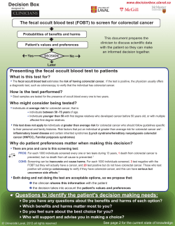

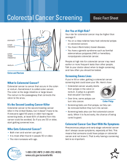


© Copyright 2026