
Document 434125
Cbdiovascular
Research
ELSEVIER
CardiovascularResearch33 (1997) 6 1l-622
Restitution of contractility in single ventricular myocytes of guinea pig
heart
Matti Vornanen ‘, Neal Shepherd *
Research Seruice/lSlL,
Department
qf Veterans Affairs, Medical Center, 508 F&m Street, Durham, NC 27705, USA
Received 6 June 1996;accepted31 October 1996
Abstract
recovered soon after a contraction and well before mechanical restitution is complete.
Keywords: Contractile function: Caffeine; Guinea pig, ventricular cells: Calcium, intracellular concentration;Calcium channel, L-type
1. Introduction
The action potential duration and the force of contraction of cardiac muscle vary with the preceding stimulus
interval. In regularly-stimulated guinea pig heart, an extra
stimulus interpolated soon after a steady-state contraction
elicits an action potential that is shorter in duration than
the control, and a twitch of markedly less force than the
control. The gradual restoration of the action potential
duration and contractile force during diastole are called
electrical and mechanical restitution, respectively [l-4].
Although these phenomena have been described in various
preparations from different mammalian species, the underlying mechanisms remain uncertain. Electrical restitution is
thought to be dependent on the recovery of membrane
currents from inactivation and/or interval-dependent
* Corresponding author. Tel. + 1 919 286-0411, ext. 6502; Fax + 1
’ 919 286-6824.
’ Presentaddress:Department of Biology, University of Joensuu,P.O.
Box 111, 80101 Joensuu,Finland.
changes in the ionic composition of the intracellular solution [3,S]. Mechanical restitution has been explained in
terms of (1) release of Ca*+ from the sarcoplasmic reticulum (SR) in proportion to the Ca2+ current (I,,) [6], (2)
slow replenishment of the releasable Ca2’ store of SR
[7,8], (3) time-dependent recovery of the SR release channel from inactivation [9], and (4) a slow reduction in
activity of the SR calcium pump during diastole [lo]. In
fact, the restitution process could be any combination of
these factors, whose contribution might vary with the
inotropic state of the muscle.
In most previous studies mechanical restitution has been
measured from multicellular preparations, and reflects a
mixture of the effects of changes in the duration of the
action potential and the effects of whichever other factors
may be important in controlling contraction strength 17,111.
In our experiments we measured electrical and mechanical
restitution (of force) in enzymatically isolated cells of
Time for primary
0008.6363/97/$17.00 Copyright 0 1997 Elsevier Science B.V. All rights reserved
PII SOOOS-6363(96)002.59-3
review 27 days.
Downloaded from by guest on November 19, 2014
Objective: Our aim was to assess the extent to which changes in intracellular Ca2+ storescontribute to mechanical restitution in heart
muscle. Methods: Single, isolated guinea pig ventricular cells were voltage clamped at -45 mV and stimulated continuously at 0.5 or 2
Hz with 200 ms depolarizing pulses (35°C). The recoveries of the peak of contraction force (F,) and the calcium current (I,,) between
beats were measured in contractions interpolated at various intervals (t,) after a conditioning twitch. Recovery of SR Ca2+ load was
inferred from the peak magnitude (C,) of similarly interpolated contractures, induced by rapid application of 5 mM caffeine. Results: For
a conditioning stimulus rate of 0.5 Hz, both Fp and I,-, were very small for small t,, and recovered along similar time courses with a t,,*
of about 50 ms. Cp was maximal at as early a time after a previous contraction as could be measured, at which time Fp was 56% of
maximal. C, declined throughout the stimulus interval to about 50% of its maximal value. Similar results were obtained for a
conditioning stimulus rate of 2 Hz, at which rate both Fp and Cp were increased by a factor of 2. Conclusions: The time course of
mechanical restitution is coincident with the recovery of f,, from inactivation. Caffeine-releasableintracellular calcium stores are fully
612
M. Vornanen, N. Shepherd / Cardiotiascular Research 33 (1997) 61 I-622
guinea pig ventricle under voltage clamp conditions, where
the duration of activation can be kept constant and the
main trigger of Ca*+ release (Zc,) can be accurately
measured. Thus we can quantitatively estimate the importance of the recovery of I,, in mechanical restitution.
Furthermore, with this preparation, we can estimate the SR
Ca’+-content from the current and force induced by fast
application of caffeine to the cell, irrespective of the state
of the SR calcium-release channel [9,12]. We find that
electrical and mechanical restitution have similar time
courses, while the recovery of caffeine-releasable calcium
stores is much more rapid than either.
2. Methods
2. I. Enzymatic isolation of ventricular myocytes
2.2. Force measurement and voltage clamp
For experimentation, the cells were placed in a Lucite
chamber (3.0 ml) and continuously superfused with a
pre-warmed (35°C) modified Tyrode solution (NT) composed of (mM): 150 NaCl, 5.4 KCl, 1.2 MgCl,, 5 Hepes,
1.8 CaCl,, 5.5 glucose, pH adjusted to 7.4 with NaOH.
Details of the force measurement from single cells has
2.3. Solution changes
Caffeine contractures were induced by brief exposure to
otherwise normal Tyrode solutions containing 5 mM caffeine. The caffeine-containing solution was applied by
means of a double jet system described previously [15].
The position of the jets was monitored by a LED/phototransistor pair, the output of which was recorded simultaneously with the electrical and mechanical signals from a
cell. In most experiments the flow velocity was approximately 50 mm/s, permitting a fast and well-controlled
release of intracellular calcium by caffeine. In other cases.
noted in the text or figure legends, the flow rate was
reduced to 13 mm/s to allow a larger percentage of
complete experiments. Some experiments were made with
both flow rates (in different cells) with no qualitative
difference in the results.
2.4. Recording and analysis
Current and force signals were digitized (Instrutech,
VR-100, 9 kHz/channel, 4 channels) and stored on video
Downloaded from by guest on November 19, 2014
Guinea-pigs weighing between 250 and 500 g were
deeply anaesthetized with 65 mg/kg sodium pentobarbital.
The heart was rapidly excised and the aorta cannulated for
coronary perfusion with oxygenated solutions (35-36”(Z).
The heart was first perfused for 5 min with a nominally
Ca2+-free, low sodium solution (LN) containing (n&I):
100 NaCl, 10 KCl, 1.2 KH,PO,, 50 taurine, 4 MgSO,, 20
glucose, 10 HEPES at pH 6.9. The perfusate was then
switched to a solution of the same composition with the
addition of 1 mg/ml collagenase (Sigma Type I), 1 mg/ml
fatty-acid-free serum albumin (Sigma A-6003) and 100
p.,M of CaCl,. CaCl, was added in 4 aliquots during the
enzyme perfusion to bring the total added calcium to 200
p,M. The heart was then minced and further incubated,
with continuous stirring, in 5-10 ml of the same enzyme
solution with the addition of 0.07 mg/ml protease (Sigma
Type XIV). At 5- 10 min intervals the supernatant, containing dissociated cells, was drawn off and replaced by
more enzyme solution. The cell suspensions were diluted
into LN, to which had been added 1 mM CaCl, and 1
ml/100 ml penicillin/streptomycin
(Sigma P-0781) and
stored at 23°C. Also, some cell suspensions were allowed
to settle for several minutes in enzyme solution (approx.
4°C). The supernatant was then decanted and the cells
were covered with a depolarizing nutrient solution for
storage at 4°C [13]. This investigation conforms with the
Guide for the Care und Use of Laboratory Animals published by the US National Institutes of Health (NIH Publication No. 85-23, revised 1985).
been described in previous papers [14,15]. In short, the
ends of an isolated cell were attached to the tips of two
glass rods (diameter = 76 p,m) using poly-L-lysine (MW
3000&70000,
Sigma) as a glue. The length, width and
thickness of the cell were measured by rotating the rod
pair by 90”, so that cross-sectional area and cell volume
could be estimated. The edge movement of the rods ( < 3%
of free cell length, i.e. l-l.5 pm) was detected by two
phototransistors located in the focal plane of the microscope objective (40 X , Nikon). The force of contraction of
a myocyte was calculated from the known compliance of
the rods (0.76 m/N) and normalized to the cross-sectional
area.
After a cell was successfully fixed on the rods, a patch
pipet was attached near the center of the cell and the
whole-cell clamp condition established. Patch pipets were
about 2 km in tip diameter with a resistance between 1.6
and 3 Ma when filled with a solution containing (mM):
130 KCl, 10 HEPES, 5 K-oxaloacetate, 5 K-succinate, 1
MgC12, 0.02 EGTA, 5 mM NaZ ATP, pH 7.4.
After obtaining the whole-cell voltage-clamp condition,
a cell was stimulated initially at a regular rate of 0.5 Hz
with 55 mV depolarizing pulses from a holding potential
of - 45 mV. The time course of mechanical restitution and
the SR calcium content were then determined for various
pulse protocols, as described below. In most cases, when
the results of multiple protocols were to be compared, the
protocols were applied in a completely bracketed format.
For example, if the results from protocols a, b and c are
to be compared, the protocols are applied to the cell in the
pattern abcba and the results from duplicate protocols are
averaged before a comparison is made among the results.
We report here only the results of complete experimentsi.e., those in which each protocol was applied in one cell.
613
M. Vornanen, N. Shepherd/ Cardiovascular Research 33 (1997) 611422
tape, with concurrent strip chart recording (Gould 2400).
Off-line analysis was made by recreating the analog signal,
redigitizing each channel at 2.5 kHz and analyzing the
records with programs written in Asyst (Keithly-Asyst).
The inward Ca2+ current (I,,) was estimated as the difference between the peak inward current and the steady-state
current at the end of the pulse (visual estimate [16]). The
peak force of the twitch in response to an electrical pulse
(F,) was measured, after filtering the signal at 100 Hz, as
the difference between the force level just prior to the
pulse and the maximum force during the pulse. For contractions interpolated at diastolic intervals of less than 200
ms, a control record was subtracted from the record of
interest to remove the relaxation phase of the previous
conditioning contraction. In practice, this affected Fp only
for contractions interpolated at intervals less than 40 ms.
The peak contracture force <C,> is the difference between
the force level before the solution-supply jets were moved
and the maximal force. A subtraction similar to that described for electrically driven contractions was performed
for contractures interpolated within 200 ms of a previous
conditioning contraction.
calcium store
To demonstrate the recovery of intracellular calcium
stores between contractions, the cells were stimulated at
0.5 or 2.0 Hz, and caffeine contractures were elicited every
5-10 beats by switching the position of the perfusion jets
at various times after a contraction. Ideally, in order to
compare mechanical restitution with the recovery of releasable intracellular calcium, we would like to be able to
release the intracellular stores by caffeine application as
quickly as possible, preferably as quickly as the release
initiated by depolarization, via calcium-induced calcium
release [ 17,181. With our technique, caffeine contractures
had rising phases comparable to those of the electrically
driven twitch, beginning after a latency of about 90 ms
(including the 35 ms needed for the caffeine to reach the
cell from the supply jet, Fig. 1). The time between the
initial motion of the jets (lowermost trace in Fig. 1A) to
the peak of contracture force for a 100 ms pulse of
caffeine was 166 + 13 ms (n = 5). Thus, the time between
the onset of contracture force and the peak of force was
about 76 ms, similar to the time to peak (TTP) of electrically driven contractions in these cells [14].
In addition to being very fast, the time courses of both
the contracture and the caffeine-induced inward current
were very reproducible (Fig. 1B). The 3 contractures for
100, 200 and 300 ms applications of caffeine were obtained sequentially, separated by 6 conditioning contractions. The rising phases of force and current are superimposable for all 3 contractures and the relaxation of the
contractures in response to 200 and 300 ms caffeine
applications were also essentially identical. The waveforms
B
1.5
lmN/mm*
AI
(1) conditioning
contractions
- ..:..............
i :
.: .:
(2) caffeine
contractures
Fig. 1. Effect of caffeine pulse duration on the amplitude of caffeine
contracture. (A) Digitized record of a conditioning contraction (I), a
caffeine contracture (2) and the first post-caffeine (3) for a 100 ms
application of caffeine. [Caffeine] = 5 mh4, temp. = 35°C [Ca’+ 1, = 1.8
mM, zero current indicated by mark at the right of each record. (B)
Control contractions and caffeine contractures, with associated inward
currents, on an expanded time scale, for caffeine applications of 20, 50,
100, 200 and 300 ms duration, with a delay of 820 ms.
of the inward currents induced by caffeine were likewise
very similar (lower traces, Fig. 1B). These currents reflect
the extrusion of calcium by the Na/Ca exchange, following the release of calcium from the SR [19]. The similarity
of the currents for all applications of caffeine of 50 ms
duration or longer suggests that the release of calcium
occurs very rapidly.
Relaxation of a contracture was slower for a 200 ms
caffeine application than for a 100 ms application (Fig.
1B). The slow relaxation could simply reflect the continued presence of caffeine and its potentiating effect on the
myofilaments [20]. Alternatively, some resequestration of
calcium by the SR might occur during relaxation after the
shorter caffeine application due to the rapid washout of
caffeine. However, assuming that calcium release by caffeine is monotonically related to the SR content, neither
incomplete release nor resequestration will qualitatively
affect our principal results or conclusions. We conclude
that the calcium release induced by the rapid application of
caffeine for 100 ms is sufficiently fast and reproducible to
reflect putative changes in SR calcium content during the
period of mechanical restitution.
Downloaded from by guest on November 19, 2014
2.5. Measurement of intracellular
catfeine
614
M. Vornanen, N. Shepherd/ Cardiovascular
Research 33 (1997) 611-622
3. Results
3.2. Restitution of I,, and twitch force
3.1. Characteristics of the electrically driven contraction
Mechanical restitution and recovery of 1c, from inactivation was determined by interpolating extra beats at
diastolic intervals (t,) between 20 and 1000 ms, with 5-10
conditioning contractions separating successive test pulses
(Fig. 2A). In Fig. 2C, Fp of the interpolated beat (or rate
of rise of force, dF/dt,,,,
or I,,) is plotted against td
(i.e., the time between the end of the control clamp pulse
and the beginning of the test clamp pulse).
I
recover very quickly after a
Fp and dF/dt,,,
pre$us depolarization, along similar trajectories (Fig. 2C
and inset). The recovery of Fp is slightly slower than that
possibly as a result of a small increase in the
of dF/dt,,,,
time to peak force (TTP) through the initial period of
diastole (see below). The recovery of I,, appears to be
biphasic, approaching its control value rather slowly after
an initial fast phase of recovery (Fig. 2C, open circles).
For quantitative comparisons among Zca, Fp and
dF/%,,, 3 we fitted a single exponential function to the
averaged results from 6 cells (Fig. 2C, see Section 4). The
recovery of I,, from inactivation had a time constant of 92
Under our standard conditions, the electrically stimulated cell responded with a two-component contraction.
The phasic component, which relaxed during depolarization, was the component which exhibited restitution (Fig.
2B). The more slowly developing tonic component relaxed
only upon repolarization, and showed no restitution between beats (Fig. 2B). The tonic component remained
steady for several hundred milliseconds if depolarization
were prolonged, and relaxed immediately upon repolarization (not shown). It is not to be confused with the oscillatory contractions associated with calcium overload which
appear after repolarization and which are associated with
an oscillatory current. The phasic component is assumed to
reflect the release of intracellular calcium stores and the
entry of calcium from the extracellular space mainly via
I,,, while the tonic component presumably reflects a balance between calcium sources and sinks, and depends to a
large extent on Naf/Ca2+ exchange [21].
B
phasic
9
Force
mNimm2
I
0.2
mtUmm2
4
Current
Y
Y
[r
11.5”A
2.5 nA
200 ms
0
200
400
diastolic
delay, td (ms)
600
800
Fig. 2. Mechanical restitution: recovery of contractile force and Ca*+ current in single guinea-pig ventricular myocyte. (A) A segment of the experimental
protocol: Low resolution digitized records of membrane voltage (lower), contraction force (upper) and membrane current (middle). t,, is the delay time,
against which the peak force (F,) is plotted in panel C. Conditioning stimulus interval = 2000 ms; pulse duration = 200 ms; holding potential = - 45
mV; pulse potential = + 10 mV; T = 35°C; [Ca*+ I, = 1.8 mM. (B) Digitized records of control twitches and 9 interpolated test twitches, and associated
currents, for r, between 20 and 820 ms. (C) Restitution of twitch force, Fr (filled circles), maximum rate of rise of force (squares), dF/dr,,,
and I,,
(open circles) for all premature contractions in 6 experiments of the kind illustrated in panels A and B. Ordinate: I,,, dF/dt,,,
and FP were normalized to
F, of the immediately preceding control contraction before averaging. Abscissa: td. Inset: The same data as in panel C were renormalized to the value of
each measurement at an interval of 1020 ms in each cell, to better show the similarity of the time courses. (D) Ordinate: The rate constant of the single
exponential fit to the time course of recovery of FP for individual cells. Abscissa: The rate constant of the fit to the time course of recovery of I,,. The
open circles indicate points from experiments in which cells were superfused at 50 mm/s.
Downloaded from by guest on November 19, 2014
A
615
M. Vomanen, N. Shepherd/ Cardiovascular Research 33 (1997) 611-622
calcium storesthat occurs throughout the stimulus interval
(Fig. 3).
The TTP for interpolated contractions was smaller than
for the conditioning contractions. At td = 260 ms the TTP
was 80% rt 10% ( + s.d., n = 6) of the TTP of conditioning contractions, recovering slowly thereafter. A precise
time course of the change in TI’P was difficult to determine due to scatterin the data, which was, in turn, due to
small amounts of mechanical noise. Nevertheless, for t,
between 260 and 720 ms, the difference between the TTP
of the interpolated beat and that of the immediately preceding conditioning contraction was statistically significant
(P < 0.05, paired t-test).
ms, while the restitution of Fp and dF/dt,,,
had time
constants of 103 and 71 ms, respectively. We also fitted
single exponentials to data from individual cells and have
plotted in Fig. 2D, for each cell, the time constant for the
recovery of I,, from inactivation (Ki) against the time
constant for mechanical restitution (K,). Though the rates
of recovery of both La and Fp varied widely among cells
in this study, the recovery of current and force always had
similar time courses in a given cell, and so K, and K,
were well correlated over the range of our results (Fig. 2D,
r = 0.92). The standard errors of the values of the rate
coefficients were too small to show in the figure, being, on
average,about 0.007 of K, and 0.002 of Ki.
At the end of the period of mechanical restitution the
ratio of Fp of the interpolated contraction to that of the
control was slightly greater than one, and similarly for
dF/%n,, (Fig. 20 This ‘overshoot’ was consistently
seenand meansthat Fp and d F/d t,,, must decline during
the latter part of the stimulus interval. This decline may be
related to the decline of caffeine-releasable intracellular
3.3. Interpolated cafleine contractures
Caffeine is known to releaseCa2+ from the SR regardless of the state of the SR release channel [9,12]. Therefore, putative inactivation of the channel following a previous depolarization and release [12] should not prevent the
Downloaded from by guest on November 19, 2014
3
1.5
mNimm2
bJ
I
0.5 nA
Oms
I
B
160
I
I
I
I
I
I
0
400
800
1200
1600
diastolic
interval
(ms)
Fig. 3. Diastolic decay of C, of interpolated contractures. (A) Caffeine contractures initiated between 5 and 1560 ms after a conditioning contraction.
Duration of caffeine application = 100 ms. (B) Superimposed caffeine contractures and associated transient inward currents from panel A on an expanded
scale; td= 5 ms (1). 705 ms (2) and 1560 ms (3). (C) Averaged values of C, and the /Ican from 5 experiments such as that in panel A. Each force
measurement was normalized before averaging to the FP of the previous conditioning contraction, then divided by the value of C, for td = 705 ms before
plotting. The integrated currents were averaged and divided by the value for t,,= 705 ms before plotting.
616
M. Vomanen. N. Shepherd/ Cardiocascular Research 33 (1997) 611422
subsequent release of SR calcium by caffeine. Thus, if the
SR calcium level were to change during the diastolic
interval, the change should be reflected in the magnitude
of the caffeine contracture and in the magnitude of the
current accompanying the contracture.
In experiments analogous to those used to study mechanical restitution, caffeine was applied for 100 ms at
intervals ranging between 5 ms and 1005 ms after a
previous contraction, with one additional application at an
interval of 1560 ms (Fig. 3). In contrast to the post-stimulation recovery of Fp which is illustrated in Fig. 2, the
strength of the interpolated caffeine contractures declined
almost monotonically throughout the diastolic interval to
47% of its maximal value (Fig. 3). A small depression of
Cr at the beginning of the interval was probably due to a
slight overlap of the caffeine application and the relaxation
of the tonic phase of the previous contraction. In other
experiments, we noted that when such overlap occurred,
the waveform of the caffeine-induced current began much
earlier and reached a smaller peak though the integral of
the caffeine-induced current was not decreased (not shown).
Consistent with this interpretation, JZcarfdeclined smoothly
throughout the interval to 58% of its maximal value. The
waveform of both the contracture and the current remained
relatively constant throughout that period (Fig. 3B). These
results suggest that the SR calcium declines markedly
between contractions under our experimental conditions.
However, the plots in Fig. 2C and Fig. 3C are not
directly comparable. In Fig. 3C, Ci, and lZcaff are plotted
against td, as defined in Fig. I, which means that the onset
and the TTP’s of the contractures are delayed by about 90
ms relative to depolarization-induced contractions plotted
on the same time scale (as they are in Fig. 2). Also, the
contracture results in Fig. 3C and the contraction results in
Fig. 2C were obtained from completely separate groups of
cells. Although the results in these two figures suggest that
the recovery of contractility after a contraction occurs
without a substantial change in the level of SR calcium, a
more rigorous comparison was devised to demonstrate this
point.
Downloaded from by guest on November 19, 2014
B
2.0
mtUmm2
0.5 nA
5mM
OmM
200 ms
2.0
mNlmm2
E
120
5:
100
P
tii
E
80
z
8
3
1.0 nA
7
60
20
400 :
0
I
100
I
200
n
.
0
i
300
CP
FP
La
I
400
I
500
delay to peak force, td, TTP, ms
Fig. 4. Comparison of caffeine contractures and electrically-driven contractions in the same cell. Panel A: Superimposed records, from one cell, of
caffeine-induced contractures (Al) and electrically-driven contractions (A2) that were interpolated at diastolic intervals so arranged that the TTP of each
contracture was nearly identical with the TIT of one of the contractions. (Al) Force (upper), current (middle) and position of the solution supply jets
(lower), for three 100 ms caffeine applications. Conditioning pulses at 0.5 Hz: r,, = - 23, 127 and 277 ms. [Cal, = 1.8 mM; Temp. = 35°C. (A2) Force
(upper) and current (lower) for electrically-driven contractions from the same cell as in A I. t,, = 70, 220 and 370 ms. Panel B: Contraction at 370 ms and
contracture at 277 ms from panel A are shown on an expanded time scale. The vertical scale of the contracture was adjusted to facilitate comparison of the
time courses. Both records begin 350 ms after the end of the control pulse. Panel C: Comparison of Cr and FP for 5 experiments of the kind shown in
panel A. Ordinate: FP and C, were normalized to FP of the preceeding conditioning pulse before averaging among the cells. The resulting averages are
scaled so that both Ct, and Fr are equal to 1 for the largest interval of interpolation. Abscissa: The mean TTP, from the end of the previous depolarizing
pulse, of either contractions or contractures. Note that this means of plotting is different from that in Figs. 2 and 3, where the abscissa was rd.
M. Vomanen, N. Shepherd / Cardiooascular Research 33 (1997) 61 I-622
3.4. SR content and mechanical restitution measured in the
same cell
theless, it was of interest to study restitution under conditions in which conditioning contractions appear to depend
to a greater extent on intracellular calcium stores for
activation.
We first show that caffeine-releasable SR calcium stores
can be loaded both by increasing the pulse potential (to
+30 mV) and by increasing the frequency of stimulation.
The increase of pulse potential was useful to facilitate
frequency-dependent potentiation, which is known to be
voltage-dependent [22]. Cells were stimulated with 3 separate protocols in a bracketed format (Fig. 5, see Section
2.2): A, 0.5 Hz with the pulse potential = + 10 mV; B,
0.5 Hz with pulses to +30 mV; C, 2.0 Hz with pulses to
+ 30 mV. In each case caffeine contractures were interpolated by switching the position of the jets 20 ms prior to
the end of the depolarizing pulse. Simply changing the
pulse potential while stimulating at 0.5 Hz increased contracture strength slightly (C, = 2.10 &- 1.42 and 2.73 -t 1.8 1
mN/mm’ for + 10 and + 30 mV, respectively; n = 4,
P < 0.05, paired t-test; Fig. 5A,B) but did not increase the
strength of the conditioning contraction (F, = 1.80 * .98
and 1.73 + .84 mN/mm’ for + 10 and + 30 mV, respectively). Increasing the stimulus frequency to 2 Hz markedly
increased both FP and C, (C = 3.82 f 1.89 mN/mm’
and FP = 4.02 k 1.40 mN/mm P, Fig. 5C). The probability
that a difference of more than 1 mN/mm’ between CP at
0.5 Hz and 2.0 Hz (+ 30 mV) was due to chance was very
small (P < 0.005, paired t-test).
A.
3.5. Voltage- and frequency-dependent potentiation
0.5Hz
*IO mV
A possible problem in interpreting the foregoing results
with respect to the underlying cause of mechanical restitution is that activation of the conditioning contractions
under the protocol given in Fig. 2 may not depend much
on intracellularly stored calcium. This is suggested by the
fact that FP of the contractions in response to stimulation
at 0.5 Hz was reduced only to 64 & 5% of control (mean
& s.d., n = 5) by a prior application of 5 mM caffeine for
200 ms during the previous diastole (from experiments in
Fig. 1). If it were assumed that caffeine released 100% of
intracellular calcium (and that there was no re-uptake by
the SR), so that the first post-caffeine contraction depended for activation entirely on extracellular calcium, the
relative contributions of extracellular calcium and intracellular calcium to activation in conditioning contractions
would be about 2:l (64:36). This is a worst case since
there may be re-uptake of calcium by the SR after the
caffeine is removed. Furthermore, the calcium content of
the SR is apparently much larger just after a depolarization
than it is just before a depolarization (Fig. 3) and I,, is
quite small immediately following a previous contraction.
Therefore, contractions interpolated early in the stimulus
interval may depend much more on intracellular calcium
for activation than do the conditioning contractions. Never-
B.
0.5Hz
+3OmV
C.
2Ok
l 30 mV
Fig. 5. Potentiation of contractions and contractures by increasing pulse
voltage and/or frequency of stimulation. (A-C) Digitized records of
force (upper) and current (lower) for 3 pulse protocols. (1) conditioning
contractions, (2) contractures elicited by 200 ms applications of caffeine
interpolated at t,, = -20 ms, and (3) the first contractions after the
caffeine contracture. Holding potential = -45 mV; [Cal, = 1.8 mM;
flow velocity = 13 mm/s. (A) Pulse potential = + 10 mV; basic rate
= 0.5 Hz. (B) Pulse potential = +30 mV; basic rate = 0.5 Hz. (Cl
Pulse potential = +30 mV, basic rate = 2 Hz. In protocol C the
depolarization immediately following the caffeine application was delayed (by omitting one beat) to permit washout of the caffeine.
Downloaded from by guest on November 19, 2014
As pointed out earlier and shown again in Fig. 4B, the
waveforms of contractions and contractures are very similar, suggesting that the processes leading from calcium
release to contraction are similar in the two cases. Therefore, in order to compare depolarization-induced calciumrelease and caffeine-induced calcium-release at the same
points in time after a previous contraction, we have compared the peak force development among contractures and
contractions having the same time to peak relative to the
end of the previous depolarizing pulse (Fig. 4A). In 5
experiments, cells were driven at 0.5 Hz and extra pulses
were interpolated at intervals of 70, 220 and 370 ms after
the end of the previous depolarization (Fig. 4A2). Then, in
the same cell, caffeine contractures were elicited by repositioning the jets at intervals of -23, 126 and 277 ms after
the end of the previous depolarization (Fig. 4AI). This
protocol resulted in 3 contractures which had nearly the
same times to peak as the 3 contractions interpolated in the
first part of the protocol (Fig. 4C). It is clear from the
experimental records in Fig. 4A and from the cumulative
results in Fig. 4C that C, of contractures elicited early in
the diastolic period actually declines (slightly) at a time
when FP is still increasing markedly. The normalized
values of C, and FP were significantly different (P < 0.001
= 130 ms and P < 0.01 for rd,TTP= 285 ms,
for
‘d.TTP
paired t-test).
617
M. Vomanen,N. Shepherd
/ Cardiovascular Research 33 (1997) 61 I-622
618
Importantly, a caffeine application between contractions
at 2 Hz reduced FP of the first post-caffeine contraction to
0.30 f 0.02 of control (Fig. 50, while at 0.5 Hz, FP of the
first post-caffeine contractions were reduced to 0.66 f 0.10
and 0.65 + 0.04 of control for a pulse potential of + 10
and + 30 mV, respectively. The strong suppression of
contraction by a prior application of caffeine suggests that
the contraction at a stimulation rate of 2 Hz depends much
more on intracellular calcium than does the contraction at
0.5 Hz. This result is consistent with the increased caffeine-releasable calcium content and the reduction of Z,,
characteristic of 2 Hz stimulation (Fig. 5).
3.6. Mechanical restitution in the potentiated state
In the potentiated state, it was not possible to study both
the time course of mechanical restitution and the restoration of SR calcium stores in the same cells. Therefore, we
compared mechanical restitution and recovery of I,, in
one group of cells, and measured the strength of interpolated caffeine contractures in another group (Figs. 6 and
7).
3.7. SR calcium content between beats in the potentiated
state
In this series of experiments the cells were driven at 2
Hz and caffeine contractures were interpolated at t, = - 20
and 80 ms (Fig. 7A). C, of the earlier contracture in each
experiment was divided by that of the later contracture and
the normalized Cp plotted against the TTP (Fig. 7). The
data from the restitution experiments shown in Fig. 6D
were replotted on the same time scale against their TTP’s.
A
B
conditioning contractions
h
a
b
c
2.0
,
k
I 1 mNlmm2
0
1OOms
Hz
/AO.,Hz
100
,
) mNlmm2
200
300
diastolic delay (ms)
Fig. 6. Restitution of contractility and recovery of I,, during diastole, with 2 Hz stimulation. (A) A strip chart record showing force (upper) and current
(lower) during an experiment in which the conditioning stimulus rate was increased from 0.5 to 2.0 Hz and stimuli were interpolated at intervals of 20, 60,
and 100 ms after a conditioning contraction. Pulse duration = 200 ms; holding potential = -45 mV; pulse potential = +30 mV; T = 35°C;
[Ca*+ 1, = 1.8 n&l. (B) Conditioning twitches and associated membrane currents at conditioning stimulation rates of 0.5 and 2.0 Hz on an expanded time
scale. From the experiment in panel A. (C) Superimposed records of conditioning contractions and 3 contractions interpolated at intervals (t,) of 20, 60
and 100 ms. (D) Recovery of I,-, and Fp between depolarizations (means of 3 experiments). Ordinate: Each value of I,, for the interpolated
depolarization was normalized to I,, during the immediately previous conditioning depolarization, then averaged at each time point. Abscissa: td. Lines
are exponentials; time constants given in text.
Downloaded from by guest on November 19, 2014
In these experiments, restitution was first measured at a
basic rate of 0.5 Hz, then at 2 Hz and then once more at
0.5 Hz. In each case extra stimuli were interpolated at
t, = 20, 60, 100 and 300 ms. FP of the interpolated
contractions was normalized to FP of the contractions at
t, = 300 ms, which for the 2 Hz case was the conditioning
contraction just preceding each interpolated contraction. FP
of the conditioning contractions at 2 Hz was 189 k 3 1%
(n = 3) of that at 0.5 Hz. Despite the increased dependence of contractility on intracellular calcium at the higher
rate of stimulation, mechanical restitution and recovery of
Zc, from inactivation again had similar time courses (Fig.
6C,D). In these experiments, the time constants of recovery of I,, and FP were 64 and 76 ms, respectively, for 0.5
Hz stimulation, and 79 and 70 ms for 2.0 Hz stimulation.
It should be noted that the TTP of contractions at 2 Hz
was smaller than that at 0.5 Hz (Fig. 6B). The TTP’s of
interpolated beats at a basic rate of 2 Hz were even smaller
than those of the conditioning contractions (e.g., 90 + 3%
for t, = 260 ms, n = 3), similar to the results obtained at a
basic rate of 0.5 Hz.
M. Vornanen, N. Shepherd/Cardiotiascular
2
mNfmm*
Research 33 (1997) 611-622
of mechanical restitution and that the rate of mechanical
restitution is controlled mainly by the rate of recovery of
Zc, from inactivation. Support for this conclusion is provided by 3 observations: (1) caffeine-releasable stores
actually decline slightly during the latter half of the period
of mechanical restitution (Fig. 41, and so at least that
portion of the restitution process cannot be attributed to
increased availability of stored calcium; (2) the time courses
of recovery of Fp and dF/dt,,,
are very similar to the
time course of recovery of I,, under conditions in which
the twitch strength should depend to a large extent on the
level of caffeine-releasable intracellular stores (Figs. 2, 4,
6 and 7); (3) the rates of mechanical restitution and
recovery of I,, correlate well in individual cells (Fig. 2D).
4.1. Intracellular
w
0
CP
FP
The results of these experiments are quite similar to the
results obtained at the lower rate of stimulation, though not
as complete (compare Figs. 4 and 7). The difference
between Cr and Fp at td,rTP= 130 ms was highly significant (P < 0.001, t-test). Likewise, the values of C,, (before
normalization) at 130 and 230 ms were not significantly
different. Thus, under conditions in which the caffeine-releasable calcium has been almost doubled, mechanical
restitution still occurs without a significant change in the
SR calcium content, and its time course closely approximates that of the recovery of I,, from inactivation.
4. Discussion
We conclude that redistribution of intracellularly stored
calcium is complete by at least the latter half of the period
calcium stores decline during diastole
One of the most often cited models of mechanical
restitution is based on the hypothetical movement of calcium within the lumen of the SR in the period between
contractions, such that the calcium taken up by the SR
pumps during a contraction is not immediately available
for subsequent release, but rather needs time to travel from
the pumping sites to the release sites [23]. On the basis of
this model it would be expected that the Cr of contractures
initiated just after a previous contraction would increase
with time after the contraction, as the SR stores became
available (i.e., the time course of Fp and Cr, just after a
contraction should be similar). This is clearly not the case
for the latter half of the period of mechanical restitution
(Figs. 4 and 7). W e interpret the nearly constant C, of the
caffeine contractures during the period of mechanical restitution to mean that the SR calcium stores are fully restored
by this point in time and that the amount released by
depolarization in this period depends directly on the magnitude of I,,. This interpretation agrees with a previous
similar conclusion based on the strength of rapid cooling
contractures initiated just after a contraction [24]. An alternative explanation which we cannot exclude is that caffeine releases calcium from all parts of the SR, while
depolarization releases calcium only from the junctional
regions of the SR.
The decrease of caffeine-releasable calcium during diastole (Fig. 3) may be due to the spontaneous quanta1 release
of calcium (‘sparks’) that has recently been demonstrated
in both guinea-pig and rat cardiocytes [25,26]. The ‘spark’
rate has been shown to depend on the level of activation of
the L-type calcium channel and thus to be voltage-dependent [25]. At the holding potential used in our experiments
(-45 mV> the rate of loss of intracellular calcium could
be in the mM/s range if the spark rate measured in rat
heart applies under our experimental conditions [26]. On
the other hand, twitch tension in guinea-pig heart declines
between beats even at the normal resting potential (e.g.,
see [27]), at which potential the spark rate should be
negligibly small.
Downloaded from by guest on November 19, 2014
Fig. 7. Comparison of caffeine contractures and electrically-driven contractions interpolated at equivalent times between potentiated contractions. (A) Superimposed records of conditioning contractions and contractures induced by 100 ms applications of caffeine for f,, = - 20 and + 80
ms, showing force (upper), current (middle) and position of solutionchanging jets (lower). Conditioning stimuli at 2 Hz; holding potential =
-45 mV; pulse potential = +30 mV; temp. = 35°C; [Cal = 1.8 mM;
flow velocity = 50 mm/s. (B) Comparison of Cr from 3 experiments
such as in panel A and FP for 3 experiments, replotted from Fig. 6.
Ordinate: FP and C, were normalized to FP of the preceding conditioning
pulse before averaging among the cells. The resulting averages are scaled
so that both CP and FP are equal to 1 for the largest interval of
interpolation. Abscissa: The mean TTP, from the end of the previous
depolarizing pulse, of contractions or contractures, as in Fig. 4. Note that
this means of plotting is different from that in Figs. 2, 3 and 6, where the
abscissa was rd.
619
620
M. Vornanen, N. Shepherd/ Cardiovascular
4.2. The relation between ZCaand FP
double exponential fits, respectively, and more detailed
analysis indicated autocorrelation in the single exponential
fit but not in the double exponential lit (Durbin-Watson
statistic was 0.344 and 2.186, respectively). However, the
FP data could not be reasonably fitted with two exponentials, probably due to the multiple influences on the time
course of FP (e.g., changes in SR calcium content and
changes in TTP). Thus, in order to compare the two sets of
data, we fitted both with single exponentials.
The fast time constant for the recovery of I,, from
inactivation in our experiments (34 ms) is similar to the
single exponential time constant measured in canine Purkinje cells (31 ms) at 37°C [31], and to the rate of action
potential restitution in guinea-pig papillary muscles (38
ms) [32]. Biphasic recovery of I,, from inactivation has
been observed in Purkinje fibers of calf heart [33] and in
the isolated myocytes of both rat and guinea-pig hearts
[34], but no quantitative data are available under experimental conditions similar to ours.
4.3. Other models of restitution
It was recently proposed that both restitution and postextrasystolic potentiation in heart muscle is the consequence of hyperactivity of the SR calcium pump immediately following a contraction-relaxation
cycle [lo]. One
basis of the hypothesis was the observation that the TTP of
early extrasystoles was smaller than later ones and that the
reduction of the TTP could not be attributed to changes in
the action potential or to changes in contraction strength
per se. We likewise observed consistent shortening of the
TTP during diastole. However, the diastolic changes of
TTP were small in our experiments, while changes in the
during restitution were large. Furthermore, if
dF/%,,,
more rapid calcium pumping were to account for the small
FP of a contraction which followed closely on another, a
similar effect should be seen on C, and on the tonic phase
of contraction, neither of which was the case. The consistent relationship between I,, and FP suggests a causal
relationship between these factors, and so we do not think
that changes in SR pump activity can account for mechanical restitution, though it seems likely to play a role in
frequency-dependent potentiation.
It is of interest that the TTP of contractions at 2 Hz was
smaller than that at 0.5 Hz (Fig. 6B), a typical result of
similar experiments with multicellular preparations of heart
muscle [35]. This might be explained by a nonlinear
relation between the intracellular calcium concentration
and the rate of SR calcium pumping [36]. In any case,
reduction of the TTP at high frequencies of stimulation is
an important feature of cardiac muscle, and appears to be
an intrinsic feature of e-c coupling in the cardiac cell.
To be complete, we should point out that the simplest
explanation for our data is that: (1) activation of contraction does not depend on calcium-induced calcium-release
(CICR) but rather depends entirely on extracellular cal-
Downloaded from by guest on November 19, 2014
The time course of mechanical restitution is quite similar to that of the recovery of Zca from inactivation, though
not identical (Fig. 2). Identical time courses would not be
anticipated since FP will depend on factors in addition to
I,..,-e.g., the amount of calcium available in the SR (Fig.
3) and the TIP at a given point in time during diastole.
The calcium content of the SR appears to decline with
time throughout the diastolic interval (Fig. 3), which would
probably increase the apparent rate of mechanical restitution. The slow recovery of the TTP would, on the other
hand, slow the recovery of FP but not the recovery of
The data in Fig. 2 are consistent with these
d F/%,x.
qualitative expectations.
The similarity of the rate of recovery of FP to that of
Zca is consistent with the known properties of the mechanism of e-c coupling in heart muscle. First, the sum of the
calcium entering the extracellular space and that released
from SR stores should be approximately proportional to
I,, since SR release is nearly proportional to I,, [25] and
since Zca is also the main path of calcium entry at a pulse
potential of + 10 mV, with some contribution from
Na+/Ca*+ exchange [28]. Second, FP has been shown to
be proportional to the peak [Cal, during a contraction [29].
Thus, Fp and dF/dt,,,
should be at least approximately
proportional to I,, if the SR calcium content is constant
and the waveform of the [Cal, transient does not vary
greatly.
In a given cell, the rate of recovery of FP was well
correlated with the rate of recovery of Zca from inactivation though both rates varied widely among cells (Fig.
2D). This suggests that the similarity in time courses is not
a coincidence, though it does not exclude the possibility
that a third factor may control both rates.
The restitution of contractile force is faster in our
preparation than in comparable preparations described in
the literature. In voltage-clamped dog ventricular trabeculae at 30°C Braveny et al. [30] found that Zc, recovered
within 200 ms of the previous depolarization while force
recovered with a half-time of 200-400 ms. The discrepancy here could well be due to the difference in experimental temperature. However, the mean time constant of
mechanical restitution for guinea-pig ventricular myocytes
in the present study (103 ms) is smaller than that measured
in cat trabeculae at a similar experimental temperature
(182 ms for a holding potential of - 45 mV at 37°C) [6]. It
is conceivable that the discrepancy here is related to the
difference in extracellular calcium concentrations (3.6 mM
in the earlier study) or the species difference, both of
which might affect the rate of mechanical restitution [27].
The Zc, data are actually better fitted with the sum of
two exponentials than with a single exponential, with time
constants of 34 and 182 ms, comprising respectively 51
and 49% of the total current. The sums of squared deviations were 7.96 X lo--’ and 1.69 X 10m3 for the single and
Research 33 (1997161 l-622
M. Vomanen, N. Shepherd/Cardiovascular
cium entry via Zca; and (2) potentiation of contraction
occurs due to a reduction of SR pumping as the SR
becomes more calcium-loaded. Although this interpretation
goes against an immense body of data supporting the
CICR hypothesis, a recent study with ferret myocytes has
shown that CICR apparently fails when the calcium content falls to 60% of its maximal value [37]. Since we did
not attempt to measure the relative calcium content of the
SR in our experiments, we cannot exclude the possibility
that we are working in a range in which CICR does not
function.
4.4. Summary
Acknowledgements
M. Vomanen was supported by the Academy of Finland
(University of Joensuu, P.O. Box 111, 80101 Joensuu,
Finland). We thank Dr. Rashid Nassar for his help in
construction of the voltage-clamp setup and the rapid
solution changer. We also thank Ms. Holly McDonough
for technical assistance and for help in preparing the
manuscript.
References
[I] Braveny P, Kruta V. Dissociation de deux facteurs: restitution et
potentiation dans l’action de l’intervalle sur l’amplitude de la contraction du myocarde. Arch Int Physiol Biochim 1958;66: 633-652.
[2] Bass BS. Restitution of the action potential in cat papillary muscle.
Am J Physiol 1975;228:1717-1724.
[3] Boyett MR. Jewel1 BR. A study of the factors responsible for
rate-dependent shortening of the action potential in mammalian
ventricular muscle. J Physiol 1978;285:359-380.
[4] Seed WA, Walker JM. Relation between beat interval and force of
the heartbeat and its clinical implications. Cardiovasc Res
1988;22:303-314.
[5] Simurda J, Simurdova M. Electrical restitution in canine ventricular
muscle. Scripta Med 1991;64:67-80.
[6] Trautwein W, McDonald TF, Tripathi 0. Calcium conductance and
tension in mammalian ventricular
muscle. Pfliigers Archiv
1975;354:55-74.
[7] Antoni H, Jacob R, Kaufman R. Mechanische Reaktionen des
Frosch- und S’ugetiermyokards bei Verindemngen der Aktionspotential-Dauer durch konstante Gleichstromimpulse. Pfliigers Archiv
1969:306:33-57.
621
t81Edman KAP,
Johannsson M. The contractile state of rabbit papillary
muscle in relation
to stimulation
frequency.
J Physiol
1976;254:565-581.
191 Fabiato A. Time and calcium dependence of activation and inactivation of calcium-induced release of calcium from the sarcoplasmic
reticulum of a skinned canine cardiac Purkinje cell. J Gen Physiol
1985;85:247-289.
[lOI Schouten VJA. Interval dependence of force and twitch duration in
rat heart explained by Ca2+ pump inactivation in sarcoplasmic
reticulum. J Physiol 1990;43 1:427-444.
[ill Wolfhart B. Relationship between peak force, action potential duration and stimulus interval in rabbit myocardium. Acta Physiol Stand
1979;106:395-409.
1121Varro A, Negretti N, Hester SB, Eisner DA. An estimate of the
calcium content of the sarcoplasmic reticulum in rat ventricular
myocytes. Pfliigers Archiv 1993;423:158-160.
U. Ca-tolerant guinea-pig
1131 Bendukidze 2, Isenberg G, Kltikner
ventricular myocytes as isolated by pronase in the presence of 250
FM free calcium. Basic Res Cardiol 1985;8dsuppl 1):13-18.
[141 Shepherd N, Vomanen M, Isenberg G. Force measurement from
voltage-clamped guinea pig ventricular myocytes. Am J Physiol
1990:258:H452-H459.
[151 Shepherd N, Fisher VJ. Combined force and voltage measurement in
rapidly superfused guinea pig heart cells. Am J Physiol
1990;258:C739-C748.
[161Isenberg G, Kliickner U. Calcium currents of isolated bovine ventricular myocytes are fast and of large amplitude. Pflliigers Archiv
1982;395:30-41.
[I71 Fabiato A. Simulated calcium current can both cause calcium loading in and trigger calcium release from the sarcoplasmic reticulum
of a skinned canine cardiac Purkinje cell. J Gen Physiol
1985;85:291-320.
D81 London B, Krueger JW. Contraction in voltage-clamped internally
perfused single heart cells. J Gen Physiol 1986;88:475-505.
[I91 Callewaert G, Cleeman L, Morad M. Caffeine-induced Ca” release
activates Ca’+ extrusion via Na+-Ca*+ exchanger in cardiac myocytes. Am J Physiol 1989;257:C147-C152.
[201Wendt IR, Stephenson DG. Effects of caffeine on Ca-activated force
production in skinned cardiac and skeletal muscle fibres of the rat.
Pfliigers Archiv 1983;398:210-216,
1211Eisner DA, Lederer WJ. Vaughan-Jones RD. The control of tonic
tension by membrane potential and intracellular Na activity in the
sheep cardiac Purkinje fibre. J Physiol 1983;335:723-743.
D.21Dubell WH, Houser SR. Voltage and beat dependence of Ca*+
transient in feline ventricular cells. Am J Physiol 1989;257:H746H759.
[=I Wood EH, Heppner L, Weidmann S. I. Positive and negative effects
of constant electric currents or current pulses applied during cardiac
action potentials. II. Hypothesis: calcium movements, excitationcontraction coupling and inotropic effects. Circ Res 1969:24:409445.
[24] Banijamali HS, Gao W-D, Macintosh BR, Ter Keurs HEDJ. Forceinterval relations of twitches and cold contractures in rat cardiac
trabeculae. Effect of ryanodine. Circ Res 1991;69:937-948.
[25] Lopez-Lopez JR, Shacklock PS, Balke CW, Wier WG. Local calcium transients triggered by single L-type calcium channel currents
in cardiac cells. Science 1995;268:1042-1045.
[26] Cannel1 MB, Cheng H, Lederer WJ. The control of calcium release
in heart muscle. Science 1995;268: 1045- 1049.
[27] Cooper IC, Fry CH. Mechanical restitution in isolated mammalian
myocardium: species differences and underlying mechanisms. J Mol
Cell Cardiol 1990;22:439-452.
[28] Vornanen M, Shepherd N, Isenberg G. Tension-voltage relationships of single myocytes reflect Ca release triggered by Na/Ca
exchange at 35°C but not at 23°C. Am J Physiol 1994;267:C623C632.
[29] Weir WG, Yue DT. Intracellular calcium transients underlying the
Downloaded from by guest on November 19, 2014
Mechanical restitution and the recovery of Zca from
inactivation have very similar time courses in isolated,
voltage-clamped guinea-pig cardiocytes, regardless of
stimulation frequency. On the other hand, caffeine-releasable intracellular stores of calcium are replete well before
complete recovery of I,,. We conclude that mechanical
restitution reflects recovery of the ability of membrane
calcium channels to deliver activating calcium (to the
myoplasm) and/or trigger calcium (to the SR release
channels), and that the intracellular calcium stores are
essentially constant during mechanical restitution.
Research 33 (1997) 611-622
622
[30]
[31]
[32]
[33]
M. Vornanen, N. Shepherd/ Cardiouascular Research 33 (1997) 61 I-622
short-term force-interval
relationship in ferret ventricular myocardium. J Physiol 1986;376:507-530.
Braveny P, Simurda J, Simurdova M. Voltage dependence of force
and slow inward current restitution in ventricular muscle. Basic Res
Cardiol 1992;87:418-427.
Tseng G-N, Boyden PA. Multiple types of Ca*+ currents in single
canine Purkinje cells. Circ Res 1989;65:1735-1750.
Bjornstad H, Tande PM, Lathrop DA, Refsum H. Effects of temperature on cycle length dependent changes and restitution of action
potential duration in guinea pig ventricular muscle. Cardiovasc Res
1993;27:946-950.
Kass RS, Sanguinetti MC. Inactivation of calcium channel current in
the calf Purkinje fiber. J Gen Physiol 1984;705-726.
[34] Josephson IR, Sanchez-Chapula J, Brown AM. A comparison of
calcium currents in rat and guinea pig single ventricular cells. Circ
Res 1984;54:144-156.
[35] Hoffman BF, Kelly JJ. Effects of rate and rhythm on contraction of
rat papillary muscle. Am J Physiol 1959;197:1199-1204.
1361 Bers DM, Berlin JR. Kinetics of [Cal, decline in cardiac myccytes
depend on peak [Cali. Am J Physiol 1995;268:C271-C277.
[37] Bassani JWM, Yuan W, Bers DM. Fractional SR Ca release is
regulated by trigger Ca and SR Ca content in cardiac myocytes. Am
J Physiol 1995;268:C1313-C1329.
Downloaded from by guest on November 19, 2014


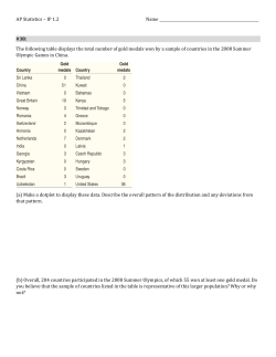


© Copyright 2026