
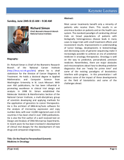
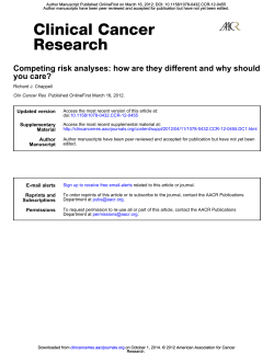
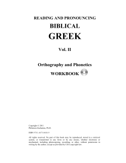
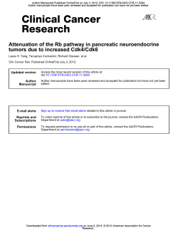
Author Manuscript Published OnlineFirst on March 25, 2014; DOI: 10.1158/1940-6207.CAPR-14-0003
Author Manuscript Published OnlineFirst on March 25, 2014; DOI: 10.1158/1940-6207.CAPR-14-0003
Author manuscripts have been peer reviewed and accepted for publication but have not yet been edited.
Chemoprevention of esophageal cancer with black raspberries,
their component anthocyanins, and a major anthocyanin
metabolite, protocatechuic acid
Daniel S. Peiffer, Noah P. Zimmerman, Li-Shu Wang, et al.
Cancer Prev Res Published OnlineFirst March 25, 2014.
Updated version
Supplementary
Material
Author
Manuscript
E-mail alerts
Reprints and
Subscriptions
Permissions
Access the most recent version of this article at:
doi:10.1158/1940-6207.CAPR-14-0003
Access the most recent supplemental material at:
http://cancerpreventionresearch.aacrjournals.org/content/suppl/2014/03/25/1940-6207.CAPR-14-00
03.DC1.html
Author manuscripts have been peer reviewed and accepted for publication but have not yet been
edited.
Sign up to receive free email-alerts related to this article or journal.
To order reprints of this article or to subscribe to the journal, contact the AACR Publications
Department at [email protected].
To request permission to re-use all or part of this article, contact the AACR Publications
Department at [email protected].
Downloaded from cancerpreventionresearch.aacrjournals.org on June 9, 2014. © 2014 American Association for
Cancer Research.
Author Manuscript Published OnlineFirst on March 25, 2014; DOI: 10.1158/1940-6207.CAPR-14-0003
Author manuscripts have been peer reviewed and accepted for publication but have not yet been edited.
Chemoprevention of esophageal cancer with black raspberries, their component
anthocyanins, and a major anthocyanin metabolite, protocatechuic acid
Daniel S. Peiffer1, Noah P. Zimmerman2, Li-Shu Wang1, , Ben Ransom3, Steven G.
Carmella3, Chieh-Ti Kuo1, Jibran Siddiqui1, Jo-Hsin Chen1, Kiyoko Oshima4,Yi-Wen
Huang5, Stephen S. Hecht3 and Gary D. Stoner1
1
Department of Medicine, Medical College of Wisconsin Cancer Center, Milwaukee, WI; 2Agro
BioSciences Inc., Milwaukee, WI., 3Masonic Cancer Center, University of Minnesota,
Minneapolis, MN; 4Department of Pathology, Medical College of Wisconsin, 5Department of
Obstetrics and Gynecology, Medical College of Wisconsin, Milwaukee, WI
Corresponding Author:
Gary Stoner, PhD
Department of Medicine
Division of Hematology and Oncology
8701 Watertown Plank Road
TRBC, RM C4815
Milwaukee, WI 53226
Phone: (414) 955-3618
FAX: (414) 955-6059
E-mail: [email protected]
Keywords: esophagus, cancer, rodent, black raspberry, anthocyanin, protocatechuic
acid, chemoprevention
Financial support: GD Stoner: NCI 5 R01 CA103180 09, AHW 5520197, L-S Wang:
NCI 5 R01 CA148818 04
Running Title: Chemoprevention of esophagus cancer with berry constituents
There are no conflicts of interest to report in regards to this publication.
1
Downloaded from cancerpreventionresearch.aacrjournals.org on June 9, 2014. © 2014 American Association for
Cancer Research.
Author Manuscript Published OnlineFirst on March 25, 2014; DOI: 10.1158/1940-6207.CAPR-14-0003
Author manuscripts have been peer reviewed and accepted for publication but have not yet been edited.
ABSTRACT
Diets containing either freeze-dried black raspberries (BRB) or their polyphenolic
anthocyanins (AC) have been shown to inhibit the development of Nnitrosomethylbenzylamine (NMBA)-induced esophageal cancer in rats. The present
study was conducted to determine if PCA, a major microbial metabolite of BRB AC, also
prevents NMBA-induced esophageal cancer in rats. F344 rats were injected with
NMBA three times a week (wk) for five weeks (wks) and then fed control or
experimental diets containing 6.1% BRB, an AC-rich fraction derived from BRB, or PCA.
Animals were exsanguinated at wks 15, 25, and 35 to quantify the development of
preneoplastic lesions and tumors in the esophagus, and to relate this to the expression
of inflammatory biomarkers. At wks 15 and 25, all experimental diets were equally
effective in reducing NMBA-induced esophageal tumorigenesis, as well as in reducing
the expression of Pentraxin-3 (PTX3), a cytokine produced by peripheral blood
mononuclear cells in response to IL-1β and TNF-α. All experimental diets were also
active at reducing tumorigenesis at wk 35; however, the BRB diet was significantly more
effective than the AC and PCA diets. Further, all experimental diets inhibited
inflammation in the esophagus via reducing biomarker (COX-2, iNOS, p-NF-κB, sEH)
and cytokine (PTX3) expression. Overall, our data suggest that BRB, their component
AC and PCA inhibit NMBA-induced esophageal tumorigenesis, at least in part, by their
inhibitory effects on genes associated with inflammation.
2
Downloaded from cancerpreventionresearch.aacrjournals.org on June 9, 2014. © 2014 American Association for
Cancer Research.
Author Manuscript Published OnlineFirst on March 25, 2014; DOI: 10.1158/1940-6207.CAPR-14-0003
Author manuscripts have been peer reviewed and accepted for publication but have not yet been edited.
INTRODUCTION
Esophageal cancer is the third most common gastrointestinal cancer and sixth
most common cancer worldwide. There are two types of esophageal cancer, squamous
cell carcinoma (SCC) and adenocarcinoma, and SCC accounts for 90% of the disease
worldwide (1, 2). The incidence of esophageal SCC is highly variable throughout the
world with more than one-half of all cases occurring in China. The occurrence of the
disease in males exceeds that in females by a factor of 3- to-4. Risk factors associated
with the etiology of esophageal SCC include tobacco and alcohol use, consumption of
foods contaminated with mold, vitamin and mineral deficiencies, temperature hot
beverages and food, inadequate intake of vegetables and fruit, and infection with
human papilloma virus (HPV) (3-8). Nitrosamine carcinogens and N-nitroso precursors
present in foodstuffs and produced in the acidic environment of the stomach are also
thought to contribute to the disease (9). Esophageal SCC likely develops through a
progressive sequence from hyperplasia >mild, moderate and severe dysplasia>
carcinoma in situ> SCC. Because esophageal cancers are generally detected in the
late stages of development, the five-year survival rate for SCC remains a dismal 1520% (10).
Life style changes such as avoidance of tobacco, alcohol and moldy foods are
likely to be effective in reducing the incidence of esophageal SCC. Chemoprevention
also has potential for reducing the risk for development of the disease. Support for this
comes from epidemiological studies which have observed protective effects of naturallyoccurring fruits and vegetables on the risk for esophageal SCC (11, 12). In that regard,
preclinical studies in our laboratory have demonstrated inhibitory effects of different
3
Downloaded from cancerpreventionresearch.aacrjournals.org on June 9, 2014. © 2014 American Association for
Cancer Research.
Author Manuscript Published OnlineFirst on March 25, 2014; DOI: 10.1158/1940-6207.CAPR-14-0003
Author manuscripts have been peer reviewed and accepted for publication but have not yet been edited.
berry types on the development of N-nitrosomethylbenzylamine (NMBA)-induced
esophageal tumors in rats (13-15) a model of human esophageal SCC. A recent phase
II clinical trial by Chen, et al. (16) demonstrated an ~ 80% reduction in histologic grade
of mildly dysplastic lesions of the esophagus of Chinese patients who ingested a total of
60g (30g, 2x/day) of freeze-dried strawberries daily in a slurry of water for six months.
The use of black raspberries (BRB) as a chemoprevention agent has gained
interest and 6 human trials have been completed to date to assess the efficacy of BRB
formulations for cancer prevention (17) . High concentrations of chemopreventive
compounds such as the anthocyanins, ellagic acid, quercetin, and β-sitosterol have
been identified in BRB (14, 18). BRB and their component anthocyanins (AC) have the
ability to inhibit cell proliferation, inflammation and angiogenesis and to stimulate
apoptosis, cell differentiation and cell adhesion (15). They do this by protectively
modulating the expression levels of multiple genes and proteins in signaling pathways
associated with various cellular functions including P13K/Akt/mTOR, AP-1, MAPK,
Erk1/2, and p38 (cell proliferation), COX-2, iNOS, NF-ĸB, CD45, IL-1β, IL-12, IL-10
(inflammation), Muc-2, and various keratin genes (differentiation), VEGF, HIF-1α and
CD34 (angiogenesis) and Bcl-2, Bax and caspase 3/7 (apoptosis) (15, 19-26). BRB also
re-activate suppressor genes that have been silenced in tumors by hypermethylation
(27, 28).
The absorption and bioavailability of BRB constituents including the AC is a
fundamental aspect of their physiological role in disease prevention. Recent studies
have demonstrated that the uptake of orally administered BRB AC into blood is less
than 1% of the administered dose (29, 30). The majority of AC enter the colon where
4
Downloaded from cancerpreventionresearch.aacrjournals.org on June 9, 2014. © 2014 American Association for
Cancer Research.
Author Manuscript Published OnlineFirst on March 25, 2014; DOI: 10.1158/1940-6207.CAPR-14-0003
Author manuscripts have been peer reviewed and accepted for publication but have not yet been edited.
they are metabolized by colonic bacteria into smaller, and more bioavailable phenolic
acids such as protocatechuic acid (PCA) (31, 32). PCA is known to function as an
antioxidant and an anti-diabetic agent (33). In addition, it is effective as a
chemopreventive against colon, bladder and liver cancer in rodents (34-36) and has
anti-proliferative and pro-apoptotic capabilities (37, 38).
Our laboratory has used the Fischer-344 (F-344) rat model for studies of the
etiology, biology and chemoprevention of esophageal SCC for several decades.
Esophageal tumors (mainly papillomas) are induced by subcutaneous (s.c.) injection of
rats with the carcinogen, NMBA (9). Repeated s.c. injections of NMBA into rats
dependably and reproducibly induce esophageal tumor formation within 15-to-26 weeks
(wks). Preneoplastic changes closely resemble changes observed in human
esophageal SCC including hyperplasia and mild, moderate and severe dysplasia. In
the present study, we evaluated the relative ability of whole BRB, their component AC
and PCA to prevent the development of esophageal cancer in F-344 rats. The actions of
these agents were quantified in terms of their effects on the prevalence of preneoplastic
lesions, tumor multiplicity and burden, and on the expression of the inflammatory
markers COX-2, iNOS, NF-κB and soluble epoxide hydrolase (sEH). The effects on sEH
expression was examined because this enzyme converts the anti-inflammatory
epoxyeicosatrienoic acids (EET) into vincinal diols which are rapidly excreted (39).
sEH, therefore, is pro-inflammatory and there is interest in developing inhibitory agents
for this enzyme. The effects of BRB, AC and PCA on the expression of pentraxin-3
(PTX3), a cytokine and anti-angiogenic factor (40), was also examined because of the
reported silencing of this gene in human esophageal SCC (41).
5
Downloaded from cancerpreventionresearch.aacrjournals.org on June 9, 2014. © 2014 American Association for
Cancer Research.
Author Manuscript Published OnlineFirst on March 25, 2014; DOI: 10.1158/1940-6207.CAPR-14-0003
Author manuscripts have been peer reviewed and accepted for publication but have not yet been edited.
MATERIALS AND METHODS
Black raspberry powder
Freeze-dried black raspberry (Rubus occidentalis) powder was purchased from Decker
Farms, Inc. (Hillsboro, OR) and from BerriProducts, Inc. (Corvallis, OR) and stored at
4°C in vacuum-sealed plastic bags at the Medical College of Wisconsin (MCW). About
100 g of each lot of powder from both vendors was shipped to Covance Laboratories
(Madison, WI) for quantification of specific minerals, phenolic acids, vitamins,
phytosterols, carotenoids, fungicides, pesticides, and herbicides as described before
(14). The content of the three major AC in each lot of BRB powder; i.e., cyanidin-3-Oglucoside, cyanidin-3-O-rutinoside and cyanidin-3-O-xylosylrutinoside, was determined
in the laboratory of Dr. Stephen Hecht via high-performance liquid chromatography
(HPLC). A portion of the powder was shipped from MCW to Dr. Hecht’s laboratory to
prepare the AC-enriched fraction, and the remaining powder was used in the
carcinogenesis bioassay conducted at MCW.
Preparation of the AC-enriched fraction
Extraction of freeze-dried BRB powder
A filter-bag, made from untreated canvas (Harris Machinery and Canvas Warehouse,
Minneapolis, MN) was placed inside a high density polyethylene (HDPE) bucket. BRB
powder (2.0 kg) and 0.1N HCl (8 L) were added to the bag and mixed briefly to ensure
homogeneity, then the mixture was stirred for 30 minutes (mins). The bag containing
the BRB/HCl slurry was then transferred to the reservoir of a modified fruit press. The
bag was sealed by rolling its top down to the surface of the mixture. Wooden blocks
6
Downloaded from cancerpreventionresearch.aacrjournals.org on June 9, 2014. © 2014 American Association for
Cancer Research.
Author Manuscript Published OnlineFirst on March 25, 2014; DOI: 10.1158/1940-6207.CAPR-14-0003
Author manuscripts have been peer reviewed and accepted for publication but have not yet been edited.
were placed on top and then, over ~45 mins, pressure was slowly applied to force the
BRB extract through the canvas filter-bag and drain it into a HDPE bucket. The filter bag
containing the extracted BRBs and residual HCl was then transferred to a clean bucket,
and the extraction process was repeated two additional times using 6 L 0.1 N HCl each
time.
Enrichment of AC
SP-710 Polystyrenic adsorbent resin (Itochu Chemical, White Plains, NY) was
conditioned overnight in 1.1 bed volumes (BV) 200 proof ethanol (Decon Laboratories,
King of Prussia, PA) and then washed with de-ionized water immediately prior to use.
The extract from above was added to the resin and the mixture was stirred for 1 h. The
resin was collected by filtering the mixture through polyester fabric netting (JoAnn
Fabrics, Hudson, OH). After filtration, the resin was stirred with the following for 15
mins each: 2 x 2 BV H2O, 1.25 BV H20, 0.25 M pH 7 potassium phosphate buffer, and
three times with 2 BV H2O. The resin was kept in the fabric throughout the washes,
allowing for rapid drainage of each wash and transfer to the next. After the final wash,
the resin, still in the polyester filter, was partially dried for ~25 mins under N2 using a
HDPE bucket with 25 ¼” holes in the bottom and an N2 line inserted in the top. After
removing most of the water, the AC were desorbed from the resin using 3 x 3 L of 200
proof ethanol. Each ethanol wash was stirred in the resin for 30 mins before collection
into a HDPE bucket. This was accomplished by draining through the polyester filter
fabric by gravity for ~5 mins followed by 5-10 seconds (s) of N2 pressure using the
same drying assembly described above. To remove any stray resin beads, the ethanol
7
Downloaded from cancerpreventionresearch.aacrjournals.org on June 9, 2014. © 2014 American Association for
Cancer Research.
Author Manuscript Published OnlineFirst on March 25, 2014; DOI: 10.1158/1940-6207.CAPR-14-0003
Author manuscripts have been peer reviewed and accepted for publication but have not yet been edited.
desorbate was filtered one final time through 4 layers of the polyester filter fabric, before
being collected in polyethylene jugs and stored at -20ºC.
Solvent removal
Most of the ethanol was first removed using a Bϋchi R-220 preparatory scale rotary
evaporator set to a temperature of 22°C, a vacuum of approximately 15 torr and a
speed of approximately 70 rpm. This stage was considered complete when there was a
marked decrease in the rate of evaporation. In the second stage, the water-rich
solutions were combined and rotary evaporated at the minimum temperature necessary
to yield a drip-rate of 2-3 droplets/second without heating above 40°C. Rotary
evaporation was stopped when the extract had reached the consistency of syrup, ~60%
solid by weight. The extract was then transferred to plastic jugs and stored at -20°C
before overnight shipment under dry-ice to the MCW. Aliquots were removed for AC
determination by HPLC and H2O content determination by lyophilization.
Analysis of AC in BRB extract
Total AC were determined by HPLC, as cyanidin-3-O-glucoside equivalents. Extract
and standard solutions of cyanidin-3-O-glucoside (Extrasynthese, Genay Cedex,
France) were prepared in 5% aqueous formic acid. The standard solution was further
diluted with 0.1N HCl, the absorbance was measured at 510 nm using a Beckman DU7400 spectrophotometer for standardization.
HPLC was performed using a Luna C18(2) 5μ 250 x 4.6 mm column (Phenomenex,
Torrance, CA), with detection at 515 nm using a SPD-10A UV-Vis detector (Shimadzu,
Columbia, IL). Solvent A was 30.5% methanol in H2O with 0.1% phosphoric acid and
solvent B was 100% methanol. Elution was isocratic in 100% A for 0-20 mins, then
8
Downloaded from cancerpreventionresearch.aacrjournals.org on June 9, 2014. © 2014 American Association for
Cancer Research.
Author Manuscript Published OnlineFirst on March 25, 2014; DOI: 10.1158/1940-6207.CAPR-14-0003
Author manuscripts have been peer reviewed and accepted for publication but have not yet been edited.
switched to 100% B in 0.5 mins and held for 5 mins, before returning to 100% A in 0.5
mins and re-equilibrating for 10 mins. The flow rate was 0.9 mL/mins.
Chemicals
NMBA was purchased from Ash Stevens (Detroit, MI) and was found to be >98% pure
by HPLC. Protocatechuic acid ethyl ester (PCA, 97% pure) was purchased from
Sigma-Aldrich (St. Louis, MO).
Diet Preparation
Diets were prepared using a Hobart mixer. BRB powder, the AC enriched fraction, or
PCA was weighed and added to American Institute of Nutrition-76A (AIN-76A) synthetic
diet (Dyets, Inc., Bethlehem, PA) at the proper concentration and allowed to mix for 20
mins. Diets were evaluated for content of BRB, AC and PCA via HPLC to ensure
homogeneity.
Animals
Male F-344 rats, 3-5 wks old, were purchased from Harlan Sprague-Dawley
(Indianapolis, IN). Rats were housed two animals per cage under standard conditions
(20 ± 2°C, 50 ± 10% relative humidity, 12-hour light/dark cycles). AIN-76A diet and
water were available ad libitum. Hygienic conditions were maintained by twice-weekly
cage changes. Food intake and body weights were taken weekly over the course of the
study. Animals were kept according to the recommendations of the American
Association of Laboratory Animal Care.
Chemoprevention bioassay
Rats were randomly assigned to five separate groups and placed on AIN-76A diet for
one week (wk) to acclimatize to the facility. They were then given s.c. injections with 0.2
9
Downloaded from cancerpreventionresearch.aacrjournals.org on June 9, 2014. © 2014 American Association for
Cancer Research.
Author Manuscript Published OnlineFirst on March 25, 2014; DOI: 10.1158/1940-6207.CAPR-14-0003
Author manuscripts have been peer reviewed and accepted for publication but have not yet been edited.
ml of either 20% DMSO in water (vehicle control) or 20% DMSO in water + NMBA (0.35
mg/kg b.w.; carcinogen control) three times per wk for five wks. Following the injections,
rats were placed on experimental diets as follows (Table 1): AIN-76A diet + DMSO in
water (Group 1), AIN-76A diet + NMBA (Group 2), 6.1% BRB powder in AIN-76A diet +
NMBA (Group 3), 3.8 μmol AC/g AIN-76A diet + NMBA (Group 4), 500 ppm PCA in
AIN-76A diet + NMBA (Group 5). A 6.1% BRB diet was prepared in order for the AC
content in Group 3 to be equivalent to that in Group 4. At wks 15 and 25, nine rats from
each group were euthanized, each esophagus was opened longitudinally, and tumors
counted, mapped and sized. Lesions greater than 0.5 mm in a single dimension were
counted as tumors. Tumor volume was calculated using the length × width × height ×
ð/6 formula (expressed as mm3). Tumor burden was calculated by summing the tumor
volume for each esophagus. The esophagi were cut in half; one-half was snap frozen in
liquid nitrogen for extraction of DNA, RNA and protein, and the other half was fixed for
24 hours (hrs) in 10% neutral buffered formalin and then stored in PBS for subsequent
histopathologic evaluation. At wk 35, all remaining rats were terminated, esophageal
tumors quantified, and tissues processed using the same protocol.
Histological analysis
Formalin-fixed esophageal tissue was paraffin embedded, cut, stained by H&E, and
evaluated by routine histopathology. Areas of normal tissue, hyperplasia, low- and
high-grade dysplasia were scored and quantified based on their occurrence within each
esophagus as described by Kresty, et al. 2001 (14).
qPCR analysis
10
Downloaded from cancerpreventionresearch.aacrjournals.org on June 9, 2014. © 2014 American Association for
Cancer Research.
Author Manuscript Published OnlineFirst on March 25, 2014; DOI: 10.1158/1940-6207.CAPR-14-0003
Author manuscripts have been peer reviewed and accepted for publication but have not yet been edited.
RNA from each esophagus was extracted using a DNA/RNA kit (Qiagen, Valencia, CA).
RNA was then standardized and converted to cDNA using Superscript III reverse
transcriptase (Life Technologies, Brown Deer, WI). COX-2, iNOS, NF-κB, sEH mRNA
expression was quantified using exon specific primers in a SYBR green based qPCR
assay, using GAPDH as an internal control (Life Technologies). These levels were
statistically analyzed using the (2−ΔΔCt) method (42). The qPCR was carried out using an
Applied Biosciences Step One Plus real-time PCR system (Life Technologies). The
conditions were 2 mins 95°C denaturation, 30 cycles of 94°C for 30s, 58°C for 30 s, and
72°C for 30 s. Final extension was completed at 72°C for five mins. See Supplementary
table 1 for the respective primer sequences.
Measurement of PTX3 in plasma
Whole blood was collected in heparin coated tubes (BD Biosciences, San Jose, CA),
centrifuged at 3,000x g for 10 mins, and plasma collected and stored at −80°C until
analyzed. 100 μL of plasma from each rat was used to quantify PTX3 concentrations
using an enzyme-linked immunosorbent assay (ELISA) (Cusabio, Carlsbad, CA).
Pyrosequencing
DNA was extracted from snap frozen esophageal epithelium (Qiagen) and standardized
to 500 ng. It was then bisulfite converted using an EZ DNA Methylation Kit (Zymo
Research, Orange, CA). A region within the PTX3 gene in each esophagus was
amplified via PCR and the product was sequenced using a pyrosequencer (Qiagen).
Gene methylation of PTX3 was quantified using PyroQ-CpG software (Qiagen).
Immunoblotting
11
Downloaded from cancerpreventionresearch.aacrjournals.org on June 9, 2014. © 2014 American Association for
Cancer Research.
Author Manuscript Published OnlineFirst on March 25, 2014; DOI: 10.1158/1940-6207.CAPR-14-0003
Author manuscripts have been peer reviewed and accepted for publication but have not yet been edited.
Esophageal tissues were disrupted by sonication and solubilized in modified RIPA
buffer (50 mM Tris-HCl, pH 7.3, 150 mM NaCl, 0.25% (v/v) sodium deoxycholate, 1.0%
(v/v) NP-40, 0.1% (v/v) SDS and 1 mM EDTA) supplemented with Protease Inhibitor
Cocktail Set III (EMD Biosciences, San Diego, CA) and 10 mM orthovanadate, 40 mM
glycerophosphate, and 20 mM sodium fluoride as phosphatase inhibitors. Lysates were
centrifuged (10,000 rpm, 10 mins, 4°C) and the supernatant collected. Protein
concentrations were measured using a DCTM protein assay kit (Bio-Rad, Berkeley, CA)
and standardized to 2 μg/μL. A total of 50 μg of protein was resolved on precasted
SDS-PAGE gels. Blots were prepared using preset transfer paper and run on the
Trans-Blot® TurboTM Transfer System (Bio-Rad, Berkeley, CA). Blots were blocked in
5% BSA for 30 mins and then incubated with primary antibody to PTX3, iNOS, sEH
(Santa Cruz Biotechnology, Berkeley, CA), COX-2 (Thermo Fisher Scientific, Waltham,
MA), NF-κB or p-NF-κB (Cell Signaling Technology, Beverly, MA). A secondary
antibody labeled with horse radish peroxidase (GE Healthcare, Pittsburgh, PA) was
used in conjunction with an ECL detection kit (GE Healthcare, Pittsburgh, PA) to detect
the presence of proteins. Western blot analysis was performed on esophagi from 3
animals per diet group (n=3). Densitometric analysis of relative protein abundance
compared to β-actin was determined using ImageLab 4.0.1 software (Bio-Rad,
Berkeley, CA).
Statistical Analysis
Body weight, food consumption, tumor multiplicity and burden, histopathological
analysis and immunohistochemical staining data, qPCR analysis, plasma PTX3 levels,
and western blot image density analysis were compared using ANOVA via Prism 5
12
Downloaded from cancerpreventionresearch.aacrjournals.org on June 9, 2014. © 2014 American Association for
Cancer Research.
Author Manuscript Published OnlineFirst on March 25, 2014; DOI: 10.1158/1940-6207.CAPR-14-0003
Author manuscripts have been peer reviewed and accepted for publication but have not yet been edited.
(GraphPad). A P value < 0.05 was considered to be statistically significant. The posthoc test used was Tukey’s test in which all treatment groups were compared to the AIN76A diet + NMBA-injection group.
RESULTS
General observations
There were no significant differences in animal body weights or food consumption
throughout the course of the study (P> 0.05; data not shown). Esophageal tumors were
examined under a light microscope and all were found to have the histological features
of squamous cell papillomas. No tumors were seen in any DMSO-injected animals at
any time point, and no invasive carcinomas were identified in the stroma or muscle
tissue of any NMBA-treated esophagi at any time point. This was not unexpected
because NMBA-treated rats are typically euthanized before carcinomas develop due to
occlusion of the lumen of the esophagus by the expanding papillomas. BRB, AC, and
PCA diets were well tolerated and did not produce any gross or histological
abnormalities in the esophagus, liver, intestinal tract, kidneys or spleen of any of the
treated rats.
Effects of diets on NMBA-induced preneoplastic lesions and tumors
Effects on NMBA-induced preneoplastic lesions
The inhibition of preneoplastic esophageal lesions by the administered diets is
summarized in Table 2. At wk 15, when compared to NMBA control rats, the esophagi
of rats fed diets supplemented with BRB, AC, or PCA had reduced areas of hyperplasia
(P< 0.05). Only two animals had high-grade dysplastic lesions at wk 15. At wk 25, the
13
Downloaded from cancerpreventionresearch.aacrjournals.org on June 9, 2014. © 2014 American Association for
Cancer Research.
Author Manuscript Published OnlineFirst on March 25, 2014; DOI: 10.1158/1940-6207.CAPR-14-0003
Author manuscripts have been peer reviewed and accepted for publication but have not yet been edited.
BRB, AC, and PCA diets reduced the occurrence of high-grade dysplasia by 60.0, 70.4,
and 69.7%, respectively (P< 0.05) when compared to NMBA control rats, but no
differences were observed in the proportion of normal epithelium, hyperplasia, or lowgrade dysplasia. At wk 35, there was a higher proportion of normal epithelial tissue as
well as a lower proportion of high-grade dysplasia in esophagi from the BRB, AC and
PCA-treated groups than in NMBA controls (P< 0.05). These results suggest that the
three dietary treatments delayed the development of preneoplastic lesions including
high grade dysplasia, and likely the subsequent conversion of high-grade dysplastic
lesions to papillomas.
Inhibition of tumorigenesis by treatment diets
No tumors were observed in NMBA-treated rats at wk 15. The effects of the BRB, AC
and PCA diets on tumor multiplicity and tumor burden at wks 25 and 35 are shown in
Figs. 1A-D. All three experimental diets were about equally effective in reducing tumor
multiplicity and burden at wk 25 (P< 0.05) as shown in Figs. 1A and 1B respectively. At
wk 35, all three experimental diets reduced tumor multiplicity and burden as shown in
Figs. 1C and 1D respectively (P< 0.05), but the BRB treatment was significantly more
effective than either the AC or PCA treatments (P< 0.05). No significant differences in
tumor size were observed across all NMBA-treated groups at any time point.
Inflammatory marker expression in NMBA-treated rats
Previous studies have shown that BRB and their component AC reduce the mRNA and
protein expression levels of the inflammatory markers, COX-2, iNOS, and NF-κB in
NMBA-treated rat esophagus (15, 21). Based on these observations, we determined if
PCA might exhibit similar down-regulatory effects on these markers. In addition, the
14
Downloaded from cancerpreventionresearch.aacrjournals.org on June 9, 2014. © 2014 American Association for
Cancer Research.
Author Manuscript Published OnlineFirst on March 25, 2014; DOI: 10.1158/1940-6207.CAPR-14-0003
Author manuscripts have been peer reviewed and accepted for publication but have not yet been edited.
effects of all three experimental diets on the expression of the pro-inflammatory
enzyme, sEH, was determined.
mRNA expression
Relative mRNA expression levels of the above-mentioned inflammatory markers in the
esophagi of all animals in the study are summarized in Figs. 2A-D. At wks 15 and 35 all
treatment groups decreased the expression levels of sEH mRNA compared to the
NMBA control rats (P< 0.05) (Fig. 2A), while no significant change was observed at wk
25 (P> 0.05). COX-2 (Fig. 2B) and iNOS (Fig. 2C) were not affected by any of the diets
at wks 15 and 25 (P> 0.05). At wk 35, all three diets reduced COX-2 (Fig. 2B) mRNA
expression levels (P<0.05), while the BRB and PCA diets, but not the AC diet,
decreased the total iNOS mRNA levels (P< 0.05) (Fig. 2C). No change in the
expression of NF-κB mRNA was found with any of the dietary treatments at any time
point (P> 0.05) (Fig. 2D). These results suggest that BRB, AC and PCA are more likely
to influence the expression levels of the inflammatory markers at a later stage of
esophageal carcinogenesis, and that these agents vary in their ability to influence the
expression levels of specific inflammatory markers.
Western blot
Protein levels of the inflammatory markers were determined by Western blot only at wk
35 (Figs. 3A-C) because the esophageal tissues at wks 15 and 25 were insufficient for
analysis. For each marker, esophagi from 3 rats per diet group were analyzed and the
band image densities quantified and compared to the β-actin band density. COX-2
protein expression was significantly reduced by the BRB and AC diets but not the PCA
diet (P< 0.05) (Fig. 3B). Expression of sEH protein (Fig. 3B) was reduced by all three
15
Downloaded from cancerpreventionresearch.aacrjournals.org on June 9, 2014. © 2014 American Association for
Cancer Research.
Author Manuscript Published OnlineFirst on March 25, 2014; DOI: 10.1158/1940-6207.CAPR-14-0003
Author manuscripts have been peer reviewed and accepted for publication but have not yet been edited.
experimental diets (P<0.05) and iNOS protein expression was reduced by the BRB and
PCA diets (P< 0.05) (Fig. 3B), but not by the AC diet (P> 0.05). These data correlated
positively with mRNA expression results. Naïve NF-κB expression was not altered by
any of the diets (P> 0.05) (Fig. 3C); however, p-NF-κB expression was reduced by all
three diets (P< 0.05) (Fig. 3C). Collectively, these data suggest that BRB, AC, and PCA
all reduce inflammation in the esophagus and, potentially, by differential effects on
individual biomarkers.
PTX3 expression
We presumed that PTX3 could be a new anti-inflammatory marker for esophageal
tumors in rats because its expression was shown to be highly down-regulated in human
ESCC cell lines and tissue through hypermethylation of the promoter region (41). In
addition, PTX3 has been shown to elicit anti-carcinogenic effects via its ability to prevent
neutrophil migration into tissue sites during acute lung injury (43), as well as antiangiogenic effects (40, 44).
Plasma PTX3 levels
No differences in plasma PTX3 levels were observed in any diet group at wk 15 (P>
0.05) (Fig. 4A). At wk 25, the plasma PTX3 level in PCA-fed rats was significantly higher
than in rats treated with NMBA only (P< 0.05) (Fig. 4A). At wk 35, BRB-, AC- and PCAfed rats all had higher plasma PTX3 levels when compared to NMBA-treated rats on
control diet (P< 0.05) (Fig. 4A).
Western blot analysis
A representative blot depicts the relative levels of PTX3 expression of the five
experimental groups (Fig. 4B). PTX3 protein expression was significantly upregulated
16
Downloaded from cancerpreventionresearch.aacrjournals.org on June 9, 2014. © 2014 American Association for
Cancer Research.
Author Manuscript Published OnlineFirst on March 25, 2014; DOI: 10.1158/1940-6207.CAPR-14-0003
Author manuscripts have been peer reviewed and accepted for publication but have not yet been edited.
by all three experimental diets to levels similar to the vehicle control based on image
density analysis (P< 0.05) (Fig. 4C).
PTX3 methylation
Extracted esophageal DNA was analyzed for the relative percentage of DNA
methylation at wks 15, 25, and 35 (Fig. 4D). No significant differences were seen in
relative DNA methylation across all diet groups and time points (P> 0.05). These results
suggest that the mechanism for downregulation of PTX3 expression in NMBA-induced
rat esophageal papillomas is not through hypermethylation of the PTX3 gene as is the
case in human esophageal SCC (41).
DISCUSSION
We reported that synthetic AIN-76A diet containing either 5% or 10% BRB was
effective at reducing NMBA-induced esophageal carcinogenesis in rats (14). Continued
research demonstrated that the four AC in BRB are nearly as effective as whole BRB in
reducing esophageal carcinogenesis (15). Results from the present study confirm the
chemopreventive activity of whole BRB and BRB AC against NMBA-induced rat
esophageal carcinogenesis and also demonstrate the ability of PCA, a major metabolite
of BRB AC, to reduce esophageal carcinogenesis. A pharmacokinetic study in humans
indicated that about 70% of the administered AC in BRB are converted to PCA in the
human gut (45, 46). Therefore, for comparative purposes, we fed rats an amount of
PCA (500 ppm in the diet) equivalent to about 70% of the anthocyanin content in the
AC-enriched diet. The observation that PCA was effective at this dose suggests that it
may be responsible for at least some of the chemopreventive activity of whole BRB and
17
Downloaded from cancerpreventionresearch.aacrjournals.org on June 9, 2014. © 2014 American Association for
Cancer Research.
Author Manuscript Published OnlineFirst on March 25, 2014; DOI: 10.1158/1940-6207.CAPR-14-0003
Author manuscripts have been peer reviewed and accepted for publication but have not yet been edited.
BRB AC. Further studies are underway to confirm this observation including a
metabolism study to determine the extent of PCA production from BRB AC by
microbiota in the rat intestine. If confirmed, then PCA would appear to be worthy of
additional evaluation as a chemopreventive agent for the esophagus and potentially
other organs. When compared to whole BRB and the AC fraction, PCA has several
advantages for chemoprevention in that it delivers a constant dose, is commercially
available at low cost, and it is more readily bioavailable than the cyanidin- type AC
found in BRB (47).
Consistent with their ability to inhibit NMBA-induced tumors in the rat esophagus,
the BRB, AC and PCA diets were all effective at reducing premalignant lesions. When
compared to the NMBA control group, all three diets appeared to cause a delay in the
formation of preneoplastic lesions in NMBA-treated esophagus as well as the
progression of these lesions to papillomas. The delay in formation of preneoplastic
lesions was observed as early as 15 wks after the first injection of NMBA (postinitiation). The BRB and AC results confirm earlier data (15, 20), and lend credence to
the concept of using these agents in human clinical trials involving patients with
endoscopically-identified preneoplastic esophageal lesions as was done with
strawberry powder in Chinese patients by Chen et al. (16). With additional
experimentation, PCA may also prove to be a viable candidate for these trials.
The present study also confirms earlier data demonstrating the inhibitory effects
of whole BRB and their component AC on inflammatory biomarkers and extends these
activities to PCA. The AC repeatedly decreased expression of the inflammatory
markers; COX-2, activated NF-ĸB, PTX3 and sEH at levels similar to whole BRB
18
Downloaded from cancerpreventionresearch.aacrjournals.org on June 9, 2014. © 2014 American Association for
Cancer Research.
Author Manuscript Published OnlineFirst on March 25, 2014; DOI: 10.1158/1940-6207.CAPR-14-0003
Author manuscripts have been peer reviewed and accepted for publication but have not yet been edited.
powder. PCA appeared to be active in reducing inflammatory biomarkers but less so
than the BRB. The relative efficacy of these three agents to reduce esophageal
tumorigenesis and preneoplastic lesions, therefore, appears to parallel their relative
inhibitory effects on the inflammatory markers. The enhanced ability of BRB to reduce
the inflammatory markers may be due to an additive effect of other compounds in BRB
such as ellagic, ferulic and chlorogenic acids and quercetin, or to their fiber content. For
example, the fiber fraction of fruits has been shown to select for, and enhance the
production of, specific bacterial types in the intestine that exhibit anti-inflammatory
effects (48). In that regard, we have shown that the residue (non-alcohol soluble) or
fiber fraction of BRB has chemopreventive activity for NMBA-treated rat esophagus
(15).
Recent work has also indicated that the PTX3 promoter is hypermethylated in
human ESCC cell lines and esophageal tumor tissue (41). This was not observed in
NMBA-treated rat esophagus in the present study. PTX3 has both anti-angiogenic and
anti-tumorigenic activity in human prostate cancer cell lines (40), and steroid-hormone
regulated tumors S115 (mouse mammary tumor cells) (49). Interestingly, PCA was the
most effective treatment at inducing PTX3 expression in the plasma at the 25 wk time
point (PTX3 plasma level = 0.47 pg/ml compared to 0.23 pg/ml in the NMBA-control),
but this was reduced to the same level as in BRB- and AC-treated rats at 35 wks. The
increased level at 25 wks may account for the decrease in high grade dysplasia seen at
25 wks in the PCA treated group. As PTX3 has been shown to alter immune cell
migration via inhibiting P-selectin mediated rolling adhesion (43), this altered cytokine
expression may lead to changes in esophagus immune cell trafficking.
19
Downloaded from cancerpreventionresearch.aacrjournals.org on June 9, 2014. © 2014 American Association for
Cancer Research.
Author Manuscript Published OnlineFirst on March 25, 2014; DOI: 10.1158/1940-6207.CAPR-14-0003
Author manuscripts have been peer reviewed and accepted for publication but have not yet been edited.
Minor differences observed in the effectiveness of the AC fraction and PCA to
reduce tumorigenesis and inflammatory biomarkers in the present study may be a result
of the kinetics of anthocyanin and PCA metabolism and bioavailability. Following oral
administration, PCA can be absorbed in the stomach and small intestine as indicated by
its recovery in the plasma within 2.9 mins and reaching a peak by 5 mins, a time period
too short for the anthocyanins to have reached the colon (50). Orally administration of
PCA would increase its exposure to absorptive sites in the GI tract allowing for higher
amounts to be absorbed into the circulation. This would allow for a longer exposure time
of PCA to tissues in animals fed PCA when compared to the PCA produced from AC by
the enteric microbiota.
In summary, results of the present study support the notion that while the AC in
BRB are important for their chemopreventive activity, PCA, a major metabolite of BRB
AC, is also effective in inhibiting tumorigenesis and inflammatory signaling. Whole BRB
appear to be more effective than either AC or PCA in reducing NMBA-induced
esophageal tumorigenesis which undoubtedly reflects their entire content of potential
chemopreventive agents. The effectiveness of PCA as a chemopreventive agent in the
present study is interesting however, because PCA is available commercially at a
reasonable cost and is more easily synthesized and stable than the AC. PCA appears
to be a viable candidate for additional mechanistic studies in preclinical rodent models
and, potentially, for human clinical trials of cancer prevention in the esophagus and
other organs.
20
Downloaded from cancerpreventionresearch.aacrjournals.org on June 9, 2014. © 2014 American Association for
Cancer Research.
Author Manuscript Published OnlineFirst on March 25, 2014; DOI: 10.1158/1940-6207.CAPR-14-0003
Author manuscripts have been peer reviewed and accepted for publication but have not yet been edited.
Figure 1. Effects of dietary BRB, AC, and PCA on tumor response and burden in NMBAtreated rat esophagus. Fig. 1A shows the number of tumors/rat was significantly
reduced by all treatment groups relative to the NMBA control at wk 25 (P< 0.05). Fig. 1B
illustrates that this reduction in tumor multiplicity correlates with a reduction in tumor
burden in the treatment groups compared to the NMBA control at wk 25 (P< 0.05). Fig.
1C reveals that tumor multiplicity was also reduced by all treatment groups at wk 35,
however the BRB group had significantly lower tumors/rat compared to the AC and PCA
groups (P< 0.05). Fig. 1D shows that total tumor burden/rat was also significantly
reduced by all treatment groups compared to the NMBA control at wk 35 (P< 0.05), and
that the BRB group significantly reduced tumor burden when compared to the AC and
PCA groups. Columns, mean (n= 9 for wk 25, n= 30 for wk 35), bars, SD. * and **
indicates results were significantly lower (P< 0.05, 0.01 respectively) than rats treated
with NMBA and fed control diet or a diet containing AC or PCA.
Figure 2. Effects of dietary BRB, AC, and PCA on mRNA expression of inflammatory
biomarkers using GAPDH as an internal control. Fig. 2A illustrates that sEH mRNA
expression was significantly reduced in all treatment groups at wks 15 and 35 (P< 0.05).
Depicted in Fig. 2B, COX-2 mRNA expression was significantly reduced at wk 35 for all
groups (P< 0.05), while Fig. 2C shows only the BRB and PCA diets reduced iNOS
mRNA expression significantly at wk 35 (P< 0.05). Fig. 2D reveals no significant change
in NF-κB mRNA expression at any time point. Columns, mean (n= 9 for wks 15 and 25,
n= 10 for wk 35), bars, SD. *, significantly lower (P< 0.05) than rats treated with NMBA
and fed control diet.
21
Downloaded from cancerpreventionresearch.aacrjournals.org on June 9, 2014. © 2014 American Association for
Cancer Research.
Author Manuscript Published OnlineFirst on March 25, 2014; DOI: 10.1158/1940-6207.CAPR-14-0003
Author manuscripts have been peer reviewed and accepted for publication but have not yet been edited.
Figure 3. Effects of dietary BRB, AC, and PCA on protein expression of inflammatory
markers in the esophagus via immunoblot at wk 35. Fig. 3A depicts representative blots
for all the inflammatory markers measured. Fig. 3B indicates that sEH protein
expression was reduced by the BRB, AC, and PCA diets as well (P< 0.05). COX-2
expression was significantly reduced by the BRB and AC diets in the esophagus (P<
0.05), while iNOS expression was reduced by the BRB and PCA diets (P< 0.05). Fig. 3C
shows that naive NF-κB expression was unchanged, while p-NF-κB expression was
significantly reduced by the BRB, AC, and PCA diets. Columns, mean (n=3), bars, SD.
*, significantly lower (P< 0.05) than rats treated with NMBA and fed control diet.
Figure 4. Effects of dietary intake of BRB, AC, and PCA on PTX3 expression in the
plasma and esophagus shown via ELISA and immunoblotting. Fig. 4A shows that the
PCA group has significantly higher PTX3 expression at wk 25, while all three
experimental diets significantly upregulated PTX3 expression globally in the plasma at
wk 35 (n=9 at wks 15 and 25, n=10 at wk 35). Fig. 4B depicts a representative blot for
PTX3 at wk 35 in the esophagus, showing increased expression of PTX3 in the BRB,
AC, and PCA dietary groups. Fig. 4C illustrates increased PTX3 expression in the
esophagus at wk 35 quantified through image density analysis. This correlated with
plasma PTX3 levels at wk 35 (n=3). Fig. 4D indicates no difference in PTX3 promoter
methylation at any time point (n=9). Columns represent mean, bars show SD. *,
significantly higher (P< 0.05) than rats treated with NMBA and fed control diet.
22
Downloaded from cancerpreventionresearch.aacrjournals.org on June 9, 2014. © 2014 American Association for
Cancer Research.
Author Manuscript Published OnlineFirst on March 25, 2014; DOI: 10.1158/1940-6207.CAPR-14-0003
Author manuscripts have been peer reviewed and accepted for publication but have not yet been edited.
References
1.
Stoner GD, Rustgi AK. Biology of esophageal squamous cell carcinoma.
Gastrointest Cancers Biol Diagn Ther. 1995;8:141-6.
2.
Beer DG, Stoner GD. Clinical models of chemoprevention for the esophagus.
Hematol Oncol Clin North Am. 1998;12:1055-77.
3.
Tuyns AJ. Recherches concernant les facteurs etiologiques du cancer de
l'oesophage dans l'ouest de la France. Bull Cancer. 1980;67:1174-85.
4.
Li MH, Cheng SJ. Carcinoma of the esophagus and gastric cardia. In: Huang GJ,
Kai WY, editors. Etiology of carcinoma of the esophagus. New York: Springer-Verlag;
1984. p. 25-51.
5.
Li MH, Cheng SJ. Occurence of nitroso compounds in fungi-contaminated foods:
a review. Nutr. Cancer. 1986;8:63-9.
6.
Togawa K, Jaskiewicz K, Takahashi H, Meltzer SJ, Rustgi AK. Human
papillomavirus DNA sequences in esophageal squamous cell carcinoma.
Gastroenterology. 1994. p. 128-36.
7.
Hecht SS, Stoner GD. Lung and esophageal carcinogenesis. In: Aisner J,
Arriagada R, Green MR, Martini N, Perry MC, editors. Comprehensive textbook of
thoracic oncology. Philidelphia: Williams and Wilkins; 1996. p. 25-50.
8.
Ribeiro J, U., Posner MC, Safalte-Ribeiro AV, Reynolds JC. Risk factors for
squamous cell carcinoma of the esophagus. Br J Surg. 1996;83:1174-85.
9.
Stoner GD, Gupta A. Etiology and chemoprevention of esophageal squamous
cell carcinoma. Carcinogenesis. 2001;22:1737-46.
10.
Rice TW, Adelstein DJ, Zuccaro G, Falk GW, Goldblum JR. Advances in
treatment of esophageal carcinoma. Gastroenterologist.1997;5:278-94.
11.
Rijken PJ, Timmer WG, van de Kooji AJ, Van Benschop IM, Wiseman SA,
Meijers M, et al. Effect of vegetable and carotenoid consumption on aberrant crypt
multiplicity, a surrogate end-point marker for colorectal cancer in azoxymethaneinduced rats. Carcinogenesis. 1999;20:2267-72.
12.
Foschi R, Pelucchi C, Dal Maso L, Rossi M, Levi F, Talamini R, et al. Citrus fruit
and cancer risk in a network of case-control studies. Cancer Causes Control.
2010;21:237-42.
13.
Carlton PS, Kresty LA, Siglin JC, Morse MA, Lu J, Morgan C, et al. Inhibition of
N-nitrosomethylbenzylamine-induced tumorigenesis in the rat esophagus by dietary
freeze-dried strawberries. Carcinogenesis. 2001;22:441-6.
23
Downloaded from cancerpreventionresearch.aacrjournals.org on June 9, 2014. © 2014 American Association for
Cancer Research.
Author Manuscript Published OnlineFirst on March 25, 2014; DOI: 10.1158/1940-6207.CAPR-14-0003
Author manuscripts have been peer reviewed and accepted for publication but have not yet been edited.
14.
Kresty LA, Morse MA, Morgan C, Carlton PS, Lu J, Gupta A, et al.
Chemoprevention of esophageal tumorigenesis by dietary administration of lyophilized
black raspberries. Cancer Res. 2001;61:6112-9.
15.
Wang LS, Hecht SS, Carmella SG, Yu N, Larue B, Henry C, et al. Anthocyanins
in black raspberries prevent esophageal tumors in rats. Cancer Prev Res. 2009;2:84-93.
16.
Chen T, Yan F, Qian J, Guo M, Zhang H, Tang X, et al. Randomized phase II
trial of lyophilized strawberries in patients with dysplastic precancerous lesions of the
esophagus. Cancer Prev Res. 2012;5:41-50.
17.
Stoner G, Wang LS. Chemoprevention of esophageal squamous cell carcinoma
with berries. In: Pezzuto JM, Suh N, editors. Natural products in cancer prevention and
therapy. Berlin: Springer; 2013. p. 1-20.
18.
Stoner GD. Foodstuffs for preventing cancer: The preclinical and clinical
development of berries. Cancer Prev Res. 2009;2:187-94.
19.
Huang C, Huang Y, Li J, Hu W, Aziz R, Tang MS, et al. Inhibition of
benzo(a)pyrene diol-epoxide-induced transactivation of activated protein 1 and nuclear
factor κB by black raspberry extracts. Cancer Res. 2002;62:6857-63.
20.
Chen T, Hwang H, Rose ME, Nines RG, Stoner GD. Chemopreventive properties
of black raspberries in N-nitrosomethylbenzylamine-induced rat esophageal
tumorigenesis: down-regulation of cyclooxygenase-2, inducible nitric oxide synthase,
and c-Jun. Cancer Res. 2006;66:2853-9.
21.
Chen T, Rose ME, Hwang H, Nines RG, Stoner GD. Black raspberries inhibit Nnitrosomethylbenzylamine (NMBA)-induced angiogenesis in rat esophagus parallel to
the suppression of COX-2 and iNOS. Carcinogenesis. 2006;27:2301-7.
22.
Hecht SS, Huang C, Stoner GD, Li J, Kenney PM, Sturla SJ, et al. Identification
of cyanidin glycosides as constituents of freeze-dried black raspberries which inhibit
anti-benzo[a]pyrene-7,8-diol-9,10-epoxide induced NFκB and AP-1 activity.
Carcinogenesis. 2006;27:1617-26.
23.
Stoner GD, Dombkowski AA, Reen RK, Cukovic D, Salagrama S, Wang LS, et
al. Carcinogen-altered genes in rat esophagus positively modulated to normal levels of
expression by both black raspberries and phenylethyl isothiocyanate. Cancer Res.
2008;68:6460-7.
24.
Zikri NN, Riedl KM, Wang LS, Lechner JF, Schwartz SJ, Stoner GD. Black
raspberry components inhibit proliferation, induce apoptosis, and modulate gene
expression in rat esophageal epithelial cells. Nutr. Cancer. 2009;61:816-26.
24
Downloaded from cancerpreventionresearch.aacrjournals.org on June 9, 2014. © 2014 American Association for
Cancer Research.
Author Manuscript Published OnlineFirst on March 25, 2014; DOI: 10.1158/1940-6207.CAPR-14-0003
Author manuscripts have been peer reviewed and accepted for publication but have not yet been edited.
25.
Bi X, Fang W, Wang LS, Stoner GD, Yang W. Black raspberries inhibit intestinal
tumorigenesis in Apc1638+/− and Muc2−/− mouse models of colorectal cancer. Cancer
Prev Res. 2010;3:1443-50.
26.
Montrose DC, Horelik NA, Madigan JP, Stoner GD, Wang LS, Bruno RS, et al.
Anti-inflammatory effects of freeze-dried black raspberry powder in ulcerative colitis.
Carcinogenesis. 2011;32:343-50.
27.
Wang LS, Dombkowski AA, Seguin C, Rocha C, Cukovic D, Mukundan A, et al.
Mechanistic basis for the chemopreventive effects of black raspberries at a late stage of
rat esophageal carcinogenesis. Mol Carcinog. 2011;50:291-300.
28.
Wang LS, Kuo CT, Stoner K, Yearsley M, Oshima K, Yu J, et al. Dietary black
raspberries modulate DNA methylation in dextran sodium sulfate (DSS)-induced
ulcerative colitis. Carcinogenesis. 2013;Epub ahead of print.
29.
Passamonti S, Vrhovsek U, Vanzo A, Mattivi F. The stomach as a site for
anthocyanins absorption from food. FEBS Lett. 2003;544:210-3.
30.
Stoner GD, Sardo C, Apseloff G, Mullet D, Wargo W, Pound V, et al.
Pharmacokinetics of anthocyanins and ellagic acid in healthy volunteers fed freezedried black raspberries daily for 7 days. J Clin Pharmacol. 2005;45:1153-64.
31.
Keppler K, Humpf HU. Metabolism of anthcyanins and their phenolic degradation
products by the intestinal microflora. Bioorg Med Chem. 2005;13:5195-205.
32.
Hidalgo M, Martin-Santamaria S, Recio I, Sanchez-Moreno C, de PascualTeresea B, Rimbach G, et al. Potential anti-inflammatory, anti-adhesive, anti/estrogenic,
and angiotensin-converting enzyme inhibitory activities of anthcyanins and their gut
metabolites. Genes Nutr. 2012;7:295-306.
33.
Lin CY, Huang CY, Yin MC. Anticoagulatory, antiinflammatory, and antioxidative
effects of protocatechuic acid in diabetic mice. J Agric Food Chem. 2009;57:6661-7.
34.
Tanaka T, Kojima T, Kawamori T, Yoshimi N, Mori H. Chemoprevention of
diethylnitrosamine-induced heptaocarcinogenesis by a simple phenolic acid
protocatechuic acid in rats. Cancer Res. 1993;53:2775-9.
35.
Tanaka T, Kojima T, Suzui M, Mori H. Chemoprevention of colon carcinogenesis
by the natural product of a simple phenolic compound protocatechuic acid: suppressing
effects on tumor development and biomarkers expression of colon tumorigenesis.
Cancer Res. 1993;53:3908-13.
36.
Mori H, Tanaka T, Sugie S, Yoshimi N, Kawamori T, Hirose Y, et al.
Chemoprevention by naturally occurring and synthetic agents in oral, liver, and large
bowel carcinogenesis. J Cell Biochem Suppl. 1997;27:35-41.
25
Downloaded from cancerpreventionresearch.aacrjournals.org on June 9, 2014. © 2014 American Association for
Cancer Research.
Author Manuscript Published OnlineFirst on March 25, 2014; DOI: 10.1158/1940-6207.CAPR-14-0003
Author manuscripts have been peer reviewed and accepted for publication but have not yet been edited.
37.
Anter J, Romero-Jiménez M, Fernández-Bedmar Z, Villatoro-Pulido M, Analla M,
Alonso-Moraga A, et al. Antigenotoxicity, cytotoxicity, and apoptosis induction by
apigenin, bisabolol, and protocatechuic acid. Med Food. 2011;14:276-83.
38.
Suzuki R, Kohno H, Sugie S, Tanaka T. Dietary protocatechuic acid during the
progression phase exerts chemopreventive effects on chemically induced tongue
carcinogenesis. Asian Pac J Cancer Prev. 2003;4:319-26.
39.
Moghaddam MF, Grant DF, Cheek JM, Greene JF, Williamson KC, Hammock
BD. Bioactivation of leukotoxins to their toxic diols by epoxide hydrolase. Nat Med.
1997;3:562-6.
40.
Ronca R, Alessi P, Coltrini D, Di Salle E, Giacomini A, Leali D, et al. Long
pentraxin-3 as an epithelial-stromal fibroblast growth factor-targeting inhibitor in prostate
cancer. J Pathol. 2013;230:228-38.
41.
Wang JX, He YL, Zhu ST, Yang S, Zhang ST. Aberrant methylation of the 3q25
tumor suppressor gene PTX3 in human esophageal squamous cell carcinoma. World J
Gastroenterol. 2011;17:4225-30.
42.
Livak KJ, Schmittgen TD. Analysis of relative gene expression data using realtime quantitative PCR and the 2(-Delta Delta C(T)) Method. Methods. 2001;25:402-8.
43.
Deban L, Russo RC, Sironi M, Moalli F, Scanziani M, Zambelli V, et al.
Regulation of leukocyte recruitment by the long pentraxin PTX3. Nat Immunol.
2010;11:328-34.
44.
Alessi P, Leali D, Camozzi M, Cantelmo A, Albini A, Presta M. Anti-FGF2
approaches as a strategy to compensate resistane to anti-VEGF therapy: pentraxin-3 as
a novel antiangiogenic FGF2-antagonist. Eur Cytokine Netw. 2009;20:225-34.
45.
Aura AM, Martin-Lopez P, O’Leary KA, Williamson G, Oksman-Caldentey KM,
Poutanen K, et al. In vitro metabolism of anthocyanins by human gut microflora. Eur J
Nutr. 2005;44:133-42.
46.
Vitaglione P, Donnarumma G, Napolitano A, Galvano F, Gallo A, Scalfi L, et al.
Protocatechuic acid is the major human metabolite of cyanidin-glucosides. J Nutr.
2007;137:2043-8.
47.
Wang D, Zou T, Yang Y, Yan X, Ling W. Cyanidin-3-O-β-glucoside with the aid of
its metabolite protocatechuic acid, reduces monocyte infiltration in apolipoprotein Edeficient mice. Biochem Pharmacol. 2011;82:713-9.
26
Downloaded from cancerpreventionresearch.aacrjournals.org on June 9, 2014. © 2014 American Association for
Cancer Research.
Author Manuscript Published OnlineFirst on March 25, 2014; DOI: 10.1158/1940-6207.CAPR-14-0003
Author manuscripts have been peer reviewed and accepted for publication but have not yet been edited.
48.
Tuohy KM, Conterno L, Gasperotti M, Viola R. Up-regulating the human intestinal
microbiome using whole plant foods, polyphenols, and/or fiber. J Agric Food Chem.
2012;60:8776-82.
49.
Leali D, Alessi P, Coltrini D, Ronca R, Corsini M, Nardo G, et al. Long pentraxine inhibits FGF8b-dependent angiogenesis and growth of steroid hormone-regulated
tumors. Mol Cancer Ther. 2011;10:1600-10.
50.
Chen W, Wang D, Wang LS, Bei D, Wang J, See WA, et al. Pharmacokinetics of
protocatechuic acid in mouse and its quantification in human plasma using LC-tandem
mass spectrometry. J Chromatogr B Analyt Technol Biomed Life Sci. 2012;908:39-44.
27
Downloaded from cancerpreventionresearch.aacrjournals.org on June 9, 2014. © 2014 American Association for
Cancer Research.
Author Manuscript Published OnlineFirst on March 25, 2014; DOI: 10.1158/1940-6207.CAPR-14-0003
Author manuscripts have been peer reviewed and accepted for publication but have not yet been edited.
Table 1. Experimental diet overview
Group
1
2
3
4
5
No. of rats
(wk 15, 25, 35)
a
Treatment
Diet
Concentration of
active compound
(µmol/g)
Addition to
diet
(% by weight)
9, 9, 30
b
DMSO
AIN-76A
N/A
N/A
9, 9, 30
9, 9, 30
9, 9, 30
9, 9, 30
c
NMBA
NMBA
NMBA
NMBA
AIN-76A
AIN-76A + BRB
AIN-76A + AC
AIN-76A + PCA
N/A
3.8
3.8
3.24
N/A
6.1
1.6
0.05
Abbreviations: Black berry raspberry powder (BRB), Anthocyanins (AC)
a
Diets fed to animals following DMSO/NMBA injection
b
c
DMSO is vehicle for NMBA (20% DMSO/Water solution)
NMBA administered by s.c injection (.35 mg/kg) in volume of 0.2 mL vehicle
Downloaded from cancerpreventionresearch.aacrjournals.org on June 9, 2014. © 2014 American Association for
Cancer Research.
Author Manuscript Published OnlineFirst on March 25, 2014; DOI: 10.1158/1940-6207.CAPR-14-0003
Author manuscripts have been peer reviewed and accepted for publication but have not yet been edited.
Table 2. Progression of preneoplastic lesions
% Normal Epithelium or Preneoplastic Esophageal Lesions (±SE)
NMBA
(0.35 mg/kg)
Diet administered
Normal
1
2
+
AIN-76A
AIN-76A
98.5 (3.1)
53.2 (21.6)
3
+
6.1% BRB
96.2 (4.2)
Group
Epithelial
hyperplasia
Low-grade
dysplasia
High-grade
dysplasia
0
7.1 (9.2)
0
0.3 (0.2)
0.6 (1.7)
0
0.4 (1.3)
0
2.5 (3.3)
0.4 (0.1)
Week 15
4
+
3.8 μmol AC/g
a
a
a
92.7 (10.9)
a
a
1.5 (3.1)
39.4 (16.2)
a
3.2 (3.5)
a
6.9 (10.6)
a
5
Week 25
+
500 ppm PCA
86.9 (12.7)
1
2
+
AIN-76A
AIN-76A
86.2 (1.2)
51.4 (3.2)
13.8 (1.2)
20.7 (2.0)
0
13.4 (0.4)
0
14.5 (0.5)
3
+
6.1% BRB
52.9 (2.9)
25.3 (1.4)
16.0 (1.6)
5.8 (0.8)
4
+
3.8 μmol AC/g
56.7 (1.3)
25.1 (0.7)
13.9 (0.5)
4.3 (0.4)
5
Week 35
+
500 ppm PCA
57.7 (2.0)
26.7 (1.1)
11.2 (0.8)
4.4 (0.5)
1
2
+
AIN-76A
AIN-76A
93.6 (0.8)
31.6 (0.5)
3
+
6.1% BRB
51.3 (1.1)
4
+
3.8 μmol AC/g
53.4 (1.2)
5
+
500 ppm PCA
50.5 (1.4)
a
a
a
a
a
10.2 (10.4)
a
a
a
6.2 (0.5)
22.4 (0.7)
4.7 (0.5)
21.3 (0.6)
22.4 (0.5)
15.2 (0.8)
21.0 (0.7)
16.0 (0.6)
25.3 (0.7)
16.6 (0.8)
a
a
a
a
a
a
0
24.7 (0.5)
a
11.1 (0.5)
a
9.6 (0.7)
a
7.7 (0.4)
a
Statistically significant relative to NMBA controls (Group 2) (P< 0.05)
BRB, AC, & PCA diets mixed with AIN-76A
Downloaded from cancerpreventionresearch.aacrjournals.org on June 9, 2014. © 2014 American Association for
Cancer Research.
Author Manuscript Published OnlineFirst on March 25, 2014; DOI: 10.1158/1940-6207.CAPR-14-0003
Author manuscripts have been peer reviewed and accepted for publication but have not yet been edited.
Figure 1
B.
No. tumors/rat
A.
*
*
*
TTotal tumor volume//rat (mm3)
Week 25
*
*
*
Week 35
*
D.
No. tumors//rat
**
*
*
TTotal tumor volume//rat (mm3)
C.
**
*
Downloaded from cancerpreventionresearch.aacrjournals.org on June 9, 2014. © 2014 American Association for
Cancer Research.
Expression levvel
Expression level
*
Expression level
Expression
n level
*
*
*
*
Author Manuscript Published OnlineFirst on March 25, 2014; DOI: 10.1158/1940-6207.CAPR-14-0003
Author manuscripts have been peer reviewed and accepted for publication but have not yet been edited.
D.
C.
*
*
*
*
*
*
COX-2
B.
A.
Downloaded from cancerpreventionresearch.aacrjournals.org on June 9, 2014. © 2014 American Association for
Cancer Research.
Figure 2
iNOS (130 kDa)
*
p-NF-κB (65 kDa)
*
NF B (65 kDa)
NF-κB
kD )
sEH (62 kDa)
*
*
*
*
*
*
*
*
COX-2 (70 kDa)
β-actin (45 kDa)
C.
Author Manuscript Published OnlineFirst on March 25, 2014; DOI: 10.1158/1940-6207.CAPR-14-0003
Author manuscripts have been peer reviewed and accepted for publication but have not yet been edited.
B.
PCA
AC
BRB
NMBA
DMSO
A.
Downloaded from cancerpreventionresearch.aacrjournals.org on June 9, 2014. © 2014 American Association for
Cancer Research.
Figure 3
Figure 4
B.
A.
*
*
*
Downloaded from cancerpreventionresearch.aacrjournals.org on June 9, 2014. © 2014 American Association for
Cancer Research.
β-actin
(45 kDa)
Author Manuscript Published OnlineFirst on March 25, 2014; DOI: 10.1158/1940-6207.CAPR-14-0003
Author manuscripts have been peer reviewed and accepted for publication but have not yet been edited.
D.
C.
PTX3
(45 kDa)
*
*
*
PCA
AC
BRB
NMBA
DMSO
*


© Copyright 2026