
Greater Manchester and Cheshire Cancer Network
Greater Manchester and Cheshire Cancer Network
Guidelines for the Management of Gynaecological Cancers
Date Agreed: JUNE 2012
Date for Review: June 2013
INTRODUCTION
One of the principal goals of the Network is to improve outcomes in cancer by achieving the
evidence-based standards set out in the COG Guidance (NHS Executive). A key objective in
reaching this aim is the establishment and maintenance of up-to-date clinical practice
guidelines. Such guidelines are intended to raise standards and ensure consistency in the
quality of care that patients receive in the Network. “Improving Outcomes in Gynaecological
Cancer” provides a model framework for multidisciplinary care by expert teams at the Centre
and in the Units. Guidelines had been produced and were last revised in May 2010. The
document has been circulated to and approved by the Clinical Subgroup. It is envisaged that
the contents will be reviewed on an annual basis to ensure that management is current and
where possible, evidence-based.
These guidelines are based on the best evidence currently available, and include diagnosis,
staging and treatment. There are several fundamental principles on which the guidance is
built: accurate pathological diagnosis and staging, multidisciplinary team decision making,
appropriate referral to the Centre, and access to clinical nurse specialists. The term ‘Centre’
refers to the four sites: St. Mary’s, Hope, Christie and Wythenshawe, where the sub-specialist
team can provide surgical and non-surgical management based on documented discussion
and decision by the specialist MDT.
It is important that eligible women are offered entry into international, national, regional and
local cancer trials. Continual improvements in data collection are required in order to comply
with NHS standards. Where relevant trials exist for each cancer site, these are described.
The Guidelines are set out by primary tumour site and include investigation and staging,
primary treatment, rarer histotypes, follow up and management of recurrent disease. There is
also a section on Supportive and Palliative care.
In 2009, guidelines drafted by the Skin Cancer MDT for skin cancer of the external female
genitalia were discussed by the Gynaecological cancer CSG, as these are generally managed
by gynaecological oncologists. The resulting agreed pathway and guidelines for management
are included under section 5. “Vulval Cancer”. The new 2009 FIGO staging rules are also
included as appendices.
2
NETWORK LOCAL AND SPECIALIST TEAMS
Specialist Gynaecological teams are based at:
Trust
Hospital
Base
Lead
Clinician
Referring Local
Teams
Central Manchester
University Hospital
Trust
St Mary’s
Hospital
Miss C
Holland
Salford Royal NHS
Foundation Trust
University Hospital
South Manchester
NHS Foundation
Trust
Hope Hospital
Mr R Slade
Wythenshawe
Hospital
Mr A Ahmed
Central
Manchester,
Pennine,
Tameside, Trafford
Wigan, Bolton,
Salford
Mid Cheshire, East
Cheshire,
Stockport, South
Manchester
Total
Catchment
Population
1,499,108
838,046
931,912
Summary of Service Provision by Trusts
Local Care Trust / Diagnostic Team
Pennine Acute
Central Manchester
Tameside
Trafford
Wrightington, Wigan & Leigh
Salford Royal
Bolton
South Manchester
Stockport
Mid Cheshire
East Cheshire
Specialist Care Hospital Base
(Associate Cancer Centre)
St Mary’s Hospital, Central Manchester
Hope Hospital, Salford
Wythenshawe Hospital, South Manchester
3
CONTENTS
1. Cervical cancer
1.1 Epidemiology
1.2 Management and treatment of microinvasive disease
1.3 Diagnosis and staging of frankly invasive disease
1.4 Radiological investigations
1.4.1 Radiological guidelines
1.5 Primary treatment
1.5.1 FIGO stage IB1/IIA
1.5.2 FIGO stage IB2/III-IV
1.5.3 Adjuvant radiotherapy
1.6 Management algorithm
1.7 Rare histotypes
1.8 Follow-up
1.8.1 Suggested follow-up protocol
1.9 Recurrent disease
1.10 Fistulae
1.11 Sexual rehabilitation clinic
2. Vaginal cancer
2.1 Diagnosis and treatment
2.2 Investigations/staging
PAGE
7
7
7
8
8
8
9
9
10
11
11
11
12
12
13
14
15
15
3. Endometrial Cancer
3.1 Epidemiology
3.2 Investigations/staging
3.2.1 Imaging guidelines in endometrial cancer
3.3 Surgical treatment
3.3.1 Stage I disease (including non-endometrioid cancers)
3.3.2 Stage II disease
3.3.3 Stage III-IV disease
3.3.4 Laparoscopic surgery in the management of endometrial
cancer
3.3.5 Management of women with severe co-morbidity
3.6 Adjuvant radiotherapy
3.7 Primary intracavitary radiotherapy
3.8 Adjuvant chemotherapy
3.9 Adjuvant progestogen treatment
3.10 Treatment algorithm for endometrial cancer
3.11 Uterine sarcoma
3.12 Follow-up
3.12.1 Suggested follow-up protocol
3.13 Treatment of recurrent endometrial carcinoma
3.14 Genetic counselling/testing
20
20
21
22
22
23
24
25
26
26
27
27
4. Ovarian cancer
4.1 Epidemiology
4.2 Diagnosis
4.3 Investigations and staging
4.4 Imaging
4.5 Primary treatment
4.5.1 Surgical treatment
4.5.2 Chemotherapy
4.5.3 Neoadjuvant chemotherapy
4.5.4 Adjuvant treatment
4.6 Management algorithm
29
29
29
29
30
31
31
32
32
33
33
16
16
17
17
17
19
19
4
4.7 Follow-up
4.7.1 Suggested follow-up protocol
4.8 Management of recurrence
4.9 Bowel obstruction in association with recurrent/progressing ovarian
cancer
4.10 Genetic counselling/testing
4.11 Rare ovarian tumours
4.12 Fallopian tube and primary peritoneal cancers
4.13 Borderline ovarian tumours
34
34
35
36
37
37
38
38
5. Vulval cancer
5.1 Epidemiology
5.2 Diagnosis
5.3 Further investigations and staging
5.3.1 Imaging guidelines
5.4 Surgical treatment
5.4.1 Stage IA disease
5.4.2 Stage IB disease
5.4.3 Stage II disease
5.4.4 Sentinel lymph node dissection
5.4.5 Stage III and IV disease
5.5 Adjuvant radiotherapy
5.6 Lymphoedema
5.7 Recurrent vulval cancer
5.8 Rare histology
5.8.1 Malignant melanoma
5.8.2 Bartholins gland carcinoma
5.8.3 Basal cell carcinoma and Verrucous carcinoma
5.9 Follow-up
5.9.1 Suggested follow-up protocol
39
39
39
40
41
41
41
42
42
43
43
45
45
45
46
46
47
47
47
47
6. Gynaecological sarcomas
6.1 Background and specific considerations in gynaecological sarcoma
6.2 Patient presentation and referral pathway
6.3 Staging investigations
6.4 Surgical treatment
6.4.1 Uterine sarcoma
6.4.2 Ovarian sarcoma
6.4.3 Vulval, vaginal and perineal sarcoma
6.5 Post-resection management
6.5.1 Pathology review
6.5.2 Post-resection staging investigations
6.5.3 Communication with sarcoma MDT
6.5.4 Adjuvant therapy
6.6 Follow-up
6.7 Relapsed or advanced disease
49
49
50
50
50
51
51
51
51
52
52
52
52
53
7 Clinical Nurse Specialist
54
8 Supportive and palliative care
8.1 Roles and responsibilities
8.1.1 Gynaecological Cancer Specialist Team
8.1.2 General practitioner
8.2 Specialist services
8.2.1 Specialist psychological support
8.2.2 Fertility
8.2.3 Sexual dysfunction assessment and management
55
56
56
56
56
57
57
58
5
8.2.4 Genetic counselling
8.2.5 Lymphoedema management
8.2.6 Fistulae
8.2.7 Cancer Information Service
8.2.8 Complementary therapies
8.2.9 Pain specialist teams
8.2.10 Specialist Palliative Care Team
Appendix I: FIGO staging rules
Appendix II: Risk of malignancy index for ovarian masses
Appendix III: Care pathway for the use of neoadjuvant chemotherapy
in ovarian cancer
Appendix IV: Trials adopted by the National Cancer Research Network
Appendix V: Local trials
58
59
59
60
60
60
60
63
68
69
70
72
6
1.0
CANCER OF THE CERVIX
1.1
Epidemiology
The incidence of cervical cancer has fallen significantly in the UK due to the success of
the Cervical Screening Programme. There are now approximately 2700 new cases in
the UK and approximately 1,000 deaths per year. Squamous carcinoma and
adenocarcinoma carry the same prognosis, which is stage dependent. The increasing
cure rate partly reflects downstaging achieved through screening and increased health
awareness by women.
1.2
Diagnosis and management of microinvasive disease (FIGO 1A1 +1A2)
This is currently defined as disease with stromal invasion measuring no m ore than 5
mm and a lesion width of no more than 7 mm. Stage 1A1 is < 3 mm invasion and 1A2
is 3 - 5 mm invasion. Accurate diagnosis requires a cone biopsy. Lymphatic/vascular
channel involvement does not influence the stage but may influence the management.
In women who may wish further children, a cone biopsy with clear margins both ectoand endo-cervically is adequate treatment for stage 1A1 disease (Grade C). If the
margins are uncertain, further surgery is required. For women wishing to have further
children, a second cone is preferable as further surgery. Cone biopsy and
lymphadenectomy is sufficient treatment for women with stage 1A2 disease (disease
>3mm and <5mm in depth) (Grade C). Cases in which conservative management of
stage 1A2 is feasible, should be discussed by the specialist MDT. In women for whom
fertility is not an issue, hysterectomy with ovarian conservation (if <45 years) may be
the preferred option.
It is imperative that all cases of cervical cancer are discussed at the Sector MDT and
are available for review by a specialist team, core member histopathologist.
1.3
Diagnosis and staging of frankly Invasive Disease (≥
≥FIGO 1B)
1.3.1
Investigations and Staging
Clinical assessment should include a full history and general examination. Bimanual
vaginal and rectal examination will usually reveal whether the tumour is confined to the
cervix or not. Usually an examination under general anaesthesia is required to stage
the disease accurately. Cystoscopy +/- sigmoidoscopy may be required where bladder
or rectal involvement is a possibility. Full blood count and serum biochemistry should
be carried out paying particular attention to anaemia and renal function.
Where renal obstructive uropathy is present, there should be discussion with the
clinical oncologist with consideration given to correction of the uropathy before transfer
of the patient e.g. nephrostomy. Also any significant anaemia (Hb<10.5gm%) should
be corrected after diagnosis by blood transfusion.
1.4
Radiological Investigations
Radiological investigation of tumours clinically stage 1B or greater and those
considered suitable for primary surgical treatment should include CXR, and MRI of the
pelvis to assess tumour volume and lymphadenopathy. CT is appropriate in patients
with inoperable disease or in patients who are not fit for surgery. CT is better than MRI
for evaluating extrapelvic disease, and will be required for radiotherapy planning. CT
should be extended to include the chest in patients with advanced or metastatic
disease or in atypical histological types of cervical cancer, such as small cell or
neuroendocrine malignancies.
Routine radiological surveillance in the follow-up of asymptomatic women is not
indicated. Patients with signs of recurrent tumour should be imaged using CT or MRI,
depending on the potential treatment options.
1.4.1 Radiological Guidelines
Diagnosis
Clinical <1B
Clinical =1B
Patient fit for surgery
Clinical >1B
Patient radiotherapy
Clinical advanced/metastatic
Small cell or other atypical histology
Follow-up
Suspected recurrence
1.5
Imaging
Not indicated
Not indicated
MRI pelvis
CXR
MRI – tumour and nodes (inc. para-aortics)
CT – if MRI contra-indicated
RT planning
CT – chest, abdomen, pelvis
CT – chest, abdomen, pelvis
Not routinely indicated
CT/MR to assess feasibility of further RX
Primary Treatment
Women with frankly invasive cervical cancer should be managed in consultation with
the MDT and referred to the Centre. MDT management plan needs to take account of
patient choice once the patient is informed of the possible clinical management
options, taking in to account reproductive and psycho-sexual needs.
1.5.1
FIGO 1b1/IIa
Low volume early stage disease (IB1/IIA1) can be managed equally effectively by
radical surgery or radiotherapy (Grade A). Surgery is generally preferred because of
ovarian preservation and avoidance of radiation effects on the vagina; however, patient
8
preference may influence management. In less fit older women, radiotherapy may be
a better option and if there is no evidence of lymphadenopathy this could take the form
of two intracavitary insertions 7-10 days apart.
Bulky (>4cm) early stage disease is better managed by radical chemoradiation (Grade
A) and to avoid both radical surgery combined with radiotherapy, which may result in
increased morbidity. Strong radiological evidence of lymphadenopathy is a
contraindication to surgical treatment.
Surgery for cervical cancer should, be undertaken by a gynaecological oncologist at
the Centre and decisions regarding adjuvant or primary radiotherapy should be made
in discussion with the clinical oncologist in the gynaecological team (Grade C). In
women with small volume stage IB1 disease (<2cm diameter) who wish to have further
children, consideration can be given to trachelectomy, which should be undertaken by
a gynaecological oncologist trained in the procedure (Grade C). Patients should be
appropriately counselled about the risks of pre-term labour, the need for specialist fetomaternal care in subsequent pregnancy and the need for Caesarean delivery (Grade
B)
Radical surgery if performed would normally comprise a Piver-Rutledge type II or III
procedure dependent on tumour size; full iliac and obturator node dissection to 2 cms
above the bifurcation of the common iliac arteries is required. Para-aortic nodes are
removed if enlarged or if pelvic nodes are suspicious, but not routinely. Hysterectomy
is performed with a 2 cm vaginal cuff. Suction drainage to the pelvis may be used
according to an individual surgeons practice but is not essential (Grade A) and
indwelling catheterisation for at least five days with a urethral or supra-pubic catheter
is usually required. Residual urine volume should be <100 ml, before permanent
removal of the catheter. As with all major pelvic surgery, thromboprophylaxis and
prophylactic IV antibiotics should be administered.
1.5.2 FIGO IB2/IIB-IV
Primary surgery is not indicated for bulky stage IB disease or above. Those patients
with bulky IB (>4cms) and locally advanced disease stages IIA2, IIB and III and some
stage IV should be offered chemoradiation if fit (Grade A). Patients must have good
performance status 0 or 1 and have adequate renal function (GFR>50ml/min) and
adequate marrow reserves if they are to receive concurrent cisplatin weekly during
their external beam radiotherapy.
9
External beam pelvic radiotherapy is administered over 4 weeks to a pelvic box
technique (if no nodal disease demonstrated) giving 4,000cGy - 4,500cGy in 20
fractions depending on the field size, together with concomitant platinum
chemotherapy. If nodal disease is demonstrated on scan a shaped 4 field technique is
used to cover the disease and nodal areas giving 4,000cGy in 20 fractions over 4
weeks.
The external beam radiotherapy is followed by a single intracavitary caesium insertion.
Brachytherapy may be delivered before, during or after the external beam treatment
depending on clinical need but is best delivered as soon after external beam
radiotherapy as possible. Gaps between intracavitary and external beam therapy
should be kept to a minimum compatible with the patient’s medical condition (RCR
document “The Avoidance of Gaps in Radiotherapy”). An external beam boost to the
cervix tumour may be given over 8-10 fractions in patients where cannulation of the
uterus is not possible or medical reasons prohibit a general anaesthetic or a patient
cannot co-operate with low dose rate brachytherapy given over 11 to 21 hours.
Patients should be given written information and advice about radiotherapy reactions,
both early and late. Written consent relating to treatment and morbidity should be
recorded by a member of the treating medical team and the patient prior to the start of
treatment.
1.5.3
Adjuvant Radiotherapy
Radiotherapy after radical hysterectomy should be considered if central tumour
margins are doubtful (i.e. <5mm), if there are positive nodes, if the primary tumour is
of poor prognostic type and large (Grade C). Concurrent cisplatin is offered during
external beam radiotherapy provided the patient has adequate renal function and
marrow reserve.
The radiotherapy to the pelvis is given in a 4-field box technique if no nodal disease is
demonstrated but an individualised conformal radiotherapy technique is used if positive
nodes have been found at surgery.
Brachytherapy may be combined with external beam in these patients if there is any
doubt about adequacy of surgical margins in the vagina or if there has been recurrence
centrally after primary surgery.
10
1.6
Management Algorithm
FIGO stage
Treatment
Stage IA1
cone biopsy or “simple” hysterectomy
Stage IA2
cone biopsy or “simple” hysterectomy plus pelvic
lymphadenectomy
Stage IB1/IIA1
Radical hysterectomy and lymphadenectomy or radical
radiotherapy. Consider trachelectomy for tumours <2cms
if further children desired
Stage IB2 or IIA2(> 4cm)
Radical radiotherapy with concomitant cisplatin
chemotherapy.
Stage IIB, III, IV
1.7
radical radiotherapy with concomitant chemotherapy.
Rare Histotypes
Squamous carcinoma, adenosquamous carcinoma and adenocarcinoma tend to be
managed along the above lines. Rare small cell tumours and neuroendocrine
tumours require treatment with chemotherapy, recognising the poor prognosis.
Aggressive surgery is probably not indicated initially as the principal risk for these
women is blood borne metastases. These women will normally be referred to the lung
cancer team for chemotherapy.
1.8
Follow-up
Patients should be followed up at 3-6 monthly intervals for the first 2 years (90% of
recurrences will occur by 2 years with 80% of recurrences occurring in the first year
after treatment). Thereafter patients will be seen at 6 monthly intervals for 5 years.
Follow-up will be to manage any potentially curable recurrence. It is always good
practice to discuss continuation or discontinuation of follow-up with individual patients
so that their views can be taken into consideration.
Routine vault smears either post radiotherapy or post radical surgery are not indicated,
and need not be performed. Cytology following radiotherapy is very unreliable and
difficult to interpret.
Routine follow-up imaging is not indicated outwith a trial or in the absence of
symptoms or abnormalities detected on clinical examination. For those patients with
bulky tumours treated with radiotherapy +/- chemotherapy, MR scans are performed at
three months following completion of treatment to check that there has been resolution
11
of disease. Follow-up interval scans may be performed if residual disease is identified
at three months when discussed at the multidisciplinary meeting at the cancer centre.
Alternate follow-up between the referring surgeon and the oncologist is desirable if this
is in accordance to the patient’s wishes. Patients with treatment effects that require
surgical intervention may require indefinite follow-up. It is acknowledged that it is
relatively unusual to detect asymptomatic recurrence in a well patient at routine followup. It should be emphasized that patient-initiated attendance with symptoms between
routine follow-up visits is more important in the detection of recurrence.
1.8.1
Suggested Follow-up intervals
Surgery Only
Year 1:
three monthly
Year 2, 3, 4 and 5:
six monthly
Radiotherapy (Primary & Adjuvant)
3 months:
Christie
6 months:
Gynaecologist
9 months:
Christie
12 months:
Gynaecologist
18 months:
Christie
24 months:
Gynaecologist
30 months:
Christie
36 months:
Gynaecologist
42 months:
Christie
48 months:
Gynaecologist
54 months:
Christie
60 months:
Gynaecologist
1.9
Recurrent disease
Patients who develop symptoms/signs suspicious of recurrence should be referred to
the multi-disciplinary team at the centre. Those patients who have received prior pelvic
radiotherapy should be assessed with a view to surgical salvage, which normally
means pelvic exenteration.
Exenteration is used in highly selected cases of recurrent pelvic cancer when the aim
is to salvage recurrence with curative intent. It is generally employed for central
recurrence of cervical cancer when radiotherapy had already been used. Under
12
optimal circumstances it is associated with a 5-year survival rate of 50% (Grade C), so
case selection is paramount.
Assessment of women for exenteration and exenterative procedures are the
responsibility of a multidisciplinary surgical team comprising a gynaecological
oncologist and a urological oncology surgeon and /or a colorectal cancer surgeon, as
appropriate. These women require careful assessment. The prognosis is extremely
poor in the presence of any sidewall disease, in which case exenteration should not be
performed.
Recurrences in radiation naïve women are usually best treated with chemoradiation;
central recurrence carries a far better prognosis than side wall recurrence.
Those patients who are inoperable or who have metastatic disease outside the pelvis
should be considered for palliative radiotherapy or chemotherapy.
1.10
Fistulae
Fistulae may arise as a consequence of advanced pelvic disease but are also late
problems following pelvic radiotherapy to locally advanced tumour where there is
invasion of adjacent bladder and bowel.
In the absence of clinical evidence of active disease, a CT scan should be performed
to assess with a view to surgical management.
Those patients with fistulae associated with progressive malignancy should have
surgical assessment to consider palliative bowel or urinary diversions.
Uncontrolled loss of small bowel contents leads to skin excoriation. Palliative care
measures include a trial of ocreotide by subcutaneous infusion (300-600µgs/24 hours)
and attempts to solidify/bulk the stool using loperamide and Fybogel. Intensive skin
care with use of barrier creams (e.g. Cavilon) is important.
1.11
Sexual Rehabilitation Clinic
Women who undergo treatments for any gynaecological cancer may experience
physical and/or psychological sexual issues afterwards, which may affect their own
sexuality, body image and fertility or their intimate relationships with their partners.
Women require information prior to treatment about possible sexual dysfunction
afterwards. Assessment of sexual function/dysfunction should be routine follow-up
13
post surgery, radiotherapy and/or chemotherapy. Following radiotherapy, to the
vagina, patients are advised and educated in the use of vaginal dilators in order to
prevent/minimise vaginal stenosis. They are also given basic information when
appropriate about returning to sexual activity.
If women have sexual dysfunction/sexuality problems beyond the scope of the team
providing follow-up they should be referred to the appropriate specialist. The Sexual
Rehabilitation Clinic at St. Mary’s Hospital offers a service to any woman post
gynaecological cancer treatment, with either physical or psychosexual problems by an
appropriately trained nurse specialist, psychosexual therapist and gynaecologist.
14
2.0
VAGINAL CANCER
2.1
Investigations/Staging
Imaging is not routinely indicated for microinvasive or clinical stage I tumours. MRI
may be helpful for surgical treatment planning in more extensive disease, and to
assess the lymph nodes. CT will be required for radiotherapy treatment planning.
2.2
Diagnosis and Treatment
Vaginal cancer is almost always squamous carcinoma and is rare. It should be
diagnosed only in the following circumstances:
(i)
In the presence of a normal cervix.
(ii)
following documented total hysterectomy.
(iii)
More than 10 years following cure of cervical cancer.
The principles of management are similar to cervical cancer. A superficial lesion at
the vault (post hysterectomy) or posterior fornix may be resectable and curable by
means of vaginectomy. More deeply infiltrative tumours are generally best treated by
radiotherapy +/- concurrent cisplatin chemotherapy, which offers vaginal preservation.
All cases of vaginal cancer should be referred to the Centre for management
decisions, allowing treatment options and side effects of treatment to be fully explained
to the patient so they can be involved in the decision making process.
15
3.0
ENDOMETRIAL CANCER
3.1
Epidemiology
The incidence of this disease is rising in line with increasing life expectancy and levels
of obesity though the incidence has fallen in the 45-55 age group. The majority of
patients have early disease, survival being 90% in Stage I. The overall 5-year survival
rate of 70% reflects the poor prognosis in more advanced disease. A significant
minority of women with endometrial cancer have significant (sometimes life-limiting)
co-morbidity and/or morbid obesity. There are significantly increased peri-operative
requirements for this group of women and an increasing dependence on High
Dependency Unit support. Pelvic surgery in women with morbid obesity can be
particularly challenging. This, together with the need for a high degree of peri-operative
support is likely to lead to an increased trend towards centralization of even “low risk”
cases in the future.
3.2
Investigations/staging
The diagnosis is usually made following a curettage, hysteroscopy or endometrial
biopsy following postmenopausal bleeding. A full history and clinical examination are
particularly relevant to identify significant co-morbidity, which is not uncommon with
this disease. A full blood count and serum biochemistry should be performed. Chest
X-ray should be performed and MR of the pelvis may also be performed, to evaluate
myometrial invasion, cervical stromal involvement and the presence of pelvic/paraaortic lymphadenopathy. Ultrasound or MR scans can assess depth of myometrial
invasion (CT not as good) and MR or CT scans can assess lymph nodes. Staging is
surgico-pathological, based on operative findings and histopathology
The value of MRI in guiding treatment decisions in endometrial malignancy is less well
evidenced than for cervical cancer. MRI is not routinely indicated in low grade (FIGO
G1), clinical early stage endometrial adenocarcinoma in women fit for hysterectomy. It
may be helpful in bulky tumours of histological grade 2 or 3 on biopsy, to identify
cervical involvement and lymphadenopathy, and therefore predict the need for
extended surgery. Locally the GMCCN has adopted the use of MRI for determining
depth of myometrial invasion although the Improving Outcomes Guidance states that
trans-vaginal ultrasound scanning should be used to assess depth of myometrial
invasion and that MRI should be available for pre-treatment “staging” at cancer
centres. Restricted local availability of MRI in cancer units may be a limiting factor in its
use for surgical treatment planning. The actual impact of MRI in guiding patient
management may also be determined in the current Network-wide audit of endometrial
16
cancer management. Recommendations may therefore change in future in light of new
information.
Tumours of non-endometrioid type (e.g. serous carcinoma, clear cell carcinoma,
carcinosarcoma) behave more aggressively, and preoperative imaging using CT of the
chest, abdomen and pelvis may be more appropriate for identifying extra-uterine
disease than pelvic MRI. CT is also required for radiotherapy planning in patients with
advanced disease or who are unfit for surgery.
3.2.1
Imaging guidelines in endometrial carcinoma
Imaging
Diagnosis
Clinical stage I
US to show endometrial thickening in PMB
Grade I
Clinical bulky stage I/II
Patient fit for surgery
Grade >I
MRI performed to confirm suitability for local
surgery. (Not required for triage if patient is
already being treated at a centre)
MRI for “staging”*
Clinical stage >II or
Patient radiotherapy
MRI – tumour and nodes
CT – if MRI contra-indicated or for RT planning
Serous, clear cell, carcinosarcoma,
MRI for assessing pelvis
CT – chest, abdomen
Follow-up
Not routinely indicated
CT / MR to assess feasibility of further Rx
Suspected recurrence
*Note: staging for endometrial cancer is surgico-pathological and not based on
radiology
3.3 Surgical Treatment
Staging in endometrial cancer is surgico-pathological and in almost all cases surgery
should be the primary treatment.
3.3.1 Stage I disease
Cases of probable Stage IA-IB / G1-2 endometrioid carcinoma (FIGO pre-2009
staging) comprise approximately 70% of cases. The risk of lymph node metastasis is
low (approximately 5%) in this group. Women in this group may be operated upon by
the Unit Lead in the Cancer Unit. Surgery should comprise the following:
17
-
Total hysterectomy
-
Bilateral salpingo-oophorectomy
-
Peritoneal washings
-
Inspection / palpation of the upper abdomen and peritoneal surfaces
Women with the following characteristics should be referred to the cancer centre for
treatment in line with Improving Outcomes Guidance:
-
evidence of >50% myometrial invasion
-
Lymphadenopathy on pre-operative imaging
-
Suspected cervical involvement by tumour (either clinically or on preoperative imaging)
-
Grade 3 endometrioid carcinoma
-
Non-endometrioid carcinoma (e.g. serous, clear cell, carcinosarcoma)
-
Where the women may not be fit for surgery
The nature of the surgery at the centre will depend upon the tumour histology, preoperative imaging and general health of the patient.
Endometrioid carcinoma
The results of the ASTEC trial show that lymphadenectomy does not improve overall
survival in women with clinical stage I endometrial carcinoma. This includes women
with stage IC (pre-2009 staging) and G3 disease. The number of treatment-related
deaths was higher in the women that had lymphadenectomy. Lymphadenectomy can
lead to lymphoedema and lymphocyst formation and these risks are increased where
adjuvant radiotherapy is given following surgery. The risks and benefits should be
therefore be carefully considered before performing systematic lymphadenectomy as
the role of lymphadenectomy appears to be limited to documentation of disease stage.
Lymphadenectomy need not therefore be performed routinely in women with
suspected stage IC (pre-2009 staging) and/or G3 disease, unless there is evidence of
lymphadenopathy (Grade A.). There may however, be individual circumstances where
knowledge of nodal status is deemed necessary to guide the application of adjuvant
treatment(s). Lymphadenectomy may be necessary under these circumstances.
Surgery should normally comprise:
- Total hysterectomy
- Bilateral salpingo-oophorectomy
18
- Peritoneal washings
- Lymph node sampling (if adenopathy is present)
- Inspection / palpation of upper abdomen and peritoneal surfaces
Non-endometrioid carcinoma
Serous carcinoma, clear cell carcinoma and carcinosarcoma (previously known as
Malignant Mixed Müllerian Tumours) behave more aggressively with higher recurrence
rates. Serous carcinomas have a tendency to intra-peritoneal spread and recurrences
are often located outside the pelvis (see section 3.8). Omental biopsy is usually
performed although the negative predictive value of a visually normal omentum is very
high. As morbidity associated with omental biopsy is low most surgeons consider this
as standard in serous disease.
Absence of myometrial invasion does not predict negative lymph nodes or exclude
extra-uterine metastasis in non-endometrioid tumours. There have been no
adequately powered prospective randomised trials addressing the role of full surgical
staging in women with these tumour types. In the absence of trial data the surgical
management of these women is generally:
-
total hysterectomy
-
bilateral salpingo-oophorectomy
-
peritoneal washings
-
lymph node sampling
-
omental biopsy (in cases of serous carcinoma)
-
inspection / palpation of upper abdomen and peritoneal surfaces
It is noted that adjuvant treatment (radiotherapy +/- chemotherapy) is often given for
these poorer prognostic types independent of findings from full staging procedures.
The benefits and risks of extended surgical staging should therefore be considered for
each patient.
3.3.2
Stage II disease
If cervical stromal involvement is evident clinically or on MR scan, then a radical
hysterectomy may be appropriate. Radical hysterectomy + BSO with
lymphadenectomy alone is a curative option in stage II disease where the nodes
contain no metastases (Grade B). Women with endometrial carcinoma frequently have
other medical problems including morbid obesity and these may preclude radical
surgery.
19
3.3.3 Stage III-IV disease
Stage III-IV disease may be managed by surgery with adjuvant radiotherapy and/or
chemotherapy and/or hormonal therapy depending on extent of disease and the
general health of the patient. Advanced disease comprises a heterogeneous group.
Prognosis varies considerably according to sub-stage. For example, women with stage
IIIA disease have a better prognosis than those with IIIC disease.
If locally advanced disease is suspected, EUA should be performed to determine
operability. If the patient is considered inoperable initially, primary radiotherapy should
be carried out and if a response occurs surgery can be considered secondarily.
Hysterectomy may provide good palliation of local symptoms even if metastatic
disease is present although the role of more aggressive debulking surgery in advanced
disease is controversial.
3.4
Laparoscopic surgery in the management of endometrial cancer
Laparoscopic surgery for endometrial cancer is associated with reduced post-operative
morbidity and can be performed with low complication rates in elderly and obese
women with endometrial cancer (Grade B). Either, a total laparoscopic hysterectomy +
BSO (TLH/BSO) or laparoscopically-assisted vaginal hysterectomy + BSO
(LAVH/BSO) may be used. The aims of surgery are the same as those with an open
surgical approach. Lymphadenectomy and/or omental biopsy are performed where
clinically indicated. As with benign conditions, laparoscopic surgery for endometrial
cancer should be performed only by surgeons with appropriate training and expertise
in this type of surgery.
NICE interventional procedure guidance 356 from September 2010 endoreses this
approach for appropriately selected patients with endometrial cancer.
Standard vaginal hysterectomy and BSO is not recommended in women who are fit for
a general anaesthetic as the peritoneal cavity and upper abdomen cannot be
adequately inspected by this approach.
3.5
Management of women with severe co-morbidity
Severe co-morbid conditions not infrequently complicate the management of women
with endometrial cancer. In the case of severe co-morbidity that prevents safe general
anaesthesia for abdominal surgery, vaginal hysterectomy alone may be performed
under regional anaesthesia with acceptable survival rates (Grade B). Imaging is
required to exclude advanced disease. If there is doubt about the suitability of a patient
for surgery, she should be referred to the centre for further assessment and
management.
20
Radiation alone can be considered for early stage disease where the patient is not fit
enough for surgery but this is associated with an increased risk of intrauterine
recurrence. In the event of intra-uterine recurrence following primary radical
radiotherapy, treatment options are limited. Hysterectomy should therefore be
performed if at all possible.
3.6 Adjuvant radiotherapy
Radiotherapy may be used as post-operative adjuvant treatment in women at high risk
of developing recurrent disease according to the following guidance:
•
Post-operative adjuvant radiotherapy improves survival in women with stage
grade 3 tumours and > 50% myoinvasion (Grade A). All such women should be
seen by a Clinical Oncologist with a view to adjuvant radiotherapy. Women with
more aggressive histological sub-types should also be referred for a discussion
regarding post-operative adjuvant treatment.
•
Post-operative adjuvant radiotherapy reduces the risk of loco-regional
recurrence in women with G3 disease and < 50% myoinvasion but this does not
translate into a survival benefit (Grade A). The following women are at high risk
of recurrence (18% compared to 5% if treated with adjuvant radiotherapy):
•
-
No or <50% myoinvasion/G3 and aged 60 years or older
-
> 50% myoinvasion/G1-2 and aged 60 years or older
These women may therefore be offered adjuvant radiotherapy and should be
seen by a Clinical Oncologist to discuss the benefits and risks. Women with
these disease characteristics and age <60 years are at low risk and adjuvant
radiotherapy is not indicated.
•
Patients with no or <50% myoinvasion/G1-2 are at low risk of recurrence and
are not given adjuvant radiotherapy (Grade A).
Adjuvant external beam radiotherapy to the pelvis alone may be offered particularly
with lymph/vascular space involvement. Vaginal vault caesium treatment is given if the
cervix is found to be involved following simple hysterectomy + BSO. i.e. Stage II
disease and above.
21
External beam radiotherapy is given to a small pelvic brick giving 4000cGy in 20
fractions over 4 weeks.
For incompletely resected stage II disease or where pelvic nodes are found to be
positive following radical hysterectomy, a vault treatment with caesium is given
following external beam RT giving 48 hour radium equivalent.
For those patients who are not considered fit for external beam radiotherapy caesium
vault treatment alone (96 hour Radium equivalent) may be offered.
Low dose rate after-loaded vault treatments are given following external beam
radiotherapy with a general anaesthetic.
3.7 Primary Intracavitary Radiotherapy
Where a woman is considered unfit for surgery, radiotherapy may be used as primary
treatment although this is not as effective as surgery (Grade B). The patient should be
scanned to assess extent of disease, preferably with MR scanning if possible or CT
scanning. Many of these patients are unable to undergo surgery due to gross obesity
and this may limit the use of imaging. Trans-vaginal ultrasound may be used if MR or
CT are not possible. If the disease is confined to the uterus (stage I and II) it may be
possible to treat the patient with intracavitary caesium alone and thus limit the side
effects of the radiotherapy. If the disease has spread beyond the uterus, then the
patient is treated with a combination of pelvic radiotherapy and intracavitary
radiotherapy.
3.8 Adjuvant chemotherapy
The benefit of adjuvant chemotherapy in endometrial cancer is not fully defined. There
have been four randomized phase III trials that have attempted to address this
question. The largest (GOG 122) reported a significant 12% improvement in 5-year
survival in patients with optimally debulked stage III/IV disease treated with
chemotherapy as opposed to whole abdominal irradiation. This result was not
reproduced in an Italian study although only 22% of patients in this study had IIIC
disease (pre-2009 staging). The use of an abbreviated (and sub-optimal)
chemotherapy regimen was also no more effective than pelvic irradiation in a
Japanese trial predominantly in patients with stage I tumours although a subset of
patients with deep myometrial invasion and grade 3 tumours appeared to gain
significant benefit from chemotherapy. All three studies indicated that the incidence of
22
distant metastases was reduced in patients receiving chemotherapy suggesting that
combining adjuvant chemotherapy and pelvic irradiation may be the most effective
adjuvant regimen. The recently presented EORTC 55591 study, added four cycles of
chemotherapy to pelvic irradiation and reported statistically significant improvements in
both disease recurrence and overall survival (10%) in patients with high risk, early
stage disease receiving chemotherapy.
Given the conflicting evidence from randomised trials, consideration should be given to
entering patients into the PORTEC3 trial which will also address the role of adjuvant
chemotherapy in addition to radiotherapy in women at high risk of recurrence (<50%
myoinvasion, grade 3 with LVSI, >50% myoinvasion grade 3, II, IIIA).
Chemotherapy is considered in the adjuvant setting in women with:
•
advanced disease (stage IIIA and above)
•
high risk, early stage disease (>50% myoinvasion / grade 3)
•
stage II with deep myometrial invasion
•
myoinvasive serous or clear cell carcinoma
•
myoinvasive carcinosarcoma
There is evidence that chemotherapy does not prevent pelvic recurrence and the
patient should normally receive pelvic radiotherapy following completion of
chemotherapy.
3.9 Adjuvant progestogen Treatment
Progestogens offer no survival benefit in the adjuvant setting and should not be
prescribed for this purpose (Grade A)
23
3.10
Treatment Algorithm for Endometrial Cancer
Histopathologically confirmed
Endometrial Cancer
CXR
MR Scan to confirm suitability for local
surgery
High risk
G3
and/or
Suspected cervical involvement
and/or
- suspicion of nodal mets on MR/CT
scan
serous/clear cell type or other nonendometrioid type
Refer to gynaecological oncologist at
Cancer Centre
-
Low risk
G1, G2
<50% myometrial involvement
Can be managed by Unit Lead Clinician
Total hysterectomy/BSO
Peritoneal washings
Lymphadenectomy (only where nodes are
enlarged or non-endometrioid histology)
Total hysterectomy/BSO
Peritoneal washing
Omental biopsy (serous carcinoma)
Refer for radiotherapy if 1C/G3
Refer for discussion re:
radiotherapy if aged >/= 60
years and either <50%
myoinvasion/G3 or >50%
invasion -G1/2 disease.
IA-B/G1/2–
radiotherapy not
required
If higher risk features are
unexpectedly found on final
histopathology, consider adjuvant
treatment in conjunction with clinical
oncologist (requires case to be
discussed at MDT) (see guidelines in
left hand box)
If high risk for recurrence,
discuss chemotherapy and
consider suitability for
PORTEC3. Chemotherapy
should be considered for nonendometrioid tumours with
proven myometrial invasion.
24
3.11 Uterine sarcoma (see also “ 6. Gynaecological sarcomas”)
Following diagnosis these patients should be referred to the Centre for management.
Careful pathology examination is required to establish the correct diagnosis and
determine prognosis and post-operative management. Sarcomas generally fall into
the following categories:
•
Leiomyosarcoma
•
Endometrial stromal sarcoma
•
Adenosarcoma
(Carcinosarcomas are now considered to be metaplastic carcinomas and not
sarcomas).
Uterine sarcomas are rare and a definitive diagnosis is usually made following
hysterectomy. Endometrial sampling may not diagnose these diseases. Surgery is the
mainstay of treatment for all uterine sarcomas. The role of adjuvant treatment in these
rare uterine tumours has not been tested in randomized trials but pelvic radiotherapy is
usually considered. Treatment decisions following surgery may require discussion with
and input from an oncologist with soft tissue sarcoma expertise.
Leiomyosarcoma
•
Imaging is not reliable in distinguishing leimyoma from leimyosarcoma and
surgical management must therefore be determined in light of the clinical
scenario.
•
Surgery for leiomyosarcoma comprises TAH/BSO. In the absence of enlarged
nodes, routine lymphadenectomy is not usually indicated. Lymph node
metastases are uncommon where there is no extra-uterine disease.
•
Radiotherapy following surgery reduces local recurrence (Grade B).
Endometrial stromal sarcoma (ESS)
Endometrial stromal sarcomas are separated into low grade and high grade tumours
using histo-pathological criteria.
•
Low grade ESS usually behaves in an indolent fashion and usually expresses
oestrogen and progesterone receptors. These tumours have a relatively good
prognosis following TAH/BSO. Recurrent low grade ESS often exhibits a good
response to hormonal treatment
25
•
High grade EES (undifferentiated stromal sarcomas) rarely express oestrogen
and progesterone receptors and have a poorer prognosis than low grade ESS.
Chemotherapy is considered following surgery and in cases of advanced
disease.
Adenosarcoma
Adenosarcomas usually present as polypoid lesions. The risk of lymph node
metastasis and the risk of recurrence increase with the proportion of sarcomatous
element in the tumour, increased depth of invasion and presence of Lymphovascular
space invasion (LVSI). 25-40% women will develop recurrence following TAH/BSO,
most commonly in the pelvis or vagina.
3.12 Follow-up
The role of routine follow-up for women with completely resected early stage
endometrial carcinoma is not well evidenced. Trials addressing the value (or otherwise)
of routine follow-up are needed. A suggested follow-up schedule is given below
although timing of follow-up visits may be modified according to individual patient
circumstances. Patients’ views need to be taken into account and it is good practice to
discuss discontinuation or continuation of follow-up with individual patients where
appropriate. It is acknowledged that it is relatively unusual to detect asymptomatic
recurrence in a well patient at routine follow-up. It should be emphasized that patientinitiated attendance with symptoms between routine follow-up visits is more important
in the detection of recurrence.
3.12.1 Suggested Follow-up intervals
Surgery Only
Year 1:
3 – 4 monthly
Year 2, 3, 4, and 5:
6 - 12 monthly
Radiotherapy (Primary & Adjuvant)
3 months:
Christie
6 months:
Gynaecologist
9 months:
Christie
12 months:
Gynaecologist
18 months:
Christie
24 months:
Gynaecologist
30 months:
Christie
36 months:
Gynaecologist
26
42 months:
Christie
48 months:
Gynaecologist
54 months:
Christie
60 months:
Gynaecologist
3.13 Treatment of Recurrent Endometrial Carcinoma
For patients who develop pelvic recurrence following surgery, radiotherapy may be
given with curative intent. Imaging of the abdomen and pelvis and chest x-ray should
be carried out to assess disease extent. The prognosis is far more favourable for
central mucosal disease. Patients are offered pelvic radiotherapy followed by vault
caesium treatment.
Extra-pelvic recurrence or recurrence following adjuvant radiotherapy should be
considered for chemotherapy which will usually be carboplatin/paclitaxel or single
agent carboplatin. Response rates of 50% have been reported with paclitaxelcontaining chemotherapy with a modest survival benefit noted on addition of paclitaxel
to platinum-based chemotherapy in a phase III trial.
Hormonal treatment, usually with high dose progestogens (Medroxyprogesterone
acetate or megestrol acetate) can be used for women with recurrence. Responses are
seen more frequently in women whose index tumour contained oestrogen and
progestogen receptors (usually G1/2 tumours). G3 tumours and non-endometrioid
tumours often lack hormone receptors and these tumours are less likely to respond.
Although some women have prolonged responses, median length of response is
usually reported as 10 months.
3.14 Genetic counselling/testing
Hereditary endometrial cancer accounts for <5% of all endometrial cancers.
Endometrial cancer is the index cancer in approximately 50% of women with hereditary
non-polyposis colon cancer syndrome (HNPCC), also known as Lynch syndrome.
•
Women with HNPCC have a 40-60% lifetime risk of developing endometrial
cancer and are likely to develop another Lynch syndrome-associated cancer
within 11 years of their index cancer. Currently prophylactic hysterectomy is the
only proven method of preventing endometrial cancer in affected women.
27
•
Recognition of women with HNPCC is important as it enables them and their
families to undergo genetic counselling and to receive appropriate screening for
bowel cancers.
The following women are more likely to have HNPCC and should be referred to the
Clinical Genetics team for further discussion/ counselling and testing for HNPCC if
appropriate:
1. Any woman with an endometrial cancer diagnosed at the age of 45 or less.
2. A patient with endometrial cancer under the age of 60 and the following family
history:
• A further case of endometrial cancer in a 1o or 2o relative under the age of 65.
• A 1o relative with bowel cancer under the age of 50.
• Two close relatives with bowel cancer under 60 on the same side of the family.
• Two close relatives on the same side of the family with ovarian or bowel cancer
where the bowel cancer is under 60.
28
4.0
OVARIAN CANCER
4.1
Epidemiology
Ovarian cancer is by far the most lethal gynaecological cancer with 5-year overall
survival of 41%. It is now the commonest gynaecological cancer and the fourth
commonest cancer in women. It usually presents at an advanced stage when cure is
unrealistic. It is a chemo responsive tumour and the best prospects for survival occur
with a protocol of maximal surgical cytoreduction to a zero residue followed by optimal
chemotherapy. In the past, ovarian cancer surgery was too often undertaken by
gynaecologists inexperienced in cytoreduction or even by general surgeons who are
unaware of the benefits of maximal debulking. Expert multidisciplinary teams working
to protocol obtain optimal survival.
4.2
Diagnosis
Ultrasound provides information on the size and morphology of an ovarian mass, and
the presence of ascites, which can help to characterise a lesion as benign or
malignant. MRI is not routinely indicated but may be helpful for further characterisation
if ultrasound appearances are inconclusive. The NICE document “The recognition and
initial management of ovarian cancer” issued in April 2011 recommends that General
Practicioners become more aware of symptoms and signs in women (especially over
the age of 50). GPs should as well, carry out appropriate tests should any of the
following symptoms persist or happen more than 12 times a month.
4.3
•
Persistent abdominal distension
•
Feeling full( early satiety) and/or loss of appetite
•
Increased urinary urgency and/or frequency
Investigation and Staging
NICE recommends measuring the serum CA125 as the first test that GPs do in women
presenting with any suspicious symptoms. Should the serum CA125 level be 35 IU/ml
or more, an ultrasound scan of the abdomen and pelvis should be arranged.
Women with a pelvic mass should have a reasonable effort made to try to establish a
pre-operative diagnosis. In addition to the CA125, CEA (also αFP, βhCG, LDH in
those <40 years) should be measured and an ultrasound examination performed.
Measurement of tumour markers associated with other malignancies (e.g. Ca15-3)
may be of occasional benefit but is not advocated as routine practice. NICE
recommends that a risk of malignancy index (see appendix) should be calculated
based on the patients’ menopausal status, ultrasound characteristics of the ovarian
29
mass and serum CA-125 level. This can be used to guide the need for central referral
(Grade B). All cases in which the RMI score is calculated at 250 or more should be
referred to a specialist multidisciplinary team. Where ovarian malignancy is likely, a
gynaecological oncologist should undertake the operation (Grade B). If bowel
symptoms exist, efforts should be made to exclude bowel cancer so that the patient
can be directed to an appropriate surgeon. There may be merit in selected cases of
advanced ovarian cancer, for surgery to be performed by a multidisciplinary surgical
team. It is recognised that a number of ovarian tumours thought likely not to be
malignant will be operated on by unit leads. It is undesirable for specialist teams to
operate on benign pelvic masses unless for strong clinical reasons.
Pre-operative Investigations should include CXR, full blood count, liver function tests,
urea and electrolytes and CA125. The bowel should be prepared pre-operatively. A
blood sample should be sent for group and save although blood may be crossmatched if major blood loss is anticipated.
With regards to management in the cancer centre: if the clinical and ultrasound
findings are those of probable borderline or stage I ovarian tumour, the true value of
imaging other than a CXR prior to a laparotomy is debateable. The use of additional
imaging is at the discretion of the treating gynaecological oncologist.
CT of the abdomen and pelvis can help with surgical planning if there are clinical signs
of complicating factors such as bowel involvement. CT may also be helpful to assess
areas that may be inaccessible to effective debulking e.g. coeliac axis / mesentery
(see section 4.5.3)
If primary chemotherapy is considered, the diagnosis of ovarian cancer would require a
pelvic mass and an elevated CA125 .NICE recommends that a diagnosis of ovarian
cancer with biopsy or cytology should be confirmed in all but exceptional cases prior to
treatment with cytotoxic chemotherapy. This should be further supported by an
appropriate immunoprofile e.g. CA125 & CK7 positive, CK20 negative. CT abdomen
and pelvis are required to document the extent of disease prior to chemotherapy.
Histological diagnosis can be obtained by either an ultrasound or CT scan guided percutaneous biopsy. Alternatively, a laparoscopically guided biopsy can be considered.
30
4.4
Imaging
Diagnosis
Staging
Clinical / US Stage I
Clinical Stage >1
Suspected bowel involvement
Gross intraperitoneal disease
Patient not fit for 10 surgery
Imaging
US for initial assessment
CXR
MRI if US/clinical diagnosis uncertain
RMI calculated on basis of USS features, Ca125
and menopausal status. Refer to centre if
RMI>250*
CT abdomen and pelvis
CT abdomen and pelvis: to assess feasibility of
optimal debulking
CT abdomen and pelvis
Image guided biopsy to confirm diagnosis
Baseline prior to chemotherapy
Follow-up
Post-op
During chemotherapy
Not routinely indicated
CT assessment of response may form
research protocol for Chemotherapy
During neoadjuvant chemotherapy CT assessment of response after 3
chemotherapy to assess suitability for
debulking surgery
After chemotherapy
CT abdomen and pelvis
CT chest, abdomen and pelvis
Recurrence
*NB: where imaging highly suggestive of malignant mass, referral to
part of
cycles
interval
centre
should be made even if RMI <250.
4.5
Primary Treatment
4.5.1 Surgical Treatment
Laparotomy is the accepted standard primary management, with the purpose of
establishing the diagnosis, staging the disease and undertaking maximal debulking.
The aim should be to leave no visible residual tumour, if this is feasible (Grade C).
This should be carried out through a vertical incision to enable whatever surgery may
be required.
In early stage disease when the tumour is confined to one or both ovaries, surgery can
be curative. It is important however that an adequate procedure has been performed to
avoid understaging. Washings, biopsy of any adhesions, careful inspection and
palpation of the whole abdominal cavity, and omental biopsy/ omentectomy should be
performed. Biopsy of the pelvic and abdominal peritoneum should be done. Occult
retroperitoneal lymph node metastases can occur in apparent early stage disease.
This calls into question the role of lymphadenectomy in the surgical staging. There is
guidance from NICE ( April 2011) that informs this topic. Systematic pelvic and paraaortic nodal dissection is not recommended in apparent early stage ovarian cancer. It
is however recommended that a retroperitoneal lymph node assessment is done intra-
31
opreratively and that this should consist of palpation of the pelvic and para-aortic
areas with sampling of any enlarged lymph nodes or random sampling if the nodes are
not enlarged.
In women who wish for further children and where the tumour appears to be confined
to one ovary, then an individual management plan needs to be drawn up in conjunction
with the patient explaining the benefits and risk of removing the one ovary ± biopsy of
the contralateral ovary. Oophorectomy together with optimal staging may suffice. A
normal looking contralateral ovary need not be biopsied. Several retrospective cohort
studies have been published reporting experience of fertility-sparing surgery in a total
of 210 patients (grade B). These have confirmed the safety of this approach in stage I
disease with an overall relapse rate of 11% (9% in the contralateral ovary).
In more advanced disease, a hysterectomy and bilateral salpingo-oophorectomy
should be undertaken and an attempt made to remove all tumour masses so that no
visible residual disease is left at the end of surgery. This will involve supra-colic
omentectomy if there is obvious tumour on the omentum. Resection of the bowel
should be performed only with palliative intent or if localised bowel involvement is the
only evidence of metastatic disease. Lymphadenectomy is not routinely performed.
In selected cases of advanced ovarian cancer, discussion regarding appropriateness
of primary surgery as opposed to primary chemotherapy (see section 4.5.3), is needed
by the Centre MDT. Those patients who may not benefit from ‘up front’ surgery
should be referred to the Christie after MDT discussion. Where there is significant
doubt about the possibility of optimal debulking, women should be offered entry into
the CHORUS trial if eligible.
4.5.2 Chemotherapy
Following pathological confirmation of ovarian cancer, the patient’s management
should be discussed with and led by a medical oncologist with an interest in ovarian
cancer. All cases should be discussed at a specialist multi-disciplinary team meeting.
For women with optimally staged low-risk disease, adjuvant chemotherapy should not
be offered. All optimally staged patients with high risk disease( stage I grade 3 or stage
Ic) should be offereced adjuvant chemotherapy with 6 cycles of carboplatin.
Women sho have had sub-optimal surgery for apparent stage I diseas should be seen
by a medical oncologist to discuss the possible benefits and side effects of adjuvant
chemotherapy.
32
Baseline investigations should include FBC, U/E, LFTs, CA125, GFR, CxR. CT of the
abdomen and pelvis should be undertaken 4-6 weeks after surgery.
4.5.3
Neoadjuvant Chemotherapy
The EORTC 55791 Trial comparing primary surgery and neo-adjuvant chemotherapy
in advanced staged disease reported equivalent overall survival in both treatment
arms. There was however an improvement in the quality of life for those women
randomised to neo-adjuvant chemotherapy. Therefore, in women with significant
medical co-morbidity or whose performance status is poor, consideration should be
given at the tumour board for recommending initial chemotherapy in place of primary
surgery. Neoadjuvant chemotherapy may also be considered if the prospects for
optimal debulking at laparotomy are remote. Surgery should be considered after 3-4
cycles of chemotherapy and a discussion at the MDT meeting should be arranged for
this purpose. The default position should be to offer surgery after 3-4 cycles of
chemotherapy though each case should be considered on an individual basis. Women
who fail to respond adequately to chemotherapy or are considered to have
irressectable disease may benefit form continuing chemotherapy.
Important factors to consider that may preclude debulking are, bulky extraabdominal
disease sites, extensive mesenteric involvement and coeliac axis disease. It should be
noted however, that CT appearances have not proven to be a reliable predictor of the
feasibility of optimal debulking in several prospective studies (Grade B).
Indications for neoadjuvant chemotherapy are:
1. Patient considered medically unfit for surgery (NB: pleural effusion does not in
isolation necessarily render a patient unfit for surgery and can be drained preoperatively if large/symptomatic).
2. Extensive mesenteric involvement.
3. Disease at coeliac axis.
4. Fixed, bulky extraabdominal disease (NB: omental caking may be
operable/amenable to debulking).
It should be emphasised that pending results of the ongoing phase III clinical trial
(CHORUS) comparing primary surgery to neoadjuvant chemotherapy with delayed
surgery, primary debulking surgery remains the management strategy of choice for the
majority of women with suspected ovarian/ primary peritoneal cancer. See appendix 2
for patient care pathway on neoadjuvant chemotherapy.
33
4.5.4
Adjuvant Treatment
ICON 1 has shown that there is a 9% improvement in 10-year survival in patients with
early ovarian cancer who are treated with platinum-based chemotherapy (Grade A). A
retrospective subset analysis of data from the ICON-1 trial has suggested that patients
with intermediate risk stage I disease (IA moderately differentiated, IB well and
moderately differentiated, IC well differentiated) do not benefit as much from adjuvant
chemotherapy as those with higher risk stage I disease. These data should be
discussed with the patient but in general, patients with FIGO IA moderately
differentiated ovarian cancer or worse are offered adjuvant carboplatin chemotherapy.
In the ACTION trial, adjuvant chemotherapy was beneficial in women who had not
been adequately staged as stage 1, but in those who had been adequately staged
(including lymph node sampling), this effect was lost.
Patients with more advanced disease (Stage II-IV) will normally all receive
chemotherapy. Currently optimal first-line chemotherapy is considered to be
paclitaxel/carboplatin or single agent carboplatin (Grade A).
All patients should be offered the opportunity to participate in clinical trials if they meet
the eligibility criteria.
4.6 Management Algorithm
Laparotomy (if fit and if optimal debulking feasible)
Staging and cytoreduction
If possible, hysterectomy and bilateral salpingo-oophorectomy
omentectomy, relevant biopsies/washings
If high risk stage 1 ( G3 or Ic) or worse
chemotherapy with carboplatin
If stage II or higher carboplatin ± paclitaxel
•
If low risk stage I (, stage Ia or Ib, G1 or 2), does not require adjuvant
chemotherapy
34
•
If the patient is not fit for primary surgery and/or if tumour appears
unsuitable for debulking after discussion at MDT, for neoadjuvant
chemotherapy and consider surgery following 3 cycles
4.7
Follow-up
At the completion of chemotherapy, a full re-staging evaluation is required. This will
take account performance status, current symptoms, findings on physical examination
and the results of full blood count, serum biochemical profile, CA125 level and
abdominopelvic CT scan. On the basis of this, remission status (complete remission,
partial remission, stable disease, progressive disease) should be assigned. In
patients with residual disease not amenable to further surgery, there is no benefit from
additional chemotherapy at this time (Grade A).
Patients should be followed up off treatment. Visits should occur three monthly in
years 1 and 2, six monthly in years 3-5 (Grade C). At each visit, patients should be
asked about symptoms and blood taken for FBC, biochemical profile and serum
CA125 level or any other markers that were elevated preoperatively. It should be
emphasized that patient-initiated attendance with symptoms between routine follow-up
visits is important in the detection of recurrence.
4.8.1
Suggested Follow-up intervals
Surgery Only
Years 1 & 2:
3 monthly
Years 3, 4 & 5:
Years 6 to 10:
6 monthly
annually
Oncology (Primary & Adjuvant)
3 months:
Christie
6 months:
Gynaecologist
9 months:
Christie
12 months:
Gynaecologist
15 months:
Christie
18 months:
Gynaecologist
24 months:
Christie
30 months:
Gynaecologist
36 months:
Christie
42 months:
Gynaecologist
48 months:
Christie
35
54 months:
Gynaecologist
60 months:
Christie
4.9
Management of recurrence
Recurrent ovarian cancer is incurable and the intent of treatment is essentially
palliative.
Patients with signs of recurrent tumour should be imaged by CT of the chest, abdomen
and pelvis.
Surgery should be considered if the recurrence occurs more than 2 years after
completion of primary treatment and appears confined to ≤ 2 sites on CT assessment
in the absence of significant ascites, or if it is thought necessary to relieve symptoms
(Grade C). Heroic debulking is unlikely to be helpful.
Chemotherapy is the mainstay of treatment for recurrent ovarian cancer. The choice of
regimen is dependent on the treatment –free interval. When cancer recurs more than
six months after completion of first-line therapy, carboplatin forms the basis of
treatment regimens. Two phase III studies (grade A) have demonstrated a survival
advantage for combination chemotherapy (carboplatin-paclitaxel OR carboplatingemcitabine) over single agent carboplatin in this group of patients.
When cancer recurs less than six months after platinum-based chemotherapy,
response rates to carboplatin are low and non-cross resistant chemotherapy regimens
should be used. NICE has approved pegylated liposomal doxorubicin (Caelyx®) and
topotecan for the treatment of platinum-resistant recurrent ovarian cancer (grade A)
and these should be administered under the supervision of a specialist ovarian cancer
non-surgical oncologist. In selected cases, the use of dose-intense platinum-based
regimens may be considered as these have demonstrated higher response rates,
albeit at the expense of greater toxicity in the phase II setting (grade B).
The choice of treatment regimen should be made in conjunction with the patient and
take into account co-morbid factors, prior chemotherapy side-effects and the patients’
wishes. It should be noted that patients will often derive benefit from receiving multiple
lines of chemotherapy after disease relapse.
When innovative phase I/II trials are available, entry into these should be considered
following discussion with the trial co-ordinator.
36
Radiotherapy should be considered for localised deposits of disease that are painful,
ulcerating or bleeding.
Psychological support is particularly important at this stage and the palliative care team
should be involved earlier rather than later. Appropriate nursing care and other
facilities can be arranged at home and if necessary, referral to a local hospice can be
made.
4.10
Bowel Obstruction in Association with Recurrent/Progressing Ovarian
Cancer
Bowel obstruction secondary to disseminated intraperitoneal tumour is a common
development in advanced ovarian cancer.
Review by the surgical team should be requested although only a few patients may be
suitable for palliative procedures to relieve or bypass the obstruction.
When surgery is not an option, it is important to achieve optimal control of nausea,
colic and other abdominal pain. This is achieved through continuous subcutaneous
infusions of antiemetics, antispasmodics and analgesics in a Graseby MS26 syringe
driver. Commonly used drugs include:
cyclizine 150 mg/24 hours + haloperidol 2.5-5 mg/24 hours
hyoscine butyl bromide 60-240 mg/24 hours (if colic) or octreotide diamorphine as
titrated
A transdermal fentanyl patch is a useful option for those who require regular strong
opioid analgesia provided that analgesia requirements are stable.
All stimulant laxatives should be avoided; softeners (docusate) may be given by mouth
if tolerated. Pro-kinetic anti-emetics eg. metoclopramide should be used with extreme
caution and discontinued if they exacerbate pain.
•
Note: It is possible to manage bowel obstruction in the terminal phase without
IV fluids and nasogastric drainage for many patients. However, high small
bowel obstruction will cause more frequent vomits.
•
A trial of hyoscine butylbromide (start at 60mg/24hours and increase in 60mg
increments every 24 hours if symptoms still poorly controlled or octreotide (300-
37
600 µgs/24 hours) may reduce these to a tolerable level. If not, a nasogastric
tube should be offered and consideration of a venting gastrostomy to manage
the problem if anticipated survival is still some weeks.
Chemotherapy may be considered in patients who develop bowel obstruction during
their initial presentation/ assessment as there is a reasonable chance of inducing
sufficient tumour shrinkage to relieve obstruction. When bowel obstruction occurs in
the context of relapsed disease, the role of chemotherapy is unclear.
The role of parenteral feeding in patients with bowel obstruction is contentious. It may
be initiated alongside chemotherapy when this is a treatment option; individual patients
may ask to continue supported feeding even if active treatment is discontinued. The
use of TPN may sometimes be appropriate in careful assessed individuals of good
performance status whose symptoms are well-controlled and who have no ascites or
other disease-related problems.
4.11
Genetic Counselling/Testing
Guidelines for referral to genetics department for assessment for BRCA1, BRCA2 or
HNPCC mutations are given below. A patient with ovarian cancer with the following
history:
1. Two or more first or second degree relatives diagnosed with ovarian cancer at
any age.
2. One first or second degree relative with ovarian cancer and one first degree or
second degree relative with breast cancer, at least one of whom was diagnosed
under the age of fifty.
3. One first or second degree relative with ovarian cancer and two first or second
degree relatives with bowel and endometrial cancer (on the same side of the
family), at least one diagnosed under the age of fifty.
4. One first degree relative with breast and ovarian cancer as primaries, at least
one diagnosed under the age of sixty.
4.12
Rare Ovarian Tumours
These include the sex cord/stromal tumours and germ cell tumours. The former
group vary in their degree of malignancy from relatively benign with a low risk of
recurrence after removal to highly malignant with high risk of recurrence. Germ cell
tumours are remarkably chemo-sensitive. These tumours often occur in younger
38
women and the most important consideration is often preservation of reproductive
function. These cases require expert care. They should be discussed at the MDT for
an individualised management decision. Following any surgical treatment provided
patients with germ cell tumours should be referred to the germ cell team (Drs Welch/
Leahy) at the Christie Hospital for treatment/ follow-up. Girls under 16 years are
treated by Dr Bernadette Brennan, Consultant Paediatric Oncologist at Pendlebury
Hospital.
4.13
Fallopian tube and primary peritoneal cancers
These less common cancer types behave very similarly to ovarian cancer and should
be managed according to the same guidelines as detailed above.
4.14
Borderline ovarian tumours
These tumours account for up to 15% of all epithelial ovarian tumours. They generally
present at a younger age than carcinomas and nearly 75% are stage I at presentation.
Adequate surgical staging, tumour sampling and expert histopathological review are
crucial in making the diagnosis. In young patients with stage I disease, fertility-sparing
surgery can be considered. In mucinous borderline tumours, particularly those
associated with mucinous ascites (pseudomyxoma peritonei) or extension outside of
the ovary, appendicectomy should be performed. If true PMP is diagnosed, further
management should be discussed with the PMP multidisciplinary team at the Christie.
5-year disease-specific survival for true stage I borderline disease is close to 100%. As
with all gynaecological cancers the value of routine follow-up is not known. For
completely resected early borderline disease with low-risk features, limited clinical
follow-up may be more appropriate. Decisions regarding follow-up should therefore be
made on an individual basis.
Relapsed disease should be managed surgically and the low risk of malignant
transformation excluded at histopathological review. In the absence of malignant
change, the role of chemotherapy is unclear and there is little evidence to suggest that
it alters the course of advanced recurrent disease in any beneficial way.
4.15 Management of emergency admissions with ovarian cancer
A significant number of women with undiagnosed ovarian cancer present to general
surgeons as an emergency with bowel obstruction requiring surgery. Where ovarian
cancer is suspected either following clinical assessment or at emergency laparotomy a
gynaecological opinion should be sought. Each unit or centre should have an agreed
39
plan for responding to this situation and this should be agreed locally. In the cancer
unit or centre, the lead gynaecological cancer clinician or a gynaecological oncologist
respectively, should be involved as soon as is practicable.
40
5.0
VULVAL CANCER
5.1
Epidemiology
Vulval cancer is rare and accounts for approximately 3-5% of all gynaecological
malignancies. Each year approximately 1000 new cases of vulval cancer are
diagnosed in the UK with around 380 deaths per year. Vulval cancer tends to occur in
older women and is particularly rare in those under 25 although an increasing number
of invasive tumours are being found in younger women, especially those who are
immuno-compromised.
Vulval maturation disorders e.g. lichen sclerosis, and Vulval Intraepithelial Neoplasia
(VIN) are known to predispose to vulval cancer. Lichen sclerosis mainly affects older
women. An increasing number of younger women are presenting with HPV-related
VIN. Other rare conditions that pre-dispose to vulval cancer are Pagets disease of the
vulva and vulval melanoma in situ.
5.2
Diagnosis
A suspicion of vulval cancer should be raised by vulval ulceration, vulval lump or nonresolving vulval irritation or discomfort. Vulval warts are uncommon in elderly women
and should be viewed with suspicion.
Diagnosis is based upon a representative biopsy of the tumour that should include the
area of epithelium where there is a transition of normal to malignant tissue. These can
generally be obtained with local anaesthetic as an out-patient. Diagnostic biopsies
should be of a sufficient size (greater than 1-mm depth) to allow measurement of
depth of invasion and orientated to allow quality pathological interpretation.
In general marginal biopsy (wedge/punch) on small tumours will suffice. For small
suspicious lesions, women should be referred to the gynaecological cancer centre,
either after a small biopsy that leaves the lesion identifiable or no biopsy at all.
The site and size of the lesion are important variables in treatment planning and these
should be assessable at the centre. Careful examination of the lesion is mandatory
and appropriate documentation of the size and location is important. Suspected spread
to adjacent structures (e.g. urethra, anus, bone) should be noted. Both groins should
be examined.
Excision biopsies prior to referral should be avoided as these are usually insufficient as
treatment and may compromise definitive surgery. It is therefore preferable to refer
suspicious lesions directly to the Gynaecological Cancer Centre without a biopsy.
41
Ideally, consideration should be given to obtaining photographic representation of all
lesions, if possible. Referral should include sending all relevant histopathological
material to the specialist gynaecological pathologist in the gynaecological cancer
centre. All new cases of vulval cancer should be discussed at the cancer centre
gynaecological multidisciplinary team meeting. [B/C]
Vulval cancer is associated with high cure rates when the disease remains localised to
the vulva. Rates of recurrence increase markedly when groin nodes become
involved, especially if bilateral. Surgery is the mainstay of treatment and less
mutilating surgery is now employed.
Surgery for vulval cancer should be performed by a gynaecological oncologist.
Therefore all patients with vulval cancer should be referred to the Cancer Centre.
5.3
Further investigations and staging
As the majority of patients with vulval cancer are in the older age group, the list of
investigations required will vary considerably to reflect the nature of any concurrent
illness or performance status.
Minimum investigations include: Chest X-ray, FBC, U+E, LFTs.
Staging is surgico-pathological. Imaging is not routinely indicated. In patients with
locally advanced disease, MRI may be helpful to assess extent of disease and identify
inguinal and pelvic lymph node metastases. CT is an alternative if radiotherapy is the
likely treatment option. [B/C]
Where locally advanced disease is suspected or where there is doubt about the
resectability of the tumour, examination under general anaesthesia (EUA) may be
required in order to plan further management. EUA may be required in order to obtain
a diagnostic biopsy in a woman who is very symptomatic with pain. Consideration
should be given to performing a joint EUA where necessary, involving other relevant
team members such as plastic/colorectal surgeons or a clinical oncologist. This should
be arranged and carried out by the centre clinician. [B/C]
Endoscopic evaluation of the bladder or rectum is very rarely required although these
should be considered together with endoscopic biopsy where there is suspected
bladder or rectal involvement and if the findings would influence management[C]
42
Clinically suspicious nodes can be sampled in the outpatient setting using fine needle
aspiration (FNA) or trucut biopsy.
5.3.1
Imaging guidelines in Vaginal / vulval carcinoma
Imaging
Diagnosis
Not indicated
Staging
Not routinely indicated in vulval Ca
MRI for staging vaginal cancer
CT if MRI contra-indicated
RT planning
Follow-up
Not routinely indicated
Suspected recurrence
CT / MR to assess feasibility of further
Rx
5.4
Surgical Treatment
Surgery is the standard treatment of vulval cancer and less mutilating surgery is now
employed; management should be concentrated in the hands of gynaecological
oncologists if optimal results are to be achieved. [B/C]
For small tumours of < 2 cms and < 1 mm invasion, excision with a 1 cm clear surgical
margin is sufficient.
Adequate disease free margins are important as these are associated with risk of
recurrence and disease free survival. The risk of recurrence increases as the disease
free histological margins decrease (> 8.0 mm: 0%; 8.0-4.8 mm: 8%; < 4.8 mm: 54%).
The disease free margin is the measured histological margin on the fixed specimen;
hence it is inevitably less than the intra-operative surgical margin. It is therefore
essential to aim for an intra-operative margin of at least 10-15 mm on the fresh
surgical specimen. Cases of vulval melanoma and Paget’s disease will require larger
surgical margins of at least 2 cm. [B]
Radical treatment should not be undertaken without prior biopsy confirmation of
malignancy [C].
43
5.4.1
Stage IA disease (<1 mm DOI)
Wide local excision, observing the above margins is sufficient. Inguino-femoral
lymphadenectomy should not be peformed as the risk of nodal metastasis is very low
(< 3%) and the associated morbidity of lymphadenectomy is high. [B]
Groin dissection should also be omitted cases of verrucous tumours of the vulva, basal
cell carcinoma and malignant melanoma [B].
5.4.2
Stage 1B disease
Where invasion exceeds 1mm or the lesion is measured at >2cm in size, inguinofemoral lymphadenectomy is required to exclude nodal metastases. Pelvic node
dissection should not be performed.
Both superficial inguinal and deep femoral nodes should be removed. Superficial groin
node dissection alone should not be performed as it is associated with a higher risk of
groin recurrence [B].
Preservation of the long saphenous vein may reduce both groin wound and
subsequent lower limb problems [C].
Lateralised lesions: these are defined as the leading medial tumour edge being at
least 1 cm away from the midline.
These are treated with radical wide local excision with adequate margins as above) in
addition to unilateral inguinofemoral lymphadenectomy. Women undergoing ipsilateral
dissection and lymphadenectomy should have clinically negative nodes.
If the ipsilateral groin nodes contain metastasis, then the contralateral groin nodes
need removal.
Midline lesions
With midline lesions, deeper lesions (≥ 5 mm) or where there is significant LVSI, or
when clinical groin adenopathy is present, management consists of radical local
excision and bilateral inguinofemoral node dissection.
5.4.3
Stage II disease
Standard treatment is radical wide local excision or radical vulvectomy (depending on
the site and size of the tumour) and bilateral inguinofemoral lymph node dissection [B]
44
The use of a “triple” incision technique (separate incisions for the groins) has
significantly reduced the surgical morbidity associated with this procedure. Unless
required by virtue of the size of the lesion, the radical vulvectomy should be modified to
a triple incision procedure [B]. If it is considered that adequate excision will leave too
large a defect, and a rotational flap is required, then the procedure should be
undertaken with a plastic surgeon.
Reconstructive surgery should be made available to all women where appropriate. This
may require plastic surgery input. Vulval reconstruction may help reduce the long term
psychological impact for this group of women and may also improve the long term
vulval appearance and function. Detailed discussion with a clinical nurse specialist or
counsellor specialising in sexual issues is valuable in these circumstances.
Ideally patients being considered for plastic surgery procedures should have a
consultation with the plastic surgeon pre-operatively to discuss the proposed
procedure and any associated risks. Where primary radical surgery is expected to
compromise sexual function, psychosexual counselling should be offered prior to any
joint plastic reconstructive procedures [B/C].
5.4.4
Sentinel lymph node dissection
This technique has the potential to reduce the surgical morbidity to the groins and
lower limbs in patients with early vulval cancer (stages I and II), the majority of whom
(70%) will have negative groin nodes. The negative predictive value of a negative
sentinel groin node in vulval cancer has been reported to approach 100%. The
GROINSS-V study reported on the safety of performing sentinel groin node dissection
in early stage I and II vulval cancers, of less than 4 cm. Groin recurrence rate was low
at 2.3%. This compares favourably with patients undergoing a formal inguinofemoral
Lymphadenectomy.
It is notable however that at least 50% of groin recurrences in the GROINSS-V study
occurred in patients with multi-focal disease and where there were protocol violations.
There is also a well characterised learning curve for this technique.
Groin recurrence is usually fatal and appropriate initial groin node dissection is the
single most important factor in decreasing mortality from vulval cancer. Therefore the
sentinel node technique for vulval cancer should currently be conducted only within the
context of a recognised clinical trial.
45
5.4.5
Advanced, Stage III and IV, Vulval Cancer
Approximately one third of patients with vulval cancer present with stage II and IV
disease and are characterised by local extension resulting in serious complex
problems. Positive groin nodes are found in more than 50% of these patients and are
often ulcerating and/or fixed to the femoral vessels. Therefore, in many patients, the
standard radical surgery may not be enough and either tailored ultraradical/exentrative
surgery or non-surgical treatment should be considered. Many of these women are
elderly and treatment plans should be individualized taking into account performance
status and morbidity of treatment. The following are required as a minimum: [B/C]
•
Formal assessment of co-morbidity, systems function and performance status
with the relevant blood and functional tests.
•
Assessment of the extent of the disease with Chest X-Ray, Abdo CT and pelvic
MR scans.
•
Joint EUA involving, the Gynaecological oncologist, clinical oncologist and
plastic, colorectal or urology surgeons depending on the extent of disease and
involved structures.
•
Early involvement of the palliative care team.
•
Discussion of the care plan and prognosis with the patient and/or carers to
ensure that the treatment plan is based on the individual needs of that patient.
Tumour deemed suitable for primary resection
Radical excision is appropriate and may be in the form of radical vulvectomy, modified
radical vulvectomy as a joint procedure including plastic reconstruction, as determined
by the extent of tumour. Bilateral groin node dissection or debulking of suspicious
enlarged/ulcerating groin nodes is also performed. Adjuvant pelvic and groin irradiation
is given if two or more groin nodes are involved. There is no role for pelvic
lymphadenectomy.
Disease extending beyond the remit of radical vulvectomy and/or impinging on
midline structures with the risk of loss of function
There is a lack of a good evidence base to guide treatment in these women and
therefore no consensus regarding the best management. Primary
(chemo)radiotherapy is indicated for women deemed unsuitable for ultra-radical
surgery and may allow subsequent sphincter-preserving surgery. Good disease control
can be achieved. Surgery may be performed following completion of treatment to
remove residual disease.
46
In cases of fixed or ulcerating groin nodes, debulking of groin nodes may be
considered prior to radiation treatment if feasible. Alternatively radiotherapy should be
considered. There is insufficient evidence to suggest the superiority of one treatment
over the other (grade B). There is no consensus as to whether to perform groin
dissection after primary radiotherapy treatment where there has been a complete
response. Surgery in an irradiated groin is associated with significant morbidity.
5.5
Adjuvant radiotherapy
The need for adjuvant radiotherapy is based upon the groin node status and the
surgical margins.
There is not enough evidence for routine radiotherapy to the vulva in patients with
close-but negative-margins. Where the closest pathological margin is <8mm
consideration should be given to further local excision, although evidence is lacking
that this will result in a reduction in local recurrence.
Adjuvant radiotherapy to the groin(s) is administered if either groin has two or more
nodes involved with microscopic metastatic disease, or if there is complete
replacement and/or extracapsular spread in any node. Treatment fields should cover
the involved groin and pelvic nodes. (Grade B/C)
Decisions regarding radiotherapy should be made with the clinical oncology team at
the Christie.
5.6
Lymphoedema
In gynaecological cancer, swelling of one or both legs in the absence of
hypoalbuminaemia or vein thrombosis is usually due to lymphatic obstruction. This
may be a consequence of treatment itself or active pelvic disease. The team has a
responsibility to refer such patients to a specialist lymphoedema service for
assessment and management. Proactive treatment can significantly reduce
lymphoedema and control swelling even in the presence of progressive disease.
Acute infective episodes may present a florid cellulitis but frequently may be a case of
mild erythema and general malaise. These should always be actively treated with
Penicillin V 500 mgs q.ds. x 2 weeks (alternatively Erythromycin in those with
sensitivity to Penicillin. Severe episodes may require in-patient treatment.
47
5.7
Recurrent vulval cancer
Both treatment and prognosis depend upon the site and extent of recurrence.
Treatment plans should be made within the context of a multi-disciplinary team.
Thorough pre-operative assessment is of paramount importance in defining the
objective of treatment (curative or palliative) and optimizing the treatment outcome.
Radical excision of localized recurrence gives an approximate 5-year survival rate of
56% when the regional nodes are not involved. In radiation naive patients radiotherapy
should be considered if surgery is likely to impair function. An exenterative procedure
may be an option in individual patients. Groin node dissection, unilateral or bilateral,
may be considered if not done previously. Indications for post-operative radiotherapy
following excision of local recurrence are not clear. Cases require careful consideration
by the MDT (Grade B/C).
Groin recurrence carries the worst prognosis and treatment options are often limited.
In those who have not received groin irradiation, radiotherapy with or without additional
surgery should be considered. If the groins have already been irradiated, palliation
(either surgical or systemic treatment) should be considered as early as possible once
recurrence is established (Grade B/C).
Systemic chemotherapy after relapse is used depending upon the patients’ general
condition and performance status as well as previous response to any chemotherapy
regimes. Recruitment to phase II trials is necessary to shed more light on this area of
vulval cancer management. To date this has proven difficult due to the rarity of this
disease and the morbid patient profile.
5.8
Rare Histology
5.8.1 Malignant melanoma
Malignant melanoma is the second commonest malignant vulval tumour. Treatment is
by radical local excision with surgical margins of at least 2cm. In the absence of
enlarged nodes, groin node dissection is not indicated. Sentinel node dissection in
vulval melanomas has been explored with encouraging results but is currently
performed only within the context of clinical trials. CT of chest, abdomen and pelvis are
performed to exclude widely metastatic disease. The most useful prognostic indicator
for vulval melanoma is Breslow`s thickness. Following surgery or where disease is
widely metastatic, women with vulval melanoma should be referred to the melanoma
48
team at Christie Hospital for further management. In the absence of obvious
metastases, careful clinical follow-up is usually advised (Grade B).
Histologically confirmed melanoma cases involving the external female genitalia
should be discussed at the specialist gynaecological cancer MDT (Salford, Central
Manchester or South Manchester) as well as the specialist skin cancer and melanoma
MDT at Salford)
Breslow`s Depth of Invasion (thickness)
Breslow`s DOI is a measurement of the depth of the lesion measured vertically in mm
from the top of the granular layer (or base of superficial ulceration) to the deepest point
of tumour involvement. Tumours are classified according to the depth:
•
< or equal to 0.75mm
•
0.76-1.5mm
•
1.51-4mm
•
> or equal to 4mm
The Clark level is another scale that is used. This does not use a measurement but
rather indicates the number of structures/layers through which the tumour has
penetrated i.e.
Level 1 - Confined to the epidermis
Level 2 - Spread to the upper part of the dermis
Level 3 - Filling of the upper dermis but no extension to lower dermis
Level 4 - Involving the lower part of the dermis
Level 5 - Penetration of fatty tissues
5.8.2
Bartholins gland carcinoma
This is a rare form of vulval cancer that tends to be deeply seated and associated with
metastatic disease. It is managed in the same way as squamous carcinoma of the
vulva. The close proximity to the anal canal may necessitate partial resection and
temporary colostomy (Grade B).
5.8.3
Basal cell carcinoma and Verrucous carcinoma
These squamous variants are rarely associated with lymph node metastasis and can
be managed by wide local/radical excision. Groin node dissection is not indicated. In
basal cell carcinoma, radiotherapy treatment should be considered if surgical resection
is thought to compromise sphincter function (Grade B).
49
Cases of basal cell carcinoma should be discussed at the specialist gynaecological
cancer MDT (Salford, Central Manchester or South Manchester).
5.8.4 Extramammary Pagets disease
Extramammary Pagets disease is a rare intra-epithelial adenocarcinoma arising from
apocrine. There is an association with adnexal and internal malignancies especially
gastro-intestinal and genito-urinary tract and therefore women should be fully screened
to exclude any other underlying malignancy. This should include pelvic and breast
examination and imaging with referral for consideration of colonoscopy and
cystoscopy.
As there is a significant risk of disease progression, women with Pagets disease
should be managed and followed-up by gynaecological oncologists or in a specialist
multi-disciplinary vulval clinic. Surgery is the mainstay of treatment but where surgery
is not appropriate, involvement of a dermatologist is advised as non-surgical
treatments (e.g. imiquimod) may be considered.
Women with a diagnosis of vulval Pagets disease should be discussed at the
gynaecological cancer MDT at the cancer centre.
5.9
Follow-up
The vulva is an area that may be difficult for women to self-monitor and the aims of
follow-up are to identify recurrence in a timely fashion as well as detect and manage
adverse effects of treatment.
As with other gynaecological cancers, there is little
robust evidence to guide appropriate follow-up intervals. A suggested guide to followup intervals is given below.
5.7.1
Suggested Follow-up Protocol
Surgery Only
Year 1:
3 monthly
Years 2, 3, 4 and 5:
6 monthly
Surgery + (chemo)radiotherapy
3 months:
Christie
6 months:
Gynaecologist
9 months:
Christie
12 months:
Gynaecologist
15 months:
Christie
50
18 months:
Gynaecologist
24 months:
Christie
30 months:
Gynaecologist
36 months:
Christie
42 months:
Gynaecologist
48 months:
Christie
54 months:
Gynaecologist
60 months:
Christie
51
6.0 GYNAECOLOGICAL SARCOMAS
6.1
Background and specific considerations
Gynaecological sarcomas comprise a number of diverse and rare tumours including
specific visceral sarcomas affecting the uterus and ovary and miscellaneous sarcomas
of soft tissue that happen to arise within the field of surgical expertise of
gynaecological oncologists in the perineum and pelvis. They include the following:
•
uterine leiomyosarcoma,
•
endometrial stromal sarcoma,
•
ovarian sarcomas,
•
perineal, vulval and vaginal sarcomas.
Carcinosarcomas are epithelial type tumours and not sarcomas and therefore do not
come under the remit of the sarcoma guidelines.
These sarcomas, like other sarcomas, would be ideally managed by teams with
sufficient case load to gain specific experience but in practice this may be very difficult
to achieve. IOG for Sarcoma specifies that patients with gynaecological sarcoma
should have treatment planning supported by joint input from a sarcoma MDT and a
gynaecological MDT. This guideline details how this will be achieved in the Greater
Manchester and Cheshire Cancer Network.
The guideline refers to the Gynaecological MDTs in GMCCN. These are the North
West Sector Gynaecological MDT hosted at Sallford Royal Hospital, North East and
Central Sector Gynaecological MDT hosted at St Mary’s Hospital, Central Manchester
and South Sector Gynaecological MDT hosted at University Hospital of South
Manchester, Wythenshawe.
In addition to provision for the GMCCN, the Sarcoma service in Manchester also acts
as a specialist supra-regional MDT and receives referrals from Lancashire and South
Cumbria Cancer Network and some referrals from the Merseyside and Cheshire
Cancer Network and the Greater Midlands Cancer Network.
6.1
Patient presentation and referral pathway
Patients with gynaecological sarcomas are likely to present with symptoms and signs
indistinguishable from other benign and malignant pelvic tumours, i.e. pelvic pain,
disturbance of micturition, vaginal bleeding etc. They are likely to present to their GP
52
or through their local DGH Emergency Medicine department and be referred on to
local DGH gynaecological services (Cancer Unit).
The Gynaecological Cancer Lead Clinician in the Cancer Unit will refer to one of the
network Gynaecological MDTs if a malignant tumour is suspected.
It is recognised that a specific pre-operative diagnosis of sarcoma will not always be
apparent although radiological appearances may sometimes suggest sarcoma. Where
sarcoma is suspected, a pre-operative biopsy may be performed in some cases but
may be omitted and can be difficult to obtain in many patients. Therefore, the practical
solution is that :
•
initial staging and work-up should be carried out by the receiving
Gynaecological MDT.
•
notification to the Sarcoma MDT should be made as soon as a sarcoma
diagnosis is suspected.
•
if a sarcoma diagnosis is made or strongly suspected pre-operatively then the
Sarcoma MDT should be involved in pre-operative treatment planning
•
where the surgical procedure for a suspected sarcoma is a standard
gynaecologic operation such as total abdominal hysterectomy or
oophorectomy this should be performed by a Gynaecological oncologist from
the referring Gynaecological MDT.
•
where the anatomic location or other factors mean that complex pelvic surgery
is required, the patient should be referred to the Pelvic MDT at the Christie
Hospital ; pre-operatively if possible or, if a surgical procedure has already
been performed then post-operatively.
6.2
Staging investigations
Where a pre-operative diagnosis of sarcoma is suspected or confirmed it is
recommended that pre-operative staging investigations should include:
•
6.3
CT scan of chest, abdomen and pelvis
Surgical treatment
6.3.1
Uterine sarcoma
Where a pre-operative diagnosis of uterine sarcoma is suspected or confirmed
it is recommended that the surgical procedure should include the following:
53
•
Total abdominal hysterectomy
•
Oophorectomy is not necessary for surgical control if disease is clinically confined
to uterus but may be considered if the patient is post-menopausal or (for palliative
control) if there is gross tumour involvement of ovaries.
•
Lymphadenectomy is not necessary for surgical control if the lymph nodes are not
clinically enlarged but nodal excision biopsy is recommended where there is
lymphadenopathy.
•
Omentectomy is not required for surgical control.
Ovarian sarcoma
Where a pre-operative diagnosis of uterine sarcoma is suspected or confirmed
it is recommended that the surgical procedure should include the following:
•
Unilateral oophorectomy
•
Contra-lateral ovarian biopsy
•
Omental biopsy
•
Hysterectomy is not required for surgical control
Vulval, vaginal and perineal sarcoma
•
Where a pre-operative diagnosis of vulval, vaginal or perineal sarcoma is
suspected or confirmed it is strongly recommended that definitive resection is
discussed with the Pelvic Surgical Team at Christie Hospital (Clinical Lead Noel
Clarke) and with the Sarcoma Team reconstructive surgeons prior to surgery
(Clinical Lead: Mr Jim Murphy). Sarcoma cases should be discussed at both the
specialist gynaecological cancer MDT (Salford, Central Manchester and South
Manchester) as well as the sarcoma MDT at the Christie Hospital.
6.4
Post-resection management
6.4.1 Pathology review
•
Expert pathological review by a recognised sarcoma pathologist is required to
comply with IOG.
•
For Uterine Leiomyosarcoma, ER/PR status should be performed.
•
For cases of endometrial stromal sarcoma, CD10 immunohistochemistry should
be performed.
•
Grade should be reported on the basis of mitotic index and morphology (NB Trojani
system is not used)
54
6.4.2 Post-resection staging investigations
If a CT scan was not performed pre-operatively, this should be done post-operatively
to complete staging.
6.4.3 Communication with Sarcoma MDT
The Sarcoma MDT should be informed of the patients details including: site,
morphology, surgeon, hospital, date of surgery stage and Gynaecological MDT plans
regarding adjuvant therapy
6.4.4 Adjuvant therapy
Network guidelines for the selection of patients with gynaecological sarcoma for
adjuvant therapy should be followed (see “3.11, Uterine Sarcoma”)
• In summary, patients with:
• completely resected FIGO stage I uterine Leiomyosarcoma (LMS) – no adjuvant
therapy
• completely resected FIGO stage II / III / IVA uterine LMS– adjuvant EBT 45 Gy in
20 fractions + vault boost by brachytherapy
• incompletely resected stage III / IV uterine LMS FIGO – treat as per completely
resected FIGO stage II / III / IVA uterine LMS (although strictly speaking this is not
adjuvant therapy).
6.5
Follow-up
Following definitive treatment follow-up will comply with the standard sarcoma followup care plan. Follow-up can be shared between surgical team and treating clinical
oncology team or with sarcoma clinical oncology team.
In summary:
Year 1 : clinic visits 3 monthly
Year 2 : clinic visits 3 monthly
Years 3-5 inc: clinic visits 6 monthly
Years 6-10 inc: annual visits
At each visit a pelvic examination and chest X-ray should be performed. No routine
blood tests are required.
55
6.6
Relapsed or advanced disease
Women with relapsed or advanced disease should be referred to Dr Mike Leahy
(Christie hospital) for re-staging and to co-ordinate multimodal therapy.
•
Patients will have re-staging CT scan of chest abdomen and pelvis
•
Systemic therapy including hormone therapy (for ESS) will be managed by Dr M
Leahy
56
7.0
Clinical Nurse Specialist
Within the MDT the CNS has a specific role for information, communication and
psychological support (Grade C/ IOG).
Key Worker
Cancer Standards require that when the patient comes under the care of that MDT
they have the name and contact details for their identified key worker and this should
be documented in the patient’s notes (Cancer Standard 2E 119,120). A key worker is
a person who, with the patient’s consent and agreement, takes a key role in coodinating the patient’s care and providing continuity (NICE Palliative Care Guidelines).
Within many MDTs the CNS is identified as the key worker.
Information
Patients indentify the CNS as an important source of information (Grade C). The CNS
should provide information relevant to the individual patient at all stages of the patient
pathway.
This should include information in different formats as appropriate and
should include: disease, treatment options, side-effects of treatment, sources of further
information and support including patient support groups.
Communication
To facilitate their role in communicating with patients, carers and the extended MDT
the CNS is required to have advanced communication skills (Cancer Standard). Each
MDT is required to have an agreed policy for communicating significant/difficult
information (Cancer Standards/Network Guidelines). The CNS plays a pivotal role in
the implementation of these standards. This communication role includes appropriate
referral to sources of specialist intervention and support, and referral back to
primary/local care at the end of an episode of specialist care. Facilitating continuity of
care has been identified as an important factor in patient satisfaction (Grade B)
Support
Support from the CNS has been identified as important in enabling patient participation
in decision making (Grade C). The CNS provides an important role in assessing and
referring for problems in the areas of: psychological distress (Grade A), sexuality, body
image, fertility, menopause, lymphoedema, incontinence, stoma care, complex
symptom management, rehabilitation, spirituality, social care and finance.
57
8.0
SUPPORTIVE AND PALLIATIVE CARE
It is recognised that women and families should have supportive care from diagnosis
onwards. This should include:
•
Access to information about their disease, aspects of management, available
services and how to access them. This may for example include local and
national patient support networks
•
Advice on available practical and financial help
•
Emotional and spiritual support, with specialist help for those with difficulties in
adjustment and coping.
•
An active rehabilitative approach to maximise functional recovery and
adaptation to consequences of cancer and its treatment
•
A meticulous approach to the relief of pain and other symptoms at any stage.
This should lead to early referral to specialist services if management of
problems should prove difficult.
•
Pain associated with advanced pelvic tumour can be complex and difficult to
control. A thorough assessment is an essential part of management: this will
identify different sites and types of pain, e.g. soft tissue, visceral or neuropathic.
Each individual pain must be dealt with appropriately.
•
If it is proving difficult to improve pain relief, or there are problems with side
effects of drugs, it is important to seek advice as early as possible (see above
in relation to other services). Uncontrolled cancer pain should be viewed as an
oncological emergency and warrants admission to hospital or hospice.
Neurolytic procedures are helpful for selected patients with sacral, perineal and
some visceral pain. Cordotomy may be considered for a few patients with
intractable, unilateral pelvic and limb pain. For others with central and bilateral
pain, control using an indwelling spinal catheter can be achieved.
Palliative care describes a multidisciplinary approach to the needs of the individual with
progressing or advanced cancer and her family, with the aim of maintaining best
quality of life and support through the terminal stage and into bereavement. Palliative
care is an integral part of the care provided by all primary and hospital teams.
Specialist palliative care is provided by those with training and who work exclusively
within this area across community, hospital and hospice settings.
8.1
Roles and Responsibilities
8.1.1 Gynaecological Cancer Specialist Team
Responsible for regular assessment of the individual and her situation as part of follow
up: her main concerns, how she is coping, her expectations and wishes. Effective and
58
time-efficient consultations benefit from training in communication skills. All senior
oncology staff should have the opportunity to undertake Advanced Communication
Skillls Training to fulfil the requirements of the Cancer Standards. Good
communication between professionals across all services is essential and the
specialist team have an important role in ensuring that others are kept up to date about
clinical developments and management decisions.
8.1.2 General Practitioner
Central to the care of the patient and family and his/her involvement usually precedes
the cancer diagnosis and may extend through bereavement and beyond. Often
he/she will maintain an overview of the situation and ensure involvement of support
services within the community.
8.2
Specialist Services
If the individuals patients needs exceeds the expertise available within tne MDT there
is a range of specialist services to which the patient can be referred. Referrals may
be made through hospital or community teams, but early identification of potential or
developing problems, and prompt referral, is essential. Specialist services in relation
to gynaecological cancer may include:
8.2.1
•
Psychological support, including psycho-sexual counselling
•
Genetic counselling
•
Lymphoedema management
•
Pain Specialists
•
Palliative Care
•
Complementary therapies
•
Cancer Information Services
•
Specialist menopause advice
•
Late effects of treatment
Specialist Psychological Support
This may be indicated for people with difficulties in psychological adjustment, leading
to disabling anxiety and depression and much less commonly, psychiatric illness coexisting with cancer. Such problems may also extend to the carers and include
complicated grief leading to abnormal bereavement.
The future provision for psycho-oncology services for the Cancer Network is under
consideration. At present resources are extremely limited. Referrals if possible should
be made to services local to the patient. If patients have been treated at Christies then
59
they can be referred to the Psycho-Oncology Team, which is led by Penny Hopwood,
NHS Consultant in Psychiatry, Christie Hospital. Dr. Hopwood has specialist expertise
in the assessment and treatment of body image disturbances and psychological
aspects of cancer genetics.
8.2.2
Fertility
Many of the treatments for gynaecological cancer can have an impact on a patient’s
fertility. This needs to be considered when discussing treatment options with patients
so that they have a full understanding of this to enable them to be involved in the
decision making process, discuss options for minimizing impact on fertility (where this
is possible) and receiving appropriate support. NICE guidance around fertility
preservation in cancer treatment (2004) states:
•
"Women preparing for medical treatment that is likely to make them infertile
should be offered oocyte or embryo cryostorage as appropriate, if they are well
enough to undergo ovarian stimulation and egg collection, provided that this will
not worsen their condition and that sufficient time is available...."
And also:
•
"People preparing for medical treatment that is likely to make them infertile
should be offered counseling from someone who is independent of the
treatment unit, to help them cope with the stress and the potential physical and
psychological implications for themselves, their partners and any potential
children resulting from cryostorage of gametes and/or embryos...."
(For more on the guidance:
http://www.nice.org.uk/guidance/index.jsp?action=byID&o=10936)
Following the publication of NICE guidance on fertility, work has been underway for
some time to develop a fertility preservation service for female patients within the
cancer network. A pilot service has started aimed at pre-menopausal female cancer
patients who may wish to discuss fertility options with a reproductive medicine
consultant. Ideally patients should be referred for a consultation at diagnosis. Due to
current NHS guidance and resources, fertility preservation options may not be
available to every patient although all relevant options would be discussed at the
patients consultation. To refer a patient into the service, professionals need to
complete and fax the urgent referral form (available from http://www.gmccn.nhs.uk).
The patient will be contacted by telephone by the Reproductive Medicine Department
with an appointment date and time. This appointment will be at St Mary’s Hospital in
60
Manchester and staff will aim to see each patient within seven to ten days of referral.
The uptake of the pilot service by patients will be monitored and if successful, it is
likely that the service will need to be expanded in the future.
8.2.3
Sexual Dysfunction Assessment & Management
Women who undergo treatment for any gynaecological cancer may experience
physical and/or psychological sexual issues afterwards, which may affect their own
sexuality, body image and fertility or their intimate relationships with their partners.
Women require information prior to treatment about possible sexual dysfunction
afterwards. Assessment of sexual function/dysfunction should be routine follow-up
post surgery, radiotherapy and/or chemotherapy. Following radiotherapy, to the
vagina, patients are advised and educated in the use of vaginal dilators in order to
prevent/minimise vaginal stenosis as per ‘Best Practice guidelines on the use of
vaginal dilators in women receiving pelvic radiotherapy’(Grade C). They are also given
basic information when appropriate about returning to sexual activity.
If women have sexual dysfunction/sexuality problems beyond the scope of the team
providing follow-up they should be referred to the appropriate specialist. The Sexual
Rehabilitation Clinic at St. Mary’s Hospital offers a service to any woman post
gynaecological cancer treatment, with either physical or psychosexual problems by an
appropriately trained nurse specialist, psychosexual therapist and gynaecologist.
Service provided at St. Mary’s Hospital (Lead Karen Donnelly, St Marys 01612768714)
8.2.4
Genetic Counselling
Provided as a specialist out-patient service at St. Mary’s Hospital and the Christie
Hospital (lead: Professor Gareth Evans).
8.2.5
Lymphoedema Management
In gynaecological cancer, swelling of one or both legs in the absence of
hypoalbuminaemia or vein thrombosis is usually due to lymphatic obstruction. This
may be a consequence of treatment itself or active pelvic disease. The team has a
responsibility to refer such patients to a specialist lymphoedema service for
assessment and management. Proactive treatment can significantly reduce
lymphoedema and control swelling even in the presence of progressive disease. It is
important to recognise the need for early referral of patients at high risk of
lymphoedema development, as well as those showing early signs of the problem.
61
Acute infective episodes may present a florid cellulitis but frequently may be a case of
mild erythema and general malaise. These should always be actively treated with
Penicillin V 500 mgs q.ds. x 2 weeks (alternatively Erythromycin in those with
sensitivity to Penicillin). Severe episodes may require in-patient treatment.
Current provision of lymphoedema services in Greater Manchester and Cheshire is
limited and patchy. Lymphoedema services are currently available, although often
limited, through the following:
•
Christie Hospital (contact: Liz Jordan and Paula Williams)
•
St. Ann’s Hospice, Heald Green, Stockport (contact: Sandra Buckley)
•
St. Ann’s Hospital, Little Hulton (contact: Ms. Kath Bateson)
•
East Cheshire Hospice, Macclesfield (contact: Joy Stevenson)
•
Oldham PCT (contact: Helen Readdie, C/O Chalmers Keddie, Royal Oldham
Hospital)
•
Dr Kershaw’s Hospice, Oldham (contact: Anne Ryder)
•
Springhill Hospice, Rochdale (contact: Donna Chapman)
•
Neil Cliffe Cancer Care Centre, St Ann’s Hospice, Wythenshawe Hospital
(contact: Karen Livingstone)
•
Alexandra Hospital, Cheadle (contact:Eleanor Thompson)
•
Willow Wood Hospice, Tameside (contact: Irene Yates)
•
Long Term Condition Unit, Boston House, Wigan (contact: Michelle Prior)
•
St Lukes Hospice, Windsford
8.2.6
Fistulae
Fistulae may arise as a consequence of advanced pelvic disease but are also late
problems following pelvic radiotherapy to locally advanced tumour where there is
invasion of adjacent bladder and bowel.
In the absence of clinical evidence of active disease, a CT scan should be performed
to assess with a view to surgical management.
Those patients with fistulae associated with progressive malignancy should have
surgical assessment to consider palliative bowel or urinary diversions.
Uncontrolled loss of small bowel contents leads to skin excoriation. Palliative care
measures include a trial of ocreotide by subcutaneous infusion (300-600µgs/24 hours)
and attempts to solidify/bulk the stool using loperamide and Fybogel. Intensive skin
care with use of barrier creams (e.g. Cavilon) is important.
62
8.2.7 Cancer Information Services
Christie Hospial/CancerBACUP Information Centre (0161-446-8100)
Macmillan Information Centre, Trafford General Hospital (0161-746
2080)
Central Manchester/St. Mary’s Cancer Information Centre, (0161-276 6868).
Macmillan Cancer Resource Centre, MDGH (Janet Dale 01625 63128)
8.2.8 Complementary Therapies
These may be available to in-patients at some hospitals (contact your local Unit for
information). Some information about complementary therapies available to outpatients may be accessed through the Cancer Information Services. They are also
part of the range of services provided for patients at hospices, both in-patient and day
care setting. They may provide a useful introduction to palliative care services.
8.2.9 Pain Specialist Teams
These are hospital based and provide out-patient clinic services. Often pain
associated with active, progressing cancer is managed by palliative care specialists as
there are often multiple co-existent problems; however pain specialists provide
valuable advice and help for those with difficult and intractable pain. Referral to a
chronic pain service may be appropriate for those patients who are cured of their
cancer but live with difficult pain as a result of treatment or the disease. Often this
management requires a multidisciplinary approach in which the focus has moved from
the cancer itself to rehabilitation.
8.2.10 Specialist Palliative Care Teams
These are multidisciplinary and have specialist palliative care nurses and doctors as
core members. The palliative care nurse specialists are often referred to “Macmillan
nurses” whether working in hospital or community, and often provide support and
advice from diagnosis onwards. (It should be noted that other staff, including cancer
nurse specialists, physiotherapists and so on may carry the Macmillan title if their posts
were pump primed by the cancer charity). In general, specialist palliative care
professionals aim to work alongside the oncology or primary care team and would not
take over care of the patient except when in an in-patient hospice setting. They
network closely with colleagues across hospital, community and hospice settings.
They should be seen as a resource, particularly in difficult and distressing situations
and those where considerable on-going support to patient and family is required.
63
Hospices are substantially funded by independently raised monies plus a small
contribution from Primary Care Trusts. They provide a range of services which
include in-patient care for symptom control, brief (1-2 week) respite for families and
terminal care. They are unable to make commitments for indefinite
intermediate/continuing care, where nursing home may be more appropriate. Hospice
services include counselling, family support, management of breathlessness and
lymphoedema. Day care at hospices provides access to a range of multi-professional
services including medical assessment, as well as support for those who are socially
isolated as a result of their malignant disease.
Palliative care advice to professionals is available through a helpline at St. Ann’s
Hospice (0880 970 7970). Unfortunately, the major issue for the Cancer Network is
the lack of palliative medicine consultants. 24 hour access to specialist advice and
care is not possible, and domiciliary outreach extremely limited. Current contacts are:
Dr. Wendy Makin, Macmillan Consultant in Palliative Care and Oncology, Christie Hospital
Dr Richard Berman, Consultant in Palliative Medicine Christie Hospital/St. Ann’s Hospice
(contact 0161-446-3559)
Dr Feargal Twomey, Consultant in Palliative Medicine, Central Manchester and Manchester
Childrens University Hosptial/St Ann’s Hospice Heald Green (contact: 0161-276-8958)
Dr. Stephanie Gomm, Consultant in Palliative Medicine, Salford and Trafford (contact: Trafford
General Hospital - 0161-746-2090/2223. Hope Hospital – 0161-787-5168).
Dr. Phil Lomax, Medical Director, St. Ann’s Hospice (contact: 0161-702-8181).
Dr. Barbara Downes, Consultant in Palliative Medicine, Medical Director, Bolton Hospice
(contact: 01204-364375).
Dr. Alison Parr, Consultant in Palliative Medicine, Royal Bolton Hospital (contact: 01204-390390). Bolton Hospice (contact: 01204-364375).
Dr. Trevor Rimmer, Consultant in Palliative Medicine, Macclesfield District General Hospital/East
Cheshire Hospice (contact: Macclesfield District General Hospital – 01625-421-000. East
Cheshire Hospice – 01625-610364).
Dr. Mike Finnegan, Consultant in Palliative Medicine and Respiratory Medicine, Bury General
Hospital (contact: 0161-764-6081).
Dr. Kate Smith, Consultant Palliative Care Physician, St. Luke’s (Cheshire) Hospice (contact:
01606 551246)
Dr Aruna Hodgson, Consultant in Palliative Medicine, Wigan and Leigh Hospice (contact: 01942
525 566)
Dr Jenny Wiseman, Consultant in Palliative Care, Royal Albert and Edward Infirmary, Wigan
(contact: 01942 822 009)
64
APPENDIX I
FIGO STAGING RULES (UPDATED 2009)
A. Carcinoma of the cervix
Stage I
The carcinoma is strictly confined to the cervix (extension to the corpus would be
disregarded)
IA
Invasive carcinoma which can be diagnosed only by microscopy with deepest
invasion < 5mm and largest extension < 7mm.
Measured stromal invasion of <3.0mm in depth and extension of <7.0mm.
Measured stromal invasion of >3.0mm and not >5.0mm with an extension of not
> than 7.0mm
IA1
IA2
IB
IB1
IB2
Stage II
Clinically visible lesions limited to the cervix uteri or pre-clinical cancers greater
than stage IA.
Clinically visible lesion <4.0cm in greatest dimension.
Clinically visible lesion >4.0cm in greatest dimension.
Cervical carcinoma invades beyond the uterus but not to the pelvic side wall or to
the lower third of the vagina.
IIA
IIA1
IIA2
Without parametrial invasion
Clinically visible lesion <4.0cm in greatest dimension.
Clinically visible lesion >4.0cm in greatest dimension.
IIB
With obvious parametrial invasion
Stage III
The tumour extends to the pelvic side wall and/or involves lower third of the
vagina and/or causes hydronephrosis or non-functioning kidney.
IIIA
Tumour involves lower third of the vagina with no extension to the pelvic side
wall.
IIIB
Extension to the pelvic side wall and/or hydronephrosis or non-functioning
kidney.
Stage IV
The carcinoma has extended beyond the true pelvis or has involved (biopsy
proven) the mucosa of the bladder or rectum. Bullous oedema alone does not
permit a case to be ascribed to stage IV.
Spread of the tumour to adjacent organs.
IVA
Spread to distant organs.
IVB
65
B. Carcinoma of the vagina
Stage 0
Intra-epithelial neoplasia grade 3 (VAIN, carcinoma in situ)
Stage I
Carcinoma limited to vaginal wall
Stage II
Carcinoma involves sub-vaginal tissue but has not extended to pelvic
wall.
Stage III
Carcinoma extends to pelvic side-wall
Stage IV
Carcinoma extends beyond the true pelvis or involves (biopsy proven)
the mucosa of the bladder or rectum. Bullous oedema alone does not
permit a case to be ascribed to stage IV.
IVA
Carcinoma involves bladder and/or rectal mucosa and/or direct
extension beyond the true pelvis.
IVB
Spread to distant organs.
66
C. Carcinoma and carcinosarcoma of the endometrium
Stage I
Tumour confined to the corpus uteri
IA
IB
No or less than 50% myometrial invasion
Invasion > 50% of myometrial thickness
Stage II
Tumour invades cervical stroma but does not extend beyond the uterus∗
Stage III
Local and or regional spread of the tumour
IIIA
Tumour invades the serosa of the corpus uteri and/or adnexae∗∗
IIIB
Vaginal and/or parametrial involvement
IIIC
IIIC1
IIIC2
Metastases to pelvic and/or para-aortic lymph nodes
Positive pelvic nodes
Positive para-aortic lymph nodes with or without positive pelvic nodes
Stage IV
Tumour invades bladder and/or bowel mucosa and/or distant
metastases
Tumour invasion of bladder and/or bowel mucosa
Distant metastases, including intra-abdominal metastases and/or
inguinal lymph nodes
∗Endocervical glandular involvement only should be considered as stage I and no
longer as stage II.
∗∗Positive cytology has to be reported separately without changing the stage
67
D. Carcinoma of the ovary
Stage I
Tumour limited to the ovaries
IA
Tumour limited to one ovary; capsule intact, no tumour on ovarian
surface; no malignant cells in ascites or peritoneal washings
IB
Tumour limited to both ovaries; capsule intact, no tumour on ovarian
surface; no malignant cells in ascites or peritoneal washings
IC
Tumour limited to one or both ovaries with any of the following:
Capsule ruptured, tumour on ovarian surface, malignant cells in ascites
or peritoneal washings.
Stage II
Tumour involves one or both ovaries with pelvic extension
IIA
Extension and/or implants on uterus and/or tube(s); no malignant cells in
ascites or peritoneal washings
IIB
Extension to other pelvic tissues; no malignant cells in ascites or
peritoneal washings.
IIC
Stage III
Pelvic extension (IIA or IIB) with malignant cells in ascites or peritoneal
washings
Tumour involves one or both ovaries with microscopically confirmed
peritoneal metastasis outside the pelvis and/or regional lymph node
metastasis
IIIA
Microscopic peritoneal metastasis beyond pelvis
IIIB
Macroscopic peritoneal metastasis beyond pelvis 2 cm or less in
greatest dimension
IIIC
Peritoneal metastasis beyond pelvis more than 2 cm in greatest
dimension and/or regional lymph node metastasis
Stage IV
Distant metastasis (excludes peritoneal metastasis)∗
∗ Liver capsule metastasis is Stage III, liver parenchymal metastasis Stage IV. Pleural
effusion must have positive cytology in order to ascribe Stage IV.
68
E. Carcinoma of the vulva
Stage I
Tumour confined to the vulva
IA
Lesions <2cm in size, confined to the vulva or perineum and with
stromal invasion <1.0mm, no nodal metastasis
IB
Lesions >2cm in size or with stromal invasion >1.omm, confined to the
vulva or perineum, with negative nodes
Stage II
Tumour of any size with extension to adjacent perineal structure (lower
1/3 urethra, lower 1/3 vagina, anus) with negative nodes
Stage III
Tumour of any size with or without extension to adjacent perineal
structures (lower 1/3 urethra, lower 1/3 vagina, anus) with positive
inguino-femoral lymph nodes
IIIA1
IIIA2
With 1 lymph node metastasis (>5mm), or
1-2 lymph node metastasis(es) (<5mm)
IIIB1
IIIB2
With 2 or more lymph node metastases (>5mm), or
3 or more lymph node metastases (<5mm)
IIIC
Positive nodes with extra-capsular spread
Stage IV
Tumour invades other regional (upper 2/3 urethra, upper 2/3 vagina), or
distant structures
IVA
Tumour invades any of the following:
(i) upper urethral and/or vaginal mucosa, bladder mucosa, rectal
mucosa or fixed to pelvic bone, or
(ii) fixed or ulcerated inguino-femoral lymph nodes
IVB
Any distant metastasis including pelvic lymph nodes
69
F. Uterine leiomyosarcomas and endometrial stromal sarcomas (ESS)
Stage I
Tumour limited to uterus
IA
<5cm
IB
>5cm
Stage II
Tumour extends beyond uterus, within the pelvis
IIA
Adnexal involvement
IIB
Involvement of other pelvic tissues
Stage III
Tumour invades abdominal tissues (not just protruding into the abdomen
One site
IIIA
More than one site
IIIB
Metastasis to pelvic and/or para-aortic lymph nodes
IIIC
Stage IV
IVA
Tumour invades bladder and/or rectum
IVB
Distant metastases
Simultaneous endometrial stromal sarcomas of the uterine corpus and ovary/pelvis in
association with ovarian/pelvic endometriosis should be classified as independent
primary tumours
70
G. Uterine adenosarcomas
Stage I
Tumour limited to uterus
IA
Tumour limited to endometrium/endocervix with no myometrial invasion
Less than or equal to half myometrial invasion
IB
More than 50% myometrial invasion
IC
Stage II
Tumour extends beyond uterus, within the pelvis
IIA
Adnexal involvement
IIB
Involvement of other pelvic tissues
Stage III
Tumour invades abdominal tissues (not just protruding into the abdomen
One site
IIIA
More than one site
IIIB
Metastasis to pelvic and/or para-aortic lymph nodes
IIIC
Stage IV
IVA
Tumour invades bladder and/or rectum
IVB
Distant metastases
71
APPENDIX II
RISK OF MALIGNANCY INDEX FOR OVARIAN MASSES
The Risk of Malignancy Index scoring system is based on ultrasound findings,
menopausal status and serum CA-125 levels. It has been validated in a series of
prospective cohort studies. More recent publications have demonstrated its value in
routine clinical practice as a tool for triaging patients for cancer centre surgery and also
indicated that it behaves comparably to more complex diagnostic models.
RMI scoring system
Ultrasound features
multilocular cyst
Solid areas
Bilateral lesions
Ascites
Intra-abdominal ascites
0= no abnormalities
1= one abnormality
3= two or more abnormalities
Menopausal status
pre-menopausal
Post-menopausal
=1
=3
CA-125
U/ml
=absolute value
RMI score= ultrasound score x menopausal status x CA125
A cut-off score of >250 has been commonly employed, with reported sensitivities of
70-75% and specificities of c90%. Positive predictive values of 85-90% have been
reported.
RefsBJOG 97: 922-9 (1990)
BJOG 103: 826-31 (1996)
BJOG 107: 1347-53 (2000)
IJGC 16(suppl 1): 30-34 (2006)
RCOG Guideline 34
72
APPENDIX III
CARE PATHWAY FOR THE USE OF NEO-ADJUVANT CHEMOTHERAPY IN
OVARIAN CANCER
Patient performance status or co-morbid factors preclude upfront
surgery OR tightly defined disease characteristics indicate effective
debulking unlikely (see section 4.5.3)
Histological confirmation of diagnosis, baseline CT assessment of disease
extent, FBC, biochemical profile, GFR
3 cycles of neoadjuvant carboplatin-based chemotherapy
Reassessment of patient performance status and radiological
assessment of disease extent 1-2 weeks after 3rd cycle chemotherapy
Review by gynaecological oncologist 3 weeks after 3rd cycle
chemotherapy- to be arranged at commencement of neoadjuvant
treatment. To assess feasibility for surgery
Surgery deemed
appropriate
Surgery not appropriate
but response to
chemotherapy
Debulking surgery
Completion of 6 cycles
chemotherapy in total
Completion of 6 cycles
chemotherapy in total
Surgery not
appropriate- disease
progression
Assess for alternative
non-surgical
management
Reassess for delayed
debulking surgery
73
APPENDIX IV
TRIALS ADOPTED BY THE NATIONAL CANCER RESEARCH NETWORK
OVARY
CHORUS (Chemotherapy Or Up-front Surgery in ovarian cancer)
- A randomised trial to determine the impact of timing of surgery and chemotherapy in
newly diagnosed patients with advanced epithelial ovarian, primary peritoneal or
fallopian tube carcinoma.
ICON6 - A randomised trial of cediranib (oral small molecule VEGFR tyrosine kinase
inhibitor) in combination with platinum-based chemotherapy in ovarian cancer
relapsing more than 6 months following completion of first-line platinum-based
treatment.
ICON7 - A randomised, multicentre Gynaecologic Cancer InterGroup trial investigating
carboplatin-paclitaxel +/- bevacizumab (anti-VEGF monoclonal antibody) in patients
with epithelial ovarian cancer. Women are eligible if they have had primary surgery for
ovarian cancer stage 1C or above. Women that have had neo-adjuvant chemotherapy
are not eligible.
UKFOCSS - United Kingdom Familial Ovarian Cancer Screening Study
SCOTROC 4 - A prospective multicentre randomised trial of carboplatin flat dosing vs
intrapatient dose escalation in first line chemotherapy of ovarian, fallopian tube and
primary peritoneal cancers. This trial is for women that are not suitable for or are
unwilling to receive paclitaxel. Women with stage 1C – stage 4 disease are eligible.
Women that have received neo-adjuvant chemotherapy are eligible.
ENDOMETRIUM
PORTEC3
Randomized Phase III Trial Comparing Concurrent Chemoradiation and Adjuvant
Chemotherapy with Pelvic Radiation Alone in High Risk and Advanced Stage
Endometrial Carcinoma: PORTEC-3.
National Study of Endometrial Cancer Genetics
Study aiming to identify endometrial cancer pre-disposition genes.
74
CERVIX
MAVARIC
Manual Assessment versus Automated Reading in Cytology
SCOTCERV
A Phase II Study of Docetaxel and Gemcitabine as Second Line Chemotherapy in
Cervical Cancer
CTCR-CE 01
Dynamic contrast enhanced MRI in combination with tumour molecular profiling as
predictors of radiation response in cervix cancer
PRECIOUS
A study comparing electronic versus paper documentation of treatment effects in
gynaecological cancer patients.
VULVA
RT3VIN
A Randomised Trial of Topical Treatment in Women with Vulval Intraepithelial
Neoplasia
GROINSS-V II (GROningen INternational Study on Sentinel nodes in Vulvar cancer II)
An observational study of sentinel node dissection in stage I and II vulvar cancer
followed by (chemo)radiation for nodal metastases.
75
APPENDIX V
LOCAL TRIALS
OVARY – There is a rolling programme of phase II trials at the Christie Hospital.
ENDOMETRIUM - No trials open at present.
CERVIX
Usefulness of a new Human Papillomavirus (HPV) test for the detection of
abnormal cervical cells – This is a joint study between the Royal Infirmary of
Edinburgh and St Marys Hospital, Manchester. This study investigates a new HPV test
based on different technology compared with the “standard” HPV test against which it
is compared. This study is open to women with women that have been referred to the
colposcopy clinic with abnormal cervical cytology or positive HPV status (enquiries to
Professor H. Kitchener, Consultant Gynacological Oncologist, St Marys Hospital).
HPV Sentinel sites implementation project – This project follows on from the NHS
pilots of HPV triage for women within the cervical screening population. Prior to
cervical cytology women will be given information about HPV testing to indicate that in
the event of borderline/mild dyskaryosis, HPV testing will be performed to determine
whether colposcopy is recommended (enquiries to Professor H. Kitchener, Consultant
Gynacological Oncologist, St Marys Hospital).
76


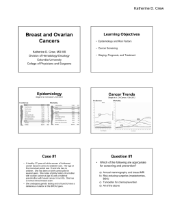


© Copyright 2026