
Document 5196
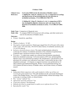
P .'—""dfTom: 1 •, wl of Urology Vol. 161 Copy.^nt © 1999 by American Urological Association. Inc. Printed in U.S.A. IMMUNOCYT*: A NEW TOOL FOR DETECTING TRANSITIONAL CELL CANCER OF THE URINARY TRACT CHRISTINE MIAN, ARMIN PYCHA, HELENE WIENER, ANDREA HAITEL, MICHELE LODDE AND MICHAEL MARBERGER From the Departments of Urology and Clinical Pathology, University of Vienna, Vienna, Austria ABSTRACT Purpose: The limitations of cytology and the invasiveness of cystoscopy for detecting bladder cancer generate increasing interest in noninvasive, urine bound diagnostic tools. We assessed the diagnostic value of the newly developed immunocytochemical test, Immunocyt, which detects cellular markers specific for transitional cell cancer in the voided urine of patients with bladder cancer. Materials and Methods: Participating in our prospective study were 264consecutive patients with a mean age of 65.9 years, including 114 in whom symptoms were suggestive of bladder cancer and 150 who were being followed after complete transurethral resection of superficial transitional cell carcinoma. Voided urine specimens were evaluated by standard cytology and the Immunocyt test, which traces the monoclonal antibodies M344, LDQ10 and 19A211 against transitional cell carcinoma in exfoliated urothelial cells. In all cases cystoscopy was subsequently performed and any suspicious lesion was evaluated by biopsy. Results: Histologically proved transitional cell carcinoma was found in 79 patients. Immunocyt with cytology had 89.9% sensitivity overall (84, 88 and 96.5% in grades 1 to 3 disease, respec tively). A total of34 (43%), 3 (3.8%) and 34 (43%) cases were positive on Immunocyt only, cytology only and both evaluations, respectively. In 8 cases (10.1%) both tests were negative. Overall Immunocyt only was 86.1% sensitive (84, 84 and 89.6% in grades 1 to 3 disease, respectively) and 79.4% specific. Overall cytology only was 46.8% sensitive (4, 52 and 79.3% in grades 1 to 3 disease, respectively) and 98.2% specific. Conclusions: Immunocyt is a noninvasive, highly sensitive test for detecting transitional cell carcinoma of all grades and stages. When combined with conventional urinary cytology, it may replace cystoscopy in select patients, especially in followup protocols of low grade transitional cell carcinoma. KEYWORDS: urinary tract; cancer, transitional cell; immunofluorescence; monoclonal antibody; cytology ucyt , , product .clinical trial bladder cancer Although cystoscopy is the most efficient method currently daily available, such as the BTA Statt and BTA Trakt as available for detecting primary or recurrent transitional cell says,7-9 and the NMP22 assay$ for nuclear matrix protein.4 carcinoma of the bladder, it is invasive and causes significant These tests are more sensitive than cytology in low grade patient discomfort. Furthermore, flat tumors or carcinoma in tumors but their specificity is so low that cystoscopy is still situ may be difficult to detect.1 Urinary cytology is noninva always essential. Moreover, in grade 3 tumors the sensitivity sive and effective for diagnosing high grade lesions but it has of these assays is approximately 25% lower than that of only 11 to 17% sensitivity in grade 1 disease, which is the routine cytology, and so cytology also remains necessary.1 In an attempt to overcome these problems Fradet and most common type of transitional cell carcinoma.1-4 The limitations of cytology and cystoscopy for making the Lockhart developed the Immunocyt test, a new approach 10 primary diagnosis and monitoring patients after transitional combining cytology and an immunofluorescence assay. Im munocyt detects cellular markers specific for bladder cancer cell carcinoma removal led to the development of new urine epithelium using 3 flu bound tests for the early detection of transitional cell carci in exfoliated cells of the transitional 10 noma.1,3,4 Methods based on the immunological detection of orescent monoclonal antibodies. Antibody 19A211 labeled a high molecular weight form of soluble antibodies in voided urine are of particular interest. with Texas red identifies 11 Kavaler et al reported that the detection of telomerase activ carcinoembryonic antigen. Antibodies MO.344 and12LDQ10 ity in the voided urine of patients with bladder cancer is 91% labeled with fluorescein are directed against mucins, which sensitive.5 Klein et al detected CK-20 in the voided urine of are expressed in most bladder cancer but not in normal transitional epithelium cells. We assess the diagnostic value patients with bladder cancer with 82.8% sensitivity and of the Immunocyt assay for detecting bladder cancer in com 100% specificity.6 However, these assays are technically com parison to and combination with conventional cytology. plicated, require highly sophisticated laboratory expertise and equipment, and are not suitable for routine cytology PATIENTS AND METHODS laboratories to perform. Recently simpler methods for detect From November 1997 to March 1998 we prospectively ob ing bladder tumor antigen in the urine have become commerA, Accepted for publication November 25, 1998. Supported by an unrestricted grant from Diagnocure, Inc., SaintFoy,Quebec, Canada. * Diagnoa ocure, Inc., Saint-Foy, Quebec, Canada. tained voided urine specimens from 264 consecutive patients, including 60 women and 204 men 21 to 93 years old (mean age 65.9) who were undergoing cystoscopy. Of the 265 pat Bard Diagnostics, Redmond, Washington. t Matritech, Inc., Newton, Massachusetts. 1486 IMMUNOCYT FOR DETECTING URINARY TRACT CANCER tients 114 had symptoms suggestive of bladder cancer and 150 were being followed after complete transurethral resec tion of transitional cell carcinoma at least 3 months previ ously. For any lesion suspicious on cystoscopy biopsy or transurethral resection was done. Histopathological classifi cation was performed according to International Union Against Cancer criteria.13 We collected 50 to 100 ml. specimens of voided urine from each patient and divided them into 2 aliquots. One aliquot was used for standard Papanicolaou and Giemsa staining, and cytological evaluation. Diagnostic results were catego rized as previously described by Koss et al.14 Briefly speci mens negative for malignancy or with atypia of any degree were categorized as negative and those considered suspicious or positive for malignancy were categorized as positive. We used 20 to 40 ml. of the sample for evaluation by the Immunocyt assay. Samples were immediately fixed with an equal volume of 50% ethanol and 1 ml. of a special fixative solution, and then incubated for 1 hour. Cells were collected by filtration through a 25 mm. polycarbonate membrane filter of 8 |am. porosity and connected to a vacuum pump. Filters were then rinsed with 3 ml. of Saccomanno solution and cells were blotted on 2 consecutive silanized slides. Cells were fixed using Merckofix* spray. Before proceeding to Im munocyt staining slides were controlled for cell content with the number of cells on a slide serving as a quality control measure. Slides containing less than 500 cells were excluded from study. A positive slide and a negative control slide guaranteed a correct staining procedure. For the Immunocyt procedure slides were initially stained by a modified Papanicolaou method using consecutive incu bation with Harris hematoxylin differentiator (70% ethanolammonium hydroxide), and OG-6 and EA-65 solutions. After rehydration in distilled water cells were incubated with 150 |al. of a blocking solution for 20 minutes at room temperature in a closed humid chamber. The blocking solution was drained from the slides, which were incubated with the Im munocyt antibody cocktail for 1 hour at room temperature. Slides were then rinsed twice in phosphate buffered saline * Merck, Darmstadt, Germany. © 1487 containing 0.5% Tween 20 and in pure phosphate buffered saline, and mounted with a coverglass. Fixative solution, negative and positive controls, blocking solution and the an tibody cocktail are provided in the Immunocyt kit. Slides were read under a fluorescence microscope using filters for fluorescein and Texas red emission light detection. Red fluorescence indicated cells positive for high molecular weight glycosylated carcinoembryonic antigen and green flu orescence indicated cells positive for bladder cancer mucins. Samples were considered positive when there was at least 1 green or 1 red fluorescent cell. Sensitivity, specificity, and the negative and positive predictive values of cytology and Im munocyt were calculated with cystoscopy and histological evaluation considered the gold standard. RESULTS Of the 264 cases 249 were evaluable. We rejected 15 spec imens because there were fewer than 500 cells per slide. Later cystoscopy revealed that all 15 cases were negative for transitional cell carcinoma. Of the remaining 249 evaluable cases histological testing verified transitional cell carcinoma of the urinary tract in 79, including 23 of 107 (21.5%) suspi cious for transitional cell carcinoma and 56 of 142 (39.4%) during followup (table 1). Since the prevalence oftransitional cell carcinoma in the 2 groups was different, predictive val ues were calculated separately. In 170 patients cystoscopy and cytology were negative. Table 2 shows the false-positive results of Immunocyt and cytology in these patients. Of the 79 cases of transitional cell carcinoma 34 (42.8%) were positive by Immunocyt only, whereas only 3 (3.8%) were positive by cytology only. Table 3 shows the sensitivity of cytology and the Immunocyt assay correlated with disease grade and stage. The sensitivity of voided urine cytology increased from 4 to 79.3% in grades 1 to 3 disease, whereas the sensitivity of Immunocyt was 84, 84 and 89.6% in grades 1 to 3 disease, respectively. However, when cytology and Immunocyt were combined, sensitivity was 84% (21 of 25 cases), 88% (22 of 25) and 96.5% (28 of 29) in grades 1 to 3 transitional cell carcinoma, respectively. For stages pTa, pTl and pT2 or greater transitional cell carcinoma the sensitivity of both tests was 88.3% (38 of 43 cases), 90% (18 of 20) and 91.6% (11 of 12), respectively. Cytology had a false-positive © A, red fluorescence shows cells positive for high molecular weight glycosylated carcinoembryonic antigen. B, green fluorescence shows cells positive for bladder cancer mucin. 1488 IMMUNOCYT FOR DETECTING URINARY TRACT CANCER TABLE 1. Patient data No. Pts. Total Beingfollowed Suspicion of transitional cell Ca Nonevaluable (less than 500 cells/slide) Transitional cell Ca bladder Transitional cell Ca ureter Free of transitional cell Ca Followup after transurethral resection Cystitis Upper tract urolithiasis Benign lesions of lower urinary tract (benign prostatic hyperplasia, nephrogenic adenoma or inverted papilloma) Renal cell, prostatic or cervical Ca Microhematuria 264 150 114 15 77 2 170 86 10 24 16 7 27 TABLE 2. Final findings in 170 patients free of transitional cell carcinoma on cystoscopy with false-positive results on Immunocyt and cytology TABLE 5. Negative and positive predictive values of Immunocyt and cytology evaluated separately in 107patients with suspected transitional cell carcinoma and 142 being followed Cytology: Diagnostic (suspicious for Ca) Followup Immunocyt: Diagnostic (suspicious for Ca) Followup Immunocyt + cytology: Diagnostic (suspicious for Ca) Followup Cystoscopy results. 9c Pos. (No. false-pos ./total No. Ca-free*) % Neg. (No. false-neg./total No. with Ca*) 93 (1/84) 92 (2/86) 90 (9/23) 72 (33/56) 54 (18/84) 73(17/86) 97 (2/23) 89 (9/56) 55(18/84) 75(17/86) 99 (1/23) 90 (7/56) nocytochemical methods using voided urine specimens have the advantage of being performed noninvasively. The deter minations of urine carcinoembryonic antigen,1 bladder tu mor antigen using the BTA Stat and BTA Trak assays,7-9 No. Pts. False-Pos./Total No. (%) and nuclear matrix protein using the NMP22 test4,16 have Immunocyt Immunocyt Cytology been investigated as new diagnostic methods to substitute for + Cytology voided urine cytology. Sarosdy et al reported 67% sensitivity Followup after transurethral 17/86 (20) 2/86 (2) 17/86 (20) and 72% specificity for the BTA Stat assay,7 whereas resection Soloway et al reported 70% sensitivity and 79% specificity for Cystitis 4/10 (40) 0/10 4/10 (40) the NMP22 test.4 Wiener et al compared the BTA Stat and Upper tract urolithiasis 2/24 (8) 0/24 2/24 (8) Benign lesions of lower urinary tract 8/16 (50) 8/16 (50) 0/16 NMP22 tests to urinary cytology, and noted 48 and 57% (benign prostatic hyperplasia, sensitivity, respectively, and approximately 70% specificity nephrogenic adenoma or inverted for both tests. Without doubt these methods facilitate the papilloma) detection of low grade tumors but specificity is not high Microhematuria 4/27 (15) 1/27 (4) 4/27 (15) Renal cell, prostatic or cervical Ca 0/7 enough to render cystoscopy unnecessary. Furthermore, 0/7 0/7 Totals these assays have lower sensitivity in high grade tumors, and 35/170 (21) 3/170 (2) 35/170 (21) so cytology is still needed as well.1,7,10, 5 The Immunocyt test is highly sensitive in all grades of TABLE 3. Sensitivity of the 2 methods according to grade and stage disease. Our study confirms the findings of Fradet and in 79 patients with transitional cell carcinoma Lockhart10 that overall sensitivity is approximately 2-fold higher than that of cytology. According to transitional cell % Sensitivity (No. pts./ Immunocyt + carcinoma grade sensitivity was much higher for Immunocyt total No ) No. Pts. . Cytologv than for cytology in low grade disease, and it reached com Cytology Immunocyt parable values in high grade disease. Similar results were Grade: obtained in correlation with tumor stage. Immunocyt speci 1 25 4 (1/25) 84 (21/25) 84 (21/25) ficity was lower than that of cytology but comparable to that 2 25 52 (13/25) 84 (21/25) 88 (22/25) reported by Fradet and Lockhart. Of the false-positive re 3 29 79 (23/29) 90 (26/29) 97 (28/29) sults 50% involved patients being followed after transure Stage: pTa 43 thral resection of transitional cell carcinoma (table 2).- These 21 (9/43) 86 (37/43) 88 (38/43) 20 pTl 70 (14/20) 85 (17/20) 90(18/20) findings may ultimately signal tumor recurrence. Combining 12 pT2 or Greater 83 (10/12) 83 (10/12) 92 (11/12) cytology with the Immunocyt assay improved sensitivity 4 pTis (Ca in situ) 100 (4/4) 100 (4/4) 100 (4/4) even further, particularly in grade 1 disease, yet specificity remained high. When cytology and Immunocyt are negative, cystoscopy may be avoided in select patients, particularly rate of 2% (3 of 170 cases) and a false-negative rate of 53% (42 after transurethral resection of low grade, low stage transi of 79), while Immunocyt had a false-positive rate of 21% (35 tional cell carcinoma that has a lower risk of recurrence. The of 170) and a false-negative rate of 14% (11 of 79). Both tests frequency of followup would be decreased to 6 instead of 3 had a false-positive rate of 21% (35 of 170 cases ) and a months. false-negative rate of 10% (8 of 79). Tables 4 and 5 show the The negative predictive value of Immunocyt in suspicious sensitivity, specificity, and positive and negative predictive and followup cases is comparable to that of the BTA Stat and values of the 2 tests. NMP22 assays,1,4'7 and higher than that of cytology. The positive predictive value of these tests is inferior to that of DISCUSSION cytology. Although the positive predictive value of cytology Monoclonal antibodies may be used for detecting transi with Immunocyt is low (55 and 75% in suspicious and fol tional cell carcinoma cells exfoliated in voided urine. Immu- lowup cases, respectively), the high positive predictive value of cytology and the high negative predictive value of Immu nocyt may lead to overall improvement in diagnostic yield. TABLE 4. Sensitivity and specificity of Immunocyt and cytology in Combining the tests provides the higher sensitivity of Immu 249 evaluable patients nocyt than conventional cytology and other commercially available diagnostic tests, while preserving the advantage of % Sensitivity 9c Specificity the high specificity of cytology. As reported by Fradet and (No. pos./79 with Ca) (No. pos./170 with Ca) Lockhart, the presence of 1 green or 1 red cell appears to be Cytology 46.8(37) 98.2 (167) the best cutoff point at which to obtain a high negative Immunocyt 86.1 (68) 79.4 (135) Immunocyt and cytology 89.9 (71) 79.4(135) predictive value, thus, avoiding false-negative results. 0 IMMUNOCYT FOR DETECTING URINARY TRACT CANCER The clinical usefulness of a diagnostic test also depends on procedure duration and technical expenditure. Immunocyt may be performed within 2 hours in specimens previously stained according to the Papanicolaou procedure for standard cytology.10 Cytology and the Immunocyt assay are done by the same technician using the same urine specimen. How ever, to evaluate the specimen trained personnel with cytological knowledge are needed. Therefore, it is advisable to perform the test at institutions where a trained cytologist and all technical equipment are available. 7. 8. CONCLUSIONS Immunocyt is a noninvasive, highly sensitive test for de tecting transitional cell carcinoma of all grades and stages. When combined with conventional urinary cytology, it may replace cystoscopy in select patients, especially in followup protocols of low grade transitional cell carcinoma. 9. Diagnocure, Inc., Saint-Foy, Quebec, Canada, provided the Immunocyt test kits. 10. REFERENCES 11. 1. Wiener, H. G., Mian, C, Haitel, A., Pycha, A., Schatzl, G. and Marberger, M.: Can urine bound diagnostic tests replace cys toscopy in the management of bladder cancer? J. Urol., 159: 1876, 1998. 2. Chopin, D. K. and Laurent, J. C: Monoclonal antibodies in bladder cancer cytology. World J. Urol., 9: 75, 1991. 3. Fradet, Y. and Cordon-Cardo, C: Critical appraisal of tumor markers in bladder cancer. Sem. Urol., 11: 145, 1993. 4. Soloway, M. S., Briggman, J. V., Carpinito, G. A., Chodak, G. W., Church, P. A., Lamm, D. L., Lange, P., Messing E., Pasciak, R. M., Reservitz, G. B., Rukstalis, D. B., Sarosdy, M. F., Stadler, W. M., Thiel, R. P. and Hayden, C. L.: Use of a new tumor marker, urinary NMP22, in the detection of occult or rapidly recurring transitional cell carcinoma of the urinary tract following surgical treatment. J. Urol., 156: 363, 1996. 5. Kavaler, E., Shu, W.-P., Chang, Y., Droller, M. J. and Liu, B. C.-S.: Detection of human bladder cancer cells in voided urine samples by assaying the presence oftelomerase activity. J. Urol., part 2, 157: 338, abstract 321, 1997. 6. Klein, A., Zemer, R., Buchumensky, V., Klaper, R., Nissenkorn, I. and Saba, K.: Detection of bladder carcinoma: a urine test, 1489 based on cytokeratin expression. J. Urol., part 2, 157: 339, abstract 1336, 1997. Sarosdy, M. F., Hudson, M. A., Ellis, W. J., Soloway, M. S., deVere White, R. W., Sheinfield, J., Jarowenko, M. V., Schellhammer, P. F., Shervish, E. W., Patel, J. V., Chodak, G. W., Lamm, D. L., Johnson, R. D., Henderson, M., Adams, G., Blumenstein, B. A., Thoelke, K. R., Pfalzgraf, R. D., Murchison, H. A. and Brunelle, S. L.: Improved detection of recurrent bladder cancer using the Bard BTA stat test. Urol ogy, 50:349, 1997. Leyh, H., Marberger, M., Pagano, P., Bassi, P., Sternberg, C. N., Pansadoro, V., Conort, P., Boccon-Gibod, L. and Thoelke, K. R.: Results of a European multicenter trial comparing the BTA stat-test to urine cytology in patients suspected of having bladder cancer. J. Urol., part 2, 157: 337, abstract 1316, 1997. Ishak, L. M., Enfield, D. L., Sarosdy, M. F. and Multicenter Group.: Detection of recurrent bladder cancer using a new quantitative assay for bladder tumor antigen. J. Urol., part 2, 157: 337, abstract 1317, 1997. Fradet, Y. and Lockhart, C: Performance characteristics of a new monoclonal antibody test for bladder cancer: Immuno cyt™. Canad. J. Urol., 4: 400, 1997. Fradet, Y., La Rue, H., Parent-Vaugeois, C, Bergeron, A., Dufour, C. and Boucher, L.: Monoclonal antibody against a tumor-associated sialoglycoprotein of superficial papillary bladder tumors and cervical condylomas. Int. J. Cancer, 46: 990, 1990. 12. Bergeron, A., Champetier, S., LaRue, H. and Fradet, Y.: MAUB is a new mucin antigen associated with bladder cancer. J. Biol. Chem., 271: 6933, 1996. 13. Sobin, L. H. and Wittekind, C: TNM Classification ofMalignant Tumours, 5th ed. International Union Against Cancer. New York: Springer-Verlag, 1997. 14. Koss, L. G., Deitch, D., Ramanathan, R. and Sherman, A. B.: Diagnostic value of cytology of voided urine. Acta Cytol., 29: 810, 1985. 15. Fraser, R. A., Ravry, M. J., Segura, J. W. and Go, V. L. W.: Clinical evaluation of urinary and serum carcino-embryogenic antigen in bladder cancer. J. Urol., 114: 226, 1975. 16. Myanaga, N., Akaza, H., Ishikawa, S., Ohtani, M., Noguchi, R., Kawai, K., Koiso, K, Kobayashi, M., Koyama, A. and Takahashi, T.: Clinical evaluation of nuclear matrix protein 22 (NMP22) in urine as a novel marker for urothelial cancer. Eur. Urol., 31: 163, 1997.


© Copyright 2026