
Urine cytology and adjunct markers for detection and Review Article
Am J Transl Res 2010;2(4):412-440
www.ajtr.org /AJTR1007003
Review Article
Urine cytology and adjunct markers for detection and
surveillance of bladder cancer
Peggy S. Sullivan1, Jessica B. Chan2, Mary R. Levin1, Jianyu Rao1
1Department of Pathology and Laboratory Medicine, David Geffen School of Medicine, University of California at Los
Angeles, 10833 Le Conte Avenue, CHS A7-149, Los Angeles, CA 90095-1732, USA; 2Kaiser Permanente, Oakland
Medical Center, Department of Pathology, 280 W. MacArthur Blvd, Oakland, CA 94611, USA.
Received July 9, 2010; accepted July 22, 2010, available online July 25, 2010
Abstract: Urine cytology coupled with cystoscopic examination has been and remains the standard in the initial
evaluation of lower urinary tract lesions to rule out bladder cancer. However, cystoscopy is invasive and may miss
some flat lesions, whereas cytology has low sensitivity in low-grade papillary disease. Additional lab-based or officebased markers are needed to aid in the evaluation of these lesions. Recently, many such markers have been developed for the purpose of improving the cytologic diagnosis of bladder malignancies. In this review, we will first discuss
conventional cytomorphologic analysis of urine cytology followed by a discussion of markers that have been developed in the past for detection and surveillance of urothelial carcinoma. We will focus on how these markers can be
used in conjunction with urine cytology in daily practice.
Keywords: Bladder cancer, urine cytology, tumor markers
Introduction
Bladder cancer, most commonly urothelial carcinoma, is the 4th most common cancer in males
in the United States [1]. However its cost per
patient is the highest of all the cancer types,
reaching approximately 200,000 U.S. dollars
per patient from diagnosis to death [2]. It has
been estimated that in 2009 approximately
70,980 new cases of bladder cancer will be
diagnosed with 14,330 deaths in this country
[1]. Approximately 75% of patients present with
superficial disease (Ta and T1) while 20% present with T2 or greater disease. The remaining
5% of patients present with metastatic disease.
Overall, 70% of treated tumors recur, with 30%
of recurrent tumors progressing to metastatic
disease [3].
Roughly 60% of patients with newly diagnosed
bladder cancer do not have muscle invasive
disease and do not require cystectomy [4]. The
majority of these patients have a recurrence
after endoscopic resection, thus lifelong surveil-
lance with cystoscopy is recommended. Unfortunately, cystoscopy, which is the “gold standard”
for the detection of de novo and recurrent bladder cancer, is an expensive and invasive procedure. In addition, it may miss a flat lesion, especially carcinoma in situ which is considered a
high grade malignant condition rather than a
precancerous lesion as in other organ systems.
Voided urinary cytology is a useful noninvasive
adjunct to cystoscopy because of its overall high
specificity. Cytology also has a relatively high
sensitivity at detecting high-grade lesions. Its
sensitivity, however, is anywhere between 20 to
50% for low-grade papillary tumors. Of the nonmuscle-invasive lesions, approximately 10% of
low-grade papillary tumors subsequently develop muscle-invasive or metastatic cancer
whereas roughly a third of high-grade tumors
progress, if not already muscle-invasive at the
time of diagnosis [4]. Therefore, close monitoring and early detection of all lesions are important for management, and noninvasive tumor
markers with high accuracy for the detection of
Urine cytology and biomarkers for bladder cancer
all grades of urothelial carcinoma will significantly reduce patient cost, anxiety and morbidity.
Urine cytomorphological analysis
Urinary cytology identifies malignant cells that
have been exfoliated from the urothelium into
the urine. The specificity of cytology is greater
than 90% [5], while the sensitivity for high-grade
disease and carcinoma-in-situ (CIS) can be as
high as 80 to 90% [6, 7]. As indicated before,
however, the main shortcoming of voided cytology is the low sensitivity (approximately 2050%) for detecting low grade neoplasms including benign papilloma, urothelial carcinoma with
low malignant potential (borderline), and low
grade papillary urothelial carcinoma (Grade 1 to
2 of 3 of the WHO classification) [3] [4, 8].
There are two main reasons for such low sensitivity. First, tumor cells of the low grade tumors
are not routinely shed into the urine because of
their cohesive nature. Second, and probably
more important, is the fact that low grade tumor
cells by definition have similar cytomorphology
to normal urothelial cells microscopically. While
increased cellularity and presence of papillary
fragments in “true” voided urine sample may be
a hint for such a low grade lesion, one has to
rule out the possibility of urothelial hyperplasia
due to various reasons such as lithiasis, infection, and instrumentation.
Probably the most common reason for the presence of increased cellularity or papillary fragments in an otherwise morphologically normal
voided urine sample is instrumentation as a
result of cystoscopy, since many such samples
are collected after the procedure is performed
even though the requisition may incorrectly
state the specimen is a "voided urine". Thus,
caution should be taken and clinical correlation
should be advised in such a setting.
Common indications for urinary cytology
Urine cytology, as an "ancient" technique, has
been used in following. First, it has been used
as a screening tool to detect urothelial cancers
in high risk populations, especially in populations exposed to chemical carcinogens through
occupational means, for example the Drake
cohort [9]. Second, it has been used as an initial test for patients presenting with hematuria
to rule out (or rule in) the possibility of urothelial
413
malignancy. Third, it has been used as a monitoring and follow-up tool for patients with a previous diagnosis of urothelial cancer to rule out
tumor recurrence. Fourth, it has been used after
transurethral resection for assessment of the
completeness of tumor removal [10]. Finally,
recently it has been applied as a test for detecting inflammation or infection, especially in kidney transplant patients where the presence or
absence of polyoma virus infection may have
significant clinical implications for rejection
[11].
Type of urine samples
The most common type of urine specimen for
cytologic analysis is voided urine. Again, keep in
mind that although the submitted sample is
marked as a "voided urine", it is important to
determine whether a cystoscopy has been performed, and if so, whether the sample is collected before or after the procedure. In collecting “true” voided urine, one should avoid a “first
morning” specimen and collect the “second
morning” voided sample, since the overnight
urine often contains many degenerated urothelial cells complicating both morphologic and
marker analysis. Although there are data to suggest that three specimens of “second morning”
voided urine collected over three consecutive
days may optimize the detection of urothelial
malignancy [12], this is not a common practice,
likely because of cost and convenience.
It is important to remember that, unlike wash or
brush samples as discussed below, a true
voided urine has the so-called "funnel" effect,
i.e., it samples the entire urinary tract system
from renal pelves (bilateral) to ureters
(bilateral), bladder, and urethra. Considering the
fact that urothelial cancer is often a "field" disease, the funnel effect ensures detection of
lesions in the entire urinary tract, especially high
grade lesions. Thus, at least in theory, voided
urine should have a higher sensitivity for detecting urothelial malignancies of the entire urinary
tract. However, the trade-off is that often the
exact location of the lesion may be difficult to
find, especially if the lesion is in the upper urinary tract (ureters and renal pelves). This may
result in a so-called “false” false positive urine
cytology. Last but not least, in female voidedurine samples, most of the epithelial cells present on the slide are squamous cells contaminated from the female genital tract. Thus, for
Am J Transl Res 2010;2(4):412-440
Urine cytology and biomarkers for bladder cancer
any type of molecular marker analysis, especially PCR-based rather than image-based analysis, the contaminated squamous cells will be
problematic. Unfortunately, the contamination
factor is often ignored in many molecular based
studies.
carcinoma in situ). A positive cytology coupled
with what appears to be a negative cystoscopy
or biopsy is another source of the so-called
"false" false positive diagnosis. More discussion
on this point will be provided in conjunction with
the discussion on tumor markers below.
Another common urine sample is catheterized
urine, which is usually more cellular than true
voided urine but is otherwise identical. Genital
contamination may be less of a problem compared to a true voided urine. The wash and
brush samples from bladder, ureter, or pelves
provide a complement to voided urine samples
for the evaluation of urinary tract lesions where
cystoscopy (or retrograde ureterocystoscopy) is
performed at the same time. Depending on
whether a suspicious lesion is seen, a washing
or brushing may be performed at the same time
as well. The advantages of washing and brushing samples include greater cellularity and a
more targeted and homogeneous population of
urothelial cells to be analyzed. For low grade
lesions, cytomorphologic analysis alone for
washing or brushing samples can be extremely
challenging. In such a setting, correlating the
cytology with the cystoscopy finding is essential.
In contrast, for high grade lesions, especially
carcinoma in situ, there may be many single or
loosely cohesive, highly atypical cells on cytology while cystoscopy may or may not show a
visible lesion. Biopsy may show only a few tumor cells on the surface (the “clinging” type of
Other specimen types include ileal conduit or
neobladder urine, which are often characterized
by the presence of many degenerated columnar
epithelial cells and inflammatory cells. On occasion, recurrent urothelial carcinoma may be
seen, and diagnosis of such lesions can be extremely difficult since many biomarkers (as discussed below) may not be helpful in such a
case.
Cells and other materials
Cells and other materials are summarized in
Box 1.
Urine sample processing
Sample fixation: The urine specimen should be
processed immediately or refrigerated at 4 degrees Fahrenheit for no longer than 24 hours. If
a delay of greater than 24 hours is anticipated,
the specimen should be fixed with an equal volume of 50% ethanol, or the specimen should be
centrifuged and the sediment mixed with an
ethanol-based fixative for liquid-based cytology
or with 50% isopropyl alcohol or denatured
Box 1
•Cells normally occurring in urine (Figures 1A-B):
- Urothelial cells - basal, intermediate, superficial (umbrella) cells
- Squamous cells - trigone, distal urethra, vagina, squamous metaplasia
•Other cells in urine:
- Glandular cells - prostate, endometrium, cystitis glandularis, paraurethra
- Renal tubular cells
- Lymphocytes, leukocytes, RBC’s
- Seminal vesicle cells (Figure 2)
•Sporadically seen in urine specimens of older patients
•Bizarre appearance with greatly enlarged nuclei and foamy fragmented cytoplasm
•Golden-brown lipofuscin pigment
•Spermatozoa accompany cells
•Abnormal DNA ploidy
- Eosinophilic inclusion bodies - giant lysosomes
•Hyaline inclusion bodies
•Non-cellular components:
- Crystals, casts, spermatozoa, corpora amylacea, mucus, fibrin, lubricant, pollen, and rarely Alternaria
spp. and microconidia
414
Am J Transl Res 2010;2(4):412-440
Urine cytology and biomarkers for bladder cancer
Figure 1. Normal cell components present in urine. (A) Basal urothelial cells have moderate dense cytoplasm with
well-defined borders. Nuclei are centrally-placed with small nucleoli and smooth nuclear contours. (B) Superficial
urothelial cells (umbrella cells) have abundant granular cytoplasm with rounded and scalloped borders. Nuclei are
large, round, frequently multiple, and centrally placed with prominent nucleoli and smooth nuclear borders. (A-B, Papanicolaou stain, 600x)
clude cytocentrifugation and SurePath or ThinPrep liquid-based techniques. The traditional
membrane filter technique is rarely used currently.
Specimen adequacy
Unlike cervical specimens, exact adequacy
guidelines for urine specimens have not been
established. In general, slides should contain at
least fifteen well-visualized basal and intermediate cells to be classified as adequate. Specimens with abnormal cells are by definition satisfactory.
Figure 2. Seminal vesicle cells may occasionally be
seen in urine. The cytoplasm may have a goldenbrown pigment. Nuclei tend to be hyperchromatic
with degenerative chromatin. Note the associated
spermatozoa. (Papanicolaou stain, 600x)
alcohol. Low pH appears to favor preservation
of urothelial cells.
Specimen processing: Specimens should be
processed by cytocentrifugation or by a liquidbased preparation. Fifty milliliters of specimen
are transferred to individual centrifuge tubes
and spun down at 10 min / 1500 rpm. The supernatant is aspirated off and the sediment is
resuspended in a balanced salt solution. Most
commonly used slide preparation methods in-
415
Diagnostic format and categories for urine
cytology specimens
The format shown in Box 2 is recommended for
urinary cytology diagnosis.
For samples that are negative for an epithelial
cell abnormality, the type of inflammation
(acute versus chronic mixed), if present, should
be characterized since the information may help
the urologist to determine potentially treatable
conditions for the patient’s urological symptoms. The presence of specific organisms, if
observed, should be specified as well. Figure 3A
shows polyoma virus-infected cells. Occasionally
SV40 immunocytochemistry may be added to
confirm the diagnosis (Figure 3B).
Therapeutic changes are the major source of
Am J Transl Res 2010;2(4):412-440
Urine cytology and biomarkers for bladder cancer
Box 2. Diagnostic format and category recommendations for urine cytology specimens:
Adequacy Statement (optional)
Satisfactory for evaluation
List any quality factor affecting specimen
Unsatisfactory for evaluation (give reason)
General Categories
Negative for epithelial cell abnormality (see descriptive diagnosis)
Epithelial cell abnormality present (see descriptive diagnosis)
Negative for epithelial cell abnormality:
Infectious agents
Bacterial organisms
Fungal organisms
Viral changes (CMV, herpes, polyomavirus, adenovirus)
Nonspecific inflammatory changes
Acute inflammation
Chronic inflammation
Changes consistent with xanthogranulomatous pyelonephritis
Cellular changes associated with:
Chemotherapy/radiation
Epithelial cell abnormality present:
Atypical urothelial cells (further comment - optional)
-Favor reactive
-Favor urothelial carcinoma
Suspicious for urothelial carcinoma
Low-grade urothelial tumor versus hyperplasia
High-grade urothelial carcinoma
(including invasive carcinoma vs. carcinoma in situ)
Atypical squamous cells
-NOS
-HPV related changes/condyloma
-Squamous cell carcinoma
Atypical glandular cells
-NOS
-Adenocarcinoma, NOS
-Adenocarcinoma of prostate
Other malignant neoplasms (specify type)
Other (Specify)
“true” false positive diagnoses. However, as a
general rule of thumb, therapeutic changes are
characterized by large cells with abundant cytoplasm, low nuclear-to-cytoplasmic ratio, and
smudgy nuclear chromatin (Figures 4A-C). Cellular-based markers, especially uCyt+ (ImmunoCyt)TM, may be useful in assisting the evaluation.
Needless to say, the category of so-called
"atypical urothelial cells" is probably the most
416
controversial diagnosis in terms of patient management. In our experience, about 20 to 25%
of all samples will be signed out as “urothelial
cell atypia” (this does not include the
“suspicious for malignancy” category - unpublished data). This category will probably benefit
the most from marker analysis. As discussed
below, we have applied marker analysis (mainly
uCyt+ and sometimes cytokeratin 20 immunocytochemistry) as a reflex test for cases signed
Am J Transl Res 2010;2(4):412-440
Urine cytology and biomarkers for bladder cancer
Figure 3. Polyoma virus-infected cells. (A) Infected cells
have an increased nuclear to cytoplasmic ratio mimicking
carcinoma in situ (“decoy cells”), a ground glass nucleus
with marginated chromatin, and occasionally short cytoplasmic tails (“comet cells”). (B) Immunocytochemistry for
SV40 is supportive of polyoma virus-infected cells. (A,
Papanicolaou stain, 1000x; B, immunoperoxidase stain,
1000x).
out as urothelial cell atypia.
As discussed before, urine samples for low
grade lesions (papilloma, borderline, and low
grade urothelial carcinoma) are more cellular
than normal, with cohesive or papillary fragments and subtle morphologic changes that
overlap greatly with hyperplastic urothelial lesions (Figures 5A-C). Therefore, these lesions
are grouped together.
High grade tumor (including carcinoma in situ)
usually shows many atypical single cells and
loosely cohesive groups, many of them degenerated. The background may be necrotic, bloody,
inflammatory, or clean. It is important to find
viable cells and carefully evaluate the nuclear-to
Figure 4. Urothelial cells with
therapeutic changes. Bacille
Calmette-Guerin (BCG), an attenuated bovine mycobacterium, is used to treat carcinoma
in situ. It also induces an inflammatory reaction, especially
submucosal granulomas, and
urothelial atypia. Urine specimens in patients treated with
BCG may have urothelial cells
with marked nuclear atypia and
hyperchromasia, however, the
nuclear to cytoplasmic ratio is
not increased and the cytoplasm has a reactive appearance. Histiocytes, including
multinucleated forms, are commonly seen. (A-C, Papanicolaou
stain, 600x)
417
Am J Transl Res 2010;2(4):412-440
Urine cytology and biomarkers for bladder cancer
Figure 5. Low grade urothelial neoplasia versus hyperplasia. The distinction may be difficult as both
entities form papillary clusters with cellular crowding.
The nuclear changes in low grade neoplasms
(increased nuclear to cytoplasmic ratio, irregular nuclear membranes, conspicuous nucleoli, haphazard
growth pattern) may be subtle. Retained umbrella
cells can be seen in both entities. (A-B, Papanicolaou
stain, 400x; C, hematoxylin and eosin stain, 400x)
-cytoplasmic ratio, nuclear membrane, and chromatin. Examples of high grade urothelial neoplasms are shown in Figures 6A-C.
Squamous cell lesions may be seen either with
418
Figure 6. High grade urothelial carcinoma. The cytologic changes are more apparent in high grade
lesions and include a very high nuclear to cytoplasmic ratio, dark coarse chromatin, irregular nuclear
borders, and occasional prominent nucleoli seen in
large, often single, cells. The background may show
degenerate cells, necrotic debris, inflammatory cells,
and blood. Glandular or squamous cytoplasmic features may be seen. Umbrella cells are mostly absent. (A-C, Papanicolaou stain, 1000x)
or without human papilloma virus (HPV) effect.
The HPV-related changes are relatively rare in
Am J Transl Res 2010;2(4):412-440
Urine cytology and biomarkers for bladder cancer
Figure 7. HPV changes of squamous cells in urine.
The changes may be seen as primary infection of
squamous cells of the urinary tract (for example,
urothelial cells with squamous metaplasia or
squamous cells lining the distal urethra) or in female
urine, as contaminations from the genital area. The
features of human papilloma HPV in urinary
squamous cells are identical to those seen in cervical
smears and include increased nuclear size, irregular
nuclear borders, hyperchromasia, and perinuclear
halos. (Papanicolaou stain, 400x).
Figure 8. Squamous cell carcinoma. Malignant cells
have abundant dense cytoplasm that can be intensely orangeophilic. Nuclei are large and hyperchromatic with irregular nuclear borders. Note the
granular, necrotic background. (Papanicolaou stain,
400x)
specimens from male patients and more common in female patients, mostly due to contamination from the gynecologic tract (Figure 7).
Squamous cell carcinoma (Figure 8) in the
United States is mostly associated with diver-
419
Figure 9. Prostate adenocarcinoma. Sheets or small
aggregates of uniform glandular cells have large
round nuclei, open chromatin, and prominent single
or double nucleoli. (Papanicolaou stain, 600x)
ticulitis while elsewhere, especially in Egypt, it is
associated with schistosomiasis.
Adenocarcinoma can be either primary (mostly
derived from glandular metaplasia or from
urachal remnants) or metastatic. Considering
the high incidence of prostate carcinoma, it is
rare to see the shedding of prostate cancer
cells, characterized by loosely cohesive or single
oval-to-low columnar cells with amphophilic cytoplasm and prominent nucleoli into urine
(Figure 9). This is because most prostate cancers arise from the peripheral zone. Only a large
tumor that extends to the urethra or a primary
central zone tumor (the so-called ductal/
endometrioid type of prostate carcinoma) may
shed into urine. Renal cell carcinoma may also
be seen in voided urine, although this is uncommon (Figure 10). For mucinous tumors, it may
be impossible to distinguish between a tumor of
primary urothelial origin, urachal origin, or metastatic from the gastrointestinal tract based
solely on cytology alone or even on a small biopsy. Currently no specific markers exist to
make this distinction, and clinical or radiologic
correlation is the only way to determine the origin of the tumor.
Potential sources of misdiagnosis
While it is uncommon to have a false positive
urine cytology, especially in the case of low
grade lesions, occasionally one may be encoun-
Am J Transl Res 2010;2(4):412-440
Urine cytology and biomarkers for bladder cancer
exact proportion of such a lesion in urine cytology.
Adjunct markers to urine cytology
Figure 10. Renal cell carcinoma. Renal cell carcinoma cells rarely shed into urine. This air-dried
smear demonstrates the abundant, vacuolated cytoplasm characteristic of renal cell carcinoma. Atypical, centrally-placed nuclei are also noted. (Diff-Quik
stain, 600x)
tered. Potential causes include the so-called
"false" false positive diagnosis, instrumentation
effect (Figure 11), stones, therapeutic changes,
including chemotherapy and radiation, and viral
infection (e.g., polyoma or CMV). The so-called
"false" false positive finding is a positive urine
cytology with lesions that are not detected clinically at the time of urine examination. Most
commonly this is a flat dysplastic or malignant
lesion or occasionally an upper tract lesion that
may be missed by cystoscopy and biopsy. There
has been no specific study to determine the
Due to its ease of accessibility, the bladder
represents an ideal model for studies in risk
assessment, early detection, and the investigation of biomarkers. The ideal biomarker should
be noninvasive, provide rapid results, be easy to
interpret with little or no variability amongst users, be cost-effective, and most importantly,
have a high sensitivity and specificity [3, 4]. Potential roadblocks in identifying the ideal marker
include the need to obtain consistent samples,
to standardize methods of fixation, to assure
quality control of assay methods, and to optimize interpretation of the data in the context of
the clinical question at hand [8]. The selection
of a biomarker depends on whether the objective is prevention, screening, surveillance, or
predicting the biological behavior (i.e., risk of
progression) of the neoplasm [8].
We will briefly highlight markers that are currently available or under investigation for the
detection and monitoring/surveillance of bladder cancer (Tables 1 and 2). As such, markers
that are useful in predicting recurrence are beyond the scope of this review, although many of
the markers herein discussed will cross over
into the other categories. We will first briefly
discuss markers currently used in clinical practice, some of which have been approved by the
Federal Drug Administration (FDA). We will follow with a preview of markers that are more
investigational but may potentially be integrated
into clinical practice in the near future.
Nuclear matrix protein 22 (NMP-22) – FDA approved
Figure 11. Instrumentation effect. Bladder washing
or voided urine after cystoscopy will contain tissue
fragments with smaller basal cells surrounded by
large binucleated umbrella cells. Note that each cell
has a distinct outline and that the tissue fragment
boundaries are not smooth. (Papanicolaou stain,
400x)
420
Nuclear matrix proteins (NMP) consist of a three
-dimensional web of RNA and proteins that supports the nuclear shape, organizes DNA, and
coordinates DNA replication, transcription, and
gene expression [3, 13]. NMP-22 is released
from the nuclei of tumor cells during apoptosis.
NMP released into the urine may be detected by
an FDA-approved NMP-22[14] enzyme-linked
assay kit (Matritech, Newton, Mass).
NMP-22 is a 238-kDa protein that may be detected at up to 25-fold greater concentration in
tumor than normal urothelium [15, 16]. The
Am J Transl Res 2010;2(4):412-440
Urine cytology and biomarkers for bladder cancer
Table 1. Adjunct markers for urine cytology
Methods
Sensitivity
Specificity
Comment
Cytology
20-90%
>90%
Low sensitivity for detecting low grade lesions
uCyt™ (Immunocyt)
67%->90%
62-84%
Technically simple, interpretation difficult;
FDA-approved
FISH (UroVysion)
30-86%
75->90%
Technically difficult; interpretation difficult;
FDA-approved
BLCA-4
89%
>90%
Needs further testing
Telomerase (TRAP)
70-90%
66-88%
Lacks standardization and technically difficult
Cytokeratin 20
(by immunocytochemistry)
65->90%
67->90%
Simple marker; exclude benign conditions to
improve specificity
Hyaluronic acid/ Hyaluronidase
83->90%
63->90%
Higher sensitivity for low grade lesions
Survivin
53->90%
88->90%
Needs further testing
Microsatellite instability
58->90%
73->90%
Technically difficult
DD23 (QFIA)
70-85%
55->90%
Needs further testing
Quanticyt
59-69%
68-70%
Low sensitivity, technically difficult
PSCA
80%
85.7%
Needs further testing
DNA methylation
69->90%
60->90%
Needs further testing
Table 2. Strip-based Adjunct Markers for Urine Cytology (all FDA-approved)
Methods
Sensitivity
Specificity
Comment
NMP 22
73%
56->90%
Good sensitivity in low grade lesions
BTA Stat
9.3-89%
50-90%
Benign hematuria lowers specificity
BTA TRAK
56-68%
54-75%
Better sensitivity, specificity still low
FDP
68%
78%
Currently not being produced
enzyme-linked immunoassay uses two monoclonal antibodies to measure the levels of complexed and fragmented forms of the mitotic apparatus in urine [15]. A cut-off of 10 u/ml is
endorsed by the manufacturer and initial study
for recurrence [17]; however, there is no universally accepted cut-off point. Other studies have
suggested cut-offs ranging from 5 to 20 u/ml
[17-21].
Some suggest NMP22 may be useful as a
screening tool [22]. Surveillance studies either
alone or as an adjunct to cytology have estimated sensitivities ranging from 32-100% and
specificities from 56-95% [23]. In a 50 study
421
meta-analysis, Lotan et al reported a median
sensitivity and specificity of 73% and 80% respectively, superior to a voided urine cytology
sensitivity of 34% [24]. Major sources of false
positivity are hematuria and pyuria [25]. This is
a serious problem since many benign urologic
conditions such as stone disease and infection
present with hematuria [26]. In general, NMP22 has a higher sensitivity than cytology, especially in detecting low grade and low stage tumors.
Multiple studies have evaluated the usefulness
of NMP-22 as a marker of tumor recurrence.
Soloway et al used NMP-22 to predict the likelihood of recurrence after transurethral resection
Am J Transl Res 2010;2(4):412-440
Urine cytology and biomarkers for bladder cancer
at subsequent cystoscopy in ninety follow-up
patients [17]. Levels less than 10 U/mL were
predictive of a low likelihood of recurrence while
levels greater than 10 U/mL were predictive of
recurrence (overall sensitivity of 69.7% and
specificity of 78.5%). Subsequent studies, however, were less impressive. Boman et al reported a sensitivity and specificity of 45% and
65% respectively [27]. Miyanaga et al reported
a sensitivity and specificity of 18.6% and 85.1%
respectively. Both studies concluded that the
low sensitivity was due to the small size of recurrent tumors [18].
A new point-of-care test for NMP22 (BladderChek test) was shown to have sensitivities ranging from 50-85% and specificities ranging from
40-90% [28-32]. Advantages include on-site
testing with immediately available qualitative
results, making the test an attractive adjunct for
cystoscopy.
Overall analysis of the data shows that the NMP
-22 test has superior sensitivity over cytology for
detection of low grade bladder cancers and may
be used to predict increased recurrence risk in
patients with elevated levels after transurethral
resections. Because of the low specificity, using
NMP-22 routinely as a primary detector of bladder cancer is not recommended. The specificity,
however, can be improved if patients with benign inflammatory conditions (infections, etc),
renal or bladder calculi, foreign bodies (stents
or nephrostomy tubes), bowel interposition,
other genitourinary cancer, and/or instrumentation are excluded [25, 33].
Bladder tumor antigen (BTA) –FDA approved
The term BTA actually describes three separate
tests: 1) BTA, 2) BTA stat, and 3) BTA TRAK.
Since the BTA tests depend on the disruption of
basement membrane, their sensitivity improves
with more invasive cancer [34]. Advantages
include increased sensitivity for invasive tumors. Disadvantages include a high rate of false
-positive readings secondary to patients with
inflammatory conditions secondary to benign
prostatic hypertrophy (BPH) and a low overall
sensitivity for detection of all bladder tumors.
The original BTA test was a latex-agglutination
test that measured levels of basement protein
antigen released into urine as a result of tumor
invading into the stroma [35]. In a review of
422
over 1000 patients (seven series), the sensitivity of the original BTA test was only 52.3%, while
the specificity was 84.6% [20] [36-41].
BTA stat and BTA TRAK detect human complement factor H-related protein (hCFH) which is
produced and secreted by several bladder and
renal cancer cell lines. The qualitative BTA stat
test costs only five dollars and is easily performed in the office with a dipstick format [42].
The overall sensitivity ranges from 9.3% to as
high as 89% with higher sensitivity in higher
grade tumors [43-50]. The specificity of the BTA
stat among healthy individuals is greater than
90%. However, it has low specificity (about 50%)
among patients with urinary tract infections,
urinary calculi (90% positive using BTA stat
[51]), nephritis, renal stones, cystitis, BPH, hematuria, and 2+ to 3+ protein on urine dip stick
[49, 52-54]. The low specificity in these conditions is secondary to the test’s ability to detect
both complement factor H-related protein and
complement factor H. Complement factor H is
present in human serum at high concentrations
and therefore BTA-Stat testing may be falsely
positive in benign, hematuria-causing conditions
[13].
BTA TRAK is a quantitative test that has a
slightly improved sensitivity over its two BTA
predecessors [55-57] but has high false positive rates for similar reasons (e.g., inflammation
and trauma) which therefore leads to low specificity [57-59]. Moreover, multi-center studies
and cohort studies have shown that the sensitivity of the BTA TRAK also varies depending
upon the cut-off limit used for the test [57, 5962].
Overall, the three BTA tests lead to an improved
sensitivity compared to cytology but lower specificity due to high false positive rates associated
with recent instrumentation, stones, inflammatory conditions, BPH, and hematuria.
Fibrin-fibrinogen degradation products (FDP) –
FDA approved
Since bladder tumor cells induce vascular permeability, cellular proteins such as plasminogen
and fibrinogen leak into the urine. Urokinase
subsequently converts plasminogen into plasmin which then converts fibrinogen into fibrinfibrinogen products (FDP) [63]. Thus, patients
with bladder cancer may have increased levels
Am J Transl Res 2010;2(4):412-440
Urine cytology and biomarkers for bladder cancer
Table 3. Tumor Recurrence in Patients with Negative Cystoscopy [78]
uCyt™
Six Month Follow-up, % (n)
One Year Follow-up, % (n)
Negative Test
13.9 (8/59)
11.9 (7/59)
Positive Test
35.7 (21/59)
47.0 (8/17)
of FDP in their urine. In a review of four series,
the sensitivity and specificity of the AccuDx-FDP
assay ranged from 68-83% and 68-100%, respectively [38, 64-66]. Advantages include highyield with invasive tumors presumably because
of increased leakage of FDP. Disadvantages
include poor sensitivities for low-grade disease
and poor specificities due to reasons previously
mentioned in association with BTA tests. The
test costs about fifteen dollars and takes less
than 10 minutes to complete [42]. However, the
assay is currently not being produced due to
issues regarding test formulation [4].
uCyt+ (ImmunoCyt)™ and tumor associated
antigens -FDA approved
uCyt+ (ImmunoCyt)™ is based on the detection
of tumor-associated antigens, mostly mucinglycoproteins, in transitional or urothelial carcinoma using monoclonal antibodies. The uCyt+™
assay is the most frequently used immunocytological test today. Three antibodies, fluorescein-labeled M344 and LDQ10 (directed
against sulfated mucinglycoproteins), and Texas
-red linked antibody 19A211 (directed against
glycosylated forms of high molecular carcinomaembryonic antigens, i.e., CEA) are used.
Many studies have evaluated the performance
of the test since 1997. Using a threshold of any
single cell positive as positive, uCyt+™ has a
sensitivity and specificity ranging from 67-100%
and 62-84% [67-76] for all tumors and is a
promising diagnostic marker for bladder cancer.
Since one of the antibodies, M344, appears to
be quite sensitive for low grade tumor cells, this
test offers an important advantage for detecting
low grade tumors. Indeed, a recent split sample
study comparing cytology, uCyt+™, and
UroVysion™ in 100 urine samples collected
from 100 bladder cancer patients (monitoring
population) showed that the sensitivity of
uCyt+™ outperforms cytology and UroVysion™
for detecting bladder cancer, especially in low
grade tumors [77]. The sensitivities of all tumors for UCyt+™, cytology, and UroVysion™
were 76%, 21%, and 13%, respectively. The
423
drawback of the test, however, is low specificity
compared to cytology or UroVysion™ (63% for
UCyt+™ versus 97% for cytology and 90% for
UroVysion™). Piaton et al [78] demonstrated
that patients who had a positive UCyt+™ but
negative cystoscopy (so called "false" positive)
had a much higher risk of tumor detection
within a 12 month-follow up (Table 3). The findings suggest that many of these so-called "false"
positive samples may actually be “false” false
positive, i.e., the test detects early lesions, or
lesions that may not be seen by cystoscopy at
the time of examination. Figure 12 shows
UCyt+™ staining in a urine sample that had
atypical urine cytology and subsequent low
grade tumor on cytology six months later. Exactly how many of the false positives belong to
the "false" false positive category versus the
true false positive category due to benign conditions such as urinary tract obstruction is not
clear.
Figure 12. UCyt+TM (ImmunoCyt) in a sample with
atypical urine cytology. Fluorescein- and Texas-redlabeled antibodies detect tumor-associated antigens
in urothelial carcinoma. Positive tumor cells are
both green and red under fluorescence microscopy.
(400x)
In addition to a relatively high sensitivity in detecting malignancy, uCyt+™ is technically simple
(about a 30 minute incubation on either ThinPrep or filtered slides) and relatively inexpen-
Am J Transl Res 2010;2(4):412-440
Urine cytology and biomarkers for bladder cancer
sive. However, the disadvantage is the false
positive result occasionally seen associated
with urinary tract obstruction due to either
stones or BPH. Also, the interpretation of the
test using fluorescence microscopy may be difficult for many cytologists, since determination of
a positive cell may not be as easy as one might
expect in some cases. Further, because the
antigens detected are mucinglycoproteins or
glycosylated CEA, colonic mucosa cells are positive for the test. As such, the test is not suitable
for loop or neobladder urine samples. Another
important point worth noting is that the FDA
approved this test with a threshold of any single
cell positive as positive. However, in our experience (data not shown), and perhaps as expected, samples that have less than or equal to
5 cells positive. Thus, in our report, we not only
provide the overall positive or negative finding
of the test, but also report samples with 0-5
cells positive as borderline and specify the number of fluorescein- or Texas-red-labeled positive
cells.
Multi-target multicolor FISH assay (UroVysion™
test) - FDA approved
Urothelial carcinomas have a number of associated cytogenetic abnormalities involving chromosomes 1, 3, 4, 7, 8, 9 11, 17, etc.[79-81].
These chromosomal abnormalities can be detected by fluorescent in situ hybridization (FISH)
using DNA probes to chromosome centromeres
or unique loci that are altered in tumor cells.
Hybridization is detected by fluorescent microscopy. By utilizing multicolored probes (i.e., different DNA probes labeled with different fluorescent dyes), sensitivity is improved compared to
using a single probe.
The UroVysion™ test is a multi-target, multicolored FISH assay that utilizes peri-centromeric
fluorescent probes for chromosomes 3, 7, 17,
and a locus-specific probe to the 9p21 (p16
locus) region. Exfoliated cells from urine specimens are fixed into 12-well slides and incubated with denatured Chromosome Enumeration Probe (CEP) 3 (spectrum red), CEP7
(spectrum green), CEP 17 (spectrum aqua), and
Locus Specific Identifier (LSI) 9p21 (spectrum
gold). The slides are counterstained and observed under a fluorescent microscope
(UroVysion™/Abbott Laboratories). Suggested
criteria for a positive assay include finding 5 or
more urinary cells with gains of 2 or more chro-
424
mosomes, or 10 or more cells with gain of a
single chromosome (e.g. trisomy 7). Also homozygous detection of 9p21 locus in greater than
20% of epithelial cells is considered a positive
test [48, 82]. However, consensus criteria for a
positive FISH test have not been determined,
and the studies evaluating the sensitivity and
the specificity of UroVysion™ utilize varying criteria for positivity.
Initial case-control cohort studies showed that
the sensitivity of UroVysion™ to detect bladder
cancer was 81-84% [83, 84]. In more recent
studies, the sensitivity ranged from 30-86% [8588]. The assay has increased sensitivity for detecting higher grade and higher stage tumors,
however, the sensitivity for detecting low grade
tumors is not clear. Low grade tumors are the
most difficult to diagnose by cytology. In fact,
our split-sample study of 100 bladder cancer
monitoring urine samples showed that the sensitivity for UroVysion™ test is substantially lower
compared to the uCyt+™ test (13% versus 76%,
respectively)
[77].
The
specificity
for
UroVysion™, however, is high in our study (90%,
similar to cytology) and varies between 75% and
100% by others [48, 85, 89-94]. Notably, the
test appears to have high specificity among patients who have a variety of benign genitourinary conditions, including microhematuria, BPH,
infections, and inflammation [48, 84, 91].
Some studies also suggest that UroVysion™ can
predict recurrence. Skacel et al in a retrospective cohort study reported that 8 out of 9 FISH
positive patients with atypical cytology but negative biopsy had biopsy proven bladder cancer
within 12 months [95]. In another study, Bubendrof et al reported that 4 of 5 so-called “falsepositive” UroVysion™ tests had recurrence
within 8 months; none of the true negative
cases recurred within 18 months. However, the
criteria used for a positive test in this study differed from those suggested by the manufacturer. Moreover, the authors concluded that not
all FISH aberrations were equally important
[96].
In summary, UroVysion™ seems to have high
specificity for the detection of bladder cancer
and for the ability to detect bladder tumor recurrence prior to clinical detection. Thus, it may be
used as a confirmatory test for either cytology or
uCyt+™ test. One major limitation to the assay
is the lack of consensus on the criteria used to
Am J Transl Res 2010;2(4):412-440
Urine cytology and biomarkers for bladder cancer
evaluate abnormal cells. Additionally, the test
has relatively low sensitivity in the detection of
low-grade bladder tumors as discussed before
and therefore may not improve the sensitivity as
an adjunct for cytomorphologic analysis.
BLCA-4
Konety and Getzenberg have described several
specific nuclear matrix proteins which are present only in patients with bladder cancer (BLCA
1-6) and three proteins that are present in normal bladder tissue (BLNL 1-3) [97]. One of
these markers, BLCA-4, is found throughout the
bladder in patients with bladder cancer, including both tumor and normal regions. The marker
is hypothesized to reflect a type of “field effect”,
which has been described by several investigators at the genetic level. Studies by Getzenberg
et al have shown that this marker appears to be
a transcriptional regulator that may play a role
in regulating gene expression in bladder cancer
[98]. In initial studies using an indirect enzyme
linked immunosorbent assay (ELISA), they report that BLCA-4 levels were significantly higher
than those found in normal controls and that 53
of 55 (96% sensitivity) samples had BLCA-4
expression [99]. Subsequent trials utilizing a
sandwich-based immunoassay examined BLCA4 expression in a variety of patients including
those with biopsy proven bladder cancer, benign urologic conditions, prostate cancer, and
normal individuals. The results of this trial demonstrated a sensitivity of 89% and a specificity
of 100% [100].
Similar proteins, namely BLCA-1, have also
been found to be potentially useful. Unlike BLCA
-4, BLCA-1 is expressed in tumor areas only and
is not seen in adjacent normal tissue or tissue
from normal individuals. An immunoassay detecting BLCA-1 in urine samples has been developed and demonstrates relatively high sensitivity and specificity [101]. However, additional
independent studies will be needed to validate
the findings.
Telomerase
Telomeres are nucleotide sequences on the
ends of chromosomes that are important in
maintaining the integrity of DNA. With each
replication cycle, a portion of the telomere is
lost, and complete loss of telomeres is associated with cell death. Telomerase is an enzyme
that lengthens telomeres; thus, increased levels
425
of telomerase allow tumor cells to maintain immortality [102].
The telomeric repeat amplification protocol
(TRAP) assay is a polymerase chain reaction
(PCR)-based test that detects increased levels
of telomerase secreted into the urine by bladder
cancer cells. Other telomerase assays are available that detect human telomerase reverse
transcriptase and its RNA component. However,
for sake of brevity, discussion will be limited to
the TRAP assay. It detects telomerase reaction
products in vitro, has a 10 hour turnaround
time, and costs around seventeen dollars [4,
102]. Generally, the TRAP assay has better sensitivity than cytology with slightly lower specificity [20, 103]. Recent studies show the sensitivity to range from 70-90% at a 50 arbitrary enzymatic unit cutoff value. Specificity is slightly
lower, ranging from 66-88% [13, 20, 104-107].
The lower specificity may be explained by contamination of benign cells with telomerase activity (e.g., lymphocytes). The sensitivity was
slightly increased with bladder washings compared to voided urine [103]. In detecting recurrent tumors, however, TRAP has a low sensitivity
(35%) [108].
Difficulties associated with the telomerase test
have limited its widespread use. Urine must be
processed within a 24 hour period [13]. At least
50 cells must express telomerase for the assay
to detect telomerase reliably [13, 109]. Finally,
false negative results may occur depending on
sample collection, processing, and the presence
of PCR inhibitors or ribonucleases [110]. Currently the TRAP assay is not recommended in
the clinical setting because of complicated laboratory procedures and the lack of standardized
sample processing to reduce false positive and
false negative results.
Cytokeratins
Cytokeratins (CK) make up a large component
of intermediate filaments that are found in
epithelial cells [111]. Twenty cytokeratins have
been identified in human cells, and their expression varies depending on epithelial cell type and
state of differentiation [112]. Expression of cytokeratins 8, 18, 19, and 20 has been evaluated as potential bladder cancer markers.
UBC-Rapid and UBC-ELISA tests (manufactured
by IDL Biotech, Börlabger Sweden) detect the
presence of cytokeratin 8 and 18 in the urine of
Am J Transl Res 2010;2(4):412-440
Urine cytology and biomarkers for bladder cancer
bladder cancer patients. UBC-Rapid is a point-of
-care test, and UBC-ELISA is a 2-hour sandwich
ELISA test. Several studies show that the sensitivity of the UBC tests to detect both primary
and recurrent bladder cancers varies from 1279% with a specificity ranging from 63-97% [46,
57, 113-121]. Several studies have also reported lower sensitivities for the detection of
low grade and low stage tumors. The sensitivity
of UBC to detect grade 1, 2, and 3 bladder tumors is 13-60%, 42-79%, and 35-75% respectively [114, 116, 117, 122]. Retrospective studies report a 21-25% sensitivity of UBC- Rapid to
detect stage Ta tumors and carcinoma in situ
(CIS) and therefore it has insufficient diagnostic
value for detecting superficial bladder cancer
[46, 122]. Compared to other bladder tumor
markers and cytology, UBC tests have generally
lower sensitivity.
The expression of cytokeratin 20 is restricted to
the superficial and occasionally the intermediate cells of the normal urothelium, but not the
basal cells. Aberrant cytokeratin 20 (CK 20)
expression is seen in bladder cancer cells
[112]. Reverse transcription (RT)-PCR assays
have been used to evaluate CK 20 expression
in urine samples. Several studies have shown
that CK 20 RT-PCR has a 78-87% sensitivity for
detecting bladder cancer in urine. The specificity of CK 20 RT-PCR ranges from 55.7% to 98%
[111, 123-129]. CK 20 RT-PCR on blood specimens has also been studied for early detection
of systemic bladder cancer progression [126].
Overall, 17-29% of bladder cancer patients were
positive for CK 20 RT-PCR [130, 131]. The high
sensitivity intrinsic to the RT-PCR methodology
may also be associated with low specificity
[125].
Cytokeratin 20 immunocytochemistry has also
been evaluated as an adjunctive marker for
atypical cytology. Klein et al reported a sensitivity of 91% and a specificity of 67% in a study of
87 patients [132]. Specimens with falsepositive results had cytology consistent with
premalignant conditions such as atypia, hyperplasia, or metaplasia [111, 132]. All completely
healthy patients had negative CK 20 levels. Lin
et al showed that overall sensitivity and specificity of CK20 immunocytochemistry for the detection of urothelial carcinoma were 94.4% and
80.5% respectively [133]. This study demonstrated that CK20 is a useful adjunct marker for
urine cytology, that is, analysis of CK20 can be
426
conveniently performed on the same slide after
routine morphological evaluation and be used
to triage atypical urine cytology into low and
high risk categories for clinical follow-up. Golijanin et al also reported a high sensitivity (82%)
for CK 20 immunocytochemistry in patients with
microhematuria and those with bladder cancer.
The specificity in this study was 76% [134]. Although overall sensitivity was high, the Golijanin
study also showed that the sensitivity varied
depending on tumor grade, with only 56.5%
sensitivity for grade 1 tumors. CK 20 staining
had a much higher sensitivity with grade 2 and
3 bladder tumors (93% and 92% respectively)
[134]. More recent studies have supported
these findings, demonstrating sensitivities ranging from 65-86%, specificities from 86-100%,
and advantages over urine cytology in the detection of primary, recurrent, stage pT1 and grade
2/3 tumors [135-137]. One of the pitfalls of the
CK20 immunocytochemical staining is that often benign umbrella cells are positive. Thus, in
our practice, we only use CK20 in samples containing few small basaloid cells and to distinguish whether these cells represent normal
basal cells (negative CK20) versus dysplastic or
malignant cells (positive CK20).
Cytokeratin 19 (CK 19) is expressed in normal
urothelium. CYFRA 21-1 is a soluble fragment of
CK 19 that can be measured in the urine when
urothelial cells are exfoliated and lysed. There
are two commercially available tests that can
measure CYFRA 21-1; one is a solid phase
sandwich immunoradiometric assay (Cis Bio
International, Gif-sur-Yvette, France) and the
other is an electrochemiluminescent immunoassay with the Elecsys 2010 system (Roche Diagnostics). CK 19 levels are measured after urinary creatinine is normalized [138]. A retrospective cohort study showed that CYFRA 21-1 levels
are increased in bladder cancer patients when
compared to patients with other urologic conditions and normal controls. The level of CYFRA
21-1 in patients with bladder cancer, patients
with other urologic conditions, and normal controls were 154.4 ng/mL, 22.3 ng/mL, and 2.4
ng/mL respectively [139]. When a cutoff level of
4 ng/mL was applied, the sensitivity and specificity of CYFRA 21-1 for detection of bladder
cancer were 96.9% and 67.2% respectively. The
low specificity was attributed to high CK 19 levels in patients with urolithiasis and urinary tract
infection. Another study reported the sensitivity
and specificity of CYFRA 21-1 to be 75.5% and
Am J Transl Res 2010;2(4):412-440
Urine cytology and biomarkers for bladder cancer
71% respectively when using the electrochemiluminescent assay [140]. This study
showed the sensitivity of detecting bladder cancer increased with higher grade tumors
(sensitivities to detect grade 1, 2, and 3 tumors
were 54.5%, 66.7%, and 88.2% respectively).
However, the study also reported a false positive rate of approximately 33% in patients with
various urologic conditions including urolithiasis, stenosis, BPH and urinary tract infections.
Subsequent later studies have shown sensitivities ranging from 43-79% and specificities ranging from 68-88% with a 4 ng/mL cut-off [118,
141-143]. Some have reported improved sensitivity over cytology in detecting Grade 1 tumors
[141].
In summary, cytokeratin 20 detected by RT-PCR
or immunocytochemistry appears to be a useful
and simple marker. However the often positive
findings of CK20 in normal umbrella cells preclude widespread application of the test as a
primary screening tool. Rather, it is better used
as a test in specific settings; for example, in
samples that contain scant small basaloid cells.
The UBC tests appear to have lower sensitivity
compared to other tumor markers and currently
do not have sufficient diagnostic value for the
detection of bladder cancer. Overall, the use of
CYFRA 21-1 is promising with some conflicting
studies of its benefit over urine cytology.
Hyaluronic acid/Hyaluronidase
Hyaluronic acid (HA) is a glycosaminoglycan that
promotes tumor cell adhesion and angiogenesis
[13, 144]. Hyaluronidase (HAase) is an enzyme
which cleaves HA into fragments; these cleaved
fragments then aid tumor growth and propagation by promoting angiogenesis [145, 146]. Initial case control studies measured both HA levels and HAase activity in the urine. The results
showed that there was a 2.5 to 6.5-fold increase in HA levels (83% sensitivity and 90.1%
specificity) in patients with bladder cancer, regardless of tumor grade [146]. HAase activity
levels were also increased 3 to 7-fold in patients with high grade bladder cancers (81.5%
sensitivity and 83.8% specificity) [147]. The
combination of tests increases the overall sensitivity to 92%.
A prospective study to monitor bladder cancer
recurrence showed that the HA-HAase was more
sensitive and more accurate than BTA stat
427
(sensitivities of 94% and 61% respectively). The
BTA stat test, however, had better specificity
than the HA-HAase test (74% and 63% respectively) [49]. Subsequent comparative studies
showed that HA-HAase testing had the highest
sensitivity in detecting both low grade/lowstage, and high grade/high stage tumors [46,
67]. Passerotti et al compared accuracy of the
HA test to UroVysion™, BTA stat, and cytology in
a prospective study involving bladder cancer
patients with either primary or recurrent tumors.
The specificity of the test was determined in
patients with a history of bladder cancer not
evident at the time of testing and in patients
with BPH. HA testing had the highest sensitivity
among all the tests (83%) and a slightly higher
specificity [148]. Eissa et al examined HAase
RNA in urine detected by RT-PCR and found superior sensitivity (90.8%) over cytology (68.9%)
and CK 20 (78.1%) with specificities of 93.4%,
98.1% and 80.2%, respectively [149].
In summary, HA-HAase testing is a promising
marker for the detection of primary and recurrent bladder tumors. The test has high sensitivity with the ability to detect low grade/low stage
and high grade/high stage tumors. The test
may be most useful in screening bladder cancer
patients for recurrence.
Survivin
Survivin is an inhibitor of apoptosis that extends
cell viability in bladder tumors [150, 151]. Survivin is undetectable in most normal adult tissue and correlates with unfavorable disease
and shortened overall survival in neuroblastoma, colorectal cancers, and non-small cell
lung cancers [152-155]. Gazzaniga et al demonstrated using RT-PCR that Survivin mRNA is expressed in 30% of bladder tumors [156].
Schultz et al found Survivin mRNA expression in
100% of bladder tumors [157].
Smith et al analyzed urine specimens with a
polyclonal antibody for Survivin and then validated findings with both western blot and RTPCR. Survivin was detected in 31 of 31 patients
with new onset or recurrent bladder cancer using the polyclonal antibody system and 15 of 15
patients with RT-PCR, giving a sensitivity of
100%. Only 3 of 35 patients with treated bladder cancers and negative cystoscopies tested
positive, suggesting Survivin could be used for
surveillance. Additional data showed Survivin
Am J Transl Res 2010;2(4):412-440
Urine cytology and biomarkers for bladder cancer
was negative in 17 healthy volunteers and 30
patients with non-urothelial genitourinary cancers. There were only 4 false positive results
amongst the 30 patients with non-neoplastic
urinary tract disease, including 3 with bladder
abnormalities on cystoscopy and one patient
with an elevated PSA. The overall specificity for
Survivin was 95% [151]. Other studies have
shown that urinary Survivin levels are higher in
patients with recurrence of carcinoma compared to those who achieved remission after
treatment with BCG or mitomycin C. The sensitivity and specificity for detecting recurrence
were 100% and 78% respectively [158]. Finally,
urinary assays detecting Survivin mRNA by RTPCR have shown sensitivities ranging from 5394% and specificities from 88-100% [159-162].
cytometry in detecting low grade tumors [166].
Moreover, such a system allows the analysis of
multiple other protein (or potentially DNA)-based
markers at the same time. Subsequent studies
have shown similar findings [92, 167]. Laser
scanning cytometry combines the advantages of
flow cytometry and ICM by laser-scanning individual cells to quantify fluorescence [168].
While numerous studies have generally shown
that imaging-based DNA content analysis is a
useful marker for detecting bladder cancer, the
need for expensive instrumentation and careful
quality control measures for fluorescence quantification precludes the widespread application
of this useful technology.
In short, Survivin could potentially be a valuable
marker for both detection and monitoring of
bladder cancer, but its validation awaits further
testing.
Similar to other malignancies, bladder cancer
DNA repair mechanisms may be defective, leading to persistent errors in replication, i.e., genomic instability. Microsatellites are inherited
tandem repeat DNA sequences that can be analyzed to detect replication errors [169, 170],
also known as microsatellite instability. PCR
amplification of these tandem repeat sequences can detect microsatellite instability and
loss of heterozygosity (LOH) of tumor suppressor genes. While microsatellite instability tends
to be found more frequently in advanced bladder cancers, it can be demonstrated in low
grade tumors when more microsatellite markers
are used. The methodology requires a substantial number of microsatellite markers to achieve
high sensitivity [171-174].
DNA ploidy and S-phase fraction
DNA ploidy and S-phase fractions can be evaluated from urine samples by either flow cytometry, image cytometry (ICM), laser scanning cytometry (LSC), or fluorescence in situ hybridization (FISH) [3]. While an FDA-approved FISH
methodology has already been discussed in this
article (UroVysion™), other methodologies such
as flow cytometry can identify neoplastic cells
with increased nuclear size and increased nuclear chromatin ratios and further determine
their DNA ploidy (e.g., diploid, tetraploid, or aneuploid). High grade tumors may be detected by
the presence of aneuploidy and a higher percentage of cells in the S phase [3]. Sensitivity
for high grade urothelial tumors or carcinoma in
situ may reach 90% [3, 163-165]. Because this
technique is expensive and requires a large
number of cells as well as highly trained personnel, flow cytometry has not gained widespread
acceptance.
Image cytometry (ICM), especially a fluorescence-based system, allows the measurement
of DNA content in each individual cell, making
this an attractive alternative to flow cytometry,
which requires a large cell population. Hemstreet's group, using a specific platform called
Quantitative Fluorescence Image Analysis
(QFIA), demonstrated that single cell-based DNA
content analysis (by detecting cells over 5c
DNA) is more sensitive than cytology or flow
428
Microsatellite instability assays
Microsatellite analysis has been used to confirm
that low grade papillary urothelial carcinoma
has instability and/or loss of chromosome 9
and p16 (MTS1) tumor suppressor gene [169,
175]. Regardless of tumor grade and stage,
bladder tumors typically have LOH in the 9p
region on microsatellite analysis [169, 176].
Using microsatellite analysis and PCR, Mao et al
were able to identify 19 of 20 patients (95%
sensitivity) with genetic alterations; however, 2
of 4 samples with inflammatory atypia were also
positive [13, 177]. Recent studies by van Rhijn
et al showed that activating FGFR3 mutations
are detectable in low grade superficial bladder
cancers, and, when used in conjunction with
microsatellite analysis, the sensitivity for bladder cancer detection increases to 89%
(compared to 71% for negative FGFR3 mutations) [178]. Other studies have shown that microsatellite instability may be used to predict
Am J Transl Res 2010;2(4):412-440
Urine cytology and biomarkers for bladder cancer
recurrence of urothelial carcinoma. The sensitivity of selected studies ranged from 58% to 95%
with specificity ranging from 73-100% [169,
177, 179-185] with some studies showing a
prediction of recurrence months before positive
cystoscopy. However, large-scale analysis will be
needed to determine specificity, especially in
symptomatic populations, to understand the
true clinical utility of the test.
DNA chips (HuSNP chip) can detect alleles differing by a single nucleotide polymorphism.
With this technology, LOH can be detected at
1500 different loci at once. Preliminary results
in thirty-one patients show LOH at 24 or more
loci, demonstrating the ability of chip technology
to detect bladder tumors with 100% sensitivity.
Nine control subjects and 4 of 5 patients with
hematuria had negative chip findings [186].
Although studies to date show that microsatellite analysis has excellent sensitivity and specificity regardless of tumor grade and stage, tumor multiplicity, or previous history of bladder
cancer, the studies are based on relatively small
sample sizes. Disadvantages include a potential
contamination of non-urothelial cells that may
cause either false negative or positive findings,
long turnaround time, high equipment cost, and
need for trained personnel, rendering this test
impractical for routine clinical use. Currently,
the testing of urine for microsatellite instability
is not recommended for monitoring or for detection of primary tumors.
DD 23
A monoclonal antibody called DD23 resulted
from the immunization of a BALB/c mouse with
fresh bladder cancer. The antigen recognized by
DD23 is identified in 81% of bladder tumors.
Testing for this antigen utilizing Quantitative
Fluorescence Image Analysis (QFIA) has an 85%
sensitivity and a 95% specificity [13, 187].
When used in combination with cytology, the
sensitivity is 94%, and the specificity is 85% [3].
UroCor, Inc., (now part of LabCorp Inc.) licensed
the DD23 monoclonal antibody, and the analytic
method was converted to an alkaline phosphatase immunohistochemical assay. A prospective study evaluating the utility of DD23
immunohistochemistry showed that the overall
sensitivity and specificity of DD23 were 81%
and 60%, respectively compared to a sensitivity
and specificity of 66% and 85%, respectively
with cytology alone. The combination of cytology
429
and DD23 had a sensitivity of 85% and specificity of 55% [188]. Another study showed a sensitivity of 70% and specificity of 60% with improved sensitivity in patients with a prior history
of intravesical treatment [189]. Overall, DD23 is
a promising monoclonal antibody that can be
used in the detection of bladder cancer. Fluorescent assays seem to have better overall sensitivity and specificity when compared to immunocytochemistry. When both the fluorescent
and the immunocytochemical assays are used
in combination with cytology, the sensitivity is
increased with a slight decrease in specificity.
Further studies are necessary, however, to validate the utility of both methods in larger prospective trials.
Quanticyt nuclear karyometry
Quanticyt is an automated quantitative karyometric cytology system that objectively interprets nuclear features (nuclear shape and DNA
content) based on microscopic images. Cytospin preparations of ethanol-polyethylene glycolfixed bladder wash specimens are made. Light
microscopy nuclear images are transferred to a
computerized image analysis system. Using an
internal lymphocyte standard, mean nuclear
shape (MPASS) and DNA content (2c deviation
index, or 2cDI) are measured. The samples are
then stratified into low, intermediate, or highrisk groups [39, 190-194].
Van der Poel et al reported that Quanticyt test
had a 59% sensitivity and a 70% specificity for
detecting bladder cancer. Wiener et al reported
a sensitivity of 69%. The sensitivity increased
for higher grade tumors, with a sensitivity of
85% for grade 3 tumors [39, 195]. Additionally,
a 2cDI of ≥ 2.00 was a significant predictor of
carcinoma in situ, invasive bladder cancer, and
progression [190].
The utility of Quanticyt is somewhat limited by
the low sensitivity. In one study by van der Poel
et al the rate of finding invasive disease was
10% among individuals classified as high-risk by
Quanticyt but only after five consecutive samples were collected from the patient [190].
Other studies suggest that Quanticyt also overestimates the risk for bladder abnormalities,
and therefore has a lower specificity than bladder wash cytology and voided urine cytology [87,
195].
Am J Transl Res 2010;2(4):412-440
Urine cytology and biomarkers for bladder cancer
In summary, Quanticyt is a potential adjunctive
test for risk stratifying patients for bladder cancer. However, the test is limited by its low sensitivity, its need for sophisticated instrumentation
and technical expertise, and its potential to
overestimate the risk of bladder cancer. At the
present time, general applicability of this methodology is restricted.
inactivates a tumor suppressor gene and results
in cancer development. Methylation of the p16/
CDKN2A gene in bladder cancer was first described by Gonzalez-Zulueta et al [201] and
since then many studies have examined various
loci and combination methylation marker panels
in urine specimens for the detection of bladder
cancer.
Prostate Stem Cell Antigen
Dulaimi et al [202] examined the methylation
status of APC, RASSF1A, and p14(ARF) tumor
suppressor genes and demonstrated an 87%
sensitivity in the urine of 45 tumor patients obtained prior to bladder cancer surgery. Others
have examined a combination of panels ranging
from 2 to 15 separate gene panels and have
reported sensitivities and specificities ranging
from 69-92% and 60-100%, respectively [203207]. Renard et al [207] recently identified two
genes, TWIST1 and NID2, frequently methylated
in bladder cancer including early-stage and low
grade tumor. The two-gene panel detected bladder cancer in three separate sets of urine specimens (selection, training, and validation sets)
for a total of 496 patients with a sensitivity of
90% and specificity of 93%.
Recent studies have shown expression of prostate stem cell antigen (PSCA), a glycosylphosphatidylinosital (GPI)-anchored cell surface antigen, to be increased in human urothelial carcinoma. PSCA expression is detected in more
than 80% of local tumors and in 60 to 100% of
metastatic tumors [196-198]. In 2003, a study
utilizing PSCA immunocytochemistry as an adjunctive marker for urothelial carcinoma in
voided urine samples showed that positive
staining with PSCA had increased sensitivity and
specificity for detection of urothelial carcinoma
when compared to cytology alone. Cheng et al
showed that the sensitivity and specificity of
PSCA staining alone were 80% and 85.7% respectively compared to cytology (46.7% sensitivity). Of the false positive cases, one had a history of interstitial cystitis and the other had a
history of hematuria. The sensitivity and specificity increased when cytology and PSCA immunocytochemical staining were combined in an
either/or situation to 83.3% and 85.7% respectively [199]. Wu et al recently conducted a genome-wide association study on 969 bladder
cancer cases and 957 controls and identified a
missense variant in the PSCA gene consistently
associated with bladder cancer in US and European populations [200].
In summary, it appears that PSCA may be a useful adjunctive marker to urine cytology in the
detection of urothelial carcinoma in voided
urine samples. Initial studies, however, need to
be validated with a larger number of samples
and in a wider variety of clinical settings before
any definite conclusions can be made.
DNA methylation
CpG dinucleotides are present in the promoters
of many genes and may become methylated
which in turn inhibits gene expression. Alteration in methylation status is a frequent occurrence in cancer where, for example, methylation
430
Studies demonstrating the use of DNA methylation for screening and surveillance are promising. Further multi-institutional studies to examine the various proposed methylation panels
and validate this methodology are required to
better understand its applicability to the management of bladder cancer.
Perspective
Cystoscopy in combination with Papanicolaou
[15] cytology remains the most effective means
of detecting bladder cancer. However, cystoscopy is an invasive procedure, and while cytology remains a useful method for detecting high
grade tumors, its utility in detecting low grade
tumors remains limited due to the lack of distinguishing cytologic features between low grade
disease and reactive processes. The selection
of the ideal biomarker depends on whether the
goal is detection/screening, monitoring/
surveillance, or predicting progression to invasion or metastatic disease. This article has focused on markers that are currently used or are
being investigated for detection purposes, keeping in mind that many of the markers can also
be used for other objectives.
Am J Transl Res 2010;2(4):412-440
Urine cytology and biomarkers for bladder cancer
Most of the current markers in use have higher
sensitivities than cytology, especially when used
to identify low grade disease. Most of these
markers also have lower specificities when compared to cytology. Furthermore, all of these
tests must still be utilized in conjunction with
cystoscopy findings. Complete elimination of
cystoscopy or cytology to detect bladder cancers
does not appear feasible, at least in the near
future. One or more of these tests may eventually prove to be a useful adjunct for cytology and
cystoscopy, but each and every one of the markers awaits further validation. Currently, with all
information on hand, the best approach stillseems to be using cytomorphologic analysis as
the initial screen test, using uCyt+™ as a reflex
test for atypical cytology, and using UroVysion™
as a confirmatory test for either positive cytology or uCyt+™. Whether such an approach
would withstand time remains to be seen.
Please address correspondence to: Peggy S. Sullivan,
MD, Department of Pathology and Laboratory Medicine, David Geffen School of Medicine at UCLA, Center for the Health Sciences, A3-231L, 10833 Le
Conte Avenue, Los Angeles, CA 90095-1732, USA.
TEl: (310) 983-1092, Fax: (310) 206-8108, Email:
[email protected]
References
[1]
[2]
[3]
[4]
[5]
[6]
431
Jemal A, Siegel R, Ward E, Hao Y, Xu J and Thun
MJ. Cancer statistics, 2009. CA Cancer J Clin
2009; 59: 225-249.
Lokeshwar VB, Habuchi T, Grossman HB, Murphy WM, Hautmann SH, Hemstreet GP, 3rd,
Bono AV, Getzenberg RH, Goebell P, SchmitzDrager BJ, Schalken JA, Fradet Y, Marberger M,
Messing E and Droller MJ. Bladder tumor markers beyond cytology: International Consensus
Panel on bladder tumor markers. Urology 2005;
66: 35-63.
Burchardt M, Burchardt T, Shabsigh A, De La
Taille A, Benson MC and Sawczuk I. Current
concepts in biomarker technology for bladder
cancers. Clin Chem 2000; 46: 595-605.
Chao D, Freedland SJ, Pantuck AJ, Zisman A
and Belldegrun AS. Bladder cancer 2000: molecular markers for the diagnosis of transitional
cell carcinoma. Rev Urol 2001; 3: 85-93.
Badalament RA, Kimmel M, Gay H, Cibas ES,
Whitmore WF, Jr., Herr HW, Fair WR and
Melamed MR. The sensitivity of flow cytometry
compared with conventional cytology in the
detection of superficial bladder carcinoma.
Cancer 1987; 59: 2078-2085.
Gregoire M, Fradet Y, Meyer F, Tetu B, Bois R,
Bedard G, Charrois R and Naud A. Diagnostic
accuracy of urinary cytology, and deoxyribonu-
[7]
[8]
[9]
[10]
[11]
[12]
[13]
[14]
[15]
[16]
[17]
[18]
cleic acid flow cytometry and cytology on bladder washings during followup for bladder tumors. J Urol 1997; 157: 1660-1664.
Brown FM. Urine cytology. It is still the gold
standard for screening? Urol Clin North Am
2000; 27: 25-37.
Rao JY. Molecular Pathology of Early Bladder
Cancer. In: Molecular Pathology of Early Cancer.
Amsterdam, Netherlands: IOS Press, 1999.
Marsh GM, Leviton LC, Talbott EO, Callahan C,
Pavlock D, Hemstreet G, Logue JN, Fox J and
Schulte P. Drake Chemical Workers' Health
Registry Study: I. Notification and medical surveillance of a group of workers at high risk of
developing bladder cancer. Am J Ind Med
1991; 19: 291-301.
Iczkowski KA, Katz G and Cascione CJ. Postoperative bladder washing cytology after transurethral resection. Can it predict the recurrence of
urothelial carcinoma? Acta Cytol 2004; 48: 380
-384.
Braun WE. BK polyomavirus: a newly recognized threat to transplanted kidneys. Cleve Clin
J Med 2003; 70: 1056, 1059-1060, 1062
passim.
Koss LG, Deitch D, Ramanathan R and
Sherman AB. Diagnostic value of cytology of
voided urine. Acta Cytol 1985; 29: 810-816.
Konety BR and Getzenberg RH. Urine based
markers of urological malignancy. J Urol 2001;
165: 600-611.
Pagano F, Bassi P, Galetti TP, Meneghini A,
Milani C, Artibani W and Garbeglio A. Results of
contemporary radical cystectomy for invasive
bladder cancer: a clinicopathological study with
an emphasis on the inadequacy of the tumor,
nodes and metastases classification. J Urol
1991; 145: 45-50.
Giannopoulos A, Manousakas T, Mitropoulos D,
Botsoli-Stergiou E, Constantinides C, Giannopoulou M and Choremi-Papadopoulou H.
Comparative evaluation of the BTAstat test,
NMP22, and voided urine cytology in the detection of primary and recurrent bladder tumors.
Urology 2000; 55: 871-875.
Keesee SK, Briggman JV, Thill G and Wu YJ.
Utilization of nuclear matrix proteins for cancer
diagnosis. Crit Rev Eukaryot Gene Expr 1996;
6: 189-214.
Soloway MS, Briggman V, Carpinito GA, Chodak
GW, Church PA, Lamm DL, Lange P, Messing E,
Pasciak RM, Reservitz GB, Rukstalis DB,
Sarosdy MF, Stadler WM, Thiel RP and Hayden
CL. Use of a new tumor marker, urinary
NMP22, in the detection of occult or rapidly
recurring transitional cell carcinoma of the urinary tract following surgical treatment. J Urol
1996; 156: 363-367.
Miyanaga N, Akaza H, Tsukamoto S, Shimazui
T, Ohtani M, Ishikawa S, Noguchi R, Manabe F,
Nishijima Y, Kikuchi K, Sato K, Hayashi H,
Kondo F, Shiraiwa H and Aoyama O. Usefulness
Am J Transl Res 2010;2(4):412-440
Urine cytology and biomarkers for bladder cancer
[19]
[20]
[21]
[22]
[23]
[24]
[25]
[26]
[27]
[28]
[29]
[30]
432
of urinary NMP22 to detect tumor recurrence of
superficial bladder cancer after transurethral
resection. Int J Clin Oncol 2003; 8: 369-373.
Shariat SF, Marberger MJ, Lotan Y, SanchezCarbayo M, Zippe C, Ludecke G, Boman H,
Sawczuk I, Friedrich MG, Casella R, Mian C,
Eissa S, Akaza H, Serretta V, Huland H, Hedelin
H, Raina R, Miyanaga N, Sagalowsky AI,
Roehrborn CG and Karakiewicz PI. Variability in
the performance of nuclear matrix protein 22
for the detection of bladder cancer. J Urol
2006; 176: 919-926; discussion 926.
Landman J, Chang Y, Kavaler E, Droller MJ and
Liu BC. Sensitivity and specificity of NMP-22,
telomerase, and BTA in the detection of human
bladder cancer. Urology 1998; 52: 398-402.
Stampfer DS, Carpinito GA, RodriguezVillanueva J, Willsey LW, Dinney CP, Grossman
HB, Fritsche HA and McDougal WS. Evaluation
of NMP22 in the detection of transitional cell
carcinoma of the bladder. J Urol 1998; 159:
394-398.
Zippe C, Pandrangi L and Agarwal A. NMP22 is
a sensitive, cost-effective test in patients at risk
for bladder cancer. J Urol 1999; 161: 62-65.
Nguyen CT and Jones JS. Defining the role of
NMP22 in bladder cancer surveillance. World J
Urol 2008; 26: 51-58.
Lotan Y and Roehrborn CG. Sensitivity and
specificity of commonly available bladder tumor
markers versus cytology: results of a comprehensive literature review and meta-analyses.
Urology 2003; 61: 109-118; discussion 118.
Sharma S, Zippe CD, Pandrangi L, Nelson D
and Agarwal A. Exclusion criteria enhance the
specificity and positive predictive value of
NMP22 and BTA stat. J Urol 1999; 162: 53-57.
Messing EM, Young TB, Hunt VB, Roecker EB,
Vaillancourt AM, Hisgen WJ, Greenberg EB,
Kuglitsch ME and Wegenke JD. Home screening for hematuria: results of a multiclinic study.
J Urol 1992; 148: 289-292.
Boman H, Hedelin H and Holmang S. Four bladder tumor markers have a disappointingly low
sensitivity for small size and low grade recurrence. J Urol 2002; 167: 80-83.
Grossman HB, Soloway M, Messing E, Katz G,
Stein B, Kassabian V and Shen Y. Surveillance
for recurrent bladder cancer using a point-ofcare proteomic assay. Jama 2006; 295: 299305.
Grossman HB, Messing E, Soloway M, Tomera
K, Katz G, Berger Y and Shen Y. Detection of
bladder cancer using a point-of-care proteomic
assay. Jama 2005; 293: 810-816.
Tritschler S, Scharf S, Karl A, Tilki D, Knuechel
R, Hartmann A, Stief CG and Zaak D. Validation
of the diagnostic value of NMP22 BladderChek
test as a marker for bladder cancer by photodynamic diagnosis. Eur Urol 2007; 51: 403-407;
discussion 407-408.
[31] Moonen PM, Kiemeney LA and Witjes JA. Urinary NMP22 BladderChek test in the diagnosis
of superficial bladder cancer. Eur Urol 2005;
48: 951-956; discussion 956.
[32] Kumar A, Kumar R and Gupta NP. Comparison
of NMP22 BladderChek test and urine cytology
for the detection of recurrent bladder cancer.
Jpn J Clin Oncol 2006; 36: 172-175.
[33] Ponsky LE, Sharma S, Pandrangi L, Kedia S,
Nelson D, Agarwal A and Zippe CD. Screening
and monitoring for bladder cancer: refining the
use of NMP22. J Urol 2001; 166: 75-78.
[34] Schamhart DH, de Reijke TM, van der Poel HG,
Witjes JA, de Boer EC, Kurth K and Schalken JA.
The Bard BTA test: its mode of action,
sensitivity and specificity, compared to cytology
of voided urine, in the diagnosis of superficial
bladder cancer. Eur Urol 1998; 34: 99-106.
[35] Pirtskalaishvili G, Konety BR and Getzenberg
RH. Update on urine-based markers for bladder
cancer. How sensitive and specific are the new
noninvasive tests? Postgrad Med 1999; 106:
85-86, 91-84.
[36] Sarosdy MF, deVere White RW, Soloway MS,
Sheinfeld J, Hudson MA, Schellhammer PF,
Jarowenko MV, Adams G and Blumenstein BA.
Results of a multicenter trial using the BTA test
to monitor for and diagnose recurrent bladder
cancer. J Urol 1995; 154: 379-383; discussion
383-374.
[37] Ianari A, Sternberg CN, Rossetti A, Van Rijn A,
Deidda A, Giannarelli D and Pansadoro V. Results of Bard BTA test in monitoring patients
with a history of transitional cell cancer of the
bladder. Urology 1997; 49: 786-789.
[38] Johnston B, Morales A, Emerson L and Lundie
M. Rapid detection of bladder cancer: a comparative study of point of care tests. J Urol
1997; 158: 2098-2101.
[39] Van der Poel HG, Van Balken MR, Schamhart
DH, Peelen P, de Reijke T, Debruyne FM,
Schalken JA and Witjes JA. Bladder wash
cytology, quantitative cytology, and the
qualitative BTA test in patients with superficial
bladder cancer. Urology 1998; 51: 44-50.
[40] Zimmerman RL, Bagley D, Hawthorne C and
Bibbo M. Utility of the Bard BTA test in detecting upper urinary tract transitional cell carcinoma. Urology 1998; 51: 956-958.
[41] Nasuti JF, Gomella LG, Ismial M and Bibbo M.
Utility of the BTA stat test kit for bladder cancer
screening. Diagn Cytopathol 1999; 21: 27-29.
[42] Ramakumar S, Bhuiyan J, Besse JA, Roberts
SG, Wollan PC, Blute ML and O'Kane DJ. Comparison of screening methods in the detection
of bladder cancer. J Urol 1999; 161: 388-394.
[43] Gutierrez Banos JL, del Henar Rebollo Rodrigo
M, Antolin Juarez FM and Garcia BM. Usefulness of the BTA STAT Test for the diagnosis of
bladder cancer. Urology 2001; 57: 685-689.
[44] Raitanen MP, Marttila T, Kaasinen E, Rintala E,
Aine R and Tammela TL. Sensitivity of human
Am J Transl Res 2010;2(4):412-440
Urine cytology and biomarkers for bladder cancer
[45]
[46]
[47]
[48]
[49]
[50]
[51]
[52]
[53]
[54]
[55]
433
complement factor H related protein (BTA stat)
test and voided urine cytology in the diagnosis
of bladder cancer. J Urol 2000; 163: 16891692.
Raitanen MP, Marttila T, Nurmi M, Ala-Opas M,
Nieminen P, Aine R and Tammela TL. Human
complement factor H related protein test for
monitoring bladder cancer. J Urol 2001; 165:
374-377.
Schroeder GL, Lorenzo-Gomez MF, Hautmann
SH, Friedrich MG, Ekici S, Huland H and
Lokeshwar V. A side by side comparison of cytology and biomarkers for bladder cancer detection. J Urol 2004; 172: 1123-1126.
Glas AS, Roos D, Deutekom M, Zwinderman AH,
Bossuyt PM and Kurth KH. Tumor markers in
the diagnosis of primary bladder cancer. A systematic review. J Urol 2003; 169: 1975-1982.
Halling KC, King W, Sokolova IA, Karnes RJ,
Meyer RG, Powell EL, Sebo TJ, Cheville JC, Clayton AC, Krajnik KL, Ebert TA, Nelson RE,
Burkhardt HM, Ramakumar S, Stewart CS,
Pankratz VS, Lieber MM, Blute ML, Zincke H,
Seelig SA, Jenkins RB and O'Kane DJ. A comparison of BTA stat, hemoglobin dipstick, telomerase and Vysis UroVysion assays for the detection of urothelial carcinoma in urine. J Urol
2002; 167: 2001-2006.
Lokeshwar VB, Schroeder GL, Selzer MG, Hautmann SH, Posey JT, Duncan RC, Watson R,
Rose L, Markowitz S and Soloway MS. Bladder
tumor markers for monitoring recurrence and
screening comparison of hyaluronic acidhyaluronidase and BTA-Stat tests. Cancer
2002; 95: 61-72.
Raitanen MP. The role of BTA stat Test in follow
-up of patients with bladder cancer: results
from FinnBladder studies. World J Urol 2008;
26: 45-50.
Wald M, Halachmi S, Amiel G, Madjar S, Mullerad M, Miselevitz I, Moskovitz B and Nativ O.
Bladder tumor antigen stat test in nonurothelial malignant urologic conditions. Isr
Med Assoc J 2002; 4: 174-175.
Heicappell R, Muller M, Fimmers R and Miller
K. Qualitative determination of urinary human
complement factor H-related protein (hcfHrp) in
patients with bladder cancer, healthy controls,
and patients with benign urologic disease. Urol
Int 2000; 65: 181-184.
Serretta V, Pomara G, Rizzo I and Esposito E.
Urinary BTA-stat, BTA-trak and NMP22 in surveillance after TUR of recurrent superficial transitional cell carcinoma of the bladder. Eur Urol
2000; 38: 419-425.
Oge O, Kozaci D and Gemalmaz H. The BTA stat
test is nonspecific for hematuria: an experimental hematuria model. J Urol 2002; 167: 13181319; discussion 1319-1320.
Heicappell R, Wettig IC, Schostak M, Muller M,
Steiner U, Sauter T and Miller K. Quantitative
detection of human complement factor H-
[56]
[57]
[58]
[59]
[60]
[61]
[62]
[63]
[64]
[65]
[66]
related protein in transitional cell carcinoma of
the urinary bladder. Eur Urol 1999; 35: 81-87.
Ellis WJ, Blumenstein BA, Ishak LM and Enfield
DL. Clinical evaluation of the BTA TRAK assay
and comparison to voided urine cytology and
the Bard BTA test in patients with recurrent
bladder tumors. The Multi Center Study Group.
Urology 1997; 50: 882-887.
Babjuk M, Soukup V, Pesl M, Kostirova M,
Drncova E, Smolova H, Szakacsova M, Getzenberg R, Pavlik I and Dvoracek J. Urinary cytology
and quantitative BTA and UBC tests in surveillance of patients with pTapT1 bladder urothelial carcinoma. Urology 2008; 71: 718-722.
Heicappell R, Schostak M, Muller M and Miller
K. Evaluation of urinary bladder cancer antigen
as a marker for diagnosis of transitional cell
carcinoma of the urinary bladder. Scand J Clin
Lab Invest 2000; 60: 275-282.
Mahnert B, Tauber S, Kriegmair M, Schmitt UM,
Hasholzner U, Reiter W, Hofmann K, Schmeller
N and Stieber P. BTA-TRAK--a useful diagnostic
tool in urinary bladder cancer? Anticancer Res
1999; 19: 2615-2619.
Chautard D, Daver A, Bocquillon V, Verriele V,
Colls P, Bertrand G and Soret JY. Comparison of
the Bard Trak test with voided urine cytology in
the diagnosis and follow-up of bladder tumors.
Eur Urol 2000; 38: 686-690.
Thomas L, Leyh H, Marberger M, Bombardieri
E, Bassi P, Pagano F, Pansadoro V, Sternberg
CN, Boccon-Gibod L, Ravery V, Le Guludec D,
Meulemans A, Conort P and Ishak L. Multicenter trial of the quantitative BTA TRAK assay in
the detection of bladder cancer. Clin Chem
1999; 45: 472-477.
Khaled HM, Abdel-Salam I, Abdel-Gawad M,
Metwally A, El-Demerdash S, El-Didi M, Morsi A
and Ishak L. Evaluation of the BTA tests for the
detection of bilharzial related bladder cancer:
the Cairo experience. Eur Urol 2001; 39: 91-94.
Tsihlias J and Grossman HB. The utility of fibrin/fibrinogen degradation products in superficial bladder cancer. Urol Clin North Am 2000;
27: 39-46.
Schmetter BS, Habicht KK, Lamm DL, Morales
A, Bander NH, Grossman HB, Hanna MG, Jr.,
Silberman SR and Butman BT. A multicenter
trial evaluation of the fibrin/fibrinogen degradation products test for detection and monitoring
of bladder cancer. J Urol 1997; 158: 801-805.
McCabe RP, Lamm DL, Haspel MV, Pomato N,
Smith KO, Thompson E and Hanna MG, Jr. A
diagnostic-prognostic test for bladder cancer
using a monoclonal antibody-based enzymelinked immunoassay for detection of urinary
fibrin(ogen) degradation products. Cancer Res
1984; 44: 5886-5893.
Topsakal M, Karadeniz T, Anac M, Donmezer S
and Besisik A. Assessment of fibrin-fibrinogen
degradation products (Accu-Dx) test in bladder
cancer patients. Eur Urol 2001; 39: 287-291.
Am J Transl Res 2010;2(4):412-440
Urine cytology and biomarkers for bladder cancer
[67] Hautmann S, Toma M, Lorenzo Gomez MF,
Friedrich MG, Jaekel T, Michl U, Schroeder GL,
Huland H, Juenemann KP and Lokeshwar VB.
Immunocyt and the HA-HAase urine tests for
the detection of bladder cancer: a side-by-side
comparison. Eur Urol 2004; 46: 466-471.
[68] Mian C, Pycha A, Wiener H, Haitel A, Lodde M
and Marberger M. Immunocyt: a new tool for
detecting transitional cell cancer of the urinary
tract. J Urol 1999; 161: 1486-1489.
[69] Olsson H and Zackrisson B. ImmunoCyt a useful method in the follow-up protocol for patients
with urinary bladder carcinoma. Scand J Urol
Nephrol 2001; 35: 280-282.
[70] Lodde M, Mian C, Negri G, Berner L, Maffei N,
Lusuardi L, Palermo S, Marberger M, Brssner C
and Pycha A. Role of uCyt+ in the detection and
surveillance of urothelial carcinoma. Urology
2003; 61: 243-247.
[71] Feil G, Zumbragel A, Paulgen-Nelde HJ, Hennenlotter J, Maurer S, Krause S, Bichler KH and
Stenzl A. Accuracy of the ImmunoCyt assay in
the diagnosis of transitional cell carcinoma of
the urinary bladder. Anticancer Res 2003; 23:
963-967.
[72] Pfister C, Chautard D, Devonec M, Perrin P,
Chopin D, Rischmann P, Bouchot O, Beurton D,
Coulange C and Rambeaud JJ. Immunocyt test
improves the diagnostic accuracy of urinary
cytology: results of a French multicenter study.
J Urol 2003; 169: 921-924.
[73] Tetu B, Tiguert R, Harel F and Fradet Y. ImmunoCyt/uCyt+ improves the sensitivity of urine
cytology in patients followed for urothelial carcinoma. Mod Pathol 2005; 18: 83-89.
[74] Lodde M, Mian C, Comploj E, Palermo S, Longhi
E, Marberger M and Pycha A. uCyt+ test: alternative to cystoscopy for less-invasive follow-up
of patients with low risk of urothelial carcinoma. Urology 2006; 67: 950-954.
[75] Mian C, Maier K, Comploj E, Lodde M, Berner L,
Lusuardi L, Palermo S, Vittadello F and Pycha A.
uCyt+/ImmunoCyt in the detection of recurrent
urothelial carcinoma: an update on 1991 analyses. Cancer 2006; 108: 60-65.
[76] Messing EM, Teot L, Korman H, Underhill E,
Barker E, Stork B, Qian J and Bostwick DG.
Performance of urine test in patients monitored
for recurrence of bladder cancer: a multicenter
study in the United States. J Urol 2005; 174:
1238-1241.
[77] Sullivan PS, Nooraie F, Sanchez H, Hirschowitz
S, Levin M, Rao PN and Rao J. Comparison of
ImmunoCyt, UroVysion, and urine cytology in
detection of recurrent urothelial carcinoma: a
"split-sample" study. Cancer Cytopathol 2009;
117: 167-173.
[78] Piaton E, Daniel L, Verriele V, Dalifard I,
Zimmermann U, Renaudin K, Gobet F, Caratero
A, Desvaux D, Pouille Y and Seigneurin D. Improved detection of urothelial carcinomas with
fluorescence immunocytochemistry (uCyt+
434
[79]
[80]
[81]
[82]
[83]
[84]
[85]
[86]
[87]
[88]
[89]
assay) and urinary cytology: results of a French
Prospective Multicenter Study. Lab Invest
2003; 83: 845-852.
Junker K, Boerner D, Schulze W, Utting M,
Schubert J and Werner W. Analysis of genetic
alterations in normal bladder urothelium. Urology 2003; 62: 1134-1138.
Knowles MA. What we could do now: molecular
pathology of bladder cancer. Mol Pathol 2001;
54: 215-221.
Cordon-Cardo C, Cote RJ and Sauter G. Genetic
and molecular markers of urothelial premalignancy and malignancy. Scand J Urol Nephrol
Suppl 2000; 82-93.
Placer J, Espinet B, Salido M, Sole F and Gelabert-Mas A. Clinical utility of a multiprobe FISH
assay in voided urine specimens for the detection of bladder cancer and its recurrences,
compared with urinary cytology. Eur Urol 2002;
42: 547-552.
Halling KC, King W, Sokolova IA, Meyer RG,
Burkhardt HM, Halling AC, Cheville JC, Sebo TJ,
Ramakumar S, Stewart CS, Pankratz S, O'Kane
DJ, Seelig SA, Lieber MM and Jenkins RB. A
comparison of cytology and fluorescence in situ
hybridization for the detection of urothelial
carcinoma. J Urol 2000; 164: 1768-1775.
Sokolova IA, Halling KC, Jenkins RB, Burkhardt
HM, Meyer RG, Seelig SA and King W. The development of a multitarget, multicolor fluorescence in situ hybridization assay for the detection of urothelial carcinoma in urine. J Mol Diagn 2000; 2: 116-123.
Varella-Garcia M, Akduman B, Sunpaweravong
P, Di Maria MV and Crawford ED. The UroVysion
fluorescence in situ hybridization assay is an
effective tool for monitoring recurrence of bladder cancer. Urol Oncol 2004; 22: 16-19.
Gudjonsson S, Isfoss BL, Hansson K, Domanski
AM, Warenholt J, Soller W, Lundberg LM, Liedberg F, Grabe M and Mansson W. The value of
the UroVysion assay for surveillance of nonmuscle-invasive bladder cancer. Eur Urol 2008;
54: 402-408.
Moonen PM, Merkx GF, Peelen P, Karthaus HF,
Smeets DF and Witjes JA. UroVysion compared
with cytology and quantitative cytology in the
surveillance of non-muscle-invasive bladder
cancer. Eur Urol 2007; 51: 1275-1280; discussion 1280.
Junker K, Fritsch T, Hartmann A, Schulze W and
Schubert J. Multicolor fluorescence in situ hybridization (M-FISH) on cells from urine for the
detection of bladder cancer. Cytogenet Genome
Res 2006; 114: 279-283.
Toma MI, Friedrich MG, Hautmann SH, Jakel
KT, Erbersdobler A, Hellstern A and Huland H.
Comparison of the ImmunoCyt test and urinary
cytology with other urine tests in the detection
and surveillance of bladder cancer. World J Urol
2004; 22: 145-149.
Am J Transl Res 2010;2(4):412-440
Urine cytology and biomarkers for bladder cancer
[90] Friedrich MG, Toma MI, Hellstern A, Pantel K,
Weisenberger DJ, Noldus J and Huland H. Comparison of multitarget fluorescence in situ hybridization in urine with other noninvasive tests
for detecting bladder cancer. BJU Int 2003; 92:
911-914.
[91] Sarosdy MF, Schellhammer P, Bokinsky G,
Kahn P, Chao R, Yore L, Zadra J, Burzon D,
Osher G, Bridge JA, Anderson S, Johansson SL,
Lieber M, Soloway M and Flom K. Clinical
evaluation of a multi-target fluorescent in situ
hybridization assay for detection of bladder
cancer. J Urol 2002; 168: 1950-1954.
[92] Dalquen P, Kleiber B, Grilli B, Herzog M, Bubendorf L and Oberholzer M. DNA image cytometry
and fluorescence in situ hybridization for noninvasive detection of urothelial tumors in voided
urine. Cancer 2002; 96: 374-379.
[93] Laudadio J, Keane TE, Reeves HM, Savage SJ,
Hoda RS, Lage JM and Wolff DJ. Fluorescence
in situ hybridization for detecting transitional
cell carcinoma: implications for clinical practice. BJU Int 2005; 96: 1280-1285.
[94] Bergman J, Reznichek RC and Rajfer J. Surveillance of patients with bladder carcinoma using
fluorescent in-situ hybridization on bladder
washings. BJU Int 2008; 101: 26-29.
[95] Skacel M, Fahmy M, Brainard JA, Pettay JD,
Biscotti CV, Liou LS, Procop GW, Jones JS, Ulchaker J, Zippe CD and Tubbs RR. Multitarget
fluorescence in situ hybridization assay detects
transitional cell carcinoma in the majority of
patients with bladder cancer and atypical or
negative urine cytology. J Urol 2003; 169: 2101
-2105.
[96] Bubendorf L, Grilli B, Sauter G, Mihatsch MJ,
Gasser TC and Dalquen P. Multiprobe FISH for
enhanced detection of bladder cancer in voided
urine specimens and bladder washings. Am J
Clin Pathol 2001; 116: 79-86.
[97] Getzenberg RH, Konety BR, Oeler TA, Quigley
MM, Hakam A, Becich MJ and Bahnson RR.
Bladder cancer-associated nuclear matrix proteins. Cancer Res 1996; 56: 1690-1694.
[98] Myers-Irvin JM, Van Le TS and Getzenberg RH.
Mechanistic analysis of the role of BLCA-4 in
bladder cancer pathobiology. Cancer Res 2005;
65: 7145-7150.
[99] Konety BR, Nguyen TS, Dhir R, Day RS, Becich
MJ, Stadler WM and Getzenberg RH. Detection
of bladder cancer using a novel nuclear matrix
protein, BLCA-4. Clin Cancer Res 2000; 6:
2618-2625.
[100] Van Le TS, Miller R, Barder T, Babjuk M, Potter
DM and Getzenberg RH. Highly specific urinebased marker of bladder cancer. Urology 2005;
66: 1256-1260.
[101] Myers-Irvin JM, Landsittel D and Getzenberg
RH. Use of the novel marker BLCA-1 for the
detection of bladder cancer. J Urol 2005; 174:
64-68.
435
[102] Orlando C, Gelmini S, Selli C and Pazzagli M.
Telomerase in urological malignancy. J Urol
2001; 166: 666-673.
[103] Kinoshita H, Ogawa O, Kakehi Y, Mishina M,
Mitsumori K, Itoh N, Yamada H, Terachi T and
Yoshida O. Detection of telomerase activity in
exfoliated cells in urine from patients with bladder cancer. J Natl Cancer Inst 1997; 89: 724730.
[104] Yoshida K, Sugino T, Tahara H, Woodman A,
Bolodeoku J, Nargund V, Fellows G, Goodison S,
Tahara E and Tarin D. Telomerase activity in
bladder carcinoma and its implication for noninvasive diagnosis by detection of exfoliated
cancer cells in urine. Cancer 1997; 79: 362369.
[105] Casadio V, Bravaccini S, Gunelli R, Nanni O, Zoli
W, Amadori D, Calistri D and Silvestrini R. Accuracy of urine telomerase activity to detect bladder cancer in symptomatic patients. Int J Biol
Markers 2009; 24: 253-257.
[106] Bravaccini S, Sanchini MA, Granato AM, Gunelli
R, Nanni O, Amadori D, Calistri D and Silvestrini
R. Urine telomerase activity for the detection of
bladder cancer in females. J Urol 2007; 178:
57-61.
[107] Sanchini MA, Gunelli R, Nanni O, Bravaccini S,
Fabbri C, Sermasi A, Bercovich E, Ravaioli A,
Amadori D and Calistri D. Relevance of urine
telomerase in the diagnosis of bladder cancer.
JAMA 2005; 294: 2052-2056.
[108] Dalbagni G, Han W, Zhang ZF, Cordon-Cardo C,
Saigo P, Fair WR, Herr H, Kim N and Moore MA.
Evaluation of the telomeric repeat amplification
protocol (TRAP) assay for telomerase as a diagnostic modality in recurrent bladder cancer.
Clin Cancer Res 1997; 3: 1593-1598.
[109] Kavaler E, Landman J, Chang Y, Droller MJ and
Liu BC. Detecting human bladder carcinoma
cells in voided urine samples by assaying for
the presence of telomerase activity. Cancer
1998; 82: 708-714.
[110] Liu BC and Loughlin KR. Telomerase in human
bladder cancer. Urol Clin North Am 2000; 27:
115-123, x.
[111] Buchumensky V, Klein A, Zemer R, Kessler OJ,
Zimlichman S and Nissenkorn I. Cytokeratin 20:
a new marker for early detection of bladder cell
carcinoma? J Urol 1998; 160: 1971-1974.
[112] Southgate J, Harnden P and Trejdosiewicz LK.
Cytokeratin expression patterns in normal and
malignant urothelium: a review of the biological
and diagnostic implications. Histol Histopathol
1999; 14: 657-664.
[113] Eissa S, Swellam M, el-Mosallamy H, Mourad
MS, Hamdy N, Kamel K, Zaglol AS, Khafagy MM
and el-Ahmady O. Diagnostic value of urinary
molecular markers in bladder cancer. Anticancer Res 2003; 23: 4347-4355.
[114] Boman H, Hedelin H, Jacobsson S and Holmang
S. Newly diagnosed bladder cancer: the relationship of initial symptoms, degree of microhe-
Am J Transl Res 2010;2(4):412-440
Urine cytology and biomarkers for bladder cancer
maturia and tumor marker status. J Urol 2002;
168: 1955-1959.
[115] Babjuk M, Kostirova M, Mudra K, Pecher S,
Smolova H, Pecen L, Ibrahim Z, Dvoracek J,
Jarolim L, Novak J and Zima T. Qualitative and
quantitative detection of urinary human complement factor H-related protein (BTA stat and
BTA TRAK) and fragments of cytokeratins 8, 18
(UBC rapid and UBC IRMA) as markers for transitional cell carcinoma of the bladder. Eur Urol
2002; 41: 34-39.
[116] Sanchez-Carbayo M, Herrero E, Megias J, Mira
A and Soria F. Comparative sensitivity of urinary
CYFRA 21-1, urinary bladder cancer antigen,
tissue polypeptide antigen, tissue polypeptide
antigen and NMP22 to detect bladder cancer. J
Urol 1999; 162: 1951-1956.
[117] Mian C, Lodde M, Haitel A, Vigl EE, Marberger
M and Pycha A. Comparison of the monoclonal
UBC-ELISA test and the NMP22 ELISA test for
the detection of urothelial cell carcinoma of the
bladder. Urology 2000; 55: 223-226.
[118] Gkialas I, Papadopoulos G, Iordanidou L, Stathouros G, Tzavara C, Gregorakis A and Lykourinas M. Evaluation of urine tumor-associated
trypsin inhibitor, CYFRA 21-1, and urinary bladder cancer antigen for detection of high-grade
bladder carcinoma. Urology 2008; 72: 11591163.
[119] May M, Hakenberg OW, Gunia S, Pohling P,
Helke C, Lubbe L, Nowack R, Siegsmund M and
Hoschke B. Comparative diagnostic value of
urine cytology, UBC-ELISA, and fluorescence in
situ hybridization for detection of transitional
cell carcinoma of urinary bladder in routine
clinical practice. Urology 2007; 70: 449-453.
[120] Shoshtari MA, Soleimani M and Moslemi M.
Comparative evaluation of urinary bladder cancer antigen and urine cytology in the diagnosis
of bladder cancer. Urol J 2005; 2: 137-140.
[121] Hakenberg OW, Fuessel S, Richter K, Froehner
M, Oehlschlaeger S, Rathert P, Meye A and
Wirth MP. Qualitative and quantitative assessment of urinary cytokeratin 8 and 18 fragments
compared with voided urine cytology in diagnosis of bladder carcinoma. Urology 2004; 64:
1121-1126.
[122] Mungan NA, Vriesema JL, Thomas CM, Kiemeney LA and Witjes JA. Urinary bladder cancer
test: a new urinary tumor marker in the followup of superficial bladder cancer. Urology 2000;
56: 787-792.
[123] Retz M, Lehmann J, Amann E, Wullich B, Roder
C and Stockle M. Mucin 7 and cytokeratin 20
as new diagnostic urinary markers for bladder
tumor. J Urol 2003; 169: 86-89.
[124] Rotem D, Cassel A, Lindenfeld N, Mecz Y, Sova
Y, Resnick M and Stein A. Urinary cytokeratin
20 as a marker for transitional cell carcinoma.
Eur Urol 2000; 37: 601-604.
[125] Cassel A, Rahat MA, Lahat N, Lindenfeld N,
Mecz Y and Stein A. Telomerase activity and
436
cytokeratin 20 as markers for the detection
and followup of transitional cell carcinoma: an
unfulfilled promise. J Urol 2001; 166: 841-844.
[126] Retz M, Lehmann J, Roder C, WeichertJacobsen K, Loch T, Romahn E, Luhl C, Kalthoff
H and Stockle M. Cytokeratin-20 reversetranscriptase polymerase chain reaction as a
new tool for the detection of circulating tumor
cells in peripheral blood and bone marrow of
bladder cancer patients. Eur Urol 2001; 39:
507-515; discussion 516-507.
[127] Guo B, Luo C, Xun C, Xie J, Wu X and Pu J.
Quantitative detection of cytokeratin 20 mRNA
in urine samples as diagnostic tools for bladder
cancer by real-time PCR. Exp Oncol 2009; 31:
43-47.
[128] Eissa S, Zohny SF, Swellam M, Mahmoud MH,
El-Zayat TM and Salem AM. Comparison of
CD44 and cytokeratin 20 mRNA in voided urine
samples as diagnostic tools for bladder cancer.
Clin Biochem 2008; 41: 1335-1341.
[129] Pu XY, Wang ZP, Chen YR, Wu YL, Wang HP and
Wang XH. [Clinical value of combined detection
with urinary bladder cancer antigen, hyaluronic
acid and cytokeratin 20 in diagnosis of bladder
cancer]. Ai Zheng 2008; 27: 970-973.
[130] Gazzaniga P, Gandini O, Giuliani L, Magnanti M,
Gradilone A, Silvestri I, Gianni W, Gallucci M,
Frati L and Agliano AM. Detection of epidermal
growth factor receptor mRNA in peripheral
blood: a new marker of circulating neoplastic
cells in bladder cancer patients. Clin Cancer
Res 2001; 7: 577-583.
[131] Okegawa T, Kinjo M, Nutahara K and Higashihara E. Value of reverse transcription polymerase chain assay in peripheral blood of patients with urothelial cancer. J Urol 2004; 171:
1461-1466.
[132] Klein A, Zemer R, Buchumensky V, Klaper R
and Nissenkorn I. Expression of cytokeratin 20
in urinary cytology of patients with bladder carcinoma. Cancer 1998; 82: 349-354.
[133] Lin S, Hirschowitz SL, Williams C, Shintako P,
Said J and Rao JY. Cytokeratin 20 as an immunocytochemical marker for detection of urothelial carcinoma in atypical cytology: preliminary
retrospective study on archived urine slides.
Cancer Detect Prev 2001; 25: 202-209.
[134] Golijanin D, Shapiro A and Pode D. Immunostaining of cytokeratin 20 in cells from
voided urine for detection of bladder cancer. J
Urol 2000; 164: 1922-1925.
[135] Soyuer I, Sofikerim M, Tokat F, Soyuer S and
Ozturk F. Which urine marker test provides
more diagnostic value in conjunction with standard cytology- ImmunoCyt/uCyt+ or Cytokeratin
20 expression. Diagn Pathol 2009; 4: 20.
[136] Bhatia A, Dey P, Kumar Y, Gautam U, Kakkar N,
Srinivasan R and Nijhawan R. Expression of
cytokeratin 20 in urine cytology smears: a potential marker for the detection of urothelial
carcinoma. Cytopathology 2007; 18: 84-86.
Am J Transl Res 2010;2(4):412-440
Urine cytology and biomarkers for bladder cancer
[137] Melissourgos ND, Kastrinakis NG, Skolarikos A,
Pappa M, Vassilakis G, Gorgoulis VG and Salla
C. Cytokeratin-20 immunocytology in voided
urine exhibits greater sensitivity and reliability
than standard cytology in the diagnosis of transitional cell carcinoma of the bladder. Urology
2005; 66: 536-541.
[138] Sanchez-Carbayo M, Ciudad J, Urrutia M, Navajo JA and Orfao A. Diagnostic performance of
the urinary bladder carcinoma antigen ELISA
test and multiparametric DNA/cytokeratin flow
cytometry in urine voided samples from patients with bladder carcinoma. Cancer 2001;
92: 2811-2819.
[139] Pariente JL, Bordenave L, Jacob F, Gobinet A,
Leger F, Ferriere JM and Le Guillou M. Analytical and prospective evaluation of urinary cytokeratin 19 fragment in bladder cancer. J Urol
2000; 163: 1116-1119.
[140] Sanchez-Carbayo M, Urrutia M, Silva JM, Romani R, De Buitrago JM and Navajo JA. Comparative predictive values of urinary cytology,
urinary bladder cancer antigen, CYFRA 21-1
and NMP22 for evaluating symptomatic patients at risk for bladder cancer. J Urol 2001;
165: 1462-1467.
[141] Nisman B, Barak V, Shapiro A, Golijanin D,
Peretz T and Pode D. Evaluation of urine CYFRA
21-1 for the detection of primary and recurrent
bladder carcinoma. Cancer 2002; 94: 29142922.
[142] Bian W and Xu Z. Combined assay of CYFRA211, telomerase and vascular endothelial growth
factor in the detection of bladder transitional
cell carcinoma. Int J Urol 2007; 14: 108-111.
[143] Fernandez-Gomez J, Rodriguez-Martinez JJ,
Barmadah SE, Garcia Rodriguez J, Allende DM,
Jalon A, Gonzalez R and Alvarez-Mugica M.
Urinary CYFRA 21.1 is not a useful marker for
the detection of recurrences in the follow-up of
superficial bladder cancer. Eur Urol 2007; 51:
1267-1274.
[144] Hautmann SH, Schroeder GL, Civantos F, Duncan RC, Gnann R, Friedrich MG, Hellstern A,
Huland H, Soloway MS and Lokeshwar VB.
[Hyaluronic acid and hyaluronidase. 2 new
bladder carcinoma markers]. Urologe A 2001;
40: 121-126.
[145] Hautmann SH, Lokeshwar VB, Schroeder GL,
Civantos F, Duncan RC, Gnann R, Friedrich MG
and Soloway MS. Elevated tissue expression of
hyaluronic acid and hyaluronidase validates the
HA-HAase urine test for bladder cancer. J Urol
2001; 165: 2068-2074.
[146] Lokeshwar VB and Block NL. HA-HAase urine
test. A sensitive and specific method for detecting bladder cancer and evaluating its grade.
Urol Clin North Am 2000; 27: 53-61.
[147] Pham HT, Block NL and Lokeshwar VB. Tumorderived hyaluronidase: a diagnostic urine
marker for high-grade bladder cancer. Cancer
Res 1997; 57: 778-783.
437
[148] Passerotti CC, Bonfim A, Martins JR, Dall'Oglio
MF, Sampaio LO, Mendes A, Ortiz V, Srougi M,
Dietrich CP and Nader HB. Urinary hyaluronan
as a marker for the presence of residual transitional cell carcinoma of the urinary bladder. Eur
Urol 2006; 49: 71-75.
[149] Eissa S, Kassim SK, Labib RA, El-Khouly IM,
Ghaffer TM, Sadek M, Razek OA and El-Ahmady
O. Detection of bladder carcinoma by combined
testing of urine for hyaluronidase and cytokeratin 20 RNAs. Cancer 2005; 103: 13561362.
[150] Ambrosini G, Adida C and Altieri DC. A novel
anti-apoptosis gene, survivin, expressed in cancer and lymphoma. Nat Med 1997; 3: 917921.
[151] Smith SD, Wheeler MA, Plescia J, Colberg JW,
Weiss RM and Altieri DC. Urine detection of
survivin and diagnosis of bladder cancer. Jama
2001; 285: 324-328.
[152] Tanaka K, Iwamoto S, Gon G, Nohara T, Iwamoto M and Tanigawa N. Expression of survivin
and its relationship to loss of apoptosis in
breast carcinomas. Clin Cancer Res 2000; 6:
127-134.
[153] Kawasaki H, Altieri DC, Lu CD, Toyoda M, Tenjo
T and Tanigawa N. Inhibition of apoptosis by
survivin predicts shorter survival rates in colorectal cancer. Cancer Res 1998; 58: 50715074.
[154] Monzo M, Rosell R, Felip E, Astudillo J, Sanchez
JJ, Maestre J, Martin C, Font A, Barnadas A and
Abad A. A novel anti-apoptosis gene: Reexpression of survivin messenger RNA as a
prognosis marker in non-small-cell lung cancers. J Clin Oncol 1999; 17: 2100-2104.
[155] Adida C, Berrebi D, Peuchmaur M, ReyesMugica M and Altieri DC. Anti-apoptosis gene,
survivin, and prognosis of neuroblastoma. Lancet 1998; 351: 882-883.
[156] Gazzaniga P, Gradilone A, Giuliani L, Gandini O,
Silvestri I, Nofroni I, Saccani G, Frati L and Agliano AM. Expression and prognostic significance
of LIVIN, SURVIVIN and other apoptosis-related
genes in the progression of superficial bladder
cancer. Ann Oncol 2003; 14: 85-90.
[157] Schultz IJ, Kiemeney LA, Karthaus HF, Witjes
JA, Willems JL, Swinkels DW, Gunnewiek JM
and de Kok JB. Survivin mRNA copy number in
bladder washings predicts tumor recurrence in
patients with superficial urothelial cell carcinomas. Clin Chem 2004; 50: 1425-1428.
[158] Hausladen DA, Wheeler MA, Altieri DC, Colberg
JW and Weiss RM. Effect of intravesical treatment of transitional cell carcinoma with bacillus
Calmette-Guerin and mitomycin C on urinary
survivin levels and outcome. J Urol 2003; 170:
230-234.
[159] Eissa S, Swellam M, Shehata H, El-Khouly IM, El
-Zayat T and El-Ahmady O. Expression of HYAL1
and survivin RNA as diagnostic molecular markers for bladder cancer. J Urol 183: 493-498.
Am J Transl Res 2010;2(4):412-440
Urine cytology and biomarkers for bladder cancer
[160] Kenney DM, Geschwindt RD, Kary MR, Linic JM,
Sardesai NY and Li ZQ. Detection of newly diagnosed bladder cancer, bladder cancer recurrence and bladder cancer in patients with hematuria using quantitative rt-PCR of urinary
survivin. Tumour Biol 2007; 28: 57-62.
[161] Horstmann M, Bontrup H, Hennenlotter J, Taeger D, Weber A, Pesch B, Feil G, Patschan O,
Johnen G, Stenzl A and Bruning T. Clinical experience with survivin as a biomarker for
urothelial bladder cancer. World J Urol 28: 399404.
[162] Moussa O, Abol-Enein H, Bissada NK, Keane T,
Ghoneim MA and Watson DK. Evaluation of
survivin reverse transcriptase-polymerase chain
reaction for noninvasive detection of bladder
cancer. J Urol 2006; 175: 2312-2316.
[163] Melamed MR. Flow cytometry of the urinary
bladder. Urol Clin North Am 1984; 11: 599608.
[164] Mora LB, Nicosia SV, Pow-Sang JM, Ku NK, Diaz
JI, Lockhart J and Einstein A. Ancillary techniques in the followup of transitional cell carcinoma: a comparison of cytology, histology and
deoxyribonucleic acid image analysis cytometry
in 91 patients. J Urol 1996; 156: 49-54; discussion 54-45.
[165] Barlandas-Rendon E, Muller MM, GarciaLatorre E and Heinschink A. Comparison of
urine cell characteristics by flow cytometry and
cytology in patients suspected of having bladder cancer. Clin Chem Lab Med 2002; 40: 817823.
[166] Parry WL and Hemstreet GP, 3rd. Cancer detection by quantitative fluorescence image analysis. J Urol 1988; 139: 270-274.
[167] Caraway NP, Khanna A, Payne L, Kamat AM
and Katz RL. Combination of cytologic evaluation and quantitative digital cytometry is reliable in detecting recurrent disease in patients
with urinary diversions. Cancer 2007; 111: 323
-329.
[168] Kamentsky LA and Kamentsky LD. Microscopebased multiparameter laser scanning cytometer yielding data comparable to flow cytometry
data. Cytometry 1991; 12: 381-387.
[169] Ross JS and Cohen MB. Ancillary methods for
the detection of recurrent urothelial neoplasia.
Cancer 2000; 90: 75-86.
[170] Brentnall TA. Microsatellite instability. Shifting
concepts in tumorigenesis. Am J Pathol 1995;
147: 561-563.
[171] Cairns P, Tokino K, Eby Y and Sidransky D. Homozygous deletions of 9p21 in primary human
bladder tumors detected by comparative multiplex polymerase chain reaction. Cancer Res
1994; 54: 1422-1424.
[172] Mao L, Lee DJ, Tockman MS, Erozan YS, Askin
F and Sidransky D. Microsatellite alterations as
clonal markers for the detection of human cancer. Proc Natl Acad Sci U S A 1994; 91: 98719875.
438
[173] Ruppert JM, Tokino K and Sidransky D. Evidence for two bladder cancer suppressor loci
on human chromosome 9. Cancer Res 1993;
53: 5093-5095.
[174] van der Riet P, Nawroz H, Hruban RH, Corio R,
Tokino K, Koch W and Sidransky D. Frequent
loss of chromosome 9p21-22 early in head and
neck cancer progression. Cancer Res 1994;
54: 1156-1158.
[175] Orntoft TF and Wolf H. Molecular alterations in
bladder cancer. Urol Res 1998; 26: 223-233.
[176] Christensen M, Jensen MA, Wolf H and Orntoft
TF. Pronounced microsatellite instability in transitional cell carcinomas from young patients
with bladder cancer. Int J Cancer 1998; 79:
396-401.
[177] Mao L, Schoenberg MP, Scicchitano M, Erozan
YS, Merlo A, Schwab D and Sidransky D. Molecular detection of primary bladder cancer by
microsatellite analysis. Science 1996; 271:
659-662.
[178] van Rhijn BW, Lurkin I, Chopin DK, Kirkels WJ,
Thiery JP, van der Kwast TH, Radvanyi F and
Zwarthoff EC. Combined microsatellite and
FGFR3 mutation analysis enables a highly sensitive detection of urothelial cell carcinoma in
voided urine. Clin Cancer Res 2003; 9: 257263.
[179] Mourah S, Cussenot O, Vimont V, Desgrandchamps F, Teillac P, Cochant-Priollet B, Le Duc
A, Fiet J and Soliman H. Assessment of microsatellite instability in urine in the detection
of transitional-cell carcinoma of the bladder. Int
J Cancer 1998; 79: 629-633.
[180] Linn JF, Lango M, Halachmi S, Schoenberg MP
and Sidransky D. Microsatellite analysis and
telomerase activity in archived tissue and urine
samples of bladder cancer patients. Int J Cancer 1997; 74: 625-629.
[181] Bartoletti R, Dal Canto M, Cai T, Piazzini M,
Travaglini F, Gavazzi A and Rizzo M. Early diagnosis and monitoring of superficial transitional
cell carcinoma by microsatellite analysis on
urine sediment. Oncol Rep 2005; 13: 531-537.
[182] Bartoletti R, Cai T, Dal Canto M, Boddi V, Nesi G
and Piazzini M. Multiplex polymerase chain
reaction for microsatellite analysis of urine
sediment cells: a rapid and inexpensive method
for diagnosing and monitoring superficial transitional bladder cell carcinoma. J Urol 2006;
175: 2032-2037; discussion 2037.
[183] Wild PJ, Fuchs T, Stoehr R, Zimmermann D,
Frigerio S, Padberg B, Steiner I, Zwarthoff EC,
Burger M, Denzinger S, Hofstaedter F, Kristiansen G, Hermanns T, Seifert HH, Provenzano M,
Sulser T, Roth V, Buhmann JM, Moch H and
Hartmann A. Detection of urothelial bladder
cancer cells in voided urine can be improved by
a combination of cytology and standardized
microsatellite analysis. Cancer Epidemiol Biomarkers Prev 2009; 18: 1798-1806.
Am J Transl Res 2010;2(4):412-440
Urine cytology and biomarkers for bladder cancer
[184] Frigerio S, Padberg BC, Strebel RT, Lenggenhager DM, Messthaler A, Abdou MT, Moch H
and Zimmermann DR. Improved detection of
bladder carcinoma cells in voided urine by standardized microsatellite analysis. Int J Cancer
2007; 121: 329-338.
[185] van der Aa MN, Zwarthoff EC, Steyerberg EW,
Boogaard MW, Nijsen Y, van der Keur KA, van
Exsel AJ, Kirkels WJ, Bangma C and van der
Kwast TH. Microsatellite analysis of voidedurine samples for surveillance of low-grade non
-muscle-invasive urothelial carcinoma: feasibility and clinical utility in a prospective multicenter study (Cost-Effectiveness of Follow-Up of
Urinary Bladder Cancer trial [CEFUB]). Eur Urol
2009; 55: 659-667.
[186] Hoque MO, Lee J, Begum S, Yamashita K,
Engles JM, Schoenberg M, Westra WH and Sidransky D. High-throughput molecular analysis
of urine sediment for the detection of bladder
cancer by high-density single-nucleotide polymorphism array. Cancer Res 2003; 63: 57235726.
[187] Grossman HB, Washington RW, Jr., Carey TE
and Liebert M. Alterations in antigen expression
in superficial bladder cancer. J Cell Biochem
Suppl 1992; 16I: 63-68.
[188] Sawczuk IS, Pickens CL, Vasa UR, Ralph DA,
Norris KA, Miller MC, Ng AY, Grossman HB and
Veltri RW. DD23 Biomarker: a prospective clinical assessment in routine urinary cytology
specimens from patients being monitored for
TCC. Urol Oncol 2002; 7: 185-190.
[189] Gilbert SM, Veltri RW, Sawczuk A, Shabsigh A,
Knowles DR, Bright S, O'Dowd GJ, Olsson CA,
Benson MC and Sawczuk IS. Evaluation of
DD23 as a marker for detection of recurrent
transitional cell carcinoma of the bladder in
patients with a history of bladder cancer. Urology 2003; 61: 539-543.
[190] van der Poel HG, van Rhijn BW, Peelen P, Debruyne FM, Boon ME and Schalken JA. Consecutive quantitative cytology in bladder cancer. Urology 2000; 56: 584-588.
[191] van der Poel HG and Debruyne FM. Can biological markers replace cystoscopy? An update.
Curr Opin Urol 2001; 11: 503-509.
[192] Witjes JA, van der Poel HG, van Balken MR,
Debruyne FM and Schalken JA. Urinary NMP22
and karyometry in the diagnosis and follow-up
of patients with superficial bladder cancer. Eur
Urol 1998; 33: 387-391.
[193] Vriesema JL, van der Poel HG, Debruyne FM,
Schalken JA, Kok LP and Boon ME. Neural
network-based digitized cell image diagnosis of
bladder wash cytology. Diagn Cytopathol 2000;
23: 171-179.
[194] van Rhijn BW, van der Poel HG, Boon ME, Debruyne FM, Schalken JA and Witjes JA. Presence of carcinoma in situ and high 2C-deviation
index are the best predictors of invasive transitional cell carcinoma of the bladder in patients
439
with high-risk Quanticyt. Urology 2000; 55: 363
-367.
[195] Wiener HG, Mian C, Haitel A, Pycha A, Schatzl G
and Marberger M. Can urine bound diagnostic
tests replace cystoscopy in the management of
bladder cancer? J Urol 1998; 159: 1876-1880.
[196] Ross S, Spencer SD, Lasky LA and Koeppen H.
Selective expression of murine prostate stem
cell antigen in fetal and adult tissues and the
transgenic adenocarcinoma of the mouse prostate model of prostate carcinogenesis. Am J
Pathol 2001; 158: 809-816.
[197] Saffran DC, Raitano AB, Hubert RS, Witte ON,
Reiter RE and Jakobovits A. Anti-PSCA mAbs
inhibit tumor growth and metastasis formation
and prolong the survival of mice bearing human
prostate cancer xenografts. Proc Natl Acad Sci
U S A 2001; 98: 2658-2663.
[198] Gu Z, Thomas G, Yamashiro J, Shintaku IP,
Dorey F, Raitano A, Witte ON, Said JW, Loda M
and Reiter RE. Prostate stem cell antigen
(PSCA) expression increases with high gleason
score, advanced stage and bone metastasis in
prostate cancer. Oncogene 2000; 19: 12881296.
[199] Cheng L, Reiter RE, Jin Y, Sharon H, Wieder J,
Lane TF and Rao J. Immunocytochemical analysis of prostate stem cell antigen as adjunct
marker for detection of urothelial transitional
cell carcinoma in voided urine specimens. J
Urol 2003; 169: 2094-2100.
[200] Wu X, Ye Y, Kiemeney LA, Sulem P, Rafnar T,
Matullo G, Seminara D, Yoshida T, Saeki N,
Andrew AS, Dinney CP, Czerniak B, Zhang ZF,
Kiltie AE, Bishop DT, Vineis P, Porru S, Buntinx
F, Kellen E, Zeegers MP, Kumar R, Rudnai P,
Gurzau E, Koppova K, Mayordomo JI, Sanchez
M, Saez B, Lindblom A, de Verdier P, Steineck
G, Mills GB, Schned A, Guarrera S, Polidoro S,
Chang SC, Lin J, Chang DW, Hale KS, Majewski
T, Grossman HB, Thorlacius S, Thorsteinsdottir
U, Aben KK, Witjes JA, Stefansson K, Amos CI,
Karagas MR and Gu J. Genetic variation in the
prostate stem cell antigen gene PSCA confers
susceptibility to urinary bladder cancer. Nat
Genet 2009; 41: 991-995.
[201] Gonzalez-Zulueta M, Bender CM, Yang AS,
Nguyen T, Beart RW, Van Tornout JM and Jones
PA. Methylation of the 5' CpG island of the
p16/CDKN2 tumor suppressor gene in normal
and transformed human tissues correlates with
gene silencing. Cancer Res 1995; 55: 45314535.
[202] Dulaimi E, Uzzo RG, Greenberg RE, Al-Saleem T
and Cairns P. Detection of bladder cancer in
urine by a tumor suppressor gene hypermethylation panel. Clin Cancer Res 2004; 10: 18871893.
[203] Yu J, Zhu T, Wang Z, Zhang H, Qian Z, Xu H, Gao
B, Wang W, Gu L, Meng J, Wang J, Feng X, Li Y,
Yao X and Zhu J. A novel set of DNA methylation
markers in urine sediments for sensitive/
Am J Transl Res 2010;2(4):412-440
Urine cytology and biomarkers for bladder cancer
specific detection of bladder cancer. Clin Cancer Res 2007; 13: 7296-7304.
[204] Hoque MO, Begum S, Topaloglu O, Chatterjee A,
Rosenbaum E, Van Criekinge W, Westra WH,
Schoenberg M, Zahurak M, Goodman SN and
Sidransky D. Quantitation of promoter methylation of multiple genes in urine DNA and bladder
cancer detection. J Natl Cancer Inst 2006; 98:
996-1004.
[205] Yates DR, Rehman I, Meuth M, Cross SS,
Hamdy FC and Catto JW. Methylational urinalysis: a prospective study of bladder cancer patients and age stratified benign controls. Oncogene 2006; 25: 1984-1988.
[206] Lin HH, Ke HL, Huang SP, Wu WJ, Chen YK and
Chang LL. Increase sensitivity in detecting superficial, low grade bladder cancer by combination analysis of hypermethylation of E-cadherin,
p16, p14, RASSF1A genes in urine. Urol Oncol
2009;
440
[207] Renard I, Joniau S, van Cleynenbreugel B, Collette C, Naome C, Vlassenbroeck I, Nicolas H,
de Leval J, Straub J, Van Criekinge W, Hamida
W, Hellel M, Thomas A, de Leval L, Bierau K
and Waltregny D. Identification and Validation
of the Methylated TWIST1 and NID2 Genes
through Real-Time Methylation-Specific Polymerase Chain Reaction Assays for the Noninvasive Detection of Primary Bladder Cancer in
Urine Samples. Eur Urol 2009.
Am J Transl Res 2010;2(4):412-440
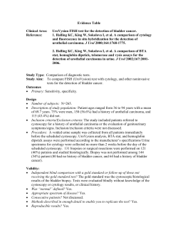


© Copyright 2026