
Intussusception in Children: Current Concepts in Diagnosis and Enema Reduction
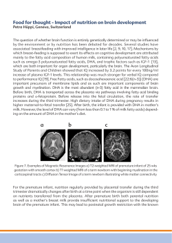
SCIENTIFIC EXHIBIT
Intussusception in
Children: Current
Concepts in Diagnosis
and Enema Reduction1
Gloria del-Pozo, MD • José C. Albillos, MD • Daniel Tejedor, MD
Rosa Calero, MD • Miguel Rasero, MD • Urbano de-la-Calle, MD
Ulpiano López-Pacheco, MD
Intussusception cannot be reliably ruled out with clinical examination
and plain radiography. However, a contrast material enema study and ultrasonography (US) allow definitive diagnosis of intussusception. The
components of an intussusception produce characteristic appearances
on US scans. These appearances include the multiple concentric ring
sign and crescent-in-doughnut sign on axial scans and the sandwich sign
and hayfork sign on longitudinal scans. Indicators of ischemia and irreducibility are trapped fluid at US and absence of blood flow at Doppler
imaging. The aim of enema therapy is to reduce the greatest number of
intussusceptions without producing perforation. Barium, water-soluble
contrast media, water, electrolyte solutions, or air may be used with radiographic or US guidance. The differences in reduction and perforation
rates between the various types of enemas are probably due more to
perforations that occurred before enema therapy and the pressure exerted within the colon than to the contrast material used. The pressure
within the colon is more constant with hydrostatic reduction than with
air reduction; this fact may explain the lower risk of perforation with hydrostatic reduction. Radiation exposure is lower with air enema therapy
than with barium enema therapy and is absent in US-guided enema
therapy.
Index terms: Children, gastrointestinal tract, 70.73 • Infants, gastrointestinal tract, 70.73 • Intestines, US, 70.1298 • Intussusception, 70.73
RadioGraphics 1999; 19:299–319
1From
the Section of Pediatric Radiology, Department of Diagnostic Radiology, Hospital Universitario Infantil 12 de
Octubre, Carretera de Andalucía Km 5,400, 28041 Madrid, Spain (G.d.P., J.C.A., R.C., M.R., U.d.l.C., U.L.P.); and the Department of Diagnostic Radiology, Hospital Universitario de la Princesa, Madrid, Spain (D.T.). Recipient of a Magna Cum
Laude award for a scientific exhibit at the 1996 RSNA scientific assembly. Received April 21, 1997; revision requested
July 11; final revision received March 30, 1998; accepted April 13. Address reprint requests to G.d.P.
© RSNA,
1999
299
■
INTRODUCTION
Intussusception is one of the most common
causes of acute abdomen in infancy. Intussusception occurs when a portion of the digestive
tract becomes telescoped into the adjacent
bowel segment. This condition usually occurs
in children between 6 months and 2 years of
age. In this age group, intussusception is idiopathic in almost all cases. The vast majority of
childhood cases of intussusception are ileocolic;
that is, the ileum becomes telescoped into the
colon. In the past, intussusception was a severe
condition with high morbidity and mortality rates.
Currently, prompt diagnosis and effective treatment lead to a favorable outcome in most cases.
In many cases, the clinical symptoms can be
confusing. In fact, only 30%–68% of children
with clinical findings suggestive of intussusception are shown to have this condition (1–8).
Therefore, it is desirable to use diagnostic tools
that are as innocuous as possible to avoid potential harm to these children, to diminish any
adverse effects on the actual diseases, and to
lessen the discomfort to the children who are
not shown to have intussusception. To this end,
the traditional diagnostic approach to childhood
intussusception of plain radiography and enema
examination (9) is being changed to plain radiography and ultrasonography (US) at some institutions (1,2,5,7,10–18).
US is highly accurate in the diagnosis of intussusception with a sensitivity of 98%–100% and
a specificity of 88%–100% (1,5,7,11,13,15,17,19,
20). Furthermore, use of US may lead to an alternative diagnosis, which is not readily achieved
with a contrast material enema study (1,10).
Therefore, the enema could be reserved for
therapeutic purposes when US is available (1,5,
10,11,18). There is continuing controversy as
to which type of enema is most efficacious.
In this article, the diagnosis and treatment of
intussusception are reviewed. The clinical features of intussusception are described, and diagnosis with plain radiography, enema examination, and US is discussed. The various appear-
300 ■
Scientific Exhibit
Figure 1. Target sign. Plain radiograph shows a
round soft-tissue mass in the right upper quadrant.
The mass contains a ringlike area of lucency.
ances of intussusception at US are presented by
using a diagram that facilitates the interpretation
of US scans at different levels of the intussusception. Variants of the US findings and the appearances of complicated intussusception are
also presented. Treatment is discussed in general, and factors that affect the outcome of reduction are presented. The advantages and disadvantages of the various types of enema therapies are compared. Finally, a management
algorithm for childhood intussusception that is
based mainly on the US findings is proposed.
The information in this article is derived from
our experience and a review of the literature.
Our experience comprises approximately 70
cases per year of intussusception diagnosed with
US during the past 10 years. In these cases, reduction of the intussusception was attempted
with a barium enema (during the first 5 years),
a saline solution enema under US guidance
(during the last 5 years), or occasionally an air
enema (17,18).
Volume 19
Number 2
gastroenteritis) or less common conditions (appendicitis, complicated Meckel diverticulum)
may initially mimic intussusception. Given the
uncertainty of achieving an accurate clinical diagnosis, a diagnosis based on imaging findings
is required in the majority of cases.
DIAGNOSIS WITH PLAIN RADIOGRAPHY
■
Figure 2. Meniscus sign. Plain radiograph shows the
meniscus sign: a rounded soft-tissue mass (the intussusceptum) protruding into the gas-filled transverse
colon.
■
CLINICAL FEATURES
The classic clinical triad of acute abdominal
pain (colic), currant-jelly stools or hematochezia, and a palpable abdominal mass is present
in less than 50% of children with intussusception (10). The onset of nonspecific abdominal
symptoms in which vomiting predominates, the
absence of passage of blood via the rectum (usually in cases of less than 48 hours duration), and
the inability to obtain a reliable history from
these nonverbal children lead to dismissal of
the diagnosis of intussusception in almost 50%
of cases (21).
Furthermore, in some instances lethargy or
convulsion is the predominant sign or symptom;
this situation results in consideration of a neurologic disorder rather than intussusception (22).
Finally, some cases in which the diagnosis is
considerably delayed manifest as shock of unknown origin. To further cloud the clinical picture, other common conditions (infantile colic,
March-April 1999
Many plain radiographic signs of intussusception have been described. The most common
is a soft-tissue mass, which is most often seen
in the right upper quadrant effacing the adjacent hepatic contour. Other signs include reduced air in the small intestine or a gasless abdomen, air in a displaced appendix, and obstruction of the small intestine (8,9,23,24). The
most specific plain radiographic findings are the
target sign and meniscus sign. The target sign
consists of a soft-tissue mass that contains concentric circular or nearly circular areas of lucency, which are due to the mesenteric fat of
the intussusceptum. The mass is most often
seen in the right upper quadrant projecting over
the right kidney (23,25) (Fig 1). The meniscus
sign consists of a crescent of gas within the colonic lumen that outlines the apex of the intussusception (the intussusceptum) (26) (Fig 2).
Conversely, identification of a cecum filled with
gas or feces in the normal location is the finding that allows exclusion of intussusception with
the most confidence (8). The accuracy of plain
radiography in diagnosis or exclusion of intussusception ranges from 40% to 90% (8,27,28).
The traditional role of plain radiography in
the evaluation of children suspected to have intussusception is threefold: (a) When the clinical suspicion is low, the role of plain radiography is to allow exclusion of intussusception and
diagnosis of other pathologic processes that are
responsible for the patient’s symptoms. (b) When
the clinical suspicion is high, the role of plain
radiography is to allow confirmation of intussusception. (c) If intussusception is present,
the role of plain radiography is to allow exclusion of intestinal obstruction or perforation.
del-Pozo et al ■
RadioGraphics ■
301
a.
Figure 3. Meniscus and coiled spring signs.
(a) Image from a barium enema study shows the
meniscus sign in the contrast material–filled
distal colon. (b) Image from a barium enema
study performed after partial reduction of the
intussusception shows the coiled spring sign.
Contrast material outlines the facing mucosal
surfaces of the intussuscipiens and the intussusceptum. (c) Image from a barium enema study
performed after complete reduction of the intussusception shows barium flowing freely into
the ileum.
b.
c.
302 ■
Scientific Exhibit
Volume 19
Number 2
When vague abdominal symptoms predominate and the clinical suspicion of intussusception is low, plain radiography may lead to the
diagnosis of other conditions. The discovery of
a large amount of air in otherwise normal
bowel loops including the cecum suggests the
diagnosis of infantile colic or adynamic ileus
caused by an intra- or extraabdominal infection.
In these cases, no further imaging is required.
Infrequently, detection of pneumonic consolidation in one of the lower lobes or of an
appendicolith leads to a diagnosis other than
intussusception. Owing to the low sensitivity
of any imaging modality in the setting of nonspecific abdominal symptoms, plain radiography could serve as the initial screening procedure.
When there are two or more of the cardinal
symptoms of intussusception, the clinical suspicion is high. In this potentially fatal disorder,
a false-negative diagnosis is untenable; such a
diagnosis may occur when only plain radiography is performed (8,26,27). Therefore, in cases
with high clinical suspicion, use of a diagnostic
tool with high sensitivity (eg, US, enema examination) is mandatory. In these cases, we
and other investigators believe that it is not
necessary to perform plain radiography, particularly if the symptoms are of short duration
(<8 hours) (8,10,13,16).
The third purpose of plain radiography is to
check for complications of prolonged intussusception: intestinal obstruction and perforation.
Intestinal obstruction is readily detected on
plain radiographs. The presence of intestinal
obstruction does not preclude attempts at nonsurgical reduction or necessarily change the
patient’s treatment. If perforation is detected,
primary surgery is indicated. However, to our
knowledge, there are no reported cases of intussusception in which pneumoperitoneum
was an initial radiographic finding, even in
cases in which obvious perforation was found
March-April 1999
at surgery (8,10,29–35). Therefore, there is a
low probability that perforation will be excluded with plain radiography.
For the aforementioned reasons, it does not
seem advisable to routinely perform plain radiography when the clinical suspicion of intussusception is high. In this setting, we believe
that it would be better to use US as the initial
imaging modality. However, when the clinical
findings are confusing and the symptoms are
vague or of unknown origin, it would be reasonable to perform plain radiography as the initial diagnostic procedure.
DIAGNOSIS WITH ENEMA EXAMINATION
■
Barium enema examination has been the standard of reference for the diagnosis of intussusception for many years. In fact, liquid enema or
air enema examination is the principal diagnostic tool at many institutions. The classic signs
of intussusception at enema examination are
the meniscus sign and coiled spring sign. The
meniscus sign at enema examination is analogous to the meniscus sign at plain radiography
and is produced by the rounded apex of the intussusceptum protruding into the column of
contrast material (Fig 3a). The coiled spring
sign is produced when the edematous mucosal
folds of the returning limb of the intussusceptum are outlined by contrast material in the lumen of the colon (Fig 3b).
The superior performance of US in the diagnosis of intussusception, the high level of patient comfort and safety allowed by US, and the
ability to arrive at alternative diagnoses with US
has led us and other investigators to reserve enemas for therapeutic purposes. A potential
shortcoming of this approach is that US is not
available 24 hours a day at all institutions.
del-Pozo et al ■
RadioGraphics ■
303
Figure 4. Structure of an intussusception. Diagram shows a longitudinal view and three axial views of an
intussusception; three bowel loops
and the mesentery can be seen. The
intussuscipiens (A) contains the two
limbs of the intussusceptum: the
everted returning limb (B), which is
edematous, and the central entering
limb (C), which is located at the center of the intussusception with the
accompanying mesentery (M). The
mesentery contains some lymph
nodes (L). MS = contacting mucosal
surfaces of the intussuscipiens and
everted limb, S = contacting serosal
surfaces of the everted limb and central limb.
■
DIAGNOSIS WITH US
US has a high sensitivity for the diagnosis of intussusception (98%–100%) (1,5,7,11,13,15,17,
19,20). The intussusception mass is a large
structure, usually greater than 5 × 2.5 cm, that
often displaces adjacent bowel loops; it is readily
identified, even by inexperienced operators
(5,10). The majority of intussusceptions (ie,
the ileocolic type) occur in the subhepatic region. Because deep penetration of the ultrasound beam is not necessary in small children,
a high-resolution transducer (5–10 MHZ) can
be used to improve the definition of the image.
Early studies of the US appearance of intussusception reported a doughnut or pseudokidney appearance composed of a hypoechoic
outer ring and a hyperechoic center. This appearance is similar to the US findings in other
pathologic conditions of the gastrointestinal
tract that cause thickening of the bowel wall
(12,36–39). Appearances that are characteristic
304 ■
Scientific Exhibit
of intussusception have also been reported.
These include the multiple concentric ring sign
(40) and crescent-in-doughnut sign (17) on
axial scans and the sandwich sign (1,41) and
hayfork sign (42) on longitudinal scans. (In US
of intussusception, the terms axial and longitudinal refer to the axis of the intussusception.)
An intussusception is a complex structure
(Figs 4, 5). The intussuscipiens (the receiving
loop) contains the folded intussusceptum (the
donor loop), which has two components: the
entering limb and returning limb. The attached
mesentery is dragged between the entering and
returning limbs. The thickest component of
the intussusceptum is the everted returning
limb, which—together with the thin intussuscipiens—forms the hypoechoic outer ring seen
on axial scans. The center of the intussusception contains the central or entering limb,
which is of normal thickness and is eccentrically surrounded by the hyperechoic mesentery (17).
Volume 19
Number 2
Figure 5. Structure of an intussusception. Axial US scans and corresponding pathologic specimens
from pigs with intussusception show the doughnut sign: a hypoechoic outer ring formed by the everted
limb of the intussusceptum (B) and the intussuscipiens (A) and a center that varies with the section
level. C = central limb of the intussusceptum. (Reprinted, with permission, from reference 17.) (a,
b) US scan (a) and pathologic specimen (b) obtained at the apex of an intussusception (section 1 in
Fig 4) show a hypoechoic center, which represents the central limb of the intussusceptum with no
mesentery present. Note the multilayered appearance, which is due to the demonstration of the five
layers of the three bowel loops involved. (c, d) US scan (c) and pathologic specimen (d) obtained at
the base of the intussusception (section 2 in Fig 4) show a hyperechoic, crescent-shaped center. This
appearance occurs when the mesentery encloses the central limb of the intussusceptum (the crescentin-doughnut sign). (e, f) US scan (e) and pathologic specimen (f) obtained at a different level of the
base (section 3 in Fig 4) show an additional hypoechoic area in the hyperechoic, crescent-shaped
center. This additional hypoechoic area represents a lymph node (L).
a.
b.
c.
d.
e.
March-April 1999
f.
del-Pozo et al ■
RadioGraphics ■
305
a.
b.
c.
Figure 6. Variable appearance of intussusception on axial US scans. C = central limb of the intussusceptum.
(a) US scan obtained at the apex of an intussusception shows a hypoechoic outer ring separated from a hypoechoic
center by a thin hyperechoic ring, which likely represents the opposed serosal surfaces of the intussusceptum
(cf Figs 4 [section 1], 5a, 5b). G = gallbladder. (b) US scan obtained near the apex shows multiple concentric
rings (a hypoechoic ring surrounding a hyperechoic ring, which surrounds another hypoechoic ring). The
hyperechoic central ring is probably formed by the addition of the hyperechoic opposed submucosal and serosal surfaces of the intussusceptum. (c) US scan obtained at the base of an intussusception shows the central
limb of the intussusceptum eccentrically surrounded by the hyperechoic mesentery (M), a situation that produces the crescent-in-doughnut sign (cf Figs 4 [section 2], 5c, 5d).
●
Axial US Scans
On axial US scans, intussusception has a variable appearance, which is primarily due to the
amount of enclosed mesentery. Enclosed mesentery is absent at the apex of the intussusception and progressively increases toward the
base (Figs 4, 5). Conversely, the everted limb
of the intussusceptum is thicker at the apex
than at the base. Therefore, an axial US scan
obtained at the apex shows a hypoechoic
outer ring with a hypoechoic center (Fig 6a).
In some instances, multiple concentric rings
can be seen near the apex (Fig 6b). As the axial
US study proceeds toward the base, the appearance changes gradually as increasing
amounts of mesentery are included in the image. At the base, the amount of enclosed mesentery is maximal; the result is a hypoechoic
outer ring with a hyperechoic, crescentic center (the crescent-in-doughnut sign) (Fig 6c).
●
Longitudinal US Scans
On longitudinal US scans, it is the arrangement
of the mesentery, which may be demonstrated
on one or both sides of the central limb of the
intussusceptum, that causes variation in the appearance. If the middle of the intussusception
is imaged along the longitudinal axis, three parallel hypoechoic bands separated by two nearly
parallel hyperechoic bands are seen. The outer
306 ■
Scientific Exhibit
hypoechoic bands represent the edematous
everted limb of the intussusceptum and the
thin intussuscipiens; the central hypoechoic
band is the central limb of the intussusceptum.
The hyperechoic bands are caused by the mesentery that is dragged along with the bowel
loop. This appearance is known as the sandwich sign (Fig 7a). The hayfork sign is a variant
of the sandwich sign that is seen at the apex of
the intussusception (42). The hayfork sign consists of three nearly parallel hypoechoic areas
(the “prongs” of the hayfork), which correspond to those in the sandwich sign. The
prongs are separated by two hyperechoic
bands formed by the mesentery. The prongs
join at the apex where the intussusceptum
folds (Fig 7b). The pseudokidney sign occurs
if the intussusception is curved or is imaged
obliquely and the mesentery (at the point of
maximal thickness) is demonstrated on only
one side of the central limb of the intussusceptum (Fig 7c).
●
Variants on Axial US Scans
Outer Ring.—The outer ring seen on axial US
scans is usually homogeneously hypoechoic,
especially at the apex of the intussusception.
Occasionally, the ring is isoechoic or even
hyperechoic relative to the adjacent hepatic
parenchyma (Fig 8a). This variant is most often
seen at the base or middle of the intussuscep-
Volume 19
Number 2
b.
c.
a.
Figure 7. Variable appearance of intussusception on longitudinal US scans. C = central limb of the intussusceptum, M = mesentery. (a) US scan obtained in the strict longitudinal plane of an intussusception slightly
away from the apex shows the sandwich sign. The outer hypoechoic bands (arrows) represent the everted
limb of the intussusceptum beside the intussuscipiens. The two hyperechoic bands represent the mesentery.
The central hypoechoic band represents the central limb of the intussusceptum. (b) US scan obtained at the
apex of an intussusception shows the hayfork sign, which differs from the sandwich sign in that the mesentery
thins as it approaches the apex. The three hypoechoic prongs of the hayfork represent the involved bowel
loops separated by the hyperechoic mesentery. L = lymph node. (c) US scan shows the pseudokidney sign.
The mesentery is demonstrated on one side of the central limb of the intussusceptum.
b.
c.
a.
Figure 8. Variant forms of the outer ring. (a) Axial US scan shows an outer ring (arrowheads) that is hyperechoic relative to the adjacent liver (L). This appearance was seen when the child had an attack of colicky
pain. (b) Axial US scan shows alternating hyperechoic and hypoechoic bands (arrows) produced by the layers
of the bowel wall of the intussuscipiens and the everted limb of the intussusceptum (multilayered image).
(c) Axial US scan shows a band of hyperechoic dots between the thin intussuscipiens (A) and the everted limb
of the intussusceptum (B). Arrowheads indicate the outer ring.
tion. We have observed that the echogenicity
of the outer ring increases when the child has
an attack of colicky pain. In other instances,
the different layers of the bowel wall of the involved loops are demonstrated and produce a
multilayered or tree ring appearance (Fig 8b).
This appearance may be related to a lesser degree of vascular compromise of the intestine
(12). The multilayered appearance usually occurs at the base of the intussusception and oc-
March-April 1999
casionally occurs at the apex of an early intussusception. Finally, hyperechoic dots are occasionally seen between the opposed mucosal
surfaces of the intussuscipiens and the everted
limb of the intussusceptum (Fig 8c). These
dots may be caused by trapped colonic gas or
mucosal ulcers.
del-Pozo et al ■
RadioGraphics ■
307
a.
b.
c.
Figure 9. Variant forms of the mesenteric crescent. C = central limb of the intussusceptum. (a) Axial US
scan obtained at the base of an intussusception shows the crescent-in-doughnut sign altered by inclusion of
two oval mesenteric lymph nodes (L) (cf Figs 4 [section 3], 5e, 5f). (b) Axial US scan obtained at the base of
an intussusception shows the crescent-in-doughnut sign altered by inclusion of the cecoappendiceal complex
(arrow). (c) Axial US scan obtained at the base of an intussusception shows the crescent-in-doughnut sign altered by inclusion of the appendix (arrow) and vessels, which appear as hypoechoic dots.
Figure 10. Variant forms of
the central limb of the intussusceptum. (a) Axial US scan
shows a small amount of fluid
(F) within the lumen of the
central limb. (b) Axial US scan
shows an intensely echogenic
area (arrow) with acoustic
shadowing. This appearance is
due to gas passing through the
lumen of the central limb.
a.
b.
a.
b.
Figure 11. Trapped peritoneal fluid. (a) Axial US scan
shows the double-crescent-indoughnut sign, which consists
of the crescent-in-doughnut
sign plus an echo-free crescent due to the trapped fluid
(F). (b) Longitudinal US scan
at the apex of the intussusception shows the intussusceptum filled with trapped fluid.
Mesenteric Crescent.—The mesentery that is
dragged into the intussusception with the intussusceptum is crescentic when imaged in the
308 ■
Scientific Exhibit
axial plane. The mesenteric crescent is hyperechoic but often contains hypoechoic areas
(lymph nodes, the cecoappendiceal complex,
vessels) (Fig 9). These variant forms are most
often found at or near the base of the intussusception (17).
Volume 19
Number 2
Figure 12. Free peritoneal fluid. Axial US scan shows a
small amount of free peritoneal fluid (F) adjacent to an intussusception that demonstrates the multiple concentric
ring sign. G = gallbladder.
Figure 13. Use of Doppler
US to evaluate intussusception. Doppler US scans obtained at the apex (a), middle
(b), and base (c) of an intussusception clearly show blood
flow within the intussusceptum (a, b) and mesentery (c).
This intussusception could be
reduced.
a.
b.
Central Limb of the Intussusceptum.—The
central limb of the intussusceptum is collapsed
in most cases. In rare instances, small amounts
of fluid can be seen within the lumen of this
limb. In addition, with real-time imaging, gas or
fluid can be seen passing through the lumen of
this limb in cases of unobstructed intussusception (Fig 10).
Intussusceptions with Trapped Peritoneal Fluid
●
The presence of trapped peritoneal fluid within an intussusception correlates significantly
with ischemia and irreducibility (P < .001, significant odds-likelihood ratio of 11.6–82.8)
(18). Fortunately, in developed countries, this
complication is seen in less than 15% of cases.
Such fluid, which reflects vascular compromise of the everted limb, accumulates between
the serosal layers of both limbs of the intussusceptum. The mesentery acts as a wedge and
impedes the exit of fluid into the peritoneal
cavity.
On axial US scans, this complication appears
as the double-crescent-in-doughnut sign. In addition to the usual crescent-in-doughnut appearance, there is an anechoic crescent that
represents the trapped ascites (Fig 11). In advanced stages, the everted bowel loop may be-
March-April 1999
c.
come dilated probably due to ischemia and the
increasing amount of trapped fluid. The dilatation occurs mainly at the antimesenteric border; the result is asymmetric distribution of the
fluid (18). At US, asymmetrically distributed
fluid can mimic a dilated bowel loop (eg, a
closed-loop obstruction) or duplication cyst
(43). Conversely, small amounts of free peritoneal fluid are seen in up to 50% of cases (Fig
12). The presence of this finding alone has not
been definitely related to ischemia or an increased risk of perforation (5,44,45).
●
Blood Flow at Doppler US
Initial reports suggested that absence of blood
flow at the apex of an intussusception at Doppler US correlated with bowel necrosis and
was a sign of irreducibility (15,46,47). Subsequently, a larger series demonstrated that
when blood flow is not visualized at Doppler
US, the rate of reduction is lower (48). However, in that series, bowel necrosis was not statistically correlated with absence of the Doppler signal. The presence of blood flow at Doppler US suggests that the intussusception
would be reduced (Fig 13). Unfortunately,
Doppler US is not available 24 hours a day at
many institutions.
del-Pozo et al ■
RadioGraphics ■
309
●
Intussusceptions with Lead Points
Most childhood cases of intussusception are idiopathic; that is, they do not have a demonstrated anatomic abnormality that functions as
a lead point except for hypertrophied lymphoid tissue. Intussusception lead points such
as a Meckel diverticulum, duplication cyst,
polyp, or tumor (eg, lymphoma) are uncommon in infants (<5% of cases). Intussusception
lead points are more common in neonates (<30
days old), older children (>5 years old), and
cases restricted to the small intestine (49). For
example, intussusception of the small intestine
is common in Peutz-Jeghers syndrome, in
Schönlein-Henoch purpura, and after surgery.
Although lead points can be detected with a
contrast enema study (50), they can easily be
missed or even reduced with this technique
(2,51). US allows better detection and characterization of lead points than does a contrast
enema study (2,10,52,53).
■
TREATMENT
In 1836, Samuel Mitchell reported nonsurgical
reduction of an intussusception. Two years
later, John Gorham reported five cases treated
by means of rectal insufflation of air. In 1876,
Harald Hirschsprung reported reduction of an
intussusception by means of a hydrostatic enema with transabdominal manipulation.
After the discovery of x rays, it became possible to monitor the reduction process in real
time by using positive contrast agents. Fluoroscopically guided enema therapy was performed in the 1920s but was not scientifically
tested and standardized until the studies of
Ravitch and McCune (54) in the 1940s (55).
Since then, contrast enema therapy has been
increasingly accepted as the treatment of
choice for the reduction of intussusception.
There is continuing discussion without wide
agreement about which type of enema is the
best for this purpose. The few randomized
studies that have been performed did not show
statistically significant differences in reduction
and perforation rates between air and liquid enemas (3). It is likely that the reported differences in reduction and perforation rates were
310 ■
Scientific Exhibit
related not to the type of enema used but to
complications that occurred before enema
therapy, the technique used, the intracolonic
pressures (56), and the patient selection criteria (10,22). These factors vary among the reported series. In some reports, it is difficult to
ascertain many of these factors.
FACTORS THAT AFFECT THE OUTCOME OF REDUCTION
■
●
Complications
Although there are reports of spontaneous reduction of intussusception (57,58), chronic
and recurrent intussusceptions, and rare cases
of spontaneous sloughing of the gangrenous intussusceptum through the rectum (59), the
usual course of an untreated intussusception is
bowel obstruction followed by bowel perforation with peritonitis and septic shock. Currently, the overall perforation rate in developed
countries is low (0%–3%) (22).
Perforation may already have occurred before enema therapy (30–32) or may occur during the reduction process. In the latter case,
perforation may occur in normal (33,60) or ischemic (31,34,35) bowel segments or in both
types (29,61). The occurrence of perforations
before enema therapy depends mainly on the
promptness of diagnosis and the quality of patient treatment. Therefore, the frequency of
such perforations varies widely. The occurrence of perforations during the reduction process depends mainly on the pressure achieved
and how long the pressure is applied and
partly on the dynamics and physical properties
of the contrast material used (62).
Most published studies do not differentiate
between perforations established before reduction and perforations induced during reduction. It is difficult to recognize a perforation established before reduction because no cases of
childhood intussusception with pneumoperitoneum as the initial finding have been reported,
to our knowledge (8,10,29–35). Perforations
may occur in the gangrenous ileum (the intussusceptum) or the overdistended colon (the intussuscipiens). An ileal perforation is covered
by the intussuscipiens, which does not allow
gas to escape into the peritoneal cavity. In a colonic perforation, the intussusceptum impedes
the exit of the proximal intestinal contents.
Volume 19
Number 2
Also, because perforation occurs in cases of intussusception in which the diagnosis is delayed, the distal colonic contents will usually
have been expelled by the time plain radiography is performed (30).
●
Technique of Reduction
The goal of any type of enema therapy is to reduce the intussusception by exerting pressure
on the apex of the intussusceptum to push it
from the pathologic position into the original
position. The reduction and perforation rates
for a specific type of enema therapy are directly proportional to the pressure applied.
Ravitch (59) found that the intracolonic
pressure achieved by placing the barium enema bag 3.5 ft (105 cm) above the table did not
reduce any intussusception in which the intestine was necrotic or incarcerated. Further studies found that a pressure that did not exceed
120 mm Hg for hydrostatic enemas and 108
mm Hg for air enemas did not perforate the colon in animals (60). The pressure can be controlled by measuring the height of the bag containing the fluid for a hydrostatic enema or using a manometer for an air enema. A pressure
of 120 mm Hg is equivalent to a 100-cm column of barium or a 150-cm column of water or
water-soluble contrast material (63). This theoretic pressure is achieved during liquid enema
therapy only if the diameter of the tubing of
the system is sufficiently large to easily transmit the pressure. In hydrostatic enema therapy,
use of tubing and a rectal tube of large caliber
may be more effective in increasing the effective intracolonic pressure than use of an increased column height (64).
Besides these theoretic considerations, it
has been found empirically that intracolonic
pressure reaches a plateau during hydrostatic
enema therapy. This pressure is more constant
than that exerted during air insufflation, which
tends to produce oscillations in the intraluminal pressure with peaks that can surpass the
pressure threshold. Such pressure fluctuations
increase the risk of perforation (62).
There is no agreement on the number and
duration of reduction attempts, the efficacy of
premedication or sedation, the use of rectal
tubes with inflatable retention balloons, or the
use of transabdominal manipulation (65–67).
This lack of agreement reflects the fact that no
large studies, to our knowledge, have demonstrated a definite improvement in the reduction
rate with any of these factors.
March-April 1999
One component of the classic “rule of
threes” is that the number of reduction attempts is capped at three. This rule has been
discarded at some institutions, and some authors use a nearly unlimited number of attempts. These authors even use delayed attempts; that is, they repeat the reduction attempt after the patient rests for up to several
hours. The aim is to improve the reduction rate
(13,17,18,68–73). Use of US guidance permits
an even more liberal approach to enema
therapy owing to the lack of radiation exposure (16).
Use of sedation had been thought to improve the reduction rate (9). However, the reduction rate has been found to be lower when
parenteral sedation is used (66). Sedation prevents the patient from performing the Valsalva
maneuver during straining. The Valsalva maneuver increases the intraluminal pressure (by
approximately 60 mm Hg) and decreases the
colonic transmural pressure gradient. This maneuver improves the effectiveness of enema
therapy and protects against perforation, particularly during air enema therapy (60,74,75).
In theory, use of a rectal tube with a retention balloon creates a closed system that transmits the effective pressure of the enema material (liquid or air) without loss of pressure. Use
of such a rectal tube could increase the rapidity and effectiveness of enema therapy. However, this closed system could block the spontaneous expulsion of liquid or gas when the
pressure suddenly increases (16). Furthermore,
we and other authors (59) have found that use
of such a rectal tube does not produce a closed
system. Many patients can readily expel the
balloon catheter during straining.
Enema therapy is usually guided with fluoroscopy when a barium, water-soluble, or air
enema is used. US guidance is used when
therapy is performed with a water, saline, or
gas enema.
●
Patient Selection Criteria
Absolute contraindications to enema therapy
are shock that is not readily corrected with intravenous hydration and an established perforation with clinical signs of peritonitis. Criteria
that are linked to a lower reduction rate and a
higher perforation rate, especially if more than
one is present, are as follows: (a) patient age
del-Pozo et al ■
RadioGraphics ■
311
Table 1
Advantages and Disadvantages of Barium
Enema Therapy
Advantages
Maximum experience with this method
Good results with optimized method (55%–
90% of cases)
Good evaluation of ileoileal residual intussusceptions
Low perforation rate (0.39%–0.7%)
Disadvantages
X-ray exposure required, thus limiting
procedure time
Perforation causes chemical peritonitis
Visualization of only intraluminal content
less than 3 months or greater than 5 years;
(b) long duration of symptoms, especially if
greater than 48 hours; (c) passage of blood via
the rectum (hematochezia); (d) significant dehydration; (e) obstruction of the small intestine; and (f) visualization of the dissection sign
during enema therapy (70,76–80). The dissection sign refers to contrast material within the
lumen of the intussuscipiens that “dissects” for
a long distance over the surface of the intussusceptum.
At some institutions, these criteria are used
with a scoring system to quantify the risk and
determine whether immediate surgery or nonsurgical reduction should be performed (71,81).
This approach is advisable for institutions that
treat only a few cases per year or lack an experienced surgical team. For these institutions, referral of high-risk cases to a tertiary-care hospital that has the experience and personnel to
deal with intussusception may be in the patient’s best interest (76).
At tertiary-care hospitals, enema therapy is
attempted in almost all cases because most patients in shock can be resuscitated with rapid
intravenous hydration and because intestinal
perforation is difficult to diagnose before enema therapy. Even those patients who ultimately undergo surgery may benefit from partial reduction of the intussusception. Such reduction may decrease the edema in some
portions of the intestine, thereby facilitating
complete reduction of the intussusception at
surgery. This approach may also reduce the frequency of intestinal resection by lessening vascular compromise (68,82).
312 ■
Scientific Exhibit
Figure 14. Barium enema therapy. Image from a
barium enema study shows perforation with free
spillage of barium into the peritoneal cavity. The
dissection sign is also evident; the intussusceptum
is outlined for several centimeters by barium in the
colon.
A potential criticism of this approach is that
performing enema therapy in a patient with an
established perforation could cause the perforation to become manifest or that a perforation
could be induced in high-risk cases. Although
this criticism has some validity, the new contrast agents do not appear to have the serious
adverse consequences of barium in such cases
(see the section titled “Types of Enema Therapies”) (78). The lesson to be learned is that in
cases with clinical or radiologic high-risk factors, a more cautious and gentle approach is
advisable during enema therapy.
■
TYPES OF ENEMA THERAPIES
●
Barium Enema
Barium enema has been validated as an effective therapy by extensive experience over a
long period. At some institutions, it remains
the therapy of choice. However, there is a tendency to replace barium with other contrast
agents, primarily because of the sequelae if
perforation occurs during barium enema therapy (65). Spillage of barium into the peritoneal
cavity produces chemical peritonitis (Fig 14).
This is obviously not a desirable outcome because barium remains in the peritoneal cavity
forever. However, the long-term consequences
of barium spillage do not seem to be any more
Volume 19
Number 2
a.
b.
Figure 15. Air enema therapy. (a) Image obtained at the beginning of an air enema study shows the
meniscus sign and a mass in the middle of the upper abdomen. (b) Image obtained after complete reduction shows gas passing freely into the small intestine.
serious than the formation of granulomas and
adhesions. In addition, these complications
seem to be related more to fecal contamination
than to barium spillage (83).
In older studies, the reduction rate was
lower with barium than with the new contrast
agents. In more recent studies (49,84), there
was no significant difference in the reduction
rate between barium and other media (3,85,86).
At institutions that treat only a few cases of intussusception per year, it is questionable
whether there is a need to replace barium enema therapy with other enema therapies until a
worldwide consensus is achieved as to which
modality is optimal. This philosophy reflects
the difficulty of stabilizing the learning curve
for a new method if only a few cases per year
are treated (87).
The advantages and disadvantages of barium
enema therapy are summarized in Table 1.
●
Water-soluble Contrast Enema
The use of water-soluble contrast agents in enema therapy has been supported in North
America (56,78). The principal reason for use
of these agents is to avoid the chemical peritonitis caused by barium if perforation occurs.
March-April 1999
The pathologic changes in the peritoneum
(granulomas, adhesions, loculated ascites) appear to be less significant when water-soluble
contrast material is used instead of barium
(88). There are few reports on the use of water-soluble contrast media. The reduction rate
(80%) and perforation rate (3%) do not surpass
those of other contrast agents (56,78).
●
Air Enema
Air was the first agent used to reduce intussusception, to our knowledge. Air enema therapy
was not used for a long time in the Western
Hemisphere but recently has become popular
in some countries, chiefly in North America
(4,6,61,69,70,81,89–92), after successful experiences in Argentina (93) and China (71,72)
were reported. This method is quick and clean
with a high reduction rate (73%–95%) (Fig 15)
and less radiation exposure than barium enema
therapy. The decreased radiation exposure is
related to the shortened time required to
achieve reduction and the milder (in terms of
kilovolts and milliamperes) radiographic technique used (4,90,94).
del-Pozo et al ■
RadioGraphics ■
313
Table 2
Advantages and Disadvantages of Air
Enema Therapy
Table 3
Advantages and Disadvantages of USguided Saline Enema Therapy
Advantages
Excellent results (70%–95.6% of cases)
Less x-ray exposure than with barium enema
Easy, quick, clean technique
Disadvantages
X-ray exposure required, thus limiting
procedure time
Higher perforation rate (0.14%–2.8%) with
risk of tension pneumoperitoneum
Visualization of only intraluminal content
Less control of residual ileoileal intussusceptions
Advantages
No x-ray exposure, thus procedure time not
limited
Excellent results (76%–95.5% of cases)
Visualization of all components of the intussusception
Easier recognition of lead points and
residual intussusceptions
Low perforation rate (0.26%)
Disadvantages
Sonographer or sonologist needed
Air enema therapy has not been universally
adopted because of the increased perforation
rate reported (2.8%) (61) and the possibility of
tension pneumoperitoneum. In addition, two
deaths were reported in one large study (71).
The pressure limit used by some authors (120
mm Hg) exceeds the pressure shown to produce perforation in experimental studies (108
mm Hg) (60,86). This fact, along with the oscillations in pressure generated by bulb insufflation (62), could account for the increased perforation rate.
Defenders of air enema therapy argue that
(a) the increased perforation rate is not seen in
all studies, decreases with experience with the
technique, and is not significantly different
from the rates of other types of enema therapy;
(b) the perforations demonstrated by air enema
are small compared with those demonstrated
by barium enema and thus less harmful; (c) tension pneumoperitoneum is rare, readily recognized, and easily treated with needle puncture;
and (d) the higher perforation rate is accompanied by a significant improvement in the reduction rate. In any case, air enema therapy requires precise control of the pressure and a
thorough knowledge of the technique and potential complications (14,84). During the transition from use of barium enema to use of air enema, a conservative approach is preferable.
During this transition, the pressure should be
raised slowly and progressively (91).
314 ■
Scientific Exhibit
The advantages and disadvantages of air enema therapy are summarized in Table 2.
●
US-guided Saline Enema
The most important studies of US guidance of
intussusception reduction have been performed in the Eastern Hemisphere, where
there is extensive experience with the technique. This method is increasingly being used
in Europe. The procedure may be performed
with water, saline solution, or Hartmann solution (Fig 16). Studies have shown a high reduction rate (76%–95%) with few complications
(one perforation in 825 reported cases), results
similar to those of other hydrostatic methods
(7,13,16–18,20,95–97).
The principal advantage of this technique
is the lack of radiation exposure. As a result,
there is no limit to the procedure time, a fact
that may improve the success rate. A potential
disadvantage is that the presence of a sonographer or sonologist is required on a 24-hour
basis. Because of the widespread support for
use of US to diagnose intussusception (1,2,5,7,
10,12,13,15–20,46), this requirement is probably not a real disadvantage. As with any new
technique, an alternative method to fall back
on may be needed until the learning curve is
stabilized. To easily visualize the reduction process, the pressure can be progressively increased from 60 to 120 mm Hg by raising the
saline bag.
The advantages and disadvantages of USguided saline enema therapy are summarized
in Table 3.
Volume 19
Number 2
Figure 16. US-guided saline enema therapy. (a) Longitudinal US scan shows the fluid outlining the intussusceptum in the transverse colon. This appearance is the US equivalent of the meniscus sign. (b – d) Longitudinal
US scans show the enema pushing the intussusceptum into the ascending colon (b), into the ileocecal valve
(c), and finally through the valve (d). Arrows in d = ileocecal valve. (e, f) Axial US scans show multiple bowel
loops (e), including the terminal ileum (f), filled with fluid. This appearance indicates that complete reduction
has been achieved. The thickened and collapsed terminal ileum must not be confused with a residual intussusception. Arrows in f = ileocecal valve. (g) Longitudinal US scan shows a thickened ileocecal valve (arrows).
(h) Axial US scan shows the Mercedes-Benz sign, which is produced by the collapsed terminal ileum.
a.
b.
d.
e.
g.
h.
March-April 1999
c.
f.
del-Pozo et al ■
RadioGraphics ■
315
US Findings in Suspected Intussusception
No intussusception
Other
pathologic
findings
Intussusception with lead
point
Intussusception in very ill
child
No enema required;
clinical reevaluation
Stop
Intussusception without
trapped fluid
or with flow
at Doppler US
Intussusception with maximum trapped
fluid or without flow at
Doppler US
Enema with
maximum
pressures and
maximum
attempts
Enema with
low pressures
and limited
attempts
Not successful
Successful
Surgery
Figure 17. Management algorithm for cases with clinical suspicion of intussusception. In
this approach, US is the principal imaging modality.
●
US-guided Air Enema
There has been little experience with USguided air enema therapy (98). This modality
attempts to unite the advantages of air enema
therapy (quick and clean method, high reduction rate) with those of US guidance (no radiation exposure). However, because air prevents
passage of the ultrasound beam, it can be difficult to visualize the ileocecal valve; therefore,
small residual ileoileal intussusceptions can be
obscured. Also, it would not be easy to detect
perforation with pneumoperitoneum (16). The
presence of a large amount of intraluminal or
intraperitoneal air could interfere with a subsequent US study if one is required (46).
■
MANAGEMENT ALGORITHM
Surgical treatment of an intussusception is indicated if there is shock that cannot be corrected; if lead points, necrosis, or perforation is
present; or if the intussusception is irreducible
(22). The presence of these features must be
determined as far as is possible to minimize unnecessary surgical procedures. Adhesions and
volvulus are late complications of surgery in
3%–6% of cases (cumulative risk) (22). Patients
who require surgical treatment have a longer
hospital stay than those in whom enema
therapy is successful (68).
Traditionally, the first imaging study performed when there is clinical suspicion of in-
316 ■
Scientific Exhibit
tussusception is plain radiography. In the setting of vague abdominal symptoms, plain radiography remains the most simple screening
test. However, plain radiography does not allow one to confidently rule out intussusception
or detect perforation. Therefore, several authors do not recommend plain radiography if
there is high clinical suspicion of intussusception (8), especially when the symptoms are of
short duration. In such cases, US should be the
initial imaging procedure (10,13,16). US allows
one to confidently diagnose or rule out intussusception, make alternative diagnoses, and
characterize lead points. If US is not available, a
contrast enema study must be performed to exclude intussusception.
Formerly, determination of the presence of
ischemia or irreducibility was based primarily
on clinical criteria. However, US can help identify these high-risk features by demonstrating
fluid trapped within the intussusception (18)
or absence of blood flow at Doppler imaging
(15,46–48). Identification of these features indicates a need to adopt a more cautious and
gentle approach.
When there are no high-risk features at clinical examination and US, enema reduction
should be attempted with no limits set on the
effort. The enema can be guided with fluoroscopy or US. We prefer the latter modality because there are no time constraints as there are
with fluoroscopy. An algorithm for the management of childhood intussusception is given in
Figure 17.
Volume 19
Number 2
■
CONCLUSIONS
Plain radiography is of limited value in the diagnosis of intussusception. US has permitted a
more comprehensive understanding of the
anatomy and pathophysiology of intussusception. An accurate and safe modality, US may
also allow one to detect lead points and identify alternative diagnoses. Thorough knowledge of the US features characteristic of intussusception (eg, the crescent-in-doughnut, multiple concentric ring, sandwich, and hayfork
signs and variants of these signs) is a prerequisite for accurate diagnosis. Indicators of ischemia and irreducibility at US are fluid
trapped within the intussusception and absence of blood flow at Doppler imaging.
Nonsurgical treatment of intussusception is
possible in most cases, especially at institutions
with pediatric surgery facilities and personnel
for use as a backup. There is a trend toward
performing enema therapy with agents other
than barium (eg, water-soluble agents, air, saline solution) because other agents will not
persist in the peritoneal cavity if perforation occurs. In the absence of large, randomized studies, the reported differences in reduction and
perforation rates are probably due more to
complications that occurred before enema
therapy, the pressures exerted, and patient selection criteria than to the type of enema used.
However, liquid agents appear to cause fewer
perforations. Use of US guidance eliminates the
disadvantage of radiation exposure, thus allowing a greater number of attempts at hydrostatic
reduction.
Acknowledgments: We thank the staff and residents of the radiology department at our institution
for their collaboration in our work. We also thank
Robert J. Starshak, MD, for his valuable assistance in
the preparation of the manuscript.
■
REFERENCES
1. Pracros JP, Tran-Minh VA, Morin de Finfe CH, Defrenne-Pracros P, Louis D, Basset T. Acute intestinal intussusception in children: contribution of ultrasonography (145 cases). Ann Radiol 1987;
30:525–530.
2. Miller SF, Landes AB, Dautenhahn LW, et al. Intussusception: ability of fluoroscopic images obtained
during air enemas to depict lead points and other
abnormalities. Radiology 1995; 197:493–496.
3. Meyer JS, Dangman BC, Buonomo C, Berlin JA. Air
and liquid contrast agents in the management of
intussusception: a controlled, randomized trial. Radiology 1993; 188:507–511.
4. Gu L, Alton DJ, Daneman A, et al. Intussusception
reduction in children by rectal insufflation of air.
AJR 1988; 150:1345–1348.
March-April 1999
5. Verschelden P, Filiatrault D, Garel L, et al. Intussusception in children: reliability of US in diagnosis—
a prospective study. Radiology 1992; 184:741–
744.
6. Shiels WE II, Maves CK, Hedlung GL, Kirks DR. Air
enema for diagnosis and reduction of intussusception: clinical experience and pressure correlates.
Radiology 1991; 181:169–172.
7. Woo SK, Kim JS, Suh SJ, Paik TW, Choi SO. Childhood intussusception: US-guided hydrostatic reduction. Radiology 1992; 182:77–80.
8. Sargent MA, Babyn P, Alton DJ. Plain abdominal radiography in suspected intussusception: a reassessment. Pediatr Radiol 1994; 24:17–20.
9. Bisset GS III, Kirks DR. Intussusception in infants
and children: diagnosis and therapy. Radiology
1988; 168:141–143.
10. Daneman A, Alton DJ. Intussusception: issues and
controversies related to diagnosis and reduction.
Radiol Clin North Am 1996; 34:743–756.
11. Shanbhogue RLK, Hussain SM, Meradji M, Robben
SGF, Vernooij JEM, Molenaar JC. Ultrasonography
is accurate enough for the diagnosis of intussusception. J Pediatr Surg 1994; 29:324–328.
12. Swischuk LE, Hayden CK, Boulden T. Intussusception: indications for ultrasonography and explanation of the doughnut and pseudokidney signs.
Pediatr Radiol 1985; 15:388–391.
13. Riebel TW, Nasir R, Weber K. US-guided hydrostatic reduction of intussusception in children. Radiology 1993; 188:513–516.
14. Kirks DR. Diagnosis and treatment of pediatric intussusception: how far should we push our radiologic techniques? Radiology 1994; 191:622–623.
15. Lim HK, Bae SH, Lee KH, Seo GS, Yoon GS. Assessment of reducibility of ileocolic intussusception in
children: usefulness of color Doppler sonography.
Radiology 1994; 191:781–785.
16. Rohrschneider WK, Tröger J. Hydrostatic reduction of intussusception under US guidance. Pediatr
Radiol 1995; 25:530–534.
17. del-Pozo G, Albillos JC, Tejedor D. Intussusception:
US findings with pathologic correlation—the crescent-in-doughnut sign. Radiology 1996; 199:688–
692.
18. del-Pozo G, González-Spinola J, Gómez-Ansón B, et
al. Intussusception: trapped peritoneal fluid detected with US—relationship to reducibility and ischemia. Radiology 1996; 201:379–386.
19. Bhisitkul DM, Listernick R, Shkolnik A, et al. Clinical application of ultrasonography in the diagnosis
of intussusception. J Pediatr 1992; 121:182–186.
20. Wang GD, Liu SJ. Enema reduction of intussusception by hydrostatic pressure under ultrasound
guidance: a report of 377 cases. J Pediatr Surg
1988; 23:814–818.
21. Beasley SW, Auldist AW, Stokes KB. The diagnostically difficult intussusception: its characteristics
and consequences. Pediatr Surg Int 1988; 3:135–
138.
22. Stringer MD, Pablot SM, Brereton RJ. Paediatric intussusception. Br J Surg 1992; 79:867–876.
del-Pozo et al ■
RadioGraphics ■
317
23. Lee JM, Kim H, Byum JY, et al. Intussusception:
characteristic radiolucencies on the abdominal radiograph. Pediatr Radiol 1994; 24:293–295.
24. Gilsanz V. Displacement of the appendix in intussusception. AJR 1984; 142:407–408.
25. Ratcliffe JF, Fong S, Cheong I, O’Connell P. Plain
film diagnosis of intussusception: prevalence of
the target sign. AJR 1992; 158:619–621.
26. Ratcliffe JF, Fong S, Cheong I, O’Connell P. The
plain abdominal film in intussusception: the accuracy and incidence of radiographic signs. Pediatr
Radiol 1992; 22:110–111.
27. Elkof O, Martelius H. Reliability of the abdominal
plain film diagnosis in pediatric patients with suspected intussusception. Pediatr Radiol 1980;
9:199–206.
28. Meradji M, Hussain SM, Robben SGF, Hop WCJ.
Plain film diagnosis in intussusception. Br J Radiol
1994; 67:147–149.
29. Daneman A, Alton DJ, Ein S, Wesson D, Superina
R, Thorner P. Perforation during attempted intussusception reduction in children: a comparison of
perforation with barium and air. Pediatr Radiol
1995; 25:81–88.
30. Mitra SK, Rao PLNG, Bhattacharyya NC, Pathak IC.
Rupture of intussuscipiens. J Pediatr Surg 1982;
17:300–301.
31. Bramson RT, Blickman JG. Perforation during hydrostatic reduction of intussusception: proposed
mechanism and review of the literature. J Pediatr
Surg 1992; 27:589–591.
32. Armstrong EA, Dunbar JS, Graviss ER, Martin L,
Rosenkrantz J. Intussusception complicated by distal perforation of the colon. Radiology 1980; 136:
77–81.
33. Bashour SB, Pierce RJ. Perforation of normal colon
by barium enema in an infant with gangrenous
ileocolonic intussusception. Am J Surg 1966; 112:
787–790.
34. Naylor HG. Hydrostatic perforation of intussusception. Br J Surg 1970; 57:69–80.
35. Humphry A, Ein SH, Mok PM. Perforation of the intussuscepted colon. AJR 1981; 137:1135–1138.
36. Uhland H, Parshley PF. Obscure intussusception
diagnosed by ultrasonography. JAMA 1978; 239:
224.
37. Weissberg DL, Scheible W, Leopold GR. Ultrasonic
appearance of adult intussusception. Radiology
1977; 124:791–792.
38. Burke LF, Clark E. Ileocolic intussusception: a case
report. JCU 1977; 5:346–347.
39. Friedman AP, Haller JO, Schneider M, Schussheim
A. Sonographic appearance of intussusception in
children. Am J Gastroenterol 1979; 72:92–94.
40. Holt S, Samuel E. Multiple concentric ring sign in
the ultrasonographic diagnosis of intussusception.
Gastrointest Radiol 1978; 3:307–309.
41. Montali G, Croce F, de Pra L, Solbiati L. Intussusception of the bowel: a new sonographic pattern.
Br J Radiol 1983; 56:621–623.
42. Alessi V, Salerno G. The “hay-fork” sign in the
ultrasonographic diagnosis of intussusception.
Gastrointest Radiol 1985; 10:177–179.
318 ■
Scientific Exhibit
43. Kenney IJ. Ultrasound in intussusception: a false
cystic lead point. Pediatr Radiol 1990; 20:348.
44. Swischuk LE, Stansberry SD. Ultrasonographic detection of free peritoneal fluid in uncomplicated
intussusception. Pediatr Radiol 1991; 21:350–351.
45. Feinstein KA, Myers M, Fernbach SK, Bhisitkul DM.
Peritoneal fluid in children with intussusception:
its sonographic detection and relationship to successful reduction. Abdom Imaging 1993; 18:277–
279.
46. Lam AH, Firman K. Value of sonography including
color Doppler in the diagnosis and management
of long-standing intussusception. Pediatr Radiol
1992; 22:112–114.
47. Lagalla R, Caruso G, Novara V, Derchi LE, Cardinale
AE. Color Doppler ultrasonography in pediatric intussusception. J Ultrasound Med 1994; 13:171–
174.
48. Kong MS, Wong HF, Lin SL, Chung JL, Lin JN. Factors related to detection of blood flow by color
Doppler ultrasonography in intussusception. J Ultrasound Med 1997; 16:141–144.
49. Eklöf OA, Johanson L, Löhr G. Childhood intussusception: hydrostatic reducibility and incidence of
leading points in different age groups. Pediatr Radiol 1980; 10:83–86.
50. Ein SH. Leading points in childhood intussusception. J Pediatr Surg 1976; 11:209–211.
51. Fecteau A, Flageole H, Nguyen LT, Laberge JM,
Shaw KS, Guttman FM. Recurrent intussusception:
safe use of hydrostatic enema. J Pediatr Surg 1996;
31:859–861.
52. Adamsbaum C, Sellier N, Helardot P. Ileocolic intussusception with enterogenous cyst: ultrasonic
diagnosis. Pediatr Radiol 1989; 19:325.
53. Lam AJ, Firman K. Ultrasound of intussusception
with lead points. Australas Radiol 1991; 35:343–
345.
54. Ravitch M, McCune RM. Reduction of intussusceptions by barium enema. Ann Surg 1948; 128:904–
917.
55. McDermott VGM. Childhood intussusception and
approaches to treatment: a historical review. Pediatr Radiol 1994; 24:153–155.
56. Swischuk LE. The current radiologic management
of intussusception: a survey and review (editorial).
Pediatr Radiol 1992; 22:317.
57. Morrison SC, Stork E. Documentation of spontaneous reduction of childhood intussusception by ultrasound. Pediatr Radiol 1990; 20:358–359.
58. Swischuk LE, John SD, Swischuk PN. Spontaneous
reduction of intussusception: verification with US.
Radiology 1994; 192:269–271.
59. Ravitch MM. Intussusception. In: Ravitch MM, ed.
Pediatric surgery. 4th ed. Chicago, Ill: Year Book
Medical, 1986; 868–882.
60. Shiels WE II, Kirks DR, Keller GL, et al. Colonic
perforation by air and liquid enemas: comparison
study in young pigs. AJR 1993; 160:931–935.
61. Stein M, Alton DJ, Daneman A. Pneumatic reduction of intussusception: 5-year experience. Radiology 1992; 183:681–684.
62. Zambuto D, Bramson RT, Blickman JG. Intracolonic pressure measurements during hydrostatic and
air contrast barium enema studies in children. Radiology 1995; 196:55–58.
Volume 19
Number 2
63. Kuta AJ, Benator RM. Intussusception: hydrostatic
pressure equivalents for barium and meglumine sodium diatrizoate. Radiology 1990; 175:125–126.
64. Schmitz-Rode T, Müller-Leisse C, Alzen G. Comparative examination of various rectal tubes and
contrast media for the reduction of intussusceptions. Pediatr Radiol 1991; 21:341–345.
65. Meyer SE. The current radiologic management of
intussusception: a survey and review. Pediatr Radiol 1992; 22:323–325.
66. Katz ME, Kolm P. Intussusception reduction 1991:
an international survey of pediatric radiologists.
Pediatr Radiol 1992; 22:318–322.
67. Grasso SN, Katz ME, Presberg HJ, Croitoru DP.
Transabdominal manually assisted reduction of pediatric intussusception: reappraisal of this historical technique. Radiology 1994; 191:777–779.
68. Connolly B, Alton DJ, Ein SH, Daneman A. Partially
reduced intussusception: when are repeated delayed reduction attempts appropriate? Pediatr Radiol 1995; 25:104–107.
69. Glover JM, Beasley SW, Phelan E. Intussusception:
effectiveness of gas enema. Pediatr Surg Int 1991;
6:195–197.
70. Katz M, Phelan E, Carlin JB, Beasley SW. Gas enema for the reduction of intussusception: relationship between clinical signs and symptoms and outcome. AJR 1993; 160:363–366.
71. Guo JZ, Ma XY, Zhou QH. Results of air pressure
enema reduction of intussusception: 6,396 cases
in 13 years. J Pediatr Surg 1986; 21:1201–1203.
72. Jinzhe Z, Yenxia W, Linchi W. Rectal inflation reduction of intussusception in infants. J Pediatr
Surg 1986; 21:30–32.
73. Saxton V, Katz M, Phelan W, Beasley SW. Intussusception: a repeat delayed gas enema increases the
nonoperative reduction rate. J Pediatr Surg 1994;
29:588–589.
74. Bramson RT, Shiels WE II, Eskey CJ, Hu SY. Intraluminal colon pressure dynamics with Valsalva maneuver during air enema study. Radiology 1997;
202:825–828.
75. Diner W, Patel G, Texter E, Baker M, Tune J, Hightower M. Intraluminal pressure measurements during barium enema: full column vs air contrast. AJR
1981; 137:217–221.
76. den Hollander D, Burge DM. Exclusion criteria and
outcome in pressure reduction of intussusception.
Arch Dis Child 1993; 68:79–81.
77. Reijnen JAM, Festen C, van Roosmalen. Intussusception: factors related to treatment. Arch Dis
Child 1990; 65:871–873.
78. Barr LL, Stansberry SD, Swischuk LE. Significance
of age, duration, obstruction, and the dissection
sign in intussusception. Pediatr Radiol 1990; 20:
454–456.
79. Stephenson CA, Seibert JJ, Strain JD, Glasier CM,
Leithiser RE Jr, Iqbal V. Intussusception: clinical
and radiographic factors influencing reducibility.
Pediatr Radiol 1989; 20:57–60.
80. Fishman MC, Borden S, Cooper A. The dissection
sign of nonreducible ileocolic intussusception. AJR
1984; 143:5–8.
March-April 1999
81. Zheng JY, Frush DP, Guo JZ. Review of pneumatic
reduction of intussusception: evolution not revolution. J Pediatr Surg 1994; 29:93–97.
82. Collins DL, Pinckney LE, Miller KE, et al. Hydrostatic reduction of ileocolic intussusception: a second attempt in the operating room with general
anesthesia. J Pediatr 1989; 115:204–207.
83. Kleinsasser LRJ, Warshaw H. Perforation of the sigmoid colon during barium enema: report of a case
with review of the literature, and experimental
study of the effect of barium sulfate injected intraperitoneally. Ann Surg 1952; 135:560–565.
84. Girdany BR. Some thoughts on pneumatic reduction of intussusception. Pediatr Radiol 1988; 18:
103–104.
85. Palder SB, Ein SH, Stringer DA, Alton D. Intussusception: barium or air? J Pediatr Surg 1991; 26:
271–275.
86. Poznansky AK. Why I still use barium for intussusception. Pediatr Radiol 1995; 25:92–93.
87. Franken EA Jr. Nonsurgical treatment of intussusception. AJR 1988; 150:1353–1354.
88. Ginai AZ. Experimentation evaluation of various
available contrast agents for use in the gastrointestinal tract in case of suspected leakage: effects on
peritoneum. Br J Radiol 1985; 58:969–978.
89. Tamanaha K, Wimbish K, Talwalkar YB, Ashimine
K. Air reduction of intussusception in infants and
children. J Pediatr 1987; 111:733–736.
90. Phelan E, de Campo JF, Malecky G. Comparison of
oxygen and barium reduction of ileocolic intussusception. AJR 1988; 150:1349–1352.
91. de Campo JF, Phelan E. Gas reduction of intussusception. Pediatr Radiol 1989; 19:297–298.
92. Kirks DR. Air intussusception reduction: “the
winds of change.” Pediatr Radiol 1995; 25:89–91.
93. Fiorito ES, Cuestas LAR. Diagnosis and treatment
of acute intestinal intussusception with controlled
insufflation of air. Pediatrics 1959; 24:241–244.
94. Persliden J, Schuwert P, Mortensson W. Comparison of absorbed radiation doses in barium and air
enema reduction of intussusception: a phantom
study. Pediatr Radiol 1996; 26:329–332.
95. Bolia AA. Diagnosis and hydrostatic reduction of
an intussusception under ultrasound guidance.
Clin Radiol 1985; 36:655–657.
96. Choi SO, Park WH, Woo SK. Ultrasound-guided
water enema: an alternative method of nonoperative treatment for childhood intussusception. J Pediatr Surg 1994; 29:498–500.
97. Peh WCG, Khong PL, Chan KL, et al. Sonographically guided hydrostatic reduction of childhood intussusception using Hartmann’s solution. AJR
1996; 167:1237–1241.
98. Todani T, Sato Y, Watanabe Y, Toki A, Uemura A,
Urushihara N. Air reduction for intussusception in
infancy and childhood: ultrasonographic diagnosis
and management without x-ray exposure. Z Kinderchir 1990; 45:222–226.
del-Pozo et al ■
RadioGraphics ■
319


© Copyright 2026