
Infectious Agents and Cancer community hospital laboratories
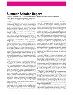
Infectious Agents and Cancer
BioMed Central
Open Access
Methodology
Routine human papillomavirus genotyping by DNA sequencing in
community hospital laboratories
Sin Hang Lee*, Veronica S Vigliotti, Jessica S Vigliotti and Suri Pappu
Address: Department of Pathology, Milford Hospital, Milford, Connecticut, USA
Email: Sin Hang Lee* - [email protected]; Veronica S Vigliotti - veró[email protected];
Jessica S Vigliotti - [email protected]; Suri Pappu - [email protected]
* Corresponding author
Published: 5 June 2007
Infectious Agents and Cancer 2007, 2:11
doi:10.1186/1750-9378-2-11
Received: 15 February 2007
Accepted: 5 June 2007
This article is available from: http://www.infectagentscancer.com/content/2/1/11
© 2007 Lee et al; licensee BioMed Central Ltd.
This is an Open Access article distributed under the terms of the Creative Commons Attribution License (http://creativecommons.org/licenses/by/2.0),
which permits unrestricted use, distribution, and reproduction in any medium, provided the original work is properly cited.
Abstract
Background: Human papillomavirus (HPV) genotyping is important for following up patients with
persistent HPV infection and for evaluation of prevention strategy for the individual patients to be
immunized with type-specific HPV vaccines. The aim of this study was to optimize a robust "lowtemperature" (LoTemp™) PCR system to streamline the research protocols for HPV DNA nested
PCR-amplification followed by genotyping with direct DNA sequencing. The protocol optimization
facilitates transferring this molecular technology into clinical laboratory practice. In particular,
lowering the temperature by 10°C at each step of thermocycling during in vitro DNA amplification
yields more homogeneous PCR products. With this protocol, template purification before
enzymatic cycle primer extensions is no longer necessary.
Results: The HPV genomic DNA extracted from liquid-based alcohol-preserved cervicovaginal
cells was first amplified by the consensus MY09/MY11 primer pair followed by nested PCR with
GP5+/GP6+ primers. The 150 bp nested PCR products were subjected to direct DNA sequencing.
The hypervariable 34–50 bp DNA sequence downstream of the GP5+ primer site was compared
to the known HPV DNA sequences stored in the GenBank using on-line BLAST for genotyping.
The LoTemp™ ready-to-use PCR polymerase reagents proved to be stable at room temperature
for at least 6 weeks. Nested PCR detected 107 isolates of HPV in 513 cervicovaginal clinical
samples, all validated by DNA sequencing. HPV-16 was the most prevalent genotype constituting
29 of 107 positive cases (27.2%), followed by HPV-56 (8.5%). For comparison, Digene HC2 test
detected 62.6% of the 107 HPV isolates and returned 11 (37.9%) of the 29 HPV-16 positive cases
as "positive for high-risk HPV".
Conclusion: The LoTemp™ ready-to-use PCR polymerase system which allows thermocycling at
85°C for denaturing, 40°C for annealing and 65°C for primer extension can be adapted for target
HPV DNA amplification by nested PCR and for preparation of clinical materials for genotyping by
direct DNA sequencing. HPV genotyping is performed by on-line BLAST algorithm of a
hypervariable L1 region. The DNA sequence is included in each report to the physician for
comparison in following up patients with persistent HPV infection, a recognized tumor promoter
in cancer induction.
Page 1 of 11
(page number not for citation purposes)
Infectious Agents and Cancer 2007, 2:11
Background
Human papillomavirus (HPV) testing was introduced to
compensate for the poor sensitivity and specificity of the
Pap smear cytology often used as diagnostic tool for borderline precancerous lesions [1]. Digene Hybrid Capture
2 (HC2) test, the only test approved by the U.S. Food and
Drug Administration (FDA), is commonly used to determine if a cervicovaginal cell suspension contains "highrisk" oncogenic HPVs [2], often functioning as a triage for
colposcopic evaluation of the cytologically borderline
cases [3-5]. However, it is now recognized that persistent
infection of a "high-risk" HPV, not the mere presence of
the HPV virus itself, is the pivotal promoter in causing cervical precancerous lesions and cancer [6-9]. Most of HPV
infections, even caused by "high-risk" genotypes, are transient with normal Pap cytology in sexually active young
women [10-13]. In 93% of initially infected women, the
same viral type is not detected upon re-examination four
menstrual cycles later [14]. The median duration of positivity detectable by PCR for a specific HPV type in these
young women is 168 days [15]. Multiple "high-risk" HPV
infections do not constitute a higher risk for the development of cervical neoplasia when compared with single
persistent high-risk HPV infection [16]. For the development and maintenance of a high-grade squamous intraepithelial lesion (SIL), the risk is greatest in women positive
for the same genotype of HPV on repeated testing [6-8].
Viral load is not a useful parameter to predict high-grade
SIL [17]. High-grade SIL is often associated with a viral
DNA load lower than that observed in less severely
affected cells [18].
In view of the recent advance in the understanding of the
relationship between persistent HPV infection and cervical neoplasia, a sensitive and specific technology to detect
and accurately genotype HPV is needed for clinical management of persistent infections. The HC2 test cannot be
converted to a genotyping assay and is associated with a
significant number of false-negative and false-positive
results when compared with other more stringent PCRbased HPV genotyping assays [19-23]. It is reported to
generate 25% false-negative results in cases with biopsyproven high-grade SIL even when all these biopsies have
been proven to contain high-risk HPV DNA by PCR [24].
"The lack of multiple, competitive, well-validated tests"
for HPV assay is quoted as being "a problem" in formulating new guidelines in management of cervical abnormalities [25].
The introduction of the type-specific Gardasil™ HPV vaccines into the sexually active female population also
requires genotype monitoring of the HPV infections
before and after immunization to develop prevention
strategy for the individual patients. Based on a "Background Document" submitted to the FDA [26], injection
http://www.infectagentscancer.com/content/2/1/11
of HPV vaccines into women who have concurrent vaccine-relevant HPV type infections may increase the risk, by
44.6%, of developing high-grade precancerous lesions in
the cervix. Therefore, it would be prudent to perform a
genotype-specific HPV assay if prior HPV infection is suspected.
Target nested PCR amplification of a conserved region of
the HPV L1 gene DNA with the consensus MY09/MY11
and GP5+/GP6+ primers, or their equivalent, followed by
genotyping with direct DNA sequencing is a generally
accepted scientific tool in research [21,22,27]. However, it
has not been used in clinical laboratories because handling the temperature-sensitive PCR reagents and the
requirement for template purification in the PCR protocols are too labor-intensive for routine applications. This
paper records our experience in using a high-processivity,
room temperature-stable robust DNA polymerase system
to facilitate the transfer of this molecular technology into
clinical laboratory practice. Each HPV-positive result is
validated by genotyping with direct DNA sequencing of a
hypervariable region of the L1 gene. The HPV DNA
sequencing information can be included in the laboratory
report for future clinical follow-up of persistent infections.
Methods
Since the PCR conditions adapted for this protocol
depended on the use of a new low-temperature ready-touse moderately thermostable DNA polymerase system,
the sensitivity and specificity of the LoTemp™ HiFi® DNA
polymerase in amplification of HPV DNA were first validated and compared with those obtained by standard
heat-resistant Taq polymerases available on the market.
The purified full-length plasmid DNAs of HPV types -16,
-18 and -6B purchased from American Type Culture Collection (ATCC) were used as standards for method development. Then the HPV type-16 DNA was used as a routine
positive control. Molecular grade pure water instead of
DNA extract was used as negative control.
Theoretical sensitivity of the PCR system chosen for this
study was determined by using serial 10-fold dilutions of
the ATCC-certified HPV standard containing 200 ng of
plasmid DNA of HPV-16, -18 and -6B with TE buffer to
single copy of genomic DNA per μL as the template to run
primary and nested PCR on each dilution in duplicate.
The theoretical number of copies of HPV DNA in the template used for each MY09/MY11 PCR was calculated
according to a generally accepted conversion formula
[28]. All nested PCR products were confirmed by DNA
sequencing to be those of respective HPV genotypes
expected.
Page 2 of 11
(page number not for citation purposes)
Infectious Agents and Cancer 2007, 2:11
For Taq PCR amplifications, the reaction mixture of 25 μL
contained 100 mM KCl, 20 mM Tris-HCl pH 8.0, 25 mM
MgCl2, 2.5 mM of each dNTP, 2.5 units of Takara Taq
polymerase (Takara Bio Inc., Shiga, Japan), 100 pmol of
each consensus primer (MY09/MY11 or GP5+/GP6+) and
1 μL of HPV DNA at various dilutions in TE buffer (or if
for nested PCR, 1 μL of the MY09/MY11 PCR products).
The reaction mixture was subjected to 35 cycles of amplification in an MJ Research thermocycler (Waltham, MA).
Each cycle consisted of a denaturing step at 94°C for 0.5
min, an annealing step at 55°C for 1 min, and a chain
elongation step at 72°C for 1 min.
In LoTemp™ HiFi® DNA polymerase PCR, the protocol
described below for clinical specimens was followed
except that a 35-cycle amplification was used for comparison with Taq PCR amplification.
The clinical samples used for this study were 515 alcoholpreserved liquid-based cervicovaginal cytology specimens
(Cytyc or Surepath) submitted by physicians in the New
Haven, Connecticut area as part of routine gynecologic
examinations. Age distribution of the patients and the cervical pathologic conditions were not the subjects of this
study. Digene HC2 test for high-risk HPV ordered by the
physicians was performed routinely on each sample by
one of the two independent clinical laboratories (Quest
Diagnostics Laboratory, Wallingford or Pathology & Laboratory Services, LLC, Woodbridge, CT) according to
instructions provided by Digene Corporation (Gaithersburg MD). In general the patients were women below age
30 who had a Pap cytology diagnosis of atypical squamous cells of undermined significance (ASCUS) or
women 30 years and older regardless of the Pap cytology
findings [4].
After the material was taken from each sample for routine
cytology and HC2 test, about 1 mL of the cell suspensions
was placed in a 1.5 mL Eppendorf tube, blind-coded with
a case number and transferred to the laboratory at Milford
Hospital for HPV PCR/DNA sequencing.
DNA extraction from the alcohol-fixed cells was accomplished according to a National Cancer Institute (NCI)
protocol [29] with minor modification. Briefly, the cell
suspension was first centrifuged in an Eppendorf microcentrifuge (model 5424) equipped with a rotor (model
FA45-24-11) for 5 min at 13,000 rpm. The cells in the pellet were washed in 1 mL reagent grade water and then in
1 mL buffer consisting of 50 mM Tris-HCl, 1 mM EDTA,
0.5% Tween 20, pH 8.1. The washed cell pellet was re-suspended and digested at 45–55°C overnight in 100 μL of
0.1 mg/mL proteinase K (Sigma Chemical Co., St. Louis,
MO) dissolved in the same washing buffer. After denaturing the proteins in the cell digestate in a metal block
http://www.infectagentscancer.com/content/2/1/11
heated to 95°C for 10 min and a final centrifugation of
the digestate at 13,000 rpm for 5 min, the supernatant was
carefully pipetted out and placed in a clean microcentrifuge tube to be used for PCR without further purification
or stored at -20°C.
The general methodology of primary PCR amplification
of a 450 bp segment of the HPV L1 gene with a pair of consensus MY09/MY11 primers followed by nested PCR with
a pair of GP5+/GP6+ general primers was used for HPV
DNA preparation. The 150 bp nested PCR products in the
positive specimens were genotyped by direct DNA
sequencing [21,22] with minor modifications briefly
summarized as follows.
For primary PCR amplification, 1 μL of the DNA extract, 1
μL of 10 μmolar MY09 primer, 1 μL of 10 μmolar MY11
primer and 2 μL of water were added to a PCR tube containing 20 μL of LoTemp™ HiFi® DNA polymerase readyto-use mix (HiFi DNA Tech, LLC, Trumbull, CT) which
contains all the components needed for low temperature
PCR, including dNTPs, Mg++, buffer, HiFi® DNA polymerases, proprietary dsDNA melting agents and dNTP preservatives, to reach a final 25 μL reaction volume. For
thermocycling, the temperature steps of a TC-412 Thermal
Cycler (Techne Incorporated, Burlington, NJ) were programmed for an initial heating at 85°C for 2 min, followed by 30 cycles at 85°C for 30 sec, 40°C for 30 sec,
and 65°C for 1 min. The final extension was 65°C for 10
min.
For nested PCR, a "trace" of the MY09/MY11 PCR products was transferred by a glass rod with clean wettable surface of about 1.5 mm in diameter to a second PCR tube
containing 25 μL of complete nested PCR reaction mixture consisting of 20 μL of LoTemp™ HiFi® DNA polymerase ready-to-use mix, 1 μL of 10 μmolar GP5+ primer, 1
μL of 10 μmolar GP6+ primer and 3 μL of water, using the
same thermocycling program as described above.
After completion of the primary and the nested PCR, a 5
μL aliquot of the PCR products was pipetted out from
each tube and mixed with 2 μL loading fluid for electrophoresis in a 2% agarose gel containing ethidium bromide. The gel was examined under UV light. Visualization
of a 450 bp PCR product band in the MY09/MY11 lane
and/or a 150 bp band in the nested PCR lane on the agarose gel provided evidence of HPV DNA in the sample,
pending genotyping with direct DNA sequencing as a
means of final validation.
For DNA sequencing, 1 μL of the nested PCR products, if
positive, was pipetted out from the nested PCR tube for
direct DNA sequencing, using 1 μL of 5 μmolar GP6+
primer as the sequencing primer, 1 μL of the BigDye® Ter-
Page 3 of 11
(page number not for citation purposes)
Infectious Agents and Cancer 2007, 2:11
minator (v 1.1/Sequencing Standard Kit), 3.5 μL 5×
buffer, and 13.5 μL water in a total volume of 20 μL for 20
enzymatic primer extension/termination reaction cycles
in an ABI thermocylcer Model 9600 according to the protocol supplied by the manufacturer (Applied Biosystems).
After dye-terminator cleanup with a Centri-Sep column
(Princeton Separations, Adelphia, NJ), the reaction mixture was loaded in an automated ABI 3130 four-capillary
Genetic Analyzer for sequence analysis. Sequence alignments were performed against various standard HPV genotype sequences stored in the GenBank database by online BLAST analysis to arrive at specific genotyping.
One μL of each DNA extract was placed in a separate PCR
tube with a β-globin primer pair [30] for human genomic
DNA amplification as a control of specimen adequacy.
The primers for β-globin gene amplification were 1 μL of
80 μmolar 5'-ACACAACTGTGTTCACTAGC and 1 μL of
80 μmolar 5'-CAACTTCATCCACGTTCACC in a 20 μL of
LoTemp™ HiFi® DNA polymerase ready-to-use mix with 2
μL water added. The LoTemp™ thermoclycling program
mentioned above was used.
Three (3) PCR tubes per sample were used routinely for
the β-globin gene, the MY09/MY11 primer and the GP5+/
GP6+ nested amplification, respectively.
To test performance reproducibility of this PCR/DNA
sequencing procedure, aliquots of the digestate of 30 individual clinical samples were tested in parallel duplicate
runs. The duplicate results of the three PCRs on each case
and the genotyping results of the positive cases by DNA
sequencing were compared.
Samples that did not show an MY09/MY11 or a GP5+/
GP6+ PCR band, but showed evidence of positive βglobin gene amplification were interpreted as HPV-negative. Specimens that did not show any PCR products in all
three lanes were considered unsatisfactory for evaluation
due to low DNA extraction or presence of a PCR inhibitor.
There were two (2) unsatisfactory cases among a total of
515 processed. The remaining 513 cases were accepted as
satisfactory for analysis. Of these 513 cases, 107 were positive for nested PCR products, all proven to be those of
HPV DNA by direct DNA sequencing, using GP6+ as the
sequencing primer.
The samples infected with more than one genotype of
HPV were indicated by the appearance of numerous
ambiguous or overlapping peaks in the DNA sequencing
tracings. For each of these mixed infections, the nested
PCR products were subjected to additional four individual
primer extension/termination reactions to rule out infection by the Gardasil™ vaccine-relevant HPV, namely HPV
types -16, -18, -6 or -11, using the following type-specific
primers [31].
http://www.infectagentscancer.com/content/2/1/11
HPV-16 type-specific sequencing primer 5'-GCTGCCATATCTACTTCAGA-3'
HPV-18 type-specific sequencing primer 5'-GCTTCTACACAGTCTCCTGT-3'
HPV -6 type-specific sequencing primer 5'-GTGCATCCGTAACTACATCTT-3'
HPV -11 type-specific sequencing primer 5'-GTGCATCTGTGTCTAAATCTG-3'
All oligonucleotides used as primers for this study were
synthesized and purified with the oligonucleotide purification cartridge (OPC) method by the Pathology Department DNA Synthesis Lab, Yale University (New Haven,
CT).
To avoid cross contamination, three separate rooms with
no air re-circulation were dedicated to nucleic acid amplification tests. Two of the rooms were each equipped with
a 32" PCR workstation (AirClean Systems, Raleigh, NC).
All pre-amplification procedures were performed in PCR
station I. All post-PCR procedures were carried out in PCR
station II, including preparations for the nested PCR and
sequencing reaction. Gel electrophoresis and DNA
sequencing were performed in the third isolation room.
No post-PCR materials or any items contaminated by
amplicons, or equipment used in the post-PCR rooms
were allowed to enter the pre-PCR working space.
Results
The LoTemp™ HiFi® DNA polymerase was about 10 times
more efficient than Taq DNA polymerases in amplifying
HPV plasmid DNA by MY09/MY11 PCR and about 100 to
1000 times more efficient when the first amplification
was followed by a GP5+/GP6+ nested PCR in tandem. The
nested PCR technology described in this paper proved to
be a sensitive method for the detection of 1–10 copies of
purified genomic DNA of HPV types -16, -18 or -6B. But
104-105 copies of genomic DNA were needed as PCR templates for UV visualization of a positive MY09/MY11
primer amplicon band after electrophoresis (Fig. 1). The
specificity of all nested PCR products was validated by
HPV genotyping with direct DNA sequencing.
Reproducibility of this nested PCR assay in the detection
of HPV DNA in clinical specimens was confirmed by running two parallel sets of PCR with a split single sample
digestate as the paired templates, including the β-globin
gene, the MY primer and the GP nested primer amplifications on each set for 30 HPV-positive cases. Pairs of identical results on electrophoresis gel were obtained in all
three amplifications for the 30 split samples (Fig. 2). The
nested PCR products obtained on the duplicate sets were
Page 4 of 11
(page number not for citation purposes)
Infectious Agents and Cancer 2007, 2:11
http://www.infectagentscancer.com/content/2/1/11
UV visualization. Without a nested PCR, the amplicon of
the MY09/MY11 PCR was often masked due to co-amplification of other DNA molecules in the clinical samples
(Fig. 2).
polymerase
Comparative
Figure 1 and
amplification
with LoTemp™
of HPV-16
HiFi®DNA
DNA with
polymerase
Taq DNA
Comparative amplification of HPV-16 DNA with Taq
DNA polymerase and with LoTemp™ HiFi®DNA
polymerase. Lanes 1–8: HPV-16 plasmid DNA template at
85 × 101, 85 × 102, 85 × 103, 85 × 104, 85 × 105, 85 × 106, 85
× 107 and 85 × 108 copies per milliliter. NC: negative control.
M: molecular marker. Arrows indicate the lower limit of
detection by nested PCR (A) and by the 1st primary PCR (B)
with Takara Taq DNA polymerase and LoTemp™ HiFi®
DNA polymerase, respectively.
confirmed by DNA sequencing to be of the same HPV
genotype.
For some isolates, notably those of HPV-39 and HPV-73,
the standard GP5+/GP6+ nested PCR failed to amplify the
target DNA fragment even when the MY09/MY11 primer
PCR amplification was successful. These cases were recognized on the gel electrophoresis, showing a positive 450
bp MY09/MY11 PCR amplicon in the absence of an
expected concomitant 150 bp nested PCR product. For
these HPV strains, a heminested PCR with MY11/GP6+
primers generated a homogeneous amplicon (Fig. 3) for
direct DNA sequencing.
After cycle sequencing, BLAST algorithms by alignment of
a 34 bp DNA sequence in the hypervariable region of the
L1 gene downstream of the GP5+ binding site against
known HPV genotype sequences stored in the GenBank
database usually determined the genotype of the HPV isolates detected [32]. A 100% "identities" match between
the "query" sequence and the "subject" sequence was
reached for each final genotyping (Fig. 4). This sequence
For clinical samples, the primary MY09/MY11 PCR generated a distinct 450 bp product band after 30 repeated
amplification cycles in only 37 of the 107 HPV-positive
cases detected by nested PCR. More than 65% of the clinical HPV-positive amplicons relied on a nested PCR for
Figure
HPV
Nested
2
PCR in Clinical Samples and Reproducibility
HPV Nested PCR in Clinical Samples and Reproducibility. Agarose gel showing PCR products of targeted DNAs
extracted from two clinical samples in duplicate. The targeted β-globin DNA amplicon is 110 bp long, as seen clearly
in lanes 25 and 26 (#1210), but is hardly visible in lanes 31
and 32 (#1211). Co-amplification of other human genomic
DNA fragments and a positive nested PCR amplicon assure
specimen adequacy in both samples. Molecular ruler = 100–
1000 bp (far left). Lanes 25/26, 27/28, 29/30 = β-globin gene,
MY09/MY11, GP5+/GP6+ PCR, respectively-sample #1210.
Lanes 31/32, 33/34, 35/36 = β-globin gene, MY09/MY11,
GP5+/GP6+ PCR, respectively-sample #1211
Figure 3Heminested PCR (clinical specimen #24)
HPV-39
HPV-39 Heminested PCR (clinical specimen #24).
Left gel: beta: β-globin gene amplification-specimen #24. my:
Positive 450 bp MY09/MY11 PCR product- #24. nested:
Negative GP5+/GP6+ nested PCR product- #24. Molecular
marker on far left. Right gel: Lane 1: #24 MY09/MY11 PCR
product, 450 bp. Lane 2 HPV-16 control Y09/MY11 PCR
product, 450 bp. Lane 3: #24 MY11/GP6+ heminested PCR
product ~ 195 bp. Lane 4: HPV-16 control GP5+/GP6+
nested PCR ~ 150 bp. Molecular marker on far left
Page 5 of 11
(page number not for citation purposes)
Infectious Agents and Cancer 2007, 2:11
http://www.infectagentscancer.com/content/2/1/11
HPV-18 100%
HPV-33 100%
HPV-39 100%
HPV-56 100%
HPV-59 100%
HPV-68 100%
HPV-58 83.3%
HPV-31 71.4%
HPV-45 66.7%
Figure
DNA
GP5+
Sequence
for
4 Genotyping
of Nested PCR Product Downstream of
DNA Sequence of Nested PCR Product Downstream
of GP5+ for Genotyping. This is a typical DNA sequence
excised from the color tracing downstream of the GP5+
binding site of the HPV DNA L1 gene. BLAST alignment analysis of a 34 (up to 50) bp sequence of this hypervariable
region provides unequivocal evidence for HPV genotype 66
based on the database stored in the GenBank.
HPV-35 50%
tracing with its on-line BLAST algorithm for genotyping
was incorporated in the report for clinical follow-up of
persistent infections. However, HPV-16 has numerous
sequence variants, some of which share an identical
sequence in this region with some strains of HPV-31 and
HPV-33, and required BLAST algorithm of a 50 bp
sequence for genotyping distinction.
Among the 107 nested PCR-positive samples, DNA
sequencing with the GP6+ consensus general primer
yielded multiple overlapping unreadable sequences in 5
cases. Using the individual type-specific primer sequencing for HPV-6,-11,-16 and -18 proved that one of them
contained HPV-16, but not the other three genotypes, and
that one contained a mixture of HPV-16 and HPV-18, but
not the other two genotypes. For the remaining 3 mixed
infection samples, repeated individual DNA sequencing
failed to produce a readable primer extension/termination reaction with any of the four type-specific primers.
Therefore, these latter 3 cases were considered to be multiple infections caused by HPVs other than the four vaccine-relevant types and grouped under the "low-risk"
category.
Of the 513 liquid-based cervicovaginal samples, the
nested PCR method detected at least one HPV strain in
107, with an overall positive rate of 20.9%, including 74
cases harboring at least one of the 13 types targeted by the
Digene HC2 "high-risk" HPV test and 33 cases containing
"low-risk" HPV types (Table 1). The most prevalent genotype in the New Haven area was found to be HPV-16, followed by HPV-56. The combined rate of prevalence of
HPV-16 and HPV-18 constituted 32.8% of the total isolates.
The HC2 high-risk HPV test identified only 11 (37.9%) of
the 29 HPV-16 cases as positive. However, it successfully
identified all the samples containing HPV-56 and HPV-18
as positive for high-risk HPV. The sensitivity of the HC2
test in detecting the predetermined "high-risk" HPV genotypes was summarized as follows:
HPV-52 50%
HPV-16 37.9%
Although HPV-54, -66, -83, -53 and -62 were not included
in the hybridization cocktail probe, these genotypes were
often reported to be positive for high-risk HPV by the HC2
test (Table 1).
Of the 513 cases studied, the Digene HC2 test classified
403 samples as negative and 75 as positive for "high-risk"
HPV, and 35 as unsatisfactory for evaluation. These results
were compared with those obtained by the nested PCR
assay (Table 2). Since the Digene test only covered the
"high-risk" HPV genotypes, specimens infected by HPV
other than the 13 types targeted by the HC2 "high-risk"
HPV cocktail probe would be classified as negative or
unsatisfactory for evaluation (unsat.). In summary, HC2
test identified 50 of the 74 "high-risk" HPV-positive sam-
Page 6 of 11
(page number not for citation purposes)
Infectious Agents and Cancer 2007, 2:11
http://www.infectagentscancer.com/content/2/1/11
Table 1: HPV genotyping by DNA Sequencing v. Digene HC2
Test
PCR/DNA Sequencing
Type Positive Cases
16
29
9
56
7
31
6
6
6
18
54
6
6
58
66
6
4
59
3
45
83
3
32
2
2
35
2
39
40
2
2
52
1
33
53
1
62
1
1
68
70
1
72
1
73
1
1
M16
1
M 16, 18
M others
3
Total
107
Prevalence (%)
(27.2)
(8.5)
(6.5)
(5.6)
(5.6)
(5.6)
(5.6)
(5.6)
(3.7)
(2.8)
(2.8)
(1.9)
(1.9)
(1.9)
(1.9)
(1.9)
(0.9)
(0.9)
(0.9)
(0.9)
(0.9)
(0.9)
(0.9)
(0.9)
(0.9)
(2.9)
(100)
Test Results by Digene HC2
High-risk+
11
9
5
2
6
3
5
6
4
2
2
0
1
2
0
1
1
1
1
1
1
0
0
1
1
1
67
Negative
18
0
2
4
0
3
1
0
0
1
1
2
1
0
2
1
0
0
0
0
0
1
1
0
0
2
40
HC2 HPV detection rate =
67/107 = 62.6%
Samples
%HPV+ 20.9
513
M16 = mixed infection with HPV 16 identified by type-specific
sequencing primer.
M 16, 18 = mixed infection with HPV 16 and HPV 18 identified by
type-specific sequencing primers.
M others = mixed infections by HPV types which cannot be
sequenced with type-specific primers for HPV 6, 11, 16 or 18.
Digene High-risk + includes those cases reported as positive for both
High-risk and Low-risk HPV types.
The underlined HPV genotypes are included in the "High-risk" Digene
HC2 cocktail probe.
ples detected by nested PCR. The analytical sensitivity of
the Digene HC2 test was calculated to be 50/74= 67.6%
against the nested PCR assay. Since HC2 reported a total
of 75 cases to be positive for "high-risk" HPV, its analytical specificity was calculated to be 50/75 = 66.7%. Two
cases found to be unsatisfactory for PCR evaluation and
negative by HC2 test were excluded from the total of 513
cases entered for analysis.
Discussion
The moderately thermostable LoTemp™ HiFi® DNA
polymerases are genetically engineered derivatives of a
Bacillus stearothermophilus (Bst) DNA polymerase which
was first introduced to resolve the hairpin structure in
classic Sanger DNA sequencing by Ye and Hong [33]. Bst
DNA polymerase with an optimum primer extension temperature at 65°C is extremely stable, capable of retaining
its sequencing quality in working solution at room temperature for several weeks under conditions of robotic
automation [34]. Modification of the amino acid
sequence of a natural Bst DNA polymerase by site-directed
genetic mutations increases the heat tolerance of the
enzyme [35], which has paved the way to development of
new thermostable DNA polymerases for repeated primer
extension reactions under 85°C. The LoTemp™ HiFi® DNA
polymerase PCR protocol exploits the proof-reading and
high-processivity properties of the modified Bst DNA
polymerases at reduced cyclic temperatures.
Using LoTemp™ HiFi® DNA polymerase for HPV DNA
amplification eliminates all template purification steps
which are usually required before PCR or sequencing reaction [21,22,32]. Since the ready-to-use polymerase mixture contains all the required ingredients for PCR, the
need for in-house pipetting is minimal. Since the DNA
polymerase and other reagent components are stabilized
for storage at room temperature, there is no need to keep
ice-cold blocks while setting up the PCR. Since this system
uses chemical melting agents for dsDNA denaturing and a
high-processivity DNA polymerase for nucleotide primer
extension under partial isostabilization, it allows thermocycling at 85°C for denaturing, 40°C for annealing and
65°C for primer extension, respectively. At lowered
cycling temperatures, the rate of heat-induced mutations
[36], namely depurination [37] and deamination [38] of
the nitrogenous bases, in the DNA molecules during PCR
amplification is reduced. As a result, the PCR products are
more homogeneous.
In this report, we have demonstrated that the LoTemp™
PCR system can amplify 1–10 copies of purified HPV-16,
-18 or -6B to generate a corresponding type-specific 150
bp nested PCR product. The sensitivity of LoTemp™ PCR
in the detection of plasmid HPV DNA is about 10× greater
than that obtained by the traditional heat-resistant Taq
DNA polymerases without nested amplification (Fig. 1).
Nested PCR has increased the analytical sensitivity of HPV
DNA detection on cervicovaginal cell suspensions
[21,22,32], which is also our experience (Fig. 2). Nested
PCR also serves to eliminate most of the interfering substances which otherwise may have to be removed by column purification in the procedure.
Although the MY09/MY11 and GP5+/GP6+ consensus
general primer pairs have been used to amplify the highly
conserved L1 gene region of all HPV genotypes, their efficiency in target DNA amplification varies from one HPV
Page 7 of 11
(page number not for citation purposes)
Infectious Agents and Cancer 2007, 2:11
http://www.infectagentscancer.com/content/2/1/11
Table 2: Comparison of HPV nested PCR/sequencing and HC2 test results
PCR Results
HC2 Results
Negative
High Risk
Low Risk
Unsat.
Total
Negative
High Risk
Low Risk
Unsat.
Total
364
8
NP
34
406
23
50
NP
1
74
16
17
NP
0
0
403
75
0
0
35
513
33
HC2, Hybrid Capture 2; HPV, human papillomavirus; PCR, polymerase chain reaction.
Unsat. Test results or specimens considered inadequate for evaluation.
High Risk HPV types 16, 18, 31, 33, 35, 39, 45, 51, 52, 56, 58, 59 and 68.
Low Risk HPV types other than those listed as High Risk.
NP = not performed.
Data are given as number of specimens
genotype to another with discordant patterns [22]. We
have found that some isolates of HPV-39 and HPV-73 are
not amplifiable by the GP5+/GP6+ primer pair either in
primary PCR or in nested PCR. A second amplification
with a GP5+/MY09 and a GP6+/MY11 primer pair generates a longer and shorter PCR product, respectively. The
GP6+/MY11 heminested PCR product (Fig. 3) is suitable
for direct DNA sequencing. GP5+/GP6+ failure in amplification of HPV-39 [39] and other HPV types [22,32] in
clinical specimens is a well-known technical problem if
this single pair of primers is relied upon for HPV detection. However, GP5+ and GP6+ are excellent nested PCR
primers for preparing templates for HPV genotyping with
direct DNA sequencing.
Optimization of the PCR protocol is essential for detection of HPV. Without optimization, a PCR method using
one set of consensus primers for amplification may have
a lower sensitivity than the Digene HC2 test [40]. Using
an initial 40-cycle amplification on clinical specimens,
Johnson et al. [21] reported that nested PCR generally
increases the sensitivity of an HPV PCR method by about
60%. With the PCR protocol presented in this paper, this
difference is augmented due to adoption of a 30-cycle
amplification. An advantage of reducing the number of
amplification cycles is that co-amplification of non-specific interfering DNA molecules is reduced in the primary
PCR in favor of generating specific nested PCR products
for direct DNA sequencing.
Using our optimized protocol, nested PCR detected 107
HPV-positive cases among 513 clinical samples (Table 1).
Twenty-three (23) HPV genotypes have been identified,
including 12 of the 13 "high-risk" genotypes, i.e. HPV-16,
-18, -31, -33, -35, -39, -45, -52, -56, -58, -59, and -68,
which are targeted by the Digene HC2 test. The lack of representation of HPV-51 which is also targeted by the HC 2
test probably reflects a low regional prevalence of this genotype. Using the same primers for nested PCR followed by
genotyping with DNA sequencing, HPV-51 was found to
be a relatively common genotype in Germany, constituting about 5% of the total HPV isolates detected [22].
However, it was not recorded even once among 894 HPV
isolates in Denmark [21].
The hypervariable region of the DNA sequence downstream of the GP5+ binding site is critical in L1 genotyping of HPV. While most HPV genotypes can be
determined through alignment of a 34-bp sequence in
this region with the GenBank database [32], accurate genotyping may require alignment of a longer sequence, for
example 50 bp long, when the result of BLAST algorithm
is ambiguous. If a 34-bp sequence is relied upon for genotyping, the number of HPV-16 infections may be underestimated.
For the development of this protocol, we found that using
1 μL of 5 μmolar OPC-purified GP6+ oligonucleotide
instead of a manufacturer-recommended 3.2 μmolar solution as the sequencing primer seems to generate more
consistent sequencing results for typing various HPV isolates. The concentration of primer may need to be
adjusted if a higher grade of GP6+ is used as the DNA
sequencing primer.
Of the 107 PCR-positive samples, the Digene HC2 test has
classified 67 as HPV-positive, a detection rate of 62.6%
(Table 1). When only the HC2-targeted "high-risk" HPV
genotypes are used for comparison, nested PCR detects 74
HPV-positive cases while HC2 test identifies 50 in this
group with an analytical sensitivity of 67.6% (50/74).
The most prevalent is HPV-16, constituting 27.2% of the
total isolates (Table 1). This percentage of HPV-16 prevalence is almost identical to those reported by others using
MY/GP nested PCR/DNA sequencing genotyping on cervicovaginal cell suspensions, e.g. 26% in Denmark [21] and
26.2% in Germany [22]. Digene HC2 test identified 11 of
Page 8 of 11
(page number not for citation purposes)
Infectious Agents and Cancer 2007, 2:11
the 29 HPV-16 PCR-positive cases with a detection rate of
37.9% which is surprisingly low. Since all the HPV-16
PCR-positive results have been validated by DNA
sequencing for genotyping with a prevalence rate similar
to those commonly found in the Western world based on
the same methodology [21,22] and since the Digene HC2
tests were performed by two independent clinical laboratories properly certified by the health authorities, the
validity of the individual test results seems not to be in
question. The discrepancy is probably due to the fact that
there are numerous HPV-16 sequence variants in a given
patient population [41], which may not be all targeted by
the HC2 RNA cocktail probe, but share a highly conserved
region of the L1 gene that the MY09/MY11 primers
amplify effectively. In support of this interpretation is the
fact that the HC2 test has correctly identified all cases of
HPV-18, HPV-39, HPV-56 and HPV-59 as "high-risk" in
the same data.
HPV-56 is the second most prevalent high-risk genotype
detected (8.5%), followed by HPV-31, -18, -54, -58 and 66, sharing about the same rate of prevalence (5.6–6.5%).
The combined number of HPV-16 and -18 cases constitutes only 32.8% of the single HPV isolates in our series.
HPV-18 also seems to play a relatively minor role in causing cervical pathology in Canada [42].
It has been reported by others [43] that the Digene HC2
high-risk test may be able to detect HPV types -53, -54, 62, -66 and -83 and label them as high-risk HPVs
although these genotypes are not intentionally targeted in
its high-risk cocktail probe. Our findings confirm these
cross-reactions (Table 1). Sequence variation within the
probe binding sites [44] and non-specific binding
between the probe and non-targeted mismatched DNA
[45-48] are well recognized sources of error if nucleic acid
hybridization is relied upon for microbial and viral genotyping. When the GP5+/GP6+ PCR products with a hypervariable DNA sequence are targeted for developing a
multiplex genotyping method [49], the DNA probe
designed for HPV-66, a recently recognized high-risk type
[50-52], is found to react with HPV-52 and the probe for
HPV-82 with HPV-51 due to cross-hybridization despite
the presence of four base mismatches in each pair. Some
experts [40] consider the unintended cross-reactions of
the Digene HC2 test with non-targeted HPV types, such as
HPV-66, which occasionally may cause cancer, to be "fortunate". However, the benefit to the patients of these
cross-reactions needs additional confirmation.
Fifty percent (50%) of the HPV-54 isolates are returned by
HC2 test as high-risk HPV. HPV-54 has been classified as
a low-risk virus [51,53], but is found to be associated with
a 40-fold increase in risk among American Indian women
with CIN 2/3. Only HPV-16 has shown a higher risk than
http://www.infectagentscancer.com/content/2/1/11
HPV-54 among this subpopulation [54]. Genetic makeup of a patient may have to be considered in using HPV
genotyping information for the follow-up of persistent
infections [55].
For the mixed infection cases, we choose single primers
specific for HPV-6, -11, -16 and -18 [31] to perform individual specific primer DNA sequencing in order to determine if the mixed infection includes any of these vaccinerelevant HPV types. The rationale for this choice is that the
majority of multiple HPV infections are transient [6-15].
The immediate concern to the patient and her health care
provider is whether the mixed infection is caused by any
of the vaccine-relevant HPV types if the patient is considering vaccine immunization.
The low percentage (< 5%) of multiple HPV infections
observed in our series might have been biased because a
large proportion of the specimens for this study was collected from a solo private practitioner's office and this
group of specimens had an exceptionally low positive rate
and no multiple HPV infections at all. The rate of multiple
HPV infections is known to vary among patient populations and is also influenced by the stage of carcinogenesis.
Multiple HPV infections were found in less than 5% of the
HPV-positive samples from patients with invasive cancer
lesions, but over 15% of the positive samples in the control group [27]. Multiple HPV infections tend to evolve
into single HPV infections as the infection becomes
chronic and persistent while the cervical cytopathology
progresses from metaplasia, LSIL, HSIL, carcinoma-in-situ
to invasive cancer [56].
In summary, we have reported our experience in adapting
the well characterized PCR/DNA sequencing protocol for
routine HPV genotyping. We believe that a sensitive and
specific HPV assay followed by genotyping with direct
DNA sequencing will generate useful information for following persistent infections while referring the patients at
"high-risk" of developing HSIL, not the patients with a
"high-risk HPV", to colposcopic evaluation.
Conclusion
The nested PCR technology using MY09/MY11 and GP5+/
GP6+ consensus primers for target HPV DNA amplification can be used in diagnostic laboratories for routine
HPV detection and to prepare clinical materials for genotyping by direct DNA sequencing. We have adapted a
newly introduced low-temperature PCR system and optimized the protocol to facilitate the transfer of this molecular technology into clinical laboratory practice for
accurate HPV genotyping which is a valuable tool for follow-up of patients with persistent HPV infection, a recognized tumor promoter in cancer induction.
Page 9 of 11
(page number not for citation purposes)
Infectious Agents and Cancer 2007, 2:11
References
1.
2.
3.
4.
5.
6.
7.
8.
9.
10.
11.
12.
13.
14.
15.
16.
17.
18.
Kjaer SK, van den Brule AJ, Bock JE, Poll PA, Engholm G, Sherman ME,
Walboomers JM, Meijer CJ: Human papillomavirus- the most
significant risk determinant of cervical intraepithelial neoplasia. Int J Cancer 1996, 65:601-6.
HC2 HPV DNA Test-REF 5199-1220 Digene Corporation,
Gaithersburg, MD 20878 USA. 2005.
Gogola J, Van Dinh T, Lucci JA 3, Smith E, Hannigan EV: Human papillomavirus testing for triage in a referral population. J Low
Genit Tract Dis 2001, 5:29-32.
ACOG Practice Bulletin No 61: Human Papillomavirus. Obstet
Gynecol 2005, 105:905-18.
Wu HH, Allen SL, Kirkpatrick JL, Elsheikh TM: Reflex high-risk
human papilloma virus DNA test is useful in the triage of
women with atypical squamous cells cannot exclude highgrade squamous intraepithelial lesion. Diagn Cytopathol 2006,
34:707-10.
Wallin KL, Wiklund F, Angstrom T, Bergman F, Stendahl U, Wadell
G, Hallmans G, Dillner J: Type-specific persistence of human
papillomavirus DNA before the development of invasive cervical cancer. N Engl J Med 1999, 341:1633-8.
Kjaer SK, van den Brule AJ, Paull G, Svare EI, Sherman ME, Thomsen
BL, Suntum M, Bock JE, Poll PA, Meijers CJ: Type specific persistence of high risk human papillomavirus (HPV) as indicator of
high grade cervical squamous intraepithelial lesions in young
women: population based prospective follow up study. BMJ
2002, 325:572-6.
Cuschieri KS, Cubie HA, Whitley MW, Gilkison G, Arends MJ, Graham C, McGoogan E: Persistent high risk HPV infection associated with development of cervical neoplasia in a prospective
population study. J Clin Pathol 2005, 58:946-50.
Brummer O, Hollwitz B, Bohmer G, Kuhnle H, Petry KU: Human
papillomavirus-type persistence patterns predict the clinical
outcome of cervical intraepithelial neoplasia. Gynecol Oncol
2006, 102:517-22.
Jacobs MV, Walboomers JM, Snijders PJ, Voorhorst FJ, Verheijen RH,
Fransen-Daalmeijer N, Meijer CJ: Distribution of 37 mucosotropic HPV types in women with cytologically normal cervical smears: the age-related patterns for high-risk and lowrisk types. Int J Cancer 2000, 87:221-7.
Herrero R, Hildesheim A, Bratti C, Sherman ME, Hutchinson M,
Morales J, Balmaceda I, Greenberg MD, Alfaro M, Burk RD,
Wacholder S, Plummer M, Schiffman M: Population-based study of
human papillomavirus infection and cervical neoplasia in
rural Costa Rica. J Natl Cancer Inst 2000, 92:464-74.
Molano M, Posso H, Weiderpass E, van den Brule AJ, Ronderos M,
Franceschi S, Meijer CJ, Arslan A, Munoz N, HPV Study Group HPV
Study: Prevalence and determinants of HPV infection among
Colombian women with normal cytology. Br J Cancer 2002,
87:324-33.
Rousseau MC, Pereira JS, Prado JC, Villa LL, Rohan TE, Franco EL:
Cervical coinfection with human papillomavirus (HPV) types
as a predictor of acquisition and persistence of HPV infection. J Infect Dis 2001, 184:1508-17.
Hinchliffe SA, van Velzen D, Korporaal H, Kok PL, Boon ME: Transience of cervical HPV infection in sexually active, young
women with normal cervicovaginal cytology. Br J Cancer 1995,
72:943-5.
Brown DR, Shew ML, Qadadri B, Neptune N, Vargas M, Tu W, Juliar
BE, Breen TE, Fortenberry JD: A longitudinal study of genital
human papillomavirus infection in a cohort of closely followed adolescent women. J Infect Dis 2005, 191:182-92.
Cuschieri KS, Cubie HA, Whitley MW, Seagar AL, Arends MJ, Moore
C, Gilkisson G, McGoogan E: Multiple high risk HPV infections
are common in cervical neoplasia and young women in a cervical screening population. J Clin Pathol 2004, 57:68-72.
Wensveen CW, Kagie MJ, Nagelkerke NJ, Veldhuizen RW, Trimbos
JB: Can viral load, semi-quantitatively evaluated, of human
papillomavirus predict cytological or histological outcome in
women with atypical squamous or glandular cells of undetermined significance cytology? Eur J Gynaecol Oncol 2005, 26:393-7.
Sherman ME, Wang SS, Wheeler CM, Rich L, Gravitt PE, Tarone R,
Schiffman M: Determinants of human papillomavirus load
among women with histological cervical intraepithelial neoplasia 3 : dominant impact of surrounding low-grade lesions.
Cancer Epidemiol Biomarkers Prev 2003, 12:1038-44.
http://www.infectagentscancer.com/content/2/1/11
19.
20.
21.
22.
23.
24.
25.
26.
27.
28.
29.
30.
31.
32.
33.
34.
35.
36.
37.
38.
39.
40.
Castle PE, Schiffman M, Burk RD, Wacholder S, Hildesheim A, Herrero R, Bratti MC, Sherman ME, Lorincz A: Restricted Cross-Reactivity of Hybrid Capture 2 with Nononcogenic Human
Papillomavirus Types. Cancer Epidemiol Biomarkers & Prev 2002,
11:1394-9.
Vernon SD, Unger ER, Williams D: Comparison of human papillomavirus detection and typing by cycle sequencing, line
blotting, and hybrid capture. J Clin Microbiol 2000, 38:651-5.
Johnson T, Bryder K, Corbet S, Fomsgaard A: Routine genotyping
of human papillomavirus samples in Denmark. APMIS 2003,
111:398-404.
Speich N, Schmitt C, Bollmann R, Bollmann M: Human papillomavirus (HPV) study of 2916 cytological samples by PCR and
DNA sequencing : genotype spectrum of patients from the
west German area. J Med Microbiol 2004, 53:125-8.
Kosel S, Burggraf S, Mommsen J, Engelhardt W, Olgemoller B: Typespecific detection of human papillomaviruses in a routine
laboratory setting–improved sensitivity and specificity of
PCR and sequence analysis compared to direct hybridisation. Clin Chem Lab Med 2003, 41:787-91.
Lonky NM, Felix JC, Naidu YM, Wolde-Tsadik G: Triage of atypical
squamous cells of undetermined significance with hybrid
capture II: colposcopy and histologic human papillomavirus
correlation. Obstet Gynecol 2003, 101:481-9.
Stoler MH, Castle PE, Solomon D, Schiffman M: The Expanded Use
of HPV Testing in Gynecologic Practice per ASCCP-Guided
Management Requires the Use of Well-Validated Assays. Am
J Clin Pathol 2007, 127:335-7.
VRBPAC Background Document: Gardasil™ HPV Quadrivalent
Vaccine. VRBPAC Meeting [http://www.fda.gov/ohrms/dockets/ac/
06/briefing/2006-4222B3.pdf]. May 18, 2006
Asato T, Maehama T, Nagai Y, Kanazawa K, Uezato H, Kariya K: A
large case-control study of cervical cancer risk associated
with human papillomavirus infection in Japan, by nucleotide
sequencing-based genotyping. J Infect Dis 2004, 189:1829-32.
[http://www.uri.edu/research/gsc/resources/cndna.html].
Protocols used at NCI & Related Information-Processing of
Microdissected Tissue for Molecular Analysis [http://cgapmf.nih.gov/Protocols/Processing/DNA.html]
Ayatollahi M, Zakerinia M, Haghshenas M: Molecular analysis of
Iranian families with sickle cell disease. J Trop Pediatr 2005,
51:136-40.
Gharizadeh B, Oggionni M, Zheng B, Akom E, Pourmand N, Ahmadian
A, Wallin KL, Nyren P: Type-specific multiple sequencing primers: a novel strategy for reliable and rapid genotyping of
human papillomaviruses by pyrosequencing technology. J
Mol Diagn 2005, 7:198-205.
Feoli-Fonseca JC, Oligny LL, Filion M, Brochu P, Simard P, Russo PA,
Yotov WV: A two-tier polymerase chain reaction direct
sequencing method for detecting and typing human papillomaviruses in pathological specimens. Diagn Mol Pathol 1998,
7:317-23.
Ye S, Hong G: Heat-stable DNA polymerase I large fragment
resolves hairpin structure in DNA sequencing. Scientia sinica
(Series B) 1987, 30:503-6.
Earley JJ, Kuivaniemi H, Prockop DJ, Tromp G: Robotic automation of dideoxyribonucleotide sequencing reactions. Biotechniques 1994, 17:156-65.
Hong GF, Huang W: DNA polymerase having ability to reduce
innate selective discrimination against fluorescent dyelabeled dideoxynucleotides. US Patent . #6,165,765
Andre P, Kim A, Khrapko K, Thilly WG: Fidelity and mutational
spectrum of Pfu DNA polymerase on a human mitochondrial DNA sequence. Genome Res 1997, 7:843-52.
Lindahl T, Nyberg B: Rate of depurination of native deoxyribonucleic acid. Biochemistry 1972, 11:3610-8.
Lindahl T, Nyberg B: Heat-induced deamination of cytosine residues in deoxyribonucleic acid. Biochemistry 1974, 13:3405-10.
Evans MF, Adamson CS, Simmons-Arnold L, Cooper K: Touchdown
General Primer (GP5+/GP6+) PCR and optimized sample
DNA concentration support the sensitive detection of
human papillomavirus. BMC Clin Pathol 2005, 5:10.
Schiffman M, Wheeler CM, Dasgupta A, Solomon D, Castle PE: The
ALTS Group. A comparison of a prototype PCR assay and
hybrid capture 2 for detection of carcinogenic human papil-
Page 10 of 11
(page number not for citation purposes)
Infectious Agents and Cancer 2007, 2:11
41.
42.
43.
44.
45.
46.
47.
48.
49.
50.
51.
52.
53.
54.
55.
56.
lomavirus DNA in women with equivocal or mildly abnormal
Papanicolaou smears. Am J Clin Pathol 2005, 124:722-32.
Tornesello ML, Duraturo ML, Salatiello I, Buonaguro L, Losito S, Botti
G, Stellato G, Greggi S, Piccoli R, Pilotti S, Stefanon B, De Palo G,
Franceschi S, Buonaguro FM: Analysis of human papillomavirus
type-16 variants in Italian women with cervical intraepithelial neoplasia and cervical cancer. J Med Virol 2004, 74:117-26.
Feoli-Fonseca JC, Oligny LL, Brochu P, Simard P, Falconi S, Yotov WV:
Human papillomavirus (HPV) study of 691 pathological
specimens from Quebec by PCR-direct sequencing
approach. J Med Virol 2001, 63:284-92.
Ortiz M, Torres M, Munoz L, Fernandez-Garcia E, Canals J, Cabornero AI, Aguilar E, Ballesteros J, Del Amo J, Garcia-Saiz A: Oncogenic
human papillomavirus (HPV) type distribution and HPV
type 16 E6 variants in two Spanish population groups with
different levels of HPV infection risk. J Clin Microbiol 2006,
44:1428-34.
Anderson TP, Werno AM, Beynon KA, Murdoch DR: Failure To
Genotype Herpes Simplex Virus by Real-Time PCR Assay
and Melting Curve Analysis Due to Sequence Variation
within Probe Binding Sites. J Clin Microbiol 2003, 41:2135-2137.
Goris J, Konstantinidis KT, Klappenbach JA, Coenye T, Vandamme P,
Tiedje JM: DNA-DNA hybridization values and their relationship to whole-genome sequence similarities. Int J Syst Evol
Microbiol 2007, 57:81-91.
Huang CC, Qiu JT, Kashima ML, Kurman RJ, Wu TC: Generation of
type-specific probes for the detection of single-copy human
papillomavirus by a novel in situ hybridization method. Mod
Pathol 1998, 11:971-7.
Naef F, Lim DA, Patil N, Magnasco M: DNA hybridization to mismatched templates: a chip study. Phys Rev E Stat Nonlin Soft Matter Phys 2002, 65(4 Pt 1):040902.
Sorokin NV, Chechetkin VR, Livshits MA, Pan'kov SV, Donnikov MY,
Gryadunov DA, Lapa SA, Zasedatelev AS: Discrimination between
perfect and mismatched duplexes with oligonucleotide gel
microchips: role of thermodynamic and kinetic effects during hybridization. J Biomol Struct Dyn 2005, 22:725-34.
Schmitt M, Bravo IG, Snijders PJ, Gissmann L, Pawlita M, Waterboer
T: Bead-based multiplex genotyping of human papillomaviruses. J Clin Microbiol 2006, 44:504-12.
Tawheed AR, Beaudenon S, Favre M, Orth G: Characterization of
human papillomavirus type 66 from an invasive carcinoma of
the uterine cervix. J Clin Microbiol 1991, 29:2656-2660.
Munoz N, Bosch FX, de Sanjose S, Herrero R, Castellsague X, Shah
KV, Snijders PJ, Meijer CJ: International Agency for Research on
Cancer Multicenter Cervical Cancer Study Group: Epidemiologic classification of human papillomavirus types associated with cervical cancer. N Engl J Med 2003, 348:518-27.
Prado JC, Calleja-Macias IE, Bernard HU, Kalantari M, Macay SA, Allan
B, Williamson AL, Chung LP, Collins RJ, Zuna RE, Dunn ST, OrtizLopez R, Barrera-Saldana HA, Cubie HA, Cuschieri K, von KnebelDoeberitz M, Sanchez GI, Bosch FX, Villa LL: Worldwide genomic
diversity of the human papillomaviruses-53, 56, and 66, a
group of high-risk HPVs unrelated to HPV-16 and HPV-18.
Virology 2005, 340:95-104.
Favre M, Kremsdorf D, Jablonska S, Obalek S, Pehau-Arnaudet G,
Croissant O, Orth G: Two new human papillomavirus types
(HPV54 and 55) characterized from genital tumours illustrate the plurality of genital HPVs. Int J Cancer 1990, 45:40-6.
Schiff M, Becker TM, Masuk M, van Asselt-King L, Wheeler CM, Altobelli KK, North CQ, Nahmias AJ: Risk factors for cervical
intraepithelial neoplasia in southwestern American Indian
women. Am J Epidemiol 2000, 152:716-26.
Trimble CL, Piantadosi S, Gravitt P, Ronnett B, Pizer E, Elko A, Wilgus
B, Yutzy W, Daniel R, Shah K, Peng S, Hung C, Roden R, Wu TC, Pardoll D: Spontaneous regression of high-grade cervical dysplasia: effects of human papillomavirus type and HLA
phenotype. Clin Cancer Res 2005, 11:4717-23.
Zuna RE, Allen RA, Moore WE, Mattu R, Dunn ST: Comparison of
human papillomavirus genotypes in high-grade squamous
intraepithelial lesions and invasive cervical carcinoma : evidence for differences in biologic potential of precursor
lesions. Modern Pathology 2004, 17:1314-22.
http://www.infectagentscancer.com/content/2/1/11
Publish with Bio Med Central and every
scientist can read your work free of charge
"BioMed Central will be the most significant development for
disseminating the results of biomedical researc h in our lifetime."
Sir Paul Nurse, Cancer Research UK
Your research papers will be:
available free of charge to the entire biomedical community
peer reviewed and published immediately upon acceptance
cited in PubMed and archived on PubMed Central
yours — you keep the copyright
BioMedcentral
Submit your manuscript here:
http://www.biomedcentral.com/info/publishing_adv.asp
Page 11 of 11
(page number not for citation purposes)


© Copyright 2026