
Abstract: lesions, forcing replication to grind to a halt. The cell must
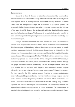
Summer Scholar Report Structure and Function: The Crystal Structure of Dpo4 with N-Acetyl-2-Aminofluorene Author: Melissa Brulotte, [email protected] Advisor: Samer Lone, PhD, [email protected] Abstract: Cancer is a leading cause of death in America. Carcinogens have the ability to attack DNA, creating DNA lesions that can cause mutations. Mutations in critical genes involving cell growth and maintenance can lead to tumor formation. How DNA lesions are handled by the cell is a key question in understanding cancer. One way is through a group of specialized DNA polymerases that perform lesion bypass, called the Y-Family. Dpo4 is a Y-Family polymerase found in archaeal cells. Little is known about the mechanism by which it is able to bypass DNA lesions. In order to further discover this mechanism, it was our goal to determine the structure of Dpo4 with the model liver carcinogen N-Acetyl-2-Aminofluorenene (AAF). We have site-specifically modified DNA template oligonucleotides with the AAF lesion, and purified the template and the necessary primer oligonucleotides. We have also purified Dpo4 protein. Co-crystallization trials were performed around a variety of precipitating reagents. Cocrystals of the Dpo4-AAF complex were obtained, and we are currently optimizing cryoprotectant conditions for X-ray diffraction analysis. A polymerase fidelity assay was performed to examine nucleotide incorporation across the AAF adducted primer-template oligonucleotides. Single nucleotide extension analysis revealed that Dpo4 preferentially incorporates the correct nucleotide across the bulky AAF adduct. Background: Cancer accounts for nearly 1 in 4 deaths in America. 1 In 2010 alone, the American Cancer Society predicted 1,529,560 new cancer cases in the United States, with 569,490 of these causing death.1 That is equivalent to over 1,500 people per day dying of cancer. Environmental exposure from sunlight, radiation, and a variety of chemicals have all been identified as sources of cancer-causing agents, known as carcinogens. 1 Carcinogens have the ability to attack DNA, creating DNA lesions that wreak havoc on the normal metabolic processes of the cell. When cells are unable to repair the damage, there is a high probability of mutation, producing cells with altered function. If these mutations are in critical genes that involve cell growth and maintenance, they can be passed on to daughter cells and lead to tumor formation and potentially cancer. How DNA lesions are handled by the cell has been a key question in understanding cancer. One way cells handle DNA lesions is through a group of specialized DNA polymerases that perform lesion bypass, termed TLS (translesion DNA synthesis) or the Y-family. Classical replicative DNA polymerases are stalled at DNA 8 The Nucleus February 2011 lesions, forcing replication to grind to a halt.2 The cell must recruit the Y-family polymerases to continue DNA replication. The exact mechanism of how the Y-family polymerases are recruited, and how they are able to replicate past DNA lesions is not entirely known. However, structural information about the Y-family polymerases themselves and the polymerases combined with undamaged DNA has revealed that they have a much more open and looser active site than the normal replicative polymerases.2 Specifically, when using the traditional “right-hand” structural comparison, Y-family polymerases have a much smaller finger and thumb than classical replicative polymerases.2 Each member of the Y-family also has a unique “little finger” or PAD (polymerase associated domain) that helps hold the DNA substrate in place.2 Finally, Y-family polymerases do not have 3’-5’ exonuclease activity like classical DNA polymerases. This means that they do not proofread the DNA to ensure that it is paired correctly and they cannot excise a nucleotide so the correct one can be inserted. This leads to low fidelity replication of DNA, which can cause mutations that lead to disease. 2-3 Therefore, while Y-family polymerases continue replication and prevent cell death, it is possible that they cause the very mutations that result from cancer lesions. Y-family polymerases share five sequence motifs in the N-terminal 250 amino acid residues, but the residues interacting with the incoming nucleotide are not the same in each polymerase. This suggests that each polymerase has specific lesions it can bypass and/or specific nucleotides it will insert. Biochemical and structural studies have indeed shown that each Y-family member exhibits different lesion bypass abilities and different mutation spectra. 2 These unique functionalities may show that the loss of even one Yfamily polymerase can trigger health problems. An example is Xeroderma pigmentosum disease, in which the human TLS polymerase Pol is inactive, which can lead to a higher chance of skin and other cancers.4 Dpo4 is a Y-family polymerase that is found in archaeal cells. While the structure of this DNA polymerase with undamaged DNA has been determined (see Figure 1), the structure with certain DNA lesions in the active site has yet to be discovered. Dpo4 is known to have a spacious active site that might be able to accommodate bulky DNA lesions. It is used as a model Y-family polymerase because its structure was the first of the Y-family to be discovered and so has been a traditional model, and because it is similar to Pol in humans and Pol 4 in bacteria. N-Acetyl-2-Aminofluorene (AAF) (Figure 2) was originally synthesized as a potential insecticide before it was discovered to cause mammalian cancer. It now serves as a model compound for other aromatic amines like 2-naphth- Summer Scholar Continued from page 8 study of this specialized group of DNA polymerases is important because it may help us determine why some people are more susceptible to cancer than others. lamine, which is a bladder carcinogen. 5 Since aromatic amines are used in industrial processes to produce dyes, pharmaceuticals, antioxidants and pesticides, it is important to study aromatic amines such as AAF to determine how the cell handles the potential carcinogens. AAF attaches to the carbon-8 position of the guanine nucleotide. This causes the guanine base to swing out of the double helix, and allows the AAF molecule to insert into the helix (see Figure 3).5 This movement causes a large distortion in the DNA. As with other DNA lesions, when a replicative DNA polymerase encounters this distortion, it gets caught in an open, inactive formation. A Y-family polymerase, such as Dpo4, is recruited to bypass the lesion so DNA replication can continue. It is the structure of the Dpo4 polymerase, including its open active site, which allows it to bypass the AAF lesion, and that is why determining the structure of Dpo4 with AAF in its active site is so important. Once the crystal structure of Dpo4 with AAF is determined, it can be used as a model for the study of the structures of other Y-family polymerases with other carcinogens. The Figure 3: An NMR structure of AAF intercalating into DNA. The circle indicates the guanine that has swung out of the helix.5 Protein expression and purificationDpo4 was obtained using standard E. coli protein expression. The plasmid DNA that encodes for Dpo4 was obtained from the Prakash Laboratory at the University of Texas Medical Branch and introduced to E. coli bacteria cells. The cells were grown in TY Medium broth and induced with IPTG to produce Dpo4. The cells were split to extract the protein. The protein was purified on a GST affinity chromatography column. The Dpo4 was removed from the column, using Prescission Protease, and the purity was tested using SDS-PAGE. The concentration of the protein was determined using a Bradford Assay. Figure 1: The structure of Dpo4 with undamaged DNA and an incoming nucleotide. Figure 2: N-Acetyl-2-Aminofluorene. DNA purification and modificationThe specific DNA oligonucleotides needed for protein crystallization were obtained as single strands from Integrated DNA Technologies and the W.M. Keck Foundation at Yale School of Medicine. The DNA was purified using polyacrylamide gel electrophoresis with 16-18% acrylamide. The gel salts were removed using Biorad Nap-25 columns, and the concentration of the DNA was determined using standard UV/Vis spectrometer techniques. The AAF lesion was ordered from NCI Chemical Carcinogen Reference Standard Repository at Midwest Research Institute in N-acetoxy-2-acetylaminofluorene form. It was dissolved in ethanol and reacted with oligonucleotide temContinued on page 10 The Nucleus February 2011 9 Summer Scholar Continued from page 9 plate in 2 mM sodium citrate buffer (pH 6.8) under argon gas. Once the reaction was started, the solution was removed from the argon gas and reacted for three hours at 37°C. Waste chemicals were removed using ether extraction, and the DNA was purified using polyacrylamide gel electrophoresis with 18% acrylamide. The concentration of the modified oligonucleotide was determined using standard UV/Vis spectrophotometry. The modified template oligonucleotide was annealed with a complementary primer oligonucleotide (see Figure 4). Figure 4: The DNA sequence used for this study. The asterisks indicates the position of the AAF modified guanine. The dNTP represents where an incoming nucleotide would bind. Figure 5: An SDS-PAGE gel used to determine if Dpo4 was successfully purified. Lane 1 contained a molecular weight marker. Lanes 2, 3, and 5 contained waste solutions that were tested to ensure that Dpo4 was not released into the waste. Lane 4 contained the Dpo4 that was tested. Lane 6 contained a standard solution of Dpo4 with known enzymatic activity. By comparing lane 4 to lane 6 and to the molecular weight marker it was determined that protein expression and purification was successful. Protein crystallizationThe double-stranded DNA oligo with a terminated primer was reacted with Dpo4 in the following conditions: 1 mM cytosine, 20 mM HEPES (pH 7.0), 60 mM sodium chloride, 5 mM MgCl2, 1.0 mM DTT, 8 mg/mL Dpo4, and 0.239 mM DNA.6 The solution was reacted for five minutes at room temperature and then kept on ice. The hanging drop method was used for protein crystallization with well solutions containing: 100 mM HEPES (pH 7.0), 2.5% glycerol, varying PEG 3350 and varying calcium acetate. Polymerase fidelity assay A polymerase fidelity assay was performed with Dpo4 and unterminated DNA to determine which nucleotide Dpo4 preferentially inserts across from AAF-modified guanine. Undamaged DNA was tested alongside the damaged DNA to serve as a basis for comparison. The reaction conditions were as follows: 1 µM Dpo4, 10 µM DNA, 1 mM dNTP, 5 mM MgCl2, 100 mM HEPES (pH 8.0), 60 mM NaCl, and 5 mM DTT.6 The protein-DNA complex was tested with no nucleotides, all nucleotides, and each of the four DNA nucleotides individually. The final volume of each mixture was 10 µL. The reactions took place at 30°C for 30 minutes, and then the mixtures were placed on ice. The results were visualized on an 18% polyacrylamide gel. Results and Discussion: Dpo4 was successfully expressed and purified (Figure 5) to a final concentration of 30.64 mg/mL. Many DNA template oligos and primers were successfully purified, and many reactions with AAF were successfully accomplished as determined by UV/Vis spectrophotometry (Figure 6). Crystals of the Dpo4-AAF complex were successfully obtained (Figure 7). However, x-ray diffraction and structural determination have yet to be accomplished. 10 The Nucleus February 2011 Figure 6: An example of a successful reaction and purification of AAF modified DNA. The DNA was diluted 1000x and this graph shows the absorbance at 260 nm. Using Beer’s Law, the concentration of the modified DNA was found to be 4.786 x 10⁻⁵ mmol. Comparing this to the original concentration of the unmodified DNA (2.682 x 10⁻⁴ mmol) reveals a 17.84% yield of product. One polymerase fidelity assay was performed to determine the nucleotide incorporation and extension across from the AAF lesion, using an undamaged template as a control. Extension was studied by reacting the Dpo4-DNA complex with the four DNA nucleotides (G,C,A,T). With undamaged DNA extension was successful, as can be seen by the variety Summer Scholar Continued from page 10 Figure 7: Crystals of the AAF-Dpo4 complex as seen under a microscope. of bands in lane 4 of Figure 8. With AAF-modified DNA extension appears to be stalled, as can be determined by the lack of upper bands in lane 11. Incorporation was studied by reacting the Dpo4-DNA complex with each of the different nucleotides. With undamaged DNA, Dpo4 preferentially inserts the correct nucleotide, cytosine (lane 6). With undamaged DNA, Dpo4 preferentially inserts the correct nucleotide, cytosine (lane 6). With AAF-modified DNA, Dpo4 also preferentially inserts cytosine (lane 13), though qualitatively the incorporation appears to be stalled compared to undamaged DNA. Future Work: The main focus of future work will be to obtain more Dpo4AAF crystals in order to get X-ray diffraction data. Crystal screens will continue to be performed in order to get the best product. The crystals will be frozen and shipped to the National Synchrotron Light Source at Brookhaven National Lab. They will be X-ray diffracted, and the resulting data set will be analyzed, using molecular replacement with Dpo4 and undamaged DNA as a search model. The polymerase fidelity assay will be repeated to confirm the results and to get better resolution. Kinetic assays will also be performed in order to determine the qualitative observations that the incorporation of cytosine and extension are stalled in AAF-modified DNA. Combined with structural data and further biochemical studies, a better understanding of how Dpo4 and other Y-family polymerase function in DNA replication and in causing DNA mutations will be obtained. Figure 8: 1. DNA primer, 2. Undamaged DNA template, 3. Dpo4-DNA complex (undamaged) with no nucleotides, 4. Dpo4-DNA complex (undamaged) with G, C, A, T nucleotides, 5. Dpo4-DNA complex (undamaged) with G, 6. Dpo-DNA complex (undamaged) with C, 7. Dpo-DNA complex (undamaged) with A, 8. Dpo-DNA complex (undamaged) with T, 9. AAF-modified DNA template, 10. Dpo4-DNA complex (AAF) with no nucleotides,11. Dpo4DNA complex (AAF) with G, C, A, T nucleotides, 12. Dpo4DNA complex (AAF) with G, 13. Dpo-DNA complex (AAF) with C, 14. Dpo-DNA complex (AAF) with A, 15. Dpo-DNA complex (AAF) with T. References: 1. American Cancer Society. Cancer Facts & Figures 2010. http://www.cancer.org/acs/groups/content/@epidemiologysurveilance/documents/document/acspc-026238.pdf 2. Yang, W., Damage repair DNA polymerases Y. Curr Opin Struct Biol 2003, 13 (1), 23-30. 3. Lehmann, A. R., et al. Translesion synthesis: Y-family polymerases and the polymerase switch. DNA Repair (Amst) 2007, 6 (7), 891-9. 4. Wang, Z., DNA damage-induced mutagenesis : a novel target for cancer prevention. Mol Interv 2001, 1 (5), 26981. 5. O’Handley, S.F. et al. Structural characterization of an Nacetyl-2-aminofluorene (AAF) modified DNA oligomer by NMR, energy minimization, and molecular dynamics. Biochemistry, 1993, 32 (10), 2481-97. 6. Rechkoblit, O. et al. Mechanism of error-free and semitargeted mutagenic bypass of an aromatic amine lesion by Y-family polymerase Dpo4. Structural & Molecular Biology, Nature, 2010, 17 (3), 379-89. The Nucleus February 2011 11


© Copyright 2026