
Full PDF - International Journal of Microbiology and Allied Sciences
International Journal of Microbiology and Allied Sciences
November 2014, 1(2):46-77
ISSN: 2382-5537
REVIEW ARTICLE
Page: 46-77
Bacterial and viral diseases in fish & shrimps
Sukhdev Singh*, Manu Bala, Akanksha, Shamsher Singh Kanwar
Department of Biotechnology, Himachal Pradesh University, Summer Hill, Shimla-171005, India.
*Corresponding author. Email address: [email protected]
ABSTRACT
Aquaculture play central role in socio-economic development of rural and coastal countries. Human
interfere displace aquatic animals from their natural ecosystems to artificial stressed environment which
often results in emergence & spread of pathogens and new diseases. Many severe pathogens have been
emerged in aquaculture important species; especially the viral ones. Some economically important
species are Atlantic salmon (Salmo salar), Rainbow trout (Oncorhynchus mykiss), Sea bream (Pagrus
major), Yellow tail (Seriola quinqueradiata), Gilthead sea bream (Sparus aurata), Ayu (Plecoglossus
altivelis), Sea bass (Dicentrarchus labrax), Turbot (Scophthalmus maximus), Flounder (Paralichthys
olivaceus) and shrimps (Penaeus monodon & P. vannamei). Permanent eradication of these pathogens
from major aquaculture producing countries is very necessary; for which the causative etiological agent
must be known. So main aim of this article is to describe these pathogens and developed diseases.
Permanent vaccines against all pathogens are not developed yet as they change their pathogenicity via
enhanced virulence. Additionally, available vaccines are unable to raise strong and long term immune
response. Amongst these pathogens, viral ones are discussed in detail as they are most contagious and
can wipe out whole farms.
Key words: Aquaculture, pathogens, diseases, vaccines, viral pathogens.
International Journal of Microbiology and Allied Sciences, Nov 2014, Volume 1 Issue 2
46
Sukhdev Singh et al. 2014
IJOMAS. November 2014, 1(2):46-77
INTRODUCTION
Fish farming is an ancient practice known to human civilization [1]. From 20th century, husbandries
were started to establish for salmon, trout and shrimp species, and thereafter aquaculture became
a major animal food producing sector [2]. Aquaculture species are farmed in fresh, brackish and
marine water environments in which they are reared for optimal production [3]. These species are
diverse in geographically mean, viz. from tropical to arctic & subarctic, from lakes & rivers to
estuaries. Principal farming species are salmon, trout, carp, oyster and shrimps. Countries like UK,
Canada, Norway, Chile and USA are dominant producers of salmon and trouts whereas Asian
countries are involved for carp, oyster and shrimps [4]. In previous decades many serious diseases
have been emerged in aquatic animals due to anthropogenic global movement of animals & their
products. These diseases are listed as ‘notifiable’ by world organization of animal health. Among
the fish pathogens, diseases caused by viruses are of great concern as they wipe out whole
aquaculture farms. Brood stocks from wild fisheries are significant healthy carrier of viral
pathogens [5] and attempts are in progress to confront these carriers using RNA interference
approach [6]. Fish cell lines are suitable subjects for modern fish pathology especially for virology
to isolate the viral agent in vitro [7]. Bacteria also cause severe diseases in fish and economic losses
in aquaculture (Table 1). Some of the bacterial pathogens among these are known to infect (via
food or contact) humans also [8]. Death outbreaks associated with usually occur due to
consumption of raw or improperly processed infected fish. Many bacterial pathogens have been
reported for histamine poisoning which occur by decarboxylation of histidine [9]. These histamine
formers grow optimally at 25 oC. Symptoms of histamine poisoning are diarrhea, dyspnoea,
pruritus, hypertension and metallic or peppery taste in mouth [9]. Like fishes, shrimps also cover
a major part of aquaculture industry and as demanding sea food. Shrimps comprise 15-20% of total
fishery products worldwide [10]. Out of various species, two principal species namely Black tiger
shrimp (Penaeus monodon) and White pacific shrimp (Penaeus vannamei) dominate the shrimp
production as animal food. Pathogens emerge in shrimps also, especially viruses (Table 2);
producing mass mortality in the farm [11]. Some immunity components have been described in
shrimps like toll like receptors [12], RNAi [13] and vaccination attempts are also in progress [14].
So in this review we have summarized the diseases in the light of expanding geographical host
range, virulence diversity, clinical signs and vaccination schemes and emphasized on the viral
pathogens. Complete diagnosis of these pathogens can’t be fully described as they require
histopathological, immunological & molecular diagnostics, in situ hybridization and cell culture
diagnosis [15].
Bacterial diseases in fish
Diseases due to Aeromonads
Aeromonads are the most common bacterial pathogens in fishes throughout the world. Species
included in this group are Aeromonas hydrophila, A. veronii and A. salmonicida which cause
serious diseases in aquatic animals and in humans too [16]. Among these A. hydrophila causes
motile aeromonas septicemia (MAS) or red sore disease [17] and A. salmonicida causes
furunculosis [18]. The most common clinical signs are hemorrhages in skin, fins, oral cavity and
muscles with superficial ulceration of epidermis [19]. Exophthalmia, ascites, swollen kidneys,
splenomegaly, necrosis in spleen and kidneys are also observed. A. salmonicida is antigenically
homogenous and can be diagnosed by immunodiagnostics and PCR [20]. Expression profiling and
virulence factors have been described in A. salmonicida species [21,22]. Bacteriovorax sp. has
recently been identified as biocontrol against A. veronii [23,24].
International Journal of Microbiology and Allied Sciences, Nov 2014, Volume 1 Issue 2
47
Sukhdev Singh et al. 2014
IJOMAS. November 2014, 1(2):46-77
Bacterial kidney disease (BKD)
Aetiological agent for BKD or Overt disease is Renibacterium salmoninarum which mainly affect
salmonids [25]. Infection symptoms are exophthalmia, petechial hemorrhages, enlargement &
necrosis in kidneys with granulomatous lesions [26]. Biochemically and antigenically R.
salmoninarum is homogenous [27] and PCR, IFAT and ELISA diagnostic methods have been
developed against the pathogen [28,29]. This pathogen spread horizontally or vertically [30,31]
and transmission dynamics & phylogenetic history have been recently demonstrated with single
nucleotide polymorphism (SNP) analysis [25].
Mycobacteriosis (Fish Tuberculosis)
Fish are convincing host of mycobacteriosis [32]. Many mycobacterium species (M. marinum, M.
fortuitum, M. chelonae, M. avium, M. gordonae, M. abscesus, M. aurum, M. parafortuitum, M.
poriferae and M. triplex) have been recognized as causative agents for this chronic disease [33]
but M. marinum is notable amongst these. Its host range is salmons, turbot, sea bass, striped bass,
sod and halibut [34]. Disease symptoms include exophthalmoses, keratitis, change in pigmentation,
skin ulcerations, granulomas in liver, spleen & kidney and hemorrhagic lesions in muscles [35].
Histopathology and PCR detection procedures have been developed to detect the pathogen [36].
Vibriosis
Pathogenic species in this group are Vibrio anguillarum, V. ordalii, V. vulnificus Biotype 2, V.
harveyii and Alivibrio salmonicida within the family Vibrionaceae [37]. V. anguillarum is
antigenically heterogeneous and approximate 20-25 ‘O’ serotypes has been identified in contrast
to other Vibrio species [38]. Among these only O1, O2 and O3 serotypes are implicated in mortality
cases. Complete genome of V. anguillarum has been sequenced recently [39]. Clinical signs
observed with vibriosis are hemorrhagic septicemia on the base of fins, edematous lesions, corneal
opacity and exophthalmia [40]. V. salmonicida causes Hitra disease in salmonids and cods and
contain marker protein VS-P1 on surface [41]. Many species have been reported to affect humans
also. V. alginolyticus reported for otitis media [42]; V. vulnificus for chronic hepatic diseases,
immunodeficiency symptoms, septicemia [43], purpuras, necrotizing dermo-hypodermatitis and
fasciitis [44] in patients; V. parahaemolyticus causes diarrhea, gastroenteritis and septicemia
[45,46]; and V. cholerae reported for cholera [47,48]. For diagnosis and identification many
molecular techniques (e.g. 16S rRNA, FISH, AFLP and RAPD) have been developed for these
species [49,50].
Flexibacteriosis
It is also named as Eroded mouth syndrome and Black patch necrosis and caused by
Tenacibaculum maritimum which has a wide host range, viz. turbot, gilthead sea bream, sea bass,
red sea bream, black sea bream sole, and salmonids [51]. Clinical symptoms are eroded and
hemorrhagic mouth, ulcerative skin lesions, frayed fins and tail rot [51]. T. maritimum is
antigenically and biochemically homogenous but two serotypes has been identified. Serological
identification, PCR based detection methods [52] and vaccination attempt have been made but an
effective vaccine is still awaited [53].
Pseudomonadiasis
Pseudomonas anguilliseptica caused mass mortality in eels of Japan, Taiwan, Scotland and
Denmark [54]. Its host range includes eels, black sea bream, ayu, wild herrings, gilthead sea bream
International Journal of Microbiology and Allied Sciences, Nov 2014, Volume 1 Issue 2
48
Sukhdev Singh et al. 2014
IJOMAS. November 2014, 1(2):46-77
and turbot. This pathogen causes red spot disease with clinical symptoms of abdominal distention
and hemorrhagic petechia in skin & lesions [55]. P. anguilliseptica has two ‘O’ serotypes, one of
eel and another of gilthead sea bream, turbot [55]. Clinical diagnostics and detection of the
pathogen by immunohistochemical and PCR methods is available [56,57]. Oil-adjuvant bacterins
were developed but any permanent inactivation is still awaited.
Streptococcosis
Cocci like Lactococcus garvieae, L. piscium, Streptococcus iniae, S. agalactiae, S. parauberis and
Vagococcus salmoninarum infect fishes throughout the world [58]. They infect species in Japan
(yellowtail, flounder); Spain, France, Italy, Israel (rainbow trout); USA (tilapia) and Australia
(barramundi). These cocci damage central nervous system with symptoms of exophthalmia and
meningo-encephalitis [59]. In humans Streptococcus iniae has been identified for cellulitis,
endocarditis, meningitis and septic arthritis [60]. Serotype studies revealed two serogroups (KGand KG+) in L. garvieae and two serotypes (I and II) in S. iniae [61]. S. agalactiae was reported
for illness and deaths in doctor fish (Garra rufa) [62]. Immunodiagnostic procedures are available
for identification of these cocci [63,64].
Diseases due to Flavobacteria
Flavobacteria are important aetiological pathogens represent by Flavobacterium psychrophilum,
F. branchiophilum and F. columnare, and cause bacterial coldwater disease (BCWD), bacterial
gill disease and columnaris disease respectively [65]. These species has wide host and geographic
ranges. CWD produce serious mortalities with clinical signs as increased mucus secretion,
epidermal hyperemia, splenomegaly and hepatomegaly [66,67]. Bacterial gill disease primarily
occurs in juvenile salmonids in spring season. Affected fish display lethargicity, high in water
column and gasping for air at surface. In columnaris disease pathological signs are erotic lesions
or deep ulcers on skin and fins. Proteome analysis of F. columnare has been performed that
exhibited virulence diversity in the species [68,69].
Edwardsiellosis
Two pathogenic species are in this group; Edwardsiella tarda and E. ictaluri. Among these E.
tarda has wide host range and reported to infect humans also [70] whereas E. ictaluri limited to
fishes only. These species cause systemic hemorrhagic septicemia and enteric septicemia in
catfishes [71]. A characteristic symptom observed is the presence of a raised or open ulcer on the
head. E. tarda causes loss of pigmentation, swelling on abdominal surface and petechial
hemorrhages in fins and skin [72,73]. Clinical diagnostic kits (i.e. ELISA, PCR and LAMP) are
available commercially to identify these species [74,75].
Enteric red mouth (ERM)
Yersinia ruckeri causes ERM which is responsible for critical economic losses. This bacterium
belongs to Enterobacteriaceae family and produce yersiniosis or ERM disease in fishes. Its
common host is Salmo gairdneri [76]. Clinical manifestations of this disease are exophthalmia,
ascites and hemorrhage & ulceration in jaw, palate, gills and operculum. Biochemically this
bacterium is very diverse and has two biotypes (1 & 2) grouped in many ‘O’ serogroups [77]. The
disease spread via direct contact with infected fish or by carriers. Progression of the pathogen in
infected rainbow trout has been demonstrated through bioluminescence imaging [78]. Recently
genetic component for holomycin resistance has been identified [79] and some probiotics and
International Journal of Microbiology and Allied Sciences, Nov 2014, Volume 1 Issue 2
49
Sukhdev Singh et al. 2014
IJOMAS. November 2014, 1(2):46-77
bacterins have also been developed for prevention of yersiniosis but permanent eradication still is
the matter of research [80,81].
Photobacteriosis
Also known as pasteurellosis, photobacteriosis is characterized by white nodules in spleen and
kidneys [82]. Photobacterium damselae subsp. piscicida is the causal agent. Mortality incidences
due to the photobacterium reported from Chesapeake Bay (white perch and striped bass), Japan
(yellowtail), Mediterranean countries of Europe (gilthead sea bream, sea bass and sole) and USA
[hybrid striped bass; 83]. Clinical diagnostics and laboratory identification (i.e. ELISA, PCR) are
available for photobacterium for disease control in farms [84].
Piscirickettsiosis
Piscirickettsia salmonis is responsible for piscirickettsiosis in rainbow trouts and salmons
principally in Coho salmon [85]. Observed clinical signs of this septicemic condition are nodules
in liver, darkening of the skin, lethargy, anorexia and respiratory distress in affected fish [86].
Commercial ELISA kits, PCR based protocols for diagnoses are available in the market [87].
Winter ulcer
It’s a winter season problem characterized by skin ulcers at scale covered surface; mainly occur in
Atlantic salmons with few incidences reported in plaice and rainbow trouts also [88]. Causal
pathogen for winter ulcer is Moretella viscosa which is represented by two groups: first one is
Norway group and second one is Iceland group [89,90].
Table 1: Bacterial pathogens in fishes.
Bacterium
Genome
size
(Mb)
4.12
Disease
Main host(s)
Vaccine(s) if any
Vibriosis
V. ordalii [92]
3.41
Vibriosis
Salmonids, cods,
red sea bream
Salmonids
V. vulnificus [93]
Aliivibrio salmonicida
[41]
Flavobacterium
psychrophilum [67]
F. branchiophilum [94]
F. columnare [95]
5.26
4.65
Vibriosis
Vibriosis
Eel, tilapia
Atlantic salmon
2.86
Coldwater disease
Salmonids, carp
Vibrio anguillarum-ordaliiYersinia ruckeri Bacterin
Vibrio anguillarum-ordaliiYersinia ruckeri Bacterin
-----Aliivibrio salmonicida
Bacterin
-------
3.56
3.2
Bacterial gill disease
Columnaris disease
Edwardsiella tarda [96]
3.76
Edwardsiellosis
Edwardsiella ictaluri
[97]
Aeromonas hydrophila
[98]
A. salmonicida [18]
A. veronii [99]
3.81
Enteric septicemia
Reared fishes
Cyprinids,
salmonids
Salmons, catfish,
carps
Catfish
4.74
Renibacterium
salmoninarum [25]
3.15
Motile aeromonas
septicemia
Furunculosis
Epizootic ulcerative
syndrome
Bacterial kidney disease
Salmonids, non
salmonids
Salmons
Salmonids, non
salmonids
Salmonids
Vibrio anguillarum [91]
4.70
4.55
------------------Edwardsiella ictaluri
Bacterin
Aeromonas hydrophila
Bacterin
A. salmonicida Bacterin
---------------
International Journal of Microbiology and Allied Sciences, Nov 2014, Volume 1 Issue 2
50
Sukhdev Singh et al. 2014
IJOMAS. November 2014, 1(2):46-77
Streptococcus
parauberis [100]
Lactococcus garvieae
[101]
Mycobacterium
marinum [102]
Yersinia ruckeri [103]
2.14
Streptococcosis
Turbot
--------
2.17
Lactococcosis
Yellowtail, eel
--------
6.64
Mycobacteriosis
--------
3.58
Enteric redmouth
Photobacterium
damselae [104]
5.05
Photobacteriosis
Sea bass, turbot,
Atlantic salmon
Salmonids, eel,
minnows
Sea bream, sea
bass, yellowtail
Yersinia ruckeri Bacterin
--------
Fungal diseases in fish
Fungal diseases are quite rare than bacterial and viral ones in mass mortality means. Fungi can
infect fishes if they are given environmental stress conditions like poor environmental conditions
(water with low circulation, low dissolved oxygen or high ammonia or high organic loads), poor
nutrition or injury. In most cases fungal infection occurs as secondary infection in association with
bacterial or viral. Common fungal infections are due to water molds (Saprolegnia). This cotton
wool fungus belongs to Saprolegniaceae family and causes saprolegniasis in fishes and fish eggs
[105]. Saprolegnia grow optimum at temperature ranges 59o to 86o F [106]. In most cases
saprolegnia attacks on existed injured tissue and spread to healthy tissues [107]. These saprophytes
transmit via motile zoospores and detritus of bottom. Saprolegniasis symptoms are superficial and
observed as fluffy tufts of cottony growth on the skin and gills in affected fish [108].
Another common fungal disease in fishes is gill rot (Branchiomycosis) that occurs mainly in carp
and eels. Its causative agents are Branchiomyces sanguinis and B. demigrans. These species
invades fish in water of low pH, low dissolved oxygen and high algal bloom. Infected fish manifest
respiratory distress, thrombosis in blood vessels of gills and necrosis in gills [109]. Another fungal
problem in fish is Icthyophonus disease caused by Icthyophonus hoferi [110]. Icthyophonus
manifest itself internally and primarily attack liver and kidney [111]. Disease is transmitted via
fungal cysts released by faeces and via cannibalism of infected fish [112]. A fish fungus is
Exophiala [113] which is represented by two important pathogenic species namely E. salmonis and
E. psychrophila [114]. These fungi produce round yellow to white granulomas in liver, kidneys
and spleen with enlargement of posterior kidney [115]. To manage fungal diseases sanitation of
aquatic system, avoidance of overcrowding to minimize injury, use of anti-fungal agents and good
nutrition are things to keep in mind.
Viral diseases in fish
Infectious hematopoietic necrosis (IHN)
Aetiological agent of IHN is infectious hematopoietic necrosis virus (IHNV). Its principle host is
rainbow trout (Oncorhynchus mykiss) and this virus causes IHN in trouts and salmonids. IHNV,
endemic in salmons of North America, emerged as pathogen in farmed rainbow trouts in USA in
1970 [116]. Thereafter, it expanded its geographical and host range by increasing virulence and
pathogenicity [117] and spread worldwide via contaminated eggs. It is an enveloped bullet shaped
virus that belongs to genus Novirhabdovirus with in Rhabdoviridae family [118] and contain –ve
sense ssRNA of approximate 11000 nucleotide length [119]. Its genome contain six genes (N-, PInternational Journal of Microbiology and Allied Sciences, Nov 2014, Volume 1 Issue 2
51
Sukhdev Singh et al. 2014
IJOMAS. November 2014, 1(2):46-77
, M-, G-, NV- and L-genes) located from 3’ to 5’ [120]. IHNV infects many economically
important species of aquaculture; principally of genus Oncorhynchus, several salmonids and nonsalmonid species [121]. This virus proliferates in vascular endothelial tissues, nephron cells and
hematopoietic tissues which results in viraemia, impairment of osmotic balance, edema and
hemorrhages. IHNV produced severe aquaculture losses in USA, Canada, Japan, Russia, Austria,
Czech Republic, France, Germany, Italy, Netherlands, Poland and Spain through infected eggs
and/ or fish [122]. Phylogenetic studies identified 5 genogroups (U, M, L, E and J) and multiple
subtypes exist in each genogroup that produce diversity in virulence and pathogenicity of virus
[123]. This virulence diversity has been demonstrated in rainbow trout [117]. In North America U,
M and L genogroups occurs [124]. Molecular epidemiology and evolution of IHNV has been
assessed using Bayesian analyses of N, G and NV genes [125]. Diagnostic and identification
methods have been developed and validated using reverse transcriptase PCR of nucleocapsid gene
[126]. Transmission of virus takes place horizontally, vertically, by biological vectors such as fish
parasites (e.g. Piscicola salmositica, Salmonicola sp.) and by carrier fish [127]. IHNV is released
in faeces, urine and external mucus. In persistently infected rainbow trout, truncated virion
particles were detected; those were proved to be mediators of persistence [128]. Several vaccine
trials have been perfomed [129], as IHNV rapidly alter its virulence by evolution under selection
pressure [122,130].
Viral hemorrhagic septicemia (VHS)
Causative agent is viral hemorrhagic septicemia virus (VHSV) which was initially assumed to be
endemic in Europe in rainbow trout but further studies indicated the larger host and geographical
range of this virus [131]. It is an enveloped ssRNA virus belonging to Novirhabdovirus genus of
Rhabdoviridae family [132]. Its genome (11 kb) has been sequenced [133] and comprises six open
reading frames (3’-N-P-M-G-NV-L-5’) encoding N-, P-, M-, G-, NV- and L- proteins [134]. Many
finfish species are susceptible to VHSV like species of family Clupidae, Pleuronectidae,
Salmonidae, Esocidae and Gadidae [15]. Multiplication of the virus takes place in endothelial cells
of blood capillaries (leading to hemorrhagic lesions in internal organs and musculature),
leucocytes, hematopoietic tissues and nephron cells. Clinical symptoms in affected fish are
hemorrhages in muscles and liver. Externally, fish may show hemorrhage on skin, around eye orbit
or exophthalmia in eye. Cytopathic effects of VHSV have been assessed on macrophage cell line
[135]. VHSV is very diverse in virulence & pathogenicity [136]. Till now four of strains (I-IV)
have been identified in VHSV with several sub strains [137] and most strains limited to northern
hemisphere. Strain I comprises European isolates; strain II include isolates from the Baltic Sea;
strain III consists of North Atlantic Ocean isolates; strain IV involve Japanese/ Korean (IVa) and
North American (IVb) isolates. In 1998, thousands of pacific herring, hakes and walley pollock
were found dead in Alaska due to American strain of VHS [138]. Pathogenicity in pacific herrings
and Atlantic salmon [139] and phylogenetic diversity & biogeography of VHSV has been assessed
using Bayesian approach [140]. A few rapid and accurate methods to detect VHSV are available
[141]. In developed countries like UK, US, Norway, Denmark; effective control has been achieved
but due to dynamic pathogenicity of the virus permanent eradication is still awaited [142].
Spring viraemia of carp (SVC)
Spring viraemia carp virus (SVCV) causes SVC in carps (Cyprinus carpio). Outbreaks usually
occur in the spring time, when water temperature ranges between 10-17 oC [143]. SVCV belongs
International Journal of Microbiology and Allied Sciences, Nov 2014, Volume 1 Issue 2
52
Sukhdev Singh et al. 2014
IJOMAS. November 2014, 1(2):46-77
to genus Vesiculovirus and it lacks the nonvirion gene [144]. Its genome consists of one molecule
of ssRNA that has been completely sequenced [145] and molecular analysis & virulence diversity
have been addressed of the virus [146]. Virion particle has an inner nucleocapsid that is surrounded
by a lipid containing envelope with spikes. Viral RNA encodes five structural proteins; L-, G-, P, N- and M- proteins [145]. SVCV isolates are classified in 4 genogroups; Ia, Ib, Ic and Id [147].
Strain Ia occurs in US and England, Ib in Ukraine [148] and Moldova [149], strain Ic in Russia
[150] and Id in UK. SVCV is mainly associated with losses in common carp and koi carp in spring
season and can infect many fish species [15]. Important disease symptoms include exophthalmia,
abdominal swelling, hemorrhages in the skin, gills & eyes and darkening of the skin [150].
Pathogenesis of SVCV infection and related proteins expression was assessed on epithelioma
papulosum cyprini cells [151]. For diagnostics; antibody detection and PCR methods are available
for epidemic alert [152]. SVCV shed in faeces, urine and sexual fluids and transmit via contact and
animate vectors such as Argulus foliaceus, Piscicola geometra [144]. Attempts for DNA vaccines
were made and permanent vaccine development is however still in progress [153].
Infectious salmon anemia (ISA)
Infectious salmon anemia virus (ISAV) is the infectious agent for ISA in salmons (Salmo salar).
ISA was first recognized in Atlantic salmons from Norway [154]. In 1996, ISA cases were reported
from Canada [155]. There ISAV was found to be associated with hemorrhagic kidney syndrome
(HKS) in Atlantic salmon [156]. Thereafter in 1998, ISA was detected in Scotland [157].
Subsequent reports came from USA [158], Chile [159], and Faroe Islands [160]. The virus was
then isolated from salmons and rainbow trout on SHK-1 cell line and well characterized [161].
ISAV belongs to the family Orthomyxoviridae and genus Isavirus [162]. It is an enveloped
pleomorphic virus with eight segments of –ve sense ssRNA as genome (14.5 kb) which encodes
hemagglutinin-esterase (HE) [163], a surface glycoprotein, responsible for strain virulence [164].
ISAV produce serious losses in aquaculture especially in salmons [161]. Systemic infection is
characterized by anemia, ascites, congestion and enlargement of the liver and spleen [159]. ISAV
is divided in two groups on the basis of HE gene; the European group and North American group
[164]. Molecular studies revealed high polymorphism regions (HPR) in the virus [162].
Pathogenesis and cell tropism in atlantic salmon gill epithelial cells has been demonstrated on
SHK-1, ASK-II and TO cells using IFAT [165]. Clinical diagnostic and laboratory identification
methods (in situ hybridization, RT-PCR, IFAT, cell lines) are available for virus [166]. The virus
transmit horizontally, vertically, by sea lice (Caligus elongates, Lepeophtheirus salmonis) or via
coprophagy [167]. For disease control several vaccination field trials were performed and effective
vaccines are available in sensitive zones; North America, Faroe Islands and Norway [168].
Alphavirus-based replicon expression vectors are gaining very well acceptance for recombinant
vaccine development [169]. HE gene has been confirmed as potential candidate gene for
vaccination and a HE based alphavirus-replicon expression system has been developed against
ISAV [170].
Epizootic hematopoietic necrosis (EHN)
Epizootic hematopoietic necrosis virus (EHNV) is the causal agent of this necrosis and was initially
identified in red fin perch (Perca fluviatilis) and rainbow trouts in Australia [171]. EHNV is a
member of Iridoviridae family and placed in the genus Ranavirus [172]. Virion particles are
icosahedral and retain dsDNA as genome [173]. It mostly infects red fin perch [174]. Some other
International Journal of Microbiology and Allied Sciences, Nov 2014, Volume 1 Issue 2
53
Sukhdev Singh et al. 2014
IJOMAS. November 2014, 1(2):46-77
species like Esox lucius, Sander lucioperca and Ameiurus melus are also susceptible to EHNV
infection [175]. Susceptibility of some other native and non-native species of southeast Australia
towards EHNV has also been confirmed [176]. EHNV causes severe necrosis in hematopoietic
tissues in perch and rainbow trout and create great loss in aquaculture production. Histopathology
of infection involves necrosis in liver, spleen & renal hematopoietic tissues [177] and ulcerative
dermatitis and swim bladder edema are observed additionally in rainbow trout [172].
Immunodiagnostic and PCR methods have been developed and validated for EHNV [178,179].
EHNV spread via spill over of virus from endemic reservoir [172]. To date no effective biosecurity
measures are available for EHN control, a permanent vaccine development against EHN is still
awaited.
Mortalities due to Red sea bream iridovirus (RSIV)
RSIV was first identified in red sea bream (Pagrus major) of Japan [180] and thereafter mortality
outbreaks were reported worldwide [181]. It was isolated from kidneys and spleen of infected fish
using cell cultures and well characterized [182]. It is a member of genus Megalocytivirus within
Iridoviridae family [183]. Other megalocytiviruses were also reported time to time in severe
outbreaks with necrosis; infectious spleen and kidney necrosis iridovirus in mandarin fish [184],
sea bass iridovirus, rockbream iridovirus [185] and orange spotted grouper iridovirus [186].
Complete genome of RSIV has been sequenced and phylogenetic analysis was performed using
major capsid protein and ATPase genes [187]. This virus affects especially juveniles of red sea
bream. Infected fish manifest anemia, petechia of the gills and enlargement of the spleen [188].
Pathogenicity of the RSIV was demonstrated in Japanese amberjack (Seriola quinqueradiata)
[189]. Histopathology of affected tissues showed development of inclusion body bearing cells that
are specific characteristic of RSIV infection [190]. Antigenic and molecular studies revealed three
lineages in megalocytiviruses [172]. Clinical diagnostics and detection strategies (e.g. serological
analysis, RT-PCR, cell culture, immuno-fluorescence) have been established for RSIV [191].
Experimental efforts for DNA vaccines were attempted time to time and a formalin inactivated
vaccine is commercialized against RSIV [192]. A recombinant vaccine (351R-GADPH fusion
protein) has found to be an effective immunogenic intervention for RSIV [193].
Viral nervous necrosis (VNN)
Nervous necrosis virus (NNV) causes severe nervous necrosis in wide range of economically
important species [194]. Although initially described in Australia in barramundi [195], this disease
was subsequently identified in turbot in Norway [196], sea bass in Martinique [197] and parrotfish
& red spotted grouper in Japan [198]. Thereafter the virus was isolated by cell culture on SSN-1
cell line [199] and genetic studies well characterized as member of genus Betanodavirus within
Nodaviridae family [200]. Virion particles are spherical, non-enveloped and retain two +ve sense
ssRNA (RNA1 and RNA2) molecules [201]; among which non-structural proteins are the product
of RNA1 and coat proteins are encoded by RNA2 [202]. VNN create serious economic loss in
aquaculture worldwide especially in larvae and juveniles [203]. This RNA virus causes
encephalopathy and retinopathy in marine fishes such as sea bass, halibut, striped jack and grouper
[204]. Nervous necrosis is characterized by vacoulation of neural cells of brain, retina and spinal
cord [205] due to which affected fish lost its balance and reflects muscular and visual dysfunction.
International Journal of Microbiology and Allied Sciences, Nov 2014, Volume 1 Issue 2
54
Sukhdev Singh et al. 2014
IJOMAS. November 2014, 1(2):46-77
Histopathology and virulence diversity of NNV has been demonstrated in larvae of humpback
grouper (Cromileptes altivelis) [206]. Betanodaviruses affect wide range of host species [207] and
to date reported from south and Mediterranean Europe and Tunisia, Norway, East Asia, Oceania,
UK, and North America. Molecular phylogenetics revealed four clades in the virus as striped jack
NNV, tiger puffer NNV, bar fin flounder NNV and red grouper NNV [208]. Phylogenetic studies
have also been performed in larva of clownfish Amphiprion seabae Bleeker [209]. Genome
sequencing coupled with transcriptome analysis (using microarray) reveal some candidate immune
genes during viral infection for vaccine development [210]. Transcriptome profiling of viral
infection has also been recently demonstrated in Atlantic cod brain [211]. Rapid and convenient
RT-PCR methods have been developed for diagnosis and epidemic alert [212]. NNV infect actually
larval stage, in which vaccination is quite difficult however potentiality of some peptide vaccine
(virus like particles; VLP) and live vaccines have been investigated [213,214]. VLPs are proved to
be effective vaccine candidates as they elicit efficient antibody response [215]. A recombinant
RGNNV capsid VLP has been recently purified [216].
Infectious pancreatic necrosis (IPN)
IPN was first recognized in brook trout (Salvelinus fontinalis) at West Virginia, USA [217] and
was the first fish virus to be isolated via cell culture approach [218]. In the following years IPNV
strains were recognized in many countries around the world [219]. This virus can transmit by
faeces, urine and sexual products of infected fish and it easily spread by shipment of contaminated
fish eggs from one country to another [220]. It may also spread by faeces of piscivorous birds, viz,
heron, crow, grackle, kingfisher, sparrows, mallards, egrets and ospreys [221]. Now IPNV is
probably present in all major trout farming countries and mainly infect fry and juveniles of rainbow
trout, brook trout and atlantic salmon. It is an icosahedral, non enveloped aquabirnavirus [217] and
very diverse in host range affecting fishes from freshwater to mariculture [222]. To date 4
serogroups (A, B, C, D), and 9 serotypes (A1-A9) has been identified within serogroup A which
are antigenically very diverse [223]. A1 serotype exists in USA; A2, A3, A4, A5 & A10 in Europe;
and A6-A9 are reported from Canada. Serotypes A1, A2 and A3 have been reported from Asia
also [224]. Virion particles retain two segments (A and B) of dsRNA which encode 5 viral proteins.
Segment B encodes VP1 protein (RNA polymerase) and segment A encodes VP2, VP3, VP4 and
VP5 proteins. Among these VP2 is expressed as epitope that determine serotype specificity [225].
VP3 is an inner structural protein that bound to RNA and create ribonucleoprotein core of the virus
[226]. VP5 protein regulates apoptosis programming of host cell. Virulence diversity (in VP2)
especially serotyping and host range among them hindered the progress of control & biosecurity
measures [227]. Recently stress induced virulence dynamics in IPNV has been studied in atlantic
salmon naive fry [228]. IPNV has been demonstrated to induce apoptosis preceding necrosis in
fish cell lines by down regulating the survival factor Mcl-1, a Bcl-2 homologue [229]. The virus
can be identified by laboratory procedures like RT-PCR, flow cytometry, cell culture and
histopathology [230]; and immunohistochemistry of viral infection has been demonstrated in
cultured rainbow trout [231]. IPNV reside in leucocytes and reduce immunity component secretion
from host cell. Innate immune components after viral infection are well described in salmonid fish
[232]. Several kinds of prevention & disease control methods are available for the pathogen and
vaccines have been commercialized against the virus [233]. Immunogenicity and antibody
components evoke against viral infection in Atlantic salmon, has been described in detail by
Munangandu and coworkers [234,235].
International Journal of Microbiology and Allied Sciences, Nov 2014, Volume 1 Issue 2
55
Sukhdev Singh et al. 2014
IJOMAS. November 2014, 1(2):46-77
Losses due to salmon alphaviruses
Some alphaviruses also pose serious threats to aquaculture [236]. Alphaviruses belongs to
Togaviridae family and cause sleeping disease and pancreas disease in rainbow trouts and Atlantic
salmon respectively. Salmon alpha virus (SAV) was identified as aetiological agent for outbreaks
in Ireland [237], Norway [238] and Scotland [239]. Sequence analyses of SAV revealed six
subtypes (SAV1-6) in the pathogen based on E2 and nsP3 genes [240]. These subtypes are reported
from Europe [241], Norway [242], Ireland, England, Germany, France, Italy, Spain and Scotland
[240]. Polyprotein sequences of Scottish and Irish isolate was compared recently in which Irish
isolate showed high viral load and cytopathic effects in cell culture [243]. Clinical signs of this
disease are cardiac myopathy and degeneration of exocrine pancreas [244]. Alphaviruses transmit
by arthropods and insects (principally mosquitoes) [245]. Virion particles are spherical, enveloped
and retain 12 kb long +ve sense ssRNA [246]. Full genome has been sequenced of some subtypes
[247]. Recently some Norwegian SAV3 strain’s genomes have been sequenced that revealed
defective viruses with some genome deletions [248]. Host pathogen interactions and innate &
adaptive immune responses against SAV-3 Norwegian strain has been well characterized in
Atlantic salmon [249]. Subunit vaccines (of spike proteins E1, E2), DNA vaccine and inactivated
whole virus vaccines have been developed for SAV control [250]. And, inactivated whole virus
vaccine has been proved better immunogenic when compared to spike protein vaccine or DNA
vaccine [251].
Diseases due to herpesviruses
Herpesviruses also produce a great loss in aquaculture production. Important members include
oncorhynchus masou virus (OMV), ictalurid herpes virus (IcHV) and koi herpes virus. OMV is
also known as salmonid herpesvirus (SaHV) and belongs to the Herpeseviridae family. This family
includes enveloped quasispherical viruses and retains a linear dsDNA as genome. OMV was
isolated in Japan from ovarian fluid of masou salmon (Oncorhynchus masou). Virus multiplies in
endothelial cells of blood capillaries, hematopoietic tissues and hepatocytes; and produce
epithelioma around upper and lower jaw. Intestinal hemorrhage and white spot on liver are
additional internal signs. It is reported to be an oncogenic virus for fishes and produce skin
ulcerative condition in affected salmon. The DNA of SaHV was shown to molecular weight of
about 100×106 Da [252]. SaHV shed by horizontal and vertical transmission [253]. In the survivors
of acute infection, epidermal papillomas are developed [254]. This virus can be identified by direct
methods as neutralization tests IFAT or ELISA [15]. Formalin-inactivated OMV vaccine is
recommended for prevention [255].
KHV belongs to Cyprinivirus genus within family Alloherpesviridae [256] and is responsible for
explosive losses in common carp and koi carp [257]. It was first recognized in UK [258] and then
reported worldwide in carp industry [259]. Recently, KHV has been detected in South Korea also
[260]. KHV is an enveloped dsDNA virus [261] with genome size 295 kb [259] and persists
initially in latent or carrier phase in infected fish without exhibiting any clinical signs [262].
Clinical symptoms may include gill mottling with red and white patches, sunken eye, pale patches
on skin and notched nose [263]. Affected fish may exhibit hyper secretion of mucus in severe stage
of infection. Detection methods for KHV like PCR, ELISA are available for diagnosis [264]. A
safe and effective vaccine is currently not available for KHV. Recently, some anti-KHV bacteria
International Journal of Microbiology and Allied Sciences, Nov 2014, Volume 1 Issue 2
56
Sukhdev Singh et al. 2014
IJOMAS. November 2014, 1(2):46-77
have been isolated from carp intestine and their habitat water [265]. A DNA vaccine comprising
ORF25 fragment has been constructed and evaluated for immunogenicity [266].
IcHV is also known as channel catfish herpesvirus (CCHV). It is responsible for economic losses
of channel catfish (Ictalurus punctatus), blue catfish (Ictalurus furcatus) and white catfish
(Ictalurus catus) fry and fingerlings in North America, Russia and Honduras [267]. IcHV genome
is 137.2 kbp long and has 77 ORFs [268]. The envelope glycoprotein is encoded by ORF 59 [269].
From all of the ORFs, ORF 50 encodes a secreted, mucin like glycoproteins (gp250) due to which
there are different isolates of IcHV [270]. A thymidine kinase (TK) is encoded by ORF 5 [271]. It
was demonstrated that IcHV virulence is reduced at temperature below 20 oC. Diagnostic and
identification can be done by IFAT, neutralization test, ELISA [272] or by PCR [273].
Recombinant TK gene deletion mutants of IcHV were constructed and exposure of TK- mutants to
fish induces protective immunity against challenge [274].
Table 2: Viruses in shrimps and their geographical distribution
Virus
Taura syndrome virus (TSV)
Genome
(+) ssRNA
Geographical distribution
Asia, Americas
(+) ssRNA
(+) ssRNA
Family
Dicistrovirida
e
Roniviridae
Nodaviridae
Yellow head virus (YHV)
Macrobrachium rosenbergii nodavirus
(MrNV)
Infectious myonecrosis virus (IMNV)
Infectious hypodermal and
hematpoietic necrosis virus (IHHNV)
Hepatopancreatic parvovirus (HPV)
White spot syndrome virus (WSSV)
Baculoviruses
(+) ssRNA
ssDNA
Totiviridae
Parvoviridae
Indonesia, china, brazil, Thailand
Asia, Africa, Americas
ssDNA
dsDNA
dsDNA
Parvoviridae
Nimaviridae
Baculoviridae
Asia, America, Africa, Madagascar
Asia, America
Asia, Africa, America, Australia
Asia, Mexico
Asia, Australia
Global impacts of fish diseases and pathogens
Emerging diseases in aquaculture have crucial effect on country economy especially of coastal
nations (Table 3). Like in 1996, 40% of total shrimp production in US was affected due to viral
diseases that were estimated up to US $ 3 billion [275]. Major significant production loss was
always immediate after any pathogen has emerged and has pronounced impact till on fish/ shrimp
industry till any biosecurity measure developed. Poor, uneducated, low income rural farmers are
critically affected. WSSV has been estimated the worst shrimp pathogen as produced US $ 4-6
billion loss in Asia till ten years after its emergence [276]. The loss to fish based industry because
of diseases caused by IHHNV and TSV is estimated to be approximately US $ 2 billion [277].
ISAV produce 30 million dollars loss in Scotland and approximate 20 million in Norway and
Canada within one year after its emergence. In 2010, ISA hit Chilean salmon industry by
decreasing their production from 600,000 (in 2008) to 100,000 metric tonnes [278]. Another
example of ISA loss was of British Columbia in 2011 [279]. KHV produced critical loss in
Indonesia [280]. These pathogens account for severe impact on susceptible species and their
biodiversity by increasing host range, changing genetic diversity and affecting predator/prey
animals [281]. For farming, aquatic animals are transferred actually to stress environment in which
they are exposed to artificial feeding, poor water quality and high stocking density. These factors
facilitate pathogen to compromise host defense system, replicate in the susceptible hosts and thus
International Journal of Microbiology and Allied Sciences, Nov 2014, Volume 1 Issue 2
57
Sukhdev Singh et al. 2014
IJOMAS. November 2014, 1(2):46-77
contribute to disease transmission [282]. Expanding world trade of aquatic animals contributes to
increase in geographical range for pathogens [283].
Table 3: Geographic prevalence of notifiable viruses in fishes and vaccines developed against
them [15].
Virus
Vaccine(s) and manufacturer(s)
VHSV
IHNV
----------Aqua Health Ltd. Novartis Canada
SVCV
Bioveta Czech Republic
EHNV
ISAV
----------FORTE VI Aqua Health
Novartis Canada;
Ltd.
Targeted antigen(s)
----------G protein
Geographic distribution
Europe, North America, Asia
Europe, North America, Asia
Inactivated virus
Americas, Europe, Asia
----------Inactivated virus
Australia
Norway, Canada, USA, Chile,
UK, Faroe islands
North America, Europe, Asia,
Australia, Africa
Asia
Americas, Europe, Asia
Microtek International Inc. British
Columbia Canada
NNV
-----------
-----------
RSIV
IPNV
----------AquaVac IPN Oral Intervet
International BV Netherland
----------VP2 and VP3
KHV
-----------
-----------
SAV
PD, Pharmaq AS Norway;
Inactivated virus
Norvax Intervet International BV
Netherland
OMV
------------
-----------
Asia, Africa, Europe, north
America, Israel
Europe, Norway, Ireland,
England, Germany, France,
Italy, Spain, Scotland
Japan
Diseases in fish or shrimps are caused by natural infection or spill-over from carrier reservoir [284].
Pathogens of these diseases especially the viral ones, have emerged in new geographical region
due to their global trade [285]. IHHNV spreads throughout Americas by translocation of infected
P. monodon from Philippines [286]. ISAV emerged in farmed Atlantic salmons worldwide from
wild species of Norway and Canada [287]. IHNV, VHSV, SVCV and KHV spread Europe, Asia
and Americas as result of prolific international trade. Similarly, there is wider transmission of many
viruses like WSSV, TSV and IMNV which as devastating pathogens for shrimps [276,288]. So the
present scenario demands effective vaccines against the mentioned pathogens for sustained
aquaculture. To date vaccines have been developed for some economic bacterial & viral pathogens
only [289] and no permanent biosecurity procedure is available for fungal & parasitic diseases.
Vaccines can be typed as inactivated, attenuated and recombinant subunit & DNA vaccines. An
ideal vaccine must be multivalent, orally deliverable and must provoke strong long lasting immune
response. Recombinant subunit & DNA vaccines are potential in this regard. Most of the vaccines
developed yet are delivered by intraperitoneal injection but it is an expensive, labor intensive and
time consuming process. So efforts are in progress to develop orally deliverable vaccines via
feeding route. Factors like increasing demand for seafood, global population and limited
production from fisheries enhancing the global trade of aquaculture to complete this demand; that
eventually led to emergence of above mentioned and new diseases in finfish & shrimp aquaculture.
Development of cell lines resulted in enhanced surveillance of aquatic virosphere. Recombinant
International Journal of Microbiology and Allied Sciences, Nov 2014, Volume 1 Issue 2
58
Sukhdev Singh et al. 2014
IJOMAS. November 2014, 1(2):46-77
technologies and analytic advanced techniques may play a promising role to overcome aquaculture
economic losses. Genome sequences of the fish pathogens provide us information about protein
expression in host which may represent key virulent proteins as suitable target(s) for vaccine
development. Understanding of molecular and metabolic mechanisms will indeed improve our
knowledge in vaccine development against these pathogens.
REFERENCES
1.
2.
3.
4.
5.
6.
7.
8.
9.
10.
11.
12.
13.
14.
15.
Erickson, C.L., 2000. An artificial landscape-scale fishery in the Bolivian Amazon.
Nature. 40, 190-193.
Jobling, M., 2013. Principles of sustainable aquaculture. Aquacul. Int. 21, 1409-1411.
Klinger, D.H., Turnipseed, M., Anderson, J.L., Asche, F., Crowder, L.B., Guttormsen,
A.G., Halpern, B.S., O’Connor, M.I., Sagarin, R., Selkoe, K.A., Shester, G.G., Smith,
M.D., Tyedmersm, P., 2013. Moving beyond the fished or farmed dichotomy. Mar. Pol.
38, 369-374.
FAO yearbook. Fishery and Aquaculture Statistics. 2010.
Naish, K.A., Taylor, J.E., Levin, P.S., Quinn, T.P., Winton, J.R., Huppert, D., Hilborn,
R., 2008. An evaluation of the effects of conservation and fishery enhancement
hatcheries on wild populations of salmon. Adv Mar Biol. 53, 61-194.
Sellars, M.J., Rao, M., Arnold, S.J., Wade, N., Cowley, J., 2011. Penaeus monodon is
protected against gill-associated virus by muscle injection but not oral delivery of
bacterially expressed dsRNAs. Dis. Aqua. Org. 95, 19-30.
Kroll, A., Kunhel, D., Schirmer, K., 2013. In: oxidative stress and nanotechnology,
Testing nonmaterial toxicity in unicellular eukaryotic algae and fish cell lines,
Armstrong, D., Bharali, D.J., (Eds.), Humana Press 1028, 165-195.
Sudheesh, P.S., Al-Ghabshi, A., Al-Mazrooei, N., Al-Habsi, S., 2012. Comparative
pathogenomics of bacteria causing infectious diseases in fish. Int. J. Evol. Biol. 2012,
1-16.
Noltkamper,
D.,
(2012).
Histamine
toxicity
from
fish
[http://emedicine.medscape.com/article/1009464-overview].
Valderrama, D., Anderson, J.L., 2011. Shrimp production review. GOAL 2011,
Santiago, Chile, November 6-9.
Seibert, C.H., Pinto, A.R., 2012. Challenges in shrimp aquaculture due to viral diseases:
distribution and biology of the major Penaeid viruses and interventions to avoid viral
incidence and dispersion. Braz. J. Microbio. 2012, 857-864.
Labreuche, Y., O’Leary, N.A., de la Vega, E., Veloso, A., Gross, P.S., Chapman, R.W.,
Browdy, C.L., Warr, G.W., 2009. Lack of evidence for Litopenaeus vannamei toll
receptor (IToll) involvement in activation of sequence-independent antiviral immunity
in shrimp. Dev. Comp. Immunol. 33, 806-810.
Johnson, K.N., Hulten, M.C.W., Barnes, A.C., 2008. Vaccination of shrimp against
viral pathogens: phenomenology and underlying mechanisms, Vaccine. 26, 4885-4892.
Lin, Y.C., Chen, J.C., Morni, W.Z.W., Putra, D.F., Huang, C.L., Li, C.C., Hsieh, J.F.,
2013. Vaccination enhances early immune responses in white shrimp Litopenaeus
vannamei after secondary exposure to Vibrio alginolyticus. PLoS ONE 8, article ID:
e69722.
World Organisation for Animal Health. Manual of Diagnostic Tests for Aquatic
Animals 2013.
International Journal of Microbiology and Allied Sciences, Nov 2014, Volume 1 Issue 2
59
Sukhdev Singh et al. 2014
16.
17.
18.
19.
20.
21.
22.
23.
24.
25.
26.
27.
28.
29.
30.
IJOMAS. November 2014, 1(2):46-77
Grim, C.J., Kozlova, E.V., Sha, J., Fitts, E.C., van Lier, C.J., Kirtley, M.L., Joseph, S.J.,
Read, T.D., Burd, E.M., Tall, B.D., Joseph, S.W., Horneman, A.J., Chopra, A.K., Shak,
J.R., 2011. Characterization of Aeromonas hydrophila wound pathotypes by
comparative genomic and functional analyses of virulence genes. mBio. 4, e00064-113.
Okumura, K., Shoji, F., Yoshida, M., Mizuta, A., Mkino, I., Higashi, H., 2011. Severe
sepsis caused by Aeromonas hydrophila in a patient using tocilizumab: a case report. J.
Med. Case Rep. 5, 499-501.
Reith, M.E., Singh, R.K.B., Curtis et al., 2008. The genome of Aeromonas salmonicida
subsp. salmonicida A449: Insights into the evolution of a fish pathogen. BMC
Genomics. 9, 427-449.
Kaskhedikar, M., Chhabra, D., 2010. Multiple drug resistance in Aeromonas
hydrophila isolates of fish. Vet. World. 3: 76-77.
Das, A., Rathore, A., Janani, C., Hemanth, C., Balakrishnan, R.A., Diagnosis of motile
Aeromonas sobria from cat fishes with septicemia by PCR. IOSR- JAVS. 2, 87-91.
Bergh, P.V., Heller, M., Braga-Lagache, S., Frey, J., 2013a. The Aeromonas
salmonicida subsp. salmonicida exoproteome: determination of the complete repertoire
of type-three secretion system effectors and identification of other virulence factors.
Prot.Sci.11, 42.
Vinay, T.N., Rajreddy, P., Suresh, B.P., Rana, R., Shankar, K.M., 2013. Evaluation of
efficiency of Aeromonas hydrophila Biofilm Vaccine in Labeorohita employing
monoclonal antibody based ELISA. 2: 684.
Bergh, P.V., Heller, M., Braga-Lagache, S., Frey, J., 2013b. The Aeromonas
salmonicida subsp. salmonicida exoproteome: global analysis, moonlighting proteins
and putative antigens for vaccination against furunculosis. Prot. Sci, 11:44.
Cao, H., Hou, S., He, S., Lu L., Yang, X., 2013. Identification of a bacteriovorax sp.
isolate as a potential biocontrol bacterium against snakehead fish-pathogenic
Aeromonas veronii. J. Fish Dis. doi:10.1111/jfd.12120.
Brynildsrud, O., Feil, E.J., Bohlin, J., Castillo R.S., Colquhoun, D, McCarthy, U., Iveta,
M.M., Rhodes, L.D., Wiens, G.D., Verner-Jeffreys D.W., 2013. Microevolution of
Renibacterium salmoninarum: evidence for intercontinental dissemination associated
with fish movements. ISME J. 2013, 1-11.
Murray, A.G., Munro, L.A., Wallace, I.S, Edmund, P.J., Mark, A.T., 2011. Bacterial
kidney disease: assessment of risk to atlantic salmon farms from infection in trout farms
and other sources. Scott. Mar. Fresh Water Sci. 2, 1-84.
Grayson, T.H., Atienzar, F.A., Alexande, S.M., Cooper, L.F., Gilpin, M.L., 2000.
Molecular diversity of Renibacterium salmoninarum isolates determined by random
amplified polymorphic DNA analysis. Appl. Environ. Microbiol. 66, 435-438.
Chambers, E.M., David, A.N., Elloway, E.A., Addison, K.L., Barker, G.A., VernerJeffreys D. W., Stone, D.M., 2009. Polymerase chain reaction detection of
Renibacterium salmoninarum in fish: Validation of a modified protocol. Aquaculture.
287, 35-39
Sudheesh, P.S., Crane, S., Cain, K.D., Strom, M.S., 2007. Sortase inhibitor phenyl vinyl
sulfone inhibits Renibacterium salmoninarum adherence and invasion of host cells. Dis.
Aqua. Org. 78, 115-127.
Richards, R., 2011. A strategy for the control of bacterial kidney disease in Great
Britain. [http://www.scotland.gov.uk/Resource/Doc/1062/0114801.pdf].
International Journal of Microbiology and Allied Sciences, Nov 2014, Volume 1 Issue 2
60
Sukhdev Singh et al. 2014
31.
32.
33.
34.
35.
36.
37.
38.
39.
40.
41.
42.
43.
44.
45.
46.
IJOMAS. November 2014, 1(2):46-77
Murray, A.G., Munro, L.A., Wallace, I.S., Allan, C.E.T., Peeler, E.J., Thrush, M.A.,
2012. Epidemiology of Renibacterium salmoninarum in Scotland and the potential for
compartmentalised management of salmon and trout farming areas. Aquaculture.324325: 1-13.
Jacobs, J.M., Stine, C.B., Baya, A.M., Kent, M.L., 2009. A review of mycobacteriosis
in marine fishes. J. Fish Dis. 32, 119-130.
Gauthier D.T., Rhodes, M.W., 2009. Mycobacteriosis in fishes: A review. Vet. J. 180,
33-47.
Francis-Floyd, R., 2011. Mycobacterial infections of fish. Southern Regional
Aquaculture Center. SRAC Publication No. 4706. 1-12.
Decostere, A., Hermans, K., Haesebrouck, F., 2004. Piscine mycobacteriosis: a
literature review covering the agent and the disease it causes in fish and humans. Vet.
Microbiol. 99, 159-166.
Han, H.J., Kim, J.H., Jeon, C.H., Kim, W.S., Kim, D.H., Jung, S.J., Oh, M.J., 2013.
Molecular and histopathological evidence of mycobacteriosis in paradise fish
Macropodus opercularis imported into Korea. Fish Aquat. Sci. 16, 165-169.
Hong, G.E., Kim, D.G., Bae, J.Y., Ahn, S.H., Bai, S.C., Kong, I.S., 2007. Species
specific PCR detection of the pathogen, Vibrio anguillarum, using the amiB gene,
which encodes N-acetyl muramoyl-L-alanine amidase. FEMS Microbiol. Lett. 269,
201-206.
Frans, I., Michiels, C.W., Bossier, P., Willems, K.A., Lievens, B., Rediers, H., 2011.
Vibrio anguillarum as a fish pathogen: virulence factors, diagnosis and prevention. J.
Fish Dis. 34, 643-661.
Guiyang, L., Mo, Z., Li, J., Xiao, P., Hao, B., 2013. Complete genome sequence of
Vibrio anguillarum M3, a serotype O1 strain isolated from Japanese flounder in China.
Genome Announc. 1, 769-813.
Gulig, P.A., Crecy-Lagard, V.D., Wright, A.C. Walts, B., Telonis-Scott, M., McIntyre,
L.M., 2010. SOLiD sequencing of four Vibrio vulnificus genomes enables comparative
genomic analysis and identification of candidate clade-specific virulence genes. BMC
Genom. 11, 512.
Gurung, M.K., Raeder, I.L.U., Altermark, B., Smalas, A.O., 2013. Characterization of
the sialic acid synthase from Aliivibrio salmonicida suggests a novel pathway for
bacterial synthesis of 7-O-acetylated sialic acids. Glycobio. 23, 806-819.
Feingold, M.H., Kumar, M.L., 2004. Otitis media associated with Vibrio
alginolyticusin a child with pressure equalizing tubes. Pediatr. Infect. Dis. J. 23, 475476.
Calif, E., Kaufman, B., Stahl, S., 2003. Vibrio vulnificus infection of the lower limb
after fish spine injuries. Clin. Orthop. 411, 274-279.
Miron, D., Lev, A., Colodner, R., Merzel, Y., 2003. Vibrio vulnificus necrotizing
fasciitis of the calf presenting with compartment syndrome. Pediatr. Infect. Dis. J. 22,
666-668.
Smolikova, L.M., Lomov, L.M., Khomenko, T.V., Murnachev, G.P., Kudriakova, T.A.,
Fetsailova, O.P., Sanamiants, E.M., Makedonova, L.D., Kachkina, G.V.,
Golenishcheva, E.N., 2001. Studies on halophilic vibrios causing a food poisoning
outbreak in the city of Vladivostok. Zh. Mikrobiol. Epidemiol. Immunobiol. 6, 3-7.
Rabbani, G.H., Greenough, W.B., 1999. Food as vehicle of transmission of cholera. J.
Diarhoeal. Dis. Res. 17, 1-9.
International Journal of Microbiology and Allied Sciences, Nov 2014, Volume 1 Issue 2
61
Sukhdev Singh et al. 2014
47.
48.
49.
50.
51.
52.
53.
54.
55.
56.
57.
58.
59.
60.
61.
IJOMAS. November 2014, 1(2):46-77
Anjay Das, S.C., Kumar, A., Kaushik, P., Kurmi, B., 2013. Detection of Vibrio
parahaemolyticus from saltwater fish samples by Vp-toxR PCR. Ind. J. Fish, 60:141143.
Galeotti, M., Romano, N., Volpatti, D., Bulfon, C., Brunetti, A., Tiscar, P.G., Mosca,
F., Bertoni, F., Marchetti, M.G., Abelli, L., 2013. Innovative vaccination protocol
against vibriosis in Dicentrarchus labrax (L.) juveniles: Improvement of immune
parameters and protection to challenge. Vaccine 31, 1224-1230.
Gallindo-Villegas, J., Mulero, I., Garcia-Alcazar, A., Munoz, I., Penalver-Mellado, M.,
Streitenberger, S., Scapigilati, G., Meseguer, J., Mulero, V., 2013. Recombinant TNF
alpha as oral vaccine adjuvant protects european sea bass against vibriosis: insights into
the role of the CCL25/CCR9 axis. Fish Shellfish Immunol. 35, 1260-1271.
Thuy, N.T.T., Nguyen, D.H., Wergeland, H.I., 2013. Specific humoral immune
response and protection against Vibrio parahaemolyticus in orange-spotted grouper
Epinephelus coioides. Int. J. Aqua. Sci. 4, 24-35.
Failde, L.D., Losada, A.P., Bermudez, R., Santos, Y., Quiroga, M.I., 2013.
Tenacibaculum maritimum infection: Pathology and immunohistochemistry in
experimentally challenged turbot (Psetta maxima L.). Microbial Pathogenesis 65, 8288.
Fringuelli, E., Savage, P.D., Gordon, A., Baxter, E.J., Rodger, H.D., Graham, D.A.,
2013. Development of a quantitative real-time PCR for the detection of Tenacibaculum
maritimum and its application to field samples. J. Fish Dis. 35, 579-590.
van Gelderen, R., Carson, J., Nowak, B., 2011. Experimentally induced marine
flexibacteriosis in atlantic salmon smolts Salmo salar. Dis. Aquat. Org. 95, 125-135.
Andree, K.B., Rodgers, C.J., Furones, D., Gisbert, E., 2013. Co-infection with
Pseudomonas anguilliseptica and Delftia acidovorans in the european eel: a case history
of an illegally trafficked protected species. J. Fish Dis. 36, 647-656.
Lopez-Romalde, S., Nunez, S., Toranzo, A.E., Romalde, J.L., 2003. Black spot
seabream (Pagellus bogaraveo), a new susceptible host for Pseudomonas
anguilliseptica. Bull. Eur. Assoc. Fish Pathol. 23, 258-264.
Romalde, J.L., Lopez-Romalde, S., Ravelo, C., Magarinos, B., Toranzo, A.E., 2004.
Development and validation of a PCR based protocol for the detection of Pseudomonas
anguilliseptica. Fish Pathol. 39, 33-41.
Magi, G.E., Lopez-Romalde, S., Magariños, B., Lamas, J., Toranzo, A.E., Romalde,
J.L., 2009. Experimental Pseudomonas anguilliseptica infection in turbot Psetta
maxima (L.): a histopathological and immunohistochemical study. Eur. J. Histochem.
53, 73-80.
Amal, M.N.A., Zamri-Saad, M., 2011. Streptococcosis in Tilapia (Oreochromis
niloticus): A Review. Pertanika J. Trop. Agric. Sci. 34, 195-206.
Baiano, C.F., Barnes, A.C., 2009. Towards Control of Streptococcus iniae. Emerg.
Infect. Dis. 15, 1891-1896.
Weinstein, M.R., Litt, M., Kertesz, D.A., Wyper, P., Rose, D., Coulter, M., McGeer,
A., Facklam, R., Ostach, C., Willey, B.M., Borczyk, A., Low, D.E., 1997. Invasive
infections due to a fish pathogen, Streptococcus iniae. N. Engl. J. Med. 337, 589-594.
Locke, J.B., Colvin, K.M., Datta, A.K., Patel, S.K., Naidu, N.N., Neely, M.N., Nizet,
V., Buchanan, J.T., 2007. Streptococcus iniae capsule impairs phagocytic clearance and
contributes to virulence in fish. J. Bacteriol. 189, 1279-1287.
International Journal of Microbiology and Allied Sciences, Nov 2014, Volume 1 Issue 2
62
Sukhdev Singh et al. 2014
62.
63.
64.
65.
66.
67.
68.
69.
70.
71.
72.
73.
74.
75.
76.
77.
IJOMAS. November 2014, 1(2):46-77
Verner-Jeffreys, D.W., Austin, C.B., Pond, M.J., Rimmer, G.S.E., Kerr, R., Stone, D.,
Griffin, R., White, P., Stinton, N., Denham, K., Leigh, J., Jones, N., Longshaw, M.,
Feist, S.W., 2012. Zoonotic disease pathogens in fish used for pedicure. Emerg. Infect.
Dis. 18.
Suebsing, R., Kampeera, J., Tookdee, B., Withyachumnarnkul, B., Turner, W.,
Kiatpathomchai, W., 2013. Evaluation of colorimetric loop mediated isothermal
amplification assay for visual detection of Streptococcus agalactiae and S. iniae in
tilapia. Lett. Appl. Microbiol. 57, 317-324.
Pretto-Giordano L.G., Ernst Eckehardt Muller, E.E., Klesius, P., da Silva, V.G., 2010.
Efficacy of an experimentally inactivated Streptococcus agalactiae vaccine in Nile
tilapia (Oreochromis niloticus) reared in Brazil. Aquacult. Res. 41, 1539-1544.
Nematollahi, A., Decostere, A., Pasmans, F., Haesebrouck, F., 2003. Flavobacterium
psychrophilum infections in salmonid fish. J. Fish Dis. 26, 563-574.
Starliper, C.E., 2011. Bacterial coldwater disease of fishes caused by Flavobacterium
psychrophilum. J. Adv. Res. 2, 97-108.
Nagara, E.F., Darmon, C.C., Bernardet, J.F., Eguchi, M., Duchaud, E., Nicolas, P.,
2013. Population structure of the fish pathogen Flavobacterium psychrophilum at
whole-country and model river levels in Japan. Vet. Res. 44, 34.
Dumpala, P.R., Gulsoy, N., Lawrence, M.L., Karsi, A., 2010. Proteomic analysis of the
fish pathogen Flavobacterium columnare. Proteome Sci. 8, 26.
Zhu, W., Yang, G., Zhang, Y., Yuan, J., An, L., 2012. Generation of biotechnologyderived Flavobacterium columnare ghosts by PhiX174 gene e-mediated inactivation
and the potential as vaccine candidates against infection in Grass Carp. J. Biomed.
Biotech. 2012, 1-8.
Park, S.B., Aoki, T., Jung, T.S., 2012. Pathogenesis of and strategies for preventing
Edwardsiella tarda infection in fish. Vet. Res. 43, 67.
Abbott, S.L., Janda, J.M., 2006. The genus Edwardsiella. Prokaryotes 6, 72-89.
Choi, S.H., Kwon, S.R., Kim, K.H., 2012. Generation of a temperatire sensitive
Edwerdsiella tarda mutant and its potential as a prophylactic vaccine in olive flounder
(Paralichthys olivaceus). J. Appl. Microbiol. 113, 248-255.
Chang, C.I., Wu, C.C., Cheng, T.C., Tsai, J.M., Lin, K.J, 2009. Multiplex nestedpolymerase chain reaction for the simultaneous detection of Aeromonas hydrophila,
Edwardsiella tarda, Photobacterium damselae and Streptococcus iniae, four important
fish pathogens in subtropical Asia. Aquacult. Res. 40, 1182-1190.
Wang, C., Hu, Y., Chi, H., Sun, L., 2013. The major fimbrial subunit protein of
Edwardsiella tarda: Vaccine potential, adjuvant effect, and involvement in host
infection. Fish Shellfish Immunol. 35, 858-865.
Yang, D., Liu, Q., Ni, C., Li, S., Wu, H., Wang, Q., Xiao, J., Zhang, Y., 2013. Gene
expression profiling in live attenuated Edwardsiella tarda vaccine immunized and
challenged zebrafish: Insights into the basic mechanisms of protection seen in
immunized fish. Dev. Comp. Immunol. 40, 132-141.
Tobback, E., Decostere, A., Hermans, K., Ryckaert, J., Duchateau, L., Haesebrouck, F.,
Chiers, K., 2009. Route of entry and tissue distribution of Yersinia ruckeri in
experimentally infected rainbow trout (Oncorhynchus mykiss). Dis. Aqua. Org. 84,
219-228.
Mendez, J., Fernandez, L., Menendez, A., Reimundo, P., Perez-Pascual, D., Navais, R.,
Guijarro, J.A., 2009. A chromosomally located traHIJKCLMN operon encoding a
International Journal of Microbiology and Allied Sciences, Nov 2014, Volume 1 Issue 2
63
Sukhdev Singh et al. 2014
78.
79.
80.
81.
82.
83.
84.
85.
86.
87.
88.
89.
90.
IJOMAS. November 2014, 1(2):46-77
putative type IV secretion system is involved in the virulence of Yersinia ruckeri. Appl.
Env. Microbiol. 75, 937-945.
Mendez, J., Guijarro, J. A., 2013. In vivo monitoring of Yersinia ruckeriin fish tissues:
progression and virulence gene expression. Environmental Microbiology Reports 5:
179-185.
Qin, Z., Baker, A.T., Raab, A., Huang, S., Wang, T., Yu, Y., Jaspars, M., Secombes,
C.J., Deng, H., 2013. The Fish Pathogen Yersinia ruckeriproduces holomycin and uses
an RNA methyltransferase for self-resistance. The journal of biological chemistry.doi:
10.1074/jbc.M112.448415.
Capkin, E., Altinok, I., 2009.Effects of dietary probiotic supplementations
onprevention/treatment of yersiniosis disease. Journal of Applied Microbiology 106,
1147-1153.
Deshmukh, S., Raida, M.K., Dalsgaard, I., Chettri, J.K., Kania, P.W., Buchmann, K.,
2012. Comparative protection of two different commercial vaccines against Yersinia
ruckeri serotype O1 and biotype 2 in rainbow trout (Oncorhynchus mykiss). Veterinary
Immunology and Immunopathology 145: 379-385.
Pellizari, C., Krasnov, A., Afanasyev, S., Vitulo, N., Franch, R., Pegolo, S., Patarnello,
T., Bargelloni, L., (2013). High mortality of juvenile gilthead sea bream from
photobacteriosis is associated with alternative macrophage activation and antiinflammatory response: result of gene expression profiling of early response in head
kidney. Fish Shellfish Immunol 34: 1269-78.
Mancuso, M., 2012. Photobacteriosis exchange between wild and farmed fish in the
mediterranean area. J Aquacult Res Dev 3: e102.
Andreoni, F., Boiani, R., Serafini, G., Amagliani, G., Dominici, S., Riccioni, G.,
Zaccone, R., Mancuso, M., Scapigliati, G., Magnani, M., 2013. Isolation of a novel
gene from Photobacteriumdamselae subsp. piscicida andanalysis of the recombinant
antigen as promising vaccine candidate. Vaccine 31: 820-826.
Yanez, A.J., Valenzuela, K., Silva, H., Retamales, J., and others 2012.Broth medium
for the successful culture of the fish pathogen Piscirickettsiasalmonis . Dis Aquat Org
97:197-205.
Salonius, K., Siderakis, C.V., Griffiths, S.G., 2003. Further characterization of
Arthrobacter davidanieli and use as a live vaccine to immunize against intracellular
pathogens of salmonids. Abstracts 3rd International Symposium on Fish Vaccinology.
Bergen, Norway, p. 41.
Department of Agriculture, Fisheries and Forestry (2013). Disease strategy:
Piscirickettsiosis (Version 1). In: Australian Aquatic Veterinary Emergency Plan
(AQUAVETPLAN), Australian Government Department of Agriculture, Fisheries and
Forestry, Canberra, ACT.
Olsen, A.B., Nilsen, H., Sandlund, N., Mikkelsen, H., Sorum, H., Colquhoun, D.J.,
2011. Tenacibaculum sp. associated with winter ulcers in sea-reared Atlantic salmon
Salmosalar. Dis Aquat Org 94:189-199.
Bjornsdottir, B., Gudmundsdottir, T., Gudmundsdottir, B.K., 2011. Virulence
properties of Moritella viscosa extracellular products. Journal of Fish Diseases 34: 333343.
Toranzo, A.E., Romalde, J.L., Magariños, B., Barja, J.L., .Present an d futu re of aquacu
l tu re vacci n esagai n st fi shbacteri al di seases. In : Rogers C. (ed.), Basurco B. (ed.).
International Journal of Microbiology and Allied Sciences, Nov 2014, Volume 1 Issue 2
64
Sukhdev Singh et al. 2014
91.
92.
93.
94.
95.
96.
97.
98.
99.
100.
101.
102.
103.
104.
105.
106.
IJOMAS. November 2014, 1(2):46-77
The use of veterinary drugsand vaccines in Mediterranean aquaculture. Zaragoza:
CIHEAM, 2009 . pp. 155-176.
Alam, A., Miller, K.A., Chaand, M., Butler, J.S., Dziejman, M., 2011. Identification of
Vibrio choleraetype III secretion system effector proteins. Infection and Immunity 79:
1728-1740.
Akayli, T., Timur, G., Albayrak, G., Aydemir, B., 2010. Identification and Genotyping
of Vibrio ordalii: A Comparison of Different Methods. The Israeli Journal of
Aquaculture - Bamidgeh, 62: 9-18.
Kim, H.S., Park, S.J., Lee, K.H., 2009. Role of NtrC-regulated exopolysaccharides in
the biofilm formation and pathogenic interaction of Vibrio vulnificus. Molecular
Microbiology 74: 436-453.
Touchon, M., Barbier, P., Bernardet, J.F., et al., 2011. Complete genome sequence of
the fish pathogen Flavobacterium branchiophilum. Applied and Environmental
Microbiology, vol. 77, no. 21, pp. 7656-7662.
Lawrence, M.L., Karsi, A., Tekedar, H.C., et al., 2009. Comparative genomics of
Flavobacterium columnare isolates from two genetic divisions and with different
pathogenic potential for channel catfish (Ictalurus punctatus). Proceedings of the
Conference on flavobacterium, Paris, France.
Wang, Q., Yang, M., Xiao J., et al., 2009. Genome sequence of the versatile fish
pathogen Edwardsiella tarda provides insights into its adaptation to broad host ranges
and intracellular niches. PLoS One 4: e7646.
Williams, M.L., Gillaspy, A.F., Dyer, D.W. et al., 2012. Genome sequence of
Edwardsiella ictaluri 93146, a strain associated with a natural channel catfish outbreak
of enteric septicemia of catfish. Journal of Bacteriology 194: 740-741.
Beatson, S.A., de Luna, M.D.G., Bachmann, N.L. et al., 2011. Genome sequence of the
emerging pathogen Aeromonas caviae,” Journal of Bacteriology 193: 1286-1287.
Li, Y., Liu, Y., Zhou, Z. et al., 2011. Complete genome sequence of Aeromonas veronii
strain B565. Journal of Bacteriology 193: 3389-3390.
Nho, S.W., Hikima, J.I., Cha, I.S. et al., 2011. Complete genome sequence and
immunoproteomic analyses of the bacterial fish pathogen Streptococcus parauberis.
Journal of Bacteriology 193: 3356-3366.
Reimundo, P., Pignatelli, M., Alcaraz, L.D., Auria, G.D., Moya, A., Guijarro, J.A.,
2011. Genome sequence of Lactococcus garvieae UNIUD 074, isolated in Italy from a
lactococcosis outbreak. Journal of Bacteriology. 193: 3684-3685.
Stinear, T.P., Seemann, T., Harrison, P.F. et al., 2008. Insights from the complete
genome sequence of Mycobacterium marinum on the evolution of Mycobacterium
tuberculosis. Genome Research. 18: 729-741.
Chen, P.E., Cook, C., Stewart, A.C. et al., 2010. Genomic characterization of the
Yersinia genus. Genome Biology 11.
Rivas, A.J., Lemos M.L., Osorio C.R., 2013. Photobacterium damselae subsp.
damselae, a bacterium pathogenic for marine animals and humans. Frontiers in
Microbiology 4: 283.
Thoen, E., Evensen, O., Skaar, I., 2011. Pathogenicity of Saprolegnia spp. to atlantic
salmon, Salmosalar L., eggs. J Fish Dis 34, 601-8.
Klinger, R.E., Floyd, R.F., 1996. Fungal diseases of fish. Fact sheet VM 97. College of
veterinary medicine, University of Florida.
International Journal of Microbiology and Allied Sciences, Nov 2014, Volume 1 Issue 2
65
Sukhdev Singh et al. 2014
107.
108.
109.
110.
111.
112.
113.
114.
115.
116.
117.
118.
119.
120.
121.
122.
123.
IJOMAS. November 2014, 1(2):46-77
Kent,
M.,
2000.
Saprolegnia:
there’s
a
fungus
among
us.
[http://tnfish.org/FishDiseasesParasites_TWRA/files/Saprolegnia.pdf].
Osman, H.M., Solman, W.E., Noor,E.l.,Deen, A.E., Mohamed, L.A., 2008. Induction
of saprolagniasis in Oreochromisniloticus with special reference to its biological
control.Global Veterinaria. 2, 32-37.
Pandey, G., Sharma, M., Shrivastav, A.B., Sahni, Y.P., 2012.Overviews of treatment
and control of common fish diseases. Int Res J Pharm 3, 123-127.
Verma, V., 2008.Fungus disease in fish, diagnosis and treatment.Veterinary world.1,
62.
Gavryuseva, T.V., 2007. First Report of Ichthyophonus hoferi Infection in Young Coho
Salmon Oncorhynchus kisutch (Walbaum) at a Fish Hatchery in Kamchatka.Russian
Journal of Marine Biology. 33, 43-48.
ICES. 2013. Ichthyophonus, a systemic mesomycetozoan pathogen of fish. Revised and
updated by S. R. M. Jones. ICES Identification Leaflets for Diseases and Parasites of
Fish and Shellfish. Leaflet No. 3, 5.
Gjessing, M.C., Davey, Marie, A., Kvellestad., Vralstad, T., 2011. Exophiala
angulospora causes systemic inflammation in Atlantic cod Gadusmorhua. Diseases of
Aquatic Organisms. 96: 209-219.
deHoog,
G.S.,Vicente,V.A.,Najafzadeh1,
M.J.,Harrak1,
M.J.,Badali1,
H.,
Seyedmousavi, S., 2012.Waterborne Exophialaspecies causing disease in cold-blooded
animals. Persoonia 27: 46-72.
Nyaoke, A.C., 2010. Exophiala Infection in Aquaria: Identification, Environmental
Study and Challenge Experiments. Doctoral Dissertations. AAI3451206.
Bootland, L.M., Leong, J.C., 1999. Infectious haematopoietic necrosis virus. In Fish
Diseases and Disorders. Viral, Bacterial and Fungal Infections; Woo, P.T.K., Bruno,
D.W., Eds.; CABI Publishing: Wallingford. 3: 57-122.
Penaranda, M.M.D., Purcel, M.K., Kurath, G., 2009. Differential virulence mechanisms
of infectious hematopoietic necrosis virus in rainbow trout (Oncorhynchus mykiss)
include host entry and virus replication kinetics, J. Gen. Virol. 90: 2172-2182.
Winton, J.R., Einer-Jensen, K., 2002. Molecular diagnosis of infectious hematopoietic
necrosis virus and viral hemorrhagic septicemia virus. In: Molecular Diagnosis of
Salmonid Diseases, Cunninham, C.O. (ed.). Kluwer Academic Publ., Dordrecht, The
Netherlands, 49-80.
Kurath, G., Winton, J., 2008. Fish rhabdoviruses, in: Mahy B.W.J., van Regenmortel
M.H.V. (Eds.), Encyclopedia of Virology, Elsevier, Oxford.221–227.
Nishizawa, T., Kinoshita, S., Kim, W.S., Higashi, S., Yoshimizu, M., 2006. Nucleotide
diversity of Japanese isolates of infectious hematopoietic necrosis virus (IHNV) based
on the glycoprotein gene, Dis. Aquat. Org. 71, 267-272.
Hadfield, C., 2013. Infectious hematopoietic necrosis virus (IHNV). American
Association of Zoo Veterinarians Disease Committee Manual .
Enzmann, P.J., Castric, J., Bovo, G., Thiery, R., Fichtner, D., Schütze, H., Wahli, T.,
2010. Evolution of infectious hematopoietic necrosis virus (IHNV), a fish rhabdovirus,
in Europe over 20 years: Implications for control. Dis. Aquat. Org. 89: 9-15.
Garver, K.A., Batts, W.N., Kurath, G., 2006. Virulence comparisons of infectious
hematopoietic necrosis virus U and M genogroups in sockeye salmon and rainbow
trout. J. Aquat. Anim. Health. 18: 232-243.
International Journal of Microbiology and Allied Sciences, Nov 2014, Volume 1 Issue 2
66
Sukhdev Singh et al. 2014
124.
125.
126.
127.
128.
129.
130.
131.
132.
133.
134.
135.
136.
137.
138.
IJOMAS. November 2014, 1(2):46-77
Breyta, R., Jones, A., Stewart, B., Brunson, R. et al., 2013. Emergence of MD type
infectious hematopoietic necrosis virus in Washington State coastal steelhead trout. Dis
Aquat Org. 104: 179-195.
He, M., Ding, N.Z., He, C.Q., Yan, X.C., Teng, C.B., 2013. Dating the divergence of
the infectious hematopoietic necrosis virus. Infection, Genetics and Evolution 18: 145150.
Purcell, M.K., Thompson, R.L., Garver, K.A., Hawley, L.M. et al., 2013. Universal
reverse-transcriptase real-time PCR for infectious hematopoietic necrosis virus
(IHNV). Dis Aquat Org. 106, 103-115.
Mulcahy, D., Klaybor, D., Batts. W.N., 1990. Isolation of infectious hematopoietic
necrosis virus from a leech (Piscicola salmositica) and a copepod (Salmincola sp.),
ectoparasites of sockeye salmon Oncorhynchus nerka. Dis. Aquat. Org 8: 29-34.
Kim, C.H., Dummer, D.M., Chiou, P.P., Leong, J.C., 1999. Truncated particles
produced in fish surviving infectious hematopoietic necrosis virus infection: Mediators
of persistence? Journal of virology73: 843-849.
Anderson, E., Clouthier, S., Shewmaker, W., Weighall, A., LaPatra, S., 2008.
Inactivated infectious haematopoietic necrosis virus (IHNV) vaccines. J. Fish Dis.31:
729-745.
Alonso, M., Leong, J.A.C., 2013. Licensed DNA Vaccines against Infectious
Hematopoietic Necrosis Virus (IHNV). Recent Patents on DNA & Gene Sequences.7:
62-65.
Sieracki, J.L., Bossenbroek, J.M., Faisal, M., 2013. Modeling the secondary spread of
viral hemorrhagic septicemia virus (VHSV) by commercial shipping in the Laurentian
Great Lakes. Biol Invasions. DOI 10.1007/s10530-013-0556-2.
Lumsden, J.S., Morrison, B., Yason, C., Russell, S., Young, K., Yazdanpanah, A., et
al., 2007. Mortality event in freshwater drum Aplodinotus grunniens from Lake
Ontario, Canada, associated with viral haemorrhagic septicemia virus Type IV. Dis.
Aquat. Organ. 76: 99-111.
Kim, J.O., Kim, W.S., Nishizawa, T., Oh, M.J., 2013. Complete genome sequence of
viral hemorrhagic septicemia virus isolated from an olive flounder in South Korea.
Genome Announc 1: e00681-13.
Skall, H.F., Olesen, N.J., Mellergaard, S., 2005. Viral hemorrhagic septicaemia virus
in marine fish and its implications for fish farming- A review. J Fish Dis. 28: 509-529.
Pham, P.H., Lumsden, J.S., Tafalla, C., Dixon, B., Bols, N.C., 2013. Differential effects
of viral hemorrhagic septicemia virus genotypes IVa and IVb on gill epithelial and
spleen macrophage cell lines from rainbow trout. Fish SheelfishImmunol. 34: 632-40.
Encinas, P., Gomez-Sebastian, S., Nunez, M.C., Casado, E.G., Escribano, J.A., Estepa,
A., Coll, J., 2011. Antibody recognition of the glycoprotein g of viral haemorrhagic
septicemia virus (VHSV) purified in large amounts from insect larvae. BMC Research
Notes. 4: 210.
Thompson, T.M., Batts, W.N., Faisal, M., Bowser, P., Casey, J.W., Phillips, K., Garver,
K.A., Winton, J., Kurath, G., 2011. Emergence of viral haemorrhagicsepticaemia virus
in the North American Great Lakes region is associated with low viral genetic diversity.
Dis. Aquat. Org. 96: 29-43.
Meyers, T.R., Short, S., Lipson. K., 1999. Isolation of North American strain of VHSV
associated with epizootic mortality in two new host species of Alaskan marine fish. Dis.
Aquat. Org. 38: 81-86.
International Journal of Microbiology and Allied Sciences, Nov 2014, Volume 1 Issue 2
67
Sukhdev Singh et al. 2014
139.
140.
141.
142.
143.
144.
145.
146.
147.
148.
149.
150.
151.
152.
153.
IJOMAS. November 2014, 1(2):46-77
Lovy, J., Piesik, P., Hershberger, P.K., Garver, K.A., 2013. Experimental infection
studies demonstrating Atlantic salmon as a host and reservoir of viral hemorrhagic
septicemia virus type IVa with insights into pathology and host immunity. Veterinary
Microbiology. 166: 91-101.
Pierce, L.R., Stepien, C.A., 2012. Evolution and biogeography of an emerging
quasispecies: Diversity patterns of the fish Viral Hemorrhagic Septicemia virus
(VHSv). Molecular Phylogenetics and Evolution 63: 327-341.
Pierce, L.R., Willey, J.C., Palsule, V.V., Yeo, J., Shepherd, B.S., Erin, L., Stepien, C.A.,
2013. Accurate Detection and Quantification of the Fish Viral Hemorrhagic Septicemia
virus (VHSv) with a Two-Color Fluorometric Real-Time PCR Assay. PLoS ONE 8:
e71851.
Phelps, N.B.D., Goodwin, A.E., Marecaux, E., Goyal, S.M., 2013. Comparison of
treatments to inactivate viral hemorrhagic septicemia virus (VHSV-IVb) in frozen
baitfish. Dis Aquat Org 102: 211-216.
Lewbart, G.A., 2013. Spring viremia of carp.American association of Zoo
Veterinarians Infectious Disease Committee Manual.
Kurath, G., Winton, J., 2008. Fish rhabdoviruses, In: Mahy B.W.J., van Regenmortel
M.H.V. (Eds.), Encyclopedia of Virology, Elsevier, Oxford. 221-227.
Teng, Y., Liu, H., Lv, J.Q., Fan, W.H., Zhang, Q.Y., et al., 2007. Characterization
ofcomplete genome sequence of the spring viremia of carp virus isolated from common
carp (Cyprinus carpio) in China. Arch Virol. 152, 1457-1465.
Zhang NZ, Zhang LF, Jiang YN, Zhang T, Xia C (2009). Molecular Analysis of Spring
Viraemia of Carp Virus in China: A Fatal Aquatic Viral Disease that Might Spread in
East Asian. PLoS ONE 4(7): e6337.
Miller O., Fuller F.J., Gebreyes W.A., Lewbart G.A., Shchelkunov I.S., Shivappa R.B.,
et al., (2007). Phylogenetic analysis of spring virema of carp virus reveals distinct
subgroups with common origins for recent isolates in North America and the UK, Dis.
Aquat. Org. 76:193-204.
Warg JV, Dikkeboom AL, Goodwin AE, Snekvik K, Whitney J (2007). Comparison of
multiple genes of spring viremia of carp viruses isolated in the United States. Virus
Genes 35: 87-95.
Shchelkunov IS (1989). Rhabdovirus carpio in herbivorous fishes: isolation, pathology
and comparative susceptibility of fishes. In: Ahne W, Kurstak E, eds.Viruses of lower
vertebrates. Heidelber: Springer Verlag. pp 333-348.
Stone DM, Ahne W, Denham KL, Dixon PF, Liu CT, et al., (2003). Nucleotide
sequence analysis of the glycoprotein gene of putative spring viraemia of carp virus and
pike fry rhabdovirus isolates reveals four genogroups. Dis Aquat Organ 53: 203-210.
Liu L, Li Q, Lin L, Wang M, Lu Y, Wang W, Yuan J, Li L, Liu X (2013). Proteome
analysis of epithelioma papulosum cyprinid cells infected with spring viremia of carp
virus. Fish Shellfish Immunol 35: 26-35.
Chen ZY, Liu H, Li ZQ, Zhang QY (2008). Development and characterization of
monoclonal antibodies to spring viraemia of carp virus. Veterinary Immunology and
Immunopathology 123: 266-276.
Phelps NB, Armien AG, Mor SK, Goyal SM, Warg JV, Bhagyam R, Monahan T
(2012). Spring viremia of carp virus in Minnehaha Creek, Minnesota. J Aquat Anim
Health 24: 232-237.
International Journal of Microbiology and Allied Sciences, Nov 2014, Volume 1 Issue 2
68
Sukhdev Singh et al. 2014
154.
155.
156.
157.
158.
159.
160.
161.
162.
163.
164.
165.
166.
167.
168.
IJOMAS. November 2014, 1(2):46-77
Falk. K., Namork. E., Rimstad. E., Mjaaland. S. and Dannevig. B.H. (1997).
Characterization of infectious salmon anaemia virus, an orthomyxo-like virus isolated
from atlantic salmon (Salmo salar L.). J. Virol. 71, 9016-9023.
Mullins. J.E., Groman. D. and Wadowska. D. (1998). Infectious salmon anaemia in salt
water atlantic salmon (Slamon salor L.) in new Brunswick. Can. Bull. Eur. Assoc. Fish
Pathol. 18, 110-114.
Bouchard, D.; Keleher, W.; Opitz, H.M.; Blake, S.; Edwards, K.C.; Nicholson, B.L.
(1999). Isolation of infectious salmon anemia virus (ISAV) from Atlantic salmon in
New Brunswick, Canada. Dis. Aquat. Org. 35, 131-137.
Rowley, H.M.; Campbell, S.J.; Curran, W.L.; Turnbull, T.; Bryson, D.G. Isolation of
infectious salmon anaemia virus (ISAV) from Scottish farmed Atlantic salmon, Salmo
salar L. J. Fish Dis. 1999, 22, 483-487.
Bouchard, D.A.; Brockway, K.; Giray, C.; Keleher, W.; Merrill, P.L. First report of
infectious salmon anaemia (ISA) in the United States. Bull. Eur. Assoc. Fish Pathol.
2001, 21, 86-88.
Godoy, M.G.; Aedo, A.; Kibenge, M.J.T.; Groman, D.B.; Yason, C.V.; Grothusen, H.;
Lisperguer, A.; Calbucura, M.; Avendaño, F.; Imilán, M.; Jarpa, M.; Kibenge, F.S.B.
First detection, isolation and molecular characterization of infectious salmon anaemia
virus associated with clinical disease in farmed Atlantic salmon (Salmo salar) in Chile.
BMC Vet. Res. 2008, 4, 28.
Christiansen, D.H.; Ostergaard, P.S.; Snow, M.; Dale, O.B.; Falk, K. A low-pathogenic
variant of infectious salmon anemia virus (ISAV-HPR0) is highly prevalent and causes
a non-clinical transient infection in farmed Atlantic salmon (Salmo salar L.) in the
Faroe Islands. J. Gen. Virol. 2011, 92, 909-918.
Dannevig, B.H.; Mjaaland, S.; Rimstad, E. Infectious salmon anemia virus. In
Encyclopaedia of Virology; Mahy, B.W.J.; Van Regenmortel, M.H.V., Eds; Elsevier:
Oxford, UK, 2008; pp. 89-95.
Cottet L., Rivas-Aravena A., Cortez-San Martin M., Sandino A.M. & Spencer E. (2011)
Infectious salmon anemia virus- genetics and pathogenesis. Virus Research 155, 10-19.
Devold, M.; Karlsen, M.; Nylund, A. Sequence analysis of the fusion protein gene from
infectious salmon anemia virus isolates: evidence of recombination and reassortment.
J. Gen. Virol. 2006, 87, 2031-2040.
McBeath, A.J.; Bain, N.; Snow, M. Surveillance for infectious salmon anaemia virus
HPR0 in marine Atlantic salmon farms across Scotland. Dis. Aquat. Org. 2009, 87,
161-169.
Weli SC, Aamelfot M, Dale OB, Koppang EO and Falk K (2013). Infectious salmon
anaemia virus infection of Atlantic salmon gill epithelial cells. Virology Journal 10:5.
Molloy S.D., Thomas E., Hoyt K. & Bouchard D.A. (2013) Enhanced detection of
infectious salmon anaemia virus using a low-speed centrifugation technique in three
fish cell lines. Journal of Fish Diseases 36, 35–44.
EFSA Panel on Animal Health and Welfare (AHAW); Scientific Opinion on infectious
salmon anaemia. EFSA Journal 2012: 10: 2971 [22 pp].
Lauscher A, Bjorn Krossoy, Petter Frost, Søren Grove, Melanie Konig, Jon Bohlin,
Knut Falk, Lars Austbo, Espen Rimstad (2011). Immune responses in Atlantic salmon
(Salmo salar) following protective vaccination against Infectious salmon anemia (ISA)
and subsequent ISA virus infection. Vaccine 29: 6392-401.
International Journal of Microbiology and Allied Sciences, Nov 2014, Volume 1 Issue 2
69
Sukhdev Singh et al. 2014
169.
170.
171.
172.
173.
174.
175.
176.
177.
178.
179.
180.
181.
182.
183.
184.
IJOMAS. November 2014, 1(2):46-77
Naeslund TI, Kostic L, Nordstrom EK, Chen M, Liljestrom P. Role of innate signalling
pathways in the immunogenicity of alphaviral replicon-based vaccines. Virol J
2011;8:36.
Wolf A, Hodneland K, Frost P, Braaen S, Rimstad E (2013). A hemagglutinin-esteraseexpressing salmonid alphavirus replicon protects Atlantic salmon (Salmo salar) against
infectious salmon anemia (ISA). Vaccine 31: 661-669.
van Regenmortel. M.H.V., Fauquet. C.M., Bishop. D.H.L., Carstens. E.B., Estes. M.K.,
Lemon. S.M., Maniloff. J., Mayo. M.A. and Wickner. R.B. (2000). Virus taxonomy.
Seventh report of the international committee on viruses. Academic press, Newyork,
NY.
Whittington R.J., Becker J.A., Dennis M.M., (2010). Iridovirus infections in finfish critical review with emphasis on ranaviruses, J. Fish Dis. 33:95-122.
Yan, X.; Yu, Z.; Zhang, P.; Battisti, A.J.; Holdaway, H.A.; Chipman, P.R.; Bajaj, C.;
Bergoin, M.; Rossmann, M.G.; Baker, T.S. 2009. The capsid proteins of a large,
icosahedral dsDNA virus. J. Mol. Biol. 385, 1287-1299.
Hyatt A.D., Gould A.R., Zupanovic Z., Cunningham A.A., Hengstberger S.,
Whittington R.J., et al., (2000). Comparative studies of piscine and amphibian
iridoviruses, Arch. Virol. 145: 301-331.
Jensen, B., Holopainen, R., Topiovaara, H and Ariel, E. 2011. Susceptibility of pike
perch Sander lucioperca to a panel of ranavirus isolates. Aquaculture 313: 24-30.
Becker, J.A., Tweedie, A., Gilligan, D., Asmus M., Whittington, R.J., (2013):
Experimental Infection of Australian Freshwater Fish with Epizootic Haematopoietic
Necrosis Virus (EHNV), Journal of Aquatic Animal Health, 25, 66-76.
Picco A.M., Collins J.P., Amphibian commerce as a likely source of pathogen pollution,
Conserv. Biol. (2008) 22: 1582-1589.
Rimmer, A., Becker, J., Tweedie, A., Whittington, R. (2012a). Development of a
quantitative polymerase chain reaction (qPCR) assay for the detection of dwarf gourami
iridovirus (DGIV) and other megalocytiviruses and comparison with the Office
International des Epizooties (OIE) reference PCR protocol. Aquaculture, 358-359, 155163.
Rimmer, A., Becker, J., Tweedie, A., Whittington, R. (2012b). Validation of high
throughput methods for tissue disruption and nucleic acid extraction for ranaviruses
(family Iridoviridae). Aquaculture, 338-341, 23-28.
Matsuoka S., Inouye K., Nakajima K., 1996. Cultured fish species affected by red sea
bream iridoviral disease from 1991 to 1995, Fish Pathol 31: 233-234.
Network of Aquaculture Centres in Asia-Pacific and Food and Agriculture
Organization of the United Nations. Quarterly Aquatic Animal Disease Report (Asia
and Pacific Region), 2013, NACA: Bangkok, Thailand.
Nakajima, K.; Maeno, Y., 1998. Pathogenicity of red sea bream iridovirus and other
fish iridoviruses to red sea bream. Fish Pathol., 33, 143-144.
Chen M.H., Hung, S.W., Chen-Hsuan Chang, Po-Yang Chen, Cheng-Chung Lin, TienHuan Hsu, Chin-Fu Cheng, Shiun-Long Lin, Ching-Yu Tu, Chau-Loong Tsang, YuHsing Lin and Way-Shyan Wang (2013). Red sea bream iridovirus infection in marble
goby (Bleeker, Oxyeleotris marmoratus) in Taiwan. Afr J Microbiol Res 7: 1009-1014.
He J.G., Wang S.P., Zeng K., Huang Z.J., Chan S.M., 2000. Systemic disease caused
by an iridovirus-like agent in cultured mandarinfish, Sinaperca chuatsi (Basilewshy),
in China, J. Fish Dis 23: 219-222.
International Journal of Microbiology and Allied Sciences, Nov 2014, Volume 1 Issue 2
70
Sukhdev Singh et al. 2014
185.
186.
187.
188.
189.
190.
191.
192.
193.
194.
195.
196.
197.
198.
199.
200.
IJOMAS. November 2014, 1(2):46-77
Do J.W., Moon C.H., Kim H.J., Ko M.S., Kim S.B., Son J.H., et al., 2004. Complete
genomic DNA sequence of rock bream iridovirus, Virology 325: 351-363.
Lu L., Zhou S.Y., Chen C., Weng S.P., Chan S.M., He J.G., 2005. Complete genome
sequence analysis of an iridovirus isolated from the orange-spotted grouper,
Epinephelus coioides, Virology 339: 81-100.
Jun Kurita and Kazuhiro Nakajima (2012). Megalocytiviruses. Viruses 2012, 4, 521538.
Wi-Sik Kim, Shin-Young Choi, Do-Hyung Kim, Myung-Joo Oh (2013b). a survey of
fish viruses isolated from wild marine fishes from the coastal waters of south korea. J
Vet Diagnost Invest 25: 750-55.
Nakajima, K.; Maeno, Y. Pathogenicity of red sea bream iridovirus and other fish
iridoviruses to red sea bream. Fish Pathol. 1998, 33, 143-144.
Aoki, T., Hikima, J., del Castillo, C.S., Jung, T.S., Kondo, H., and Hirono, I. 2011.
Molecular immunity in the interaction between fish and pathogen for DNA vaccine, pp.
253-268. In Bondad-Reantaso, M.G., Jones, J.B., Corsin, F. and Aoki, T. (eds.).
Diseases in Asian Aquaculture VII. Fish Health Section, Asian Fisheries Society,
Selangor, Malaysia. 385 pp.
Takafumi Ito, Yasutoshi Yoshiura,Takashi Kamaishi, Kazunori Yoshida and Kazuhiro
Nakajima (2013). Prevalence of red sea bream iridovirus among organs of Japanese
amberjack (Seriola quinqueradiata) exposed to cultured red sea bream iridovirus. J Gen
Virol 94: 2094-2101.
Caipang, C.M., Takano, T., Hirono, I. and Aoki, T. 2006. Genetic vaccines protect red
seabream, Pagrus major, upon challenge with red seabream iridovirus (RSIV). Fish and
Shellfish Immunology 21: 130-138.
Hajime Shimmoto, Kenji Kawai, Takuya Ikawa and Syun-ichirou Oshima (2010).
Protection of red sea bream Pagrus major against red sea bream iridovirus infection by
vaccination with a recombinant viral protein. Microbiol Immunol 2010; 54: 135-142.
Hsiao-Che Kuo, Ting-Yu Wang, Peng-Peng Chen, Young-Mao Chen, Hui-Ching
Chuang and Tzong-Yueh Chen (2011). Real-Time Quantitative PCR Assay for
Monitoring of Nervous Necrosis Virus Infection in Grouper Aquaculture. J. Clin.
Microbiol. 2011, 49: 1090.
Glazebrook J.S., Heasman M.P., Debeer S.W., 1990. Picorna-like viral particles
associated with mass mortalities in larval barramundi, Lates calcarifer Bloch, J. Fish
Dis. 13:245-249.
Bloch B., Gravningen K., Larsen J.L., 1991. Encephalomyelitis among turbot
associated with a picorna-like agent, Dis. Aquat. Org. 10: 65-70.
Breuil G., Bonami J.R., Pepin J.F., Pichot Y., 1991. Viral infection (picorna-like virus)
associated with mass mortalities in hatchery-reared sea bass (Dicentrarchus labrax)
larvae and juveniles, Aquaculture 97: 109-116.
Mori K.I., Nakai T., Muroga K., Arimoto M., Mushiake K., Furusawa I., 1992.
Properties of a new virus belonging to Nodaviridae found in larval striped jack
(Pseudocaranx dentex) with nervous necrosis, Virology 187: 368-371.
Frerichs G.N., Rodger H.D., Peric Z., 1996. Cell culture isolation of piscine neuropathy
nodavirus from juvenile sea bass, Dicentrarchus labrax, J. Gen. Virol. 77: 2067-2071.
Thiery R., Cozien J., de Boisseson C., Kerbart- Boscher S., Nevarez L., 2004. Genomic
classification of new betanodavirus isolates by phylogenetic analysis of the coat protein
gene suggests low host-fish species specificity, J. Gen. Virol. 85: 3079-3087.
International Journal of Microbiology and Allied Sciences, Nov 2014, Volume 1 Issue 2
71
Sukhdev Singh et al. 2014
201.
202.
203.
204.
205.
206.
207.
208.
209.
210.
211.
212.
213.
214.
215.
IJOMAS. November 2014, 1(2):46-77
Yanuhar, U., 2011. The Function of Receptor Protein Humpback Grouper Cromileptes
altivelis in Expression and Proliferation of CD4 and CD8 cells in Defence Immunity of
Viral Nervous Necrotic Infection. International Journal of Bioscience, Biochemistry
and Bioinformatics 1: 119-124.
Nagai, T., Nishizawa, T., 1999. Sequence of the non-structural protein gene encoded
by RNA1 of striped jack nervous necrosis virus. J. Gen. Virol. 1999, 80, 3019-3022.
Kai, Y.H., Chi, S.C., 2008. Efficacies of inactivated vaccines against betanodavirus in
grouper larvae (Epinephelus coioides) by bath immunization. Vaccine 2008, 26, 14501457.
Kuo H.C., Wang T.Y., Hsu H.H., Chen P.P., Lee S.H., Young-Mao Chen, Tieh-Jung
Tsai, Chien-Kai Wang, Hsiao-Tung Ku, Gwo-Bin Lee, Tzong-Yueh Chen (2012).
Nervous Necrosis Virus Replicates Following the Embryo Development and Dual
Infection with Iridovirus at Juvenile Stage in Grouper. PLoS ONE 7: e36183.
Hellberg H, Kvellestad A, Dannevig B, Born G, Modah I, Haldorsen R N, Vik-Mo F,
Ottesen K, Saetre E M and Sindre H (2010). Outbreaks of viral nervous necrosis in
juvenile and adult farmed Atlantic cod, Gadus morhua L., in Norway. Journal of Fish
Diseases 2010, 33, 75-81.
Rani Yuwanita, Uun Yanuhar, Hardoko (2013). Pathognomonic of Viral Nervous
Necrotic (VNN) Virulence on Larvae of Humpback Grouper (Cromileptes altivelis).
Adv Env Biol 7: 1074-1081.
Gomez, D.K.; Mori, K.I.; Okinaka, Y.; Nakai, T.; Park, S.C., 2010. Trash fish can be a
source of betanodavirus for cultured marine fish. Aquaculture, 302, 158-163.
Pakingking, R.; Bautista, N.B.; de Jesus-Ayson, E.G.; Reyes, O. Protective immunity
against viral nervous necrosis (VNN) in brown-marbled grouper (Epinephelus
fuscogutattus) following vaccination with inactivated betanodavirus. Fish Shellfish
Immunol. 2010, 28, 525-533.
C P Binesh, K Renuka, N Malaichami and C Greeshma (2013). First report of viral
nervous necrosis-induced mass mortality in hatchery-reared larvae of clownfish,
Amphiprion sebae Bleeker. Journal of Fish Diseases 2013, 36, 1017-1020.
Star, B., Jentoft, S., 2012. Why does the immune system of Atlantic cod lack MHC II?
Bioessays 34, 648-651.
Aleksei Krasnova, Oyvind Kilengb, Stanko Skugora, Sven Martin Jorgensena, Sergey
Afanasyeva, Gerrit Timmerhausa, Ann-Inger Sommerb, Ingvill Jensen (2013).
Genomic analysis of the host response to nervous necrosis virus in Atlantic cod (Gadus
morhua) brain. Molecular Immunology 54: 443-452.
Mu Yinnan, Lin Kebing, Chen Xinhua, Ao Jingqun. 2013. Diagnosis of nervous
necrosis virus in orange-spotted grouper, Epinephelus coioides, by a rapid and
convenient RT-PCR method. Acta Oceanologica Sinica, 32: 88-92.
Oh MJ, Kim WS, Seo HG, Gye HJ and Nishizawa T (2013a). Change in infectivity titre
of nervous necrosis virus (NNV) in brain tissue of sevenband grouper, Epinephalus
fasciatus Thunberg, with Poly(I:C) administration. Journal of Fish Diseases 36: 159162.
Myung-Joo Oh, Hyun Jung Gye, Toyohiko Nishizawa (2013b). Assessment of the
sevenband grouper Epinephelus septemfasciatus with a live nervous necrosis virus
(NNV) vaccine at natural seawater temperature. Vaccine 31: 2025-2027.
Yu-Xiong Lai, Bao-Lei Jin, Yu Xu, Li-jie Huang, Run-Qing Huang, Yong Zhang,
Jimmy Kwang, Jian-Guo Hea, Jun-Feng Xie (2013). Immune responses of orangeInternational Journal of Microbiology and Allied Sciences, Nov 2014, Volume 1 Issue 2
72
Sukhdev Singh et al. 2014
216.
217.
218.
219.
220.
221.
222.
223.
224.
225.
226.
227.
228.
229.
230.
IJOMAS. November 2014, 1(2):46-77
spotted grouper, Epinephelus coioides, against virus-like particles of betanodavirus
produced
in
Escherichia
coli.
Vet.
Immunol.
Immunopathol
[http://dx.doi.org/10.1016/j.vetimm.2013.10.003].
Yoo Ri Choi, Hyoung Jin Kim, Jae Young Lee, Hyun Ah Kang, Hong-Jin Kim (2013).
Chromatographically-purified capsid proteins of red-spotted grouper nervous necrosis
virus expressed in Saccharomyces cerevisiae form virus-like particles. Protein
Expression and Purification 89 (2013) 162–168.
Evensen, O; Santi, N. Infectious pancreatic necrosis virus. In Encyclopaedia of
Virology; Mahy, B.W.J., Van Regenmortel, M.H.V., Eds; Elsevier: Oxford, UK, 2008;
pp. 83-89.
Reno, P.W. Infectious pancreatic necrosis and associated aquatic birnaviruses. In Fish
Diseases and Disorders. Volume 3: Viral, Bacterial and Fungal Infections; Woo,
P.T.K., Bruno, D.W., Eds.; CABI Publishing: Wallingford, UK, 1999; pp. 1-56.
Maryam Dadar, Rahim Peyghan, Hamid Rajabi Memari, Masod Reza Seifi Abad
Shapouri, Reza Hasanzadeh, Laleh Moazzami Goudarzi, Vikram N. Vakharia (2013).
Sequence analysis of infectious pancreatic necrosis virus isolated from Iranian reared
rainbow trout (Oncorhynchus mykiss) in 2012. Virus Genes 47: 574-578.
Molloy SD, Pietrak MR, Bricknell I, Bouchard DA (2013). Experimental Transmission
of Infectious Pancreatic Necrosis Virus from the Blue Mussel, Mytilus edulis, to
Cohabitating Atlantic Salmon (Salmo salar) Smolts. Appl. Environ. Microbiol 79:
5882-5890.
McAllister. P.E. and Owens. W.J. (1992). Recovery of infectious pancreatic necrosis
virus from the faeces of wild piscivorous birds. Aquaculture 106: 227-232.
Isshiki, T.; Nagano, T.; Kanehira, K.; Suzuki, S. 2004. Distribution of marine birnavirus
in cultured marine fish species from Kagawa prefecture, Japan. J. Fish Dis. 27, 89-98.
Davies, K.R.; McColl, K.A.; Wang, L.-F.; Yu, M.; Williams, L.M.; Crane, M.St.J.
2010. Molecular characterisation of Australasian isolates of aquabirnavirus. Dis. Aquat.
Org. 93, 1-15.
Hill. B.J. and Way. K. (1995). Serological classification of IPNV and other aquatic
birnaviruses. Annu. Rev. Fish Dis. 5, 55-77.
Heppell J, Tarrab E, Lecomte J, Berthiaume L, Arella M, 1995. Strain Variability and
Localization of Important Epitopes on the Major Structural Protein (VP2) of Infectious
Pancreatic Necrosis Virus. Virology 214: 40-49.
Hjalmarsson A, Everitt E, 1999. Identification of IPNV-specified components released
from productively infected RTG-2 cells following massive cytopathic effect. Arch
Virol 144: 1487-1501.
Astrid Skjesol, Ingrid Skaeveland, Marianne Elaes, Gerrit Timmerhaus, Borge N
Fredriksen, Sven Martin Jorgensen, Aleksei Krasnov and Jorunn B Jorgensen (2011).
IPNV with high and low virulence: host immune responses and viral mutations during
infection. Virology Journal 2011 8: 396.
Gadan K, Sandtro A, Marjara IS, Santi N, Munangandu HM, Oystein Evensen (2013).
Stress-Induced Reversion to Virulence of Infectious Pancreatic Necrosis Virus in Naive
Fry of Atlantic Salmon (Salmo salar L.). PLoS ONE 8: e54656.
Hong. J.R., Hsu. Y.L. and Wu. J.L. (1999). IPNV induced apoptosis due to
downregulation of survival factor MCL-1 protein. Virus res. 63, 75-83.
Ronneseth A, Haugland GT, Wergeland HI (2013). Flow cytometry detection of
infectious pancreatic necrosis virus (IPNV) within subpopulations of atlantic salmon
International Journal of Microbiology and Allied Sciences, Nov 2014, Volume 1 Issue 2
73
Sukhdev Singh et al. 2014
231.
232.
233.
234.
235.
236.
237.
238.
239.
240.
241.
242.
243.
244.
IJOMAS. November 2014, 1(2):46-77
leucocytes after vaccination and during the time course of experimental infection. Fish
Shellfish Immunol 34: 1294-305.
N Ahmadi, A Oryan, M Akhlaghi and A Hossein (2013). Tissue distribution of
infectious pancreatic necrosis virus serotype Sp in naturally infected cultured rainbow
trout, Oncorhynchus mykiss (Walbaum): an immunohistochemical and nested-PCR
study. Journal of Fish Diseases 36, 629-637.
Collet, B. 2013. Innate immune responses of salmonid fish to viral infections. Dev.
Comp. Immunol. [http://dx.doi.org/10.1016/j.dci.2013.08.017].
J Kumari, J Bogwald and RA Dalmo (2013). Vaccination of Atlantic salmon, Salmo
salar L., with Aeromonas salmonicida and infectious pancreatic necrosis virus (IPNV)
showed a mixed Th1/Th2/Treg response. Journal of Fish Diseases 2013, 36, 881-886.
Munangandu, H.M., Fredriksen, B.N., Mutoloki, S., Dalmo, R.A., Evensen, O., 2013a.
Antigen dose and humoral immune response correspond with protection for inactivated
infectious pancreatic necrosis virus vaccines in Atlantic salmon (Salmo salar L.). Vet
Res 44, 7.
Hetron Mweemba Munangandu, Stephen Mutoloki, Oystein Evensen (2013b).
Acquired immunity and vaccination against infectious pancreatic necrosis virus of
salmon. Dev. Comp. Immunol. [http://dx.doi.org/10.1016/j.dci.2013.08.008].
Atkins GJ (2013). The pathogenesis of alphaviruses. ISRN Virology Article ID
861912, 22 pages.
Nelson, R.T.; McLoughlin, M.F.; Rowley, H.M.; Platten, M.A.; McCormick, J.I. 1995.
Isolation of a toga-like virus from farmed Atlantic salmon Salmon salar with pancreas
disease. Dis. Aquat. Org. 22, 25-32.
Christie, K.E.; Fyrand, K.; Holtet, L.; Rowley, H.M. 1998. Isolation of pancreas disease
virus from farmed Atlantic salmon, Salmo salar L., in Norway. J. Fish Dis. 21, 391394.
Rowley, H.M.; Doherty, C.E.; McLoughlin, M.F.; Welsh, M.D. 1998. Isolation of
salmon pancreas disease virus (SPDV) from farmed Atlantic salmon, Salmo salar L., in
Scotland. J. Fish Dis. 21, 469-471.
Fringuelli, E.; Rowley, H.M.; Wilson, J.C.; Hunter, R.; Rodger, H.; Graham, D.A.
2008. Phylogenetic analyses and molecular epidemiology of European salmonid
alphaviruses (SAV) based on partial E2 and nsP3 gene nucleotide sequences. J. Fish
Dis. 31, 811-823.
McLoughlin, M.F.; Graham, D.A. 2007. Alphavirus infections in salmonids- A review.
J. Fish Dis. 30, 511-531.
Hodneland, K.; Bratland, A.; Christie, K.E.; Endresen, C.; Nylund, A. 2005. New
subtype of salmonid alphavirus (SAV), Togaviridae, from Atlantic salmon Salmo salar
and rainbow trout Oncorhynchus mykiss in Norway. Dis. Aquat. Org. 66, 113-120.
Iveta Matejusova, Katherine Lester, Ziduo Li, Jimena Bravo, Fiona Bland, Bertrand
Collet (2013). Comparison of complete polyprotein sequences of two isolates of salmon
alphavirus (SAV) type I and their behavior in a salmonid cell line. Arch Virol 158:
2143-2146.
Taksdal T, Olsen AB, Bjerkas I, Hjortaas MJ, Dannevig BH, Graham DA, McLoughlin
MF 2007. Pancreas disease in farmed Atlantic salmon, Salmo salar L., and rainbow
trout, Oncorhynchus mykiss (Walbaum), in Norway. J Fish Dis 30: 545-558.
International Journal of Microbiology and Allied Sciences, Nov 2014, Volume 1 Issue 2
74
Sukhdev Singh et al. 2014
245.
246.
247.
248.
249.
250.
251.
252.
253.
254.
255.
256.
257.
258.
259.
260.
IJOMAS. November 2014, 1(2):46-77
McLoughlin, M.F.; Nelson, R.T.; Rowley, H.M.; Cox, D.I.; Grant, A.N. 1996.
Experimental pancreas disease virus in Atlantic salmon Salmo salar post-smolts
induced by salmon pancreas disease virus (SPDV). Dis. Aquat. Org. 26, 117-124.
Karlsen, M.; Villoing, S.; Rimstad, E.; Nylund, A. 2009. Characterisation of
untranslated regions of the salmonid alphavirus 3 (SAV3) genome and construction of
a SAV3 based replicon. Virol. J. 6, 173.
Weston, J.; Villoing, S.; Bremont, M.; Castric, J.; Pfeffer, M.; Jewhurst, V.;
McLoughlin, M.; Rodseth, O.; Christie, K.E.; Koumans, J.; Todd, D. 2002. Comparison
of two aquatic alphaviruses, salmon pancreas disease virus and sleeping disease virus,
by using genome sequence analysis, monoclonal reactivity, and cross-infection. J.
Virol. 76, 6155-6163.
Elin Petterson, Marit Stormoen, Oystein Evensen, Aase B. Mikalsen and Oyvind
Haugland (2013). Natural infection of Atlantic salmon (Salmo salar L.) with salmonid
alphavirus 3 generates numerous viral deletion mutants. J Gen Virol 94: 1945-1954.
Cheng Xu, Tz-Chun Guo, Stephen Mutoloki, Oyvind Haugland and Oystein Evensen
(2012a). Gene expression studies of host response to Salmonid alphavirus subtype 3
experimental infections in Atlantic salmon. Veterinary Research 43:78.
Moriette, C.; LeBerre, M.; Lamoureux, A.; Thanh-Lan, L.; Bremont, M. 2005.
Recovery of a recombinant salmonid alphavirus fully attenuated and protective for
rainbow trout. J. Virol. 80, 4088-4098.
Cheng Xu, Stephen Mutoloki, Oystein Evensen (2012b). Superior protection conferred
by inactivated whole virus vaccine over subunit and DNA vaccines against salmonid
alphavirus infection in Atlantic salmon (Salmo salar L.). Vaccine 30: 3918-3928.
Guo. D.F., Kubota. H., Onuma. M. and Kodama. H. (1991b). Detection of OMV in fish
by southern blot technique. J. Vet. Med. Sci. 53, 43-48.
Kumagai. A., Takahashi. K. and Fakuda. H. (1997). Infection source of herpesvirus
disease in coho salmon culture and its control. Fish pathol. 32, 103-108.
Kimura. T. and Yoshimizu. M. (1989). Salmon herpesvirus: OMV, Oncorhynchus
masau virus. In: Ahne. W. and kurstak (eds), Viruses of lower vertebrates, pp.171-183.
Springer-Verlag, Heidelberg.
Yoshimizu M. and Kasai H. (2011). Chapter 7: Oncogenic viruses and Oncorhynchus
masou virus. In: Fish Diseases and Disorders, Vol. 3, Second Edition: Viral, Bacterial
and Fungal Infections, Woo P.T.K. & Bruno D.D., eds., CAB International, UK, 276301.
Kurita J., Yuasa K., Ito T., Sano M., Hedrick R.P., Engelsma M.Y., et al., 2009.
Molecular epidemiology of koi herpesvirus, Fish Pathol. 44: 59-66.
Waltzek T.B., Kelley G.O., Alfaro M.E., Kurobe T., Davison A.J. & Hedrick R.P.
(2009). Phylogenetic relationships in the family Alloherpesviridae. Dis. Aquat. Org.,
84, 179-194.
Haenen OLM, Way K, Bergmann SM, Ariel E (2004). The emergence of koi
herpesvirus and its significance to European aquaculture. Bulletin of the European
Association of Fish Pathologists 24: 293-307.
Ilouze M., Davidovich M., Diamant A., Kotler M. & Dishon A. (2011). The outbreak
of carp disease caused by CyHV-3 as a model for new emerging viral diseases in
aquaculture: a review. Ecol. Res., 26, 885-892.
Kim HJ, Kwon SR (2013) Evidence for two koi herpesvirus (KHV) genotypes in South
Korea. Dis Aquat Org 104: 197-202.
International Journal of Microbiology and Allied Sciences, Nov 2014, Volume 1 Issue 2
75
Sukhdev Singh et al. 2014
261.
262.
263.
264.
265.
266.
267.
268.
269.
270.
271.
272.
273.
274.
275.
276.
IJOMAS. November 2014, 1(2):46-77
Aoki T., Hirono I., Kurokawa K., Fukuda H., Nahary R., Eldar A., et al., 2007. Genome
sequences of three koi herpesvirus isolates representing the expanding distribution of
an emerging disease threatening koi and common carp worldwide, J. Virol. 81: 50585065.
St-Hilaire S., Beevers N., Way K., Le Deuff R.M., Martin P., Joiner C., 2005.
Reactivation of koi herpesvirus infections in common carp Cyprinus carpio, Dis. Aquat.
Org. 67: 15-23.
Kathleen H. Hartman, Roy P.E. Yanong, Deborah B. Pouder, B. Denise Petty, Ruth
Francis-Floyd, Allen C. Riggs, and Thomas B. Waltzek (2013). Koi Herpesvirus
Disease (KHVD). University of florida, IFAS Extension Factsheet VM-149.
Lewbart GA (2013). Koi Herpesvirus. American association of Zoo Veterinarians
Infectious Disease Committee Manual 2013.
Yoshida N, Sasaki RK, Kasai H and Yoshimizu M (2013). Inactivation of koiherpesvirus in water using bacteria isolated from carp intestines and carp habitats.
Journal of Fish Diseases 2013, 36, 997-1005.
Zhou X, Wang H, Li XW, Zhu X, Lu WL, Zhang DM (2013). Construction of KHCCJ ORF25 DNA vaccine and immune challenge test. J Fish Dis doi: 10.1111/jfd.12105.
van Regenmortel. M.H.V., Fauquet. C.M., Bishop. D.H.L., Carstens. E.B., Estes. M.K.,
Lemon. S.M., Maniloff. J., Mayo. M.A. and Wickner.R.B., 2000. Virus taxonomy.
Seventh report of the international committee on viruses. Academic press, Newyork,
NY.
Davison. A.J. and Davison. M.D. (1995). Identification of structural proteins of CCV
by mass spectrometry. Virology 206, 1035-1043.
Kucuktas. H., Brady. Y.J., Tuzun. S., 1998. Cloning and expression of a putative
glycoprotein gene of CCV using baculovirus expression system. Dis. Aquat. Org. 34,
231-237.
Vanderheijden. N., Hanson. L.A., Thiry. E. and Marital. J. A. (1999). Channel catfish
virus gene 50 encodes a secreted, mucin-like glycoprotein. Virology 257, 220-227.
Hanson. L.A. and Thune. R.L. (1993). Characterization of thymidine kinase encoded
by CCV. J. Aquat. Anim. Health. 5, 199-204.
Crawford. S.A., Gardner. I.A. and Hedrick. R.P. (1999). An ELISA for detection of
antibodies to channel catfish virus in channel catfish. J. Aquat. Anim. Health. 11, 148153.
Gray. W.L., Williams. R.J. and Griffin. B.R. (1999). Detection of CCV DNA in acutely
infected channel catfish, Ictalurus punctatus, using the PCR. J. Fish Dis. 22, 111-116.
Tham. K.M. and Moon. C.D. (1996). PCR amplification of the TK and protein kinase
related genes of CCV and a putative pilchard herpesvirus. J. Virol. Methods 61, 65-72.
Israngkura A., Sae-Hae S., A review of economic impacts of aquatic animal disease,
in: Arthur J.R., Phillips M.J., Subasinghe R.P., Reantaso M.B., McCrae I.H. (Eds.),
Primary Aquatic Animal Health Care in Rural, Small-scale Aquaculture Development,
Technical Proceedings of the Asia Regional Scoping Workshop. FAO Fisheries
Technical Paper 406, FAO, Rome, 2002, pp. 55–61.
Lightner D.V., 2003. The penaeid shrimp viral pandemics due to IHHNV, WSSV, TSV
and YHV: history in the Americas and current status, Proceedings of the 32nd Joint
UJNR Aquaculture Panel Symposium, Davis and Santa Barbara, California, USA, pp.
17-20.
International Journal of Microbiology and Allied Sciences, Nov 2014, Volume 1 Issue 2
76
Sukhdev Singh et al. 2014
277.
278.
279.
280.
281.
282.
283.
284.
285.
286.
287.
288.
289.
IJOMAS. November 2014, 1(2):46-77
Hasson K.W., Lightner D.V., Mohney L.L., Redman R.M., Poulos B.T., White B.M.,
Taura syndrome virus (TSV) lesion development and the disease cycle in the Pacific
white shrimp Penaeus vannamei, Dis. Aquat. Org. (1999) 36: 81-93.
Smith MD, Roheim CA, Crowder LB, Halpern BS et al., (2010). Economics,
sustainability and global seafood. Science 327: 784-786.
Sandell T, Winton J, Kibenge F (2011). Wild fish conservancy response to ISAV
detection in BC. Wild Fish Conservancy.
Bondad-Reantaso M.G., Subasinghe R.P., Arthur J.R., Ogawa K., Chinabut S., Adlard
R., et al., 2005. Disease and health management in Asian aquaculture, Vet. Parasitol.
132: 249-272.
Whittington R.J., Crockford M., Jordan D., Jones B., 2008. Herpesvirus that caused
epizootic mortality in 1995 and 1998 in pilchard, Sardinops sagax neopilchardus
(Steindachner), in Australia is now endemic, J. Fish Dis. 31: 97-105.
Walker P.J., Mohan C.V., 2009. Viral disease emergence in shrimp aquaculture:
origins, impacts and the effectiveness of health management strategies, Rev. Aquac. 1:
125-154.
Subasinghe R.P., Bondad-Reantaso M.G., 2008. The FAO/NACA Asia regional
technical guidelines for the responsible movement of live aquatic animals: lessons
learned from their development and implementation, Rev. Sci. Tech. Off. Int. Epizoot.
27: 54-63.
Ward J.R., Lafferty K.D., 2004. The elusive baseline of marine disease: are diseases in
ocean ecosystems increasing? PLOS Biol. 2: 542-547.
Groocock G.H., Getchell R.G., Wooster G.A., Britt K.L., Batts W.N., Winton J.R., et
al., 2007. Detection of viral hemorrhagic septicemia in round gobies in New York State
(USA) waters of Lake Ontario and the St. Lawrence River, Dis. Aquat. Org. 76: 187192.
Tang K.F., Lightner D.V., 2002. Low sequence variation among isolates of infectious
hypodermal and hematopoietic necrosis virus (IHHNV) originating from Hawaii and
the Americas, Dis. Aquat. Org. 49: 93-97.
Kibenge F.S.B., Godoy M.G., Wang Y.W., Kibenge M.J.T., Gherardelli V., Mansilla
S., et al., 2009. Infectious salmon anaemia virus (ISAV) isolated from the ISA disease
outbreaks in Chile diverged from ISAV isolates from Norway around 1996 and was
disseminated around 2005, based on surface glycoprotein gene sequences, Virol. J. 6:
88.
Briggs M., Funge-Smith S., Subasinghe R., Phillips M., 2004. Introductions and
movement of Penaeus vannamei and Penaeus stylirostris in Asia and the Pacific, RAP
publication 2004/10, FAO Regional Office for Asia and the Pacific, Bangkok, 2004.
Dhar AK and Allnutt FCT (2011). Challenges and opportunities in developing oral
vaccines against viral diseases of fish. J Marine Sci Res Development S1: 003.
International Journal of Microbiology and Allied Sciences, Nov 2014, Volume 1 Issue 2
77

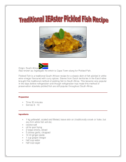


© Copyright 2026