
Glaucoma-II Free Papers
Glaucoma-II
Free Papers
Contents
GLAUCOMA – II
Transscleral Pars Plana Cyclodiode Laser in Neovascular Glaucoma...........325
Dr. Ajay Dudani
Role of Lens Extraction in Management of Primary Angle Closure Disease
(PACD).....................................................................................................................327
Dr. Tiwari Uma Sharan, Dr. Kapil Barange
To Compare the Outcome, Complications and Management of Complications of
Trabeculectomy with Ologen Implant Versus Trabeculectomy with MMC......330
Dr. Arijit Mitra, Dr. Rama Krishnan R, Dr. Mohideen Abdul Kadar PMT, Dr. Debarpita
Chaudhury
Ahmed Glaucoma Valve (AGV) Implantation and Intraocular Pressure Outcome
in Patients with Pre Existing Scleral Buckle......................................................335
Dr. Janvi Jhamnani, Dr. Jyoti Shetty
Secondary Glaucoma following Descemet’s Stripping Endothelial Keratoplasty
and Its Management..............................................................................................338
Dr. Samar Kumar Basak, Dr. Ayan Mohanta, Dr. Arup Bhaumik
Sturgeweber Syndrome – Difficult Surgical Proposition – Outcome, Complications
and Management of Complications.....................................................................342
Dr. Arijit Mitra, Dr. Rama Krishnan R, Dr. Mohideen Abdul Kadar, Dr. Debarpita Chaudhury
Effect of snake bite on Intra Ocular pressure....................................................348
Dr. Chikkabasavaiah Shivaprasad, Dr. Renuka Srinivasan, Dr. Benjamin Nongrum,
Pratyusha Ganne
Aniridic Glaucoma: Long Term Outcomes and Phenotypic Associations.....350
Dr. Viney Gupta, Dr. Amit Jain, Dr. Paromita Dutta, Dr. Ramanjit Sihota, Dr. Reena Sharma
Diagnosis and Management of Cyclodialysis Clefts.........................................353
Dr. Neha Shrirao, Dr. Balekudaru Shantha
An Analysis of Glaucoma Following Penetrating Keratoplasty and Its Relation
with Graft Failure...................................................................................................360
Dr. Stuti Kapur, Dr. D.J. Pandey, Dr.S.K.Satsangi, Dr. H.K. Bist
Comparative Study of MMC Augmented Trabeculectomy Vs An Ologen Implant
in Open Angle Glaucoma......................................................................................363
Dr. Devendra Maheshwari, Dr. Ankit Gupta, Dr. Ramakrishnan
155
Glaucoma Free Papers
GLAUCOMA - II
Chairman: Dr. Barun Kumar Nayak; Co-Chairman: Dr.Zutshi Rajiv
Convenor: Dr. Sushmita Kaushik; Moderator: Dr. Nangia Vinay Kumar B.
Transscleral Pars Plana Cyclodiode Laser in
Neovascular Glaucoma
Dr. Ajay Dudani
T
his is a study of hundred cases of neovascular glaucoma treated by
cyclodiode laser to pars plana region applied transsclerally.
MATERIALS AND METHODS
100 cases of neovascular glaucoma with rubeosis iridis due to various causes
like proliferative diabetic retinopathy,central retinal vein occlusion, advanced
Eales’ disease etc were treated with 810 nm diode laser (quantel medical,
France Iridis laser machine).
These patients were on maximum medical therapy dispite which the IOP was
in high levels of 40 to 50 mm.
Technique
In this procedure retrobulbar anesthesia is required due to the intraoperative
pain involved. The laser probe of Iridis is like a bullet probe with exposed
fiberoptic tip which is applied 3 mm posterior to the limbus. Laser settings are
1.5 to 2 milliwatt spots of 1 to 2 minute duration each. 30 to 50 applications are
done depending on the IOP eg 30 spots for IOP of 30. Fifty percent pop sounds
which signify rupture of pars plana epithelium, are tried for. 280 degrees of
the circumference is treated leaving super nasal quadrant virgin as a protocol,
to prevent excessive hypotony.
Postoperatively all patients receive analgesics orally and topical steroid and
atropine drops for 3 weeks. Few patients received a combination of intravitreal
avastin with cyclodiode laser.
RESULTS
All the treated patients responded very well in a few days the IOP reduced to
low teens. This was maintained in 80 percent of them with creeping increase of
IOP in 20 percent over a period of 6 to 12 months needing a repeat procedure.
Maximum pressure reduction was achieved in one month.
Complications and Mechanism of Action
In immediate postoperative period pain, white conjunctival burns, anterior
325
Glaucoma Free Papers
chamber flare and cells, hyphema are common. Transient IOP rise is common
and is controlled with acetazolamide hypotony is a uncommon complication.
In our series as we ablate over pars plana region which enhances aqueous
outflow by either transscleral filtration or uveoscleral outflow. We have
shown in postoperative ultrasound biomicroscope of the pars plana which
shows hollow punch out areas of the epithelium. In a monkey study at 3 mm
fom limbus treatment, histopathology shows enhanced uveoscleral outflow
showing tracer elements in enlarged extracellular spaces of ciliary stroma
from the anterior chamber to the suprachoroidal space.
In conclusion pars plana cyclodiode laser treatment is a very effective treatment
for neovascular glaucoma which acts by increasing aqueous outflow through
the uveoscleral pathway.
REFERENCES
1. Ando F, Kawai T, Transscleral contact cyclophotocoagulation for refractory
glaucoma,comparison of the results of pars plicate and pars plana irradiation.
Lasers Light Ophthalmol 1993,5:143.
Role of Lens Extraction in Management of
Primary Angle Closure Disease (PACD)
Dr. Tiwari Uma Sharan, Dr. Kapil Barange
L
ens continues to grow in size and hardness which can compromise the
filtration by angle closure in predisposed eye. Removing the lens creates
more space in anterior chamber and widens the angle, which may be enough
to achieve intraocular pressure (IOP) control. This prospective study on 30
eyes was undertaken to find out role of lens extraction in management of
primary angle closure disease (PACD) with visually significant cataract.
MATERIALS AND METHODS
Thirty eyes of 30 patients having PACD were recruited in this prospective
study. Based on the gonioscopic findings and optic disc evaluation, the patients
were divided into Three groups: Group A occludable angle (15 cases), Group B
primary angle closure (8 cases) and Group C primary angle closure glaucoma
(7 cases ).
After obtaining informed consent, all the cases underwent Phaco with foldable
IOL implantation under local anaesthesia and followed for at least 3 months.
Outcome measures included IOP, need for anti glaucoma medication, visual
acuity and gonioscopic appearance. Statistical analysis were performed using
SPSS software and epicalc 2000. Differences of mean +/- standard deviation
327
70th AIOC Proceedings, Cochin 2012
(SD) between pre operative and post-operative values were assessed by means
of the paired t-test. A p-value of less than 0.05 was considered statistically
significant.
RESULTS
The mean age of 30 patients was 54.20(+/- 4.81) years. The sex distribution was
20 female and 10 male patients, with 23 right eye and 7 left eyes. The findings
were compiled and analysed as follows:
Post OP IOP control:
Group A
IOP
Pre-opPost-op
Mean
19.06 16
S.D.
1.271.69
P-value0.000005
Student’s t test
5.61
Group B
Pre-opPost-op
IOP
Mean
31.2518.25
S.D.
1.833.28
P-value0.0000001
Student’s t test
9.79
Group C
Pre-opPost-op
IOP
Mean
37.1420.57
S.D.
2.545.85
P-value0.000017
Student’s t test
6.87
It was observed that in all the groups, P-value as well as student’s t test were
highly significant.
Post OP Visual acuity:
Pre-op BCVA
Post-op BCVA
Best Corrected ≤0.2 (i.e. ≥6/9) ≥0.3 (i.e. ≤6/12) ≤0.2 (i.e. ≥6/9)
Visual Acuity
(In Log MAR
equivalent)
Group A
0
15
10
≥0.3 (i.e. ≤6/12)
5
Group B085 3
Group C071 6
The causes of subnormal BCVA post operatively in group A were ARMD
328
Glaucoma Free Papers
(3 cases), Amblyopia (1 case) and Cystoid macular edema or CME (1 case).
In group B, subnormal BCVA was due to ARMD (1 case), CME (1 case) and
Amblyopia (1case). In group C, subnormal BCVA was due to glaucomatous
optic neuropathy (6 cases); besides, 3 cases were also found to have CME and
2 cases had ARMD.
Need for antiglaucoma medication in postoperative period
In post-op period, there was no need for anti glaucoma medication in group
A cases. In group B only 1 case (12.5 %) required antiglaucoma medication
(Timolol 0.5% bid). In group C, 2 cases (28.57%) required antiglaucoma
medication as combination therapy (Timolol and Brimonidine).
Post-Op Gonioscopic appearance
All patients in group A, 7 patients (87.5%) in group B, 5 patients(71.43%) in group
C were found to have PAS absent or less than 270 degree post-operatively. One
case (12.5%) in group B and 2 cases (28.57%) in group C showed >270 degree
peripheral anterior synechiae (PAS) in post-operative period.
Applying Chi Square test we assume that there is no significant difference
between expected and observed values (NULL HYPOTHESIS).
Groups
Total
Improvement Improvement Chi Square
Cases Expected Observed Value
P Value
A
15
15
15
0
1
B
8
8
7
0.125
0.9394
0.571
C
7
7
5
TOTAL
30
30
27
0.3
0.7516
0.8607
Degree of Freedom = 2
Applying Chi square formula, we observed that all the p values are > 0.05
which are non-significant indicating the difference is not significant but is due
to chance or other factors; Thus, null hypothesis is true.
DISCUSSION
The primary angle closure disease (PACD) has been classified as primary angle
closure suspect (PACS) or occludable angle, primary angle closure (PAC) and
primary angle closure glaucoma (PACG). The PACS is defined as nonvisibility of
pigmented trabecular meshwork in >270 degree with normal intraocular pressure
(IOP) and no peripheral anterior synechiae (PAS). When high IOP and /or PAS
are added to the PACS, the condition is termed as PAC and when glaucomatous
optic neuropathy and/or field defects are added to PAC, the condition is termed
as PACG. It is understood that the crystalline lens has a pivotal role in primary
angle closure (PAC), both in the pathogenesis of pupil block and by exacerbating
the effect of non-pupil block mechanisms such as peripheral iris crowding. Eyes
329
70th AIOC Proceedings, Cochin 2012
with angle closure tend to have shallow anterior chambers and thick, anteriorly
positioned lenses when compared with normal eyes. Removing the lens creates
more space in the anterior chamber and widens the angle, which may be enough
to achieve intraocular pressure (IOP) control.
The role of lens extraction as a treatment for angle closure has been debated for
many years. But with the knowledge that the lens is the single most important
contributing factor to the angle closure process, and having acquired the
technology and skills to perform relatively safe phaco surgery, should we now
be thinking about performing early lens extraction in angle closure patients
with the aim of preventing the development of glaucomatous optic neuropathy.
Theoretically, removing the lens at an early stage will deepen the anterior
chamber and open the angle, thus hindering the formation of peripheral
anterior synechiae (PAS) and improving the prospects for good long term IOP
control. In addition, many of these patients will eventually require surgery for
visually significant cataract at some stage.
From the observation in this study, it is evident that occludable angles are
cured by lens extraction very easily. Cases of PAC and PACG who do not
have extensive synechiae are also cured. However cases having >270 degree
PAS are the crucial cases. It is hypothetised that during the phaco surgery
injecting lot of visco during CCC may help in visco-dissection of PAS. Besides
lens extraction itself which may create more space in the chamber angle
making some more trabeculum available for the drainage of aqueous. In
present series 3 cases were found to have extensive PAS that persisted after the
lens extraction. These cases having >270 degree PAS required antiglaucoma
medication post-operatively. It is suggested that the goniosynechiolysis in
these cases at the conclusion of lens extraction should be done to improve the
outcome of the procedure. The goniosynechiolysis can be performed with the
help of cyclodialysis spatula after injecting visco in the anterior chamber.
To Compare the Outcome, Complications
and Management of Complications of
Trabeculectomy with Ologen Implant Versus
Trabeculectomy with MMC
Dr. Arijit Mitra, Dr. Rama Krishnan R., Dr. Mohideen Abdul Kadar P.M.T.,
Dr. Debarpita Chaudhury
T
rabeculectomy was introduced as far back as 1968 and is now the most
common operation for the treatment of glaucoma worldwide.1,2 However,
wound healing and scar formation may result in fibrosis of the bleb and
330
Glaucoma Free Papers
obstruction of the drainage fistula, eventually leading to bleb failure.4 Hence,
the inhibition of scar formation during the wound-healing process should
promote greater success.
Ologen Collagen Matrix is an artificial extracellular matrix (ECM) specifically
configured to support repair in connective and epithelial ocular tissue.
The implantation of this bioengineered, biodegradable, porous collagenglycoaminoglycan matrix implant in the subconjunctival space offers an
alternative method for controlling the wound-healing process following
filtration surgery, avoiding the complications of the administration of
antifibrotic agents and offering the potential for maintaining long-term
intraocular pressure (IOP) control.5,6
The purpose of this study was to compare the outcomes of trabeculectomy
with OloGen implant and trabeculectomy with MMC in patients requiring
glaucoma surgery for uncontrolled IOP. Other outcomes measured were the
number of postoperative medications used and any complications.
MATERIALS AND METHODS
A group of Glaucoma patients who needed surgical intervention was chosen.
The group comprised of a total of 64 patients. The members were divided into
two groups by random allocation to undergo Trabeculectomy with Ologen or
MMC. The Trab with Ologen group comprised 28 patients while the Trab with
MMC group had 36 patients. The minimum follow up period was 6 months.
The inclusion and exclusion criterias are mentioned below:
Inclusion Criteria
• Age 18 years or over
• Uncontrolled open-angle glaucoma
• Subject is willing to sign informed consent
• Subject is able and willing to complete post-operative follow-up
requirements
Exclusion Criteria
• Inflammatory eye diseases
• Angle-closure glaucoma
• Subjects having single functional eye
• Previous conjunctival surgery
• Known allergic reactions to ingredients of Ologen Collagen Matrix
• Excessive myopia (axial length (AL)> 27 mm or more than -10 diopters)
331
70th AIOC Proceedings, Cochin 2012
• Previous vitrectomy eye surgery
• Subjects do not consent to participate
Preoperative data included age, gender, IOP and number of preoperative
glaucoma medications. Postoperative IOP, number of postoperative glaucoma
medications and postoperative complications were recorded. In cases which
developed complications appropriate management options were taken to
effectively deal with the situation. Each patient was followed up for at least 6
months. Primary Outcome Measure was Intraocular pressure (IOP) reduction.
Secondary Outcome Measures included incidence of complications and
reduction in the number of Antiglaucoma medications.
RESULTS
The Age distribution was (Mean ±SD) 62.43±14.43 years in the Trab. with
MMC group and 61.22±12.24 years in the Trab. with Ologen group. The
gender distribution in the Trab. with MMC group comprised of 22 males
and 14 females while in the Trab. with Ologen group it was 16 males
and 12 females.
Table 1: Type of Glaucoma
Trab with MMCTrab with Ologen
POAG
2158.33
19 67.86
PXF
1233.33
6 21.43
PG
25.56
1 3.57
Post Traumatic Glaucoma
1
2
2.78
7.14
Table 2: Pre Operative IOP
Trab with MMC
Trab with OloGen
P value
Pre-op IOP (Mean±SD)
30.2±8.4
28.4±8.42
0.388
Range
22 – 44
21 – 36 Table 3: Comparison of pre-op and Post-op IOP between
the two groups
Sl No.
Trab with MMC
Mean
P value
SD
10.6
9.66
0.388
1
Pre op
2
1 week
8.44
3.12
8.58
3.45
0.854
3
1 m
12.46
4.74
12.56
3.43
0.993
4 3m
14.47
3.8313.02
3.560.345
5 6m
14.37
4.53 13.3
3.430.364
332
32.4
Trab with OloGen
SDMean
28.2
Glaucoma Free Papers
Table 4: Mean number of AGM’s pre-op and post-op
Trab with MMC Trab with OloGen
Mean
SD
Mean SD
Pre-op
3.40.6 3.2 0.3
Final FU
0.4
0.7
0.5
0.6
Table 5 : Comparison of Success between the two groups
Trab with MMC Trab with OloGen
n%
n%
Complete Success
29
80.56
22
78.57
Qualified Success
05
13.89
04
14.29
Table 6 : Complications
Trab with MMC Trab with Ologen
n %
n%
Hypotony
1 2.78
13.57
Shallow AC
1
2.78
0
-
Positive Seidel’s
0
-
1
3.57
Encapsulated Bleb
1
2.78
1
3.57
Implant Exposure
0
-
2
7.14
Blebitis
0 -
13.57
• Mean IOP was significantly lower than preop level at 1 and 6m(P<0.05)in both
groups (28.4±8.4 to 13.3±3.4-Ologen) and (30.2±3.4 to 14.3±4.5-MMCgroup).
• AGM use dropped from preop-3.4±0.6 to 6m postop 0.4±0.7 in Ologen and
3.2±0.3 to 0.5±0.6 (P<0.001)in MMC group.
• 6 months postop 22(78.57%) Ologen and 29(80.56%) MMC had complete
success and 04 (14.29%) Ologen and 04 (11.11%) MMC had qualified success.
Table 7: Management of Complications
Trab with MMC
Trab with OloGen
ManagementManagement
Hypotony
ConservativeConservative
Shallow AC
Conservative
–
Positive Seidel’s
–
Conservative
Encapsulated Bleb
Initiation of AGM
Initiation of AGM
Implant Exposure
–
Blebitis
–
Conjunctival autografting
Implant removal,
Aggressive therapy
333
70th AIOC Proceedings, Cochin 2012
DISCUSSION
Wound healing and scar formation causing fibrosis and obstruction of aqueous
outflow is one of the most common reasons for the failure of glaucoma surgery
.The survival of trabeculectomy has improved with use of intraoperative
antimetabolite as an adjuvant.7
However, the use of mitomycin and 5-FU have been associated with loss
of integrity of the conjunctival barrier, resulting in a thin walled avascular
drainage bleb, which may lead to hypotony and infection occurring years
after trabeculectomy.8 Other agents such as corticosteroids, growth-factor
inhibition and amniotic membrane have been applied to enhance the results
of antiglaucoma surgery.9
The use of Ologen has shown to offers the potential for a new means of
providing controlled resistance between the anterior chamber and the
subjconjuctival space in the early postoperative period, as well as maintaining
long-term IOP control by avoiding early scar formation and creating a loosely
structured filtering bleb.
In our study we found that the success rates were comparable between the
two groups and the IOP reduced from a pre-op value of 28.4±8.4 mm of Hg to
13.3±3.4 in the Ologen Group and from 30.2±3.4 mm of Hg to 14.3±4.5 in the
MMC group. The AGM use dropped from pre-op 3.4±0.6 to a 6 m post-op of
0.4±0.7 in the Ologen Group and from 3.2±0.3 to 0.5±0.6(P<0.001)in the MMC
group. However the complications were more in the Ologen group with 2 cases
(7.14%) developing Implant exposure and 1 case (3.57%) developing Blebitis.
In conclusion Ologen appears to be an alternative to the use of antimetabolites
for Trabeculectomy by preventing an early scar formation and creating a loosely
structured filtering bleb. However in view of the complications encountered
by us in our short term study period we would like to advise caution to the use
of Ologen. A proper case selection is very important and a meticulous surgical
technique with a good conjunctival hooding over the implant is necessary.
Long term follow up may yield further important and interesting information
with regards to the use of Ologen in Trabeculectomy.
REFERENCES
1.
Cairns J E: Trabeculectomy. Preliminary report of a new method. Am J Ophthalmol
1968;66:673–9.
2. Watson PG and Barnett F: Effectiveness of trabeculectomy in glaucoma. Am J
Ophthalmol 1975;79:831–45.
3.
Spaeth G L: A prospective, controlled study to compare the Scheie procedure with
Watson’s trabeculectomy. Ophthalmic Surg. 1980;11:688–94.
4.
Skuta GL and Parrish RK II: Wound healing in glaucoma filtering surgery. Surv
Ophthalmol 1987;32:149–70.
334
Glaucoma Free Papers
5.
Chen HS, Ritch R, Krupin T and Hsu WC: Control of filtering bleb structure
through tissue bioengineering: an animal model. Invest Ophthalmol Vis Sci. 2006;47:
5310–4.
6.
Hsu WC, Ritch R, Krupin T and Chen HS: Tissue bioengineering for surgical bleb
defects: an animal study. Graefes Arch Clin Exp Ophthalmol. 2008;246:709–17.
7.
Goldenfeld M, Krupin T, Ruderman JM, Wong PC, Rosenberg LF, Ritch R, Liebmann
JM and Gieser DK: 5-Fluorouracil in initial trabeculectomy. A prospective,
randomized, multicenter study. Ophthalmology 1994;101:1024–9.
8.
Franks WA and Hitchings RA: Complications of 5-fluorouracil after trabeculectomy.
Eye 1991;5:385–9.
9.
Sugar H S: Clinical effect of corticosteroids on conjunctival filtering blebs; a case
report. Am. J Ophthalmol 1965;59:854–60.
Ahmed Glaucoma Valve (AGV) Implantation
and Intraocular Pressure Outcome in Patients
with Pre Existing Scleral Buckle
Dr. Janvi Jhamnani, Dr. Jyoti Shetty
T
he risk of primary open angle glaucoma in general population is 1.1% to
3.0%, and 4.0% to 5.8% in patients with retinal detachment. Angle closure
glaucoma has been reported in 0.4% to 4.4% of patients after scleral buckling
procedure. Post-operative glaucoma has been reported in 2% to 48% of eyes
with retinal detachment treated with intraocular gas or silicone oil tamponade.
Patients with medically uncontrolled glaucoma who have undergone previous
scleral buckling procedure often present a difficult management challenge.
Conjunctival scarring and recession caused by previous retinal surgery
may decrease the likelihood of successful filtration surgery even with the
adjunctive use of an antimetabolite. Cyclodestructive procedures also are not
advisable due to their unpredictable results and significant complication rates,
especially in eyes with good visual potential.
Glaucoma drainage devises in such situation offer an important alternative
surgical approach .The purpose of this study was to describe surgical insertion
of Ahmed Glaucoma Valve and intraocular pressure control with it, in patients
with a pre-existing scleral buckle.
MATERIALS AND METHODS
A prospective, interventional short case series is hereby reported which
includes 5 patients of Bangalore West Lions Superspeciality Eye Hospital.
All 5 patients had pre-existing scleral buckle with uncontrolled glaucoma in
335
70th AIOC Proceedings, Cochin 2012
spite of maximum medical therapy. Data collected pre-operatively included;
demographics, duration from previous RD surgery and type of buckle used
in scleral buckle procedure. Routine preoperative evaluation of the patients
included vision, slit lamp examination, fundus evaluation by indirect
ophthalmoscopy and intraocular pressure recording by Perkins tonometry.
Surgical Technique
The surgical procedure performed was similar in all patients. Detailed
assessment of the eye was done to select a proper quadrant for AGV
implantation. A fornix-based conjunctival flap and tenons capsule was raised
in that quadrant. Adequate exposure of encapsulated episcleral encircling
band was done and the dissection continued till adequate space was created
for the implant above the buckle. Mitomycin C in concentration of 0.02mg/
dl was applied for 2 minutes to the undersurface of the conjunctiva. Ahmed
Glaucoma Valve (FP7) was then primed and positioned between the adjacent
recti muscle and over the scleral buckle. No trimming of the implant was
required. The implant was then sutured to the sclera. The tube was cut to
appropriate length and inserted in anterior chamber through scleral tract
created with 23G needle. Corneal/ Scleral graft was used to cover the limbal
portion of the tube and a water-tight conjunctival closure was ensured. Postoperatively all the patients were monitored for 6 months for IOP control and
complications.
RESULTS
Demographics of the patients is as follows
Case Age Gender Previous
Size of
Time from No. of
No.Surgery Buckle Prev Drugs
Surgery
1
14
M
SB + SOI
42 #
21mths
2 50 FSB
40#
18mths 342
3
24
F
SB + SOI
276 tire+ 240# 15mths
4
38
M
SB+Cryo+VIT42#
5 15MSB+VIT+SOI
40#
3
IOP
26
3
32
34mth
3
34
42mth
332
Age of the patients ranged from 14years to 50 years. All had undergone scleral
buckling procedure and 3 of them also had silicone oil implantation. The size
of the buckle varied in all the patients. Mean interval between scleral buckling
procedure and AGV implantation was 28 month, range being 15 months to 42
months. All patients were on maximum medical therapy on which the IOP
ranged from 26 mm Hg to 42 mm Hg.
The patients were followed for 6 months. Following were the pre and post
operative IOP values observed.
336
Glaucoma Free Papers
Case No.
Pre-Op IOP
1
Post-Op IOP
2614
2
4212
3
3212
4
348
5
3210
There was significant and consistent reduction in IOP observed at the end of
6 mth. This signifies the effectiveness of AGV implantation surgery. There
were no intra-operative complications.
Postoperatively one patient showed
hyphema
which
spontaneously
resolved in 2 weeks. 3 patients had
few emulsified silicone oil bubbles
in anterior chamber but no tubal
obstruction was noticed. Drainage
implant remained functional in all 5
patients. None of the cases showed any
hypotony or hypotonous maculopathy
changes, diplopia or motility problems,
AGV implant migration/exposure or epithelial in growth.
DISCUSSION
Refractory glaucoma after RD surgery can be exceptionally difficult to
manage especially if it is post-scleral buckling procedure. The conjunctiva is
frequently scarred or recessed because of previous surgery. Such eyes are at
high risk for filtering surgery failure even if adjunctive antimetabolite is used.
Drainage implants are only alternative in patients with pre-existing scleral
buckle with visual potential. Our study describes the alternative management
in these eyes by using Ahmed Glaucoma Valve. By doing this procedure, not
only adequate and consistent reduction of intraocular pressure was achieved,
but it was also found that dissection of fibrous capsule was possible inspite
of varied duration of previous scleral buckle surgery. No trimming of the
implant was required inspite of different sizes of buckles used in these eyes.
Adequate forward movement of conjunctiva with watertight closure of the
wound was possible in all patients. Cosmetically acceptable protuberance was
seen in all patients.
The prerequisites for good surgical success are 1) Presence of a relatively healthy
sclera and conjunctiva in at least one quadrant. 2) Proper dissection of the
fibrous capsule on the buckle to give adequate space for body of the implant.
In conclusion Ahmed glaucoma implantation is a good and effective treatment
337
70th AIOC Proceedings, Cochin 2012
option for management of refractory glaucoma in patients who had undergone
previous scleral buckling procedure. The presence of scleral buckle causing
mechanical impedance to surgical dissection and placement of Valve should
not deter us from doing this procedure.
REFERENCES
1.
Baerveldt Drainage Implants in eyes with a pre-existing sclaral buckle – Ingrid U
Scott, MD, MPH, Steven J Gdde MD et al. Arch Ophthalmol 2000;118:1509-13.
2. Modified Aqueous Drainage Implants in the treatment of complicated glaucoma
in eyes with pre-existing episclaral bands. M Fran Smith MD, J William Doyle MD
Ophthalmology 1998:105:2237-42.
3.
Schocket SS, Lakhanpal V, Richards RD. Anterior chamber tube shunt to an
encircling band in the treatment of neovascular glaucoma. Ophthalmology
1982:89:1188-94.
4. Sidoti PA, Minckler DS, Baerveldt G, et al. Aqueous tube shunt to a preexisting
episcleral encircling element in the treatment of complicated glaucomas.
Ophthalmology 1994;101:1036-43.
Secondary Glaucoma following Descemet’s
Stripping Endothelial Keratoplasty and Its
Management
Dr. Samar Kumar Basak, Dr. Ayan Mohanta, Dr. Arup Bhaumik
D
escemet’s stripping endothelial keratoplasty (DSEK) has become a
preferred surgical treatment for corneal endothelial decompensation
because it provides rapid visual recovery, uses a smaller wound size, minimizes
surgically-induced astigmatism and, most importantly, better maintains globe
integrity than penetrating keratoplasty (PKP).1
The relatively high rate of secondary glaucoma after PKP has significant
implications. It is a significant clinical problem because of its frequency
of occurrence, difficulty in diagnosis and monitoring, and complexity of
management. The incidence of glaucoma following PKP is reported to be 9–31%
in the early postoperative period and 18–65% in the late postoperative period.2
However, DSEK may also be associated with post-procedure intraocular
pressure elevation and secondary glaucoma, and presents unique surgical
challenges. Pupillary block glaucoma, steroid-induced IOP elevation, and
less commonly peripheral anterior synechiae development have also been
reported after DSEK.3
The purpose of the present study is to identify the incidence and causative
factors of secondary glaucoma after DSEK procedure with their management.
338
Glaucoma Free Papers
MATERIALS AND METHODS
It was a retrospective review of 550 consecutive DSEK procedures alone, or
in combination with cataract surgery with IOL implantation. All cases were
performed by three surgeons at a tertiary large eye hospital between 2006 and
2011, with an average follow-up of 2.3 years.
The entire patients had followed up on Day 1, Day 2, Day 7, after 3 weeks,
after 3 months, 6 months and then yearly. IOP was monitored with non contact
tonometry (NCT) during first 3 weeks and then measured by Goldmann
applanation tonometer (GAT). Eyes that developed IOP elevation above 21 mm
Hg after DSEK measured by NCT or GAT and requiring initiation or escalation
of glaucoma therapy and/or surgery were evaluated.
Numbers of patients developed secondary glaucoma were divided into two
groups: Early (within 3 weeks) and Late postoperative group. The preoperative
diagnosis, preexisting glaucoma, previous surgical intervention(s), and
additional procedure required during DSEK – all analyzed to find out
causative factors. The mode of treatment for secondary glaucoma and their
outcomes were also analyzed.
RESULTS
The types of operation and indications are given in Table 1 and table 2
respectively. A total of 118 eyes (21.5%) had some form of secondary glaucoma:
Early in 55 cases and late in 63 eyes.
Table 1: Types of indications in DSEK and incidence of secondary
glaucoma
Causes
Number 2ndary GL Percentage P value
Pseudophakic bullous keratopathy
24062 19.8%
PCIOL
202
44
21.8%
PCIOL vs ACIOL
ACIOL
38
18
47.4%<0.01
Fuchs’ dystrophy with PBK
PCIOL
ACIOL
78
67
11
15
9
6
19.2%
13.4%
PCIOL vs ACIOL
54.5%<0.01
Fuchs’dystrophy with cataract
129
11
8.5%
Post PK Failed graft
47
16
34.1%
<0.05
Aphakic bullous keratopathy
19
6
31.5%
<0.05
ICE syndrome
6
3
50%
<0.01
Cong hereditary endothelial
dystrophy
5
2
40%
<0.01
Posterior polymorphous dystrophy 4
0
0%
Failed DSEK (Re DSEK)
22
3
13.6%
Total
550
<0.5
<0.5
118
339
70th AIOC Proceedings, Cochin 2012
Table: 2 Types of DSEK operation performed and glaucoma
Operation Performed
Number
2ndary glaucoma
Percentage
DSEK Alone
382
94
24.6%
DSEK + Phaco/SICS and IOL
129
11
8.5%
DSEK + Anterior vitrectomy
14 6
42.8%
DSEK + IOL exchange/2ndary IOL
13
3
23.0%
DSEK + IVTA injection
3
1
33%
DSEK + SOR
2
1
50%
7
2
2.8%
DSEK + Trabeculectomy
Total
550118
In post-operative early period (within 3 weeks) of surgery the causes were:
pupillary block by air in 51 eyes (9.3%) and toxic anterior segment syndrome
(TASS) in 4 cases (0.7%).
Management in early cases: All cases of pupillary block were initially
managed by simple dilation of the pupil and injection Mannitol. 35 (68.8%) of
them responded with this treatment. The rest 17 cases they were taken into
the operation theatre and managed by manipulation through side-ports under
topical anesthesia.
In all cases, TASS occurred in DSEK combined with cataract surgery with
PCIOL. The predisposing factors were: 2 cases were with a new viscoelastics
and in two cases probably with trypan blue dye. All cases of TASS were
managed medically with copious topical steroids and anti-glaucoma
medications within 3 months.
In late post-operative period: Total number of 63 (11.5%) patients developed
secondary glaucoma. The main causes were given in Table 3. Among these,
the most common cause was steroid-induced secondary glaucoma. In some
cases more than one factor was responsible.
Table 3: Causes and number of patients developed Late secondary
glaucoma
Causes
Number of eyes
Total Percentage
Steroid responders
47 8.5%
Vitreous disturbances
(ACIOL/ABK/IOL exchange)
26 4.8%
Known POAG patient using medication
19
3.6%
Known PACG with YAG PI done
13 2.4%
Operated Glaucoma patients
7
1.3%
Late secondary angle closure glaucoma
6
1.1%
Known Irido Corneal Endothelial (ICE) syndrome
4
0.7%
The causes may be multifactorial in many cases
340
Glaucoma Free Papers
Fuchs’ dystrophy, rebubbling for donor dislocation, combined DSEK/cataract
surgery, or re-DSEK were not significant factors for development of elevated
IOP, but Vitreous disturbances (ACIOL/ABK/IOL exchange), Post PK failed
graft, history of previous glaucoma (POAG or ACG) or glaucoma surgery was
significant.
Management in late cases
The cases were managed medically or surgically. The Medical treatment
was with conventional medicines: like Oral acetazolamide, Beta blockers,
Prostaglandin analogues etc. Topical carbonic anhydrase inhibitor was
avoided in most of these cases. The intervened surgical methods were:
Trabeculectomy with MMC in 7 cases, Ahmed Glaucoma Valve (AGV) in 2
cases. Re-DSEK surgery was required in 7 (1.3%) cases. Most of these cases
were doing fine with clear graft till their last visit. Cyclocryopexy required in
2 cases of secondary absolute glaucoma: one was post trabeculectomy cornea
decompensation cases and 2nd case was CHED. Among the 6 cases of 360
degree donor adhesion (causing late secondary angle closure glaucoma, 3
cases required simple breaking of adhesion and three cases required Re-DSEK
after breaking the adhesion.
DISCUSSION
Current limited data suggest that DSEK may be a suitable surgical alternative
to PKP in patients with corneal endothelial disease and coexistent glaucoma
with or without prior glaucoma procedures with faster recovery and good
visual outcomes. Allen et al. showed that only significant risk factor for
development of elevated IOP in our series was a previous history of glaucoma
or OHTN.4
As in PKP, steroid-induced secondary glaucoma is also significant in DSEK.
In our study 6 of our cases developed secondary late angle closure glaucoma
which is not there in the literature. This is probably due to small cornea,
shallow AC in Asian and relatively large size donor which caused synechial
closure. ICE syndrome in late stage may not be a good indication for DSEK,
as it is a continuous disease process and 3 of our cases failed. There are few
studies available in the literature. All of them showed preexisting glaucoma
or OHT or operated trabeculectomy are the risk factor.5-6 As because of Indian
eye has shallow anterior chamber and higher prevalence of chronic ACG, we
have to be very careful in patients with YAG PI.
The limitations of this study are: i) Correlation with donor thickness, serial
anterior segment OCT maybe important here, ii) We do not have preoperative
IOP findings in most of the cases as majority of the cases had moderate to
severe corneal edema and only we had to rely on finger tension before surgery.
341
70th AIOC Proceedings, Cochin 2012
In conclusion, secondary glaucoma following DSEK is a significant problem
and most of the cases can be managed. A close postoperative IOP monitoring
is warranted in all cases of DSEK. As DSEK continues to gain popularity and
advance with more studies performed, our understanding of DSEK-associated
secondary glaucoma-related complications will become more complete.
REFERENCES
1.
Lee WB, Jacobs DS, Kaufman SC, Reinhart WJ, Shtein RM. DSEK: Safety and
outcomes: a report by the American Academy of Ophthalmology. Ophthalmology.
2009;116:1818-30.
2. Greenlee EC, Kwon YH. Graft failure: III. Glaucoma escalation after penetrating
keratoplasty. Int Ophthalmol. 2008;28:191-207.
3.
Banitt MR, Chopra V. DSAEK and glaucoma. Curr Opin Ophthalmol. 2010;21:144-9.
4.
Allen MB, Lieu P, Mootha VV, et al. Risk factors for intraocular pressure elevation
after DSAEK. Eye Contact Lens. 2010;36:223-7.
5.
Espana EM, Robertson ZM, Huang B. Intraocular pressure changes following
DSEK. Graefes Arch Clin Exp Ophthalmol. 2010;248:237-42.
6.
Vajaranant TS, Price MO, Price FW, et al. Visual acuity and intraocular pressure
after DSEK in eyes with and without preexisting glaucoma. Ophthalmology.
2009;116:1644-50.
Sturgeweber Syndrome – Difficult Surgical
Proposition - Outcome, Complications and
Management of Complications
Dr. Arijit Mitra, Dr. Rama Krishnan R, Dr. Mohideen Abdul Kadar P.M.T.,
Dr. Debarpita Chaudhury
T
he Sturge Weber syndrome (SWS) or encephalo-trigeminal angiomatosis
is a phakomatosis that is often associated with Glaucoma. In contrast to
other pediatric glaucomas, a significant larger proportion of patients with SWS
require surgical intervention when compared with patients having congenital
and aphakic glaucoma.1 Although surgical management of Glaucoma in
case of Sturge Weber Syndrome is fairly common for adequate intraocular
pressure (IOP) control, there remains much uncertainty as to the optimal
surgical choice for these patients.
The association of facial angiomas and glaucoma was first described by
Schirmer2 in 1860. Later on, Sturge3 reported an association between facial
angiomas and vascular malformations in the brain. In 1879, he described a
342
Glaucoma Free Papers
patient with a facial angioma and seizures that he speculated were secondary
to a similar lesion in the brain.4 In 1922, Weber5 reported the characteristic
curvilinear double-contoured line radiographic appearance of the intracranial
lesion. Since then, the triad of hemangiomas of the face, leptomeninges and
choroid has been referred to as the SWS.
Pathogenesis: The pathogenesis of SWS is poorly understood. During the
sixth week of intrauterine life, a vascular plexus develops around the cephalic
portion of the neural tube and under the ectoderm in the region destined
to become facial skin. In SWS, this vascular plexus fails to regress, as is
normal during the ninth week, resulting in the angiomatosis of the related
tissues. Variation in the locus of the persistent vascular plexus accounts for
its unilaterality or bilaterality and also for an incomplete SWS syndrome in
which the leptomeninges, but not the facial tissues, are affected. It has been
suggested that the trabecular meshwork anomalies seen on gonioscopy are
secondary to abnormal neural crest cell proliferation and migration that may
play an important role in the development of Glaucoma ssociated with Sturge
Weber Syndrome.
MATERIALS AND METHODS
A retrospective review of records of patients with Sturge Weber Syndrome
who presented to the Glaucoma Clinic at Aravind Eye Hospital Tirunelveli
between 2005 and 2010 was done. A total of 37 patients were noted. Of these
26 eyes of 26 patients were diagnosed to have Glaucoma associated with Sturge
Weber syndrome. Of these 24 eyes of 24 patients required some sort of surgical
intervention for the management of Glaucoma The records of these patients
who underwent surgery were collected and then analysed.
Preoperatively all the patients underwent a complete ophthalmic evaluation
which comprised of Visual acuity, slit lamp examination, IOP measurement
using GAT, Gonioscopy, Posterior segment examination and Humphreys
Visual Field examination wherever possible.
Two patients were found to have mild glaucomatous damage evident on Optic
disc examination and single field analysis of HFA and were thus managed
with antiglaucoma medications.
The indication for surgical intervention was medically uncontrolled glaucoma
in 15, documented progression of optic disc cupping or visual field defect in
5, and advanced glaucomatous atrophy and uncontrolled intraocular pressure
on initial presentation in 4 patients.
Different intervention methods included four primary modalities of
management. These were Trabeculotomy plus Trabeculectomy, Trabeculectomy
343
70th AIOC Proceedings, Cochin 2012
with MMC, Ahmed Valve implantation and Cyclophotocoagulation. Outcome
measure was success at last follow up.
To compare these results with those of others, the surgery was considered
a “complete success” when the intraocular pressure was ≤21 mm Hg and ≥5
mm of Hg without glaucoma medications, a “qualified success” when the
intraocular pressure was ≤ 21 mm Hg with antiglaucoma medications and a
“failure” when IOP was uncontrolled, progressive glaucomatous atrophy with
visual field loss was documented or when an eye required a further glaucoma
drainage operation, developed phthisis bulbi, or lost light perception at last
follow up. The success of the technique was analysed using a Kaplan-Meier
cumulative survival curve.
RESULTS
The gender distribution showed that a 14 ( 58.33%) of the subjects were male
and 10 ( 41.67%) were female.
Table 1: Table showing age at surgical intervention
Age at surgical intervention
Number (n =24)
Percentage
< 6 months
4
16.67
6m – 1 yr.
6
25
1yrs – 5 yrs.
5
20.83
5 yrs. – 15 yrs.
2
8.33
15 – 25 yrs. 4
16.67
25 – 45 yrs. 3
12.5
Table 2: Table showing indication for surgery
Indication for surgery
Number
Percentage
Medically uncontrolled Glaucoma
15
62.5
5
20.83
4
16.67
Documented progression of visual
field changes
Advanced cupping and uncontrolled IOP
Table 3: Pre operative Intraocular Pressure Range
IOP mm of Hg
20 – 30
Number
Percentage (%)
8
33.33
31-40
10
41.67
41-50
4
16.67
51-60
2
8.33
344
Glaucoma Free Papers
Table 4: Comparison of Pre-operative and post operative IOP
Sl. Follow n
Mean
noup Mean
SD diff
sig
Pre op
Follow
Pre op Follow
up up
1
1w
24 32.162113.2516
9.72
5.66 18.91P<0.001
2
1m 23 32.113214.2545
9.68
4.89 17.86P<0.001
3
6m 23 32.113214.2887
9.74
4.76 17.82P<0.001
4
12m 22 31.433115.1734
9.86
3.22 16.26P<0.001
5
24m 21 29.182215.5789
10.14
2.64 13.60P<0.001
6
48m 18 28.292815.6674
10.16
2.16 12.63 P<0.001
Table 5: Type of Surgical Intervention/ Procedure
Type of Surgical Intervention
Number
%
Trab +Trab
15
62.50
Trab with MMC
4
16.67
AGV implantation
3
12.50
2
8.33
DLCP
Table 6: Reduction of IOP in relation to type of procedure
S.no Type of
n
Procedure
Mean
SD
Mean P value
diff
Pre op Follow Pre op Follow
upup
1
Trab+Trab
15 32.27
15.42
7.86
3.57
16.85P<0.001
2
Trab with MMC
4
28.44
14.41
4.24
3.42
14.03 P<0.001
3
AGV
3
33.44 15.22
4.53
3.21
18.22P<0.001
4
DLCP
2
46.45
12.65
5.67
28.03P<0.001
18.42
The mean number of AGM’s was statistically significantly reduced in the post
operative period.
Table 7: Outcome
S.no
Number Percentage
1
Complete Success
9
37.5
2
Qualified success
10
41.67
5
20.83
3Failiure
345
70th AIOC Proceedings, Cochin 2012
Table 8: Complications
S.NoComplication
Number
Percentage
1RD
1
4.17
2
Suprachoroidal Hge
1
4.17
3
Conjunctival retraction
1
4.17
4Hyphaema
2
8.33
5
Scleral Thinning
1
4.17
6
Staphyloma formation
1
4.17
Table 9: Management of complications
S No
Complication
Management
1
RD
RD Surgery(PPV+SB+EL+FAE+SOI)
2
Suprachoroidal Haemorrhage
Conservative Management
3
Conjunctival Retraction
Scleral Patch Graft&Conj Autograft
4Hyphaema
Conservative
5
Scleral Patch Graft
Scleral Thinning
DISCUSSION
Management of Glaucoma associated with Sturge Weber Syndrome has
always been a therapeutic challenge and remains a difficult issue. Early
treatment with ocular hypotensive medications alone fails to control IOP
in most patients with glaucoma and SWS. After an extensive review of the
variety of surgical procedures performed in the literature, the treatment of
choice still remains controversial. Although goniotomy or trabeculotomy have
been recommended as the primary treatment in early-onset Glaucoma with
Sturge Weber Syndrome, the success rate of these procedures in the longterm control of IOP has been relatively low. Ultimately, most of these patients
require additional surgery to achieve their target IOP. Filtering surgery, such
as a trabeculectomy or valve implantation, has been used with greater success.
However, these procedures have been hampered by their risk for serious
complications such as choroidal hemorrhage or effusion and a high failure
rate in infancy.
In our study we found good IOP control with all the three types of procedures
employed namely Trabeculotomy with Trabeculectomy, Trabeculectomy with
MMC and Ahmed valve implantation. The mean number of AGM’ s was also
significantly reduced in the post operative period. The complete success rate
was 9(37.5%), the qualified success rate was 10(41.67%) and the failure rate was
5 (20.83%). In the cases with no visual potential DLCP was effective in the
346
Glaucoma Free Papers
reduction of IOP. There were a few complications noted post filtering surgery.
One case developed an RD which was treated with RD surgery. One eye
which developed suprachoroidal haemorrhage was managed conservatively
as were two cases of hyphaema. One case of conjunctival retraction required a
conjunctival autograft with scleral patch graft while a case of scleral thinning
was managed with a scleral patch graft.
Although filtering procedures have been associated with increased risk of
complications, they seem to be superior to goniotomy or trabeculotomy in longterm IOP control. From the study results we recommend Trabeculotomy plus
Trabeculectomy for patients in the age group less than 15 years. Trabeculectomy
with MMC can serve as a very good procedure for IOP control in patients in
the older age group. Ahmed Valve implantation was done in only 3 cases but
the results were very promising. We also recommend performing an inferior
sclerostomy intra-operatively to prevent suprachoroidal and expulsive
haemorrhage The lower complication rate, that makes goniotomy an attractive
surgical alternative, becomes less appealing when the need for multiple repeat
surgeries is appreciated. Most SWS patients will require filtering procedures
for adequate IOP control that ultimately exposes them to the same risk, if not
an increased risk, as the primary filter procedure group.
In conclusion Surgical intervention,primarily Trabeculotomy with
Trabeculectomy, Trabeculectomy and Ahmed Glaucoma Valve Implantation
gives good results on the long term follow up in cases of glaucoma in Sturge
Weber Syndrome. Cases with high IOP pre operatively should be managed
very carefully with an inferior sclerostomy to be done intra-operatively to
prevent suprachoroidal and expulsive haemorrhage. Cases with no visual
potential should be managed either conservatively or a cyclodestructive
procedure can be done.
REFERENCES
1.
Taylor RH, Ainsworth JR, Evans AR. The epidemiology of pediatric glaucoma: the
Toronto experience. J AAPOS. 1999;5:308–15.
2. Schirmer R. Ein Fall von Telangiektasie. Albrecht Bon Graefes Arch Klin Ophthalomol
1860;7:119.
3.
Sturge WA. A case of partial epilepsy, apparently due to a lesion of one of the
vasomotor centers of the brain. Trans Clin Soc Lond. 1879;12:162.
4.
Weber FP. Right-sided hemi-hypotrophy resulting from right-sided congenital
spastic hemiplegia, with a morbid condition of the left side of the brain, revealed
by radiograms. J Neurol Psychopathol. 1922;3:134.
5.
Resse AB. Tumors of the Eye. 3rd ed. New York: Harper and Row; 1976
347
70th AIOC Proceedings, Cochin 2012
Effect of snake bite on Intra Ocular pressure
Dr. Chikkabasavaiah Shivaprasad, Dr. Renuka Srinivasan,
Dr. Benjamin Nongrum, Pratyusha Ganne
I
ndia being a tropical country abounds in snakes of great varieties, poisonous
and harmless as well. Snake venom is a complex mixture of several enzymes
and proteins, toxic polypeptides, and inorganic components. It contains
numerous toxins, and their combined action has a more potent effect than
that of their individual effects. In general, venoms are described as either
neurotoxic or hematotoxic. Systemic manifestations of snakebites depend on
specific toxins that constitute the venom.1
To best of our knowledge and after pubmed search, there are few reports in
literature with regards to ocular complications following snakebites especially
in relation to intraocular pressure. However, subconjunctival hemorrhage,
hyphema, retinal and vitreous hemorrhages are well-known effects of viper
envenomation. Other complications that have been described included ptosis,
ophthalmoplegia, keratomalacia, uveitis, central retinal artery occlusion,
unilateral or bilateral optic neuritis, visual loss due to cortical infarction, and
macular infarction.2
The Russell’s viper (Daboia russelii) belonging to the viperidae is commonly
seen in India. Hemorrhagins – complement-mediated toxic components of
Viperidae snake venom –may provoke severe vascular spasm, endothelial
damage, and increased vascular permeability, oedema of the ciliary body
and iris along with a component of pupillary block preventing the aqueous
flow from the posterior chamber to the anterior chamber thereby causing
secondary angle closure glaucoma. A good response to a conservative
line of treatment indicates the importance of an early identification of rare
complications following viper envenomation along with the early initiation of
treatment in order to prevent disastrous visual (ocular) complications.3
Vitreous hemorrhage can also lead to secondary glaucoma, producing a “ghost
cell glaucoma”. Changes may begin in three days and are usually completed
within three weeks.4
To evaluate the ocular manifestations in cases of snake bite. Study the changes
in intraocular pressure (IOP) in cases of snake bite.
MATERIALS AND METHODS
We conducted a Descriptive Study of all snake bites that were admitted in
medicine ward in our hospital for a period of 10 months from April 2008
to January 2009. Consecutive cases of snake bites were included in our
study and most of them were seen bed side. Ocular examination was done,
348
Glaucoma Free Papers
anterior chamber depth was noted. Visual acuity was recorded once patient
returned to normal sensorium. Fundus examination was done to note
cupping changes and evidence of intraocular hemorrhage. IOP was recorded
using Schiotz tonometer. Systemic parameters including pulse rate, blood
pressure, temperature, blood urea, creatinine, hemoglobin, platelet count
and prothrombin time- International normalized ratio (PT-INR) values were
recorded. Patients were reviewed every day for the initial 3 days.
RESULTS
We studied 35 cases of snake bite which included 30 cases of haemotoxic and
five cases of neurotoxic snake bites. We could not asses visual acuity at initial
examination in most of the patients as the patients were in altered sensorium..
IOP levels ranged from 12.2 mm Hg to 48 mm Hg. Four patients developed high
IOP and had shallow anterior chamber, three of them were the ones that had to
be taken for dialysis. Those four patients had haemotoxic snake bite. Three of
these patients were managed with topical medications including 0.5% timolol
and pilocarpine eye drops. IOP in all the three returned to normal levels on 5th
to 7th day after snake bite. One patient succumbed to renal failure and expired
on the second day after snake bite. Blood urea ranged from 36 mg% to 120 mg%
and eight patients had to undergo dialysis. Those patients whose creatinine was
above 4mg% and potassium levels above 6 meq/l were taken for dialysis. Seven
out of eight patients who underwent dialysis, survived and one patient expired
due to renal failure. Antisnake venom was administered in all the patients.
DISCUSSION
We found that nearly half of the patients who required hemodialysis showed
an increase in IOP. Ocular involvement following hemotoxic snake bites is rare.
Raised IOP in such cases may be explained by the damage to the capillary
endothelium leading to edema of the ciliary body.3,5 A case report by Davenport
RC and Budden FH, describes optic atrophy following hemotoxic snake bite.
They hypothesized retinal ischemia following capillary endothelial damage
to be the cause.6 We would like to stress the importance of raised IOP as a
cause of retinal ganglion cell damage. In our study, 4 out of 35 patients of
snake bite (11.4%) developed raised IOP and was controlled with conservative
line of management using topical antiglaucoma medications. Prolonged
period of raised IOP along with retinal ischemia by capillary damage, may
cause a permanent damage to retinal ganglion cells. The long term optic nerve
changes in such patients needs to be evaluated by visual field evaluation. Role
of antisnake venom also needs to be evaluated. A larger population based study
may reveal a clear picture in the pathogenesis of snake bite induced glaucoma.
In conclusion ophthalmologist referral is recommended in cases of hemotoxic
349
70th AIOC Proceedings, Cochin 2012
snake bites in particular, those requiring hemodialysis to look for complications
like high IOP.
REFERENCES
1. Tungpakorn N et al. Unusual visual loss after snakebite. The Journal of Venomous
Animals and Toxins including Tropical Diseases 2010;16:519-23.
2. Rao BM. A case of bilateral vitreous haemorrhage following snake bite. Indian J
Ophthalmol 1977;25:1-2.
3.
Renuka Srinivasan, Subashini Kalaiperumal. Bilateral angle closure glaucoma
following snake bite. JAPI 2005;53:46-8.
4.
Ledy Rojas et al. Ghost Cell Glaucoma Related to Snake Poisoning. Arch Ophthalmol.
2001;119:1212-3.
5.
Mohd Haneef, Veena V.A. Acute angle closure glaucoma: A rare complication of
viper bite. KMJ. 2008;2:27-8.
6.
Davenport RC, Budden FH. Loss of Sight following Snake Bite. Br J Ophthalmol.
1953; 37:119–21.
Aniridic Glaucoma: Long Term Outcomes and
Phenotypic Associations
Dr. Viney Gupta, Dr. Amit Jain, Dr. Paromita Dutta, Dr. Ramanjit Sihota,
Dr. Reena Sharma
G
laucoma occurs in 50-75% of aniridic patients and typically presents in
adolescence.1 Since most aniridic glaucomas are difficult to treat with
medical therapy alone, surgery remains the mainstay of therapy for many.1
The surgical options include trabeculotomy3, goniosurgery4,trabeculectomy
with antimitotic agents, glaucoma drainage devices, and cyclodestructive
procedures. However prognosis remains poor.2 Also various ocular conditions
associated, requiring surgery (keratoplasty/cataract surgery) may affect the
visual outcomes among eyes with aniridic glaucoma.
Lee et al. studied 11 patients and concluded that aniridia has a poor prognosis
despite early and aggressive treatment and usually requires multiple surgeries.2
There is no literature that dwells on the long term outcomes of therapy among
aniridic glaucomas. The aim of the present study was to determine long term
outcomes of therapy among aniridic glaucomas and to evaluate factors that
determined long term visual outcomes.
MATERIALS AND METHODS
This study included 64 aniridic glaucoma patients (128 eyes) aged more than 5
years that were managed at a tertiary eye centre.
350
Glaucoma Free Papers
Detailed history and a clinical examination that included visual acuity at
presentation, slit lamp evaluation, baseline IOP using Goldmann applanation
tonometer, central corneal thickness, fundus evaluation was recorded.
Information regarding age at presentation, anti glaucoma medications,
ocular conditions other than glaucoma like nystagmus, corneal dystrophy/
keratopathy, cataract, retinal detachment and any surgical procedures related
to those were noted.
Visual outcomes were assessed .Parameters that were associated with visual
outcomes studied included age at presentation, familial history, baseline IOP,
associated ocular comorbidities and total number surgeries.
Statistical analysis: The means, standard deviations (SD) and 95% confidence
intervals were calculated for one eye of each patient. To look for factors that
determined final visual outcomes (dependent variable) a multiple linear
regression using the enter method for all variables was carried out. A p value
<0.05 was considered significant.
RESULTS
The clinical and demographic profile of patients studied is given in Table 1.
Only 31.7% had vision better than 6/60. 53.7% patients were blind as per WHO
definition. Only 5 patients (7 eyes) had visual acuity better or equal to 6/18. 22
out of 128 eyes were pl negative (17.18%) whereas another 11 eyes had vision
only PL+ (25.78% </= PL+). A total of 17 eyes underwent into pthisis (26.5% of
patients). Various co-morbidities were found to be associated with glaucoma,
details of which are given in Table 2. Table 3 includes various surgeries the
patients underwent. In eyes with visual acuity <3/60 (pthisical eyes being
excluded as they would have caused bias due to low pressure) mean baseline
IOP was 36.51 mm of Hg whereas those with visual acuity >/= 3/60 had mean
baseline IOP 23.41 mmHg (p=0.032). Visual acuity in eyes undergoing one
or less surgeries was 1.84 logmar units while those with 2 or more surgeries
had mean visual acuity of 2.417 logmar units (p=0.04). A regression analysis
was performed to look for factors associated with poorer visual outcomes. The
phenotypic variables included were age, sex, family history, mean baseline
IOP, total number of surgeries in each eye. Family history and high baseline
IOP were found to be associated with poorer prognosis.
Table 1: Clinical and Demographic Profile of Aniridic Glaucoma Patients
Number
64 patients (128 eyes)
Age range
5-47 years (15.86 ± 10.11)
Sex
Male
Female
42/64 (65.63%)
22/64 (34.37%)
351
70th AIOC Proceedings, Cochin 2012
Average follow up
1-17 years(7.69 ± 4.98 years)
Mean logmar Visual Acuity
In best eye In worst eye
In either eye
1.537 ± 0.739
2.41 ± 1.039
1.975 ± 0.999
Mean baseline IOP
22-69 mmHg (31.86 ± 14.0)
Average number of surgeries/patient
0-4 (1.6944 ± 1.26)
Average no. of surgeries/eye
0-3 (0.859 ± 0.78)
Incidence of pthisis bulbi (either eye)
26.56%
Incidence of Wilms tumour
1/64 (1.56%)
Table 2: Associated Ocular co-morbidities among aniridic glaucomas
Cataract
37 eyes (28.125%)
Keratopathy
10 eyes (7.8125%)
Nystagmus
18 eyes (14.06%)
Microphthalmos
2 eyes (1.5625%)
Retinal Detachment
4 eyes (3.125%)
Table 3: Surgery details per eye
Glaucoma interventions
60 (46.875% )
Trabeculectomy + MMC 34 (26.5625%)
Trabeculectomy + Trabeculotomy
14 (10.9375%)
Re-Trabeculectomy 5 (3.90%)
AGV 4 (3.125%)
DLCP
3 (2.34%)
Cataract surgery
41 (32.03%)
Penetrating Keratoplasty
5 (3.906%)
Vitreoretinal surgery
4 (3.125%)
DISCUSSION
Aniridic glaucoma is difficult to manage. Need for multiple surgeries; corneal,
lens and vitreo retinal make the management of these cases complex. We
analysed the visual outcomes of patients treated for aniridic glaucoma and
other ocular co-morbidities in a hospital based setting. Long term visual
outcomes after therapy in aniridic glaucoma patients remains poor even in a
tertiary center. In our study more than half of the patients were legally blind
after a follow up of 7 years. Higher baseline IOP and need for greater surgical
interventions were associated with poorer visual outcomes.
Aniridic glaucoma patients also have other ocular co-morbid conditions that
affect visual acuity. Apart from glaucomatous optic neuropathy, keratopathy
is the major cause of poor vision in these patients. Penetrating keratoplasty
352
Glaucoma Free Papers
also has poor visual outcomes due to the associated ocular surface disease.
Patients with familial aniridia had poorer visual prognosis.
Limitations of the present study included the retrospective nature and the fact
that many patients who did not regularly follow up or were lost to follow up in
between may have had uncontrolled IOP and could have compounded to the
progression of their disease. Lack of knowledge about the disease and poor
literacy also compound to this problem.
REFERENCES
1.
Grant WM, Walton DS. Progressive changes in the angle in congenital aniridia
with the development of glaucoma. Am J Ophthalmol. 1974;78:842-7.
2. Lee H, Meyers K, Lanigan B, O’Keefe M. Complications and visual prognosis in
children with aniridia. Journal of Pediatric Ophthalmology and Strabismus. 2010;47:20510.
3.
Adachi M, Dickens CJ, Hetherington J Jr. et al. Clinical experience of trabeculotomy
for the surgical treatment of aniridic glaucoma. Ophthalmology. 1997;104:2121-5.
4.
Chen TC, Walton DS. Goniosurgery for prevention of aniridic glaucoma. Arch
Ophthalmol. 1999;117:1144-8.
Diagnosis and Management of Cyclodialysis
Clefts
Dr. Neha Shrirao, Dr. Balekudaru Shantha
C
yclodialysis (CD) cleft is the separation of the insertion of the ciliary muscle
fibres from the scleral spur.1-3,5,7-13,15-17 This results in hypotony; and has
been exploited in the past as a surgical treatment for aphakic glaucoma.19 Other
causes include blunt trauma and anterior segment surgeries. Hypotony is due
to increase in the uveoscleral outflow, due to the formation of a new drainage
channel. Shallow anterior chamber, induced hyperopia, retinochoroidal folds,
optic nerve edema, retinal venous engorgement and stasis and choroidal
effusion are known complications.1,3
Clinical diagnosis of CD cleft is difficult. Gonioscopy is technically difficult
in a hypotonous eye due to presence of corneal edema or descemet’s folds.
Investigations such as ultrasonography,21 ultrasound biomicroscopy2,3,4,5,11,13,14
and anterior segment optical coherence tomography 21 help to confirm the
diagnosis in suspicious cases.
Various modalities have been described for the treatment of CD clefts.
Medications in the form of cycloplegics, topical steroids and oral steroids have
been advocated.3,4,5,6,11,13,20 Argon laser photocoagulation directly to cleft22,23
transscleral diathermy 1, cryotherapy 11 have been tried with variable results.
353
70th AIOC Proceedings, Cochin 2012
Numerous surgical procedures have been tried for this condition. These
include direct cyclopexy;2,3,26 indirect Cyclopexy24 as well as placement of
a capsular tension ring in the sulcus and suturing it to the sclera (for 3600
cyclodialysis).16 Gas endotamponade along with cryotherapy has helped to
achieve closure of the cleft.17,25 This procedure has also been combined with
vitrectromy.25 Anterior buckling procedures with or without cryotherapy
has proved to be successful.15 Though surgical procedures have had a good
outcome they come with their own set of complications like ciliary body
haemorrhage, endophthalmitis, erosion and induced astigmatism (in cases of
buckling procedures). Successful closure of the cleft may lead to transient rise
in the IOP in the immediate post operative period.
The aim of our study was to assess the utility of ultrasound biomicroscopy
(UBM) in the diagnosis of CD clefts and to assess the outcome of various
treatment strategies in terms of visual acuity and intra ocular pressure.
MATERIALS AND METHODS
A retrospective analysis of the case records was performed of 24 eyes of 24
consecutive patients who presented to our hospital between January 2006
to December 2010 and were diagnosed with Cyclodialysis (CD) cleft. Details
obtained from medical records included demographic details,best corrected
visual acuity (BCVA), details of slit lamp biomicroscopy, IOP with goldmann
applanation tonometry, gonioscopy with 4 mirror lens(Ocular Instruments
Inc), fundus examination with 20D and 90D lenses. Snellen’s visual acuity was
converted to LogMar for the ease of statistical analysis. Details of medical,
laser and surgical treatment were noted. Details of Ultrasound biomicroscopic
examination were noted as well.
Medical treatment was used in all patients; those patients who did not respond
to medications underwent laser and or surgical therapy..Primary surgery was
performed in patients who had other problems such as cataract or retinal
pathology which needed surgical treatment. 2 patients were left untreated due
to poor visual prognosis. 1 patient underwent cataract surgery alone as he
presented with normal IOP. 2 were lost to follow up. These patients were not
included in the final analysis.
Medical treatment was given in the form of cycloplegics, topical and/or
oral steroids. Oral steroids (Prednisolone) was given to those patients who
had Choroidal detachment on fundus examination or UBM. Non invasive
treatment was given by performing Argon laser photocoagulation (PHC) to
the cleft or Trans Conjunctival Cryotherapy (TCC).
A combination of surgeries were performed for CD cleft These included
3600 Belt Buckle with Trans Scleral Cryotherapy (TSC); Indirect and direct
cyclopexy and these were combined with procedures for other associated
ocular indications as required.
354
Glaucoma Free Papers
Complete success was defined as increase in IOP > 6 mm of Hg and
improvement in best corrected visual acuity(BCVA) by 2 lines on the snellen’s
visual acuity chart. Partial success was defined as increase in IOP > 6 mm
of Hg and improvement in BCVA by 1 line on snellen’s visual acuity chart.
Treatment was termed as failure if the IOP remained < 6 mm of Hg with same
or decreased visual acuity.
RESULTS
Patient demographics are depicted in Table 1. Most common cause of CD
cleft was blunt trauma in 12 (50%) patients. In 5 (20.8%) patients the cause
was unknown. In 2 patients each, the cleft was a consequence of Vitreoretinal
surgery and Phacoemulsification with intraocular lens implantation. The
mean visual acuity at presentation was 1.2620291 log units (0.9096).
AGE
Range
Mean
13-75 years
50.34 +/- 21.83 years
GENDER
Male : Female
17 : 7
PRESENTING IOP
Range
Mean IOP
0-14 mm of hg
3.92 +/- 4.117 mm of hg
PRESENTING VISUAL ACUITY
<6/60
6/18 to 6/60
6/6 to 6/12
10
9
5
GONIOSCOPY
No of Patients
Performed in
Cleft visualised in
Extent of cleft:1 clock hour
4 clock hours
19
6
3
1
Associated features on gonioscopy
Varying degrees of angle closure
7
>270 degrees of angle closure
2
Blood in schlemm’s canal
1
Vitreous in angle
1
Angle recession
1
The associated ocular findings included hypotonic maculopathy in 5 patients,
vitreous haemorrhage in 3 and traumatic cataract in one patient.
UBM confirmed the diagnosis of cyclodialysis cleft in the 21 patients. These
included the 13 patients in whom the cleft was not visualized on gonioscopy;
6 patients in whom the cleft was detected on gonioscopy (findings were
consistent with gonioscopy); and 2 patients in whom gonioscopy was not
performed. Most common location of the cleft was in the superior quadrant in
355
70th AIOC Proceedings, Cochin 2012
13 (54.16%) patients. The extent of CD clefts ranged from one clock hour in 10
(41.7%) patients;2 clock hour in 6 (25%) patients; 3 clock hours in 2 patients to 4
clock hours in one patient. Most commonly associated feature was supraciliary
effusion 12 (50%) patients and choroidal detachment in 4 (16.7%) patients. In 3
patients, the presence of a CD cleft could not be confirmed on UBM All these 3
patients presented with hypotony (IOP</= 6 mm of hg). One of these patients
was diagnosed to have an occult cleft; one was a silicon oil filled eye post
retinal detachment surgery and so was treated passively by deferring silicon
oil removal; one was started on medical treatment but lost to follow up. The
last 3 were excluded from further analysis
Medical treatment was started as a primary modality for 13 patients. We used
topical prednisolone acetate drops, cycloplegics (atropine or homatropine) and
oral Prednisolone on variable combinations. Oral Prednisolone was started in
patients with choroidal detachment. In 2 patients this treatment was successful
(1 partial, 1 complete) ; 2 were lost to follow up and it failed in the others. 4 of
these patients finally underwent surgery and laser/ cryotherapy was tried in
3. 2 patients were lost to follow up.
After medical treatment alone the mean IOP at presentation (3.7 mm of H
g) increased to 4.8 mm of hg. The mean increase in IOP was not statistically
significant (p=0.714)
Non invasive modes of treatment were tried in the form of Argon laser
photocoagulation directly to the cleft in 5 patients. It was a complete success
in 2 patients and failed in the other 3, which finally underwent surgery. One
patient underwent Trans conjunctival cryotherapy which failed to raise the
IOP.
The mean change in visual acuity in patients treated with medical or laser
therapy alone was 0.37826 log units; which wasn’t found to be statistically
significant (p=0.1637)
Surgical procedures were performed as a primary modality in 5 patients (due
to presence of other indications for surgery). In all these patients it proved
to be successful (4-complete, 1-partial). In the other 5 patients surgery was
carried out after failure of laser or medical treatment. It was successful in 3
patients (1-complete, 2-partial) and failed in 2 patients.
Most commonly performed surgery for CD cleft was 3600 belt buckle with
trans scleral cryotherapy to the cleft alone in 3 patients and combined with
other procedures in 2. Additional procedures performed were Vitrectomy
(3 patients); Lensectomy with Scleral fixated IOL (1 patient). It proved to be
completely successful in 4 patients and partially in 1. Direct cyclopexy was
perfomed in 3 patients, combined with cataract surgery in one. In one patient
it showed complete success and in one it showed partial success. Indirect
356
Glaucoma Free Papers
cyclopexy was performed in one patient. One patient underwent choroidal
drainage and anterior chamber reformation.
There were no complications encountered due to laser and no intra or
postoperative complications in any of the surgeries. We did not encounter any
IOP spike post operatively, post laser or after starting medical treatment.
The outcome in patients treated primarily with surgery was better as surgery
proved successful in all. The mean final IOP in those treated surgically was
9.8 ±3.73 mm of hg. The mean increase in IOP was found to be statistically
significant (p=0.006). The mean visual acuity from 1.515836(SD=1.0533) at
presentation to 0.78(SD=0.696) and it was statistically significant (p=0.028).
DISCUSSION
Cyclodialysis cleft is an uncommon condition and thus a few centres have
experience treating a large series of these patients. Very few studies with a
large sample size have been conducted on this condition and each of them
advocate different strategies; making it difficult to derive a conclusion
regarding the modalities for diagnosing and treating CD clefts. It should be
suspected in all cases of hypotony; post intraocular surgeries, post trauma and
even otherwise; as in 20.8% of cases presenting to our clinic did not have any
history that may have caused cyclodialysis.
Trauma was the most common cause encountered in our study (50%). UBM
proved very beneficial in the accurate diagnosis of cyclodialysis clefts in our
experience. It aided in diagnosis of cyclodialysis cleft in those patients in whom
gonioscopy did not reveal anything and comfirmed the diagnosis in the rest.
Features like Choroidal detachment,suprachoroidal effusion, angle recession
well delineated by UBM. This demonstrates the diagnostic precision of UBM
over gonioscopy and should ideally be performed in all cases of hyoptony.
A conservative approach towards management of CD clefts which are
smaller and which present early has been described in literature. Ormerod
et al.20 advocated the use of topical 1% atropine to help appose the detached
ciliary muscle to the sclera by prolonged relaxation. Use of steroids in this
condition has mixed views. Corticosteroids seemingly help in the resolution of
choroidal effusion and detachment and thus prove to be helpful. Combination
of topical/oral steroids along with cycloplegics has thus been suggested as a
primary therapy of choice in case of cyclodialysis.5,6 However, some authors
strongly maintain that steroids; topical or oral have no role in the treatment.2,6
Steroids also help in stabilising the macular edema.6 Goldberg6 has mentioned
that steroids may initially increase the IOP by decreasing trabecular flow
and inhibiting the ciliary body hyposecretion secondary to inflammation;
but at the same time they may prolong the patency of cyclodialysis cleft by
reducing the inflammation. This may have been the main reason for the high
357
70th AIOC Proceedings, Cochin 2012
rate of failure of medical treatment in our series as steroids were given to all
or patients along with the cycloplegic therapy. Also in case of post operative
CD clefts the tapering or stopping of topical steroids has been tried which
increases the inflammatory reaction causing adhesions at the cleft site. We
tried it only in one patient but it was not found to be successful.2
Non invasive procedures have been tried to promote closure of the cleft.
Argon laser photocoagulation directly to the cleft has been tried.2,6 The laser
presumably causes swelling of the choroid and blocks the aqueous flow; also
induces inflammation and leads to closure of cleft.6 Kuchle and Naumann26
have mentioned the probable causes why the laser treatment may fail. There
is continuous flow of aqueous humour through the uveoscleral route and
prevents scarring. Also, the antiproliferative factors in the aqueous wash
away the fibrin before clotting or adhesions takes place.26 In our study 2 out of
5 patients improved after laser.
Anterior scleral buckling has been performed with success for CD clefts.15
In our series 5 patients underwent this procedure. We assume that the belt
buckle along aided in cleft closure by causing constant apposition between the
detached ciliary muscle and the sclera; after scar induction with cryotherapy.
Kuchle et al.26 achieved cleft closure by direct cyclopexy in their series of 29
patients. We were successful in restoring the intraocular pressure in 2 out of 3
patients in our study.
Successful closure of CD clefts leading to a variable period of IOP spikes
postoperatively after medical,laser or surgerical therapy has been described in
literature.1,2,3,23,26 This has been attributed to the possible collapse of trabecular
meshwork during the period of prolonged hypotony and inability to re
establish the flow once IOP has normalised.23 However we did not encounter
raised IOP in any of our patients postperatively or after starting medical
therapy.
One of the drawbacks of our study was that we could not use closure of the
cleft as a criteria for success as neither goniocopic nor UBM documentation of
the closure of the cleft was present in any of the patients’ charts.
In conclusion we found UBM extremely useful for the accurate diagnosis and
delineation of Cyclodialysis clefts and other associated features. We suggest
that it must be performed in all cases of hypotony to rule out this potentially
treatable condition. Surgery was the most successful modality of treatment
and was useful in restoring the IOP and visual acuity in those patients in
whom conservative measures failed.
REFERENCES
1. Alexander S, Ioannidis AS, Barton K. Cyclodialysis cleft: causes and repair. Curr
Opin Ophthalmol. 2010;21:150-4. Review.
358
Glaucoma Free Papers
2. Hwang JM, Ahn K, Kim C, Park KA, Kee C. Ultrasonic biomicroscopic evaluation
of cyclodialysis before and after direct cyclopexy. Arch Ophthalmol. 2008;126:1222-5.
3.
Aminlari A, Callahan CE. Medical, laser and surgical management of inadvertent
cyclodialysis clefts with hypotony. Arch ophthalmol. 2004;122:399-404.
4. Jürgens I, Pujol O.Ultrasound biomicroscopic imaging of a surgically reattached
cyclodialysis cleft. Br J Ophthalmol. 1995;79:961.
5.
Vinay A shah, Ajit B Majji. Ultrasound biomicroscopic documentation of traumatic
cyclodialysis cleft closure with hypotony by medical therapy. Eye 2004;18:857-8.
6. Goldberg I. Traumatic cyclodialysis cleft. J. Glaucoma. 1998;7:430-3.
7.
Caronia RM, Sturm RT, Marmor MA, Berke SJ. Treatment of a cyclodialysis cleft
by means of ophthalmic laser microendoscope endophotocoagulation. Am J
Ophthalmol. 1999;128:760-1.
8.
Gnanaraj L, Lam WC, Rootman DR, Levin AV. Endoscopic closure of a cyclodialysis
cleft. J AAPOS. 2005;9:592-4.
9.
Saha N, MacNaught AI, Gale RP.Closure of cyclodialysis cleft using diode laser.
Eye (Lond). 2003;17:527-8.
10. Ceruti P, Tosi R, Marchini G.Gas tamponade and cyclocryotherapy of a chronic
cyclodialysis cleft. Br J Ophthalmol. 2009;93:414-6.
11. Malandrini A, Balestrazzi A, Martone G, Tosi GM, Caporossi A. Diagnosis and
management of traumatic cyclodialysis cleft. J Cataract Refract Surg. 2008;34:1213-6.
12. Mardelli PG.Closure of persistent cyclodialysis cleft using the haptics of the
intraocular lens. Am J Ophthalmol. 2006;142:676-8.
13. Bhende M, Lekha T, Vijaya L, Gopal L, Sharma T, Parikh S.Ultrasound
Biomiscroscopy in the diagnosis and management of cyclodialysis clefts. Indian J
Ophthalmol. 1999;47:19-23.
14. Gentile RC, Pavlin CJ, Liebmann JM, Easterbrook M, Tello C, Foster FS, Ritch R.
Diagnosis of traumatic cyclodialysis by ultrasound biomicroscopy. Ophthalmic
Surg Lasers. 1996;27:97-105.
15. Mandava N, Kahook MY, Mackenzie DL, Olson JL. Anterior scleral buckling
procedure for cyclodialysis cleft with chronic hypotony. Ophthalmic Surg Lasers
Imaging. 2006;37:151-3.
16. Yuen NS, Hui SP, Woo DC.New method of surgical repair for 360-degree
cyclodialysis. J Cataract Refract Surg. 2006;32:13-7.
17. Hoerauf H, Roider J, Laqua H Treatment of traumatic cyclodialysis with vitrectomy,
cryotherapy, and gas endotamponade. J Cataract Refract Surg. 1999;25:1299-301.
18. Hansen SM, Laursen AB. Visualised cyclodialysis: an additional option in
glaucoma surgery. Acta Ophthalmol. 1986;99:5-7.
19. Ormerod LD, Beerveldt G, Sunalp MA, Riekhof FT. Management of the hypotonous
cyclodialysis cleft. Ophthalmology. 1991;98:1384-93.
20. Mateo-montoya A, Dreifuss S. Anterior segment optical coherence tomography as
a diagnostic tools for cyclodialysis clefts. Arch Ophthalmol. 2009;127:109-10.
21. Joondeph HC. Management of post traumatic and post operative clyclodialysis
clefts with argon laser photocoagulation. Ophthalmic surg. 1980;11:186-8.
359
70th AIOC Proceedings, Cochin 2012
22. Harbin TS Js. Treatment of cyclodialysis clefts with argon laser photocoagulation.
Ophthalmology 1982;89:1082-3.
23. McCannel MA. A retrievable suture idea for anterior uveal problems. Ophthalmic
Surg. 1976;7:98-104.
24. Ceruti P, Tosi R, Marchini G. Gas endotamponade and cyclocryotherapy in a
hronic cyclodialysis cleft. Br J Opthhalmol. 2009;93:414-6.
25. Kuchle M, Naumann GO. Direct cyclopexy for traumatic cyclodialysis with
persisting hypotony. Report in 29 consecutive patients. Ophthalmology. 1995;
102:322-33.
An Analysis of Glaucoma Following Penetrating
Keratoplasty and Its Relation with Graft Failure
Dr. Stuti Kapur, Dr. D.J. Pandey, Dr.S.K.Satsangi, Dr. H.K. Bist
A
ccording to the World Health Organisation (WHO) calculations there are
about 10 million blind and visually disabled persons in India alone .Of
these, nearly two million persons have corneal blindness. For most of these
patients, the only option left for visual rehabilitation is corneal transplantation
or keratoplasty. Glaucoma following Penetrating Keratoplasty is a serious
clinical problem as it is the most common cause of irreversible visual loss
and second leading cause of graft faliure following Penetrating Keratoplasty.
Post PK glaucoma is defined as an elevated IOP> 21 mm Hg with or without
visual field loss or optic nerve head changes. An increase in IOP any time after
penetrating keratoplasty leads to endothelial cell loss with grave consequences
as the endothelial reserve is already low. Timely diagnosis with initiation of
appropriate treatment is mandatory to preserve optimal graft function and
optic nerve head function.
MATERIALS AND METHODS
Eighty one patients underwent keratoplasty from January 2009 to January
2011. Of these, 74 patients were selected according to the following criteriaInclusion Criteria
1. Pre operative IOP<21 mmHg.
2. Patients were able to be followed up for at least 6 months postoperatively.
Exclusion Criteria
1. Pre operative uncontrolled IOP(>21 mmHg).
2. Patients lost to follow up.
A detailed history of the patients was taken. Pre operative and post operative
360
Glaucoma Free Papers
examination(on Day 1, Day 7, 1 month and 6 months follow up) included:
1)
Visual acuity
2)
Intra ocular pressure was measured by Applanation Tonometry. In cases
not possible, schiotz tonometry or digital assessment of IOP was done.
3)
Complete slit lamp examination.
4) Fundus examination was not possible in most of the cases. A B-scan
ultrasonography was done in these cases.
5)
In cases with a clear cornea, angle assessment was done by gonioscopy. In
cases where gonioscopy was not possible, an anterior segment OCT was
done.
OBSERVATIONS-On the basis of history and pre-op examination, following
were the causes of PK in the hospital.
Etiology
No. of patients
Adherent leucoma following infectious keratitis
25
Adherent leucoma following trauma
10
Psuedophakic bullous keratopathy
18
Aphakic bullous keratopathy
5
Regrafting
7
Corneal dystrophies 9
The post operative IOP of the patient was as followsCases IOP
Normal
No.
49
%
66.24% Total
Raised 25
74
33.76%
100
Distribution of raised IOP in these 25 Post PK glaucoma cases in the follow up
period was as follows.
IOP (mm Hg) Day 1 Day 7 1 month 6 month
No.% No.% No. %No. %
Raised
11
44
07
28
11
Normal
14
56
18
72
14
44
16
56
9
64
36
The etiology of post keratoplasty glaucoma was found to be multifactorial.
However, the major causes leading to early post operative rise were as follows.
Cause of glaucoma Post operative inflammation
No.of cases
2
Hyphaema1
Crowding of the anterior chamber angle
6
Pupillary block
3
361
70th AIOC Proceedings, Cochin 2012
Elevated IOP in the late post operative period was mainly due to.
Causes of glaucoma No. of cases
Peripheral anterior synechiae 6
Steroid induced 7
• The relationship between the indication of PK and post-op glaucoma was
determined.
Etiology Total no.of cases Cases with
%
raised IOP Adherent leucoma 35
14
40%
Psuedophakic bullous keratopathy
18
7
38%
Aphakic bullous keratopathy 5
1
20%
Regrafting 7
2
28%
Corneal dystrophies
9
1
11%
The relationship between graft failure and glaucoma was also studied
No. of cases with failed graft
No. of cases with successful graft
Mean
Total no. of
cases(n= 74)
Cases with glaucoma
(n=25)
19
9
55
16
64.5024.50
SD
13.446.36
P value
0.053
DISCUSSION
1) According to the present study, the incidence of glaucoma following
penetrating keratoplasty is 33.75% (25/74). Out of these 25 cases,12(48%)
had raised IOP in the early post operative period, 9 of these (34%)
responded to medical treatment and the IOP came down to normal
within 6 months. 3 cases did not respond to treatment and IOP remained
elevated at 6 months. 13 other cases(52%) developed a rise in IOP in the
late post operative period. Thus ,16 cases(64%) had a raised IOP at the end
of study period.
2)
The etiology of post penetrating keratoplasty glaucoma is multifactorial.
In the early post operative period, angle compression is the major cause
whereas peripheral anterior synechiae and steroid induced glaucoma are
the important causes in late post operative period.
3)
Patients with leucoma adherence are at a greater risk of development of
glaucoma post operatively.
4)
Post PK glaucoma is significantly related to graft failure( P=0.053).
362
Glaucoma Free Papers
Comparative Study of MMC Augmented
Trabeculectomy Vs An Ologen Implant in Open
Angle Glaucoma
Dr. Devendra Maheshwari, Dr. Ankit Gupta, Dr. Ramakrishnan
L
ong-term intraocular pressure (IOP) control following trabeculectomy
may be limited by filtration failure, because of scarring at the level of the
conjunctiva–Tenon’s–episcleral interface,the scleral flap, its overlying episclera,
or the internal ostium.1 Adjunctive anti-fibrotic agents such as 5-fluorourail
or mitomycin C (MMC) have a potent inhibitory effect on postoperative
scarring and a positive effect on surgical success rates.2,3 These agents inhibit
fibroblastic proliferation in wound healing, thus preventing excessive scar
tissue formation at the level of the subconjunctival plane, which may otherwise
compromise aqueous flow and lead to surgical failure.
The Ologen collagen matrix implant is a disc-shaped porcine derived
biodegradable collagen matrix that has been developed to prevent excessive
scarring after trabeculectomy.
The purpose of this study was to compare the outcomes of trabeculectomy
with OloGen implant and trabeculectomy with MMC and without implant in
patients requiring glaucoma surgery for uncontrolled IOP in primary open
angle glaucoma. Other outcomes measured were the number of postoperative
medications used and any complications.
MATERIALS AND METHODS
Forty eyes of 40 patients were enrolled in the study and divided randomly
into two groups .Patients were randomized into two groups to receive either
trabeculectomy (control group) or trabeculectomy with OloGen Implant (study
group).
Patients with angle-closure glaucoma, posttraumatic,uveitic, neovascular, or
dysgenetic glaucoma were not considered for this study. Patients with an
allergy to collagen, preliminary conjunctival damage (trauma, vitreo–retinal
surgery, previous glaucoma surgery, and other) or those of 18 years of age
were excluded from the study.
In the MMC group (20 patients), trabeculectomy was performed according
to standard protocols. In the Ologen group (20 patients) after standard
trabeculectomy the implant was positioned on top of the scleral flap and no
MMC was applied. All the participants received a standardized regimen of
postoperative topical antibiotics and steroids.
The ophthalmologic examination included visual acuity testing, IOP
363
70th AIOC Proceedings, Cochin 2012
measurement by Goldmann applanation tonometry and slit lamp
biomicroscopic examination at each visit.Follow-up was continued for 12
months after surgery and included testing of intraocular pressure (IOP),
visual acuity and filtering bleb . Side effects and complications were recorded
during postoperative visits.
RESULTS
There were no significant differences between the groups in terms of age,
gender, eye laterality, preoperative IOP and number of prescribed topical or
systemic antiglaucoma medications.
The mean preoperative IOP was 26.7±7.9 mm Hg for all patients enrolled. At
1 day after surgery the mean postoperative IOP was 10.1±5.6mmHg for the
ologen group (P<0.001 compared with preoperative IOP) and 10.8±4.7mmHg
for the MMC group (P<0.001), respectively. At 1 year after surgery, the mean
IOP was 15.6±2.4mm Hg in the Ologen group (P<0.011, 43% reduction) and
10.5±4.1 mm Hg in the MMC group (P<0.011, 50% reduction), whereas 7
patients in the ologen group required topical medications.
A significant difference in IOP was determined from 1 month to 1 year after
surgery. During postoperative follow-up visits, we could not detect any
possible ologen specific side effects such as allergy or translocation of the
implant.
DISCUSSION
Penetrating anti-glaucomatous surgical procedures allow a powerful
reduction of IOP. Trabeculectomy as the standard procedure in penetrating
anti-glaucomatous surgery was introduced by Cairns in 1968. Various studies
demonstrated.
significant enhancement of success rates and postoperative IOP through
intraoperative use of MMC.
In summary of the previous studies, the use of the ologen implant promises
comparable IOP reduction after trabeculectomy and a lower risk profile
in comparison with the use of anti-metabolites, for example, MMC and
5-fluorouracil, although the use of ologen implant does not seem to offer a
significant advantage compared with trabeculectomy alone in our study.
Our study reveals that trabeculectomy with implantation of an ologen implant
is a safe method for penetrating anti-glaucomatous surgery and we did
not encounter any serious complications but IOP lowering was more with
trabeculectomy with MMC.
With regard to the need for topical anti-glaucomatous drugs in seven cases
in the ologen group (50% complete success) and the complete success rate of
364
Glaucoma Free Papers
100% in the MMC group, MMC is clearly more effective for augmentation in
trabeculectomy.
In conclusion the complete success rate using trabeculectomy with the ologen
implant is lower than that achieved by trabeculectomy with MMC.
REFERENCES
1.
Azuara-Blanco A, Katz LJ. Dysfunctional filtering blebs. Surv Ophthalmol 1998;43:
93–126.
2. Martini E, Laffi GL, Sprovieri C, Scorolli L. Low-dosagemitomycin C as an adjunct
to trabeculectomy. A prospective controlled study. Eur J Ophthalmol 1997;7:40–8.
3.
Wilkins M, Indar A, Wormald R. Intra-operative mitomycin C for glaucoma
surgery. Cochrane Database Syst Rev 2005 Issue 4. Art. no.: CD002897..
4. Dimitris Papaconstantinou, Ilias Georgalas,Trabeculectomy with OloGen versus
trabeculectomy for the treatment of glaucoma: A Pilot Study Acta Ophthalmol. 2010;
88:80–5.
5. Aptel F, Dumas S, Denis P. Ultrasound biomicroscopy and optical coherence
tomography imaging of filtering blebs after deep sclerectomy with new collagen
implant. Eur J Ophthalmol 2009;19:223-30.
365
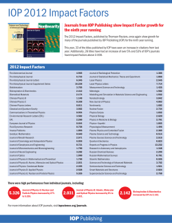


© Copyright 2026