
Type of article: Original - Hindawi Publishing Corporation
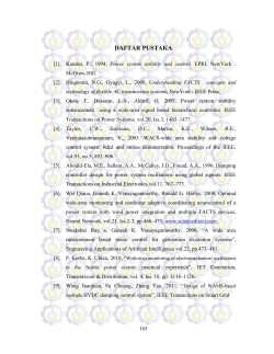
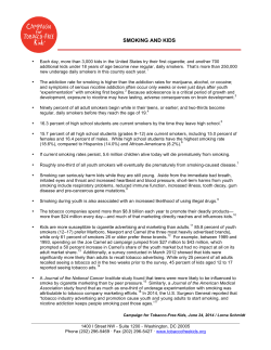
Abstract Page
Title of the article:
Protective The Effect of a 12-week Moderate Intensity Interval Training Program on the
Antioxidant Defense Capability and Lipid Profile in Men Smokers Cigarettes or Hookah. A
cohort study.
Abstract:
Aim. This study To examine d the impact of interval training program on blood lipids and
oxidative stress on the antioxidant defense capability and lipid profile in men smokers
cigarettes or hookah unable or unwilling to quit smoking. Methods. Thirty five participants
performed an interval training (2:1 work: rest ratio) 3 times a week for 12 weeks at an
intensity of 70% of VO2max. All subjects were subjected to a biochemical test session before
and after the training program. Results. The increase of Total antioxydant status (TAS),
Glutathion peroxydase (GPX) and α-tocopherol, is significant only for cigarette smokers
(CS) and hookah smokers (HS) groups. Similarly, tThe decrease of Malondialdehyde (MDA)
and the increase of Glutathion reductase (GR) is more pronounced in (CS) and (HS) smoker
groups compared to that of non-smokers (NS). (p <0.01; p <0.01 p <0.05 respectively ). All
Participants showed an increased Superoxyde dismutase (SOD) increase in NS, CS and HS by
10.1%, 19.5% and 13.3% respectively (p <0.001). it is respectively of 10.1%, 19.5% and
13.3% (p <0.001).The intermittent training induced a concentrations improvement of all lipid
parameters, but it is only significant at the level of height-density lipoprotein cholesterol
(HDL-C) and TC / HDL-C ratio of both groups CS and HS. Likewise, a significant
improvement of high-density lipoprotein cholesterol (HDL-C) and TC / HDL-C ratio was
observed in CS and HS groups (p < 0.05). Conclusion. Although the interval training program
our training program does not have a significant effect on blood lipid levels, it seems to be
very beneficial in the defense and prevention programs of oxidative stress.
Key-words: Cigarette smokers; hookah smokers; antioxidant; lipid; intermittent training.
1
Text
Introduction:
Smoking is the most strongly modifiable risk factor implicated in cardiovascular, respiratory
and cancer morbidity and mortality [1, 2]. The adverse effects of tobacco on health, and the
benefits of its stopping, have been widely demonstrated.
Smoking contains an abundance of free radicals and pro-oxidant species known to negatively
influence human health [1]. Increased production of free radicals from tobacco is recognized
because of the more than 4,000 chemical substances found in tobacco [2]. Therefore, illhealth related to smoking may be linked to increased production of free radicals.
Cigarettes and hookah consumption has risks of addiction, illness or death. Koubaa et al. [3]
evaluated the consumption harms of hookah in relation to cigarettes among sedentary adults,
by measuring biochemical and cardiorespiratory parameters. This study reinforces the
evidence that the hookah consumption
is associated with exposure to toxic substances and
has adverse effects on the cardio-respiratory and metabolic quality, and produces in some
cases the same effects as cigarettes. A lot of evidence suggests that hookah has harmful
effects similar to cigarettes smoking.
Oxidative stress having a strong association with many disease states including cardiovascular
diseases (CVD), have recently been presented [4, 5]. Oxidative stress describes a state of
physiological stress in the body that arises from exposure to high levels of reactive oxygen
species (ROS) to an extent that overwhelms the antioxidant defense system [6]. Cigarette
smoking exacerbates ROS formation [7], evidenced by the increase in oxidative stress
biomarkers in smokers compared with no smokers [8–9]. Free radicals can interact with
molecules in the body and damage various cell components such as DNA, protein and lipids,
giving rise to various disease states [10]. Halliwell. B et al [11] suggest that oxidative damage
of cell components has been implicated in the pathogenesis of a wide variety of diseases, most
notably heart disease and cancer. A growing body of evidence suggests that oxidation of lowdensity lipoprotein (LDL), are believed to promote atherosclerosis [12]. Recent studies
suggest that free radicals may be involved in the development of pulmonary disorders such as
asthma [13]. Other major pathologies that may involve free radicals include neurological
disorders and cataracts [14].
In preventing or slowing the progression of both heart disease and some forms of cancer,
previous studies suggest that antioxidants may play a pivotal role, [15, 16] and a Low blood
levels of antioxidants is related with increased cancer risk. [16] Cigarettes smoking or hookah
have an increased risk of CVD, possibly by increased production of free radicals (FR) risk.
2
Smoking aggravates the formation of FR and poses a significant oxidative stress [6]. Previous
surveys indicate that smokers have oxidative stress levels of rest that are higher compared to
non-smokers and this can be explained in part, by reduced blood antioxidant capacity [17,
18]. Cigarette smoke-induced oxidative stress poses a significant human health concern,
especially as related to cardiovascular disease [19].
So far, interventions to reduce harmful effects of tobacco have focused on the use of
innovative tobacco products and the reduction of tobacco consumption and pharmaceutical
drugs. Therefore, there is a need to expand the range of potentially effective strategies to
reduce the harmful effects of tobacco in smokers who are unable or unwilling to quit. Our
objective is that physical activity has the potential to become such a strategy so that it can
delay the onset of disease and premature death initiated by tobacco. Our objective is whether
physical activity has the potential to become such a strategy so that it can strengthen the
antioxidant defense system and improve the lipid profile and therefore to overcoming these
known unrest initiated by tobacco.
The lipid is an integral part of the search for the factors of cardiovascular risk. Previous
studies have shown that smoking cigarettes or hookah, product significant increase in LDL-C
and associated with a high TG concentration and a reduced HDL-C [20, 21]. Other published
reports suggest that cigarette smoking has been found to alter the lipoprotein levels [22].
Also, the effects of elevated lipid levels and changes in lipoprotein among cigarette smokers
were demonstrated earlier [23, 24, 25]. Other studies also show an increase in HDL-C, a
decrease in triglycerides and low LDL-C values in physically active populations. In contrast,
in the ability to control and to address this disturbance, several prior studies showed the
physical activity effect on lipid profile improvement. The results of Thune et al study [26],
showed a dose-dependent inverse relationship between physical activity and lipid parameters.
They found a significant decrease in total cholesterol (TC), triglycerides and TC / HDL ratio,
thereby an increase of HDL-C.
Most studies consider that continuous training is beneficial for maintaining cardiovascular
health. However, the exercise with intervals can be more effective [27]. They suggested that
the intermittent exercise of moderate intensity is beneficial for cardiovascular condition.
DeBusk et al. [28] found that subjects who exercised intermittent training sessions showed a
gain of fitness and improvement in their blood lipid profile. Previous studies have suggested
that short intermittent periods of walking improve plasma lipoproteins and blood pressure
compared to a continuous training session [29]. In addition McFarlane et al. [30] suggested
3
that training periods with intermittent exercise led to a significant improvement in
cardiovascular fitness. Physical activity reduces, therefore, the risk of cardiovascular disease
and prevents strokes [29].
Exercise may have the potential to mitigate some of the negative consequences of smoking
cessation. Bock et al. [31] and Marcus et al. [32, 34] have conducted a number of studies to
examine this issue and they suggested that vigorous exercise (60-85% of heart rate reserve)
can be a useful aid to quit smoking. To the best of our knowledge, there is a lack of empirical
evidence to document if there are beneficial effects on the physiological symptoms of people
who continue smoking.
Anaerobic training has been shown capable of increasing muscle antioxidants (SOD, Gpx,
GR) and strengthening the body against new oxidative attack [35, 36]. Some studies have
reported a decrease in antioxidant defenses of the body, which could be due, in part, to an
excessive production of RL by cumulative effect of anaerobic exercise [37, 38], or to a food
intake, least rich in antioxidants during the intervention period of participants [39, 40]. Within
certain limits, it seems possible that anaerobic training can result in attenuation of rest
oxidative stress similar to the aerobic training.
Although most training programs have involved continuous aerobic and anaerobic training
sessions of long durations, recent work demonstrated that interval training can stimulate
similar, if not superior, changes in cardiovascular function in both healthy [41] and clinical
populations [42, 43]. The potential stimulus provided by intermittent training at a high
intensity yet often at a reduced total exercise volume; offers an efficacious alternative to
continuous training.
Training benefits are optimized when programs are planned to meet the individual capacities
of the participants. Therefore respiratory capacity must be taken into account in order to meet
individual needs in training of sedentary smoker participants. The interest in assessing
intermittent training was based upon previous experience of the acceptance of this training
type in clinical practice [44] and because interval exercise resembles the daily life activity
pattern in smokers more than continuous exercise.
Interval exercise seems to be an important factor for improving aerobic capacity,
cardiovascular function and quality of life in smoker participants. That may have important
implications in antioxidant capacity and serum lipid concentrations.
The aim of O our work represents the first study is to examine the effects of intermittent
training on blood lipids and oxidative stress antioxidant defense capacity in sedentary male
4
smokers, and to check the difference of these individual effects of training among cigarette
smokers compared to hookah.
Subjects and Methods:
Our population was composed of adults matched in gender and age from the same ethnicity
and socio-economic environment. In fact, thirty five sedentary men smokers and non-smokers
in good health from the general community of Tunisia (North Africa), which belongs to the
public function (profession does not require physical exertion), volunteered to participate in
this study and were recruited within pharmacology laboratory of the Faculty of Medicine,
university of Sfax, Tunisia. Their mean values of age, height and weight were respectively
44.7 ± 4.5 years, 174.3 ± 2.3 cm, 71.3 ± 2.7 kg. The anthropometric and physical
Characteristics of participant are shown in [Table 1].
Table 1: physical characteristics of smokers and non-smokers (Means±SD)
78 ±4
Cigarette
Smokers
91 ±2
v VO2max (km.h-1)
10.5 ±0.9
11.6 ±0.7
9.9 ±0.6
VO2max (ml.min.kg-1)
37.5 ±1.6
38.9 ±2.5
36.6 ±1.2
Non Smokers
Rest HR (beats.min-1)
Hookah Smokers
93 ±4
Legend: VO2max = maximum oxygen uptake; vVO2max = velocity at maximum
oxygen uptake; Rest HR = rest heart rate.
Table 1: Anthropometric and physical Characteristics of participants (Means±SD)
Parameters
Non-smokers
Cigarette smokers
Hookah smokers
ANOVA
Age (yrs)
44.5 ±1.3
45.5 ±1.7
44 ±1.7
F(2;33) = 0.84 ; p = 0.47; ηp2 = 0.17
Height (cm)
173.8 ±1.3
175.8 ±2.8
173 ±1.6
F(2;33) = 0.91 ; p = 0.43; ηp2 = 0.15
Weight (kg)
71.4±2.2
72.7 ±3
69.6 ±2.2
F(2;33) = 1.65 ; p = 0.24; ηp2 = 0.25
BMI (kg.m-2)
25.1±1.1
24.4±1
24.7±1.2
F(2;33) = 1.2 ; p = 0.31; ηp2 = 0.07
Resting Heart Rate (bpm)
78±4
91±2***
93±4***
F(2;33) = 66.52 ; p < 0.001; ηp2 = 0.80
Resting SBP (mmHg)
131±3
138±3***
138±3***
F(2;33) = 27.91 ; p < 0.001; ηp2 = 0.63
Resting DBP (mmHg)
85±6
87±5
86±4
F(2;33) = 0.48 ; p = 0.62; ηp2 = 0.03
VO2max (ml.min.kg-1)
37.5±1.6
38.9±2.5
36.6±1.2##
F(2;33) = 4.79 ; p = 0.015; ηp2 = 0.22
Legend: BMI = body mass index; Resting SBP = systolic blood pressure at rest; Resting DBP = diastolic blood
pressure at rest; VO2max = maximum oxygen uptake; *** = significant differences compared to non-smokers at
p< 0.001; ## = significant differences compared to cigarette smokers at p <0.01.
Participants were admitted to the training program after approval of cardiologist physician.
They were normolipidemic (fasting triglycerides <1,7 mmol/L), nonobese. No subject used
nutritional supplements or medications. Presence of any kind of disease (based on history,
medical examination, and exercise stress testing) and involvement in regular physical activity
5
or exercise program during the previous 12 month were also exclusion criteria. On the basis
of these criteria, 7 subjects from 42 were excluded. Finally, 35 subjects were included in
subsequent analysis.
After receiving a complete verbal description of protocol, risks and benefits of the study,
subjects provided written consent to an experimental protocol approved by the Researsh
Ethics Committee of the Faculty of Medicine, University of Sfax, Tunisia.
Cigarette smokers and hookah were recruited according to the number of cigarettes and
hookah per day and their career period. We consider cigarette smokers all subjects with
consumption greater or equal to 10 pack-years (PY) and an average score of tobacco
dependence of 4.33 ±1.67, measured by the Fagerström nicotine dependence test [45]. In the
absence of specific international codification, we quantified hookah consumption, as in the
study of Kiter et al. [46] in NA and kg of cumulative tobacco. The tobacco used in a hookah
weighs between 10 and 25 grams [47]. In fact, regular hookah smoker subjects are those
having consumption greater or equal to 5 Hookah- years (HY) [48].
They were divided into three equal groups, and they were performed an interval training 3
times a week for 12 weeks at an intensity of 70% of VO2max. A cigarette smokers group
(CS) (n = 11), a hookah smokers group (HS) (n = 12) and other nonsmokers group (NS) (n =
12). All subjects were subjected to a biochemical and metabolic test session before and after
the training program. The session includes a biochemical analysis and oxidative stress review
anthropometric and physical tests, a biochemical analysis and antioxidant status review. All
these measurements were performed by the same examiner to avoid methodological
uncertainties. An exercise testing was performed before program, to quantify the training
individual loads.
6
Body weight was measured to the nearest 100 grams using a calibrated electronic scale
(TANITA TBF.350 model), and height was measured to the nearest 1mm with a fixed
stadiometer.
Analyses were performed in the laboratory of Pharmacology, Faculty of Medicine of Sfax.
Smokers were instructed to refrain from smoking for a one hour period prior to reporting to
the lab as suggested by Dietrich et al. [49].
Venous blood samples (ante-cubital vein) were performed in dry tubes under basal conditions
(8 am morning). After centrifugation, the sera were frozen at -80 ° C until analysis. Total
cholesterol (TC), triglycerides (TG), and high-density lipoprotein cholesterol (HDL-C) were
measured in all subjects after a 12-hour overnight fast using the standardized techniques
described by Wegge et al. [50] Low-density lipoprotein cholesterol (LDL-C) was calculated
with the Friedewald formula [51]:
[LDL = TC - HDL - (TG /2, 18)]
Plasma concentrations of SOD, GPx and the TAS were measured spectrophotometrically
using a spectrophotometer type DU-640 (Beckman Instruments, Inc.., California. United
States) and the dosage kits of TAS and antioxidant enzymes (SOD and GPx) were learned
from Randox laboratories.
VO2 max and max heart rate measurements during exercise were examined at the triangular
test with speedwalk (COSMED Pulmonary-Function Equipment 37 Via dei Piani di monte
Savello I-00040 Rome ITALY). This dynamic and maximum test, until fatigue, consists in
increasing the speed of 1km /h every 2 min, after warm up for 5 min with a 6km / h speed.
Heart rate and VO2 during the test were measured using an analyzer (version 1.2 PRO Fit
mate COSMED).
Subjects of three groups underwent an intermittent training program that was consisted of 3
sessions per week lasting 30 min, during a 3-months period. The intensity of the exercise was
controlled by time and traveled distance. All warm-ups before training should be between
50% and 60% of maximum heart rate for a period of about 10 minutes.
The subjects were trained participated in training sessions at evening using intermittent
exercises. It is to run periods of 2 min race interspersed with recovery periods of one minute
(2:1 work: rest ratio). The exercise intensity was 70% of VO2max. The load increase during
the training period was provided by the repetition number. The training load was insured by
time and traveled distance and controlled by beep sounds: T= d/V (T: the time between two
7
studs; d: distance between two studs; V: proposed speed). All participants did their best and
they successfully completed the training period and no recorded absences during all training
sessions. Furthermore, we have verified that there was no involvement in physical activity or
exercise program elsewhere during the 12 week training period.
All statistical tests were processed using STATISTICA Software (StatSoft, France). All
values The data are expressed as mean ± SD (standard deviation). After normality verification
with the Shapiro-Wilk’s w test, and homogeneity of variances with Levene’s test, parametric
tests were performed. One-way ANOVA was used to indicate inter group differences in the
baseline subjects’ characteristics. Inter and intra-group comparisons of the variables were
made by two-way ANOVA (group vs. training) with repeated measurements. Least
Significant Different (LSD) post-hoc analysis was used to identify significant group
differences that were indicated by one-way and two-way ANOVA.
Effect sizes were
calculated as partial eta-squared (ηp2) to estimate the meaningfulness of significant findings.
A probability level of 0.05 was selected as the criterion for statistical significance.
Paired Student’s t-test was used to analyze within group changes and unpaired Student’s ttest was used for comparison between the three groups (NS), (CS) and (HS). ANOVAs were
used to compare the responses of different groups, in pre and post the program. Tukey’s post
hoc test was used to compare means and to evaluate the relationships between various
parameters. A p value of less than or equal to 0.05 was considered statistically significant.
Results:
Through the anthropometric and physical measurements of participants [Table 1], no
statistical differences were noted between 3 groups for the four above variable: age, Height,
Weight and BMI (p>0.05).
The LSD post-hoc test for means comparisons allowed us to conclude that the two
groups CS and HS have resting heart rate and resting SBP similar and significantly higher
than those of non-smokers (p <0.001). Furthermore cigarette smokers have developed a
VO2max statistically higher than hookah smokers.
Before our training program, most of the antioxidant blood concentrations were similar in all
of smoking subjects cigarette and hookah and different to those of non-smokers [Table 2].
Defense capability of SOD and the oxidative stress indicator level (MDA) of hookah smokers
were significantly superior to those of cigarette smokers (p <0.05). No significant differences
in GPX and TAS concentrations of the three group subjects.
8
Table 2: Antioxidant concentrations before training (Means±SD)
Parameters
P-values ANOVA
Non Smokers
Cigarette Smokers
Hookah Smokers
GPX (U.gHg )
37.12 ±2.6
34.84 ±4.31
33.84 ±5.07
F(2;33) = 2.01 ; p = 0.15; ηp2 = 0.11
SOD (U.gHg-1)
1651.3 ±105.2
1432.1 ±171.2***
1545.1 ±105.9*
F(2;33) = 8.1 ; p = 0.0014; ηp2 = 0.33
MDA (µmol.l-1)
1.454 ±0.17
1.517 ±0.095
1.663 ±0.111***#
F(2;33) = 8.1 ; p = 0.0014; ηp2 = 0.33
GR (U.gHg-1)
10.46 ±2.01
8.3 ±1.55**
8.66 ±1.6*
F(2;33) = 5.43 ; p = 0.009; ηp2 = 0.25
TAS (mmol.l-1)
1.8 ±0.02
1.78 ±0.01
1.79 ±0.02
F(2;33) = 2.7 ; p = 0.082; ηp2 = 0.14
α-tocopherol (µmol)
6.78 ±0.95
5.24 ±0.88***
5.19 ±0.88***
-1
F(2;33) = 12.42 ; p < 0.001; ηp2= 0.43
Legend: GPx = glutathione peroxidase; SOD = superoxide dismutase; MDA= malondialdehyde; GR =
glutathione reductase; TAS= total antioxidant status; *, **, *** = significant differences compared to nonsmokers at p< 0,05, p< 0,01, p< 0,001 respectively; # = significant differences compared to cigarette smokers at
p <0.05.
The differences in the antioxidants values (Δ) of the three groups of our population pre vs.
post program are summarized in [Table 3]. In CS and HS groups, the GPX increase is
significant; it is of the order of 6.5 ± 5.04 (U.gHg-1) and 7.23 ± 4.79 (U.gHg-1) respectively (p
<0.01) while it is only 2.8 ± 5.19 (U.gHg-1) in NS group (p> 0.05).
Table 3: Antioxidants Improvement rate (∆) of the three groups after 12 weeks intermittent training
(∆): Means±SD
Results signification
Parameters
NS
CS
HS
NS
CS
HS
GPX (U.gHg-1)
2.8±5.19
6.5±5.04
7.23±4.79
ns
‡
‡
SOD (U.gHg-1)
190.8±129.2
167.4±191,9
300.8±126.9
‡
‡
‡
-0.16±0.18
-0.25±0.23
-0.19±0.09
†
‡
‡
1.24±1.7
2.49±0.88*
2.44±1.25*
†
‡
‡
TAS (mmol.l )
0.01±0.03
0.04±0.02*
0.02±0.03
ns
‡
†
α-tocopherol (µmol)
0.48±0.78
1.05±1.53
1.45±0.8
ns
†
‡
-1
MDA (µmol.l )
-1
GR (U.gHg )
-1
Legend: (∆) = mean change; NS = nonsmokers; CS = cigarette smokers; HS = hookah smokers; ns = no
significant; GPx = glutathion peroxydase; SOD = Superoxyde dismutase; MDA = malondialdehyde; GR =
glutathion reductase; TAS = total antioxydant status; * = significant differences compared to non-smokers at p<
0,05; †, ‡ = significant difference pre Vs. post training program at p< 0, 05, p < 0,01 respectively
Similarly, the MDA decrease is more pronounced in CS and HS groups compared to that of
NS. It is respectively -0.25 ± 0.231 (µmol.l-1), -0.186 ± 0.09 (µmol.l-1) and -0.158 ± 0.177
(µmol.l-1) (p <0.01, p <0.01 and p <0.05, respectively). The GR increase follows the same
pattern. It is 2.49 ± 0.88 (U.gHg-1) in CS and 2.44 ± 1.25 (U.gHg-1) in HS while it is only 1.24
± 1, 7 (U.gHg-1) in the NS group (0.05 <p <0.001). Concerning blood levels of TAS and αtocopherol, the increase is significant only of CS and HS subjects. This is respectively 2.2%
(p <0.01) and 20% (p <0.05) in CS group and 1.1% (p <0.05) and 28% (p <0.01) respectively
9
in HS group [Figure 1]. Finally, subjects in CS, HS and NS groups, showed SOD values
increased. The increase is respectively 10.1%, 19.5% and 13.3% (p <0.01).
Figure1: Antioxidants improvement rate in percentage of the three groups after training program. GPx =
glutathion peroxydase; SOD = Superoxyde dismutase; MDA = malondialdehyde; GR = glutathion reductase;
TAS = total antioxydant status;*P<0.05; **P<0.01; ***P<0.001.
Before training, blood concentrations of TC and LDL-C were significantly similar in the three
groups. Concerning HDL-C/TG and TC / HDL-C reports and lipid concentrations in HDL-C
and TG, [Table 4]shows significant differences between smokers and non-smokers (p
<0.001). Our results uncover a single significant difference between
cigarette smokers and
hookah smokers and it is at the TC (p = 0.035).
Table 4: Blood lipid levels of smokers and nonsmokers before training program (Means±SD)
Parameters
NonSmokers
Cigarette Smokers
Hookah Smokers
P-values ANOVA
HDL-C (mmol.l )
1.12 ±0.12
0.99 ±0.04 ***
0.97 ±0.05 ***
F(2;33) = 12.19 ; p < 0.001; ηp2 = 0.42
LDL-C (mmol.l-1)
2.89 ±0.22
2.9 ±0.1
2.75 ±0.17
F(2;33) = 2.75 ; p = 0.079; ηp2 = 0.14
TG (mmol.l-1)
0.9 (0.2)
1.28 (0.22) ***
1.38 ±0.32 ***
F(2;33) = 12.51 ; p < 0.001; ηp2 = 0.43
TC (mmol.l-1)
4.42 ±0.12
4.48 ±0.09
4.36 ±0.11 #
F(2;33) = 3.73 ; p = 0.035; ηp2 = 0.18
HDL-C/TRIG
1.29 ±0.32
0.8 ±0.15 ***
0.74 ±0.15 ***
F(2;33) = 22.45 ; p < 0.001; ηp2 = 0.58
4 ±0.44
4.52 ±0.18 ***
4.49 ±0.22 ***
F(2;33) = 11.09 ; p < 0.001; ηp2 = 0.40
-1
TC/HDL-C
Legend: HDL-C = height density lipoprotein cholesterol; LDL-C = low density lipoprotein cholesterol; TC =
total cholesterol; TG = triglycerides; *** = significant difference compared with nonsmokers at p< 0,001; # =
significant difference compared with cigarettes smokers at p = 0,035
Our intermittent training program induced a concentrations decrease of all parameters, but it
is only significant at the level of CL / HDL-C ratio of both groups CS and HS. This is
10
respectively 0.2 ± 0.28 and 0.2 ± 0.24 (p <0.01). Further, the HDL-C increase was significant
after our program in both groups CS and HS. It is respectively 0.04 ± 0.06 (mmol.l-1) and 0.03
± 0.05(mmol.l-1) (p <0.05). All data are presented in [Table 5].
Table 5: Lipid improvement rate (∆) of the three groups after 12 weeks intermittent training
(∆) : Means ± SD
Parameters
Results signification
NS
CS
HS
NS
CS
HS
0.03 ± 0.05
0.04 ± 0.06
0.03 ± 0.05
ns
†
†
-0.06 ± 0.1
-0.05 ± 0.15
-0.05 ± 0.14
ns
ns
ns
TG (mmol.l )
-0.03 ± 0.12
-0.08 ± 0.13
-0.07 ± 0.16
ns
ns
ns
TC (mmol.l-1)
-0.04 ± 0.08
-0.05 ± 0.1
-0.05 ± 0.1
ns
ns
ns
HDL-C/TG
0.03 ± 0.22
0.07 ± 0.08
0.05 ± 0.07
ns
ns
ns
TC/HDL-C
-0.13 ± 0.16
-0.2 ± 0.28
-0.2 ± 0.24
ns
††
††
HDL-C (mmol.l-1)
-1
LDL-C (mmol.l )
-1
Legend: (∆) = mean change; NS = nonsmokers; CS = cigarette smokers; HS = hookah smokers; HDL-C = height
density lipoprotein cholesterol; LDL-C = low density lipoprotein cholesterol; TC = total cholesterol; TG =
triglycerides; ns = no significant difference (p > 0.05); †, ‡ = significant difference pre Vs. post training at p
<0.05, p <0.01, respectively.
In the three groups, [Figure 2] showed a reduction of CL TC, TG and LDL-C, and an increase
in HDL-C/TG report ratio. However, these parameters improvement wasn’t were not
significant (p> 0.05), and was were lower in NS subjects.
Figure2: Lipid improvement rate in percentage of the three groups after training program. HDL-C = height
density lipoprotein cholesterol; LDL-C = low density lipoprotein cholesterol; TC = total cholesterol; TG =
triglycerides; *P<0.05; **P<0.01.
11
Discussion:
This study indicates that either cigarette smokers or hookah have low basal antioxidant
capacity and have, therefore, important levels of oxidative stress compared to non-smokers
[Table 2]. These data suggest that smoking can independently promote a negative change
according to the number of years of smoking. Many researchers have reported high levels of
oxidative stress in smokers compared to nonsmokers [17, 18].
Indeed, several studies have examined, using different protocols, the effect of physical
exercise or training, on antioxidant status. However, no study has determined the independent
and overall contribution of intermittent trainings on blood antioxidants and oxidative stress
biomarkers in sedentary smoker adults. This is why we chose to measure blood antioxidant
capacity and lipids serum. For this, we chose to determine the intermittent trainings
contribution on the antioxidant defense capacity in sedentary adult smokers.
It is possible that randomized workouts can promote a lower antioxidant capacity and
increased oxidative stress. However, According to Bloomer et al. [6, 53], anaerobic exercises
could help to increase rest antioxidant defenses and reduce the oxidants production during and
after exercise.
In our study, the antioxidants values
defenses were increased at the main effect of
intermittent training. These results are consistent with the Finaud’s study which in turn
showed an increased antioxidant defenses capacity [54].
At the end of the training period proposed to our subjects, changes in their antioxidant
capacity were illustrated. They are characterized by an increase of superoxide dismutase
(SOD) and glutathione reductase (GR) and a decrease of Malondialdehyde (MDA) retaining
the glutathione peroxidase (GPX) and α-tocopherol for nonsmokers. Our findings are
concomitant with the results of previous studies [6, 52, 55]. This increase is similar to that
found by Bloomer et al. [6, 52].
In addition, the α-tocopherol improvement recorded in our study varies by groups. It is greater
in subjects of CS and HS compared to those of NS. These results join those published by
Cuevas et al. [35] and differ from those reported by Pialoux et al. [38].
The studies dealing with the effect of training on the antioxidants of smoker subjects showed
varying changes [37, 38]. This divergence could be explained in part, by the diversity of
protocols implemented (Training methods, protocol duration, age of participants, smoking
duration...) and the individual responses of each subject.
12
Several hypotheses could be advanced to explain this variety of intergroup response. The first
ascertainment is that food intake is not the same from one subject to another. So, there are
variable effects on oxidative stress. It seems that a diet rich in fat could make excessive
reactions of oxidative stress. This idea was verified by Ma J et al. [22], Palaniappan et al.
[23], and bloomer et al.[36]. The second ascertainment is that Blood levels of antioxidants,
before study, were not the same for cigarettes and hookah smokers and the nonsmokers.
However, Because we found no statistical difference between the two smoking groups for all
variables after training, we have no reason to believe, in this regard, a group was affected
more than another group.
It should be noted that, despite the absence of a dietary survey, antioxidant improvement was
significant in most cases for the three participating groups and was more pronounced in
smokers versus non-smokers. The nutritional intake was possibly involved in the antioxidants
defenses increase observed after training. Future studies, with samples having similar food
intake, are necessary to determine the independent and global effect of intermittent training
and antioxidants dietary intake, to corroborate our variable findings between smokers and
nonsmokers and expand on these results.
It seems that this training method was sufficient to allow positive adaptations of antioxidant
capacity, and in the oxidative stress treatment in smokers and non-smokers. Exercises of
different intensity and duration may be ineffective to decrease the known dangers of smoking
overcoming these known unrest initiated by smoking.
Several studies examining the intermittent training effect on lipid profile showed a
controversial effect [56, 59], but, to our knowledge, it was not yet shown if this training
method would have more favorable effects on blood lipid profiles, especially in adult
smokers. Indeed, our intermittent training program has no effect on lipid metabolism of
nonsmoking subjects. A wide literature showeds little change in the control groups when they
were participated in training protocols [60, 63].
There is a The broad consensus in the literature [57,63,64], and these the study findings of
this study have shown that intermittent training has no significant effects on the CT TC rate
and LDL-C levels. which are similar to study findings of Tjonna et al.[44]. However, these
results are not univocal because several other studies have shown that intermittent training
may actually decrease the CT TC rate [56,65] and LDL-C [39,45,47] found that intermittent
exercise training and had no effect on HDL-C levels [58,64,66]. While in some cases
[56,63,65,67], including those of the present study, intermittent training was found to increase
13
HDL-C of 4%. Our results are different from those reported by Tjonna et al. [44], who found
a HDL-C increase by 25%. One A possible explanation is that the studies that have found
decreased HDL-C levels, used, perhaps, samples with lower baseline HDL-C levels and
shorter studies periods.
As in the case of this study case, which showed a decrease in TC / HDL-C ratio for CS and
HS groups, several other studies have shown also a decrease in this ratio [56,68]. Than more
Moreover, intermittent exercise training has had no effect on triglycerides and LDL-C / TG
ratio report. These results confirm the Frey MA findings [58].
In short, our study showed that the intermittent training method was not associated with
favorable changes in lipid and lipoprotein levels in all of our smoker subjects both cigarette
and hookah. It can not,t Therefore, can not prevent the progress of cardiovascular diseases.
The variety of different training methods make easy the attempt determine if a training
method can indeed change favorably lipid and lipoprotein profiles
Therefore, a perfect combination of training load of intensity and repetitions number may
favorably alter the lipid and lipoprotein profile of sedentary smokers, and thus reduce their
risk of Coronary heart disease (CHD).
Finally, Oour study proposed an additional demonstration concerning intermittent training
method which could be prescribed and recommended in smoker subjects. This training
protocol improves blood antioxidants, and therefore reduces oxidative stress. It has no
significant effect on lipids and lipoprotein profile, except HDL-C and CL / HDL-C ratio of
smoker participants. This training method seems to have a more transparent effect by
combining to a dietary survey follow-up.
Conclusion
The present study demonstrates that training with intermittent exercises improves blood
antioxidants. Intensity ies, recovery ies and training volumes have been closely monitored to
demonstrate the intermittent exercise importance to reduce oxidative stress in cigarette and
hookah smokers. Physical training with intermittent exercises seems to be very beneficial in
the prevention of oxidative stress. These results could have important implications for this
physical training method in defense and prevention programs. Although our study using
interval exercises does not have a significant effect on blood lipid levels, other studies using
other training methods will be needed to advance our conclusions. We believe that the
14
continuous exercise training programs could affect more favorably the lipid and lipoprotein
profile of smoker subjects than training programs with intermittent exercises.
Practical implications
- Smokers before training presents higher oxidative stress compared with no smokers.
- Improvement in antioxidant system capacity is significantly higher in smokers than in no
smokers.
- Smokers before training presents worst lipid profile compared with no smokers (HDL-C,
TG, HDL/TG ratio and TC/HDL-C).
- Significant improvements obtained only in HDL-C and TC/HDL-C ratio in smokers
- People who are unable to quit smoking could focus at improving leisure time physical
activity (by intermittent exercises) to reduce the harm caused by smoking.
Limitations of the study
One limitation of the study is that diet during the training period was not controlled. However,
study requires that participants follow the same diet in the 3 days preceding each blood
sampling, and during the training period.
The lack of a control group (smokers follow the same daily activity during the protocol
period) may be considered a limitation of the present study.
Finally, our relatively small sample size could have limited our ability to detect group
differences in our chosen markers. This is indeed a limitation of this work, and should be
considered relative to our findings.
Disclosure
These data have not been published or presented elsewhere.
Conflict of Interests
The authors declare that there is no conflict of interests regarding the publication of this
paper.
15
Acknowledgement:
The authors would like to thank Nizar Tounsi and the subjects who participated in this study
by sharing their valuable time and efforts. This study was conducted with the approval of the
Research Ethics Committee of the faculty of medicine of Sfax, Tunisia.
References:
1. W. A Pryor, and K. Stone. “Oxidants in cigarette smoke, radicals, hydrogen peroxide,
peroxynitrate, and peroxynitrite,” Ann N Y Acad Sci, vol. 28, no. 686, pp. 12–27, 1993.
2. D. E. Church, and W. A. Pryor. “Free-radical chemistry of cigarette smoke and its
toxicological implications,” Environ Health Perspect, vol. 64, pp. 111–126, 1985.
3. A. Koubaa , H. Trabelsi , L. Masmoudi , M. Triki , Z. Sahnoun , K. Zeghal K, and A.
Hakim, “Water pipe tobacco smoking and cigarette smoking: comparative analysis of
the smoking effects on antioxidant status, lipid profile and cardiopulmonary quality in
sedentary smokers Tunisian,” IJPSI, vol. 2, no. 4, pp. 51-57, 2013.
4. A. Ceriello, and E. Motz, “Is oxidative stress the pathogenic mechanism underlying
insulin resistance, diabetes, and cardiovascular disease? The common soil hypothesis
revisited,” Arterioscler Thromb Vasc Biol, vol. 24, no. 5, pp. 816-823, 2004.
5. R. Stocker, and J.F. Keaney, “Role of oxidative modifications in atherosclerosis,”
Physiol Rev, vol. 84, pp. 1381-1478, 2004.
6. R. J. Bloomer, and A. H. Goldfarb, “Anaerobic exercise and oxidative stress: A
review,” Can J Appl Physio, vol. 29, no. 3, pp. 245-263, 2004.
7. A. Alberg. “The influence of cigarette smoking on circulating concentrations of
antioxidant micronutrients,” Toxicology, vol. 180, no. 2, pp. 121–137, 2002.
8. A. Burke, and G. A. Fitzgerald. “Oxidative stress and smoking-induced vascular
injury,” Prog. Cardiovasc. Dis, vol. 46, no. 1, pp. 79–90, 2003.
9. J. D. Morrow, B. Frei, A.W. Longmire, J. M. Gaziano, S.M. Lynch, Y. Shyr, W. E.
Strauss, J. A. Oates, and L. J. Roberts, “Increase in circulating products of lipid
peroxidation (F2-isoprostanes) in smokers,” N. Engl. J. Med, vol. 332, no. 18, pp.
1198–1203, 1995.
10. L. Migliore, and F. Coppedè, “Genetic and environmental factors in cancer and
neurodegenerative diseases,” Mutat Res, vol. 512, no. 2-3, pp. 135-153, 2002.
11. B. Halliwell, “Free Radicals, Antioxidants, and Human Disease: Curiosity, Cause, or
Consequence?” Lancet, vol. 344, no. 8924, pp. 721-724, 1994.
12. I. Jialal, and C. J. Fuller, “Oxidized LDL and Antioxidants,” Clin Cardiol, vol. 16,
no. (4 suppl 1), pp. I6-9, 1993.
13. L.S. Greene, “Asthma and Oxidant Stress: Nutritional, Environmental, and Genetic
Risk Factors,” J Am Coll Nutr, vol. 14, no. 4, pp. 317-324, 1995.
14. J. P. Kehrer, and C. V. Smith, “Free Radicals in Biology: Sources, Reactivities, and
Roles in the Etiology of Human Diseases,” ch 2, p25-62, In: Natural Antioxidants in
Human Health and Disease. ed. Frei, B. Academic Press: San Diego, 1994.
15. C. H. Hennekens, and J. M. Gaziano, “Antioxidants and Heart Disease: Epidemiology
and Clinical Evidence,” Clin Cardiol, vol. 16, no. (4 suppl 1), pp. I10-13, 1993.
16. G. Block, B. Patterson, A. Subar, “Fruit, Vegetables, and Cancer Prevention: A
Review of the Epidemiological Evidence,” Nutr Cancer, vol. 18, no. 1, pp. 1-29, 1992.
16
17. M. Lodovici, C. Casalini, R. Cariaggi, L. Michelucci, and P. Dolara, “Levels of 8hydroxydeoxyguanosine as a marker of DNA damage in human leukocytes, Free
Radic Biol Med, vol. 28, no. 1, pp. 13-17, 2000.
18. Y. Yamaguchi, J. Haginaka, S. Morimoto, Y. Fujioka, and M. Kunitomo, “Facilitated
nitration and oxidation of LDL in cigarette smokers,” Eur J Clin Invest, vol. 35, no. 3,
pp. 186-193, 2005.
19. J. A. Ambrose, and R. S. Barua, “The pathophysiology of cigarette smoking and
cardiovascular disease: An Update,” J. Am. Coll. Cardiol, vol. 43, no. 10, pp. 1731–
1737, 2004.
20. C. O. Caroline, A. Shihadeh, M. F. Weaver, and T. Eissenberg, “Waterpipe Tobacco
Smoking and cigarette smoking: A direct comparison of Toxicant Exposure and
subjective effects,” Nicotine Tob Res, vol. 13, no. 2, pp. 78-87, 2011.
21. C. Kong, L. Nimmo, T. Elatrozy, V. Anyaoku, C. Hughes, S. Robinson, W.
Richmond, and R. S. Elkeles, “Smoking is associated with increased hepatic lipase
activity, insulin resistance, dyslipidaemia and early atherosclerosis in Type 2
diabetes,” Atherosclerosis, vol. 156, no. 2, pp. 373–378, 2001.
22. G. D. Friedman, L. G Dales, and H. K. Ury, “Mortality in middle-aged smokers and
non-smolers. N Eng l Med, vol. 300, no. 5, pp. 213-217, 1979.
23. R. J. Garrison, W.B. Kannel, M. Feinleib, W. P. Castelli, P. M. McNamara, and S. J.
Padjett, “Cigarette smoking and HDL cholesterol: the Framingham offspring study,”
Atherosclerosis, vol. 30, no. 1, pp. 17-25, 1978.
24. C. Bain, C. H. Hennekens, B. Rosner, F. E. Speizer, and M. J. Jesse, “Cigarette
consumption and deaths from coronary heart- disease,” Lancet, vol. 1, no. 8073, pp.
1087-1088, 1978.
25. J. Cluette-Brown, J. Mulligan, K. Doyle, S. Hagan, T. Osmolski, and J. Hojnacki,
“Oral nicotine induces an atherogenic lipoprotein profile,” Proc Soc Exp Biol Med,
vol. 182, no. 3, pp 409-413, 1986.
26. I. Thune, I. Njolstad, M.L. Lochen, and O. H. Forde, “Physical activity improves the
metabolic risk profiles in men and women: the Tromso Study,” Arch Intern Med, vol.
158, no. 15, pp. 1633-1640, 1998.
27. J. O. Lyndon, B. Jacob, I. K. Leslie, and P. G. Andrew, “Exercise training and
cardiovascular health with aging,” Clinical Geriatrics, vol. 13, no. 4, pp. 40-46, 2005.
28. R.F. DeBusk, U. Stenestrand, M. Sheehan, and W.L. Haskell, “Training effects of
long versus short bouts of exercise in healthy subjects,” Am J Cardiol, vol. 65, no. 15,
pp. 1010-1013, 1990.
29. W.L. Haskell, I.M. Lee, R.R. Pate, K.E. Powell, S.N. Blair, B.A. Franklin, C.A.
Macera, G.W. Heath, P.D. Thompson, and A. Bauman, “Physical activity and public
health: updated recommendation for adults from the American College of Sports
Medicine and the American Heart Association,” Med Sci Sports Exerc, vol. 39, no. 8,
pp. 1423-1434, 2007.
30. D.J. Macfarlane, L.H. Taylor, and T.F. Cuddihy, “Very short intermittent vs
continuous bouts of activity in sedentary adults,” Prev Med, vol. 43, no. 4, pp. 332336, 2006.
31. M.C. Bock, B.H. Marcus, T.K. King, B. Borrelli, and M.R. Roberts, “ Exercise effects
on withdrawal and mood among women attempting smoking cessation,” Addict
Behav, vol. 24, no. 3, pp. 399-410, 1999.
17
32. B.H. Marcus, A.E. Albrecht, R.S. Niaura, D.B. Abrams, and P.D. Thompson,
“Usefulness of physical exercise for maintaining smoking cessation in women,” Am J
Cardiol, vol. 68, no. 4, pp. 406-407, 1991.
33. B.H. Marcus, A.E. Albrecht, T.K. King, A.F. Parisi, B.M. Pinto, M. Roberts, R.S.
Niaura, and D.B. Abrams, “The efficacy of exercise as an aid for smoking cessation in
women: a randomized controlled trial,” Arch Intern Med, vol. 159, no. 11, pp. 12291234, 1999.
34. B.H. Marcus, B.A. Lewis, J. Hogan, T.K. King, A.E. Albrecht, B. Bock, A.F. Parisi,
and D.B. Abrams, “.Rationale, design, and baseline data for commit to quit II: an
evaluation of the efficacy of moderate- intensity physical activity as an aid to smoking
cessation in women,” Prev Med, vol. 36, no. 4, pp. 479-492, 2003.
35. M.J. Cuevas, M. Almar, J.C. García-Glez, D. García-López, J.A. De Paz, I. AlvearOrdenes, and J. Gonzàlez-Gallego, “Changes in oxidative stress markers and NFkappaB activation induced by sprint exercise,” Free Radic Res, vol. 39, no. 4, pp.
431-439, 2005.
36. R.J. Bloomer, M.J. Falvo, A.C. Fry, B.K. Schilling, W.A. Smith, and C.A. Moore,
“Oxidative stress response in trained men following repeated squats or sprints,” Med
Sci Sports Exerc, vol. 38, no. 8, pp. 1436-1442, 2006.
37. J.F. Liu, W.Y. Chang, K.H. Chan, W.Y. Tsai, C.L. Lin, and M.C. Hsu, “ Blood lipid
peroxides and muscle damage increased following intensive resistance training of
female weightlifters,” Ann N Y Acad Sci, vol. 1042, pp. 255-261, 2005.
38. V. Pialoux, R. Mounier, E. Ponsot, E. Rock, A. Mazur, S. Dufour, R. Richard, J.P.
Richalet, J. Coudert, and N. Fellmann, “ Effects of exercise and training in hypoxia on
antioxidant/pro-oxidant balance” Eur J Clin Nutr, vol. 60, no. 12, pp. 1345-1354,
2006.
39. J. Ma, J.S. Hampl, and N.M. Betts, “Antioxidant intakes and smoking status: data
from the continuing survey of food intakes by individuals 1994–1996,” Am J Clin
Nutr, vol. 71, no. 3, pp. 774-780, 2000.
40. U. Palaniappan, L. Jacobs Starkey, J. O'Loughlin, and K. Gray-Donald, “Fruit and
vegetable consumption is lower and saturated fat intake is higher among Canadians
reporting smoking,” J Nutr, vol. 131, no. 7, pp. 1952-1958, 2001.
41. M. J. Gibala, “High-intensity interval training: a time-efficient strategy for health
promotion?,” Curr Sports Med Rep, vol. 6, no. 4, pp. 211-213, 2007.
42. B. H. Amundsen, Q. Rognmo, G. Hatlen-Rebhan, and S. A. Slordahl, “High-intensity
aerobic exercise improves diastolic function in coronary artery disease,” Scand
Cardiovasc J, vol. 42, no. 2, pp. 110-117, 2008.
43. U. Wisloff, A. Stoylen, J. P. Loennechen, M. Bruvold, Q. Rognmo, P. M. Haram, A.
E. Tjonna, J. Helgerud, S. A. Slordahl, S. J. Lee, V. Videm, A. Bye, G. L. Smith, S.
M. Najjar, Q. Ellingsen, and T. Skjaerpe, “Superior cardiovascular effect of aerobic
interval training versus moderate continuous training in heart failure patients: a
randomized study,” Circulation, Vol. 115, no. 24, pp. 3086-3094, 2007.
44. V. N. Smodlaka, D. R. Adamovich, “Reconditioning of emphysema patients using
interval training,” NY State J Med, vol. 74, no. 6, pp. 951-955, 1974.
45. T. F. Heatherton, L. T. Kozlowski, R. C. Frecker, and K. O. Fagerström , “The
Fagerström Test for Nicotine Dependence: a revision of the Fagerström Tolerance
Questionnaire,” Br J Addict, vol. 86, no. 9, pp. 1119-1127, 1991.
46. G. Kiter, E. S. Ucan, E. Ceylan, and O. Kilinc, “Water-pipe smoking and pulmonary
functions,” Respir Med, vol. 94, no. 9, pp. 891-894, 2000.
47. B. Knishkowy, and Y. Amitai, “Water-pipe (narghile) smoking: an emerging health
risk behavior,” Pediatrics, vol. 116, no. 1, pp. 113-119, 2005.
18
48. H. Ben Saad, “The narghile and its effects on health. Part I: The narghile, general
description and properties,” Rev Pneumol Clin, vol. 65, no. 6, pp. 369-375, 2009.
49. M. Dietrich, G. Block, E. P. Norkus, M. Hudes, M. G. Traber, C. E. Cross, and L.
Packer, “Smoking and exposure to environmental tobacco smoke decrease some
plasma antioxidants and increase gamma-tocopherol in vivo after adjustment for
dietary antioxidant intakes,” Am J Clin Nutr, vol. 77, pp. 160-166, 2003.
50. J. K. Wegge, C. K. Roberts, T.H. Ngo, and R. J. Barnard, “Effect of diet and exercise
intervention on inflammatory and adhesion molecules in postmenopausal women on
hormone replacement therapy and at risk for coronary artery disease,” Metabolism,
vol. 53, no. 3, pp. 377-381, 2004.
51. W. T. Friedewald, R. I. Levy, and D. S. Fredrickson, “Estimation of the concentration
of low-density lipoprotein cholesterol in plasma, without use of the preparative
ultracentrifuge,” Clin Chem, vol. 18, no. 6, pp. 499-502, 1972.
52. R. J. Bloomer, A. H. Goldfarb, M. J. McKenzie, T. You, and L. Nguyen, “Effects of
antioxidant therapy in women exposed to eccentric exercise,” Int J Sport Nutr Exerc
Metab, vol. 14, no. 4, pp. 377-388, 2004.
53. R. J. Bloomer, and K. H. Fisher, “Blood Oxidative Stress Biomarkers: Influence of
Sex, Training Status, and Dietary Intake,” Gend Med, vol. 5, no. 3, pp. 218-228, 2008.
54. J. Finaud, F. Degoutte, V. Scislowski, M. Rouveix, D. Durand, and E. Filaire,
“Competition and food restriction effects on oxidative stress in judo,” Int J Sports
Med, vol. 27, no. 10, pp. 834-841, 2006.
55. C. K. Chang, H. F. Tseng, Y. D. Hsuuw, W.H. Chan, and L. C. Shieh, “Higher LDL
oxidation at rest and after a rugby game in weekend warriors, “ Ann Nutr Metab, vol.
46, no. 3-4, pp. 103-107, 2002.
56. D. I. Musa, S. A. Adeniran, A.U. Dikko, and S. P. Sayers, “The effect of a highintensity interval training program on high-density lipoprotein cholesterol in young
men,” J Strength Cond Res, vol. 23, no. 2, pp. 587-592, 2009.
57. T. R. Thomas, S. B. Adeniran, P. W. Iltis, C. A. Aquiar, and J. J. Albers, “Effects of
interval and continuous running on HDL-cholesterol, apoproteins A-1 and B, and
LCAT,” Can J Appl Sport Sci, vol. 10, no. 1, pp. 52-59, 1985.
58. M. A. Frey, B. M. Doerr, L. L. Laubach, B. L. Mann, and C. J. Glueck, “Exercise
does not change high-density lipoprotein cholesterol in women after ten weeks of
training,” Metabolism, vol. 31, no. 11, pp. 1142-1146, 1982.
59. A. C. Perry, J. Tapp, and L. Weeks, “The effects of interval aerobic training on plasma
lipid fractions of male and post-menopausal sedentary faculty,” J Sports Med Phys
Fitness, vol. 26, no. 2, pp. 186-193, 1986.
60. P. D. Wood, R. B. Terry, and W. L. Haskell, “Metabolism of substrates: diet,
lipoprotein metabolism, and exercise,” Fed Proc, vol. 44, no. 2, pp. 358-363, 1985.
61. C. A. Milesis, M. L. Pollock, M. D. Bah, J. J. Ayres, A. Ward, and A. C. Linnerud,
“Effects of Different Durations of Physical Training on Cardiorespiratory
Function, Body Composition, and Serum Lipids,” Res Q, vol. 47, no. 4, pp. 716-725,
1976.
62. L. R. Gettman, M. L. Pollock, J. L. Durstine, A. Ward, and A. C. Linnerud,
“Physiological responses of men to 1, 3, and 5 day per week training programs,”
Res Q, vol. 47, no. 4, pp. 638-646, 1976.
63. A. E. Tjonna, S. J. Lee, O. Rognmo, T. O. Stolen, A. Bye, P. M. Haram, J. P.
Loennechen, Q. Y. Al-share, E. Skogvoll, S. A. Slordahl, O. J. Kemi, S. M. Najjar, and
U. Wisloff, “Aerobic Interval Training Versus Continuous Moderate Exercise as a
Treatment for the Metabolic Syndrome: A pilot study,” Circulation, vol. 118, no. 4,
pp. 346-354, 2008.
19
64. A. C. Corte de Araujo, H. Roschel, A. R. Picanco, D. M. do Prado, S. M. Villares, A. L.
de Sà Pinto, and B. Gualano, “Similar Health Benefits of Endurance and HighIntensity Interval Training in Obese Children,” PLoS ONE, vol. 7, no. 8, e42747,
2012.
65. S. Lamina, and G. C. Okoye, “Therapeutic effect of a moderate intensity interval
training program on the lipid profile in men with hypertension: A randomized
controlled trial,” Niger J Clin Pract, vol. 15, no. 1, pp. 42-47, 2012.
66. M. Heydari, J. Freund, and S. H. Boutcher, “The effect of high-intensity intermittent
exercise on body composition of overweight young males,” J Obes, 2012, (doi:
10.1155/2012/480467).
67. J. E. Donnelly, D. J. Jacobsen, K. S. Heelan, R. Seip, and S. Smith, “The effects of 18
months of intermittent vs continuous exercise on aerobic capacity, body weight and
composition, and metabolic fitness in previously sedentary, moderately obese
females,” Int J Obes Relat Metab Disord, vol. 24, no. 5, pp. 566-572, 2000.
68. P. C. de Groot, N. Hjeltnes, A. C. Heijboer, W. Stal, and K. Birkeland, “ Effect of
training intensity on physical capacity, lipid profile and insulin sensitivity in early
rehabilitation of spinal cord injured individuals,” Spinal Cord, vol. 41, no. 12, pp.
673-679, 2003.
20


© Copyright 2026