
PDF-1348K() - World Journal of Gastroenterology
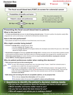
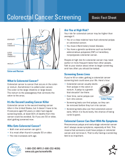
World J Gastroenterol 2014 December 28; 20(48): 18404-18412
ISSN 1007-9327 (print) ISSN 2219-2840 (online)
Submit a Manuscript: http://www.wjgnet.com/esps/
Help Desk: http://www.wjgnet.com/esps/helpdesk.aspx
DOI: 10.3748/wjg.v20.i48.18404
© 2014 Baishideng Publishing Group Inc. All rights reserved.
RETROSPECTIVE STUDY
Clinicopathologic and prognostic relevance of ARID1A
protein loss in colorectal cancer
Xiao-Li Wei, De-Shen Wang, Shao-Yan Xi, Wen-Jing Wu, Dong-Liang Chen, Zhao-Lei Zeng, Rui-Yu Wang,
Ya-Xin Huang, Ying Jin, Feng Wang, Miao-Zhen Qiu, Hui-Yan Luo, Dong-Sheng Zhang, Rui-Hua Xu
Xiao-Li Wei, De-Shen Wang, Dong-Liang Chen, ZhaoLei Zeng, Ying Jin, Feng Wang, Miao-Zhen Qiu, HuiYan Luo, Dong-Sheng Zhang, Rui-Hua Xu, Department of
Medical Oncology, Sun Yat-Sen University Cancer Center, State
Key Laboratory of Oncology in South China, Collaborative
Innovation Center for Cancer Medicine, Guangzhou 510060,
Guangdong Province, China
Shao-Yan Xi, Department of Pathology, Sun Yat-Sen University
Cancer Center, Guangzhou 510060, Guangdong Province, China
Wen-Jing Wu, Department of Breast Surgery, Sun Yat-Sen
Memorial Hospital, Sun Yat-Sen University, Guangzhou 510120,
Guangdong Province, China
Rui-Yu Wang, Ya-Xin Huang, Zhongshan School of Medicine,
Sun Yat-Sen University, Guangzhou 510080, Guangdong
Province, China
Author contributions: Wei XL, Wang DS and Xi SY
contributed equally to the study; Wei XL and Wang DS drafted
the manuscript; Wei XL, Wang RY and Huang YX collected the
clinical data of the colorectal cancer patients; Xi SY, Wu WJ,
Wang DS and Zeng ZL performed the immunohistochemical
staining; Wang F, Qiu MZ and Luo HY collected tumor slices; Xi
SY and Zhang DS reviewed the pathologic slices and scored the
immunohistochemical staining; Jin Y and Wang F performed the
statistical analysis; Xu RH conceived, designed and coordinated
the study, and offered help with preparation of the manuscript; all
authors have read and approved the final manuscript.
Supported by National High Technology Research and
Development Program of China (863 Program), No.
2012AA02A506; National Natural Science Foundation of China,
No. 81372570; the Science and Technology Foundation of
Guangdong Province, China, No. 2012B031800088; and the Science
and Technology Foundation of Guangdong Province, China, No.
C2011019
Correspondence to: Rui-Hua Xu, MD, PhD, Department of
Medical Oncology, Sun Yat-Sen University Cancer Center, State
Key Laboratory of Oncology in South China, Collaborative
Innovation Center for Cancer Medicine, 651 Dong Feng Road
East, Guangzhou 510060, Guangdong Province,
China. [email protected]
Telephone: +86-20-87343468 Fax: +86-20-87343468
Received: May 21, 2014
Revised: June 29, 2014
Accepted: July 16, 2014
Published online: December 28, 2014
WJG|www.wjgnet.com
Abstract
AIM: To explore the association between AT-rich
interactive domain 1A (ARID1A) protein loss by
immunohistochemistry and both clinicopathologic
characteristics and prognosis in patients with colorectal
cancer.
METHODS: We retrospectively collected clinicopathologic
data and archived paraffin-embedded primary colorectal
cancer samples from 209 patients, including 111
patients with colon cancer and 98 patients with rectal
cancer. The tumor stage ranged from stage Ⅰ to stage
th
Ⅳ according to the 7 edition of the American Joint
Committee on Cancer tumor-node-metastasis (TNM)
staging system. All patients underwent resection of
primary colorectal tumors. The expression of ARID1A
protein in primary colorectal cancer tissues was
examined by immunohistochemical staining. The
clinicopathologic association and survival relevance of
ARID1A protein loss in colorectal cancer were analyzed.
RESULTS: ARID1A loss by immunohistochemistry was
not rare in primary colorectal cancer tumors (25.8%).
There were 7.4%, 24.1%, 22.2% and 46.3% of
patients with ARID1A loss staged at TNM stage Ⅰ, Ⅱ,
Ⅲ and Ⅳ, respectively, compared with 20.0%, 22.6%,
27.7% and 29.7% of patients without ARID1A loss
staged at TNM stage Ⅰ, Ⅱ, Ⅲ and Ⅳ, respectively. In
patients with ARID1A loss, the distant metastasis rate
was 46.3%. However, only 29.7% of patients without
ARID1A loss were found to have distant metastasis. In
terms of pathologic differentiation, there were 25.9%,
66.7% and 7.4% with poorly, moderately and well
differentiated tumors in patients with ARID1A loss,
and 14.2%, 72.3% and 13.5% with poorly, moderately
and well differentiated tumors in patients without
ARID1A loss, respectively. ARID1A loss was associated
with late TNM stage (P = 0.020), distant metastasis
(P = 0.026), and poor pathological classification (P =
18404
December 28, 2014|Volume 20|Issue 48|
Wei XL et al . ARID1A loss and colorectal cancer
0.035). However, patients with positive ARID1A had
worse overall survival compared to those with negative
ARID1A in stage Ⅳ colorectal cancer (HR = 2.49,
95%CI: 1.13-5.51).
CONCLUSION: ARID1A protein loss is associated with
clinicopathologic characteristics in colorectal cancer
patients and with survival in stage Ⅳ patients.
© 2014 Baishideng Publishing Group Inc. All rights reserved.
Key words: AT-rich interactive domain 1A; Switching
defective/sucrose non-fermenting complexes; Colorectal
cancer; Clinicopathologic characteristics; Prognosis
Core tip: AT-rich interactive domain 1A (ARID1A)
(BAF250A) is a member of the switching defective/
sucrose non-fermenting (BAF) complexes, which
remodel nucleosomes. ARID1A gene mutation and
protein loss have been detected in many human
cancers. However, research on their clinical association
in colorectal cancer is limited and requires further
exploration. We found that ARID1A loss was not rare
in primary colorectal cancer tumors (25.8%), and it
was associated with clinicopathologic characteristics in
colorectal cancer patients and with survival in stage Ⅳ
patients.
Wei XL, Wang DS, Xi SY, Wu WJ, Chen DL, Zeng ZL, Wang
RY, Huang YX, Jin Y, Wang F, Qiu MZ, Luo HY, Zhang DS,
Xu RH. Clinicopathologic and prognostic relevance of ARID1A
protein loss in colorectal cancer. World J Gastroenterol 2014;
20(48): 18404-18412 Available from: URL: http://www.wjgnet.
com/1007-9327/full/v20/i48/18404.htm DOI: http://dx.doi.
org/10.3748/wjg.v20.i48.18404
INTRODUCTION
Colorectal cancer (CRC) is the most common cancer
of the digestive system. Diverse treatment strategies
have been developed for better management of CRC[1,2].
Although mortality due to CRC has declined steadily
during the past decades, CRC is still the second leading
cause of cancer-related death in males and the third
leading cause in females [3]. Patients with early stage
CRC have a chance of being cured. For patients with
stage Ⅳ CRC, the prognosis is poor. The tumor-nodemetastasis (TNM) staging system is the most effective
and commonly recognized clinical prognostic factor.
However, even in the same stage, the prognoses of
patients vary enormously due to molecular differences.
Many prognostic biomarkers have been suggested, but
only the test for RAS mutation [4] and microsatellite
instability (MSI)[5,6] have become part of the routine
clinical management of CRC. Other biomarkers, such
as allelic imbalance at chromosome 18q [7-9], BRAF
mutation[10,11], and p53 alterations[12,13], are not used in
WJG|www.wjgnet.com
clinical practice due to the discrepancies among research
reports. To better categorize CRC by biomarkers, more
translational studies are warranted to identify putative
biomarkers and validate them.
AT-rich interactive domain 1A (ARID1A) protein
(BAF250a) is a member of the switching defective/
sucrose non-fermenting (SWI/SNF) complexes, which
function as ATP-dependent chromatin remodelers[14,15].
These complexes remodel nucleosomes and modulate
transcription utilizing the energy of ATP hydrolysis.
Inactivating mutations of several subunits of these
complexes have frequently been detected in various
tumors, indicating a tumor suppressor function of the
SWI/SNF complexes[16-20].
Since late 2010, next-generation sequencing technologies
have brought the emergence of a wide variety of cancerassociated gene mutations. Among them, inactivating
mutations in ARID1A are frequently and repeatedly
detected in various tumors. According to the reports, loss
of ARID1A was detected in 30%-60% of ovarian clear
cell and ovarian endometrioid carcinomas[18,21,22]. It was
speculated that ARID1A loss mediated the transformation
from endometriosis to cancer[18,23]. ARID1A loss has also
been identified in some other cancers, such as endometrial
cancer [24] , clear cell renal cell carcinoma [25] , breast
cancer[26], Burkitt lymphoma[27], lung adenocarcinoma[28],
neuroblastoma[29], hepatocellular carcinoma[30], and gastric
cancer[31]. Related research in CRC is limited[32,33], thus we
conducted this study to investigate the clinicopathologic
and prognostic relevance of ARID1A loss in CRC.
MATERIALS AND METHODS
Ethics statement
All patients provided written informed consent for
their information to be stored and used in the hospital
database. Study approval was obtained from the
independent ethics committee at the Cancer Center
of Sun Yat-Sen University. The study was undertaken
in accordance with the ethical standards of the World
Medical Association Declaration of Helsinki.
Patient information and tissue specimens
This study was conducted using a total of 209 archived
paraffin-embedded primary CRC samples. All patients
underwent resection of primary tumors between 2001 and
2009 at Sun Yat-Sen University Cancer Center. All patients
received standard post-operative chemotherapy according
to the National Comprehensive Cancer Network
guidelines. None of the patients had preoperative
chemotherapy or preoperative radiotherapy. The staging
of tumors was determined according to the American
Joint Committee on Cancer (AJCC) TNM staging system.
Each tumor was pathologically classified according to
the World Health Organization classification criteria. For
survival information, patients were followed-up by the
follow-up department or the outpatient department after
discharge from hospital. Overall survival (OS) was defined
18405
December 28, 2014|Volume 20|Issue 48|
Wei XL et al . ARID1A loss and colorectal cancer
by further incubation with the streptavidin horseradish
peroxidase complex. The sections were developed
with diaminobenzidine tetrahydrochloride and then
counterstained with hematoxylin.
Two independent observers who were blinded to
patient clinical and pathological information reviewed
and scored the immunostained sections. At least 1000
tumor cells were analyzed for each slide. The proportion
of the stained cells and the extent of staining were used
as the criteria for evaluation. The percentage of positive
cells was scored as ≤ 10% = 0, > 10% to ≤ 25% = 1,
> 25% to ≤ 50% = 2, > 50% to ≤ 75% = 3, and >
75% = 4. Nuclear immunoreactivity was considered as
positive expression. The intensity of staining was scored
as negative = 0, weak = 1, moderate = 2, and strong = 3.
The two scores were then multiplied to calculate the final
score. Based on the overall score, the immunostained
sections were further divided into two groups: negative
ARID1A expression group (overall score = 0) and
positive ARID1A expression group (overall score ≥ 1).
The positive expression group was further divided into
three groups: low ARID1A expression (1 ≤ overall score
≤ 4), moderate ARID1A expression (4 < overall score
≤ 8) and high ARID1A expression group (8 < overall
score ≤ 12).
Table 1 Clinicopathologic characteristics and AT-rich interactive
domain 1A expression in 209 patient samples of colorectal
cancer n (%)
Characteristics
Gender
Male
Female
Age (yr)
Median
Range
Tumor location
Colon
Rectal
TNM stage (AJCC)
Ⅰ
Ⅱ
Ⅲ
Ⅳ
T stage
T1
T2
T3
T4
N stage
N0
N1
N2
M stage
M0
M1
Pathologic differentiation
Poor
Moderate
Well
Tumor size (cm)
≤5
>5
Expression of ARID1a
Negative
Positive
Value
176 (84.2)
33 (15.8)
55
19-88
111 (53.1)
98 (46.9)
35 (16.7)
48 (23.0)
55 (26.3)
71 (34.0)
10 (4.8)
35 (16.7)
53 (25.4)
111 (53.1)
98 (46.9)
56 (26.8)
55 (26.3)
138 (66.0)
71 (34.0)
Statistical analysis
All statistical analyses were performed using SPSS 13.0
statistical software. A P value < 0.05 was considered
statistically significant in all cases. The association
between ARID1A expression and the clinicopathological
characteristics was analyzed by χ 2 test or Kruskal-Wallis
H test based on the type of data. Survival curves were
plotted by the Kaplan-Meier method and compared
using the log-rank test. Survival data were evaluated using
univariate and multivariate Cox regression analyses.
36 (17.2)
148 (70.8)
25 (12.0)
136 (65.1)
73 (34.9)
54 (25.8)
155 (74.2)
AJCC: American Joint Committee on Cancer; TNM: Tumor-nodemetastasis; ARID1A: AT-rich interactive domain 1A.
as the interval between the date of surgery and the date
of death or the last known follow-up if the patient was
alive. The clinicopathologic information of the study
subjects and primary tumor samples are listed in Table
1. All excised samples were obtained from tumor tissues
within 1 h after surgery. For each specimen, half was
placed in liquid nitrogen until analysis, and the remainder
was fixed with formalin for immunohistochemistry (IHC).
IHC and scoring
The protein expression levels of ARID1A were
investigated in 209 primary CRC samples using IHC.
The procedure used was described previously[34]. Briefly,
the tissue sections were first deparaffinized, rehydrated,
endogenous peroxide blocked and antigen retrieved, then
incubated with ARID1A (PSG3): sc-32761 antibody
(Santa Cruz Biotechnology, Inc., CA, United States)
overnight at 4 ℃. Next, after washing with phosphatebuffered saline Tween-20, tissue sections were treated
with anti-mouse secondary antibody for 30 min, followed
WJG|www.wjgnet.com
RESULTS
ARID1A expression in CRC primary tumors
IHC analysis was conducted on 209 primary CRC tumor
samples. Fifty-four (25.8%) primary CRC tumors had
negative ARID1A expression, 107 (51.2%) tumors
had low ARID1A expression, 39 (18.7%) tumors had
moderate ARID1A expression, and 9 (4.3%) tumors had
high ARID1A expression. Typical immunostaining of
negative, low, moderate and high ARID1A expression are
shown in Figure 1.
Loss of ARID1A expression was associated with
clinicopathological features of CRC
All 209 primary CRC tumors were included in this analysis.
Fifty-four (25.8%) had negative ARID1A expression,
and 155 (74.2%) cases had positive ARID1A expression.
The correlations between ARID1A expression (negative/
positive) and clinicopathological features of CRC are
listed in Table 2. As shown in Table 2, loss of ARID1A
expression was not associated with gender (male/female),
18406
December 28, 2014|Volume 20|Issue 48|
Wei XL et al . ARID1A loss and colorectal cancer
A
B
100 mm
C
100 mm
D
100 mm
100 mm
Figure 1 Typical immunohistochemical staining of AT-rich interactive domain 1A (× 100). A: Negative AT-rich interactive domain 1A (ARID1A) expression; B:
Low ARID1A expression; C: Moderate ARID1A expression; D: High ARID1A expression.
age (≤ 55/> 55 years), tumor location (colon/rectal), T
stage (T1/T2/T3/T4), N stage (N0/N1/N2), or tumor
size (≤ 5/> 5 cm). However, the associations between
ARID1A expression and TNM (AJCC) stage, M stage and
pathologic differentiation were all statistically significant
(P = 0.020, P = 0.026, and P = 0.035, respectively). Loss
of ARID1A expression was significantly associated with
late TNM stage, distant metastasis, and poor pathologic
differentiation.
Association between ARID1A loss and CRC survival
Kaplan-Meier analysis was used for the initial analysis
of the influence of ARID1A loss on OS in CRC
patients. ARID1A expression (negative/positive) was not
associated with OS in all 209 CRC patients (P = 0.538).
However, when the population was split by TNM stage,
ARID1A expression (negative/positive) was significantly
associated with OS in stage Ⅳ patients (P = 0.027,
Figure 2), but a significant relationship was not observed
in stage Ⅰ-Ⅲ patients. Thus, we further analyzed the
association between ARID1A expression (negative/
positive) and OS in stage Ⅳ patients. The results of
univariate and multivariate analyses are listed in Table 3.
In stage Ⅳ CRC patients, potential prognostic
factors, including N stage (N0/N1/N2), pathologic
differentiation (poor/moderate/well), tumor size
(≤ 5/> 5 cm), metastatic site (single/multiple), and
metastasis limited to the liver (no/yes) were not found
to be significantly prognostic. However, ARID1A
expression (negative/positive), T stage (T1/T2/T3/T4)
WJG|www.wjgnet.com
and resection of metastatic tumors (no/yes) were found
to be significant prognostic factors in univariate analyses
(P = 0.027, P = 0.038, and P = 0.002, respectively, Table
3). Factors significantly prognostic in univariate analysis
were included in multivariate analysis. All three of the
above factors were independent prognostic factors in
multivariate analysis. Positive ARID1A expression was
significantly and independently associated with worse
OS in stage Ⅳ CRC patients compared with negative
ARID1A expression (HR = 2.49, 95%CI: 1.13-5.51,
Table 3). The Kaplan-Meier survival curves of ARID1A
(negative/positive) for patients with stage Ⅳ CRC are
shown in Figure 2.
DISCUSSION
In the present study, we demonstrated that loss of
ARID1A expression was associated with late TNM stage,
distant metastasis and poor pathologic differentiation.
These findings indicated that ARID1A may play an
important role in the progression of CRC. In addition,
the survival analyses also indicated that loss of ARID1A
protein expression was a prognostic factor for better OS
in stage Ⅳ CRC.
Epigenetic regulators modulate gene expression, and
thereby influence cell function. Alterations in epigenetic
regulators have been shown to be one of the key
characteristics in tumorigenesis[35]. Of these epigenetic
regulators, the SWI/SNF chromatin remodeling
complexes have been demonstrated to be repeatedly
18407
December 28, 2014|Volume 20|Issue 48|
Wei XL et al . ARID1A loss and colorectal cancer
Table 2 Correlation between AT-rich interactive domain 1A
expression and clinicopathologic characteristics of colorectal
cancer patients n (%)
Characteristics
Gender
Male
Female
Age (yr)
≤ 55
> 55
Tumor location
Colon
Rectal
TNM stage (AJCC)
Ⅰ
Ⅱ
Ⅲ
Ⅳ
T stage
T1
T2
T3
T4
N stage
N0
N1
N2
M stage
M0
M1
Pathologic differentiation
Poor
Moderate
Well
Tumor size (cm)
≤5
>5
Characteristics
P value
ARID1a
Negative
Table 3 Univariate and multivariate analyses of various
prognostic parameters in stage Ⅳ patients with colorectal
cancer using Cox-regression analysis n (%)
P value
0.132
42 (77.8)
12 (22.2)
134 (86.5)
21 (13.5)
30 (55.6)
24 (44.4)
76 (49.0)
79 (51.0)
32 (59.3)
22 (40.7)
79 (51.0)
76 (49.0)
4 (7.4)
13 (24.1)
12 (22.2)
25 (46.3)
31 (20.0)
35 (22.6)
43 (27.7)
46 (29.7)
1 (1.9)
6 (11.1)
20 (37.0)
27 (50.0)
9 (5.8)
29 (18.7)
33 (21.3)
84 (54.2)
Gender
Male
Female
Age (yr)
≤ 55
> 55
ARID1a expression
Negative
Positive
T stage
T1
T2
T3
T4
N stage
N0
N1
N2
Pathologic
differentiation
Poor
Moderate
Well
Tumor size (cm)
≤5
>5
Metastatic site
Single
Multiple
Metastasis limited to
the liver
No
Yes
Resection of
metastatic tumors1
No
Yes
0.409
0.293
0.020
0.771
0.638
23 (42.6)
17 (31.5)
14 (25.9)
75 (48.4)
39 (25.1)
41 (26.5)
29 (53.7)
25 (46.3)
109 (70.3)
46 (29.7)
14 (25.9)
36 (66.7)
4 (7.4)
22 (14.2)
112 (72.3)
21 (13.5)
0.026
0.035
0.170
31 (57.4)
23 (42.6)
105 (67.7)
50 (32.3)
AJCC: American Joint Committee on Cancer; TNM: Tumor-nodemetastasis; ARID1A: AT-rich interactive domain 1A.
1.0
95%CI P value
0.027
2.49
1.13-5.51
0.024
0.038
2.01
1.08-3.75
0.029
0.33
0.16-0.67
0.002
0.258
40 (56.3)
31 (43.7)
0.934
39 (54.9)
32 (45.1)
25 (35.2)
46 (64.8)
1 (1.4)
5 (7.0)
43 (60.6)
22 (31.0)
0.929
12 (16.9)
27 (38.0)
32 (45.1)
0.458
17 (23.9)
51 (71.8)
3 (4.3)
0.261
43 (60.6)
28 (39.4)
0.806
52 (73.2)
19 (26.8)
0.519
37 (52.1)
34 (47.9)
0.002
21 (29.6)
50 (70.4)
Resection of metastasis: Including R0 and R1 resection of all metastatic
tumors. ARID1A: AT-rich interactive domain 1A.
Positive
Negative-censored
Cum survival
Multivariate analysis
HR
1
ARID1A
Negative
0.8
Univariate analysis
Positive
Positive-censored
0.6
0.4
0.2
P = 0.022
0.0
0.00
20.00
40.00 60.00 80.00 100.00 120.00 140.00
OS (m)
Figure 2 Kaplan-Meier curves. Kaplan-Meier curves with univariate analyses
(log-rank) for stage Ⅳ patients with negative AT-rich interactive domain 1A
(ARID1A) expression vs positive ARID1A expression.
mutated in a wide range of carcinomas [36] . These
complexes consist of several highly related multiunit
complexes, and the occurrence of specific inactivating
WJG|www.wjgnet.com
mutations in subunits were frequently authenticated,
including the SNF5, BAF 180, BRM/SWI2-related
gene 1, as well as ARID1A[36]. Functionally, ARID1A is
required for nucleosome substrate binding and occupancy
by SWI/SNF complexes[37]. Knockdown of ARID1A
abrogates normal cell cycle arrest in osteoblasts cells,
indicating the potential tumor suppressor function
of ARID1A as well as the relevance of ARID1A loss
and tumorigenesis [14]. In addition, Dykhuizen et al [38]
discovered that SWI/SNF complexes were essential for
the binding of topoisomerase Ⅱalpha to approximately
12000 sites across the genome by directly interacting with
topoisomerase Ⅱalpha through ARID1A protein, which
resulted in decatenation defects.
The tumor suppressor function of ARID1A has
been verified in a wide variety of carcinomas, with
significant research in gynecological carcinomas. There
18408
December 28, 2014|Volume 20|Issue 48|
Wei XL et al . ARID1A loss and colorectal cancer
are few investigations regarding ARID1A in CRC,
which requires further clarification. The mutation rate
of ARID1A reported in CRC is low, and was reported
by Jones et al[39] to be 10% and Kim et al[40] found no
mutations. Thus, the importance of ARID1A loss in CRC
may be underestimated. In our study, loss of ARID1A
protein by IHC occurred in 25.8% of primary CRC
tumors, with an even higher proportion of 35.2% in
stage Ⅳ CRC, suggesting that loss of ARID1A was not
uncommon in CRC. Moreover, loss of ARID1A was of
clinicopathological significance.
There are a large number of reports on the relevance
of ARID1A mutation or protein loss to survival in several
carcinomas. However, there is controversy regarding the
results of these studies. Some studies found that ARID1A
mutation or protein loss was a predictor of worse
survival in cervical cancer[41], and gastric cancer[42]. Others
suggested no association between ARID1A mutation or
protein loss and survival in clear cell carcinoma of the
endometrium[43], and ovarian clear cell adenocarcinomas[22].
There are also reports indicating that ARID1A mutation
or protein loss was related to a survival advantage in
endometrial carcinoma[44] and gastric cancer[31]. Our study
is the first to explore the influence of ARID1A loss on
CRC survival. It was found that ARID1A loss predicted
superior OS in stage Ⅳ CRC. ARID1A loss was also
related to microsatellite instability (MSI) in endometrial
cancer[45]. Previous research demonstrated that ARID1A
mutation occurred more frequently in CRC with high
MSI (MSI-H) compared with those with microsatellite
stable CRC[32,40]. It was shown that MSI-H predicts better
survival in early stage CRC[46]. In stage Ⅳ CRC, Liang et
al[5] reported that MSI-H predicted better chemosensitivity
to high-dose 5-fluorouracil plus leucovorin chemotherapy
for stage Ⅳ sporadic colorectal cancer after palliative
bowel resection. In our study, all stage Ⅳ patients
underwent resection of primary CRC tumors. Thus, the
survival advantage in patients with ARID1A loss may
be partially explained by its association with MSI-H.
However, one limitation of our study was that the status
of MSI was not tested. Another was that the association
between ARID1A loss and chemosensitivity in stage Ⅳ
patients was not explored as only 21 (29.6%) cases did
not receive metastatic tumor resection and had evaluable
lesions. These associations need to be verified and the
mechanisms clarified in future investigations.
Other limitations in our study were as follows:
All stage Ⅳ patients underwent resection of primary
tumors, thus those with a heavy tumor burden or poor
performance status were not included. Therefore, the
finding that ARID1A loss predicted better OS in stage Ⅳ
CRC patients could only be applied to those with good
performance status or low tumor burden. In addition,
patients with early stage CRC and metastatic CRC
received different drug regimens. The duration of our
study was nine years from 2001-2009. During this period,
the results of some clinical trials contributed to changes
in clinical practice. Thus, even for patients with the same
WJG|www.wjgnet.com
stage, their treatment was different. For these reasons, the
associations between ARID1A loss and the effects of the
chemotherapy and radiotherapy were not analyzed in this
study.
In conclusion, ARID1A loss was not rare in CRC. It
was associated with late TNM stage, distant metastasis,
and poor pathologic differentiation. In addition, stage
Ⅳ patients with ARID1A protein loss in primary
tumors had longer survival than those with ARID1A
positive tumors. ARID1A may be a candidate prognostic
biomarker in CRC. In addition, considering that
epigenetic alterations are potentially reversible, CRC
patients with ARID1A loss may benefit from therapeutics
which target chromatin-modifying enzymes.
ACKNOWLEDGMENTS
We thank Professor Liu Qing in the Epidemiology
Department for his suggestions on the statistical analysis;
and all the staff members in our department for their
support and suggestions in this study.
COMMENTS
COMMENTS
Background
AT-rich interactive domain 1A (ARID1A), a member of the switching defective/
sucrose non-fermenting (SWI/SNF) complexes, has been shown to be mutated
in many human cancers. However, studies on their clinical association in
colorectal cancer are limited. The identification of new biomarkers is warranted
to direct the categorization and treatment of colorectal cancer.
Research frontiers
ARID1A gene mutation has been repeatedly detected in many cancers, particularly
in ovarian clear cell and ovarian endometrioid carcinomas. ARID1A loss is
considered to be an important early event in the tumorigenic transformation from
endometriosis to cancer. It was also found to be associated with microsatellite
instability in endometrial cancer. ARID1A gene mutation was detected in colorectal
cancer in previous studies, however, the association between ARID1A protein loss
by immunohistochemistry and both clinicopathologic characteristics and prognosis
in colorectal cancer is unclear.
Innovations and breakthroughs
The authors found that ARID1A loss was not rare in primary colorectal cancer
tumors (25.8%), and was associated with late TNM stage, distant metastasis,
and poor pathological classification in patients with colorectal cancer. However,
ARID1A loss predicted better overall survival in stage Ⅳ patients.
Applications
This study indicated an important role for ARID1A protein loss in colorectal
cancer. ARID1A protein loss was found to be associated with late TNM stage,
distant metastasis and poor pathological differentiation, suggesting that it
might play a role in the progression of colorectal cancer. However, in stage Ⅳ
patients, ARID1A loss was found to predict better survival. This could partially
be explained by its association with microsatellite instability. Further studies are
needed to clarify the detailed mechanisms involved.
Terminology
ARID1A, also known as BAF250a, is a member of the SWI/SNF complexes,
which function as ATP-dependent chromatin remodelers. The SWI/SNF
complexes remodel nucleosomes and modulate transcription utilizing the
energy of ATP hydrolysis.
Peer review
This study demonstrated the clinical association of ARID1A protein loss by
immunohistochemistry in colorectal cancer. ARID1A protein loss was found
to be related with late TNM stage, distant metastasis and poor pathological
differentiation. In addition, it was found to predict better survival in stage Ⅳ
patients.
18409
December 28, 2014|Volume 20|Issue 48|
Wei XL et al . ARID1A loss and colorectal cancer
REFERENCES
1
2
3
4
5
6
7
8
9
10
11
12
13
Mellas N, Benbrahim Z, El Mesbahi O. Colorectal cancer:
new developments after the 2013 ECCO/ESMO congress.
Chin J Cancer 2014; 33: 218-221 [PMID: 24589209 DOI:
10.5732/cjc.013.10203]
Chang DZ, Kumar V, Ma Y, Li K, Kopetz S. Individualized
therapies in colorectal cancer: KRAS as a marker for response
to EGFR-targeted therapy. J Hematol Oncol 2009; 2: 18 [PMID:
19386128 DOI: 10.1186/1756-8722-2-18]
Siegel R, Naishadham D, Jemal A. Cancer statistics, 2012.
CA Cancer J Clin 2012; 62: 10-29 [PMID: 22237781 DOI:
10.3322/caac.20138]
Douillard JY, Oliner KS, Siena S, Tabernero J, Burkes R,
Barugel M, Humblet Y, Bodoky G, Cunningham D, Jassem
J, Rivera F, Kocákova I, Ruff P, Błasińska-Morawiec M,
Šmakal M, Canon JL, Rother M, Williams R, Rong A,
Wiezorek J, Sidhu R, Patterson SD. Panitumumab-FOLFOX4
treatment and RAS mutations in colorectal cancer. N Engl
J Med 2013; 369: 1023-1034 [PMID: 24024839 DOI: 10.1056/
NEJMoa1305275]
Liang JT, Huang KC, Lai HS, Lee PH, Cheng YM, Hsu HC,
Cheng AL, Hsu CH, Yeh KH, Wang SM, Tang C, Chang
KJ. High-frequency microsatellite instability predicts better
chemosensitivity to high-dose 5-fluorouracil plus leucovorin
chemotherapy for stage IV sporadic colorectal cancer after
palliative bowel resection. Int J Cancer 2002; 101: 519-525
[PMID: 12237891 DOI: 10.1002/ijc.10643]
Hemminki A, Mecklin JP, Järvinen H, Aaltonen LA, Joensuu
H. Microsatellite instability is a favorable prognostic
indicator in patients with colorectal cancer receiving
chemotherapy. Gastroenterology 2000; 119: 921-928 [PMID:
11040179 DOI: 10.1053/gast.2000.18161]
Jen J, Kim H, Piantadosi S, Liu ZF, Levitt RC, Sistonen
P, Kinzler KW, Vogelstein B, Hamilton SR. Allelic loss of
chromosome 18q and prognosis in colorectal cancer. N Engl
J Med 1994; 331: 213-221 [PMID: 8015568 DOI: 10.1056/
NEJM199407283310401]
Ogino S, Nosho K, Irahara N, Shima K, Baba Y, Kirkner
GJ, Meyerhardt JA, Fuchs CS. Prognostic significance and
molecular associations of 18q loss of heterozygosity: a
cohort study of microsatellite stable colorectal cancers. J Clin
Oncol 2009; 27: 4591-4598 [PMID: 19704056 DOI: 10.1200/
JCO.2009.22.8858]
Carethers JM, Hawn MT, Greenson JK, Hitchcock CL, Boland
CR. Prognostic significance of allelic lost at chromosome
18q21 for stage II colorectal cancer. Gastroenterology 1998; 114:
1188-1195 [PMID: 9609755]
Ogino S, Nosho K, Kirkner GJ, Kawasaki T, Meyerhardt JA,
Loda M, Giovannucci EL, Fuchs CS. CpG island methylator
phenotype, microsatellite instability, BRAF mutation and
clinical outcome in colon cancer. Gut 2009; 58: 90-96 [PMID:
18832519 DOI: 10.1136/gut.2008.15547]
Bardelli A, Siena S. Molecular mechanisms of resistance
to cetuximab and panitumumab in colorectal cancer. J Clin
Oncol 2010; 28: 1254-1261 [PMID: 20100961 DOI: 10.1200/
JCO.2009.24.6116]
Munro AJ, Lain S, Lane DP. P53 abnormalities and outcomes
in colorectal cancer: a systematic review. Br J Cancer 2005; 92:
434-444 [PMID: 15668707 DOI: 10.1038/sj.bjc.6602358]
Iacopetta B, Russo A, Bazan V, Dardanoni G, Gebbia N,
Soussi T, Kerr D, Elsaleh H, Soong R, Kandioler D, Janschek
E, Kappel S, Lung M, Leung CS, Ko JM, Yuen S, Ho J, Leung
SY, Crapez E, Duffour J, Ychou M, Leahy DT, O’Donoghue
DP, Agnese V, Cascio S, Di Fede G, Chieco-Bianchi L,
Bertorelle R, Belluco C, Giaretti W, Castagnola P, Ricevuto
E, Ficorella C, Bosari S, Arizzi CD, Miyaki M, Onda M,
Kampman E, Diergaarde B, Royds J, Lothe RA, Diep CB,
Meling GI, Ostrowski J, Trzeciak L, Guzinska-Ustymowicz
WJG|www.wjgnet.com
14
15
16
17
18
19
20
21
22
23
18410
K, Zalewski B, Capellá GM, Moreno V, Peinado MA,
Lönnroth C, Lundholm K, Sun XF, Jansson A, Bouzourene
H, Hsieh LL, Tang R, Smith DR, Allen-Mersh TG, Khan ZA,
Shorthouse AJ, Silverman ML, Kato S, Ishioka C. Functional
categories of TP53 mutation in colorectal cancer: results of
an International Collaborative Study. Ann Oncol 2006; 17:
842-847 [PMID: 16524972 DOI: 10.1093/annonc/mdl035]
Nagl NG, Patsialou A, Haines DS, Dallas PB, Beck GR,
Moran E. The p270 (ARID1A/SMARCF1) subunit of
mammalian SWI/SNF-related complexes is essential for
normal cell cycle arrest. Cancer Res 2005; 65: 9236-9244 [PMID:
16230384 DOI: 10.1158/0008-5472.CAN-05-1225]
Wang X, Nagl NG, Wilsker D, Van Scoy M, Pacchione S,
Yaciuk P, Dallas PB, Moran E. Two related ARID family
proteins are alternative subunits of human SWI/SNF
complexes. Biochem J 2004; 383: 319-325 [PMID: 15170388
DOI: 10.1042/BJ20040524]
Versteege I, Sévenet N, Lange J, Rousseau-Merck MF,
Ambros P, Handgretinger R, Aurias A, Delattre O.
Truncating mutations of hSNF5/INI1 in aggressive
paediatric cancer. Nature 1998; 394: 203-206 [PMID: 9671307
DOI: 10.1038/28212]
Varela I, Tarpey P, Raine K, Huang D, Ong CK, Stephens
P, Davies H, Jones D, Lin ML, Teague J, Bignell G, Butler
A, Cho J, Dalgliesh GL, Galappaththige D, Greenman C,
Hardy C, Jia M, Latimer C, Lau KW, Marshall J, McLaren S,
Menzies A, Mudie L, Stebbings L, Largaespada DA, Wessels
LF, Richard S, Kahnoski RJ, Anema J, Tuveson DA, PerezMancera PA, Mustonen V, Fischer A, Adams DJ, Rust A,
Chan-on W, Subimerb C, Dykema K, Furge K, Campbell
PJ, Teh BT, Stratton MR, Futreal PA. Exome sequencing
identifies frequent mutation of the SWI/SNF complex gene
PBRM1 in renal carcinoma. Nature 2011; 469: 539-542 [PMID:
21248752 DOI: 10.1038/nature09639]
Wiegand KC, Shah SP, Al-Agha OM, Zhao Y, Tse K, Zeng
T, Senz J, McConechy MK, Anglesio MS, Kalloger SE, Yang
W, Heravi-Moussavi A, Giuliany R, Chow C, Fee J, Zayed
A, Prentice L, Melnyk N, Turashvili G, Delaney AD, Madore
J, Yip S, McPherson AW, Ha G, Bell L, Fereday S, Tam A,
Galletta L, Tonin PN, Provencher D, Miller D, Jones SJ,
Moore RA, Morin GB, Oloumi A, Boyd N, Aparicio SA, Shih
IeM, Mes-Masson AM, Bowtell DD, Hirst M, Gilks B, Marra
MA, Huntsman DG. ARID1A mutations in endometriosisassociated ovarian carcinomas. N Engl J Med 2010; 363:
1532-1543 [PMID: 20942669 DOI: 10.1056/NEJMoa1008433]
Wong AK, Shanahan F, Chen Y, Lian L, Ha P, Hendricks K,
Ghaffari S, Iliev D, Penn B, Woodland AM, Smith R, Salada G,
Carillo A, Laity K, Gupte J, Swedlund B, Tavtigian SV, Teng
DH, Lees E. BRG1, a component of the SWI-SNF complex, is
mutated in multiple human tumor cell lines. Cancer Res 2000;
60: 6171-6177 [PMID: 11085541]
Reisman DN, Sciarrotta J, Wang W, Funkhouser WK,
Weissman BE. Loss of BRG1/BRM in human lung cancer
cell lines and primary lung cancers: correlation with poor
prognosis. Cancer Res 2003; 63: 560-566 [PMID: 12566296]
Maeda D, Mao TL, Fukayama M, Nakagawa S, Yano T,
Taketani Y, Shih IeM. Clinicopathological significance of
loss of ARID1A immunoreactivity in ovarian clear cell
carcinoma. Int J Mol Sci 2010; 11: 5120-5128 [PMID: 21614196
DOI: 10.3390/ijms11125120]
Yamamoto S, Tsuda H, Takano M, Tamai S, Matsubara O.
PIK3CA mutations and loss of ARID1A protein expression
are early events in the development of cystic ovarian clear
cell adenocarcinoma. Virchows Arch 2012; 460: 77-87 [PMID:
22120431 DOI: 10.1007/s00428-011-1169-8]
Yamamoto S, Tsuda H, Takano M, Tamai S, Matsubara O.
Loss of ARID1A protein expression occurs as an early event
in ovarian clear-cell carcinoma development and frequently
coexists with PIK3CA mutations. Mod Pathol 2012; 25:
December 28, 2014|Volume 20|Issue 48|
Wei XL et al . ARID1A loss and colorectal cancer
24
25
26
27
28
29
30
31
615-624 [PMID: 22157930 DOI: 10.1038/modpathol.2011.189]
Liang H, Cheung LW, Li J, Ju Z, Yu S, Stemke-Hale K,
Dogruluk T, Lu Y, Liu X, Gu C, Guo W, Scherer SE, Carter
H, Westin SN, Dyer MD, Verhaak RG, Zhang F, Karchin R,
Liu CG, Lu KH, Broaddus RR, Scott KL, Hennessy BT, Mills
GB. Whole-exome sequencing combined with functional
genomics reveals novel candidate driver cancer genes in
endometrial cancer. Genome Res 2012; 22: 2120-2129 [PMID:
23028188 DOI: 10.1101/gr.137596.112]
Lichner Z, Scorilas A, White NM, Girgis AH, Rotstein
L, Wiegand KC, Latif A, Chow C, Huntsman D, Yousef
GM. The chromatin remodeling gene ARID1A is a new
prognostic marker in clear cell renal cell carcinoma. Am J
Pathol 2013; 182: 1163-1170 [PMID: 23416164 DOI: 10.1016/
j.ajpath.2013.01.007]
Mamo A, Cavallone L, Tuzmen S, Chabot C, Ferrario
C, Hassan S, Edgren H, Kallioniemi O, Aleynikova O,
Przybytkowski E, Malcolm K, Mousses S, Tonin PN, Basik
M. An integrated genomic approach identifies ARID1A
as a candidate tumor-suppressor gene in breast cancer.
Oncogene 2012; 31: 2090-2100 [PMID: 21892209 DOI: 10.1038/
onc.2011.386]
Love C, Sun Z, Jima D, Li G, Zhang J, Miles R, Richards KL,
Dunphy CH, Choi WW, Srivastava G, Lugar PL, Rizzieri
DA, Lagoo AS, Bernal-Mizrachi L, Mann KP, Flowers CR,
Naresh KN, Evens AM, Chadburn A, Gordon LI, Czader
MB, Gill JI, Hsi ED, Greenough A, Moffitt AB, McKinney
M, Banerjee A, Grubor V, Levy S, Dunson DB, Dave SS. The
genetic landscape of mutations in Burkitt lymphoma. Nat
Genet 2012; 44: 1321-1325 [PMID: 23143597 DOI: 10.1038/
ng.2468]
Imielinski M, Berger AH, Hammerman PS, Hernandez B,
Pugh TJ, Hodis E, Cho J, Suh J, Capelletti M, Sivachenko A,
Sougnez C, Auclair D, Lawrence MS, Stojanov P, Cibulskis
K, Choi K, de Waal L, Sharifnia T, Brooks A, Greulich H,
Banerji S, Zander T, Seidel D, Leenders F, Ansén S, Ludwig C,
Engel-Riedel W, Stoelben E, Wolf J, Goparju C, Thompson K,
Winckler W, Kwiatkowski D, Johnson BE, Jänne PA, Miller
VA, Pao W, Travis WD, Pass HI, Gabriel SB, Lander ES,
Thomas RK, Garraway LA, Getz G, Meyerson M. Mapping
the hallmarks of lung adenocarcinoma with massively
parallel sequencing. Cell 2012; 150: 1107-1120 [PMID:
22980975 DOI: 10.1016/j.cell.2012.08.029]
Sausen M, Leary RJ, Jones S, Wu J, Reynolds CP, Liu X,
Blackford A, Parmigiani G, Diaz LA, Papadopoulos N,
Vogelstein B, Kinzler KW, Velculescu VE, Hogarty MD.
Integrated genomic analyses identify ARID1A and ARID1B
alterations in the childhood cancer neuroblastoma. Nat Genet
2013; 45: 12-17 [PMID: 23202128 DOI: 10.1038/ng.2493]
Fujimoto A, Totoki Y, Abe T, Boroevich KA, Hosoda F,
Nguyen HH, Aoki M, Hosono N, Kubo M, Miya F, Arai Y,
Takahashi H, Shirakihara T, Nagasaki M, Shibuya T, Nakano
K, Watanabe-Makino K, Tanaka H, Nakamura H, Kusuda
J, Ojima H, Shimada K, Okusaka T, Ueno M, Shigekawa Y,
Kawakami Y, Arihiro K, Ohdan H, Gotoh K, Ishikawa O,
Ariizumi S, Yamamoto M, Yamada T, Chayama K, Kosuge T,
Yamaue H, Kamatani N, Miyano S, Nakagama H, Nakamura
Y, Tsunoda T, Shibata T, Nakagawa H. Whole-genome
sequencing of liver cancers identifies etiological influences
on mutation patterns and recurrent mutations in chromatin
regulators. Nat Genet 2012; 44: 760-764 [PMID: 22634756 DOI:
10.1038/ng.2291]
Wang K, Kan J, Yuen ST, Shi ST, Chu KM, Law S, Chan TL,
Kan Z, Chan AS, Tsui WY, Lee SP, Ho SL, Chan AK, Cheng
GH, Roberts PC, Rejto PA, Gibson NW, Pocalyko DJ, Mao
M, Xu J, Leung SY. Exome sequencing identifies frequent
mutation of ARID1A in molecular subtypes of gastric cancer.
Nat Genet 2011; 43: 1219-1223 [PMID: 22037554 DOI: 10.1038/
ng.982]
WJG|www.wjgnet.com
32
33
34
35
36
37
38
39
40
41
42
43
44
45
18411
Cajuso T, Hänninen UA, Kondelin J, Gylfe AE, Tanskanen
T, Katainen R, Pitkänen E, Ristolainen H, Kaasinen E,
Taipale M, Taipale J, Böhm J, Renkonen-Sinisalo L, Mecklin
JP, Järvinen H, Tuupanen S, Kilpivaara O, Vahteristo P.
Exome sequencing reveals frequent inactivating mutations
in ARID1A, ARID1B, ARID2 and ARID4A in microsatellite
unstable colorectal cancer. Int J Cancer 2014; 135: 611-623
[PMID: 24382590 DOI: 10.1002/ijc.28705]
Cancer Genome Atlas Network. Comprehensive molecular
characterization of human colon and rectal cancer.
Nature 2012; 487: 330-337 [PMID: 22810696 DOI: 10.1038/
nature11252]
Teng KY, Qiu MZ, Li ZH, Luo HY, Zeng ZL, Luo RZ, Zhang
HZ, Wang ZQ, Li YH, Xu RH. DNA polymerase η protein
expression predicts treatment response and survival of
metastatic gastric adenocarcinoma patients treated with
oxaliplatin-based chemotherapy. J Transl Med 2010; 8: 126
[PMID: 21110884 DOI: 10.1186/1479-5876-8-126]
Hanahan D, Weinberg RA. Hallmarks of cancer: the next
generation. Cell 2011; 144: 646-674 [PMID: 21376230 DOI:
10.1016/j.cell.2011.02.013]
Wilson BG, Roberts CW. SWI/SNF nucleosome remodellers
and cancer. Nat Rev Cancer 2011; 11: 481-492 [PMID: 21654818
DOI: 10.1038/nrc3068]
Chandler RL, Brennan J, Schisler JC, Serber D, Patterson C,
Magnuson T. ARID1a-DNA interactions are required for
promoter occupancy by SWI/SNF. Mol Cell Biol 2013; 33:
265-280 [PMID: 23129809 DOI: 10.1128/MCB.01008-12]
Dykhuizen EC, Hargreaves DC, Miller EL, Cui K,
Korshunov A, Kool M, Pfister S, Cho YJ, Zhao K, Crabtree
GR. BAF complexes facilitate decatenation of DNA by
topoisomerase IIα. Nature 2013; 497: 624-627 [PMID:
23698369 DOI: 10.1038/nature12146]
Jones S, Li M, Parsons DW, Zhang X, Wesseling J, Kristel P,
Schmidt MK, Markowitz S, Yan H, Bigner D, Hruban RH,
Eshleman JR, Iacobuzio-Donahue CA, Goggins M, Maitra A,
Malek SN, Powell S, Vogelstein B, Kinzler KW, Velculescu
VE, Papadopoulos N. Somatic mutations in the chromatin
remodeling gene ARID1A occur in several tumor types.
Hum Mutat 2012; 33: 100-103 [PMID: 22009941 DOI: 10.1002/
humu.21633]
Kim MS, Je EM, Yoo NJ, Lee SH. Loss of ARID1A expression
is uncommon in gastric, colorectal, and prostate cancers.
APMIS 2012; 120: 1020-1022 [PMID: 23030592 DOI: 10.1111/
j.1600-0463.2012.02930.x]
Cho H, Kim JS, Chung H, Perry C, Lee H, Kim JH. Loss
of ARID1A/BAF250a expression is linked to tumor
progression and adverse prognosis in cervical cancer. Hum
Pathol 2013; 44: 1365-1374 [PMID: 23427874 DOI: 10.1016/
j.humpath.2012.11.007]
Wang DD, Chen YB, Pan K, Wang W, Chen SP, Chen JG,
Zhao JJ, Lv L, Pan QZ, Li YQ, Wang QJ, Huang LX, Ke ML,
He J, Xia JC. Decreased expression of the ARID1A gene is
associated with poor prognosis in primary gastric cancer.
PLoS One 2012; 7: e40364 [PMID: 22808142 DOI: 10.1371/
journal.pone.0040364]
Fadare O, Gwin K, Desouki MM, Crispens MA, Jones HW,
Khabele D, Liang SX, Zheng W, Mohammed K, Hecht JL,
Parkash V. The clinicopathologic significance of p53 and
BAF-250a (ARID1A) expression in clear cell carcinoma of
the endometrium. Mod Pathol 2013; 26: 1101-1110 [PMID:
23524907 DOI: 10.1038/modpathol.2013.35]
Allo G, Bernardini MQ, Wu RC, Shih IeM, Kalloger S,
Pollett A, Gilks CB, Clarke BA. ARID1A loss correlates with
mismatch repair deficiency and intact p53 expression in
high-grade endometrial carcinomas. Mod Pathol 2014; 27:
255-261 [PMID: 23887303 DOI: 10.1038/modpathol.2013.144]
Bosse T, ter Haar NT, Seeber LM, v Diest PJ, Hes FJ, Vasen
HF, Nout RA, Creutzberg CL, Morreau H, Smit VT. Loss
December 28, 2014|Volume 20|Issue 48|
Wei XL et al . ARID1A loss and colorectal cancer
46
of ARID1A expression and its relationship with PI3K-Akt
pathway alterations, TP53 and microsatellite instability in
endometrial cancer. Mod Pathol 2013; 26: 1525-1535 [PMID:
23702729 DOI: 10.1038/modpathol.2013.96]
Gavin PG, Colangelo LH, Fumagalli D, Tanaka N,
Remillard MY, Yothers G, Kim C, Taniyama Y, Kim SI, Choi
HJ, Blackmon NL, Lipchik C, Petrelli NJ, O’Connell MJ,
Wolmark N, Paik S, Pogue-Geile KL. Mutation profiling and
microsatellite instability in stage II and III colon cancer: an
assessment of their prognostic and oxaliplatin predictive
value. Clin Cancer Res 2012; 18: 6531-6541 [PMID: 23045248
DOI: 10.1158/1078-0432.CCR-12-0605]
P- Reviewer: de Bree E, De Re V, Patanè S S- Editor: Gou SX
L- Editor: Webster JR E- Editor: Wang CH
WJG|www.wjgnet.com
18412
December 28, 2014|Volume 20|Issue 48|
Published by Baishideng Publishing Group Inc
8226 Regency Drive, Pleasanton, CA 94588, USA
Telephone: +1-925-223-8242
Fax: +1-925-223-8243
E-mail: [email protected]
Help Desk: http://www.wjgnet.com/esps/helpdesk.aspx
http://www.wjgnet.com
I S S N 1 0 0 7 - 9 3 2 7
4 8
9 7 7 1 0 0 7 9 3 2 0 45
© 2014 Baishideng Publishing Group Inc. All rights reserved.


© Copyright 2026