
Olfactory receptors are sensitive to molecular volume of
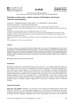
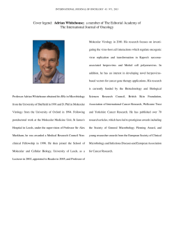
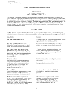
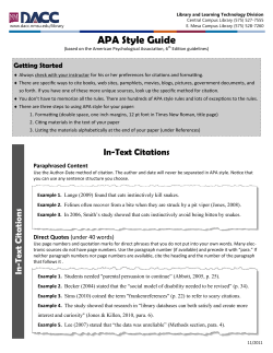
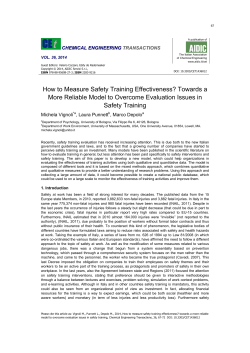
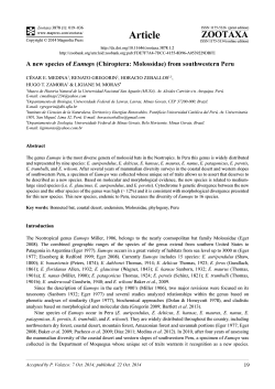
bioRxiv preprint first posted online January 6, 2015; doi: http://dx.doi.org/10.1101/013516; The copyright holder for
this preprint is the author/funder. All rights reserved. No reuse allowed without permission.
Olfactory receptors are sensitive to molecular
volume of odorants
Majid Saberi
Hamed Seyed-allaei∗
School of Cognitive Science,
Institute for Research in Fundamental Sciences (IPM),
Tehran, Iran
Abstract
To study olfaction, first we should know which physical or chemical properties of
odorant molecules determine the response of olfactory receptor neurons, and then
we should study the effect of those properties on the combinatorial encoding in
olfactory system.
In this work we show that the response of an olfactory receptor neuron in
Drosophila depends on molecular volume of an odorant; The molecular volume
determines the upper limits of the neural response, while the actual neural response
may depend on other properties of the molecules. Each olfactory receptor prefers
a particular volume, with some degree of flexibility. These two parameters predict
the volume and flexibility of the binding-pocket of the olfactory receptors, which
are the targets of structural biology studies.
At the end we argue that the molecular volume can affects the quality of perceived smell of an odorant via the combinatorial encoding, molecular volume may
mask other underlying relations between properties of molecules and neural responses and we suggest a way to improve the selection of odorants in further experimental studies.
1
Introduction
Survival of many species depends on their olfactory system. They use it
to search for food, avoid poison, escape from danger, find mate, and bind
to their offspring. An olfactory system detects volatile chemicals in the
∗
[email protected]
1
bioRxiv preprint first posted online January 6, 2015; doi: http://dx.doi.org/10.1101/013516; The copyright holder for
this preprint is the author/funder. All rights reserved. No reuse allowed without permission.
1 Introduction
(a) Binding-pocket volume
2
(b) Binding-pocket flexibility
Fig. 1: This figure shows different scenarios that may happen when an odorant molecule (ligand) binds to a receptor. Fig. 1a shows the effect
of binding-pocket volume. From left to right, misfit because of small
volume of molecule, perfect match and misfit because of large molecular volume. Fig. 1b demonstrates that the flexibility of a receptor
may compensate for the volume mismatches. The red disks (dark
grey in b&w) are odorant molecule, and the blue shapes (light grey
in b&w) are olfactory receptor and binding-pocket.
surrounding, encodes the results and transmit them to limbic system and
cortex.
The front end of the olfactory system are olfactory receptor neurons.
Each neuron expresses only one kind of olfactory receptor (in insects they
are co-expressed with Orco [1]), neurons of the same type converge into the
same glomeruli of the olfactory bulb (or antenatal lobe in insects), so that
each glomerulus of olfactory bulb receives an amplified signal from only one
type of olfactory receptor [2–10].
From neural recordings we know that the olfactory systems use a combinatorial code: an olfactory receptor can be triggered by different odorant
molecules, and an odorant molecule can excite different olfactory receptors [11]. The combinatorial code helps the olfactory system to discriminates trillion odors [12]. However, it is not clear yet which properties of a
molecule contribute to its smell, it is a topic of ongoing researches and there
are many theories [13–24].
In this study, we investigate the relation between molecular volumes
of odorants and the responses of olfactory receptor neurons. Our results
suggest that molecular volume is a considerable factor, but not the only
factor that determines the neural response of the olfactory receptor neurons.
The olfactory receptors are transmembrane proteins. In vertebrates,
they are metabotropic receptors, they belong to the family of g-protein coupled receptor (GPCR), Linda B. Buck and Richard Axel won the Nobel
bioRxiv preprint first posted online January 6, 2015; doi: http://dx.doi.org/10.1101/013516; The copyright holder for
this preprint is the author/funder. All rights reserved. No reuse allowed without permission.
1 Introduction
3
Prize in Physiology or Medicine, in 2004, for the discovery of this [25].
There are many similarities between the olfactory system of insects and
vertebrates [26, 27], and it was assumed that insects use the same kind of
signal transduction [28, 29], but recently, it has been argued that the olfactory receptors in insects are inotropic [30–33], their topology is different
from vertebrates [34, 35], and they function in presence of another common
receptor, called Orco [1].
Regardless of the signal transduction, all olfactory receptor have the
same function, they have a binding-pocket (also known as binding-cavity and
binding-site), where the ligands (odorants) bind to. This binding activates
the receptors and the activated receptor changes the potential of the cell,
directly (inotropic) or indirectly (metabotropic).
The amount of change in the membrane potential of a olfactory receptor
neuron depends on the number of activated olfactory receptor proteins and
the time that they remain activated, which are determined by various physiochemical properties of the ligand (odorant) and the receptor [13, 15, 19],
but here we focus only on two of them: the volume and the flexibility of
the binding-pocket. The molecular volume of a ligand should match the
dimensions of the binding-pocket of the receptor, then it fits into the bindingpocket of the receptor and triggers the signal transduction. Any mismatch in
the volumes will affect the neural responses (Fig. 1a), on the other hand the
flexibility of the binding-pocket can compensate for the volume mismatch
(Fig. 1b),
We could know the volume and flexibility of the binding-pocket, if we
knew its three dimensional structure. But this is not the case here, it is
not easy to know the structure of integral proteins [36, 37], including olfactory receptor. It is the topic of ongoing researches, using various methods
like Molecular Dynamic (MD) simulations, mutagenesis studies, heterologus
expression studies, and homology modeling [38–46]. In this study, we use
neural recording to predict the volume and flexibility of binding-pocket of
olfactory receptors, in-vivo.
In this study we suggest a functional relation between molecular volume
and the neural responses, we provide a methodology to estimate chemical
range or tuning function of olfactory receptors, and then we predict the
structural properties of the binding-pocket of olfactory receptor - the volume and the flexibility of binding-pocket. Our results may help to odorant
selection of new experimental studies, may provide additional information
about the structure of olfactory receptors to structural biologists, and may
contribute to the study of olfactory coding.
To perform this study we use a public domain, well structured database
bioRxiv preprint first posted online January 6, 2015; doi: http://dx.doi.org/10.1101/013516; The copyright holder for
this preprint is the author/funder. All rights reserved. No reuse allowed without permission.
2 Material and methods
4
Frequency
100
80
60
40
20
0
0
50
100
150
200
250
° 3)
Molecular Volume (A
Fig. 2: Density function of molecular volumes (g(v)), considering all
molecules of DoOR database. The actual density function of molecular of volumes in each experiment (g(v)) might be slightly different
because each experiment uses a different subset of molecules. The
solid line is a Gaussian fit (Eq. 5) and the dashed line shows the
median, which is slightly different from the mean.
– DoOR – that includes the neural responses of most olfactory receptors
(OR) of Drosophila to many odorants [47]. This database has collected its
data from many other sources [18, 20, 48–60].
2
Material and methods
We want to study the relation between neural responses and molecular volumes, so we need the respective data. We take the neural data of DoOR
database [47] and we calculate molecular volume (supplemental file 3) using
a computational chemistry software – VEGA ZZ [61]. We used GNU R to
analyse the data [62].
DoOR database can be summarized in an N × M matrix. Its elements
rnm , are the response of neuron n to odorant m. This matrix is normalized
between 0 and 1 so we have 0 ≤ rnm ≤ 1, where 1 is the strongest response.
The only problem is that this matrix has some Not Available (NA) values,
different P
neurons are excited by different
set of odorants, so when summing
P
over m, m , we are calculating m:rnm 6=NA , but for simplicity, we use the
former notation.
The response rnm depends on the molecular volume of the odorant, vm ,
and other physio-chemical properties of the molecule m; We assume that we
can separate the response rnm into two terms:
rnm = fn (vm )ψnm .
(1)
The first term, fn (vm ), depends only on the molecular volume of odorants.
bioRxiv preprint first posted online January 6, 2015; doi: http://dx.doi.org/10.1101/013516; The copyright holder for
this preprint is the author/funder. All rights reserved. No reuse allowed without permission.
2 Material and methods
5
The second term, ψnm include every other influential properties of molecules,
but the molecular volume. Both terms are characteristic of each receptor,
and they might vary from neuron to neuron. In fact, the first term, fn (v),
is the tuning curve of neuron n in respect to the molecular volumes, it can
be approximated with a Gaussian function
fn (v) = e
−
(v−vn )2
2
2σn
,
(2)
where, vn is the preferred molecular volume of receptor n and σn represents
its flexibility. In this work we want to estimate vn and σn . To doP so, first
m vm rnm
we calculate the response weighted average of molecular volumes, P
m rnm
and then we use (1):
X
X
vm rnm
vm fn (vm )ψnm
m
X
rnm
= mX
m
Here we can approximate
.
(3)
fn (vm )ψnm
m
P
R
with , which is common in statistical physics:
Z ∞
X
. . . fn (vm )ψnm ≈ hψnm im
. . . fn (v)g(v)dv.
(4)
0
m
In which, hψnm im denotes the average of ψnm over all m : rnm 6= NA. It
can be moved out of the integral for it is independent of v. In the above
equation, g(v) is the density of states, g(v)dv indicates how many molecules
have a molecular volume in the range of v and v + dv. This function can be
approximated by a Gaussian function, Fig.2,
−
g(v) = e
(v−vg )2
2
2σg
,
(5)
ideally, g(v) should not depend on the neuron n, it is the property of ensemble of odorant molecules, not neurons. But here, we have many missing
values (rnm = N A), so we have to calculate g(v) for each neuron separately;
Therefore, vgn and σgn are the average and standard deviation of molecular
volume while rnm 6= NA. Now we rewrite equation (3) using equation (4):
Z
X
vm rnm
vfn (v)gn (v)dv
m
X
≈ Z
.
(6)
rnm
fn (v)gn (v)dv
m
bioRxiv preprint first posted online January 6, 2015; doi: http://dx.doi.org/10.1101/013516; The copyright holder for
this preprint is the author/funder. All rights reserved. No reuse allowed without permission.
2 Material and methods
6
We replace the product of fn (v) and gn (v) in the above equation with
hn (v) = fn (v)gn (v), to make a simpler form
Z
X
vm rnm
vhn (v)dv
m
X
rnm
≈ Zv
.
(7)
hn (v)dv
m
v
The function hn (v) is a Gaussian function because it is the product of two
Gaussian functions,
−
hn (v) = e
(v−µh )2
n
2σ 2
hn
,
(8)
so the right hand side of equation 7 is nothing but µhn and in a similar way,
we can calculate σhn from the neural data
X
vm rnm
µhn
≈
m
X
(9)
rnm
m
X
σh2n
≈
2
vm
rnm
m
X
rnm
− µ2hn
(10)
m
We knew the mean vgn and standard deviation σgn of gn (v) from the
molecular volumes of the ensembles of odorants. We just calculated the
mean µhn and standard deviation σhn of hn (v) from the neural data. Now
calculating the mean vn and the standard deviation σn of fn (v) is trivial,
first we calculate σn from
1
1
1
= 2 − 2
σn2
σ gn
σhn
(11)
and then we calculate vn :
vn = σn2
vg
µhn
− 2n
2
σ gn
σhn
!
.
(12)
The calculated vn and σn are in supplemental file 1. The resulting fn (v)
are plotted over the actual data, for 32 receptors (Fig. 3a), in which the
relative error of vn is lesser than 25% and σn < 80˚
A3 , and for one receptor
just magnify the details (Fig. 3b). Now we know the preferred volume vn of
each receptor and also its flexibility σn .
bioRxiv preprint first posted online January 6, 2015; doi: http://dx.doi.org/10.1101/013516; The copyright holder for
this preprint is the author/funder. All rights reserved. No reuse allowed without permission.
3 Results and discussions
3
7
Results and discussions
There are two main assumption in this work: First we assumed that the
response of an olfactory receptor can be factorized into two terms, according
to (1). Second, we assumed that the volume dependence factor fn (vm ) in
(1) have a Gaussian form (Eq. 2). Considering the physics and chemistry
behind the binding-process (Fig. 1), and the neural responses (Fig. 3), these
assumptions are logical.
The function fn (v) can be considered as the tuning curve of olfactory
receptor n in response to molecular volume (Fig. 3). Each receptor has a
preferred molecular volume vn and shows some flexibility σn . We calculated
the parameters of fn (v) for 32 receptors (Fig. 3). The calculated values, vn
and σn are in Fig. 4a and 4b respectively. Figure 4a demonstrate that the
molecular volume preference of receptors are different. Figure 4b illustrate
that the flexibility of receptors are also different.
This diversity is important in perceiving the quality of smells. In a hypothetical experiment, assume that every characteristic of odorant molecules
are the same but their molecular volume. If all olfactory receptors had
the same preferred volume and flexibility, any change in the molecular volume would change only the intensity of smell not its quality. But olfactory
receptors have different preferred volumes and flexibilities, so any change
in the molecular volume of an odorant results in a different combinatorial
encoding which affects the quality of perceived smell as well. That may
describe the difference in the smell of methanol, ethanol, propanol and butanol. Methanol smells pungent, ethanol smells pleasant and winy, propanol
smells like ethanol while butanol is similar to ethanol with little banana like
aroma. The molecular volume affects the combinatorial encoding.
Here we showed that the responses of olfactory receptor neurons are
related to the molecular volume of odorants, apart from that, it is not clear
which other features of molecules are measured by olfactory receptors. It
is a topic of ongoing researches , there are many works that try to connect
the physio-chemical properties of molecules to the evoked neural response
or perceived smells. But the non-linear volume dependence (Eq. 1 and Eq.
2) may mask important relations between molecules and neural responses.
By considering the effect of molecular volume on the response of olfactory
receptor neurons, one might discover more subtle dependence between other
molecular features and neural responses, by studding ψnm , which otherwise
would be masked by this non-linear relation fn (v).
We also predict some in-vivo structural aspects of the binding-pocket of
olfactory receptors: the preferred volume of each receptor results from the
bioRxiv preprint first posted online January 6, 2015; doi: http://dx.doi.org/10.1101/013516; The copyright holder for
this preprint is the author/funder. All rights reserved. No reuse allowed without permission.
3 Results and discussions
8
Or10a
Or1a
Or22a
Or22b
Or23a
Or24a
Or2a
Or30a
Or33a
Or35a
Or42a
Or42b
Or43a
Or43b
Or45b
Or46a
Or47a
Or49a
Or49b
Or59a
Or59b
Or67a
Or67b
Or67c
Or71a
Or85a
Or85b
Or85c
Or85f
Or94b
Or98a
Or9a
0.8
0.4
0
0.8
0.4
0
0.8
0.4
0
200
0
100
200
0
100
200
0
100
200
0
100
200
0
100
200
0
100
200
0
100
200
0
100
0.8
0.4
0
(a)
1
Response
0.8
0.6
0.4
0.2
0
0
50
100
150
Molecular Volume
200
250
3
(A° )
(b)
Fig. 3: Response of olfactory receptors versus molecular volume of odorants
(Circles), the fitted functions fn (v) from Eq. 1 (solid lines), and the
error bars of the mean of fn (v) (red vertical lines), for 32 selected
receptors (Fig. 3a) and for one selected receptor Or35a (Fig. 3b) just
to magnify details.
bioRxiv preprint first posted online January 6, 2015; doi: http://dx.doi.org/10.1101/013516; The copyright holder for
this preprint is the author/funder. All rights reserved. No reuse allowed without permission.
3 Results and discussions
9
Prefered Volume
3
(A° )
150
100
50
Or10a
Or1a
Or22a
Or22b
Or23a
Or24a
Or2a
Or30a
Or33a
Or35a
Or42a
Or42b
Or43a
Or43b
Or45b
Or46a
Or47a
Or49a
Or49b
Or59a
Or59b
Or67a
Or67b
Or67c
Or71a
Or85a
Or85b
Or85c
Or85f
Or94b
Or98a
Or9a
0
(a) The preferred volumes of 32 receptors (vn ), and their error bars. The error
bars are calculated using Jack-Knife method. Some receptors prefer smaller
molecules - like Or59b, Or67b and Or85a, but some other receptors prefer larger
molecules - like Or85c, Or1a and Or49a.
Flexibility
3
(A° )
100
80
60
40
20
Or10a
Or1a
Or22a
Or22b
Or23a
Or24a
Or2a
Or30a
Or33a
Or35a
Or42a
Or42b
Or43a
Or43b
Or45b
Or46a
Or47a
Or49a
Or49b
Or59a
Or59b
Or67a
Or67b
Or67c
Or71a
Or85a
Or85b
Or85c
Or85f
Or94b
Or98a
Or9a
0
(b) The flexibility of each receptor (σn ), the error bars are calculated using JackKnife method. Some receptors like Or46a, Or71a and Or22b are volume selective, but some other receptors like Or22a, Or67b and Or33a show flexibility
and respond to broader range of molecular volumes.
Fig. 4
bioRxiv preprint first posted online January 6, 2015; doi: http://dx.doi.org/10.1101/013516; The copyright holder for
this preprint is the author/funder. All rights reserved. No reuse allowed without permission.
4 Acknowledgments
10
volume of the binding-pocket, the flexibility of a receptor results from the
rigidity or flexibility of the binding-pocket; These data add some constrains
over the 3d structure of olfactory receptors, which may help the prediction
and calculation of 3d structure of these proteins.
The method of this work can be combined with mutagenesis as well.
Some genes of an olfactory receptor are mutated, then its response to a
selection of molecules are measured and finally the preferred volume and
flexibility are calculated. In this way we can understand which amino acids of
the olfactory receptor contribute to the volume and flexibility of the bindingpocket, as well as affecting the function of the receptors.
Our finding can also save time and expenses of experiments by suggesting
important odorants for every receptors. To study ψnm of a receptor, it is
better to have many data points and those data points are better to be
around the preferred volume of the receptor. But this is not the case in
current data, for many receptors, most data points are on the tails of fn (v),
which is close to zero. We suggested the best selection of odorants for each
of 32 studied receptors (see Venn diagram in Fig. 5 and supplemental file
2).
Although this work is on the data of Drosophila, we expect that the
general principle and methodology of this work hold for vertebrates as well.
But considering the similarities and dissimilarities between insects and vertebrate, this should be verified and more work are necessary.
4
Acknowledgments
We are especially grateful to B. N. Araabi, S. Aghvami and N. Doostani
for the careful reading of the manuscript; and P. Carloni for the fruitful
discussion.
References
[1] Mattias C Larsson et al. “Or83b encodes a broadly expressed odorant receptor essential for Drosophila olfaction.” In: Neuron 43.5 (Oct.
2004), pp. 703–14. doi: 10.1016/j.neuron.2004.08.019.
[2] Cory M Root et al. “Propagation of olfactory information in Drosophila”.
In: Proceedings of the National Academy of Sciences 104.28 (2007),
pp. 11826–11831.
bioRxiv preprint first posted online January 6, 2015; doi: http://dx.doi.org/10.1101/013516; The copyright holder for
this preprint is the author/funder. All rights reserved. No reuse allowed without permission.
REFERENCES
11
Number of Odorants
240
200
160
120
80
40
Or10a
Or1a
Or22a
Or22b
Or23a
Or24a
Or2a
Or30a
Or33a
Or35a
Or42a
Or42b
Or43a
Or43b
Or45b
Or46a
Or47a
Or49a
Or49b
Or59a
Or59b
Or67a
Or67b
Or67c
Or71a
Or85a
Or85b
Or85c
Or85f
Or94b
Or98a
Or9a
0
Fig. 5: Venn diagram of DoOR database and our suggested important odorants of each receptor. The database includes 240 odorant molecules,
only a fraction of them are used to study an olfactory receptor (blue
shades) and data for the rest of odorants are not available (pink
shades). The hatched area are odorants with molecular volume close
to the preferred volume of each receptor (vn ± σ2 ). We consider them
as important samples to study the molecular basis of olfaction, We already know the neural response of hatched blue area, but the hatched
pink odorants could be the target of further experiments.
bioRxiv preprint first posted online January 6, 2015; doi: http://dx.doi.org/10.1101/013516; The copyright holder for
this preprint is the author/funder. All rights reserved. No reuse allowed without permission.
REFERENCES
12
[3] Allison F Carey and John R Carlson. “Insect olfaction from model systems to disease control.” In: Proceedings of the National Academy of
Sciences of the United States of America 108.32 (Aug. 2011), pp. 12987–
95. doi: 10.1073/pnas.1103472108.
[4] Leslie B Vosshall, Allan M Wong, and Richard Axel. “An Olfactory
Sensory Map in the Fly Brain”. In: Cell 102.2 (July 2000), pp. 147–
159. doi: 10.1016/S0092-8674(00)00021-0.
[5] Africa Couto, Mattias Alenius, and Barry J Dickson. “Molecular, anatomical, and functional organization of the Drosophila olfactory system.”
In: Current biology : CB 15.17 (Sept. 2005), pp. 1535–47. doi: 10.
1016/j.cub.2005.07.034.
[6] Elane Fishilevich and Leslie B Vosshall. “Genetic and Functional Subdivision of the¡ i¿ Drosophila¡/i¿ Antennal Lobe”. In: Current Biology
15.17 (2005), pp. 1548–1553.
[7] Qian Gao, Bingbing Yuan, and Andrew Chess. “Convergent projections of Drosophila olfactory neurons to specific glomeruli in the antennal lobe”. In: Nature neuroscience 3.8 (2000), pp. 780–785.
[8] Fan Wang et al. “Odorant receptors govern the formation of a precise
topographic map”. In: Cell 93.1 (1998), pp. 47–60.
[9] Peter Mombaerts et al. “Visualizing an olfactory sensory map”. In:
Cell 87.4 (1996), pp. 675–686.
[10] Robert Vassar et al. “Topographic organization of sensory projections
to the olfactory bulb”. In: Cell 79.6 (1994), pp. 981–991.
[11] Bettina Malnic et al. “Combinatorial Receptor Codes for Odors”. In:
96 (2000), pp. 713–723.
[12] C Bushdid et al. “Humans can discriminate more than 1 trillion olfactory stimuli.” In: Science (New York, N.Y.) 343.6177 (Mar. 2014),
pp. 1370–2. doi: 10.1126/science.1249168.
[13] Luca Turin, Gower Street, and Gower Street. “ORIGINAL RESEARCH
PAPER A Spectroscopic Mechanism for Primary Olfactory”. In: ().
[14] Andreas Keller and Leslie B Vosshall. “A psychophysical test of the
vibration theory of olfaction.” In: Nature neuroscience 7.4 (Apr. 2004),
pp. 337–8. doi: 10.1038/nn1215.
[15] Ricardo C Araneda, Abhay D Kini, and Stuart Firestein. “The molecular receptive range of an odorant receptor”. In: 3.12 (2000).
bioRxiv preprint first posted online January 6, 2015; doi: http://dx.doi.org/10.1101/013516; The copyright holder for
this preprint is the author/funder. All rights reserved. No reuse allowed without permission.
REFERENCES
13
[16] Jennifer Brookes et al. “Could Humans Recognize Odor by Phonon
Assisted Tunneling?” In: Physical Review Letters 98.3 (Jan. 2007),
p. 038101. doi: 10.1103/PhysRevLett.98.038101.
[17] Maria Isabel Franco et al. “Molecular vibration-sensing component in
Drosophila melanogaster olfaction.” In: Proceedings of the National
Academy of Sciences of the United States of America 108.9 (Mar.
2011), pp. 3797–802. doi: 10.1073/pnas.1012293108.
[18] Daniela Pelz et al. “The Molecular Receptive Range of an Olfactory
Receptor in vivo ( Drosophila melanogaster Or22a )”. In: November
(2006), pp. 1544–1563. doi: 10.1002/neu.
[19] Stephan Gabler et al. “Physicochemical vs. Vibrational Descriptors for
Prediction of Odor Receptor Responses”. In: Molecular Informatics
32.9-10 (Oct. 2013), pp. 855–865. doi: 10.1002/minf.201300037.
[20] Michael Schmuker et al. “Predicting olfactory receptor neuron responses from odorant structure.” In: Chemistry Central journal 1 (Jan.
2007), p. 11. doi: 10.1186/1752-153X-1-11.
[21] Rafi Haddad et al. “Predicting the receptive range of olfactory receptors.” In: PLoS computational biology 4.2 (Feb. 2008), e18. doi:
10.1371/journal.pcbi.0040018.
[22] Kobi Snitz et al. “Predicting odor perceptual similarity from odor
structure.” In: PLoS computational biology 9.9 (Jan. 2013), e1003184.
doi: 10.1371/journal.pcbi.1003184.
[23] Adi Yablonka et al. “Odorant similarity in the mouse olfactory bulb”.
In: 109.43 (2012), pp. 2916–2917. doi: 10.1073/pnas.1211623109.
[24] Simon Gane et al. “Molecular vibration-sensing component in human
olfaction”. In: PloS one 8.1 (2013), e55780.
[25] Linda Buck and Richard Axel. “A novel multigene family may encode
odorant receptors: A molecular basis for odor recognition”. In: Cell
65.1 (Apr. 1991), pp. 175–187. doi: 10.1016/0092-8674(91)90418X.
[26] Rachel I Wilson. “NIH Public Access”. In: (2014), pp. 217–241. doi:
10.1146/annurev-neuro-062111-150533.Early.
[27] U Benjamin Kaupp. “Olfactory signalling in vertebrates and insects:
differences and commonalities.” In: Nature reviews. Neuroscience 11.3
(Mar. 2010), pp. 188–200. doi: 10.1038/nrn2789.
bioRxiv preprint first posted online January 6, 2015; doi: http://dx.doi.org/10.1101/013516; The copyright holder for
this preprint is the author/funder. All rights reserved. No reuse allowed without permission.
REFERENCES
14
[28] Thomas Brody and Anibal Cravchik. “Drosophila melanogaster G
Protein-coupled Receptors”. In: 150.2 (2000), pp. 83–88.
[29] Catherine A. Hill et al. “G Protein-Coupled Receptors in Anopheles
gambiae”. In: Science 298.5591 (2002), pp. 176–178. doi: 10.1126/
science.1076196. eprint: http://www.sciencemag.org/content/
298/5591/176.full.pdf.
[30] Koji Sato et al. “Insect olfactory receptors are heteromeric ligandgated ion channels.” In: Nature 452.7190 (Apr. 2008), pp. 1002–6.
doi: 10.1038/nature06850.
[31] Dieter Wicher et al. “Drosophila odorant receptors are both ligandgated and cyclic-nucleotide-activated cation channels.” In: Nature 452.7190
(Apr. 2008), pp. 1007–11. doi: 10.1038/nature06861.
[32] Katherine I Nagel and Rachel I Wilson. “Biophysical mechanisms underlying olfactory receptor neuron dynamics.” In: Nature neuroscience
14.2 (Feb. 2011), pp. 208–16. doi: 10.1038/nn.2725.
[33] Yueguang Rong et al. “Correction for Rong et al., Spinster is required for autophagic lysosome reformation and mTOR reactivation
following starvation”. In: Proceedings of the National Academy of Sciences 108.27 (June 2011), pp. 11297–11297. doi: 10 . 1073 / pnas .
1108410108.
[34] Richard Benton, Kirsten S Vannice, and Leslie B Vosshall. “An essential role for a CD36-related receptor in pheromone detection in
Drosophila.” In: Nature 450.7167 (Nov. 2007), pp. 289–93. doi: 10.
1038/nature06328.
[35] Renee Smart et al. “Drosophila odorant receptors are novel seven
transmembrane domain proteins that can signal independently of heterotrimeric G proteins.” In: Insect biochemistry and molecular biology
38.8 (Aug. 2008), pp. 770–80. doi: 10.1016/j.ibmb.2008.05.002.
[36] Yang Zhang. “Progress and challenges in protein structure prediction.”
In: Current opinion in structural biology 18.3 (June 2008), pp. 342–8.
doi: 10.1016/j.sbi.2008.02.004.
[37] Paola Lupieri et al. “Computational molecular biology approaches to
ligand-target interactions.” In: HFSP journal 3.4 (Aug. 2009), pp. 228–
39. doi: 10.2976/1.3092784.
[38] Kamil Khafizov et al. “Ligand specificity of odorant receptors.” In:
Journal of molecular modeling 13.3 (Mar. 2007), pp. 401–9. doi: 10.
1007/s00894-006-0160-9.
bioRxiv preprint first posted online January 6, 2015; doi: http://dx.doi.org/10.1101/013516; The copyright holder for
this preprint is the author/funder. All rights reserved. No reuse allowed without permission.
REFERENCES
15
[39] Orna Man and Yoav Gilad. “Prediction of the odorant binding site
of olfactory receptor proteins by human - mouse comparisons”. In:
(2004), pp. 240–254. doi: 10.1110/ps.03296404.lical.
[40] Peter C Lai, Michael S Singer, and Chiquito J Crasto. “Structural
activation pathways from dynamic olfactory receptor-odorant interactions.” In: Chemical senses 30.9 (Nov. 2005), pp. 781–92. doi: 10 .
1093/chemse/bji070.
[41] Nagarajan Vaidehi et al. “Prediction of structure and function of G
protein- coupled receptors”. In: (2002).
[42] Wely B Floriano, Nagarajan Vaidehi, and William a Goddard. “Making sense of olfaction through predictions of the 3-D structure and
function of olfactory receptors.” In: Chemical senses 29.4 (May 2004),
pp. 269–90. doi: 10.1093/chemse/bjh030.
[43] Kristin Schmiedeberg et al. “Structural determinants of odorant recognition by the human olfactory receptors OR1A1 and OR1A2.” In:
Journal of structural biology 159.3 (Sept. 2007), pp. 400–12. doi: 10.
1016/j.jsb.2007.04.013.
[44] Sayako Katada et al. “Structural basis for a broad but selective ligand spectrum of a mouse olfactory receptor: mapping the odorantbinding site.” In: The Journal of neuroscience : the official journal
of the Society for Neuroscience 25.7 (Feb. 2005), pp. 1806–15. doi:
10.1523/JNEUROSCI.4723-04.2005.
[45] Aya Kato, Sayako Katada, and Kazushige Touhara. “Amino acids involved in conformational dynamics and G protein coupling of an odorant receptor: targeting gain-of-function mutation.” In: Journal of neurochemistry 107.5 (Dec. 2008), pp. 1261–70. doi: 10.1111/j.14714159.2008.05693.x.
[46] Jean-Pierre Rospars. “Interactions of odorants with olfactory receptors
and other preprocessing mechanisms: how complex and difficult to
predict?” In: Chemical senses 38.4 (May 2013), pp. 283–7. doi: 10.
1093/chemse/bjt004.
[47] C Giovanni Galizia et al. “Integrating heterogeneous odor response
data into a common response model: A DoOR to the complete olfactome.” In: Chemical senses 35.7 (Sept. 2010), pp. 551–63. doi:
10.1093/chemse/bjq042.
bioRxiv preprint first posted online January 6, 2015; doi: http://dx.doi.org/10.1101/013516; The copyright holder for
this preprint is the author/funder. All rights reserved. No reuse allowed without permission.
REFERENCES
16
[48] Marien De Bruyne, Peter J Clyne, and John R Carlson. “Odor Coding
in a Model Olfactory Organ : The Drosophila Maxillary Palp”. In:
19.11 (1999), pp. 4520–4532.
[49] Marien De Bruyne et al. “Odor Coding in the Drosophila Antenna”.
In: 30 (2001), pp. 537–552.
[50] Anna A Dobritsa et al. “Integrating the Molecular and Cellular Basis
of Odor Coding in the Drosophila Antenna”. In: 37 (2003), pp. 827–
841.
[51] Aaron L Goldman et al. “Coexpression of two functional odor receptors in one neuron.” In: Neuron 45.5 (Mar. 2005), pp. 661–6. doi:
10.1016/j.neuron.2005.01.025.
[52] Elissa A Hallem et al. “The Molecular Basis of Odor Coding in the
Drosophila Antenna”. In: 117 (2004), pp. 965–979.
[53] Elissa a Hallem and John R Carlson. “Coding of odors by a receptor
repertoire.” In: Cell 125.1 (Apr. 2006), pp. 143–60. doi: 10.1016/j.
cell.2006.01.050.
[54] Scott a Kreher, Jae Young Kwon, and John R Carlson. “The molecular
basis of odor coding in the Drosophila larva.” In: Neuron 46.3 (May
2005), pp. 445–56. doi: 10.1016/j.neuron.2005.04.007.
[55] Scott a Kreher et al. “Translation of sensory input into behavioral
output via an olfactory system.” In: Neuron 59.1 (July 2008), pp. 110–
24. doi: 10.1016/j.neuron.2008.06.010.
[56] Jae Young Kwon et al. “The molecular basis of CO2 reception in
Drosophila.” In: Proceedings of the National Academy of Sciences of
the United States of America 104.9 (Mar. 2007), pp. 3574–8. doi: 10.
1073/pnas.0700079104.
[57] M. C. Stensmyr. “Novel natural ligands for Drosophila olfactory receptor neurones”. In: Journal of Experimental Biology 206.4 (Feb. 2003),
pp. 715–724. doi: 10.1242/jeb.00143.
[58] Stephanie Lynn Turner and Anandasankar Ray. “Modification of CO2
avoidance behaviour in Drosophila by inhibitory odorants.” In: Nature
461.7261 (Sept. 2009), pp. 277–81. doi: 10.1038/nature08295.
[59] Wynand van der Goes van Naters and John R Carlson. “Receptors
and neurons for fly odors in Drosophila.” In: Current biology : CB
17.7 (Apr. 2007), pp. 606–12. doi: 10.1016/j.cub.2007.02.043.
bioRxiv preprint first posted online January 6, 2015; doi: http://dx.doi.org/10.1101/013516; The copyright holder for
this preprint is the author/funder. All rights reserved. No reuse allowed without permission.
REFERENCES
17
[60] C Andrea Yao, Rickard Ignell, and John R Carlson. “Chemosensory
coding by neurons in the coeloconic sensilla of the Drosophila antenna.” In: The Journal of neuroscience : the official journal of the
Society for Neuroscience 25.37 (Sept. 2005), pp. 8359–67. doi: 10 .
1523/JNEUROSCI.2432-05.2005.
[61] Alessandro Pedretti, Luigi Villa, and Giulio Vistoli. “VEGA - An open
platform to develop chemo-bio-informatics applications, using plug-in
architecture and script programming”. In: Journal of Computer-Aided
Molecular Design 18.3 (Mar. 2004), pp. 167–173. doi: 10.1023/B:
JCAM.0000035186.90683.f2.
[62] R Core Team. R: A Language and Environment for Statistical Computing. R Foundation for Statistical Computing. Vienna, Austria, 2014.


© Copyright 2026