
Tumor Necrosis Factor-a Modulation of Glycoprotein
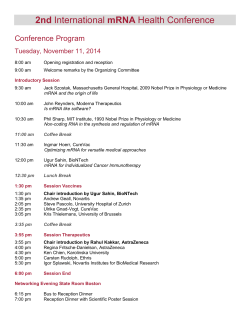
From www.bloodjournal.org by guest on January 12, 2015. For personal use only.
Tumor Necrosis Factor-a Modulation of Glycoprotein Iba Expression in Human
Endothelial and Erythroleukemia Cells
By Venkataraman Rajagopalan, David W. Essex, Sandor S.Shapiro, and Barbara A. Konkle
Glycoprotein Iba (Gplba) is a platelet membrane Gp that
binds von Willebrand factor and mediates platelet adhesion
to subendothelium. We have found both Gplba mRNA and
pratein in human umbilical vein endothelial cells (HUVEC). In
previously publishedwork we reported that combined treatment with interferon? (1FN-y) and tumor necrosis factor(TNF-a) markedly increased the Gplba mRNA level in HUVEC.
We have now documented that TNF-a alone induces Gplba
mRNA and protein expression, studied the kinetics of this
response, and investigated potential mechanisms of the
TNF-a effect. Gplba mRNA induction by TNF-a is detectable
as early as 2 hours after exposure to this cytokine, and
reaches a maximal level after 20 to 24 hours. Using a nuclear
run-on assay we found that Gplba gene transcription is
increased approximately IO-fold after 2 hours of TNF-a
treatment. Furthermore, using two monoclonal antibodies
that recognizedifferent epitopes of Gplba, we found that the
protein expression in endothelial cells is markedly increased
by TNF-a Interleukin-I (IL-1) and the phorbol ester phorbol
myristate acetate, which mimic many effects of TNF-a on
endothelial cells, have no effect on endothelial or human
erytholeukemia (HEL)-cell Gplba mRNA. TNF-a treatment for
24 hours increases the HEL cell Gplba mRNA level approximately fourfold, showing a time- and dose-dependent effect
similar to that seen In HUVEC. TNF-a-induced Gplba mRNA
and protein synthesis may play a role in mediating platelet or
other cell interaction with activated endothelium. Unlike
other endothelial pro-thrombotic and pro-adhesive proteins
induced by 7°F-a. Gplba is not induced by IL-I treatment,
which suggests a novel pathway for induction of this protein.
o 1902by The American Society of Hematology.
G
tor-a (TNF-a). In endothelial cells TNF-a induces a variety
of changes which, in general, are prothrombotic and promote cell adhesion. These include increased tissue factor
activity,12J3prothrombinase complex formation,14 and plasminogen activator inhibitor-1 synthesis (PAI-l),15 and decreased thrombomodulin expression,’6 as well as increased
expression of the leukocyte and lymphocyte adhesion proteins ELAM-1, VCAM-1, and ICAM-l.17-19 To begin to
elucidate the role of GpIba in this setting, we have
characterized the TNF-a effect on GpIba gene transcription and on mRNA and protein expression. This allows us
to compare the response of GpIba to TNF-a treatment with
that of other endothelial proteins. We have also found that
TNF-a treatment increases GpIba mRNA levels in HEL
cells in a manner analogous to that seen in HUVEC.
LYCOPROTEIN Ib (GpIb) exists in platelets as a
transmembrane protein composed of two disulfidelinked chains, termed the a and the P chains. In the
platelet, GpIba is a receptor for von Willebrand factor
(vWF) and, through this ligand, mediates platelet adhesion
to the subendothelium.i,2 GpIba also contains a binding site
for thrombin, although its physiologic significance is not
clear.
Endothelial cells and platelets both contain a number of
proteins involved in hemostasis, including the adhesive
proteins vWF, fibronectin, thrombospondin, and GMP-140,
as well as the integrin G P I I I ~ . Earlier
~,~
studies in our
laboratory, and independently by Asch et al, demonstrated
that human umbilical vein endothelial cells (HUVEC)
synthesize a protein immunologically related to platelet
GpIba.5,6 In analogy with platelets, ristocetin-dependent
endothelial cell vWF binding and ristocetin-dependent
endothelial cell agglutination in the presence of vWF were
found. Both of these effects were inhibited by a monoclonal
antibody (MoAb) to GpIba (AP-1).6 We recently reported
studies extending these original observations. GpIba protein was identified in tonsilar endothelium by immunohistochemistry? We also showed that the GpIba gene was
transcribed in HUVEC, that partial endothelial GpIba
cDNA clones were identical in sequence to portions of
HEL cell GpIba cDNA, and that the GpIba mRNA species
in HUVEC, total tonsilar tissue, and human erythroleukemia (HEL) cells were of the same size (approximately 2.8
kb).
HEL cells, a line derived from a patient with erythroleukemia, express, in addition to erythroid proteins, numerous
megakaryocyte proteins, including GpIba, especially when
stimulated with dimethyl sulfoxide or phorbol esters.8.9 In
fact, the cDNAs for GpIba and GpIbP were originally
cloned from an HEL-cell cDNA library.lOJ1It is known that
HEL-cells contain the same GpIba and GpIbP mRNA
species as platelets?
We reported previously7 that GpIba mRNA expression
was markedly increased in HUVEC after combined treatment with interferon-y (IFN-y) and tumor necrosis facBlood, VOl80, NO 1 (July I), 1992:pp 153-161
MATERIALS AND METHODS
Cell culture. HUVEC were isolated and propagated from
pooled primary cultures of human umbilical veins as previously
From The Cardeza Foundation for Hematologic Research, the
Department of Medicine, Jefferson Medical College of Thomas Jefferson University, Philadelphia, PA.
Submitted October 28, 1991; accepted March 11, 1992.
Supported in part by Grants HL09163 (S.S.S.), A G 04861 (S.S.S.),
and HL44956 (B.A.K.) from the National Institutes of Health, and
fellowships (KR.)from the American Heart Association of Delaware,
Inc, the Brandywine Valley Hemophilia Foundation, and the Delaware Valley Chapter of the National Hemophilia Foundation. This
work wasperfomted during the tenure of an American Heart Association-Squibb Corporation Clinician Scientist Award to B.A.K.
Presented in part at the Thiriy-second Annual Meeting of the
American Society of Hematology, December 1,1990, Boston, MA.
Address reprint requests to Barbara A . Konkle, MD, Cardeza
Foundation for Hematologic Research, Jefferson Medical College,
Thomas JeffersonUniversity, I015 Walnut Si, Philadelphia, PA 19107.
The publication costs of this article were defrayed in part by page
charge payment. This article must therefore be hereby marked
“advertisement” in accordance with 18 U.S.C.section 1734 soleb to
indicate this fact.
0 1992 by The American Society of Hematology.
0006-4971/92/8001-0024$3.00/0
153
From www.bloodjournal.org by guest on January 12, 2015. For personal use only.
RAJAGOPALAN ET AL
154
de~cribed.~
In a given experiment all HUVEC were grown in a
single lot of fetal bovine serum and endothelial cell growth factor.
HUVEC were passaged as a 1:4 split, fed every 1 to 3 days, and
used at the second to fourth passage. Human recombinant IFN-y
(> 2 x lo7 U/mg), TNF-a (> 2 x lo7 U/mg), and interleukin-1
(IL-1) ( > lo7 U/mg) were purchased from Boehringer Mannheim
(Indianapolis, IN). Recombinant TNF-a ( > 2 x lo7 U/mg) was
also purchased from Sigma Chemical Company (St Louis, MO).
HEL cells were obtained from the American Type Culture Collection (ATCC TIB 180, HEL 92.1.7; Rockville, MD)9 and propagated in RPMI 1640 (Sigma) containing 10% fetal bovine serum. A
stock solution of the phorbol ester 12-myristate 13-acetate (PMA)
was made in dimethyl sulfoxide and added to the culture medium
at a final concentration of 100 nmol/L.
RNA isolation and evaluation. Total cellular RNA was isolated
by solubilization of the confluent cells in guanidine hydrochloride,
as previously described2O Poly (A+) RNA was isolated directly
from cells using oligo (dT)-cellulose (Invitrogen, San Diego, CA).
RNA was analyzed by electrophoresis in a denaturing formaldehyde gel followed by Northern blotting onto a nylon membrane
(Hybond-N, Amersham Corp, Arlington Heights, IL). The RNA
was fixed to the membrane with ultraviolet irradiation, prehybridized in a solution of 1 mol/L NaC1, 0.1% sodium dodecyl sulfate
(SDS), 1.5 mg/mL sonicated herring sperm DNA, and 10%
dextran for 3 hours at 68“C, and hybridized at 68°C for 12 to 24
hours in the prehybridization solution with additional herring
sperm DNA (1.5 mg/mL) and the appropriate radiolabeled probe.
Probes used were: (1) GpIba cDNA, clone G ~ I b 2 . 4(kindly
~~
provided by Dr Jose Lopez, Gladstone Laboratories, San Francisco, CA); (2) cDNA for phosphoglycerate kinase (PGK), the
1.8-kb PstI insert of pHPGK-7eZ1; (3) endothelial cell GpIba
cDNA, clone GP@47;and (4) PAI-1 cDNA, the 2-kb EcoRI insert
of PAIB6? The cDNA inserts were radiolabeled directly in
low-melting agarose by random hexamer priming.23The blots were
washed to high stringency in 0.1X SSC (2.25 mol/L NaCI, 0.225
mol/L sodium citrate), 0.1% SDS, 1 mmol/L Na2-EDTA, 10
mmol/L sodium phosphate (pH 6.8), at 68°C and analyzed by
autoradiography. Individual blots were rehybridized with a second
radiolabeled probe after incubation in a solution of 5 mmol/L
Tris-HC1 (pH 7.5), 2 mmol/L Na2-EDTA, 0.1X Denhardt’sZ4at
68°C for 3 to 4 hours to remove the original probe. Successful
“stripping” was confirmed by autoradiography. Densitometry of
the autoradiographs was performed using an LKB Ultrascan XL
Laser Densitometer (Piscataway, NJ).
Nuclear run-on transctiption assay. HUVEC were washed with
Dulbecco’s Ca2+-free, Mg2+-free phosphate-buffered saline (DPBS) (GIBCO, Grand Island, NY), exposed to trypsin-EDTA
(GIBCO) for 10 seconds, and harvested by rinsing with D-PBS.
Nuclei were harvested by gently vortexing the cells in Nonidet-40
lysis buffer (Particle Data Labs LTD, Elmhurst, IL) (10 mmol/L
Tris-Hcl [pH 7.41, 10 mmol/L NaCI, 3 mmol/L MgC12, 0.5%
Nonidet-40), followed by centrifugation at 80Og. Newly transcribed
RNA was isolated as described by Lindsten et al? The radiolabeled RNA was hybridized to pUC-based plasmids (5 pg) with
gene-specific inserts as indicated, denatured by alkali treatment,
and transferred to nitrocellulose filters in a slot-blot apparatus.
Plasmids contained cDNA inserts for (1) GpIba, (2) vWFpVWH33, containing a 3.8 kb vWF cDNAEcoRI insert, or vWFD2,
an 8.5-kb full-length cDNA,Z6 or (3) tissue-type plasminogen
activator (t-PA) cDNA, the 2-kb BglII fragment (middle probe) of
full-length t-PA cDNA?~In addition, the plasmid pGEM without a
cDNA insert was used as a control.
Immunohistochemical studm AP-lZ8and 6D-l,29 murine MoAbs recognizing different epitopes in the 45-Kd N-terminus of
human platelet GpIba, were generous gifts of Dr Robert Montgomery (Blood Center of SE Wisconsin, Milwaukee) and Dr Barry
Coller (SUNY Stony Brook, Stony Brook, NY), respectively.
MOPC 21 (raised against mineral oil and obtained from Sigma
Chemical Co,St Louis, M a ) was used as a negative control. Rabbit
antimouse IgG and alkaline phosphatase-anti-alkaline phosphatase (APAAP) complex were obtained from the DAKO Corporation (Carpinteria, CA), and the substrate reagent for alkaline
phosphatase was obtained from Sigma. Confluent primary
HUVEC cultures, prepared as described above, were detached by
incubation with 0.05% trypsin and 0.53 mmol/L EDTA at room
temperature for 5 minutes, seeded at half-confluent concentrations
onto four-chamber Lab-Tek slides (Nunc, Inc, Napervilld, IL) that
had been coated with 0.2% gelatin, and grown to confluence (2 to 3
days). Confluent HUVEC were treated with TNF-a (50 U/mL) or
with buffer for 24 hours (37°C in a 5% COz incubator), after which
the slides were washed twice with Medium 199 (GIBCO) and
air-dried for 2 hours. The monolayers were then fixed with acetone
at 4°C for 30 minutes, incubated with AP-1for 1hour, and probed
for AP-1 binding using a slight modification of the published
APAAP technique30: The monolayers were washed for 1 to 2
minutes with TBS (0.05 mol/L Tris, 0.15 mol/L NaCI, pH 7.6), and
then incubated with rabbit antimouse IgG for 30 minutes. After
further washing, the monolayers were incubated with APAAP
complex for 30 minutes and washed. The incubations with rabbit
antimouse IgG and APAAP were repeated, this time for Ib
minutes each, after which the alkaline phosphatase substrate was
added. After incubation for 20 to 30 minutes, the endothelial
monolayers were washed and counterstained with hematoxylin.
Western blotting ofplatelet and HUVECproteins. Platelets were
prepared as previously de~cribed.~’
After the final wash the platelet
pellet was solubilized in 1% SDS with or without 10% B-mercaptoethanol (BME). Confluent second passage HUVEC were detached by incubation for 20 minutes at 37°C in a buffer composed
of 150 mmol/L NaCI, 2 mmol/L Tris, 10 mmol/L EDTA, 1
mmol/L phenylmethylsulfonyl fluoride (PMSF), 5 mmol/L benzamidine, 200 kU/mL aprotinin, and 200 p,g/mL leupeptin, pH 7.4.
The cells were centrifuged at 1,lOOg for 10 minutes and an equal
volume of 2% SDS with or without 10% BHE was added to
solubilize the pellet. The equivalent of 1 x lo5 HUVEC or 1 x lo7
platelets was added per lane and subjected to SDS-polyacrylamide
gel electrophoresis (SDS-PAGE) using a 4% to 20% linear
gradient of acrylamide. Proteins were electrophoretically transferred to a nitrocellulose membrane (BioRad, Richmond, CA),
incubated with a polyclonal rabbit antibody to human platelet
glycocalicin (a generous gift of Dr Graham Jamieson, American
Red Cross, Bethesda, MD), and developed using a horseradish
peroxidase-conjugated antibody to rabbit IgG supplied with the
BioRad Western blotting kit.
RESULTS
Time and dose dependence of TNF-a effect on H W E C
GpZba “A.
Northern blot analysis of poly (A+) RNA
extracted from untreated HUVEC and probed with either
HEL-cell derived GpIba or an endothelial GpIba clone
(Gpa.4) demonstrated a GpIba mRNA species only after a
10- to 14-day exposure of the autoradiograph (data not
shown). However, treatment of HUVEC with TNF-a (50
U/mL) induced GpIba mRNA within 4 hours; fhe inorease
of GpIba mRNA was still greater after 24 hours (Fig 1).
Figure 2 illustrates a more detailed study of the time course
of the TNF-a effect and shows that the increase in OpIba
From www.bloodjournal.org by guest on January 12, 2015. For personal use only.
155
TNF-a MODULATION OF GLYCOPROTEIN Iba
J
0
Qt
I-
z
0
o
L
L
r
t
d
d
z
I-
cu
z
I-
Fig 1. TNF-m effect on Gplba mRNA In HWEC. Poly (A+)enrldnd
RNA was extracted from confluent third passage HWEC before
(control)orafter4 hounor24 hountmatmentwithTNF-a(50U/mL).
Four micrograms of RNA was analyzed by Northern blotting using
radlolabeled Gplba cDNA as the probe.
mRNA was detectablewithin 2 hours and increased further
with continued T N F a treatment. Results of this experiment and of others not shown suggest that the Gplba
mRNA level plateaus after approximately 20 hours of
TNF-a treatment. A dose-dependent increase in G p h
mRNA was observed Over the range of T N F a concentra-
tions tested (10 to 500 U/mL) (Fig 3). Concentrations of
T N F a above 500 U/mL were not used because at higher
levelsgreater than 20% of the cells detached by 20 hours.
TNF-ag e c t on Gplbagene tmnscnption in HLNEC. TO
determine the mechanisms involved in the TNFa-induced
increase in GpIba mRNA levels, we evaluated the effect of
TNF-a on GpIba gene transcription using nuclear run-on
assays. While low levels of transcription of the G p h gene
could be detected in untreated cells, after 2 hours T N F a
treatment transcription was increased approximately 10fold. With longer treatment the transcription rate decreased but remained approximatelyfourfold above control
after 24 hours of treatment (Figs 2 and 4). T N F a did not
increase transcription of the PGK, vWF, or t-PA genes in
endothelial cells (Fig 4).
TNF-ag e c t on Gplbapmtein expression. Immunohistochemical staining of untreated fixed HUVEC with the
anti-GpIbu MoAb AP-1showed a diffuse positivity in 75%
to 90% of cells (Fig 5A). Treatment of HUVEC with
TNF-a markedly increased G p h positivity (Fig 5B). The
previously described TNFa-induced alteration in H W E C
to a fibroblast-likemorphology3*is evident. The anti-GpIba
MoAb 6D1 gave similar results (data not shown). We have
previously reported that Triton X-100(Sigma) extracts of
HUVEC subjected to WGA-Sepharose (Pharmacia, Piscataway, NJ) chromatography, 'zI-labeling, and then immunoprecipitation with AP-1 demonstrated the same size bands
on SDS-PAGEas found with similarly treated platelet^.^ As
shown in Fig 6, analysis of Gplba protein in untreated
HUVEC by Western blotting also shows the same size
bands as those obtained with solubilized platelets.
Efect of IFN-yon TNF-a-induced enhancement of Gplba
"A apwssion. Pretreatment of cells with IFN-y has
been shown to enhance the effect of T N F a in a number of
~ y s t e m s ? We
~ . ~ had
~ initially observed that TNF-a had a
greater effect on G p h mRNA expression when the cells
had been pretreated with IFN-y? We have now quantitated
this enhancement. Treatment of endothelial cells with 100
--
--"-
G p l b a GENE TRANSCRIPTION
Gplbar mRNA
PGK mRNA
m0 m
2hr
4hr
14hr
24hr
Fig2 nmecou~otTNF-aeffectonGplbamRNAand0.mtcrmafption.C o n f l w n t t h I r d ~ H W E C m n t c r ~ ~ T N F - a ( 5 0 U / m L )
for 2,4,14, or 24 houn. Poly (A') enrkhed RNA was extracted and 4 pg of RNA was analyzed by Northern bloning using radiolabeledGplbacDNA
as probe (middle panel). The blot was stripped and reprobed with radiolabeled PGK to document RNA quantitation (lower panel). The upper panel
demonstrates a nuclear " o n assay In HUVEC treated wkh TNFQ (50 U/mL) for 2,4,14, and 24 houn. Nuclei were harvested and transcription
was allowedto continue in the presence of UP CTP.RNA was extractedand used as a probe against immobilizedplasmids (5 pg) containingcDNA
Inserts for: (1) vWF, as a poskive control; (2) Gplba and (3)the plasmidpGEM as a negative control. Only the Gplba signal is shown. There was no
hybridizationto the POEMcontrol.
From www.bloodjournal.org by guest on January 12, 2015. For personal use only.
RAJAGOPALAN ET AL
158
U/mL 1FN-y for 48 hours before addition of T N F a (50
U/mL) enhances the T N F a effect on GpIba mRNA level
(Fig 7). Data from two independent experiments show an
approximately twofold increase over the effect of T N F a
alone. When IFN-y was added at the same time as TNFa,
or pretreatment was for short time periods (1 to 4 hours),
no enhancement of the T N F a effect was seen (data not
shown). A similar time dependence has been observed for
other interactions of IFN-y and T N F - U . ~ ~ - ~ ~
Effect of IL-1 and PMA on Gplba mRNA erpression in
HVVEC. ILl and PMA induce a variety of effects in
endothelial cells that are similar to those induced by
TNF-a.'* However, addition of ILl (2.5 to 10 U/mL, 2 to
24 hours) did not induce GpIba mRNA in HUVEC (data
not shown). In the same experiments IL-1 increased PAL1
C
TNFb
24hr
Fig 4. Effect of TNF-a on Gplbm gono tmnraiption in HWEC.
Nuclei were hawasted from confluent third passage HWEC, untreated (C) or treated with TNFQ (50 U/mL) for 24 hours and
transcription allowed t o continue in the presenceof WP-CTP. The RNA
was extracted and used as a probe against plasmids immobilized on
nitrocellulosefilters and containing c D N h for: (1) PGK; (2) vWF; (3)
Gplba; and (4)tissue plasminogenactivator (tPA).
mRNA levels, an effect that has been previously rep~rted.'~
PMA (100 nmol/L) treatment for 4 hours and 24 hours did
not induce GpIba mRNA in HUVEC (data not shown).
Cytokhe effecrson HEL cell Gplba mRNA. Treatment
of HEL cells with TNFa for 24 hours increased GpIba
B T N F a CONCENTRATION
Wml)
Fig 3. Concentdon dependence of the TNF-a
effect. Confluent third passage HUMC were treated
for 24 houn with TNF- in a concentration of 10
U/mL, 50 UlmL. 200 UlmL, or 500 U/mL, respectively. Poly (A+)enriched RNA was extracted and 4
pg wus analyzed by Northern blotting using radiolabeled Gplba cDNA as probe. The blot was stripped
and reprobed with phosphoglyceratekinase (PGK) to
document RNAquantttation(E).Relative GplbamRNA
induction at the different concentrations of TNFQ
used, as measuredby denskometry, is shown in (A).
From www.bloodjournal.org by guest on January 12, 2015. For personal use only.
TNF-a MODULATION OF GLYCOPROTEINlba
Fig 5. Effect of TNF-a on
Gplba protein expression in
HUVEC. Confluent HUVEC were
cultured in either medium alone
(A) or in medium containing
TNF-a(50U/mL)(B)for24hours.
After fixationwith methanol, cells
were incubated with the Gplbaspeclflc MoAb AP-1, washed, and
then probedfor monoclonalbinding by the APAAP technique as
described in Materials and Methods.
157
L.
mRNA levels approximately fourfold (Fig 8). Additional
experiments (not shown) revealed that the time course of
the TNF-a effect on GpIba was similar to that seen in
HUVEC. Treatment with IL-1 alone had no effect, nor did
IL-1 enhance the stimulationof GpIba mRNA produced by
TNF-a (Fig 8).
DISCUSSION
We have previously reported the presence of GpIba in
tonsilar endothelium. Since the tonsils had been removed
because of inflammation,we postulated that GpIba expression might be induced in inflamed endothelium. Indeed, we
found that combined treatment with IFN-y and TNF-a
markedly increased GpIba mRNA expression in cultured
HUVEC.7 These findings suggested that GpIba may play a
complementav pro-adhesive role in activated endothelium. To elucidate the role of GpIba in this setting, we
wished to know whether GpIba is induced at similar TNF-a
concentrations as other proadhesive proteins, how the time
course of the effect compares with the induction of other
proteins, and whether other cytokines, such as IL-1, also
induce GpIba expression.
The findingspresented in this report allow us to compare
the effects of TNF-a on GpIba expression with the effects
of TNF-a on other genes. In fibroblasts, TNF-a induces
cyos, c-myc, and c-jun expression early, within 1 hour after
From www.bloodjournal.org by guest on January 12, 2015. For personal use only.
RAJAGOPALAN ET AL
158
PLTS
I major histocompatibility complex antigen is also induced
in HUVEC by TNFa, but not by IL1.32
The twofold IFN-., enhancement of the TNFa-induced
increase in GpIba mRNA is similar to the effect of these
two cytokines on endothelial procoagulant activity. Both
effects require treatment of the endothelial cells with IFN-y
before the addition of T N F - u . ~IFN-y
~
acts synergistically
with TNF-a in a number of biologic effect^.^^-^".^^ One
mechanism for this synergy may be the induction of T N F a
receptors by IFN-y. Such an effect has been reported by
Ruggiero et al in human colon carcinoma (HT-29) and
HeLa D98/AH2 cell lines.&
The induction of Gplba mRNA by T N F a is, at least
in part, caused by increased transcriptional activity. In
HUVEC, TNF-a has been shown to increase gene transcription of tissue factori4and IL-647while decreasing transcription of the thrombomodulin gene.I6 It is becoming increasingly clear that the receptor for T N F a is present on most
cells and that the biologic effects of this cytokine are
mediated through a wide array of intracellular second
The pathways for signal transduction include
induction or activation of a variety of transcriptional factors
such as NF-KB,AP-1, NF-GMa, and NF-IL-6.42.*-57The 5’
upstream region of the published GpIba gene” has no
homologous binding regions for any of these transcriptional
factors. IL-1. which we have shown does not increase
- E C i
4
kD
205
-
117-
77
-
47
-
*
A -
I,
PGK
R
NR
-
R
Flg 6. Westem blot analysh of platelet and HWEC Gplba Triton
X-100 extraeta of washed platelets (PLrS) and HWEC (EC) not
treated with TNF-a were prepared. Nonreduced (NR) or r e d d (R)
samples were subjected to SDS-PAGE, transferred to nih.ocellulose,
incubated with a polyclonai antiglycocalicin (Gplba) antibody, and
developed with peroxidase-labeledgoat antirabbit IgG.
GpIbat5
These “early response” genes may themselves induce some of the later effects of T N F a on other
Tissue factor, PAI-1, and ELAM-1 are examples of
proteins whose mRNAs are increased maximally in cultured endothelial cells 4 to 8 hours after treatment with
TNFa.413J7.43
The TNFa concentration dependence we
observed is very similar to that described for the induction
of tissue factor and PAL1 activity by TNF-a.12.M
However,
Gplba mRNA, like VCAM-1 mRNA, is increased maximally after approximately20 hours of treatment,’” and may
play a role in a later response to TNF-a.
Like TNFa, IL-1 is capable of stimulating prothrombinase complex formation and tissue factor, PAI-1, ELAM-1,
VCAM-1, and ICAM-1 expression in endothelial cells.
However, unlike these proteins, HUVEC GpIba mRNA is
not increased by IL-1 treatment. Similar to GpIba, the class
IA
z
t
+
z
IA
n
\
Fig 7. Effect of IFNy on Gplba mRNA In HWEC. HWEC were
grown to confluence duringthird passage in control medium and then
treated with TNF-a (Sa U/mL) for 24 hours or grown to confluence (2
days) during third passage in the presence of lFNr (100 U/mL) and
then treated with TNF-a (Sa U/mL) for 24 hours. Poly (A*)-enrkhed
RNA was harvested and 4 pg of RNA analyzed by Northern blotting
using radiolabeled Gplba cDNA as probe. The blot was stripped and
reprobed with radiolabeled PGK to document RNA quantitation.
From www.bloodjournal.org by guest on January 12, 2015. For personal use only.
159
TNF-a MODULATION OF GLYCOPROTEIN Iba
PGK-
-I
r
0
1
-I
w
K
I-
z
0
0
b
8
LL
LL
C
I-
z
z
+
r
I
-I
M
Fig E. Qtoklm efhct8 on HEL cells. HEL d h wore treated with
medium alone (control) or with TNFQ (50 U/mL), IL-1 (2.5 U/mL) or
bath, for 24 hours. Total cellular RNA was extracted and 10 pg of RNA
analyzed by Northern blottlng. The lower panel shows the blot
probed with radiolabeled Gplba cDNA. The upper panel shows the
same blot stripped and reprobed with radiolabeled PGK cDNA to
document quantitation of RNA.
Gplba expression, is also a potent activator of NF-KB."
The fact that PMA induces AP-1 and NF-KB binding in a
number of systems, but does not increase GpIba expression
in HUVEC, further supports the notion that NF-KBand
probably the other three transcriptional factors are not
involved in Gplba induction. Experiments are underway to
evaluate the mechanisms of GpIba induction by TNFa.
T N F a treatment also increases Gplba mRNA levels in
HEL cells. The effect on HEL cell Gplba is similar in time
and dose dependence to that seen in HUVEC, although the
magnitude of the increase is smaller. Because HEL cell
lines express numerous megakaryocyteproteins, it is intriguing to postulate that exposure of megakaryocytesto T N F a
might result in an increased number of GpIb receptors on
the surface of subsequently produced platelets, thereby
increasing the thrombotic potential of the platelet.
In platelets, Gplba exists in a complex with GpIbp,
GpIX, and, according to a recent report, with GPV.'."~ A
number of findings suggest that this complex is integral to
GpIba function. Patients with the inherited bleeding disorder Bernard-Soulier syndrome lack the platelet membrane
glycoproteinsGpIba, GpIbp, GpIX, and GpV.'*m*61
Recent
studies suggest that this disease can be produced by
different mechanisms as it may occur secondary to a
mutation in the Gplba d i n g sequencem or with an
apparently normal Gplba gene?' Lopez et a16* have expressed HEL cell-derived cDNAs for GpIba, Gplbp, and
GpIX in Chinese hamster ovary cells, and have found that
all three must be present for efficient functional expression
of GpIba on the cell surface. Whether GpIba exists in a
complex in endothelial cells is unknown. We have recently
demonstrated GpIbp and GpIX mRNA and Gplba, GpIbp,
and GpIX protein expression in endothelial cellsaf'" (and
unpublished observations, May 1991). Studies are underway in our laboratories to evaluate GpIb complex formation, membrane insertion, and functional activity in HUVEC. These studies should help address the role of the
endothelial GpIb complex in inflamed and noninflamed
endothelium.
ACKNOWLEDGMENT
The authors thank Mike Kelly for excellent technical assistance,
Andrew Likens for expert photo-illustration, and Janine Chavous
for typing the manuscript. We also thank Dr Roland Schwartingfor
his generous help with the APAAP technique.
REFERENCES
1. Clemetson KI:Platelet membrane glycoproteins, in Jamieson
GA (ed): Platelet Membrane Receptors. New York, NY. 1988. p 33
2. Roth GJ: Developing relationships: Arterial platelet adhesion, glycoprotein Ib and leucine-rich glycoproteins. Blood 775,
1991
3. Jaffe EA:The role of blood vessels in hemostasis, in Williams
JW,Beutler E, Erslev AJ. Lichtman MA (eds): Hematology. New
York, NY. McGraw-Hill, 1990, p 1322
4. Johnston GI, Cook RG, Mcever R P Cloning of GMP-140, a
granule membrane protein of platelets and endothelium: Sequence
similarity to proteins involved in cell adhesion and inflammation.
Cell 561033,1989
5. Sprandio JD. Shapiro SS.Thiagarajan P, McCord S Cultured
human umbilical vein endothelial cellscontain a membrane glycop
rotein immunologically related to glycoprotein Ib. Blood 71:234,
1988
6. Asch AS, Adelman B, Fujimoto M, Nachman RL: Identifica-
tion and isolation of platelet Gplb-like protein in human umbilical
vein endothelial cells and bovine aortic smooth muscle cells. J Clin
Invest 81:1600,1988
7. Konkle BA, Shapiro SS,Asch AS, Nachman R L Cytokine enhanced expression of glycoprotein Iba in human endothelium. J
Biol Chem 265:19833, 1990
8. Kieffer N, Debili N, Wicki A, Titeux M, Henri A, Mishal 2,
Gorius JB, Vainchenker W. Clemetson KJ: Expression of platelets
glycoprotein Iba in HELcells. J Biol Chem 261:15854.1986
9. Martin P. Papayannopoulou T HEL cells: A new human
erythroleukemia cell line with spontaneous and induced globin
expression. Science 21619833,1990
10. Lopez JA, Chung DW. Fujikawa K, Hagen Fs. Papayannopoulou T,Roth GJ: Cloning of the a chain of human platelet
glycoprotein I b A transmembrane protein with homology to
leucine rich az-glycoprotein. Proc Natl Acad Sci USA 84:5615,
1987
11.
Lopez JA, Chung DW, Fujikawa K, Hagen Fs,Davie EW,
Roth GJ: The a and f3 chains of human platelet glycoprotein Ib are
both transmembrane proteins containing a leucine-rich amino acid
sequence. Prof Natl Acad Sci USA 85:2135,1988
12. Namoth PP, Stem DM: Modulation of endothelial cell
hemostatic properties by tumor necrosis factor. J Exp Med 163:740,
1986
13. Conway EM, Bach R, Rosenberg RD, Konigsberg WH:
Tumor necrosis factor enhances expression of tissue factor mRNA
in endothelial cells. Thromb Res 53:231.1989
14. Stem D, Namoth P. Handley D, Kisiel W An endothelial
From www.bloodjournal.org by guest on January 12, 2015. For personal use only.
160
cell dependent pathway of coagulation. Proc Natl Acad Sci USA
82:2523,1985
15. Schleef RR, Bevilacqua MP, Sawdey M, Gibrone MA,
Loskutoff DJ: Cytokine activation of vascular endothelium: Effects
on tissue-type plasminogen activator and type 1 plasminogen
activator inhibitor. J Biol Chem 2635797,1988
16. Conway EM, Rosenberg RD: Tumor necrosis factor suppresses transcription of the thrombomodulin gene in endothelial
cells. Mol Cell Biol8:5588,1988
17. Bevilacqua MP, Stengelin S, Gimbrone MA Jr, Seed B:
Endothelial leukocyte adhesion molecule 1: An inducible receptor
for neutrophils related to complement regulatory proteins and
lectins. Science 243:1160,1989
18. Osborn L, Hession C, Tizard R, Vassallo C, Luhowsky S,
Chi-Ross0 G, Lobb R: Direct expression cloning of vascular cell
adhesion molecule 1, a cytokine-induced endothelial protein that
binds to lymphocytes. Cell 591203,1989
19. Mantovani A, Dejana E: Cytokines as communication signals between leukocyte and endothelial cells. Immunol Today
10370,1989
20. Konkle BA, Ginsburg D: The addition of endothelial cell
growth factor and heparin to human umbilical vein endothelial cell
culture decreases plasminogen activator inhibitor-1 expression. J
Clin Invest 82579,1988
21. Michelson AM, Markham AF, Orkin SH: Isolation and
DNA sequence of a full-length cDNA clone for human X chromosome encoded phosphoglycerate kinase. Proc Natl Acad Sci USA
80:472,1983
22. Ginsburg D, Zeheb R, Yang A, Rafferty UM, Andreasen
PA, Nielsen L, Dano K, Lebo RV, Felehrter TD: cDNA cloning of
human plasminogen activator inhibitor from endothelial cells. J
Clin Invest 78:1673,1986
23. Feinberg AP, Vogelstein B: A technique for radiolabeling
DNA restriction endonuclear fragments to high specific activity.
Anal Biochem 1326,1983
24. Sambrook J, Fritsch EF, Maniatis T (eds): Molecular Cloning, A Laboratory Manual. Cold Spring Harbor, NY,Cold Spring
Harbor Laboratory, 1989
25. Lindsten T, June CH, Thompson CB: Multiple mechanisms
regulate c-myc gene expression during normal T cell activation.
EMBO J 72787,1985
26. Ginsburg D, Handin RI, Bonthron DT, Fonlon TA, Bruns
GAP, Latt SA, Orkin SH: Human von Willebrand factor (vWF):
Isolation of complementary DNA (cDNA) clones and chromosomal localization. Science 228:1401,1985
27. Degen SJF, Rajput B, Reich E: The human tissue plasminogen activator gene. J Biol Chem 261:6972,1986
28. Montgomery RR, Kunicki TJ, Taves C, Pidard D, Corcoran
M: Diagnosisof Bernard-Souliersyndrome and Glanzman’sthrombasthenia with a monoclonal assay on whole blood. J Clin Invest
71:385,1983
29. Coller BS, Peerschke EI, Schudder LE, Sullivan C A Studies
with a murine monoclonal antibody that abolishes ristocetininduced binding of von Willebrand factor to platelets: Additional
evidence in support of GpIb as a platelet receptor for von
Willebrand factor. Blood 61:99,1983
30. Cordell J, Falini B, Erber W, Ghosh A, Abdulaziz Z,
MacDonald S, Pulford K, Stein H, Mason D: Immunoenzymatic
labeling of monoclonal antibodies using immune complexes of
alkaline phosphatase and monoclonal anti-alkaline phosphatase
(APAAP complexes).J Histochem Cytochem 32:219,1984
31. Thiagarajan P, Shapiro SS, Levine E, DeMarco L, Yalcin A
A monoclonal antibody to human platelet GpIIIa detects a related
protein in cultured human endothelial cells. J Clin Invest 75896,
1985
RAJAGOPALAN ET AL
32. Cotran RS, Pober JS: Endothelial activation. Its role in
inflammatoryand immune reactions, in SimionescuN, Simionescu
M (eds): Endothelial Cell Biology. New York, NY,Plenum, 1988, p
335
33. Williamson BD, Carswell EA, Rubin BY, Prendergast JS,
Old U:Human tumor necrosis factor produced by human B cell
lines: Synergistic cytotoxic interaction with human interferon. Proc
Natl Acad Sci USA 805397,1983
34. Lee SH, Aggarwal BB, Rinderknechl E, Assisi F, Chiu H:
The synergisticanti-proliferativeeffect of y-interferon and human
lymphotoxin. J Immunol133:1083,1984
35. Yarden A, Kimchi A Tumor necrosis factor reduces c-myc
expression and cooperates with interferon-y in the HEL cells.
Science 2341419,1986
36. Esparza L, Mannel D, Ruppel A, Falk W, Krammer P H
Interferon-y and lymphotoxin or tumor necrosis factor act synergistically to induce macrophage killing of tumor cells and schistosomula of schistosomamansoni. J Exp Med 166589,1987
37. Malek TR, Davis KM, Codian E K Tumor necrosis factor
synergistically acts with IFN-y to regulate Ly-6A/E expression in T
lymphocytes, thrombocytes and bone marrow cells. J Immunol
142:1929,1989
38. Turner M, Chantry D, Bruchan G, Barrett K, Feldman M:
Regulation of expression of human IL-la and IL-1p genes. J
Immunol143:3556,1989
39. Barker JN, Darma V, Mitra RS, Dixit VM, Nickoloff BJ:
Marked synergism between tumor necrosis factor-a and interferon-y in regulation of keratinocyte-derivedadhesion molecules
and chemotactic factors. J Clin Invest 85:6505,1990
40. Lin J-X, Vilcek J: Tumor necrosis factor and interleukin-1
cause a rapid and transient stimulation of c-fos and c-myc mRNA
levels in human fibroblasts. J Biol Chem 26211908,1987
41. Brenner DA, OHara M, Angel P, Chojkier M, Karin M:
Prolonged activation of jun and collagenase genes by tumor
necrosis factor-a. Nature 337:661,1989
42. Vilcek J, Lee TH: Tumor necrosis factor-New insights into
the molecular mechanisms of its multiple actions. J Biol Chem
266:7313,1991
43. Medina R, Socher SH, Han JH, Friedman P A Interleukin-1, endotoxin or tumor necrosis factor/cachectin enhance the
level of plasminogen activator inhibitor messenger RNA in bovine
aortic endothelial cells. Thromb Res 54:41,1989
44. van den Berg EA, Sprengers ED, Jaye M, Burgess W,
Maciag T, van Hinsbergh VWM: Regulation of plasminogen
activator inhibitor-1 mRNA in human endothelial cells. Thromb
Haemost 6063,1988
45. Zuckerman SH, Surprenant YM: Inducing endothelial cell/
macrophagesprocoagulant activity: Synergistic stimulatesby gamma
interferon and granulocyte-macrophage colony stimulating factor.
Thromb Haemost 61:178,1989
46. Ruggiero V, Tavernier J, Fiers W, Baglioni C Induction of
the synthesis of tumor necrosis factor receptors by interferon-y. J
Immunol136:2445,1986
47. May LT, Torcia G, Cozzolino F, Ray A, Tatter SB, Santhanam U, Sehgal PB, Stern D: Interleukin-6 gene expression in
human endothelial cells: RNA start sites, multiple IL-6 proteins
and inhibition of proliferation. Biochem Biophys Res Commun
159:991,1989
48. Israel A, LeBail 0, Hetat D, Piette J, Kieran M, Lugeat E,
Wallach D, Fellous M, Kourilsky P: TNF stimulates expression of
mouse MHC Class I genes by inducing an NFKB-like enhancer
binding activity which displaces constitutive factors. EMBO J
8:3793,1989
49. Ron D, Brasier AT, Wright KA, Tate JL, Halbener J F An
inducible 50-kilodalton NFKB-like protein and a constitutive pro-
From www.bloodjournal.org by guest on January 12, 2015. For personal use only.
TNF-a MODULATION OF GLYCOPROTEIN Ibu
tein both bind the acute-phase response element of the angiotensinogen gene. Mol Cell Biol 10:1023,1990
50. Shimizu H, Mitomo K, Watanabe T, Okamoto S, Yamamoto
K Involvement of NF-&-like transcription factor in the activation
of the interleukin-6 gene by inflammatory lymphokines. Mol Cell
Biol10561,1990
51. Zhang Y, Lin JX, Vilcek J: Interleukin-6 induction by tumor
necrosis factor and interleukin-1 in human fibroblasts involves
activation of a nuclear factor binding to a &-like sequence. Mol
Cell BiollO3818,1990
52. Lowenthal JW,Ballard DW, Bohnlein E, Greene W C
Tumor necrosis factor-a induces proteins that bind specifically to
&-like enhancer elements and regulate interleukin 2 receptor-a
chain gene expression in primary human T lymphocytes. Proc Natl
Acad Sci USA 86:2331,1989
53. Sen R, Baltimore D: Inducibility of K immunoglobulin
enhance-binding protein NFK-B by a post-translational mechanism. Cell 46:921,1986
dorsal and related matters. Cell
54. Gilmore TD: NF&, KBF-~,
622341,1990
55. Osbom L, Kunkel S, Nabel GJ: Tumor necrosis factor-a and
interleukin-1 stimulate the human immunodeficiency virus enhancer by activation of the nuclear factor &. Proc Natl Acad Sci
USA 86:2336,1989
56. Shannon MF, Pel1 LM, Lenardo MJ, Kuczek ES, Occhiodoro FS, Dunn SM, Vadas MA: A novel tumor necrosis factor
responsive transcription factor which recognizes a regulatory
element in hemopoieticgrowth factor genes. Mol Cell Biol10:2950,
1990
161
57. Akira S, Isshiki H, Sugifa T, Tanabe 0, Kinoshita S, Nishio
V, Nakajima T, Hirano T, Kishimoto T A nuclear factor for IL-6
expression (NF-IL6) is a member of a C/EBP family. EMBO J
9:1897, 1990
58. Wenger RH, Wicki AN, Kieffer N, Adolph S, Hameister H,
Clemeston KJ: The 5' flanking region and chromosomal localization of the gene encoding platelet membrane glycoprotein Iba.
Gene 85517,1989
59. Modderman PW, Admiraal LG, vondem Borne AEGK
Glycoproteins GpIb/IX and GpV form a non-covalent complex in
the platelet membrane. Thromb Haemost 65:1071,1991 (abstr)
60. Finch CN, Miller JL, Lyle VA, Handin RI: Evidence that an
abnormality in the glycoprotein Ib alpha gene is not the cause of
abnormal platelet function in a family with classic Bernard-Soulier
disease. Blood 752357,1990
61. Ware J, Russel S, Vicente V, Scharg RE, Tomer A,
McMillan R, Ruggeri ZM: Nonsense mutation in the glycoprotein
Iba coding sequence associated with Bernard-Soulier syndrome.
Proc Natl Acad Sci USA 872026,1990
62. Lopez JA, Leung B, Fox JEB: Expression of functional
human glycoprotein Ib-IX complex in hamster cells: Subunit
requirements for membrane expression. Circulation 28597, 1990
(abstr)
63. Essex D, Rajagopalan V, Konkle BA, Schwarting R, Shapiro
SS: Human endothelium express glycoprotein Iba, Ibp, IX and V.
Thromb Haemost 65789,1991 (abstr)
64. Rajagopalan V, Essex D, Kelly MD, Shapiro SS, Konkle B A
Glycoprotein Ibp expression in human endothelium. Thromb
Haemost 65:790,1991 (abstr)
From www.bloodjournal.org by guest on January 12, 2015. For personal use only.
1992 80: 153-161
Tumor necrosis factor-alpha modulation of glycoprotein Ib alpha
expression in human endothelial and erythroleukemia cells
V Rajagopalan, DW Essex, SS Shapiro and BA Konkle
Updated information and services can be found at:
http://www.bloodjournal.org/content/80/1/153.full.html
Articles on similar topics can be found in the following Blood collections
Information about reproducing this article in parts or in its entirety may be found online at:
http://www.bloodjournal.org/site/misc/rights.xhtml#repub_requests
Information about ordering reprints may be found online at:
http://www.bloodjournal.org/site/misc/rights.xhtml#reprints
Information about subscriptions and ASH membership may be found online at:
http://www.bloodjournal.org/site/subscriptions/index.xhtml
Blood (print ISSN 0006-4971, online ISSN 1528-0020), is published weekly by the American
Society of Hematology, 2021 L St, NW, Suite 900, Washington DC 20036.
Copyright 2011 by The American Society of Hematology; all rights reserved.


© Copyright 2026