
expression of the tumor necrosis factor locus is not necessary for the
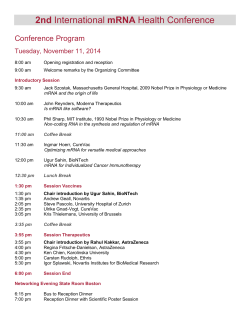
0022-1767/88/1406-1916S02.00/0
THE JOURNAL OF ~ M M U N O L ~ C V
Copyrlght 0 1988 by The Amerlcan Assoclatlon of Immunologists
Vol. 140.1916-1922. No. 6.March 15. 1988
Printed In U.S.A.
EXPRESSION OFTHE TUMOR NECROSIS FACTOR LOCUS I S NOT NECESSARY
FOR THE CYTOLYTIC ACTIVITY OF T LYMPHOCYTES
C.VICTOR
JONGENEEL,*SERGE1 A. NEDOSPASOV,+ GEERT PLAETINCK,* PHILIPPE NAQUET,*
AND J.-C. CEROTTINI*
From the *Ludwig Institute
for Cancer Research. Lausanne Branch, CH-1066 Epalinges, Switzerland.
'Institute of Molecular Biology, USSR Academy of Sciences, B-334 Moscow, USSR, 'Department
of Genetics, Swfss Instftute
for Experimental Cancer Research, CH-1066 Epalinges. Switzerland. and 'Centre d'lmmunologfe
d e Marseille-Lumfny.
F- 13288 Marseille, France
In order to test whether tumornecrosis factorsa
(TNF-a) or 8 (TNF-8. also known as lymphotoxin) are
involved in the lysis of target cells by cytolytic T
lymphocytes, we probed forthe presence of the
TNF
mRNAs inseveralquiescentandactivated
CTL
clones. No TNF mRNA could be found in constitu-
although theydo not seemto be related to the classI or I1
MHC genes.
It has been proposed that TNF-/3has a directrole in the
cytolytic activity mediated by cytolytic T lymphocytes
(18).Indeed, TNF-/3 has properties that are consistent
with those of a putative toxin injected by CTL into their
tively cytolytic Lyt-2+ clones, and only two outof targets: cell culture supernatants containing TNF-@can
three clones tested accumulated TNF mRNA after cause the rapid death of target cells if the cells are substimulation with phorbol myristate acetate and io- jected to a concomitant osmotic shock (19). and can innomycin. Of two L3T4+clones that canbe induced duce the degradation of target cell DNA, a phenomenon
to become cytolytic by a combinationof antigen and that is also seen in CTL-mediated killing (20, 21).
The
IL-1, only one accumulated
TNF-8 mRNA in the proc- recent demonstration that T lymphocytes can produce
ess. The PC60 rat X mouse T cell hybrid,which TNF-CY
as well as TNF-/3(22.23)
has opened the possibility
becomes cytolytic in response to a combination of that both forms ofTNF could be used a s mediators of
I C 1 and IL-2,also failed to accumulate TNF mRNA
afterstimulationwith these agents. Our results CTL activity.
strongly suggest thatTNF-a or -8 are not necessary Having in hand the molecular tools necessary to meaand TNF-/3 mRNA
agents of the cytolytic activity exhibited
by antigen- sure precisely the amounts of TNF-CY
present
in
cell
populations,
we
undertook
to re-evaluate
specific T lymphocytes.
the capacity ofCTL clones to produce TNF. Our salient
conclusion is that, whereassome CTL clones certainly do
Tumor necrosis factor /3 was originally described as a accumulate TNFmRNA after activation, this is not a
cytotoxic activity present in
the supernatants
of mitogen- prerequisite for their cytolytic activity.
Subsequent work showed that it
activated T cells (1-3).
MATERIALS AND METHODS
has toxic effects on a variety of cultured tumor cell lines
CTL clones and cell lines. Lyt-2+ CTL clones were derived from
and causes necrosis of some tumors, but seems inactive
alloreactive (H-2banti-H-2d) populations. Clones 7 and 11 were deon "normal" cells (4.5).Recently, it has become evident rived
from peritoneal exudate T lymphocytes isolated from C57BL/
that TNF-P, beside its toxic properties, is also a lympho- 6 (H-zb) mice immunized against P815 (H-2d)tumor cells by using a
kine with pleiotropic effects on cells of various origins. micromanipulation technique described elsewhere (24). Clone 7 5
by micromanipulation of C57BL/6 anti-DBA/2 T cells
similarly toits close relative, TNF-a (6-10).
In fact, in all was derived
from a day 5 secondary mixed lymphocyte culture prepared as
experimental systems where it has been tested, TNF-P described(25). The cloned cells were maintainedinculture
by
has effects indistinguishable from those of TNF-a, pre- periodic stimulation with appropriate stimulatorcells and a source
sumably because the receptor for the two proteins is the of interleukin 2. L3T4+ CTL clones were derived from lymph node
cells ofA.TL(H-2')
mice immunized with the synthetic polymer
same (11).
poly(glu60. ala30, tyrl0)(GAT]a s described (26).The PC60 cell line,
Determination of the primary structure of human TNF- derived from a cell fusion between a mouse CTL clone and a the C58
a and TNF-@has established that the two proteins are rat thymoma line (27). waskindly provided by Dr. M. Nabholz (Swiss
Institute for ExperimentalCancerResearch.Epalinges,Switzerclosely related. The cloning of cDNAs for human, mouse, land).
and rabbit TNF-a, as well as for the human and mouse
The P815 mastocytoma line used as a source of target cells. the
TNF-P, has made it possible to study the organization of EL4 thymona line, and theWEHI 164 clone 13 line used inthe TNF
assay (28)were maintained in culture using standardmethods. The
In man a s well a s in mouse, the genes CTLL-2 line was maintained in the
the TNF genes (8).
presence of exogenous IL-2.
are tandemly arranged (12,
13),
and their structures reEnzymes and chernlcals. Phage SP6 RNA polymerase, RNaseveal an common evolutionary origin (14,15). In both free DNase, and Escherichia coli DNA polymerase I Klenow fragment were from Boehringer-Mannheim (Mannheim, FRG). RNase A
species, the genes are located within the MHC (1 6, 17).
(bovine pancreatic) was from Sigma Chemical Co. (St. Louis. MO).
Received for publicationSeptember 28, 1987
Accepted for publication December8. 1987
The costs of publication of this article were defrayed in part by the
payment of page charges. This article must therefore be hereby marked
aduertfsementin accordance with 18 U.S.C. Section 1734 solely to fndlcate thls fact.
Lyophilized RNase T1 (from Aspergillus oryzae).a Sankyo product.
was purchasedfrom Calbiochem (San Diego. CA). Proteinase K was
from Merck (Darmstadt, FRG). 32P-labeled UTP (400 Ci/mmol) and
'251-labeledUdR were from Amersham plc (Amersham, United Kingdom).Recombinant human IL-16 was a gift of Dr. J. M. Dayer
(University of Geneva). Recombinant human IL-2 was a gift of Biogen
S A , Geneva.
1916
1917
TNF EXPRESSION IN CTL CLONES
Probes. In most of the experiments described in this paper, we
used 3aP-labeled RNA probes transcribed by the SP6 phage RNA
polymerase from recombinantplasmids constructed in the
pSP64 or
pSp65 vectors. The inserted fragments, all of murine origin, were
derived as follows: TNF-B, a 950-bp PuuII genomic fritgmentextending from themiddle of the first intron 350 bp into the fourthexon
(13);TNF-a. a 450-bp HinclI to EcoRI fragment entirely contained
within the fourthexon (13);IFN-y. a Hind111 to CLaI fragment covering the firstexon (subcloned from a n IFN-y genomic clone obtained
from W. Fiers, State University of Ghent. Ghent. Belgium); Thy- 1, a
600-bp Pstl fragment
corresponding to thecoding portion of the TM8
cDNA clone described by Hedrick et al. (29);B-actin, a 200-bp PstI
to BglII fragment subcloned from the pAL41 @-actin cDNA clone
described by Minty et al. (30).To probe for Granzyme A mRNA, we
used 32P-labeledoverlapping synthetic oligonucleotides as described
by Garcia-Sanz et al. (31).
Blots and RNase protection assays. Total cytoplasmic RNA was
extracted from the postnuclear supernatant
of cells lysed in isotonic
buffer containing 0.1% Nonidet P-40 (32). For Northern blots. 5 pg
of RNA were separated ina 1.4%formaldehyde-agarose gel (32) and
transferred in 20 x SSC (1 X SSC is 0.15 M NaCI. 0.015 M sodium
citrate, pH 7.0) to Hybond N filters (Amersham. United Kingdom).
After baking, the filterswere prehybridized in 5 x SSC. 50%formamide, 2.5 X Denhardt's solution. 0.2% SDS. 10 mM EDTA. 50 mM
sodium phosphate (pH 6.5). and 200 &ml of denatured herring
sperm DNA for 4 h a t 55°C. and hybridized for 16 h in the same
solution containing 1 to 5 X lo7cpm 32P-labeledRNA probe. Washing
was in 0.1 x SSC, 0.1% SDS a t 65°C. Slot blotswereprepared
according to the manufacturer's instructions
(Schleicher & Schuell,
Keene, NH), using Hybond N filters. Hybridizations were performed
as for Northern blots. For densitometric scanning of the autoradiograms, we used a Zeineh s o f t laser integrating densitometer.
RNase protection assays were performed essentially as described
by Zinn etal. (33).A 2-pg sample of cytoplasmic RNA was hybridized
to 1 to 5 x IO5 cpm of RNA probe a t 53°C overnfght in 30 p1 of 80%
formamide. 50 mM PIPES (pH 6.4). 0.4 M NaCI. 1 mMEDTA. The
hybrids were digested with 100 pg/ml of RNase A and 4 pglml of
RNase T I in 300 pl of 0.3 M NaCI. 10 mM Tris . HCI (pH 7.4) for 45
rnin at 28°C. After digestion with 100 @/ml of proteinase K in the
presence of 0.6%SDS (30 rnin a t 37'C). phenol-chloroform extraction, and precipitation in ethanol, the protected 32P-labeled RNA
fragments were separated in 5%acrylamide/urea gels at 55°C and
visualized by autoradiography of the fixed and dried gels.
"Cr and Iz5I release assays. 51Crrelease assayswere performed
as described previously (25).DNA fragmentation assayswere carried
out as follows; P815 target cells were incubated overnight a t 37°C
in DMEM 5%' containing 4 pCi/ml of['Z51]UdR and washed three
times in the samemedium without ['251]UdR.Ten thousand labeled
target cells weremixed with effector cells in a total volume of 100 pl
and incubated at37°C. At the end of the incubation period. Triton
X-100 was added to a final concentration of 0.2%and EDTA to 5
mM. the lysed cells were centrifuged at 1000 X G for 5 rnin and the
supernatants were harvested and counted in a gamma counter.
Specific release is expressed as 100 x (cpm released - spontaneous
release)/(maximum release - spontaneous release). The maximum
release value was obtained by incubating Triton-lysed cells with 4 0
U/ml of micrococcal nuclease (Worthington) and counting the
label in the 1000X G supernatant.
Assaysfor TNF and IFN-y activities. The cytostatic activity of
TNF on WEHI 164 clone 13 cells was measured using the
vital stain
3-[4,5-dimethylthiazol-2-yl]-3.5-diphenyltetrazolium
bromide (MTT;
Sigma M2 128) (26). 2 X 1O4 WEHI 164/13 cells were incubated with
dilutions of the supernatants be
to tested in 200
p l of culture medium
for 4 8 h a t 37°C. After this period, MTT was added to 1.25 mglml.
and the incubation was continued for another
4 h. The culture
medium was thenremoved, and thecells were lysed in 100 pl of 3%
SDS, 40 mMHCI in isopropyl alcohol. After all of the debris had
lysed. the OD5,,,of the wells was read in a n ELISA reader.
The macrophage-activating activity of the cell supernatants was
measured as described (34). Briefly, bone marrow-derived macrophages were incubated with dilutions of the supernatants to be
tested in the presenceof 200 ng/ml of LPS, and thentested for their
cytolytic activity on S'Cr-labeled P8 15 target
cells. IFN-y levels were
defined as the reciprocal of the supernatant dilution yielding 50%
maximal "Cr release.
' Abbreviations used In this paper: DMEM 5%. Dulbecco's modified
Eagle's medium containing 5%fetal bovfne serum and 1 0 m M HEPES:
MTT. 3-[4.5-dimethylthiazoi-2-yl]-3.5-dipheny~tetr~oiium
bromide.
RESULTS
Detection of TNF mRNA. While testing various cell
lines for the presence of TNF mRNA, we discovered that
the CTLL-2 line that we commonly use for IL-2 assays
(35)contains large amounts of the TNF-P message and
lower amounts of the TNF-a message, although it does
not seem to secrete biologically active TNF into the culture supernatant (unpublished observation). Therefore,
we used CTLL-2 RNA as a positive control for the experiments described in this paper.
Figure 1A shows a Northern blot ofRNAs extracted
from EL4 cells (which do not express either TNF-a or
TNF-b),from CTLL-2 cells, and from LPS-activated bone
marrow-derived macrophages probed with a TNF-a or a
TNF-0 probe. Our TNF-6 probe detected a 1.4-kb mRNA
in theCTLL-2 cells that was not present in macrophages.
This mRNA was clearly distinct from that of TNF-a.
which had a size of 1.7 kb, and was present in large
amounts in LPS-activated macrophages and in much
smaller amounts inCTLL-2 cells.
A s a more sensitive alternative, we used the RNase
protection technique described by Zinn et al.(33).where
liquid phase hybridization of a labeled RNA probe to
cellular RNA is followed by digestion with RNase A and
RNase T1 and separation of the protected fragments on
denaturing acrylamide gels. Figure 1B shows thatusing
a genomic probe, which contains the second, third, and
part of the fourthexon (up to the PuuII site) of the mouse
TNF-P (13).and hybridizing it to CTLL-2 RNA, we could
detect the predicted protected fragments of 106 (exon 2).
100 (exon 3), and 335 (exon 4, up to PuuII site] bases.
The largest protected fragment, which is derived from
exon 4, contained the most radioactive label and was
used a s a n indicator of the presence of TNF-8 mRNA.
Hybridization to the TNF-a probe produced a single band
of 450 bases,as expected from the structureof the probe
(data not shown]. By comparing the signal strengths obtained withprobes of identical specific activities specific
for TNF-a, TNF-/3, &actin, and the
Thy- 1 surface antigen,
we estimated that TNF-P mRNA was present at 500 to
1000 copies/cell in the CTLL-2 line, and TNF-a at about
100 copies/cell.
TNF mRNA expression in Lyt-2+ CTL clones. In a first
series of experiments, we analyzed three independently
derived Lyt-2+ CTL clones (clones 7, 11, and 75) for the
presence and inducibility of the TNFmRNA.All three
clones are directed against H-2d alloantigens and are
maintained in cultureby repeated antigenic stimulation
in thepresence of IL-2. To make sure that the
clones had
maintained theirCTL phenotype, we examined their ability to kill P8 15 (H-2d)target cells and to inducerapid DNA
degradation in these same targets. The data shown in
Figure 2 demonstrate that all three clones were actively
cytolytic by these criteria. They were able to induce 5'Cr
release from P815 targets and to cause extensive DNA
degradation (as measured by the release of lZ5I from the
nuclei of ['251]UdR-labeledcells) within 30 min of contact.
We analyzed cytoplasmic RNA extracted from these
CTL clones 7 days after the last antigenic stimulation,
which is the time at which theywere assayed for cytolytic
activity, and failed to detect any TNF-PmRNA (Fig. 3.
lanes c a n d n . From prolonged autoradiographic exposures of our gels,we estimate that TNF-@ mRNAis more
1918
TNF EXPRESSION IN CTL CLONES
A
tion with phorbol myristate acetate, an activator of protein kinase C. and ionomycin. which causes an influx of
Ca2+ions into the cells. The combination ofPMA and
a b c d
e
f
g
ionomycin has been shown to induce manyof the functions specific of activated T cells, and in particular the
secretion of IFN-y and of IL-2 (36).
1.7 kb +
Figure 3 shows the results
of an RNase protection assay
performed on RNA extracted before and 24h after stimulation of CTL clones 7 and 75with PMA and ionomycin.
Although the clone 7 cells accumulatedappreciable
amounts of TNF-8 RNA after stimulation (lane d ) , we
failed to detect any TNF-B mRNA in the clone 75 cells 24
6
h after stimulation (lane9).Probing the sameRNA preparations with a IFN-?.probe (lower panel), we could easily
detect the expected accumulation of IFN-?. mRNA
in stim344
C exon 4 (to Pvu I1 site)
ulated cells. The RNA extracted fromLPS-activated macrophages (lane b). used as a negative control, did not
298 4
contain messengers for either lymphokine,even though
it did contain large amounts of TNF-a mRNA (data not
shown).
To obtain the experimental results shown inFigure 4,
237
we stimulated all three CTL clones with PMA and ionomycin and harvested the cells 1. 5, and 24 h later. The
220
abundance of mRNAs for TNF-a, TNF-B, IFN-7.and Granzyme A was determined by densitometric scanning of
"slot blots" hybridized with the relevant probes, and normalized against the signal obtainedby rehybridizing the
same blot with a @-actin probe. Because the absolute
abundances of the mRNAs cannot be estimated withthis
method, the data are
presented in terms of relative signal
strength.
with
the
unstimulated
cells serving a s a refer154
ence. Note that they-axis scalesof Figure 4 are different,
to accommodate a much larger increase in IFN-y levels
relative to TNF-a. TNF-8, or Granzyme A. The culture
media were kept and assayed forthe presence of biologic
activities associated withIFN-y and TNF (Table I). IFN-7
was detected by its macrophage-activating activity,
whereas TNF was assayedby its cytotoxicity for the WEHI
164 clone 13 line.
109
The data inFigure 4 show that stimulation of the CTL
clones withPMA and ionomycin resulted inthe transient
accumulation of IFN-y mRNA in all three, albeitat vastly
c exon 2
different levels (>lOO-fold increase inclone 7, 40-fold in
* exon 3
clone 1 1, andeight-fold in clone 75). Maximum levels of
mRNA were reached around 5 h after stimulation. Although a rapid accumulation of TNF-a and TNF-p mRNA
(about 15-fold in 1 h for clone 7) could be detected in
Flgure 1. Detectlon of TNF-f3mRNA in Northern blots and RNase
protectlon assays. A. 5 pg samples of total cytopiasmlc RNA were sepaclones 7 and 11. weobserved no hybridization over backrated on anagarose/formaldehyde gel and hybridized wlth a TNF-f3(lanes
a. b. and c) or TNF-a (lanes e . f ,and g ) probe. Lanes a and e. EL-4 cells: ground in RNA samples from stimulated clone 75 cells.
lanes b andf. CTLL-2 cells: lanes c and g . bone marrow-derlvedmacro- RNase protection assays (datanot shown) confirmedthat
phages exposed to 10 pg/ml of LPS for 4 h: lane d : m.w. markers (the the increased hybridization to TNF-a and TNF-B probes
main band vlsible on this portlon of the gel corresponds to a size of 1.7
in clone 1 1RNA was indeed due to the presence of the
kb). E. 2 pg of CTLL-2 cytoplasmic RNA were hybridized wlth a genomic
probe covering exons 2 . 3 . and part ofexon 4. The RNase protectlon assay
corresponding mRNAs.
was performed a s descrlbed in Materlals and Methods.The slzes of m.w.
A s a control, we also probed our blots with an oligomarkers are Indicated on the left.
nucleotide specific for the mRNA of Granzyme A. a serine
cytolytic granules (Fig. 4). Although the
than 100-fold less abundant in the CTL clones than in protease found in
CTLL-2 cells. This would put a n upper limit for TNF-B mRNA was easily detectable inall CTL clones (the signal
mRNA abundance to 5 to 10 copies/cell. Similar results strength was much higher than for TNF-B or IFN-y in
were obtained with a TNF-a probe in RNase protection unstimulated cells, and approximately equal in all three
clones), stimulationof the cells with PMA and ionomycin
assays (datanot shown).
In order to test thepossibility that TNF-a and/or TNF- did not dramaticallyalter its abundance.
B are synthesized in a short burst after stimulation and Table 1 shows the TNF and IFN-?.biologic activities
subsequently stored(e.g.. in cytolytic granules), we meas- measured in the culture supernatants of the cells from
ured TNF mRNA levels in our CTL clones after stimula- which the RNAs were extracted. For both lymphokines,
-
1919
TNF EXPRESSION IN CTL CLONES
100
100
I
7 80
0
2
1
0
2
1
Incubation time (hours)
Ffgure 2. Cytolytlc activlty of CTL clones 7. 11, and 75
on P815 target cells. A. Klnetlcs of "Cr release from P815 cells exposed to a IO-fold excess
of cloned CTL. E. Klnetics of 12'1 release from the nuclei of P815 cells whose DNA had been labeled with 112'I)UdR. A. no CTL control: 0 CTL clone 7:
m, CTL clone 11: A. CTL clone 75.
a
b
C
d
e
9
f
Figure 3. RNase protectfon assay forTNF-j3 mRNA
In CTL clones. 2 gg of total cytoplasmlc RNA were
hybridized with 32P-labeled RNA probes specific for
TNF-j3 (top)or IFN-y (bottom)as described In Materfals and Methods.
and theprotected fragments were
separated on a 5% acrylamide gel. Lanes a and e.
CTLL-2 cells: lane b. bone marrow-derived macrophages after 4 h of exposure to LPS (1 pg/ml): lane c.
CTL clone 7 cells 7days after thelast antigenic stimulatlon: lane d . CTL clone 7 cellsafter 24 h of exposure to PMA (10 ng/ml) and lonomycln (500 ng/ml):
l a n e f . CTL clone 7 5 cells 7 days after last antigenic
stimulatlon: lane g. CTL clone 75 cells after 24 h of
exposure
to
PMA and ionomycln.
20
TN F-p
1FN-y
i
-I
24 hJ
24 h medium
120
"
100
15
Granzyrne A
80
10
60
40
5
20
0
0
Clone 7
Clone 11
Clone 75
Clone 7
Clone 1 1
Clone 75
Ffgure 4. Kinetics of induction of TNF-a, TNF-8. IFN-y. and Granzyme A mRNAs after exposure of CTL clones to PMA (10 ng/ml) and ionomycln
(500 ng/ml). Relative abundances of the mRNAs were determlned by blottlng serlal dllutions of cytoplasmic RNA (0.05to 5 pg) onto nylon filters.
hybrldizlng with the relevant probes. and scanning the exposed autoradiograms. All results were normallzed against the signalobtained by
rehybridizing the same filters
with a j3-actln probe, and areexpressed a s fold stimulatlon relative tothe hybridlzation signal obtainedfrom cells before
exposure to PMA and ionomycin.
1920
TNF EXPRESSION IN CTL CLONES
TABLE I
Bfologfc actfuftfes
measured In CTL culture supernatants
Cell llne and
Stlmulatlon"
b
c
d
e
TNF
Macrophage-actlvatrng
Actlvlty"
CTL clone 7
h)
None (24
PMA + lonomycin
l h
5h
24 hr
CTL clone 11
h) None (24
PMA + ionomycin
l h
5h
24 h
CTL clone 75
h)
None (24
PMA + lonomycin
l h
5h
24 h
a
ActlvltyC
5
<8
9
450
>IO00
16
>256
>256
<3
<8
<3
<8
30
80
<8
32
<3
<8
<3
3
25
<8
TNF-P
<8
<8
a Cells were stimulated by exposure to PMA and ionomycln in DMEM
IFN-y 4
5%or Incubated In DMEM 5%alone (none)for the Indlcated times. The
culture medium was then harvested and assayed for
blologlc actlvity.
Macrophage-activatingactlvltyof the culture supernatants was measured a s described in Materfalsand Methods. The results are expressed
Ffgure6. RNase protectlon assay of RNA extracted fromL3T4+ T cell
as the reclprocal of the dilutlon ylelding 50%maximal cytolytic activity.
'TNF cytotoxic activlty of the culture supernatants was measured
on clones. A 2-& sample of cytoplasmic RNA was hybridized to a mlxture of
WEHI 164/13 cellsa s described in Materfalsand Methods. The results TNF-/3and IFN-7 RNA probes and processed as before. Lane a. Unstlmare expressed as the reciprocal of the highest dilution causing more thanulated AT20 cells: lane b. AT20 cells stlmulated wlth GAT and IL-1 for
12 h: lane c. unstimulated ATX5.3 cells: lane d . ATX5.3 cells stimulated
50%vlablllty loss as measured In the MTT assay.
with GAT and IL-I: lane e. CTLL-2 cells.
a
b
c
d
e
f
IL-1 (Ph. Naquet. manuscript in preparation). Figure 5
shows the results of an RNase protection assay performed on RNA extracted from these two clones before
TNF- p
and after stimulation with GAT and IL-1. Although one
of the two clones (AT20) clearly accumulated TNF-j3
mRNA. the other onedid not. No IFN--y mRNA (Fig. 5) or
biologic activity could be detected in either clone. RNase
protection assays performed with a TNF-a probe showed
IFN- y
a low constitutive level of TNF-a mRNA in both clones,
and assays of the culture supernatants in theL929 cell
cytotoxicity assay suggested a low levelof TNF-a production. Whether this production of TNF-a is necessary for
the cytotoxicity of these L3T4' clones remains to be
Thy-1
determined.
TNF mRNA does not accumulate in inducible rat X
mouse
hybridCTL. The PC60 cell line wasderived from
Ffgure5. Northern blot of RNA extracted from PC60 cells exposed to
a cell fusion between a mouse CTL clone and theC58 rat
different comblnatlonsof Interleukins. A 5-flgsampleof cytoplasmlc RNA
was separatedIn a 1.4% agarose/formaldehydegel, transferred to nylon.
thymoma line (27). These
cells grow in a n 11-2-independand thefllter was hybridized successively wlththe indlcated RNA probes.
ent manner and are not cytolytic. However, when culLane a. CTLL-2 cells: lane b. uninduced PC60 cells: lane c. PC60 exposed
tured in the presence of IL-l and IL-2. they accumulate
to recornblnant human IL-2 (100 U/ml) for 4 days: lane d . PC60 exposed
to recomblnant human IL-ls
(5ng/ml): lane e. PC60 exposed to IL-18 and
mRNA for IL-2, IL-2 receptor, and Granzyme A. syntheIL-2: lanef. PC60 exposed to 1L-1 and a supernatant from concavavalin
size perforin, the pore-forming protein present in CTL
A-actlvated spleen cells.
granules, andbecome cytolytic (39)(G. Plaetinck, unpubthe biologic activities correlated closely with the RNA lished observations).
levels measured by hybridization. In particular, the suWe assayed for the appearance of TNF mRNA after
pernatants of activated clone 75 cells contained no meas- stimulation of PC60 cells with various combinations of
urable TNF activity. It should be noted that the WEHI lymphokines. The Northern blot hybridization data pre164/13 cytotoxicity assay does notallow us to distinguish sentedin Figure 6 clearly show that although IFN--y
between the effectsof TNF-a and TNF-j3.
mRNA accumulated in PC60 cells 4 days after stimulaTNF-j3 mRNA expression in L3T4' clones. Because a tion by a combination of IL-1 and IL-2. no TNF-j3 mRNA
strong correlation between cytolytic activity and TNF-j3 could be found. Similarly, although TNF-a mRNA could
secretion had been reported in L3T4+ T cell clones (20. easily bedetected in theCTLL-2 cells, none was found in
level of
37. 38),we examined TNF-j3 expression in two cytolytic stimulated PC60 cell RNA (data not shown). The
L3T4' clones (AT20and ATX5.3) that arespecific forthe the mRNA for the T cell-specific Thy-1 surface antigen,
synthetic polymer GAT and restricted by I-Ak (26). These which is not regulated under these conditions, remained
two clones grow independently of exogenous IL-2. and constant.Similarresults
were obtained by using an
can be induced to become cytolytic to I-Ak-positivefibro- RNase protection assay (data not shown). We also asblasts after exposure to their cognate antigen(GAT)and sayed forthe presence of TNF-j3 mRNA at different times
TNF EXPRESSION IN CTL CLONES
1921
after induction, with similarly negative results (data not that produced TNF. This correlation did not hold true in
the L3T4+ clones or under all conditionsof cell stimulashown).
tion. In general, our data agree with those presented by
DISCUSSION
Cuturi et al. (22) for human peripheral blood lymphoIn the experiments described in this paper, we have cytes.
If TNF is not an obligatory mediator ofCTL cytolytic
examined the presence and accumulation of TNF-a and
activity,
what thenis its function? A possible answer can
TNF-PmRNA in six different murine
cell lines: three
constitutively cytolytic Lyt-2+ T cell clones, two L3T4' be found in the pleiotropic effects of both TNF-a and
clones whose cytolytic activity is inducible, and one in- TNF-j3. Although the two proteins are cytotoxic to a reladucible T cell hybridoma. None of these cell lines ex- tively small number of transformed cell lines, they are
pressed TNF mRNA in a constitutive fashion, and only nontoxic or stimulatory tomost nontransformed cells (40,
three out of six could be induced to accumulate TNF 41). and theyhave many activitiesinvolved in the develof granmRNA by stimuli known to activate other CTL-specific opment of the inflammatory response: activation
ulocytes
(42,
43),
differentiation
of
monocyte
precursors
functions. It is significant that one of the two L3T4+
clones as well as the T cell hybridoma failed to accumu- (44, 45). induction of 1L-1 (46). and GM-CSF (47) synthelate anyTNF-j3 mRNA under conditions where theywere sis, induction ofMHC antigens (48) and other surface
acquiring cytolytic activity. We believe that these data markers (49). and antiviral activity (50, 51), with new
provide strong evidence for the lack of involvement of activities described at an accelerating pace. Therefore, it
TNF-j3 in CTL-mediated cytolysis. Although we cannot is clear that therole of TNF is not limited to the lysis of
rule out a role for TNF-a in the cytotoxic activity of the target cells (although it may be involvedin thedestruction
L3T4' clones, TNF-a mRNA is not produced by the Lyt- of some tumors), but ratherthat TNF is another member
2+ clone 75 or by the induced PC60 hybridoma, nor is of the growing family of soluble mediators that help
any TNF biologic activity found in the supernatants of increase theefficiency of the imjune response.
activated clone 75 cells.
It could be argued that very low levels of TNF mRNA.
Acknowledgments. We wish to acknowledge Josk Alescaping detectionby our assaymethods, could be suffi- berto Garcia-Sanz for the Granzyme A oligonucleotide
cient for the synthesisof the TNF required to kill target probe, and Clotilde Horvath for skillfully maintaining the
cells. We think this unlikely, however, because of the CTL clones and performing the biologic assays. We thank
high sensitivity of the RNase protection assays, and be- Jurg Tschopp and Markus Nabholz for valuable discuscause no correlation could be found between the ability sions.
of T cells to synthesize TNF and their cytolytic activity.
In particular, the two L3T4+ clones behaveidentically in
REFERENCES
their ability to kill LAk+ target cells in the presence of
1 . Granges. G. A.. and T.W.Williams. 1968. Lymphocyte cytotoxicity
GAT and IL-1 (Ph. Naquet, in preparation), and yet difin vitro: activation and release of a cytotoxic factor. Nature
218:1253.
fered very clearly in their accumulation
of TNF-6 mRNA.
N. H.. and B. H.Waksman. 1968. Cytotoxictty mediated by
It should beclearly stated that our data
do not challenge 2 . Ruddle,
soluble antigen andlymphocytes in delayed hypersensitivity. 111.
the notion that secretion of TNF-a and/or TNF-j3 can be
Analysis of mechanism. J. Exp. Med. 128:1267.
3. Shacks,S. J., J. Chiller, and 0. A. Granger. 1973. Studies on in
triggered by contact between a specific CTL and a cell
vitro models of cellular immunity: the role of T and B cells in the
presenting its cognate antigen. In fact, several of the T
secretion of lymphotoxin.Cell. immunol. 7:313.
4. Gray, P. W..B. B. Aggamal. C. V. Benton. T. S. Bringman. W. J.
cell clones that we examined responded to stimulation
by
Henzel, J. A. Jarrett, D. W. Leung, B. Moffat, P.
Ng,L.P.Svedersky.
synthesizing mRNA for both TNFs, and we could also
M. A. Palladino, and 0. Nedwin. 1984. Cloning and expression of
detect the mRNAs in antigen-stimulated mixed lymphocDNA for human lymphotoxin, a lymphokine with tumor necrosis
activity. Nature 3 1 2 7 2 1 .
cyte cultures (unpublished results). The important point
5. Aggarwal, B. B.. W. J. Henzel. B. Moffat. W. J. Kohr, and R.N.
is that T cells can be cytolytic and induce DNA degradaHarkins. 1985. Primary structure of human lymphotoxin derived
tion intheir targets without expressing
TNF.
from 1788 lymphoblastoid cellline. J.Blol. Chem. 260:2334.
6. Beutler. B., and A. Cerami. 1986. Cachectin and tumour necrosis
The data we obtained with Lyt-2+ CTL clones seemed
factor as two sides of the Same biologicalcoin. Nature 320:584.
to show a correlation between the level of expression of
7. Cerami. A.. and B. Beutler. 1986. Cachectln: the dark side of tumor
IFN-7 and that of TNF-8, which could be interesting in
necrosis factor. cold Spring Harbor Symp.Quant. Biol. 51:625.
8. Goeddel. D. V.. B. B. Aggarwal, P. W. Gray. D. W. Leung. G . E.
view of the synergistic biologic activities of these two
Nedwin, M. A. Palladino. J. S. Patton, D. Pennica, H. M. Shepard.
lymphokines. The correlation does not hold true, howB. J. Sugarman. and G. H. W.Wong. 1986. Tumor necrosis factors:
gene structure and biological activitles. Cold Sprtng Harbor Symp.
ever. We could not demonstrate anyTNF-j3 mRNA after
Quant. Biol. 51:597.
activation of the CTL clone 75 cells, even though they
did
9. Old, L.J. 1985. Tumor necrosis factor (TNF).Science 230:630.
synthesize low amounts of IFN-y. Conversely, the L3T4+ io. Ruddle, N. H. 1987. Tumor necrosis factor and related cytotoxins.
immunol. Today 8:129.
clone AT20 accumulated TNF-13 mRNA after activation,
B. B.. T. E. Eessalu. and P. Hass. 1985. Characterization
but no IFN-y mRNA. Therefore. it seems unlikely that 1 1 . ABgarwal.
of receptors forhuman tumour necrosls factor and their regulation
the two genes are coordinately regulated. In view of the
by y-interferon. Nature 318:665.
close physical linkageof their genes,it was interestingto 12. Nedospasov. S.A.. A. N. Shakhov, R. L.Turetskaya. V. A. Mett. M.
M. Azizov. G . P. Georgiev, V. G . Korobko. V. N. Dobrynin. S . A.
compare the levels of TNF-a and TNF-j3 mRNA in these
Filippov. N. s. Bystrov. E. F. Boldyreva, s. A. Chuvpilo. A. M.
Chumakov. L. N. Shingarova, and Y. A. Ovchinnikov. 1986. Tanpopulations. It has been reported that PMA and ionomydem arrangement ofgenes coding for tumornecrosls factor (TNF-a)
cin preferentially induce the expression of TNF-a in T
and lymphotoxin (TNF-8) the
in human genome. Cold Spring Harbor
cells, although stimulation with lectins induces mostly
Symp. Quant. Blol. 51:611.
13.
Nedospasov.
S
.
A..
B.
Hirt.
A. N. Shakhov. V. N. Dobrynin, E.
TNF-j3 (22, 23).In our hands, TNF-a and TNF-j3 mRNAs
Kawashima. R. S. Accolla, and C. V. Jongeneel. 1986. The genes
seemed to appear simultaneously and to be stimulated to
for tumor necrosis factor (TNF-a) and lymphotoxin (TNF-0) are
tanapproximately the same extent in those
Lyt-2+ clones
demly arranged on chromosome of
17the mouse. Nuclelc Aclds Res.
1922
TNF EXPRESSION IN CTL CLONES
14:7713.
14. Nedwin. G.E.. S. Naylor. A. Y. Sakaguchi. D. Smith, J. JarrettNedwin, D. Pennica, D. V. Goeddel. and P. W. Gray. 1985. Human
lymphotoxin and tumor necrosis factor genes: structure, homology
and chromosomal localization. Nucleic Acids Res. 13:6361.
15. Aggarwal. B. E..W. J. Kohr, P. E. Hass, B. Moffat, S. A. Spencer.
W. J. Henzel, T. S. Bringman, 0. E. Nedwin, D. V. Goeddel, and R.
N. Harkins. 1985. Human tumor necrosis factor: production. purification. and characterization.J. Blol. Chern. 260:2345.
16. Miiller, U..C. V. Jongeneel. S. A. Nedospasov. K. Fischer Lindahl.
and M. Steinmetz. 1987. Tumour necrosls factor and lymphotoxin
genesmap close to H-2D in the mouse major histocompatibility
complex. Nature 325265.
17. Spies, T., C. C. Morton, S. A. Nedospasov, W. Fiers, D.Pious, and
J. L. Strominger. 1986. Genes for the tumor necrosis factors alpha
and beta are linked to the humanmajor histocompatibility complex.
Proc. Natl. Acad. Scl. USA 83:8699.
18. Ruddle, N. H.. and D. S. Schmid. 1987. The role of lymphotoxin in
T-cell-mediated cytotoxicity. Ann. Inst. Pasteur Immunol. 138:314.
19. Schmid, D. S.. M. B. Powell, K. A. Mahoney, and N. H. Ruddle.
1985. A comparison of lysis mediated by Lyt-2' TNP-specific cytotoxic-T-lymphocyte (CTL) lines with that mediated by rapidly internalized lymphotoxin-containing supernatant fluids: evidence for a
role of soluble mediators in CTL-mediated killing. Cell. Irnmunol.
93:68.
20. Schmid, D. S., J. P. Tite. and N. H. Ruddle. 1986. DNA fragmentation: manifestation of target cell destruction mediated by cytotoxic
T-cell lines. lymphotoxin-secreting helperT-cell clones, and cell-free
lymphotoxin-containing supernatant. Proc. Natl. Acad. Scl. USA
83:1881.
2 1. Dealtry, G. B., M. S. Naylor, W. Fiers, and F. R. Balkwill. 1987. DNA
fragmentation and cytotoxicity caused by tumor necrosis factor is
enhanced by interferon-y. Eur. J . Immund. 17:689.
22. Cuturi. M. C.. M. Murphy, M. P. Costa-Giomi, R. Weinmann. B.
Perussia, andG. Trinchieri. 1987. Independent regulation of tumor
necrosis factor and lymphotoxin production by human peripheral
blood lymphocytes. J. Exp. Med. 165: 1581.
23. Kobayashi, Y.,M. Asada, and T.Osawa. 1987. Production of lymphotoxin and tumour necrosis factor by a T-cell hybridoma. Immunology 60:213.
24. MacDonald. H.R., N. Thiemesse. and J.-C. Cerottiii. 1981.Inhibition of T cell-mediated cytolysis by monoclonal antibodies directed
against Lyt-2: heterogeneity of inhibition at the clonal level. J . Immunol. 126:1671.
25. Cerottini, J.4.. H. D.Engers, H.R. MacDonald, and K. T. Brunner.
1974. Generation of cytotoxic T lymphocytes in vitro. I. Response of
normal and immunespleen cellsin mixed leukocyte cultures. J . Exp.
Med. 140: 703.
26. Naquet, P.. A. Pierres, and M. Pierres. 1983. Dissection of the
poly(glu60 ala30 tyrlO] [GAT)-specificT-cell repertoire in H-21kmice.
I. GAT plus self-I-A*-reactfveT-cell clones can recognize alloactivating and/or restriction determinants on nonself-Ia molecules. Immunogenetlcs I8:475.
27. Silva, A.,H.R.
MacDonald, A. Conzelmann, P. Corthhy. and M.
Nabholz. 1983. Rat x mouse T-cell hybrids with inducible specific
cytolytic activity. Imrnunol. Rev. 76: 105.
28. Espevik. T., and J. Nissen Meyer. 1986. A highly sensitive cell line,
WEHI 164 clone 13. for measuring cytotoxic factor/tumor necrosis
factor from human monocytes. J. Irnmunol. Methods 9599.
29. Hedrick. S. M.. D. J. Cohen, H. A. Nielsen. and M. M. Davis. 1984.
Isolation of cDNA clones encoding T cell-specific membrane-associated proteins. Nature 308:I49.
30. Minty, A. J.,S. Alonso, J. L. Guenet, and M. E. Buckingham. 1983.
Number and organization of actin-related sequences in the mouse
genome. J . Mol. Biol. 167:77.
31, Garcia-Sanz, J. A.. G . Plaetinck, F. Velotti, D. Masson, J. Tschopp.
H. R. MacDonald, and M. Nabholz. 1987. Perforin is present only in
normal activated Lyt-2+ T lymphocytes and not in L3T4' cells, but
the serine protease Granzyme A is made by both subsets. EMSO J.
6:933.
32. Maniatis. T., E. F. Fritsch.and J. Sambrmk. 1982. Molecular
Clonlng. Cold Spring Harbor Laboratory, Cold Spring Harbor. NY.
T. Maniatis. 1983. Identification of two
distinct regulatory regions adjacent to the human &interferon gene.
Cell 34:865.
34. Kelso, A., H. R. MacDonald, K. A. Smith, J. C. Cerottini, andK.T.
Brunner. 1984. Interleukin-2 enhancement of lymphokine secretion
by T lymphocytes: analysis of established clones and primary Iimiting dilution microcultures. J . Imrnunol. 1322932.
35. Kelso. A., and H.R. MacDonald. 1982. Precursor frequencyanalysis
of lymphokine-secreting alloreactive T lymphocytes. Dissociation of
subsets producing interleukin-2, macrophage-activating factor, and
granulocyte-macrophage colony-stimulating factor on the basis of
Lyt-2 phenotype. J . Exp. Med. 156: 1366.
36. MacDonald, H. R., and M. Nabholz. 1986. T-cell activation. Annu.
Rev. Cell 5101.2:231.
37. Tite, J. P.. M. B. Powell, and N. H. Ruddle. 1985. Protein-antigen
specific Ia-restricted cytolytic T cells: analysis of frequency, target
cell susceptibility. and mechanism of cytolysis. J. Irnmunol. 135:25.
38. Shiohara. T., N. H.Ruddle, M. Horowitz, G. E. Moellmann. and A.
B. Lerner. 1987. Anti-tumor activity of class I1 MHC antigen-restricted cloned autoreactive T cells. 1. Destruction of B16 melanoma
cells mediated by bystander cytolysis in vitro. J . Imrnunol. 138: 1971.
39. Erard. F., P. Corthby, K. A. Smith, W. Fiers, A. Conzelmann. and
M. Nabholz. 1984. Characterization of soluble factors that induce
the cytolytic activity and the expression of T cell growth factor
receptors of a T cell hybrid. J. Exp. Med. 160~584.
M. A.
40. Sugannan, B. J., B. B. Aggarwal, P. E. Hass, 1. S.
Palladino. and H. M. Shepard. 1985. Recombinant human tumor
necrosis factor: effects on proliferation of normal and transformed
cells in vitro. Science 230:943.
41. Vilcek. J.. V. J. Palombella, D. Henriksen DeStefano. C. Swenson,
and R. Feinman. 1986. Fibroblast growth enhancing activity of
tumor necrosis factor
and itsrelationship to other
polypeptide growth
factors. J . Exp. Med. 163:632.
42. Klebanoff, S. J., M. A. Vadas, J. M. Harlan, L. H. Sparks, J. R.
Gamble, and J. M. Agosti. 1986. Stimulation of neutrophils by tumor
necrosis factor. J . Immunol. 136:4220.
43. Shalaby. M. R., B. B. Aggarwal. E. Rinderknecht, L. P. Svedersky.
B. S. Finkle, and M. A. Palladino. Jr. 1985. Activation of human
polymorphonuclear neutrophil functions by interferon-7 and tumor
necrosis factors. J.Imrnunol. 135:2069.
44. Takeda, K.,S. Iwamoto. H. Sugimoto, T. Takuma. N. Kawatani, M.
N o d a , A. Masaki. H. Morise. H. Arimura. and K. Konno.1986.
Identity of differentiation inducing factor and tumour necrosis factor. Nature 323:338.
45. Trinchieri, 0.. M. Kobayashi, M. Rosen, R. Loudon. M. Murphy.
and B. Perussia. 1986. Tumornecrosisfactor
and lymphotoxin
induce differentiation of human myeloid cell lines in synergy with
immune interferon. J . Exp. Med. 164: 1206.
46. Nawroth. P. P.. I. Bank, D. Handley, J. Cassimens, L. Chess. and
D. Stem. 1986. Tumor necrosisfactor/cachectin interacts with endothelial cell receptors to induce release of interleukin 1. J. Exp. Med.
163: 1363.
47. Munker, R.. J. Gasson, M. Ogawa, and H. P. Koeffler. 1986. Recombinant human TNF induces production of granulocyte-monocyte
colony-stimulating factor.Nature 323~79.
48. Collins, T., L.A. Lapieme, W. Fiers. J. L. Strominger. and J. S .
Pober. 1986. Recombinant human tumor necrosis factor increases
mRNA levels and surface expression of HLA-A. B antigens in vascular endothelial cells and dermal fibroblasts in vitro. Proc. Natl.
Acad. Sct. USA 83,446.
49. Pober, J. S., M. P. Bevilacqua, D. L. Mendrick. L. A. Lapierre, and
W. Fiers. 1986. Two distinct monokines. interleukin 1 and tumor
necrosis factor, each independently induce biosynthesis and transient expression of the same antigen on the surface of cultured
human vascular endothelial cells. J . Immunol. 136: 1680.
50. Mestan. J.. W. Digel. S. Mittnacht. H. Hillen, D. BIohm, A. Moller.
H. Jacobsen, and H. Kirchner. 1986. Antiviral effects of recombinant tumour necrosis factorin vitro. Nature 323:816.
51. wong, G. H., a n d D. V. Goeddel. 1986. Tumour necrosis factors
alpha and betainhibit virus replication and synergize with interferons. Nature 323:819.
33. Zinn. K., D. DiMaio. and
Figari.


© Copyright 2026