
(exchange pictures for a selected image of resident research) DEPARTMENT OF RADIOLOGY
DEPARTMENT OF RADIOLOGY
(exchange pictures for a selected image of resident
research)
DIAGNOSTIC RADIOLOGY
RESIDENCY and FELLOWSHIP
PROGRAM MANUAL
FOR 2010 – 2011
INCLUDING GOALS AND OBJECTIVES
(Revised: March 24, 2010)
1
___________________________________________________CONTENTS
INTRODUCTION AND OVERVIEW
Letter from the Chairman and Program Directors
Radiology Faculty
Radiology Residents
Education Committee
EDUCATIONAL PROGRAMS
General Diagnostic Radiology
Resident Four-Year Curriculum
Resident Third Year Curriculum
Rotation Schedule
1-9
4-5
6
7-8
9
10
10
10
10
CONFERENCES
12
Departmental
Interdepartmental
Core Curriculum
RESOURCES
13
Departmental library
Teaching files
RESIDENT AND FELLOW DUTIES AND RESPONSIBILITIES
14-15
14
15
16
17
17
Night call and weekend duties
Post-call Time Off
The Wet Desk
Night-call Qualifying Examination
Vacations
REQUIREMENTS FOR RESIDENT ADVANCEMENT
18
Promotion and Reappointment
Disciplinary Action
Remediation
DEPARTMENTAL POLICIES
19-21
Moonlighting policy
Resident supervision
Resident teaching
Sickness/leave of absence
Finance/Purchasing Policies
Travel and conferences
RESIDENT AND FELLOW EVALUATION PROCESS
Evaluation of residents and fellows
APPENDIX I: EDUCATIONAL GOALS AND OBJECTIVES
Educational Goals and Objectives
1. Abdomen
2. Chest
3. Emergency
4. Magnetic Resonance Imaging
5. Mammography
6. Musculoskeletal
7. Neuroradiology
8. Nuclear Radiology
9. Pediatric
10. Physics of Medical Imaging
11. Ultrasound
a. Obstetrical/Pelvic UItrasound
12. Vascular Intervention
2
22
23-73
APPENDIX II.
FELLOWSHIP GOALS AND OBJECTIVES
Goals and Objectives
74-116
APPENDIX III.
EVALUATION FORMS
Resident Evaluation Forms
117-143
APPENDIX IV.
GENERAL COMPETENCIES
Six General Competencies
ACGME General Competencies
144-151
3
INTRODUCTION AND OVERVIEW
LETTER FROM THE CHAIRMAN AND PROGRAM DIRECTORS
Welcome to Radiology!
On behalf of the faculty, I would like to welcome you to the Department of Radiology and the Texas Tech
University HSC-El Paso Residency Program in Diagnostic Radiology and the newly established Diagnostic Imaging
Fellowship. Texas Tech has a distinguished history in residency training and the beginning of the radiology training
program in 2008 was a most welcomed addition. The residency training program is divided into specialty radiology
sections of Abdominal Imaging, Angiography and Interventional Radiology, Chest Radiology, Diagnostic
Ultrasound, Emergency Radiology, Magnetic Resonance Imaging, Mammography, Musculoskeletal Radiology,
Neuroradiology, Nuclear Radiology, Pediatric Radiology, and Radiation Physics. The residents rotate through these
sub-specialty area and work closely with faculty members who have established expertise in these disciplines.
University Medical Center is the primary teaching hospital and is also a Level I trauma center. Radiology residents
are able to gain experience working with patients who have both common and complex medical problems.
Radiology residents interact regularly with residents from other residency programs. The technology base of the
department is “state of the art” with the latest CT, MR, and information systems technology. The department has
operated in a filmless and paperless environment since 2005.
The imaging department at UMC is in the midst of a substantial expansion with new facilities for emergency
radiology to the west, and a new outpatient radiology section to the east. New radiology offices, conference area
and teaching component will open in 2011. The children‟s hospital is scheduled to open in 2012 and its radiology
department will adjoin the outpatient radiology section and will be an integral part of the training program.
The training program is designed to enable residents and fellows to acquire and demonstrate the knowledge, skills,
and judgment necessary for competence in the practice of Radiology. This is primarily achieved by involvement in
clinical case management under direct supervision and guidance of the faculty. Formal teaching supplements this
educational process by providing lectures, demonstrations and conferences as well as electronic teaching aids.
Competency in radiology consists of the ability to plan appropriate and most cost effective imaging sequences,
obtaining these studies with utmost concern for patient care, and transferring necessary information to the referring
clinician in a timely manner. Residents not only learn interpretation skills in evaluating radiological images but
obtain competency in transferring this information in an efficient and effective manner. This is based on having the
resident obtain background knowledge of the patient which may modify radiographic appearance or examination
requests. The core knowledge, with which the resident radiologist must become familiar (and which will require
continuing study for the rest of one‟s professional life) concerns the variety of pathologic findings that may be
detected on images due to disease and the differentiation of these from normal, normal variations and technical
artifacts. The resident must also learn to work closely with support personnel on patient care and administrative
matters in order to assume competent and responsive consultative services and quality patient care.
4
The orientation program, for new residents, consists of early rotations through the basic services of chest,
musculoskeletal, pediatric, and abdominal radiology. Orientation also includes a series of special teaching sessions
on common radiology practices and procedures. Further instruction is presented during regularly scheduled
departmental conferences. Residents are to be concerned and involved with quality assurance and other non-imaging
issues involving total patient management during the radiologic process and are required to participate in a monthly
quality assurance conference.
The 12 month Imaging Fellowship includes intensive added experience in computed tomography, magnetic
resonance imaging, and diagnostic ultrasound. Included with the fellowship is the equivalent of 2 months of
imaging research.
This manual describes the clinical, educational, and research opportunities for residents and fellows at Texas Tech
University HSC-El Paso during the 2009 academic year. It can be anticipated that the training experience will
continue to prepare young radiologists to become most competent clinical practitioners and/or academicians with
outstanding credentials. The learning experience begins on the first day of the residency and continues through
every day of each rotation. Our residents need to stay focused and work hard throughout their residency so that they
can be a successful addition to the practice of Radiology in Southwestern US or wherever they choose to go.
Welcome to the Radiology Residency and Imaging Fellowship Programs for 2010-2011.
Arvin E. Robinson, M. D., MPH
Professor and Chairman of Radiology
Radiology Residency Program Director
J. Edmundo Calleros-Macias M.D.
Assistant Professor and Program Director
Radiology Imaging Fellowship
5
FACULTY
Faculty (arranged alphabetically)
Anoop Ayyappan, MD
Imaging, Cardiac MRI
Fernando Azarcon, MD
CT, Ultrasound, and MRI
Noemi Brunner MD
Chest Radiology, Emergency Radiology
J. Edmundo Calleros MD Imaging , Muaculoskeletal, Fellowship Program Dir
Humera Chaudhary MD
CT, Ultrasound, MRI
John Crossen M.D. FACR
General Radiology and Mammography
Adolfo Escobar-Prieto MD
Chest Imaging, Vice-Chair of Radiology
Melhem Ghaleb MD
Vascular and Interventional Radiology
Hugo Isuani MD
Musculoskeletal Radiology, Mammography
Sanja Kupesic MD,PhD
Obstetric and Gynecological Ultrasound
Shaked Lak MD (eff 9/20/10) CT, Ultrasound, MRI
Llewelyn Lee, M D
Vascular and Interventional Radiology,WBAMC
Lloyd Mark MD
General Radiology, Nuclear Medicine
Albert Moreno, MD
Nuclear Medicine, WBAMC
Kevin Schlegal, DO
Nuclear Medicine, WBAMC
Anthony Naylor, MD
General and Interventional Radiology, Mammography
Norris Parks PhD
Radiation physics
Branko Plavsic, MD, PhD
Abdominal Radiology, Dir. Radiology Research
Luis Ramos, MD Neuroradiology,Cardiac Imaging, MRI
Arvin Robinson, MD, MPH, FACR Ped Radiology, Chair and Residency Program Dir
Jose Ulloa, MD
Research Associate
6
RESIDENTS (2010-2011)
Radiology IV
Demetrios Agriantonis M.D. (Chief Resident 09-10)
Medical school: New Jersey Medical School
PGY I: Lehigh Valley Hospital
Residency: Nuclear Medicine, University of Wisconsin Medical Center, Madison
Tejal Mody, M. D.
Medical School: Kasturba University, India
PGY I: Transitional, Texas Tech University HSC-El Paso
Residency: Nuclear Medicine, Sloan Kettering Memorial Cancer Center, New York
Radiology III
Jesus Diaz M.D. (Chief Resident 08-09)
Medical School:Universidad Autonoma de Chihuahua
PGY I: Transitional, Texas Tech University HSC-El Paso
Residency: Nuclear Medicine,_National Center Specialty Hospital, Mexico City
Shilpa Pandey M.D.(Chief Resident 10-11)
Medical School: Medical College of Virginia, Virginia Commonwealth Univ.
PGYI:_MetroWest Medical Center, Framingham, Ma
Radiology II
Laura Gillihan M.D.
Medical School:_Federal University of Costa Rica
PGY I: Preliminary Surgery,Union Memorial Hospital, Baltimore
Melanie Flores D.O.
Medical School:Lake Erie College of Osteopathic Medicine, Erie, Pa.
PGY I: Family Medicine, Duke/Southern Reg. Area Health Education Ctr
Radiology I (09-10)
Brian Foley, M. D.
Medical School: University of Arizona College of Medicine
PGY-1: Preliminary Medicine, University of Arizona
7
Thomas O‟Neill M.D.
Medical School: University of Texas at Houston
PGY-1: General Surgery, Atlanta Medical Center
Resident I (2011-2012)
John Chua-Lian MD
Medical School: University of Texas, Southwestern, Dallas
PGY I: ________:
Kevan Stewart MD
Medical School: Marshall Univ. School of Medicine, West Va.
PGY I: Transitional, Texas Tech Univ. HSC-El Paso
Imaging Fellow
2010-2011: Jorge Sarmiento MD
2011-2012: Tejal Mody MD
8
EDUCATION COMMITTEE:
This committee, with faculty and resident representation, is charged with oversight of all issues related to the
department‟s residency and fellowship program. The committee meets monthly with the faculty as a whole, more
often as needed. The committee regularly evaluates the effectiveness of the teaching programs in meeting goals and
objectives, reviews resident, fellow, and program evaluation, resident and fellow call and rotation schedules, and has
input in choosing chief residents and resident award nominees. Residents who have issues to be addressed by the
Education Committee are to bring them through the chief resident. Fellows are to take issues to the fellowship
program director
(2010-2011)
Branko Plavsic MD, PhD, Chairman
Noemi Brunner-Reynolds, MD
Shaked Lak MDShilpa Pandey, MD, Chief Resident
Adolfo Escobar M.D.
Arvin E. Robinson, MD, Program Director, ex-officio
Jesus Edmundo Calleros, MD, ex-officio, (Fellowship Director)
9
EDUCATIONAL PROGRAMS
A. RESIDENCY CURRICULUM
(change to new block curriculum eff 2010)
REQUI
Chest ( inc. CT)
Abdomen (inc. CT)
Pediatric ( inc. CT)
US/ OB US
MRI
Neuroradiology
Interventional
Nuclear Radiology **
Musculoskeletal
Mammography
Night Float
Elective (inc.AFIP)
Selective
Total Blocks
**William Beaumont Army Medical Center
Chest ( inc. CT)
Abdomen (inc. CT)
Pediatric ( inc. CT)
US/ OB US
MRI
Neuroradiology
Interventional
Nuclear Radiology **
Musculoskeletal
Mammography
Elective (inc.AFIP and
research)
Total Months
PGY 2
1st
PGY 3
2nd
PGY 4
3rd
PGY 5
4th
2
2
1
1
1
1
1
1
1
1
2
2
2
1
0
1
2
1
1
0
1
1
0
1
1
1
2
1
1
0
1
1
1
1
1
1
1
1
1
2
12
3
12
12
12
**William Beaumont AMC
10
B. Imaging Fellowship Curriculum
Duration of Assignment
Year 1 Year 2 Year 3
Year 4
12
16
16
Summary of Training
Ultrasound
Computed tomography
Magnetic resonance imaging
Research
Elective time (if not included above)
Vacation
4
2
2
Total (in weeks)
52
11
Total
CONFERENCES
Resident and fellow education occurs through teaching at the workstation, conferences, courses and independent
study.
Departmental:
Two daily lectures or case conferences are given by faculty on Monday through Friday 12 noon – 1:00 pm and 4:00
pm – 5:00 pm. Grand Rounds, presented by a visiting professor, faculty, or resident, are held on Tuesday or Friday
from 12noon-1:00pm. Residents and fellows are required to give Grand Rounds once each year. Resident
attendance at these conferences is required, and reported with each resident‟s semi-annual performance evaluation.
Imaging fellows are to attend imaging conferences. Each resident is responsible for arriving at conference on time.
A monthly conference schedule is distributed to all residents, fellows and faculty.
Journal Club:
Journal Club conference is held on a monthly basis and provides the resident an opportunity to discuss topics found
in current peer-reviewed medical literature.
Interdepartmental Conferences:
There are numerous interdepartmental conferences and work rounds. At these, cases of interest to the relevant
service are discussed primarily from a clinical point of view, but with varying degrees of radiological relevance.
Depending upon the clinical work situation, residents and fellows are encouraged to attend.
Core Curriculum Presentations:
Presentations on core curriculum topics are provided by both the institution throughout the year, and as a joint effort
by the American College of Radiology (ACR) and the Association of Program Directors in Radiology (APDR). The
ACR/APDR curriculum consists of a series of videotaped lectures on non-interpretive issues that are shown to
residents throughout the year. Topics include:
Job Search and Contracting Issues,
Practical Business Issues in Radiology,
Critical Thinking Skills,
Ethics,
ACR Standards, Accreditation Programs and Appropriateness Criteria,
Service Orientation/Interpersonal Skills, and
Medical Organizational Politics,
Other items considered to be part of the core curriculum are Professionalism, Resident Fatigue and Impairment,
Competency and Education in Legal Medicine
12
RESOURCES
Departmental Library
In addition to the medical school library, there is a departmental library adjacent to the resident office suite. The
reading room is stocked with books of interest to radiology. Periodicals and journals are also available through the
chairman‟s office and other faculty.
Video presentations and the computerized teaching files are available on the residents‟ computer located in the
residents‟ office area along with CD ROM. The fellow office has a similar computer. The teaching files are an
excellent teaching aid as one progress through the residency; they are especially helpful in the resident‟s later years
for general review. Additional teaching file materials and periodicals are available in many faculty offices.
13
RESIDENT and FELLOW DUTIES AND RESPONSIBILITIES
Night Call and Weekend Duties
Call responsibilities are divided into short and long call divisions. Short call is taken in addition to the residents and
fellows primary rotations with appropriate scheduling to meet duty hour guidelines. Long call is included in the
annual rotation schedule as one-half block assignments. Middle of the block relief to meet duty hour guidelines is
assigned on a rotational basis by the chief resident
Residents and Fellow work days are generally from 8am to 5pm every weekday. However, fluoroscopy begins at
7:30am and wet desk continues to 6pm if there is no resident assigned to short call.
Resident and Fellow call duties in house are no more than every fourth night on average
a. Resident short night: 5pm to 10pm weeknights in house (with faculty, fellow, and/or senior
residents for first twelve months of residency)
b. Resident short weekend day: 8am-6pm (with faculty, fellow, and/or senior resident for first
twelve months of residency)
c. Resident and fellow long night: 6pm to 8am in house
Faculty back up for diagnostic/trauma and interventional procedures
Faculty review of resident and fellow interpretations:
a. 7am every day for overnight long call
b. ongoing during day
Residents and fellow are encouraged to work as a team so that prompt accurate medical care is provided.
They provide preliminary interpretation of conventional studies for both inpatients and emergency department
patients and ensure that pertinent information is accurately and efficiently conveyed to the referring physicians. At
times, this requires a phone and/or a typed note in PACS. They are representatives of the department and are
responsible for ensuring the smooth operation of our services while on their shift. They need to be available for
consultation with emergency department staff and other resident physicians to demonstrate radiology‟s commitment
to attentive, responsive medical service.
With regards to all call, if ever a clinical problem arises with which the resident or fellow is uncomfortable
in handling, there is always faculty on duty or on call for immediate assistance. The resident or fellow will be asked
to make judgments as to whether imaging is indicated in a particular clinical setting, and if so, what sequence should
be followed. Coordination with on-call technologists and physicians is necessary. It is expected that they will be
able to expedite most problems. To do this effectively, they have to maintain a close liaison with clinical colleagues
and be familiar with the patient.
On duty residents and fellow are responsible for reviewing examinations with radiology faculty at the 7am
review time and before going off duty at the end of the day as appropriate. As in regular daily work, follow-up of
interesting patients can be very educationalSimilar responsibilities apply to weekends and holidays. The resident or
fellow who is to be relieved has an obligation to stay (within duty hour limitations) and continue to function until an
orderly transition is possible. The incoming resident or fellow should receive a verbal/written sign out regarding
any problems or scheduled examinations still to be performed.
The chief resident is in charge of resident daily schedules including conferences and handles on-call issues.
Evening call rotation, weekends and holidays, are scheduled as evenly as possible, by the chief residents. Holiday
coverage can usually be arranged so that no resident will be required to cover the same holiday during consecutive
years. All residents are expected to share call. Absence due to illness or other emergencies will have to be “repaid”
to the replacement resident. If there is variation from the printed monthly call schedule, the resident on duty is
responsible to inform the hospital operator of the change. All changes in call need to be submitted in writing to the
program coordinator to assure that there are no duty hours violations.
14
Post Call Time Off
Residents and fellow are on short and/or long call an average of every fourth night (including weekends) throughout
the four years of residency. On call residents and fellows presently follow a night float schedule which has
residents off duty for patient care for 10 hours between shifts and off duty for 24 hours every 7 th consecutive night.
Duty hours average 65hours/week over a 4 week period
15
The Wet Desk
In olden times, the “wet desk” was the on-line film consultation service in a busy x-ray department. It was
called the “wet desk” because the radiologist was often asked to review x-rays that were still in film processing
tanks and had not been hand passed to the dryers (which took another 20 minutes). Even though we have gone from
hand processing, to automatic processors, and now to PACS, the name is still appropriate for on-line image
consultation.
There is a resident run wet desk in the main reading room from 8am-6pm weekdays and weekends. The
residents develop a schedule with daily assignments to the wet desk appropriate for their service obligations. This
schedule usually accommodates special circumstances, such as;
1. excluding residents who are assigned away from the main department. i.e. Beaumont nuclear medicine,
mammographNortheast Clinic
2. interventional residents
The faculty in the reading room cover during conferences and lunch. Other residents cover when the assigned
resident needs to be away.
The residents manning the wet desk continue to work on their service obligations, but should answer all
telephone and on-site consult requests and/or refer them to the appropriate reading station. There is nothing
wrong with taking down the identifying information and calling back with a report within a reasonable time
frame. All verbal consultations need to be accompanied by a written preliminary note in the PACS. As with
other preliminary reports, major discrepancies with the final written report need to be communicated to a
referring physician as a critical finding. The wet desk is responsible for facilitating final interpretations on all
overnight trauma patients by 10am.
The real or virtual wet desk is at extension 1267. The hospital operator and Imaging receptionists is
instructed to refer all calls to that number first. This has been a most productive experience for everyone.
16
Night Call Qualifying Examination
Before beginning long emergency radiology rotations, every resident must pass a Night Call Qualifying
Examination. A passing grade for those to be qualified must fall within 2 standard deviations of those taking the
same exam who are already qualified The exam is given in the Spring. If necessary, repeat examination is offered
before the resident is allowed to proceed to long night duty rotations.
Vacations
Vacation is approved for not more than 15 working days for PGY levels 1-2 and not more than 20 working days for
PGY-3 level and above, subject to residency program requirements. Any variance from this policy must be justified
by the Program Director/Chair, recommended by the GME Chair, and approved in advance by the Dean. Timing
and scheduling of vacations is at the discretion of the individual department. Vacation benefits do not carry forward
from year to year and must be taken within the current contract agreement year. Unused vacation benefits are not
paid upon completion or termination of the agreement.
Leave request forms must be completed by the resident and fellow and signed by the appropriate section-chief prior
to submission to the chief residents. No more than one week of vacation per 3-blocks of service assignment is
permitted. Approval for more than one consecutive week of vacation must be scheduled before the beginning of the
year so that it can span two blocks (last week of one and first week of the other) and is not guaranteed.
Every effort is made to allow residents and fellows to obtain vacation when they wish, provided service assignments
and night/weekend call can be adequately covered. They can expect to have limited vacation time available during
July when fewer trained residents are on duty. Times of the major radiological meetings (RSNA, ARRS, AUR) and
American Board of Radiology or in-service examination times are also not suitable for vacation.
Up to four academic days are allotted for job/fellowship interviews; any time away beyond four days is counted as
vacation time. Please plan appropriately.
The proper procedure to request away time is:
d by the Program Director
17
RESIDENT REQUIREMENTS FOR ADVANCEMENT
Resident Promotion and Reappointment
the six areas of clinical competency. Residents must be deemed competent or show consistent improvement in all
the six areas of competency as defined in the appendix by the Association of Program Directors in Radiology
(APDR).
.
-call qualifying examination will be given each year. Residents are required to pass this examination prior to
any overnight solo call assignments.
10th percentile on the ACR in-service examination is expected of all residents.
Poor evaluations and a score below will place the resident on observation and a comparable medical knowledge
makeup examination will be given in three months. If the resident does not improve his/her clinical performance
and pass this make up examination, they will be on probation. Further insufficiencies can be grounds for dismissal.
Residents who pass the diagnostic written boards are exempt from this examination..
Paul L Foster School of Medicine medical image library
annually. These cases will be used as part of the resident/fellow portfolio. The cases should stress evidence-based
medicine as defined by the clinical competencies.
days per year, the committee will review the resident‟s/fellow activities to determine if additional days of training
need to be added to that year before advancement to the next year or graduation from the program.
Disciplinary Action
Standards, of academic performance and personal professional development, are the responsibility of the Program
Director, Chairman and Education Committee. A resident/fellow experiencing difficulty with academic
performance, impairment or professional misconduct may have disciplinary action taken in one of the following
ways as outlined by the institution current Housestaff Policies and Procedures.
Remediation
Educational difficulties are sometimes experienced and at the direction of the Program Director and the Education
Committee, a resident/fellow may be recommended to seek remedial assistance to provide a good academic
experience within the department. Departmental mentorship is offered to trainees. Selective rotations may be
assigned to specific areas of weakness for remediation.
18
DEPARTMENT POLICIES
Moonlighting Policy
There is no permission for external moonlighting during the residency or fellowship. However, our residents and
fellows are free to moonlight during full week vacations only subject to the following limitations:
not interfere with appropriate study or duties within the department.
devoted to that activity are accounted for in the training program work hours.
per year and also on any Chairman/Program Director work hour surveys.
r of hours a trainee may work as outlined by ACGME guidelines and
institution policy.
must be approved by the Chairman/ Program Director.
ice and workers‟ compensation coverage from
any outside employer. The professional liability insurance provided by the University‟s insurance program covers
only those activities that are required by the training program. There is no coverage for professional activities
outside the scope of this residency program.
Those training with a J-1 visa are not eligible to moonlight.
Internal moonlighting is allowed for fellows who have faculty appointment and credentialing.
Resident and Fellow Supervision
The faculty is on-call during off duty hours for Diagnostic Radiology and Interventional Radiology. The
faculty is on-site everyday from 7:00 a.m. to 6:00 p.m.
Faculty members are present during all interventional procedures throughout training. There is a
documented, supervised experience in interventional procedures as described by the ACGME Special Requirements
for Core Residency Programs in Diagnostic Radiology. Data is entered into a database after each interventional
procedure. A paper copy is given to each resident upon completion. Procedure documentation is also kept on file in
the residency office for each resident during the entire residency training.
All studies preliminarily interpreted and/or performed by residents and fellows are promptly reviewed and
edited by a faculty member before final reports are submitted. Any significant difference between preliminary and
final reports is to be promptly called to the attention of the patient‟s physician as a “Critical Finding”.
Resident and Fellow Teaching
After the first year, residents/fellows are encouraged to assume teaching responsibilities both for medical
students and other residents. Any resident who has a special interest in teaching should make this known. It is
anticipated that fellows would always be involved in formal and informal teaching of residents. Residents and
fellows are involved in medical student teaching in all four years of the medical school curriculum
19
Sick Time
Residents and fellows are required to notify the residency coordinator of any absence due to sickness. If the resident
is away for more than three consecutive days, a physician affirmation must be presented before returning to work.
Sick leave entitlement may be approved for up to twelve working days per year and may be carried forward from
one contract year to another. Residents or fellows will not be compensated for accumulated sick leave. If this
number is exceeded, the resident or fellow must extend the residency or use vacation days. Once notified of an
unscheduled absence, the residency coordinator will notify the chief residents and current rotation section faculty in
order to assure adequate coverage. Excessive use of sick time is serious cause for concern as it impacts patient care,
education and workflow and does not conform to departmental standards for professionalism. The Department of
Radiology adheres to GME policies in regards to disability and sick time, but if that disability or sick time is in
excess of twelve days per year, the residency committee will review the resident‟s or fellow‟s activities and
determine if additional days of training need to be added to that year before advancement to the next year or
graduation from the program. These determinations will be made and information will be relayed to the resident or
fellow in a timely manner so that arrangements in the individual‟s personal schedule/commitments can be adjusted
accordingly.
The Education Committee reserves the right to take action in excessive use of sick time and the resident or fellow
might be required to undergo remedial action. Vacation days for the following year may not be used to make-up
time lost.
Leave of Absence
Should a resident or fellow need to request a Leave of Absence, discussion with the Program Director is the first
step. The department adheres to the procedures as outlined by the institution in the current GME Policies and
Procedures Manual.
Short Term Disability/ Maternity Leave
Those who need to apply for Short-Term Disability or maternity leave need to make arrangements with the Program
Director and must adhere to University guidelines as outlined in the GME Policies and Procedures Manual.
Paternity Leave
Paternity leave is provided to residents under the Family Medical Leave Act and allows for up to 12 weeks unpaid
leave for the birth or adoption of a child. In order to qualify a minimum of 1,250 hours must be worked during the
preceding year (12-month period).
It is the resident‟s responsibility to make his request directly to the Program Director well in advance in order to
provide adequate coverage and make appropriate schedule and rotation adjustments, as well as filing the appropriate
paperwork with the residency office.
Declared Pregnancy and Radiation Safety
A resident or fellow who is pregnant, for her safety and that of the child, must notify the Program Director as
quickly as possible, in order to insure that proper safety measures are taken in conjunction with guidelines set by the
hospital.
20
Reimbursement and Purchasing Guidelines
The residents and fellow are permitted a $2,500 professional allowance designed to cover radiology professional
expenses over their training program. Residents may use the professional account money to pay for a radiology
“review course” – registration, housing, airfare. Residents may NOT use the „professional account money” for any
other expenses, including: computers, cameras, ABR fees, USMLE fees, etc. Any and all purchases and/or
reimbursements must be approved in writing by the Program Director.
the understanding that the University does NOT reimburse for sales tax. The Radiology Accounting office and the
Department Chair reserve the right to refuse reimbursement with adequate explanation. It is best to inquire prior to
purchase
are not an allowable expense from the book fund
Travel and Conference
Residents and fellows are also entitled to reimbursement for allowable expenses where the resident or fellow is the
first author for an oral presentation, scientific poster presentation or as an invited speaker at a national meeting with
prior permission for abstract submission obtained from the program director
These are the allowable expenses, with appropriate receipts, for all meeting travel:
– requires airline receipt with original boarding passes. If the university has NOT paid for this travel
in advance, then a credit card statement and/or bank statement is necessary for this claim
Travel mileage if travel by personal vehicle
– requires hotel check-out statement, with statement showing payment
These items are not allowed for reimbursement:
ident attending the conference
tart of residency training
Radiologic/pathologic correlation (insert from PIF)
21
RESIDENT AND FELLOW EVALUATION PROCESS
(Fellows are considered PGY 6 residents and have the same evaluation process)
Evaluation and Competency of Residents
ion is primarily based on the resident's understanding of disease processes, anatomy, imaging and
procedural skills, in keeping with the level of training.
ts, performance at
image interpretation sessions, participation in conferences, observation of technical skills, and feedback from staff,
and other residents.
o Professionalism
o Medical Knowledge
o Patient Care
o Interpersonal and Communication Skills
o Practice-Based Learning and Improvements
o System-Based Practice
Residents are expected to participate in the 360 0 evaluation process via the currently used program. In this process,
they are given the opportunity to evaluate other residents on their service
22
RESIDENT GOALS AND OBJECTIVES BY ROTATION
The Department of Radiology follows the competency requirements set by the Accreditation Council for
Graduate Medical Education (ACGME) and follows its guidelines. Each 4-week block rotation, therefore,
has incorporated these six competencies into its educational goals and objectives. These competencies are
listed in RED at the beginning of each goal and/or objective. Residents receive these goals and objectives
prior to the start of their rotations.
23
ABDOMINAL IMAGING
ABDOMINAL IMAGING
The radiology resident will serve four 4-week block rotations in abdominal imaging, which includes GI, GU, and
abdominal CT, within the first three years of residency training.idency. Additional selective opportunities are
available for thefourth year of the radiology residency program.
The learning experience takes place in the following ways:
1.one-on-one training during the clinical workday,
2.weekly resident conferences , which will both be didactic and case conference in approach, and
3. weekly imaging conferences.
General abdominal imaging textbooks and specific articles are recommended to the resident throughout the year.
I. First block
GASTROINTESTINAL RADIOLOGY
The resident should:
Patient Care, Practice-based Learning and Improvement
• Learn the skills of fluoroscopy and become facile in the operation of the fluoroscope:
o Develop hand/eye coordination during the examination
o Demonstrate competence in basic fluoroscopic procedures:
upper GI-single and biphasic,
esophagram-single and biphasic,
modified barium swallow study,
barium enema-single and double contrast,
small bowel exam-per oral and entercolysis,
t-tube cholangiography, and
NJ and NG tube placement under fluoroscopy.
Medical Knowledge, System-based Practice
o Become familiar with radiation concerns during GI fluoroscopy:
the various components contributing to patient radiation during the exam, and
methods to decrease both patient and staff radiation exposure without compromise of diagnostic outcome
o Learn to discriminate between the diagnostic adequacies of various abdominal examinations.
o Learn of the appropriate use of various contrast agents related to the GI tract and the pancreaticobiliary tree,
including adverse effects and safety precautions.
o Develop a basic understanding of the normal, normal variants, and pathologic states of the oral pharynx,
esophagus, stomach, duodenum, mesenteric small bowel, and colon as examined with contrast material.
o Demonstrate the ability to provide image interpretation of conventional images and ERCP examinations
Professionalism, Interpersonal and Communication Skills
• Recognize the importance of patient/MD relationship during the GI examination with proper respect for patient
privacy, comfort, and safety.
• Demonstrate the importance of communication with the referring clinician, both in regards to a well crafted
prompt report, as well as when to make immediate contact when pertinent abnormal findings are discovered at
examination.
24
GENITOURINARY RADIOLOGY
The resident should:
Patient Care, Medical Knowledge, Practice-based Learning and Improvement
• Learn the basic skills of plain abdominal image interpretation and intravenous contrast examinations.
• Become facile in the performance of basic fluoroscopic procedures:
cystography,
voiding cystourethrography,
retrograde urethrography,
nephrostography, and
hysterosalpingography
• Become familiar with radiation concerns during GU fluoroscopy, the various components of patient radiation
exposure during the exam, and ways to decrease both patient and staff radiation exposure without compromising the
diagnostic outcome.
Demonstrate a basic understanding of the normal, normal variants, and pathologic GU anatomy.
• Learn the appropriate use of various contrast agents related to the GU tract
• Learn to recognize and treat contrast reactions
Professionalism, Interpersonal and Communication Skills
• Be aware of the importance of the patient/MD relationship during the GU examination.
• Be aware of the importance of communication with the referring clinician. This includes a well-crafted prompt
report, as well as when to make immediate contact when pertinent abnormal findings are discovered at examination.
ABDOMINAL COMPUTED TOMOGRAPHY
Practice-based Learning, System-based Practice
• Understand the basic physics of CT including slice thickness, pitch, helical vs. multi-row scanners, effects of mA
and kV.
• Learn the basic principles of contrast distribution particularly as applied to arterial and venous phase scanning
• Demonstrate the ability to follow protocols and monitor CT studies. Modify protocols when appropriate
• Learn to recognize and treat contrast reactions
• Develop skills in interpretation of basic CT pathology
Interpersonal and Communication Skills, Professionalism
• Develop skills in consultation with house staff and referring physicians
• Learn the appropriate format for dictation of CT reports
Medical Knowledge
Recognize the CT appearance of the following pathology
• Splenic and liver laceration
• Aortic dissection
• Aortic aneurysm and pseudoaneurysm
• Ascites
• Bowel perforation with free air
• Obstructive hydronephrosis due to ureteral calculus
• Bowel obstruction
• Active arterial extravasation
• Shock bowel
• Post traumatic urinary bladder leak
• Colon carcinoma
• Esophageal carcinoma
• Liver cancer
• Pancreas cancer
• Gastric cancer
• Renal Tumors
25
•
•
•
•
•
•
•
•
•
•
Abdominal Lymphoma
Retroperitoneal adenopathy
Adrenal adenoma and myelolipoma
Gallstones
Cavernous hemangioma of the liver
Cirrhotic liver
Liver metastases
Pancreatitis with pseudocyst
Renal cystic disease
CT artifacts
II. Second block
A. Gastrointestinal Radiology
Medical Knowledge, Patient Care
• The residents will review the skill needed at fluoroscopy. Not only will the resident be instructed on how to
become more facile with the fluoroscopic exam, he/she will also now be taught nuances to facilitate the various GI
examinations at fluoroscopic exam. The resident should continue to increase in the facilitation of the basic GI
examinations as stated under the first year rotations. A review and more information related to radiation concerns
will be given. Further detail will be given to the GI tract pathology as begun in the first year rotations.
Practice Based Learning, System Based Practice
• There will be increasing application of correlation between cross-sectional images obtained prior or following the
examination done in fluoroscopy. Also this resident should be made aware of the place of the various imaging
techniques of the abdomen in order to structure an imaging approach to different clinical problems using
fluoroscopy, plain film, and axial imaging studies.
B. GENITOURINARY RADIOLOGY
Patient Care, Medical Knowledge
• The residents will review their skill of fluoroscopy. Not only will the resident be instructed on how to become
more facile with the fluoroscopic exam, he/she will also be taught nuances to facilitate the various GU examinations
at fluoroscopic exam. The resident should continue to increase their efficiency of the basic examinations as stated
under the first year rotation.
• Further detail will be given to the GU tract pathology as begun in the first rotation.
Practice Based Learning, System Based Practice
• There will be increasing correlation between cross-sectional images obtained prior or following the examination
done in fluoroscopy. The resident should be made aware of the place of various imaging techniques of the abdomen
in order to structure an imaging approach to different clinical problems using fluoroscopy, plain film, and axial
imaging studies.
Interpersonal and Communication Skill
• The residents should be able to assist in organizing the workday and working with the first year radiology
residents.
C. ABDOMINAL COMPUTED TOMOGRAPHY
Medical Knowledge, Patient Care
• Refine interpretive skills with complex pathology
• Understand the principles of computed tomography angiography
• Be able to identify life-threatening findings, particular in trauma patients
• Identify the CT appearance of the following pathology:
• Hepatic abscess
• Pancreatic abscess
26
• Renal abscess
• Groin pseudoaneurysm
• Biliary cancer
• Budd Chiari Syndrome
• Carcinomatosis with ascites
•
• Diaphragmatic hernia
• Interloop abscess
• Focal nodular hyperplasia of the liver
• Hepatocellular carcinoma
• Islet cell tumor of the pancreas
• Renal oncocytoma
• Complications of renal transplantation
• Cystic pancreatic neoplasm
• Portal hypertension and varices
• Retroperitoneal fibrosis
• Von Hippel Lindau Syndrome
Interpersonal and Communication Skills, Professionalism
• Provide emergent provisional interpretation as needed.
Practice Based Learning
• Demonstrate the ability to direct the choice of imaging modality and protocol emergent studies.
• Understand when referral to other imaging modalities is necessary.
III. Blocks 3 and 4 (and fourth years selectives
A. GASTROINTESTINAL RADIOLOGY
Practice Based Learning
• The resident should now be comfortable with the basic fluoroscopic exams and will be encouraged to further
modify the basic exam to answer specific questions at fluoroscopy and develop their own fluoroscopic pattern.
• The resident should be able to demonstrate the ability to safely carry out all technical procedures.
Patient Care, Professionalism, Interpersonal and Communication Skills
• During these rotations, there will be brief reviews as needed in the following areas: radiation concerns, various
contrast agents, patient/physician relationship, and the importance of reporting to the clinician.
Medical Knowledge
• There will be a continued discussion of the various pathologies related to the GI tract. with further axial imaging
studies, these discussions can be carried into both CT, ultrasound, and MRI, as well as contrast and plain film
abnormalities related to the abdomen and GI tract.
System Based Practice
Other imaging studies that are associated with the case should be correlated with the present exam in forming an
understanding of imaging strategies for the order of examination.
B. GENITOURINARY RADIOLOGY
Patient Care, Medical Knowledge, Professionalism, Interpersonal and Communication Skills
• The residents should now be comfortable with the basic fluoroscopic exams and will be encouraged to further
modify the basic exam to answer specific questions at fluoroscopy and develop their own fluoroscopic pattern
• During this rotation there will be brief reviews as needed in the following areas: radiation concerns, various
contrast agents, patient/physician relationship, safety precautions, and the importance of reporting to the clinician.
27
System Based Practice, Practice Based Learning
• There will be a continued discussion of the various pathologies related to the GU tract. With further axial imaging
studies, these discussions can be carried into CT, MRI, and ultrasound as well as contrast and plain film
abnormalities related to the abdomen and GU tract.
28
C. ABDOMINAL COMPUTED TOMOGRAPHY
Practice Based Learning and Improvement, Professionalism, Patient Care
• Continue to expand the knowledge of CT anatomy and pathology begun in the previous rotations
• Assist technical staff in the performance of CT angiography and its interpretation
Medical Knowledge
Identify the CT appearance of the following pathology:
•
•
•
•
CT angiography of endostent placement
Renal artery stenosis
Accessory renal arteries
Virtual colonoscopy
References:
Federle et al: Diagnostic Imaging Series, Abdomen. Saunders 2005
Halpert RD: Gastrointestinal Imaging, The Requisites, 3rd edition. Mosby 2006
ACR Syllabus, on disc
29
CHEST
All residents will receive 4.5 blockss of instruction in chest imaging, including CT and cardiac imaging. Clinical
responsibilities will include all chest imaging with the specific goals and objectives listed below for each year of
training.
Year One: First and Second blocks
Goals
Professionalism, Medical Knowledge, lnterpersonal and Communication Skills, System-based Practice,
Practice Based Learning and Improvement
After completion of the first year chest rotations, resident should;
-based objectives
tandard patient positioning in chest imaging
Objectives
Medical Knowledge
At the end of the first-year chest rotations, the resident should know basic radiological presentation of;
ing and support devices – “tubes and lines”
rways
Interpersonal and Communication Skills, Professionalism.
At the end of the first year rotations, the resident should be able to:
exam, brief and concise description of the findings and short impression
imaging findings and document who was called and
the date and time of the call in the dictated report
questions for referring clinicians
the rotation assignment on time and be prepared for interpretation sessions by reviewing recommended
study materials
30
Practice Based Learning and Improvement
interpretation of current studies
Conferences and study materials
the teaching file, each rotation. The case should
have references to indicate an understanding of evidence-based medicine
Study materials
Differential Diagnoses, Reed, 5e, Mosby, St. Louis, „03
Year Two: 1.5 blocks
Goals
Medical Knowledge, Systems-based Practice
After completion of the second year chest rotations, the resident will
ves
Objectives
At the end of the second year chest rotations, the resident should be able to:
Medical Knowledge
demonstrate more advanced knowledge of the objectives introduced in Year One
Practice Based Learning and Improvement
Interpersonal and Communication Skills, System based Practice
by choosing and showing appropriate interesting or quality assurance cases, interacting with residents and guiding
them though the cases, and being prepared to present brief discussion of the diagnoses for each case
Appropriateness Criteria
Patient Care
ty to safely manage a patient contrast reaction that occurs during a chest CT examination
Conferences
Study Materials
- Diagnostic Imaging Series by Gurney. Amirsys, 2006
– Pulmonary and Cardiovascular, by Webb, Higgins, Lippicott, 2005
31
Year Three (one block) and Year Four selectives
After completion of the third or fourth year chest rotations, the resident should be able to:
Medical Knowledge, Interpersonal and Communication Skills, Practice Based Learning and Improvement
-based objectives
her
Objectives
At the end of the sixth chest rotation, the resident should;
Medical Knowledge
Demonstrate a thorough knowledge of the objectives introduced in Year One, in addition to the objectives listed
below:
bronchiolar opacities (“tree-in-bud”), air trapping, cysts and ground glass opacities are seen in HRCT
vascular anatomy and pathological processes in the chest
Interpersonal and Communication Skills, Professionalism
Practice Based Learning and Improvement
lmonary case, with a confirmed diagnosis, correlating clinical history with
pathology and imaging, to residents and faculty.
teaching other residents and medical students assigned to chest radiology.
Conferences
Study Materials
rd ed,
-ROM Chest Teaching File
32
Lippincott-Raven, Phila, 2000.
EMERGENCY
Goals
Patient Care, Professionalism, Interpersonal and Communication Skills
Radiology residents rotate through the Emergency Section on nights and weekends during all four years of training.
Over this time, it is expected that residents progressively develop their abilities to interpret imaging studies of
emergency patients. Residents are taught the practical clinical skills necessary to interpret CR images. The skills to
interpret “after hours” Neuroradiology, Ultrasound, Chest, Abdomen and Musculoskeletal CT scans, Nuclear
Radiology, GI/GU and MRI exams are taught in each of their respective sections. The residents learn about
Musculoskeletal, Chest and Abdominal conditions, which can be seen in an Emergency Radiology situation. The
radiographic abnormalities are taught with one-on-one teaching and in Radiology Conference presentations. A
recommended reading list is provided to assist the radiology residents with their individual study efforts to learn
about radiographic findings in these emergent conditions/diseases. The residents learn to dictate concise and
appropriate radiographic reports and to serve as consultants to referring physicians.
During the day (Mon-Fri 8AM-5PM) , the resident learn to provide prompt written or phone reports for Emergency
patients and other Out-patient Clinic patients which need a STAT report through their daytime assigned rotations.
During the evening hours and on weekends, the resident also provide STAT preliminary reports in an efficient and
professional manner.
First year Residents
Evening from 5-10pm weekdays ) and weekends/ holidays 8am-6pm on average of 7 calls/four-week block.
Knowledge Based Objectives
Medical Knowledge
on normal anatomy and normal variants on CR exams.
l
conditions.
diseases in the setting of acute trauma.
the emergent Nuclear Medicine exams: eg. V/Q lung
scans, cerebral blood flow studies.
Decision-Making/Value Judgment Skills
Patient Care
review the preliminary interpretations with an attending radiologist.
appropriate referring physician and give the final interpretation report.
ultation for a complex child case (such as
intussusception reduction).
Interpersonal and Communication Skills, Professionalism
use staff and clinic physicians on routine emergent imaging
studies.
physicians on emergent radiologic exams.
33
Second, Third, and Fouth Year Residents
Either evenings short call(5-10 PM) or long call (night float6pm-8am) & Weekends/ holidays short call (8 am-6 pm)
or long call night float (6pm-8am).
Knowledge Based Objectives
Medical Knowledge
thophysiology of trauma, tumors, infections,
and inflammatory diseases.
e anatomy, especially on cross-sectional imaging modalities.
Decision-Making and Value Judgment Skills
Patient Care, Interpersonal and Communication Skills, Professionalism, System Based Practice, Practice
Based Learning and Improvement
ing
studies. Learn when to call an attending radiologist to provide expertise for complex CT, US, or
fluoroscopic exams
interpretations
CT, US, and MRI studies. Learn to set up and refine imaging protocols in CT and MRI
based on specific clinical indications. Be able to modify imaging protocols based on identification of unexpected or
novel findings at the time of scanning
ltant for house staff and attending physicians in the Emergency Department
cases that require the additional expertise in assessment of imaging studies
34
Emergency Radiology Curriculum
Adapted from the Society of Emergency Radiology Edited By R. A. Novelline, MD
Objective:
To define the content, structure for residents in Emergency Radiology.
Medical Knowledge
Central Nervous System
Skull fractures
Brain
-axial hemorrhages: subdural and epidural hematoma
on, venous infarction,
-traumatic hemorrhage
terior leukoencephalopathy syndrome
-out fracture
-orbital-ethmoid fractures
ractures: dentoalveolar, maxillary sagittal, LeFort
Mandible fractures
Ocular injuries: rupture, cellulitis
Spine
-risk patients; high-risk patients (multitrauma), and patients with neurologic deficits
Cervical Trauma
Cranio-cervical / C1-C2
Occipital condyle fracture
Atlanto-occipital dislocation / subluxation
Jefferson burst fracture, C1 - posterior arch
Dens fracture, Hangman‟s fracture
Anterior subluxation / whiplash syndromes
abnormalities
slocation with fracture
Thoraco-lumbar trauma
-dislocation
35
36
Chest
Chest trauma
ism
-drowning, fat embolism syndrome
Cardiovascular Emergencies
Myocardial infarction, laceration, contusion
Pericardial effusion. tamponade, pneumopericardium
Abdomen
s assessment
enal and adrenal injuries
-traumatic Abdominal Emergencies
ct
-obstructive
ection
e non-traumatic scrotal conditions
37
Upper Extremity
Dislocations:
le
Fractures:
and capetellar
Pelvis and Hip
Pelvis
Fractures of isolated bones of the pelvis that do not involve the pelvic ring
- sartorius m
- rectus femoris m
berosity - hamstring ms
– iliopsoas
Pelvic ring disruption. Disruption, ie., fracture or diastasis at two or more sites, the anterior and posterior
pelvic arcs
-book pelvic ring disruption
pes of pelvic ring disruption
- book
Insufficiency fractures, Stress fractures
Acetabular fractures (Involve only one side of the pelvic ring.
Posterior column (most common) rim, anterior column, both columns
Hip
Dislocation
-dislocation. Fracture involves posterior or
posterosuperior acetabular rim
Fractures (usually associated with dislocation).
lve the acetabular "tear-drop")
Proximal femur
-Harris physeal injuries
Head - usually associated with hip dislocation
Neck - subcapital, transcervical, basicervical
Trochanteric, intertrochanteric
subtrochanteric, isolated fracture, greater trochanter
Avascular necrosis
Lower Extremity
afond fracture (pilon fractures), ankle mortise injury,
38
s, Tarso-metatarsal fracture dislocations (Lisfranc.s fracture)
Compartment syndrome
39
MAGNETIC RESONANCE IMAGING
Through the four year residency in diagnostic radiology, each resident competes three 4-week block rotations in
MRI. The following outline presents the goals and objectives for each rotation.
First Rotation
Goals
Medical Knowledge, Practice Based Learning and Improvement, Interpersonal and Communication skills,
Patient Care
asic physics of MR including TR, TE, T1W, T2W, Spin echo, Gradient Recall
Echo imaging, and Inversion Recovery.
phase scanning.
studies. Modify protocols when appropriate.
ts.
Objectives
Medical Knowledge
Identify the MR appearance of the following pathology:
renal adenoma and cancer
Second Rotation
Goals
Medical Knowledge, Interpersonal and Communication skills, Professionalism, Patient Care, Problem Based
Learning and Improvement, System Based Practice
nance angiography
-threatening findings, particularly with aortic aneurysms and grafts
emergent studies
40
Objectives
Medical knowledge
Identify the MR appearance of the following pathology.
ry cancer
-Chiari Syndrome
oplasm
Retroperitoneal fibrosis
Von Hippel Lindau Syndrome
Third Rotation
Goals
Medical Knowledge, Interpersonal and Communication Skills, Professionalism, Patient Care
and pathology begun in the first two rotations.
MR angiography and its interpretation.
Objectives
Medical Knowledge
Identify the MR appearance of the following pathology:
imaging including functional cardiac assessment
41
Body MR suggested Reading:
Body MRI:
1. Abdominal-Pelvic MRI. Semelka. Wiley-Liss. 2002
2. Body MRI. Siegelman. Saunders. 2005
3. Clinical Magnetic Resonance Imaging. Saunders. 3 rd Edition. 2006
4. CT and MRI of the Abdomen and Pelvis: A Teaching File. Ros. Williams and Wilkins.1997
5. Magnetic Imaging Review. Lippincott Williams. Wheeler. March 1996
6. Magnetic Resonance Imaging. Stark. Mosby. 1999
7. Magnetic Resonance Imaging of the Body. Higgins. November 1996.
8. Pocket Atlas of MRI Body Anatomy. Berquist. August 1995.
9. Primer on MR Imaging of the Abdomen and Pelvis. Martin. Wiley. 2005.
10. Sectional Anatomy by MRI. 2nd Edition. El-Khoury. Churchhill-Livingstone. 1995.
11. Variants and Pitfalls in Body Imaging. Shirkhoda. Lippincott. 1999.
Cardiac MRI:
1. Cardiac Imaging. The Requisites. Miller. Saunders. 2005
2. Cardiovascular Magnetic Resonance. Manning. Saunders. 2002
3. Cardiovascular MRI and MRA. Higgins. Lippincott Wiliams. Sept. 2002
4. Cardiovascular MR Imaging: Physical Priniciples to Practical Protocols. Lee. Lippincott Williams
December 2005
5. Clinical Cardiac MRI. Bogaert. Springer. 2005
6. MRI and CT of the Cardiovascular System. Higgins. Oct 2005
MRI Physics:
1. How does MRI Work? Weishaupt. Springer.2003.
2. Magnetic Resonance Imaging: Physical Principles and Sequence Design. Haacke. Wiley.
3. MRI Principles. Mitchell and Cohen. Saunders. 2004.
4. MRI: The Basics. Hashemi. Lippincott Williams. Sept 2003.
5. Questions and Answers in Magnetic Resonance Imaging. Elster. Mosby. 2001.
42
1999.
Mammography
Residents complete three four week block rotations in mammography, which includes breast ultrasound, breast
interventional procedures, and breast MRI during their radiology residency. The first rotation is taken during the
first year of training, the second rotation during the third year, and the final rotation during the fourth year of
residency. This schedule allows residents to be qualified to interpret mammograms independently after successful
completion of their residency. Residents are instructed in breast anatomy, physiology, pathology, mammography
interpretation and problem solving methodology, equipment and quality control issues, breast ultrasound, breast
interventional procedures, mammographic reporting, and medicolegal aspects of mammography.
First Rotation
Goals
System Based Practice, Patient care, Interpersonal and Communication Skills
diagnostic study
-up and evaluation process for breast masses and calcifications
Objectives
Medical Knowledge, System Based Practice, Patient Care, Professionalism
At the end of the first rotation, the resident should demonstrate knowledge of the following:
(identity normal breast structures including skin, Cooper's ligaments, pectoral muscle,
inframammary fold, retromammary fat.)
rmed adequately by the ACR
criteria
and distribution. Be able to recognize obviously benign calcifications such as, fat necrosis, secretory disease, and
dystrophic calcifications, indeterminate calcifications, and malignant calcifications
y benign or malignant depending on their morphology,
density, and margins. Be able to differentiate cysts, fibroadenomas, hematomas, phyllodes tumors, intramammary
lymph nodes, and malignancy
st masses
be able to describe principles, indications, contraindications for excisional biopsies
ty to apply the
triangulation principle
43
Second Rotation
Goals
Medical knowledge, Patient Care, System Based Practice
-solving techniques
owledge base of pathologic processes of the breast
Participate in Breast Tumor Board presentations, breast biopsies, and needle localizations
.
Objectives
Interpersonal and Communication Skills, Medical Knowledge, Professionalism
By the end of the second mammography rotation the resident should be able to
compression views, magnifications views, 90 degree ML or LM views, rolled views, axillary tail,
implant displaced views, tangential views, exaggerated CC views
nt ruptures both intra and extracapsular. Be able to discuss workup of patients with
suspected implant rupture and limitations and indications of mammography, US, and MRI
ular)
renal failure, venous or lymphatic obstruction, infection, radiation changes
biopsies under US guidance and be able to perform these procedures
biopsy procedures performed
repeat analysis), semiannual (darkroom fog, screen-film contact test, compression device performance), and
annually (AEC, focal spot condition, radiation output etc) QA Tests
post biopsy patients looking for recurrence, lesions only seen on one mammoview in a dense breast)
Third Rotation
Goals
Interpersonal and Communication Skills, Medical Knowledge, Professionalism, System Based
Practice
for mammographic evaluation
recommended screening guidelines and screening theory
mmography audit
Perform 5 mammography biopsy or localization cases and present a mammography case conference
Interpret at least 200 mammographic studies.
44
MUSCULOSKELETAL
The musculoskeletal radiology rotation includes 4 four-week block rotations, one in each year of training. During
each rotation, the resident is actively involved in the management and interpretation of musculoskeletal plain films,
MR and CT examinations from the emergency room, orthopedic surgery clinic, primary care clinics and inpatients.(patient care) In addition, the resident has the opportunity to learn protocols for CT and MRI examinations
of the spine and the extremities.(practice based learning and improvement) The resident is expected to have an
understanding of the underlying anatomy, physiology, and pathology of the musculoskeletal system.(medical
knowledge) In addition, the resident will need to know the relative indications and advantages and disadvantages of
different imaging modalities, such as plain films, CT scanning, radionuclide bone scanning, and MR
scanning.(system based practice) During interpretation of the films, the resident is expected to recognize conditions
that require urgent or emergency management. These cases need to be quickly reviewed with supervising faculty,
and then the appropriate clinical staff contacted with results of the study. (Interpersonal and communication skills)
When dealing with patients and technologists, the resident will be expected to demonstrate professional behavior in
all situations. (professionalism)
The didactic material for the rotations is drawn from the curriculum for musculoskeletal radiology developed by the
Education Committee of the Society of Skeletal Radiology and the American Society of Musculoskeletal Radiology.
In addition, the resident will need to learn common orthopedic procedures used for treatment of spine and sports
medicine pathology. They will also learn proper imaging parameters of MR imaging and how to interpret in detail
MR imaging of the spine and extremities. In addition to these imaging skills, the resident will be expected to learn
the techniques for fluoroscopic guided injections of peripheral joints as needed for arthrography.
First rotation
Goals
Professionalism, Medical Knowledge, Patient Care, Practice Based Learning and Improvement,
Interpersonal and Communication Skills
After completion of the first musculoskeletal rotation, the resident will:
ent positioning in skeletal radiology
Objectives
Medical Knowledge, Interpersonal and Communication Skills
At the end of the first musculoskeletal rotation, the resident will demonstrate learning of the entities included in the
congenital anomalies and trauma section of the musculoskeletal objective. These objectives are outlined in detail
and available upon request:
ities
-Like Lesions
45
Second rotation
Goals
Medical Knowledge, Practice Based Learning and Improvement, Interpersonal and Communication Skills,
System Based Practice
After completion of the second musculoskeletal rotation, the resident will:
-based learning objectives with emphasis on infections and joint
disorders, neoplasms and arthritis
terpretive skills
Objectives
The resident will demonstrate learning of the tumors and arthritis section of the musculoskeletal objectives.
Third and Fourth rotations
Goals
Medical Knowledge, Practice Based Learning and Improvement, Interpersonal and Communication Skills
After completion of the third and fourth musculoskeletal rotation, the resident will:
learning of knowledge-based objectives with emphasis on congenital tumors by metabolic and
hematologic disorders
a more autonomous consultant and teacher
Objectives
At the end of the fourth musculoskeletal rotation, the resident will demonstrate thorough knowledge of the
knowledge-based musculoskeletal objectives previously listed.
46
NEURORADIOLOGY
Goals
Residents rotate through Neuroradiology during each of their three years of training. Selective
opportunities are available for the fourth year. Over this time, it is expected that residents will progressively develop
their abilities to perform and interpret imaging studies of the central nervous system. Residents will be taught the
practical clinical skills necessary to interpret plain radiographs, CT scans, and MRI exams of 1) brain and skull; 2)
spinal cord and vertebral column and; 3) head and neck.(Patient Care) They will be instructed in the performance
and interpretation of invasive procedures including cerebral angiography, myelography/spinal canal puncture, and
imaged guided biopsies of the spine and skull base.(Practice Based Learning and Improvement) Intravascular
interventional procedures will also be covered during the Vascular and Interventional rotations. The goals and
objectives listed below are, therefore, outlined by level of training. The residents will receive instruction in the
science that underlies clinical neuroradiology, in particular neuroanatomy and neuropathology. They will learn the
physical principles of CT, MR, conventional radiography, and angiography. (Medical Knowledge) They will learn
the relative value of each modality, enabling to them to choose the appropriate study and the appropriate protocol for
each patient. (System Based Practice)
It is expected that residents will participate in the performance of examinations done by the section.
Residents will protocol and monitor CT and MR exams after they have demonstrated a sufficient level of knowledge
and experience to perform these tasks. Residents will aid in the performance of invasive procedures including
myelograms, spinal taps and vertebral biopsies. (Practice Based Learning) They will learn to explain these
procedures to the patients and their families obtain pre-procedure consent and write pre- and post-procedure orders.
(Interpersonal and Communication Skills, Professionalism) They will learn to recognize and treat complications of
these invasive procedures. (Practice Based Learning and Improvement) The residents will learn to dictate concise
and appropriate radiographic reports and to serve as consultants to referring physicians. (Interpersonal and
Communication Skills)
First Rotation
During the first rotation, the resident should;
Medical Knowledge
canal, and head and neck as identified on plain radiographs, CT, and MRI.
Learn to interpret CT scans of the brain, spine, and head and neck with a particular emphasis on studies performed
on individuals presenting with acute or emergent clinical abnormalities.
o
Brain - Infarction, spontaneous intracranial hemorrhage, aneurysmal subarachnoid hemorrhage, traumatic
brain injury, infection, hydrocephalus, brain edema, and brain
herniation.
o
Head and Neck - fractures (orbital, facial and petrous), infection (sinusitis, orbital cellulitis, and neck
abscess) and airway obstruction.
o
Spine - trauma (stable and unstable injuries), degenerative disease, infection, neoplasm (vertebral
metastases), and cord compression.
CT imaging protocols and imaging techniques including:
o
Use of various window and level settings;
o
Use of soft tissue and bone algorithms;
-row scanner imaging parameters.
value of commonly utilized pulse sequences.
Patient Care, Interpersonal and Communication Skills, Practice Based Learning and Improvement
patient
47
Interpersonal and Communication Skills
ologic imaging studies
studies
Second Rotation
Medical Knowledge, Practice Based Learning and Improvement, Patient Care
nd knowledge of the anatomy of the brain and spine
Become familiar with the complex anatomy of the orbit, petrous bone, skull base and soft tissues of the
neck (supra- and infra hyoid) as displayed on plain radiographs CT and MR. Have knowledge of established
anatomic classification systems for each of these areas
head & neck including neoplastic and inflammatory lesions
the imaging features CT and MR of hyperacute infarction. Become familiar with the use of MR sequences
(diffusion, perfusion, and MR spectroscopy) for the detection of these lesions
anatomic location (e.g. intra- vs. extra-axial), contour, intensity and enhancement pattern
indications, limitations, risks and benefits for each technique used for visualization of vascular anatomy
changes on MR in a variety of lesions (e.g. intracranial hemorrhage)
t at the assessment of the spine and contents of the spinal canal using a variety of imaging
techniques including plain radiographs, CT, MR and myelography. The resident should;
1. understand spinal anatomy as displayed on multiplanar images including reformatted helical CT scans
andMR scans
2. be able to diagnose and differentiate degenerative spinal diseases including disc herniations, spinal
stenosis, endplate changes, and facet joint disease
3. be able to characterize traumatic lesions and identify signs of instability
4. be able to identify spinal cord compression and the cause for the compression (e.g. neoplastic
involvement of the vertebral body, infection, and trauma)
5. Learn the imaging features that allow for spatial classification of spinal lesions (extradural, intra-dural
extra-medullary, and intra-medullary)
6.
-spinal spaces
and soft tissues of the neck
on inflammatory processes in the paranasal sinuses
and mastoid bones
established classification nomenclature
sions of the soft tissues of the neck and have
knowledge of criteria for identification and differentiation of causes of cervical adenopathy
48
Patient Care, Interpersonal and Communication Skills, Practice Based Learning and Improvement
informed consent for invasive procedures, including myelography
and image guided biopsies. The resident should understand and be able to explain the risks,
benefits and complications of these procedures to patients and their families
uoroscopically guided punctures of the lumbar spinal canal for the purpose of
myelography, spinal fluid collection, and intrathecal injection of medications
Patient Care, Interpersonal and Communication Skills, Professionalism
unexpected or novel findings
the Emergency department
needed
onal faculty expertise in assessment of imaging studies
Third Rotations
Medical Knowledge, Interpersonal and Communication Skills, Practice Based Learning and Improvement,
System Based Practice
connections of the brain and spine begun during the
first two rotations on the service
and head & neck including neoplastic, vascular, and inflammatory lesions
with acute ischemic stroke, hemorrhage, and trauma
iology using CT, MRA, catheter
angiography, and ultrasound. Be familiar with strengths and weaknesses of these techniques for common imaging
indications, and pitfalls in image interpretation
ncluding neuronal migration disorders, metabolic
disease, and disorders of myelination
tumor board, teaching, and management
Patient Care, Interpersonal and Communication Skills, Professionalism
angiography and image guided biopsies. The resident must understand and be able to explain the risks, benefits and
complications of these procedures to patients and their families.
r spinal
canal for the purpose of myelography, spinal fluid collection, and intrathecal injection of medications
appropriate
Interpersonal and Communication Skills, Professionalism, Patient Care
specific clinical indications. Be able to modify imaging protocols based on identification of unexpected or novel
findings at the time of scanning
he choice of imaging modality and protocol emergent studies
studies
49
Fourth Rotation
Goals
Medical Knowledge, Interpersonal and Communication Skills, Professionalism, System Based Practice
precise differential diagnoses of a variety of common and uncommon lesions of the brain, skull,
and spine
-operative and post-radiation exams
studies of diseases of the orbits, petrous bones, skull base, and soft tissues of the neck
the appropriate study (e.g. CT vs. MR) and the appropriate protocol in a variety of clinical circumstances
-guided biopsies of the spine. Gain
experience in the performance and interpretation of cerebral angiography and myelography
ultation skills
Responsibilities
Patient Care, Interpersonal and Communication Skills, Professionalism
Participate in the general performance of tasks within the neuroradiology and neurointerventional sections (e.g.
contrast injection, patient monitoring) -see above
house staff and attending physicians.
Monitor and protocol CT and MR exams
) under the
guidance of attending radiologists.
Neuroradiology Suggested Reading
General Texts
- Anne G. Osborn, CV Mosby
- H. Ric Harnsberger, CV Mosby
- Ray H. Hashemi and William G. Bradley, Williams and Wilkins
Reference Texts
of the Brain and Spine - Scott W. Atlas, Lippincott (Companion CD available)
- Peter M. Som and Hugh D. Curtin, CV Mosby
- A. James Barkovich, Raven Press
Journals
)
50
NUCLEAR RADIOLOGY
Each resident will have one rotation/year at William Beaumont Army Medical Center Nuclear Medicine Department
under the supervision of the nuclear medicine physicians and their staff. The residents will participate in the
performance, interpretation, and consultation of all procedures performed during their rotations, including bone
densitometry and nuclear cardiology. However, they should concentrate their studies on those aspects emphasized
in the medical knowledge sections for each rotation.
ROTATION I
Medical Knowledge
At the end of the rotation, the resident should be able to:
radiopharmaceutical and dose), and scintigraphic findings in:
pulmonary (emboli) ventilation and perfusion imaging,
liver/spleen imaging
bone scanning and densitometry
cerebral blood flow imaging – in brain death
compounding of radiopharmaceuticals for nuclear radiology procedures
Practice Based Learning and Improvement
At the end of the first rotation, the resident should be able to:
Recognize limitations in personal knowledge and skills, being careful to not make decisions beyond the level of
personal competence.
Patient Care, Interpersonal and Communication Skills, Medical Knowledge, System Based Practice
At the end of the first rotation, the resident should be able to:
clinical symptoms, to evaluate for contraindications to the study, and to advise technologists
about special views or specific parameters of the study that require special attention
conditions do not fit the criteria of the standard dose
ormed, as well as all the
infrequently ordered studies
repeat views are needed accordingly
Rotation II
Medical Knowledge, Interpersonal and Communication Skills, Professionalism
At the end of the second rotation, the resident should be able to
radiopharmaceutical and dose) and scintigraphic findings in:
o renal and urinary tract studies
o GI tract imaging and functional studies
o thyroid imaging and functional studies
o brain imaging and functional studies
o tumor and abscess imaging
o bone imaging and densitometry
for therapeutic purposes
and participate in the use of I-131 for treatment of hyperthyroidism and thyroid
malignancies,
including protocol for hospitalization and monitoring of patients who receive over 30 mCi of
activity.
51
Interpersonal and Communication Skills, Patient Care, Systems Based Practice, Professionalism, Medical
Knowledge
At the end of the second rotation, the resident should be able to:
with radioactive therapy treatments, making sure the consent form is completed properly and that the
appropriate dose is administered, giving particular attention to radiation safety practices during the procedure
cases for resident film review
Practice Based Learning and Improvement, Interpersonal and Communication Skills,
Professionalism
At the end of the second rotation, resident should be able to:
making sure dictations and
consultations, are checked, by the faculty radiologist
in regard to notification of the referring physician if the faculty radiologist is not immediately available for
consultation
ROTATION III
Medical Knowledge, System Based Practice
At the end of the third rotation, the resident should be able to:
cals used in nuclear radiology
studies:
o production of isotopes
o physical properties of isotopes
o general elution and quality control
o compounding of radiopharmaceuticals
o radiochemical quality control
o bio-distribution and mechanisms of localization
patient parameters
practice of nuclear radiology as outlined in 10CFR20 and other
appropriate sources
radiation safety and radioisotope receipt/use/disposal
strate an in-depth understanding of the physics of nuclear radiology
Medical Knowledge, System Based Practice
At the end of the third rotation, the resident should be able to:
ocedures
l procedures on cameras, well/uptake probes, and dose calibrators
System Based Practice, Patient Care, Interpersonal and Communication Skills, Professionalism
At the end of the third rotation, the resident should be able to:
Carry out the practice of nuclear radiology with due regard to quality control, quality assurance,
and radiation safety for the patient and personnel.
52
ROTATION IV
Medical knowledge, Interpersonal and Communication Skills
At the end of the fourth rotation, the resident should be able to
o myocardial viability studies
o myocardial infarct imaging
o multi-gated acquisition perfusion imaging and function studies (rest and stress)
e radiopharmaceuticals and collimators used in cardiac nuclear studies, including the methods of red
blood cell labeling, patient dosages and physical properties of the isotopes
in relation to exercise and drug stress
studies
evaluate cardiac disease
Medical Knowledge, Interpersonal and Communication Skills, System Based Practice
At the end of the fourth rotation, the resident should be able to:
symptoms
53
PHYSICS AND RADIOPHARMACY (incorporated into each rotation)
Medical Knowledge, System Based Practice
Provisions will be made throughout the rotations to:
studies:
o production of radionuclides
o physical properties of radionuclides
o generator elution and quality control of eluate
o compounding of radiopharmaceuticals
o quality control of radiopharmaceuticals
o biodistribution and mechanisms of localization
patient parameters
r instrument quality control in nuclear medicine
Nuclear Radiology Suggested Reading Texts:
1. Nuclear Medicine Diagnosis and Therapy. Harbert. Thieme. 1996
2. Nuclear Medicine. Henkin. Mosby. 2006
3. Nuclear Medicine in Clinical Diagnosis and Treatment. Murray. Churchill Livingstone.
1994
4. Clinical Nuclear Cardiology: State of the Art and Future Directions. Zaret. Elsevier. 2004
5. Nuclear Cardiology. Heller. McGraw-Hill. 2003
6. Nuclear Cardiac Imaging. Iskandrian. Oxford. 2003
7. Nuclear Cardiology and Correlative Imaging. Delbeke. 2004. Springer-Verlag
8. Pulmonary Nuclear Medicine. Atkins. Marcel Dekker. 2001
9. Nuclear Oncology: Diagnosis and Therapy. Khalkhali. 2000. Lippincott Williams &
Wilkins
10. Orthopedic Nuclear Medicine. Elgazzar. Springer-Verlag. 2004
11. Pediatric Nuclear Medicine/PET. Treves. Springer-Verlag.2006
12. Pediatric Nuclear Imaging. Miller. Elsevier. 1994
13. Atlas of Nuclear Medicine in Sports Medicine. Cooper. McGraw-Hill. 2003
14. Atlas of Clinical Positron Emission Tomography. Barrington. Oxford. 2005
15. Clinical Atlas of PET. Kipper. Saunders. 2004
16. PET and PET/CT. Abass Alavi. Thieme. 2005
17. Teaching Atlas of Nuclear Medicine. Donohoe. Thieme. 2000
18. Atlas of Nuclear Medicine. Coel. Saunders. 1996
19. Nuclear Medicine in the Management of Inflammatory and Infectious Diseases: When
and How. Signore. Springer-Verlag. 2003
20. Functional cerebral SPECT and PET Imaging. Van Heertum. Lippincott Williams &
Wilkins. 2000
21. Nuclear Medicine in Psychiatry. Otte. Springer-Verlag. 2004
54
Nuclear Radiology Suggested Reading Journals:
1.
2.
3.
4.
5.
6.
7.
8.
9.
Journal of Nuclear Medicine
European Journal of Nuclear Medicine and Molecular Imaging
Journal of Nuclear Cardiology
World Journal of Nuclear Medicine
Clinical Nuclear Medicine
Nuclear Medicine Communications
Seminars in Nuclear Medicine
Quarterly Journal of Nuclear Medicine
Yearbook of Nuclear Medicine. Elsevier
10. Nuclear Medicine Annual. Lippincott Williams & Wilkins
55
PEDIATRIC
(see revision in the PIF)
Goals
Residents rotate through pediatric radiology for 4 four-weeks blocks during their four years of training. The
experience includes exposure to conventional imaging, GI and GU fluoroscopic procedures, body and
musculoskeletal CT and MR, as well as all ultrasound procedures on children.
Over this time, it is expected that each resident will progressively develop the ability to perform, interpret, and
report results of imaging studies of children. Residents are taught the practical skills needed to supervise, perform,
interpret, and report fluoroscopic procedures and ultrasound examinations, to supervise, interpret, and report
conventional images and body CT scans in children.
What is taught and applied in the pediatric radiology complements and supplements what is taught in the similar
modality rotations that the residents experience.
Competencies and expectations are tailored to the residents‟ level of expertise, usually related generally to his/her
level of training.
Objectives
Patient Care, Practice Based Learning and Improvement
Provide pediatric imaging patient care through safe, efficient, appropriately utilized, and quality-controlled
diagnostic imaging techniques
Accurately interpret procedure findings
Effectively communicate with the referring practitioner in a timely manner. This includes evaluating exam requests
and relaying results (see communication skills below)
Learn to prioritize urgent work
disease
or congenital anomalies including:
-rays of children of all ages
rmalities associated with congenital heart disease on the chest radiographs of children of all ages
skeletal
findings in trauma (including child abuse), neoplastic disease, bone dysplasias, hematologic and metabolic
disorders, bone age, and congenital/acquired scoliosis
gastrointestinal tract, and genitourinary tract in fluoroscopic examinations including single and double
contrast upper GI, single and double contrast lower GI, pharyngogram, pH probe placement, VCUG,
feeding tube evaluation and placement or replacement (except interventional procedures), and other
“sinogram” studies on children of all ages utilizing low dose radiation exposure techniques
normal and abnormal chest, abdomen and pelvis findings in disease or congenital anomalies of children of
all ages by CT scan
ess, and perform when needed, pediatric ultrasound examinations of
the brain, spine, general abdomen, pylorus, kidneys, pelvis, scrotum, hips, superficial lesions, and vascular
lesions on children of all ages.
Medical Knowledge (see also patient care above):
Continuously learn about the diseases, especially related to imaging findings and procedures using current evidence.
Apply appropriate diagnostic techniques to meet the imaging needs of the patients, referring practitioners, and health
care system (see systems practice below). Information, feedback, and guidance is available from the pediatric
imaging attendings and other clinicians. Books are available in the pediatric radiology section and in the department
56
and hospital libraries; journals are available in libraries or web-based. Self instructional material is also available on
the internet. This standard does not vary during the training.
57
Practice Based Learning and Improvement
Self assessment and review with pediatric imagers regarding technique and reports. Be able to ask for and accept
help.
Other radiology issues include coordinating multiple studies for one patient related to the timing and sequence of the
studies, sharing of vascular contrast in multi-level examinations, timing of sedation.
Safety issues include: radiation doses, IV contrast, allergies and latex precautions, respiratory stability, dangers of
aspirated contrast, leaked contrast, and hypertonic intestinal contrasts of diatrizoate meglumine with diatrizoate
sodium (“Gastrografin”).
Interpersonal & Communication Skills (see also patient care above):
Produce concise, yet thorough and grammatically correct, dictated reports on studies reviewed.
Effectively and appropriately communicate with patients, families, technical and clerical staff, other radiologists,
clinicians concerning appropriateness of requested studies, consent if needed, safety issues, and results.
Improvement is through monitoring practice based learning (above). Be able to ask for and accept help.
Professionalism
Commit to high standards of professional conduct; demonstrate altruism, compassion, honesty, and integrity. Follow
principles of ethics and confidentiality. Consider age, religious, ethnic, gender, educational, and other differences in
interacting with patients and their parents or other caretakers and with other members of the health care team. The
resident should maintain a personal appearance appropriate for a pediatric care physician. This standard does not
vary by level of training.
Systems Based Practice
Understand how the components of the local and national healthcare system function interdependently and how
changes to improve the system involve group and individual efforts.
Optimize coordination of patient care both within one‟s own practice and within the healthcare system. Consult with
other healthcare professionals, and educate healthcare consumers, regarding the most appropriate utilization of
imaging resources.
Expected competence by level of training
A resident should not limit involvement to only a specific level of training, but should seek to learn from all
exceptional cases that are available.
First rotation emphasis:
Patient Care 1
the mechanics of
dictation, the use of the fluoroscope, and basic radiographic interpretation principles.
Interpersonal & Communication Skills 1
extremities and performing and interpreting common and uncomplicated fluoroscopic studies: upper GI, single
contrast lower GI, pharyngogram, VCUG. This will include studies from the ED and from the pediatric and neonatal
intensive care units.
The resident should interact professionally and pleasantly with patients, families, technical and clerical staff, other
radiologists, including explaining examinations and results to families, sharing results with other clinicians
58
Practice Based Learning and Improvement 1
technologists
priately request help in planning, performing,
and interpreting studies
to fall and should be protected from loose needles and other dangerous objects
should be considered before high dose examinations
the relative dangers of barium vs. water-soluble contrast material in various body cavities
(“Gastrografin”) are hypertonic and there are potential consequences
Systems Based Practice 1
Second Rotation General Competencies
Patient Care , Practice Based Learning and Improvement
learned; this will be particularly important in those
residents whose initial pediatric imaging rotations were early in the residency
should seek to learn studies with more potential for complications such as intussusception reduction and tube
placements
by urgency
The resident should participate more in review of Neonatal ICU radiographs
modalities to the first rotation skills
t, abdomen, and pelvis CT, the resident should seek to learn how to coordinate
studies involving multiple body parts (e.g. body with neuro imaging or with musculoskeletal, or CT with nuclear
medicine or MRI), and to coordinate patients requiring sedation
ultrasound studies of hips, spine, pylorus, and brain in infants
re complex studies but should seek to
reinforce previously learned material and to share unusual cases with other imaging residents including those with
less training
By the second rotation, but resident should be better able to give initial urgent preliminary readings and to work with
the referring physician, evaluating the requests made by referring physicians, and explaining results
dical students and pediatric
residents.
Systems Based Practice 2
By the second rotation, residents will generally have experience in multiple modalities, and should be able to start to
optimize coordination of patient care, to consult with other healthcare professionals, and to educate healthcare
consumers regarding the most appropriate utilization of imaging resources.
59
Third and Fourth Rotations
More experience in all of the above
The third and fourth rotation resident should be able to
ultrasound, brain ultrasound, and pyloric stenosis evaluation
, fluoroscopy,
ultrasound, body and musculoskeletal CT and MRI
tric GI and GU fluoroscopic procedures, ultrasound procedures, and in prescribing
protocols for performing and interpreting pediatric body CT and MRI evaluations
ccept the graded responsibility provided, including providing substantially help to technicians and more junior
residents
References:
Donnelly et al: Diagnostic Imaging, Pediatrics. Saunders 2006
Kuhn et al: Caffey‟s Pediatric Diagnostic Imaging, 10 th Edition. Mosby 2005
Hilton and Edwards: Practical Pediatric Radiology, 3 rd Edition. Saunders 2006
60
PHYSICS OF MEDICAL IMAGING
This course provides residents with comprehensive instruction in the basic physical processes and technology of the
various methods of medical imaging. The modalities covered include: Conventional screen-film radiology,
conventional tomography, mammography, fluoroscopy, digital radiography, computed tomography, ultrasound,
magnetic resonance imaging, and nuclear medicine according to the AAPM developed curriculum. Basic physical
principles include: Radiation and atomic physics, interaction of radiation with matter, x-ray production, computer
technology, image quality, radioactivity, radiation detection, nuclear magnetic resonance, radiation biology, and
radiation protection.
Conferences
The primary course consists of weekly one-hour lectures from September to June that survey all of the topics listed
above and provide exposure to the kinds of problems encountered on the ABR physicsboard examination or the
ABR core examination. In preparation for the ABR written boards in September, approximately nine, two-hour
weekly focused review sessions are offered every summer. The review course emphasizes problem solving.
Goal
Medical Knowledge
Understanding of the basic physics and technology related to the nature and production of radiation and its
interactions with matter.
Knowledge Based Objectives
-rays and gamma rays
-ray production, x-ray tubes and generators
Suggested Reading and Study Materials
Essential Physics of Medical Imaging, 2nd Edition by JT Bushberg et al.
, 4th Edition, T. S. Curry et al.
Goal
Understanding of the nature and components of image quality and the physics and technology of
fundamental imaging techniques (not requiring computer assistance).
Knowledge Based Objectives
Medical Knowledge
o Film
o Screen- Film systems
o Film Processing and QA
o Geometrical considerations in imaging
o Conventional tomography and chest radiography
Suggested Reading and Study Materials
, 2nd Edition by JT Bushberg et al.
61
Goal
Understanding of the physics and technology of the more technically sophisticated methods of medical imaging
evolved over the last 40 years (requiring computer assistance).
Knowledge Based Objectives
Medical Knowledge
o Radioactivity and Nuclear Transformation
o Radionuclide production and radiopharmaceuticals
o Radiation detection and measurement
o Nuclear imaging methods
o Molecular biology
Suggested Reading and Study Materials
2nd Edition
by JT Bushberg, et al.
l Physics by W Huda
- the basics, 5th Edition by R Chandra
Goal
ding the principles and technology of Nuclear Magnetic Resonance and Magnetic
Resonance Imaging
Knowledge Based Objectives
Medical Knowledge
ces
Suggested Reading and Study Materials
, 2nd Edition
by JT Bushberg et al
-Mathematic Approach to Basic MRI by H-J Smith & F Ranallo
62
ULTRASOUND
Each resident will have 4 four-week block rotations through ultrasound. In addition to observing,
interpreting, and providing consultation on procedures, each resident is expected to gain proficiency in performing
US examinations. The resident will keep a log of those examinations performed and review the log with the
attending radiologist upon the completion of each rotation. Residents are expected to perform at least 200
ultrasound examinations during their residency.
Rotation I
Medical Knowledge, Practice Based Learning and Improvement, Patient Care,
o
o
o
o
cholelithiasis
cholecystitis
und
obstruction
renal failure
o venous thrombosis of extremities
-abdominal organs-liver, spleen,
kidneys, gallbladder, biliary tree, aorta, IVC
Be familiar with common intra-abdominal pathology
Understand basic ultrasound physics and its clinical applications
and annotate images
c doppler and vascular principles to interpret extremity DVT studies and flow (i.e. portal vein
thrombosis)
System Based Practice
– which exams should be performed by US, and which by another modality, i.e. MRI, CT
Technical and Performance Skills
Medical Knowledge, Patient Care, Interpersonal and Communication Skills
At the end of the rotation, the resident should be able to
bdominal ultrasound
special attention
Decision Making and Value Judgment Skills
Medical Knowledge, Interpersonal and Communication Skills, Patient Care, Professionalism
At the end of the rotation, the resident should be able to
views or repeat views are needed
omen/pelvis/extremity cases
Recommended Reading List
o Abdominal Ultrasound chapters
63
Rotation II
Behavioral Objectives
Medical Knowledge, Patient Care, Interpersonal and Communication Skills
Knowledge Based Objectives
At the end of the rotation, the resident should be able to:
apher
with performance of the following studies:
o biliary obstruction
o tumors
o transplant evaluation
pancreas
o inflammatory processes
o tumors
renal
o tumors
o inflammatory processes
pelvis
o uterine leiomyoma
o ovarian neoplastiic disease
o non-neoplastic disease
o thyroid, scrotal, etc.
listed in #1 above.
Technical Skills
Interpersonal and Communication Skills, Patient Care, Professionalism
At the end of the rotation, the resident should be able to
s for the ultrasound/imaging conference
Quality Assurance Conference
Assist in interventional procedures guided by ultrasound; demonstrating appropriate patient interaction and
knowledge of safety precautions.
Decision Making and Value Judgment Skills
Patient Care, Professionalism, Interpersonal and Communication Skills
At the end of the rotation, the resident should be able to:
t is not
available for consultation.
Recommended Reading List
Small Parts chapter
Vascular, Pelvic and Small Parts chapters
d in OB & Gynecology by Callen
64
Rotation III
Behavioral Objectives
Knowledge Based Objectives
Medical Knowledge, Interpersonal and Communication Skills, Professionalism
At the end of the rotation, the resident should be able to:
ultrasound imaging, including indications, pathology, and correlative
studies used for each examination
answering any questions the students may pose
Technical Skills
Patient Care, Medical Knowledge
At the end of the rotation, the resident should be able to
transplants
docavitary scanning (transvaginal, transrectal)
Decision Making and Value Judgment Skills
Medical Knowledge, Patient Care, Interpersonal and Communication Skills, Professionalism, System Based
Practice, Professionalism
At the end of the rotation, the resident should be able to
-out all US exams performed on service – pre-dictate routine cases
and recognize the
need to obtain assistance in situations that require immediate expertise of the faculty radiologist
Recommended Reading List
Rotation IV
Behavioral Objectives
Medical Knowledge, Patient Care, Interpersonal and Communication Skills, Professionalism
Knowledge based objectives
At the end of the rotation, the resident should be able to:
– QA all exams, supervise junior residents or medical students,
sonographers; check exams for adequacy, appropriateness, add or subtract exams,etc.
operative US, endoluminal (GI)US, and contrast US exams
with patients, staff, and referring physicians
65
Technical Objectives
Medical Knowledge, Interpersonal and Communication Skills, Professionalism
At the end of the rotation, the resident should be able to
ecialty exams
Recommended Reading List
Rubens
OBSTETRICAL AND PELVIC ULTRASOUND
Following the one day/week of each rotation specifically dedicated to obstetrical and gynecological ultrasound, the
resident should understand and be able to discuss:
Technical and Performance Skills
Medical Knowledge, Patient Care, Interpersonal and Communication Skills, Professionalism
Basic physical principles of medical ultrasound:
1.
2.
3.
4.
5.
Relevant principles of acoustics, attenuation, absorption, reflection and speed of ultrasound;
Biological, thermal and non-thermal effects of pulsed and continuous wave ultrasound beams;
Basic operating principles of ultrasound equipment, Doppler and color Doppler imaging and signal
processing;
How to interpret and avoid artifacts;
The principles of measuring, storage and analysis of the ultrasound and Doppler data;
Behavioral Objectives
Medical Knowledge, Interpersonal and Communication Skills
Gynecology and Human Reproduction:
1.
2.
3.
4.
5.
6.
Normal pelvic anatomy: a) Uterus (size, position, shape; cyclical morphological changes of the
endometrium; measurement of the endometrial thickness); b) Ovaries (size, position, measurement; cyclical
morphological changes; assessment of the size and growth of the follicles, detection of ovulation; c)
evaluation of the retrouterine space;
Gynecological abnormalities of the: a) Uterus (fibroids, adenomyosis, endometrial hyperplasia, polyps,
endometrial carcinoma, location of intrauterine contraceptive devices); b) Ovaries (cysts, endometriosis,
differential diagnosis of pelvic masses, benign and malignant tumors, ovarian carcinoma); c) Tubes
(hydrosalpinx, and tumors of the Fallopian tube);
Infertility (monitoring of the follicular development in spontaneous and stimulated cycles, detection of
ovarian hyperstimulation syndrome, diagnosis of ovarian (polycystic ovaries), uterine (uterine anomalies
and abnormalities) and tubal (hydrosalpinx) causes of infertility; know how to perform hystero-contrastsono-salpingography);
The resident should be able :to identify emergency gynecological problems by transvaginal and
transabdominal ultrasound (differential diagnosis of acute abdomen, detection of the different sites of
ectopic pregnancy),
To apply Doppler and color Doppler ultrasound in oncologic cases.
To interact effectively with patients and staff in a professional, safe, and compassionate manner.
66
67
Obstetrics:
1.
2.
3.
4.
5.
6.
7.
8.
9.
10.
11.
12.
13.
14.
15.
16.
17.
18.
Investigation of early pregnancy (measurement of the gestational sac, yok sac, embryonic pole, crownrump length of the fetus, detection of multiple pregnancy and evaluation of the chorionicity and
amnionicity);
Evaluation of the fetal anatomy and embryonic-fetal biometry;
Detection of fetal viability;
Measurement of the nuchal translucency, detection of cystic hygroma;
Ultrasound features of early pregnancy failure, threatened abortion, missed abortion, anembryonic
pregnancy, and chorionic trophoblastic disease (e.g. hydatidiform mole);
Ultrasound features of different sites of ectopic pregnancy; correlation of the biochemical and ultrasound
data;
Assessment of the amniotic fluid (estimation of the amniotic fluid volume), umbilical cord (number of cord
vessels, presence of the cysts, coiling) and placenta (location and morphology); evaluation of the cervix
(length, shape);
Assessment of the fetal anatomy at 18 to 22 weeks (shape of the skull, assessment of the face and profile,
brain structures – cerebral ventricles, cysterna magna, choroid plexus; longitudinal and transverse
assessment of the fetal spine; evaluation of the hear size and position, heart rate and rhythm, four chamber
view; outflow tract; thorax (size and morphology of fetal lungs) and abdomen (with emphasis to outer
shape and continuity of the abdominal wall); abdomen: visualization of the stomach, liver, kidneys, urinary
bladder and umbilicus; limbs: femur, tibia, humerus, radius, ulna, hands, and feet, evaluation of bone
echogenicity, shape and movement of the extremities;
Detection, evaluation, differential diagnosis, epidemiology and natural history of structural and functional
fetal anomalies of the – skeletal system, central nervous system, cardiovascular system, anomalies of the
intrathoracic cavity, renal and gastrointestinal system, abdominal wall and diaphragm.
Detection of the ultrasound markers of chromosomal abnormalities;
Detection of the functional abnormalities, such as amniotic fluid abnormalities (polyhydramnios,
oligohydramnios, fetal hydrops)
Perform fetal biometry (assessment of the fetal size, including biparietal diameter, head circumference,
abdominal circumference, and femur length);
Perform the measurements of the central nervous system to diagnose cranial anomalies; anterior/posterior
horn of the lateral ventricle, transcerebellar diameter;
Estimation of the gestational age;
Assessment of the fetal growth and fetal weight estimation;
Perform and interpret biophysical profile; analysis of fetal body movements, fetal breathing, heart rate and
rhythm, eye movements, breathing movements;
Use Doppler for assessment of the blood flow velocity measurements and waveform analysis of normal and
complicated pregnancies (e.g. intrauterine growth retardation);
Clinical implications of Doppler studies in monitoring rhesus iso-immunization and diabetic pregnancy;
detection and follow-up of twin-to-twin transfusion syndrome.
Decision Making and Value Judgment Skills
Medical Knowledge, Patient Care, Interpersonal and Communication Skills, rofessionalism
Medico-legal implications, bioethical principles and patient information confidentiality
68
VASCULAR and INTERVENTIONAL RADIOLOGY
Throughout the first three years,, each resident completes 3 four-week block rotations in Vascular and
Interventional Radiology. Additional selective rotations are available in the fourth year. The ensuing outline will
detail the goals and objectives for each rotation on the service.
First Rotation
Goals
Medical Knowledge, Practice Based Learning and Improvement
ent information from the PACS system and the
patient chart prior to the performance of an invasive procedure
Professionalism
Patient Care, System Based Practice
follow patients assigned to him/her during the rotation and have at hand
pertinent clinical information i.e. chest tube output, abscess tube output, findings from follow-up
chest/abdomen/pelvic CT scans
Objectives
Medical Knowledge, Practice Based Learning and Improvement
drainage, and a percutaneous nephrostomy or abscess drainage
al anatomy of the lower extremity, the pelvis, and the abdomen
Second Rotation
Goals
Interpersonal and Communication Skills
Medical Knowledge, Practice Based Learning and Improvement, Patient Care
nephrostomy, biliary drainage, abscess drainage
arp objects from the tray and placement in the sharp's box
and therapeutic treatment options
ormal variants (i.e. replaced right hepatic
artery, left gastric-left hepatic etc.)
69
Objectives
Medical Knowledge, Practice Based Learning and Improvement
xternal and internal iliac arteries
system and svc and IVC
t
relationship to percutaneous nephrostomy
biliary complications that can result from laparoscopic gallbladder removal
ax secondary to percutaneous lung biopsy
-traumatic and give an
example of each
are helpful
jejunostomy tube placement
Third Rotation
Goals
Medical Knowledge, Practice Based Learning and Improvement
ncluding risk factors, evolution of atherosclerotic
plaques, site of plaque formation
modalities (history, physical, duplex sonography, MR angiography, diagnostic arteriography)
complications and their management
emedication
prior to contrast administration as well as medical protocol
Medical Knowledge, Practice Based Learning and Improvement, Patient Care
ous access routes for hemodialysis: Cimino-Brescia fistula, Bridge and Loop
Gortex Dialysis Grafts, and dialysis catheters
access sites that appropriate for placement
catheter
st induced nephrotoxicity
removal and if imaging is needed
available FDA approved filters, its respective advantages and disadvantages
pacer is required
prior to pulmonary arteriography. Discuss the contraindications to pulmonary arteriography
would be appropriate/ inappropriate
70
Selective Rotations
Goals
Medical Knowledge, Practice Based Learning and Improvement
ons for renal angioplasty and stent placement
radiofrequency ablation of masses
Objectives:
Medical Knowledge, Patient Care
e. liquids, particulates, coils; discuss when each type
of agent is clinically indicated and contraindicated
-embolization syndrome
ter uterine
artery embolization
in, coumadin) and
where in the coagulation cascade each works; discuss antiplatelet agents (ReoPro, Aggrastat,
integrelin, clopidogrel)
discuss the hepatic venous anatomy and indications for free and wedged hepatic venous hemodynamic assessment
specifically, discuss how to lessen the incidence of air embolism and treatment if this were to
happen
relates to reperfusion of a threatened ischemic extremity
is echocardiography a
necessary test prior to initiating local thrombolytic therapy in acute ischemia secondary to embolic
disease
o varicocele formation in the male
and their treatment options
71
TECHNICAL SKILLS THAT SHOULD BE MASTERED DURING THE FIRST THREE BLOCKS ON
VASCULAR-INTERVENTIONAL RADIOLOGY:
First Rotation
Technical Skills
Patient Care
luoroscopic and ultrasound guidance
Second Rotation
Technical Skills
Patient Care
OS Omni
ques; knows when to use absorbable
and nonabsorbable suture
-guided puncture of the internal jugular vein
Third Rotations
Technical Skills
Patient Care
trasound guided percutaneous nephrostomy and biliary drainage
Selective Rotations
Technical Skills
Patient Care
m
-traumatic embolization of pelvic bleeders
raphy
Goals
Medical Knowledge, Practice Based Learning and Improvement
ngioplasty and stent placement
ablation of masses.
72
Imaging Fellowship Goals and Objectives
Goals and Objectives
Goals
The overall goal of the fellowship in radiological imaging is to provide educational and training
experience which:
1. Focus on the enhanced development of specific skills in the selective performance and
interpretation in all areas of cross-sectional imaging including Computed Tomography
(CT), Ultrasound (US) and Magnetic Resonance (MR) imaging;
2. Enhance capability for independent and accurate clinical decision making in all areas
of cross-sectional imaging;
3. Enhance skills in diagnostic and therapeutic consultation practice
4. Provide opportunity for research in cross-sectional imaging.
Objectives: (related to goals above)
1. Clinical Experience and Skills:
At the end of each monthly CT rotation, the Fellow must have reviewed and read 160 CT
At the end of each monthly MRI rotation, the Fellow must have reviewed and read 80 MRIs
At the end of each monthly US rotation, the Fellow must have reviewed and read 240 USs
Monthly evaluation should have scores of (2) at level expected for training or (3) above level
expected
2 and 3. Skills in Consultative Practice: Preliminary interpretations should be provided within 30
minutes of request and final interpretations within 24 hours. Significant error rate for preliminary
reports should be less than 10%
4. Research Goal:
As a demonstration of completion of the research experience, the fellow is required to present the
results in an assigned Grand Rounds session and is to prepare the research study for poster or
paper presentation at a local, regional, or national meeting. Peer-reviewed publication is
expected.
73
GOALS AND OBJECTIVES BY ROTATION
COMPUTED TOMOGRAPHY
• Understand the basic physics of CT including slice thickness, pitch, helical vs. multi-row scanners, effects of mA
and kV.
• Learn the basic principles of contrast distribution particularly as applied to arterial and venous phase scanning
• Demonstrate the ability to follow protocols and monitor CT studies. Modify protocols when appropriate
• Learn to recognize and treat contrast reactions
• Develop skills in interpretation of CT pathology
• Demonstrate the ability to direct the choice of imaging modality and protocol emergent studies
• Refine techniques of reformatting CT images for vascular detail
interpretations. Preliminary interpretations should be provided within 30 minutes of request and final interpretations
within 24 hours. Significant error rate for preliminary reports should be less than 10%.
and refine imaging protocols in CT based on specific clinical
indications. Be able to modify imaging protocols based on identification of unexpected or novel findings at the time
of scanning
uoroscopically guided punctures of the lumbar spinal
canal for the purpose of CT myelography
(supra- and infra hyoid) as displayed on CT. Have knowledge of established anatomic classification systems for
each of these areas
head & neck including neoplastic and inflammatory lesions
ecome familiar with the use of contrast enhanced CT sequences (perfusion) for the detection of
neuroradiological lesions
anatomic location (e.g. intra- vs. extra-axial), contour, intensity and enhancement pattern
cations, limitations, risks and benefits
for each technique used for visualization of vascular anatomy
canal using CT, myelography
Patient Care
Appropriateness Criteria
Assist technical staff in the performance of CT angiography and its interpretation
Work in the reading room independently, assisting clinicians with radiologic interpretation and
teaching residents and medical students assigned to chest radiology
System-based Practice
Understand when referral to other imaging modalities is necessary
inent patient information relative to chest imaging examinations
Appropriateness Criteria
74
Interpersonal and Communication Skills
Develop skills in consultation with house staff and referring physicians
Learn the appropriate format for dictation of CT reports
The report will include the following items at a minimum:
I.
II.
III.
IV.
V.
VI.
Patient demographics
- Name of patient, age or date of birth
- Medical record number
- Name or type of examination
- Date and time (if relevant) of the examination
Clinical information
Comparison examination
Procedure and Material
- The report will include description of the study or procedure performed, any contrast media or
device used. Any significant patient reaction, or complication treatment and patient response
is recorded.
Findings: Precise anatomic and radiologic terminology is used to describe the findings
Impressions: Precise diagnosis is given wherever possible. A differential diagnosis when
appropriate is given. When appropriate the impression should include follow up or additional
diagnostic studies to clarify or confirm.
ACR Standards for Communication,”
faculty by choosing and showing appropriate interesting or quality assurance cases, interacting with residents and
guiding them though the cases, and being prepared to present brief discussion of the diagnoses for each case.
residents and faculty
Professionalism
• Provide emergent provisional interpretation as needed.
1.
2.
3.
reporting to work on time
providing consultation in time to effect appropriate patient care
not leaving an assigned area before the work is completed, or incomplete studies are appropriately handed
off to the next radiologist on duty
studies
Medical Knowledge
Recognize the CT appearance of the following pathology
ABDOMINAL CT
• Splenic and liver laceration
• Aortic dissection
• Aortic aneurysm and pseudoaneurysm
• Ascites
• Bowel perforation with free air
• Obstructive hydronephrosis due to ureteral calculus
• Bowel obstruction
• Active arterial extravasation
• Shock bowel
75
• Post traumatic urinary bladder leak
• Esophageal carcinoma
• Liver cancer
• Pancreas cancer
• Gastric cancer
Renal Tumors
• Abdominal Lymphoma
• Retroperitoneal adenopathy
• Adrenal adenoma and myelolipoma
• Gallstones
• Cavernous hemangioma of the liver
• Cirrhotic liver
• Liver metastases
• Pancreatitis with pseudocyst
• Renal cystic disease
• CT artifacts
• Refine interpretive skills with complex pathology
• Colon carcinoma
• Be able to identify life-threatening findings, particular in trauma patients
Identify the CT appearance of the following pathology:
• Hepatic abscess
• Pancreatic abscess
• Renal abscess
• Groin pseudoaneurysm
• Biliary cancer
• Budd Chiari Syndrome
• Carcinomatosis with ascites
• Sequela of cryoablation
• Diaphragmatic hernia
• Interloop abscess
• Focal nodular hyperplasia of the liver
• Hepatocellular carcinoma
• Islet cell tumor of the pancreas
• Renal oncocytoma
• Complications of renal transplantation
• Cystic pancreatic neoplasm
• Portal hypertension and varices
• Retroperitoneal fibrosis
• Von Hippel Lindau Syndrome
• CT angiography
• Renal artery stenosis
• Accessory renal arteries
• Virtual colonoscopy
76
CHEST CT
At the end of the CHEST CT rotations, the FELLOW should know basic radiological presentation of;
Recognize normal vascular anatomy and pathological processes in the chest
– “tubes and lines”
, monitoring, and interpretation of HRCT scans
perilymphatic nodules, bronchiolar opacities (“tree-in-bud”), air trapping, cysts and ground glass
opacities are seen in HRCT
Recognize normal vascular anatomy and pathological processes in the chest
EMERGENCY CT
Emergency Radiology Curriculum
Adapted from the Society of Emergency Radiology Edited By R. A. Novelline, MD
Central Nervous System
Skull fractures
Brain
-axial hemorrhages: subdural and epidural hematoma
-traumatic hemorrhage
e
77
Face and Neck
Facial fractures
-out fracture
-orbital-ethmoid fractures
Mandible fractures
Ocular injuries: rupture, cellulitis
sitis
Spine
Initial assessment issues = "Clearance" in the Emergency Department.
The evaluation of low-risk patients; high-risk patients (multitrauma), and patients with neurologic deficits
Concept and assessment of instability
Concept of: mechanism of injury, radiographic patterns, normal variants, frequent types of injuries
Cervical Trauma
Cranio-cervical / C1-C2
Occipital condyle fracture
Atlanto-occipital dislocation / subluxation
Jefferson burst fracture, C1 - posterior arch
Dens fracture, Hangman‟s fracture
Anterior subluxation / whiplash syndromes
pinal cord injury without radiographic abnormalities
eardrop)
Thoraco-lumbar trauma
-dislocation
/discitis, epidural abscess
Chest
Chest trauma
ar, diaphragm injury
-drowning, fat embolism syndrome
78
Cardiovascular Emergencies
Myocardium and Pericardium
Myocardial infarction, laceration, contusion
Pericardial effusion. tamponade, pneumopericardium
Aorta laceration, dissection, aneurysm
Pulmonary edema, various etiologies
Pulmonary embolism
Abdomen
Abdominal Trauma
Hemoperitoneum and intraperitoneal fluid
Hemodynamic status assessment
Retroperitoneal hemorrhage
Gas collections: intraperitoneal and retroperitoneal
Active arterial extravasation on CT
Splenic and liver injuries
Gallbladder and biliary injuries
Bowel and mesenteric injuries
Pancreatic injuries
Renal and adrenal injuries
Bladder injuries: intraperitoneal and extraperitoneal
Abdominal wall injuries and diaphragmatic hernias
Non-traumatic Abdominal Emergencies
Peritoneal cavity
Ascities, peritonitis, abdominal abscess
Liver and biliary tract
Jaundice: obstructive and non-obstructive
Cholecystitis, pancreatitis
Urinary tract
Urinary stones, infection
Pyelonephritis, renal abscess
Gastrointestinal tract
Gastrointestinal hemorrhage
Bowel obstruction, bowel infarction, bowel infection
Appendicitis, diverticulitis, Infectious enteritis and colitis
Inflammatory bowel disease: Crohn disease, ulcerative colitis
Male Genitourinary emergencies
Urethral and penile trauma, foreign bodies, stones
Scrotal and testicular trauma
Acute non-traumatic scrotal conditions
Acute fluid collections (Hydrocele, hematocele, pyocele)
Infarction, Fornier‟s Gangrene
Abcess
Upper Extremity
Dislocations:
Scapulothoracic, Clavicle
Sternoclavicular, Acromioclavicular, Glenohumeral , elbow
Fractures:
Scapular fractures
Humerus fractures
Proximal (head & neck), Shaft, Supracondylar, intra articular, including unicondylar, bicondylar
and capetellar
Forearm fractures, wrist
79
Pelvis and Hip
Pelvis
Pelvic ring disruption. Disruption, ie., fracture or diastasis at two or more sites, the anterior and posterior
pelvic arcs
diffuse: open-book pelvic ring disruption
vertical shear
Types of pelvic ring disruption
Malgaigne (ipsilateral)
open - book
Insufficiency fractures, Stress fractures
Acetabular fractures (Involve only one side of the pelvic ring.
Posterior column (most common) rim, anterior column, both columns
Hip
Dislocation
Posterior or posterosuperior pure fracture-dislocation. Fracture involves posterior or
posterosuperior acetabular rim
Anterior (obturator)
Fractures (usually associated with dislocation).
Posterior or posterosuperior acetabular rim
Anterior (Involve the acetabular "tear-drop")
Proximal femur
Slipped capital femoral epiphysis (SCFE)
Salter-Harris physeal injuries
Fractures
Head - usually associated with hip dislocation
Neck - subcapital, transcervical, basicervical
Trochanteric, intertrochanteric
subtrochanteric, isolated fracture, greater trochanter
Avascular necrosis
Lower Extremity
Fractures:
Femoral shaft, Patella fractures
Tibial plateau, Tibial spine avulsion
Tibial stress fractures, tibial and fibular shaft fractures
Tibial plafond fracture (pilon fractures), ankle mortise injury,
Tarsal fractures, metatarsal fractures, Toe fractures
Knee dislocations, Tarso-metatarsal fracture dislocations (Lisfranc.s fracture)
Septic arthritis, Diabetic foot infections
Musculoskeletal CT (Non Trauma)
Congenital and Developmental Abnormalities
Joint Disorders
Infection
Tumors and Tumor-Like Lesions
Basic metabolic, systemic, and hematological disorders
80
NEURORADIOLOGY CT
1. understand spinal anatomy as displayed on multiplanar images including reformatted helical CT scans
2. be able to diagnose and differentiate degenerative spinal diseases
including disc herniations, spinal stenosis, endplate changes, and facet joint disease
3. be able to characterize traumatic lesions and identify signs of instability
be able to identify spinal cord compression and the cause for the compression (e.g. neoplastic involvement of the
vertebral body, infection, and trauma)
Learn the imaging features that allow for spatial classification of spinal lesions (extradural, intra-dural extramedullary, and intra-medullary)
Learn the differential diagnosis for pathology in each of the intra-spinal spaces
Be able to identify and characterize common inflammatory processes in the paranasal sinuses
and mastoid bones
Identify common inflammatory and neoplastic mass lesions of the soft tissues of the neck and have knowledge of
criteria for identification and differentiation of causes of cervical adenopathy
Develop the ability to accurately describe complex findings and generate comprehensive yet
precise differential diagnoses of a variety of common and uncommon lesions of the brain, skull,
and spine
Learn the imaging features of post-operative and post-radiation exams
Develop the ability to use neuroimaging studies to solve a broad range of clinical problems. Learn how to choose
the appropriate study (e.g. CT vs. MR) and the appropriate protocol in a variety of clinical circumstances
Neuroradiology Suggested Reading
General Texts
Diagnostic Neuroradiology - Anne G. Osborn, CV Mosby
Handbook of Head and Neck Imaging, - H. Ric Harnsberger, CV Mosby
MRI, the basics - Ray H. Hashemi and William G. Bradley, Williams and Wilkins
Reference Texts
Magnetic Resonance Imaging of the Brain and Spine - Scott W. Atlas, Lippincott (Companion CD available)
Head and Neck Imaging - Peter M. Som and Hugh D. Curtin, CV Mosby
Pediatric Neuroimaging - A. James Barkovich, Raven Press
Journals
CT ROTATION EVALUATION
At the end of each monthly rotation, the Fellow must have reviewed and read 160 CT
30 Chest CTs Non Trauma
30 Abdominal CTs Non Trauma
40 Trauma CTs ( Abdomen, chest, CNS)
30 MSK CTs (15 Trauma and 15 Non trauma)
30 CNS CTs
Preliminary interpretations should be provided within 30 minutes of request and final interpretations within 24
hours. Significant error rate for preliminary reports should be less than 10%
Monthly evaluation should have scores of (2) at level expected for training or (3) above level expected. If not, a
remedial learning plan will be developed by the Program Director.
Every 2 weeks during his rotation, the fellow will meet with the Fellowship director to review the log book in which
the Fellow will provide the Accession number and the name of the Faculty who reviewed the CT.
81
MAGNETIC RESONANCE IMAGING
Goals and Ojectives
Practice Based Learning and Improvement
Understand the basic physics of MR including TR, TE, T1W, T2W, Spin echo, Gradient Recall
Echo imaging, and Inversion Recovery.
Learn the basic principles of contrast distribution, particularly as applied to arterial and venous
phase scanning
Understand the principle of a saline chaser
Learn to recognize and treat contrast reactions
Develop skills in interpretation of MR pathology
Understand the principles of Magnetic Resonance angiography
Demonstrate ability to identify life-threatening findings, particularly with aortic aneurysms and grafts
Provide emergent provisional interpretation as needed
System Based Practice
Demonstrate ability to direct the choice of imaging modality and protocol emergent studies
Understand when referral to other imaging modalities is necessary
Assist technical staff in performance of CT angiography and its interpretation
Patient Care
Protocol and monitor MR studies. Modify protocols when appropriate
Interpersonal and Communication skills
Learn the appropriate format for dictation of MR reports
Medical Knowledge
Identify the MR appearance of the following pathology:
Liver cancer
Benign liver lesions such as cysts and hemangioma
Pancreas cancer
Lymphoma
Retroperitoneal adenopathy
Aortic Aneurysm
Aortic Dissection
Adrenal adenoma and cancer
Gallstones
Choledocholithiasis
Ascites
Cirrhotic liver
Pancreatitis with pseudocyst and necrosis
Renal cystic disease
Obstructive hydronephrosis due to ureteral calculus
Active arterial extravasation
Hepatic abcess
Pancreatic abcess
Renal abcess
Groin pseudoaneurysm.
Biliary cancer
Budd-Chiari Syndrome
Carcinomatosis with ascites
Sequela of cryoablation
Diaphragmatic hernia
82
Focal nodular hyperplasia of the liver
Hepatocellular carcinoma
Islet cell tumor of the pancreas
Renal oncocytoma
Cystic pancreatic neoplasm
Renal hypertension and varices
Retroperitoneal fibrosis
Von Hippel Lindau Syndrome
MR angiography of liver transplant candidates
Renal artery stenosis
Accessory renal arteries.
Thoracic outlet syndrome
Uterine fibroids
Uterine anomalies
Ovarian cysts
Ovarian cancer
Pelvic abscess
Cardiac MR imaging including functional cardiac assessment
Arrythmogenic right ventricular dysplasia
Cardiac perfusion imaging
Myocardial viability
Adult congenital heart disease
Pediatric congenital heart disease
Valvular heart disease
Musculoskeletal MRI
Congenital and Developmental Abnormalities
ongenital Hip Dysplasia
Avascular Necrosis
Slipped Capital Head
Trauma
Joint Disorders
Internal derangement of the joints
MRI Arthrography
Infection
Osteomyelitis
Septic Arthritis
Soft tissue abscess
Tumors and Tumor-Like Lesions
Basic metabolic, systemic, and hematological disorders
Neuroradiology MRI
Become familiar with the complex anatomy of the orbit, petrous bone, skull base and soft tissues of the neck
(supra- and infra hyoid) as displayed on MRI. Have knowledge of established anatomic classification systems for
each of these areas.
Develop a greater understanding of the basic pathology and pathophysiology of disease of the brain, spine, and
head & neck including neoplastic and inflammatory lesions
Become familiar with the use of MR sequences (diffusion, perfusion, and MR spectroscopy) for the detection of
these lesions
Develop the ability to use imaging findings to differentiate different types of focal intracranial
lesions based on anatomic location (e.g. intra- vs. extra-axial), contour, intensity and
enhancement pattern
Learn to identify and differentiate diffuse intracranial abnormalities (e.g. hydrocephalus and
atrophy)
Learn the vascular anatomy of the neck and head as displayed on MR angiography. Learn the indications,
limitations, risks and benefits for each technique used for visualization of vascular anatomy
83
Develop a more detailed understanding of causes of SIGNAL INTENSITY changes on MRI
Become proficient at the assessment of the spine and contents of the spinal canal using MRI, myelography
1. understand spinal anatomy as displayed on multiplanar
2. be able to diagnose and differentiate degenerative spinal diseases
including disc herniations, spinal stenosis, endplate changes, and facet joint disease
3. be able to characterize traumatic lesions and identify signs of instability
be able to identify spinal cord compression and the cause for the compression (e.g. neoplastic involvement of the
vertebral body, infection, and trauma)
Learn the imaging features that allow for spatial classification of spinal lesions (extradural, intra-dural extramedullary, and intra-medullary)
Learn the differential diagnosis for pathology in each of the intra-spinal spaces
Be able to identify and characterize common inflammatory processes in the paranasal sinuses
and mastoid bones
Identify common inflammatory and neoplastic mass lesions of the soft tissues of the neck and have knowledge of
criteria for identification and differentiation of causes of cervical adenopathy
Develop the ability to accurately describe complex findings and generate comprehensive yet
precise differential diagnoses of a variety of common and uncommon lesions of the brain, skull,
and spine
Learn the imaging features of post-operative and post-radiation exams
Body MR suggested Reading:
Body MRI:
1. Abdominal-Pelvic MRI. Semelka. Wiley-Liss. 2002
2. Body MRI. Siegelman. Saunders. 2005
3. Clinical Magnetic Resonance Imaging. Saunders. 3 rd Edition. 2006
4. CT and MRI of the Abdomen and Pelvis: A Teaching File. Ros. Williams and Wilkins.1997
5. Magnetic Imaging Review. Lippincott Williams. Wheeler. March 1996
6. Magnetic Resonance Imaging. Stark. Mosby. 1999
7. Magnetic Resonance Imaging of the Body. Higgins. November 1996
8. Pocket Atlas of MRI Body Anatomy. Berquist. August 1995
9. Primer on MR Imaging of the Abdomen and Pelvis. Martin. Wiley. 2005
10. Sectional Anatomy by MRI. 2nd Edition. El-Khoury. Churchhill-Livingstone. 1995
11. Variants and Pitfalls in Body Imaging. Shirkhoda. Lippincott. 1999
Cardiac MRI:
1. Cardiac Imaging. The Requisites. Miller. Saunders. 2005
2. Cardiovascular Magnetic Resonance. Manning. Saunders. 2002
3. Cardiovascular MRI and MRA. Higgins. Lippincott Wiliams. Sept. 2002
4. Cardiovascular MR Imaging: Physical Priniciples to Practical Protocols. Lee. Lippincott Williams
December 2005
5. Clinical Cardiac MRI. Bogaert. Springer. 2005
6. MRI and CT of the Cardiovascular System. Higgins. Oct 2005
MRI Physics:
1. How does MRI Work? Weishaupt. Springer.2003
2. Magnetic Resonance Imaging: Physical Principles and Sequence Design. Haacke. Wiley, 1999
3. MRI Principles. Mitchell and Cohen. Saunders. 2004
4. MRI: The Basics. Hashemi. Lippincott Williams. Sept 2003
5. Questions and Answers in Magnetic Resonance Imaging. Elster. Mosby. 2001
Neuroradiology Suggested Reading
General Texts
Diagnostic Neuroradiology - Anne G. Osborn, CV Mosby
Handbook of Head and Neck Imaging, - H. Ric Harnsberger, CV Mosby
MRI, the basics - Ray H. Hashemi and William G. Bradley, Williams and Wilkins
84
Reference Texts
Magnetic Resonance Imaging of the Brain and Spine - Scott W. Atlas, Lippincott (Companion CD available)
Head and Neck Imaging - Peter M. Som and Hugh D. Curtin, CV Mosby
Pediatric Neuroimaging - A. James Barkovich, Raven Press
EVALUATION
At the end of each monthly rotation, the Fellow must have reviewed and read 80 MRIs.
10 Chest and Cardiac
10 Abdominal MRIs
30 CNS (15 Head; 15 Spine)
30 MSK MRIs (15 Trauma and 15 Non trauma)
Preliminary interpretations should be provided within 30 minutes of request and final interpretations within 24
hours. Significant error rate for preliminary reports should be less than 10%
Monthly evaluation should have scores of (2) at level expected for training or (3) above level expected. If not, a
remedial learning plan will be developed by the Program Director.
Every 2 weeks during his rotation, the fellow will meet with the Fellowship director to review the log book in which
the Fellow will provide the Accession number and the name of the Faculty who reviewed the MRIs.
85
ULTRASOUND
Each fellow will have 4 one-month rotations through ultrasound. In addition to observing, interpreting, and
providing consultation on procedures, each fellow is expected to gain proficiency in performing US examinations.
The fellow will keep a log of those examinations performed and review the log with the attending radiologist every
2 weeks during each rotation. Fellows are expected to perform at least 200 ultrasound examinations during their
rotation.
Fellows are expected to read and dictate 240 ultrasound exams monthly:
50 Obstetric Ultrasounds
50 Abdominal Ultrasounds
50 Pelvic Ultrasounds
30 Scrotal Ultrasounds
30 Vascular Ultrasounds
30 Small Parts Ultrasounds (Salivary Gland, Thyroid, MSK, Superficial Soft Tissues)
Preliminary interpretations should be provided within 30 minutes of request and final interpretations within
24 hours. Significant error rate for preliminary reports should be less than 10%
The Log book with the accessions numbers and the name of the faculty member who reviewed the exams
will be evaluated biweekly by the Fellowship supervisor.)
Practice Based Learning and Improvement
At the end of the rotations, the fellow should be able to:
Discuss the ultrasound procedures and findings in
gallbladder/biliary tree ultrasound
o
cholelithiasis
o
cholecystitis
renal ultrasound
o
obstruction
o
renal failure
duplex Doppler
o venous thrombosis of extremities
Understand anatomic relationships and be able to individually image intra-abdominal organs-liver, spleen,
kidneys, gallbladder, biliary tree, aorta, IVC
Be familiar with common intra-abdominal pathology
Understand basic ultrasound physics and its clinical applications
Demonstrate ability to turn on a machine, select the appropriate exam program and transducer, enter patient data
and annotate images
Understand basic Doppler and vascular principles to interpret extremity DVT studies and flow (i.e. portal vein
thrombosis)
Identify and localize fluid collections in the chest, abdomen and pelvis for aspiration
Given an ultrasound case, make a preliminary review of the images and advise the sonographers when additional
views or repeat views are needed
Make preliminary decisions on all matters of interpretation and consultation and recognize the
need to obtain assistance in situations that require immediate expertise of the faculty radiologist
Decide on the appropriateness of procedures
Schedule procedures
Demonstrate ability to run the entire US service – QA all exams, supervise residents or medical students,
sonographers; check exams for adequacy, appropriateness, add or subtract exams, etc.
86
Patient Care
Perform basic abdominal ultrasound
Advise the sonographer about special views or specific parameters of the study that require
special attention
Perform pelvic, scrotal, thyroid, and DVT (upper and lower extremity) exams
Assist in interventional procedures guided by ultrasound; demonstrating appropriate patient interaction and
knowledge of safety precautions
Become adept at performance of doppler examinations, including carotids, extremities, abdomen, renal and liver
transplants
Become proficient in endocavitary scanning (transvaginal, transrectal
-Perform interventional procedures guided by ultrasound
Become familiar with specialty US exams; transcranial doppler, penile doppler, shunt and fistulae exams, intraoperative US, endoluminal (GI)US, and contrast US exams
Medical Knowledge
Demonstrate thorough knowledge of the ultrasound procedure through performing or assisting the sonographer
with performance of the following studies:
liver/biliary tree
o biliary obstruction
o tumors
o transplant evaluation
pancreas
o inflammatory processes
o tumors
renal
o tumors
o inflammatory processes
pelvis
o uterine leiomyoma
o ovarian neoplastiic disease
o non-neoplastic disease
small parts transducer scans
o thyroid, scrotal, etc.
System Based Practice
Learn basic US triage – which exams should be performed by US, and which by another
modality, i.e. MRI, CT
Review all scans as they are performed for significant findings that require prompt attention
Interpersonal and Communication Skills
Assist with the preparation and presentation of cases for the ultrasound/imaging conference
Discuss cases with the medical students and residents on rotation in ultrasound during the reading session
Prepare cases for the monthly Quality Assurance (QA) and teaching file and present them at the Ultrasound
Quality Assurance Conference
Demonstrate appropriate interactive skills with patients and staff.
Provide preliminary reports on routine abdomen/pelvis/extremity cases
Make decisions in regard to notification of the referring physician, if the faculty radiologist is not
available for consultation
Discuss all aspects of ultrasound imaging, including indications, pathology, and correlative
studies used for each examination
Discuss with medical students and residents, anatomical findings, pathology and reasons for doing the study,
87
Evaluate and read-out all US exams performed on service – pre-dictate routine cases
Professionalism
Demonstrate ability to interact appropriately with patients, staff, and referring physicians
Recommended Reading List
Diagnostic Ultrasound by Carol M Rumack
o Abdominal Ultrasound chapters
Ultrasound: The Requisites by Middleton
Ultrasound Secrets by V Dogra, D Rubens
Ultrasound Secrets by V Dogra, D Rubens
Ultrasound in OB & Gynecology by Callen
Vascular Ultrasound by Zweibel, et al
Specific articles as needed from Radiology, AJR, JUM pertinent to topics
88
OBSTETRICAL AND PELVIC ULTRASOUND
In addition, following rotation in obstetrical and gynecological ultrasound, the fellow should understand and be able
to discuss:
Basic physical principles of medical ultrasound:
1. Relevant principles of acoustics, attenuation, absorption, reflection and speed of
ultrasound;
2. Biological, thermal and non-thermal effects of pulsed and continuous wave ultrasound
beams;
3. Basic operating principles of ultrasound equipment, Doppler and color Doppler imaging
and signal processing;
4. How to interpret and avoid artifacts;
5.
The principles of measuring, storage and analysis of the ultrasound and Doppler data.
Behavioral Objectives
89
Gynecology and Human Reproduction:
Obstetrics:
19. Investigation of early pregnancy (measurement of the gestational sac, yok sac, embryonic pole, crownrump length of the fetus, detection of multiple pregnancy and evaluation of the chorionicity and
amnionicity);
20. Evaluation of the fetal anatomy and embryonic-fetal biometry;
21. Detection of fetal viability;
22. Measurement of the nuchal translucency, detection of cystic hygroma;
23. Ultrasound features of early pregnancy failure, threatened abortion, missed abortion, anembryonic
pregnancy, and chorionic trophoblastic disease (e.g. hydatidiform mole);
24. Ultrasound features of different sites of ectopic pregnancy; correlation of the biochemical and ultrasound
data;
25. Assessment of the amniotic fluid (estimation of the amniotic fluid volume), umbilical cord (number of cord
vessels, presence of the cysts, coiling) and placenta (location and morphology); evaluation of the cervix
(length, shape);
26. Assessment of the fetal anatomy at 18 to 22 weeks (shape of the skull, assessment of the face and profile,
brain structures – cerebral ventricles, cysterna magna, choroid plexus; longitudinal and transverse
assessment of the fetal spine; evaluation of the hear size and position, heart rate and rhythm, four chamber
view; outflow tract; thorax (size and morphology of fetal lungs) and abdomen (with emphasis to outer
shape and continuity of the abdominal wall); abdomen: visualization of the stomach, liver, kidneys, urinary
bladder and umbilicus; limbs: femur, tibia, humerus, radius, ulna, hands, and feet, evaluation of bone
echogenicity, shape and movement of the extremities;
27. Detection, evaluation, differential diagnosis, epidemiology and natural history of structural and functional
fetal anomalies of the – skeletal system, central nervous system, cardiovascular system, anomalies of the
intrathoracic cavity, renal and gastrointestinal system, abdominal wall and diaphragm.
28. Detection of the ultrasound markers of chromosomal abnormalities;
29. Detection of the functional abnormalities, such as amniotic fluid abnormalities (polyhydramnios,
oligohydramnios, fetal hydrops)
30. Perform fetal biometry (assessment of the fetal size, including biparietal diameter, head
circumference, abdominal circumference, and femur length);
31. Perform the measurements of the central nervous system to diagnose cranial anomalies;
anterior/posterior horn of the lateral ventricle, transcerebellar diameter;
32. Estimation of the gestational age;
33. Assessment of the fetal growth and fetal weight estimation;
34. Perform and interpret biophysical profile; analysis of fetal body movements, fetal breathing, heart
rate and rhythm, eye movements, breathing movements;
35. Use Doppler for assessment of the blood flow velocity measurements and waveform analysis of
normal and complicated pregnancies (e.g. intrauterine growth retardation);
36. Clinical implications of Doppler studies in monitoring rhesus iso-immunization and diabetic
pregnancy; detection and follow-up of twin-to-twin transfusion syndrome.
90
Imaging Research
The fellowship curriculum includes four weeks of equivalent time for imaging research. This may be obstetrical
and pelvic ultrasound research under the supervision of Drs. Gonzales and/or Kupesic. During this time, they will
learn
1)effective means to peruse the scientific literature for background material,
2)methods to establish an hypothesis and research design,
3) means to pursue assistance in statistical analysis of results, and
4) development of an appropriate discussion and conclusion
If ultrasound research is selected, the fellow will learn advanced obstetrical and pelvic imaging skill in the use of
Doppler and 3-D imaging
As a demonstration of completion of this research, the fellow is required to present the results in an assigned Grand
Rounds session and is to prepare the research study for poster or paper presentation at a local, regional, or national
meeting. Peer-reviewed publication is expected.
Evaluations
Evaluation and Competency of Fellow
Evaluation is primarily based on the fellow‟s understanding of disease processes, anatomy,
imaging and procedural skills, in keeping with the level of training
Evaluation of the fellow will include, but is not limited to, attendance, number and quality of reports, performance
at image interpretation sessions, participation in conferences, observation of technical skills, and feedback from
staff, and residents
Evaluations are based on the six competencies as required by the ACGME.
o Professionalism
o Medical Knowledge
o Patient Care
o Interpersonal and Communication Skills
o Practice-Based Learning and Improvements
o System-Based Practice
Fellows are expected to participate in the 360 0 evaluation process via the currently used program. In this process,
they are given the opportunity to evaluate residents on their service.
Monthly evaluation should have scores of (2) at level expected for training or (3) above level expected. If not, a
remedial learning plan will be developed by the Program Director.
91
GOALS AND OBJECTIVES BY ROTATION
COMPUTED TOMOGRAPHY
• Understand the basic physics of CT including slice thickness, pitch, helical vs. multi-row scanners, effects of mA
and kV.
• Learn the basic principles of contrast distribution particularly as applied to arterial and venous phase scanning
• Demonstrate the ability to follow protocols and monitor CT studies. Modify protocols when appropriate
• Learn to recognize and treat contrast reactions
• Develop skills in interpretation of CT pathology
• Demonstrate the ability to direct the choice of imaging modality and protocol emergent studies
Refine techniques of reformatting CT images for vascular detail
Consult on Level 1 and Level 2 Trauma Emergencies to coordinate emergency radiologic exams and
interpretations. Preliminary interpretations should be provided within 30 minutes of request and final interpretations
within 24 hours. Significant error rate for preliminary reports should be less than 10%
Protocol and monitor CT studies. Learn to set up and refine imaging protocols in CT based on specific clinical
indications. Be able to modify imaging protocols based on identification of unexpected or novel findings at the time
of scanning
Expand clinical consultation and technical experience for fluoroscopically guided punctures of the lumbar spinal
canal for the purpose of CT myelography
Become familiar with the complex anatomy of the orbit, petrous bone, skull base and soft tissues of the neck
(supra- and infra hyoid) as displayed on CT. Have knowledge of established anatomic classification systems for
each of these areas
Develop a greater understanding of the basic pathology and pathophysiology of disease of the brain, spine, and
head & neck including neoplastic and inflammatory lesions
Become familiar with the use of contrast enhanced CT sequences (perfusion) for the detection of
neuroradiological lesions
Develop the ability to use imaging findings to differentiate different types of focal intracranial
lesions based on anatomic location (e.g. intra- vs. extra-axial), contour, intensity and
enhancement pattern
Learn to identify and differentiate diffuse intracranial abnormalities (e.g. hydrocephalus and
atrophy)
Learn the vascular anatomy as displayed on CTangiography. Learn the indications, limitations, risks and benefits
for each technique used for visualization of vascular anatomy
Develop a more detailed understanding of causes of density changes on CT
Become proficient at the assessment of the spine and contents of the spinal canal using CT, myelography
Patient Care
Act as a consultant for referring clinicians, recommending appropriate imaging studies based on “ACR
Appropriateness Criteria
Assist technical staff in the performance of CT angiography and its interpretation
Work in the reading room independently, assisting clinicians with radiologic interpretation and
teaching residents and medical students assigned to chest radiology
Act as a consultant for house staff and attending physicians in the Emergency Department.
Provide emergent provisional interpretations as needed
Direct the choice of imaging modality and protocol emergent studies
System-based Practice
Understand when referral to other imaging modalities is necessary
Demonstrate the ability to obtain pertinent patient information relative to chest imaging examinations
Acts as a consultant for referring clinicians, recommending appropriate imaging studies based on “ACR
Appropriateness Criteria
92
Interpersonal and Communication Skills,
Develop skills in consultation with house staff and referring physicians
Learn the appropriate format for dictation of CT reports
The report will include the following items at a minimum:
IV.
V.
VI.
IV.
VII.
VIII.
Patient demographics
- Name of patient, age or date of birth
- Medical record number
- Name or type of examination
- Date and time (if relevant) of the examination
Clinical information
Comparison examination
Procedure and Material
- The report will include description of the study or procedure performed, any contrast media or
device used. Any significant patient reaction, or complication treatment and patient response
is recorded.
Findings: Precise anatomic and radiologic terminology is used to describe the findings.
Impressions: Precise diagnosis is given wherever possible. A differential diagnosis when
appropriate is given. When appropriate the impression should include follow up or additional
diagnostic studies to clarify or confirm.
Communicate effectively with referring clinicians and supervisory staff
Demonstrate knowledge of the “ACR Standards for Communication,”
Demonstrate the ability to effectively present interesting cases at the radiology case conference to residents and
faculty by choosing and showing appropriate interesting or quality assurance cases, interacting with residents and
guiding them though the cases, and being prepared to present brief discussion of the diagnoses for each case.
Present interesting cases, with a confirmed diagnosis, correlating clinical history with pathology and imaging, to
residents and faculty
Professionalism
Provide emergent provisional interpretation as needed.
Demonstrate a responsible work ethic by:
4. reporting to work on time
5. providing consultation in time to effect appropriate patient care
6. not leaving an assigned area before the work is completed, or incomplete studies are appropriately handed
off to the next radiologist on duty
Demonstrate the ability to identify those cases that require the additional expertise in assessment of imaging
studies
Medical Knowledge
Recognize the CT appearance of the following pathology
93
ABDOMINAL CT
• Splenic and liver laceration
• Aortic dissection
• Aortic aneurysm and pseudoaneurysm
• Ascites
• Bowel perforation with free air
• Obstructive hydronephrosis due to ureteral calculus
• Bowel obstruction
• Active arterial extravasation
• Shock bowel
• Post traumatic urinary bladder leak
• Esophageal carcinoma
• Liver cancer
• Pancreas cancer
• Gastric cancer
•Renal Tumors
• Abdominal Lymphoma
• Retroperitoneal adenopathy
• Adrenal adenoma and myelolipoma
• Gallstones
• Cavernous hemangioma of the liver
• Cirrhotic liver
• Liver metastases
• Pancreatitis with pseudocyst
• Renal cystic disease
• CT artifacts
• Refine interpretive skills with complex pathology
• Colon carcinoma
• Be able to identify life-threatening findings, particular in trauma patients
Identify the CT appearance of the following pathology: • Hepatic abscess
• Pancreatic abscess
• Renal abscess
• Groin pseudoaneurysm
• Biliary cancer
• Budd Chiari Syndrome
• Carcinomatosis with ascites
• Sequela of cryoablation
• Diaphragmatic hernia
• Interloop abscess
• Focal nodular hyperplasia of the liver
• Hepatocellular carcinoma
• Islet cell tumor of the pancreas
• Renal oncocytoma
• Complications of renal transplantation
• Cystic pancreatic neoplasm
• Portal hypertension and varices
• Retroperitoneal fibrosis
• Von Hippel Lindau Syndrome
• CT angiography
• Renal artery stenosis
• Accessory renal arteries
• Virtual colonoscopy
94
CHEST CT
At the end of the CHEST CT rotations, the FELLOW should know basic radiological presentation of;
Normal anatomy of the chest in regard to heart, lungs, mediastinum, and bony thorax
Identify the major anatomic structures of the chest and mediastinum on chest CT
Recognize normal vascular anatomy and pathological processes in the chest
Interstitial lung disease
Alveolar lung disease
Monitoring and support devices – “tubes and lines”
Mediastinal masses
Solitary and multiple pulmonary nodules
Acute chest trauma
Chest wall, pleura and diaphragm
Upper lung zone disease
Atelectasis
Peripheral lung disease
Central and peripheral airways
Unilateral hyperlucent lung
Neoplasms of the lung
Immunocompromised lung disease
Congenital and acquired heart disease
Develop skills in protocoling, monitoring, and interpretation of HRCT scans
Identify a secondary pulmonary lobule on HRCT
Identify and give appropriate differential diagnoses when the patterns of septal thickening
perilymphatic nodules, bronchiolar opacities (“tree-in-bud”), air trapping, cysts and ground glass
opacities are seen in HRCT
Identify the major anatomic structures of the chest and mediastinum on chest CT
Recognize normal vascular anatomy and pathological processes in the chest
95
EMERGENCY CT
Emergency Radiology Curriculum
Adapted from the Society of Emergency Radiology Edited By R. A. Novelline, MD
Central Nervous System
Skull fractures
Brain
Extra-axial hemorrhages: subdural and epidural hematoma
Parenchymal injuries: cortical contusion, gray matter, brainstem
Subarachnoid hemorrhage, Vascular injuries
Penetrating injuries
Herniation syndromes
Cerebral infarction: arterial infarction, venous infarction,
Diffusion perfusion imaging appearance Non-traumatic hemorrhage
Subarachnoid, parenchymal hemorrhage
Central Nervous System infections
Meningitis, abscess/cerebritis, subdural empyema
Dural sinus thrombosis
Reversible posterior leukoencephalopathy syndrome
Pituitary apoplexy
Face and Neck
Facial fractures
Orbital fractures: blow-out fracture
Zygoma , Isolated arch, zygomatic complex
Nasal fractures, naso-orbital-ethmoid fractures
Frontal fractures
Maxillary fractures: dentoalveolar, maxillary sagittal, LeFort
Mandible fractures
Ocular injuries: rupture, cellulitis
Paranasal sinusitis
Spine
Initial assessment issues = "Clearance" in the Emergency Department
The evaluation of low-risk patients; high-risk patients (multitrauma), and patients with neurologic deficits
Concept and assessment of instability
Concept of: mechanism of injury, radiographic patterns, normal variants, frequent types of injuries
Cervical Trauma
Cranio-cervical / C1-C2
Occipital condyle fracture
Atlanto-occipital dislocation / subluxation
Jefferson burst fracture, C1 - posterior arch
Dens fracture, Hangman‟s fracture
Anterior subluxation / whiplash syndromes
Hyperextension sprain / spinal cord injury without radiographic abnormalities
Wedge compression, spinous process fractures
Burst compression, flexion tear drop fracture, facet dislocation
Articular mass and transverse process fractures
Corner avulsion fracture (extension teardrop)
Laminar fractures
Facet dislocation with fracture
Thoraco-lumbar trauma
Compression fracture, burst fracture
Chance fracture, complex fracture-dislocation
96
Pathological fracture, traumatic injuries to intervertebral disks
Ostoemyelitis /discitis, epidural abscess
Chest
Chest trauma
Rib fractures, sternal and manubrial fractures
Hemothorax, mediastinal hemorrhage
Pneumothorax and pneumomediastinum
Pulmonary contusion, laceration, hematoma
Tracheobronchial injury
Esophageal tear, diaphragm injury
Pulmonary embolism
Acute pulmonary infections
Aspiration pneumonia
Airway foreign bodies Obstructive airway disease
ARDS: near-drowning, fat embolism syndrome
Esophageal rupture
Cardiovascular Emergencies
Myocardium and Pericardium
Myocardial infarction, laceration, contusion
Pericardial effusion. tamponade, pneumopericardium
Aorta laceration, dissection, aneurysm
Pulmonary edema, various etiologies
Pulmonary embolism
Abdomen
Abdominal Trauma
Hemoperitoneum and intraperitoneal fluid
Hemodynamic status assessment
Retroperitoneal hemorrhage
Gas collections: intraperitoneal and retroperitoneal
Active arterial extravasation on CT
Splenic and liver injuries
Gallbladder and biliary injuries
Bowel and mesenteric injuries
Pancreatic injuries
Renal and adrenal injuries
Bladder injuries: intraperitoneal and extraperitoneal
Abdominal wall injuries and diaphragmatic hernias
97
Non-traumatic Abdominal Emergencies
Peritoneal cavity
Ascities, peritonitis, abdominal abscess
Liver and biliary tract
Jaundice: obstructive and non-obstructive
Cholecystitis, pancreatitis
Urinary tract
Urinary stones, infection
Pyelonephritis, renal abscess
Gastrointestinal tract
Gastrointestinal hemorrhage
Bowel obstruction, bowel infarction, bowel infection
Appendicitis, diverticulitis, Infectious enteritis and colitis
Inflammatory bowel disease: Crohn disease, ulcerative colitis
Male Genitourinary emergencies
Urethral and penile trauma, foreign bodies, stones
Scrotal and testicular trauma
Acute non-traumatic scrotal conditions
Acute fluid collections (Hydrocele, hematocele, pyocele)
Infarction, Fornier‟s Gangrene
Abcess
Upper Extremity
Dislocations:
Scapulothoracic, Clavicle
Sternoclavicular, Acromioclavicular, Glenohumeral , elbow
Fractures:
Scapular fractures
Humerus fractures
Proximal (head & neck), Shaft, Supracondylar, intra articular, including unicondylar, bicondylar
and capetellar
Forearm fractures, wrist
Pelvis and Hip
Pelvis
Pelvic ring disruption. Disruption, ie., fracture or diastasis at two or more sites, the anterior and posterior
pelvic arcs.
diffuse: open-book pelvic ring disruption
vertical shear
Types of pelvic ring disruption
Malgaigne (ipsilateral)
open - book
Insufficiency fractures, Stress fractures
Acetabular fractures (Involve only one side of the pelvic ring
Posterior column (most common) rim, anterior column, both columns
Hip
Dislocation
Posterior or posterosuperior pure fracture-dislocation. Fracture involves posterior or
posterosuperior acetabular rim
Anterior (obturator)
Fractures (usually associated with dislocation)
98
Posterior or posterosuperior acetabular rim
Anterior (Involve the acetabular "tear-drop")
Proximal femur
Slipped capital femoral epiphysis (SCFE)
Salter-Harris physeal injuries
Fractures
Head - usually associated with hip dislocation
Neck - subcapital, transcervical, basicervical
Trochanteric, intertrochanteric
subtrochanteric, isolated fracture, greater trochanter
Avascular necrosis
Lower Extremity
Fractures:
Femoral shaft, Patella fractures
Tibial plateau, Tibial spine avulsion
Tibial stress fractures, tibial and fibular shaft fractures
Tibial plafond fracture (pilon fractures), ankle mortise injury,
Tarsal fractures, metatarsal fractures, Toe fractures
Knee dislocations, Tarso-metatarsal fracture dislocations (Lisfranc.s fracture)
Septic arthritis, Diabetic foot infections
Musculoskeletal CT (Non Trauma)
Congenital and Developmental Abnormalities
Joint Disorders
Infection
Tumors and Tumor-Like Lesions
Basic metabolic, systemic, and hematological disorders
NEURORADIOLOGY CT
understand spinal anatomy as displayed on multiplanar images including reformatted helical CT scans
be able to diagnose and differentiate degenerative spinal diseases
including disc herniations, spinal stenosis, endplate changes, and facet joint disease
be able to characterize traumatic lesions and identify signs of instability
be able to identify spinal cord compression and the cause for the compression (e.g. neoplastic involvement of the
vertebral body, infection, and trauma)
Learn the imaging features that allow for spatial classification of spinal lesions (extradural, intra-dural extramedullary, and intra-medullary)
Learn the differential diagnosis for pathology in each of the intra-spinal spaces
Be able to identify and characterize common inflammatory processes in the paranasal sinuses
and mastoid bones
Identify common inflammatory and neoplastic mass lesions of the soft tissues of the neck and have knowledge of
criteria for identification and differentiation of causes of cervical adenopathy
Develop the ability to accurately describe complex findings and generate comprehensive yet
precise differential diagnoses of a variety of common and uncommon lesions of the brain, skull,
and spine
Learn the imaging features of post-operative and post-radiation exams
Develop the ability to use neuroimaging studies to solve a broad range of clinical problems. Learn how to choose
the appropriate study (e.g. CT vs. MR) and the appropriate protocol in a variety of clinical circumstances
99
Neuroradiology Suggested Reading
General Texts
Diagnostic Neuroradiology - Anne G. Osborn, CV Mosby
Handbook of Head and Neck Imaging, - H. Ric Harnsberger, CV Mosby
MRI, the basics - Ray H. Hashemi and William G. Bradley, Williams and Wilkins
Reference Texts
Magnetic Resonance Imaging of the Brain and Spine - Scott W. Atlas, Lippincott (Companion CD available)
Head and Neck Imaging - Peter M. Som and Hugh D. Curtin, CV Mosby
Pediatric Neuroimaging - A. James Barkovich, Raven Press
Journals
CT ROTATION EVALUATION
At the end of each monthly rotation, the Fellow must have reviewed and read 160 CT
30 Chest CTs Non Trauma
30 Abdominal CTs Non Trauma
40 Trauma CTs ( Abdomen, chest, CNS)
30 MSK CTs (15 Trauma and 15 Non trauma)
30 CNS CTs
Preliminary interpretations should be provided within 30 minutes of request and final interpretations within 24
hours. Significant error rate for preliminary reports should be less than 10%
Monthly evaluation should have scores of (2) at level expected for training or (3) above level expected. If not, a
remedial learning plan will be developed by the Program Director.
Every 2 weeks during his rotation, the fellow will meet with the Fellowship director to review the log book in which
the Fellow will provide the Accession number and the name of the Faculty who reviewed the CT.
100
MAGNETIC RESONANCE IMAGING
Goals and Ojectives
Practice Based Learning and Improvement
Understand the basic physics of MR including TR, TE, T1W, T2W, Spin echo, Gradient Recall
Echo imaging, and Inversion Recovery
Learn the basic principles of contrast distribution, particularly as applied to arterial and venous
phase scanning
Understand the principle of a saline chaser
Learn to recognize and treat contrast reactions
Develop skills in interpretation of MR pathology
Understand the principles of Magnetic Resonance angiography
Demonstrate ability to identify life-threatening findings, particularly with aortic aneurysms and grafts
Provide emergent provisional interpretation as needed
System Based Practice
Demonstrate ability to direct the choice of imaging modality and protocol emergent studies
Understand when referral to other imaging modalities is necessary
Assist technical staff in performance of CT angiography and its interpretation
Patient Care
Protocol and monitor MR studies. Modify protocols when appropriate
Interpersonal and Communication skills
Learn the appropriate format for dictation of MR reports
101
Medical Knowledge
Identify the MR appearance of the following pathology:
Liver cancer
Benign liver lesions such as cysts and hemangioma
Pancreas cancer
Lymphoma
Retroperitoneal adenopathy
Aortic Aneurysm
Aortic Dissection
Adrenal adenoma and cancer
Gallstones
Choledocholithiasis
Ascites
Cirrhotic liver
Pancreatitis with pseudocyst and necrosis
Renal cystic disease
Obstructive hydronephrosis due to ureteral calculus
Active arterial extravasation
Hepatic abcess
Pancreatic abcess
Renal abcess
Groin pseudoaneurysm.
Biliary cancer
Budd-Chiari Syndrome
Carcinomatosis with ascites
Sequela of cryoablation
Diaphragmatic hernia
Focal nodular hyperplasia of the liver
Hepatocellular carcinoma
Islet cell tumor of the pancreas
Renal oncocytoma
Cystic pancreatic neoplasm
Renal hypertension and varices
Retroperitoneal fibrosis
Von Hippel Lindau Syndrome
MR angiography of liver transplant candidates
Renal artery stenosis
Accessory renal arteries.
Thoracic outlet syndrome
Uterine fibroids
Uterine anomalies
Ovarian cysts
Ovarian cancer
Pelvic abscess
Cardiac MR imaging including functional cardiac assessment
Arrythmogenic right ventricular dysplasia
Cardiac perfusion imaging
Myocardial viability
Adult congenital heart disease
Pediatric congenital heart disease
Valvular heart disease
102
Musculoskeletal MRI
Congenital and Developmental Abnormalities
Congenital Hip Dysplasia
Avascular Necrosis
Slipped Capital Head
Trauma
Joint Disorders
Internal derangement of the joints
MRI Arthrography
Infection
Osteomyelitis
Septic Arthritis
Soft tissue abscess
Tumors and Tumor-Like Lesions
Basic metabolic, systemic, and hematological disorders
Neuroradiology MRI
Become familiar with the complex anatomy of the orbit, petrous bone, skull base and soft tissues of the neck
(supra- and infra hyoid) as displayed on MRI. Have knowledge of established anatomic classification systems for
each of these areas
Develop a greater understanding of the basic pathology and pathophysiology of disease of the brain, spine, and
head & neck including neoplastic and inflammatory lesions
Become familiar with the use of MR sequences (diffusion, perfusion, and MR spectroscopy) for the detection of
these lesions
Develop the ability to use imaging findings to differentiate different types of focal intracranial
lesions based on anatomic location (e.g. intra- vs. extra-axial), contour, intensity and
enhancement pattern
Learn to identify and differentiate diffuse intracranial abnormalities (e.g. hydrocephalus and
atrophy)
Learn the vascular anatomy of the neck and head as displayed on MR angiography. Learn the indications,
limitations, risks and benefits for each technique used for visualization of vascular anatomy
Develop a more detailed understanding of causes of SIGNAL INTENSITY changes on MRI
Become proficient at the assessment of the spine and contents of the spinal canal using MRI, myelography
1. understand spinal anatomy as displayed on multiplanar
2. be able to diagnose and differentiate degenerative spinal diseases
including disc herniations, spinal stenosis, endplate changes, and facet joint disease
3. be able to characterize traumatic lesions and identify signs of instability
be able to identify spinal cord compression and the cause for the compression (e.g. neoplastic involvement of the
vertebral body, infection, and trauma)
Learn the imaging features that allow for spatial classification of spinal lesions (extradural, intra-dural extramedullary, and intra-medullary)
Learn the differential diagnosis for pathology in each of the intra-spinal spaces
Be able to identify and characterize common inflammatory processes in the paranasal sinuses
and mastoid bones
Identify common inflammatory and neoplastic mass lesions of the soft tissues of the neck and have knowledge of
criteria for identification and differentiation of causes of cervical adenopathy
Develop the ability to accurately describe complex findings and generate comprehensive yet
precise differential diagnoses of a variety of common and uncommon lesions of the brain, skull,
and spine
Learn the imaging features of post-operative and post-radiation exams
103
Body MR suggested Reading:
Body MRI:
1. Abdominal-Pelvic MRI. Semelka. Wiley-Liss. 2002
2. Body MRI. Siegelman. Saunders. 2005
3. Clinical Magnetic Resonance Imaging. Saunders. 3rd Edition. 2006
4. CT and MRI of the Abdomen and Pelvis: A Teaching File. Ros. Williams and Wilkins.1997
5. Magnetic Imaging Review. Lippincott Williams. Wheeler. March 1996
6. Magnetic Resonance Imaging. Stark. Mosby. 1999
7. Magnetic Resonance Imaging of the Body. Higgins. November 1996
8. Pocket Atlas of MRI Body Anatomy. Berquist. August 1995
9. Primer on MR Imaging of the Abdomen and Pelvis. Martin. Wiley. 2005
10. Sectional Anatomy by MRI. 2nd Edition. El-Khoury. Churchhill-Livingstone. 1995
11. Variants and Pitfalls in Body Imaging. Shirkhoda. Lippincott. 1999
Cardiac MRI:
1. Cardiac Imaging. The Requisites. Miller. Saunders. 2005
2. Cardiovascular Magnetic Resonance. Manning. Saunders. 2002
3. Cardiovascular MRI and MRA. Higgins. Lippincott Wiliams. Sept. 2002
4. Cardiovascular MR Imaging: Physical Priniciples to Practical Protocols. Lee. Lippincott Williams
December 2005
5. Clinical Cardiac MRI. Bogaert. Springer. 2005
6. MRI and CT of the Cardiovascular System. Higgins. Oct 2005
MRI Physics:
1. How does MRI Work? Weishaupt. Springer.2003
2. Magnetic Resonance Imaging: Physical Principles and Sequence Design. Haacke. Wiley, 1999
3. MRI Principles. Mitchell and Cohen. Saunders. 2004
4. MRI: The Basics. Hashemi. Lippincott Williams. Sept 2003
5. Questions and Answers in Magnetic Resonance Imaging. Elster. Mosby. 2001
Neuroradiology Suggested Reading
General Texts
Diagnostic Neuroradiology - Anne G. Osborn, CV Mosby
Handbook of Head and Neck Imaging, - H. Ric Harnsberger, CV Mosby
MRI, the basics - Ray H. Hashemi and William G. Bradley, Williams and Wilkins
Reference Texts
Magnetic Resonance Imaging of the Brain and Spine - Scott W. Atlas, Lippincott (Companion CD available)
Head and Neck Imaging - Peter M. Som and Hugh D. Curtin, CV Mosby
Pediatric Neuroimaging - A. James Barkovich, Raven Press
104
EVALUATION
At the end of each monthly rotation, the Fellow must have reviewed and read 80 MRIs
10 Chest and Cardiac
10 Abdominal MRIs
30 CNS (15 Head; 15 Spine)
30 MSK MRIs (15 Trauma and 15 Non trauma)
Preliminary interpretations should be provided within 30 minutes of request and final interpretations within 24
hours. Significant error rate for preliminary reports should be less than 10%
Monthly evaluation should have scores of (2) at level expected for training or (3) above level expected. If not, a
remedial learning plan will be developed by the Program Director.
Every 2 weeks during his rotation, the fellow will meet with the Fellowship director to review the log book in which
the Fellow will provide the Accession number and the name of the Faculty who reviewed the MRIs.
105
ULTRASOUND
Each fellow will have 4 one-month rotations through ultrasound. In addition to observing, interpreting, and
providing consultation on procedures, each fellow is expected to gain proficiency in performing US examinations.
The fellow will keep a log of those examinations performed and review the log with the attending radiologist every
2 weeks during each rotation. Fellows are expected to perform at least 200 ultrasound examinations during their
rotation.
Fellows are expected to read and dictate 240 ultrasound exams monthly:
50 Obstetric Ultrasounds
50 Abdominal Ultrasounds
50 Pelvic Ultrasounds
30 Scrotal Ultrasounds
30 Vascular Ultrasounds
30 Small Parts Ultrasounds (Salivary Gland, Thyroid, MSK, Superficial Soft Tissues)
Preliminary interpretations should be provided within 30 minutes of request and final interpretations within
24 hours. Significant error rate for preliminary reports should be less than 10%
The Log book with the accessions numbers and the name of the faculty member who reviewed the exams
will be evaluated biweekly by the Fellowship supervisor.)
Practice Based Learning and Improvement
At the end of the rotations, the fellow should be able to:
Discuss the ultrasound procedures and findings in
gallbladder/biliary tree ultrasound
o
cholelithiasis
o
cholecystitis
renal ultrasound
o
obstruction
o
renal failure
duplex Doppler
o venous thrombosis of extremities
Understand anatomic relationships and be able to individually image intra-abdominal organs-liver, spleen,
kidneys, gallbladder, biliary tree, aorta, IVC
Be familiar with common intra-abdominal pathology
Understand basic ultrasound physics and its clinical applications
Demonstrate ability to turn on a machine, select the appropriate exam program and transducer, enter patient data
and annotate images
Understand basic Doppler and vascular principles to interpret extremity DVT studies and flow (i.e. portal vein
thrombosis)
Identify and localize fluid collections in the chest, abdomen and pelvis for aspiration
Given an ultrasound case, make a preliminary review of the images and advise the sonographers when additional
views or repeat views are needed
Make preliminary decisions on all matters of interpretation and consultation and recognize the
need to obtain assistance in situations that require immediate expertise of the faculty radiologist
Decide on the appropriateness of procedures
Schedule procedures
Demonstrate ability to run the entire US service – QA all exams, supervise residents or medical students,
sonographers; check exams for adequacy, appropriateness, add or subtract exams,etc.
106
Patient Care
Perform basic abdominal ultrasound
Advise the sonographer about special views or specific parameters of the study that require
special attention
Perform pelvic, scrotal, thyroid, and DVT (upper and lower extremity) exams
Assist in interventional procedures guided by ultrasound; demonstrating appropriate patient interaction and
knowledge of safety precautions
Become adept at performance of doppler examinations, including carotids, extremities, abdomen, renal and liver
transplants
Become proficient in endocavitary scanning (transvaginal, transrectal
-Perform interventional procedures guided by ultrasound
Become familiar with specialty US exams; transcranial doppler, penile doppler, shunt and fistulae exams, intraoperative US, endoluminal (GI)US, and contrast US exams
Medical Knowledge
Demonstrate thorough knowledge of the ultrasound procedure through performing or assisting the sonographer
with performance of the following studies:
liver/biliary tree
o biliary obstruction
o tumors
o transplant evaluation
pancreas
o inflammatory processes
o tumors
renal
o tumors
o inflammatory processes
pelvis
o uterine leiomyoma
o ovarian neoplastiic disease
o non-neoplastic disease
small parts transducer scans
o thyroid, scrotal, etc.
System Based Practice
Learn basic US triage – which exams should be performed by US, and which by another
modality, i.e. MRI, CT
Review all scans as they are performed for significant findings that require prompt attention
Interpersonal and Communication Skills
Assist with the preparation and presentation of cases for the ultrasound/imaging conference
Discuss cases with the medical students and residents on rotation in ultrasound during the reading session
Prepare cases for the monthly Quality Assurance (QA) and teaching file and present them at the Ultrasound
Quality Assurance Conference
Demonstrate appropriate interactive skills with patients and staff
Provide preliminary reports on routine abdomen/pelvis/extremity cases
Make decisions in regard to notification of the referring physician, if the faculty radiologist is not
available for consultation
Discuss all aspects of ultrasound imaging, including indications, pathology, and correlative
studies used for each examination
Discuss with medical students and residents, anatomical findings, pathology and reasons for doing the study,
Evaluate and read-out all US exams performed on service – pre-dictate routine cases
107
Professionalism
Demonstrate ability to interact appropriately with patients, staff, and referring physicians
Recommended Reading List
Diagnostic Ultrasound by Carol M Rumack
o Abdominal Ultrasound chapters
Ultrasound: The Requisites by Middleton
Ultrasound Secrets by V Dogra, D Rubens
Ultrasound Secrets by V Dogra, D Rubens
Ultrasound in OB & Gynecology by Callen
Vascular Ultrasound by Zweibel, et al
Specific articles as needed from Radiology, AJR, JUM pertinent to topics
108
OBSTETRICAL AND PELVIC ULTRASOUND
In addition, following rotation in obstetrical and gynecological ultrasound, the fellow should understand and be able
to discuss:
Technical and Performance Skills
Basic physical principles of medical ultrasound:
1. Relevant principles of acoustics, attenuation, absorption, reflection and speed of ultrasound;
2. Biological, thermal and non-thermal effects of pulsed and continuous wave ultrasound
beams;
3. Basic operating principles of ultrasound equipment, Doppler and color Doppler imaging and
signal processing;
4. How to interpret and avoid artifacts;
5. The principles of measuring, storage and analysis of the ultrasound and Doppler data.
Behavioral Objectives
109
Gynecology and Human Reproduction:
1. Normal pelvic anatomy: a) Uterus (size, position, shape; cyclical morphological changes of
the endometrium; measurement of the endometrial thickness); b) Ovaries (size, position,
measurement; cyclical morphological changes; assessment of the size and growth of the
follicles, detection of ovulation; c) evaluation of the retrouterine space;
2. Gynecological abnormalities of the: a) Uterus (fibroids, adenomyosis, endometrial
hyperplasia, polyps, endometrial carcinoma, location of intrauterine contraceptive devices);
b) Ovaries (cysts, endometriosis, differential diagnosis of pelvic masses, benign and
malignant tumors, ovarian carcinoma); c) Tubes (hydrosalpinx, and tumors of the Fallopian
tube);
3. Infertility (monitoring of the follicular development in spontaneous and stimulated cycles,
detection of ovarian hyperstimulation syndrome, diagnosis of ovarian (polycystic ovaries),
uterine (uterine anomalies and abnormalities) and tubal (hydrosalpinx) causes of infertility;
know how to perform hystero-contrast-sono-salpingography);
4. The resident should be able :to identify emergency gynecological problems by transvaginal
and transabdominal ultrasound (differential diagnosis of acute abdomen, detection of the
different sites of ectopic pregnancy);
5. To apply Doppler and color Doppler ultrasound in oncologic cases;
6. To interact effectively with patients and staff in a professional, safe, and compassionate
manner.
Obstetrics:
1. Investigation of early pregnancy (measurement of the gestational sac, yok sac, embryonic
pole, crown-rump length of the fetus, detection of multiple pregnancy and evaluation of the
chorionicity and amnionicity);
2. Evaluation of the fetal anatomy and embryonic-fetal biometry;
3. Detection of fetal viability;
4. Measurement of the nuchal translucency, detection of cystic hygroma;
5. Ultrasound features of early pregnancy failure, threatened abortion, missed abortion,
anembryonic pregnancy, and chorionic trophoblastic disease (e.g. hydatidiform mole);
6. Ultrasound features of different sites of ectopic pregnancy; correlation of the biochemical and
ultrasound data;
7. Assessment of the amniotic fluid (estimation of the amniotic fluid volume), umbilical cord
(number of cord vessels, presence of the cysts, coiling) and placenta (location and
morphology); evaluation of the cervix (length, shape);
8. Assessment of the fetal anatomy at 18 to 22 weeks (shape of the skull, assessment of the face
and profile, brain structures – cerebral ventricles, cysterna magna, choroid plexus;
longitudinal and transverse assessment of the fetal spine; evaluation of the hear size and
position, heart rate and rhythm, four chamber view; outflow tract; thorax (size and
morphology of fetal lungs) and abdomen (with emphasis to outer shape and continuity of the
abdominal wall); abdomen: visualization of the stomach, liver, kidneys, urinary bladder and
umbilicus; limbs: femur, tibia, humerus, radius, ulna, hands, and feet, evaluation of bone
echogenicity, shape and movement of the extremities;
110
9. Detection, evaluation, differential diagnosis, epidemiology and natural history of structural
and functional fetal anomalies of the – skeletal system, central nervous system,
cardiovascular system, anomalies of the intrathoracic cavity, renal and gastrointestinal
system, abdominal wall and diaphragm;
10. Detection of the ultrasound markers of chromosomal abnormalities;
11. Detection of the functional abnormalities, such as amniotic fluid abnormalities
(polyhydramnios, oligohydramnios, fetal hydrops);
12. Perform fetal biometry (assessment of the fetal size, including biparietal diameter, head
circumference, abdominal circumference, and femur length);
13. Perform the measurements of the central nervous system to diagnose cranial anomalies;
anterior/posterior horn of the lateral ventricle, transcerebellar diameter;
14. Estimation of the gestational age;
15. Assessment of the fetal growth and fetal weight estimation;
16. Perform and interpret biophysical profile; analysis of fetal body movements, fetal breathing,
heart rate and rhythm, eye movements, breathing movements;
17. Use Doppler for assessment of the blood flow velocity measurements and waveform analysis
of normal and complicated pregnancies (e.g. intrauterine growth retardation);
18. Clinical implications of Doppler studies in monitoring rhesus iso-immunization and diabetic
pregnancy; detection and follow-up of twin-to-twin transfusion syndrome.
Imaging Research
The fellowship curriculum includes four weeks of equivalent time for imaging research. This may be obstetrical
and pelvic ultrasound research under the supervision of Drs. Gonzales and/or Kupesic. During this time, they will
learn
1)effective means to peruse the scientific literature for background material,
2)methods to establish an hypothesis and research design,
3) means to pursue assistance in statistical analysis of results, and
4) development of an appropriate discussion and conclusion.
If ultrasound research is selected, the fellow will learn advanced obstetrical and pelvic imaging skill in the use of
Doppler and 3-D imaging.
As a demonstration of completion of this research, the fellow is required to present the results in an assigned Grand
Rounds session and is to prepare the research study for poster or paper presentation at a local, regional, or national
meeting. Peer-reviewed publication is expected.
111
Evaluations
Evaluation and Competency of Fellow
Evaluation is primarily based on the fellow‟s understanding of disease processes, anatomy,
imaging and procedural skills, in keeping with the level of training
Evaluation of the fellow will include, but is not limited to, attendance, number and quality of reports, performance
at image interpretation sessions, participation in conferences, observation of technical skills, and feedback from
staff, and residents
Evaluations are based on the six competencies as required by the ACGME.
o Professionalism
o Medical Knowledge
o Patient Care
o Interpersonal and Communication Skills
o Practice-Based Learning and Improvements
o System-Based Practice
Fellows are expected to participate in the 360 0 evaluation process via the currently used program. In this process,
they are given the opportunity to evaluate residents on their service.
Monthly evaluation should have scores of (2) at level expected for training or (3) above level expected. If not, a
remedial learning plan will be developed by the Program Director.
112
APPENDIX 1
Resident Evaluation Forms
113
TTUHSC-Paul L. Foster School of Medicine
Department of Radiology
EVALUATION OF COMPETENCEInterpersonal and
Communication Skills
Resident: __________________________________________ PGY level: ________
Evaluator: __________________________________________
Rotation: _______________________________
Dates: ____________________
Interpersonal and Communication Skills is defined by the ACGME as a competency in which the resident
demonstrates skills that result in effective information exchange and teaming with patients, patients‟ families, and
professional associates. As determined by the Association of Program Directors in Radiology (APDR) Education
Committee, Radiology residents are expected to:
Provide a clear and informative written radiologic report including a precise diagnosis whenever possible, a
differential diagnosis when appropriate, and recommended follow-up or additional studies when
appropriate
Provide direct communication to the referring physician or appropriate clinical personnel when
interpretation reveals an urgent or unexpected finding and document this communication in the radiologic
report
Demonstrate effective skills of face-to-face listening and speaking with physicians, patients, patients‟
families and support personnel
Demonstrate appropriate telephone communication skills
Demonstrate skills in obtaining informed consent, including effective communication to patients of the
procedure, alternatives and possible complications
Instruction to the evaluator: Please use the key to evaluation categories to indicate, in the table below, the degree
to which the named resident evidences achievement in the competency domain of Interpersonal and Communication
Skills taking into consideration the resident‟s level of education and training.
Key to evaluation categories:
1 = performance below level expected for year in training
2 = performance at level expected for year in training
3 = performance above level expected for year in training
0 = not observed
Evaluation
Interpersonal and Communication Skills Competency Components
Writes radiologic reports that are clear and concise and which contain the necessary elements
related to patients‟ diagnoses including recommendations for follow-up or additional studies as
necessary
Immediately contacts the referring physician or appropriate clinical personnel when
circumstances dictate
Engages in active listening when interacting with patients and patients‟
families and speaks to them in a manner that reflects cultural awareness
Is able to sustain a good working relationship with other physicians and support personnel
Speaks clearly and concisely when speaking to others via the telephone
Appropriately obtains informed consent
Cleary explains to patients what their procedure(s) involve(s)
Provides effective instruction to medical students
114
Please check the source(s) by which your assessment of the resident’s performance is derived:
_____ Direct observation
_____ Record review
_____ Chart simulation recall
_____ Procedure skills log
_____ Live/recorded performance
_____ Global rating
_____ Patient Survey results
_____ Examination
For any area in which the resident received a score of “1”, please provide an explanation highlighting what
the resident can do to improve in the area of deficiency.
Component of competency in which a deficiency is noted:______________________________
____________________________________________________________________________
Was the deficiency discussed with the resident at different times during the rotation? __Yes __No
Suggestions for Improvement:
115
TTUHSC-Paul L. Foster School of Medicine
Department of Radiology
EVALUATION OF COMPETENCE
Medical Knowledge
Resident: ______________________________________________________ PGY level: __________
Evaluator: ____________________________________________________
Rotation: ______________________________________________
Dates: ___________________
Medical Knowledge is described by the ACGME as the ability to demonstrate an understanding of the scope of
established and evolving biomedical and clinical sciences and the application of this information to patient care. As
determined by the Association of Program Directors in Radiology (APDR) Education Committee, Radiology
residents are expected to:
Demonstrate sufficient knowledge of medicine and apply this knowledge to radiologic studies
Demonstrate progressive acquisititon of radiological knowledge
Demonstrate knowledge of the principles of research design and implementation
Generate a clinically appropriate diagnostic treatment plan
Demonstrate the ability to use all relevant information resources to acquire evidence-based data
Understand how radiologic equipment can be used to generate appropriate and diagnostic images
Instruction to the evaluator: Please use the key to evaluation categories to indicate, in the table below, the degree
to which the named resident evidences achievement in the competency domain of Medical Knowledge taking into
consideration the resident‟s level of education and training.
Key to evaluation categories:
1 = performance below level expected for year in training
2 = performance at level expected for year in training
3 = performance above level expected for year in training
4 = not observed
Level of
Performance
Achieved
Medical Knowledge Competency Components
Recalls facts of structure, function, pathology, vocabulary for symptoms and signs
relative to presented case
Is able to interpret and critically appraise data to generate differential diagnoses for
presented case
Gives evidence of engaging in self-directed learning and is able to apply that learning
to presented case in a systematic way
Is able to synthesize information from different data sources and draw appropriate
conclusions
Is able to utilize the principles of research design and implementation
Is able to clearly explain various radiologic procedures and how each is used to
generate appropriate diagnoses
116
Please check the source(s) by which your assessment of the resident’s performance is derived:
_____ Direct observation
_____ Record review
_____ Chart simulated recall
_____ Procedure skills log
_____ Live/recorded performance
_____ Global rating
_____ Patient Survey results
_____ Examination
For any area in which the resident received a score of “1”, please provide an explanation highlighting what
the resident can do to improve in the area of deficiency.
Area of competency in which a deficiency is noted: _______________________________________
________________________________________________________________________________
Was the deficiency discussed with the resident at different times during the rotation? __Yes __No
Suggestions for Improvement:
117
TTUHSC-Paul L. Foster School of Medicine
Department of Radiology
EVALUATION OF COMPETENCE
Patient Care
Resident: ______________________________________________ PGY level: __________
Evaluator: ______________________________________________________
Rotation: __________________________________ Dates: ______________________
Patient Care is defined by the ACGME as the provision of care that is compassionate, appropriate and effective for
the treatment of health problems and the promotion of health. As determined by the Association of Program
Directors in Radiology (APDR) Education Committee, Radiology residents are expected to:
Gather essential and accurate information about patients
Develop a diagnostic plan based upon the clinical question(s) and relevant clinical, radiologic and
pathologic information
Oversee diagnostic imaging to ensure adequacy of studies performed
Demonstrate the ability to use the Internet as an educational instrument to expand medical knowledge
Demonstrate a basic understanding of electronic patient information systems
Counsel patients concerning preparation for diagnostic testing
Demonstrate knowledge of the levels of ionizing radiation related to specific imagining procedures and
employ measures to minimize radiation dose to the patient
Perform radiologic examinations appropriately and safely, assuring that the correct examination is ordered
and performed
Instruction to the evaluator: Please use the key to evaluation categories to indicate, in the table below, the degree
to which the named resident evidences achievement in the competency domain of Patient Care taking into
consideration the resident‟s level of education and training.
Key to evaluation categories:
1 = performance below level expected for year in training
2 = performance at level expected for year in training
3 = performance above level expected for year in training
0 = not observed
Level of
Performance
Achieved
Patient Care Competency Components
Gathers essential and accurate information about the patient
Queries clinically inappropriate requests
Counsels the patient in preparation for diagnostic testing
Selects an appropriate management plan based on radiologic findings and clinical
information
Demonstrates proper technique in planning and performing image-guided procedures
Ensures adequacy of diagnostic studies performed
Applies safe radiation practice
Demonstrates ability to utilize the PACS, EMR and Internet
Handles transfer of care appropriately
118
Please check the source(s) by which your assessment of the resident’s performance is derived:
_____ Direct observation
_____ Record review
______ Chart simulated recall
_____ Procedure skills log
______ Live/recorded performance
_____ Global rating
______ Patient Survey results
_____ Examination
For any area in which the resident received a score of “1”, please provide an explanation highlighting what
the resident can do to improve in the area of deficiency.
Component of competency in which a deficiency is noted:
_____________________________________________________________________________
_____________________________________________________________________________
Was the deficiency discussed with the resident at different times during the rotation?_YES _NO
Suggestions for Improvement:
119
TTUHSC-Paul L. Foster School of Medicine
Department of Radiology
EVALUATION OF COMPETENCE
Practice-based Learning and Improvement
Resident: ___________________________________
PGY level: _________
Evaluator: ___________________________________
Rotation: ________________________________
Dates: __________________
Practice-based Learning and Improvement is defined by the ACGME as the ability of a resident to investigate
and evaluate his/her patient care practices and appraise and assimilate scientific evidence in order to improve his/her
practice. As determined by the Association of Program Directors in Radiology (APDR) Education Committee,
Radiology residents are expected to:
Analyze practice experience and perform practice-based improvement in cognitive knowledge and
observational skills, formulating a synthesis and impression, and procedural skills
Demonstrate critical assessment of the scientific literature
Demonstrate knowledge of and apply the principles of evidence-based medicine in practice
Use multiple sources, including information technology, to optimize life-long learning and support patient
care decisions
Facilitate the learning of students, peers and other health care professionals
Instruction to the evaluator: Please use the key to evaluation categories to indicate, in the table below, the degree
to which the named resident evidences achievement in the competency domain of Practice-based learning and
Improvement taking into consideration the resident‟s level of education and training.
Key to evaluation categories:
1 = performance below level expected for year in training
2 = performance at level expected for year in training
3 = performance above level expected for year in training
0 = not observed
Level of Performance
Achievement
Practice-based Learning and Improvement Competency Components
Demonstrates a desire to learn
Critiques personal practice outcomes and makes efforts to recognize and correct personal errors
Participates in quality improvement and quality assurance activities
Utilizes library databases, internet and other information resources to support patient care decisions
Adequately assesses research design and results in medical literature and their applicability to particular case
presentations
Seeks and responds to feedback
Demonstrates awareness of strengths and limitations
Provides appropriate instruction to medical students
Establishes a learning plan and makes adjustments as necessary to achieve the goals and objectives of plan
Possesses sufficient oral presentation skills
120
Please check the source(s) by which your assessment of the resident’s performance is derived:
_____Direct observation
____Record review
_____Chart simulated recall
____Procedure Skills log
_____Live/recorded performance
____Global rating
_____Patient Survey results
____Examination
For any area in which the resident received a score of “1”, please provide an explanation highlighting what
the resident can do to improve in the area of deficiency.
Component of competency in which a deficiency is noted:
Was the deficiency discussed with the resident at different times during the rotation? __Yes __No
Suggestions for improvement:
121
TTUHSC-Paul L. Foster School of Medicine
Department of Radiology
EVALUATION OF COMPETENCE
Professionalism
Resident: ________________________________________________ PGY level:______
Evaluator: ________________________________________________
Rotation: ________________________________________________ Dates: _____________
Professionalism is defined by the ACGME as the demonstration of commitment to carrying out professional
responsibilities, adherence to ethical principles, and sensitivity to a diverse patient population. As determined by the
Association of Program Directors in Radiology (APDR) Education Committee, Radiology residents are expected
to:
Demonstrate altruism (putting the interests of patients and others above one‟s own self-interest)
Demonstrate compassion: be understanding and respectful of patients, patients‟ families, staff and
physicians caring for patients
Demonstrate excellence: perform responsibilities at the highest level and continue active learning
throughout one‟s career
Be honest with patients and all members of the health care team
Demonstrate honor and integrity: avoid conflicts of interest when accepting gifts from patients or vendors
Interact with others without discriminating on the basis of religious, ethnic, sexual or educational
differences and without employing sexual or other types of harassment
Demonstrate knowledge of issues of impairment (i.e., physical, mental and alcohol and substance),
obligations for impaired physician reporting, and resources and options for care of self impairment or
impaired colleagues
Demonstrate positive work habits, including punctuality and professional appearance
Demonstrate an understanding of the broad principles of biomedical ethics
Demonstrate principles of confidentiality with all information transmitted during a patient encounter
Demonstrate knowledge of regulatory issues pertaining to the use of human subjects in clinical research
Instruction to the evaluator: Please use the key to evaluation categories to indicate, in the table below, the degree
to which the named resident evidences achievement in the competency domain of Professionalism taking into
consideration the resident‟s level of education and training.
Key to evaluation categories:
1 = performance below level expected for year in training
2 = performance at level expected for year in training
3 = performance above level expected for year in training
0 = not observed
Level of Performance
Achieved
Professionalism Competency Components
Is responsive to the needs of patients and advocates for their care in a manner that supercedes own selfinterest
Demonstrates awareness of diversity in the patient population and engages in speech and conduct which
reflects that awareness
Shows respect for patients‟ expectation of confidentiality
Follows HIPAA regulations regarding the management of patients‟ health information
Conducts self in an manner reflecting awareness and understanding of the ethical principles which guide
the profession
Acts to ensure the continuity of patient care
Solicits and accepts feedback from other physicians and support personnel for performance improvement
Accepts responsibility for errors
Maintains appropriate boundaries in interactions with patients, patients‟ families, and other professional
associates
122
Maintains composure in a difficult situation
Is punctual
Completes assigned responsibilities in a timely and reliable fashion
Maintains a professional appearance
Demonstrates understanding of the issues pertaining to human research
Shows awareness of the impact of impairment on personal health and patient care
Please check the source(s) by which your assessment of the resident’s performance is derived:
_____Direct observation
_____Record review
_____Chart simulated recall
_____Procedure skills log
_____Live/recorded performance
_____Global rating
_____Patient Survey results
_____Examination
For any area in which the resident received a score of “1”, please provide an explanation highlighting what
the resident can do to improve in the area of deficiency.
Component of competency in which a deficiency is noted:
Was the deficiency discussed with the resident at different time during the rotation? __Yes __No
Suggestions for improvement:
123
TTUHSC-Paul L. Foster School of Medicine
Department of Radiology
EVALUATION OF COMPETENCE
Professionalism
Resident: ________________________________________________
Evaluator: ________________________________________________
Rotation: ________________________________________________
PGY level:______
Dates: _____________
Professionalism is defined by the ACGME as the demonstration of commitment to carrying out professional
responsibilities, adherence to ethical principles, and sensitivity to a diverse patient population. As determined by the
Association of Program Directors in Radiology (APDR) Education Committee, Radiology residents are expected
to:
Demonstrate altruism (putting the interests of patients and others above one‟s own self-interest)
Demonstrate compassion: be understanding and respectful of patients, patients‟ families, staff and
physicians caring for patients
Demonstrate excellence: perform responsibilities at the highest level and continue active learning
throughout one‟s career
Be honest with patients and all members of the health care team
Demonstrate honor and integrity: avoid conflicts of interest when accepting gifts from patients or vendors
Interact with others without discriminating on the basis of religious, ethnic, sexual or educational
differences and without employing sexual or other types of harassment
Demonstrate knowledge of issues of impairment (i.e., physical, mental and alcohol and substance),
obligations for impaired physician reporting, and resources and options for care of self impairment or
impaired colleagues
Demonstrate positive work habits, including punctuality and professional appearance
Demonstrate an understanding of the broad principles of biomedical ethics
Demonstrate principles of confidentiality with all information transmitted during a patient encounter
Demonstrate knowledge of regulatory issues pertaining to the use of human subjects in clinical research
Instruction to the evaluator: Please use the key to evaluation categories to indicate, in the table below, the degree
to which the named resident evidences achievement in the competency domain of Professionalism taking into
consideration the resident‟s level of education and training.
Key to evaluation categories:
1 = performance below level expected for year in training
2 = performance at level expected for year in training
3 = performance above level expected for year in training
0 = not observed
Level of
Performance
Achieved
Professionalism Competency Components
Is responsive to the needs of patients and advocates for their care in a manner that supercedes
own self-interest
Demonstrates awareness of diversity in the patient population and engages in speech and
conduct which reflects that awareness
Shows respect for patients’ expectation of confidentiality
Follows HIPAA regulations regarding the management of patients’ health information
Conducts self in an manner reflecting awareness and understanding of the ethical principles
which guide the profession
Acts to ensure the continuity of patient care
Solicits and accepts feedback from other physicians and support personnel for performance
improvement
Accepts responsibility for errors
124
Maintains appropriate boundaries in interactions with patients, patients’ families, and other
professional associates
Maintains composure in a difficult situation
Is punctual
Completes assigned responsibilities in a timely and reliable fashion
Maintains a professional appearance
Demonstrates understanding of the issues pertaining to human research
Shows awareness of the impact of impairment on personal health and patient care
Please check the source(s) by which your assessment of the resident’s performance is derived:
_____Direct observation
_____Chart simulated recall
_____Live/recorded performance
_____Patient Survey results
_____Record review
_____Procedure skills log
_____Global rating
_____Examination
For any area in which the resident received a score of “1”, please provide an explanation highlighting what
the resident can do to improve in the area of deficiency.
Component of competency in which a deficiency is noted:
Was the deficiency discussed with the resident at different time during the rotation? __Yes __No
Suggestions for improvement:
125
TTHSC-Paul L. Foster School of Medicine
Department of Radiology
EVALUATION OF COMPETENCE
Systems-based Practice
Resident: ______________________________
PGY level: ________
Evaluator: ______________________________
Rotation: _________________________________ Dates: ___________________
System-based Practice is defined by the ACGME as the demonstration of an awareness of and responsiveness to
the larger context and system of health care and the ability to effectively call on system resources to provide care
that is of optimal value. As determined by the Association of Program Directors in Radiology (APDR) Education
Committee, Radiology residents are expected to:
Demonstrate ability to design cost-effective care plans based on knowledge of best practices
Demonstrate knowledge of the sources of financing for U.S. health care including Medicare, Medicaid, the
Veteran‟s Administration, Department of Defense, public health systems, employer-based private health
plans, and patients‟ own funds
Demonstrate knowledge of basic health care reimbursement methods
Demonstrate knowledge of the regulatory environment including state licensing authority, state and local
public health rules and regulations, and regulatory agencies such as Centers for Medicare and Medicaid
Services (CMS) and the Joint Commission of the Accreditation of Healthcare Organizations (JCAHO)
Demonstrate knowledge of basic practice management principles such as budgeting, recordkeeping,
medical records, and the recruitment, hiring, supervision and management of staff
Instruction to evaluator: Please use the key to evaluation categories to indicate, in the table below, the degree to
which the named resident evidences achievement in the competency domain of Systems-based Practice taking into
consideration the resident‟s level of education and training.
Key to evaluation categories:
1 = performance below level expected for year in training
2 = performance at level expected for year in training
3 = performance above level expected for year in training
0 = not observed
Level of Performance
Achieved
Systems-based Practice Competency Components
Utilizes knowledge of evidence based indications for imaging procedures to design cost-effective care plans
Demonstrates familiarity with current American College of Radiology (ACR) Appropriateness Criteria
Demonstrates familiarity with billing compliance practices
Demonstrates knowledge of risk-benefit analysis
Understands and applies knowledge related to reimbursement methods utilized by private, state and federal health
plans
Demonstrates an understanding of local population trends and resources
Advocates for quality patient care and assists patients in dealing with system complexities
Understands the reciprocal impact of personal professional practice, health care teams, and the health care
organization on the community
Participates in training related to establishing a medical practice
126
Please check the source(s) by which your assessment of the resident’s performance is derived:
_____Direct observation
_____Record review
_____Chart simulated recall
_____Procedure skills log
_____Live/recorded performance
_____Global rating
_____Patient Survey results
_____Examination
For any area in which the resident received a score of “1”, please provide an explanation highlighting what
the resident can do to improve in the area of deficiency.
Competency component in which a deficiency is noted:
Was the deficiency discussed with the resident at different times during the rotation? __Yes __No
Suggestions for Improvement:
127
TTUHSC-Paul L. Foster School of Medicine
Department of Radiology
Referring Physician Survey
Resident: ______________________________________ PGY Level: _______
Your name: ____________________________________
Instructions: The following statements describe physician behaviors. Indicate how much you
agree with the statements listed using the scale categories provided as applied to the resident
named on this form. If you feel any item is NOT relevant to you, mark the item, “Unable to
Assess/Not Applicable”. Indicate your responses with a check mark (√).
Strongly
Agree
Disagree
Neutral
Agree
Strongly
Agree
The reports prepared by this resident:
1. accurate and concise
2. grammatically clear
3. timely
4. answer the intended questions
5. direct management appropriately
6. suggest appropriate consultation
7. discuss the limitations of the study
8. offer a differential diagnosis, when
appropriate
The resident:
9. performs urgent investigations
promptly
10. communicates urgent results
promptly
11. consults well with colleagues
12. is available for consultation
13. responds in a timely fashion when on
call
14. exhibits professional and ethical
behavior towards medical colleagues
NOTE: Form adapted from College of Physicians and Surgeons (Province of Alberta) Physician Achievement Review,Referring Physician
Questionnaire
128
Unable to
Assess/Not
Applicable
TTUHSC-Paul L. Foster School of Medicine
Department of Radiology
Medical Support Staff Survey
Resident: _____________________________________________________ PGYLevel: ___
Your name: ______________________________________ Position: ____________________
Instructions: The following statements describe physician behaviors. Indicate how much you agree with the
statements listed using the scale categories provided as applied to the resident named on this form. If you feel an
item is NOT relevant to you, mark the item, “Unable to Assess/Not Applicable”. Indicate your responses with a
check mark (√).
Strongly
Agree
Disagree
This resident:
1. is punctual
2. is approachable
3. works efficiently
4. introduces him/herself
appropriately to patients
5. helps patients understand what
will occur during their procedure(s)
6. listens to patients
7. answers patient‟s questions
appropriately
8. demonstrates respect for patients
regardless of gender, ethnicity or
disability
9. respects patient confidentiality
10. treats me with respect
11. demonstrates respect for medical
support staff regardless of gender,
ethnicity or disability
12. demonstrates appropriate behavior
in stressful situations
13. works to resolve conflict in the
workplace
14. responds professionally and
skillfully to urgent situations
15. writes clearly
16. speaks clearly and appropriately
129
Neutral
Agree
Strongly
Agree
Unable to
Assess/Not
Applicable
DEPARTMENT OF RADIOLOGY ORAL
PRESENTATION GRADING FORM*
Resident:
Date:
Title of Presentation:
Start Time:
Evaluator:
End Time:
Content Evaluation (60%)
Item
Topic Selections
Objectives
Introduction
Organization
Primary literature Citation &
Analysis
Outstanding (5
Points)
Meets Expectations
(4 Points)
Needs Improvement
(2.5 Points)
Unsatisfactory (0
Points)
▪ Relevant to
current radiology
practice
▪
Interesting to broad
audience
▪ Timely/cutting
edge (e.g., new
data or controversy)
▪ Scope/focues
appropriae (not too
broad or narrow).
All 4 elements
present
▪ All objectives
clearly described
and use
measurable terms
AND
▪ No overlap of
objectives AND
▪ All objectives
addressed AND
▪ Appropriate
number of
objectives (4)
▪ Intro captured
audience attention
AND
▪
Thesis/purpose
exceptionally cler
AND
▪ Intro
concise & well
organized AND
▪ Provided clear
overview of talk
▪ Concise and
complete intro &
conclusion AND
▪ Clear and logical
progression
througout AND
▪ All facts linked to
topic & objectives
AND ▪ All major
points highlighted
▪ Comprehensive
incorporation of
primary liteature
with most
relevant/timely
references
elaborated upon
AND
▪
Analysis of
▪ Relevant to current
radiology practice
▪ Interesting to broad
audience
▪ Timely/cutting edge
(e.g., new data or
controversy)
▪ Scope/focues
appropriae (not too
broad or narrow).
3 of 4 elements
present
▪ Relevant to current
radiology practice
▪ Interesting to broad
audience
▪ Timely/cutting edge
(e.g., new data or
controversy)
▪ Scope/focues
appropriae (not too
broad or narrow).
2 of 4 elements
present
▪ Relevant to current
radiology practice
▪
Interesting to broad
audience
▪
Timely/cutting edge (e.g.,
new data or controversy)
▪ Scope/focues
appropriae (not too broad
or narrow).
0 or 1 element present
▪ Most objectives
clearly described and
use measurable
terms
▪ Little overlap in
objectives
▪ Most objectives
addressed
▪ Number of
objectives reasonable
▪ Objectives unclear
& ill defined
▪ Objectives overlap
considerabley in
action verbs
▪ Most objectives not
addressed
▪ Inappropriate # for
presentation length
▪ No objectives identified
OR
▪
Objectives do not relate
to presentation
▪ Captured some of
audience attention
▪ Thesis/purpose
somewhat clear
▪ At times wordy or
too brief; mostly
organized
▪ Generally clear
overview of talk
▪ Did not capture
audience attention
▪ Thesis/purpose not
clear
▪ Too wordy or too
brief and vague
▪ Preview of talk
confusing &
disorganized
▪ No introduction
presented in talk OR
Intro no relevant ot
presentation
▪ Somewhat brief
introduction &
conclusion
▪ Mostly clear and
logical progression
▪ Most facts linked to
topic and objectives
▪ Most major points
highlighted
▪ Minimal introduction
& conclusion
▪ Progression
throughout difficult to
follow
▪ Little link
between facts &
topic/objectives
▪ Major points
sparsely highlighted
▪ No introduction or
conclusion used
▪ No logical progression
of ideas
▪ Facts not linked to topic
and objectives
▪ Major points not
highlighted
▪ Most key primary
lieterature cited &
incorporated
▪ Most literature
current/timely
▪ Analysis of literature
and trial design
limited to provided
author's conclusion(s)
▪ Little primary
lieterature used in
talk
▪ Some key articles
missing
▪ Much literature outof-date
▪ Little analysis of
literature and trial
▪ Relied on secondary or
tertiary literature (key
primary literature missed)
OR
▪ No current literature
cited OR
▪ No analysis of literature
or trial design
130
Points
Factor
5%
5%
▪
10%
20%
20%
Yield
lietrature and trial
design insightful &
accurate
Statistical Interpretation of
Data
Anaylsis and Application in
Practice
Response to Questions
▪ Tests named,
explained, justified,
and critiqued with
alternative better
tests identified AND
▪ Number needed to
treat (NNT) or harm
(NNH) calculated
for all appropriate
for data
▪ Addressed both
contemporary and
future practice AND
▪ Gave well
thought-out,
detailed
recommendation on
how to apply
including additional
data needed
▪ Broad perspective
given
▪ All questions were
answered correctly
AND
▪ Was able to justify
answers AND
▪ Paraphrased
understanidng of all
questions
design; recited data
▪ Tests named,
explained, justified
▪ NNT or NNH
calculated for some
data
▪ Tests named but not
explained or justified
▪ No NNT or NNH
calculated
▪ No statistical tests
named
▪ Addressed both
contemporary and
future practice
▪ Perspective limited
▪ Addressed on to
one specific setting or
perspective
▪ Superficially
addressed setting
and/or perspective
▪ Did not address a
specific setting or
perspective
▪ Majority of
questions were
answered correctly
▪ Most answeres
were justified
▪ Paraphrased
understanidng of
most questions
▪ Majority of
questions were only
partially answered or
not answered
correctly
▪ Majority of answers
poorly justified
▪ Paraphrased
understanidng of few
questions
▪ Questions were not
answered or justified
▪ Paraphrased
understanding of no
questions
Outstanding (5
Points)
Meets Expectations
(4 Points)
Needs Improvement
(2.5 Points)
Unsatisfactory (0
Points)
▪ Exceptionally
dressed (business
attire) AND
▪ Formal tone and
attitude displayed
AND
▪ May serve as a
positive role model
for future
presenters
▪ All transitions
between major
areas in talk
exceptionally clear
and appropriate
AND
▪ Brief
summaries of key
points provided for
all major topic areas
▪ Amount of
material on slide
facilitated
understanding of
presentation AND
▪ Slides contained
quality pictures,
diagrams, tables,
and/or animations
AND
▪ Slide background
and font were
▪ Mostly appropriately
dressed (business
attire)
▪ Acceptable tone and
attitude displayed
▪ Possibly could serve
as a positive role
model for future
presenters
▪ Somewhat
appropriately dressed
(business casual)
▪ Tone and attitude
too informal
▪ Questionable ability
to serve as a positive
role model for future
presenters
▪ Inappropriately dressed
▪ Would not serve as a
positive role model for
future presenters
▪ Most transitions
between major areas
in talk exceptionally
clear and appropriate
▪ Brief summaries of
key points provided
for most major topic
areas
▪ Few transitions
between major areas
in talk exceptionally
clear and appropriate
▪ Few brief
summaries of key
points provided for
major topic areas
▪ Transitions between
major areas in talk
unclear inappropriate
▪ No brief summaries of
key points provided for
major topic areas
▪ Some slides
contained too much
or too little
information
▪ Slides mostly text,
some inclusion of a
few basic tables, or
clip art as pictures
▪ Slide background
and font was
acceptable and
readable
▪ Most slides
contained too much
or too little
information
▪ Slides consisted
almost entirely of text;
tables, diagrams, or
pictures rarely used
▪ Background and
font unprofessional
and/or distracting
and/or compromised
▪ All slides contained
wither too much or too
little information OR
▪ All slides were text; no
tables, diagrams, or
pictures used OR
▪ Slide background and
font was unradable and
completely distracting
OR
▪ Laden with errors and
were unreadable
10%
15%
15%
Communication Skills
Evaulation (40%)
Item
Professionalism
Transitions
Slides and Graphics
131
Points
Factor
10%
15%
20%
Yield
Presentation Style
Elocution
Accuracy & Completeness of
References
Time Management (Target
Length 40 min.)
professional and
enhanced
readability AND ▪
Slides free from
typos and
grammatical errors,
abbreviations
defined
▪ Mostly free from
typos and
grammatical errors,
most abbreviations
defined
readability
▪ Many typos and
grammatical errors,
few abbreviations
defined
▪ Maintains eye
contact with
audience AND
▪ Rarely returns to
notes AND
▪ Exceptional and
consistent facial
expressions,
gestures, and
posture.
▪ No distracting
movements or
jestures
▪ Always articulates
with no
pronunciation or
grammatical errors
AND
▪ Always uses
correct
medical/scientific
nomenclature AND
▪ All word fillers
(e.g., "um")
appropriate and not
distracting AND
▪ All audience
members can hear
presentation
▪ Bibliography
compete, in proper
format, and no
errors AND
▪ All graphs, charts,
and tables
appropriately
referenced
▪Spends an
appropriate amount
of time on the major
sections of the
presentation AND
▪ Presentation
within 2.5 minutes
of target length
▪ Eye contact
maintained most of
the time
▪ Returns to notes
occassionally
▪ Acceptable facial
expressions,
gestures, and
posture.
▪ Minimal distracting
movements or
jestures
▪ Eye contact made
rarely
▪ Most of presentation
read
▪ Inconsistent and
incongruent facial
expressions,
gestures, and
posture.
▪ Some distracting
movements or
jestures
▪ Does not makeeye
contact
▪ Reads entire
presentation
▪ Consistently poor and
incongruent facial
expressions, repetitive,
distracting gestures, and
poor posture.
▪ Mostly audible and
articulate with few (23) pronunciation or
grammatical errors
▪ Rarely uses
incorrect
medical/scientific
nomenclature
▪ Word fillers mostly
appropriate and
rarely distracing
▪ Most audience
members can hear
presentation
▪ Mostly inaudible and
inarticulate with
several (3-5)
pronunciation or
grammatical errors
▪ Frequently uses
incorrect
medical/scientific
nomenclature
▪ Word fillers frequent
and distracting
▪ Many audience
members can not
hear presentation
▪ Inaudible and
nonarticulate with
numerous errors (>5)
▪ Constantly uses
incorrect
medical/scientific
nomenclature
▪ Constant use of word
fillers that are distracting
and inappropriate
▪ Bibliography mostly
complete, in proper
format, with few (<2)
errors
▪ Most graphs, charts,
and tables
appropriately
referenced
▪Spends an
appropriate amount
of time on a majority
of the major sections
of the presentation
▪ Presentation within
2.5-5 minutes of
target length
▪ Bibliography mostly
incomplete, not in
proper format, with
few (>2) errors
▪ Most graphs, charts,
and tables not
appropriately
referenced
▪Spends an
appropriate amount
of time on the
majority of sections of
the presentation (too
much too little)
▪ Presentation within
5-10 minutes of target
length
▪ No bibliography
provided OR
▪ No graphs, charts, and
tables were appropriately
referenced
▪Spends an appropriate
amount of time on all the
majority of sections of the
presentation
▪ Presentation under 10
minutes of target or >10
minutes and forced to
stop: results in 69%
grade for entire
presentation
Communication
Grade
Strengths of Presentation:
Weakness of Presentation:
Overall Presentation
Grade:
132
20%
15%
5%
15%
*Adapted from the TTUHSC School of Pharmacy Grand Rounds Grading Form
133
APPENDIX 2
Faculty Evaluation Form
134
TTUHSC-Paul L. Foster School of Medicine
Department of Radiology
Evaluation of Faculty by Residents
Rotation:________________________________________________
Dates of training: _______________________
Check (√) the name of the faculty member being evaluated:
__Noemi Brunner-Reynolds
__Melhem Ghaleb
__Hugo Isuani
__Albert Moreno
__Jesus Calleros
__Sanja Kupesic
__Anthony Naylor
Ramos-Duran
__Arvin Robinson
__Jose Gavito
__Llewellyn Lee
__Norris Parks
__Lloyd Mark
__Branko Plavsic
__Luis
__Jose Ulloa
(add Escobar, Choudhary)
How much interaction did you have with the faculty member identified:
Little interaction (less than 1 day per
week)
One to two days per week
Three or more days per week
Call exposure to faculty member only
Instructions: Please indicate your responses to the items listed below with a check mark (√) in the appropriate
column. If an item is NOT relevant to the faculty member being evaluated mark the item “Unable to Assess/Not
Applicable”.
Strongly
Disagree
Provides clear expectations to guide my learning and performance
Is available for personal instruction and consultation
Is supportive of my efforts and provided meaningful feedback
Effectively communicates during workstation instruction which
helped facilitate my learning and skill development
Is respectful at all times
Serves as a role model in establishing good relationships with
patients
Serves as a role model in establishing collegial relationships with
other physicians and other healthcare professionals
Helps me to develop a personal learning plan to achieve education
and training goals
Assists me in developing/selecting topics for research and quality
improvement projects
Assists me in developing an appropriate research design and
selecting appropriate statistical tests
Assists me in conducting an appropriate and effective review of the
literature
Models oral presentation skills to facilitate my professional
development in oral presentation
Maintains a professional appearance
Evaluates me fairly
135
Disagree
Neutral
Agree
Strongly
Agree
Unable to
Assess/Not
Applicable
Provides an opportunity for me to remediate any identified
deficiency
136
APPENDIX 3
Program Evaluation Form
137
Texas Tech HSC- Paul L. Foster School of Medicine
Department of Radiology
ANNUAL PROGRAM EVALUATION
RESIDENT QUESTIONNAIRE
Check one:
PGY-2 __
PGY-3
__
PGY-4 __
PGY-5 __
This questionnaire is a vital piece of the annual assessment of the residency program for program
improvement.
Your responses are confidential. DO NOT PUT YOUR NAME ON THIS
QUESTIONNAIRE. The questionnaire should take no more than 20 minutes to complete.
Please feel free to provide comments in the section designated at the end of the questionnaire.
TRAINING:
1. Were you provided with a copy of the program‟s educational goals and objectives? Yes ___
(Please note that this item does not refer to rotation goals and objectives)
Does your program prepare you to:
No
No ____
To a
limited
extent
To a
moderate
extent
To a great
extent
To a
limited
extent
To a
moderate
extent
To a great
extent
2. perform thorough assessment of your patients
3. develop treatment plans using clinical and scientific data and patient preferences
4. use medical knowledge to think through medical problems
5. stay current with up-to-date medical knowledge
6. critically appraise evidence about treatment effectiveness
7. implement a method to assess the effectiveness of your patient care activities
8. collaborate and communicate effectively with patients and their families
9. work effectively with other health care professionals
10. apply professional and ethical principles to your practice of medicine
11. respond sensitively to patients‟ culture, age, gender, and disabilities
12. identify ways delivery systems affect care quality and patient safety
13. use system resources to provide cost-conscious care
14. develop and utilize a personal program of learning
To what extent are the following types of learning experiences provided in your
program:
15.
16.
17.
18.
19.
20.
21.
22.
23.
24.
25.
Clinical teaching rounds
Performance feedback
Computer modules for self-directed learning
High-tech simulations
Role play or simulations
Clinical conferences, lectures, seminars
Scholarly activities, such as working on research for publication or presentation
Use of standardized patients
Use of anatomic or animal models
Games
Educational Conferences are:
pertinent to clinical medicine
cite relevant research
provide time for discussion
are scheduled at convenient times
are of high quality
138
Not
at
all
/////////
/////////////
//////////
EVALUATION OF RESIDENT PERFORMANCE:
Respond to the following items by checking Always, Sometimes or Never
26. I have a formal evaluation session with the Program Director every six
months of the training year AND receive a written evaluation
27. I am familiar with the evaluation procedures in the training program and find
that the system is:
fair
accurate
consistent
organized
28. I receive sufficient informal performance feedback from the supervising
faculty on each rotation
29. The opportunity for remediation is available
30. The process for resident discipline is clearly articulated
31. Disciplinary actions are appropriate and consistent
32. The process for resident complaint is clearly articulated
33. Rotation performance evaluations are easily accessible to me
34. My performance evaluations have been helpful in improving my performance
and competence
35. A variety of assessment methods are used to evaluate my performance
Always
Sometimes
Never
//////////
///////////////
//////////
Always
Sometimes
Never
WORK LOAD AND CALL SCHEDULE:
Respond to the following items by checking Always, Sometimes or Never
36. The workload averages no more than 80 hours/week over the rotation period
(includes hospital, clinics, home calls, approved internal moonlighting)
37. Consecutive work does not exceed 24 hours (6 additional hours are permitted
for non-patient care duties and are included in the 80-hour schedule)
38. There is at least a minimum of 10 consecutive hours for rest between on-duty
assignments
39. There is at least a minimum of one 24-hour non-working time per week,
averaged over the rotation period (free from all residency responsibilities including
pager)
40. On-call assignment is not more frequent than every third night, averaged over
the rotation period
SUPERVISION:
Respond to each item by checking Always, Sometimes or Never
41. The residency program provides adequate and prompt supervision of
residents
42. The residency program educates (e.g., classes, rounds, discussions) its
residents about the management of stress and fatigue
43. A supervising physician is in the hospital 24 hrs/day
44. I have adequate opportunity to learn required procedures for my specialty
45. I am adequately supervised while learning required procedures
46. The program provides the appropriate number of procedures to meet training
requirements
47. A remediation plan is developed to remedy any identified deficiencies
139
Always
Sometimes
Never
WORK ENVIRONMENT AND ANCILLARY SERVICES:
Respond to the following items by checking Always, Sometimes or Never for
the participating institutions in which you have rotated
48. On-call rooms are clean and secure
49. Adequate security is provided for hospital grounds and clinics
50. Food is available at all times
51. Adequate library, computer access are available
52. Medical records are accessible 24 hours/day
53. There is sufficient professional, technical and clerical support personnel to
support the residency program
54. Adequate statistical consultation services are available
Always
Sometimes
Never
EVALUATION OF PROGRAM FACULTY AND PROGRAM DIRECTOR:
Respond to the following items by checking Always, Sometimes or Never
55. Faculty teach and supervise in ways that facilitate my learning
56. Faculty demonstrate a strong interest in the quality of the residents education
57. Faculty engage in scholarly activities
58. Faculty involve residents in scholarly activities
59. Faculty are available for consultation at any time
60. The Program Director:
is decisive when addressing residents‟ concerns
recognizes and resolves problems in a timely fashion
is progressive in managing the residency program
is easily approachable
is reasonable in the demands placed on residents
Always
Sometimes
////////
////////////
Never
//////////
USE THE SPACE BELOW TO PROVIDE ANY COMMENTS YOU’D LIKE TO SHARE
WITH THE INTERNAL REVIEW PANEL:
140
APPENDIX
GENERAL COMPETENCIES IN RADIOLOGY TRAINING
DEFINITIONS, SKILLS, EDUCATION AND ASSESSMENT
Prepared by the Association of Program Directors in Radiology (APDR) Education
Committee, January 2002
Jannette Collins, MD, Med, Chair (1)
Melissa Rosado de Christenson, Col, USAF, MC [2]
Linda Gray, MD [3]
Charles Hyde, MD [4]
Kelly K. Koeller, MD, CAPT (S), MC, USN [2]
Fred Laine, MD [5]
Beverly Wood, MD, MSc [6]
Introduction
The Accreditation Council for Graduate Medical Education (ACGME) Outcome Project is a long-term initiative by
which the ACGME is increasing emphasis on educational outcomes in the accreditation of residency programs
(http://www.acgme.org). The impetus for this project is based on a system of medical education that relies heavily
on public funding and is therefore accountable to the public in terms of meeting public needs and preparing wellqualified new physicians in the most cost-effective way possible. The current model of accreditation focuses on the
potential of a residency program to educate residents (i.e. whether the program complies with the requirements, has
established objectives and an organized curriculum, and evaluates the residents and itself). However, measuring
program quality by examining structure and process is not a direct or complete measure of the quality of the
educational outcomes of a program. In the future, accreditation will focus on actual accomplishments of a program,
through assessment of program outcomes (i.e. whether the residents achieve the learning objectives set by the
program, whether the program provides evidence of this achievement, and whether the program demonstrates
continuous improvement in its educational process). The ACGME Outcome Project Advisory Committee identified
six general competencies that were subsequently endorsed by the ACGME in February 1999. They are patient care,
medical knowledge, practice based-learning and improvement, interpersonal and communication skills,
professionalism, and systems-based practice. All Residency Review Committees (RRCs) must include minimum
language regarding the general competencies and evaluation processes in their respective Program Requirements by
July 2002. A major activity of the Outcome Project was the identification and development of measurement tools
for programs to use as part of an overall evaluation system. The ACGME and the American Board of Medical
Specialties (ABMS) collaborated on the development of a “Toolbox” (http://www.acgme.org) of assessment
methods. The Toolbox (© Copyright 2000 Accreditation Council for Graduate Medical Education and American
Board of Medical Specialties, Version 1.1, September 2000) includes descriptions of instruments recommended for
use by programs as they assess the outcomes of their educational efforts. In addition to a description, the Toolbox
includes information pertaining to the use, psychometric qualities, and feasibility/practicality of different assessment
methods.
A radiology “quadrad”, composed of representatives from the Radiology RRC (including a resident member of the
RRC), the American Board of Radiology, and the Association of Program Directors in Radiology (APDR) was
formed in the spring of 2000 to interpret the six competencies as they relate to radiology, and choose evaluation
methods from the ACGME/ABMS Toolbox that are most appropriate for evaluating radiology resident competence.
As an extension of the work done by the quadrad, the APDR Education Committee developed and adopted the
following descriptions of the six competencies, outlining for each the definition of the competency, resident skills
and education related to the competency and assessment of resident competence.
141
PATIENT CARE: Provide patient care that is compassionate, appropriate and effective
Skills
• Gather essential and accurate information about patients
• Develop a diagnostic plan based upon the clinical question/s and relevant clinical, radiologic and pathologic
information
• Oversee diagnostic imaging to ensure adequacy of studies performed
• Counsel patients concerning preparation for diagnostic testing
• Demonstrate a basic understanding of electronic patient information systems
• Demonstrate the ability to use the Internet as an educational instrument to expand medical knowledge
• Demonstrate knowledge of the levels of ionizing radiation related to specific imaging procedures and employ
measures to minimize radiation dose to the patient
• Perform radiologic examinations appropriately and safely, assuring that the correct examination is ordered and
performed
Education (with graduated faculty supervision and feedback)
• Practical experience in developing a differential diagnosis and management plan based upon clinical data, imaging
findings and other medical test results
• Active participation in journal reviews to determine the effectiveness of diagnostic imaging for specific diagnostic
questions
• Graduated responsibility in performing radiologic procedures
• Didactic instruction in radiation safety
• Preparation and presentation of radiologic cases to other members of the health care team
Assessment
• Global ratings by faculty
• 360 degree examination
• Procedure log
• Objective structured clinical examination
MEDICAL KNOWLEDGE: Residents must demonstrate knowledge about established and evolving biomedical
and clinical sciences and the application of this knowledge to patient care.
Skills
• Demonstrate sufficient knowledge of medicine and apply this knowledge to radiological studies in a clinical
context to generate meaningful differential diagnoses
• Demonstrate progressive acquisition of radiological knowledge
• Demonstrate knowledge of the principles of research design and implementation
• Generate a clinically appropriate diagnostic treatment plan
• Demonstrate the ability to use all relevant information resources to acquire evidence-based data
• Understand how radiologic equipment can be used to generate appropriate and diagnostic images
Education
• Didactic lectures and self-directed learning on the science and practice of radiology
• Participation in departmental and inter-departmental case conferences
• Participation in the clinical activities of the radiology department
• Departmental or institutional training programs on research design and implementation
Assessment
• Global ratings by faculty
• Program-developed written examinations
• ACR in-training examination
• Written ABR examination
• Oral ABR examination
• Raphex physics examination
142
INTERPERSONAL AND COMMUNICATION SKILLS: Residents must demonstrate interpersonal and
communication skills that result in effective information exchange with patients, patient family members,
medical students, other residents, supervising faculty, referring physicians, technologists, nurses and
other members of the health care team.
Skills
• Provide a clear and informative written radiologic report including a precise diagnosis whenever
possible, a differential diagnosis when appropriate, and recommended follow-up or additional studies
when appropriate
• Provide direct communication to the referring physician or appropriate clinical personnel when
interpretation reveals an urgent or unexpected finding and document this communication in the
radiologic report
• Demonstrate effective skills of face-to-face listening and speaking with physicians, patients, patient‟s
families and support personnel
• Demonstrate appropriate telephone communication skills
• Demonstrate skills in obtaining informed consent, including effective communication to patients of the procedure,
alternatives and possible complications
Education (with graduated faculty supervision and feedback)
• Participation as an active member of the radiology team by communicating face-to- face with clinicians,
answering the telephone, providing consults, problem solving and decision-making
• Act as the contact person for technologists and nurses in managing patient and imaging issues
• Active participation in preparing and moderating multi-disciplinary conferences
• Practical experience in dictating radiological reports
Assessment
• Global ratings by faculty
• 60° evaluations
• Oral ABR examination
• Record review (systematic evaluation of resident dictations)
143
PROFESSIONALISM: Demonstrate a commitment to carrying out professional responsibilities,
adherence to ethical principles, and sensitivity to a diverse patient population.
Skills
• Demonstrate altruism (putting the interests of patients and others above own self-interest)
• Demonstrate compassion: be understanding and respectful of the patients, patient families, and staff and physicians
caring for patients
• Demonstrate excellence: perform responsibilities at the highest level and continue active learning throughout one‟s
career
• Be honest with patients and all members of the health care team
• Demonstrate honor and integrity: avoid conflicts of interest when accepting gifts from patients or
vendors
• Interact with others without discriminating on the basis of religious, ethnic, sexual or educational
differences and without employing sexual or other types of harassment
• Demonstrate knowledge of issues of impairment (i.e. physical, mental and alcohol and substance
abuse), obligations for impaired physician reporting, and resources and options for care of self impairment or
impaired colleagues
• Demonstrate positive work habits, including punctuality and professional appearance
• Demonstrate an understanding of broad principles of biomedical ethics
• Demonstrate principles of confidentiality with all information transmitted during a patient encounter
• Demonstrate knowledge of regulatory issues pertaining to the use of human subjects in research
Education
• Discussion of conflicts of interest and the ethics of conducting research during departmental or
institutional conferences and daily clinical work
• Training programs (i.e. videotapes) on the issues of harassment and discrimination.
• Didactic presentations on the recognition and management of the “impaired physician”
• Participation in hospital-sponsored core curriculum educational activities (i.e. lectures, web-based programs)
• Didactic lecture/training program on the broad principles of medical ethics
• Institutional web-based self-directed learning and assessment programs on human subjects research guidelines
Assessment
• Global ratings by faculty
• 360° evaluations
• Conference attendance logs
• Resident self-assessment
• Written ABR examination
144
PRACTICE BASED LEARNING AND IMPROVEMENT: Residents must be able to investigate and evaluate
their patient care practices, and appraise and assimilate scientific evidence in order to improve their radiologic
practices.
Skills
• Analyze practice experience and perform practice-based improvement in cognitive knowledge,
observational skills, formulating a synthesis and impression, and procedural skills
• Demonstrate critical assessment of the scientific literature
• Demonstrate knowledge of and apply the principles of evidence-based medicine in practice
• Use multiple sources, including information technology to optimize life-long learning and support patient care
decisions
• Facilitate the learning of students, peers and other health care professionals
Education
• Participate in critical assessment of the scientific literature through journal clubs, clinical conferences and
independent learning
• Didactic lectures on the assessment of scientific literature, study designs and statistical methods
• Teaching students, peers and other health care professionals, with graduated supervision and
feedback from supervising faculty
• Active participation in departmental or institutional quality assurance (QA)/quality improvement (QI)
activities with faculty supervision
Assessment
• Global ratings by faculty
• ACR in-service examination
• Written ABR examination
• QA/QI conference attendance logs
• Global ratings by students
• Procedure log
145
SYSTEMS BASED PRACTICE: Demonstrate an awareness and responsiveness to the larger context and system
of health care and the ability to effectively call on system resources to provide optimal care.
Skills
• Demonstrate the ability to design cost-effective care plans based on knowledge of best practices
• Demonstrate knowledge of the sources of financing for U.S. health care including Medicare, Medicaid, the
Veteran‟s Affairs and Department of Defense, public health systems, employer-based private health plans, and
patient‟s own funds
• Demonstrate knowledge of basic health care reimbursement methods
• Demonstrate knowledge of the regulatory environment including state licensing authority, state and local public
health rules and regulations, and regulatory agencies such as Centers for Medicaid and Medicare Services (CMS)
and Joint Commission for the Accreditation of Healthcare Organizations (JCAHO)
• Demonstrate knowledge of basic practice management principles such as budgeting, record keeping, medical
records, and the recruitment, hiring, supervision and management of staff
Education
• Systematic review of appropriate literature, including current American College of Radiology (ACR)
Appropriateness Criteria, to develop knowledge of evidence based indications for imaging procedures
• Attendance and active participation in departmental and multi-disciplinary conferences where there is discussion of
the imaging evaluation of specific diseases and most appropriate and cost-effective methods for establishing a
diagnosis
• Interaction with department administrators and knowledgeable faculty to gain an understanding of the costs of
diagnostic examinations and the influence of the type of payer system on reimbursement
• ACR/APDR non-interpretive skills videotapes
• Membership and active participation in local and national radiological societies
• Departmental or institutional presentations on health care funding and regulation
Assessment
• Global ratings by faculty
• Written ABR examination
• ACR in-training examination
• Multi-disciplinary conference attendance logs
• Documented membership and participation in radiologic societies and other health care organizations
Appendix. Descriptions of Selected Assessment Methods
360º evaluation. 360º evaluations consist of measurement tools completed by multiple people in a person‟s sphere
of influence. Evaluators can include superiors, peers, subordinates, patients, and patient families. Most 360º
evaluation processes use a survey or questionnaire to gather information about an individual‟s performance on
several topics (e.g. teamwork, communication, management skills, decisionmaking), and use rating scales to assess
how frequently a behavior is performed. Reproducible results are most easily obtained when five to ten nurses rate a
resident, while a greater number of faculty and patients are needed for the same degree of reliability.
Global ratings. Global rating forms are distinguished from other rating forms in that a rater judges general
categories of ability (e.g. patient care skills, medical knowledge, interpersonal and communication skills) instead of
specific skills, tasks or behaviors. The ratings are completed retrospectively based on general impressions collected
over a period of time (e.g. at the end of a clinical rotation) derived from multiple sources of information
(e.g. direct observations or interactions; input from other faculty, residents, or patients; review of work products or
written materials). Typical rating scales consist of qualitative indicators and often include numeric values
for each indicator. Written comments are important to allow evaluators to explain the ratings. Scores can be highly
subjective when raters are not well trained. Sometimes all competencies are rated the same regardless of
performance. Reproducibility is easier to achieve for ratings of
knowledge and more difficult to achieve for patient care and interpersonal and communication skills.
146
Objective structured clinical examination (OSCE). In an objective structured clinical examination (OSCE), one
or more assessment tools are administered at 12 to 20 separate stations, each station lasting ten-fifteen minutes. All
candidates move from station to station in sequence on the same schedule. Radiologic OSCEs include image
interpretation and procedural exercises. OSCEs are only cost-effective when many candidates are examined at one
administration.
Procedure case logs. Procedure logs document each patient encounter by medical conditions seen, procedure
performed, and complications. Regular review of logs can be used to help residents track what cases or procedures
must be completed in order to meet residency requirements or specific learning objectives. Logs documenting
experience for the entire residency can serve as a summative report of that experience, but the numbers do not
necessarily indicate competence.
Record review. Trained persons (e.g. radiology faculty members) perform a review of patient records produced by
the resident (e.g. dictations). A checklist of predefined criteria can be used to abstract information from the records.
The record review can provide evidence about observational, synthesis, management and communication skills.
Standardized oral examination. The standardized oral examination is a type of performance
assessment using realistic radiologic cases with a trained radiologist questioning the examinee. Resident skills in
observation, synthesis and management can be assessed. Fifteen of the 24 American Board of Medical Specialties
(ABMS) Member Boards use standardized oral examinations as the final examination for initial certification. “mock
orals”, that use cases but with much less standardization compared to board oral examinations, are often used in
residency training programs to help familiarize residents with the oral examinations conducted for board
certification.
Standardized written examination. A written or computer-based examination is composed, usually, of multiplechoice questions (MCQ) to sample medical knowledge and understanding of a defined body of knowledge, not just
factual or easily recalled information. The examination can include image interpretation items. Medical knowledge
and understanding can be measured by MCQ examinations. Comparing the test scores on in-training examinations
with national statistics can serve to identify strengths and limitations of individual residents to help them improve.
Comparing test results aggregated for residents in each year of a program can be helpful to identify residency
training experience that might be improved. All of the 24 ABMS Member Boards use MCQ examinations for initial
certification.
147
ACGME GENERAL COMPETENCIES
PATIENT CARE
COMPETENCY DEFINITION
PRACTICE PERFORMANCE MEASUREMENTS
Provide patient care through safe, efficient, appropriately utilized, quality-controlled diagnostic
and/or interventional radiology techniques and effectively communicate results to the referring
physician and/or other appropriate individuals in a timely manner.
Global faculty evaluation (to include evaluation of knowledge about safety issues such as radiation dose, MRI
safety, correct patient- exam-site verification, use of standard abbreviations)
Case/procedure logs (to be included in the
resident learning portfolio)
OSCE (Objective Standardized Clinical Examination) or direct observation of selected
procedures and other critical processes (such as obtaining informed consent)
360 degree evaluations
MEDICAL KNOWLEDGE
COMPETENCY DEFINITION
PRACTICE PERFORMANCE MEASUREMENTS
Engage in continuous learning using up to date evidence and apply appropriate state of the art
diagnostic and/or interventional radiology techniques to meet the imaging needs of patients,
referring physicians and the health care system
Global faculty evaluation (which includes the 6
competencies)
Yearly objective test (e.g., mock oral boards, ABR in-service test, ABR written examination)
Resident learning portfolio (including documentation of conferences attended, courses
attended, self-assessment modules completed, etc.)
Journal club to evaluate skills in accessing, interpreting and applying best evidence in the
radiology literature to patient care.
PRACTICE BASED LEARNING AND IMPROVEMENT
OMPETENCY DEFINITION
PRACTICE PERFORMANCE MEASUREMENTS
Participation in evaluation of one's personal practice utilizing scientific evidence, "best
practices" and self-assessment programs in order to optimize patient care through lifelong learning.
Global faculty evaluation
Resident learning portfolio (to include utilization of
self-assessment modules)
Documentation of participation in departmental
QI/QA and regulatory activities
INTERPERSONAL AND COMMUNICATION SKILLS
COMPETENCY DEFINITION
PRACTICE PERFORMANCE MEASUREMENTS
Communicate effectively with patients, colleagues referring physicians and other members of the
health care team concerning imaging appropriateness, informed consent, safety issues
and results of imaging tests or procedures.
Global faculty evaluation
360 degree evaluations
Evaluation of quality of reports
OSCE or direct observation of communication issues (e.g., informed consent, speaking with
patients about adverse events or outcomes of imaging tests, consultation with referring clinicians)
148
PROFESSIONALISM
COMPETENCY DEFINITION
PRACTICE PERFORMANCE MEASUREMENTS
Commit to high standards of professional conduct, demonstrating altruism, compassion, honesty and integrity.
Follow principles of ethics and confidentiality and consider religious, ethnic,
gender, educational and other differences in interacting with patients and other members of the
health care team
Global faculty evaluation
360 degree evaluations
Verify status of medical license, if appropriate
Documentation of compliance with institutional and departmental policies (e.g., conference attendance, HIPPA,
JCAHO, dress code)
SYSTEM-BASED PRACTICE
COMPETENCY DEFINITION PRACTICE PERFORMANCE MEASUREMENTS
Understand how the components of the local and national healthcare system function
interdependently and how changes to improve the System, involve group and individual efforts.
Optimize coordination of patient care both within one's own practice and within the healthcare
system. Consult with other healthcare professionals, and educate healthcare consumers,
regarding the most appropriate utilization of imaging resources.
Global faculty evaluation
Documentation of resident participation in analysis
of systems-based problem
Resident learning portfolio (to include
documentation of active participation in multidisciplinary
conferences)
149


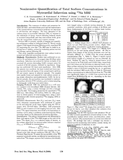


© Copyright 2026