
25700___ - Radboud Repository
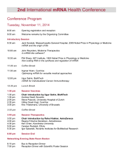
PDF hosted at the Radboud Repository of the Radboud University Nijmegen The following full text is a publisher's version. For additional information about this publication click this link. http://hdl.handle.net/2066/25700 Please be advised that this information was generated on 2015-01-21 and may be subject to change. Early Elevation of Transforming Growth Factor-6, Decorin, and Biglycan mRNA Levels During Cartilage Matrix Restoration After Mild Proteoglycan Depletion PETER JVL van der KRAAN, HARRIE L. GLANSBEEK, ELLY L. VITTERS, and WIM B. van den BERG ABSTRACT Objective. To elucidate the role of transforming growth factor-(TGF-fi) and the small proteogly cans biglycan and decorin in the repair of articular cartilage after proteoglycan depletion. Methods. Limited and reversible proteoglycan depletion was induced by injection of murine knee joints with 0.5% papain. Proteoglycan content of patellar cartilage was examined by safranin O staining on histological sections and overall proteoglycan synthesis was measured by incorporation of 35S sulfate. Changes in mRNA expression of TGF-B, aggrecan, decorin, and biglycan were deter mined by semiquantitative reverse transcription polymerase chain reaction. Results. Papain injection led to rapid depletion of proteoglycans, which was partly overcome 7 days after injection, while total replenishment of the cartilage matrix with proteoglycans was observed on Day 24. The incorporation of radiolabeled sulfate in patellar proteoglycans was initially decreased (up to Day 3), but significantly enhanced on Days 4 and 7 after papain injection. Upregulation of TGF-8, decorin, and biglycan mRNA in patellar cartilage was observed on Day 2, markedly before elevation of overall proteoglycan synthesis. mRNA levels were less augmented on Day 7, and on Day 24 all messenger RNA levels had returned to control values. As well, in the soft tissue adjoin ing the patella swift upregulation of TGF-fi mRNA was observed, Conclusion. mRNA of both TGF-B and the small proteoglycans decorin and biglycan are elevated at an early phase during cartilage repair after moderate proteoglycan depletion, implying a func tional role for these molecules in this repair process. (./ Rheumatol I997;24:543-9) Key Indexing Terms: CARTILAGE DECORIN REPAIR TRANSFORMING GROWTH FACTOR-B BIGLYCAN AGGRECAN Articular cartilage covers the ends of the long bones and makes smooth and nearly frictionless movement of the artic ulating surfaces possible under normal conditions. However, during pathology such as rheumatoid arthritis (RA) or osteoarthritis articular cartilage is degraded, result ing in loss of joint function 1,2. One factor contributing to the irreversible damage of articular cartilage in these diseases is the limited capability of articular cartilage for repair of the extracellular matrix3. The extracellular matrix of articular cartilage consists mainly of collagen (e.g., type II, IX, XI) and proteoglycans such as aggrecan and to a lesser extent decorin, biglycan, and fibromodulin4“6. All these matrix molecules are synthesized and degraded by the articular chondrocytes. Factors suggested to be important in repair processes in From the Department of Rheumatology, University Hospital Nijmegen, Nijmegen, The Netherlands. Supported by the Nationaal Reumafonds (Dutch League Against Rheumatism). P.M. van der Kraan, PhD; H.L Glansbeek, MS; E.L. Vitters; W. van den Berg, PhD, Department o f Rheumatology. Address reprint requests to Dr. P.M. van der Kraan, Department o f Rheumatology, University Hospital Nijmegen, Geert Grooteplein 8, 6525 GA Nijmegen, The Netherlands. Submitted March 27, 1996 revision accepted September 4, 1996. van der Kraan, et al: Cartilage repair miscellaneous tissues are the various transforming growth factor-13 (TGF-B) subtypes7. In mice, TGF-B is upregulated in an early phase during adult and embryonic wound repair, but temporal and spatial differences between the 3 mam malian isoforms were observed8,9. Application of additional TGF-fi appears to lead in several systems to stimulation of the repair process. Closure of skull defects, repair of macu lar holes, recovery of damaged heart tissue, and repair of incisional wounds can be accelerated by application of TGF-fl,(M3. As well, it has been suggested that the presence of TGF-61 in articular cartilage lesions as a i*esult of RA is an indication for a role for TGF-B 1 in tissue repair at these sites14. In addition, stimulation of murine articular cartilage repair after a simulated arthritic insult, induced by intraarticular interleukin 1 injection, was found by oar group15,16. Both the large aggregating proteoglycan of cartilage, aggrecan, and the so-called small proteoglycans like decorin, biglycan, and fibromodulin are structural and regu latory components of articular cartilage5. The major func tion of aggrecan is hydration of the extracellular matrix, while the functions of the small proteoglycans in cartilage are largely unknown. Immunohistochemical studies have shown that biglycan has a different localization from decorin in articular cartilage, suggesting different func tions17*18. Decorin was distributed throughout the matrix, 543 whereas biglycan was found mainly in the pericellular space of chondrocytes. Proteoglycan synthesis in connective tissues can be mod ulated by TGF-B. Incubation of human fibroblast with TGFB results in upregulation of biglycan mRNA, while decorin expression is reduced or unaltered19,20. Also, in isolated human articular chondrocytes biglycan mRNA expression was increased, while decorin mRNA was diminished under the influence of TGF-B21. In mesangial cells both decorin and biglycan synthesis is reported to be upregulated up to 50-fold by TGF-B22. Moreover, TGF-B not only interferes with the synthesis of the small proteoglycans, but these mol ecules also interact on the protein level23,24. Both decorin and biglycan are able to bind TGF-B, which can result in either inhibition of TGF-B biological activity25,26 or stimula tion of TGF-B action by these molecules27. In this respect biglycan as well as decorin can be considered as both nega tive and/or positive modifiers of TGF-B activity, regulating the potential repair stimulating ability of TGF-B. For insight into the roles of TGF-B and the cartilage pro teoglycans aggrecan, decorin, and biglycan during restora tion of the articular cartilage matrix after moderate proteo glycan depletion we studied temporal expression of mRNA of these molecules during in vivo cartilage repair. MATERIALS AND METHODS Animals. Male C57BI/6 mice at 10-12 weeks of age were used in all exper iments. The animals were kept in cages with wood chip bedding in air con ditioned rooms at constant temperature. They were fed a standard laborato ry diet and had access to tap water ad libitum. Induction of mild proteoglycan depletion. Proteoglycan depletion was induced as described by van der Kraan, et a/28. The right knee joint of mice was injected once, intraarticularly along the patellar ligament, with 6 pi of 0.5% papain solution (type IV, 15 units/mg; Sigma, St. Louis, MO, USA). To activate the papain, the solution was supplemented with 0.03 M L-cysteine HC1 (Sigma). The left (control) knees were injected with physiologi cal saline. The papain injection in the patellar cartilage leads to limited, reversible proteoglycan depletion and inhibition of proteoglycan synthesis one and 2 days after the injection, followed by supranormal proteoglycan synthesis later28. At several time points after papain injection, patellae were isolated and used for measurement of proteoglycan synthesis and determi nation of mRNA levels. Surrounding tissue was used for detection of mRNA levels only. Determination of patellar proteoglycan synthesis. Patellar cartilage proteo glycan synthesis was measured ex vivo according to the method of van den Berg» et aP9. Whole patellae, with a standard amount of surrounding tissue, were dissected from the knee joints. Patellae were labeled immediately after isolation with 35S sulfate (LI MBq/ml) for 2 h at 37°C in R P M I1640 DM medium (Flow Laboratories, Irvine, UK). After labeling, the patellae were washed, fixed, decalcified, and punched out of the surrounding tissue. The patellae were dissolved in lumasolve (Lumac, Groningen, The Netherlands) and the quantity of incorporated 35S sulfate was detenuined by liquid scintillation counting. Whole knee joint histology. Mice were killed by cervical dislocation, and carefully dissected knee joints were fixed in phosphate buffered formalin (pH 7.4) for 5 days and subsequently decalcified in 5% formic acid for 4 days, Standard processing of the knee joints in an automatic tissue pro cessing apparatus was followed by embedding of the knee joints in paraf fin wax. Frontal knee joint sections were prepared (6 pm) and stained with safranin O and fast green. 544 Isolation o f patellar cartilage and synovial tissue. Patellae were dissected from the murine knee joints and immediately decalcified for 4 h at 4°C in 3.5% EDTA (Sigma). After décalcification the complete articular cartilage layer was stripped from the underlying bone. The isolated cartilage was instantly put in TRIzol reagent (Life Technologies) for RNA extraction. In control experiments it was shown that this procedure did not affect the RNA isolation or reverse transcription polymerase chain reaction (RT-PCR) performance negatively (data not shown). The patellar tendon and synovium of knee joints were dissected and 2 pieces of synovial tissue were punched out from the tissue on a standard location (area 7 mm2). Directly after punching out, the synovial tissue was quickly frozen in liquid nitrogen. RNA isolation and RT-PCR. Total RNA was isolated from patellar cartilage and synovial tissue by TRIzol extraction. RNA was directly extracted from cartilage but synovial tissue was first homogenized and then put in TRIzol reagent. Cartilage and synovial tissue of 5 mice were pooled. Before reverse transcription the isolated RNA was treated with DNAse I (Life Technologies). The reverse transcription reaction was performed with Moloney murine leukemia virus (M-MLV) reverse transcriptase (Life Technologies) using an oligo(Dt)ls primer (Eurogentec, Liege, Belgium). Amplification of DNA was accomplished by using Taq DNA polymerase (Life Technologies) up to a cycle number of 35. To determine the relative mRNA levels samples were taken at different cycle numbers and the amount of amplified product was calculated as described below. All RTPCR reactions were performed in duplicate. The amplification products were labeled by digoxigenin labeled nucleotides (Boehringer, Mannheim, Germany). After separation on a 1.5% agarose gel the products were blotted on positively charged nylon mem branes (Boehringer). Amplification products were detected by chemiluminescence using alkaline phosphatase labeled antidigoxigenin antibodies in combination with lumigen PPD according to the manufacturer (Boehringer). Spots were scanned on the photographic film using an image analyzer. Only spots lying within the linear part of the PCR were used for scanning. The results are presented as relative mRNA levels using glyceraldehyde 3-phosphate dehydrogenase (GAPDH) levels as an internal con trol. Due to the limited amount of tissue and quantities of RNA, measure ment of the amount of total RNA was not possible. The following nucleotide primers were used in the amplification reac tions. Aggrecan primers were used as described by Grover and Roughly30 (product 501 bp), while decorin primers were used according to Asundi and Dreher31 (product 400 bp). The biglycan upper primer had the following sequence: S'-AGAAGGCCTTTAGCCCTCTG-S', and the lower primer 5'ACTTTGCGGATACGGTTGTC-3' (product 130 bp). Murine TGF-B 1 primers were derived from Clontech (Palo Alto, CA, USA) (product 525 bp), whereas TGF-B3 primers were used as described by Mulheron, et aP2 (product 380 bp). To detect GAPDH, the primers 5'-AACTCCCTCAAGATTGTCAGCA-3' and 5'-TCCACCACCCTGTTG CTGTA-3 ' were used (product 553 bp). Primer sequences were selected with the computer pro grams Primer (Whitehead Institute, Cambridge, MA, USA) and Oligo 4.0 (National Biosciences, Plymouth, MN, USA), RESULTS To obtain reversible depletion of proteoglycans from the patellar cartilage 0.5% papain was injected into the right knee joints of the mice. Papain injection resulted after one day in obvious depletion of proteoglycans, measured by safranin O staining, in the patellar cartilage (Figure 1). Moderate inflammation of the joint, characterized by granu locyte infiltration, was seen at this time. Three days after papain injection the inflammation had largely vanished,i but the amount of proteoglycan depletion was not different from one day after papain injection. On Day 7 the proteoglycan The Journal of Rheumatology 1997; 24:3 Figure I. Effect of papain injection on proteoglycan content of patellar car tilage. These are frontal sections of the murine knee joint stained with safranin 0 and fast green (original magnification x 100). Depletion of pro- depletion of the patellar cartilage was partly overcome, while in the patellaris femoris minor replenishment of the articular cartilage matrix was still observable. The cartilage matrix of both patella and opposing femur obtained a nor mal appearance 24 days after papain injection. At several times after papain injection patellar proteogly can synthesis was measured ex vivo by incorporation of radiolabeled sulfate. Inhibition of proteoglycan synthesis was measured up to 3 days after the injection of papain (Figure 2). It is possible that part of the newly synthesized proteoglycan is degraded by papain remaining in the carti lage. However, patellar proteoglycan content is restored within 10 days, making it unlikely that active papain persists in the patellar cartilage. On Days 4 and 7 proteoglycan syn thesis was supranormal, roughly doubled compared to the control knee joints. Twenty-four days after papain injection radiolabel incorporation into the papain injected knee joints had reached control levels. Since it has been suggested that TGF-B might play a role van der Kraan, et al: Cartilage repair teoglycan can be seen on Day 1 and 7, whereas normal staining is observed at Day 24. A: control joint, B: Day 1, C: Day 7, D: Day 24 (c: cartilage, b: bone, f: femur, p: patella). sulfate incorporation days after papain injection Figure 2. Proteoglycan synthesis measured by incorporation of 35S sulfate in patellar cartilage after injection of 0.5% papain on Day 0. Sulfate incor poration is compared to the control knee joint. Values are means ± SD of 6 patella. This is one representative experiment out of 3, 545 in the stimulation of matrix synthesis during repair of con nective tissues such as articular cartilage, we studied the mRNA expression of TGF-B in patellar cartilage and neigh boring soft tissue. Both in the surrounding tissue and the patellar cartilage there was early elevation of messenger RNA of TGF-ft 1 and TGF-B3 (Figure 3), Even on Day 2 after papain injection, mRNA in the surrounding tissue and patellar cartilage was elevated 3 to 6-fold. In the surround ing tissue the TGF-B1 message appeared to be upregulated more strongly than the TGF-133. From Day 3 the mRNA lev els of both TGF-B1 and TGF-B3 declined, and on Day 7 only TGF-63 mRNA in the cartilage still appeared to be elevated. Twenty-four days after papain injection mRNA levels of TGF-fil and TGF-B3 were normal. To investigate if there was an association between the upregulation of TGF-B mRNA and mRNA levels of proteomRNA levels (ratio to control) *• •» ;i • . >» * » ■ ’*» ‘Z.. •>•t••»• '* • . > . • » > » « • > * . « . > m1\ -*•*V-•• *»* ■ % w•>■’’’V’-ì . »•• ► ►; • ’ - ........................ .... m , , , .................... . . . * . * ► .......................................... » ' ► ► I * ....................... ..... i y>......... . « • ►................................................ ►......... .... ■ ......... ► ■. ■• •• ‘ • • >r r * ' . •• * «• ' . . • ' •« ’ I *.......... . . . . .............. i :: : > » »»* » ► » * » » « . # • ••• • ». •'*V:/:: >>*>••»»>.*«!•»*••*!«>> ..i JÌ,\ h Ì!‘IÌ'X;ÌV^ > ■I \ * 5 • .............. »►►►#. ► > .............................. ... > » . > » » • - »• M » , . . . . . . . . . . » « > » > • . . . i || t > >* I« » ................................... » * - v:>\ ■i* I **. . . . . . . . . . . . . . . . . ». . . » I » « . ►* t ULU: ............................................... * •• ;. » , , , :: :::::: 1 1» ■ ........................»» , » ►k M #II ............................................................................. I I ► •lit * •* .......................... Ì!]■"^1: »• 1 il ' ..................... *» I :>v ► *.. * ► I ..................• • ::: ::: » . ' ► ► y .y :9 .m \y '' : .'!• .. ’ • • ri'jsir::’:: I .. I . :v: !?■?!?.•?::•• . , , 4 . < . < ><................... , ... : i f r ; :r:; ■ ’ • • • .« • . ^ y . y. 4• » p. ** , > '. I » * . , > . 1*: >r : : : s ’ :!; :* ' «*11; j; ::: , . ........ l,i f I I’ ' ' ¿ y ; , : is- r, T*' *r* I• irm I 4I •ì ••• *•*•* • fc’ ....................... • *. I I » . , || . 9 ....................• *' ' ... ....................... ' ' ! ;; •-..r-£s 'r*: — ••T 5• ► -*[*»«I*“^'•-•* j•j; - ; •• ;> m ;1;j! *!!*. \.!t!’"'91'9?J! !Lf1:\!' ! V . » i l ......................... .... » ■ ; • - ■ 11 < I kI I jI ................................................ 1 • • • ;« • rT T !. .................................! . .. . .. .. . .. . .. . .. . .. .! ! '• ! ... > »> ... )I • • • . . . . . . . . . . . . . . . . . .I . ill I I I I I■ I I p l l l l l l . l * f * , l l , fl* • ' ■ > ■ ► » • I f M u I 1 1 .................................... ................................................................................... • • • • . . . «. s i . J ............................... ................................... • • • ili iii irr • •• p i;> ' i ' . , . . i d. i « I • • k•► • »» > ................................................. .1.. .............................. . « • • • I *I I I la f .................................. ............................................... • III • • pI I ... . . . . . . . . . . . . . . . . •• I ». . ......................................... . . . ....................................................... . . . . . . . . . . . . . . . . . . . . . . . 4.S!................................. .. . .. ......................................................................h «« -Mi« • • * .................................... « h ............................................ I . . . . . . . . . . . . . . «« « ............................................................I • • • j • ' »»I • . . . I .................................... ................................................................................. • • • ....................................... • ' • . , . *, % ......................................... .. I: y ; ; \ 1' " 11 . • . ......................... . . . . . .......................................... . .• # k. # , ............................................................................................................ « ‘ .................... v i-: ........ i \ /: >:■ 1^ > »k. 41 , . > • • > > > > » r ......................*.......................... j :*:::;;;,; I I y m * * • . . II • • > I ' .... > > > ' t t ■t• ■ '» , I .4 I I . . . . . . . . ■►• • I» ► • ..................... ..................................... ». > i ► > m > >> > > >> > > ........................................... » » .................................................................................. ............................................... * • • • y ,:;y\:,y . .. . .. .. . .. . .. . .. . .. . »M v "Vr:Ê: !l:rrT-:Vv -jf :!I : f > . • . » > > > ! » * > ’ »I .......................................................... ‘ " :: :';v. ;. M \ :* • lr-; ■ 8 .•! , ;.:s :: \ 10 #: r 12 • . > ‘t ; 14 •. ^ 16 18 20 .. 22 24 day s after papain Injection glycans in the recovering patellar cartilage we measured mRNA levels of aggrecan, decorin, and biglycan in this structure. Messenger RNA levels of the small proteogly cans, decorin and biglycan, were already elevated 2 days after papain injection, while aggrecan mRNA in the papain injected knees was comparable to the control knee joints (Figure 4). Proteoglycan synthesis measured by incorpora tion of radiolabeled sulfate was noticeably inhibited up to Day 3. Also on Days 3 and 4 upregulation of biglycan and decorin mRNA was observed, whereas on Day 7 the levels were diminished compared to the levels on Day 2. Aggrecan mRNA levels were never strikingly altered during the peri od of the experiment. On Day 24 all mRNA levels were sim ilar to the control levels. DISCUSSION Murine knee joints were injected with papain to study the repair response of articular cartilage after limited proteogly can depletion. This model was used since papain injection in the dosage we used leads to proteoglycan depletion with a restricted inflammatory response. Our results show that patellar proteoglycan depletion after papain injection can be restored to normal by the articular chondrocytes. Even 7 days after the induction of proteoglycan depletion, part of the matrix was restored, while after 24 days no signs of pro teoglycan depletion could be detected by safranin 0 stain of the matrix. At least one of the mechanisms leading to replen ishment of the cartilage matrix with proteoglycans appeared to be enhanced synthesis of proteoglycans, starting between 3 and 4 days after papain injection. Even at an early time point during the repair phase, sev eral days before increased incorporation of radiolabeled sul- mRNA levels (relative to control) T : ^ !•? |! . ■1 .. »» ’i nrr » » »m i*1 » ’* 3 ** • ♦ kI .r »..................... . .• ‘ ^j ,* ■.\ . ’ r ; l :. .i.’# ' 4 . ► .. „ I •, ; ’ ^, I S' •••*..I! .............. * > • .' !» r 6 ri ‘ ■ r ‘ 1 . . . . . ‘‘ mRNA expression (ratio to control) ► ;); .* ............ I v »! »! * *, -*! ; * ' » . * ; ,, . . ............................................... .................................. ». > , ! i . »’ . VI ! »! : »*" I • « ........................................................... » » ► ►I I I I ►► ►I » I 1 » * I 4 » » I » > . . . .. . . . . . . . . . . . . . . . . . . . . . . . .. . . . . ..................................................... . . . . . .«•«»«•«. * » .......................... »..................................................... . . . » ...................... ‘ . ’ rrr .r: ;r•!: ' . :;r t : , ..• » ► ^ .......................... M * ( ! ! ................. .. ‘ ::: |.. V :::::: :;; :.. . .. . .. .. . .. . .. . .. . .. .. . . ♦ ;I ; * * 9 * M I ;; • • ;:, ;;:t' r► » II %, i . .. . .. .. . .. . .. 1 4 * » . » 1 1 4 » ! « ” » » - » ............................. * , ,. » ..................................................% . III • • •• 1 * I «»«....................... . . . . . . . . . . . . . . . .. . . ............ 1' * “ -» ■ :— + % >• .......................... i* ‘ l i " ' I ' ‘ I . • ► - • ->- - - V ' to .> > k **. #*#. **. ; j , *: I;• ;:• :• ;;' * . :«»;*[; ;;# ;• II ► ► ;; ;*; % p.»' • . »I . ' . % . .. . .. .. . .. . .. . .. . .. . • • : : : : : ■ ••• • *........ .................... «• ■ . ........................... ► ' , 1 ' l , I ,1 ! !■ ’ ’1;tt’**' t** . ............................... ...................................» » • ........................ ; ! ! * * • m i • * ! ! M ;► ^ ^ . j; ; * ; *j* *;*• * *; J.** ► j .• • • • j ;;_.► *;• f• • ;;’ ;’ ;; , " .................. I . .I • ‘r «« • I « f kI i » . • • II • > . .j *• rib.».-- I ........................... . .. * , '!««»• * * * > • *• • « » « * * * « ' k 1 ' " ............ .. - • “ * ; k 1 1«n ■ 1 1 ' * « I * I • ! !;k > • 4 t • .* . . .. . .. .. . .. . .. . .. . .. .. . .. . ..*».* ,r ................. ........................... . ' ' l - i ' 11J ' j i ' m' *** ' •I * » ............................................................................................. *1* .................................... . . , , .,. ,, . * . i i . ....... : *•.,, !l(, • ' ’ ............. ' ' ' " I I »* »•1 *» ' . . . . ................................ . . . ^ ^ ....................................................... I . « k»»k II • « < 1 1 * ’ *■ • ; ; ; ; j ; « ^ I « > y. .«!> ,k • • • • • M M < < V < « • • .........kk!''i p « . . . . . . . . • • • ^ k ,i , , b - • •• ,t r •, , . • * L ,• • • . . i •• • b ............................................ . ! . . 1 . > ; . y j * • ** * ! •*! ; : ............................ I II • ’ • I I 4 M b . <i I I I * <1 1<< » t 4 ' ¡ ¡ ¡ a : i : ......................... .... <i » t , t iiMi , l ( l , , *4* i i I • pj 4 I * * « I I IS **•>b. a » ».»I . . . *r « m I :j; : r .4 I b I I I «• I* i ! ! r * ' j , I ' I • . »• «**»*I l l *1l f j b *^j «<<<<<<, , ,(, » » » • f •4 4 * « II »»»»I M t i< i i r ! i i i -• • •- * " •, • ' J y , ‘ : "t ' I J M M m “ • • * ' • * 4 ' 4 •• ' 4 4 , y 14 1 ' M I ' I I I I ............... v « I k r a n « i m * , , , i; : i f : : , .• > r r • ‘ 4 1 1, 4 ‘ V -.. 1i : 1’ ! -- i1 i i i i !i 1i i f i i i bv i j : ! i 1. : •. ¡iis^'*4* , 4, \ , i » \ \ \ 4 , 4 y , ,4 a 41 * . I * ' r• i l î ' 4 . • !* ‘ î i< rî < « * f î ? i ! ! i 1•i ‘ : ' : : : ^ 1 1 i : *' v - * î ' i * i j j / *••111 4 m < » ‘ <<>*m- , ,4 ,4 4 : •,, 1 ' v ; • v j j , ^ iniMn'« ^ m < ; M * < ' : : : : ! • ! • « i f i 4 . \ y i:v i ; 4 m < : * •y M M « . .< t 1 j <11 . 1 •<«. m aggrecan : .................. I ;: . . : : ' : r f , . y <! b ,, ' r ■ M « , [ y * * ! . . < ! ! < ! . . . . i * é y, \ i } \ * : : : * ' 4 4 * * ’ •* ’ • * * : * * * * • * ' • ; r - î y “ ■.- ‘y . . Y ' t i y w - é . * \ 4 ' y è. y w y * . ' < ; ,ki .< ; : • • . . 4 I ■# ■:, « « • • ■ « ......................... ... . «. , \ , 4y 9 '< j\ \ J\ \ , \ « •y \ . i s ; . i , ^ ] m <m 4««: ; ; ; 1 ; ; 1 : « ' \ ' : ! 1' ■« '. i * <ai « . . « y \ •! < ■' ' '4 < ;; ;; .j; * •: ^ : ■, ' 1 ’ • 1 11 • •• v y y , ’ i ' 1 1 , y 4 . * !' . ' M * ' •* * ' * ■* 'i ' : y ...................... 14 p m * ê \ \ \ , \ H . * « i * ••• ; •• '"-w •!•••»'. , i . ' '\ >4 * 1 b 1 1 1 « . y ',, 1 m'mimi \ *•* ' * ■: M* i ** • • ; J . . I • • I I • I il I I f , • • I - 4 I1• 4 < | ’• < 4 41<l|..<lll r:. r . ü: * ' ::: li» I • 1 » • I < < '. : 'ü \ I M 1 • . M i l l 9 • | • « 1 i ( I 1 '■ r MMl II I ;;;r 4 < < I I < < < < 4 I M ' I I I M M « M b l l . M L I I M M M , ê m 1 1 11. r : \ izy, * M t • I I < ►M II ' H ' V I M M 1 ^ « « M I I , < M < * J t ■ i J U ' U , »»*b « ........................ ... ........................................ ......................................................................................... I Ml' p I I I * . M , y . i « « , M ' . .I *. • ' • « « ................................................ «« M i l ........................................ ... am • • • 41 •i J-’ .yy.yy.y '* y 1’ 2i‘ * ;;•!]. :jrr: - 14 16 18 20 22 24 • * *4 . • I M days after papain Injection Figure 3. Messenger RNA levels of TGF-131 and TGF-I33 in patellar carti lage (top panel) and neighboring soft tissue (lower panel) after injection of 0.5% papain. Values are expressed as a ratio of the RNA messenger levels in control knee joints (ratio of 1 is similar to control). For each time point, results from 5 patellae or soft tissue sections were pooled. This is one rep resentative experiment out of 3. All RT-PCR were carried out at least in duplicate. Levels are normalized to GAPDH and control joints injected with physiological saline. • < < • < * 44 19. < < b , <1 y 4M , 444<t <<4• 546 i i J )«| , * ' *j • • • • r * , . rf t M M ‘M < << b !M '4 *4 < « P,. * ‘4 8 1 *' • • ;• • •• • • 4 • • »bm • 1• 44 . .! , < b < b( 1 . . < < < 4 * • ! • ; : : r , , ' * ■* ! ! ! . * f i : i i i f f ! v I [r!T.:,l ‘ I '‘ **' i*'* « « « ' « f i M " •'••••• ‘ •' . 4\4.y. yy . y 44y4 ,:4y ?* j-4 <411 « • I: :.P!: M « ;! . * ‘ , ‘ • ; * ! - ] ‘ ; f I ; \ •; \ ; ] [ ‘ \ • • : é » ••\ \ ] \ ; •“ ‘ *> ‘ ‘ ‘ ‘ ‘ b l»M :• -/ : ! «« ' ] ] . ; ; j ;•;; y,y.\ ! ! M m M l !, \ \\ . 1 ' j • • : j! !! n 4 i i4111 < *m i ' i;’ :, ^ :;;. ;:, ;*:, ; II 4 ; j • ' • ‘ • •: ! y i ! i i y. : i < 1 ,1 j .»»► ► ► IMI * ii4 .4 ' l l / .Mi l I ( j .i! • * • 1ê • • ; ; ij . I »»»I»» i l i m .. j . . j . •;* m I « » « . H «4«*<*«4«<««4<<<«M<« M J*. 1 . J *J ,è : , y i “ M I s : r : : : / M t . 4 ! ! * ’ J < « , < < < , I M M M M 4 « | | • ■ LJJ!J! Jî ^ : 4 * ; , r: I. .....................I i!*7 , • *I • I ■ I ■ ■^I »I I ; !! I »I I n f i l i l i .I * r ; r •r • ; : iii <• <<<• i l l b i i «i ^ ' . »s» ' ;: \\\\ • * <<! * ! ! , ‘ II < 4IIHi . I I • • if:; > ■- • I « I * • i m j ¡ i : i «< , 1 * y , , • A H * • I > w i y - • • • I • *• • • *!• !! *J< 1 1 ' ' Jb ''M 11 ’ * 1 • < < < -1 * .. . «»«b I. ► •' j J \ , , J J \ ( J ^ • , 1 b » 1 < - <M .............. ’fir • * , ..................... . * >i M ' ;;J .i 41 < < * 4 y.yyy4 . y'yy.y.4 ,. !.. * « • y.yyy.i: i <<: 4‘ ' • ••' • •••••••*I ' ’ ' 1 14 .1::! m 41 :rIl ■ i:::1:!:■ f‘ ^ < m < < m * ><<>•> < • : < ■./' , i '' t M M • * . p 4<< ii << ■ ■ < <<* I I, t y. • IIIIè,1< ÉI p I« 10 16 20 22 days after papain Injection Figure 4. Messenger RNA levels of aggrecan, decorin, and biglycan in patellar cartilage after injection of 0.5% papain. Values are expressed as a ratio of the RNA messenger levels in control knee joints (ratio of 1 is sim ilar to control). For each time point results of 5 patellae were pooled. This is one representative experiment out of 3. All RT-PCR were carried out at least in duplicate. Levels are normalized to GAPDH and control joints injected with physiological saline. The Journal o f Rheumatology 1997; 24:3 fate, TGF-fl mRNA levels were augmented, both in patellar cartilage and the soft tissue adjoining the patellae. This indi cates that upregulation of TGF-B mRNA is an early event during the restoration of the cartilage matrix after proteo glycan depletion by papain, suggesting a role for TGF-B in the repair process. The elevation of TGF-B mRNA was not very longlasting, and 7 days after papain injection the mes sage levels were lower than on Day 2, whereas on Day 24 mRNA levels had reached control values. TGF-B2 was mea sured in the surrounding tissue and patellar cartilage on Day 2 only. It also appeared to be elevated in both compartments at this time (data not shown). These observations are in accord with the data of Martin, et al , who observed in mouse embryos rapid induction and clearance of TGF-B after wounding11. With respect to the bioavailability of TGF-B, newly syn thesized TGF-B protein is only one of the sources of TGF-B. TGF-B is bound to the extracellular matrix by several bind ing proteins33-36. In the matrix of bovine articular cartilage considerable amounts of TGF-B were detected that appeared to be inaccessible to the chondrocytes under normal circum stances37. However, it can be imagined that as a result of proteolytic action the majority of matrix bound TGF-B in cartilage can become available to the chondrocytes after exposure to papain. Moreover, TGF-B being synthesized as a latent molecule might be activated by the proteolytic activ ity of papain and also in this way TGF-B bioactivity can be enhanced by papain. On the other hand, papain might degrade active TGF-B and in this way lower the bioavail ability of TGF-B. The early upregulation of TGF-B mRNA coincided with a simultaneous increase of decorin and biglycan mRNA lev els. In patellar cartilage chondrocytes, mRNA levels of decorin and biglycan were elevated about 3 to 4-fold even 2 days after papain injection, when glycosaminoglycan syn thesis appeared to be diminished by about 50%. Not before Day 4 could enhanced synthesis of proteoglycans, measured by 35S sulfate incorporation, be observed in the patellar car tilage after papain injection. During the whole study period the mRNA levels of aggrecan, the proteoglycan contributing predominantly to the incorporation of sulfate in cartilage, did not change strikingly. This indicates that at least part of the proteoglycan synthesis, as measured by sulfate incorpo ration, is not regulated by transcription of the aggrecan core protein gene but by other factors, such as upregulation of aggrecan core protein synthesis due to posttranslational mechanisms. Increased addition of glycosaminoglycan chains to the aggrecan core protein during the repair phase seems unlikely, since no changes in the size of aggrecan could be detected using Sepharose Cl 2B chromatography (data not shown). The early elevation of the small proteoglycans decorin and biglycan can be interpreted in several ways. Since no upregulation has been found after injection of proteins that van der Kraan, et al: Cartilage repair j cause inflammation similarly to papain but without proteo glycan degradation, elevated mRNA levels as a reaction to the inflammatory response in the joint seem unlikely. However, TGF-B mRNA levels can be affected by the inflammatory response in the joint. Upregulation of mRNA of the small proteoglycans could occur as a result of TGF-B activity on patellar chondrocytes. This would be a conse quence of release and activation of inactive TGF-B by papain or a result of enhanced TGF-B synthesis. In this way the small proteoglycans could function as enhancers of TGF-B bioactivity or function in a negative biofeedback mechanism to control the TGF-B action on the chondro cytes25“27. The observations of Roughly, et cil2\ with incu bation of isolated human chondrocytes with TGF-B leading to increased biglycan mRNA and decreased decorin mRNA levels, appear to contradict a role for TGF-B in upregulation of mRNA of both small proteoglycans. Partly in parallel with the studies of Roughly, et al , Collier and Ghosh observed increased biglycan synthesis and unaltered decorin synthesis after exposure of meniscal chondrocytes to TGFB38. However, both studies were performed with chondro cytes of different species and with isolated chondrocytes, instead of an in vivo model as described here. Nevertheless, TGF-B could play a role in regulation of cartilage proteoglycan synthesis: the elevation of decorin and biglycan mRNA we observed could be independent from TGF-B action. The early elevation o f biglycan and decorin mRNA suggests an intrinsic role for these proteo glycans in assembly of the cartilage matrix during the repair period, irrespective of their relation to TGF-B. Decorin is able to influence the fibrillogenesis of type I and type II col lagen and has been shown to bind to collagen type VI in binding assays39,40. In addition, decorin has been reported to interact with fibronectin and thrombospondin, both extra cellular matrix components41'42. Biglycan has a different dis tribution than decorin within the cartilage matrix, implicat ing different functions for biglycan than decorin in cartilage physiology17’18. Upregulation of mRNA of decorin and biglycan might be a functional mechanism in the recon struction of damaged cartilage matrix, independent of their role in control of TGF-B activity. In accord with our results, Cs-Szabo, et al found 5 to 10-fold higher mRNA levels of biglycan and decorin in human osteoarthritic cartilage com pared to normal cartilage, suggesting a role for these pro teoglycans in local cartilage repair foci that can be found in os teoarthriti c c artil age43. In conclusion, our results indicate that during restoration of the patellar cartilage matrix after moderate proteoglycan depletion, upregulation of TGF-B and decorin and biglycan messenger RNA in patellar cartilage and TGF-B in adjoining soft tissue is an early event. Upregulation of mRNA is initi ated days before enhancement of proteoglycan synthesis, as measured by incorporation of radiolabeled sulfate. These results suggest that both TGF-B and the small proteoglycans 547 decorin and biglycan might play a role in regulation of the chondrocyte metabolism, while the small proteoglycans could have a potential structural function in repair of dam aged articular cartilage matrix. 21. 22. REFERENCES 1. Fassbender HG: Histomorphological basis of articular cartilage destruction in rheumatoid arthritis. Coll Relat Res 1983;3:141-51. 2. Brandt KD: Osteoarthritis. Clin Geriatr Med 1988;4:279-93. 3. Man kin HJ: Current concept review. The response of articular cartilage to injury. J Bone Joint Surg 7952;64A:460-6. 4. Hardingham TE, Bayliss MB: Proteoglycans of articular cartilage: Changes in aging and in joint disease, Semin Arthritis Rheum 1990;20:12-33. 5. Hardingham TE, Fosang AJ: Proteoglycans: Many forms and many functions. FASEB J /992;6:8^i-70. 6. Eyre D, Wu JJ, Apone S: A growing family of collagens in articular cartilage: Identification of 5 genetically distinct types. J Rheumatol 1987;14:25-1. 7. Border WA, Ruoslahti E: Transforming growth factor-8 in disease: The dark side of tissue repair. J Clin invest ¡992;90:1—7. 8. Levine JH, Moses HL, Gold Li, Nanney LB; Spatial and temporal patterns of immunoreactive transforming growth factor fill, JJ2 and 133 during excisional wound repair. Am J Pathol 1993; 143:368-80. 9. Martin P, Dickson MC, Mill an FA, Akhurst RJ: Rapid induction and clearance of TGF 8 J is an early response to wounding in the mouse embryo. Develop Gen 1993; 14:225-38. 10. Beck LS, DeGuzman L, Lee WP, et al: TGF-B induces bone closure of skull defects. J Bone Min Res 199l;6:1257-65. 11. Glaser BM, Michels RG, Kuppermann BD, Sjaarda RN, Pena RA: Transforming growth factor-B2 for the treatment of full thickness macular holes. Ophthalmology J992; 99:1162-73. 12. Lefer AM: Mechanisms of the protective effects of transforming growth factor B in reperfusion injury. Biochem Pharmacol 7997/42:1323-7. 13. Cromack DT, Porras-Reyes B, Purdy JA, Pierce GF, Mustoe TA: Acceleration of tissue repair by transforming growth factor Bl: Identification of in vivo mechanism of action with radiotherapyinduced specific healing deficits. Surgery 1993; 113:36-42. 14. Chu CQ, Field M, Allard S, Abney E, Feldmann M, Maini RN: Detection of cytokines at the cartilage/pannus junction in patients with rheumatoid arthritis: Implications for the role of cytokines in cartilage destruction and repair. Br J Rheumatol 7992/31:653-61. 15. Van Beuningen HM, van der Kraan PM, Arntz OJ, van den Berg WB: Protection from interleukin-1 induced destruction of articular cartilage by transforming growth factor ft: Studies in anatomically intact cartilage in vitro and vivo. Ann Rheum Dis 1993/52:185-91. 16. Van Beuningen HM, van der Kraan PM, Arntz OJ, van den Berg WB: In vivo protection against interleukin-1-induced articular cartilage damage by transforming growth factor-Bl. Age related differences. Ann Rheum Dis 1994;53:593-600. 17. Poole AR, Webber C, Pidoux I, Choi H, Rosenberg LC: Localization of a derm at an sulfate proteoglycan (DS-PGII) in cartilage and the presence of an immunologically related species in other tissues. J Histochem Cytochem 1986;34:619-25. 18. Poole AR, Fisher LW, Young MF, Termine JD, Robey PG: Expression and localization of the 2 small proteoglycans biglycan and decorin in developing human skeletal and non-skeletal tissues. J His toehem Cytochem 1990;38:1549-63. 19. Romaris M, Heredia A, Molist A, Bassols A: Differential effect of transforming growth factor 13 on proteoglycan synthesis in human embryonic lung fibroblasts. Biochim Biophys Acta 1991;1093:229-33. 20. Kahari VM, Larjava H, Uitto J: Differential regulation of 23. 24. 25. i 548 26. 27. 28. 29. 30. 31. 32. 33. 34. 35. extracellular matrix proteoglycan (PG) gene expression. J Biol Cheml991;266:10608-15. Roughley PJ, Melching LI, Recklies AD: Changes in the expression of decorin and biglycan in human articular cartilage with age and regulation by TGF-ii Matrix Biol 1994;14:51-9. Border WA, Okuda S, Languino LR, Ruoslahti E: Transforming growth factor-J3 regulates production of proteoglycans by mesangial cells. Kidney ¡nt 7990/37:689-95. Ruoslahti E, Yamague hi Y, Hildebrand A, Border WA: Extracellular matrix/growth factor interactions. Cold Spring Harbor Symp Quant Biol 7992/72:309-15. Hildebrand A, Romaris M, Rasmussen LM, et al: Interaction of the small interstitial proteoglycans biglycan, decorin and fibromodulin with transforming growth factor B. Biochem J ¡994;302:527-34. Border WA, Noble NA, Yamamoto T, Tomooka S, Kagami S: Antagonist of transforming growth factor-B. A novel approach to treatment of glomerulonephritis and prevention of glomerulosclerosis. Kidney Int 7992/41:566-70. Hausser H, Groning A, Haslik A, Schonherr E, Kresse H: Selective inactivity of TGF-B/decorin complexes. FEBS Lett 7994/353:243—5. Takeuchi Y, Kodama Y, Matsumoto T: Bone matrix decorin binds transforming growth factor-B and enhances its bioactivity. J Biol Chem 7994/269:32634-8, Van der Kraan PM, Vitters EL, van de Putte LB A, van den Berg WB: Development of osteoarthritic lesions in mice by “metabolic” and “mechanical” alterations in the knee joints. Am J Pathol 7959/135:1001-14. Van den Berg WB, Kruijsen MWM, van de Putte LB A: The mouse patella assay, An easy method of quantitating articular cartilage chondrocyte function in vivo and vitro. Rheumatol hit 7952/1:165-9. Grover J, Roughly PJ: Versican gene expression in human articular cartilage and comparison of mRNA splicing with aggrecan. Biochem J 7993/291:361-7. Asundi VK, Dreher KL: Molecular characterization of vascular smooth muscle decorin: Deduced core protein structure and regulation of gene expression. Eur J Cell Biol 7992/59:314-21. Mulheron GW, Mulheron JG, Danielpour D, Schomberg DW: Porcine granulosa cells do not express transforming growth factorB2 (TGF 32) messenger ribonucleic acid: Molecular basis for their inability to produce TGF-B activity comparable to that of rat granulosa cells. Endocrinology 7992/131:2609-14. Biitzow R, Fukushima D, Twardik DR, Ruoslahti E: A 60-Kd protein mediates the binding of transforming growth factor-B to the cell surface and extracellular matrix proteoglycans. J Cell Biol 7995/122:721-7. Miyazono K, Ichijo H, Heldin CH: Transforming growth factor-B: Latent forms, binding proteins and receptors. Growth Fact 7993/8:11-22. Taipale J, Miyazono K, Heldin CH, Keski-Oja J: Latent transforming growth factor-Bl associates to fibroblasts extracellular matrix via latent TGF-B binding protein. J Cell Biology 7994/125:171-81. Moren A, Olofsson A, Stenman G, et al: Identification and characterization of LTBP-2, a novel latent transforming growth factor-fi-binding protein. J Biol Chem 7994/269:32469-78. Morales TI, Joyce MA, Sobel ME, Danielpour D, Roberts AB: Transforming growth factor-B in calf articular cartilage organ cultures: Synthesis and distribution. Arch Biochem Biophys 7997/288:397-405. Collier S, Ghosh P: Effects of transforming growth factor beta on proteoglycan synthesis by cell and explant cultures derived from the knee joint meniscus. Osteoarthritis Cart 7995/3:127—38. Vogel KG, Paulsson M, Heinegard D: Specific inhibition of type I and type II collagen fibrillogenesis by the small proteoglycan of i 36. 37. ' __ •k 4 38. 39. m The Journal o f Rheumatology 1997; 24:3 tendon. BiochemJ 7954/223:587-97. 40. Bidanset DJ, Guidry C, Rosenberg LC, Choi HU, TimpI R, Hook M: Binding of proteoglycan decorin to collagen type VI. J Biol Chem 7992/267:643-8. 41, Winnemoller M, Schmidt G, Kresse H: Influence of decorin on fibroblast adhesion to fibronectin. Eur J Cell Biol 1991;54:10-1. van der Kraan, et al: Cartilage repair 42. Winnemöller M, Schön P, Vischer P, Kresse H: Interactions between thrombospondin and the small proteoglycan decorin: Interference with cell attachment. Eur J Cell Biol 1992;59:41-55, 43. Cs-Szabo G, Roughly PJ, Melching LI, Kuettner KE, Giant TT: Expression of proteoglycans in human osteoarthritic cartilage measured by quantitative ~PCR (abstr). Transactions of the 42nd Annual Meeting. ORS 1996;21:221. 549


© Copyright 2026