
Chewing prevents stress-induced hippocampal LTD formation and
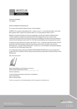
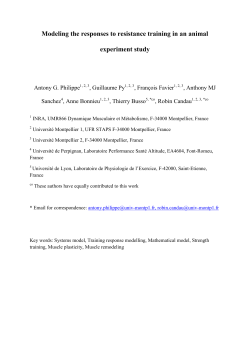
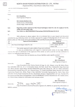
Chewing prevents stress-induced hippocampal LTD formation and
anxiety-related behaviors: a possible role of the dopaminergic system
Yumie Ono1*, So Koizumi2, Minoru Onozuka3
1
Health Science and Medical Engineering Laboratory, School of Science and Technology, Meiji
University, Room A806, 1-1-1 Higashi-Mita, Tama-ku, Kawasaki, Kanagawa 214-8571, Japan
2
Orthodontic Science, Division of Oral Science, Graduate School, Kanagawa Dental University, 82
Inaoka-cho Yokosuka Kanagawa, 238-8580, Japan
3
Department of Judo Therapy and Medical Science, Faculty of Medical Science, Nippon Sport
Science University, 1221-1 Kamoshida-cho, Aoba-ku, Yokohama 227-0033, Japan
Email addresses:
Yumie Ono:
[email protected]
So Koizumi:
[email protected]
Minoru Onozouka: [email protected]
*
Correspondence to:
Yumie Ono
Department of Electronics and Bioinformatics, School of Science and Technology, Meiji University
Room A806, 1-1-1 Higashi-Mita, Tama-ku, Kawasaki, Kanagawa 214-8571, Japan
Email: [email protected]
Abstract
The present study examined the effects of chewing on stress-induced long-term depression (LTD)
and anxiogenic behavior. Experiments were performed in adult male rats under three conditions:
restraint stress condition, voluntary chewing condition during stress, and control condition without
any treatments except handling. Chewing ameliorated LTD development in the hippocampal CA1
region. It also counteracted the stress-suppressed number of entries to the center region of the open
field when they were tested immediately, 30 min, or 60 min after restraint. At the latter two
post-stress time periods, chewing during restraint significantly increased the number of times of
open arm entries in the elevated plus maze, when compared with those without chewing. The in vivo
microdialysis further revealed that extracellular dopamine concentration in the ventral hippocampus,
which is involved in anxiety-related behavior, was significantly greater in chewing rats than in those
without chewing from 30 to 105 min after stress exposure. Development of LTD and anxiolytic
effects ameliorated by chewing were counteracted by administering the D1 dopamine receptor
antagonist SCH23390, which suggested that chewing may activate the dopaminergic system in the
ventral hippocampus to suppress stress-induced anxiogenic behavior.
1. Introduction
Chewing has been known as one of the active coping strategies to suppress stress. We have
previously shown that chewing during stress exposure significantly attenuates neuronal responses to
stress and the subsequent stress-related cognitive deficits in the hippocampus, such as impairment of
spatial memory [1, 2]. Stress-induced changes in neuronal electrical properties in the hippocampus
not only contribute to memory functions, but also mediate anxiety behavior [3, 4]; the hippocampus
is a key region to express stress- and anxiety-related behaviors via its reciprocal connection with the
amygdala and the prefrontal cortex [5]. In addition to its possible role in memory formation with
counterpart phenomenon, long-term potentiation, hippocampal long-term depression (LTD) has been
suggested to have a correlation with anxiety behavior [6]. Therefore, it is intriguing to investigate
whether chewing under stressful conditions could interfere with stress-induced LTD and
corresponding anxiety-like behavior. The present electrophysiological study evaluated the effects of
spontaneous chewing during exposure to restraint stress on stress-related LTD development in the rat
hippocampal CA1 region. We also used two measures of anxiety-like behaviors, exploration in a
novel open field and an elevated plus maze, to determine time-course changes in stress-induced
anxiety-like behavior in rats that were stressed with or without coping. We also incorporated in vivo
microdialysis analysis to measure monoaminergic signals in the hippocampus that relate to different
anxiety-like behaviors found in rats depending on their coping styles [7]. Finally, we tested whether
the pharmacological inhibition of the dopaminergic D1 receptor could counteract the effect of
chewing in hippocampal LTD and in anxiety-like behavior. These results helped to determine the
possible role of the dopaminergic signaling pathway in the mechanism of how chewing interacts
with hippocampal synaptic plasticity to suppress stress-induced anxiety-like behavior.
2. Materials and Methods
Animals
Ten-week-old male Sprague–Dawley rats were maintained in a temperature-controlled room (22 ±
3 °C) with a 12-h light/dark cycle (lights on at 7:00 a.m.). The rats had free access to water and food.
All experiments were in accordance with the guidelines for Animal Experimentation of Kanagawa
Dental University. All efforts were made to minimize the number of animals used and their suffering.
Stress protocol and drug administration
To produce restraint stress, we tied a rat to a wooden board in a spread-eagle supine position for 30
min using leg fasteners as previously described [8]. Rats were randomly assigned to one of the three
conditions of (1) stressed (ST), (2) stressed and chewing (SC), and (3) control (CT). Rats in group
ST were restrained and left alone for the entire restraint period, and those in group SC were allowed
to chew on a wooden stick (diameter, 5 mm) that was placed near the animal’s mouth during
restraint. Every rat in group SC responded to the wooden stick by chewing on it with a rapid and
repetitive sequence of jaw opening and closing movements for at least two-thirds of the total
restraint period. Rats in group CT were handled in the same manner as those in group ST and group
SC, but were returned to their home cage for 30 min instead of restraint. Some of the rats in group
SC were administered intraperitoneal injections of 0.3 mg/kg SCH23390, a selective dopaminergic
D1-receptor antagonist that penetrates the blood–brain barrier [9], which was dissolved in 0.9%
saline 15 min prior to the stress protocol. All stress inducement and handling manipulations were
performed between 9:00 a.m. and 11:00 a.m.
Electrophysiology
A total of 21 rats were used in the LTD experiment. Six rats were assigned to each group of CT, ST,
and SC, respectively, and the remaining three rats were assigned to group SC+SCH23390.
Immediately
after
the
stress
protocol,
we
anesthetized
rats
with
2-bromo-2-chloro-1,1,1-trifluoroethane (300 µL/100 g; Takeda Chemical Industries, Osaka, Japan),
decapitated them, quickly removed their brains, and iced them in artificial cerebrospinal fluid
(ACSF) containing 119 NaCl, 2.5 KCl, 26.2 NaHCO3, 1 NaH2PO4, 1.3 MgSO4, 2.5 CaCl2, and 11
glucose (in mM, bubbled with 95% O2–5% CO2). We dissected the hippocampi, embedded them in
agar blocks for slicing, cut transverse slices (450 µm thick) with a vibrating tissue slicer (Dosaka,
Kyoto, Japan), and transferred them to a holding chamber at room temperature (25 °C). We allowed
the slices to recover for at least 60 min, then transferred them to an immersion-type recording
chamber perfused at 1 mL/min with ACSF containing 0.1 mM picrotoxin (Sigma-Aldrich, Tokyo,
Japan) at room temperature. To prevent epileptiform discharge of pyramidal neurons, we made a cut
at the border between the CA1 and CA3 areas. A glass pipette filled with 3 M NaCl and positioned in
the stratum radiatum of the CA1 area recorded the field excitatory postsynaptic potential (fEPSP).
Bipolar stainless-steel electrodes (World Precision Instruments, Sarasota, FL, USA) placed in the
stratum radiatum on opposite sides of the recording pipette stimulated the Schaffer collateral
branches. We adjusted the intensity of fEPSP in the baseline period to around 50% of the maximal
response and then recorded stable baseline fEPSP activity by applying a 40-µs voltage pulse at the
determined intensity every 30 s for at least 10 min. LTD was induced by low-frequency stimulation
(LFS) of 900 pluses at 1 Hz (15 min). All signals were filtered at 2 kHz using a low-pass Bessel
filter and digitized at 5 kHz using a MultiClamp 700A interface running pCLAMP software (Axon
Instruments, Union City, CA, USA). We measured initial slopes of the fEPSP and normalized them
to the average of the values measured over the baseline period. The average slope size of the fEPSPs
recorded between 30 and 40 min after the end of the LFS provided the basis for our statistical
comparisons. We used a single slice from a single animal for subsequent analysis.
Open-field test (OFT) and elevated plus maze test (EPT)
Eighty-nine and 87 rats were used in the OFT and EPT experiments, respectively. OFT and EPT
were performed in a sound-isolated room to analyze anxiety-related behavior in a novel environment
[10]. The open field consisted of a circular arena (150 cm in diameter) surrounded by walls 40 cm in
height. A video camera (DCR-HC1000, Sony, Tokyo, Japan) was suspended from the ceiling above
the arena to observe and record animal behavior. The field was constantly illuminated with an
intense light (600 Lx at the floor of the arena). The animals in their home cage were brought into the
experimental room at least 1 h before the beginning of the first trial of the day to acclimatize.
At the beginning of the OFT, the rat was placed at the center of the arena. The field was
divided into two subdivisions of center (75 cm in diameter) and peripheral areas for scoring
ambulatory activity. Rats were allowed to freely explore the arena for 5 min. The number of
entrances into the subdivisions was recorded using a video tracking system (Top Scan, Clever Sys,
Reston, VA, USA) as parameters for OFT. An entrance to a subdivision was counted if the center of
gravity of the rat body passed through the border of the subdivision from outside to inside. After the
completion of each trial, the animal was returned to the home cage and the field was cleaned with
30% ethanol.
The elevated plus maze had dimension of 1,100 mm in width and length. The corridors
were 500 mm in length and 100 mm in width, and everything was raised 500 mm above the floor.
Two facing corridors were closed by walls 450 mm in height (closed arms), and the rest remained
open (open arms). The rat was initially placed at the center of the maze facing the open arm and was
allowed to freely explore the maze for 5 min. The number of entrances to the open arm was
measured for scoring anxiety behavior. The entrance to the open arm was counted if the center of
gravity of the rat body passed through the border of the open arm from the center of the maze. After
completion of each trial, the animal was returned to the home cage and the field was cleaned with
30% ethanol.
Rats were tested in either OFT or EPM at 0, 30, and 60 min after stress exposure. Fifty-one
rats were administered SCH23390 and tested at 60 min after stress exposure.
Microdialysis
Concentrations of norepinephrine (NE), dopamine (DA), and serotonin (5-HT) were determined in
the right ventral hippocampus (5.0 mm posterior, 5.0 mm lateral, and 3.0 mm inferior from the
bregma) and were measured in an additional group of 15 rats. The ventral hippocampus was chosen
because of its specific role in regulating anxiety-related behavior [11]. These rats were first
implanted with a dialysis guide cannula with a dummy probe and divided into two subgroups that
were treated in an identical fashion to the ST (n=7) and SC (n=8) groups after recovery of 4–6 days.
Food and water were freely provided by the time of microdialysis measurement, and the rats were
housed individually after probe implantation. On the day of stress exposure, a dialysis probe
(A-I-8-02, Eicom, Kyoto, Japan) was inserted into the guide cannula instead of the dummy probe.
The dialysis tube was directly connected to a high-performance liquid chromatography apparatus
(Eicom, Kyoto, Japan) for online analysis of NE, DA, and 5-HT. A microperfusion pump perfused
the hippocampus with normal Ringer’s solution (147 mM NaCl, 4 mM KCl, 2.3 mM CaCl2) through
the dialysis tube at a flow rate of 1 mL/min. The dialysis sample was injected every 15 min via
auto-injector (EAS-20, Eicom, Kyoto, Japan). The mobile phase consisted of 99% 0.1 M sodium
phosphate-buffered solution, 1% methanol, 500 mg/L sodium 1-decanesulfonate, and 50 mg/L
Na2-EDTA. NE, DA, and 5-HT were separated on a 30 mm × 4.6 mm diameter Eicompack CAX
column at 35 °C. The working electrode (HTEC-500; Eicom, Kyoto, Japan) was composed of
graphite, and the flow rate was set at 250 mL/min. Concentrations of NE, DA, and 5-HT were
measured using known concentrations of the corresponding standard, which were quantified by
means of the peak area ratio. All chemicals and drugs used as corresponding standards and the
internal standard were purchased from Sigma (Tokyo, Japan). Chromatograms were obtained using
the appropriate software (Power Chrom version 2; eDAQ Pty.).
Statistics
All values shown are mean ± S.E.M. Statistical analysis was conducted using IBM SPSS Statistics
21 (IBM, Armonk, NY, USA) and R [12]. We compared values using the one-way analysis of
variance (ANOVA) test with the post-hoc Tukey’s multiple comparisons or the Kruskal–Wallis test
and the following pairwise comparisons depending on normality of the data. Because microdialysis
data were repeated measures from animals in either condition of ST of SC, a two-way ANOVA with
repeated measures and the post-hoc multiple comparisons with Bonferroni correction were applied.
We consider P values < 0.05 to be statistically significant, unless otherwise stated.
3. Results
Chewing rescued stress-related LTD
In agreement with earlier findings [6, 13], LTD was not induced in control rats, but in stressed rats
(Figure 1a). Chewing rescued stress-induced formation of LTD. The average slope size of fEPSPs
was significantly suppressed in group ST (77.9 ± 3.3 %) compared with group CT (105.2 ± 4.6 %)
and group SC (106.4 ± 4.8 %; F(2,17) = 14.11, P < 0.001). These results suggested an ameliorative
effect of chewing on stress-related induction of LTD in the adult hippocampus. Administration of
SCH23390 counteracted the effect of chewing in group SC. Blockage of dopaminergic
neurotransmission during stress exposure and chewing decreased a late phase of fEPSP slope and
induced LTD (Figure 1b).
Chewing rescued anxiety behavior and facilitated post-stress exploratory activity
OFT results (Figure 2a) showed that restraint stress significantly reduced the number of entries to the
center region in group ST (1.3 ± 0.8 times; P < 0.017), but not in group SC (2.3 ± 1.2 times),
compared with group CT (9.2 ± 1.7 times; Kruskal–Wallis chi-squared = 8.90) immediately after
stress. The suppressed number of entries to the center region in group ST lasted for at least 1 h (0.9 ±
0.4 and 1.3 ± 0.6 times at 30 and 60 min from stress exposure, respectively, P = 0.005 and 0.026 in
group ST, respectively). These results indicated acute restraint stress suppressed exploratory activity,
which was counteracted by active coping with chewing.
The number of open arm entries in the EPM (Figure 2b) also demonstrated a stress-related
decrease in the number of entries to the open arm in group ST (1.3 ± 0.4 times; P = 0.02) but not in
group SC (2.7 ± 1.3 times), compared with group CT (6.6 ± 1.6 times; F(2,16) = 5.10, P = 0.022)
immediately after stress. However, the suppressed number of entries to the open arm recovered at 30
min after stress exposure. Interestingly, group SC rats exhibited more open arm entries after stress
exposure compared with group ST at 30 min (7.8 ± 0.9 v.s. 3.3 ± 0.9 times; P < 0.034) and at 60 min
(7.6 ± 0.8 v.s. 2.9 ± 0.8 times; P = 0.006) after stress. Note that the mean number of open arm entries
in group SC was larger than in group CT in these two post-stress timings, although they did not
reach statistical significance. These results suggest an anxiolytic effect of chewing under stress.
Chewing increased post-stress hippocampal DA concentrations
There was a statistically significant interaction between time and group in the time-course change of
NE and DA concentrations in the ventral hippocampus, but not in 5-HT concentrations. The post-hoc
multiple comparisons found significant differences between groups ST and SC in NE concentrations
at 195 and 225 min after stress exposure, respectively (Figure 3a), and those in DA concentrations
from 30 to 105 min after stress exposure, respectively (Figure 3b). The time in which DA
concentration significantly increased in group SC overlapped with the time in which rats from the
same group exhibited significantly increased exploratory behavior in the EPM (30 and 60 min after
stress exposure, respectively).
Antagonizing DA transmission inhibited the anxiolytic effect of chewing
The synchronous increase in hippocampal DA concentrations and exploratory behavior further
motivated us to investigate the role of dopaminergic transmission on the anxiolytic effect of chewing.
As expected, systemic administration of SCH23390 suppressed the effect of chewing during
exploratory activity at 60 min after stress exposure (Figure 4). There was no statistically significant
difference among groups in the number of entries to the center region of the OFT and in those to the
open arm in the EPM.
4. Discussion
The key finding of the present study is that active coping to restraint stress by chewing prevented
stress-induced LTD in the adult male hippocampus, as well as post-stress anxiety-like behavior. The
development of the anxiolytic effect by chewing coincided with increased DA concentrations in the
ventral hippocampus, which plays an important role in regulating anxiety-related behavior [11].
Pharmacological blockage of the dopaminergic D1-receptor counteracted the effect of chewing, both
on LTD and anxiety behaviors. These results suggest an ameliorative effect of chewing on
stress-induced anxiety-like behavior via activation of the hippocampal dopaminergic signaling
pathway.
Yang et al. [14] reported that stress facilitates LTD induction in adult rats, which is
mediated through the activation of glucocorticoid receptors to affect extrasynaptic NR2B-contatining
NMDARs to undergo the induction of LTD. The present results confirmed the effect of stress to
induce hippocampal LTD in adult rats (Figure 1a). Mockett et al. [15] further demonstrated that
dopamine D1/D5 receptor activation counteracts NMDAR-dependent LTD, which is also consistent
with our results showing that blockade of D1-receptor activation impaired the effect of chewing in
rats that normally chewed during stress exposure (Figure 1b). The present results suggest that both a
reduction in glucocorticoid plasma concentration [16] and facilitation of neuronal DA concentrations
in the hippocampus are essential to prevent hippocampal LTD development in rats that chewed
during stress exposure.
Our previous studies in rats demonstrated that chewing relieves stress by suppressing
stress responses in endocrine and autonomic nervous systems. Active chewing during stress
exposure suppresses metabolic activity in the hypothalamus [16], the higher control center of
systemic stress response, to prevent stress-related secretion of corticosterone and norepinephrine in
plasma [8, 16, 17]. We have also previously found a possible involvement of histaminergic neuronal
pathways to rescue stress-suppressed long-term potentiation in hippocampal neurons [18]. The
current study further added a novel role of chewing in preventing stress-related anxiety behavior,
possibly mediated by facilitating dopaminergic systems. The accumulating results suggest that
chewing involves both direct hormonal effects and indirect neuronal mechanisms of the neural
monoaminergic system to rescue NMDAR functions.
The dopaminergic system is essentially involved in rhythmic movement, including
mastication [19, 20]. Reduced masticatory activity by molar extraction or powder-diet feeding
reduces the response of hippocampal DA neurons, impairing hippocampal learning ability in the
step-through passive avoidance test [21] and in the novel-object recognition test [22]. Results from
the present study using microdialysis further demonstrated that active mastication facilitates DA
release in the hippocampus (Figure 3). These results suggest a possible interaction between
masticatory function and cognitive function in the hippocampus via modulation of DA responses.
The late development of LTD in SCH23390-treated rats in group SC (Figure 1b) also supports the
involvement of the DA pathway, because dopaminergic projection is essential for settlement of
long-term plasticity in the hippocampus [23].
A limitation of this study was that we did not specifically block D1 receptors in the ventral
hippocampus, but used systemic administration of D1 receptor antagonists, which may act in
different regions of the brain and affect exploratory behavior. These results were consistent with a
previous report that systemic D1-antagonism reduces spatial exploration in a novel environment [24].
However, it would not affect the interpretation of the results that increased DA concentrations in the
ventral hippocampus by chewing is critical for preventing anxiety-like behavior, because intraventral
hippocampal administration of SCH23390 itself has no modulatory effect on exploratory behavior
and anxiety-like behavior [25, 26]. Further research is required to resolve the above methodological
interference and confirm the results that we found in the current study.
5. Conclusions
Chewing during stress exposure could be an active coping strategy to relieve stress-induced
anxiety-like behavior in rats. Behavioral examinations in various post-stress time periods
demonstrated a significant anxiolytic effect with chewing in stressed rats at 30 and 60 min after
stress exposure, which corresponds with enhanced dopamine release in the ventral hippocampus.
These results indicate possible involvement of the dopaminergic neuronal pathway in the
stress-relieving mechanism of chewing.
Conflict of Interests
All authors declare no conflict of interest regarding the publication of this paper.
Acknowledgments
The Japanese Ministry of Education, Culture, Sports, Science, and Technology supported this work
with Kakenhi Grant No. 10360207 (to Y. Ono), and an Open Research Center subsidy for the
Research Center of Brain and Oral Science, Kanagawa Dental College. Additionally, the authors
acknowledge Megumi Fukui D.V.M. for her technical assistance in the behavior experiment.
References
[1] Y. Ono, T. Yamamoto, K. Y. Kubo, M. Onozuka, “Occlusion and brain function: mastication as
a prevention of cognitive dysfunction,” Journal of Oral Rehabilitation, vol. 37, no. 8, pp.
624-640, 2010.
[2] S. Miyake, G. Yoshikawa, K. Yamada, et al., “Chewing ameliorates stress-induced suppression
of spatial memory by increasing glucocorticoid receptor expression in the hippocampus,” Brain
Research, vol. 1446, pp. 34-39, 2012.
[3] J. J. Kim, M. R. Foy, R. E. Thompson, “Behavioral stress modifies hippocampal plasticity
through N-methyl-D-aspartate receptor activation,” Proceedings of the National Academy of
Sciences of the United States of America, vol. 93, no. 10, pp. 4750-4753, 1996.
[4] W. P. Ma, J. Cao, M. Tian, et al., “Exposure to chronic constant light impairs spatial memory
and influences long-term depression in rats,” Neuroscience Research, vol. 59, no. 2, pp.
224-230, 2007.
[5] B. Leuner, T. J. Shors, “Stress, anxiety, and dendritic spines: what are the connections?,”
Neuroscience, vol. 251, pp. 108-119, 2013.
[6] K. Martinowich, R. J. Schloesser, Y. Lu, et al., “Roles of p75(NTR), long-term depression, and
cholinergic transmission in anxiety and acute stress coping,” Biological psychiatry, vol. 71, no.
1, pp. 75-83, 2012
[7] D. A. Zhukov, K. P. Vinogradova, “Learned helplessness or learned inactivity after inescapable
stress? Interpretation depends on coping styles,” Integrative physiological and behavioral
science: the official journal of the Pavlovian Society, vol. 37, no. 1, pp. 35-43, 2002.
[8] Y. Ono, T. Kataoka, S. Miyake, et al., “Chewing ameliorates stress-induced suppression of
hippocampal long-term potentiation,” Neuroscience, vol. 154, no. 4, pp. 1352-1359, 2008.
[9] B. Clausen, T. R. Schachtman, L. T. Mark, et al., “Impairments of exploration and memory after
systemic or prelimbic D1-receptor antagonism in rats,” Behavioural Brain Research, vol. 223,
no. 2, pp. 241-254, 2011.
[10] V. Carola, F. D'Olimpio, E. Brunamonti, et al., “Evaluation of the elevated plus-maze and
open-field tests for the assessment of anxiety-related behaviour in inbred mice,” Behavioural
Brain Research, vol. 134, no. 1-2, pp. 49-47, 2002.
[11] K. J. Kjelstrup, F. A. Tuvnes, H. A. Steffenach, et al., “Reduced fear expression after lesions of
the ventral hippocampus,” Proceedings of the National Academy of Sciences of the United
States of America, vol. 99, no. 16, pp. 10825-10830, 2002.
[12] R Development Core Team, “R: A language and environment for statistical computing,” R
Foundation for Statistical Computing, Vienna, Austria, URL http://www.R-project.org/, 2011.
[13] J. J. Wagner, B. E. Alger. “GABAergic and developmental influences on homosynaptic LTD
and depotentiation in rat hippocampus,” Journal of Neuroscience, vol. 15, no. 2, pp. 1577-1586,
1995.
[14] C. H. Yang, C. C. Huang, K. S. Hsu, “Behavioral stress enhances hippocampal CA1 long-term
depression through the blockade of the glutamate uptake” Journal of Neuroscience, vol. 25, no.
17, pp. 4288-4293, 2005.
[15] B. G. Mockett, D. Guévremont, J. M. Williams et al., “Dopamine D1/D5 receptor activation
reverses NMDA receptor-dependent long-term depression in rat hippocampus,” Journal of
Neuroscience, vol. 27, no. 11, pp. 2918-2926, 2007
[16] Y. Ono, H. C. Lin, K. Y. Tzen, et al., “Active coping with stress suppresses glucose metabolism
in the rat hypothalamus,” Stress, vol. 15, no. 2, pp. 207-217, 2012.
[17] S. Koizumi, S. Minamisawa, K. Sasaguri et al., “Chewing reduces sympathetic nervous
response to stress and prevents poststress arrhythmias in rats,” American Journal of Physiology
Heart and Circulatory Physiology, vol. 301, no. 4, pp. H1551-1558, 2011.
[18] Y. Ono, T. Kataoka, S. Miyake, et al., “Chewing rescues stress-suppressed hippocampal
long-term potentiation via activation of histamine H1 receptor,” Neuroscience Research, vol. 64,
no. 4, pp. 385-390, 2009.
[19] S. H. Chandler, L. J. Goldberg, “Differentiation of the neural pathways mediating cortically
induced and dopaminergic activation of the central pattern generator (CPG) for rhythmical jaw
movements in the anesthetized guinea pig,” Brain Research, vol. 323, no. 2, pp. 297-301, 1984.
[20] N. P. Lapointe, P. Rouleau, R. V. Ung, P. A. Guertin, “Specific role of dopamine D1 receptors in
spinal network activation and rhythmic movement induction in vertebrates,” Journal of
Physiology, vol. 587, pt. 7, pp. 1499-1511, 2009.
[21] S. Kushida, K. Kimoto, N. Hori, et al., “Soft-diet feeding decreases dopamine release and
impairs aversion learning in Alzheimer model rats,” Neuroscience Letters, vol. 439, no. 1, pp.
208-211, 2008.
[22] M. Kawahata, Y. Ono, A. Ohno, et al., “Loss of molars early in life develops behavioral
lateralization and impairs hippocampus-dependent recognition memory,” BMC Neuroscience,
vol. 15: 4, 2014.
[23] J. E. Lisman, A. A. Grace, “The hippocampal-VTA loop: controlling the entry of information
into long-term memory,” Neuron, vol. 46, no. 5, pp. 703-713, 2005.
[24] B. Clausen, T. R. Schachtman, L. T. Mark, et al., “Impairments of exploration and memory after
systemic or prelimbic D1-receptor antagonism in rats,” Behavioural Brain Research, vol. 223,
no. 2, pp. 241-254, 2011.
[25] M. Nasehi, F. Kafi, M. R. Zarrindast, “Differential mechanisms of opioidergic and
dopaminergic systems of the ventral hippocampus (CA3) in anxiolytic-like behaviors induced
by cholestasis in mice,” European Journal of Pharmacology, vol. 714, no. 1-3, pp. 352-358,
2013.
[26] M. R. Zarrindast, N. Naghdi-Sedeh, M. Nasehi, et al., “The effects of dopaminergic drugs in the
ventral hippocampus of rats in the nicotine-induced anxiogenic-like response,” Neuroscience
Letters, vol. 475, no. 3, pp. 156-160, 2010.
Figure legends
Figure 1
Effects of acute stress and SCH23390 on hippocampal CA1 LTD. (a) Time course of mean fEPSP
slopes for different experimental groups. CT, ST, and SC refer to rats in the control, stressed, and
stressed-with-chewing groups, respectively, without SCH23390 administration. LFS was applied
during 20–35 min to induce LTD. (b) Time course of mean fEPSP slopes for SCH23390-treated SC
groups (SC+SCH23390). Data from groups ST and SC were taken from (a) and superimposed for
the comparison.
Figure 2
Effects of acute stress and chewing on post-stress exploratory behavior in different durations after
stress exposure. (a) The number of entries to the center region of the OFT. The number of animals
used were n = (0 min: 5, 6, 6), (30 min: 5, 8, 7), and (60 min: 9, 9, 8) after stress exposure in the (CT,
ST, and SC) groups, respectively. (b) Number of entries to the open arm of the EPM. The number of
animals used were n = (0 min: 5, 6, 6), (30 min: 5, 8, 7), and (60 min: 9, 8, 8) after stress exposure in
the (CT, ST, and SC) groups, respectively. Asterisks indicate statistically significant differences (*P
< 0.05, **P < 0.01).
Figure 3
Effects of acute stress and chewing on concentration changes of norepinephrine (a), dopamine (b),
and serotonin (c) in the ventral hippocampus. The thick horizontal bar indicates exposure to restraint
stress. Asterisks indicate statistically significant differences (P < 0.05). (d) A representative brain
slice showing the tract of microdialysis probe. Postmortem brain slices of all tested rats were Nissl
stained to confirm the microdialysis tract in the ventral hippocampus and were analyzed using a light
microscope. Scale bar equals 1 mm.
Figure 4
Effect of SCH23390 on exploratory behavior at 60 min after stress exposure. The number of animals
administered SCH23390 (0.3 mg/kg i.p.) were n = (9, 9, 8) in the OFT and (9, 8, 8) in the EPM for
(CT, ST, and SC) groups, respectively. Data of ‘no treatment’ were taken from Figure 2 and shown
for comparison. Asterisks indicate statistically significant differences (*P < 0.05, **P < 0.01).
Figure 1
Figure 2
Figure 3
Figure 4


© Copyright 2026