
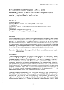
VOLUME - X ISSUE - LV JAN / FEB 2013
VOLUME - X
ISSUE - LV
JAN / FEB 2013
1 Editorial
Disease
2
Diagnosis
Trouble
5 Shooting
6 Bouquet
7 Interpretation
8 Tulip News
Chronic myelogenous (or myeloid) leukemia (CML), also known as chronic
granulocytic leukemia (CGL), is a cancer of the white blood cells. It is a form of leukemia
characterized by the increased and unregulated growth of predominantly myeloid cells in
the bone marrow and the accumulation of these cells in the blood. CML is a clonal bone
marrow stem cell disorder in which proliferation of mature granulocytes (neutrophils,
eosinophils, and basophils) and their precursors is the main finding. It is a type of
myeloproliferative disease associated with a characteristic chromosomal translocation
called the Philadelphia chromosome. CML is now largely treated with targeted drugs
called tyrosine kinase inhibitors (TKIs), such as Gleevec/Glivec (imatinib), Sprycel
(dasatinib), Tasigna (nilotinib), or Bosulif (bosutinib) which have led to dramatically
improved long term survival rates (95.2%) since the introduction of Gleevec in 2001.
These drugs have revolutionized treatment of this disease and allow most patients to have
a good quality of life when compared to the former chemotherapy drugs. The DISEASE
DIAGNOSIS segment of this issue delves deep into the clinico - diagnostic issues related
to CML.
At the request of our friends from a Central Asian Republic, this whole issue is dedicated to
the diagnosis of CML.
Thus, TROUBLESHOOTING section outlines “Leucocyte Alkaline Phosphatase” for you
and at the same time it briefly presents the special staining techniques as applied to
various haematologic malignancies. The process is explained in layman’s language.
The INTERPRETATION portion completes the CML diary by discussing Philadelphia
Chromosome in ample detail.
Hereafter, never should we fumble while diagnosing CML, it should be as easy as
reporting iron deficiency anaemia.
is now almost a decade old. For the tenth time hence, we wish you
A VERY HAPPY PROSPEROUS, PEACEFUL AND PLEASURABLE NEW YEAR.
1
JAN/FEB
Clinical History
The clinical manifestations of chronic myelogenous leukemia (CML) are
insidious. The disease is often discovered incidentally in the chronic phase,
when an elevated white blood cell (WBC) count is revealed by a routine
blood count or when an enlarged spleen is found on a general physical
examination. Nonspecific symptoms of fatigue and weight loss may occur
long after the onset of the disease. Loss of energy and decreased exercise
tolerance may occur during the chronic phase after several months. Patients
often have symptoms related to enlargement of the spleen, liver, or both. The
large spleen may encroach on the stomach and cause early satiety and
decreased food intake. Left upper quadrant abdominal pain described as
"gripping" may occur from spleen infarction. The enlarged spleen may also
be associated with a hypermetabolic state, fever, weight loss, and chronic
fatigue. The enlarged liver may contribute to the patient's weight loss. Some
patients with CML have low-grade fever and excessive sweating related to
hypermetabolism. In some patients who present in the accelerated, or acute,
leukemia phase of the disease (skipping the chronic phase), bleeding,
petechiae, and ecchymoses may be the prominent symptoms. In these
situations, fever is usually associated with infections. Bone pain and fever,
as well as an increase in bone marrow fibrosis, are harbingers of the blast
phase.
DISEASE DIAGNOSIS
CHRONIC MYELOID LEUKEMIA
Background
Chronic myelogenous leukemia (CML), also known as chronic myeloid
leukemia, is a myeloproliferative disorder characterized by increased
proliferation of the granulocytic cell line without the loss of their capacity to
differentiate. Consequently, the peripheral blood cell profile shows an
increased number of granulocytes and their immature precursors, including
occasional blast cells. CML is one of the few cancers known to be caused by
a single, specific genetic mutation. More than 90% of cases result from a
cytogenetic aberration known as the Philadelphia chromosome. CML
progresses through 3 phases: chronic, accelerated, and blast. In the chronic
phase of disease, mature cells proliferate; in the accelerated phase,
additional cytogenetic abnormalities occur; in the blast phase, immature
cells rapidly proliferate. Approximately 85% of patients are diagnosed in the
chronic phase and then progress to the accelerated and blast phases after 35 years. The diagnosis of CML is based on the histopathologic findings in the
peripheral blood and the Philadelphia chromosome in bone marrow cells.
CML accounts for 20% of all leukemias affecting adults. It typically affects
middle-aged individuals. Uncommonly, the disease occurs in younger
individuals. Younger patients may present with a more aggressive form of
CML, such as in accelerated phase or blast crisis. Uncommonly, CML may
appear as a disease of new onset in elderly individuals. The goals of
treatment are to achieve hematologic, cytogenetic, and molecular
remission. Although a variety of medications have been used in CML,
including myelosuppressive agents and interferon alfa, the tyrosine kinase
inhibitor imatinib mesylate is currently the agent of choice, and other drugs in
this category are playing increasingly important roles. However, allogeneic
bone marrow transplantation is currently the only proven cure for CML.
Physical Examination
Splenomegaly is the most common physical finding in patients with chronic
myelogenous leukemia (CML). In more than 50% of the patients with CML,
the spleen extends more than 5 cm below the left costal margin at time of
discovery. The size of the spleen correlates with the peripheral blood
granulocyte counts, with the largest spleens being observed in patients with
high WBC counts. A very large spleen is usually a harbinger of the
transformation into an acute blast crisis form of the disease. Hepatomegaly
also occurs, although less commonly than splenomegaly. Hepatomegaly is
usually part of the extramedullary hematopoiesis occurring in the spleen.
Physical findings of leukostasis and hyperviscosity can occur in some
patients, with extraordinary elevation of their WBC counts, exceeding
300,000-600,000 cells/ìL. Upon funduscopy, the retina may show
papilledema, venous obstruction, and hemorrhages. The blast crisis is
marked by an increase in the bone marrow or peripheral blood blast count or
by the development of soft-tissue or skin leukemic infiltrates. Typical
symptoms are due to increasing anemia, thrombocytopenia, basophilia, a
rapidly enlarging spleen, and failure of the usual medications to control
leukocytosis and splenomegaly.
Pathophysiology
CML is an acquired abnormality that involves the hematopoietic stem cell. It
is characterized by a cytogenetic aberration consisting of a reciprocal
translocation between the long arms of chromosomes 22 and 9 [t(9;22)]. The
translocation results in a shortened chromosome 22, an observation first
described by Nowell and Hungerford and subsequently termed the
Philadelphia (Ph1) chromosome after the city of discovery. The Philadelphia
chromosome, which is a diagnostic karyotypic abnormality for chronic
myelogenous leukemia, is
shown in this picture of the
banded chromosomes 9 and
22. Shown is the result of the
reciprocal translocation of
22q to the lower arm of 9 and
9q (c-abl to a specific
breakpoint cluster region
[bcr] of chromosome 22
indicated by the arrows). This translocation relocates an oncogene called
ABL from the long arm of chromosome 9 to a specific breakpoint cluster
region (BCR) in the long arm of chromosome 22. The ABL oncogene
encodes a tyrosine protein kinase. The resulting BCR/ABL fusion gene
encodes a chimeric protein with strong tyrosine kinase activity. The
expression of this protein leads to the development of the CML phenotype,
through processes that are not yet fully understood. The presence of
BCR/ABL rearrangement is the hallmark of CML, although this
rearrangement has also been described in other diseases. It is considered
diagnostic when present in a patient with clinical manifestations of CML. The
initiating factor of CML is still unknown, but exposure to ionizing radiation has
been implicated, as observed in the increased prevalence among survivors
of the atomic bombing of Hiroshima and Nagasaki. Other agents, such as
benzene, are possible causes.
Diagnostic Considerations
Problems to be considered include the following: Acute myeloid leukemia,
Chronic myelomonocytic leukemia, Chronic neutrophilic leukemia,
Thrombocythemia, Leukemoid reactions from infections (chronic
granulomatous [eg, tuberculosis]), Tumor necrosis.
Differential Diagnoses
Agnogenic Myeloid Metaplasia with Myelofibrosis, Essential
Thrombocytosis, Myelodysplastic Syndrome, Myeloproliferative Disease,
Polycythemia Vera.
Approach Considerations
The workup for chronic myelogenous leukemia (CML) consists of a
complete blood count with differential, peripheral blood smear, and bone
marrow analysis. Although typical hepatomegaly and splenomegaly may be
imaged by using a liver/spleen scan, these abnormalities are often so
obvious clinically that radiologic imaging is not necessary. The diagnosis of
CML is based on the histopathologic findings in the peripheral blood and the
Philadelphia (Ph1) chromosome in bone marrow cells. Other laboratory
abnormalities include hyperuricemia, which is a reflection of high bone
marrow cellular turnover, and markedly elevated serum vitamin B12–binding protein (TC-I). The latter is synthesized by the granulocytes and
reflects the degree of leukocytosis.
2
JAN/FEB
JAN/FEB
3
JAN/FEB
JAN/FEB
4
JAN/FEB
JAN/FEB
What if one suspects CML but doesn’t have access to a cytogenetic or
molecular lab (to look for the Philadelphia chromosome or the bcr-abl
translocation)? Well, first you’d look for all the morphologic clues you could.
CML usually presents with a marked leukocytosis (the WBC is often over
100,000), with a left shift all the way back to myeloblasts (though there are
relatively few myeloblasts around). A benign left shift usually presents with a
mild to moderate leukocytosis (the neutrophil count is often just above
normal; it’s generally nowhere near the magnitude often seen in CML), and
the neutrophils are shifted back to the metamyelocyte or myelocyte stages
(you’ll very rarely see promyelocytes, and you’ll virtually never see
myeloblasts). Also, CML tends to have a “bulge” at the metamyelocyte
stage, whereas a benign left shift does not (the cells are more or less present
in decreasing amounts by stage of maturation, i.e., there are more
segmented than band neutrophils, more bands than metamyelocytes, more
metamyelocytes than myelocytes, more myelocytes than promyelocytes
and blasts are basically nonexistent). Finally, CML has a basophilia,
whereas a benign left shift does not. But if you wanted more proof that your
case was CML, you could do a leukocyte (or neutrophil) alkaline
phosphatase (LAP). This test is not done as much as it used to be, because
now everyone goes right to cytogenetics or molecular testing in order to find
the Philadelphia chromosome or the bcr-abl translocation. But it’s still a good
test, and it would be a good thing to do if you couldn’t look for the Philadelphia
chromosome. Here’s the principle behind the test. LAP is an enzyme present
in normal neutrophils, but absent (or present at very low concentrations) in
malignant neutrophils (i.e., the ones in CML). So if you have a whole bunch
of neutrophils around, and the LAP is strongly positive in those cells, as in the
top image, you can be quite sure that it is a benign bunch of neutrophils.
However, if the LAP is negative, or only weakly positive, as in the bottom
image, that probably means that those neutrophils are malignant and that
you’re dealing with a case of CML. You’d still want to send off a blood or bone
marrow specimen to a cytogenetics and/or molecular diagnostics lab, but in
the meantime, the LAP can help you quickly assess and triage your patient.
Classification of acute leukemias by morphologic and cytochemical
criteria (modified from Löffler)
Stem cell leukemia
peroxidase
0
periodic acid Schiff
0
undifferentiated type
alpha-napthylacetate esterase
0
Myeloblastic leukemia (AML)
peroxidase
(-)
periodic acid Schiff
0-(+) Peroxidase type 1 and 2
alpha-napthylacetate esterase
(+)
Promyelocytic leukemia
peroxidase
+
periodic acid Schiff
(+)
Peroxidase type 3
alpha-napthylacetate esterase
(+)
Myelomonocytic leukemia
peroxidase
+
periodic acid Schiff
(+)
Peroxidase esterase type
alpha-napthylacetate esterase
+
Monocytic leukemia
peroxidase
(+)
periodic acid Schiff
0-(+) Esterase type
alpha-napthylacetate esterase
+
Lymphoblastic leukemia (ALL)
preoxidase
0
periodic acid Schiff
+
periodic acid Schiff type
alpha-naphthylacetate esterase
0
0 = negative; (+) = weakly positive < 25%; + = strongly positive > 50
TROUBLESHOOTING
LEUCOCYTE ALKALINE PHOSPHATASE
How the Test is Performed
Blood is typically drawn from a vein, usually from the inside of the elbow or
the back of the hand. The site is cleaned with germ-killing medicine
(antiseptic). The health care provider wraps an elastic band around the
upper arm to apply pressure to the area and make the vein swell with blood.
Next, the health care provider gently inserts a needle into the vein. The blood
collects into an airtight vial or tube attached to the needle. The elastic band is
removed from your arm. Once the blood has been collected, the needle is
removed, and the puncture site is covered to stop any bleeding. In infants or
young children, a sharp tool called a lancet may be used to puncture the skin
and make it bleed. The blood collects into a small glass tube called a pipette,
or onto a slide or test strip. A bandage may be placed over the area if there is
any bleeding. A laboratory specialist separates the white blood cells from the
rest of the blood sample and watches to see if any substances stick to
specific colored dyes. Substances that contain phosphate, such as ALP,
attach to certain colored dyes.
How to Prepare for the Test
One should not eat or drink for 6 hours before the test. Certain medicines
may affect the test results. Your health care provider may tell you to stop
taking such medications. These medications include: Allopurinol,
Androgens, Anti-inflammatory medicines, Birth control pills, Certain
antibiotics, Certain arthritis drugs, Certain diabetes drugs (taken by mouth),
Chlorpromazine, Cortisone, Methyldopa, Narcotics, Propranolol,
Tranquilizers, Tricyclic antidepressants. No medicine should be stopped
without medical advice.
Why the Test is Performed
ALP is found in different forms throughout the body. This test is done to
confirm a number of different medical conditions, including certain types of
anemia and leukemia. It may also be done if a person has an increase in
platelet levels in the blood.
Normal Results
A staining score of 20 - 100 (out of a maximum of 400) is considered normal.
Note: Normal value ranges may vary slightly among different laboratories.
Talk to your doctor about the meaning of your specific test results. The
examples above show the common measurements for results for these
tests. Some laboratories use different measurements or may test different
specimens.
What Abnormal Results Mean
Higher-than-normal results may be due to: Essential thrombocytosis,
Leukemoid reaction, Myelofibrosis, Polycythemia vera.
Lower-than-normal results may be due to: Aplastic anemia, Chronic
myeloid leukemia, Pernicious anemia.
Leukocyte alkaline phosphatase
STRONG LAP POSITIVE
NEUTROPHIL
LAP NEGATIVE
NEUTROPHIL
5
JAN/FEB
JAN/FEB
Acute eosinophilic leukemia is diagnosed by examination of the bone
marrow, since eosinophils usually are not increased in the peripheral blood.
The predominant marrow cells are abnormal eosinophils (immature,
pleomorphic forms, some with coarse, dark-blue granules, cytoplasmic
vacuoles, distinct nucleoli). Diagnosis relies on the cytochemical detection
of naphthol-AS-D-chloracetate esterase in the granules. Auer rods are
unusual. Acute basophilic leukemia is evidenced by an extreme increase in
the basophilic granulated cells of granulocytopoiesis. The granules are very
atypical (large, coarse, hyperchromic), and Auer rods may be present The
diagnosis is confirmed by the metachromatic reaction to toluidine blue in the
cells. Four forms are recognized: (1) basophilic blast crises in CML, (2)
promyelocytic basophilic type, (3) histiobasophilic type, (4) basophiliceosinophilic type. It is very difficult to establish a diagnosis of acute
megakaryoblastic leukemia, designated M7 in the FAB classification of
acute leukemias. The blast cells in the peripheral blood and bone marrow
present a variety of morphologies. They may appear as small cells with a
narrow cytoplasmic border and dense chromatin, resembling lymphoblasts
(L1), or they may resemble L2 cells with or without granules. The nuclei are
round, finely reticular, and have one to three prominent nucleoli. The cells
vary greatly in size and may be two to three times larger than normal
lymphocytes. Sometimes one finds cytoplasmic vesicles or differentiated
megakaryocytes with adjacent platelets or bare nuclei nested in clusters of
platelets. Occasional megakaryocytic nuclei are found in the peripheral
blood. It is often difficult to obtain a bone marrow specimen by aspiration,
and marrow biopsy is usually indicated. Cytochemical methods may
contribute to the diagnosis. The Sudan black and peroxidase reaction are
negative. The monocytes can be a source of confusion. Often they are
positive for alpha-napthylesterase and naphthyl-ASD-acetate esterase, in
which case these enzymes canbe inhibited by fluoride. While the monocytes
usually show a diffuse positivity for these esterase enzymes, the reaction in
megakaryoblasts tends to be localized. PAS and acid phosphatase positivity
are also localized. A platelet peroxidase occuring on a nuclear membrane
and in the endoplasmic reticulum of megakaryoblasts can distinquish these
cells from myeloblasts on electron microscope examination. This can also
be accomplished by the use of monoclonal or polyclonal platelet-specific
antibodies.
Wisdom Whispers
BOUQUET
Always Look On The Bright Side
A pessimist sees the difficulty in every opportunity; an optimist sees opportunity
in every difficulty.
Serenity
The real sign of serenity is not seen so much in the face, as found in the depth and
stillness of the eyes.
Notice the Difference
Replace the words "I have to" with "I choose to" and notice the difference in how
you feel.
Courage
Courage is taking a step forward into an area of difficulty without a solution in
mind, trusting that whatever help you need will become available.
Self-Respect
Experience the bliss of Self-Respect and give respect to others at all times.
When I am prejudiced against another, my narrow vision and small heart lower
my self-dignity and self-worth.
Balance
Maintain the balance of responding to situations with a cool head and to people
with a warm heart.
Pure and Gentle
Success is merged in every step of those who have a pure and gentle nature.
The Happiness You Give
The happiness you give makes you more happy than the happiness you receive.
Lotus Life
The lotus is a symbol of purity.
Its roots are in the mud, but the flower remains above dirty water.
Live a lotus life. Be in the world, but unaffected by impurities.
Determination
Determination brings the strength to continue, the steadiness to succeed,
and the wisdom to slip past difficulties undisturbed.
In Lighter Vein
--NO SPEAKAH DE ENGLISH
A bus stops and 2 Italian men get on. They sat down and engage in an
animated conversation. The lady sitting next to them ignores them at first, but
her attention is galvanized when she hears one of them say the following:
Emma come first.
Den I come.
Den two asses come together.
I come once-a-more!
Two asses, they come together again.
I come again and pee twice.
Then I come one lasta time.'
The lady can't take this anymore, "You foul- mouthed sex obsessed pig!" She
retorted indignantly. 'In this country, we don't speak aloud in public places
about our sex lives!"
'Hey, coola down lady,' said the man, 'Whooza talkin' about sex, I'm a justa
tellin' my frienda how to spell ' Mississippi '...
--I asked my new girlfriend what sort of books she's interested in. She said:
Cheque books.
-- What's the difference between a good lawyer and a great lawyer?
A good lawyer knows the law. A great lawyer knows the judge.
-- Nurse: A beautiful woman who holds your hand for one full minute and then
expects your pulse to be normal.
-- A Blonde enters kitchen, opens sugar container, looks inside and
closes it. She does this again and again.
Why? Because his Doctor told her to check the sugar level regularly.
Diagnose the following haematologic malignancies from their peripheral smear images
1
2
3
4
Answers: 1 – Acute Myeloblastic Leukemia 2. – Plasma Cell Leukemia 3. – Chronic Lymphocytic Leukemia 4. – Chronic Myelocytic Leukemia.
Brain Teasers
6
JAN/FEB
JAN/FEB
important drugs against a variety of cancers including CML, renal cell
carcinoma (RCC) and gastrointestinal stromal tumors (GISTs). The fused
BCR-Abl protein interacts with the interleukin-3 receptor beta(c) subunit.
The ABL tyrosine kinase activity of BCR-Abl is elevated relative to wild-type
ABL. Since ABL activates a number of cell cycle-controlling proteins and
enzymes, the result of the BCR-Abl fusion is to speed up cell division.
Moreover, it inhibits DNA repair, causing genomic instability and potentially
causing the feared blast crisis in CML.
Nomenclature
Philadelphia chromosome is designated Ph (or Ph') chromosome and the
translocation is termed t(9;22) (q34.1;q11.2).
Therapy
Tyrosine kinase inhibitors
INTERPRETATION
Ph CHROMOSOME (Philadelphia chromosome)
Philadelphia chromosome or Philadelphia translocation is a specific
chromosomal abnormality that is associated with chronic myelogenous
leukemia (CML). It is the result of a reciprocal translocation between
chromosome 9 and 22, and is specifically designated t(9;22)(q34;q11). The
presence of this translocation is a highly sensitive test for CML, since 95% of
people with CML have this abnormality (the remainder have either a cryptic
translocation that is invisible on G-banded chromosome preparations, or a
variant translocation involving another chromosome or chromosomes as
well as the long arm of chromosomes 9 and 22). However, the presence of
the Philadelphia (Ph) chromosome is not sufficiently specific to diagnose
CML, since it is also found in acute lymphoblastic leukemia (ALL, 25–30% in
adult and 2–10% in pediatric cases) and occasionally in acute myelogenous
leukemia (AML).
History
The Philadelphia chromosome was first discovered and described in 1960
by Peter Nowell from University of Pennsylvania School of Medicine and
David Hungerford from the Fox Chase Cancer Center's Institute for Cancer
Research and was therefore named after the city in which both facilities are
located. In 1973, Janet D. Rowley at the University of Chicago identified the
mechanism by which the Philadelphia chromosome arises as a
translocation.
Molecular biology
Crystal structure of Abl kinase domain (blue) in complex with 2nd
generation tyrosine kinase inhibitor (TKI) nilotinib (red)
Main article: Bcr-Abl tyrosine-kinase inhibitor
In the late 1990s, STI-571 (imatinib, Gleevec/Glivec) was identified by the
pharmaceutical company Novartis (then known as Ciba Geigy) in highthroughput screens for tyrosine kinase inhibitors. Subsequent clinical trials
led by Dr. Brian J. Druker at Oregon Health & Science University in
collaboration with Dr. Charles Sawyers and Dr. Moshe Talpaz demonstrated
that STI-571 inhibits proliferation of BCR-ABL-expressing hematopoietic
cells. Although it did not eradicate CML cells, it did greatly limit the growth of
the tumor clone and decreased the risk of the feared "blast crisis". In 2000
John Kuriyan determined the mechanism by which STI-571 inhibits the Abl
kinase domain. It was marketed in 2001 by Novartis as imatinib mesylate.
Other pharmacological inhibitors are being developed, which are more
potent and/or are active against the emerging Gleevec/Glivec resistant
BCR-abl clones in treated patients. The majority of these resistant clones
are point-mutations in the kinase of BCR-abl. New inhibitors include
dasatinib and nilotinib, which are significantly more potent than imatinib and
may overcome resistance. Treatment of pediatric Ph+ ALL with a
combination of standard chemotherapy and RTK inhibitors may result in
remission, but the curative potential is unknown.
Blood or marrow transplants
COG study AALL 0031, which examines the use of Gleevec with standard
chemotherapeutic regimens and bone marrow transplant from HLAmatched related donors for high risk ALL (including Ph+ ALL), has
concluded, and findings will be published in the near future. A potentially
curative, but risky, option for pediatric Ph+ ALL or Ph+ CML is bone marrow
transplant or cord blood transplant, but chemotherapy is favored by some for
achieving first remission (CR1). For some, bone marrow transplant from a
matched sibling donor or a matched, unrelated donor may be favored when
remission is obtained. Cord blood transplant is favored by some when a
10/10 bone marrow match is not available, and cord blood transplant may
have some advantages, including a reduced incidence of graft-vs-host
disease (GVHD), which is a common and significant complication of
transplant. However, transplant with cord blood sometimes requires longer
periods of time for engraftment, which may increase the potential for
complications due to infection. Regardless of the type of transplant,
transplant-related mortality and relapse are possible, and the rates may
change as treatment protocols improve. For second remission (CR2), if
achieved, both chemotherapy and transplant options are possible, and
many physicians prefer transplant.
Schematic representation of formation of the Philadelphia Chromosome
The exact chromosomal defect in Philadelphia chromosome is a
translocation, in which parts of two chromosomes, 9 and 22, swap places.
The result is that a fusion gene is created by juxtapositioning the Abl1 gene
on chromosome 9 (region q34) to a part of the BCR ("breakpoint cluster
region") gene on chromosome 22 (region q11). This is a reciprocal
translocation, creating an elongated chromosome 9 (der 9), and a truncated
chromosome 22 (the Philadelphia chromosome). In agreement with the
International System for Human Cytogenetic Nomenclature (ISCN), this
chromosomal translocation is designated as t(9;22)(q34;q11). Abl stands for
"Abelson", the name of a leukemia virus which carries a similar protein. The
result of the translocation is the oncogenic BCR-ABL gene fusion, located on
the shorter derivative 22 chromosome. This gene encodes the Bcr-abl fusion
protein. Depending on the precise location of the fusion the molecular weight
of the protein can range from 185 to 210 kDa. For this reason bcr-abl is
sometimes called p210 or p185. Three clinically important variants are the
p190, p210 and p230 isoforms. p190 is generally associated with acute
lymphoblastic leukemia (ALL), while p210 is generally associated with
chronic myeloid leukemia but can also be associated with ALL. p230 is
usually associated with chronic neutrophilic leukemia. Additionally, the p190
isoform can also be expressed as a splice variant of p210. Because the Abl
gene expresses a membrane-associated protein, a tyrosine kinase, the
BCR-Abl transcript is also translated into a tyrosine kinase, adding a
phosphate group to tyrosine. Although the BCR region also expresses
serine/threonine kinases, the tyrosine kinase function is very relevant for
drug therapy. Tyrosine kinase inhibitors (such as imatinib and sunitinib) are
7
JAN/FEB
JAN/FEB
TULIP NEWS
Printed and published by D.G. Tripathi, Edited by Dr. Ramnik Sood, M.D. (Path.) for and on behalf of Tulip Diagnostics Private Ltd., Gitanjali, Tulip Block, Dr. Antonio Do Rego Bagh, Alto Santacruz, Bambolim Complex Post Office, Goa - 403 202, INDIA.
Fax: (0832) 2458544. E-mail: [email protected] Website: www.tulipgroup.com
JAN/FEB


© Copyright 2026